Send Orders for Reprints to [email protected] Current Medicinal Chemistry, 2014, 21, 3775-3784 3775 Nutritional Iron Deficiency: The Role of Oral Iron Supplementation J.I. Lachowicz* ,a , V.M. Nurchi a , D. Fanni b , C. Gerosa b , M. Peana c and M.A. Zoroddu c a Dipartimento di Scienze Chimiche e Geologiche, University of Cagliari, Cittadella Universitaria, I-09042 Monserrato, Italy; b Dipartimento di Citomorfologia, Sez. Anatomia Patologica, University of Cagliari, Via Ospedale 46, I-09124 Cagliari, Italy; c Dipartimento di Chimica e Farmacia, University of Sassari, Via Vienna 2, I-07100 Sassari, Italy Abstract: Nutritional iron deficiency represents a relevant health problem mainly in developing countries. Children and pregnant women represent the main target of this disease, and the low amount of bio-available iron mostly depends on plant-based diets. Iron deficiency may have serious consequences, with severe impairment of the immune function leading to infectious diseases. The brain development in embryos and fetuses during gestation can be greatly affected by iron de- ficiency of the mother with heavy outcomes on the cognition status of children. A better understanding of molecular pathways involved in iron absorption and metabolism are the basis for new strategies for developing a therapy for iron de- ficiency. Different therapeutic strategies are summarized, and iron fortification appears the best tool. Keywords: Iron deficiency, iron metabolism, iron absorption, iron supplement, iron-fortified food. 1. INTRODUCTION Iron deficiency represents an important clinical problem for pediatricians as well as for gynecologists and geriatrics, as young children, women, and older people are the groups with the highest possibility of iron deficiency. Iron- deficiency anemia is characterized by a lower-than-normal red blood cell count, or when a correct count does not in- clude an adequate amount of hemoglobin (the iron-protein devoted to the transport of oxygen from lungs to other tis- sues). Iron-deficiency anemia usually develops over time, when the intestines do not absorb enough iron to carry to bone marrow hematopoietic cells and build healthy red blood cells. This leads to fatigue, chest pain, shortness of breath, and other minor symptoms. Severe iron-deficiency anemia can retard growth and development in children, lead to heart problems, infections, and a number of other complications. Iron deficiency is considered one of the most important risk factors of death and disability among some two billion people, mainly children and young adults in developing countries [1]. In infancy, insufficient iron intake has severe complications in brain development, resulting not only in diminished school performance [2], but also in cognitive limitations that, in many cases, persist into adulthood [3]. The fundamental role of iron in the immune system is con- firmed by the higher frequency of infections in children af- fected by iron-deficiency-related anemia, in whom T- lymphocytes are about 20% reduced, and cell-mediated im- mune response is decreased [4]. This paper reviews the most important phases of iron me- tabolism in humans. We report the complex pathogenesis of anemia related to iron deficiency, and analyze the clinical *Address correspondence to this author at the Dipartimento di Scienze Chi- miche e Geologiche, University of Cagliari, Cittadella Universitaria, I- 09042 Monserrato, Italy; Tel: +39 070 675 4471; Fax: +39 070 675 4478; E-mail: [email protected] consequences of iron deficiency. The final part of this review is dedicated to iron deficiency therapy. We directed particu- lar attention to iron supplementation and fortification of foods, promising, effective strategies that could ameliorate the lack of dietary iron and reduce iron deficiency, particu- larly in developing countries. 2. IRON IN HUMAN METABOLISM Iron Daily Needs The “recommended daily allowance” (RDA) for adult males is estimated at around 10 mg of iron per day. Consid- ering an assimilation of 10%, this amount results in a net uptake of 1.0 mg iron per day, an amount adequate to restore daily iron losses [5], and fulfill the daily necessities [6]. RDA values differ greatly by gender, and change markedly with age. The RDA values for males and females are re- ported in (Table 1) as a function of age, and are relative to US and UK regulations [7], together with the values in preg- nancy and lactation. A male weighting 70 kg accumulates about 4 g of iron in his body during his growth. About 2.5 g of iron is found in hemoglobin and about 1 g in liver cells as ferritin or he- mosiderin. While men absorb and expel approximately 0.8 mg of iron per day, women absorb an almost double amount (1.4 mg per day) to cope with menstrual losses [8]. As such the RDA of iron for women aged 19 and >50 years has been set at 14.8 and 8.7 mg/day, respectively [7]. The iron requirements for pregnant women is 2-6 times greater than those of men [9]. Women in their fecund age need a nutritional supply of 15 mg iron per day. Since a typi- cal Western-type 1000 kcal diet contains about 6 mg iron, many young women often do not consume enough iron in their diets [10]. During pregnancy, iron consumption increases signifi- cantly. The body transfers approximately 15% of a mother’s /14 $58.00+.00 © 2014 Bentham Science Publishers

Welcome message from author
This document is posted to help you gain knowledge. Please leave a comment to let me know what you think about it! Share it to your friends and learn new things together.
Transcript
Send Orders for Reprints to [email protected] Current Medicinal Chemistry, 2014, 21, 3775-3784 3775
Nutritional Iron Deficiency: The Role of Oral Iron Supplementation
J.I. Lachowicz*,a, V.M. Nurchia, D. Fannib, C. Gerosab, M. Peanac and M.A. Zorodduc
aDipartimento di Scienze Chimiche e Geologiche, University of Cagliari, Cittadella Universitaria, I-09042 Monserrato, Italy; bDipartimento di Citomorfologia, Sez. Anatomia Patologica, University of Cagliari, Via Ospedale 46, I-09124 Cagliari, Italy; cDipartimento di Chimica e Farmacia, University of Sassari, Via Vienna 2, I-07100 Sassari, Italy
Abstract: Nutritional iron deficiency represents a relevant health problem mainly in developing countries. Children and pregnant women represent the main target of this disease, and the low amount of bio-available iron mostly depends on plant-based diets. Iron deficiency may have serious consequences, with severe impairment of the immune function leading to infectious diseases. The brain development in embryos and fetuses during gestation can be greatly affected by iron de-ficiency of the mother with heavy outcomes on the cognition status of children. A better understanding of molecular pathways involved in iron absorption and metabolism are the basis for new strategies for developing a therapy for iron de-ficiency. Different therapeutic strategies are summarized, and iron fortification appears the best tool.
Keywords: Iron deficiency, iron metabolism, iron absorption, iron supplement, iron-fortified food.
1. INTRODUCTION Iron deficiency represents an important clinical problem
for pediatricians as well as for gynecologists and geriatrics, as young children, women, and older people are the groups with the highest possibility of iron deficiency. Iron-deficiency anemia is characterized by a lower-than-normal red blood cell count, or when a correct count does not in-clude an adequate amount of hemoglobin (the iron-protein devoted to the transport of oxygen from lungs to other tis-sues). Iron-deficiency anemia usually develops over time, when the intestines do not absorb enough iron to carry to bone marrow hematopoietic cells and build healthy red blood cells. This leads to fatigue, chest pain, shortness of breath, and other minor symptoms. Severe iron-deficiency anemia can retard growth and development in children, lead to heart problems, infections, and a number of other complications.
Iron deficiency is considered one of the most important risk factors of death and disability among some two billion people, mainly children and young adults in developing countries [1]. In infancy, insufficient iron intake has severe complications in brain development, resulting not only in diminished school performance [2], but also in cognitive limitations that, in many cases, persist into adulthood [3]. The fundamental role of iron in the immune system is con-firmed by the higher frequency of infections in children af-fected by iron-deficiency-related anemia, in whom T-lymphocytes are about 20% reduced, and cell-mediated im-mune response is decreased [4].
This paper reviews the most important phases of iron me-tabolism in humans. We report the complex pathogenesis of anemia related to iron deficiency, and analyze the clinical *Address correspondence to this author at the Dipartimento di Scienze Chi-miche e Geologiche, University of Cagliari, Cittadella Universitaria, I-09042 Monserrato, Italy; Tel: +39 070 675 4471; Fax: +39 070 675 4478; E-mail: [email protected]
consequences of iron deficiency. The final part of this review is dedicated to iron deficiency therapy. We directed particu-lar attention to iron supplementation and fortification of foods, promising, effective strategies that could ameliorate the lack of dietary iron and reduce iron deficiency, particu-larly in developing countries.
2. IRON IN HUMAN METABOLISM Iron Daily Needs
The “recommended daily allowance” (RDA) for adult males is estimated at around 10 mg of iron per day. Consid-ering an assimilation of 10%, this amount results in a net uptake of 1.0 mg iron per day, an amount adequate to restore daily iron losses [5], and fulfill the daily necessities [6]. RDA values differ greatly by gender, and change markedly with age. The RDA values for males and females are re-ported in (Table 1) as a function of age, and are relative to US and UK regulations [7], together with the values in preg-nancy and lactation.
A male weighting 70 kg accumulates about 4 g of iron in his body during his growth. About 2.5 g of iron is found in hemoglobin and about 1 g in liver cells as ferritin or he-mosiderin. While men absorb and expel approximately 0.8 mg of iron per day, women absorb an almost double amount (1.4 mg per day) to cope with menstrual losses [8]. As such the RDA of iron for women aged 19 and >50 years has been set at 14.8 and 8.7 mg/day, respectively [7].
The iron requirements for pregnant women is 2-6 times greater than those of men [9]. Women in their fecund age need a nutritional supply of 15 mg iron per day. Since a typi-cal Western-type 1000 kcal diet contains about 6 mg iron, many young women often do not consume enough iron in their diets [10].
During pregnancy, iron consumption increases signifi-cantly. The body transfers approximately 15% of a mother’s
/14 $58.00+.00 © 2014 Bentham Science Publishers
3776 Current Medicinal Chemistry, 2014, Vol. 21, No. 33 Lachowicz et al.
bodily iron content to the fetus and placenta during preg-nancy. She further loses 1 mg of basal iron daily through cell desquamation, primarily in the gastrointestinal tract. Throughout gestation, 450 mg of iron are necessary for the development of red cell mass, while 270-300 mg iron are consumed by the fetus and 25-50 mg by the placenta [11]. The maternal RDA rises sharply during the 3rd trimester when the fetus requires 1.6 up to 2.0 mg iron per day [12]. As a consequence, consumption of 30 mg iron per day by pregnant women is recommended during the last three months [9]. In areas of the world where women are particu-larly at risk of iron deficiency, pregnant women require a supplement of 60 mg iron per day in the second half of ges-tation, as it is presently carried out in Indonesia [13].
The net iron requirement during pregnancy has been es-timated to be approximately 1 g (corresponding to the amount contained in 4 units of blood), mostly during the last two trimesters [6]. The recommended quantity of iron sup-plementation in mass programs did not change from the above-cited suggestions, apart from the pregnancy dose, which was changed to 60 mg/day. The effectiveness of iron absorption is enhanced in the cases of severe iron deficiency. An amount of 60 mg/day should constitute a convenient supply of iron to women who do not present a severe ane-mia, when there is time for a prolonged treatment. A greater dosage (120 mg/day) is suggested when supplementation in gestation is short [14].
A particular consideration should be given to adolescent girls, for whom a weekly supplement of 60 mg iron for 3-4 months (minimum) per year is suggested [13]. Newborns and infants grow quickly and, according to their body weight, have higher iron needs [15]; iron stores formed due to the high intrauterine iron supply are used in the first 4-6 months of postnatal life. In the following months, at normal nutri-tional iron intakes, children are at high risk for iron defi-ciency [16]. The Committee on Nutrition of the American
Academy of Pediatrics suggests an intake of 1 mg of iron per kg of body weight per day between the ages of 4 months and 3 years [17]. The American Academy of Pediatrics for Pre-mature Babies suggests the delivery of 2 mg of iron per kg per day as a supplement to breast milk during the first year of life. Pre-term newborns are at higher risk for iron defi-ciency compared to at-term newborns. 3-4 mg iron/(kg x day) has been recommended for pre-term newborns weigh-ing less than 1.5 Kg at birth [18, 19]. A 6-l2 month old infant normally absorbs approximately 0.1 mg of iron per kg of body weight per day [20]. During the first months of devel-opment, infants absorb approximately 0.5 mg of iron per day, which exceeds the amount normally lost by the body [8].
Iron Absorption Iron bioavailability is low and depends on the chemical
form of ingested iron and the presence of dietary components that may enhance or inhibit absorption [21]. Heme (organic) and non-heme (inorganic) iron each have a definite trans-porter [22]. The heme iron transporter (HCP) picks up heme iron, which withstands endocytosis and is released in the reduced form Fe2
! within the endosome (or lysosome). Non-heme iron consists of both ferrous and ferric forms. Ascorbic acid reduces Fe3
! to Fe2! in the intestinal lumen; membrane
ferri-reductases, which also comprise duodenal cytochrome B (DCYTB), perform the same reduction. Dietary ferric non-heme iron, which is the principal form of nutritional iron in the majority of populations, has to be reduced before absorp-tion by cytochrome B reductase 1 (CYBRD1) [23]. The pre-cise role of CYBRD1 is still unknown. Intestinal ferro-reductases, which contain cytochrome B, ferric/cupric reduc-tase19 and Steap2, may reduce iron [24]. DMT1 transports ferrous iron into enterocytes, accompanied by proton gradi-ent from the external to the internal side of intestinal cells. Dietary iron in the heme form derives mainly from meat
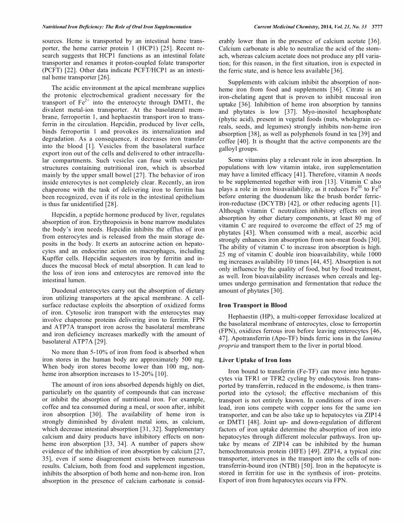
Table 1. Recommended daily allowances of iron, expressed as mg/day, for males and for females as a function of age, relative to UK [7] and US regulations [8], with values for pregnancy and lactation
United Kingdom [7] United States [8]
Age Males Females Pregnancy Age Males Females Pregnancy Lactation
0-6 month 0.27 0.27
<1 year 5.4 5.4 7-12 month 11 11
1-3 6.9 6.9 1-3 years 7 7
4-6 6.1 6.1 4-8 years 10 10
7-10 8.7 8.7 9-13 years 8 8
11-14 11.3 14.8 14-18 years 11 15 27 10
15-18 11.3 14.8 14.8 19-30 years 8 18 27 9
19-50 8.7 14.8 14.8 31-50 years 8 18 27 9
>50 8.7 8.7 14.8 50-70 years 8 8
>70 years 8 8
Nutritional Iron Deficiency: The Role of Oral Iron Supplementation Current Medicinal Chemistry, 2014, Vol. 21, No. 33 3777
sources. Heme is transported by an intestinal heme trans-porter, the heme carrier protein 1 (HCP1) [25]. Recent re-search suggests that HCP1 functions as an intestinal folate transporter and renames it proton-coupled folate transporter (PCFT) [22]. Other data indicate PCFT/HCP1 as an intesti-nal heme transporter [26].
The acidic environment at the apical membrane supplies the protonic electrochemical gradient necessary for the transport of Fe2+ into the enterocyte through DMT1, the divalent metal-ion transporter. At the basolateral mem-brane, ferroportin 1, and hephaestin transport iron to trans-ferrin in the circulation. Hepcidin, produced by liver cells, binds ferroportin 1 and provokes its internalization and degradation. As a consequence, it decreases iron transfer into the blood [1]. Vesicles from the basolateral surface export iron out of the cells and delivered to other intracellu-lar compartments. Such vesicles can fuse with vesicular structures containing nutritional iron, which is absorbed mainly by the upper small bowel [27]. The behavior of iron inside enterocytes is not completely clear. Recently, an iron chaperone with the task of delivering iron to ferritin has been recognized, even if its role in the intestinal epithelium is thus far unidentified [28].
Hepcidin, a peptide hormone produced by liver, regulates absorption of iron. Erythropoiesis in bone marrow modulates the body’s iron needs. Hepcidin inhibits the efflux of iron from enterocytes and is released from the main storage de-posits in the body. It exerts an autocrine action on hepato-cytes and an endocrine action on macrophages, including Kupffer cells. Hepcidin sequesters iron by ferritin and in-duces the mucosal block of metal absorption. It can lead to the loss of iron ions and enterocytes are removed into the intestinal lumen.
Duodenal enterocytes carry out the absorption of dietary iron utilizing transporters at the apical membrane. A cell-surface reductase exploits the absorption of oxidized forms of iron. Cytosolic iron transport with the enterocytes may involve chaperone proteins delivering iron to ferritin. FPN and ATP7A transport iron across the basolateral membrane and iron deficiency increases markedly with the amount of basolateral ATP7A [29].
No more than 5-10% of iron from food is absorbed when iron stores in the human body are approximately 500 mg. When body iron stores become lower than 100 mg, non-heme iron absorption increases to 15-20% [10].
The amount of iron ions absorbed depends highly on diet, particularly on the quantity of compounds that can increase or inhibit the absorption of nutritional iron. For example, coffee and tea consumed during a meal, or soon after, inhibit iron absorption [30]. The availability of heme iron is strongly diminished by divalent metal ions, as calcium, which decrease intestinal absorption [31, 32]. Supplementary calcium and dairy products have inhibitory effects on non-heme iron absorption [33, 34]. A number of papers show evidence of the inhibition of iron absorption by calcium [27, 35], even if some disagreement exists between numerous results. Calcium, both from food and supplement ingestion, inhibits the absorption of both heme and non-heme iron. Iron absorption in the presence of calcium carbonate is consid-
erably lower than in the presence of calcium acetate [36]. Calcium carbonate is able to neutralize the acid of the stom-ach, whereas calcium acetate does not produce any pH varia-tion; for this reason, in the first situation, iron is expected in the ferric state, and is hence less available [36].
Supplements with calcium inhibit the absorption of non-heme iron from food and supplements [36]. Citrate is an iron-chelating agent that is proven to inhibit mucosal iron uptake [36]. Inhibition of heme iron absorption by tannins and phytates is low [37]. Myo-inositol hexaphosphate (phytic acid), present in vegetal foods (nuts, wholegrain ce-reals, seeds, and legumes) strongly inhibits non-heme iron absorption [38], as well as polyphenols found in tea [39] and coffee [40]. It is thought that the active components are the galloyl groups.
Some vitamins play a relevant role in iron absorption. In populations with low vitamin intake, iron supplementation may have a limited efficacy [41]. Therefore, vitamin A needs to be supplemented together with iron [13]. Vitamin C also plays a role in iron bioavailability, as it reduces FeIII to FeII before entering the duodenum like the brush border ferric-iron-reductase (DCYTB) [42], or other reducing agents [1]. Although vitamin C neutralizes inhibitory effects on iron absorption by other dietary components, at least 80 mg of vitamin C are required to overcome the effect of 25 mg of phytates [43]. When consumed with a meal, ascorbic acid strongly enhances iron absorption from non-meat foods [30]. The ability of vitamin C to increase iron absorption is high. 25 mg of vitamin C double iron bioavailability, while 1000 mg increases availability 10 times [44, 45]. Absorption is not only influence by the quality of food, but by food treatment, as well. Iron bioavailability increases when cereals and leg-umes undergo germination and fermentation that reduce the amount of phytates [30].
Iron Transport in Blood Hephaestin (HP), a multi-copper ferroxidase localized at
the basolateral membrane of enterocytes, close to ferroportin (FPN), oxidizes ferrous iron before leaving enterocytes [46, 47]. Apotransferrin (Apo-TF) binds ferric ions in the lamina propria and transport them to the liver in portal blood.
Liver Uptake of Iron Ions Iron bound to transferrin (Fe-TF) can move into hepato-
cytes via TFR1 or TFR2 cycling by endocytosis. Iron trans-ported by transferrin, reduced in the endosome, is then trans-ported into the cytosol; the effective mechanism of this transport is not entirely known. In conditions of iron over-load, iron ions compete with copper ions for the same ion transporter, and can be also take up to hepatocytes via ZIP14 or DMT1 [48]. Joint up- and down-regulation of different factors of iron uptake determine the absorption of iron into hepatocytes through different molecular pathways. Iron up-take by means of ZIP14 can be inhibited by the human hemochromatosis protein (HFE) [49]. ZIP14, a typical zinc transporter, intervenes in the transport into the cells of non-transferrin-bound iron (NTBI) [50]. Iron in the hepatocyte is stored in ferritin for use in the synthesis of iron- proteins. Export of iron from hepatocytes occurs via FPN.
3778 Current Medicinal Chemistry, 2014, Vol. 21, No. 33 Lachowicz et al.
Distribution of Iron to Tissues The highest consumption of iron occurs in erythroid pre-
cursors of bone marrow, which need 20–25 mg daily in order to mature into erythrocytes. Erythroid cells receive iron only from transferrin bounded to TFR1 on the cell surface. The complex formed, Fe-TF/TFR1, is internalized into endosomes. Here, Steap3 reduces Fe3+ to Fe2+, which DMT1 then trans-ports into the cytosol. Erythroid cells in the stage of growth are the major iron-user cells in the body, and obtain iron to-tally through the Fe-TF/TFR1 cycle. Endosomal Steap3 favors the reduction of iron before its DMT1- mediated transport into the cytosol. Iron acquired by maturing erythroid cells goes to mitochondria, where it is utilized for the biosynthesis of heme and the assemblage of Fe-S cluster protein [51]. Mitochondrial iron transporter (Mitoferrin), necessary for mitochondrial iron assimilation, is an iron transporter situated on the inner mem-brane of mitochondria [52]. The iron chaperone Frataxin (FXN) transports iron to the site of the Fe-S cluster formation and the heme synthesis [53]. Eventually, Fe-S and heme are included into proteins containing complexes I-IV of the elec-tron transport chain. The synthesis of heme in growing erythroid cells consumes about 70% of all bodily iron.
Macrophages located in the spleen are extremely important in iron metabolism: they phagocyte senescent red blood cells and recover their iron. The phagolysosomes break down hemo-globin in red cells and further metabolize the remaining heme to release iron. The free iron can be exported, accumulated, or used by the cell. The iron attendance by macrophages has been carefully studied [54]. Macrophages of the reticuloendothelial system can assume iron from Fe-TF, but the major part of their iron is received by phagocytosis of matured erythrocytes.
Iron liberated from the protoporphyrin ring by heme oxygenase is used by the cell, accumulated in ferritin, or released from the cell by FPN. Natural resistance associated macrophage protein 1 (Nramp1), a transporter of divalent metal ions, is placed to the late phagolysosome of macro-phages, where it promotes recycling of iron ions [55]. RE macrophages constitute a great store of iron, which contain about half of the total iron in the body [54].
A consensus on the transport of iron from blood to the brain is lacking. The function of Fe-TF versus central nerv-ous system cells, regarding the presence of DMT1 in brain capillary endothelial cells, is still unknown [56-59]. It is thought that neurons obtain iron by means of TFR1, whereas glial cells might uptake iron ions via DMT1. Ferritin might be more abundant in glial cells than in neurons, suggesting a major role for glial cells in iron uptake and redistribution to other cell types in the brain. Plasma ferritin might also repre-sent a relevant source of brain iron [60].
All these data imply that DMT1 probably plays a major role in flow of iron ions from the central nervous system to blood across the blood brain barrier (BBB), whereas move-ment of iron from blood through the BBB towards neurons and glial cells might require TFR1 [61].
3. IRON DEFICIENCY Etiology
Iron deficiency constitutes one of the main global risk factors for disability and death for some 2 billion people [1].
Anemia represents one of the most important clinical conse-quences of iron deficiency, both in industrialized and in de-veloping countries. According to the WHO, 39% of children below 5 years, 48% of children between 5-14, 42% of all women, and 52% of pregnant women in developing coun-tries are anemic [62]. Iron deficiency is the major cause of anemia in developing countries, accounting for about 50% of total cases [63]. Surprisingly, iron deficiency is also a com-mon health problem in industrialized countries with 21% of British females between 11-18 being deficient [21]. In the USA, 29% of pregnant women in the third trimester have been found to be affected by iron deficiency anemia [64].
How to Treat Iron Deficiency Iron deficient anemia can usually be effectively managed.
The type of treatment derives from the cause of iron defi-ciency as well as its severity. Treatments consist of dietary changes, drugs, and surgery. The most severe cases may in-volve hospital treatments with blood transfusions and intra-venous iron therapy. In many circumstances, iron deficiency may be treated by dietary modifications or diversification, which is the most sustainable approach [1]. When dietary modifications are not sufficient to solve the problem, two other strategies may be utilized to increase ingestion of iron and its bioavailability: iron supplements (i.e. the supply of iron, often in high doses without food), and iron enriched foods [65]. Ferrous iron salts, including ferrous sulfate and ferrous gluconate, are generally preferred as oral supple-ments due to their low cost and high bioavailability. The chemical species of iron found in supplements is extremely important because it determines the therapeutic effects and the toxicity. For example, ferrous species in oral iron sup-plements are more bioavailable than ferric species, but are less tolerated and cause significant gastrointestinal distur-bances [66, 67]. Standard therapy for iron-deficiency ane-mia, even in pregnant women, is the ingestion of a 300-mg tablet of ferrous sulfate 3-4 times per day. Iron salt absorp-tion is better on an empty stomach, but side effects, includ-ing epigastric pain and nausea, may occur. The side effects of ferric salts are more severe, probably due to the compara-tively slower absorption of ferric iron compared to ferrous iron. Various ferrous and ferric impurities have been identi-fied in ferrous sulfate and ferrous fumarate based drugs [68]. In mammals, iron is mainly absorbed by the epithelium of the proximal small intestine, mainly the duodenum, which absorbs ferrous iron better than ferric [42].
Even if oral iron is the preferred treatment for iron defi-ciency anemia, intravenous administration of iron is indis-pensable in some clinical settings, particularly when severe anemia affects pregnant women [69]. While oral iron sup-plements are efficacious, their effectiveness is highly de-pendent on patient compliance. Poor compliance, dependent on the length of the treatment and on related gastrointestinal side-effects, is the most frequent cause of the inefficacy of oral supplements [70]. The patient’s iron stores would not be reloaded by oral treatments if they do not continue oral iron ingestion for 3-6 months after reestablishing standard hema-tological values [69]. Iron stores are reloaded with greater efficacy with parenteral iron [71], though there is often no other advantage to parenteral administration [71]. Parenteral iron is the only effective therapy to supply a sufficient
Nutritional Iron Deficiency: The Role of Oral Iron Supplementation Current Medicinal Chemistry, 2014, Vol. 21, No. 33 3779
amount of iron for erythropoiesis in cases of severe anemia [72, 73]. Moreover, in cases of severe anemia in pregnant women, oral treatments do not stimulate erythropoiesis rap-idly and consistently for clinical use [74]. Simply taking iron tablets incorrectly with meal is sufficient to cause the failure of the therapeutic strategy, as food reduces the absorption of therapeutic iron by approximately two-thirds [75]. Guidelines published in 1997 (now extensively accepted) encourage the regular use of intravenous treatments rather than oral iron supplements [76].
When choosing a strategy for iron deficiency therapy, physicians must remember that the amount of iron adminis-tered, not its form, is the main cause of any undesirable side effects [77-79]. Cost is also a factor since the intravenous treatments are far more costly than oral supplementation [36]. Weekly iron supplement treatment should be consid-ered as an alternative to daily treatments. This diminishes costs and limits the effects of other micronutrients (such as zinc) on iron absorption [80, 81]. It also decreases the risk of iron overload, enhances the compliance of patients, and ul-timately reduces the participation of health operators. Results indicate that the variations of hemoglobin in weekly patients are similar to those in patients treated daily. Taking vitamin A together with iron supplements improves counts more than taking iron alone.
Daily iron supplements may negatively affect the bodily zinc status, so zinc must be included in supplement treat-ments. Thus, a supplement regimen of iron, vitamin A, and zinc is more effective than unaided iron supplementation in ameliorating the status of micronutrients. Ideally, a supple-ment for children should include 15 mg iron, 15 mg zinc, and 350 !g vitamin A [13].
Iron supplementation strategies have to consider numer-ous factors: the iron complex formation with food ligands and physiological factors on one side, and social factors, cost, and achievability on the other [82]. Readily available iron supplementation that does not create gastrointestinal discomfort during digestion is greatly needed [36].
Total iron content, type of iron, and the occurrence of particular dietary enhancing and inhibiting factors influence iron bioavailability and availability for use and storage [83]. As a result of heme iron and an unknown enhancing factor in meat, diets containing meat favor iron bioavailability more than meat-free diets with an equal amount of iron [84]. On this basis, a greater percentage of iron is absorbed or con-served from diets containing meat [85]. Many iron supplements of varying quality are presently used. The qual-ity of a supplement together with its marketing determines its success with consumers. Iron supplement programs for preg-nant women normally suggest reasonably priced, easily transported and stored tablets. UNICEF manufactured a tab-let containing 60 mg iron (as FeSO4) with 250 !g folic acid (later increased to 400 !g). A liquid supplement, which can be directly dropped into the mouth, is necessary for children younger than 2 years, even if a powder or tablets can be eas-ily crushed and mixed with infant foods. The higher price of liquid formulations must be weighed against their easier utilization, which determines a greater compliance by users [14].
Iron Fortification Iron fortified food is one of the main strategies for pre-
venting iron deficiency and is the most sustainable, conven-ient, and cost-effective tool for its management [86]. How-ever, many problems exist related to the ability of iron ions to react with a number of components of foods. Iron pro-duces changes in color, flavor, and oxidation of fats [87]. In order to avoid such undesired sensory changes in fortified food, the less soluble iron compound should be used instead of the most bioavailable water-soluble iron compounds [88]. The fortification of wheat flour is a rather simple procedure. A number of foods have been effectively fortified with iron, including salt, sugar, curry powder, and fish sauce. Milk, dried milk, and various milk-derived foods have been forti-fied with iron in South America. Iron-fortified baby food is a great tool to effectively control iron deficiency anemia [30].
A variety of bioavailable iron compounds, inorganic and organic, are used to fortify drinking water that can be acces-sible at low price with little effort to communities. Drinking water represents a valuable and economical tool to provide iron in the diet [89].
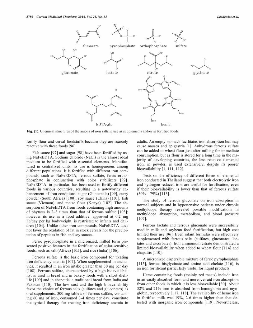
Babies accumulate approximately 250 mg of iron throughout gestation, which are then utilized during breast-feeding, since breast milk can only supply 0.15 mg of the daily absorbed iron against a daily necessity of 0.55 mg [90]. A premature utilization of cows’ milk can also contribute to iron deficiency in early childhood [91]. An Indian study on the control and prevention of anemia with iron-fortified foods presented an iron intake of 10-15 mg per day using iron-fortified salt (NaCl) [92]. Further 5 mg per day were supplied with fortified sugar, and 8-10 mg iron per day with fortified rice [93]. Four studies on infants utilizing iron-fortified foods have shown the absence of significant side effects; iron appreciably protects infants from infectious dis-eases, and particularly from respiratory tract infections [94]. In the following, the characteristics of the most-used iron supplements are presented. The chemical structures of the anions of these salts are shown in (Fig. 1).
Literature results indicate that ferrous sulfate, ferrous gluconate, and ferrous fumarate are equal as regards iron availability. According to FDA requirements, less than 2% of ferric iron should be contained in ferrous fumarate [95]. Ascorbic acid added to ferrous sulfate increases iron avail-ability. The availability of the polysaccharide-iron complex was found to be considerably lower than that of all the other iron compounds [36].
Food utilization can differ considerably, day to day, as well as by person. Furthermore, large quantities of ferrous salts may modify the color and taste of food, while iron-phosphates are inadequately absorbed. However, consump-tion of iron-fortified milk (12 mg/L) is adequate to preserve a normal state of iron that cannot be improved by further iron administration (6 mg iron/kg body weight for 3 months) [82]. A micronized, soluble ferric pyrophosphate [82], fer-rous bisglycinate, and ferrous complex with the amino acid glycine, are three iron fortificants of great utility in liquid products. The bioavailability of ferric orthophosphates is lower than that that of ferrous sulfate, ranging between 5% and 60%. Nevertheless, they are largely used in the USA to
3780 Current Medicinal Chemistry, 2014, Vol. 21, No. 33 Lachowicz et al.
fortify flour and cereal foodstuffs because they are scarcely reactive with these foods [96].
Fish sauce [97] and sugar [98] have been fortified by us-ing NaFeEDTA. Sodium chloride (NaCl) is the almost ideal medium to be fortified with essential elements. Manufac-tured in centralized units, its use is homogeneous among different populations. It is fortified with different iron com-pounds, such as NaFeEDTA, ferrous sulfate, ferric ortho-phosphate in conjunction with color stabilizers [92]. NaFeEDTA, in particular, has been used to fortify different foods in various countries, resulting in a noteworthy en-hancement of iron conditions: sugar (Guatemala) [99], curry powder (South Africa) [100], soy sauce (China) [101], fish sauce (Vietnam), and maize flour (Kenya) [102]. The ab-sorption of NaFeEDTA from foods containing high amounts of phytates is 2–3 times than that of ferrous sulfate [103]; however its use as a food additive, approved at 0.2 mg Fe/day per kg bodyweight, is restricted to infants and chil-dren [104]. Unlike other iron compounds, NaFeEDTA does not favor the oxidation of fat in stock cereals nor the precipi-tation of peptides in fish and soy sauces.
Ferric pyrophosphate in a micronized, milled form pre-sented positive features in the fortification of color-sensitive foods, such as salt (Africa) [105], and rice (India) [106].
Ferrous sulfate is the basic iron compound for treating iron deficiency anemia [107]. When supplemented in ancho-vies, it resulted in an iron intake greater than 30 mg per day [108]. Ferrous sulfate, characterized by a high bioavailabil-ity, is used in bread and in bakery foods with a short shelf-life [109] and in chapattis, a traditional bread from India and Pakistan [110]. The low cost and the high bioavailability favor the choice of ferrous salts (sulfates and gluconates) as oral supplements. 300-mg tablets of ferrous sulfate, contain-ing 60 mg of iron, consumed 3-4 times per day, constitute the typical therapy for treating iron deficiency anemia in
adults. An empty stomach facilitates iron absorption but may cause nausea and epigastria [1]. Anhydrous ferrous sulfate can be added to wheat flour just after milling for immediate consumption, but as flour is stored for a long time in the ma-jority of developing countries, the less reactive elemental iron, in powder, is used extensively, despite its poorer bioavailability [1, 111, 112].
Tests on the efficiency of different forms of elemental iron conducted in Thailand suggest that both electrolytic iron and hydrogen-reduced iron are useful for fortification, even if their bioavailability is lower than that of ferrous sulfate (50% – 79%) [113].
The study of ferrous gluconate on iron absorption in normal subjects and in hypertensive patients under chronic methyldopa therapy revealed possible modifications in methyldopa absorption, metabolism, and blood pressure [107].
Ferrous lactate and ferrous gluconate were successfully used in milk and soybean food fortification, but high cost limited their use [96]. Even infant formulas were effectively supplemented with ferrous salts (sulfates, gluconates, lac-tates and ascorbates). Iron ammonium citrate demonstrated a limited bioavailability when added to wheat flour [114] and chapattis [110].
A micronized dispersible mixture of ferric pyrophosphate [115], ferrous bisglycinate and amino acid chelate [116], is an iron fortificant particularly useful for liquid products.
Heme containing foods (mainly red meats) include iron in an easily absorbed form and moreover aid iron absorption from other foods in which it is less bioavailable [30]. About 32% and 21% iron is absorbed from hemoglobin and myo-globin, respectively [117, 118]. The availability of heme iron in fortified milk was 19%, 2-6 times higher than that de-tected with inorganic iron compounds [119]. Nevertheless,
Fig. (1). Chemical structures of the anions of iron salts in use as supplements and/or in fortified foods.
Nutritional Iron Deficiency: The Role of Oral Iron Supplementation Current Medicinal Chemistry, 2014, Vol. 21, No. 33 3781
the availability of heme iron strongly depends on the food matrix. At alkaline and neutral pH, purified hemin forms poorly-absorbed, high-molecular-weight aggregates; the ad-dition of proteins can reduce the formation of these aggre-gates [120]. Concurrent consumption of meat facilitates the absorption of heme iron [121]. Meat eating plays an impor-tant role in the balance of iron and enhances non-heme iron absorption [122]. Chocolate cookies fortified with heme iron were used in a national program in Chile [123]. 30–70% of the iron contained in meat is heme iron, about 15–35% of which is absorbed by the body. The absorption rate of non-heme iron is generally less than 10%, but can be increased by meat consumption and by ascorbic acid, while phytates, polyphenols, and calcium exert an inhibitory role [87, 124].
An extensively used parenteral iron preparation for intra-venous administration based on iron dextran had been dem-onstrated to be extremely useful in the treatment of iron defi-ciency [125]. However, iron dextran has been removed from the market because of the high number of undesirable reac-tions, so a proper alternative for the treatment of iron defi-ciency is required [126]. The nonionic iron (III)-hydroxide is bound in a polymeric form to dextran. Iron ions interact strongly with endogenous iron binding proteins, resulting in denaturation products responsible for the toxicological ef-fects [126].
Elemental iron as a fine powder (e.g. electrolytic iron) presents an acceptable bioavailability that allowed their use to fortify flour and bakery foods [127].
Plant breeding or genetic engineering are some of the most recent approach for biofortifying foods. Selective breeding may increase iron content in basic foods. The high diversity in iron content between wheat (25–56 mg/kg) and rice (7–23 mg/kg) is almost eliminated by the milling treat-ments [1].
4. CONCLUSION Nutritional iron deficiency, whether accompanied by nu-
tritional anemia or not, still represents a common health problem in developing countries, where plant-based diets provide low amounts of bioavailable iron. Young and preg-nant women and children are the main sufferers of iron defi-ciency. Iron deficiency may have significant consequences on the health of affected subjects, with severe weakening of the immune function resulting in susceptibility to infectious diseases. Moreover, iron deficiency could play a relevant role in brain development during gestation, with conse-quences on the later cognition of children. Increased under-standing of molecular pathways involved in iron absorption and metabolism here reported should be the basis for new strategies of iron deficiency therapy in affected subjects.
Among different therapeutic strategies here summarized, iron fortification is the best tool for halting iron deficiency, though technical problems limit the quantity of iron that may be put into different foods. The consequences of iron fortifi-cation on the status of other trace elements, including zinc and copper, should be taken into consideration when plan-ning iron fortification strategies, given the competition for absorption and transport of iron ions and other trace metal ions.
ABBREVIATIONS Apo-Tf = Apo-transferrin ATP7A = Copper-transporting ATPase 1 BBB = Blood brain barrier DCYTB = Duodenal cytochrome B DMT1 = Divalent metal transporter 1 FDA = Food and Drug Administration Fe-TP = Iron transferrin FPN = Feroportin FXN = Frataxin HCP = Heme iron transporter HFE = Hemochromatosis protein HP = Hephaestin NaFeEDTA = Sodium iron etilendiaminetetra
acetate Nramp1 = Natural resistance associated
macrophage protein 1 PCFT = Proton coupled folate transporter RDA = Recommended daily allowance RE macrophages = Reticuloendothelial macrophages Steap = Six-transmembrane epithelial anti
gen of prostate TFR = Transferrin receptor ZIP14 = Zinc transporter 14
CONFLICT OF INTEREST The author(s) confirm that this article content has no con-
flict of interest.
ACKNOWLEDGEMENTS JIL gratefully acknowledges financial support from RAS
for the project “Integrated approach in the design of metal chelators for human diseases,” and by VMN, MP, and MAZ for the project “CRP-26712”.
REFERENCES [1] Zimmermann, M.B.; Hurrell, R.F.; Nutritional iron deficiency.
Lancet, 2007, 370, 511-520. [2] Soemantri, A.G.; Pollitt, E.; Kim, I. Iron deficiency anemia and
educational achievement. Am. J. Clin. Nutr.; 1985, 42, 1221-1228. [3] Beard, J.L. Why iron deficiency is important in infant develop-
ment. J. Nutrition, 2008, 138, 2534-2536. [4] Berger, I.; Schneider, D.; Dyck, J.L.; Joseph, A. Aplogan, A.;
Galan, P.; Hercberg, S.; Iron-deficiency, cell-mediated immunity and infection among 3-36 month old children living in rural togo. Nutr. Res., 1992, 12, 39-49.
[5] Green, R.; Charlton, R.; Seftel, H.; Bothwell, T.; Mayet, F.; Ad-ams, B.; Finch, C.; Layrisse, M. Body iron excretion in man. A col-laborative study. Am. J. Med., 1968, 45, 336-353.
[6] Bothwell, T.H.; Chariton, R.W, Cook, J.D.; Finch, C.A. Iron me-tabolism in men. Blackwell Scientific Publications, 1979, eds. Ox-ford.
3782 Current Medicinal Chemistry, 2014, Vol. 21, No. 33 Lachowicz et al.
[7] Dietary reference values for food energy and nutrients for the united kingdom. HMSO 1994, Committee on Medical Aspects of Food Policy Panel on Dietary Reference Values.
[8] Institute of Medicine, Iron. In: Dietary reference intakes for vita-min a, vitamin k, arsenic, boron, chromium, copper, iodine, iron, manganese, molybdenum, nickel, silicon, vanadium, and zinc. Washington, DC: National Academy Press, 2001, 290-393.
[9] Herbert, V. Recommended dietary intakes (rdi) of iron in humans. Am. J. Clin. Nutr., 1987, 45, 679-686.
[10] Barber, S.A.; Bull, N.L.; Buss, D.H. Low iron intakes among young women in britain. Brit. Med. J., 1985, 290, 743-744.
[11] Hallberg, L. Iron balance in pregnancy. In: Berger h (ed.) vitamins and minerals in pregnancy and lactation. Nestle ltd. New York, 1988, 115-117.
[12] Ehrenkranz, R.A. Iron requirements of preterm infants. Nutrition, 1994, 10, 77-78.
[13] Schultink, W.; Dillon, D. Supplementation strategies to alleviate iron deficiency: Experiences from indonesia. Nutr. Res., 1998, 18, 1943-1952.
[14] Stoltzfus, R.J.D.; M. L. Guidelines for the use of iron supplements to prevent and treat iron deficiency anemia. International Nutri-tional Anemia Consultative Group (INACG), ILSI PRESS, USA, 2003.
[15] Iron deficiency in the united states. JAMA, 1968, 203, 407-412. [16] Duggan, M.; Steel, G.; Elwys, G.; Harbottle, L.; Noble, C. Iron
status, energy intake, and nutritional status of healthy young asian children. Arch. Dis. Childhood, 1991, 66, 1386-1389.
[17] Desforges, J.F.; Oski, F.A. Iron deficiency in infancy and child-hood. New Engl. J. Med., 1993, 329, 190-193.
[18] Friel, J.K.; Andrews, W.L.; Matthew, J.D.; Long, D.R.; Cornel, A.M.; Cox, M.; Skinner, C.T. Iron status of very-low-birth-weight infants during the first 15 months of infancy. CMAJ, 1990, 143, 733-737.
[19] Siimes, M.A.; Järvenpää, A.-L. Prevention of anemia and iron deficiency in very low-birth-weight infants. J. Pediatrics, 1982, 101, 277-280.
[20] Hallberg L, S.B.A. Iron, zinc and other trace elements. In human nutrition and dietetics. J.S. Garrow and W.P.T. James, editors, Ed-inburgh: Churchill Livingstone, 1994, 174-207.
[21] Heath, A.L.; Fairweather-Tait, S.J. Clinical implications of changes in the modern diet: Iron intake, absorption and status. Best Pract. Res. Clin. Haematol., 2002, 15, 225-241.
[22] Qiu, A.; Jansen, M.; Sakaris, A.; Min, S.H.; Chattopadhyay, S.; Tsai, E.; Sandoval, C.; Zhao, R.; Akabas, M.H.; Goldman, I.D. Identification of an intestinal folate transporter and the molecular basis for hereditary folate malabsorption. Cell, 2006, 127, 917-928.
[23] McKie, A.T.; Barrow, D.; Latunde-Dada, G.O.; Rolfs, A.; Sager, G.; Mudaly, E.; Mudaly, M.; Richardson, C.; Barlow, D.; Bomford, A. An iron-regulated ferric reductase associated with the absorption of dietary iron. Science, 2001, 291, 1755-1759.
[24] Ohgami, R.S.; Campagna, D.R.; McDonald, A.; Fleming, M.D. The steap proteins are metalloreductases. Blood, 2006, 108, 1388-1394.
[25] Shayeghi, M.; Latunde-Dada, G.O.; Oakhill, J.S.; Laftah, A.H.; Takeuchi, K.; Halliday, N.; Khan, Y.; Warley, A.; McCann, F.E.; Hider, R.C. Identification of an intestinal heme transporter. Cell, 2005, 122, 789-801.
[26] Laftah, A.H.; Latunde-Dada, G.O.; Fakih, S.; Hider, R.C.; Simp-son, R.J.; McKie, A.T. Haem and folate transport by proton-coupled folate transporter/haem carrier protein 1 (slc46a1). Brit. J. Nutr., 2009, 101, 1150-1156.
[27] Hallberg, L. Does calcium interfere with iron absorption? Am. J. Clin. Nutr., 1998, 68, 3-4.
[28] Shi, H.; Bencze, K.Z.; Stemmler, T.L.; Philpott, C.C. A cytosolic iron chaperone that delivers iron to ferritin. Science, 2008, 320, 1207-1210.
[29] Stoltzfus, R.J.; Dreyfuss, M.L.; Organization, W.H.; Guidelines for the use of iron supplements to prevent and treat iron deficiency anemia. Ilsi Press Washington D.C.: 1998.
[30] Stoltzfus RJ, D.M. Guidelines for the use of iron supplements to prevent and treat iron deficiency anemia. International Nutritional Anemia Consultative Group (INACG) ILSI PRESS, 2003.
[31] Hallberg, L.; Rossander-Hulthen, L.; Brune, M.; Gleerup, A. Inhi-bition of haem-iron absorption in man by calcium. Brit. J. Nutr., 1993, 69, 533-540.
[32] Gleerup, A.; Rossander-Hulten, L.; Hallberg, L. Duration of the inhibitory effect of calcium on non-haem iron absorption in man. Eur. J. Clin. Nutr. 1993, 47, 875-879.
[33] Hallberg, L.; Brune, M.; Erlandsson, M.; Sandberg, A.-S.; Ros-sander-Hulten, L.; Calcium: Effect of different amounts on non-heme-and heme-iron absorption in humans. The Am. J. Clin. Nutr. 1991, 53, 112-119.
[34] Fogelholm, M.; Alopaeus, K.; Silvennoinen, T.; Teirila, J.; Factors affecting iron status in non-pregnant women from urban south fin-land. Eur. J. Clin. Nutr., 1993, 47, 567-574.
[35] Cook, J.D.; Dassenko, S.A.; Whittaker, P. Calcium supplementa-tion: Effect on iron absorption. Am. J. Clin. Nutr., 1991, 53, 106-111.
[36] Glahn, R.P.; Rassier M.; Goldman, M.I.; Lee, O.A.; Cha J. A com-parison of iron availability from commercial iron preparations us-ing an in vitro digestion/caco-2 cell culture model. J. Nutr. Bio-chem., 2000, 11, 62-68.
[37] Lynch, S.R.; Dassenko, S.A.; Morck, T.A.; Beard, J.L.; Cook, J.D. Soy protein products and heme iron absorption in humans. Am. J. Clin. Nutr., 1985, 41, 13-20.
[38] Brune, M.; Rossander-Hultén, L.; Hallberg, L.; Gleerup, A.; Sand-berg, A.-S. Iron absorption from bread in humans: Inhibiting ef-fects of cereal fiber, phytate and inositol phosphates with different numbers of phosphate groups. J. Nutr., 1992, 122, 442-449.
[39] Disler, P.; Lynch, S.; Charlton, R.; Torrance, J.; Bothwell, T.; Walker, R.; Mayet, F. The effect of tea on iron absorption. Gut, 1975, 16, 193-200.
[40] Morck, T.A.; Lynch, S.; Cook, J. Inhibition of food iron absorption by coffee. Am. J. Clin. Nutr., 1983, 37, 416-420.
[41] Bloem, M.W. Interdependence of vitamin a and iron: An important association for programmes of anaemia control. Proc. Nutr. Soc., 1995, 54, 501-508.
[42] McKie, A.T.; Barrow, D.; Latunde-Dada, G.O.; Rolfs, A.; Sager, G.; Mudaly, E.; Mudaly, M.; Richardson, C.; Barlow, D.; Bomford, A.; et al. An iron-regulated ferric reductase associated with the ab-sorption of dietary iron. Science, 2001, 291, 1755-1759.
[43] Hallberg, L.; Brune, M.; Rossander, L. Iron absorption in man: Ascorbic acid and dose-dependent inhibition by phytate. Am. J. Clin. Nutr., 1989, 49, 140-144.
[44] Hallberg, L. Wheat fiber, phytates and iron absorption. Scand. J. Gastroenterol., 1987, 22, 73-79.
[45] Bendich, A.; Cohen, M. Ascorbic acid safety: Analysis of factors affecting iron absorption. Toxicol. Lett., 1990, 51, 189-201.
[46] Vulpe, C.D.; Kuo, Y.M.; Murphy, T.L.; Cowley, L.; Askwith, C.; Libina, N.; Gitschier, J.; Anderson, G.I. Hephaestin, a ceruloplas-min homologue implicated in intestinal iron transport, is defective in the sla mouse. Nat. Genet., 1999, 21, 195-199.
[47] Han, O.; Kim, E.Y. Colocalization of ferroportin-1 with hephaestin on the basolateral membrane of human intestinal absorptive cells. J. Cell. Biochem., 2007, 101, 1000-1010.
[48] Collins, J.F.; Prohaska, J.R.; Knutson, M.D. Metabolic crossroads of iron and copper. Nutr. Rev., 2010, 68, 133-147.
[49] Gao, J.; Zhao, N.; Knutson, M.D.; Enns, C.A. The hereditary hemochromatosis protein, hfe, inhibits iron uptake via down-regulation of zip14 in hepg2 cells. J. Biol. Chem., 2008, 283, 21462-21468.
[50] Liuzzi, J.P.; Aydemir, F.; Nam, H.; Knutson, M.D.; Cousins, R.J. Zip14 (slc39a14) mediates non-transferrin-bound iron uptake into cells. Proc. Natl. Acad. Sci., 2006, 103, 13612-13617.
[51] Ponka, P.; Sheftel, A.D.; Zhang, A.S. Iron targeting to mitochon-dria in erythroid cells. Biochem. Soc. Trans., 2002, 30, 735-738.
[52] Shaw, G.C.; Cope, J.J.; Li, L.; Corson, K.; Hersey, C.; Ackermann, G.E.; Gwynn, B.; Lambert, A.J.; Wingert, R.A.; Traver, D.; et al. Mitoferrin is essential for erythroid iron assimilation. Nature, 2006, 440, 96-100.
[53] Bencze, K.Z.; Yoon, T.; Millán-Pacheco, C.; Bradley, P.B.; Pastor, N.; Cowan, J.A.; Stemmler, T.L. Human frataxin: Iron and ferro-chelatase binding surface. Chem. Commun., 2007, 1798-1800.
[54] Knutson, M.; Wessling-Resnick, M. Iron metabolism in the reticu-loendothelial system. Crit. Rev. Biochem. Mol. Biol., 2003, 38, 61-88.
[55] Soe-Lin, S.; Apte, S.S.; Andriopoulos, B.; Andrews, M.C.; Schranzhofer, M.; Kahawita, T.; Garcia-Santos, D.; Ponka, P. Nramp1 promotes efficient macrophage recycling of iron following erythrophagocytosis in vivo. Proc. Natl. Acad. Sci. USA, 2009, 106, 5960-5965.
Nutritional Iron Deficiency: The Role of Oral Iron Supplementation Current Medicinal Chemistry, 2014, Vol. 21, No. 33 3783
[56] Burdo, J.R.; Simpson, I.A.; Menzies, S.; Beard, J.; Connor, J.R. Regulation of the profile of iron-management proteins in brain mi-crovasculature. J. Cereb. Blood Flow Metab., 2004, 24, 67-74.
[57] Garrick, M.D.; Garrick, L.M. Cellular iron transport. Biochimica et Biophysica Acta , 2009, 1790, 309-325.
[58] Moos, T.; Skjoerringe, T.; Gosk, S.; Morgan, E.H. Brain capillary endothelial cells mediate iron transport into the brain by segregat-ing iron from transferrin without the involvement of divalent metal transporter 1. J. Neurochem., 2006, 98, 1946-1958.
[59] Rouault, T.A. Systemic iron metabolism: A review and implica-tions for brain iron metabolism. Pediatr. Neurol., 2001, 25, 130-137.
[60] Fisher, J.; Devraj, K.; Ingram, J.; Slagle-Webb, B.; Madhankumar, A.B.; Liu, X.; Klinger, M.; Simpson, I.A.; Connor, J.R. Ferritin: A novel mechanism for delivery of iron to the brain and other organs. Am. J. Physiol. Cell Physiol., 2007, 293, C641-C649.
[61] Wang, X.; Li, G.J.; Zheng, W. Efflux of iron from the cerebrospi-nal fluid to the blood at the blood-csf barrier:Effect of manganese exposure. Exp. Biol. Med., 2008, 233, 1561-1571.
[62] WHO/UNICEF/UNU Iron deficiency anemia assessment. Preven-tion, and control. . Geneva: World Health Organization, 2001.
[63] DeMaeyer E, A.-T.M. The prevalence of anemia in the world. World Health Statistics Quarterly, 1985, 38, 303-316.
[64] Scholl, T.O. Iron status during pregnancy: Setting the stage for mother and infant. Am. J. Clin. Nutr., 2005, 81, 1218S-1222S.
[65] Baltussen, R.; Knai, C.; Sharan, M. Iron fortification and iron sup-plementation are cost-effective interventions to reduce iron defi-ciency in four subregions of the world. J. Nutrition, 2004, 134, 2678-2684.
[66] Jeppsen, R.B.; Borzelleca, J.F. Safety evaluation of ferrous bisgly-cinate chelate. Food Chem. Toxicol., 1999, 37, 723-731.
[67] Davidsson, L.; Kastenmayer, P.; Szajewska, H.; Hurrell, R.F.; Barclay, D. Iron bioavailability in infants from an infant cereal for-tified with ferric pyrophosphate or ferrous fumarate. Am. J. Clin. Nutr., 2000, 71, 1597-1602.
[68] M. Sud, N.B.; Liveleen, Vishwamittar, S.P. Puri, Indian J. Pure Appl. Phys., 1988, 26, 701.
[69] Bhatt, R.V. Pregnancy anemia: Diagnosis and management. In: DASGUFTA s, ed. Recent Advances in Obstetrics and Gynecology 2. New Delhi: Jaypee Medical Publishers, 1996.
[70] Rushwan, H.; Anemia in pregnancy: Strategies for prevention and management. . In: POPKIN DR, PEDDLE KJ, eds. Women’s Health Today. Proceedings of the XIV World Congress of Gyne-cology and Obstetrics, Montreal, 1994. London: Parthenon Pub-lishing Group, 1994, 57-164.
[71] Letsky, E.A. Erythropoiesis in pregnancy. J. Perinatal Med., 1995, 23, 39-45.
[72] Hamstra, R.D.; Block, M.H.; Schocket, A.L. Intravenous iron dex-tran in clinical medicine. J. Am. Med. Assoc., 1980, 243, 1726-1731.
[73] Macdougall, I.C.; Hutton, R.D.; Cavill, I.; Coles, G.A.; Williams, J.D. Poor response to treatment of renal anaemia with erythro-poietin corrected by iron given intravenously. Brit. Med. J., 1989, 298, 157-158.
[74] Breymann, C.; Major, A.; Richter, C.; Huch, R.; Huch, A. Recom-binant human erythropoietin and parenteral iron in the treatment of pregnancy anemia: A pilot study. J. Perinatal Med., 1995, 23, 89-98.
[75] Cook, J.D. Diagnosis and management of iron-deficiency anaemia. Best Pract. Res. Clin. Haematol., 2005, 18, 319-332.
[76] NKF-DOQI. Clinical practice guidelines for hemodialysis ade-quacy: Anemia of chronic renal failure: Iii. Iron support: Guide-lines 5–10: Part i. National Kidney Foundation, New York, NY, USA 1997.
[77] Cook, J.D.; Ready, M.B. Efficacy of weekly compared with daily iron supplementation. Am. J. Clin. Nutr., 1995, 62, 117-120.
[78] Frykman, E.; Bystrom, M.; Jansson, U.; Edberg, A.; Hansen, T. Side effects of iron supplements in blood donors: Superior toler-ance of heme iron. J. Lab. Clin. Med., 1994, 123, 561-564.
[79] Coplin, M.; Schuette, S.; Leichtmann, G.; Lashner, B. Tolerability of iron: A comparison of bis-glycino iron ii and ferrous sulfate. Clin. Ther., 1990, 13, 606-612.
[80] Solomons, N.W. Competitive interaction of iron and zinc in the diet: Consequences for human nutrition. J. Nutrition, 1986, 116, 927-935.
[81] Rossander-Hulten, L.; Brune, M.; Sandstrom, B.; Lonnerdal, B.; Hallberg, L. Competitive inhibition of iron absorption by manga-nese and zinc in humans. Am. J. Clin. Nutr., 1991, 54, 152-156.
[82] Irigoyen, M.; Davidson, L.L.; Carriero, D.; Seaman, C. Random-ized, placebo-controlled trial of iron supplementation in infants with low hemoglobin levels fed iron-fortified formula. Pediatrics, 1991, 88, 320-326.
[83] Carpenter, C.E.; Mahoney, A.W. Contributions of heme and non-heme iron to human nutrition. Crit. Rev. Food Sci. Nutr., 1992, 31, 333-367.
[84] Moore, C.; Dubach, R. Observations on the absorption of iron from foods tagged with radioiron. Trans. Assoc. Am. Phys., 1950, 64, 245-256.
[85] Johnson, J.M.; Walker, P.M. Zinc and iron utilization in young women consuming a beef-based diet. J. Am. Dietetic Assoc., 1992, 92, 1474-1478.
[86] Laxminarayan, R.; Mills, A.J.; Breman, J.G.; Measham, A.R.; Alleyne, S.; Claeson, M.; Jha, P.; Musgrove, P.; Chow, J.; Shahid-Salles, S.; et al. Advancement of global health: Key messages from the disease control priorities project. Lancet, 2006, 367, 1193-1208.
[87] Hurrell, R. How to ensure adequate iron absorption from iron-fortified food. Nutr. Rev., 2002, 60, S7-S15.
[88] Allen, L.; de Benoist, B.; Dary, O. Who guidelines on food fortifi-cation with micronutrients. Hurrell R Eds, Geneva, WHO, 2006.
[89] Dutra de Oliveira, J.E.; Marchini, J.S. Drinking water as an iron carrier to control iron deficiency. Nutrition, 2006, 22, 853.
[90] Fomon, S.J. Iron. In: Nutrition of normal infants. St. Louis: Mosby-Year Book 1993, 239-259.
[91] Moy, R. Prevalence, consequences and prevention of childhood nutritional iron deficiency: A child public health perspective. Clin. Lab. Haematol., 2006, 28, 291-298.
[92] Working group on fortification of salt with iron. Use of common salt fortified with iron in the control and prevention of anemia: A collaborative study. Am. J. Clin. Nutr., 1982, 35, 1442-1451.
[93] Cook, J.D.; Reusser, M.E. Iron fortification: An update. Am. J. Clin. Nutr., 1983, 38, 648-659.
[94] Gera, T.; Sachdev, H.P.S. Effect of iron supplementation on inci-dence of infectious illness in children: Systematic review. Brit. Med. J., 2002, 325, 1142-1144.
[95] Code of federal regulations, title 21, volume 3, 2013. [96] Shah, B.G.; Giroux, A.; Belonje, B. Bioavailability of iron from
iron phosphates in cereals and infant foods. J. Agr. Food Chem., 1979, 27, 845-847.
[97] Garby, L.; Areekul, S. Iron supplementation in thai fish sauce. Ann. Trop. Med. Parasitol., 1974, 68, 467-476.
[98] Disler, P.B.; Lynch, S.R.; Charlton, R.W.; Torrance, J.D.; Both-well, T.H.; Walker, R.B.; Mayet, F. The effect of tea on iron ab-sorption. Gut, 1975, 16, 193-200.
[99] Viteri, F.E.; Alvarez, E.; Bulux, J.; González, H.; Pineda, O.; Me-jía, L.A.; Batres, R.; Torún, B. Iron fortification in developing countries. Prog. Clin. Biol. Res., 1981, 77, 345-354.
[100] Ballot, D.E.; MacPhail, A.P.; Bothwell, T.H.; Gillooly, M.; Mayet, F.G. Fortification of curry powder with nafe(111)edta in an iron-deficient population: Report of a controlled iron-fortification trial. Am. J. Clin. Nutr., 1989, 49, 162-169.
[101] Huo, J.; Sun, J.; Miao, H.; Yu, B.; Yang, T.; Liu, Z.; Lu, C.; Chen, J.; Zhang, D.; Ma, Y.; et al. Therapeutic effects of nafeedta-fortified soy sauce in anaemic children in china. Asia Pacific J. Clin. Nutr., 2002, 11, 123-127.
[102] Andang'o, P.E.; Osendarp, S.J.; Ayah, R.; West, C.E.; Mwaniki, D.L.; De Wolf, C.A.; Kraaijenhagen, R.; Kok, F.J.; Verhoef, H. Ef-ficacy of iron-fortified whole maize flour on iron status of school-children in kenya: A randomised controlled trial. Lancet, 2007, 369, 1799-1806.
[103] Bothwell, T.H.; MacPhail, A.P. The potential role of nafeedta as an iron fortificant. Int. J. Vitamin Nutr. Res., 2004, 74, 421-434.
[104] World health organization, international programme on chemical safety. Joint fao/who expert committee on food additives (jecfa). Fifty-third meeting, rome, 1–10 june 1999 -additives and contami-nants. http://www.who.int/ipcs/food/jecfa/ summa-ries/en/summary_53.pdf 2007.
[105] Zimmermann, M.B.; Wegmueller, R.; Zeder, C.; Chaouki, N.; Rohner, F.; Saïssi, M.; Torresani, T.; Hurrell, R.F. Dual fortifica-tion of salt with iodine and micronized ferric pyrophosphate: A randomized, double-blind, controlled trial. Am. J. Clin. Nutr., 2004, 80, 952-959.
3784 Current Medicinal Chemistry, 2014, Vol. 21, No. 33 Lachowicz et al.
[106] Cook, J.D.; Boy, E.; Flowers, C.; Daroca, M.D.C. The influence of high-altitude living on body iron. Blood, 2005, 106, 1441-1446.
[107] Campbell, N.; Paddock V Fau - Sundaram, R.; Sundaram, R. Al-teration of methyldopa absorption, metabolism, and blood pressure control caused by ferrous sulfate and ferrous gluconate. Clin. Pharmacol. Ther., 1988, 43(4), 381-386.
[108] Fukui, T. Fortification of the diet with iron. In: Proceedings of the international workshop on practical approaches to eradication of subclinical iron deficiency. Tokyo; US-Japan Panel on Malnutri-tion, 1982, 30-34.
[109] Callender, S.T.; Warner, G.T. Iron absorption from brown bread. Lancet, 1970, 295, 546 - 547.
[110] Elwood, P.C.; Benjamin, I.T.; Fry, F.A.; Eakins, J.D.; Brown, D.A.; De Kock, P.C.; Shah, J.U. Absorption of iron from chapatti made from wheat flour. Am. J. Clin. Nutr., 1970, 23, 1267-1271.
[111] Swain, J.H.; Newman, S.M.; Hunt, J.R. Bioavailability of elemen-tal iron powders to rats is less than bakery-grade ferrous sulfate and predicted by iron solubility and particle surface area. J. Nutrition, 2003, 133, 3546-3552.
[112] Hurrell, R.; Bothwell, T.; Cook, J.D.; Dary, O.; Davidsson, L.; Fairweather-Tait, S.; Hallberg, L.; Lynch, S.; Rosado, J.; Walter, T.; Whittaker, P.; SUSTAIN Task Force. The usefulness of ele-mental iron for cereal flour fortification: A sustain task force re-port. Sharing united states technology to aid in the improvement of nutrition. Nutr. Rev., 2002, 60(12), 391-406.
[113] Zimmermann, M.B.; Winichagoon, P.; Gowachirapant, S.; Hess, S.Y.; Harrington, M.; Chavasit, V.; Lynch, S.R.; Hurrell, R.F. Comparison of the efficacy of wheat-based snacks fortified with ferrous sulfate, electrolytic iron, or hydrogen-reduced elemental iron: Randomized, double-blind, controlled trial in thai women. Am. J. Clin. Nutr., 2005, 82(6), 1276-1282.
[114] Callender, S.T.; Warner, G.T. Iron absorption from bread. Lancet, 1970, 295, 546 - 547.
[115] Fidler, M.C.; Walczyk, T.; Davidsson, L.; Zeder, C.; Sakaguchi, N.; Juneja, L.R.; Hurrell, R.F. A micronised, dispersible ferric py-rophosphate with high relative bioavailability in man. Br. J. Nutr., 2004, 91(1), 107-112.
[116] van Stuijvenberga, M.E.; Smutsa, C.M.; Wolmaransa, P.; Lombarda, C.J.; Dhansaya, M.A. The efficacy of ferrous bisglyci-nate and electrolytic iron as fortificants in bread in iron-deficient school children. Brit. J. Nutr., 2006, 95, 532-538.
[117] Bjorn Rasmussen, E.; Hallberg, L.; Isaksson, B.; Arvidsson, B. Food iron absorption in man. Applications of the two pool extrinsic tag method to measure heme and nonheme iron absorption from the whole diet. J. Clin. Investig., 1974, 53, 247-255.
[118] Martínez-Torres, C.; Layrisse, M. Iron absorption from veal mus-cle. Am. J. Clin. Nutr., 1971, 24, 531-540.
[119] Hertrampf, E.; Amar, M. , Steckel, A. Absorption of heme-iron preparations given with milk in infants. XII. Congress Int. Soc. Hematol.; 1978, Paris, France 1978.
[120] Conrad, M.E.; Weintraub, L.R.; Sears, D.A.; Crosby, W.H. Ab-sorption of hemoglobin iron. Am.J.Physiol., 1966, 211, 1123-1130.
[121] Hallberg, L.; Bjorn Rasmussen, E.; Howard, L.; Rossander, L. Dietary heme iron absorption. A discussion of possible mecha-nisms for the absorption promoting effect of meat and for the regu-lation of iron absorption. Scand. J. Gastroenterol., 1979, 14, 769-779.
[122] Hallberg, L.; Rossander, L. Improvement of iron nutrition in devel-oping countries: Comparison of adding meat, soy protein, ascorbic acid, citric acid, and ferrous sulphate on iron absorption from a simple latin american-type of meal. Am. J. Clin. Nutr., 1984, 39, 577-583.
[123] Walter, T.; Hertrampf, E.; Pizarro, F.; Olivares, M.; Llaguno, S.; Letelier, A.; Vega, V.; Stekel, A. Effect of bovine-hemoglobin-fortified cookies on iron status of schoolchildren: A nationwide program in chile. Am. J. Clin. Nutr., 1993, 57(2), 190-194.
[124] Zimmermann, M.B.; Chaouki, N.; Hurrell, R.F. Iron deficiency due to consumption of a habitual diet low in bioavailable iron: A longi-tudinal cohort study in moroccan children. Am. J. Clin. Nutr., 2005, 81, 115-121.
[125] Burns, D.L.; Mascioli, E.A.; Bistrian, B.R. Parenteral iron dextran therapy: A review. Nutrition, 1995, 11, 163-168.
[126] Singh, K.; Fong, Y.F.; Kuperan, P. A comparison between intrave-nous iron polymaltose complex (ferrum hausmann) and oral ferrous fumarate in the treatment of iron deficiency anaemia in pregnancy. Eur. J. Haematol., 1998, 60(2), 119-124.
[127] Waddell, J. The bioavailability of iron sources and their utilization in food enrichment. A report prepared for the FDA Division of Nu-trition. Bethesda MD: Federation of American Societies for Ex-perimental Biology, 1973, 3-92.
Received: March 22, 2014 Revised: April 07, 2014 Accepted: April 07, 2014
Related Documents