Themed issue: Biomaterials Science Emerging Investigators 2021 Biomaterials Science rsc.li/biomaterials-science ISSN 2047-4849 Volume 9 Number 12 21 June 2021 Pages 4217-4512 PAPER Ambika G. Bajpayee et al. Milk exosomes with enhanced mucus penetrability for oral delivery of siRNA

Welcome message from author
This document is posted to help you gain knowledge. Please leave a comment to let me know what you think about it! Share it to your friends and learn new things together.
Transcript

Themed issue: Biomaterials Science Emerging Investigators 2021
Biomaterials Science
rsc.li/biomaterials-science
ISSN 2047-4849
Volume 9Number 1221 June 2021Pages 4217-4512
PAPER Ambika G. Bajpayee et al. Milk exosomes with enhanced mucus penetrability for oral delivery of siRNA

BiomaterialsScience
PAPER
Cite this: Biomater. Sci., 2021, 9,4260
Received 4th September 2020,Accepted 30th November 2020
DOI: 10.1039/d0bm01497d
rsc.li/biomaterials-science
Milk exosomes with enhanced mucus penetrabilityfor oral delivery of siRNA†
Matthew R. Warren,a Chenzhen Zhang,a Armin Vedadghavami,a Krister Bokvist,b
Pradeep K. Dhal c and Ambika G. Bajpayee *a,d
Bovine milk-derived exosomes have recently emerged as a promising nano-vehicle for the encapsulation
and delivery of macromolecular biotherapeutics. Here we engineer high purity bovine milk exosomes
(mExo) with modular surface tunability for oral delivery of small interfering RNA (siRNA). We utilize a low-
cost enrichment method combining casein chelation with differential ultracentrifugation followed by size
exclusion chromatography, yielding mExo of high concentration and purity. Using in vitro models, we
demonstrate that negatively charged hydrophobic mExos can penetrate multiple biological barriers to oral
drug delivery. A hydrophilic polyethylene glycol (PEG) coating was introduced on the mExo surface via
passive, stable hydrophobic insertion of a conjugated lipid tail, which significantly reduced mExo degra-
dation in acidic gastric environment and enhanced their permeability through mucin by over 3× com-
pared to unmodified mExo. Both mExo and PEG-mExo exhibited high uptake by intestinal epithelial cells
and mediated functional intracellular delivery of siRNA, thereby suppressing the expression of the target
green fluorescence protein (GFP) gene by up to 70%. We also show that cationic chemical transfection is
significantly more efficient in loading siRNA into mExo than electroporation. The simplicity of isolating
high purity mExo in high concentrations and equipping them with tunable surface properties, demon-
strated here, paves way for the development of mExo as an effective, scalable platform technology for
oral drug delivery of siRNA.
1. Introduction
Oral delivery of drugs is the most preferred mode of thera-peutic intervention. Particularly for the treatment of chronicdiseases, significant patient compliance with orally-adminis-tered therapeutics arises from the flexibility of dosing, abilityfor self-administration, simplicity of formulations and storagecapability.1–3 However, the oral bioavailability of macromolecu-lar drugs such as proteins, peptides, nucleic acids etc. is sig-nificantly limited due to several biological barriers, whichinclude the propensity for enzymatic and acidic degradation inthe gastrointestinal (GI) tract and considerably limited per-meation across the intestinal mucosal and epithelial cell
layers.4–6 Although permeation enhancers like caprates haveshown some promise with a GLP-1 peptide drug approved forclinical use, the oral bioavailability of the drug from this for-mulation is only about 1%.7 While particles such as liposomescan encapsulate and protect therapeutics against enzymaticdegradation, the negatively charged, hydrophobic mucosalmembrane lining the intestinal epithelium presents signifi-cant steric hindrance to the diffusive transport of such particu-late systems.8 Thus, an efficient, versatile encapsulation systemfor orally delivered therapeutics remains elusive.
Exosomes are cell-derived, membranous vesicles present innearly all bodily fluids. With sizes ranging between 35–120 nmin diameter, exosomes are composed of a phospholipid bilayerderived from the membrane of the cell of origin.9 Initiallythought to be a means of waste disposal for the cell, exosomeshave recently gained attention due to their natural role of shut-tling molecular cargos (e.g. DNA, small RNAs, proteins, andlipids) between distant cells in the body. Due to their capacityfor protection and precise delivery of bioactive macro-molecules, exosomes have been identified as a promising drugdelivery platform, including for oral delivery.10,11 For instance,exosomes have been evaluated for the delivery of small-inter-fering RNA (siRNA).12,13 Delivery of siRNA presents a signifi-cant challenge due to several factors; for instance, these large
†Electronic supplementary information (ESI) available. See DOI: 10.1039/d0bm01497d
aDepartments of Bioengineering, Northeastern University, Boston, MA, 02115, USA.
E-mail: [email protected], [email protected],
[email protected] SA Global R&D, Frankfurt am Main, Germany. E-mail: [email protected] SA Global R&D, Waltham, MA 02451, USA.
E-mail: [email protected] Engineering, Northeastern University, Boston, MA, 02115, USA.
E-mail: [email protected]; Tel: +1 617-373-7018
4260 | Biomater. Sci., 2021, 9, 4260–4277 This journal is © The Royal Society of Chemistry 2021
Publ
ishe
d on
07
Dec
embe
r 20
20. D
ownl
oade
d on
2/2
1/20
22 3
:09:
58 P
M.
View Article OnlineView Journal | View Issue
(∼13 kDa), negatively charged macromolecules face a steepthermodynamic barrier in crossing cell membranes. Moreover,systemically-delivered siRNAs are prone to enzymatic degra-dation by circulating RNAse.14 While traditional syntheticvehicles for siRNA delivery such as liposomes and polymericnanoparticles face limited tissue targeting ability and toxicityconcerns,15 exosomes hold significant promise as a non-immunogenic, non-inflammatory, and biocompatible deliverysystem due to their inherent compatibility with recipient cells.
There remain considerable challenges to translatingexosome-based therapeutics into clinical use. For instance,there is no standard source of exosomes, as they are found innearly all bodily fluids.9 Many studies use exosomes isolatedfrom cell culture media;16–18 however, culture media typicallysuffers from a low yield of exosomes. Moreover, there areimmunological safety concerns with repurposing vesiclesderived from immortalized cell lines for use in humans.19
Exosomes derived from bovine milk have recently gained inter-est as drug carriers, in particular for oral delivery, since bovinemilk represents a low cost, scalable source from which a highyield of exosomes can be extracted.20 Due to frequent con-sumption by the majority of the population, bovine milk isconsidered safe,2 and bovine milk-derived exosomes exhibitlow systemic immunogenicity and inflammatory propertiesin vivo.20,21 Moreover, milk exosomes are naturally suited forintestinal uptake.22,23 In fact, it has been proposed that milkexosomes evolved as a means for the transfer of biologicallyactive molecules from mother to child via the oral route.2
Recent studies have shown that milk-derived exosomes andtheir microRNAs are stable under degrading gastric conditions(low pH) and exhibit significant bioavailability following oraladministration.2,24,25
In this study, we have engineered highly pure bovine milkexosomes (mExo) with modular surface tunability as vehiclesfor the oral delivery of siRNA. We employ a low-cost enrich-ment process that combines casein chelation with differentialultracentrifugation followed by size exclusion chromatographyto yield mExo of high purity and concentration. A polyethyleneglycol (PEG) coating was introduced on the mExo surface viapassive, stable hydrophobic insertion of a conjugated lipid tailin order to provide improved stability in the stomach’s acidicenvironment and facilitate transport through multiple physio-logical barriers to oral delivery, including the intestinal mucusand cells. PEGylation reduced mExo degradation in the gastricenvironment and significantly enhanced mExo permeabilitythrough intestinal mucin without affecting their uptake by theintestinal epithelial cells. Subsequently, we have directly com-pared the loading efficiency of commonly used methods(including cationic chemical transfection reagents and electro-poration) for the encapsulation of siRNA into the lumen ofmilk exosomes; these methods were assessed for any down-stream adverse effects on the integrity of mExo membrane.Finally, we have demonstrated that siRNA-loaded, surface-modified mExo can functionally deliver siRNA and silence atarget gene in vitro, making them promising naturally-derivedcarriers for oral delivery of siRNA.
2. Methods2.1 Materials
Fat-free bovine milk (Hood) was purchased from a local super-market. EDTA was purchased from Quality Biological(Gaithersburg, MD). Polycarbonate, round-bottom centrifugetubes, micro-bicinchoninic acid (BCA) assay kit, dibenzocyclo-octyne-Cy5 (DBCO-Cy5) and 1,2-distearoyl-sn-glycero-3-phos-phoethanolamine-N-[azido(polyethylene glycol)-2000](DSPE-PEG(2000)-azide) were bought from Thermo Fisher(Waltham, MA). qEV original 35 nm size exclusion chromato-graphy columns were bought form Izon Science (Christchurch,New Zealand). Western blotting reagents were purchased asfollows: Laemmli sample buffer from Alfa Aesar (Haverhill,MA), animal-free blocking buffer from Cell SignalingTechnology (Danvers, MA), horseradish peroxidase (HRP) sub-strate from Millipore (Burlington, MA). The ExoGlow Proteinfluorescent labeling kit and the ExoFect transfection kit werebought from System Biosciences (Palo Alto, California). Cellculture media components were purchased as follows: high-glucose Dulbecco’s modified Eagle’s medium (DMEM), non-essential amino acids, fetal bovine serum and GlutaMAX fromGibco (Carlsbad, CA), trypsin-EDTA from Thermo Fisher.Lipofectamine-2000 (Lipofect) reagent and Ambion SilencerCy3-conjugated siRNA were purchased from Thermo Fisher.Bovine serum albumin, fluorescein isothiocyanate isomer I(FITC), 100 kDa molecular weight cut-off (MWCO) Amicon®Ultra Centrifugal Filters, purified type II porcine mucin, phos-phate buffered saline (PBS) and other salts and reagents werepurchased from Sigma-Aldrich (St Louis, MO). GFP-targetingsiRNA duplex was purchased from Horizon Discovery.
2.2 Milk exosome harvesting
2.2.1 Isolation and purification of mExo. Pasteurizedbovine skim milk was purchased from a local supermarketand frozen in 50 mL aliquots at −80 °C until use. Aliquotswere used within one month of freezing. After thawing at37 °C, 18 mL of milk was diluted with 30 mL of PBS and themixture was centrifuged at 3000g for 15 min in order to pelletcells and cellular debris. 17 mL of the supernatant was dilutedwith an equal volume of 0.25 M EDTA for 15 min on ice tochelate casein–calcium complexes.26,27 This mixture was thensubjected to successive ultracentrifugation steps of 12 000g,35 000g, and 70 000g for 1 h each in 36 mL round-bottom ultra-centrifuge tubes on an ultracentrifuge (Sorvall WX80, ThermoFisher), recovering the supernatant and discarding the pelletbetween steps in order to remove protein aggregates and largercontaminating vesicles. The supernatant was recovered, fil-tered through 0.22 µm syringe filters, and subjected to ultra-centrifugation at 100 000g for 2 h to pellet milk extracellularvesicles (mEVs). The mEV pellet was resuspended in 600 µL ofPBS, which was immediately run through a commercial sizeexclusion chromatography (SEC) column (Izon qEV, 35 nmpore size) to remove any contaminating vesicles as well as pro-teins and protein aggregates, with 500 µL fractions being col-lected. Following individual fraction characterization, mExo
Biomaterials Science Paper
This journal is © The Royal Society of Chemistry 2021 Biomater. Sci., 2021, 9, 4260–4277 | 4261
Publ
ishe
d on
07
Dec
embe
r 20
20. D
ownl
oade
d on
2/2
1/20
22 3
:09:
58 P
M.
View Article Online
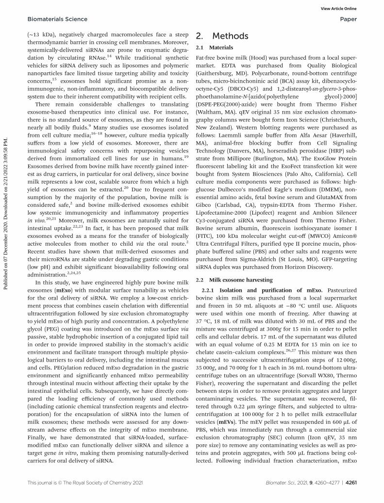
fractions (8–9) were pooled, and aliquots were frozen at −80 °Cfor downstream applications. An overview of the isolation andpurification process is shown in Fig. 1.
2.3 Exosome characterization
2.3.1 Total protein content. The total protein content ofindividual mExo fractions and pooled samples was determinedby a commercial bicinchoninic acid (BCA) assay kit.Absorbance values were measured using a plate reader(Synergy H1, Biotek) and quantified against a bovine serumalbumin standard (0–1000 µg mL−1) with r2 > 0.98. All sampleswere diluted 10× prior to testing to ensure that absorbancevalues fell within the standard curve.
2.3.2 Yield and purity. Individual mExo fractions 7–11 andmEVs were analyzed for size distribution and particle concen-tration within one day of SEC (stored at 4 °C), using theSpectradyne nCS1 microfluidic resistive pulse sensing appar-atus. Prior to testing, fractions were diluted at a 1 : 200 ratio in20 nm-filtered PBS with 0.8% Tween-20 surfactant and 200 nmdiameter polystyrene calibration beads (1010 particles per mLfinal concentration). For testing, 5 µL of sample was loadedinto Spectradyne TS-300 or TS-400 cartridges, and at least 15acquisitions (n ≥ 20 000 particles) were measured per sample.Individual sample size distribution data was processed usingthe Spectradyne software for calibration and filtering of false-positive particle detection events. The filtering criteria were asfollows: events with <60 µs transit time and <210 nm measureddiameter were accepted, and the automated background sub-traction feature was implemented. Filtered data was then cali-brated for diameter and concentration based on the distri-bution of 200 nm control beads, and final concentration
values were determined by integration of the size distributioncurve. Fraction purity was calculated as the ratio of particlecount to µg of total protein (from BCA) in correspondingsamples.
2.3.3 Transmission electron microscopy imaging. PooledmExo samples were imaged by negative stain transmissionelectron microscopy to examine particle size and morphology.Briefly, 5 µL of pooled sample was placed on formvar coatedcopper grids for 2 min, followed by washing with ultrapurewater and staining with 1% uranyl acetate. Samples were thenviewed with a transmission electron microscope (2100F, JEOL).
2.3.4 Western blotting for exosome biomarkers. IndividualmExo fractions 8–11 and mEVs were probed for select proteinmarkers by western blotting using standard protocol. Sampleswere prepared for sodium dodecyl sulphate-polyacrylamide gelelectrophoresis (SDS-PAGE) by incubating 10 µg of mExoprotein equivalent with β-mercaptoethanol at 70 °C for10 min, then diluting in a 1 : 1 ratio of Laemmli sample buffer.Reduced proteins were electrophoresed using the BioRadsystem for 45 min at 150 V in NuPAGE 4–12% precast Bis-Trisgels. After separation, proteins were transferred to a nitrocellu-lose membrane (pre-treated with methanol) in a BioRad blot-ting module at 75 V for 2.5 h. After confirmation of successfultransfer with Ponceau-S staining dye, membranes wereblocked using animal-free blocking buffer for 1 h, and thentreated overnight with primary antibodies (1:1000 dilution ofmouse anti-TSG101, sc-7964; 1:1000 mouse anti-Flot1, sc-74566; 1:600 mouse anti-Alix, sc-53540; Santa Cruz). Afterwashing thrice in PBS-Tween 20 washing buffer, membraneswere incubated for 1 h in secondary antibody (1:1000 dilutionof mouse IgGk binding protein-HRP, sc-516102, Santa Cruz).
Fig. 1 Isolation and purification process of exosomes from bovine milk. Pasteurized milk samples were first diluted with PBS and centrifuged at3000g to pellet cells and debris. The supernatant was then treated with an equal volume of 0.25 M EDTA to chelate casein micelles and casein-coated exosomes. This was followed by differential ultracentrifugation to remove large protein aggregates and contaminating microvesicles. Thesample was subsequently centrifuged at 100 000g for 2 h to pellet extracellular vesicles (mEV). Finally, the resuspended mEV pellet was purified bysize-exclusion chromatography (SEC) using a commercially available column.
Paper Biomaterials Science
4262 | Biomater. Sci., 2021, 9, 4260–4277 This journal is © The Royal Society of Chemistry 2021
Publ
ishe
d on
07
Dec
embe
r 20
20. D
ownl
oade
d on
2/2
1/20
22 3
:09:
58 P
M.
View Article Online

Finally, membranes were washed, incubated for 5 min in HRPsubstrate, and chemiluminescence was imaged using a BioRadChemiDoc CCD imaging system.
2.3.5 Proteome analysis. The protein content of pooledmExo fractions 8–9 was analyzed by mass spectrometry (MS).To prepare for MS, samples were reduced and digested withtrypsin (Promega) at 25 °C overnight. Peptides were desalted,vacuum centrifuged, and stored at −80 °C until use. For MS,peptides were separated by reverse phase HPLC (Thermo EasynLC1000) before nanoelectrospray using a QExactive HF-Xmass spectrometer (Thermo Fisher Scientific). Then the fullMS scan was followed by MS/MS for the top 15 precursor ionsin each cycle. Raw mass spectral data files were searched usingProteome Discoverer (Thermo Fisher Scientific) and Mascotversion 2.4.1 (Matrix Science). Only peptides with a Mascotscore greater than or equal to 25 and an isolation interferenceless than or equal to 30 were included in the data analysis.
2.4 Surface modification of mExo with polyethylene glycol(PEG)
2.4.1 Preparation of fluorescent-conjugated DSPE-PEG-Azide.DSPE-PEG-Azide (hereby DPA) was utilized to enable a hydro-phobically-inserted PEG coating on the mExo surface. Anaqueous stock solution was prepared by first dissolving DPA inDMSO at 2 mg mL−1, followed by dropwise addition to 1× PBSwith continuous stirring for a final DPA concentration of0.02 mg mL−1 (7 μM). For studies involving quantification ofDPA concentration (i.e. measurement of mExo surface loadingand anchor stability studies), DPA was conjugated to the fluo-rescent moiety DBCO-Cy5 via the azide-DBCO click chemistryreaction. DBCO-Cy5 (1 mg mL−1 in DMSO) was added drop-wise with continuous stirring to aqueous DPA stock for a 1 : 4final molar ratio. This solution was then incubated for 6 h atroom temperature with light shaking to facilitate the click reac-tion. After the click reaction, the solution containing DPA-Cy5and excess DBCO-Cy5 was added directly to exosome samplesfor insertion.
2.4.2 Synthesis of PEGylated mExo. The hydrophobic inser-tion of DPA into the mExo membrane was facilitated by apassive, biocompatible process involving co-incubation ofmExo and DPA molecules. First, 125 μL mExo stock was addeddropwise to aqueous DPA with continuous stirring at roomtemperature, at a final reaction mixture volume of 1.5 mL. Thismixture was then incubated at 37 °C for 1 h with light shakingto form PEG-mExo. Subsequently, non-inserted DPA moleculeswere separated from PEG-mExo via ultrafiltration by centrifu-gation (UFC) on a 100 kDa MWCO centrifugal filter. Whenusing UFC to purify PEG-mExo, we used 2 sequential spins for10 min each at 4000g, between which the concentrated PEG-mExo sample was diluted to 1.5 mL with PBS for washing.
When quantifying surface loading efficiency, DPA-Cy5 wasused in place of DPA, using the same loading process. Toquantify DPA surface loading, we carried out the processdescribed above using various molar ratios of DPA to mExoparticles – 5000 : 1, 10 000 : 1, and 20 000 : 1 – all reacted in1.5 mL mixtures, followed by purification by UFC. Cy5 fluo-
rescence was measured in each final PEG-mExo sample usinga plate reader and was compared to a standard curve ofDBCO-Cy5 to quantify the concentration of DPA inserted intothe mExo surface. The number of DPA molecules per mExoparticle was then estimated using the following formula:
#PEG ¼ ½DPA‐Cy5�mExoprotein½ � � 3� 109
particlesμg
� � :
It was assumed that all Cy5 signal retained in the UCFfilters was from DPA inserted into the mExo membrane.However, to account for possible background signal fromDPA-Cy5 micelles retained in the filter as well as excessDBCO-Cy5 associated with the mExo surface, control mixtureswere prepared with no DPA (only DBCO-Cy5 and mExo) and nomExo (only DPA-Cy5), followed by purification and quantifi-cation of retained Cy5 signal as described above. Cy5 signalfrom both of these control samples was subtracted frommeasurements of samples with graded DPA :mExo molarratios.
2.4.3 Measurement of binding affinity of mExo and DPAusing microscale thermophoresis. Binding affinity betweenmExo and DPA was quantified based on microscale thermo-phoresis (MST) using the Monolith NT.115 instrument(NanoTemper Technologies, Munich, Germany). Internallyfluorescent mExo was used as the target molecule while non-labeled DPA was chosen as the ligand for the assay. mExointernal protein was fluorescently labelled with ExoGlow-greendye as described in Methods 2.5.1. Internally fluorescent mExoat a fixed concentration of 5 nM was incubated with varyingconcentrations of DPA (between 0.81 μM to 1.66 mM) in PBSpH 7.4 for 1 h at 37 °C under gentle shaking. Samples wereloaded on standard treated capillaries and MST was performedunder 20% excitation power and 40% MST power at 22.8 °C.Laser on and off times were selected as 1 s and 20 s, respect-ively. Fluorescence of each sample at 5 s was normalized to theinitial fluorescence and plotted against the ligand concen-tration to obtain the binding curve using NanoTemper analysissoftware. The Hill equation was fitted to the binding curve toobtain the dissociation constant (KD) and the Hill coefficient(n), which describes cooperativity of the binding, using the fol-lowing equation:
FnormðCDPAÞ � FnormðunboundÞFnormðboundÞ � FnormðunboundÞ ¼
1
1þ KD
CDPA
� �n
where Fnorm(unbound) is the normalized fluorescence of onlyunbound mExo, Fnorm(bound) is normalized fluorescence ofthe mExo-PEG complex at saturated bound state andFnorm(CDPA) is normalized fluorescence measured at titrationconcentration of DPA.
2.4.4 Confocal microscopy of dual-labeled PEG-mExo.Insertion of DPA-Cy5 on the mExo surface was confirmed byvisualizing dual-labeled PEG-mExo under confocal microscopy.Dual-labeled PEG-mExo was synthesized by loading DPA-Cy5
Biomaterials Science Paper
This journal is © The Royal Society of Chemistry 2021 Biomater. Sci., 2021, 9, 4260–4277 | 4263
Publ
ishe
d on
07
Dec
embe
r 20
20. D
ownl
oade
d on
2/2
1/20
22 3
:09:
58 P
M.
View Article Online
as mentioned in Methods 2.4.2 onto mExo pre-modified withfluorescently-labeled internal proteins (Methods 2.5.1). A layerof the PEG-mExo solution was then placed between a coverslipand glass slide. The sample was imaged under FITC and Cy5channels using a confocal microscope (Zeiss LSM 800) at 60×magnification. The fluorescent images were overlapped to mapthe position of DPA-Cy5 with respect to mExo.
2.4.5 Stability of DPA inserted on mExo. The temporalstability of the hydrophobic DSPE anchor in the mExo mem-brane was evaluated over the course of 1 h, in the absence andpresence of mucin proteins. mExo loaded with DPA-Cy5 at a20 000 : 1 molar ratio was incubated in either PBS or a biosimi-lar mucin gel for various times at 30 °C with light shaking.Samples were subsequently subjected to two 10 min UFC spinson 100 kDa filters at 4000g to elute DPA-Cy5 molecules thathad partitioned out of the mExo membrane. Cy5 signal wasmeasured in the eluted sample for each time point and com-pared to a DBCO-Cy5 standard curve. In parallel, free DPA-Cy5was incubated in PBS and mucin gel for 1 h at the same con-centration as PEG-mExo and separated by the same process todefine a point of comparison for 100% DPA-Cy5 dissociationfrom the mExo membrane. This accounts for the possibilitythat diffusion of DPA-Cy5 molecules through UFC spin filtersmay be hindered by the presence of mucin proteins. Thus, dis-sociation of DPA-Cy5 from PEG-mExo at each time point wasquantified as the percentage of eluted DPA-Cy5 compared to100% dissociation controls for both PBS and mucin.Biosimilar mucin was prepared by reconstituting purified type-II porcine intestinal mucin in 20 mM HEPES/20 mM NaCl vialight stirring overnight at 4 °C, as previously described.28
2.5 In vitro evaluation of PEG-mExo for oral delivery
2.5.1 mExo fluorescent labelling. For cell uptake andmucus penetration studies, internal exosome proteins werelabelled with a green fluorescent dye using the ExoGlowprotein labelling kit, following the manufacturer’s instruc-tions. Briefly, 200–500 µg of mExo suspended in 500 µL PBSwere incubated with 1 µL of labelling dye for 20 min at 37 °Cwith shaking. To separate labelled mExo from free labellingdye, samples were purified by UFC on 100 kDa spin filters,using one spin for 15 min at 4000g. Labelled mExo were storedat 4 °C and used within one day of labelling.
2.5.2 Stability of mExo and PEG-mExo in different pHbuffers mimicking stomach acidity. The stability of mExo inthe presence of acidic conditions representative of the humanstomach was evaluated. Phosphate buffers were prepared atpH 2.2 (adult human stomach) and pH 4.5 (infant humanstomach);29 pH 7 PBS was used as control. 80 μg of nakedmExo and PEG-mExo was added to 1.5 mL of each buffer andincubated for 1 h at 37 °C with light shaking. Samples werethen subjected to one UFC spin filtration step for 15 min at4000g. The protein content of recovered mExo samples wasmeasured by BCA assay as described above and compared tothe starting value of 80 μg to calculate the percent proteinrecovery. Recovery percentages were normalized to protein
recovery in corresponding pH 7 samples to allow comparisonbetween mExo and PEG-mExo values.
2.5.3 Intestinal mucus transport. The mucus transport pro-perties of unmodified and PEGylated mExo were evaluatedusing an in vitro transwell model as previously described.30–34
Native porcine intestinal mucus was harvested as previouslydescribed30 and frozen at −80 °C until use. The transwellsetup was as follows: 0.4 μm polycarbonate membrane trans-well inserts (0.33 cm2 surface area) were used to separate thedonor and acceptor compartments. 600 μL of filtered PBS wasput in the acceptor chamber, and 40 μL of mucus (approx.0.2 mm thickness layer) was added to the donor side of themembrane. mExo was labelled as described in Methods 2.5.1.For each well, 15 µL of labeled native or PEGylated mExo wasadded to the surface of the mucus layer, such that the mucuswas not diluted. The well plate was then immediately coveredand moved to 37 °C with light shaking for the designatedamount of time. Finally, the PBS from the acceptor chamberwas removed, and mExo fluorescence was measured on theSynergy H1 plate reader; the fluorescence of the original mExosolution initially added to mucus was also measured. A separ-ate plate was prepared for each time point measured(0–30 min), such that continuous removal of PBS from theacceptor chamber throughout the course of the experimentwas avoided. In parallel, the mucus penetration of 30 μM FITCdye was measured using the same protocol as a control. Foreach sample, the apparent permeability coefficient, Papp, wascalculated by the following formula:
Papp ¼ dRFUdt
� 1A � RFU0
;
where RFU is the acceptor chamber fluorescence reading,RFU0 is the starting solution fluorescence, and A is the surfacearea of the transwell membrane.
2.5.4 Caco-2 and HEK293 cell culture. Human adeno-carcinoma (Caco-2) cells were used as a model for intestinalepithelial uptake. Cells were purchased from ATCC and seededat 104 cells per cm2 within one day of arrival. The completemedium used was high-glucose (4.5 g L−1) DMEM sup-plemented with 10% (v/v) fetal bovine serum, 1% GlutaMAX,1% nonessential amino acids, and 1% penicillin–streptomy-cin. Cells were incubated at 37 °C with 5% CO2 in a humidifiedincubator. Media was changed every 2–3 days, and cells werepassaged at ∼75% confluence. Cells were passaged at leasttwice before use in uptake and silencing experiments. HEK293cells from Cell BioLabs (San Diego, CA) were cultured with thesame medium and conditions.
2.5.5 Cellular uptake of mExo and PEG-mExo. The satur-ation kinetics of unmodified mExo into Caco-2 cells wasmeasured using a fluorescence assay on a Synergy H1 platereader. Cells were seeded at a density of 20 000 cells per well ofa 96-well plate and allowed to adhere for 36 h. 12.5–100 µg offluorescently-labelled mExo were added to 200 µL of mediaand incubated with cells for 2.5 h. After uptake, cells werewashed three times with cold PBS, detached using phenol-redfree 0.25% trypsin-EDTA, and fluorescence was read at 490/
Paper Biomaterials Science
4264 | Biomater. Sci., 2021, 9, 4260–4277 This journal is © The Royal Society of Chemistry 2021
Publ
ishe
d on
07
Dec
embe
r 20
20. D
ownl
oade
d on
2/2
1/20
22 3
:09:
58 P
M.
View Article Online

520 nm excitation/emission. Uptake was quantified by compar-ing cell fluorescence to a standard curve of a known weight oflabelled mExo suspended in PBS, using cells incubated inmExo-free media as a blank. Uptake data was fit to theMichaelis–Menten kinetic equation, modelling mExo uptakeby endocytosis as a saturable process. Data was fit to the fol-
lowing equation: RU ¼ Vmax � ½mExo�Km þ ½mExo� , where Ru is the rate of
mExo transport, Vmax is the maximum Ru at which endocytosissaturates, and Km is the Michaelis–Menten constant, definedas the mExo concentration at which Ru = 0.5 × Vmax.
Uptake of mExo and PEG-mExo into Caco-2 cells was alsomeasured using fluorescence imaging. Cells were seeded at adensity of 20 000 cells per well of a 96-well plate and allowed toadhere for 36 h. 130 µg of fluorescently-labelled mExo anddual-labelled PEG-mExo (prepared as described in Methods2.4.4) were added to 200 µL of media and incubated with cellsfor 2.5 h. After uptake, cells were washed three times with coldPBS and fixed for 10 min in 4% paraformaldehyde. Cell nucleiwere then stained by incubating in DAPI for 5 min. Fixed,stained cells were then washed three times with cold PBS andimaged using a Nikon Eclipse Ts2R fluorescence microscopewith constant exposure time and 10× magnification. Imageswere processed using ImageJ software. To account for potentialsources of background signal in images with single- and dual-labels, cells were incubated with free DBCO-Cy5, DPA-Cy5, andExoGlow dye at both high and low concentrations, aligningwith concentrations of these moieties in mExo samples pre-and post-purification. These cells were imaged as describedabove to evaluate the extent to which these potential sources ofbackground signal contribute to single- and dual-labelleduptake images.
2.6 In vitro gene silencing by mExo-siRNA
2.6.1 Quantification of siRNA loading into mExo. mExowere loaded with siRNA by two commonly used methods: theExoFect exosome transfection kit and Lipofectamine-2000. Todetermine the loading efficiency with each method, mExo wereloaded with Cy3-conjugated, eGFP-targeting siRNA (ThermoFisher) and separated from free siRNA and complexes asdescribed below (2.6.2). Following separation of siRNA-loadedmExo from non-loaded siRNA complexes, the loadingefficiency was quantified by reading Cy3 fluorescence of resus-pended mExo on a Synergy H1 plate reader. Fluorescencevalues were compared against a standard curve of known Cy3-siRNA concentration (0–150 nM) to estimate pmol siRNAloaded. Additionally, the exosome protein content of loadedmExo samples was measured by BCA assay as described abovein order to normalize siRNA loading to µg mExo-protein.Following loading, mExo zeta potential was measured in PBSusing a Malvern ZetaSizer Nano, and size distribution wasmeasured on the Spectradyne nCS-1 as described above.
2.6.2 Loading protocol for ExoFect and Lipofectamine2000. Exosomes were loaded by ExoFect following the manu-facturer’s protocol. Briefly, 120 µL of mExo was incubated with
60 pmol siRNA and 10 µL ExoFect reagent for 10 min at 37 °Cwith light shaking. Similarly, mExo were loaded byLipofectamine 2000 (hereby Lipofect) by incubation of 1–10 µLLipofect reagent, 60 pmol siRNA, and 120 µL mExo for 30 minat room temperature. Unfused siRNA-ExoFect orsiRNA-Lipofect complexes were then separated from exosomesusing ExoQuick-TC. Samples were incubated with a propor-tionate volume (20% v/v) of ExoQuick-TC reagent for 30 minon ice to facilitate exosome separation. Samples were then cen-trifuged at 15 000g for 5 min, and pellets were resuspended in100 µL PBS for downstream loading analysis, or 50 µL com-plete DMEM for cell culture studies.
2.6.3 In vitro silencing of GFP in HEK293 cells. Green fluo-rescent protein (GFP) was targeted in GFP-expressing HEK293cells by the Lipofect-mExo-siRNA formulation with andwithout surface PEGylation. 24 h prior to treatment, cells wereseeded at 25 000 cells per well in 48-well plates in antibiotic-free media. mExo-siRNA formulations were prepared asdescribed above, using 80 pmol of siGFP and 5 µL of Lipofectin the initial mixture, and diluted to 12 μg per well mExoprotein for addition to cells. Simultaneously, cells were treatedwith lower concentrations of the mExo-siRNA formulation toinvestigate a dose-dependent silencing response. To accountfor silencing by Lipofect-siRNA complexes that co-precipitatewith mExo during Exo-Quick TC purification, the loading/puri-fication process was carried out in the absence of mExo, andthe resulting samples (termed “Residual Lipofect”) were alsoevaluated for silencing capability. In parallel, Lipofectamine-siRNA complexes were added directly to cells as a positivecontrol (termed “Lipofect-siRNA”). In a separate experiment,PEG-mExo-siRNA was prepared by mixing purified mExo-siRNA with 20 000 : 1 molar excess DPA via dropwise additionto a 1.5 mL reaction mixture with continuous stirring, followedby incubation at 37 °C for 1 h with light shaking. 12 μg perwell of mExo-siRNA and PEG-mExo-siRNA was added directlyto cells.
For all silencing experiments, mExo internalization andgene knockout occurred over 72 h. After this time, cells wereimaged using the FITC filter of a Nikon Eclipse Ts2R fluo-rescence microscope, with constant exposure time. To quantifysilencing of GFP, cell FITC fluorescence was measured using aBeckman Coulter Cytoflex flow cytometer. Briefly, followingsiRNA treatment, cells were detached from the well plate with0.25% trypsin-EDTA and washed twice by centrifugation andresuspension in PBS. siRNA conditions were tested in dupli-cate, and 10 000 events were acquired per sample and con-stricted to a custom FC/SC gate as designated by control cells.
2.6.4 Statistical analysis. All data presented herein rep-resents mean ± 95% confidence interval calculated by samplevariance, unless otherwise noted. Experiments were conductedusing at least n = 3 sample replicates in multiple experimentalrepeats. All fluorescence measurements were repeated in tripli-cate on one sample. Where appropriate due to low samplesize, confidence intervals were calculated using an empiricalbootstrap method.35 P ≤ 0.05 was used for statisticalsignificance.
Biomaterials Science Paper
This journal is © The Royal Society of Chemistry 2021 Biomater. Sci., 2021, 9, 4260–4277 | 4265
Publ
ishe
d on
07
Dec
embe
r 20
20. D
ownl
oade
d on
2/2
1/20
22 3
:09:
58 P
M.
View Article Online
3. Results3.1 Isolation, purification and characterization
Exosomes were harvested from bovine milk using differentialultracentrifugation followed by purification using SEC (Fig. 1).The total protein content of each SEC fraction (500 µL) wasdetermined by BCA assay (Fig. 2A). The elution profile shows apeak in total protein content in fractions 7–11, with fractions 8and 9 containing maximum concentrations. This fraction rangecorresponds to the elution of exosomes (mExo) and other milkextracellular vesicles (mEV), which is in agreement with themanufacturer’s instructions. This protein elution profile wasconsistently observed across multiple batches of mExo isolation,suggesting that SEC using the qEV column is a reliable andreproducible method for isolating exosomes from bovine milk.The trend towards increased protein content in later fractions(12–16) suggests the presence of contaminating proteins, aggre-gates, and smaller vesicles in these fractions. As a result, onlymEV fractions (7–11) were used for further studies.
The particle concentrations for mEV and mExo fractionswere determined by using the high-resolution resistive pulsesensing technique (Fig. 2B). The majority of particles wereobserved in fractions 8 and 9. In comparison to the particle con-centration in the pre-SEC samples, nearly all mExo particleswere recovered in fractions 8 and 9. Although some particles(<0.25 × 10¹² particles per mL) were observed in later fractions(10–11), these fractions contain less than 10% of the total par-ticles isolated from a single SEC run. Moreover, these particlescontain a considerably high protein concentration, indicating aparticle population of significantly lower purity (as expressed bythe particle-to-protein ratio). Interestingly, purity of particlesdecreased with increased elution volume (Fig. 2C), withexosome fractions exhibiting 2.2-fold higher purity than pre-SECsamples. Taken together, these results suggest that an exosomepopulation of significantly higher purity is present in fractions 8and 9 compared to pre-SEC samples, while recovering nearly allparticles present in the initial sample.
The size distribution of pooled mExo samples (fractions8–9) was measured by resistive pulse sensing and confirmedby negative-stain transmission electron microscopy (TEM)(Fig. 2D and E). A majority of measured particles displayed adiameter between 65–90 nm, with a median diameter ofapproximately 70 nm (10th percentile of 62.2 nm, 90th percen-tile of 90.4 nm). This is supported by TEM, which displays par-ticles in this size range and reveals the spherical morphologyof the isolated exosomes. From 18 mL of bovine milk, 650 µgof purified mExo was recovered in 1 mL final volume. Theresults on the physical properties of the purified mExosamples are summarized in Table 1.
3.2 Proteome and protein biomarkers
Western blotting was performed on individual mExo fractions(8–11) to qualitatively confirm the presence of exosomes basedon common protein biomarkers (Fig. 3). The samples wereprobed for the presence of two proteins related to the endo-somal sorting complex required for transport (ESCRT) –
TSG-101 and Alix – and two exosome-associated membraneproteins – Flotillin-1 and CD63.36 For comparison, pre-SECmEV samples were also analyzed for these markers. All fourdiagnostic markers were present in mEV samples, stronglysupporting the presence of exosomes in the crude samplesbefore SEC. Following purification, fractions 8 and 9 containedall biomarkers. Conversely, fractions 10 and 11 did not showthe presence of TSG-101 or Alix. This suggests that mExo arepresent in lesser amounts in fractions 10 and 11, further sup-porting the lower purity of those fractions (Fig. 3A). Therefore,due to the ubiquitous presence of exosome protein bio-markers, as well as higher purity and particle content in frac-tions 8 and 9, these fractions were pooled to generate finalmilk exosome samples for further investigations.
Mass spectrometry was performed to identify the proteomeof final pooled mExo samples (see Table 2). Raw spectral datafrom mass spectrometry was processed to generate a list ofobserved proteins (ESI Table S1†). In total, the presence of 214proteins was observed. After careful analysis, the list was nar-rowed down to 51 high-confidence proteins (for which two ormore unique peptide sequences were detected). Proteins unre-lated to EVs, such as cell debris markers, were excluded. A fulllist of the mExo proteome is presented in the ESI.† Out of the51 shortlisted proteins in the sample, 25 were considered to berelated to EVs (i.e. previously reported exosome biomarkers,proteins related to extracellular vesicle biogenesis, and mem-brane proteins), 9 proteins were deemed milk proteins, andthe remaining 17 proteins were cell-related (Fig. 3B). Proteinswere categorized and screened for following the recommen-dation of the International Society of Extracellular Vesicles.36
Mass spectrometry confirmed the presence of commonlyreported exosome biomarkers related to the formation andsecretion of exosomes from donor cells, including tetraspanins(CD9, CD81), heat shock proteins (Hsp70), Rab proteins andannexins. Additionally, a variety of milk-related proteins werefound in the final exosome samples, including casein, albu-mins, lipoproteins and lactoglobulin, which are potentiallyremnants from the ultracentrifugation process. Importantly,proteomic data was screened for the presence of proteinsreported as microvesicle- and cell debris-related contaminants,such as ER-related calnexin and endoplasmin (HSP90B1);membrane-associated integrin-b1, CD40 and CD62P; Golgimarker GM130; and mitochondrial cytochrome C (see Table 2,row 3).36 None of these proteins were found in proteomic data,suggesting high purity of final samples and absence of majorcontaminants such as cellular debris or non-exosome vesicles.
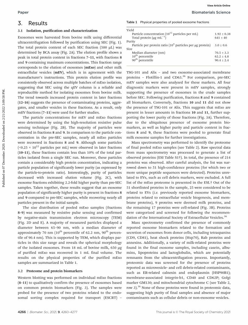
Table 1 Physical properties of pooled exosome fractions
YieldParticle concentration (1012 particles per mL) 1.92 ± 0.38Total protein (μg mL−1) 641 ± 40
PurityParticle per protein ratio (109 particles per μg protein) 3.0 ± 0.6
SizeMedian diameter (nm) 70.5 ± 3.310th percentile 62.2 ± 4.890th percentile 90.4 ± 2.4
Paper Biomaterials Science
4266 | Biomater. Sci., 2021, 9, 4260–4277 This journal is © The Royal Society of Chemistry 2021
Publ
ishe
d on
07
Dec
embe
r 20
20. D
ownl
oade
d on
2/2
1/20
22 3
:09:
58 P
M.
View Article Online

Fig. 2 Physical characteristics of individual mEV fractions following size exclusion chromatography. (A) Total protein concentration of individualSEC fractions 1–16, measured by BCA assay. (B) Particle concentration in exosome fractions (7–11), measured by Spectradyne nCS1. (C) Purity ofmilk exosome (mExo) fractions, represented as particle-to-total protein ratio. (D) Representative size distribution of pre-SEC mEV sample andpooled fractions 8 and 9, measured by Spectradyne nCS1. (E) Transmission electron microscopy images of pooled mExo sample showing sphericalmorphology. Scale bars on left and middle images represent 100 nm and 500 nm, respectively. Data are presented as mean ± 95% confidence inter-vals (calculated by t-distribution).
Biomaterials Science Paper
This journal is © The Royal Society of Chemistry 2021 Biomater. Sci., 2021, 9, 4260–4277 | 4267
Publ
ishe
d on
07
Dec
embe
r 20
20. D
ownl
oade
d on
2/2
1/20
22 3
:09:
58 P
M.
View Article Online

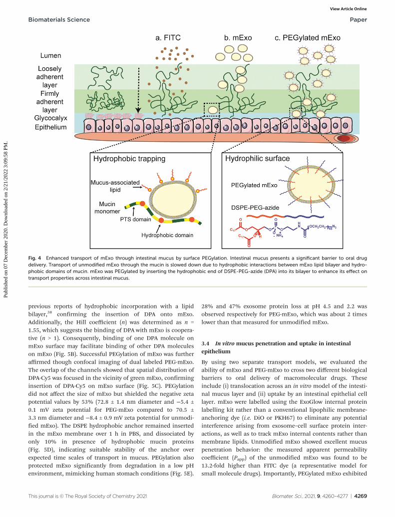
3.3 PEGylation of mExo membrane by hydrophobic insertion
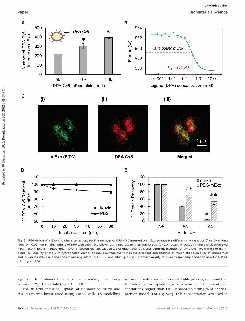
Stable insertion of PEG chains into the mExo lipid bilayer wasachieved by using a conjugated, bivalent lipid tail(DSPE-PEG-Azide or DPA; Fig. 4).37 Azide-derivatized PEG waschosen as a modular moiety for mExo surface engineering, asthe terminal azide group enables simple conjugation of avariety of peptides or proteins by using azide–alkyne clickchemistry, enabling modulation of mExo surface properties.
mExo mixed with DPA-Cy5 at a molar ratio of 20 000 DPAper mExo particle resulted in approximately 394 PEG chainsincorporated onto each mExo particle (Fig. 5A, ESI Fig. S1†).To confirm hydrophobic insertion of DPA into the mExo lipidbilayer, the affinity of the binding event between mExo andDPA was measured using MST and the resultant binding curvewas fit to the Hill model. The dissociation constant (KD) of thebinding was estimated to be 347 μM, which is consistent with
Table 2 Proteome analysis of pooled exosome fractions by mass spectrometry
Classification Location Family Proteins
Exosome marker Membrane receptor Tetraspanins CD9, CD63, CD81Membrane/lipid bound Rab proteins Rab 11b, 15, 18, 7a, 1b, 5c
Annexins Annexin A1, A2, A5, A7Cytosolic Heat shock proteins Hsc70, Hsp70
Eukaryotic translationelongation factor (EEF)
EEF1α2, EEF2
Lacto proteins Lactoadherin (MFG-38),lactotransferrin, lactoperoxidase
Fatty acid synthase FASNProtein zeta/delta YWHAE
Milk-Related Proteins Surrounding solution, cytosol,membrane and lipid bound
Caseins α-Casein, B-casein, κ-caseinAlbumins Serum albumin, lactalbuminLipoproteins Apolipoproteins A-I, A-IV, EOthers Immunoglobulin, B-lactoglobulin
Negative controls (notpresent in sample)
Cytosolic, membrane Endoplasmic reticulum Calnexin, endoplasmin (HSP90B1)Transmembrane Integrin-B1, CD40, CD62PGolgi GM130Mitochondria Cytochrome C
Fig. 3 Screening for the presence of protein exosome biomarkers in mExo samples. (A) Western blotting for the presence of a variety of exosomemarkers, including membrane proteins CD63 and Flotillin-1 and ESCRT proteins (TSG-101 and Alix). Individual fractions after SEC are compared tothe resuspended mEV pellet after ultracentrifugation and before SEC. (B) Breakdown of the categories of proteins present in exosome proteomicsanalysis by mass spectrometry.
Paper Biomaterials Science
4268 | Biomater. Sci., 2021, 9, 4260–4277 This journal is © The Royal Society of Chemistry 2021
Publ
ishe
d on
07
Dec
embe
r 20
20. D
ownl
oade
d on
2/2
1/20
22 3
:09:
58 P
M.
View Article Online
previous reports of hydrophobic incorporation with a lipidbilayer,38 confirming the insertion of DPA onto mExo.Additionally, the Hill coefficient (n) was determined as n =1.55, which suggests the binding of DPA with mExo is coopera-tive (n > 1). Consequently, binding of one DPA molecule onmExo surface may facilitate binding of other DPA moleculeson mExo (Fig. 5B). Successful PEGylation of mExo was furtheraffirmed though confocal imaging of dual labeled PEG-mExo.The overlap of the channels showed that spatial distribution ofDPA-Cy5 was focused in the vicinity of green mExo, confirminginsertion of DPA-Cy5 on mExo surface (Fig. 5C). PEGylationdid not affect the size of mExo but shielded the negative zetapotential values by 53% (72.8 ± 1.4 nm diameter and −5.4 ±0.1 mV zeta potential for PEG-mExo compared to 70.5 ±3.3 nm diameter and −8.4 ± 0.9 mV zeta potential for unmodi-fied mExo). The DSPE hydrophobic anchor remained insertedin the mExo membrane over 1 h in PBS, and dissociated byonly 10% in presence of hydrophobic mucin proteins(Fig. 5D), indicating suitable stability of the anchor overexpected time scales of transport in mucus. PEGylation alsoprotected mExo significantly from degradation in a low pHenvironment, mimicking human stomach conditions (Fig. 5E).
28% and 47% exosome protein loss at pH 4.5 and 2.2 wasobserved respectively for PEG-mExo, which was about 2 timeslower than that measured for unmodified mExo.
3.4 In vitro mucus penetration and uptake in intestinalepithelium
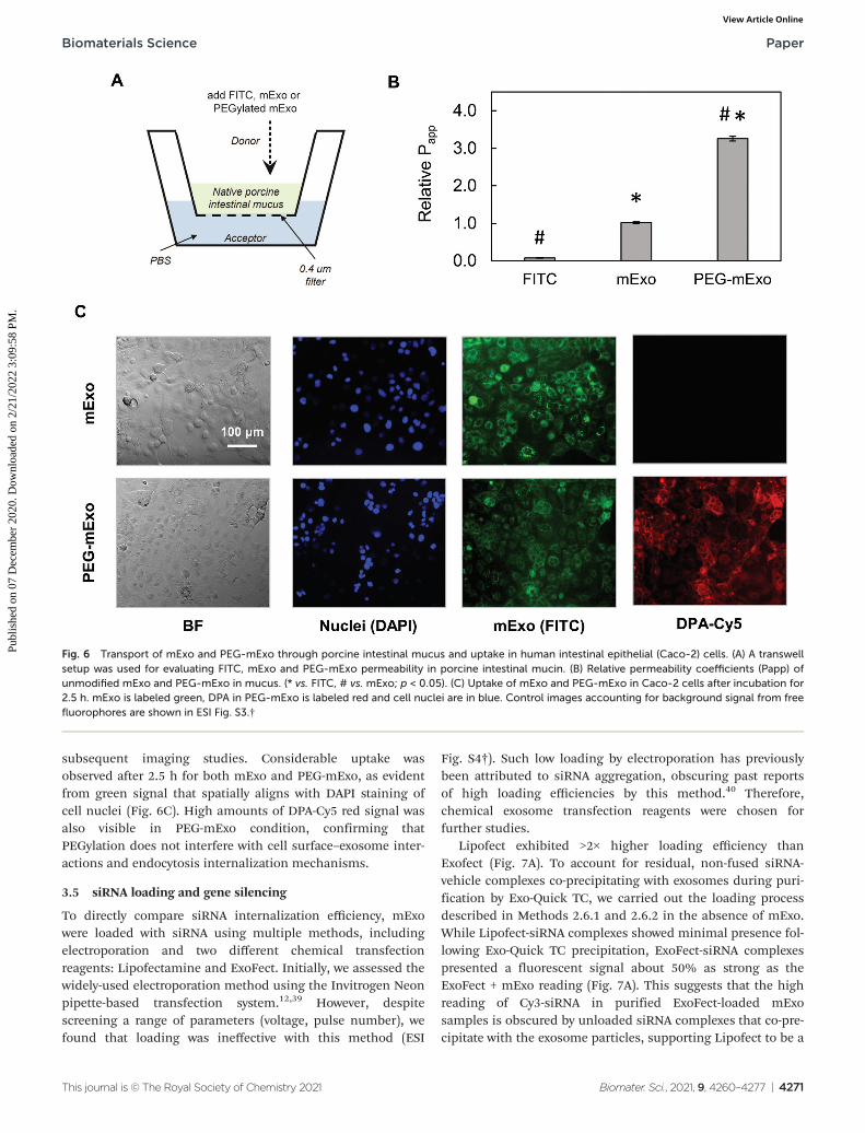
By using two separate transport models, we evaluated theability of mExo and PEG-mExo to cross two different biologicalbarriers to oral delivery of macromolecular drugs. Theseinclude (i) translocation across an in vitro model of the intesti-nal mucus layer and (ii) uptake by an intestinal epithelial celllayer. mExo were labelled using the ExoGlow internal proteinlabelling kit rather than a conventional lipophilic membrane-anchoring dye (i.e. DiO or PKH67) to eliminate any potentialinterference arising from exosome–cell surface protein inter-actions, as well as to track mExo internal contents rather thanmembrane lipids. Unmodified mExo showed excellent mucuspenetration behavior: the measured apparent permeabilitycoefficient (Papp) of the unmodified mExo was found to be13.2-fold higher than FITC dye (a representative model forsmall molecule drugs). Importantly, PEGylated mExo exhibited
Fig. 4 Enhanced transport of mExo through intestinal mucus by surface PEGylation. Intestinal mucus presents a significant barrier to oral drugdelivery. Transport of unmodified mExo through the mucin is slowed down due to hydrophobic interactions between mExo lipid bilayer and hydro-phobic domains of mucin. mExo was PEGylated by inserting the hydrophobic end of DSPE-PEG-azide (DPA) into its bilayer to enhance its effect ontransport properties across intestinal mucus.
Biomaterials Science Paper
This journal is © The Royal Society of Chemistry 2021 Biomater. Sci., 2021, 9, 4260–4277 | 4269
Publ
ishe
d on
07
Dec
embe
r 20
20. D
ownl
oade
d on
2/2
1/20
22 3
:09:
58 P
M.
View Article Online
significantly enhanced mucus penetrability, increasingmeasured Papp by 3.2-fold (Fig. 6A and B).
The in vitro intestinal uptake of unmodified mExo andPEG-mExo was investigated using Caco-2 cells. By modelling
mExo internalization rate as a saturable process, we found thatthe rate of mExo uptake begins to saturate at treatment con-centrations higher than 130 μg based on fitting to Michaelis–Menten model (ESI Fig. S2†). This concentration was used in
Fig. 5 PEGylation of mExo and characterization. (A) The number of DPA-Cy5 inserted on mExo surface for different mixing ratios (* vs. 5k mixingratio; p < 0.05). (B) Binding affinity of DPA with the mExo bilayer using microscale thermophoresis. (C) Confocal microscopy images of dual-labeledPEG mExo. mExo is marked green; DPA is labeled red. Spatial overlap of green and red signal confirms insertion of DPA-Cy5 into the mExo mem-brane. (D) Stability of the DPA hydrophobic anchor on mExo surface over 1 h in the presence and absence of mucin. (E) Tolerability of unmodifiedand PEGylated mExo in conditions mimicking infant (pH = 4.5) and adult (pH = 2.2) stomach acidity. (* vs. corresponding condition at pH 7.4, # vs.mExo; p < 0.05).
Paper Biomaterials Science
4270 | Biomater. Sci., 2021, 9, 4260–4277 This journal is © The Royal Society of Chemistry 2021
Publ
ishe
d on
07
Dec
embe
r 20
20. D
ownl
oade
d on
2/2
1/20
22 3
:09:
58 P
M.
View Article Online
subsequent imaging studies. Considerable uptake wasobserved after 2.5 h for both mExo and PEG-mExo, as evidentfrom green signal that spatially aligns with DAPI staining ofcell nuclei (Fig. 6C). High amounts of DPA-Cy5 red signal wasalso visible in PEG-mExo condition, confirming thatPEGylation does not interfere with cell surface–exosome inter-actions and endocytosis internalization mechanisms.
3.5 siRNA loading and gene silencing
To directly compare siRNA internalization efficiency, mExowere loaded with siRNA using multiple methods, includingelectroporation and two different chemical transfectionreagents: Lipofectamine and ExoFect. Initially, we assessed thewidely-used electroporation method using the Invitrogen Neonpipette-based transfection system.12,39 However, despitescreening a range of parameters (voltage, pulse number), wefound that loading was ineffective with this method (ESI
Fig. S4†). Such low loading by electroporation has previouslybeen attributed to siRNA aggregation, obscuring past reportsof high loading efficiencies by this method.40 Therefore,chemical exosome transfection reagents were chosen forfurther studies.
Lipofect exhibited >2× higher loading efficiency thanExofect (Fig. 7A). To account for residual, non-fused siRNA-vehicle complexes co-precipitating with exosomes during puri-fication by Exo-Quick TC, we carried out the loading processdescribed in Methods 2.6.1 and 2.6.2 in the absence of mExo.While Lipofect-siRNA complexes showed minimal presence fol-lowing Exo-Quick TC precipitation, ExoFect-siRNA complexespresented a fluorescent signal about 50% as strong as theExoFect + mExo reading (Fig. 7A). This suggests that the highreading of Cy3-siRNA in purified ExoFect-loaded mExosamples is obscured by unloaded siRNA complexes that co-pre-cipitate with the exosome particles, supporting Lipofect to be a
Fig. 6 Transport of mExo and PEG-mExo through porcine intestinal mucus and uptake in human intestinal epithelial (Caco-2) cells. (A) A transwellsetup was used for evaluating FITC, mExo and PEG-mExo permeability in porcine intestinal mucin. (B) Relative permeability coefficients (Papp) ofunmodified mExo and PEG-mExo in mucus. (* vs. FITC, # vs. mExo; p < 0.05). (C) Uptake of mExo and PEG-mExo in Caco-2 cells after incubation for2.5 h. mExo is labeled green, DPA in PEG-mExo is labeled red and cell nuclei are in blue. Control images accounting for background signal from freefluorophores are shown in ESI Fig. S3.†
Biomaterials Science Paper
This journal is © The Royal Society of Chemistry 2021 Biomater. Sci., 2021, 9, 4260–4277 | 4271
Publ
ishe
d on
07
Dec
embe
r 20
20. D
ownl
oade
d on
2/2
1/20
22 3
:09:
58 P
M.
View Article Online
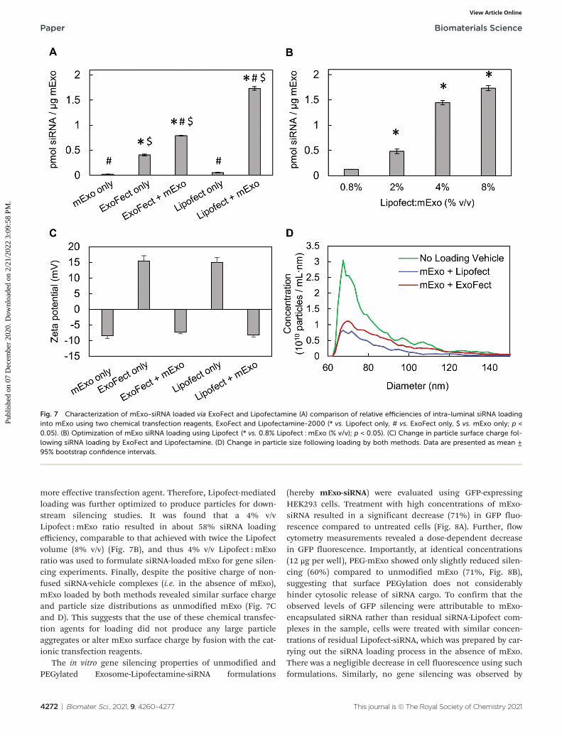
more effective transfection agent. Therefore, Lipofect-mediatedloading was further optimized to produce particles for down-stream silencing studies. It was found that a 4% v/vLipofect : mExo ratio resulted in about 58% siRNA loadingefficiency, comparable to that achieved with twice the Lipofectvolume (8% v/v) (Fig. 7B), and thus 4% v/v Lipofect : mExoratio was used to formulate siRNA-loaded mExo for gene silen-cing experiments. Finally, despite the positive charge of non-fused siRNA-vehicle complexes (i.e. in the absence of mExo),mExo loaded by both methods revealed similar surface chargeand particle size distributions as unmodified mExo (Fig. 7Cand D). This suggests that the use of these chemical transfec-tion agents for loading did not produce any large particleaggregates or alter mExo surface charge by fusion with the cat-ionic transfection reagents.
The in vitro gene silencing properties of unmodified andPEGylated Exosome-Lipofectamine-siRNA formulations
(hereby mExo-siRNA) were evaluated using GFP-expressingHEK293 cells. Treatment with high concentrations of mExo-siRNA resulted in a significant decrease (71%) in GFP fluo-rescence compared to untreated cells (Fig. 8A). Further, flowcytometry measurements revealed a dose-dependent decreasein GFP fluorescence. Importantly, at identical concentrations(12 μg per well), PEG-mExo showed only slightly reduced silen-cing (60%) compared to unmodified mExo (71%, Fig. 8B),suggesting that surface PEGylation does not considerablyhinder cytosolic release of siRNA cargo. To confirm that theobserved levels of GFP silencing were attributable to mExo-encapsulated siRNA rather than residual siRNA-Lipofect com-plexes in the sample, cells were treated with similar concen-trations of residual Lipofect-siRNA, which was prepared by car-rying out the siRNA loading process in the absence of mExo.There was a negligible decrease in cell fluorescence using suchformulations. Similarly, no gene silencing was observed by
Fig. 7 Characterization of mExo-siRNA loaded via ExoFect and Lipofectamine (A) comparison of relative efficiencies of intra-luminal siRNA loadinginto mExo using two chemical transfection reagents, ExoFect and Lipofectamine-2000 (* vs. Lipofect only, # vs. ExoFect only, $ vs. mExo only; p <0.05). (B) Optimization of mExo siRNA loading using Lipofect (* vs. 0.8% Lipofect : mExo (% v/v); p < 0.05). (C) Change in particle surface charge fol-lowing siRNA loading by ExoFect and Lipofectamine. (D) Change in particle size following loading by both methods. Data are presented as mean ±95% bootstrap confidence intervals.
Paper Biomaterials Science
4272 | Biomater. Sci., 2021, 9, 4260–4277 This journal is © The Royal Society of Chemistry 2021
Publ
ishe
d on
07
Dec
embe
r 20
20. D
ownl
oade
d on
2/2
1/20
22 3
:09:
58 P
M.
View Article Online
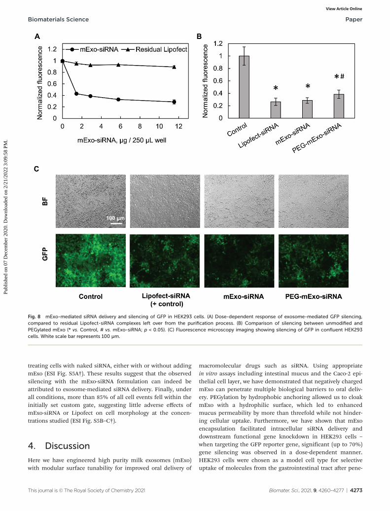
treating cells with naked siRNA, either with or without addingmExo (ESI Fig. S5A†). These results suggest that the observedsilencing with the mExo-siRNA formulation can indeed beattributed to exosome-mediated siRNA delivery. Finally, underall conditions, more than 85% of all cell events fell within theinitially set custom gate, suggesting little adverse effects ofmExo-siRNA or Lipofect on cell morphology at the concen-trations studied (ESI Fig. S5B–C†).
4. Discussion
Here we have engineered high purity milk exosomes (mExo)with modular surface tunability for improved oral delivery of
macromolecular drugs such as siRNA. Using appropriatein vitro assays including intestinal mucus and the Caco-2 epi-thelial cell layer, we have demonstrated that negatively chargedmExo can penetrate multiple biological barriers to oral deliv-ery. PEGylation by hydrophobic anchoring allowed us to cloakmExo with a hydrophilic surface, which led to enhancedmucus permeability by more than threefold while not hinder-ing cellular uptake. Furthermore, we have shown that mExoencapsulation facilitated intracellular siRNA delivery anddownstream functional gene knockdown in HEK293 cells –
when targeting the GFP reporter gene, significant (up to 70%)gene silencing was observed in a dose-dependent manner.HEK293 cells were chosen as a model cell type for selectiveuptake of molecules from the gastrointestinal tract after pene-
Fig. 8 mExo-mediated siRNA delivery and silencing of GFP in HEK293 cells. (A) Dose-dependent response of exosome-mediated GFP silencing,compared to residual Lipofect-siRNA complexes left over from the purification process. (B) Comparison of silencing between unmodified andPEGylated mExo (* vs. Control, # vs. mExo-siRNA; p < 0.05). (C) Fluorescence microscopy imaging showing silencing of GFP in confluent HEK293cells. White scale bar represents 100 µm.
Biomaterials Science Paper
This journal is © The Royal Society of Chemistry 2021 Biomater. Sci., 2021, 9, 4260–4277 | 4273
Publ
ishe
d on
07
Dec
embe
r 20
20. D
ownl
oade
d on
2/2
1/20
22 3
:09:
58 P
M.
View Article Online

tration through the intestinal mucosal layer and epithelium.There have been several prior reports describing the use ofengineered exosomes to encapsulate siRNA for delivery to avariety of cell types.10,41 For example, Alvarez-Erviti et al. haveused cell-derived exosomes to encapsulate siRNA to delivercargo across the blood brain barrier.12 Similarly, Aqil et al.have used folic acid functionalized exosomes to encapsulatesiRNA for targeted delivery to tumor cells.13 However, in com-parison to cell- and plasma-derived vesicles,42 milk exosomeshave garnered less attention for siRNA delivery, in particularvia the oral route.
In previous reports, differential ultracentrifugation anddensity-gradient centrifugation have been considered adequatefor isolating milk exosomes.20–22,43 In our studies, we haveemployed size-exclusion chromatography as an additionalpurification step following ultracentrifugation27 (Fig. 1). Thisprocess produced exosomes with a 2.2-fold increase in purity,and nearly 100% recovery of mExo particles was achieved fromthe column (Fig. 2, Table 1). An important part of this iso-lation process is the use of EDTA prior to ultracentrifugation,because EDTA chelates casein micelles that naturally formaround Ca2+ cores in milk.26,27 Immediately following ultracen-trifugation, there is likely a considerable amount of free caseintrapped in the mEV pellet, which is subsequently removedfrom exosomes via SEC, eluting in later fractions (12+).
We have demonstrated a high yield of milk exosomes iso-lated by the process used herein (1.92 ± 0.38 × 10¹² particlesper mL). Furthermore, these particles exhibit spherical mor-phology, with a particle size of about 70 nm diameter (Fig. 2).The measured particle-to-protein ratio of 3.0 ± 0.6 × 10⁹ par-ticles per μg protein is consistent with a previous theoreticalestimate, which has been calculated based on the proteincontent of cell membranes; according to this estimate, pureexosome samples should contain approximately 2 × 10⁹ par-ticles per μg protein.44 While the Spectradyne nCS-1 is highlysensitive in capturing polydispersity in particle count and sizedistribution for particles like exosomes, it is restricted by alower sensitivity limit of 65 nm for TS-400 cartridges (with alarger nano-constriction) and 50 nm for TS-300 cartridges. Inthis study, we have used TS-300 cartridges to measure the sizedistribution down to 50 nm. However, due to frequent cloggingof the smaller constriction by particles, TS-400 cartridges wereused for higher-throughput particle count measurements. Foraccuracy, we have reported particles that are reliably measuredin the 65–120 nm size range. However, since exosomes can beas small as 35 nm diameter,9 there may be more particles ofsizes smaller than 65 nm present in analyzed samples, whichmight have been excluded from the final measurementsduring data filtering. Spectradyne nCS-1 measurements offer ahigh-accuracy compliment to traditional optical measurements(i.e. dynamic light scattering and nanoparticle tracking ana-lysis) for the characterization of mExo particles.
Analysis of the protein content of mExo by both westernblotting and mass spectrometry indicated the presence ofseveral known exosome marker proteins (e.g. ESCRT proteins,tetraspanins, etc.). In addition, an abundance of milk proteins
(e.g. casein, apolipoproteins) carried over from the purificationprocess were also present. It is possible that the presence ofmilk proteins could interfere with downstream processes formodifications of mExo, such as surface anchoring of func-tional moieties with a lipid tail,16,18 click chemistry,45 orsiRNA encapsulation. Therefore, incorporation of SEC purifi-cation was important to reduce these protein impurities(Fig. 2). This extra purification step facilitated isolation ofmExo particles that could be subjected to efficient PEG anchor-ing (Fig. 5) and siRNA loading (Fig. 7).
Intestinal mucus is a hydrated, viscoelastic and dynamicgel layer composed of high molecular weight glycoproteins,which contain negatively charged domains and hydrophobic,cysteine-rich cross-linking regions.46 This mucosal layer pre-sents a barrier to oral drug delivery,8 and encapsulationsystems must be capable of rapid trans-mucosal transport toachieve clinical translatability. Given their size and negativesurface charge, exosomes would intuitively be expected to beinefficient at penetrating through mucus layers due to charge–charge repulsions and steric hindrance. However, the resultsreported herein suggest that unmodified milk exosomesexhibit increased trans-mucosal penetration compared to arepresentative small molecule (FITC). Hydrophobic inter-actions with mucus proteoglycans can hinder diffusion, ren-dering small hydrophobic molecules less able to penetratemucus than larger, more hydrophilic species.4 The strategy ofexosome particle encapsulation has been used previously toovercome this obstacle and enable transport of paclitaxel, ahighly hydrophobic small molecule that would likely exhibitminimal permeability across the intestinal mucosal layer.43 Inour studies, we set out to enhance mucus penetrability of theexosome carrier itself. Strategies for increasing nanoparticlemucus transport rate typically involve imparting a hydrophilicsurface coating (e.g. PEGylation) to minimize particle inter-action with hydrophobic domains of mucin and mucus-associ-ated lipids.8,34,47 Here, by simple mixing, the lipophilicsegment of DPA was automatically inserted into the lipidbilayer of mExo, making it hydrophilic and shielding its netnegative charge. The resulting modified mExo showed 3.2-foldhigher mucus permeability compared to native mExo (Fig. 6);moreover, PEGylated mExo showed increased tolerability inacidic conditions mimetic of the stomach (Fig. 5). Thus,surface PEGylation holds potential to significantly improveoral bioavailability of mExo-encapsulated drugs.
Various approaches have been taken to introduce functionalmolecules to the surface of exosomes and EVs, includingdirect conjugation to native surface proteins48,49 and clickchemistry to exosomes engineered with azide-containinglipids45 or proteins.50 In contrast, lipophilic insertion ofsurface peptides via a hydrophobic anchor such as DSPE isemerging as a simple, fast and modular technique forexosome surface engineering.17,18,37 A post-insertion approachoffers advantages over alternative strategies in that it super-sedes the need for genetic engineering of producer cells anddoes not interfere with exosome surface proteins that are criti-cal for recipient cell endocytosis and immune system
Paper Biomaterials Science
4274 | Biomater. Sci., 2021, 9, 4260–4277 This journal is © The Royal Society of Chemistry 2021
Publ
ishe
d on
07
Dec
embe
r 20
20. D
ownl
oade
d on
2/2
1/20
22 3
:09:
58 P
M.
View Article Online
evasion.11,51 We verified hydrophobic insertion of DPA into themExo surface by using microscale thermophoresis andmeasured a KD of 347 μM for binding between DPA and mExo,which is in agreement with the previously measured hydro-phobic binding affinity of liposomes decorated with glyco-sphingolipids towards cellular lipid bilayers (∼500 μM).38
Additionally, the spatial overlap of DPA-Cy5 and mExo in dual-label confocal images further confirmed insertion of DPA onthe mExo surface. The incorporated terminal azide groupsimpart modular surface tunability via a chemical handle forstraightforward conjugation of different molecules such asproteins, peptides, dyes and other agents to the mExo surfacevia azide–alkyne click chemistry. In this study, we have useddibenzocyclooctyne (DBCO)-Cy5 as the functional alkyne tolink a fluorescent dye to the mExo surface for particle tracking.In principle, a variety of biologically active reagents includingproteins and peptides can be covalently linked to the mExosurface using this approach. For instance, mucus penetrabilitycould be further enhanced by functionalizing mExo withzwitterionic peptides,28,31,46 mimicking surface properties ofmucus-penetrating viruses.8 Such viruses utilize weak, revers-ible charge–charge interactions to transiently bind to weaklynegatively charged mucin constituents,46 which enable rapidpenetration through the full thickness of the mucin.28,52–54
A key design challenge in repurposing exosomes as ansiRNA delivery vehicle is the efficient incorporation of highmolecular weight RNA oligonucleotides into the exosome corewithout disrupting the lipid membrane. This is a limitation ofexosomes compared to other vectors like liposomes and nano-particles, since siRNA can be loaded into synthetic deliveryvehicles during their preparation process. In this study, severalpreviously used methods were tested for siRNA loading intomExo, with varying degrees of success. Electroporation using anext-generation transfection system (the Invitrogen Neonsystem, as opposed to traditional cuvette-based machines) wasnot effective in loading milk exosomes with siRNA, despite pre-vious reports of success.12,39,42 It is possible that milk exo-somes are impenetrable by electroporation, or the high voltageelectric fields may have caused mExo to rupture. Nevertheless,electroporation has been reported with results of low loadingefficiency (∼5%),13 and the focus of further experimentationwas therefore directed toward chemical transfection methodsfor siRNA encapsulation. Lipofectamine was found to be themost effective transfecting agent, resulting in an estimatedloading efficiency of 58%. No change in mExo zeta potentialwas observed, which suggests that most siRNA was incorpor-ated into the core of the mExo particles and siRNA adherenceto the exosome surface was minimal. As a proof of principleusing GFP as the target gene, we demonstrated that mExo-encapsulated siRNA molecules are capable of efficient genesilencing in HEK293 cells, with and without surfacePEGylation. Our findings are in agreement with previously-reported successes using milk exosomes or extracellular vesi-cles in silencing oncogenes such as K-Ras13 in mice tumorxenograft models and B-catenin in hepatocellular carcinomacells in vitro.55 Thus, the results reported herein further
support the utility of mExo as suitable delivery vectors forsiRNA-mediated knock down of therapeutically relevant genes.
Previous work has identified milk exosome cell uptake asan energy-dependent process via clathrin- and caveolae-depen-dent mechanisms.56 Notably, prior studies have reportedmarkedly decreased uptake of milk exosomes into intestinalcells following protease treatment and in the presence of endo-cytosis inhibitors and carbohydrate competitors, which high-lights the importance of interactions between mExo surfaceproteins and cell membrane receptors/glycoproteins to facili-tate endocytosis.22 Our results show that PEGylation did notaffect cellular uptake of mExo (Fig. 6 and ESI Fig. S6†) butyielded a slightly reduced silencing effect compared to unmo-dified mExo (Fig. 8). To exhibit a knockdown effect, mExomust exploit a mechanism to avoid lysosomal degradativepathways and release intraluminal siRNA contents into thecytosol.51 Furthermore, with surface-engineered exosome-siRNA platforms, it is critical to preserve endosomal escapefunctionality via surface protein interactions, especially con-sidering that siRNA delivered by lipid-based vehicles haveshown less than 2% endosomal escape efficiency.57 Althoughthe intrinsic ability for unmodified exosomes to partially avoidlysosomal degradation has been directly observedpreviously58,59 and is apparent by the silencing capability bymExo-siRNA, it is unknown whether the endosomal escape ofmExo-encapsulated siRNA may be attenuated by dense surfacePEGylation. Whether the disparity in silencing withPEGylation observed herein is attributable to reduced celluptake or less efficient endosomal escape of PEG-mExoremains to be elucidated. Nevertheless, PEG-mExoexhibited silencing capability within 15% of unmodifiedmExo, supporting this modality as an efficient vehicle for oralsiRNA delivery.
5. Conclusion
We have demonstrated high-purity bovine milk exosomes withtunable surface properties as a new class of naturally deriveddrug delivery systems, which can be used to successfullyencapsulate siRNA for oral delivery to silence intracellulargene targets. We show that mExo exhibit the ability to efficien-tly diffuse across mucosal barriers and can be taken up by theintestinal epithelium, two key barriers to oral drug delivery.Furthermore, we have shown that PEGylation of the mExosurface is achievable by stable, passive hydrophobic insertion,and that a PEG coating improves mucin penetration capabilityand particle stability in acidic conditions representative of thestomach. Incorporation of different functional reagents on themExo surface by click chemistry offers an efficient andmodular method to create functionalized mExo particles withtuned surface properties. This combined with a simplified iso-lation procedure offers a modular and scalable platform togenerate highly purified exosome vectors for the delivery ofmacromolecular drugs across biological barriers. Futurestudies should focus on evaluating transmucosal transport of
Biomaterials Science Paper
This journal is © The Royal Society of Chemistry 2021 Biomater. Sci., 2021, 9, 4260–4277 | 4275
Publ
ishe
d on
07
Dec
embe
r 20
20. D
ownl
oade
d on
2/2
1/20
22 3
:09:
58 P
M.
View Article Online
mExo surface-modified with electrically charged and mucus-penetrating peptides via this click chemistry platform.
Conflicts of interest
There are no conflicts to declare.
Acknowledgements
This study was supported by the Sanofi Innovation Award andby the National Institute of Health Trailblazer R21 grantEB028385. We would like to thank Dr Jiahe Li for his guidanceand expertise, Dr Rebecca Carrier for providing porcine mucin,and the lab of Dr Mansoor Amiji for allowing access to theirultracentrifugation equipment and for guidance in experi-mental protocols.
References
1 E. B. Odom, K. B. Patel and D. C. Odom, Inpatient Careversus Subacute Care for Long Term IntravenousAntibiotics: Cost from the Patient Perspective, Journal ofAcademic Hospital Medicine, 2016, vol. 8, issue 3.
2 J. L. Betker, B. M. Angle, M. W. Graner andT. J. Anchordoquy, J. Pharm. Sci., 2019, 108, 1496–1505.
3 J. Shepard, W. Ward, A. Milstone, T. Carlson, J. Frederick,E. Hadhazy and T. Perl, JAMA Surg., 2013, 148, 907–914.
4 S. K. Lai, Y. Y. Wang and J. Hanes, Adv. Drug Delivery Rev.,2009, 61, 158–171.
5 A. Hayward, T. Bensel, H. Mazdiyasni, J. Rogner,A. R. Kirtane, Y. L. Lee, T. Hua, A. Bajpayee, J. Collins,S. McDonnell, C. Cleveland, A. Lopes, A. Wahane,R. Langer and G. Traverso, Sci. Rep., 2018, 8, 11816.
6 A. R. Kirtane, T. Hua, A. Hayward, A. Bajpayee, A. Wahane,A. Lopes, T. Bensel, L. Ma, F. Z. Stanczyk, S. Brooks,D. Gwynne, J. Wainer, J. Collins, S. M. Tamang, R. Langerand G. Traverso, Sci. Transl. Med., 2019, 11(521), eaay2602.
7 C. Twarog, S. Fattah, J. Heade, S. Maher, E. Fattal andD. J. Brayden, Pharmaceutics, 2019, 11, 78.
8 T. L. Carlson, J. Y. Lock and R. L. Carrier, Annu. Rev.Biomed. Eng., 2018, 20, 197–220.
9 M. Colombo, G. Raposo and C. Thery, Annu. Rev. Cell Dev.Biol., 2014, 30, 255–289.
10 X. Luan, K. Sansanaphongpricha, I. Myers, H. Chen,H. Yuan and D. Sun, Acta Pharmacol. Sin., 2017, 38, 754–763.
11 J. L. Hood, Nanomedicine, 2016, 11, 1745–1756.12 L. Alvarez-Erviti, Y. Seow, H. Yin, C. Betts, S. Lakhal and
M. J. Wood, Nat. Biotechnol., 2011, 29, 341–345.13 F. Aqil, R. Munagala, J. Jeyabalan, A. K. Agrawal,
A. H. Kyakulaga, S. A. Wilcher and R. C. Gupta, CancerLett., 2019, 449, 186–195.
14 R. Kanasty, J. R. Dorkin, A. Vegas and D. Anderson, Nat.Mater., 2013, 12, 967–977.
15 H. Bahadar, F. Maqbool, K. Niaz and M. Abdollahi, Iran.Biomed. J., 2016, 20, 1–11.
16 S. A. A. Kooijmans, J. Gitz-Francois, R. M. Schiffelers andP. Vader, Nanoscale, 2018, 10, 2413–2426.
17 M. S. Kim, M. J. Haney, Y. Zhao, D. Yuan, I. Deygen,N. L. Klyachko, A. V. Kabanov and E. V. Batrakova,Nanomedicine, 2018, 14, 195–204.
18 T. J. Antes, R. C. Middleton, K. M. Luther, T. Ijichi,K. A. Peck, W. J. Liu, J. Valle, A. K. Echavez and E. Marban,J. Nanobiotechnol., 2018, 16, 61.
19 W. Whitford and P. Guterstam, Future Med. Chem., 2019,11, 1225–1236.
20 R. Munagala, F. Aqil, J. Jeyabalan and R. C. Gupta, CancerLett., 2016, 371, 48–61.
21 M. Somiya, Y. Yoshioka and T. Ochiya, J. Extracell. Vesicles,2018, 7, 1440132.
22 T. Wolf, S. R. Baier and J. Zempleni, J. Nutr., 2015, 145,2201–2206.
23 G. Carobolante, J. Mantaj, E. Ferrari and D. Vllasaliu,Pharmaceutics, 2020, 12, 226.
24 S. Shandilya, P. Rani, S. K. Onteru and D. Singh, J. Agric.Food Chem., 2017, 65, 9506–9513.
25 Y. Liao, X. Du, J. Li and B. Lonnerdal, Mol. Nutr. Food Res.,2017, 61(11), 1700082.
26 T. Morcol, Q. He and S. J. D. Bell, Biotechnol. Prog., 2001,17, 577–582.
27 K. Vaswani, Y. Q. Koh, F. B. Almughlliq,H. N. Peiris and M. D. Mitchell, Reprod. Biol., 2017, 17,341–348.
28 L. D. Li, T. Crouzier, A. Sarkar, L. Dunphy, J. Han andK. Ribbeck, Biophys. J., 2013, 105, 1357–1365.
29 D. E. Beasley, A. M. Koltz, J. E. Lambert, N. Fierer andR. R. Dunn, PLoS One, 2015, 10, e0134116.
30 T. L. Carlson, H. Yildiz, Z. Dar, J. Y. Lock and R. L. Carrier,PLoS One, 2018, 13, e0209151.
31 W. Shan, X. Zhu, W. Tao, Y. Cui, M. Liu, L. Wu, L. Li,Y. Zheng and Y. Huang, ACS Appl. Mater. Interfaces, 2016, 8,25444–25453.
32 H. Friedl, S. Dunnhaupt, F. Hintzen, C. Waldner, S. Parikh,J. P. Pearson, M. D. Wilcox and A. Bernkop-Schnurch,J. Pharm. Sci., 2013, 102, 4406–4413.
33 A. C. Groo, P. Saulnier, J. C. Gimel, J. Gravier, C. Ailhas,J. P. Benoit and F. Lagarce, Int. J. Nanomed., 2013, 8, 4291–4302.
34 J. Leal, T. Dong, A. Taylor, E. Siegrist, F. Gao,H. D. C. Smyth and D. Ghosh, Int. J. Pharm., 2018, 553, 57–64.
35 J. Orloff and J. Bloom, 18–05 Introduction to Probabilityand Statistics, Spring 2014. Massachusetts Institute ofTechnology: MIT OpenCouseWare, https://ocw.mit.edu/.License: Creative Commons BY-NC-SA.
36 J. Lotvall, A. F. Hill, F. Hochberg, E. I. Buzas, D. Di Vizio,C. Gardiner, Y. S. Gho, I. V. Kurochkin, S. Mathivanan,P. Quesenberry, S. Sahoo, H. Tahara, M. H. Wauben,K. W. Witwer and C. Thery, J. Extracell. Vesicles, 2014, 3,26913.
Paper Biomaterials Science
4276 | Biomater. Sci., 2021, 9, 4260–4277 This journal is © The Royal Society of Chemistry 2021
Publ
ishe
d on
07
Dec
embe
r 20
20. D
ownl
oade
d on
2/2
1/20
22 3
:09:
58 P
M.
View Article Online
37 S. A. A. Kooijmans, L. A. L. Fliervoet, R. van der Meel,M. Fens, H. F. G. Heijnen, P. M. P. van Bergen EnHenegouwen, P. Vader and R. M. Schiffelers, J. ControlledRelease, 2016, 224, 77–85.
38 A. Kunze, M. Bally, F. Hook and G. Larson, Sci. Rep., 2013,3, 1452.
39 T. N. Lamichhane, R. S. Raiker and S. M. Jay, Mol. Pharm.,2015, 12, 3650–3657.
40 S. A. A. Kooijmans, S. Stremersch, K. Braeckmans, S. C. deSmedt, A. Hendrix, M. J. A. Wood, R. M. Schiffelers,K. Raemdonck and P. Vader, J. Controlled Release, 2013,172, 229–238.
41 M. Lu, H. Xing, Z. Xun, T. Yang, P. Ding, C. Cai, D. Wangand X. Zhao, Asian J. Pharm. Sci., 2018, 13, 1–11.
42 J. Wahlgren, T. D. L. Karlson, M. Brisslert, F. Vaziri Sani,E. Telemo, P. Sunnerhagen and H. Valadi, Nucleic AcidsRes., 2012, 40, e130.
43 A. K. Agrawal, F. Aqil, J. Jeyabalan, W. A. Spencer, J. Beck,B. W. Gachuki, S. S. Alhakeem, K. Oben, R. Munagala,S. Bondada and R. C. Gupta, Nanomedicine, 2017, 13, 1627–1636.
44 E. D. Sverdlov, Bioessays, 2012, 34, 873–875.45 M. Wang, S. Altinoglu, Y. S. Takeda and Q. Xu, PLoS One,
2015, 10, e0141860.46 A. Vedadghavami, C. Zhang and A. G. Bajpayee, Nano
Today, 2020, 34, 100898.47 S. K. Lai, D. E. O’Hanlon, S. Harrold, S. T. Man, Y. Y. Wang,
R. Cone and J. Hanes, Proc. Natl. Acad. Sci. U. S. A., 2007,104(5), 1482–1487.
48 T. Tian, H.-X. Zhang, C.-P. He, S. Fan, Y.-L. Zhu, C. Qi,N.-P. Huang, Z.-D. Xiao, Z.-H. Lu, B. A. Tannous and J. Gao,Biomaterials, 2018, 150, 137–149.
49 S. Shi, T. Li, X. Wen, S. Y. Wu, C. Xiong, J. Zhao,V. R. Lincha, D. S. Chow, Y. Liu, A. K. Sood and C. Li,Bioconjugate Chem., 2019, 30, 2675–2683.
50 P. Zhang, B. Dong, E. Zeng, F. Wang, Y. Jiang, D. Li andD. Liu, Anal. Chem., 2018, 90, 11273–11279.
51 D. E. Murphy, O. G. de Jong, M. Brouwer, M. J. Wood,G. Lavieu, R. M. Schiffelers and P. Vader, Exp. Mol. Med.,2019, 51, 1–12.
52 A. Vedadghavami, E. K. Wagner, S. Mehta, T. He,C. Zhang and A. G. Bajpayee, Acta Biomater., 2019, 93, 258–269.
53 A. G. Bajpayee and A. J. Grodzinsky, Nat. Rev. Rheumatol.,2017, 13, 183–193.
54 C. C. Young, A. Vedadghavami and A. G. Bajpayee,Bioelectricity, 2020, 2, 68–81.
55 A. Matsuda and T. Patel, Methods Mol. Biol., 2018, 1740,187–197.
56 F. Aqil, R. Munagala, J. Jeyabalan, A. K. Agrawal andR. Gupta, AAPS J., 2017, 19, 1691–1702.
57 M. Maugeri, M. Nawaz, A. Papadimitriou, A. Angerfors,A. Camponeschi, M. Na, M. Holtta, P. Skantze,S. Johansson, M. Sundqvist, J. Lindquist, T. Kjellman,I. L. Martensson, T. Jin, P. Sunnerhagen, S. Ostman,L. Lindfors and H. Valadi, Nat. Commun., 2019, 10,4333.
58 T. Tian, Y. Wang, H. Wang, Z. Zhu and Z. Xiao, J. Cell.Biochem., 2010, 111, 488–496.
59 W. Heusermann, J. Hean, D. Trojer, E. Steib, S. von Bueren,A. Graff-Meyer, C. Genoud, K. Martin, N. Pizzato, J. Voshol,D. V. Morrissey, S. E. Andaloussi, M. J. Wood andN. C. Meisner-Kober, J. Cell Biol., 2016, 213, 173–184.
Biomaterials Science Paper
This journal is © The Royal Society of Chemistry 2021 Biomater. Sci., 2021, 9, 4260–4277 | 4277
Publ
ishe
d on
07
Dec
embe
r 20
20. D
ownl
oade
d on
2/2
1/20
22 3
:09:
58 P
M.
View Article Online
Related Documents