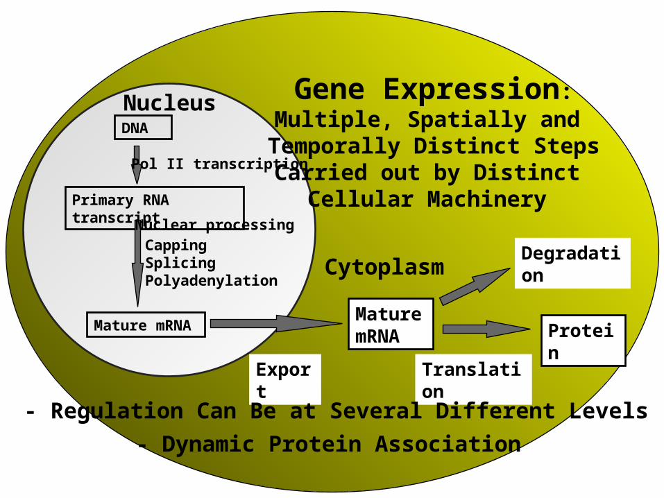
Nucleus DNA Primary RNA transcript Mature mRNA Pol II transcription Mature mRNA Protei n Nuclear processing Capping Splicing Polyadenylation Expor t Translati on Degradati on Gene Expression: Multiple, Spatially and Temporally Distinct Steps Carried out by Distinct Cellular Machinery Cytoplasm - Regulation Can Be at Several Different Levels - Dynamic Protein Association

Nucleus DNA Primary RNA transcript Mature mRNA Pol II transcription Mature mRNA Protein Nuclear processing Capping Splicing Polyadenylation ExportTranslation.
Dec 17, 2015
Welcome message from author
This document is posted to help you gain knowledge. Please leave a comment to let me know what you think about it! Share it to your friends and learn new things together.
Transcript
NucleusDNA
Primary RNA transcript
Mature mRNA
Pol II transcription
Mature mRNA
Protein
Nuclear processingCappingSplicingPolyadenylation
Export
Translation
Degradation
Gene Expression:Multiple, Spatially and
Temporally Distinct StepsCarried out by Distinct
Cellular Machinery
Cytoplasm
- Regulation Can Be at Several Different Levels
- Dynamic Protein Association
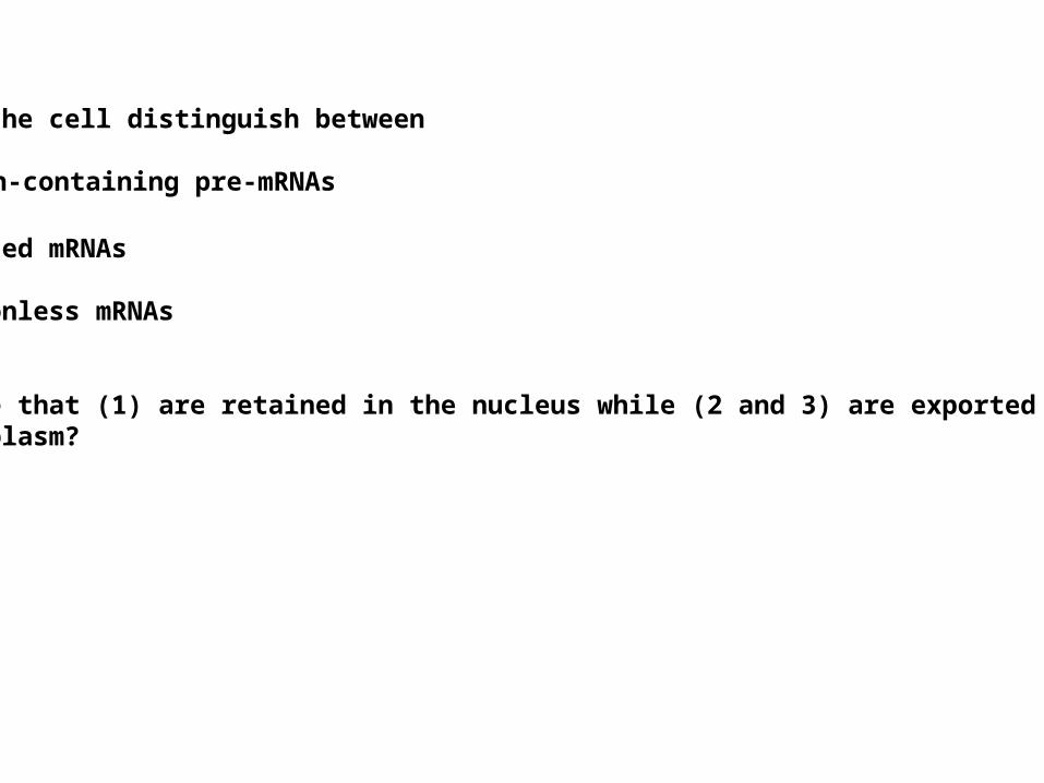
How can the cell distinguish between
(1) intron-containing pre-mRNAs
(2) spliced mRNAs
(3) intronless mRNAs
to ensure that (1) are retained in the nucleus while (2 and 3) are exported tothe cytoplasm?
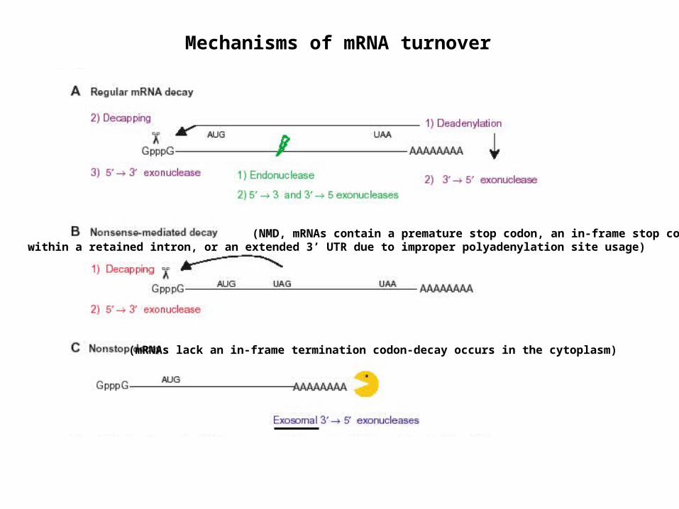
Mechanisms of mRNA turnover
(NMD, mRNAs contain a premature stop codon, an in-frame stop codonwithin a retained intron, or an extended 3’ UTR due to improper polyadenylation site usage)
(mRNAs lack an in-frame termination codon-decay occurs in the cytoplasm)
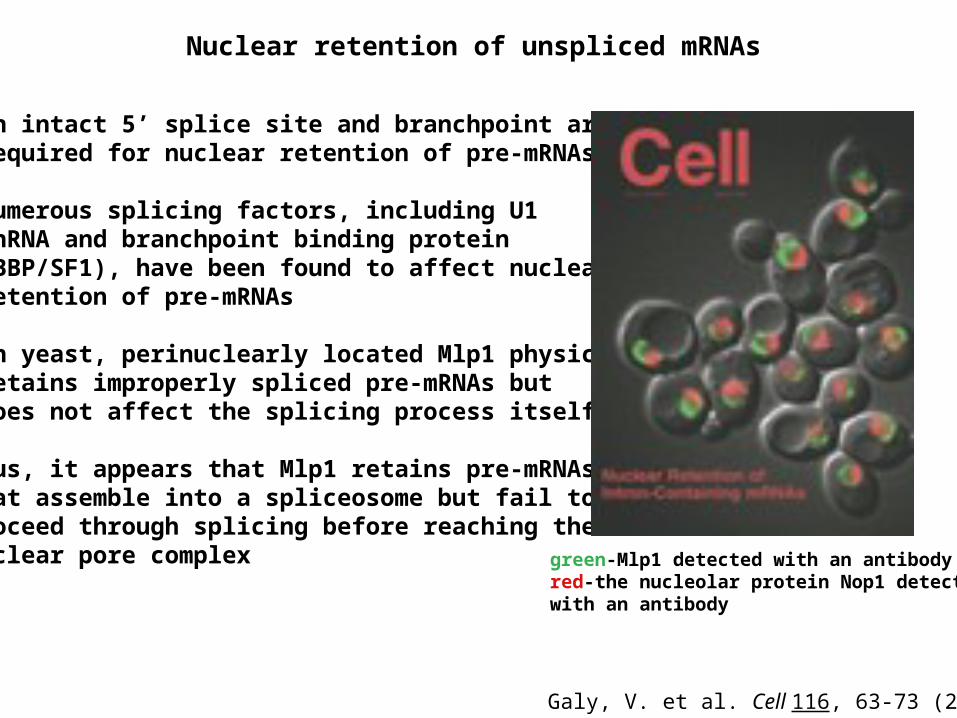
•An intact 5’ splice site and branchpoint are required for nuclear retention of pre-mRNAs
•Numerous splicing factors, including U1 snRNA and branchpoint binding protein (BBP/SF1), have been found to affect nuclear retention of pre-mRNAs
•In yeast, perinuclearly located Mlp1 physically retains improperly spliced pre-mRNAs but does not affect the splicing process itself
Thus, it appears that Mlp1 retains pre-mRNAsthat assemble into a spliceosome but fail toproceed through splicing before reaching thenuclear pore complex
green-Mlp1 detected with an antibodyred-the nucleolar protein Nop1 detectedwith an antibody
Galy, V. et al. Cell 116, 63-73 (2004)
Nuclear retention of unspliced mRNAs
Coupling of Transcriptional and Post-Transcriptional Events
mRNA Surveillance
RNA-Mediated Gene Silencing

A) CTD of RNA Pol II - binding platform for mRNA processing components B) TREX - couples TRanscription and EXportC) Exon Junction Complex (EJC) - splicing mark and coupler
Coupling of Transcriptional and Post-Transcriptional Events
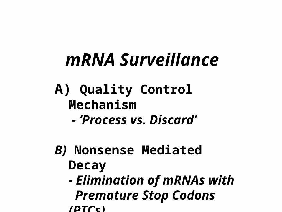
mRNA Surveillance
A) Quality Control Mechanism - ‘Process vs. Discard’
B) Nonsense Mediated Decay- Elimination of mRNAs with Premature Stop Codons (PTCs)
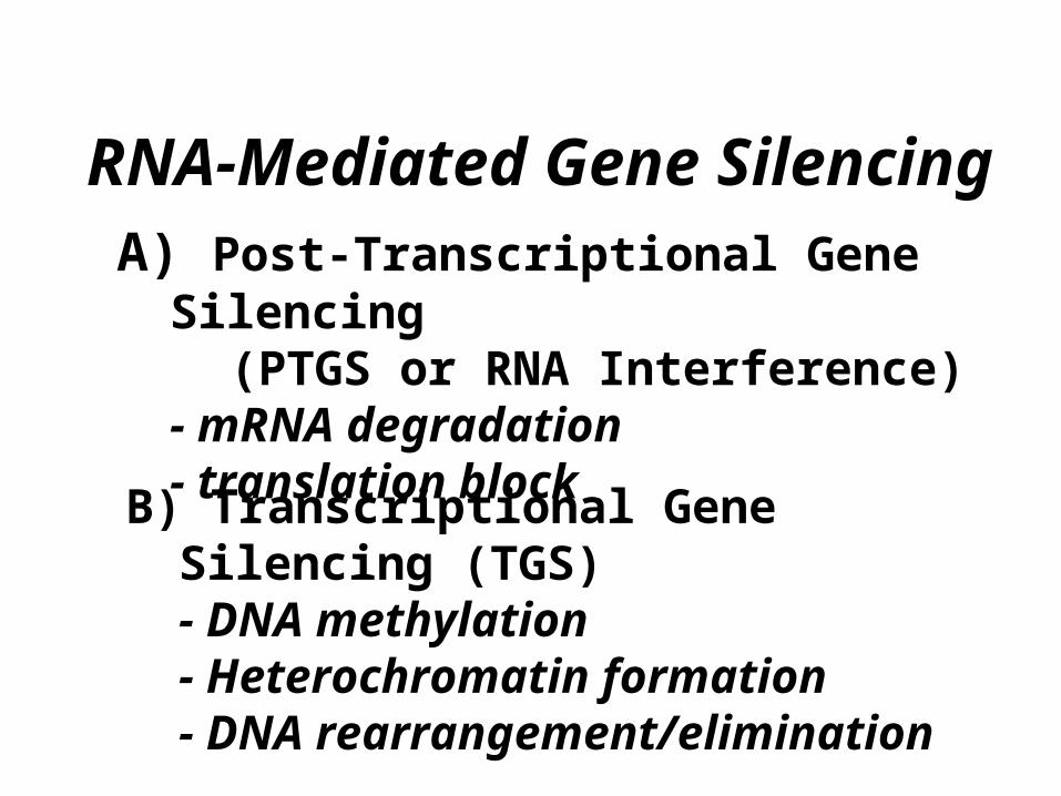
RNA-Mediated Gene SilencingA) Post-Transcriptional Gene Silencing (PTGS or RNA Interference)
- mRNA degradation- translation block
B) Transcriptional Gene Silencing (TGS)- DNA methylation - Heterochromatin formation - DNA rearrangement/elimination
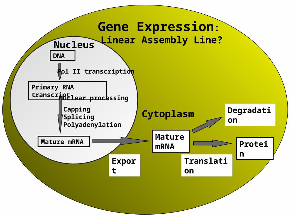
NucleusDNA
Primary RNA transcript
Mature mRNA
Pol II transcription
Mature mRNA
Protein
Nuclear processing
Export
Translation
DegradationCytoplasm
Gene Expression: Linear Assembly Line?
CappingSplicingPolyadenylation
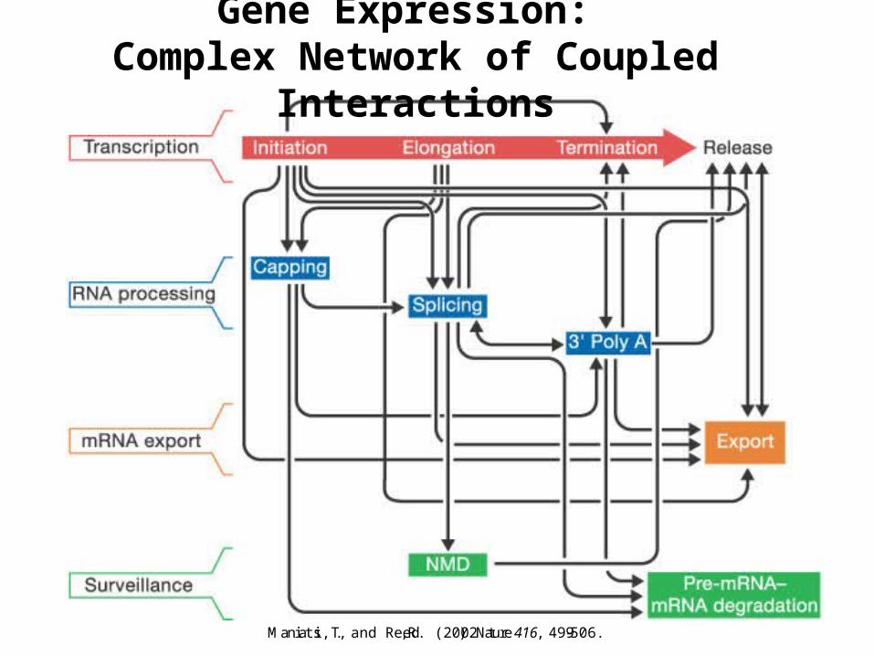
Gene Expression: Complex Network of Coupled Interactions
Maniatis, T., and Reed, R. (2002). Nature 416, 499-506.
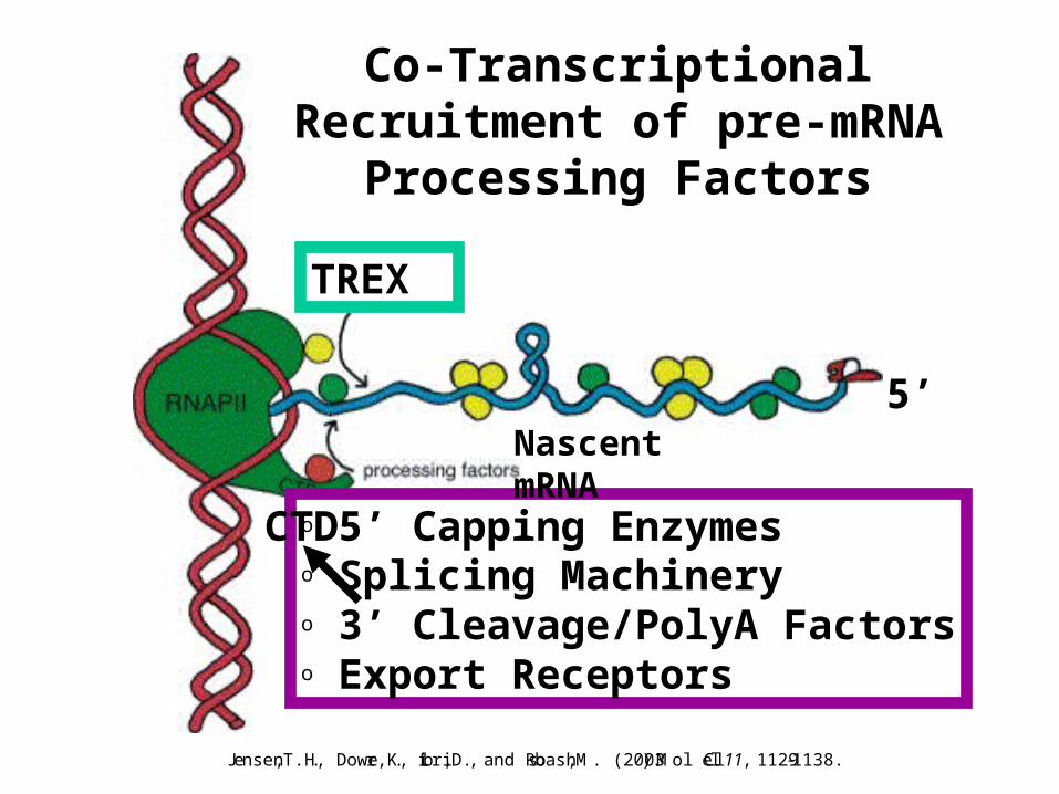
o 5’ Capping Enzymeso Splicing Machineryo 3’ Cleavage/PolyA Factorso Export Receptors
CTD
5’
Co-Transcriptional Recruitment of pre-mRNA Processing Factors
TREX
Nascent mRNA
Jensen, T.H., Dower, K., Libri, D., and Rosbash, M. (2003). Mol Cell 11 , 1129-1138.
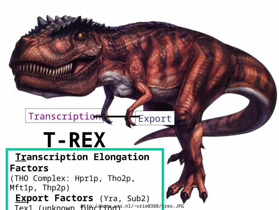
T-REX Transcription Elongation Factors (THO Complex: Hpr1p, Tho2p, Mft1p, Thp2p)
Export Factors (Yra, Sub2) Tex1 (unknown function)
Transcription Export
http://home.wxs.nl/~vrie0388/trex.JPG
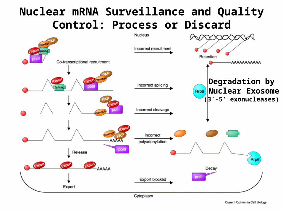
Degradation by Nuclear Exosome
(3’-5’ exonucleases)
Nuclear mRNA Surveillance and Quality Control: Process or Discard
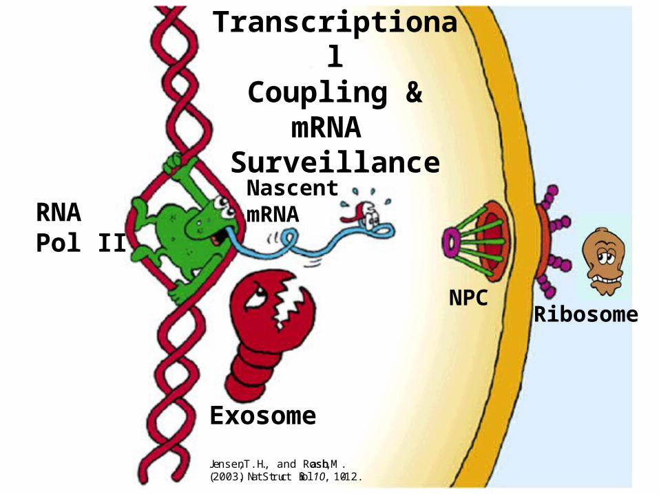
Exosome
RNA Pol II
Ribosome
TranscriptionalCoupling & mRNA
Surveillance
Nascent mRNA
NPC
Jensen, T.H., and Rosbash, M.(2003). Nat Struct Biol 10, 10-12.
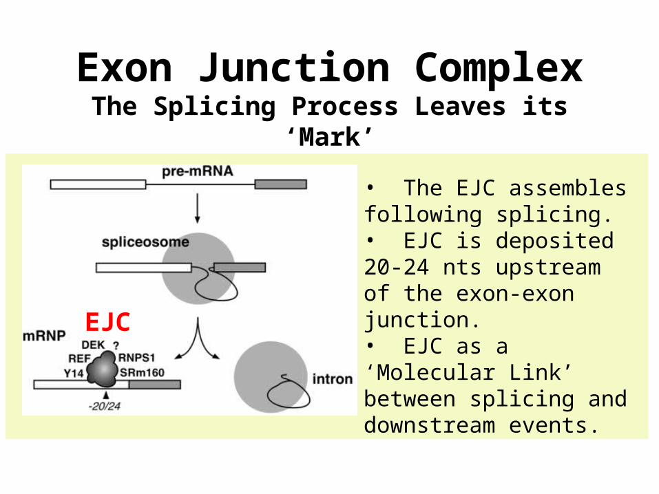
• The EJC assembles following splicing. • EJC is deposited 20-24 nts upstream of the exon-exon junction.• EJC as a ‘Molecular Link’ between splicing and downstream events.
Exon Junction ComplexThe Splicing Process Leaves its ‘Mark’
EJC
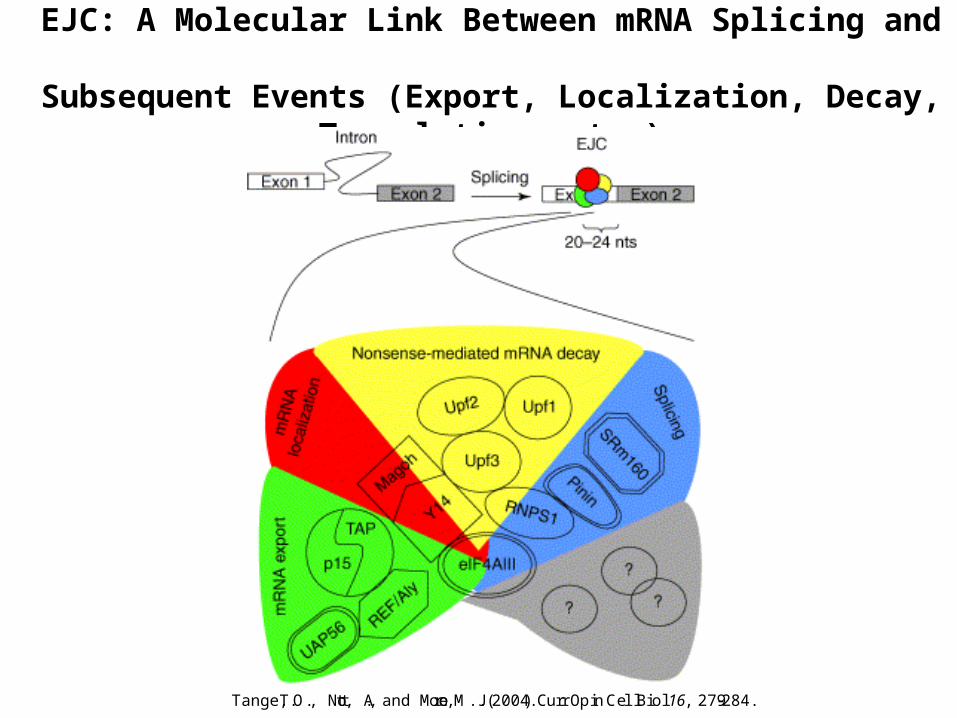
EJC: A Molecular Link Between mRNA Splicing and Subsequent Events (Export, Localization, Decay, Translation, etc.)
Tange, T.O., Nott, A., and Moore, M.J. (2004). Curr Opin Cell Biol 16, 279-284.
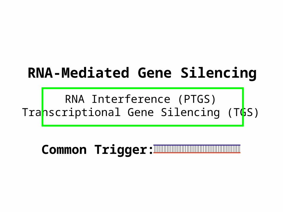
RNA-Mediated Gene Silencing
RNA Interference (PTGS)Transcriptional Gene Silencing (TGS)
Common Trigger:
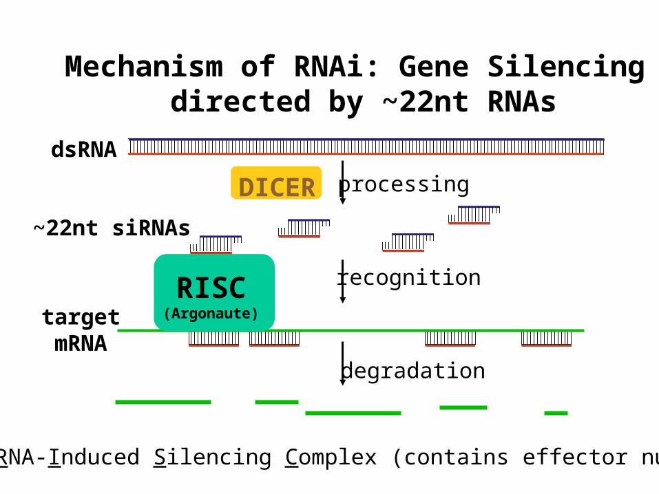
Mechanism of RNAi: Gene Silencing directed by ~22nt RNAs
dsRNA
~22nt siRNAs
targetmRNA
processing
degradation
recognition
DICER
RISC(Argonaute)
RISC: RNA-Induced Silencing Complex (contains effector nuclease)
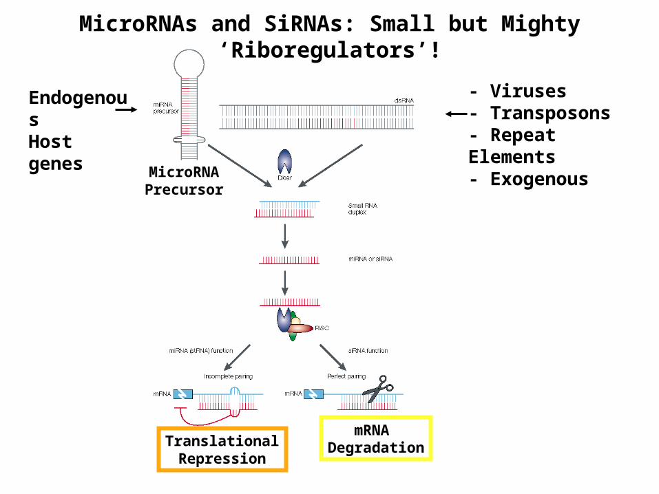
EndogenousHost genes
- Viruses- Transposons- Repeat Elements- Exogenous
MicroRNAs and SiRNAs: Small but Mighty ‘Riboregulators’!
mRNA DegradationTranslational
Repression
MicroRNAPrecursor
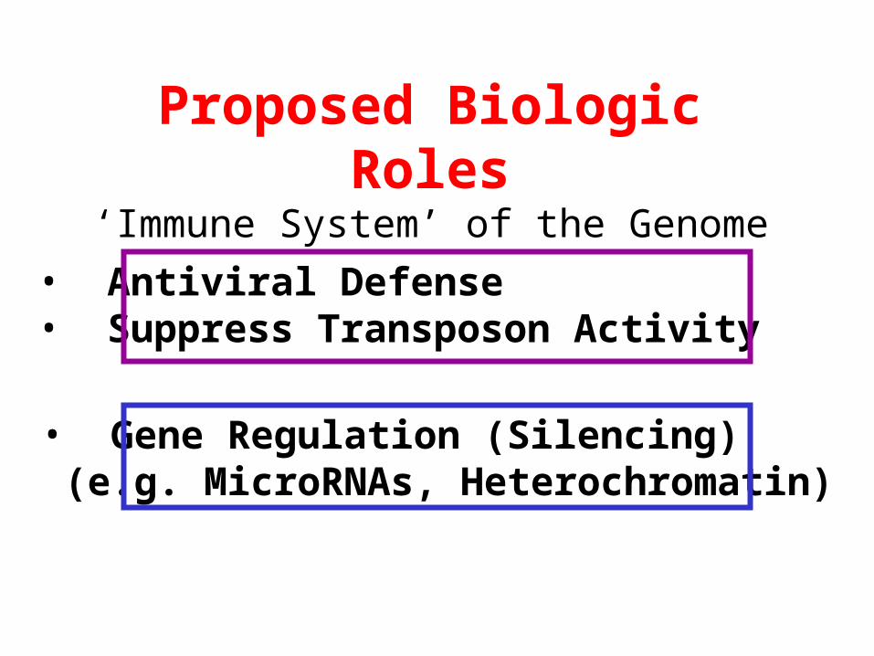
Proposed Biologic Roles
• Antiviral Defense• Suppress Transposon Activity
• Gene Regulation (Silencing) (e.g. MicroRNAs, Heterochromatin)
‘Immune System’ of the Genome
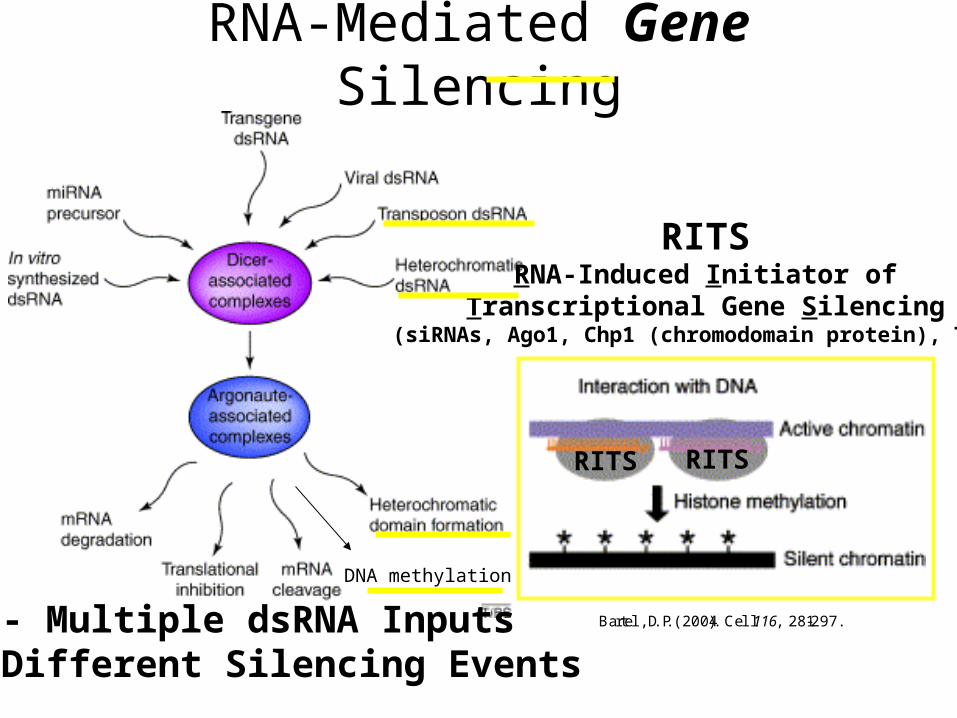
RNA-Mediated Gene Silencing
RITSRNA-Induced Initiator of
Transcriptional Gene Silencing(siRNAs, Ago1, Chp1 (chromodomain protein), Tas3)
RITSRITS
- Multiple dsRNA Inputs - Different Silencing Events
Bartel, D.P. (2004). Cell 116 , 281-297.
DNA methylation
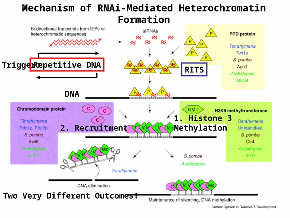
Repetitive DNARITS
Mechanism of RNAi-Mediated Heterochromatin Formation
DNA
1. Histone 3Methylation2. Recruitment
Two Very Different Outcomes!
Trigger:

TRANSLATION
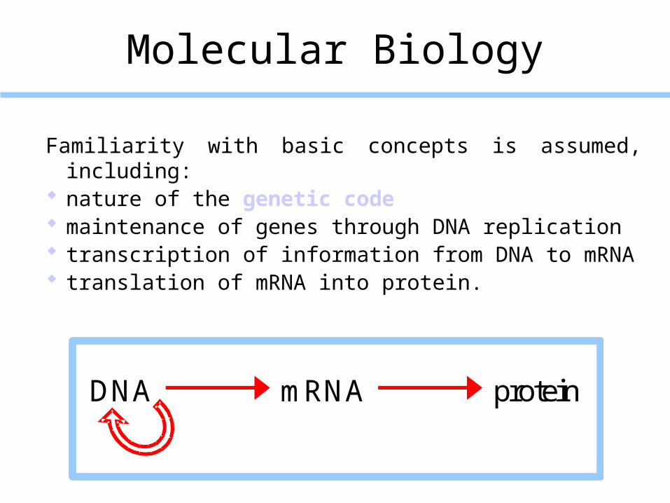
Molecular Biology
Familiarity with basic concepts is assumed, including: nature of the genetic code maintenance of genes through DNA replication transcription of information from DNA to mRNA translation of mRNA into protein.
DNA mRNA protein
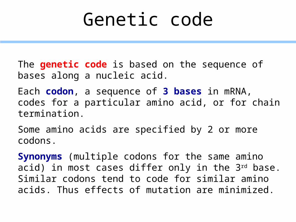
Genetic code
The genetic code is based on the sequence of bases along a nucleic acid.
Each codon, a sequence of 3 bases in mRNA, codes for a particular amino acid, or for chain termination.
Some amino acids are specified by 2 or more codons.
Synonyms (multiple codons for the same amino acid) in most cases differ only in the 3rd base. Similar codons tend to code for similar amino acids. Thus effects of mutation are minimized.
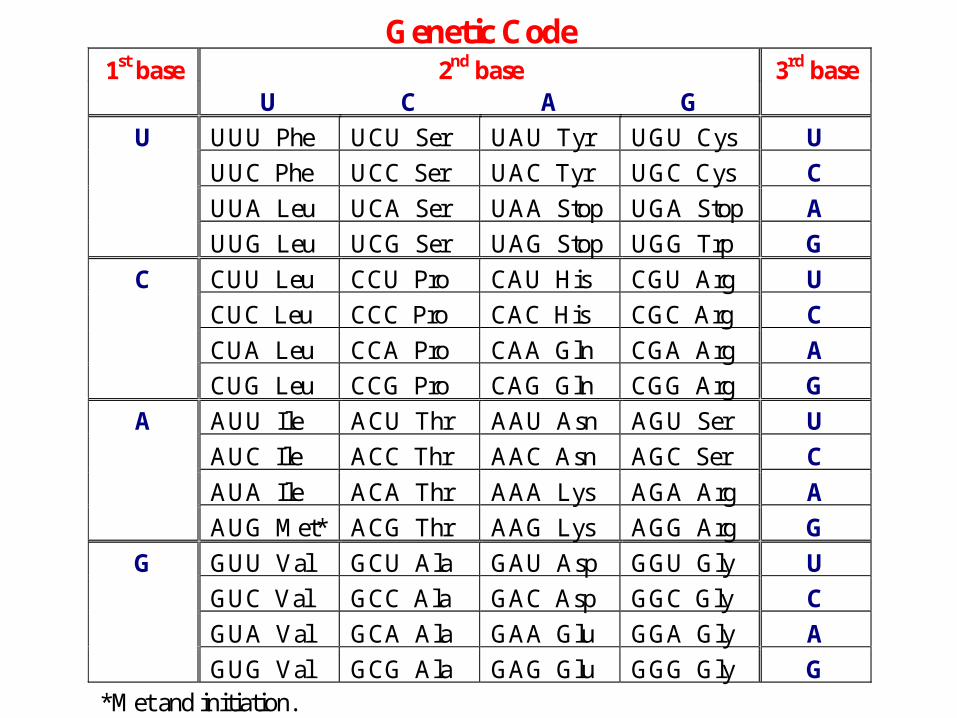
Genetic Code2nd base1st base
U C A G3rd base
UUU Phe UCU Ser UAU Tyr UGU Cys U
UUC Phe UCC Ser UAC Tyr UGC Cys C
UUA Leu UCA Ser UAA Stop UGA Stop A
U
UUG Leu UCG Ser UAG Stop UGG Trp G
CUU Leu CCU Pro CAU His CGU Arg U
CUC Leu CCC Pro CAC His CGC Arg C
CUA Leu CCA Pro CAA Gln CGA Arg A
C
CUG Leu CCG Pro CAG Gln CGG Arg G
AUU Ile ACU Thr AAU Asn AGU Ser U
AUC Ile ACC Thr AAC Asn AGC Ser C
AUA Ile ACA Thr AAA Lys AGA Arg A
A
AUG Met* ACG Thr AAG Lys AGG Arg G
GUU Val GCU Ala GAU Asp GGU Gly U
GUC Val GCC Ala GAC Asp GGC Gly C
GUA Val GCA Ala GAA Glu GGA Gly A
G
GUG Val GCG Ala GAG Glu GGG Gly G*Met and initiation.
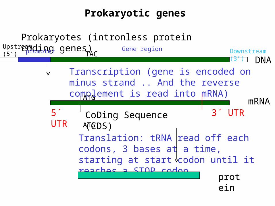
Prokaryotic genes
Downstream (3’)
Prokaryotes (intronless protein coding genes)
promoter Gene regionUpstream (5’)
Transcription (gene is encoded on minus strand .. And the reverse complement is read into mRNA)
DNA
mRNA5´ UTR 3´ UTRCoDing Sequence (CDS)
ATG
ATG
TAC
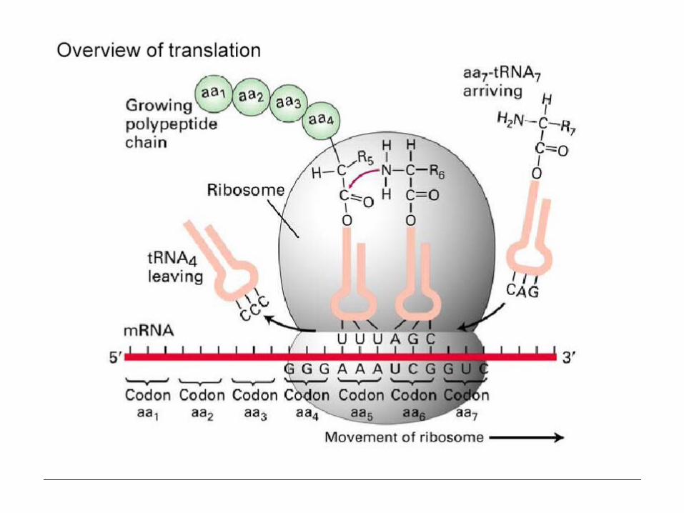
Translation: tRNA read off each codons, 3 bases at a time, starting at start codon until it reaches a STOP codon.
protein
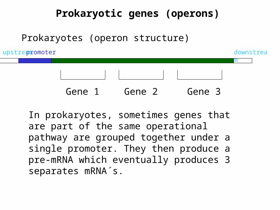
Prokaryotic genes (operons)
downstream
Prokaryotes (operon structure)
promoterupstream
Gene 1 Gene 2 Gene 3
In prokaryotes, sometimes genes that are part of the same operational pathway are grouped together under a single promoter. They then produce a pre-mRNA which eventually produces 3 separates mRNA´s.
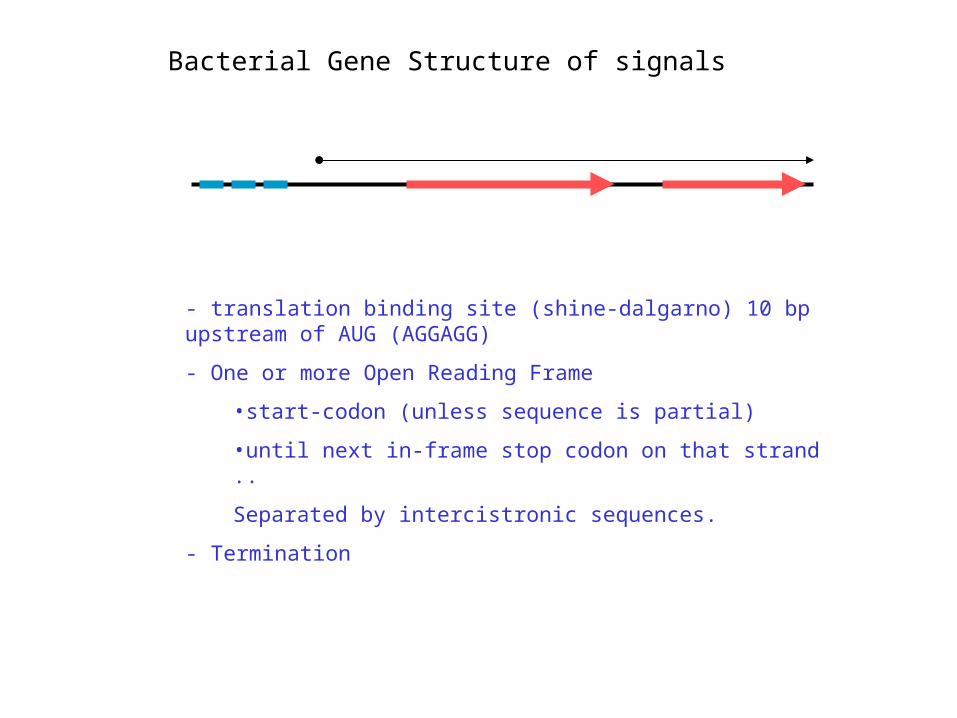
- translation binding site (shine-dalgarno) 10 bp upstream of AUG (AGGAGG)
- One or more Open Reading Frame
•start-codon (unless sequence is partial)
•until next in-frame stop codon on that strand ..
Separated by intercistronic sequences.
- Termination
Bacterial Gene Structure of signals
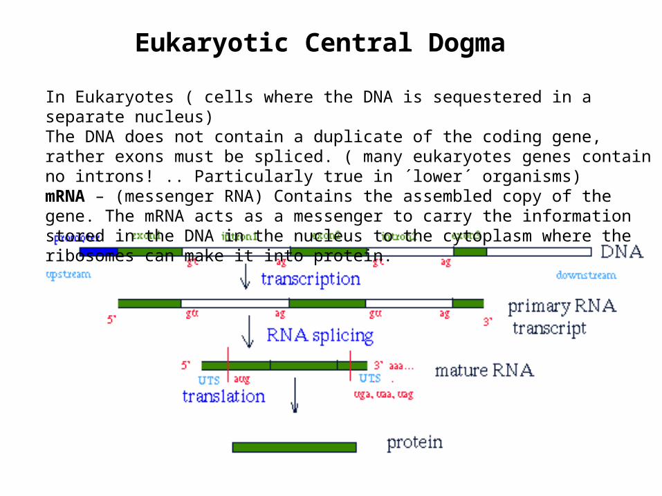
In Eukaryotes ( cells where the DNA is sequestered in a separate nucleus) The DNA does not contain a duplicate of the coding gene, rather exons must be spliced. ( many eukaryotes genes contain no introns! .. Particularly true in ´lower´ organisms)mRNA – (messenger RNA) Contains the assembled copy of the gene. The mRNA acts as a messenger to carry the information stored in the DNA in the nucleus to the cytoplasm where the ribosomes can make it into protein.
Eukaryotic Central Dogma
tRNA
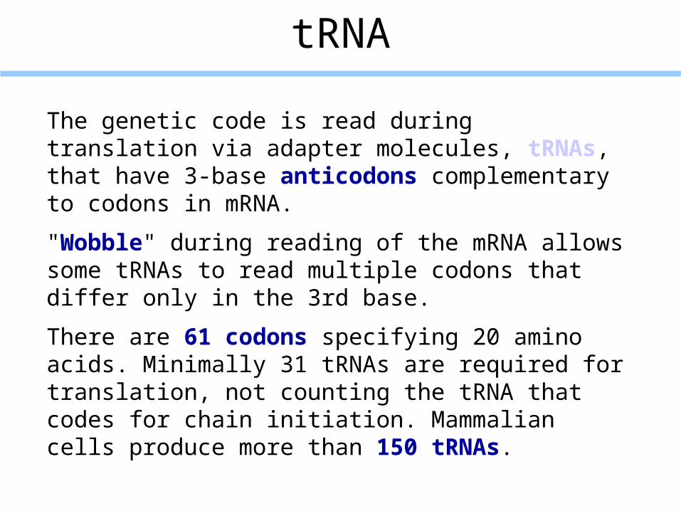
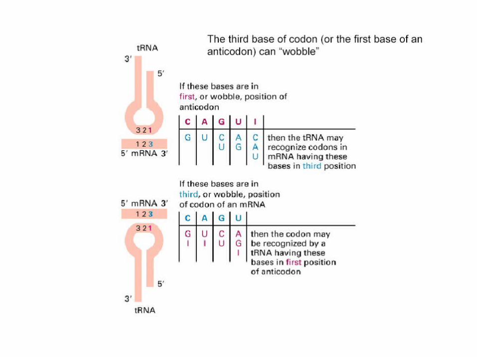
The genetic code is read during translation via adapter molecules, tRNAs, that have 3-base anticodons complementary to codons in mRNA.
"Wobble" during reading of the mRNA allows some tRNAs to read multiple codons that differ only in the 3rd base.
There are 61 codons specifying 20 amino acids. Minimally 31 tRNAs are required for translation, not counting the tRNA that codes for chain initiation. Mammalian cells produce more than 150 tRNAs.
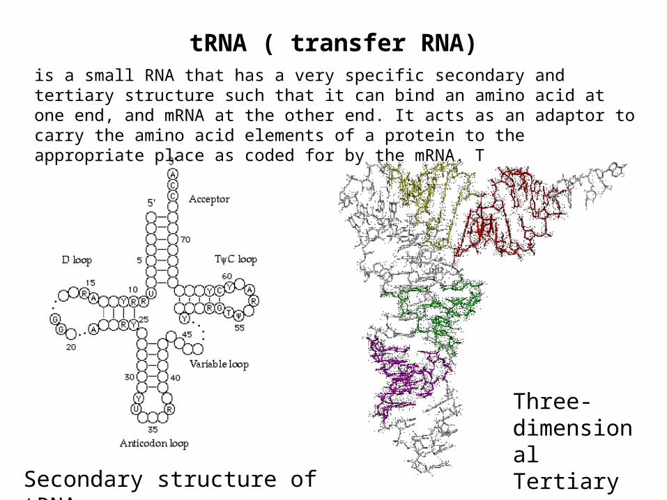
tRNA ( transfer RNA)is a small RNA that has a very specific secondary and tertiary structure such that it can bind an amino acid at one end, and mRNA at the other end. It acts as an adaptor to carry the amino acid elements of a protein to the appropriate place as coded for by the mRNA. T
Secondary structure of tRNA
Three-dimensional Tertiary structure
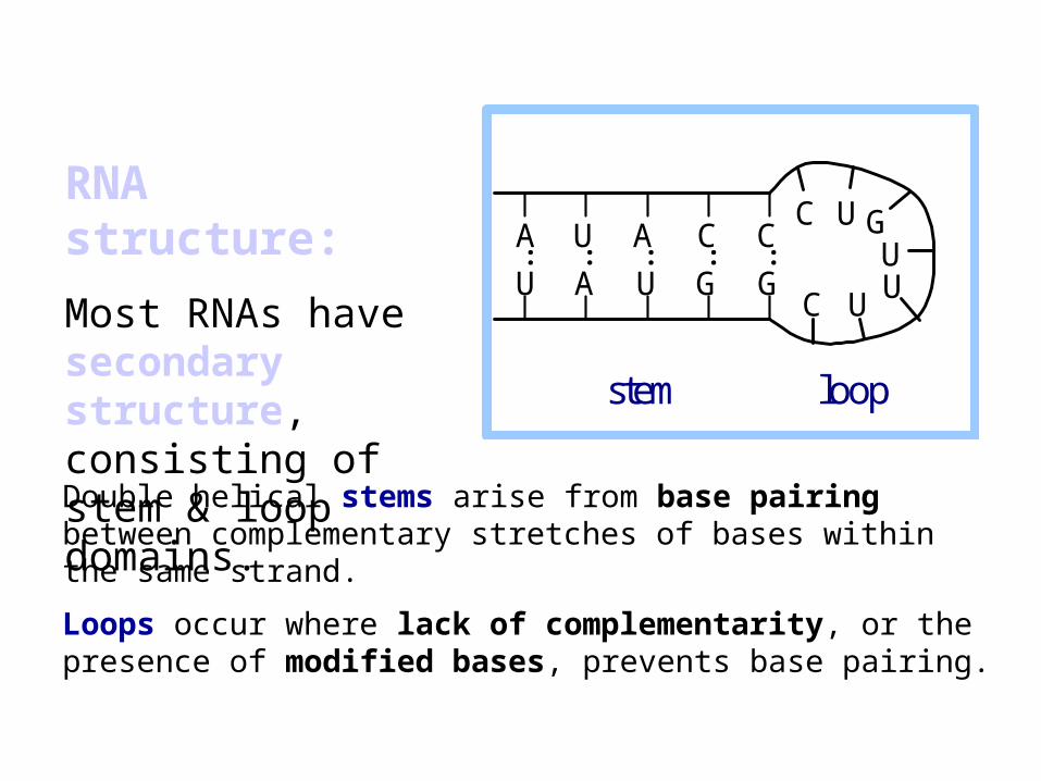
Double helical stems arise from base pairing between complementary stretches of bases within the same strand.
Loops occur where lack of complementarity, or the presence of modified bases, prevents base pairing.
A U A C C
U A U G G
C U
C U
G U U
stem loop
: : : : :
RNA structure:
Most RNAs have secondary structure, consisting of stem & loop domains.
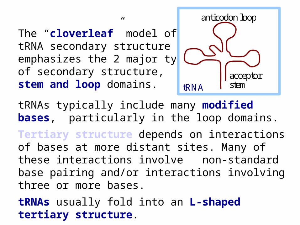
The “cloverleaf” model of tRNA secondary structure emphasizes the 2 major typesof secondary structure, stem and loop domains.
tRNAs typically include many modified bases, particularly in the loop domains.
Tertiary structure depends on interactions of bases at more distant sites. Many of these interactions involve non-standard base pairing and/or interactions involving three or more bases.
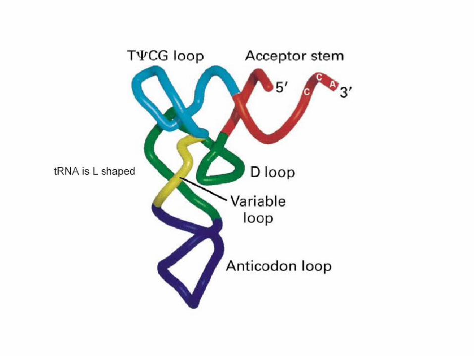
tRNAs usually fold into an L-shaped tertiary structure.
anticodon loop
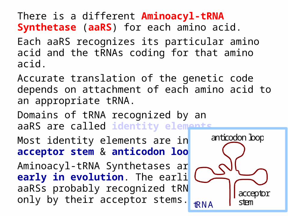
acceptor stem tRNA

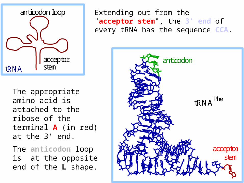
The appropriate amino acid is attached to the ribose of the terminal A (in red) at the 3' end.
The anticodon loop is at the opposite end of the L shape.
anticodon
acceptorstem
tRNAPhe
anticodon loop
acceptor stem tRNA
Extending out from the "acceptor stem", the 3' end of every tRNA has the sequence CCA.

Tertiary base pairs
#46(m7G)
#22G #13
C
Tertiary basepairs in tRNAPhe
#46(m7G)
#22G
#13C
Tertiary basepairs in tRNAPhe
Non-standard H bond interactions, some linking 3 bases, help stabilize the L-shaped tertiary structure of tRNA. This example is from NDB file 1TN2. H atoms are not shown.

Aminoacyl-tRNA Synthetases catalyze linkage of the appropriate amino acid to each tRNA. The reaction occurs in two steps.
In step 1, an O atom of the amino acid -carboxyl attacks the P atom of the initial phosphate of ATP.
O
OHOH
HH
H
CH2
H
OPOPOP O
O
O O
O O
O
RHC C
NH3+
O
O
O
OHOH
HH
H
CH2
H
OPOC
O
O
HCR
NH2
O
Adenine
Adenine
ATP Amino acid
Aminoacyl-AMP
PPi
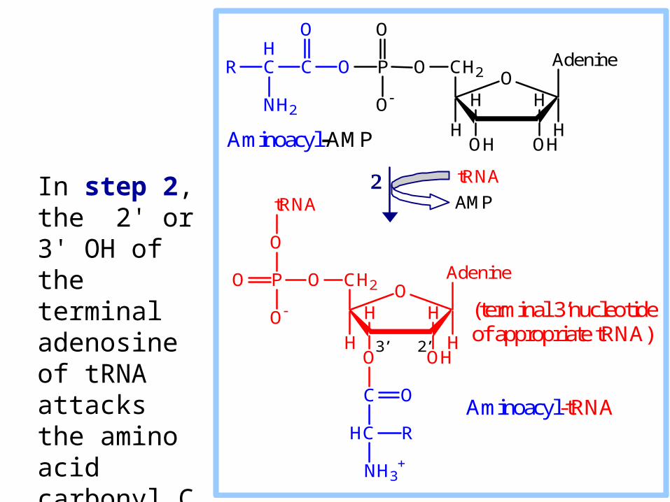
In step 2, the 2' or 3' OH of the terminal adenosine of tRNA attacks the amino acid carbonyl C atom.
O
OHOH
HH
H
CH2
H
OPOC
O
O
HCR
NH2
O
Adenine
O
OHO
HH
H
CH2
H
OPO
O
O
Adenine
tRNA
C
HC
O
NH3+
R
tRNA
AMP
Aminoacyl-AMP
Aminoacyl-tRNA
(terminal 3’nucleotide of appropriate tRNA)
3’ 2’
Aminoacyl-tRNA Synthetase
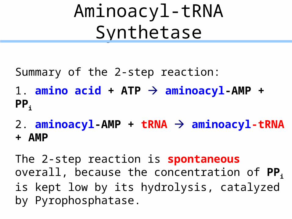
Summary of the 2-step reaction:
1. amino acid + ATP aminoacyl-AMP + PPi
2. aminoacyl-AMP + tRNA aminoacyl-tRNA + AMP
The 2-step reaction is spontaneous overall, because the concentration of PPi is kept low by its hydrolysis, catalyzed by Pyrophosphatase.
There is a different Aminoacyl-tRNA Synthetase (aaRS) for each amino acid.
Each aaRS recognizes its particular amino acid and the tRNAs coding for that amino acid.
Accurate translation of the genetic code depends on attachment of each amino acid to an appropriate tRNA.
Domains of tRNA recognized by an aaRS are called identity elements.
Most identity elements are in the acceptor stem & anticodon loop.
Aminoacyl-tRNA Synthetases arose early in evolution. The earliest aaRSs probably recognized tRNAsonly by their acceptor stems.
anticodon loop
acceptor stem tRNA
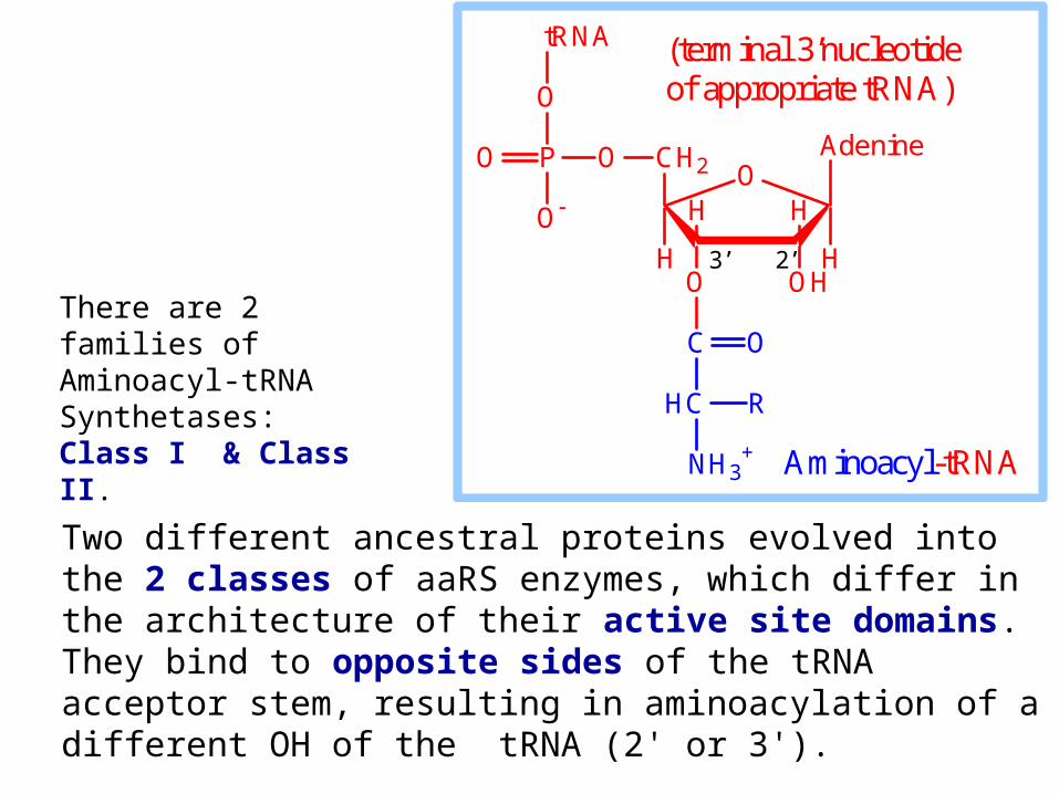
Two different ancestral proteins evolved into the 2 classes of aaRS enzymes, which differ in the architecture of their active site domains. They bind to opposite sides of the tRNA acceptor stem, resulting in aminoacylation of a different OH of the tRNA (2' or 3').
O
OHO
HH
H
CH2
H
OPO
O
O
Adenine
tRNA
C
HC
O
NH3+
R
Aminoacyl-tRNA
(terminal 3’nucleotide of appropriate tRNA)
3’ 2’
There are 2 families of Aminoacyl-tRNA Synthetases: Class I & Class II.

Class I aaRSs:
Identity elements usually include residues of the anticodon loop & acceptor stem.
Class I aaRSs aminoacylate the 2'-OH of adenosine at their 3' end.
Class II aaRSs:
Identity elements for some Class II enzymes do not include the anticodon domain.
Class II aaRSs tend to aminoacylate the 3'-OH of adenosine at their 3' end.
Proofreading/quality control:
Some Aminoacyl-tRNA Synthetases are known to have separate catalytic sites that release by hydrolysis inappropriate amino acids that are misacylated or mis-transferred to tRNA.
E.g., the aa-tRNA Synthetase for isoleucine (IleRS) a small percentage of the time activates the closely related amino acid valine to valine-AMP.
After valine is transferred to tRNAIle, to form Val-tRNAIle, it is removed by hydrolysis at a separate active site of IleRS that accommodates Val but not the larger Ile.
In some bacteria, editing of some misacylated tRNAs is carried out by separate proteins that may be evolutionary precursors to editing domains of aa-tRNA Synthetases.
Some amino acids are modified after being linked to tRNA.
E.g., in prokaryotes & in mitochondria the initiator tRNAfMet is first charged with methionine.
Methionyl-tRNA formyltransferase then catalyzes formylation of the methionine moiety, using THF as formyl donor, to yield fMet-tRNAfMet.
In some prokaryotes, a non-discriminating aaRS loads aspartate onto tRNAAsn.
The aspartate moiety is then converted by an amido-transferase to asparagine, yielding Asn-tRNAAsn.
Glu-tRNAGln is similarly formed and converted to Gln-tRNAGln in such organisms.

RIBOSOMES
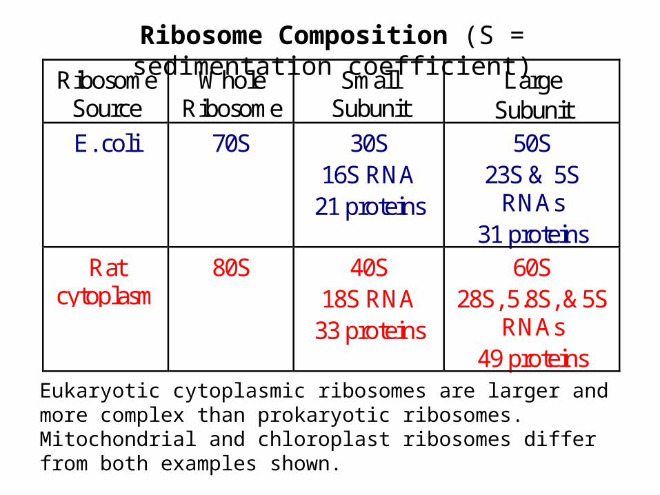
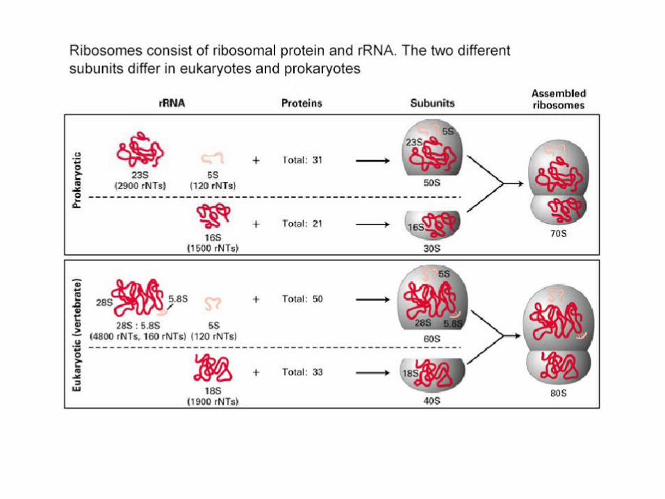
Eukaryotic cytoplasmic ribosomes are larger and more complex than prokaryotic ribosomes. Mitochondrial and chloroplast ribosomes differ from both examples shown.
RibosomeSource
WholeRibosome
SmallSubunit
LargeSubunit
E. coli 70S 30S16S RNA
21 proteins
50S23S & 5S
RNAs31 proteins
Ratcytoplasm
80S 40S18S RNA
33 proteins
60S28S, 5.8S, &5S
RNAs49 proteins
Ribosome Composition (S = sedimentation coefficient)
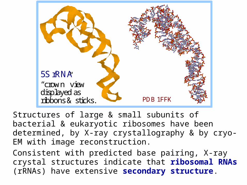
Structures of large & small subunits of bacterial & eukaryotic ribosomes have been determined, by X-ray crystallography & by cryo-EM with image reconstruction.
Consistent with predicted base pairing, X-ray crystal structures indicate that ribosomal RNAs (rRNAs) have extensive secondary structure.
5S rRNA “crown” view displayed as ribbons & sticks. PDB 1FFK
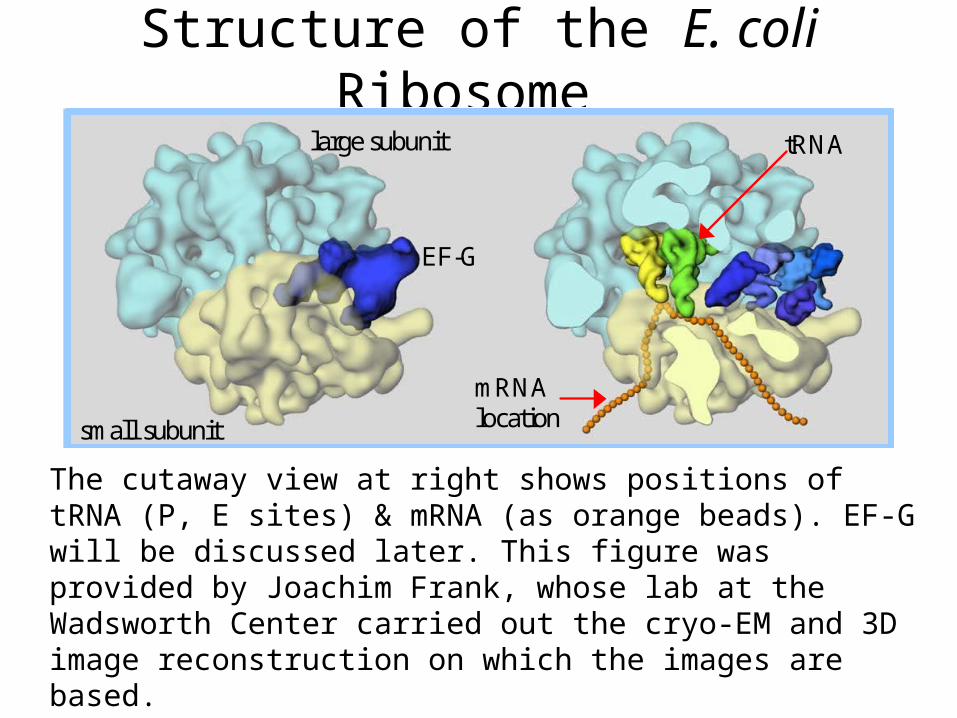
Structure of the E. coli Ribosome
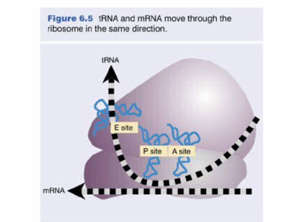
The cutaway view at right shows positions of tRNA (P, E sites) & mRNA (as orange beads). EF-G will be discussed later. This figure was provided by Joachim Frank, whose lab at the Wadsworth Center carried out the cryo-EM and 3D image reconstruction on which the images are based.
small subunit
large subunit
mRNAlocation
EF-G
tRNA
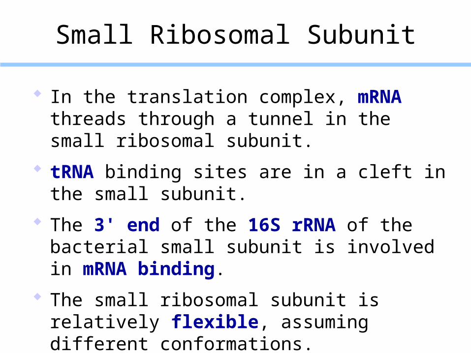
Small Ribosomal Subunit
In the translation complex, mRNA threads through a tunnel in the small ribosomal subunit.
tRNA binding sites are in a cleft in the small subunit.
The 3' end of the 16S rRNA of the bacterial small subunit is involved in mRNA binding.
The small ribosomal subunit is relatively flexible, assuming different conformations.
E.g., the 30S subunit of a bacterial ribosome was found to undergo specific conformational changes when interacting with a translation initiation factor.
The overall shape of the 30S ribosomal subunit is largely determined by the rRNA. The rRNA mainly consists of double helices (stems) connected by single-stranded loops.The proteins generally have globular domains, as well as long extensions that interact with rRNA and may stabilize interactions between RNA helices.
30S ribosomal subunit PDB 1FJF Small ribosomal subunit of a thermophilic bacterium: rRNA in monochrome;proteins in varied colors. spacefill display ribbons

Large ribosome subunit:
The interior of the large subunit is mostly RNA.
Proteins are distributed mainly on the surface.
Some proteins have long tails that extend into the interior of the complex.
These tails, which are highly basic, interact with the negatively charged RNA.
PDB 1FFK
Large Ribosome Subunit
"Crown" view with RNAs blue, in spacefill; proteins red, as backbone.
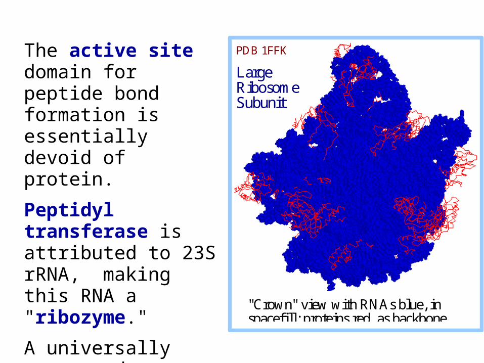
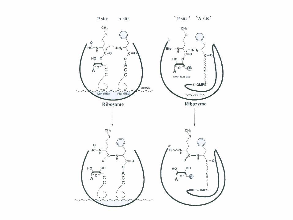
The active site domain for peptide bond formation is essentially devoid of protein.
Peptidyl transferase is attributed to 23S rRNA, making this RNA a "ribozyme."
A universally conserved adenosine base serves as a general acid base during peptide bond formation.
PDB 1FFK
Large Ribosome Subunit
"Crown" view with RNAs blue, in spacefill; proteins red, as backbone.
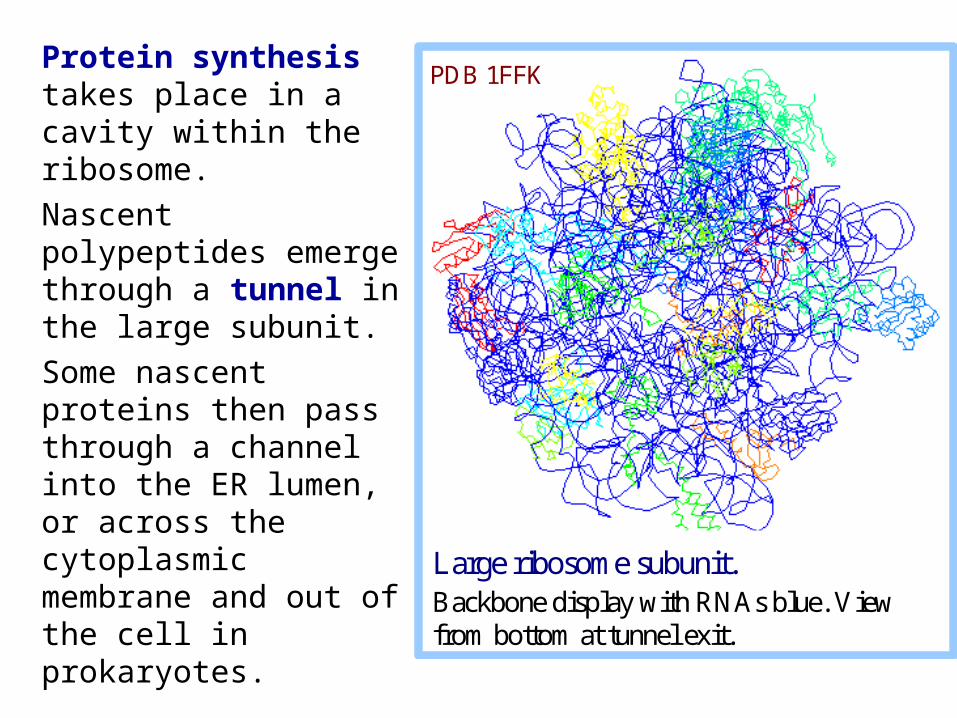
Protein synthesis takes place in a cavity within the ribosome.
Nascent polypeptides emerge through a tunnel in the large subunit.
Some nascent proteins then pass through a channel into the ER lumen, or across the cytoplasmic membrane and out of the cell in prokaryotes.
Large ribosome subunit. Backbone display with RNAs blue. View from bottom at tunnel exit.
PDB 1FFK
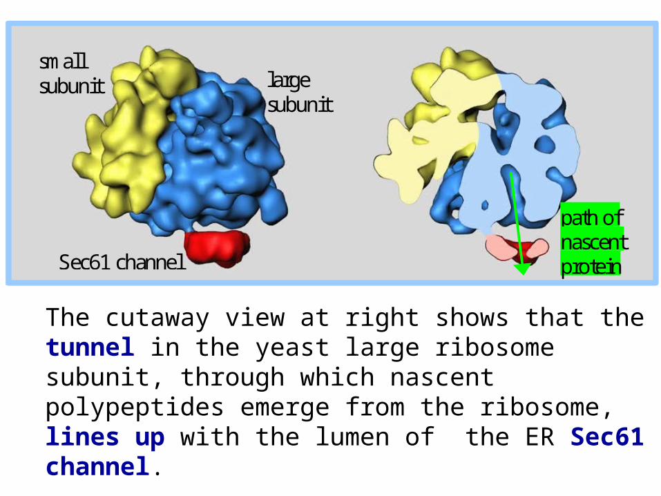
The cutaway view at right shows that the tunnel in the yeast large ribosome subunit, through which nascent polypeptides emerge from the ribosome, lines up with the lumen of the ER Sec61 channel.
small subunit large
subunit
Sec61 channel
path of nascent protein
Related Documents