Copyright 0 1996 by the Genetics Society of America Nucleotide Variation at the Gpdh Locus in the Genus DrosophiZa R. Spencer Wells Museum of Comparative Zoology, Haruard University, Cambridge, Massachusetts 02138 Manuscript received May 27, 1995 Accepted for publication February 8, 1996 ABSTRACT The Gpdh locus was sequenced in a broad range of Drosophila species. In contrast to the extreme evolutionary constraint seen at the amino acid level, the synonymous sites evolve at rates comparable to those of other genes. Gpdh nucleotide sequences were used to infer a phylogenetic tree, and the relationships among the species of the obscura group were examined in detail. A survey of nucleotide polymorphism within D. pseudoobscura revealed no amino acid variation in this species. Applying a modified McDonald-Kreitman test, the amino acid divergence between species in the obscura group does not appear to be excessive, implying that drift is adequate to explain the patterns of amino acid change at this locus. In addition, the level of polymorphism at the Gpdh locus in D. pseudoobscura is comparable to that found at other loci, as determined by a Hudson-Kreitman-Aguade test. Thus, the pattern of nucleotide variation within and between species at the Gpdh locus is consistent with a neutral model. T HE study of nucleotide variation in natural popula- tions has revolutionized experimental evolution- ary genetics. By comparing DNA sequences within and between closely related species, we are able to detect the results of purifying selection (KREITMAN 1983), bal- ancing selection (HUDSON et al. 1987) and adaptive evo- lution (MCDONALD and KREITMAN 1991). The dualna- ture of a DNA coding sequence allows these inferences to be made: each sequence of nucleotides consists of those that are likely to be subject to strong selective forces (nonsynonymous sites) and those that arelargely neutral (synonymous sites). If nucleotide variation is partitioned into synonymous and nonsynonymous classes, the action of selection is detected as a significant deviation from the expected values in either class. Be- cause of the decoupling of synonymousand nonsynony- mous variation, DNA sequences are much more infor- mation-rich than amino acid sequences. While evolutionary geneticists have historically stud- ied those genes showing a moderate-to-high level of protein variation within species (KEITH 1983; KREITMAN 1983; RILEY et al. 1992), the phenomenon of interspe- cific protein variation for genes that are monomorphic within species has not been adequately investigated. For example, the Adh locus, encoding the enzyme alcohol dehydrogenase (E.C. 1.1.1.99), has been sequenced in many species of the genus Drosophila. As it shows no electrophoretic variation in many of these species, it is oftentaken as the paradigm of monomorphic locus evolution (SCHAEFFER and MILLER 1992a). However, when levels of electrophoretic variation across many species are compared, Adh falls in the middle of the Curresponding author; R. Spencer Wells, Department of Structural Biology, Fairchild Center, Stanford University School of Medicine, Stanford, CA 94305. E-mail: [email protected] Genetics 143: 375-384 (May, 1996) distribution of average heterozygosities (POWELL 1976; WELLS 1995). The Gpdh locus (encoding glycerol-3- phosphate dehydrogenase, E.C. 1.1.1.8), in contrast, is monomorphic in almost all Drosophila species. Of the 205 species that have been assayed for electrophoretic variation at this locus, only two exhibit variant alleles at frequencies high enough to be classified aspolymor- phic: D. melanogaster and D. subarctica (LAKOVAARA et al. 1977; DE STORDEUR and PASTEUR, 1978; CoWE et al. 1979; LAJSOVAARA and KERANEN 1980). The Gpdh locus then is an excellent model system for studies on the molecular evolution of monomorphic proteins. I have undertaken a study of the nucleotide sequence variation and divergence at this locus in a broadsample of Drosophila species: D. pseudoobscura, D. miranda, D. af$nis, D. azteca, D. ambigua, D. bfasciata, D. subobscura (all members of the obscura species group and with D. melanogaster, part of the subgenus Sophophora), D. mela- nogaster, D. busckii (subgenus Dorsilopha), D. virilis (sub- genus Drosophila) and D. lebanonensis (subgenus Scapte drosophila). The species were chosen to provide a range of comparisons among both closely and distantly re- lated taxa, and population variation was assayed within a species (D. pseudoobscura) where good comparative data exists from other loci. Two questions have been adressed in thepresent study: (1) Is theamino acid divergence between species at this locus the result of drift or adaptive evolution? (2) What insights can this locus provide into the phylogenetic relationships among the species studied? MATERIALS AND METHODS Fly stocks: Stocks of D. .finis, D. azteca, D. ambigua, D. bijasciata, D. busckii, and D. lebanonensis were obtained from the National Drosophila Species Resource Center (Bowling Green, OH). D. subobscura genomic DNA was obtained from

Welcome message from author
This document is posted to help you gain knowledge. Please leave a comment to let me know what you think about it! Share it to your friends and learn new things together.
Transcript

Copyright 0 1996 by the Genetics Society of America
Nucleotide Variation at the Gpdh Locus in the Genus DrosophiZa
R. Spencer Wells
Museum of Comparative Zoology, Haruard University, Cambridge, Massachusetts 02138 Manuscript received May 27, 1995
Accepted for publication February 8, 1996
ABSTRACT The Gpdh locus was sequenced in a broad range of Drosophila species. In contrast to the extreme
evolutionary constraint seen at the amino acid level, the synonymous sites evolve at rates comparable to those of other genes. Gpdh nucleotide sequences were used to infer a phylogenetic tree, and the relationships among the species of the obscura group were examined in detail. A survey of nucleotide polymorphism within D. pseudoobscura revealed no amino acid variation in this species. Applying a modified McDonald-Kreitman test, the amino acid divergence between species in the obscura group does not appear to be excessive, implying that drift is adequate to explain the patterns of amino acid change at this locus. In addition, the level of polymorphism at the Gpdh locus in D. pseudoobscura is comparable to that found at other loci, as determined by a Hudson-Kreitman-Aguade test. Thus, the pattern of nucleotide variation within and between species at the Gpdh locus is consistent with a neutral model.
T HE study of nucleotide variation in natural popula- tions has revolutionized experimental evolution-
ary genetics. By comparing DNA sequences within and between closely related species, we are able to detect the results of purifying selection (KREITMAN 1983), bal- ancing selection (HUDSON et al. 1987) and adaptive evo- lution (MCDONALD and KREITMAN 1991). The dual na- ture of a DNA coding sequence allows these inferences to be made: each sequence of nucleotides consists of those that are likely to be subject to strong selective forces (nonsynonymous sites) and those that are largely neutral (synonymous sites). If nucleotide variation is partitioned into synonymous and nonsynonymous classes, the action of selection is detected as a significant deviation from the expected values in either class. Be- cause of the decoupling of synonymous and nonsynony- mous variation, DNA sequences are much more infor- mation-rich than amino acid sequences.
While evolutionary geneticists have historically stud- ied those genes showing a moderate-to-high level of protein variation within species (KEITH 1983; KREITMAN
1983; RILEY et al. 1992), the phenomenon of interspe- cific protein variation for genes that are monomorphic within species has not been adequately investigated. For example, the Adh locus, encoding the enzyme alcohol dehydrogenase (E.C. 1.1.1.99), has been sequenced in many species of the genus Drosophila. As it shows no electrophoretic variation in many of these species, it is often taken as the paradigm of monomorphic locus evolution (SCHAEFFER and MILLER 1992a). However, when levels of electrophoretic variation across many species are compared, Adh falls in the middle of the
Curresponding author; R. Spencer Wells, Department of Structural Biology, Fairchild Center, Stanford University School of Medicine, Stanford, CA 94305. E-mail: [email protected]
Genetics 143: 375-384 (May, 1996)
distribution of average heterozygosities (POWELL 1976; WELLS 1995). The Gpdh locus (encoding glycerol-3- phosphate dehydrogenase, E.C. 1.1.1.8), in contrast, is monomorphic in almost all Drosophila species. Of the 205 species that have been assayed for electrophoretic variation at this locus, only two exhibit variant alleles at frequencies high enough to be classified as polymor- phic: D. melanogaster and D. subarctica (LAKOVAARA et al. 1977; DE STORDEUR and PASTEUR, 1978; CoWE et al. 1979; LAJSOVAARA and KERANEN 1980). The Gpdh locus then is an excellent model system for studies on the molecular evolution of monomorphic proteins.
I have undertaken a study of the nucleotide sequence variation and divergence at this locus in a broad sample of Drosophila species: D. pseudoobscura, D. miranda, D. af$nis, D. azteca, D. ambigua, D. bfasciata, D. subobscura (all members of the obscura species group and with D. melanogaster, part of the subgenus Sophophora), D. mela- nogaster, D. busckii (subgenus Dorsilopha), D. virilis (sub- genus Drosophila) and D. lebanonensis (subgenus Scapte drosophila). The species were chosen to provide a range of comparisons among both closely and distantly re- lated taxa, and population variation was assayed within a species (D. pseudoobscura) where good comparative data exists from other loci. Two questions have been adressed in the present study: (1) Is the amino acid divergence between species at this locus the result of drift or adaptive evolution? (2) What insights can this locus provide into the phylogenetic relationships among the species studied?
MATERIALS AND METHODS
Fly stocks: Stocks of D. .finis, D. azteca, D. ambigua, D. bijasciata, D. busckii, and D. lebanonensis were obtained from the National Drosophila Species Resource Center (Bowling Green, OH). D. subobscura genomic DNA was obtained from

376 R. S. Wells
TABLE 1
Number of interspecific synonymous (Ks, above diagonal) and nonsynonymous (k, below diagonal) substitutions per site in the Gpdh coding region using the method of b et al. (1985)
aff amb azt bif bus leb me1 mir Pse sub* vir
aff - 0.2295 0.0569 0.2576 1.0276 1.0176 0.9090 0.1721 0.1932 0.2877 0.7195 amb 0.0035 - 0.2288 0.1182 0.9850 1.0350 0.8561 0.1518 0.1677 0.1859 0.7859 az t 0.0012 0.0024 - 0.2632 1.0285 1.0701 0.8367 0.1766 0.1978 0.2926 0.7344 bif 0.0047 0.0047 0.0059 - 1.0015 0.9945 0.9437 0.1533 0.1695 0.1762 0.8259 bus 0.0073 0.0109 0.0085 0.0121 - 0.9623 1.0680 1.0236 1.0744 1.1113 0.6922 leb 0.0143 0.0107 0.0131 0.0131 0.0149 - 1.3121 0.9905 1.0436 1.1199 1.0029 me1 0.0053 0.0062 0.0065 0.0062 0.0074 0.0093 - 0.9109 0.9191 0.9563 1.1210 mir 0.0024 0.0012 0.0012 0.0047 0.0097 0.0119 0.0077 - 0.0260 0.2256 0.8052 pse 0.0024 0.0012 0.0012 0.0047 0.0097 0.0119 0.0077 0.0000 - 0.2441 0.8595 sub* 0.0037 0.0037 0.0024 0.0049 0.0101 0.0111 0.0068 0.0024 0.0024 - 0.8807 vir 0.0121 0.0109 0.0109 0.0122 0.0094 0.0125 0.0099 0.0097 0.0097 0.0089 -
Abbreviations: aff, D. afinis; amb. D. ambigua; azt, D. azteca; bif, D. bifasciata; bus, D. busckii; leb, D. lebanonensis; mel, D. melanogastm; mir, D. miranda; pse, D. pseudoobscura; sub, D. subobscura; vir, D. virilis. Mean number of synonymous sites compared = 235.3.
* Comparisons to U . subobscura do not include exons 7 and 8.
J. ROZAS, University of Barcelona (extracted from an isofem- ale line caught in Barcelona, Spain). A D. miranda genomic library in AEMBL4 was provided by R. NORMAN, Arizona State University. One D. pseudoobscura sequence (line JR45) has been published previously (WELLS 1995). The other D. pseude obscura sequences used in intraspecific sequence comparisons were obtained from genomic DNA provided by S. SCHAEFFER, Pennsylvania State University (lines PS281, PS297, PS298, PS299, PS314, PS315 collected at Kaibab National Forest, Ari- zona). All D. pseudoobscura lines were isochromosomal for chromosome 4, which contains both Adh and Gpdh.
Genomic DNA preparation and cloning: DNA was ex- tracted from adult flies by standard methods (AUSUBEI. et al. 1987; ASHBURNER 1989) and cut with restriction endonucle- ases EcoRI, BamHI, HindIII, h t I , Sac1 and XhoI. After separa- tion on a 0.7% agarose gel and capillary transfer to a nylon membrane, the blot was probed using a mixture of the partial 11. melanogaster cDNA probes Gpd-411 (VON KA1.M et al. 1989) and pNB-1 (provided by R. MACINTSRE). A single band was seen for at least one restriction enzyme digest in each species, and these enzymes were used for library construction. The enzymes used were as follows: D. afJinis, EcoRI; D. ambigua,
BamHI; D. azteca, BamHI; D. bifasciata, HindIII; D. busckii, EcoRI; D. lebanonensis, BamHI; D. subobscura, HindIII.
Libraries were constructed in ADASH (Stratagene). Ap- proximately 5 pg of genomic DNA were cut to completion for each library with the appropriate restriction enzyme, ex- tracted with phenol/chloroform and ethanol precipitated. ADASH was cut with the corresponding enzyme and XhoI to prevent self-ligation. Ligation of genomic DNA and vector was performed overnight at 4". Packaging reactions were per- formed according to manufacturer's directions (Gigapack I1 Gold, Stratagene), and phage were titered on P2392 cells. Approximately 50,000 recombinant phage were screened, and two positives were picked for each species. Recombinant phage (100,000) were screened for the D. mirandu library, yielding two positive clones.
A DNA was extracted using standard procedures (AUSUBEL et al. 1987) and cut with the same enzyme used to construct the library (the D. miranda Gpdh region was subcloned using EcoRI). For each species, a fragment corresponding to the correct size was subcloned into pEMBL19 (+), and the dou- ble-stranded plasmid DNA was used as a sequencing template. In some cases, the restriction enzyme used in constructing
TABLE 2
Amino acid variation at the Gpdh locus in the genus Drosophila
Amino acid position
Species 3 28 45 47 72 81 171 172 178 194 196 288 315 337 351 353 362
D.mrlanogastrr-F D A L D P V T D V D A E G N D S N D. a f in i s - S M * O K . D. azteca - S M * - K K * D. ambigua a s * * K K - * T D. bifasciata * v . E * K * * T D. subobscura * M e . A - * K K * * * D. pseudoobsrura * S M - K K * * T D. miranda . S M * * K K * T D. uirilis E - M * T * * E . D * K * F T D. busckii E S M . * K * s - F D. 1QbanOnmSi.Y . - E * * N e I G . * K E * T
e , identity with the D. mel~noguster-Fsequence.

Gpdh Evolution in Drosophila 377
I D. lebanonensis
- D. busckii - 54 - - D. virilis
D.melanogaster-F -
- 95
D. pseudoobscura
D. miranda
I
r‘ D. subobscura
D. bifasciata
D. ambigua * D. azteca
D. aftinis
FIGURE 1.-Maximum parsimony tree of Drosophila Gpdh data. The complete nucleotide sequence of Gpdh exons 1-6 was used to generate the tree in PAUP (SWOFFORD 1991). The branch-and-bound search algorithm was used, and boot- strapping was performed with the same search algorithm. Bootstrap values (number of times supported out of 100 repli- cates) are shown inside the nodes.
the library resulted in a truncated Gpdh clone. PCR amplifica- tion from genomic DNA and direct sequencing (see below for methods) and/or recloning from the same library were then used to obtain the missing regions. D. subobscuru exons 7 and 8 could not be cloned or PCR amplified, and are not included in the present analysis.
PCR: The Cpdh genomic region was PCR amplified from D. pseudoobscura genomic DNA using two sets of primers (nu-
cleotide positions in the sequence of WELLS 1995): 140-162 and 829-851 for exons 1 and 2; 3510-3532 and 6429-6451 for exons 3-8. After amplification with one phosphorylated primer, the double-stranded product was digested with A-exo- nuclease to yield a single-stranded template for sequencing (HIGUCHI and OCHMAN 1989), following the procedure de- scribed by BERRY et al. (1991).
Sequencing: Both strands were sequenced by the dideoxy chain termination method (Sequenase 2.0, US Biochemical) using oligonucleotides specific for the Cpdh genomic region. Electrophoresis was carried out on “Long Ranger” gels (AT Biochem). Sequence was obtained from exons 1-8, encom- passing the entire translated portion of the C$dh gene. In addition, introns 1, 3, 4 and 5 were sequenced in the D. pseudoobscura lines.
Analysis: Nucleotide sequences were aligned manually with the Cpdh sequences of D. melanogaster (BEWEY et al. 1989), D. virilis (TOMINAGA et al. 1992) and D. pseudoobscura (WEI.I.S 1995). Estimates of synonymous and nonsynonymous substitu- tion were calculated using the program of LI et al. (1985). Phylogenetic analyses were performed with the programs MacClade (WDISON and MADDISON 1992), PAUP (Sb’OF- FORD 1991) and MEGA (KUMAR et al. 1993).
RESULTS
Species divergence: A total of 1098 bp of sequence was obtained for each species except D. subobscura, where exons 7 and 8 (encompassing positions 1054- 1098 in the exon contig, where position 1 is the first nucleotide of the initiation methionine) were not se- quenced because they could not be cloned or PCR- amplified using obscura group-specific primers. No in- sertion-deletion events were observed in the coding re- gion. The overall level of amino acid replacement was low, a result consistent with the slow evolutionary rate of this protein (BEWLEY et al. 1989; WELLS 1995). The synonymous sites, however, evolved at a rate similar to that of other genes that have been compared between these species (WELLS 1995). The estimated number of synonymous and nonsynonymous replacements per site, K, and K,, are shown in Table 1.
TABLE 3
Test of rate constancy at the Cpdh locus using the method of TAJIMA (1993)
No. of unambiguous
Species substitutions
I 2 3 ml mz X Y P
aff aff aff aff amb amb amb bif bif Pse me1 bus
amb bif Pse sub bif Pse sub Pse sub sub Pse vir
me1 me1 me1 me1 me1 me1 me1 me1 me1 me1 leb leb
25 24 18 25 9
13 13 18 17 23 69 51
17 24 18 24 17 21 20 18 16 22 55 54
1.52 0 0 0.02 2.46 1.88 1.48 0 0.03 0.02 1.58 0.09
0.22 1 .oo 1 .oo 0.89 0.12 0.17 0.22 1 .oo 0.86 0.88 0.21 0.77

378 R. S. Wells
TABLE 4
Nucleotide variation in seven D. pseudoobscura lines at the Cpdh locus ~ ~~~ ~~~~~~
3 3 3 3 3 3 4 4 4 4 4 4 4 4 4 4 4 4 4 4 4 4 4 4 4 4 2 3 3 3 3 4 5 6 6 9 9 9 9 0 0 1 1 2 2 2 2 2 2 2 3 3 3 3 3 4 4 4 4 8 0 2 2 7 1 4 1 4 0 1 2 9 1 2 0 8 0 4 7 7 7 8 9 0 0 1 3 8 8 9 9 9 5 0 3 4 8 5 1 9 3 7 9 8 4 5 7 5 9 1 6 1 8 9 5 5 1 3 9 2 0 9 0 4 8
JR45 C T T C G T A G C T C G C T T T T G G G T A , , G T G C T C A A A C C
PS297 * * - - A C G A * C G . - * * * . * A * C A . * * T A T - T G T T PS298 * * . A . . . . A C . C . . . . . . . . * A * C C T * T . C - - - ~ ~ 2 9 9 A . G . . . G . A c . . . . . c c . . . . A . . . T . T . c . . . ~ ~ 3 1 4 . . . . . . . . . c . . . . . . . . . . . A . . . T . T G c . T . ps315 . . . . . . . . . C . . T C . . . . . . * A A * C T * T G T G T T
pS281 . A l . . . . . . . C . . . . G . . A . C C A . . . T . T . C . . .
Numbers refer to the sequence of WEI.I.S (1995); A , , deletion of TGGCCCT repeat unit; A4, deletion of AA.
The inferred amino acid sequences are extremely conserved between species (Table 2). There are only 17 variable amino acid positions among these species, and the largest number of painvise differences is 12 (between D. lebanonensis and D. busckii). Six sites exhibit more than one amino acid replacement (sites 28, 45, 171, 194, 337 and 362). Interestingly, the majority of these multiple changes are parallelisms or reversals. The high level of homoplasious amino acid replace- ment among species at this locus is discussed elsewhere (WELLS 1996) and will not be presented in detail here.
Nucleotide sequences of the coding region of exons 1-6, a total of 1050 nucleotides, were used to construct a phylogenetic tree. D. kbanonensis was used as the out- group; several recent studies have shown that the subge- nus Scaptodrosophila, of which D. lebanonesis is a member, represents the most ancient bifurcation in the genus Drosophila ( GRIMALDI 1990; VILLAROYA and JUAN 1991 ; DESALLE 1992; KWIATOWSKI et al. 1994). The single max- imum parsimony (MP) result is shown in Figure 1. There were 175 phylogenetically informative sites, and the treelength is 373. Two approaches were used to assess the reliability of this tree. First, the dataset was bootstrapped (FELSENSTEIN 1985), and the resulting val- ues are shown in Figure 1. Second, neighbor joining (NJ; SAITOU and NEI 1987) trees were constructed using both synonymous and nonsynonymous sites. The tree topology obtained using synonymous sites (correction ofJUKES and CANTOR 1969) is identical to that of the MP tree shown in Figure 1, while the nonsynonymous sites produced a completely nonsensical tree, given our current knowledge of relationships in the genus Ilro- sophila. For instance, D. pseudoobscura, D. miranda, D. azteca and D. ambigua form a monophyletic group, as do D. melanogaster and D. busckii. Because of the high level of amino acid homoplasy in the dataset (WELLS 1996), it seems reasonable to exclude nonsynonymous sites from the NJ analysis. Thus, the NJ tree is identical to the MP tree.
subgenera to assess whether the rate of nucleotide sub- stitution among these species is constant. Briefly, this test measures whether the number of unambiguous changes on the terminal branches of a tree is the same between two species with a known outgroup. The results are shown in Table 3. None of the comparisons are significant, implying that the rate of nucleotide varia- tion at the Gpdh locus is uniform among these species. Within-species variation: Thirty-three variable nucle-
otide positions were found within the 1461 nucleotides assayed for variation in D. pseudoobscura (Table 4), 16 in the amino acid coding region of the exons and 17 in the introns. All of the variable positions are silent (either synonymous or noncoding). Six of the seven lines sequenced are from a single population (Kaibab National Forest, Arizona), and the other line (JR45) is from a population 400 miles away (James Reserve, California). Previous nucleotide polymorphism studies have shown, however, that there is no detectable popu- lation substructure among North American D. pseudooh- scura (RILEY et al. 1989; SCHAEFFER and MILLER 1992b), and samples from different geographic locations can be treated as a single population; this is in contrast to the high degree of interpopulation differentiation seen for chromosomal inversions in this species (LEWONTIN et al. 1981). The number of painvise differences be- tween D. pseudoobscura Gpdh haplotypes ranges from six to 17 (mean = 10.9), and JR45 is not excessively differ- ent from the other lines. This is confirmed in the N J
PS298
PS299 -
JR4.5 PS281
- ~ PS314
PS291
PS31.5
The “molecular clock” test of TAJIMA (1993) was ap- FIGURE 2.-Neighbor joining tree of D. pseudoobscura Cpdh plied to the species in the obscura group and to the alleles (correction of JUKES and CANTOR 1969).

q d h Evolution in Drosophila 379
TABLE 5
McDonald-Kreitman analysis of Cpdh divergence w i t h i the obscura group, comparing to 16 synonymous
segregating sites and 0 nonsynonymous segregating sites within D. pseudobscura
Fixed differences
Species Synonymous Nonsynonymous p D. afinis 36 2 0.49 I). azteca 38 1 0.71 D. nmbigua 33 1 0.68 D. bifasciatn 33 4 0.23 D. miranda 4 0 1 .00 D. subobscura 44 2 0.55
Fisher's exact p values shown are for the 2 X 2 contingency table. D. subobscura comparison does not include exons 7 and 8.
tree (Figure 2) where JR45 clusters among the Kaibab lines. Thus, all lines were combined as a single popula- tion sample for the following analyses.
The test of MCDONALD and KKEITMAN (1991) was applied to the data from the obscura group species; com- parisons among more distantly related species were not performed to avoid having to correct for multiple hits. For the purposes of the present analysis, population data from a single species was used (the seven D. pseudo- obscura lines discussed above). This should not bias the test excessively, as the infinite alleles model (which holds approximately for most DNA polymorphism data) predicts that new segregating sites should be found as frequently within a sample from one species (population) as another, assuming the long-term effec- tive population sizes of the two species (populations) are the same (Lr 1977; KIMURA 1983). The results are shown in Table 5. None are significant, implying that drift alone can explain the amino acid divergence be- tween these species.
The test of HUDSON, KREITMAN and AGUADE (1987) was applied to the Cpdh polymorphism data using Adh as the comparison locus. For this test, I used the Adh polymorphism data of SCHAEFFER and MILLER (1993) from seven lines of D. pseudoobscura, six of which (the Kaibab lines) were the same as those from which Gpdh was sequenced; the other line, MV21, was collected in Mesa Verdi., Colorado (SCHAEFFER and MILLER 1992, 1993). D. ambigua was used as the comparison species for both loci. Only synonymous positions in the coding region were used, as these could be aligned unambigu- ously between the species and there is little worry of adaptive fixation at these sites elevating the divergence estimates. The results are shown in Table 6. The ob- served level of synonymous nucleotide polymorphism at the Cpdh locus is not significantly higher than that ex- pected from the divergence data ( P = 0.36). Polymor- phism is not distributed uniformly along the sequence, as there is an excess in exon 4. Of the 16 segregating sites in the coding region, 10 are found in exon 4 (uniform
TABLE 6
Hudson-Kreitman-Aguade analysis of Gpclh synonymous sites
No. of segregting sites No. of differences
LOCUS Obselved Expected Ohsenzed Expected
Gpdh 16 12.316 36 39.684 Adh 11 14.684 51 47.316
x' = 0.842, f~ = 0.36.
expectation = 5.68; x' = 5.09, 1 d.f., P = 0.024). This is demonstrated graphically in a sliding-window plot of the number of segregating sites (Figure 3).
DISCUSSION
Pattern of nucleotide substitution: The number of synonymous substitutions per site, K,, at the (+dh locus is comparable to that observed for other genes com- pared between two reference species, D. melanogastcr and D. pseudoobscura (WELLS 1995). The level of nonsyn- onymous substitution, however, is the lowest yet found for a gene compared between these two species (WELLS
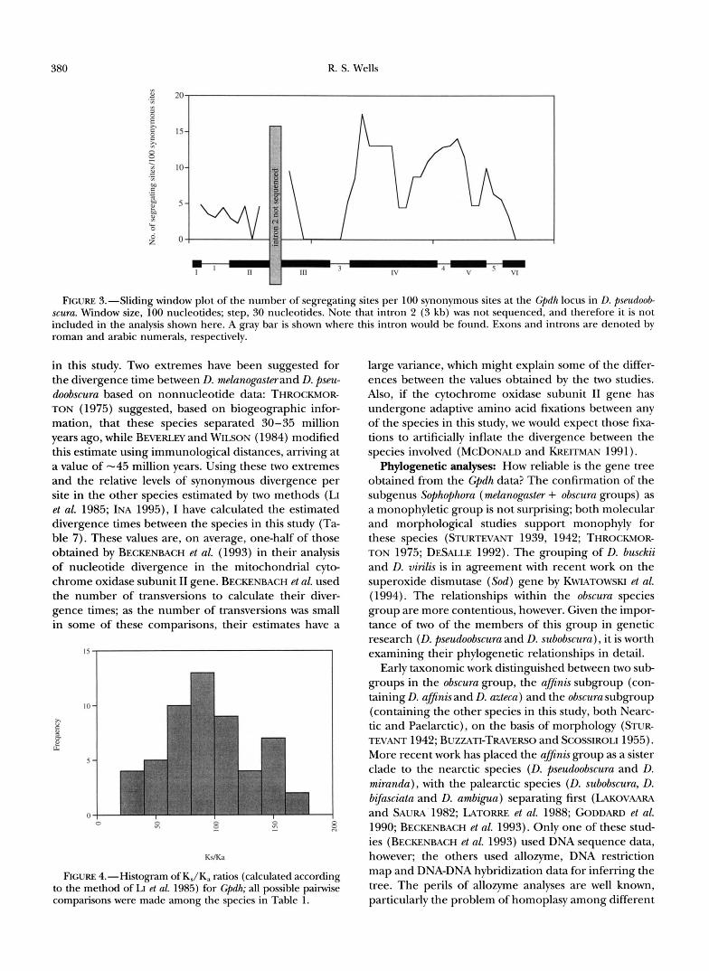
1995). The ratio of these two values provides a measure of the strength of purifying selection acting on the amino acid sequence of the protein (RILEY 1989). In Figure 4, the distribution of K,/K,! values among all possible pairwise species comparisons is shown. The val- ues at the lower end of the distribution show evidence of a higher level of amino acid substitution relative to their level of synonymous substitution, although not enough to result in a significant deviation using the test of MCDONALD and KREITMAN (1991). Those values at the upper end of the distribution exhibit a dearth of amino acid substitutions for their level of synonymous divergence; the highest K,/K, value obtained is 171 in the comparison of D. melanogastcr and D. af3ni.r; which have a level of synonymous divergence comparable to that found in comprisons of D. melanogastmwith other obscura group species but differ at only three amino acid positions (other obscura group species differ at four to five amino acid positions). The mean K,/K, value obtained from the distribution (94.6) is comparable to that obtained from four phylogenetically independent observations (afJinis us. mdanogastcr, pseudoobsrura us. ambigua, subobscura us. bifasciata, and busckii us. virilis; mean K,/K, = 105.1). It is interesting to note that the number of differences between D. pseudoobsrura JR45 and D. mirunda (six) is within the range for the number of differences between the I). pseudoobscura alleles (six to 17). These species are estimated to have diverged 1-4 million years ago (SCHAEFFER and MILLER 1992a; BECKENBACH et al. 1993); more I). miranda sequences should be obtained to confirm this observation.
Divergence times: The values of K, given in Table 1 can be used to infer the divergence times of the taxa
380 R. S. Wells
.- z! 20 2. n a E c 15- z ?
A 7
s $ IO-
P 24 5 -
a
. - .- ;
'i' z
e GJ
m .- - 2 I
';1 5.4
L N
g o I I .- 3 rl 1 1 1 1 -
FIGURE 3.-Sliding window plot of the number of segregating sites per 100 synonymous sites at the Cpdh locus in D. pseudoob scuru. Window size, 100 nucleotides; step, 30 nucleotides. Note that intron 2 (3 kb) was not sequenced, and therefore it is not included in the analysis shown here. A m y bar is shown where this intron would be found. Exons and introns are denoted by roman and arabic numerals, respectively.
in this study. Two extremes have been suggested for the divergence time between D. mlanogasterand D. pseu- doobscura based on nonnucleotide data: THROCKMOR- TON (1975) suggested, based on biogeographic infor- mation, that these species separated 30-35 million years ago, while BEVERLEY and WILSON (1984) modified this estimate using immunological distances, arriving at a value of -45 million years. Using these two extremes and the relative levels of synonymous divergence per site in the other species estimated by two methods (LI et al. 1985; INA 1995), I have calculated the estimated divergence times between the species in this study (Ta- ble 7). These values are, on average, one-half of those obtained by BECKENBACH et al. (1993) in their analysis of nucleotide divergence in the mitochondrial cyto- chrome oxidase subunit I1 gene. BECKENBACH et al. used the number of transversions to calculate their diver- gence times; as the number of transversions was small in some of these comparisons, their estimates have a
" I
>I 2 a G- E
5
0 0 w
KdKa
FIGURE 4.-Histogram of KJK, ratios (calculated according to the method of LI et ul. 1985) for Gpdh; all possible pairwise comparisons were made among the species in Table 1 .
large variance, which might explain some of the differ- ences between the values obtained by the two studies. Also, if the cytochrome oxidase subunit I1 gene has undergone adaptive amino acid fixations between any of the species in this study, we would expect those fixa- tions to artificially inflate the divergence between the species involved (MCDONALD and KREITMAN 1991).
Phylogenetic analyses: How reliable is the gene tree obtained from the Cpdh data? The confirmation of the subgenus Sophophora (mlanogaster + obscura groups) as a monophyletic group is not surprising; both molecular and morphological studies support monophyly for these species (STURTEVANT 1939, 1942; THROCKMOR- TON 1975; DESALLE 1992). The grouping of D. busckii and D. virilis is in agreement with recent work on the superoxide dismutase (Sod) gene by KWIATOWSKI et al. (1994). The relationships within the obscura species group are more contentious, however. Given the impor- tance of two of the members of this group in genetic research (D. pseudoobscura and D. subobscura), it is worth examining their phylogenetic relationships in detail.
Early taxonomic work distinguished between two sub groups in the obscura group, the afJinis subgroup (con- taining D. afJinis and D. azteca) and the obscura subgroup (containing the other species in this study, both Nearc- tic and Paelarctic), on the basis of morphology (STUR-
More recent work has placed the ufJinis group as a sister clade to the nearctic species (D. pseudoobscura and D. miranda), with the palearctic species (D. subobscura, D. bijimciata and D. ambigua) separating first (LAKOVAARA and SAURA 1982; LATORRE et al. 1988; GODDARD et al. 1990; BECKENBACH et al. 1993). Only one of these stud- ies (BECKENBACH et al. 1993) used DNA sequence data, however; the others used allozyme, DNA restriction map and DNA-DNA hybridization data for inferring the tree. The perils of allozyme analyses are well known, particularly the problem of homoplasy among different
TEVANT 1942; BUZZATI-TRAVERSO and SCOSSIROLI 1955).

Gpdh Evolution in Drosophila 38 1
TABLE 7 Estimates of divergence time based on Gpdh synonymous substitutions per site
No. of painvise Mean K, Divergence Mean K, Divergence Taxa compared comparisons (LWL) time (LWL) (IN.4 time (INA)
D. melanogaster-obscura group 7 0.9045 30.0 45.0 0.7041 30.0 45.0 afJinis subgroupnearctic obscura 4 0.1850 6.1 9.2 0.1418 6.0 9.1 afinis subgrouppalearctic obscuru 6 0.2599 8.6 12.9 0.2077 8.8 13.3 palearctic obscurcmearctic obscura 6 0.1853 6.1 9.2 0.1420 6.1 9.1 Sophophma-Dmsilopha 8 1.0400 34.5 51.7 0.8749 36.0 55.9 Sophophmu-Drosophila 8 0.8415 27.9 41.9 0.6826 29.1 43.6 Sophophma-Scaptodrosophila 8 1.0729 35.6 53.4 0.8948 38.1 57.2 Dorsilopha-Drosophila 1 0.6922 23.0 34.4 0.5478 23.3 35.0
Estimates were determined using the methods of LI et al. (LWL; 1985) and INA (1995).
electromorphs (LEWONTIN 1991). The mtDNA restric- tion map survey of LATORRE et ul. (using sixcutter re- striction fragment length polymorphisms) is also plagued by the problem of homoplasy, as well as the fact that D. uztecu was the only ufJinis subgroup species included. The DNA-DNA hybridization data of GOD- DARD et ul., which may be more representative of the entire genome than are single-gene trees, have not been analyzed using improved methods of tree reconstruc- tion (e.g., neighborjoining), and there are no estimates of reliability on the Fitch-Margoliash and UPGMA trees presented.
In their analysis, BECKENBACH et ul. (1993) used both complete sequences and transversions at the mitochon- drial cytochrome oxidase subunit I1 locus to infer trees of the obscuru group species. While analyses based solely on transversions obviate the problem of biased transi- tion/transversion ratios inherent in mitochondrial studies (TAMURA and NEI 1993), the number of infor- mative sites is reduced. The bootstrap value that they obtain for the nearctic obscuru/ufJinis subgroup clade is 62, lower than that obtained for the nearctic obscuru/ palearctic obscuru clade in the Gpdh analysis, 73. How- ever, in the MP analysis of the Gpdh nucleotide data, the treelength only increases from 373 to 377 if the a@nis/uzteca clade is placed as a sister group to the pseudoobscuru/mirundu clade, showing that the most likely relationship among these subgroups may be a trichotomy. The trichotomy hypothesis is supported by an examination of the level of synonymous substitution between the obscuru group species (Table 1). D. subobsc- uru shows a higher K, relative to pseudoobscuru/mirundu than do the other two palearctic species, resulting in the palearctic species grouping further from pseudoobscuru/ mirundu than do ufJinis/uztecu; when D. subobscuru is re- moved, the three groups appear to be roughly equidis- tant from each other. This agrees with BARRIO et ul.’s (1994) recent phylogenetic analysis of a 2-kb region of the mitochondrial genome in the obscuru group. De- pending on the gene analyzed and the algorithm used, different topologies are obtained for the obscuru and ufJinis group species. BARRIO et ul. conclude that the
Palearctic obscuru group is polyphyletic (split into subob- scuru and bi$usciutu/umbiguu lineages), and that the over- all branching pattern is consistent with a rapid phyletic radiation of the subgroup species. While the Cpdh data do not support polyphyly for the Palearctic obscura group (although D. subobscuru does seem to be the most divergent species), they are consistent with a rapid radi- ation of the subgroups.
STURTEVANT (1942) originally split the obscuru group into the ufJinis and obscuru subgroups on the basis of differences in the number of achrostichal hairs on the mesonatum, the number of teeth on the sex combs, and the shape of the carina and testes. Hybridization experiments show that D. umbiguu is capable of hybridiz- ing to both D. pseudoobscuru and D. persimilis (KOSKE 1953; BUZZATI-TRAVERSO and SCOSSIROLI 1955), and suggest that D. bqusciutu and D. persimilis can hybridize as well (KOSKE 1953). In contrast, no crosses between ufJinis subgroup species and either Palearctic or Nearc- tic obscuru subgroup species have produced viable larvae (BUZZATI-TRAVERSO and SCOSSIROLI 1955). Thus, much of the nonmolecular data appears to agree with the present grouping of Nearctic and Paelarctic obscurusub- groups as sister taxa. Molecular analysis of two “odd” species, D. ulpinu (thought to be the most basal branch in the Palearctic obscuru subgoup; LAKOVAARA and SAURA 1982) and D. helveticu (the only Palearctic mem- ber of the ufJinis subgroup, it has some affinities to the Nearctic obscuru subgroup species; LAKOVAARA and SAURA 1982) might shed some light on the relationships between the three subgroups.
MCDONALD-KF~EITMAN analysis: The test of MCDON- ALD and KREITMAN (1991) does not yield a significant result for these data, implying that adaptation has not played a major role in the amino acid divergence be- tween species at the Gpdh locus. This test is incapable of detecting evidence for adaptation when the number of amino acid replacements between species is small, however. Single (or a few) amino acid replacements may compose a substantial portion of adaptations at the molecular level (PERUTZ 1984); if so, then some of the variation found to be evolving “neutrally” using this

382 R. S. Wells
test may in fact be adaptive. Unfortunately, although statistical approaches that detect the “footprint” of nat- ural selection are at present better than functional ap- proaches used to decipher the causes of variation, there is an inherent limit to the types of events that they can detect. In the case of the MCDONALDKREITMAN test, this limit, given a level of intraspecific variation with a synonymous:nonsynonymous polymorphism ratio of 16:O (that of Gpdh in the present study), would be nine to 11 nonsynonymous replacements between the spe- cies shown in Table 5 (calculations not shown). For proteins that diverge very slowly between species, this level of nonsynonymous divergence is never realized. The question of whether amino acid variation in slowly evolving proteins is adaptive remains largely unanswer- able with current statistical methods.
HKA analysis: The HUDSON-KREITMAN-ACUAD~ test shows that the level of nucleotide polymorphism within the Gpdh coding region does not deviate from neutral expectations, implying that the locus is not subject to balancing selection and that it has not undergone a recent slective sweep. Thus, as with the Adh locus in D. psewloobscura (SCHAEFFER and MILLER 1992a), the electrophoretic monomorphism of the GPDH protein seems to result from strong purifying selection acting to remove amino acid variation. It is interesting, how- ever, that there appears to be an excess of polymor- phism in the exon 4intron 4 region (Figure 3) when there is no amino acid variation. Perhaps there is a functionally important change at a synonymous site, as has been described recently by &CHARD and BECKMAN ( 1995).
The D. pseudoobscura polymorphism data may help to explain an anomalous result obtained by TAKANO et al. (1993) in their survey of nucleotide polymorphism at the D. mlanogaster Gpdh locus. In D. mlanogaster there is a fast/slow electrophoretic polymorphism caused by a T/A transversion at site 3338 in exon 6 of the gene (BEWLEY et al. 1989). This polymorphism has several features in common with the fast/slow polymorphism at Adh in this species, including a significant cline in allele frequencies on three continents ( O A K E S H O ~ et al. 1982, 1984). In their investigation of polymorphism at the Gpdh locus, TAKANO et al. found an excess of polymorphism in the coding region relative to the in- trons and the 5’-flanking region, similar to the situation seen at Adh (HUDSON and KREITMAN 1991). Unfortu- nately, this excess is centered on exon 4, 500 bp u p stream (5‘) from the fast/slow site. Thus, the prediction of STROBECK (1983) with respect to balanced polymor- phisms is not seen: that the excess of linked polymor- phism seen at a site subject to balancing selection is centered around the selected site, and the excess arises as a consequence of divergence between the two allelic lineages within this region. TAKANO et al. were unable to explain this anomalous result. Interestingly, in D. pseudoobscura, we also see an excess of polymorphism in
0.06
me1 Adh
e
exon 4 other exons introns
FIGURE 5.-Comparison of values of 8 (silent sites) for dif- ferent regions of Adh in D. mlanogasterand Cpdh in D. melane gasterand D. pseudoobscura. The Adh fast/slow site in D. m h n e gaster is found in exon 4, while the Cpdh fast/slow site in D. mlanogaster is found in exon 6. This figure is based on one shown in TAKANO et al. (1993).
exon 4 (Figure 5) relative to introns and other exons. That this excess is not sufficient to cause a significant deviation from the neutral expectation for the entire locus implies that in D. mlanogaster, which has not been analyzed using an HKA test (TAKANO et al. 1993), we might also expect to find that the excess is not signifi- cant. This result, coupled with the phylogenetic rela- tionships among the D. mlanoguster Gpdh alleles (which show evidence for multiple mutations at the fast/slow site; TAKANO et al. 1993), argues against the selective maintenance of the fast/slow Gpdh polymorphism in D. mlanogaster. Two questions are raised by the possibility that this polymorphism is not maintained by selection: (1) Why is there an excess of polymorphism in exon 4 in both species, when this exon does not exhibit a concomitant excess of interspecific divergence (WELLS 1995)? (2) Why is there a cline in fast/slow allele fre- quencies in D. mlanogastm? Further sequencing surveys in both species should begin to address these questions and delineate the forces controlling variation at the Gpdh locus.
I thank A. BERRY, R C. LEWONTIN, A. BARBADIU and R. KLIMAN for advice and discussion. R. NORMAN provided the D. mimnda genomic library, and J. ROZAS provided the D. svbobsncra DNA. Thanks to the National Drosophila Species Stock Center for providing fly stocks and to D. WEINREICH for sequencing assistance. This study was sup ported by National Institutes of Health grant GM-21179 to R. C. LEWONTIN. The author was a Howard Hughes Medical Institute Pre- doctoral Fellow during this study.
LITERATURE CITED
ASHBURNER, M., 1989 Drosophila: A Laboratq Manual. Cold Spring Harbor Laboratory Press, Cold Spring Harbor, NY.
AUSUBEL, F. M., R BRENT, R. E. KINGSTON, D. D. MOORE, J. G. SEID- MAN et al. (Editors), 1987 Current Protocols in Molecular Biology. Wiley Interscience, New York.
BARRIO, E., A. LATORRE and A. MOYA, 1994 Phylogeny of the Drosoph-

Gpdh Evolution in Drosophila 383
ila obscura species group deduced from mitochondrial DNA se- quence. J. Mol. Evol. 3 9 478-488.
BECKENBACH, A. T., Y. W. WEI and H. LIU, 1993 Relationships in the Drosophila obscuraspecies group, inferred from mitochondrial cytochrome oxidase I1 sequences. Mol. BIOI. Evol. 10 619-634.
BERRY, A. J., J. W. AJIOKA and M. KREITMAN, 1991 Lack of polymor- phism on the Drosophila fourth chromosome resulting from selec- tion. Genetics 129: 1111-1117.
BEVERLEY, S. M., and A. C. WILSON, 1984 Molecular evolution in Drosophila and the higher Diptera 11. A time scale for fly evolu- tion. J. Mol. Evol. 21: 1-13.
BEWI.EY, G. C., J. L. COOK, S. KUSAKABE, T. MUKAI, D. L. RIGBY et al., 1989 Sequence, structure and evolution of the gene coding for seglycerol-%phosphate dehydrogenase in Drosophila melanogaster. Nucleic Acids Res. 17: 8553-8567.
BurzxrI-TRAvmso, A. A,, and R. E. SCOSSIROLI, 1955 The “obscura” group of the genus Drosophila. Adv. Genet. 7: 47-92.
COYNE, J. A,, W. F. EANES, J. A. RAMSHAW and R. K. KOEHN, 1979 Electrophoretic heterogeneity of a-glycerophosphate dehydroge- nase among many species of Drosophila. Syst. Zool. 28:164-175.
DE STORDEUR, E., and S. PASTEUR, 1978 Sur l’evolution de l’enzyme a-Gpdh chez les Drosophilidts. C. R. Acad. Sc. Paris 287: 93-95.
DESALLE, R., 1992 The phylogenetic relationships of flies in the family Drosophilidae deduced from mtDNA sequences. Mol. Phy- logenet. Evol. 1: 31-40.
FEUENSTEIN, J,, 1985 Confidence limits on phylogenies: an ap- proach using the bootstrap. Evolution 39: 783-791.
GODDARD, K., A. CACCONE and J. R. POWEI.L, 1990 Evolutionary implications of DNA divergence in the Drosophila obscura group. Evolution 44: 1656-1670.
GRIMAI.DI, D. A,, 1990 A phylogenetic, revised classification of gen- era in the Drosophilidae (Diptera). Bull. Am. Mus. Nat. Hist.
HICXJCHI, R. G., and H. OCHMAN, 1989 Production of single- stranded DNA templates by exonuclease digestion following the polymerase chain reaction. Nucleic Acids Res. 17: 5865.
HUDSON, R. R., M. KREITMAN and M. AGUADE, 1987 A test of neutral molecular evolution based on nucleotide data. Genetics 116: 153-159.
INA, Y., 1995 New methods for estimating the number of synony- mous and nonsynonymous substitutions. J. Mol. Evol. 40:
JUKES, T. H., and C. R. CANTOR, 1969 Evolution of protein mole- cules, pp. 21-132 in Mammalian Protein Metabolism, edited by H. N. MUNRO. Academic Press, New York.
KEITH, T. P. 1983 Frequency distribution of esterase-5 alleles in two populations of Drosophila pseudoobscura. Genetics 105: 135-155.
KIMURA, M., 1983 The Neutral Theoly of Molecular Evolution. Cam- bridge University Press, New York.
KOSKE, T., 1953 Artkreuzungsveruche in der obscura-Gruppe der Gattung Drosophila. Z . Indukt. Abstamm. Verebungsl. 85:
KREITMAN, M., 1983 Nucleotide polymorphism at the alcohol dehy- drogenase locus of Drosophila melanogaster. Nature 304: 412-417.
KUMAR, S., K. TAMURA and M. NEI, 1993 MEGA: MolecularEvolution- ary Genetics Analysis, version 1.0. The Pennsylvania State Univer- sity, University Park, PA.
KWIATOWSKI, J., D. SKAECKY, K. BAILEY and J. AYALA, 1994 Phylogeny of Drosophila and related genera inferred from the nucleotlde sequence of the Cu, Zn Sod gene. J. Mol. Evol. 38: 443-454.
LAKOVAARA, S., and L. KERANEN, 1980 Variation at the a-Cpdh locus of Drosophilids. Hereditas 92: 251 -258.
LAKOVMRA, S., and A. SAURA, 1982 Evolution and speciation in the Drosophila obscura group, pp. 1-59 in The Genetics and Biology of Drosophila, Vol. 3b, edited by M. ASHBURNER, H. L. CARSON and J. N. THOMPSON. Academic Press, London.
LAKOVAAIII\, S., A. SAURA and P. LANKINEN, 1977 Evolution at the a-Cpdh locus in Drosophilidae. Evolution 31: 319-330.
LATORRE, A,, E. BARRIO, A. MOYA and F. J. AYALA, 1988 Mitochon- drial DNA evolution in the Drosophila obscura group. Mol. Biol. Evol. 5: 717-728.
LEWONI’IN, R. C., 1991 Twenty-five years ago in Genetics: electropho- resis in the development of evolutionary genetics: milestone of millstone? Genetics 128: 657-662.
LEWONTIN, R. C., J. A. MOORE, W. PROVINE and B. WALLACE, 1981
197: 1-139.
190-226.
373-381.
Dobzhansky ’s Gaetics of Natural Populations. Columbia University Press, New York.
LI, W.-H., 1977 Distribution of nucleotide differences between two randomly chosen cistrons in a finite population. Genetics 85:
LI, W.-H., C.4. Wu and C . 4 . LUO, 1985 A new method for estimat- ing synonymous and nonsynonymous rates of nucleotide substi- tution considering the relative likelihood of nucleotide and co- don usage. Mol. Biol. Evol. 2 150-174.
MADDISON, W. P., and D. R. MADDISON, 1992 MacClade: Analysis of Phylogeny and Character Evolution, version 3.0. Sinauer Associates, Sunderland, MA.
MCDONAID, J. H., and M. KREITMAN, 1991 Adaptive evolution at the Adh locus in Drosophila. Nature 351: 652-654.
OAKESHOTT, J. G., J. B. GIBSON, P. R. ANDERSON, W. R. KNIBR, D. G. ANDERSON et al., 1982 Alcohol dehydrogenase and glycerol-3- phosphate dehydrogenase clines in Drosophila melanogasteron dif- ferent continents. Evolution 3 6 86-96.
OAKESHOTT, J. G., S. W. MCKECHNIE and G. K. CHAMBERS, 1984 Popu- lation genetics of the metabolically related Adh, C+dh and Tpi polymorphisms in Drosophila melanogaster. I. Geographic variation in Cpdh and Tpi allele frequencies in different continents. Genet- ica 63: 21-29.
PERUTL, M. F., 1984 Species adaptation in a protein molecule. Mol. Biol. Evol. 1: 1-28.
POWEI.I., J. R., 1976 Protein variation in natural populations of ani- mals. Evol. Biol. 8: 79-119.
RICHARD, I., and J. S. BECKMAN, 1995 How neutral are synonymous codon mutations? Nature Genet. 10 259.
RILEY, M. A,, 1989 Nucleotide sequence of the Xdhregion in Drosoph- ila pseudoobscura and an analysis of the evolution of synonomous codons. Mol. Biol. Evol. 6 33-52.
RIIXY, M. A,, M. E. HAILAS and R. C. LEWONTIN, 1989 Distinguishing the forces controlling genetic variation at the Xdh locus in Dre sophila pseudoobscura. Genetics 123: 359-369.
RILEY, M. A,, S. R. KAPIAN and M. VEUII.I.E, 1992 Nucleotide poly- morphism at the xanthine dehydrogenase locus in Drosophila pseudoobscura. Mol. Biol. Evol. 9: 56-69.
SAITOU, N., and M. NEI, 1987 The neighbor-joining method: a new method for reconstructing phylogenetic trees. Mol. Biol. Evol. 4: 406-425.
SCHAEFFER, S., and E. L. MILLER, 1992a Molecular population genet- ics of an electrophoretically monomorphic protein in the alcohol dehydrogenase region of Drosophila pseudoobscura. Genetics 132:
SCHAEFFER, S. W., and E. L. MII.I.ER, 199% Estimates of gene flow in Drosophila pseudoobscura determined from nucleotide se- quence analysis of the alcohol dehydrogenase region. Genetics 132: 471-480.
SCHAEFFER, S. W., and E. L. MILLER, 1993 Estimates of linkage dis- equilibrium and the recombination parameter determined from segregating nucleotide sites in the alcohol dehydrogenase region of Drosophila pseudoobscura. Genetics 135: 541-552.
STRORECK, C., 1983 Expected linkage disequilibrium for a neutral locus linked to a chromosomal arrangement. Genetics 103: 545-555.
STURTEVANT, A. H., 1939 On the subdivision of the genus Drosophila. Proc. Natl. Acad. Sci. USA 3: 137-141.
STURTEVANT, A. H., 1942 The classification of the genus Drosophila with description of nine new species. Univ. Texas Publ. 4213: 5-51.
SWOFFORD, D. L., 1991 PAUP: Phylogenetic Analysis Using Parsimony, version 3.0s. Illinois Natural History Survey, Champaign, IL.
TAIIMA, F., 1993 Simple methods for testing the molecular evolu- tionary clock hypothesis. Genetics 135: 599-607.
T W O , T. S., S. KUSAKARE and T. MUKAI, 1993 DNApolymorphism and the origin of protein polymorphism at the Cpdh locus of Drosophila melanogaster, pp. 179-190 in Mechanisms of Molecular Evolution, edited by N. TAKAHATA and A. G. CLARK. Sinauer, Sunderland, MA.
TAMuRA, K., and M. NEI, 1993 Estimation of the number of nuclec- tide substitutions in the control region of mitochondrial DNA in humans and chimpanzees. Mol. Biol. Evol. 10: 512-526.
THROCKMORTON, L. H., 1975 The phylogeny, ecology and geogra- phy of Drosophila, pp. 421-469 in Handbook of Genetics, vol. 3, edited by R. C. KING. Plenum, New York.
331-337.
163-178.

384 R. S. Wells
TOMINAGA, H., T. SHIM and S. NARISE, 1992 Structure of Drosophila phosphate dehydrogenase-encoding gene of Drosophila mlanc- vin'lis glycerol-%phosphate dehydrogenase gene and a compari- gaster. Proc. Natl. Acad. Sci. USA 86 5020-5024. son with the Drosophila mlanogastergene. Biochim. Biophys. Acta WELLS, R. s., 1995 Sequence and evolution of the Drosophila pseudo- 1131: 233-238. obscuru glycerol-3-phosphate dehydrogenase locus. J. Mol. Evol.
VILLAROYA, A,, and E. JUAN, 1991 ADH and phylogenetic relation- ships of Drosophila lebanonensis. J. Mol. Evol. 32 421-428.
VON KALM, L. J., J. WEAVER, J. DEMARCO, R. J. MACINTYRE and D. T.
41: 886-893. WELLS, R. S., 1996 Excessive homoplasy in an evolutionarily con-
strained protein. Proc. Roy. SOC. Lond., Ser. B (in press).
SULLIVAN, 1989 Structural characterization of the a-glycerol-3- Communicating editor: A. G. CLARK
Related Documents