Nucleotide excision repair in Trypanosoma brucei: specialization of transcription-coupled repair due to multigenic transcription Carlos R. Machado, 1 ** † João P. Vieira-da-Rocha, 1† Isabela Cecilia Mendes, 1,2 Matheus A. Rajão, 3 Lucio Marcello, 2 Mainá Bitar, 1 Marcela G. Drummond, 4 Priscila Grynberg, 1 Denise A. A. Oliveira 4 , Catarina Marques, 2 Ben Van Houten 5 and Richard McCulloch 2 * 1 Departamento de Bioquímica e Imunologia, ICB, Universidade Federal de Minas Gerais, Av. Antônio Carlos, 6627, Caixa Postal 486, Belo Horizonte 30161-970, MG, Brazil. 2 The Wellcome Trust Centre for Molecular Parasitology, College of Medical, Veterinary and Life Sciences, Institute of Infection, Immunity and Inflammation, University of Glasgow, Sir Graeme Davies Building, 120 University Place, Glasgow, G12 8TA, UK. 3 Coordenação de Pesquisa, Instituto Nacional de Câncer, Rua André Cavalcanti, 37 Fátima, Rio de Janeiro 20231-050, RJ, Brazil. 4 Laboratório de Genética Animal, Escola de Veterinária, Universidade Federal de Minas Gerais, Av. Antônio Carlos, 6627, Caixa Postal 486, Belo Horizonte 30161-970, MG, Brazil. 5 Department of Pharmacology and Chemical Biology, University of Pittsburgh School of Medicine and The University of Pittsburgh Cancer Institute, Hillman Cancer Center, Pittsburgh, PA 15213, USA. Summary Nucleotide excision repair (NER) is a highly conserved genome repair pathway acting on helix distorting DNA lesions. NER is divided into two subpathways: global genome NER (GG-NER), which is responsible for repair throughout genomes, and transcription- coupled NER (TC-NER), which acts on lesions that impede transcription. The extent of the Trypanosoma brucei genome that is transcribed is highly unusual, since most genes are organized in multigene tran- scription units, each transcribed from a single pro- moter. Given this transcription organization, we have addressed the importance of NER to T. brucei genome maintenance by performing RNAi against all predicted contributing factors. Our results indicate that TC-NER is the main pathway of NER repair, but only CSB, XPBz and XPG contribute. Moreover, we show that UV lesions are inefficiently repaired in T. brucei, perhaps due to preferential use of RNA polymerase translesion synthesis. RNAi of XPC and DDB was found to be lethal, and we show that these factors act in inter- strand cross-link repair. XPD and XPB appear only to act in transcription, not repair. This work indicates that the predominance of multigenic transcription in T. brucei has resulted in pronounced adaptation of NER relative to the host and may be an attractive drug target. Introduction DNA genomes are exposed to a myriad of agents that modify or alter their structures. Some of these agents, like reactive oxygen species and reactive nitrogen species, can be produced endogenously, while others, such as ionizing radiation, UV light and harmful chemicals, are derived from the environment. To maintain genomic stabil- ity most living organisms possess multiple pathways to repair such DNA damage. Nucleotide excision repair (NER) is responsible for removing a remarkable variety of DNA lesions, including bulky adducts covalently attached to DNA and other forms of damage that distort the double helix structure (Naegeli and Sugasawa, 2011). NER defects in humans are associated with the cancer-prone syndrome xeroderma pigmentosum (XP) and with the developmental disorders Cockayne syndrome and trico- thiodistrophy (Lehmann, 2001; Vermeulen et al., 2001; Emmert et al., 2009; Cameroni et al., 2010). NER is com- posed of two subpathways: Global Genome Nucleotide Excision Repair (GG-NER), which recognizes lesions genome-wide, and Transcription-Coupled NER (TC-NER), which recognizes DNA lesions that block elongation of RNA polymerase (Pol) in the transcribed strand of active Accepted 22 March, 2014. For correspondence. *E-mail richard [email protected]; Tel. (+44) 141 330 5946; Fax (+44) 141 330 8269. **E-mail [email protected]; Tel. (+55) 31 3409 2643; Fax (+55) 31 3409 2984. † These authors contributed equally to this work. Molecular Microbiology (2014) 92(4), 756–776 ■ doi:10.1111/mmi.12589 First published online 24 April 2014 © 2014 The Authors. Molecular Microbiology published by John Wiley & Sons Ltd. This is an open access article under the terms of the Creative Commons Attribution License, which permits use, distribution and reproduction in any medium, provided the original work is properly cited.

Welcome message from author
This document is posted to help you gain knowledge. Please leave a comment to let me know what you think about it! Share it to your friends and learn new things together.
Transcript
Nucleotide excision repair in Trypanosoma brucei:specialization of transcription-coupled repair due tomultigenic transcription
Carlos R. Machado,1**† João P. Vieira-da-Rocha,1†
Isabela Cecilia Mendes,1,2 Matheus A. Rajão,3
Lucio Marcello,2 Mainá Bitar,1
Marcela G. Drummond,4 Priscila Grynberg,1
Denise A. A. Oliveira4, Catarina Marques,2
Ben Van Houten5 and Richard McCulloch2*1Departamento de Bioquímica e Imunologia, ICB,Universidade Federal de Minas Gerais, Av. AntônioCarlos, 6627, Caixa Postal 486, Belo Horizonte30161-970, MG, Brazil.2The Wellcome Trust Centre for Molecular Parasitology,College of Medical, Veterinary and Life Sciences,Institute of Infection, Immunity and Inflammation,University of Glasgow, Sir Graeme Davies Building, 120University Place, Glasgow, G12 8TA, UK.3Coordenação de Pesquisa, Instituto Nacional deCâncer, Rua André Cavalcanti, 37 Fátima, Rio deJaneiro 20231-050, RJ, Brazil.4Laboratório de Genética Animal, Escola de Veterinária,Universidade Federal de Minas Gerais, Av. AntônioCarlos, 6627, Caixa Postal 486, Belo Horizonte30161-970, MG, Brazil.5Department of Pharmacology and Chemical Biology,University of Pittsburgh School of Medicine and TheUniversity of Pittsburgh Cancer Institute, Hillman CancerCenter, Pittsburgh, PA 15213, USA.
Summary
Nucleotide excision repair (NER) is a highly conservedgenome repair pathway acting on helix distorting DNAlesions. NER is divided into two subpathways: globalgenome NER (GG-NER), which is responsible forrepair throughout genomes, and transcription-coupled NER (TC-NER), which acts on lesions thatimpede transcription. The extent of the Trypanosomabrucei genome that is transcribed is highly unusual,
since most genes are organized in multigene tran-scription units, each transcribed from a single pro-moter. Given this transcription organization, we haveaddressed the importance of NER to T. brucei genomemaintenance by performing RNAi against all predictedcontributing factors. Our results indicate that TC-NERis the main pathway of NER repair, but only CSB, XPBzand XPG contribute. Moreover, we show that UVlesions are inefficiently repaired in T. brucei, perhapsdue to preferential use of RNA polymerase translesionsynthesis. RNAi of XPC and DDB was found to belethal, and we show that these factors act in inter-strand cross-link repair. XPD and XPB appear only toact in transcription, not repair. This work indicatesthat the predominance of multigenic transcription inT. brucei has resulted in pronounced adaptation ofNER relative to the host and may be an attractive drugtarget.
Introduction
DNA genomes are exposed to a myriad of agents thatmodify or alter their structures. Some of these agents, likereactive oxygen species and reactive nitrogen species,can be produced endogenously, while others, such asionizing radiation, UV light and harmful chemicals, arederived from the environment. To maintain genomic stabil-ity most living organisms possess multiple pathways torepair such DNA damage. Nucleotide excision repair(NER) is responsible for removing a remarkable variety ofDNA lesions, including bulky adducts covalently attachedto DNA and other forms of damage that distort the doublehelix structure (Naegeli and Sugasawa, 2011). NERdefects in humans are associated with the cancer-pronesyndrome xeroderma pigmentosum (XP) and with thedevelopmental disorders Cockayne syndrome and trico-thiodistrophy (Lehmann, 2001; Vermeulen et al., 2001;Emmert et al., 2009; Cameroni et al., 2010). NER is com-posed of two subpathways: Global Genome NucleotideExcision Repair (GG-NER), which recognizes lesionsgenome-wide, and Transcription-Coupled NER (TC-NER),which recognizes DNA lesions that block elongation ofRNA polymerase (Pol) in the transcribed strand of active
Accepted 22 March, 2014. For correspondence. *E-mail [email protected]; Tel. (+44) 141 330 5946; Fax (+44)141 330 8269. **E-mail [email protected]; Tel. (+55) 31 34092643; Fax (+55) 31 3409 2984. †These authors contributed equally tothis work.
Molecular Microbiology (2014) 92(4), 756–776 ■ doi:10.1111/mmi.12589First published online 24 April 2014
© 2014 The Authors. Molecular Microbiology published by John Wiley & Sons Ltd.This is an open access article under the terms of the Creative Commons Attribution License, which permits use, distribution andreproduction in any medium, provided the original work is properly cited.
genes (Naegeli and Sugasawa, 2011). It is widely consid-ered that GG-NER and TC-NER operate together in allorganisms to maintain genome fidelity. In this study wehave asked whether or not such parallel functioning of thetwo NER pathways is maintained in trypanosomatids,eukaryotes in which gene expression is highly diverged.
During GG-NER in humans XPC (XP factor C), togetherwith RAD23b and Centrin-2 (Mu et al., 1995; Araujo et al.,2000; Araki et al., 2001; Nishi et al., 2005), recognizeslocal strand openings associated with double strand helix-distorting lesions (Min and Pavletich, 2007). In somecases, XPC recruitment can be aided by a dimer of theUV-damaged DNA binding (UV-DDB) protein, composedof DDB1 and XPE (DDB2) subunits, the latter of whichrecognizes UV-induced lesions in the context of chromatin(Feldberg and Grossman, 1976; Chu and Chang, 1988).The damage is then handed off to XPC, which promotesloading of the TFIIH transcription factor complex throughthe NER protein’s C-terminal domain. TFIIH is composedof 10 proteins, divided into two subcomplexes: the core,which contains two helicases, XPB and XPD, and fiveassociated proteins (p62, p52, p44, p34 and p8/TTD-A);and CAK, the kinase activating complex (Compe andEgly, 2012). TFIIH is recruited by XPC interaction withXPB and with p62, allowing damage excision to begin byopening the DNA helix around the lesion. The XPB andXPD helicases act in this reaction, though it appears thatXPD helicase activity is crucial, while XPB providesATPase, but not helicase, activity (Oksenych and Coin,2009). Once DNA denaturation is established, RPA isloaded to protect the single-stranded DNA in the bubble.At the same time XPA is also loaded in order to stabilizethe TFIIH-XPC complex, as well as to cause release of theCAK subcomplex, which coincides with recruitment of theNER incision endonucleases, XPF/ERCC1 and XPG.XPF/ERCC1 catalyses incision 5′ of the lesion, while XPGexecutes an incision 3′. This leads to excision of a DNAoligonucleotide containing the lesion, though the 3′ inci-sion appears to occur after Replication Factor C andProliferating Cell Nuclear Antigen are recruited andco-ordinate loading of the replication machinery neededfor repair, composed of DNA Pols delta and kappa innon-dividing cells, and DNA Pol epsilon in replicative cells(Ogi et al., 2010). Following gap-filling, the remaining nickis sealed by DNA ligase I or DNA ligase III in dividing andnon-dividing cells respectively (Fagbemi et al., 2011).
TC-NER differs from GG-NER in the DNA damage rec-ognition step, which is made through identification ofstalled RNA Pol at lesions in transcribed genes. RNA PolII (and perhaps also RNA Pol I) blockage increases inter-action of the polymerase with the Cockayne syndrometype B (CSB) protein, an SNF2 ATPase, which may stimu-late RNA Pol translocation along the DNA strand, allowingTFIIH complex access to the lesion site (Hanawalt and
Spivak, 2008). From then, the TC-NER and GG-NERpathways proceed in the same way. In TC-NER ongoingtranscription can be aborted if RNA Pol disengages withthe template, or may resume if the enzyme complexremains attached, which may require the action of CSA ineukaryotes, perhaps by antagonizing CSB (Hanawalt andSpivak, 2008). XPG has also been shown to interact withCSB and RNA Pol II, and may be responsible for allowingNER to occur without release of RNA Pol (Sarker et al.,2005). NER may not occur in all cases, however. In somecircumstances, RNA Pol can insert nucleotides oppositelesions, including UV damage (Walmacq et al., 2012), andextend beyond the blockage in an error-prone fashion, aprocess termed transcriptional mutagenesis (Saxowskyand Doetsch, 2006; Bregeon and Doetsch, 2011). Thoughsuch translesion transcription through damaged DNA tem-plates can lead to mutant transcripts, this process rescuescells from death by severe stalling of RNA Pol, a strongsignal for apoptosis.
Trypanosomatids are flagellated protists, which mayhave emerged early during eukaryotic evolution anddisplay several highly diverged biological processes.Trypanosoma cruzi, Trypanosoma brucei and Leishmaniasp are among the most studied organisms of this family andare sources, respectively, of Chagas disease in LatinAmerica, sleeping sickness in sub-Saharan Africa andleishmaniasis in tropical areas, collectively affecting morethan 20 million people (http://www.who.int/topics/en/, lastaccessed 2012). To date, only a few studies have exam-ined NER in these organisms. The machinery is largelyconserved (see Results), though genome sequencingfailed to reveal orthologues of the smaller subunit of theUV-DDB dimer (DDB2), XPAor CSA(Berriman et al., 2005;El-Sayed et al., 2005; Ivens et al., 2005), and two distinctXPB-like proteins were identified, designated TbXPB andTbXPBz (Lecordier et al., 2007; Badjatia et al., 2013).Furthermore, trypanosomatid TFIIH lacks the CAKcomplex and associates with two trypanosomatid-specificproteins, TSP1 and TSP2 (Lee et al., 2009). The implica-tions for NER of these changes are unknown; for instance,CAK disassociation from TFIIH is considered a critical stepfor recruitment of XPF/ERCC1 and XPG to the lesion siteduring NER (Svejstrup et al., 1995; Coin et al., 2008). Morebroadly, the genomes of trypanosomatids provide a uniqueeukaryotic landscape in which NER must operate. Here,the vast majority of protein-coding genes are found indirectional gene clusters, each of which is transcribed froma single promoter, generating primary transcripts fromwhich individual mRNAs are derived by trans-splicing(Daniels et al., 2010). Thus, trypanosomatid chromo-somes contain few promoters (Siegel et al., 2009), whichmay be constitutively active, meaning that the extent of thegenome that is transcribed and the uniform level of thistranscription is unprecedented in eukaryotes, where one
Specialization of T. brucei nucleotide excision repair 757
© 2014 The Authors. Molecular Microbiology published by John Wiley & Sons Ltd., Molecular Microbiology, 92, 756–776
gene/one promoter and variable gene transcription ratesare considered the norm. The consequences of thisgenetic organization for trypanosomatid genome stabilityhave been little explored. For instance, collisions betweenthe DNA replisome and RNA Pol at genes transcribedduring S phase are associated with genomic instability,which is limited by the evolution of replication and tran-scription co-directionality in some highly expressed genesin bacteria and humans (Kim and Jinks-Robertson, 2012).Remarkably, replication origin mapping in T. brucei hasshown that multigenic transcription units can meet replica-tion forks head-on and slow their progress, despite theprediction that such collisions would have a pronouncedcost in this genome (Tiengwe et al., 2012).
In this study, we have examined how trypanosomatidgene organization influences NER. A priori, it might bepredicted that the transit of RNA Pol through promoter-distal genes of a multigene transcription unit encounters agreater number of lesions than promoter-proximal genes.Indeed, UV treatment provided the first evidence for mul-tigene transcription in T. brucei, causing greater impedi-ment to the transcription in cell extracts of a Variant SurfaceGlycoprotein (VSG) gene ∼ 60 kbp from its RNA Pol Ipromoter than Expression Site Associated Genes foundwithin the same transcription unit but closer to the promoter(Johnson et al., 1987; Coquelet et al., 1989). The samedifferential UV effect is seen for genes residing in RNA PolII multigene transcription units (Martinez-Calvillo et al.,2003; 2004). Despite these observations, there is no evi-dence that mRNA levels show a simple inverse correlationwith distance of genes from the promoter (Poon et al.,2012). If, and how, NER in these parasites might contributeto the maintenance of gene expression and function in thecontext of multigenic transcription has not been examined.To investigate this question, we have performed RNAiagainst all putative annotated NER factors in bloodstreamform (BSF; mammal-derived) T. brucei, both in theabsence and presence of genotoxic agents, and have
analysed relative survival and mRNA abundance acrossan RNA Pol II polycistron. We have also evaluated DNArepair kinetics in T. brucei and T. cruzi cells exposed to UVirradiation and cisplatin. We show that TbXPC, TbDDB,TbXPB and TbXPD are essential in T. brucei, but we findno evidence that these factors act in NER. Our data showthat TC-NER is the major NER pathway in this organism,though only TbCSB, TbXPG and TbXPBz act in this reac-tion, at least among the genes examined. In addition, wesuggest that translesion RNA synthesis is favoured overrepair and this pathway selection is mediated by CSB.Taken together, these results suggest that genome predic-tions of parallel functioning of TC-NER and GG-NER areincorrect and instead there is pronounced separation ofNER pathways in T. brucei, an evolutionary adaptation thatis associated with specialization of the NER machinery.
Results
Growth of bloodstream form T. brucei following RNAiknockdown of putative NER components
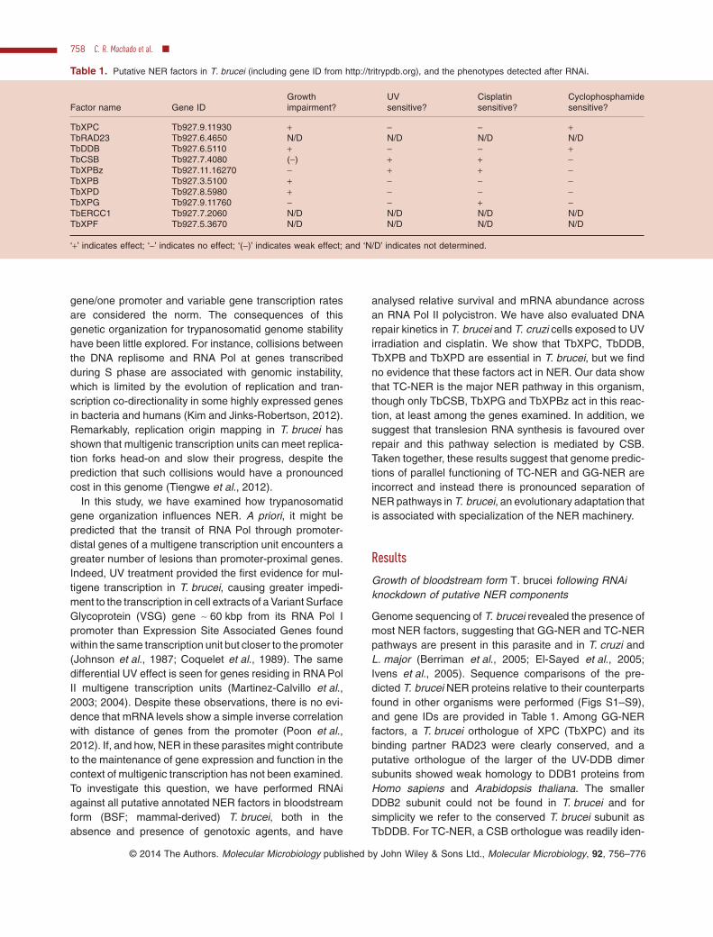
Genome sequencing of T. brucei revealed the presence ofmost NER factors, suggesting that GG-NER and TC-NERpathways are present in this parasite and in T. cruzi andL. major (Berriman et al., 2005; El-Sayed et al., 2005;Ivens et al., 2005). Sequence comparisons of the pre-dicted T. brucei NER proteins relative to their counterpartsfound in other organisms were performed (Figs S1–S9),and gene IDs are provided in Table 1. Among GG-NERfactors, a T. brucei orthologue of XPC (TbXPC) and itsbinding partner RAD23 were clearly conserved, and aputative orthologue of the larger of the UV-DDB dimersubunits showed weak homology to DDB1 proteins fromHomo sapiens and Arabidopsis thaliana. The smallerDDB2 subunit could not be found in T. brucei and forsimplicity we refer to the conserved T. brucei subunit asTbDDB. For TC-NER, a CSB orthologue was readily iden-
Table 1. Putative NER factors in T. brucei (including gene ID from http://tritrypdb.org), and the phenotypes detected after RNAi.
Factor name Gene IDGrowthimpairment?
UVsensitive?
Cisplatinsensitive?
Cyclophosphamidesensitive?
TbXPC Tb927.9.11930 + − − +TbRAD23 Tb927.6.4650 N/D N/D N/D N/DTbDDB Tb927.6.5110 + − − +TbCSB Tb927.7.4080 (−) + + −TbXPBz Tb927.11.16270 − + + −TbXPB Tb927.3.5100 + − − −TbXPD Tb927.8.5980 + − − −TbXPG Tb927.9.11760 − − + −TbERCC1 Tb927.7.2060 N/D N/D N/D N/DTbXPF Tb927.5.3670 N/D N/D N/D N/D
‘+’ indicates effect; ‘−’ indicates no effect; ‘(−)’ indicates weak effect; and ‘N/D’ indicates not determined.
758 C. R. Machado et al. ■
© 2014 The Authors. Molecular Microbiology published by John Wiley & Sons Ltd., Molecular Microbiology, 92, 756–776

tified, but no clear CSA-encoding gene. Sequence analy-sis of TbXPB, TbXPBz and TbXPD made previously(Lecordier et al., 2007; Badjatia et al., 2013) was con-firmed (data not shown). The presence of two XPB-likeproteins is not unique to trypanosomatids, but is also seenin plants (Morgante et al., 2005) and in most crenarchaea(Richards et al., 2008). However, the trypanosomatid pro-teins, like those in the archaea, display considerablesequence and size divergence, whereas the two plantXPBs are very closely related paralogues (Fig. S6). Thus,functional divergence of the two XPB factors is more likelyin trypanosomatids and in archaea than in plants. Finally,the predicted NER endonucleases could be identified.TbXPG is substantially smaller (746 amino acid residues)than H. sapiens and A. thaliana orthologues (both greaterthan 1100 amino acids; Fig. S7), due to the absence ofresidues in the parasite protein within the human XPG‘spacer’ region that is important in TFIIH interaction andseparates conserved N- and I-region nuclease domains(Dunand-Sauthier et al., 2005). Putative orthologues ofboth subunits of the XPF/ERCC1 complex could be iden-tified, though TbXPF was more clearly conserved thanTbERCC1.
Constructs for the generation of stem-loop RNAs againstall the above ORFs except TbRAD23 were producedand introduced into BSF T. brucei cells that allow thetetracycline-controlled induction of RNAi (Alsford andHorn, 2008; Jones et al., 2014). To test for the induction ofRNAi, mRNA levels of each target NER gene from therespective cell line were measured by quantitative RT-PCRin the presence and absence of tetracycline; significantloss of mRNA was detectable for most genes 24–48 hpost RNAi induction (Fig. 1). However, RNAi of TbXPF orTbERCC1, though resulting in pronounced cell growth andDNA damage response phenotypes, unexpectedly causedincreased, not decreased, levels of mRNA, a response thatwe cannot yet explain (data not shown). For this reason,the effects of RNAi on these two putative NER genes arenot detailed here. Cell growth was measured to determinethe effect of the RNAi inductions (Fig. 1), which revealedthat TbXPC, TbDDB, TbXPB and TbXPD are essential inthis life cycle stage, since pronounced growth impairmentwas seen following RNAi of each gene product (Fig. 1A, B,D and F). In all cases, growth reduction was seen 24–48 hpost RNAi induction, and this was followed in 24–48 h bycell death. The growth profile we observed following RNAiof TbXPB and TbXPD closely matched that describedpreviously (Lecordier et al., 2007). In contrast, RNAi offurther putative T. brucei NER components had much lessimpact. RNAi of TbCSB resulted in a slightly reducedgrowth rate compared with non-induced controls, thoughthis was only detectable beyond 4 days after RNAi induc-tion (Fig. 1C and data not shown). RNAi against TbXPBzand TbXPG had no discernible effect on cell growth
(Fig. 1E and G), even up to 7 days post induction (data notshown); for TbXPBz, this finding is consistent with obser-vations by Badjatia et al. (2013).
TbCSB and TbXPBz are involved in DNA damagerecovery after UV irradiation
Ultraviolet radiation causes cyclobutane pyrimidinedimers (CPDs) and 6-4 photoproduct (6-4 PP) lesions inDNA, which can be repaired either by direct repair inorganisms endowed with photolyase (Beukers et al.,2008) or by NER. To determine the role of the putativeT. brucei NER genes in DNA damage recovery after UVirradiation, growth of the RNAi cells was measured aftertreatment with UVC. To do this, 24 h after the addition oftetracycline to induce RNAi, the cells were exposed to 0,500, 1500 or 3000 J m−2 UVC and cell density measured24 h later; control cells in which RNAi was not inducedwere treated in the same way. Survival of the cells wasdetermined by calculating the cell density of the RNAi-induced or uninduced cells at the different levels of UVCrelative to untreated cells (Fig. 2). TbCSB and TbXPBzwere found to play an important function in T. brucei sur-vival after UV exposure, since RNAi against these twofactors resulted in a pronounced increase in cell deathrelative to non-RNAi induced controls. In contrast, RNAi ofTbXPG led to somewhat increased survival, suggestingthat the endonuclease antagonizes UV damage process-ing, at least in these conditions. Finally, RNAi of TbXPC,TbDDB, TbXPB or TbXPD had no effect on survival afterUV irradiation, suggesting that UV lesions in T. brucei arenot recognized by the putative GG-NER proteins TbXPCand TbDDB, and nor are they acted upon by the parasiteTFIIH-associated helicases.
UV-induced lesions are not always repairedin trypanosomatids
The above observations could be explained by UV-induced lesions being repaired predominantly byTC-NER, mediated by TbCSB and TbXPBz. Alternatively,UV damage may in some circumstances be bypassed,such as by translesion RNA synthesis, a hitherto unseenpathway in trypanosomatids. In order to address this, thekinetics of UV-induced DNA damage repair were analysedusing a quantitative, PCR-based technique (Santos et al.,2006). Cells were treated with 1500 J m−2 of UV radiationand lesion density within a ∼ 10 kbp region of transcribednuclear DNA measured at a number of time points afterexposure. At this level of UV exposure, ∼ 1.5 PCR-blocking lesions were found in the 10 kbp locus tested,and repair was remarkably slow, since there was no clearreduction in lesion density for up to 10 h post treatment(Fig. 3A). The DNA repair kinetic analysis was performed
Specialization of T. brucei nucleotide excision repair 759
© 2014 The Authors. Molecular Microbiology published by John Wiley & Sons Ltd., Molecular Microbiology, 92, 756–776

only within this time frame as it corresponds with theapproximate doubling time of BSF T. brucei cells, thusminimizing the effect of newly synthesized DNA, whichwould dilute the damaged templates. To address if DNAdamage caused by UV radiation persists beyond a singlecell cycle, antiserum against CPDs and 6-4 PPs wereused to detect these lesions by immunofluorescence, andsignal was readily detected 24 h after UV treatment(Fig. 3B). To determine if these slow repair kinetics arelimited to T. brucei, the PCR assay was performed in
epimastigote T. cruzi cells. PCR-blocking lesions accumu-lated to a very similar extent, and there was little evidencefor repair of the UV-induced lesions for up to 24 h posttreatment (Fig. 3C).
TbCSB, TbXPBz and TbXPG participate in TC-NER oflesions induced by cisplatin
Cisplatin has been used for over 30 years in the treatmentof many types of cancer (Stordal and Davey, 2007). It
Fig. 1. Growth curves of T. brucei cells following RNAi of NER factors. Growth curves are shown of T. brucei bloodstream form cells in theabsence (solid lines) or presence (dotted lines) of tetracycline induction of RNAi against the putative NER factors TbXPC, TbDDB, TbCSB,TbXPB, TbXPBz, TbXPD and TbXPG (A–G respectively). Cell concentrations were multiplied by dilutions made during growth to obtain thecumulative amount of parasites during the time-course. Data points are the mean of three experiments, each performed in triplicate, andvertical lines denote standard deviation. In each growth curve, the insert shows qRT-PCR quantification of the levels of mRNA for theRNAi-targeted gene 24 (white box) or 48 h (hatched) after induction of RNAi, relative to mRNA levels before induction (black); vertical linesdenote standard deviation from three experiments and *, ** and *** indicate P-values of < 0.05, < 0.01 and < 0.001 respectively.
760 C. R. Machado et al. ■
© 2014 The Authors. Molecular Microbiology published by John Wiley & Sons Ltd., Molecular Microbiology, 92, 756–776
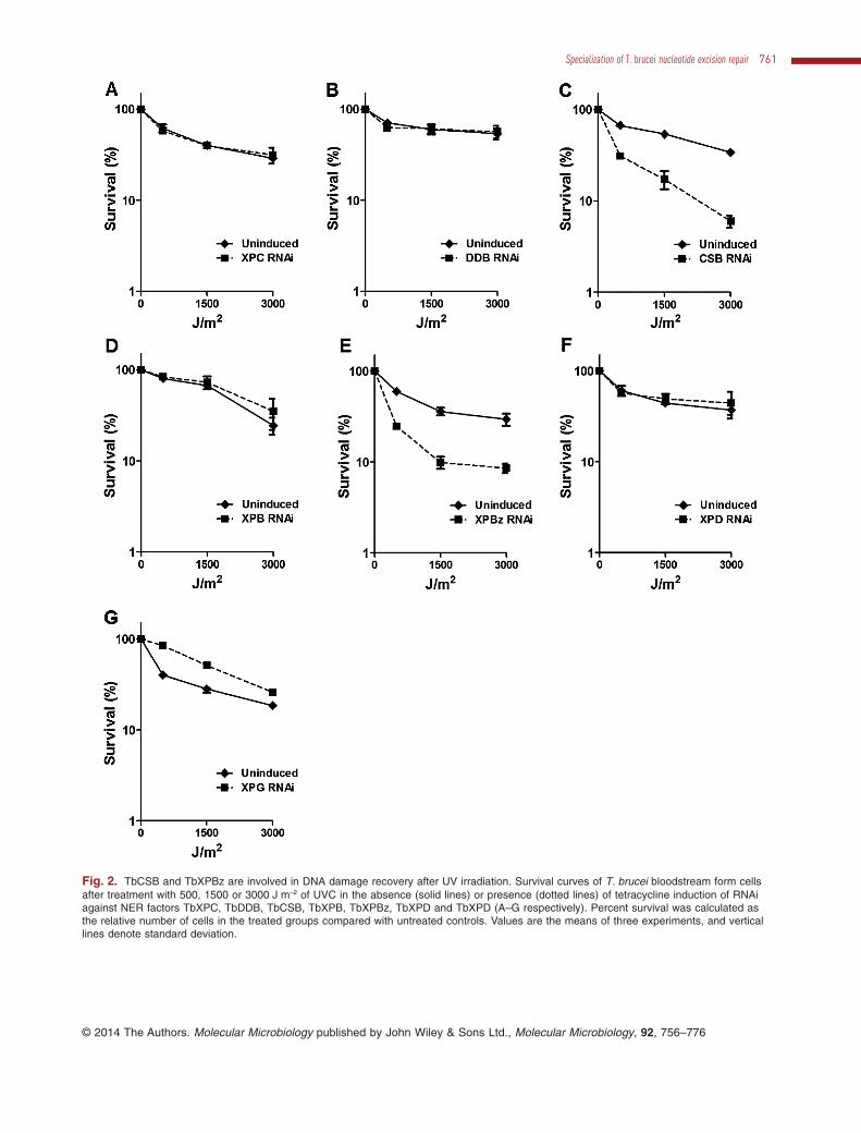
Fig. 2. TbCSB and TbXPBz are involved in DNA damage recovery after UV irradiation. Survival curves of T. brucei bloodstream form cellsafter treatment with 500, 1500 or 3000 J m−2 of UVC in the absence (solid lines) or presence (dotted lines) of tetracycline induction of RNAiagainst NER factors TbXPC, TbDDB, TbCSB, TbXPB, TbXPBz, TbXPD and TbXPD (A–G respectively). Percent survival was calculated asthe relative number of cells in the treated groups compared with untreated controls. Values are the means of three experiments, and verticallines denote standard deviation.
Specialization of T. brucei nucleotide excision repair 761
© 2014 The Authors. Molecular Microbiology published by John Wiley & Sons Ltd., Molecular Microbiology, 92, 756–776

induces predominantly intrastrand cross-links betweenadenine and guanine residues and between adjacentguanines; a minor proportion of cisplatin damage is inter-strand cross-links (Masters and Koberle, 2003). Cisplatin-induced intrastrand cross-links are mainly repaired byNER. To investigate the role of NER in responding tocisplatin in T. brucei, the BSF RNAi cell lines were culti-vated in the presence of 0, 1, 2 or 5 μM cisplatin, with andwithout RNAi induction, and survival measured as before(Fig. 4). RNAi knockdown of TbCSB or TbXPBz resultedin increased sensitivity to cisplatin, similar to that seen forUV. In addition, RNAi of TbXPG, which did not appear toact in the response to UV, also rendered the parasitessensitive to cisplatin. These data indicate that TC-NERfacilitates repair of cisplatin DNA damage, recruiting theXPG endonuclease for this purpose. In contrast, RNAiagainst TbXPC, TbDDB or TbXPB resulted in increasedtolerance to cisplatin, perhaps suggesting that these puta-tive GG-NER factors can recognize adducts induced bycisplatin, but this does not direct the lesions to repair.RNAi of TbXPD did not affect cisplatin sensitivity.
Lesions generated by cisplatin are removed fromtrypanosomatid DNA with high efficiency by TC-NER
To determine the efficiency of removal of lesions generatedby cisplatin, the kinetics of DNA repair were measured bythe quantitative PCR assay after T. brucei BSF cells hadbeen exposed to 100 μM cisplatin for one hour. In theseconditions ∼ 1.5 lesions were formed in the 10 kbp regionanalysed and DNA repair was highly efficient, since virtu-ally all lesions were removed after one hour (Fig. 5A). TheDNA repair efficiency was also measured in epimastigoteT. cruzi cells, revealing the same rapid repair (Fig. 5B).These data indicate that DNA injury caused by cisplatin isprocessed differently from UV. To test if the removal ofcisplatin is mediated by TC-NER, we performed immuno-fluorescence with antiserum against cisplatin (Fig. 5C andD). TbCSB RNAi cells were exposed to 50 μM cisplatin forone hour, with or without prior induction of RNAi for 24 h,and the cells were visualized at this time point and at oneand 24 h subsequently. With or without RNAi, cisplatindamage was detectable in the nucleus of the cells imme-
Fig. 3. UV-induced lesions are not always repaired in T. brucei.A. Number of lesions per 10 kb in the nucleus of bloodstream form T. brucei versus time in hours, after 1500 J m−2 of UVC irradiation. Theconcentration of DNA lesions was measured up to 10 h after DNA damage induction. Each value is the mean of two biological replicates, andwere derived from relative quantitative PCR-amplification values obtained from two independent measurements.B. Immunofluorescence microscopy of 6-4 photoproducts (6-4 PP; upper) and cyclobutane pyrimidine dimers (CPD; lower) in bloodstreamform T. brucei after treatment with 1500 J m−2 of UVC. The scale bar represents a length of 10 μM.C. Number of lesions per 10 kb in the nucleus of epimastigote from T. cruzi versus time in hours, after exposure to 1500 J m−2 of UVC. Theconcentration of DNA lesions was measured up to 24 h after treatment, as above.
762 C. R. Machado et al. ■
© 2014 The Authors. Molecular Microbiology published by John Wiley & Sons Ltd., Molecular Microbiology, 92, 756–776

diately after treatment. In the absence of RNAi the nuclearsignal was no longer detectable one and 24 h later, con-sistent with the rapid repair indicated by the quantitativePCR assay. In contrast, a pronounced nuclear signal forcisplatin was seen in the TbCSB RNAi-induced cells one
and 24 h after exposure. Moreover, quantification of thecisplatin signal in individual nuclei confirmed increasedpersistence of cisplatin lesions in individual cells after RNAiof TbCSB. These data indicate that cisplatin repair isimpeded by the loss of TbCSB.
Fig. 4. TbCSB, TbXPBz and TbXPG participate in TC-NER of lesions induced by cisplatin. Survival curves of TbXPC, TbDDB, TbCSB,TbXPB, TbXPBz, TbXPD and TbXPD (A–G respectively) RNAi depleted cells (dotted lines) compared with respective uninduced controls(solid lines) after exposure to 1, 2 or 5 μM cisplatin. Percent survival was calculated as the relative number of cells present in the treatedgroups compared with untreated control. Values are means of three experiments, and vertical lines denote standard deviation.
Specialization of T. brucei nucleotide excision repair 763
© 2014 The Authors. Molecular Microbiology published by John Wiley & Sons Ltd., Molecular Microbiology, 92, 756–776
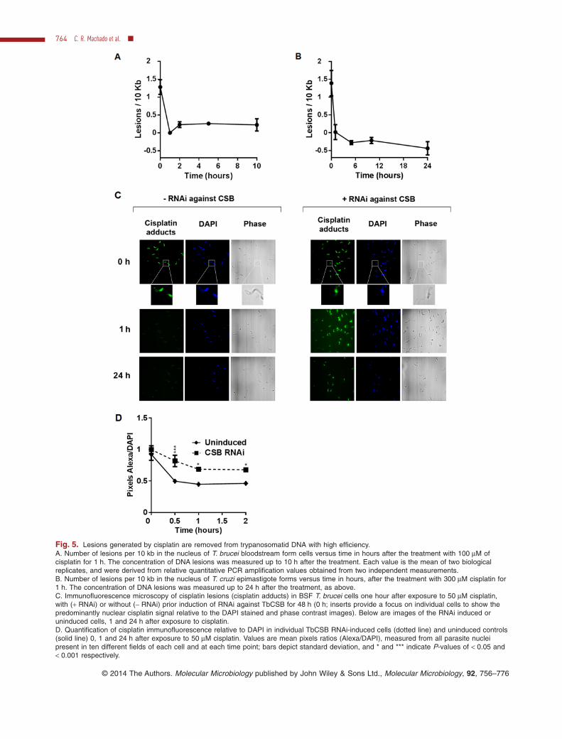
Fig. 5. Lesions generated by cisplatin are removed from trypanosomatid DNA with high efficiency.A. Number of lesions per 10 kb in the nucleus of T. brucei bloodstream form cells versus time in hours after the treatment with 100 μM ofcisplatin for 1 h. The concentration of DNA lesions was measured up to 10 h after the treatment. Each value is the mean of two biologicalreplicates, and were derived from relative quantitative PCR amplification values obtained from two independent measurements.B. Number of lesions per 10 kb in the nucleus of T. cruzi epimastigote forms versus time in hours, after the treatment with 300 μM cisplatin for1 h. The concentration of DNA lesions was measured up to 24 h after the treatment, as above.C. Immunofluorescence microscopy of cisplatin lesions (cisplatin adducts) in BSF T. brucei cells one hour after exposure to 50 μM cisplatin,with (+ RNAi) or without (− RNAi) prior induction of RNAi against TbCSB for 48 h (0 h; inserts provide a focus on individual cells to show thepredominantly nuclear cisplatin signal relative to the DAPI stained and phase contrast images). Below are images of the RNAi induced oruninduced cells, 1 and 24 h after exposure to cisplatin.D. Quantification of cisplatin immunofluorescence relative to DAPI in individual TbCSB RNAi-induced cells (dotted line) and uninduced controls(solid line) 0, 1 and 24 h after exposure to 50 μM cisplatin. Values are mean pixels ratios (Alexa/DAPI), measured from all parasite nucleipresent in ten different fields of each cell and at each time point; bars depict standard deviation, and * and *** indicate P-values of < 0.05 and< 0.001 respectively.
764 C. R. Machado et al. ■
© 2014 The Authors. Molecular Microbiology published by John Wiley & Sons Ltd., Molecular Microbiology, 92, 756–776
Evaluation of mRNA levels following RNAi of NERfactors in T. brucei
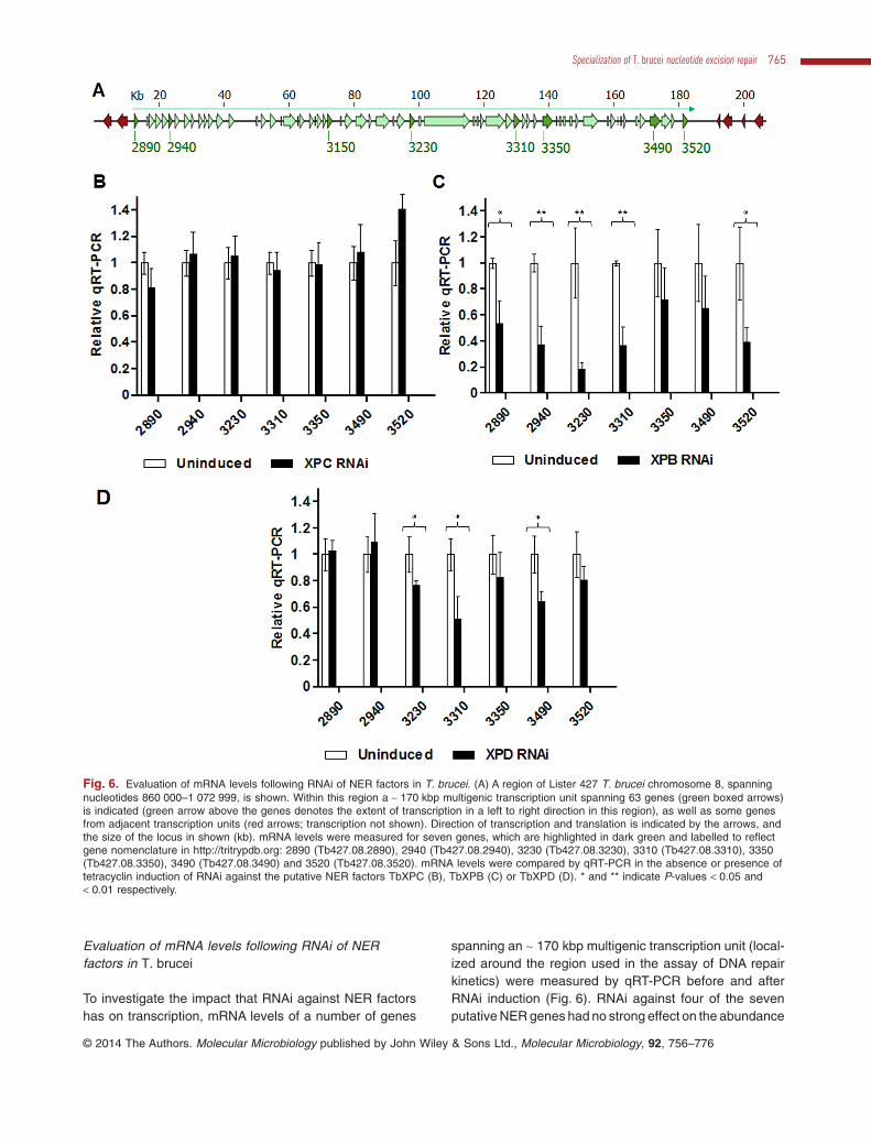
To investigate the impact that RNAi against NER factorshas on transcription, mRNA levels of a number of genes
spanning an ∼ 170 kbp multigenic transcription unit (local-ized around the region used in the assay of DNA repairkinetics) were measured by qRT-PCR before and afterRNAi induction (Fig. 6). RNAi against four of the sevenputative NER genes had no strong effect on the abundance
Fig. 6. Evaluation of mRNA levels following RNAi of NER factors in T. brucei. (A) A region of Lister 427 T. brucei chromosome 8, spanningnucleotides 860 000–1 072 999, is shown. Within this region a ∼ 170 kbp multigenic transcription unit spanning 63 genes (green boxed arrows)is indicated (green arrow above the genes denotes the extent of transcription in a left to right direction in this region), as well as some genesfrom adjacent transcription units (red arrows; transcription not shown). Direction of transcription and translation is indicated by the arrows, andthe size of the locus in shown (kb). mRNA levels were measured for seven genes, which are highlighted in dark green and labelled to reflectgene nomenclature in http://tritrypdb.org: 2890 (Tb427.08.2890), 2940 (Tb427.08.2940), 3230 (Tb427.08.3230), 3310 (Tb427.08.3310), 3350(Tb427.08.3350), 3490 (Tb427.08.3490) and 3520 (Tb427.08.3520). mRNA levels were compared by qRT-PCR in the absence or presence oftetracyclin induction of RNAi against the putative NER factors TbXPC (B), TbXPB (C) or TbXPD (D). * and ** indicate P-values < 0.05 and< 0.01 respectively. This figure is available in colour online at wileyonlinelibrary.com.
Specialization of T. brucei nucleotide excision repair 765
© 2014 The Authors. Molecular Microbiology published by John Wiley & Sons Ltd., Molecular Microbiology, 92, 756–776
of the transcripts examined (mRNA levels are shown inFig. 6B for TbXPC as an example; TbDDB, TbXPBz andTbXPG, data not shown). The effect of TbCSB RNAi isdiscussed below (Fig. 7). RNAi of TbXPB or TbXPDcaused a decrease in mRNA levels: RNAi of the formersignificantly reduced mRNA levels for five of the seven
analysed genes, while RNAi of the later significantlyreduced mRNAlevels for three of the seven genes (Fig. 6Cand D). This most likely reflects the role of TbXPB andTbXPD as constituents of the TbTFIIH complex in vivo, andis consistent with TbXPD contributing to SL-RNAtranscrip-tion (Lee et al., 2009).
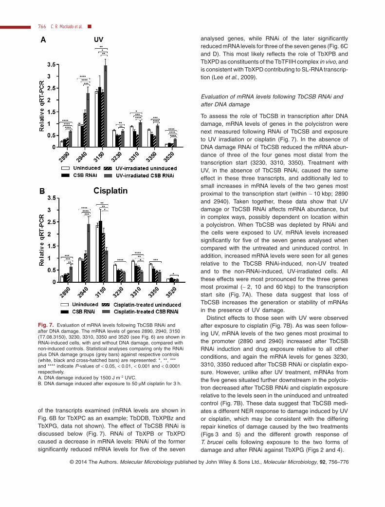
Evaluation of mRNA levels following TbCSB RNAi andafter DNA damage
To assess the role of TbCSB in transcription after DNAdamage, mRNA levels of genes in the polycistron werenext measured following RNAi of TbCSB and exposureto UV irradiation or cisplatin (Fig. 7). In the absence ofDNA damage RNAi of TbCSB reduced the mRNA abun-dance of three of the four genes most distal from thetranscription start (3230, 3310, 3350). Treatment withUV, in the absence of TbCSB RNAi, caused the sameeffect in these three transcripts, and additionally led tosmall increases in mRNA levels of the two genes mostproximal to the transcription start (within ∼ 10 kbp; 2890and 2940). Taken together, these data show that UVdamage or TbCSB RNAi affects mRNA abundance, butin complex ways, possibly dependent on location withina polycistron. When TbCSB was depleted by RNAi andthe cells were exposed to UV, mRNA levels increasedsignificantly for five of the seven genes analysed whencompared with the untreated and uninduced control. Inaddition, increased mRNA levels were seen for all genesrelative to the TbCSB RNAi-induced, non-UV treatedand to the non-RNAi-induced, UV-irradiated cells. Allthese effects were most pronounced for the three genesmost proximal (∼ 2, 10 and 60 kbp) to the transcriptionstart site (Fig. 7A). These data suggest that loss ofTbCSB increases the generation or stability of mRNAsin the presence of UV damage.
Distinct effects to those seen with UV were observedafter exposure to cisplatin (Fig. 7B). As was seen follow-ing UV, mRNA levels of the two genes most proximal tothe promoter (2890 and 2940) increased after TbCSBRNAi induction and drug exposure relative to all otherconditions, and again the mRNA levels for genes 3230,3310, 3350 reduced after TbCSB RNAi or cisplatin expo-sure. However, unlike after UV treatment, mRNAs fromthe five genes situated further downstream in the polycis-tron decreased after TbCSB RNAi and cisplatin exposurerelative to the levels seen in the uninduced and untreatedcontrol (Fig. 7B). These data suggest that TbCSB medi-ates a different NER response to damage induced by UVor cisplatin, which may be consistent with the differingrepair kinetics of damage caused by the two treatments(Figs 3 and 5) and the different growth response ofT. brucei cells following exposure to the two forms ofdamage and after RNAi against TbXPG (Figs 2 and 4).
Fig. 7. Evaluation of mRNA levels following TbCSB RNAi andafter DNA damage. The mRNA levels of genes 2890, 2940, 3150(T7.08.3150), 3230, 3310, 3350 and 3520 (see Fig. 6) are shown inRNAi-induced cells, with and without DNA damage, compared withnon-induced controls. Statistical analyses comparing only the RNAiplus DNA damage groups (grey bars) against respective controls(white, black and cross-hatched bars) are represented: *, **, ***and **** indicate P-values of < 0.05, < 0.01, < 0.001 and < 0.0001respectively.A. DNA damage induced by 1500 J m−2 UVC.B. DNA damage induced after exposure to 50 μM cisplatin for 3 h.
766 C. R. Machado et al. ■
© 2014 The Authors. Molecular Microbiology published by John Wiley & Sons Ltd., Molecular Microbiology, 92, 756–776
Predicted GG-NER proteins in T. brucei are involved ininter-strand cross-link repair
Cyclophosphamide is an alkylating agent that adds mono-functional adducts at the N7 position of guanine and leadsto the formation of DNA inter-strand cross-links (ICLs)involving two guanines (Povirk and Shuker, 1994). It hasbeen used as a chemotherapeutic agent in a set of cancersand autoimmune diseases (Dollery, 1999). ICLs are pro-cessed in eukaryotes by the cooperation of three DNArepair and/or bypass pathways: NER, homologous recom-bination and translesion synthesis (McVey, 2010). Toinvestigate the role of the predicted T. brucei NER genesduring ICL repair, the BSF RNAi cell lines were cultivated inthe presence of different doses of cyclophosphamide andsurvival measured before and after RNAi induction (Fig. 8).RNAi against TbXPC and TbDDB resulted in increasedsensitivity to cyclophosphamide, indicating that thesegenes could be important in ICL repair. In contrast, RNAidepletion of TbCSB, TbXPBz, TbXPG, TbXPD or TbXPBhad no effect on cyclophosphamide sensitivity. These datasuggest that the separation of function we observedbetween the predicted GG-NER and TC-NER proteins inT. brucei’s response to UV or cisplatin is mirrored in theresponse to the cyclophosphamide: though TbXPC andTbDDB act in ICL repair, TbCSB, TbXPBz and TbXPG donot. Furthermore, these data add to the UV and cisplatinsurvival curves to provide more evidence that TbXPB orTbXPD do not act in DNA repair.
Discussion
Much of the genome of trypanosomatids appears to betranscribed continuously, since most genes are arranged inmultigene units, each transcribed from a single promoter(Vanhamme and Pays, 1995; Siegel et al., 2009; 2011).This unusual mode of gene expression prompted us toinvestigate how NER operates, and in this study we havepresented a near comprehensive functional analysis of thepredicted machinery of NER in T. brucei. Our findingssuggest that NER in T. brucei, and therefore most likely inrelated trypanosomatids, has undergone extensive func-tional diversification relative to other eukaryotes. Despiteconservation of most predicted NER proteins, our dataindicate that only TbCSB, TbXPBz and TbXPG contributeto UV and/or cisplatin repair, suggesting that TC-NERpredominates. TbXPC and TbDDB, which would beexpected to mediate GG-NER, show no evidence for a rolein UV or cisplatin repair. Despite this, RNAi indicates thatthese factors are essential and act in ICL repair, suggestingthat the GG-NER subpathway acts predominantly in anunknown but critical process in trypanosomatid genomemaintenance. Finally, TbXPB and TbXPD show no evi-dence for a role in repair of any lesions tested, suggesting
that T. brucei NER does not involve the TFIIH complex.Taken as a whole, this work suggests that genome main-tenance by NER in this parasite may be quite unlike that ofmost eukaryotes: contrary to the expectation that theTC-NER and GG-NER subpathways should act in paralleland feed into a common lesion excision pathway, itappears that T. brucei has evolved a profound focus onTC-NER and has largely or completely uncoupled theGG-NER factors and TFIIH from this reaction.
Does NER in T. brucei utilize a simplified machinerycompared with other eukaryotes?
The analytical work reported here and elsewhere (seebelow) suggests that predictions of NER conservation inT. brucei based on homology masks a streamlining of themachinery devoted to NER, as well as neofunctionaliza-tion. A major element of NER streamlining is non-engagement of the TFIIH complex during the reaction,which appears to be associated with the evolution of arepair-specific XPB-related helicase, TbXPBz (or TbXPBr;Badjatia et al., 2013). Here we show that loss of TbXPBz byRNAi does not affect T. brucei viability, but that such RNAirenders BSF T. brucei cells sensitive to UV and cisplatindamage, showing that TbXPBz acts in DNA repair. Incontrast, RNAi of TbXPB or TbXPD was lethal, but wefound no evidence that loss of either helicase increasedsensitivity to DNA damage. These findings are consistentwith other studies. Lecordier et al. (2007) reported verysimilar rates of cell death following RNAi of TbXPB orTbXPD in procyclic form (PCF) and BSF cells, and Badjatiaet al. (2013) have demonstrated that null mutants ofTbXPBz can be generated in PCF T. brucei and display thesame increased sensitivity to UV and cisplatin as TbXPBzRNAi-induced BSF cells (this study). Thus, the distinctfunctions of the related helicases TbXPBz and TbXPBappear to be retained throughout the parasite life cycle.Moreover, though TbXPB and TbXPD act in concert ascomponents of the T. brucei TFIIH complex and are impor-tant in transcription (Lecordier et al., 2007), there is noevidence that TbXPBz associates with the TFIIH complexor contributes to transcription (Lee et al., 2009; Badjatiaet al., 2013).
TbXPBz orthologues in trypanosomatids retain all con-served domains related to DNA helicase function(Lecordier et al., 2007; Badjatia et al., 2013), suggestingthat XPB duplication arose to provide helicase-relatedactivities for DNA repair that replace, or assume greaterimportance than, TbXPB-TFIIH. Though it has been pro-posed from sequence alignments that TbXPBz is capableof interacting with TbXPC and thus mediating GG-NER(Badjatia et al., 2013), the lack of evidence for TbXPC (orTbDDB) involvement in NER appears inconsistent withthis (see below). In contrast, the phenotypes of TbXPBz
Specialization of T. brucei nucleotide excision repair 767
© 2014 The Authors. Molecular Microbiology published by John Wiley & Sons Ltd., Molecular Microbiology, 92, 756–776

Fig. 8. Predicted GG-NER proteins in T. brucei are involved in inter-strand cross-link repair. Survival curves of TbXPC, TbDDB, TbCSB,TbXPB, TbXPBz, TbXPD and TbXPD (A–G respectively) RNAi depleted cells (dotted lines) compared with respective uninduced controls(solid lines), after treatment with 2.5, 5 or 10 mM cyclophosphamide. Percent survival was calculated as the relative number of cells present inthe treated groups compared with the untreated control. Values are means of three experiments, and vertical lines denote standard deviation.
768 C. R. Machado et al. ■
© 2014 The Authors. Molecular Microbiology published by John Wiley & Sons Ltd., Molecular Microbiology, 92, 756–776

RNAi strongly phenocopy those of TbCSB RNAi, suggest-ing that TbXPBz predominantly acts together with TbCSBin TC-NER. Whether or not this aspect of NER speciali-zation is specific to kinetoplastids, or is also found in thewider range of protists that possess two XPBs (Badjatiaet al., 2013), is unknown. Moreover, what aspects ofTbXPBz prevent its recruitment into TFIIH and favours itsassociation with TbCSB are unknown. Indeed, howTC-NER in T. brucei might operate without engagingTFIIH is unclear. In other eukaryotes, XPB is a weakhelicase and ablation of this activity does not impedeNER, while loss of XPD helicase is detrimental to NER(Coin et al., 2007), suggesting that XPD plays the greaterrole in extrusion of the lesion strand. Given these obser-vations, the question of whether TbXPBz associates withanother helicase in a functionally distinct variant of theTFIIH complex, lacking TbXPB and TbXPD, or acts in anunrelated complex, merits further investigation. In thislight, the observations that TbXPBz interacts with Tbp52(Badjatia et al., 2013), and that RNAi of Tbp52 or Tbp44results in MMS and/or UV light sensitivity (Lecordier et al.,2007; Badjatia et al., 2013), are intriguing. One interpre-tation of these data is that at least some components oftrypanosomatid TFIIH act in the DNA damage response,perhaps consistent with remodelling to incorporateTbXPBz and exclude TbXPB and TbXPD. However,sequence analysis of TbXPG provides further evidencethat T. brucei NER has evolved to act without TFIIH.
We have shown that RNAi of TbXPG leads to increasedsensitivity to cisplatin, indicating a role in TC-NER thatwould be consistent with work in other eukaryotes, whereXPG has been shown to interact with CSB and play anearly role in this NER subpathway (Iyer et al., 1996;Sarker et al., 2005). However, the polypeptide sequenceof TbXPG displays notable sequence losses in the centralspacer region relative to XPG orthologues (Fig. S7). Sincethe spacer region mediates the interaction of XPG withTFIIH (Dunand-Sauthier et al., 2005), mutations in thispart of parasite protein suggest that engagement ofTbXPG during NER is achieved without TFIIH. Indeed,lack of T. brucei TFIIH function in NER may underlie thepotential absence of an identifiable trypanosmatid XPAorthologue, whose role appears to be in stabilizing TFIIHassociation with the NER machinery (Krasikova et al.,2010). Superficially, this putative bypass of TFIIH appearsreminiscent of archaea, where XPB can interact withBax1, a functional equivalent of the XPG nuclease (Rouil-lon and White, 2011). However, direct interaction betweenTbXPBz and TbXPG has not been observed, and a staticcomplex appears incompatible with the differing responseto UV exposure following RNAi against the two factors inT. brucei (discussed below). In addition, whether or notfurther endonucleases, such as XPF/ERCC1, contributeto NER in T. brucei awaits further investigation.
Is translesion RNA synthesis an important alterative toTC-NER in T. brucei?
A number of observations lead us to propose thatT. brucei may not always respond to transcription-blocking lesions by executing TC-NER. First, RNAiagainst TbCSB and TbXPBz resulted in increased sensi-tivity to both UV and cisplatin, whereas RNAi againstTbXPG only caused increased sensitivity to cisplatin, notUV. Second, measuring repair kinetics shows that UVlesions are repaired substantially more slowly than cispla-tin lesions in trypanosomes, and that TbCSB is needed torepair the latter type of damage. Finally, analysis of anumber of genes spanning a T. brucei multigene tran-scription unit indicates differing changes in mRNA abun-dance following exposure to UV or cisplatin and afterRNAi against TbCSB: increased mRNA levels were seenfor promoter-distal genes after UV exposure, but reducedlevels for the same genes after cisplatin. We suggest thatthese observations are most simply explained by RNA PolII bypass of some lesions in some circumstances, andthat CSB plays a pivotal role in the decision to invoke sucha bypass or to enact TC-NER (Lee et al., 2002). Transle-sion synthesis by any RNA Pol has not been documentedin trypanosomatids, but is well established in othereukaryotes. Lesions occurring in transcribed DNA strandslead to RNA Pol II blockage or slowdown, increasing CSBaffinity with TFIIH, a recruitment that mediates the prefer-ential repair of lesions localized in transcribed strandsthrough TC-NER (Hanawalt and Spivak, 2008). Alterna-tively, DNA lesions present in the template strand can bebypassed by RNA Pol II (Saxowsky and Doetsch, 2006;Bregeon and Doetsch, 2011). CPDs can be bypassedduring transcription because RNA Pol II has the ability toincorporate A residues in front a CPD dimer: the first A isincorporated in a non-template fashion, following the‘A-rule’, while the second is inserted in a template-directed manner. When one U is incorporated instead ofthe second A, this mismatch induces RNA Pol II stalling.Thus, at least in S. cerevisae, the complete translesiontranscription process through CPDs can be error-free(Brueckner et al., 2007; Walmacq et al., 2012). Thoughmore work is needed, we suggest that the choice betweentranslesion RNA synthesis and TC-NER is able to explainall the data we present.
We suggest that UV damage in T. brucei can bebypassed by translesion synthesis or removed by NER,and that TbCSB is pivotal in detecting such lesions andchannelling them to TC-NER only if needed. Bypass ofsuch lesions may normally be favoured due to multigenictranscription, avoiding accumulation of stalled RNA Pol IIand promoting continued movement of the transcriptionmachinery. In fact, this may be the reason that TFIIH isuncoupled from T. brucei NER, thus limiting CSB’s ability
Specialization of T. brucei nucleotide excision repair 769
© 2014 The Authors. Molecular Microbiology published by John Wiley & Sons Ltd., Molecular Microbiology, 92, 756–776

to engage NER. A preference for UV lesion bypass wouldexplain why we see little evidence for rapid repair of UVdamage. When TbCSB is removed, this control point islost and TC-NER cannot occur, meaning that bypass pre-dominates further and explaining why mRNA levelsincreased after TbCSB RNAi. It is likely that lesion bypassis a short-term response to minimal damage, and mightexplain the increased survival we observed in T. bruceiBSF TbCSB RNAi-induced cells within the first four hoursafter UV exposure (Fig. S10). However, if bypass tran-scription is error-prone, it could lead to an overproductionof mutated mRNAs that could be toxic for the parasite,explaining the increased sensitivity in TbCSB RNAi cells24 h after UV irradiation. We further suggest that cisplatin-induced adducts have a more pronounced effect onT. brucei transcription than UV, inducing RNA Pol II block-ages that cannot be as efficiently bypassed (Damsmaet al., 2007). Thus, the primary response to cisplatin, andperhaps other platinum compounds, is that TbCSBsignals for lesion removal by enacting TC-NER andengaging TbXPG. This model explains why TbXPG RNAicells are more sensitive to cisplain, but not UV, and whycisplatin is seen to be rapidly removed in the repair assay.The model also explains why cisplatin treatment results indecreased mRNA levels of most genes in the polycistronafter TbCSB RNAi: here, TC-NER is impaired and cispla-tin adducts cause increasing transcription impairmentthrough polycistons.
Why is loss of some predicted T. brucei NERfactors lethal?
We have argued above that NER in T. brucei is largelyconducted by a simplified TC-NER pathway, involvingTbCSB, TBXPBz and TbXPG. In this light, the data that wepresent on the functions provided by the predictedT. brucei GG-NER factors are remarkable, since RNAi ofTbXPC or TbDDB results in cell death, at least in the BSF.The essentiality of TbXPB and TbXPD has been docu-mented before in T. brucei (Lecordier et al., 2007) and isexplained by these factors’ roles as components of theTFIIH complex; hence, their removal impedes transcrip-tion, as seen by reduced mRNA levels following RNAi (thisstudy) and evaluated by in vitro transcription assays(Badjatia et al., 2013). In contrast, why the GG-NERfactors are essential is less clear. In S. cerevisiae, nullmutants of RAD4 (XPC), RAD23, RAD1 (XPF) and RAD10(ERCC1) have been documented and are viable (Giaeveret al., 2002). Phenotypes of NER mutation appear moresevere in multicellular eukaryotes but, even here, at leastsome NER components appear to be dispensable. InA. thaliana XPF/ERCC1 null mutants are viable (Vannieret al., 2009), while in mice substantial gene deletions ofXPC are viable (Sands et al., 1995) and a near complete
null of ERCC1 does not prevent embryo growth until aftergestation. In C. elegans, loss of RAD23 or XPC is notimmediately lethal, with the former ablation allowing com-plete embryonic development but maternal sterility(Kamath et al., 2003). The essentiality of TbDDB andTbXPC in T. brucei appears not to be due to critical contri-butions to NER, since we find no evidence that TbDDB orTbXPC repair UV or cisplatin damage. However, this DNAdamage may have the most pronounced effect in the highlytranscribed core of the T. brucei genome, and we cannotyet discount that GG-NER is important in the subtelom-eres, which harbour thousands of VSG genes and arelargely transcriptionally silent (Berriman et al., 2005;Marcello and Barry, 2007). Nonetheless, we have providedpreliminary evidence that GG-NER may have assumed apredominant role in non-NER DNA repair.
The lack of recognition of UV-induced lesions by TbXPCand TbDDB is remarkable, since UV-induced lesions arerecognized by XPC and XPE during GG-NER in moststudied organisms (Feldberg and Grossman, 1976; Chuand Chang, 1988). Rad4, the S. cerevisiae homologue ofXPC, recognizes (together with RAD23) ssDNA structuresassociated with bulky lesions. To do this, a β-hairpindomain of Rad4 is inserted through the DNA double helix,evicting the damaged strand away from its binding site.This mechanism of XPC DNA damage recognitionaccounts for the remarkable range of bulky lesions tar-geted by GG-NER (Min and Pavletich, 2007). To ask if thelack of UV damage repair by GG-NER may be accountedfor by alterations in TbXPC, the three dimensional structureof the parasite protein was modelled on the structure ofRad4 protein bound to DNA (Fig. 9). By measuring thedistance between the ‘arms’ that form the lesion recogni-tion site in the two proteins, we calculated the approximatesize of the DNAbinding site to be around 13 Å for Rad4 andaround 27 Å for TbXPC. Since the diameter of a DNAdouble helix is approximately 20 Å, it is plausible to inferthat Rad4 can only recognize one DNA strand, whileTbXPC may bind both strands. This modelling raises thepossibility that TbXPC can bind both damaged and undam-aged DNA strands, and might explain why GG-NER intrypanosomes is not targeted to DNA damage caused byUV and cisplatin. This putative alteration in TbXPC DNAbinding may also be related to the more pronounced rolewe see for the T. brucei GG-NER proteins acting in ICLrepair, based on the observation that RNAi of TbXPC orTbDDB caused sensitivity to the ICL inducer cyclophos-phamide.
ICLs are highly genotoxic DNA lesions whose repairrequires several factors derived from a number of inde-pendent DNA repair pathways (McVey, 2010). A role forGG-NER proteins in ICL repair has been seen in S. cerevi-siae (Saffran et al., 2004), where rad4Δ mutants (XPCmutants) shows sensitivity to the ICL-causing agents
770 C. R. Machado et al. ■
© 2014 The Authors. Molecular Microbiology published by John Wiley & Sons Ltd., Molecular Microbiology, 92, 756–776

mechlorethamine, mitomycin C and cisplatin (McHughet al., 1999; Wu et al., 2004). However, mammal cellsdeficient for XPA or XPC present only moderate sensitivityto ICL-inductors (Clingen et al., 2007). High sensitivity toICL-inducing agents, as well as bone marrow failure andhigh cancer frequencies, are observed in humans harbour-ing mutations in Fanconi Anemia core complex genes(Deans and West, 2011), which appear to be missing inlower eukaryotes. Thus, in single-celled eukaryotes likeyeast and trypanosomatids ICL recognition and repair byGG-NER proteins may assume greater importance.However, if ICL repair is the main role of the GG-NERpathway in T. brucei, it remains unclear why this activityshould assume such great importance. From the molecularmodelling above, we infer a novel mechanism employed byTbXPC for DNA lesion binding relative to Rad4 (Min andPavletich, 2007). However, it is unclear if this novelty is dueto greater commitment of the T. brucei GG-NER system toICL recognition, since Rad4 can act in ICL repair and,indeed, the human XPC-Rad23b complex also binds to a
psoralen ICLs (Thoma et al., 2005). Further investigationwill be necessary to address the lesion spectrum targetedby TbXPC and, until then, it remains unclear what essentialpurpose these GG-NER factors provide. Nonetheless, thepossibility that TbXPC and TbDDB, previously thought toact in NER, might have assumed a distinct and essentialgenome maintenance role is interesting. If it can be shownthat GG-NER provides an activity distinct from the host,this repair pathway might represent a target for therapeuticintervention against Sleeping Sickness and other trypano-somatid diseases.
Experimental procedures
Parasite growth
Epimastigote forms of Trypanosoma cruzi CL Brener strainwere cultivated at 28°C in LIT (Liver Infusion Tryptose)medium (pH 7.3; Camargo, 1964) supplemented with 10%heat-inactivated fetal bovine serum (FBS, Cultilab) plus100 U ml−1 penicillin and 100 μg ml−1 streptomycin (Invitro-
Fig. 9. TbXPC presents a larger DNA damage recognition site when compared with its yeast homologue. Left: a comparison of the structureof Rad4 (green) and the predicted structure of TbXPC (purple); secondary structures are shown, overlapped with the surface, and thedistance difference between the ‘arms’, which constitutes the DNA lesion recognition site, of both proteins is depicted. Right: Surfacerepresentation of the structure of Rad4 (green) and the predicted structure of TbXPC (purple), bound to a double-stranded DNA molecule.
Specialization of T. brucei nucleotide excision repair 771
© 2014 The Authors. Molecular Microbiology published by John Wiley & Sons Ltd., Molecular Microbiology, 92, 756–776
gen). Bloodstream form Trypanosoma brucei were cultivatedat 37°C in a humidified incubator with 5% CO2. Parasiteswere grown in HMI-9 (Hirumi and Hirumi, 1989) supple-mented with 20% heat-inactivated FBS (Sigma) plus 1% (v/v)of penicillin and streptomycin (Sigma).
Growth after RNAi in the presence and absence ofgenotoxic agents
To measure growth, the parasites were diluted to a density of1 × 105 cells ml−1 in HMI-9 plus selective drugs (hygromycinand phleomycin), with and without tetracycline (1 μg ml−1, toinduce RNAi). Cell cultures were diluted 10-fold daily and cellnumbers counted in a heamocytometer chamber at 24 h inter-vals; each growth curve was carried out three times indepen-dently for each RNAi cell line. To evaluate sensitivity to DNAdamage, the cells were similarly diluted to 1 × 10⁵ cells ml−1,and tetracycline added as before. After 24 h the cells wereexposed to the genotoxic agents, and the cell growth allowedto proceed as before. Survival rate was calculated from the cellcounts of the drug-exposed cells relative to the untreatedcontrols. All survival curves were performed in triplicate. Forthe cisplatin survival curve, parasites were treated with 1, 2 or5 μM of cisplatin (Sigma). In the UV irradiation survival curve,parasites received doses of 500, 1500 or 3000 J m−2 of UVCusing a Stratalinker® UV Crosslinker (Stratagene). For cyclo-phosphamide (Sigma) exposure, parasites were grown inconcentrations of 2.5, 5 and 10 mM.
RNAi
The primers utilized for RNAi constructs were designed usingsoftware available online (http://trypanofan.path.cam.ac.uk/software/RNAit.html); in each case Attb1 and Attb2 recombi-nation sites were added to the termini of the primers. Thesequences of the primers used to PCR-amplify the RNAicassettes are on Table S1. All primers were purchased fromEurofins MWG Operon (http://www.eurofinsdna.com). TheAttb1 and Attb2 sites allowed Gateway recombination in asingle reaction, inserting two copies of the product in opposingdirections into the pGL2084 vector to generate a stem-loopRNAi producing construct (Jones et al., 2014). For PCR, amaster mix was used: 1× Phusion Buffer, 0.2 mM dNTP mix,50 ng of template genomic DNA from Lister 427 T. brucei,2 μM of each primer and 20 u ml−1 of Phusion enzyme (Invit-rogen). The PCR reactions were carried out with settings of3 min at 98°C, 30 cycles of 98°C for 30 s, 60°C for 30 s and72°C for 30 s, and a final step for 10 min at 72°C. PCRproducts were purified from 1% agarose gels using the QiagenMinElute Kit and used in RNAi construct assembly intopGL2084. For the assembly reaction ≥ 10 ng of PCR product,150 ng of pGL2084 vector, 0.25 μl of BP Clonase™ enzymemix (Invitrogen), Tris-HCl 3.75 mM and EDTA375 μM (pH 8.0)were used. The mix reaction was incubated at room tempera-ture for 1 h. To transform Max efficiency DH5α E. coli (Invitro-gen Cat. No. 18258-012), 1 μl of BP clonase reaction wasused, as described by the manufacturer. The clones obtainedfor each reaction were grown in LB medium supplementedwith ampicilin at 100 μg ml−1 at 37°C overnight and the RNAiconstructs were then isolated using Qiagen QIAprep Spin Kit.
To test if the extracted plasmids were successfully recom-bined, double digestions with BamHI and XbaI, and singledigestions with StuI or ClaI, were performed. Next, the RNAiconstructs were linearized by digestion with AscI and 5 μg oflinearized plasmid were used to transfect BSF T. brucei 2T1cells (Alsford and Horn, 2008). For transfection 1 × 10⁷ cellswere harvested by centrifugation for 10 min for 1500 g. Aftermedia was removed, cells were resuspended in 100 μl ofAmaxa Nucleofector T-cell buffer, transferred to a cuvette and10 μl of the linearized DNA were added. Electroporation wasperformed using the program X-001 on the Nucleofector IImachine. Cells were then transferred to 30 ml of HMI-9medium without selective drugs. After 6 h, the cells werediluted at 1:20 and 1:40 in media containing selective drugs:hygromycin (2.5 μg ml−1) and phleomycin (0.5 μg ml−1). Antibi-otic resistant clones were selected for at least 7 days. In orderto test if the RNAi cassette had been correctly integrated, theparasites’ sensitivity to puromycin (1.0 μg ml−1) was tested.
Real-time PCR
To validate the RNAi, levels of target mRNAs for each genewere measured by quantitative reverse transcriptase PCR(qRT-PCR). A total of 2 × 10⁷ cells were harvested from RNAi-induced and uninduced cultures at the times stated, and totalRNA was isolated using the Qiagen RNeasy Kit (withon-column DNaseI digestion). For all qRT-PCRs a master mixfor 30 reactions was made in which each reaction had 12.5 μlof SYBR Green PCR Master Mix (Applied Biosystems), 1.0 μlof each primer (300 nM stock), 9.5 μl of dH2O, and 1.0 μlcDNA (generated by SuperScript First-Strand SynthesisSystem for RT-PCR (Invitrogen) from the total RNA, accord-ing to manufacturer’s instructions). Reactions were run on anABI Prism 7000 thermocycler and mRNA levels quantifiedfrom the amplifications according to the manufacturer’sinstructions; conditions for all reactions were 50°C for 2 min,95°C for 10 min, followed by 40 cycles of 95°C for 15 s and60°C for 1 min. Primers recognizing GPI8 (CTOL27 andCTOL28) were used as a control (Tiengwe et al., 2012).Primer pairs for each putative NER gene are provided onTable S1. The mRNA levels from the polycistron genes werealso assessed by qRT-PCR (for primers details, see TableS1), using the same procedures. All primers were purchasedfrom Eurofins MWG Operon. All qRT-PCR experiments wereperformed at least three times independently. Statisticalanalysis was performed using unpaired t-test for compari-sons of mRNA levels between RNAi cell lines and uninducedcontrols. An ordinary one-way ANOVA followed by Tukey’s testwas performed to compare mRNA levels after 24–48 h ofRNAi induction, while a two-way ANOVA followed by Tukey’stest was performed to compare mRNA levels after RNAiinduction in the presence of DNA damage. All statisticalanalysis was conducted using the software GraphPad Prism.
Comparative modelling and docking
A TbXPC molecular structure was generated by comparativemodelling. The search for candidate template structures wasperformed by BLAST searches of the PDB database(Westbrook et al., 2003). The template used for modelling was
772 C. R. Machado et al. ■
© 2014 The Authors. Molecular Microbiology published by John Wiley & Sons Ltd., Molecular Microbiology, 92, 756–776
Rad4 2QSH chain A (S. cerevisiae XPC homologue; Min andPavletich, 2007). The TbXPC molecular model was built basedonly on the β-hairpin domains of Rad4, corresponding topositions 400–700 of the full-length protein. Sequence align-ments between TbXPC and its structural template were gen-erated by Promals 3D (Pei et al., 2008) and manually curated.Molecular modelling was performed using the program Mod-eller (version 9.7; Eswar et al., 2007), which generated ahundred possible structures. The validation of obtainedmodels according to stereochemical quality was performedthrough the analysis of Ramachadran plots generated by theprogram Procheck (version 3.5.4; Morris et al., 1992) and theenergetic characteristics were assessed using ProSA (ProSa2003; Wiederstein and Sippl, 2007). Perl scripts weredesigned and used to automatically retrieve the percentage ofresidues in the most favoured regions of the Ramachandranplot and the Z-score of ProSA for each one of the 100 struc-tures. Three different types of software were used to determinepotential protein-DNAbinding sites: DNABindR, BindN (and itsvariant BindNPlus) and DBSPSSM (Ahmad and Sarai, 2005;Wang and Brown, 2006; Yan et al., 2006). Results were clus-tered and all amino acids predicted as DNA-binding by at leastone of the three algorithms were considered as active residuesduring docking experiments. Docking calculations werecarried out in Haddock (Dominguez et al., 2003) advancedguru interface, defining active residues in the proteins asdescribed above, and defining passive residues as all resi-dues within a radius of 6 angstroms around the active resi-dues. For the DNAmolecules, all nucleotides were consideredas active residues in protein-DNA interaction. All additionalparameters were used as default values. The generated com-plexes were analysed through stereochemical, cluster-sizesand energy evaluations.
Immunofluorescence
Approximately 1 × 10⁶ cells were collected, washed once inPBS, and allowed to attach for 4 min to 12-well multi-wellglass slides (Thermo Scientific) that had been previouslycoated with Poly-L-lysine (Sigma Aldrich). The cells werethen fixed with 4% formaldehyde in PBS for 15 min, washedtwice with PBS, incubated with 0.5% Triton X-100 for 20 min,and further washed with PBS (twice). Next, the cells wereincubated for 30 min with 2 M HCL, washed 5 times withPBS, and then blocked with 20% heat-inactivated FBS(Sigma-Aldrich) for 30 min at 37°C, after which the wells werewashed 5 times with PBS. Primary antisera were usedaccording to the manufacturer instructions: mouse anti-(6-4)phosphoproducts (6-4 PPs) antibody (Cosmo Bio Co., Ltd) ata 1:300 dilution, mouse anti-cyclobutane pyrimidine dimers(CPDs) antibody (Cosmo Bio Co., Ltd) at a 1:1500 dilution,and mouse anti-cisplatin antibody (Abcam) at a 1:400 dilu-tion. In all cases, the antiserum was added to the cells in PBScontaining 5% FBS, and incubated for 30 min at 37°C. Thecells were then washed 5 times with PBS, and further incu-bated for 30 min at 37°C with Alexa Fluor 594 goat anti-mouse IgG (H+L) (Invitrogen) antibody at a dilution of 1:100in PBS containing 5% FBS. The wells were then washed 5times with PBS and the slides mounted in VECTASHIELDmounting medium with DAPI (1.5 μg ml−1) (Vector Laborato-ries). The slides were examined, and images acquired, using
an Axioskop 2 fluorescence microscope (Zeiss). Images wereprocessed and assembled in Adobe Photoshop (AdobeSystems). A two-way ANOVA followed by a Bonferroni post-test was performed to compare the levels of cisplatin-induceddamage in experimental repeats comparing TbCSB RNAiinduced cells and uninduced controls. Statistical analysis wasconducted using the software GraphPad Prism.
DNA lesion detection through quantitative PCR
To detect the level of DNA damage in the nuclear genome, aquantitative PCR-based technique was used according topreviously described protocols (Santos et al., 2006). Briefly,lesion frequency was assessed through relative PCR-amplification of a 10 kb fragment of the T. brucei or T. cruzigenome, comparing amplification after treatment with geno-toxic agents relative to untreated controls. A small, internalDNA fragment, comprising 204 and 250 bp (in T. brucei anT. cruzi respectively) was PCR-amplified from the 3′ extremityof the larger molecule in order to correct for DNAloading errorsor PCR bias and assure that amplification conditions were thesame for all samples. For DNA damage induction in T. brucei,approximately 1 × 10⁸ BSF cells of strain Lister 427 wereharvested by centrifugation for 15 min at 3000 g at 4°C andresuspended in the same volume of 1× PBS. Next, cells wereexposed to 100 μM cisplatin for 1 h. For UVC exposure,1 × 108 parasites were concentrated in 5 ml of media. The cellswere then spread in a small Petri plate and exposed to1500 J m−2 of UVC in a Stratalinker® UV Crosslinker (Strata-gene). For DNA damage induction in T. cruzi, 1 × 10⁸ epimas-tigote form cells, CL Brener strain, were used. Parasites werecentrifuged as for T. brucei and treated with 300 μM of cisplatinin 1× PBS for 1 h. For UV treatment, 5 ml of culture containing1 × 108 T. cruzi cells were spread in a small Petri plate andirradiated with 1500 J m−2 of UVC. In all cases, the cells werecentrifuged after treatment (as before) and resuspended inconditioned medium, i.e. the medium used in the originalculture. Untreated samples were processed in the same way.Samples were collected at appropriate time points subsequentto the above treatments (0, 1, 2, 5 and 10 h after treatment forT. brucei, and 0, 1, 5 and 24 h for T. cruzi), centrifuged at 3000g and 4°C for 15 min, and the resulting cell pellet immediatelyfrozen at −80°C. DNA was prepared from the cells using theBlood & Cell Culture DNA Mini Kit (QIAGEN), according to themanufacturer’s instructions for DNA extraction from tissues.DNA was then quantified using PicoGreen dye (MolecularProbes) and standard curves, as described by Santos et al.(2006). PCR was performed from 15 ng of template DNAusingthe GeneAmp XL PCR Kit (Applied Biosystems) and theresulting amounts of PCR product quantified, again usingPicoGreen and as described by Santos et al. (2006). Theprimers used in the amplification reaction of the large fragmentin T. brucei were qPCRFoward (5′-GTTGCTCACTTTCACCACGTATTCGGGAACCTGT-3′) and qPCRReverse (5′-CCACTGAATGCTGTATCCGGCATTTAGTCGTGTCTATGGG-3′);for T. cruzi the large fragment used the primers QPCRNuc2F(5′-GCACACGGCTGCGAGTGACCATTCAACTTT-3′) andQPCRNuc2R (5′-CCTCGCACATTTCTACCTTGTCCTTCAATGCCTGC-3′). To PCR-amplify the small fragment, used asthe internal control, the primers qPCRFI (5′-TTACAGCACCCAGGTTTATACCGCACGAAAGTGG-3′) and qPCRRe-
Specialization of T. brucei nucleotide excision repair 773
© 2014 The Authors. Molecular Microbiology published by John Wiley & Sons Ltd., Molecular Microbiology, 92, 756–776

verse were used in T. brucei, while the primers QPCRNuc2Int(5′-TCGAGCAAGCTGACACTCGATGCAACCAAAG-3′) andQPCRNuc2R were used in T. cruzi. PCR reactions must to becarried out only until the logarithmic phase, when the amplifi-cation yields are directly proportional to the starting amount oftemplate. To meet these requirements, PCR conditions in thisexperiment need to optimized for each lab, as they aredependent on many factors, including the type of PCRmachine used and the accuracy of genomic DNA templatequantification using the PicoGreen dye system (see Santoset al., 2006 for guidelines and for the quantification strategy).
Acknowledgements
This work was in part supported by the Welcome Trust(083485) and by CAPES, CNPq and FAPEMIG. The Well-come Trust Centre for Molecular Parasitology is supported bya core grant from the Wellcome Trust (085349). We thankNitika Badjatia and Arthur Gunzl for sharing their data with usprior to publication, and we thank our many colleagues inGlasgow and Belo Horizonte for their input.
References
Ahmad, S., and Sarai, A. (2005) PSSM-based prediction ofDNA binding sites in proteins. BMC Bioinformatics 6: 33.
Alsford, S., and Horn, D. (2008) Single-locus targeting con-structs for reliable regulated RNAi and transgene expres-sion in Trypanosoma brucei. Mol Biochem Parasitol 161:76–79.
Araki, M., Masutani, C., Takemura, M., Uchida, A.,Sugasawa, K., Kondoh, J., et al. (2001) Centrosomeprotein centrin 2/.caltractin 1 is part of the xeroderma pig-mentosum group C complex that initiates global genomenucleotide excision repair. J Biol Chem 276: 18665–18672.
Araujo, S.J., Tirode, F., Coin, F., Pospiech, H., Syvaoja, J.E.,Stucki, M., et al. (2000) Nucleotide excision repair of DNAwith recombinant human proteins: definition of the minimalset of factors, active forms of TFIIH, and modulation byCAK. Genes Dev 14: 349–359.
Badjatia, N., Nguyen, T.N., Lee, J.H., and Gunzl, A. (2013)Trypanosoma brucei harbors a divergent XPB helicaseparalog that is specialized in nucleotide excision repair andconserved among kinetoplastid organisms. Mol Microbiol90: 1293–1308.
Berriman, M., Ghedin, E., Hertz-Fowler, C., Blandin, G.,Renauld, H., Bartholomeu, D.C., et al. (2005) The genomeof the African trypanosome Trypanosoma brucei. Science309: 416–422.
Beukers, R., Eker, A.P., and Lohman, P.H. (2008) 50 yearsthymine dimer. DNA Repair (Amst) 7: 530–543.
Bregeon, D., and Doetsch, P.W. (2011) Transcriptionalmutagenesis: causes and involvement in tumour develop-ment. Nat Rev Cancer 11: 218–227.
Brueckner, F., Hennecke, U., Carell, T., and Cramer, P.(2007) CPD damage recognition by transcribing RNA poly-merase II. Science 315: 859–862.
Camargo, E.P. (1964) Growth and differentiation in Trypano-soma cruzi. I. Origin of metacyclic trypanosomes in liquidmedia. Rev Inst Med Trop Sao Paulo 6: 93–100.
Cameroni, E., Stettler, K., and Suter, B. (2010) On the traces
of XPD: cell cycle matters – untangling the genotype-phenotype relationship of XPD mutations. Cell Div 5: 24.
Chu, G., and Chang, E. (1988) Xeroderma pigmentosumgroup E cells lack a nuclear factor that binds to damagedDNA. Science 242: 564–567.
Clingen, P.H., Arlett, C.F., Hartley, J.A., and Parris, C.N.(2007) Chemosensitivity of primary human fibroblasts withdefective unhooking of DNA interstrand cross-links. ExpCell Res 313: 753–760.
Coin, F., Oksenych, V., and Egly, J.M. (2007) Distinct roles forthe XPB/p52 and XPD/p44 subcomplexes of TFIIH indamaged DNA opening during nucleotide excision repair.Mol Cell 26: 245–256.
Coin, F., Oksenych, V., Mocquet, V., Groh, S., Blattner, C.,and Egly, J.M. (2008) Nucleotide excision repair driven bythe dissociation of CAK from TFIIH. Mol Cell 31: 9–20.
Compe, E., and Egly, J.M. (2012) TFIIH: when transcriptionmet DNA repair. Nat Rev Mol Cell Biol 13: 343–354.
Coquelet, H., Tebabi, P., Pays, A., Steinert, M., and Pays, E.(1989) Trypanosoma brucei: enrichment by UV of inter-genic transcripts from the variable surface glycoproteingene expression site. Mol Cell Biol 9: 4022–4025.
Damsma, G.E., Alt, A., Brueckner, F., Carell, T., and Cramer,P. (2007) Mechanism of transcriptional stalling at cisplatin-damaged DNA. Nat Struct Mol Biol 14: 1127–1133.
Daniels, J.P., Gull, K., and Wickstead, B. (2010) Cell biologyof the trypanosome genome. Microbiol Mol Biol Rev 74:552–569.
Deans, A.J., and West, S.C. (2011) DNA interstrand crosslinkrepair and cancer. Nat Rev Cancer 11: 467–480.
Dollery, C.T. (1999) Drug discovery and development in themolecular era. Br J Clin Pharmacol 47: 5–6.
Dominguez, C., Boelens, R., and Bonvin, A.M. (2003)HADDOCK: a protein-protein docking approach based onbiochemical or biophysical information. J Am Chem Soc125: 1731–1737.
Dunand-Sauthier, I., Hohl, M., Thorel, F., Jaquier-Gubler, P.,Clarkson, S.G., and Scharer, O.D. (2005) The spacerregion of XPG mediates recruitment to nucleotide excisionrepair complexes and determines substrate specificity. JBiol Chem 280: 7030–7037.
El-Sayed, N.M., Myler, P.J., Bartholomeu, D.C., Nilsson, D.,Aggarwal, G., Tran, A.N., et al. (2005) The genomesequence of Trypanosoma cruzi, etiologic agent of Chagasdisease. Science 309: 409–415.
Emmert, S., Ueda, T., Zumsteg, U., Weber, P., Khan, S.G.,Oh, K.S., et al. (2009) Strict sun protection results inminimal skin changes in a patient with xeroderma pigmen-tosum and a novel c.2009delG mutation in XPD (ERCC2).Exp Dermatol 18: 64–68.
Eswar, N., Webb, B., Marti-Renom, M.A., Madhusudhan,M.S., Eramian, D., Shen, M.Y., et al. (2007) Comparativeprotein structure modeling using MODELLER. Curr ProtocProtein Sci Chapter 2: Unit 2.9.
Fagbemi, A.F., Orelli, B., and Scharer, O.D. (2011) Regula-tion of endonuclease activity in human nucleotide excisionrepair. DNA Repair (Amst) 10: 722–729.
Feldberg, R.S., and Grossman, L. (1976) A DNA bindingprotein from human placenta specific for ultravioletdamaged DNA. Biochemistry 15: 2402–2408.
Giaever, G., Chu, A.M., Ni, L., Connelly, C., Riles, L.,
774 C. R. Machado et al. ■
© 2014 The Authors. Molecular Microbiology published by John Wiley & Sons Ltd., Molecular Microbiology, 92, 756–776

Veronneau, S., et al. (2002) Functional profiling of theSaccharomyces cerevisiae genome. Nature 418: 387–391.
Hanawalt, P.C., and Spivak, G. (2008) Transcription-coupledDNA repair: two decades of progress and surprises. NatRev Mol Cell Biol 9: 958–970.
Hirumi, H., and Hirumi, K. (1989) Continuous cultivation ofTrypanosoma brucei blood stream forms in a medium con-taining a low concentration of serum protein without feedercell layers. J Parasitol 75: 985–989.
Ivens, A.C., Peacock, S.C., Worthey, E.A., Murphy, L.,Aggarwal, G., Berriman, M., et al. (2005) The genome ofthe kinetoplastid parasite, Leishmania major. Science 309:436–442.
Iyer, N., Reagan, M.S., Wu, K.J., Canagarajah, B., andFriedberg, E.C. (1996) Interactions involving the humanRNA polymerase II transcription/nucleotide excision repaircomplex TFIIH, the nucleotide excision repair protein XPG,and Cockayne syndrome group B (CSB) protein. Biochem-istry 35: 2157–2167.
Johnson, P.J., Kooter, J.M., and Borst, P. (1987) Inactivationof transcription by UV irradiation of T. brucei provides evi-dence for a multicistronic transcription unit including a VSGgene. Cell 51: 273–281.
Jones, N.G., Thomas, E.B., Brown, E., Dickens, N.J.,Hammarton, T.C., and Mottram, J.C. (2014) Regulators ofTrypanosoma brucei cell cycle progression and differentia-tion identified using a kinome-wide RNAi screen. PLoSPathog 10: e1003886.
Kamath, R.S., Fraser, A.G., Dong, Y., Poulin, G., Durbin, R.,Gotta, M., et al. (2003) Systematic functional analysis ofthe Caenorhabditis elegans genome using RNAi. Nature421: 231–237.
Kim, N., and Jinks-Robertson, S. (2012) Transcription as asource of genome instability. Nat Rev Genet 13: 204–214.
Krasikova, Y.S., Rechkunova, N.I., Maltseva, E.A.,Petruseva, I.O., and Lavrik, O.I. (2010) Localization ofxeroderma pigmentosum group A protein and replicationprotein A on damaged DNA in nucleotide excision repair.Nucleic Acids Res 38: 8083–8094.
Lecordier, L., Devaux, S., Uzureau, P., Dierick, J.F.,Walgraffe, D., Poelvoorde, P., et al. (2007) Characteriza-tion of a TFIIH homologue from Trypanosoma brucei. MolMicrobiol 64: 1164–1181.
Lee, J.H., Jung, H.S., and Gunzl, A. (2009) Transcriptionallyactive TFIIH of the early-diverged eukaryote Trypanosomabrucei harbors two novel core subunits but not a cyclin-activating kinase complex. Nucleic Acids Res 37: 3811–3820.
Lee, S.K., Yu, S.L., Prakash, L., and Prakash, S. (2002) YeastRAD26, a homolog of the human CSB gene, functionsindependently of nucleotide excision repair and base exci-sion repair in promoting transcription through damagedbases. Mol Cell Biol 22: 4383–4389.
Lehmann, A.R. (2001) The xeroderma pigmentosum group D(XPD) gene: one gene, two functions, three diseases.Genes Dev 15: 15–23.
McHugh, P.J., Gill, R.D., Waters, R., and Hartley, J.A. (1999)Excision repair of nitrogen mustard-DNA adducts in Sac-charomyces cerevisiae. Nucleic Acids Res 27: 3259–3266.
McVey, M. (2010) Strategies for DNA interstrand crosslink
repair: insights from worms, flies, frogs, and slime molds.Environ Mol Mutagen 51: 646–658.
Marcello, L., and Barry, J.D. (2007) Analysis of the VSG genesilent archive in Trypanosoma brucei reveals that mosaicgene expression is prominent in antigenic variation and isfavored by archive substructure. Genome Res 17: 1344–1352.
Martinez-Calvillo, S., Yan, S., Nguyen, D., Fox, M., Stuart, K.,and Myler, P.J. (2003) Transcription of Leishmania majorFriedlin chromosome 1 initiates in both directions within asingle region. Mol Cell 11: 1291–1299.
Martinez-Calvillo, S., Nguyen, D., Stuart, K., and Myler, P.J.(2004) Transcription initiation and termination on Leishma-nia major chromosome 3. Eukaryot Cell 3: 506–517.
Masters, J.R., and Koberle, B. (2003) Curing metastaticcancer: lessons from testicular germ-cell tumours. Nat RevCancer 3: 517–525.
Min, J.H., and Pavletich, N.P. (2007) Recognition of DNAdamage by the Rad4 nucleotide excision repair protein.Nature 449: 570–575.
Morgante, P.G., Berra, C.M., Nakabashi, M., Costa, R.M.,Menck, C.F., and Van Sluys, M.A. (2005) Functional XPB/RAD25 redundancy in Arabidopsis genome: characteriza-tion of AtXPB2 and expression analysis. Gene 344:93–103.
Morris, A.L., MacArthur, M.W., Hutchinson, E.G., andThornton, J.M. (1992) Stereochemical quality of proteinstructure coordinates. Proteins 12: 345–364.
Mu, D., Park, C.H., Matsunaga, T., Hsu, D.S., Reardon, J.T.,and Sancar, A. (1995) Reconstitution of human DNA repairexcision nuclease in a highly defined system. J Biol Chem270: 2415–2418.
Naegeli, H., and Sugasawa, K. (2011) The xeroderma pig-mentosum pathway: decision tree analysis of DNA quality.DNA Repair (Amst) 10: 673–683.
Nishi, R., Okuda, Y., Watanabe, E., Mori, T., Iwai, S.,Masutani, C., et al. (2005) Centrin 2 stimulates nucleotideexcision repair by interacting with xeroderma pigmentosumgroup C protein. Mol Cell Biol 25: 5664–5674.
Ogi, T., Limsirichaikul, S., Overmeer, R.M., Volker, M.,Takenaka, K., Cloney, R., et al. (2010) Three DNA polymer-ases, recruited by different mechanisms, carry out NERrepair synthesis in human cells. Mol Cell 37: 714–727.
Oksenych, V., and Coin, F. (2009) The long unwinding road:XPB and XPD helicases in damaged DNA opening. CellCycle 9: 90–96.
Pei, J., Kim, B.H., and Grishin, N.V. (2008) PROMALS3D: atool for multiple protein sequence and structure align-ments. Nucleic Acids Res 36: 2295–2300.
Poon, S.K., Peacock, L., Gibson, W., Gull, K., and Kelly, S.(2012) A modular and optimized single marker system forgenerating Trypanosoma brucei cell lines expressing T7RNA polymerase and the tetracycline repressor. Open Biol2: 110037.
Povirk, L.F., and Shuker, D.E. (1994) DNA damage andmutagenesis induced by nitrogen mustards. Mutat Res318: 205–226.
Richards, J.D., Cubeddu, L., Roberts, J., Liu, H., and White,M.F. (2008) The archaeal XPB protein is a ssDNA-dependent ATPase with a novel partner. J Mol Biol 376:634–644.
Specialization of T. brucei nucleotide excision repair 775
© 2014 The Authors. Molecular Microbiology published by John Wiley & Sons Ltd., Molecular Microbiology, 92, 756–776
Rouillon, C., and White, M.F. (2011) The evolution andmechanisms of nucleotide excision repair proteins. ResMicrobiol 162: 19–26.
Saffran, W.A., Ahmed, S., Bellevue, S., Pereira, G., Patrick,T., Sanchez, W., et al. (2004) DNA repair defects channelinterstrand DNA cross-links into alternate recombinationaland error-prone repair pathways. J Biol Chem 279: 36462–36469.
Sands, A.T., Abuin, A., Sanchez, A., Conti, C.J., and Bradley,A. (1995) High susceptibility to ultraviolet-induced carcino-genesis in mice lacking XPC. Nature 377: 162–165.
Santos, J.H., Meyer, J.N., Mandavilli, B.S., and Van Houten,B. (2006) Quantitative PCR-based measurement ofnuclear and mitochondrial DNA damage and repair inmammalian cells. Methods Mol Biol 314: 183–199.
Sarker, A.H., Tsutakawa, S.E., Kostek, S., Ng, C., Shin, D.S.,Peris, M., et al. (2005) Recognition of RNA polymerase IIand transcription bubbles by XPG, CSB, and TFIIH:insights for transcription-coupled repair and Cockayne syn-drome. Mol Cell 20: 187–198.
Saxowsky, T.T., and Doetsch, P.W. (2006) RNA polymeraseencounters with DNA damage: transcription-coupled repairor transcriptional mutagenesis? Chem Rev 106: 474–488.
Siegel, T.N., Hekstra, D.R., Kemp, L.E., Figueiredo, L.M.,Lowell, J.E., Fenyo, D., et al. (2009) Four histone variantsmark the boundaries of polycistronic transcription units inTrypanosoma brucei. Genes Dev 23: 1063–1076.
Siegel, T.N., Gunasekera, K., Cross, G.A., and Ochsenreiter,T. (2011) Gene expression in Trypanosoma brucei: lessonsfrom high-throughput RNA sequencing. Trends Parasitol27: 434–441.
Stordal, B., and Davey, M. (2007) Understanding cisplatinresistance using cellular models. IUBMB Life 59: 696–699.
Svejstrup, J.Q., Wang, Z., Feaver, W.J., Wu, X., Bushnell,D.A., Donahue, T.F., et al. (1995) Different forms of TFIIHfor transcription and DNA repair: holo-TFIIH and a nucleo-tide excision repairosome. Cell 80: 21–28.
Thoma, B.S., Wakasugi, M., Christensen, J., Reddy, M.C.,and Vasquez, K.M. (2005) Human XPC-hHR23B interactswith XPA-RPA in the recognition of triplex-directed psoralenDNA interstrand crosslinks. Nucleic Acids Res 33: 2993–3001.
Tiengwe, C., Marcello, L., Farr, H., Dickens, N., Kelly, S.,Swiderski, M., et al. (2012) Genome-wide analysis revealsextensive functional interaction between DNA replicationinitiation and transcription in the genome of Trypanosomabrucei. Cell Rep 2: 185–197.
Vanhamme, L., and Pays, E. (1995) Control of gene expres-sion in trypanosomes. Microbiol Rev 59: 223–240.
Vannier, J.B., Depeiges, A., White, C., and Gallego, M.E.(2009) ERCC1/XPF protects short telomeres from homolo-gous recombination in Arabidopsis thaliana. PLoS Genet5: e1000380.
Vermeulen, W., Rademakers, S., Jaspers, N.G., Appeldoorn,E., Raams, A., Klein, B., et al. (2001) A temperature-sensitive disorder in basal transcription and DNA repair inhumans. Nat Genet 27: 299–303.
Walmacq, C., Cheung, A.C., Kireeva, M.L., Lubkowska, L.,Ye, C., Gotte, D., et al. (2012) Mechanism of translesiontranscription by RNA polymerase II and its role in cellularresistance to DNA damage. Mol Cell 46: 18–29.
Wang, L., and Brown, S.J. (2006) BindN: a web-based tool forefficient prediction of DNA and RNA binding sites in aminoacid sequences. Nucleic Acids Res 34: W243–W248.
Westbrook, J., Feng, Z., Chen, L., Yang, H., and Berman,H.M. (2003) The Protein Data Bank and structural genom-ics. Nucleic Acids Res 31: 489–491.
Wiederstein, M., and Sippl, M.J. (2007) ProSA-web: interac-tive web service for the recognition of errors in three-dimensional structures of proteins. Nucleic Acids Res 35:W407–W410.
Wu, H.I., Brown, J.A., Dorie, M.J., Lazzeroni, L., and Brown,J.M. (2004) Genome-wide identification of genes confer-ring resistance to the anticancer agents cisplatin, oxalipla-tin, and mitomycin C. Cancer Res 64: 3940–3948.
Yan, C., Terribilini, M., Wu, F., Jernigan, R.L., Dobbs, D., andHonavar, V. (2006) Predicting DNA-binding sites of pro-teins from amino acid sequence. BMC Bioinformatics 7:262.
Supporting information
Additional supporting information may be found in the onlineversion of this article at the publisher’s web-site.
776 C. R. Machado et al. ■
© 2014 The Authors. Molecular Microbiology published by John Wiley & Sons Ltd., Molecular Microbiology, 92, 756–776
Related Documents