JOURNAL OF VIROLOGY, May 2008, p. 4762–4773 Vol. 82, No. 10 0022-538X/08/$08.000 doi:10.1128/JVI.00077-08 Copyright © 2008, American Society for Microbiology. All Rights Reserved. Nucleolin Is Required for an Efficient Herpes Simplex Virus Type 1 Infection Aleth Calle ´, 1,2,3 Iva Ugrinova, 4,5 † Alberto L. Epstein, 1,2,3 Philippe Bouvet, 4,5 Jean-Jacques Diaz, 1,2,3 and Anna Greco 1,2,3 * Universite ´ de Lyon, Lyon F-69003, France 1 ; Universite ´ Lyon 1, Lyon F-69003, France 2 ; CNRS, UMR5534, Centre de Ge ´ne ´tique Mole ´culaire et Cellulaire, 16 rue Dubois, Villeurbanne, F-69622, France 3 ; Universite ´ de Lyon, Ecole Normale Supe ´rieure de Lyon, CNRS USR 3010, Laboratoire Joliot-Curie, 46 Alle ´e d’Italie, Lyon, France 4 ; and Laboratoire de Biologie Mole ´culaire de la Cellule, CNRS UMR 5239, IFR128 Biosciences, 46 Alle ´e d’Italie, Lyon, France 5 Received 11 January 2008/Accepted 27 February 2008 Productive infection by herpes simplex virus type 1 (HSV-1), which occurs in the host cell nucleus, is accompanied by dramatic modifications of the nuclear architecture, including profound alterations of nucle- olar morphology. Here, we show that the three most abundant nucleolar proteins—nucleolin, B23, and fibrillarin—are redistributed out of the nucleoli as a consequence of HSV-1 infection. We show that the amount of nucleolin increases progressively during the course of infection. We demonstrate for the first time that a nucleolar protein, i.e., nucleolin, colocalizes with ICP8 in the viral replication compartments, at the time when viral replication is effective, suggesting an involvement of nucleolin in the HSV-1 DNA replication process. At later times of infection, a granular form of nucleolin localizes to the cytoplasm, in structures that display the characteristic features of aggresomes, indicating that this form of nucleolin is very probably destined for degradation. The delocalization of nucleolin from the nucleoli requires the viral ICP4 protein or a factor(s) whose expression involves ICP4. Using small interfering RNA technology, we show that viral replication requires a high level of nucleolin expression, demonstrating for the first time a direct role for a nucleolar protein in herpes simplex virus biology. Herpes simplex virus type 1 (HSV-1) is a human herpesvirus consisting of an outer envelope, a tegument, a capsid, and a linear double-stranded DNA. Productive infection consists of a highly ordered program of viral gene expression, DNA repli- cation, and virion assembly that leads to the formation of infectious viral progeny and cell death (48). The viral DNA contains at least 80 genes whose expression is sequentially and temporally regulated by complex regulatory mechanisms. Viral genes can be divided into immediate-early, early, and late genes, according to their kinetics of expression. Proteins en- coded by immediate-early genes are involved in the regulation of the synthesis of early and late proteins. Proteins encoded by early genes participate in viral DNA replication, and proteins encoded by late genes are mainly the structural components of the viral particles. Viral transcription, DNA replication, assembly of new cap- sids, and packaging of HSV-1 DNA occur in the host cell nucleus. As a consequence, HSV-1-infected cells undergo a variety of changes including dramatic modifications of the nu- clear architecture. The formation of viral replication compart- ments (VRC), which are the sites of replication, transcription, and encapsidation of HSV-1 genomes, is accompanied by the marginalization of chromatin and the disruption of the nuclear lamina and of PML bodies, as well as by a profound modifi- cation of nucleolar morphology (2, 15, 33, 39). Soon after infection, nucleoli increase in size, localize close to the nuclear membrane, and finally become fragmented in small pieces (39, 49). In addition, several viral proteins, including ICP0, ICP4, ICP27, US11, and gamma 34.5, localize at least transiently to nucleoli during infection (8, 10, 31, 37, 40, 50). The nucleolus is the most prominent compartment of the cell nucleus and the most extensively studied nuclear domain. It is well known to be the site of rRNA transcription, process- ing, and assembly into the ribosomal subunits. Recent studies, however, have highlighted the fact that nucleoli are dynamic structures involved in additional nonclassical roles, including cell cycle regulation, viral replication, and cellular stress re- sponses (3, 11). Moreover, it is becoming more and more clear that different types of viruses induce important alterations of nucleoli and that these alterations may participate directly in specific processes that are crucial for the outcome of infection, like viral DNA replication, virus assembly, and control of intracellular trafficking (for reviews see references 22 and 23). The nucleolar structure integrity depends on RNA polymer- ase (pol) I activity and on the correct expression of the nucle- olar protein nucleolin (65). Nucleolin is one of the most abun- dant nonribosomal proteins of the nucleolus (6). It represents about 5 to 10% of the nucleolar protein content of exponen- tially growing eukaryotic cells. Nucleolin is a ubiquitous, highly phosphorylated, multifunctional, and mobile protein that shut- tles between nucleoli, nucleoplasm, cytoplasm, and cell sur- face. It has been described as being a part of many pathways, from interactions with viruses at the cellular membrane (42) to essential roles in the regulation of gene expression, like chro- matin remodeling (1), DNA recombination and replication, * Corresponding author. Mailing address: CNRS/UCBL, UMR5534, Centre de Ge ´ne ´tique Mole ´culaire et Cellulaire, 16 rue Dubois, Villeur- banne F-69622, France. Phone: 33 472 431325. Fax: 33 472 432685. E- mail: [email protected]. † Present address: Institute of Molecular Biology, BAS, Acad G. Bonchev str. B121, 1113 Sofia, Bulgaria. Published ahead of print on 5 March 2008. 4762

Welcome message from author
This document is posted to help you gain knowledge. Please leave a comment to let me know what you think about it! Share it to your friends and learn new things together.
Transcript
JOURNAL OF VIROLOGY, May 2008, p. 4762–4773 Vol. 82, No. 100022-538X/08/$08.00�0 doi:10.1128/JVI.00077-08Copyright © 2008, American Society for Microbiology. All Rights Reserved.
Nucleolin Is Required for an Efficient Herpes SimplexVirus Type 1 Infection�
Aleth Calle,1,2,3 Iva Ugrinova,4,5† Alberto L. Epstein,1,2,3 Philippe Bouvet,4,5
Jean-Jacques Diaz,1,2,3 and Anna Greco1,2,3*Universite de Lyon, Lyon F-69003, France1; Universite Lyon 1, Lyon F-69003, France2; CNRS, UMR5534, Centre de GenetiqueMoleculaire et Cellulaire, 16 rue Dubois, Villeurbanne, F-69622, France3; Universite de Lyon, Ecole Normale Superieure de Lyon,
CNRS USR 3010, Laboratoire Joliot-Curie, 46 Allee d’Italie, Lyon, France4; and Laboratoire de Biologie Moleculairede la Cellule, CNRS UMR 5239, IFR128 Biosciences, 46 Allee d’Italie, Lyon, France5
Received 11 January 2008/Accepted 27 February 2008
Productive infection by herpes simplex virus type 1 (HSV-1), which occurs in the host cell nucleus, isaccompanied by dramatic modifications of the nuclear architecture, including profound alterations of nucle-olar morphology. Here, we show that the three most abundant nucleolar proteins—nucleolin, B23, andfibrillarin—are redistributed out of the nucleoli as a consequence of HSV-1 infection. We show that the amountof nucleolin increases progressively during the course of infection. We demonstrate for the first time that anucleolar protein, i.e., nucleolin, colocalizes with ICP8 in the viral replication compartments, at the time whenviral replication is effective, suggesting an involvement of nucleolin in the HSV-1 DNA replication process. Atlater times of infection, a granular form of nucleolin localizes to the cytoplasm, in structures that display thecharacteristic features of aggresomes, indicating that this form of nucleolin is very probably destined fordegradation. The delocalization of nucleolin from the nucleoli requires the viral ICP4 protein or a factor(s)whose expression involves ICP4. Using small interfering RNA technology, we show that viral replicationrequires a high level of nucleolin expression, demonstrating for the first time a direct role for a nucleolarprotein in herpes simplex virus biology.
Herpes simplex virus type 1 (HSV-1) is a human herpesvirusconsisting of an outer envelope, a tegument, a capsid, and alinear double-stranded DNA. Productive infection consists of ahighly ordered program of viral gene expression, DNA repli-cation, and virion assembly that leads to the formation ofinfectious viral progeny and cell death (48). The viral DNAcontains at least 80 genes whose expression is sequentially andtemporally regulated by complex regulatory mechanisms. Viralgenes can be divided into immediate-early, early, and lategenes, according to their kinetics of expression. Proteins en-coded by immediate-early genes are involved in the regulationof the synthesis of early and late proteins. Proteins encoded byearly genes participate in viral DNA replication, and proteinsencoded by late genes are mainly the structural components ofthe viral particles.
Viral transcription, DNA replication, assembly of new cap-sids, and packaging of HSV-1 DNA occur in the host cellnucleus. As a consequence, HSV-1-infected cells undergo avariety of changes including dramatic modifications of the nu-clear architecture. The formation of viral replication compart-ments (VRC), which are the sites of replication, transcription,and encapsidation of HSV-1 genomes, is accompanied by themarginalization of chromatin and the disruption of the nuclearlamina and of PML bodies, as well as by a profound modifi-
cation of nucleolar morphology (2, 15, 33, 39). Soon afterinfection, nucleoli increase in size, localize close to the nuclearmembrane, and finally become fragmented in small pieces (39,49). In addition, several viral proteins, including ICP0, ICP4,ICP27, US11, and gamma 34.5, localize at least transiently tonucleoli during infection (8, 10, 31, 37, 40, 50).
The nucleolus is the most prominent compartment of thecell nucleus and the most extensively studied nuclear domain.It is well known to be the site of rRNA transcription, process-ing, and assembly into the ribosomal subunits. Recent studies,however, have highlighted the fact that nucleoli are dynamicstructures involved in additional nonclassical roles, includingcell cycle regulation, viral replication, and cellular stress re-sponses (3, 11). Moreover, it is becoming more and more clearthat different types of viruses induce important alterations ofnucleoli and that these alterations may participate directly inspecific processes that are crucial for the outcome of infection,like viral DNA replication, virus assembly, and control ofintracellular trafficking (for reviews see references 22 and 23).
The nucleolar structure integrity depends on RNA polymer-ase (pol) I activity and on the correct expression of the nucle-olar protein nucleolin (65). Nucleolin is one of the most abun-dant nonribosomal proteins of the nucleolus (6). It representsabout 5 to 10% of the nucleolar protein content of exponen-tially growing eukaryotic cells. Nucleolin is a ubiquitous, highlyphosphorylated, multifunctional, and mobile protein that shut-tles between nucleoli, nucleoplasm, cytoplasm, and cell sur-face. It has been described as being a part of many pathways,from interactions with viruses at the cellular membrane (42) toessential roles in the regulation of gene expression, like chro-matin remodeling (1), DNA recombination and replication,
* Corresponding author. Mailing address: CNRS/UCBL, UMR5534,Centre de Genetique Moleculaire et Cellulaire, 16 rue Dubois, Villeur-banne F-69622, France. Phone: 33 472 431325. Fax: 33 472 432685. E-mail: [email protected].
† Present address: Institute of Molecular Biology, BAS, Acad G.Bonchev str. B121, 1113 Sofia, Bulgaria.
� Published ahead of print on 5 March 2008.
4762

RNA transcription by RNA pol I and II, rRNA processing,mRNA stabilization, cytokinesis, and apoptosis (for reviewssee references 38 and 61). Nucleolin is also a major actor inpromoting cell proliferation. The nucleolin level is higher intumors and actively dividing cells (13, 52, 59) whereas nucleo-lin depletion by RNA interference promotes cell cycle arrestand apoptosis (65). B23 and fibrillarin are two other abundantnucleolar proteins involved in ribosome biogenesis and inother functions. B23 constantly shuttles between nucleoli andthe cytoplasm (5); it is also redistributed from nucleoli inresponse to cytotoxic drugs and genotoxic stress (68) and playsa role in the cell cycle (13, 59). B23 binds different cellular andviral proteins (for reviews see references 22 and 23).
At present, the roles that may be played by the HSV-1-induced nucleolar perturbations in viral replication and hostcell functions are not elucidated, although two reports describethe delocalization of nucleolar proteins at the late stage ofinfection (34, 47). Nucleolin and fibrillarin are delocalizedfrom nucleoli in HSV-1-infected cells, and the leaky late UL24viral protein is necessary for the late dispersal of nucleoplasmicnucleolin. However, nothing is known about the early delocal-ization of nucleolar proteins, and the fine kinetics of this pro-cess is unknown.
Therefore, because of the importance of nucleolar proteinslike nucleolin for nucleolar structure integrity and function andbecause some of the nucleolar functions accomplished by theseproteins could be required at some stage of the viral cycle, wehave hypothesized that HSV-1-induced nucleolar perturba-tions might occur through the deregulation of these nucleolarproteins. Furthermore, if a nucleolar “experience” is requiredfor HSV-1 infection, we might expect that some nucleolarproteins, at least, are involved in this process.
Here, we have analyzed the behavior of nucleolin and alsothat of two other abundant nucleolar proteins, B23 and fibril-larin, during the course of infection. We have investigated theexpression and distribution of these proteins using extensivetime course analyses spanning from 3 h postinfection (p.i.) to24 h p.i. We have tracked the intracellular distribution of theseproteins by fine cell fractionation procedures and immunoflu-orescence experiments. We show that these proteins are redis-tributed out of the nucleoli after infection through an imme-diate-early viral gene expression-dependent mechanism. Usingsmall interfering RNA (siRNA) technology, we show that viralreplication requires a high level of nucleolin expression, dem-onstrating for the first time a direct role for a nucleolar proteinin HSV biology.
MATERIALS AND METHODS
Cell lines, virus strain, and infection of cells. HeLa monolayer cells weregrown in Eagle’s minimum essential medium (E-MEM; Sigma-Aldrich, St. Louis,MO) containing 100 U/ml penicillin and 100 �g/ml streptomycin and with orwithout 5% heat-inactivated fetal calf serum (FCS; Eurobio, Les Ulis, France) at37°C under 5% CO2. HSV-1 strains used in this study included the wild-type (wt)17� strain (HSV-1-17) and dl110 (HSV-1�ICP-0; ICP0 gene deleted) obtainedfrom P. Lomonte (Universite Lyon 1, Villeurbanne, France); HP66 (HSV-1�DNAPol; UL30 gene deleted) was provided by D. Coen (Harvard University,Boston, MA) (36) and grown on polB3 cells expressing the HSV UL30 gene (24)(provided by C. Hwang, SUNY Health Science Center, Syracuse, NY). HSV-1-KOS 5dl1.2 (HSV-1�ICP-27; ICP27 gene deleted) and HSV-1-17 Cgal delIE3(HSV-1�ICP-4; ICP4 gene deleted) were produced, respectively, on Vero cellsor on the 7b complementing cell line (34, 35) (provided by P. Marconi, Universityof Ferrara, Ferrara, Italy) expressing both the ICP27 and the ICP4 proteins.
Cells were infected just before confluence with the HSV-1 strain at a multi-plicity of infection (MOI) of 10 PFU per cell, or at the MOI specified in thefigure legend, in E-MEM in the presence of 1% FCS. After 1 h of adsorption ofthe virus at 33°C under 5% CO2, the medium was removed and the cells werewashed and then incubated in E-MEM at 37°C until harvesting. Times postin-fection were calculated from the time of addition of the virus.
Plaque assays. Vero cells in six-well plates were inoculated with serial 10-folddilutions of progeny virus. After a 1-h adsorption period at 33°C, the inoculumwas removed and the cells were thoroughly washed with phosphate-bufferedsaline (PBS) and then overlaid with E-MEM. Plaques were counted and visual-ized at 72 h p.i. by fixing and staining the cells with 5% methylene blue in 70%methanol.
To measure virus production after inhibition of nucleolin expression, cellswere treated with nucleolin siRNAs for 5 days. The cell monolayers were theninfected with HSV-1 at an MOI of 0.4 to 10 PFU per cell. Cells and cell culturesupernatants were collected at the indicated times postinfection. Virus titerswere determined by plaque assay.
Pharmacological agents. The following agents (all from Sigma-Aldrich exceptwhere noted) were used at the indicated concentration: cycloheximide (20 �g/ml), nocodazole (5 �g/ml), N-acetyl-leucyl-leucyl-norleucinal (ALLN) (5 �g/ml),and actinomycin D (10 ng/ml). For drug treatments, nocodazole and ALLN wereadded to the culture medium 12 h before infection.
Subcellular fractionation. At the indicated times after infection, the cell me-dium was removed and cells were washed with cold PBS, pH 7.4, and thenscraped into PBS. The whole-cell lysates or cytoplasmic, nuclear, nucleolar, andnucleoplasmic extracts were prepared as described previously (54). Briefly, nu-cleoli were purified using a procedure adapted from previously published pro-tocols (41, 44). HeLa cells were incubated on ice in a hypotonic buffer (10 mMTris-HCl, pH 7.4, 10 mM NaCl, and 1 mM MgCl2) and then lysed by addition of0.3% Nonidet P-40. After centrifugation, the supernatant and the pellet contain-ing the cytoplasmic and nuclear fractions, respectively, were harvested. Nucleiwere then purified by centrifugation through an 0.88 M sucrose cushion contain-ing 0.05 mM MgCl2. Purified nuclei were resuspended in 0.34 M sucrose-0.05mM MgCl2 and disrupted by ultrasonication. Nucleoli were then purified bycentrifugation of the resulting homogenate through an 0.88 M sucrose cushioncontaining 0.05 mM MgCl2. The supernatant containing the nucleoplasmic frac-tion devoid of nucleoli and the pellet containing the nucleoli were harvested.
siRNA transfection. Two siRNA duplexes, siRNA#2 (5�-UCCAAGGUAACUUUAUUUCUU-3�) and siRNA#4 (5�-UUCUUUGACAGGCUCUUCCUU-3�), obtained from Dharmacon (Perbio Science, France) and targeting se-quences in exon 13 and exons 4 and 5, respectively, of human nucleolin, wereused to deplete nucleolin as previously described (65). Briefly, 1 day beforetransfection, HeLa cells were trypsinized, resuspended in medium without anti-biotics, and transferred to six-well plates at a density of 5 � 105 cells per well.Transfection was performed with lipid transfection reagent DharmaFECT 1from Dharmacon (Perbio Science, France) according to the manufacturer’sinstructions, with a final concentration of 100 nM of mixtures of the two func-tional siRNA duplexes specific for human nucleolin. A nonspecific siRNA duplex(RISC-free siRNA Dharmacon D-001600-01-05) was used for control transfec-tions. Mock-transfected cells were treated with transfection reagent only. Oneday after, cells were submitted to a second transfection under the same condi-tions. Five days after the first transfection, cells were subjected to HSV-1 infec-tion for different periods of time and then processed for appropriate analysis.
Western blot analysis. Proteins were resolved by sodium dodecyl sulfate-polyacrylamide gel electrophoresis and then transferred onto a polyvinylidenedifluoride membrane (Immobilon-P; Millipore). The membranes were incubatedwith a 500� dilution of either a rabbit polyclonal antinucleolin antibody (19);antifibrillarin (N-15; Santa Cruz), anti-ICP27, or anti-US11 antibodies (14); ormouse monoclonal anti-B23 (H-106; Sigma), anti-�-actin (AC-15; Sigma), anti-histone H3 (Abcam), or anti-UL42 antibodies. Anti-ICP27 and anti-UL42 anti-bodies were kindly provided by H. Marsden (antibodies 42 and Z1F11, respec-tively) (53, 58). Proteins were revealed by chemiluminescence (ECL fromAmersham Biosciences) using an anti-rabbit or an anti-mouse peroxidase-con-jugated antibody (Sigma) or an anti-goat peroxidase-conjugated antibody (SantaCruz), diluted 1:10,000.
Indirect immunofluorescence assay and confocal microscopy. For immuno-staining, HeLa cells grown on glass slides were mock infected or infected forvarious times with HSV-1, as indicated in the figure legends. Then, cells werewashed with PBS, fixed with formaldehyde (3% in PBS), and then permeabilizedwith 1% Triton in PBS. Glass slides were incubated at room temperature for 1 hwith the blocking buffer (1% bovine serum albumin and 0.5% Tween 20 in PBS)containing 3% FCS and then for 2 h with the primary antibodies diluted in theblocking buffer plus 1% FCS. Cells were then washed several times before
VOL. 82, 2008 NUCLEOLIN IS ESSENTIAL FOR HSV-1 INFECTION 4763
treatment for 45 min with the secondary antibodies in the same manner. Sub-sequently, glass slides were washed three times with PBS containing 10 mMglycine and mounted in Fluoromount G mounting medium (Southern Biotech-nology) containing DAPI (4�,6�-diamidino-2-phenylindole; Fluka) to stain theDNA. Cells were analyzed by confocal laser scanning microscopy (Leica TCSSP2). Appropriate emission filters were used for double- and triple-labelingexperiments. Images were merged by computer. To control for cross-reactivity,samples were stained with one primary antibody and appropriate secondaryantibodies. No overlap between the channels was observed for any of the samplesat the settings used.
The primary antibodies were either rabbit polyclonal antibodies—antinucleo-lin (1:200 dilution) (19), anti-US11 (1:200 dilution) (14), and anti-ICP27 (1:100dilution)—or mouse monoclonal antibodies—anti-B23 (H-106; Sigma), anti-ICP8 (39-S; ATCC) (1:5 dilution), anti-lamin A/C (636; Santa Cruz), anti-�-tubulin (GTU-88; Sigma), and anti-vimentin (V9; Dako). The conjugated sec-ondary antibodies used were donkey anti-rabbit or anti-mouse Fluoprobes 488(green)- or 546 (red) (Molecular Probes)-conjugated goat anti-rabbit antibody orgoat anti-rabbit or anti-mouse Alexa Fluor 633 (long red) (Invitrogen)-conju-gated antibody.
The immunofluorescence figures in this report show representative data. Eachexperiment was reproduced multiple times, and the cells are representative ofthe overall effects observed under each set of conditions.
RESULTS
(i) Increase in the amount of nucleolar proteins duringHSV-1 infection. We investigated whether the steady state,profiles, and intracellular distribution of nucleolin, B23, and
fibrillarin were modified after HSV-1 infection. HeLa cellswere infected for different periods of time. Light microscopyobservations revealed that, as expected, morphology of nu-cleoli were modified during the course of infection, althoughthe nucleoli remained visible even at late times of infection(data not shown).
The amounts of nucleolar proteins were tracked by Westernblot analyses. We first focused on the total amount of nucleolinin infected cells. As seen in Fig. 1A, during the first hours ofinfection the global amount of nucleolin was not dramaticallymodified. Then, from 6 h p.i., this amount was progressivelyincreased, whereas that of �-actin, a control housekeepingprotein, was not affected and that of the late viral proteinUS11, used as a control for viral infection, increased progres-sively.
Western blot analyses were then performed with proteinspurified from cytoplasmic and nuclear fractions of mock-in-fected cells and cells infected for 4 to 24 h. Data presented inFig. 1B show that, as expected, nucleolin was barely detectablein the cytoplasm of mock-infected cells. This was the case alsoin cells infected for 4 h. At 6 h p.i. the amount of nucleolinincreased in the nuclei as well as in the cytoplasm. Histone H3used as a control for specific nuclear protein was present in
FIG. 1. Analysis of nucleolar protein behavior during HSV-1 infection. HeLa cells were either mock infected (0 h p.i.) or infected for differentperiods of time (4 to 24 h) with 10 PFU of the 17� wt strain of HSV-1 per cell. Quantitation of the amounts of nucleolar proteins was performedby Western blot analysis (A to C) using polyclonal anti-human nucleolin (NUC) or fibrillarin (FIB) antibodies or monoclonal anti-human B23antibody (B23). (A) Nucleolin analysis from 20 �g total cell protein extracts. (B) Nucleolin and B23 analysis in protein extracts from 20-�gcytoplasmic and 20-�g nuclear subfractions. (C) Nucleolin, B23, and fibrillarin analysis in protein extracts from 10-�g cytoplasmic (lane C), 10-�gnuclear (lane N), 10-�g nucleoplasmic (lane Np, nuclei devoid of nucleoli), and 2.5-�g nucleolar (lane No) subfractions. Antibodies to the virallate protein US11 (A and C), to the housekeeping �-actin protein (ACT) (A), or to the nuclear protein histone H3 (H3) (B) were used as a controlfor infection, protein loading, or the quality of the fractionation procedure, respectively. (D) Fluorescence microscopy views (magnification, �40)of intracellular location of nucleolin and B23. Mock-infected and 8-h-infected HeLa cells grown on glass slides were fixed and stained with a rabbitpolyclonal antibody to human nucleolin (a and d) and with a mouse monoclonal antibody to B23 (b and e). Merged images of the two channelsignals are given (c and f).
4764 CALLE ET AL. J. VIROL.
large amount in the nuclei and almost undetectable in thecytoplasm, indicating that the presence of cytoplasmic nucleo-lin in infected cells was not due to a contamination of thecytoplasmic fraction with nuclei during the fractionation pro-cedure. These results suggest that the intracellular distributionof nucleolin is modified during the course of infection.
To verify whether these modifications are common to otherabundant nucleolar proteins, we performed the same analysiswith B23 and found that the amount of B23 protein was lessmodified in the nuclei and cytoplasm of infected cells (Fig. 1B).
To gain insight into the intracellular distribution of nucleo-lar proteins in infected cells, we compared the amounts ofnucleolin, B23, and fibrillarin in different subcellular compart-ments, including cytoplasm, nuclei, nucleoli, and nucleoplasm(nuclei devoid of nucleoli), in mock-infected cells, and in cellsinfected for 6 h. The results of Western blot analysis in Fig. 1Cconfirm the localization of nucleolin in the cytoplasm of in-fected cells and reveal that the amount of nucleolin in thenucleolar fraction was strongly decreased since it was not de-tectable at 6 h p.i. In contrast, at 6 h p.i., B23 and fibrillarinwere barely or not at all detectable in the cytoplasm of infectedcells; these two proteins remained essentially nuclear. How-ever, the amount of B23 and fibrillarin in nucleoli decreased,and the decrease of nucleolar B23 was more drastic than thatof nucleolar fibrillarin.
Immunofluorescence analyses performed on mock-infectedand 8-h-infected cells confirmed that nucleolin and B23 arevisible essentially in nucleoli of mock-infected cells where theycolocalize (Fig. 1D, panels a to c). Conversely, in infected cells,the localization of the two proteins was very different from thatin mock-infected cells, and at 8 h p.i. only a part of nucleolinand B23 colocalized (Fig. 1D, panels d to f).
These results indicate that upon HSV-1 infection the amountand localization of nucleolar proteins are differentially affected.
(ii) Nucleolin localizes in different intracellular compart-ments during HSV-1 infection. To investigate the behavior ofnucleolin during HSV-1 infection, we carried out a time courseof nucleolin delocalization in HeLa cells infected for 4 to 24 hwith a wt strain of HSV-1. The intracellular distribution ofnucleolin was examined by multiple-immunofluorescence de-tection of proteins, followed by confocal microscope observa-tions (Fig. 2). The VRC were visualized with specific antibodydirected against the ICP8 viral protein. ICP8 is an essentialDNA replication protein with single-stranded DNA (ssDNA)-binding activity that is found exclusively in the VRC in thenucleus where viral transcription and DNA replication occur(7). Cells were simultaneously counterstained with the DNA-binding fluorochrome DAPI to reveal the intranuclear distri-bution of the proteins. In these and all subsequent colocaliza-tion experiments, representative cells are shown, and in allinstances, the majority of the cells displayed the colocalizationpattern shown and results have been verified in at least twoindependent experiments.
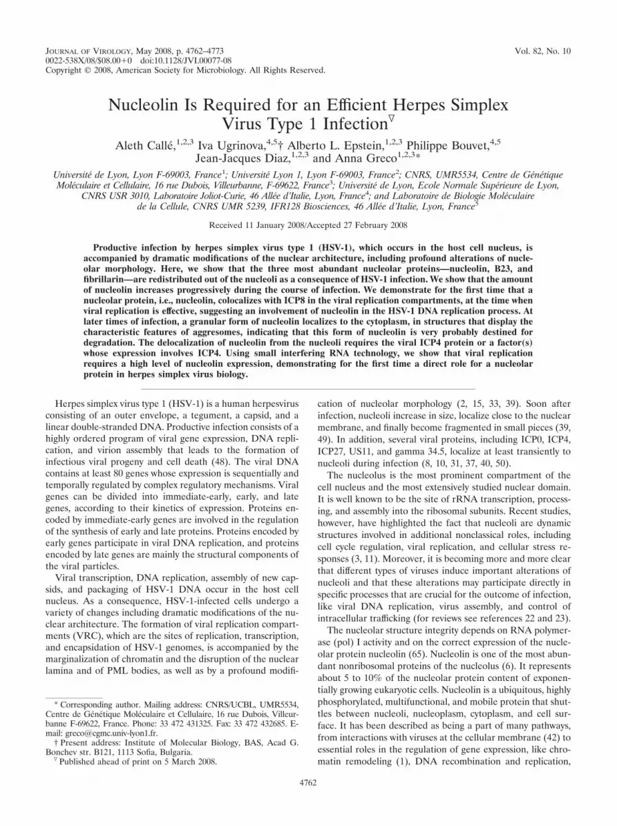
In mock-infected cells and at 4 h p.i., nucleolin was foundexclusively in nucleoli (Fig. 2A). Later, infection of cells leadsto rapid changes in nucleolin localization. From 6 to 24 h p.i.,nucleolin exhibited different patterns and was present in twodifferent forms, a diffuse one and a granular one indicated bya red arrow. From 6 h p.i., nucleolin increased in amount, andup to 6 to 8 h p.i., it was mainly present in the nucleus. As early
as ICP8 was detected (6 h p.i.) and while nuclear membraneinvagination occurred, part of nucleolin colocalized with sitesof ICP8 staining. From 6 to14 h p.i., the signals of ICP8 and ofpart of nucleolin clearly overlapped, indicating that at least afraction of nucleolin colocalizes with ICP8 in the VRC in thenucleus during the process of infection.
As infection progressed, the presence of nucleolin was notrestricted to the VRC. Indeed, nucleolin was found also instructures devoid of ICP8 (Fig. 2A, 14 to 24 h p.i.); granulardots of nucleolin appeared as early as 6 to 8 h after infection,and they progressively increased in size and amount. At 14 hp.i. a part of nucleolin was still present in the VRC; however,the granular form of nucleolin became predominant and ac-cumulated in structures that did not contain ICP8. This gran-ular form of nucleolin was located in the part of the cell thatwas not stained by DAPI, suggesting an extranuclear localiza-tion. At 24 h p.i. nucleolin did not colocalize anymore withICP8 and was present mainly in structures located at theperiphery of the nucleus and in the invagination of the nucleus.These structures that contain granular nucleolin resemble ag-gresome in their morphology and localization (see below).
The pattern of delocalization of nucleolin seems to be spe-cific since the nucleolar protein B23 behaves in a different way,as seen in Fig. 2B. Until 6 h p.i., B23 remained in structuresthat displayed a nucleolar morphology. Then, from 8 to 24 hp.i., it localized in a structure that was devoid of the charac-teristic blue DAPI staining, indicating that this could be VRC.At 24 h p.i. part of B23 was present at the periphery of thenucleus. In contrast to nucleolin, B23 was never seen in agranular form.
Therefore, during HSV-1 infection, the two abundant nucle-olar proteins, nucleolin and B23, are delocalized from nucleoli.Nucleolin was detected in VRC as early as 6 h p.i., whereasB23 localized from 8 h p.i. in a structure that could be VRCand continued to localize in this structure at very late times ofinfection (24 h p.i.) when nucleolin seemed to be excludedfrom the VRC and was instead present in granular structuresand at the periphery of the nucleus. At 24 h p.i. parts of bothnucleolin and B23 were present at the periphery of the nucleus.In addition, the DAPI labeling confirmed that, from 4 h p.i.,the nucleus was morphologically modified and the labeling alsorevealed that granular nucleolin was proximal to the invagina-tion of the nucleus. This was particularly obvious at late timesof infection.
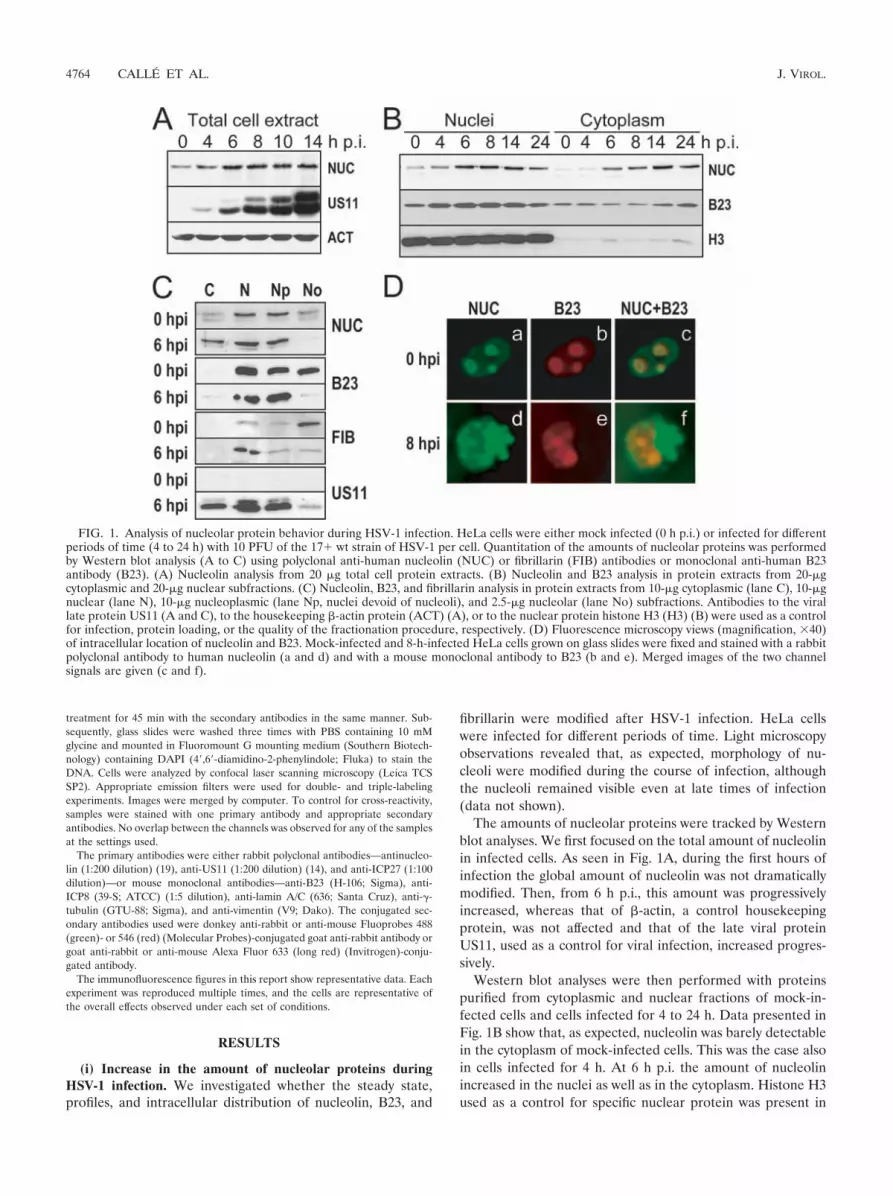
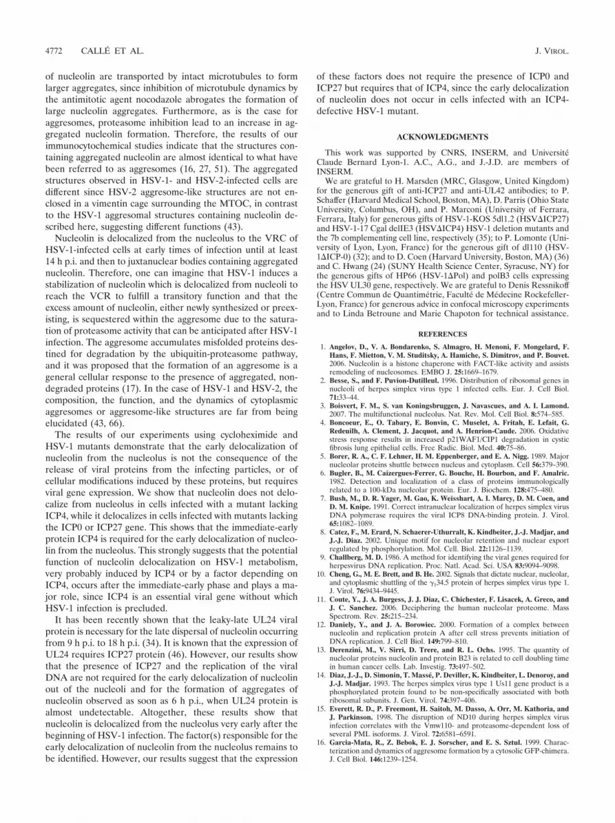
(iii) Immediate-early viral functions are necessary for nucleo-lin delocalization. Infection of cells with HSV-1 results in theselective and progressive inhibition of cellular protein synthesis(reviewed in reference 49). The delocalization of nucleolinupon infection could be the result of the inhibition of cellularprotein synthesis. To test this hypothesis, mock-infected andinfected cells were treated with cycloheximide, which inhibitsthe elongation of polypeptides during the translation process.In infected cells, cycloheximide was added either simulta-neously with the viral particles or from 2 or 4 h p.i. until theend of the experiment. Its effect on nucleolin was observed byimmunofluorescence analysis (Fig. 3A). These observationsshow that when cycloheximide was added together with theinfectious viral particles, nucleolin remained exclusively nucle-olar (Fig. 3A, fourth panel from left), as was the case formock-infected control cells (compare Fig. 3A, two leftmost
VOL. 82, 2008 NUCLEOLIN IS ESSENTIAL FOR HSV-1 INFECTION 4765

panels and panel fourth from left). In contrast, addition ofcycloheximide to cells from 2 or 4 h p.i. led to delocalization ofnucleolin out of the nucleolus (Fig. 3A, two rightmost panels,respectively) in a manner similar to that of infected controlcells that were not submitted to treatment (Fig. 3A, third panelfrom left). At these times of infection at least some of theimmediate-early and early proteins, respectively, are alreadysynthesized. Thus, the delocalization of nucleolin in infectedcells requires ongoing viral gene expression and is not simply aconsequence of the inhibition of host cell translation.
In order to better understand the viral requirements for the
delocalization of nucleolin and for the formation of nucleolingranular dots, we analyzed the nucleolin localization by immu-nofluorescence in cells infected with HSV-1 strains defective inspecific viral genes. The mutant viruses used derived from wtstrains and correspond to viruses in which the ICP0 (dl110),ICP4 (HSV-1-17 Cgal delIE3), and ICP27 (HSV-1-KOS 5dl1.2)immediate-early genes or the DNA pol (HP66) early viral genehas been deleted. Results of immunofluorescence analysis arepresented in Fig. 3B. Redistribution of nucleolin was seen in cellsinfected with HSV-1 mutants with deletions of ICP0 and ICP27immediate-early genes (Fig. 3B, top row, second and fourth pan-
FIG. 2. Dynamic localization of nucleolin and B23 during the course of HSV-1 infection. HeLa cells grown in a coverslip chamber were infectedwith the HSV-1 17� strain at a multiplicity of 10 PFU per cell. The cells were examined at different times postinfection (4 to 24 h), andmock-infected HeLa cells were used as a control. (A) Immunofluorescence and subsequent confocal laser scanning microscopy analysis werecarried out with antibodies to either nucleolin (NUC; red) or ICP8 (green) as a VRC-specific protein and with DAPI (blue) to stain the DNA.The positions of the VRC are indicated by white arrows, and those of aggregated nucleolin are indicated by red arrows. The merge of nucleolin,ICP8, and DAPI signals is given. Representative pictures of three independent experiments. (B) Colabeling of B23 nucleolar protein (red) andDAPI (blue) in HeLa cells from the same experiment as that presented in panel A. The confocal images were collected with a focal depth of0.5 �m.
4766 CALLE ET AL. J. VIROL.
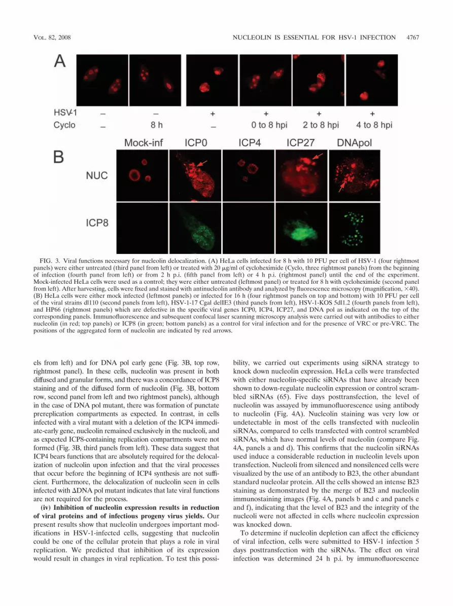
els from left) and for DNA pol early gene (Fig. 3B, top row,rightmost panel). In these cells, nucleolin was present in bothdiffused and granular forms, and there was a concordance of ICP8staining and of the diffused form of nucleolin (Fig. 3B, bottomrow, second panel from left and two rightmost panels), althoughin the case of DNA pol mutant, there was formation of punctateprereplication compartments as expected. In contrast, in cellsinfected with a viral mutant with a deletion of the ICP4 immedi-ate-early gene, nucleolin remained exclusively in the nucleoli, andas expected ICP8-containing replication compartments were notformed (Fig. 3B, third panels from left). These data suggest thatICP4 bears functions that are absolutely required for the delocal-ization of nucleolin upon infection and that the viral processesthat occur before the beginning of ICP4 synthesis are not suffi-cient. Furthermore, the delocalization of nucleolin seen in cellsinfected with �DNA pol mutant indicates that late viral functionsare not required for the process.
(iv) Inhibition of nucleolin expression results in reductionof viral proteins and of infectious progeny virus yields. Ourpresent results show that nucleolin undergoes important mod-ifications in HSV-1-infected cells, suggesting that nucleolincould be one of the cellular protein that plays a role in viralreplication. We predicted that inhibition of its expressionwould result in changes in viral replication. To test this possi-
bility, we carried out experiments using siRNA strategy toknock down nucleolin expression. HeLa cells were transfectedwith either nucleolin-specific siRNAs that have already beenshown to down-regulate nucleolin expression or control scram-bled siRNAs (65). Five days posttransfection, the level ofnucleolin was assayed by immunofluorescence using antibodyto nucleolin (Fig. 4A). Nucleolin staining was very low orundetectable in most of the cells transfected with nucleolinsiRNAs, compared to cells transfected with control scrambledsiRNAs, which have normal levels of nucleolin (compare Fig.4A, panels a and d). This confirms that the nucleolin siRNAsused induce a considerable reduction in nucleolin levels upontransfection. Nucleoli from silenced and nonsilenced cells werevisualized by the use of an antibody to B23, the other abundantstandard nucleolar protein. All the cells showed an intense B23staining as demonstrated by the merge of B23 and nucleolinimmunostaining images (Fig. 4A, panels b and c and panels eand f), indicating that the level of B23 and the integrity of thenucleoli were not affected in cells where nucleolin expressionwas knocked down.
To determine if nucleolin depletion can affect the efficiencyof viral infection, cells were submitted to HSV-1 infection 5days posttransfection with the siRNAs. The effect on viralinfection was determined 24 h p.i. by immunofluorescence
FIG. 3. Viral functions necessary for nucleolin delocalization. (A) HeLa cells infected for 8 h with 10 PFU per cell of HSV-1 (four rightmostpanels) were either untreated (third panel from left) or treated with 20 �g/ml of cycloheximide (Cyclo, three rightmost panels) from the beginningof infection (fourth panel from left) or from 2 h p.i. (fifth panel from left) or 4 h p.i. (rightmost panel) until the end of the experiment.Mock-infected HeLa cells were used as a control; they were either untreated (leftmost panel) or treated for 8 h with cycloheximide (second panelfrom left). After harvesting, cells were fixed and stained with antinucleolin antibody and analyzed by fluorescence microscopy (magnification, �40).(B) HeLa cells were either mock infected (leftmost panels) or infected for 16 h (four rightmost panels on top and bottom) with 10 PFU per cellof the viral strains dl110 (second panels from left), HSV-1-17 Cgal delIE3 (third panels from left), HSV-1-KOS 5dl1.2 (fourth panels from left),and HP66 (rightmost panels) which are defective in the specific viral genes ICP0, ICP4, ICP27, and DNA pol as indicated on the top of thecorresponding panels. Immunofluorescence and subsequent confocal laser scanning microscopy analysis were carried out with antibodies to eithernucleolin (in red; top panels) or ICP8 (in green; bottom panels) as a control for viral infection and for the presence of VRC or pre-VRC. Thepositions of the aggregated form of nucleolin are indicated by red arrows.
VOL. 82, 2008 NUCLEOLIN IS ESSENTIAL FOR HSV-1 INFECTION 4767

4768 CALLE ET AL. J. VIROL.

using an anti-ICP8 antibody (Fig. 4B). It appears very clearlythat in cells transfected with nucleolin siRNAs, the ICP8 stain-ing was restricted to the small numbers of cells that have notbeen transfected by the siRNA showing a detectable amount ofnucleolin (Fig. 4B, panels g to i), while it was detected in allcontrol cells, both in mock-transfected cells (Fig. 4B, panels ato c) and in cells transfected with scrambled siRNAs (Fig. 4B,panels d to f). Western blot analysis presented in Fig. 4Cconfirmed that, in mock-infected cells and in cells infected for24 h, the amount of nucleolin was drastically decreased in cellstransfected with nucleolin siRNAs (NUC), compared tomock-transfected cells (MT) or cells transfected with scram-bled siRNAs (SC). This indicates that the efficient down-reg-ulation of nucleolin was specific and was not due to the trans-fection procedure. In addition, by using antibodies directedagainst the late viral protein US11 and against the housekeep-ing protein �-actin we observed that in infected cells, the levelsof US11 protein were strongly decreased only in cells trans-fected with nucleolin siRNAs while the level of the control�-actin was not significantly modified. These data confirmedthe results of the immunofluorescence experiment and dem-onstrate that viral expression was inhibited in cells wherenucleolin expression was down regulated. Altogether, theseresults suggest that HSV-1 expression requires the presence ofnucleolin, since both the expression of the early ICP8 (Fig. 4B,panels h and i) and that of the late US11 (Fig. 4C) viral proteinis inhibited in its absence.
It has been shown that nucleolin could interfere with viralpenetration. Indeed, in the case of human immunodeficiencyvirus, nucleolin is a receptor for viral penetration, and func-tional anchorage of virus particles on target cells requiresnucleolin (42). Therefore, the effects of changes in nucleolinexpression on virus adsorption and production were estimatedby plaque titration assays (Fig. 5).
Five days after transfection with siRNAs, cells were infectedfor 2 h at 4°C using various amounts of HSV-1 particles(MOI � 0.4 to 10 PFU per cell) and then harvested (Fig. 5A).These experimental conditions allowed viral adsorption andnot viral replication. There was no significant difference in theamounts of infectious viral particles adsorbed on the surface ofcells where nucleolin was knocked down compared to cellshaving a normal nucleolin level, even when infection was per-formed with high amounts of virus (MOI � 10 PFU per cell).This suggests that adsorption of the virus is not repressed incells where nucleolin expression is inhibited.
We then determined the effect of repression of nucleolinexpression on the amounts of progeny virus produced in cellsinfected for 16 h or 24 h (Fig. 5B). We observed a modest
reduction in the titers of virus produced from cells transfectedwith the control scrambled siRNAs compared to mock-trans-fected control cells. This indicates that transfection of cellswith nonspecific siRNAs has no significant consequence onviral production. In contrast, virus yields were reduced 23- and10-fold in cells transfected with nucleolin siRNA and infectedfor 16 or 24 h, respectively, at an MOI of 0.4 PFU/cell, com-pared to cells transfected with control scrambled siRNAs. Incells infected at a very high MOI (10 PFU/cell), virus yieldswere reduced more than threefold in nucleolin-knockdowncells. These data clearly confirm that viral infection requiresnucleolin for efficient infection.
(v) Granular nucleolin mainly localized in extranuclear ag-gresome structures in infected cells. Our results show thatnucleolin is required for the outcome of HSV-1 infection andthat, soon after infection, nucleolin increases in amount and isfirst delocalized in the VRC and then accumulates in granulardots located in juxtanuclear structures that display morpholog-ical homology with aggresomes. It has been established thataggresomes are formed by the deposition of misfolded proteinsin a large structure surrounding the microtubule-organizingcenter (MTOC) and is enclosed in a characteristic vimentincage. This process, which is accompanied by a reorganizationof the intermediate filaments of the cytoskeleton, requires anintact microtubule cytoskeleton and does not require protea-some inhibition (16, 26, 27).
We therefore undertook analyses in order to verify whetherthese structures containing granular dots of nucleolin satisfythe definition of aggresomes. For this, mock-infected cells and
FIG. 4. Down-regulation of nucleolin by siRNA induces inhibition of HSV-1 infection. HeLa cells were transfected with either nucleolinsiRNAs (NUC siRNAs) or control scrambled siRNAs (SC siRNAs) or were mock transfected (MT). After 24 h, cells were transfected again withthe same siRNAs. Five days after the first transfection, cells were infected for 24 h with 10 PFU of wt HSV-1 per cell. Six days after the firsttransfection, cells were submitted either to immunofluorescence or to Western blot analysis. Nucleolin was visualized by the use of a specificpolyclonal antibody (in green). (A) Detection of nucleolin (NUC) in green (a, c, d, and f) of mock-infected cells 6 days after the first transfection,showing effective depletion of nucleolin. Detection of B23 in red (b, c, e, and f) was used as a control for nucleolar integrity. (B) Immunofluo-rescence images of transfected cells infected for 24 h showing inhibition of viral infection in nucleolin knockdown cells. Detection of nucleolin (a,c, d, f, g, and i). Detection of ICP8 (b, c, e, f, h, and i) was used as a control for viral infection and for the VRC. DAPI staining (D) is in blue.(C) Western blot analysis of experiments presented in panels A and B, using antibodies to nucleolin and the viral late protein US11 or to thehousekeeping �-actin protein as a control for infection or for protein loading, respectively.
FIG. 5. Knockdown of endogenous nucleolin by siRNA results indecrease in virus titers. HeLa cells were transfected as indicated in thelegend to Fig. 4. Five days after the first transfection cells were infectedat an MOI of 0.4 or 10 PFU of wt HSV-1 per cell, as indicated in eachpanel. After harvesting, virus titers were determined by plaque assay.(A) Estimation of the amount of viral particles adsorbed on the cellsurface. Cells were harvested after the first 2 h of contact with the virusat 4°C. Viral titers were determined from cells after removal of themedium and thorough washing of the cells. (B) Estimation of theamounts of progeny viruses from cells and cell culture supernatantsharvested after 16 or 24 h of infection at 37°C as indicated.
VOL. 82, 2008 NUCLEOLIN IS ESSENTIAL FOR HSV-1 INFECTION 4769

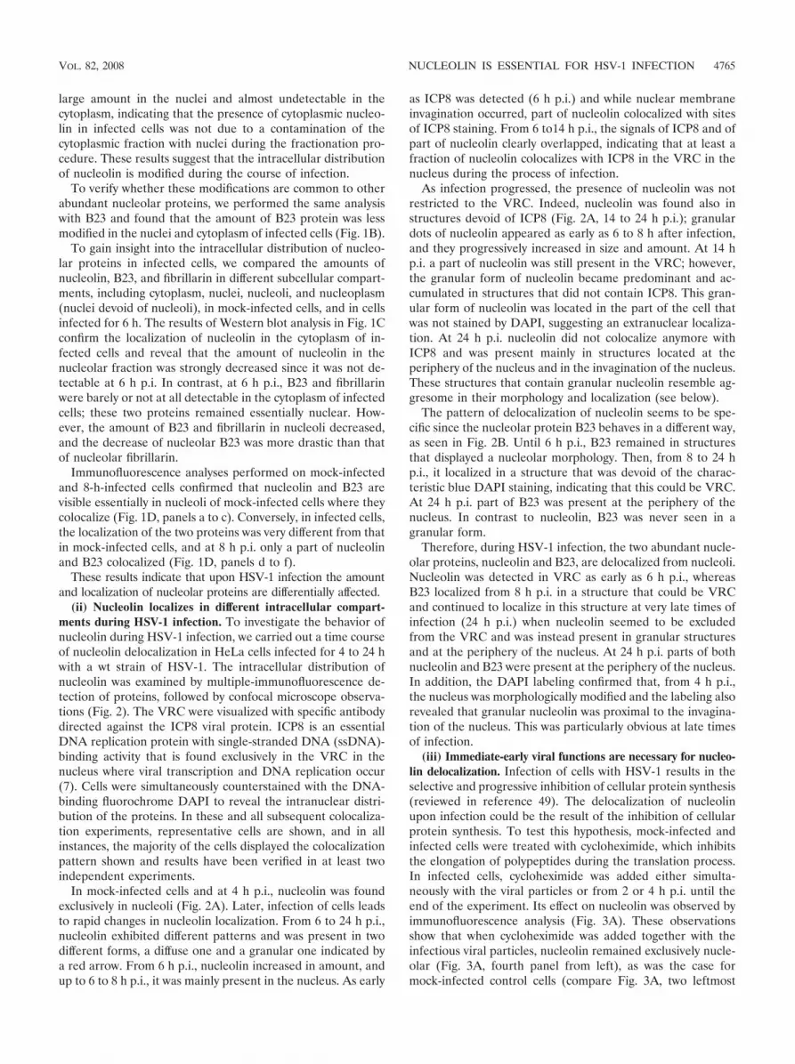
cells infected for 8 h were submitted to indirect coimmunofluo-rescence experiments using specific antibodies to nucleolin andto lamin, which is a component of the lamina that delimitatethe inner face of the nuclear envelope (55). In mock-infectedcells, nucleolin staining was exclusively in the nucleus (Fig. 6A,panels a to c). At 8 h p.i., the nongranular form of nucleolinwas located inside the lamin layer, while the granular dots ofnucleolin were located outside the lamin layer (Fig. 6A, panelsd to f). This result led us to conclude that the structurescontaining the granular dots of nucleolin were in the cytoplasmof infected cells and raises the question of whether they rep-resent some type of aggresome.
To answer this question, we first performed colocalizationstudies using antibodies specific to nucleolin and to �-tubulin,a marker of MTOC. We showed that in infected cells granulardots of nucleolin localized around �-tubulin at the MTOC(indicated by an arrow in Fig. 6B, panels b and c), near thecharacteristic distortion region of the nucleus, indicating thatthey localize around the MTOC. Then using antibodies tovimentin (Fig. 6C), we show that at 8 h p.i. vimentin wasdistributed throughout the cell and in concentrated foci withinthe cytoplasm near the distortion of the nucleus, and vimentinformed a ring surrounding the core of the intense perinuclearsignal of granular nucleolin, indicating that this large structurecontaining nucleolin was enclosed inside a typical vimentincage.
Since granular dots of nucleolin have typical features ofproteins present in aggresomes, we further tested whether theformation of these nucleolin-containing structures depends onmicrotubule integrity. Cells were treated prior to infection with5 �M nocodazole, which is an antimitotic agent that causes thedepolymerization of microtubules (67). In cells infected for 8 h,treatment with nocodazole alone resulted in an increase in thenumber of cytoplasmic structures containing nucleolin (com-pare panel a of Fig. 6D to panel a of Fig. 6C). These structureswere smaller and were located throughout the nucleus and thecytoplasm of infected cells as evidenced by nucleolin-vimentinand DAPI staining (Fig. 6D, panels a to c). These smallernucleolin structures were not surrounded by a vimentin cage,indicating that the formation of large perinuclear structurescontaining nucleolin requires intact microtubules.
FIG. 6. Aggregated nucleolin has the characteristics of aggresomes.(A) Aggregated nucleolin has a cytoplasmic localization. The subcel-lular localization of aggregated nucleolin was determined by doubleimmunolabeling of HeLa cells either mock infected (a to c) or infectedfor 8 h with 10 PFU per cell of the 17� wt strain of HSV-1 (d to f).Confocal images were obtained from immunofluorescence analysiscarried out with antibodies to nucleolin (NUC in green, panels a andd) and to lamin (LAM in red, panels b and e), a component of thelamina that delimitates the inner face of the nuclear envelope. DAPIstaining (D) is in blue. The white and green arrowheads indicate VRCand aggregated nucleolin, respectively. The merge of nucleolin, lamin,and DAPI signals is given in panels c and f. (B) Aggregated nucleolin
localizes around the centrosome. HeLa cells infected for 8 h werelabeled with antibodies to nucleolin (NUC in red) and to �-tubulin(TUB in green) and with DAPI (D) as indicated at the top of theconfocal images. The position of the centrosome is indicated by a whitearrow. (C) Aggregated nucleolin is enclosed in a vimentin cage. HeLacells infected for 8 h were stained with antibodies to nucleolin (NUCin green) and to vimentin (VIM in red). DAPI staining (D) is in blue.(D) Formation of large aggregates of nucleolin depends on microtu-bule integrity and not on proteasome inhibition. HeLa cells wereincubated for 12 h with 5 �g/ml nocodazole (Noco) (a to c), 5 �g/mlALLN (d to f), or both drugs (g to i) and then infected for 8 h asindicated in panel A, in the presence of the same concentrations ofdrugs. Formation of the large perinuclear aggregated nucleolin is pre-vented by nocodazole treatment (a to c and g to i) and not by ALLNtreatment (d to f). DAPI staining (D) is indicated. In panels C and D,green arrows indicate the position of the caged nucleolin. Digitalimages were obtained by confocal laser scanning. Representative datafrom several independent experiments.
4770 CALLE ET AL. J. VIROL.

To assess the role of the proteasome in the formation ofgranular dots of nucleolin, HeLa cells were treated with 5�g/ml of the specific proteasome inhibitor ALLN prior toinfection. At this concentration there was no clear evidence ofcell toxicity, as reported by other studies using ALLN (4, 18).In the presence of ALLN alone, there was accumulation oflarge nucleolin-containing structures surrounded by vimentin(Fig. 6D, panels d to f), indicating that inhibition of protea-some has no effect on the formation and localization of gran-ular nucleolin dots.
In cells treated with both nocodazole and ALLN, the amountand the size of nucleolin aggregates were increased compared tocells treated with nocodazole alone (compare panels a and g ofFig. 6D). However, these aggregates were smaller than thosepresent in untreated cells or in cells treated with ALLN alone(compare panel a in Fig. 6C and panel d in Fig. 6D to panels a andg in Fig. 6D) and were not caged by vimentin.
Altogether, these results indicate that in infected cells, theperinuclear structures containing granular nucleolin have thecharacteristics of aggresomes.
DISCUSSION
In this paper we present evidence that the behavior of nu-cleolar proteins is rapidly modified after infection with HSV-1.The analysis of the intracellular distribution of the three mostabundant nucleolar proteins—nucleolin, B23, and fibrillarin—revealed that after infection, these proteins are delocalizedfrom the nucleoli. This is in agreement with previous reports(34, 47). In addition, we demonstrate that at early times ofinfection, while nucleolin is translocated to both the nucleo-plasm and the cytoplasm, the two other proteins remain inlarge part nuclear.
In addition, the amount of nucleolin increases progressivelyduring the course of infection. This is obvious from 6 h p.i. inthe nuclei of infected cells, and the increase is more pro-nounced in the cytoplasmic fraction, given that cytoplasmicnucleolin is almost undetectable in mock-infected cells andbecomes very abundant as early as 6 h p.i. However, the levelsof B23 and fibrillarin are less significantly affected in HSV-1-infected cells. Therefore, the expression and localization ofthese three nucleolar proteins are differentially regulated dur-ing the course of infection.
It is well documented that in HSV-1-infected cells there is arepression of the synthesis of most of the host proteins. Inprevious reports we have speculated that the small number ofcellular proteins that continue to be efficiently synthesizedupon infection could play a major role in determining theoutcome of infection (20, 21, 57). We have already shown thatthis is the case for the S-adenosylmethionine decarboxylase,whose synthesis is increased after infection, and the inhibitionof its activity prevents HSV-1 infection. Our present observa-tion demonstrating that knockdown of nucleolin expression bythe siRNA strategy results in a drastic decrease in the produc-tion of infectious virus strongly supports the notion thatnucleolin is also a member of this class of cellular proteinswhose function is required for an efficient HSV-1 outcome.This result is reinforced by the observation that infection oc-curs exclusively in the small number of cells in which nucleolinremains detectable after siRNA treatment.
Several publications have reported that nucleolin is involvedin different steps during infection with various viruses (forreviews see references 22 and 23). At this stage of the study wecannot firmly conclude by which molecular mechanism nucleo-lin promotes HSV-1 outcome, although, based upon our re-sults, it can be proposed that nucleolin very probably plays arole during the early phase of the viral cycle and could affectviral DNA replication. Obviously the initial step of infection isnot inhibited, since viral adsorption is as efficient in cells trans-fected with nucleolin siRNA as in cells transfected with scram-bled siRNAs or in mock-transfected cells. This clearly showsthat nucleolin is not involved in the virus penetration step, asis the case for human immunodeficiency virus (42). One cannotexclude as well the possibility that HSV-1-induced alteration ofnucleolin and B23 is related to the blockage of cell cycle pro-gression at the S phase that is observed after infection, since ithas been shown recently that inhibition of nucleolin expressionresults in a cell growth arrest, accumulation in G2, and anincrease of apoptosis (65).
At early times of infection, nucleolin first delocalizes fromthe nucleolus to the nucleoplasm and colocalizes in part withICP8 in the VRC where viral DNA replication takes place.ICP8 is one of the seven early viral proteins essential for viralgenome replication that interacts with ssDNA (9, 29, 30).Therefore, because nucleolin is a multifunctional protein, in-volved in chromatin structure and dynamics (1, 38, 60, 61), andbecause it localizes in the VRC at the time when viral repli-cation is effective, it can be speculated that nucleolin is in-volved in some aspects of viral DNA replication. Indeed, sev-eral reports suggest that nucleolin could play a role in DNAmetabolism including DNA replication, recombination, andrepair. In vitro, nucleolin forms a ternary complex with thesimian virus 40 helicase T-antigen and topoisomerase (56).Nucleolin mediates the cohesion of the T-antigen helicaseholoenzyme during plasmid unwinding, enabling the formationof a functional complex at the replication fork.
It has been also shown that nucleolin interacts in vitro withreplication protein A (RPA) (12, 28). RPA is an ssDNA bind-ing protein of eukaryotic cells which is necessary for bothinitiation and elongation steps of chromosomal DNA replica-tion (23, 25), and it has been shown that RPA is associated withICP8 within VRC (64), although it is not known if RPA isinvolved in HSV replication. Nucleolin binding to RPA inhib-its DNA replication initiation (12). B23 is also translocated ina structure that could be VRC, and this protein was also shownto play a role in DNA replication (45, 62, 63). However, thepresence of B23 in this structure is delayed compared to lo-calization of nucleolin in VRC, suggesting that the two pro-teins very probably display different functions.
Later on, nucleolin delocalizes from the nucleus to the cy-toplasm of infected cells. However, while nuclear nucleolin hasa diffused pattern, the cytoplasmic nucleolin is granular and isconcentrated at the periphery of the nucleus. These nucleolin-containing aggregates have the characteristic localization andmorphology of aggresomes. Our data clearly demonstrate thatthis is the case. Indeed, the aggregate structures containingnucleolin are localized in the cytoplasm of infected cells. Likeaggresomes, they are located close to centrosomes and areenclosed in a characteristic vimentin cage. In further agree-ment with aggresomes, our data suggest that small aggregates
VOL. 82, 2008 NUCLEOLIN IS ESSENTIAL FOR HSV-1 INFECTION 4771
of nucleolin are transported by intact microtubules to formlarger aggregates, since inhibition of microtubule dynamics bythe antimitotic agent nocodazole abrogates the formation oflarge nucleolin aggregates. Furthermore, as is the case foraggresomes, proteasome inhibition lead to an increase in ag-gregated nucleolin formation. Therefore, the results of ourimmunocytochemical studies indicate that the structures con-taining aggregated nucleolin are almost identical to what havebeen referred to as aggresomes (16, 27, 51). The aggregatedstructures observed in HSV-1- and HSV-2-infected cells aredifferent since HSV-2 aggresome-like structures are not en-closed in a vimentin cage surrounding the MTOC, in contrastto the HSV-1 aggresomal structures containing nucleolin de-scribed here, suggesting different functions (43).
Nucleolin is delocalized from the nucleolus to the VRC ofHSV-1-infected cells at early times of infection until at least14 h p.i. and then to juxtanuclear bodies containing aggregatednucleolin. Therefore, one can imagine that HSV-1 induces astabilization of nucleolin which is delocalized from nucleoli toreach the VCR to fulfill a transitory function and that theexcess amount of nucleolin, either newly synthesized or preex-isting, is sequestered within the aggresome due to the satura-tion of proteasome activity that can be anticipated after HSV-1infection. The aggresome accumulates misfolded proteins des-tined for degradation by the ubiquitin-proteasome pathway,and it was proposed that the formation of an aggresome is ageneral cellular response to the presence of aggregated, non-degraded proteins (17). In the case of HSV-1 and HSV-2, thecomposition, the function, and the dynamics of cytoplasmicaggresomes or aggresome-like structures are far from beingelucidated (43, 66).
The results of our experiments using cycloheximide andHSV-1 mutants demonstrate that the early delocalization ofnucleolin from the nucleolus is not the consequence of therelease of viral proteins from the infecting particles, or ofcellular modifications induced by these proteins, but requiresviral gene expression. We show that nucleolin does not delo-calize from nucleolus in cells infected with a mutant lackingICP4, while it delocalizes in cells infected with mutants lackingthe ICP0 or ICP27 gene. This shows that the immediate-earlyprotein ICP4 is required for the early delocalization of nucleo-lin from the nucleolus. This strongly suggests that the potentialfunction of nucleolin delocalization on HSV-1 metabolism,very probably induced by ICP4 or by a factor depending onICP4, occurs after the immediate-early phase and plays a ma-jor role, since ICP4 is an essential viral gene without whichHSV-1 infection is precluded.
It has been recently shown that the leaky-late UL24 viralprotein is necessary for the late dispersal of nucleolin occurringfrom 9 h p.i. to 18 h p.i. (34). It is known that the expression ofUL24 requires ICP27 protein (46). However, our results showthat the presence of ICP27 and the replication of the viralDNA are not required for the early delocalization of nucleolinout of the nucleoli and for the formation of aggregates ofnucleolin observed as soon as 6 h p.i., when UL24 protein isalmost undetectable. Altogether, these results show thatnucleolin is delocalized from the nucleolus very early after thebeginning of HSV-1 infection. The factor(s) responsible for theearly delocalization of nucleolin from the nucleolus remains tobe identified. However, our results suggest that the expression
of these factors does not require the presence of ICP0 andICP27 but requires that of ICP4, since the early delocalizationof nucleolin does not occur in cells infected with an ICP4-defective HSV-1 mutant.
ACKNOWLEDGMENTS
This work was supported by CNRS, INSERM, and UniversiteClaude Bernard Lyon-1. A.C., A.G., and J.-J.D. are members ofINSERM.
We are grateful to H. Marsden (MRC, Glasgow, United Kingdom)for the generous gift of anti-ICP27 and anti-UL42 antibodies; to P.Schaffer (Harvard Medical School, Boston, MA), D. Parris (Ohio StateUniversity, Columbus, OH), and P. Marconi (University of Ferrara,Ferrara, Italy) for generous gifts of HSV-1-KOS 5dl1.2 (HSV�ICP27)and HSV-1-17 Cgal delIE3 (HSV�ICP4) HSV-1 deletion mutants andthe 7b complementing cell line, respectively (35); to P. Lomonte (Uni-versity of Lyon, Lyon, France) for the generous gift of dl110 (HSV-1�ICP-0) (32); and to D. Coen (Harvard University, Boston, MA) (36)and C. Hwang (24) (SUNY Health Science Center, Syracuse, NY) forthe generous gifts of HP66 (HSV-1�Pol) and polB3 cells expressingthe HSV UL30 gene, respectively. We are grateful to Denis Ressnikoff(Centre Commun de Quantimetrie, Faculte de Medecine Rockefeller-Lyon, France) for generous advice in confocal microscopy experimentsand to Linda Betroune and Marie Chapoton for technical assistance.
REFERENCES
1. Angelov, D., V. A. Bondarenko, S. Almagro, H. Menoni, F. Mongelard, F.Hans, F. Mietton, V. M. Studitsky, A. Hamiche, S. Dimitrov, and P. Bouvet.2006. Nucleolin is a histone chaperone with FACT-like activity and assistsremodeling of nucleosomes. EMBO J. 25:1669–1679.
2. Besse, S., and F. Puvion-Dutilleul. 1996. Distribution of ribosomal genes innucleoli of herpes simplex virus type 1 infected cells. Eur. J. Cell Biol.71:33–44.
3. Boisvert, F. M., S. van Koningsbruggen, J. Navascues, and A. I. Lamond.2007. The multifunctional nucleolus. Nat. Rev. Mol. Cell Biol. 8:574–585.
4. Boncoeur, E., O. Tabary, E. Bonvin, C. Muselet, A. Fritah, E. Lefait, G.Redeuilh, A. Clement, J. Jacquot, and A. Henrion-Caude. 2006. Oxidativestress response results in increased p21WAF1/CIP1 degradation in cysticfibrosis lung epithelial cells. Free Radic. Biol. Med. 40:75–86.
5. Borer, R. A., C. F. Lehner, H. M. Eppenberger, and E. A. Nigg. 1989. Majornucleolar proteins shuttle between nucleus and cytoplasm. Cell 56:379–390.
6. Bugler, B., M. Caizergues-Ferrer, G. Bouche, H. Bourbon, and F. Amalric.1982. Detection and localization of a class of proteins immunologicallyrelated to a 100-kDa nucleolar protein. Eur. J. Biochem. 128:475–480.
7. Bush, M., D. R. Yager, M. Gao, K. Weisshart, A. I. Marcy, D. M. Coen, andD. M. Knipe. 1991. Correct intranuclear localization of herpes simplex virusDNA polymerase requires the viral ICP8 DNA-binding protein. J. Virol.65:1082–1089.
8. Catez, F., M. Erard, N. Schaerer-Uthurralt, K. Kindbeiter, J.-J. Madjar, andJ.-J. Diaz. 2002. Unique motif for nucleolar retention and nuclear exportregulated by phosphorylation. Mol. Cell. Biol. 22:1126–1139.
9. Challberg, M. D. 1986. A method for identifying the viral genes required forherpesvirus DNA replication. Proc. Natl. Acad. Sci. USA 83:9094–9098.
10. Cheng, G., M. E. Brett, and B. He. 2002. Signals that dictate nuclear, nucleolar,and cytoplasmic shuttling of the �134.5 protein of herpes simplex virus type 1.J. Virol. 76:9434–9445.
11. Coute, Y., J. A. Burgess, J. J. Diaz, C. Chichester, F. Lisacek, A. Greco, andJ. C. Sanchez. 2006. Deciphering the human nucleolar proteome. MassSpectrom. Rev. 25:215–234.
12. Daniely, Y., and J. A. Borowiec. 2000. Formation of a complex betweennucleolin and replication protein A after cell stress prevents initiation ofDNA replication. J. Cell Biol. 149:799–810.
13. Derenzini, M., V. Sirri, D. Trere, and R. L. Ochs. 1995. The quantity ofnucleolar proteins nucleolin and protein B23 is related to cell doubling timein human cancer cells. Lab. Investig. 73:497–502.
14. Diaz, J.-J., D. Simonin, T. Masse, P. Deviller, K. Kindbeiter, L. Denoroy, andJ.-J. Madjar. 1993. The herpes simplex virus type 1 Us11 gene product is aphosphorylated protein found to be non-specifically associated with bothribosomal subunits. J. Gen. Virol. 74:397–406.
15. Everett, R. D., P. Freemont, H. Saitoh, M. Dasso, A. Orr, M. Kathoria, andJ. Parkinson. 1998. The disruption of ND10 during herpes simplex virusinfection correlates with the Vmw110- and proteasome-dependent loss ofseveral PML isoforms. J. Virol. 72:6581–6591.
16. Garcia-Mata, R., Z. Bebok, E. J. Sorscher, and E. S. Sztul. 1999. Charac-terization and dynamics of aggresome formation by a cytosolic GFP-chimera.J. Cell Biol. 146:1239–1254.
4772 CALLE ET AL. J. VIROL.

17. Garcia-Mata, R., Y. S. Gao, and E. Sztul. 2002. Hassles with taking out thegarbage: aggravating aggresomes. Traffic 3:388–396.
18. Gerber, A., A. Heimburg, A. Reisenauer, A. Wille, T. Welte, and F. Buhling.2004. Proteasome inhibitors modulate chemokine production in lung epithe-lial and monocytic cells. Eur. Respir. J. 24:40–48.
19. Ghisolfi-Nieto, L., G. Joseph, F. Puvion-Dutilleul, F. Amalric, and P. Bouvet.1996. Nucleolin is a sequence-specific RNA-binding protein: characteriza-tion of targets on pre-ribosomal RNA. J. Mol. Biol. 260:34–53.
20. Greco, A., N. Bausch, Y. Coute, and J. J. Diaz. 2000. Characterization bytwo-dimensional gel electrophoresis of host proteins whose synthesis is sus-tained or stimulated during the course of herpes simplex virus type 1 infec-tion. Electrophoresis 21:2522–2530.
21. Greco, A., A. Calle, F. Morfin, D. Thouvenot, M. Cayre, K. Kindbeiter, L.Martin, O. Levillain, and J. J. Diaz. 2005. S-Adenosyl methionine decar-boxylase activity is required for the outcome of herpes simplex virus type 1infection and represents a new potential therapeutic target. FASEB J. 19:1128–1130.
22. Hiscox, J. 2007. RNA viruses: hijacking the dynamic nucleolus. Nat. Rev.Microbiol. 5:119–127.
23. Hiscox, J. 2002. The nucleolus—a gateway to viral infection? Arch. Virol.147:1077–1089.
24. Hwang, Y. T., B. Y. Liu, D. M. Coen, and C. B. Hwang. 1997. Effects ofmutations in the Exo III motif of the herpes simplex virus DNA polymerasegene on enzyme activities, viral replication, and replication fidelity. J. Virol.71:7791–7798.
25. Iftode, C., Y. Daniely, and J. A. Borowiec. 1999. Replication protein A(RPA): the eukaryotic SSB. Crit. Rev. Biochem. Mol. Biol. 34:141–180.
26. Johnston, J. A., M. E. Illing, and R. R. Kopito. 2002. Cytoplasmic dynein/dynactin mediates the assembly of aggresomes. Cell Motil. Cytoskeleton53:26–38.
27. Johnston, J. A., C. L. Ward, and R. R. Kopito. 1998. Aggresomes: a cellularresponse to misfolded proteins. J. Cell Biol. 143:1883–1898.
28. Kim, K., D. D. Dimitrova, K. M. Carta, A. Saxena, M. Daras, and J. A.Borowiec. 2005. Novel checkpoint response to genotoxic stress mediated bynucleolin-replication protein A complex formation. Mol. Cell. Biol. 25:2463–2474.
29. Lee, C. K., and D. M. Knipe. 1985. An immunoassay for the study ofDNA-binding activities of herpes simplex virus protein ICP8. J. Virol. 54:731–738.
30. Lehman, I. R., and P. E. Boehmer. 1999. Replication of herpes simplex virusDNA. J. Biol. Chem. 274:28059–28062.
31. Leopardi, R., and B. Roizman. 1996. Functional interaction and colocaliza-tion of the herpes simplex virus 1 major regulatory protein ICP4 with EAP,a nucleolar-ribosomal protein. Proc. Natl. Acad. Sci. USA 93:4572–4576.
32. Lomonte, P., and R. D. Everett. 1999. Herpes simplex virus type 1 immedi-ate-early protein Vmw110 inhibits progression of cells through mitosis andfrom G1 into S phase of the cell cycle. J. Virol. 73:9456–9467.
33. Lopez-Iglesias, C., F. Puvion-Dutilleul, J. Cebrian, and M. E. Christensen.1988. Herpes simplex virus type 1-induced modifications in the distributionof nucleolar B-36 protein. Eur. J. Cell Biol. 46:259–269.
34. Lymberopoulos, M. H., and A. Pearson. 2007. Involvement of UL24 inherpes-simplex-virus-1-induced dispersal of nucleolin. Virology 363:397–409.
35. Marconi, P., D. Krisky, T. Oligino, P. L. Poliani, R. Ramakrishnan, W. F.Goins, D. J. Fink, and J. C. Glorioso. 1996. Replication-defective herpessimplex virus vectors for gene transfer in vivo. Proc. Natl. Acad. Sci. USA93:11319–11320.
36. Marcy, A. I., D. R. Yager, and D. M. Coen. 1990. Isolation and character-ization of herpes simplex virus mutants containing engineered mutations atthe DNA polymerase locus. J. Virol. 64:2208–2216.
37. Mears, W. E., and S. A. Rice. 1998. The herpes simplex virus immediate-earlyprotein ICP27 shuttles between nucleus and cytoplasm. Virology 242:128–137.
38. Mongelard, F., and P. Bouvet. 2007. Nucleolin: a multiFACeTed protein.Trends Cell Biol. 17:80–86.
39. Monier, K., J. C. Armas, S. Etteldorf, P. Ghazal, and K. F. Sullivan. 2000.Annexation of the interchromosomal space during viral infection. Nat. CellBiol. 2:661–665.
40. Morency, E., Y. Coute, J. Thomas, P. Texier, and P. Lomonte. 2005. Theprotein ICP0 of herpes simplex virus type 1 is targeted to nucleoli of infectedcells. Brief report. Arch. Virol. 150:2387–2395.
41. Muramatsu, M., and T. Onishi. 1978. Isolation and purification of nucleoliand nucleolar chromatin from mammalian cells. Methods Cell Biol. 17:141–161.
42. Nisole, S., B. Krust, and A. Hovanessian. 2002. Anchorage of HIV on permis-sive cells leads to coaggregation of viral particles with surface nucleolin atmembrane raft microdomains. Exp. Cell Res. 276:155–173.
43. Nozawa, N., Y. Yamauchi, K. Ohtsuka, Y. Kawaguchi, and Y. Nishiyama.2004. Formation of aggresome-like structures in herpes simplex virus type2-infected cells and a potential role in virus assembly. Exp. Cell Res. 299:486–497.
44. Ochs, R. L. 1998. Methods used to study structure and function of thenucleolus. Methods Cell Biol. 53:303–321.
45. Okuwaki, M., A. Iwamatsu, M. Tsujimoto, and K. Nagata. 2001. Identifica-tion of nucleophosmin/B23, an acidic nucleolar protein, as a stimulatoryfactor for in vitro replication of adenovirus DNA complexed with viral basiccore proteins. J. Mol. Biol. 311:41–55.
46. Pearson, A., D. M. Knipe, and D. M. Coen. 2004. ICP27 selectively regulatesthe cytoplasmic localization of a subset of viral transcripts in herpes simplexvirus type 1-infected cells. J. Virol. 78:23–32.
47. Puvion-Dutilleul, F., E. Pichard, P. Sheldrick, F. Amalric, and E. Puvion.1986. Appearance of host-specific nucleolar proteins in intranuclear “densebodies” following herpes simplex infection. Eur. J. Cell Biol. 39:458–468.
48. Roizman, B., and A. Sears. 1990. Herpes simplex viruses and their replica-tion, p. 1795–1842. In B. N. Fields, D. M. Knipe, R. M. Chanock, M. S.Hirsch, J. L. Melnick, T. P. Monath, and B. Roizman (ed.), Fundamentalvirology. Raven Press, New York, NY.
49. Roizman, B., and A. E. Sears. 1993. Herpes simplex viruses and their repli-cation, p. 11–68. In B. Roizman, R. J. Whitley, and C. Lopez (ed.), Thehuman herpes viruses, 3rd ed. Raven Press, New York, NY.
50. Roller, R. J., L. L. Monk, D. Stuart, and B. Roizman. 1996. Structure andfunction in the herpes simplex virus 1 RNA-binding protein US11: mappingof the domain required for ribosomal and nucleolar association and RNAbinding in vitro. J. Virol. 70:2842–2851.
51. Rosevear, E. R., M. McReynolds, and R. D. Goldman. 1990. Dynamic prop-erties of intermediate filaments: disassembly and reassembly during mitosisin baby hamster kidney cells. Cell Motil. Cytoskeleton 17:150–166.
52. Roussel, P., and D. Hernandez-Verdun. 1994. Identification of Ag-NORproteins, markers of proliferation related to ribosomal gene activity. Exp.Cell Res. 214:465–472.
53. Schenk, P., S. Pietschmann, H. Gelderblom, G. Pauli, and H. Ludwig. 1988.Monoclonal antibodies against herpes simplex virus type 1-infected nucleidefining and localizing the ICP8 protein, 65K DNA-binding protein andpolypeptides of the ICP35 family. J. Gen. Virol. 69:99–111.
54. Scherl, A., Y. Coute, C. Deon, A. Calle, K. Kindbeiter, J.-C. Sanchez, A.Greco, D. Hochstrasser, and J.-J. Diaz. 2002. Functional proteomic analysisof the human nucleolus. Mol. Biol. Cell 13:4100–4109.
55. Schirmer, E. C., and L. Gerace. 2005. The nuclear membrane proteome:extending the envelope. Trends Biochem. Sci. 30:551–558.
56. Seinsoth, S., H. Uhlmann-Schiffler, and H. Stahl. 2003. Bidirectional DNAunwinding by a ternary complex of T antigen, nucleolin and topoisomeraseI. EMBO Rep. 4:263–268.
57. Simonin, D., J. J. Diaz, T. Masse, and J. J. Madjar. 1997. Persistence ofribosomal protein synthesis after infection of HeLa cells by herpes simplexvirus type 1. J. Gen. Virol. 78:435–443.
58. Sinclair, M., J. McLauchlan, H. Marsden, and S. Brown. 1994. Character-ization of a herpes simplex virus type 1 deletion variant (1703) which under-produces Vmw63 during immediate early conditions of infection. J. Gen.Virol. 75:1083–1089.
59. Sirri, V., P. Roussel, M. C. Gendron, and D. Hernandez-Verdun. 1997.Amount of the two major Ag-NOR proteins, nucleolin, and protein B23 iscell-cycle dependent. Cytometry 28:147–156.
60. Srivastava, M., and H. B. Pollard. 1999. Molecular dissection of nucleolin’srole in growth and cell proliferation: new insights. FASEB J. 13:1911–1922.
61. Storck, S., M. Shukla, S. Dimitrov, and P. Bouvet. 2007. Functions of thehistone chaperone nucleolin in diseases. Subcell. Biochem. 41:125–144.
62. Takemura, M., K. Sato, M. Nishio, T. Akiyama, H. Umekawa, and S. Yoshida.1999. Nucleolar protein B23.1 binds to retinoblastoma protein and synergisti-cally stimulates DNA polymerase alpha activity. J. Biochem. (Tokyo) 125:904–909.
63. Tarapore, P., M. Okuda, and K. Fukasawa. 2002. A mammalian in vitrocentriole duplication system: evidence for involvement of CDK2/cyclin E andnucleophosmin/B23 in centrosome duplication. Cell Cycle 1:75–81.
64. Taylor, T., and D. Knipe. 2004. Proteomics of herpes simplex virus replica-tion compartments: association of cellular DNA replication, repair, recom-bination, and chromatin remodeling proteins with ICP8. J. Virol. 78:5856–5866.
65. Ugrinova, I., K. Monier, C. Ivaldi, M. Thiry, S. Storck, F. Mongelard, and P.Bouvet. 2007. Inactivation of nucleolin leads to nucleolar disruption, cellcycle arrest and defects in centrosome duplication. BMC Mol. Biol. 8:66.
66. Wileman, T. 2007. Aggresomes and pericentriolar sites of virus assembly:cellular defense or viral design? Annu. Rev. Microbiol. 61:149–167.
67. Wojcik, C., D. Schroeter, S. Wilk, J. Lamprecht, and N. Paweletz. 1996.Ubiquitin-mediated proteolysis centers in HeLa cells: indication from stud-ies of an inhibitor of the chymotrypsin-like activity of the proteasome. Eur.J. Cell Biol. 71:311–331.
68. Wu, M. H., J. H. Chang, C. C. Chou, and B. Y. Yung. 2002. Involvement ofnucleophosmin/B23 in the response of HeLa cells to UV irradiation. Int. J.Cancer 97:297–305.
VOL. 82, 2008 NUCLEOLIN IS ESSENTIAL FOR HSV-1 INFECTION 4773
Related Documents