Executive Function, Neural Circuitry, and Genetic Mechanisms in Schizophrenia Daniel Paul Eisenberg 1 and Karen Faith Berman* ,1 1 Section on Integrative Neuroimaging and Clinical Brain Disorders Branch, Genes, Cognition, and Psychosis Program, National Institute of Mental Health, NIH, DHHS, Bethesda, MD, USA After decades of research aimed at elucidating the pathophysiology and etiology of schizophrenia, it has become increasingly apparent that it is an illness knowing few boundaries. Psychopathological manifestations extend across several domains, impacting multiple facets of real-world functioning for the affected individual. Even within one such domain, arguably the most enduring, difficult to treat, and devastating to long-term functioningFexecutive impairmentFthere are not only a host of disrupted component processes, but also a complex underlying dysfunctional neural architecture. Further, just as implicated brain structures (eg, dorsolateral prefrontal cortex) through postmortem and neuroimaging techniques continue to show alterations in multiple, interacting signaling pathways, so too does evolving understanding of genetic risk factors suggest multiple molecular entry points to illness liability. With this expansive network of interactions in mind, the present chapter takes a systems-level approach to executive dysfunction in schizophrenia, by identifying key regions both within and outside of the frontal lobes that show changes in schizophrenia and are important in cognitive control neural circuitry, summarizing current knowledge of their relevant functional interactions, and reviewing emerging links between schizophrenia risk genetics and characteristic executive circuit aberrancies observed with neuroimaging methods. Neuropsychopharmacology Reviews (2010) 35, 258–277; doi:10.1038/npp.2009.111; published online 19 August 2009 Keywords: schizophrenia; cognition; executive function; working memory; neuroimaging; genetics INTRODUCTION Psychiatry has long appreciated deficits in higher-order thought processes in schizophrenia, with relative sparing of many basic cognitive abilities (Kraepelin, 1909–1913), and indeed, the modern era of neuropsychological research has accumulated data from schizophrenic patients showing significant impairments in complex tasks requiring a range of advanced cognitive processes, collectively described as executive functions (Bozikas et al, 2006; Carter et al, 2001; Tan et al, 2006; Weinberger et al, 1986). Executive functions rely heavily on frontal lobe structures and include: directed attention and inhibition, task management, planning, monitoring, and coding of representations in working memory (Smith and Jonides, 1999). Subsets of these functions have shown a close relationship to both negative symptoms (O’Leary et al, 2000; Pantelis et al, 2001), thought disorder (Perlstein et al, 2001; Stirling et al, 2006), and functional outcomes in schizophrenia (Kurtz et al, 2005; Liddle, 2000), in line with the suggestion that frontal lobe dysfunction is crucially important in schizophrenic psy- chopathology (Elvevag and Goldberg, 2000; Weinberger et al, 1994). Accumulated evidence from over two decades of neuroimaging experiments has confirmed executive-task- related functional abnormalities of the prefrontal cortex in schizophrenia; however, despite the numerous replications of this finding, the precise nature of illness-related frontal local circuit aberrancies contributing to executive dysfunc- tion remains incompletely defined and remains the focus of ongoing investigation. Furthermore, because (1) functional abnormalities in schizophrenia are not exclusive to the frontal cortex, and (2) executive processes, though heavily reliant on the frontal cortex, also require cooperation from structures outside of the frontal lobes, schizophrenia research has increasingly turned its eye toward discerning how extended neural circuit dynamics contribute to illness- related cognitive phenotypes. Ultimately, if both these local prefrontal and extended distributed network characteristics in schizophrenia are relevant to the neurobiology of this disorder, then it may be possible to examine these systems through the lens of genetics. As schizophrenia likely Received 24 May 2009; revised 20 July 2009; accepted 21 July 2009 *Correspondence: Dr KF Berman, Section on Integrative Neuroimaging, National Institute of Mental Health, NIH, 9000 Rockville Pike, Building 10, Room 3C209, Bethesda, MD 20892-1365, USA, Tel: +1 301 402 5483, Fax: + 1 301 496 7437, E-mail: [email protected] Neuropsychopharmacology REVIEWS (2010) 35, 258–277 & 2010 Nature Publishing Group All rights reserved 0893-133X/10 $32.00 ............................................................................................................................................................... 258 www.neuropsychopharmacology.org REVIEW .............................................................................................................................................. Neuropsychopharmacology REVIEWS

Welcome message from author
This document is posted to help you gain knowledge. Please leave a comment to let me know what you think about it! Share it to your friends and learn new things together.
Transcript
Executive Function, Neural Circuitry, and GeneticMechanisms in Schizophrenia
Daniel Paul Eisenberg1 and Karen Faith Berman*,1
1Section on Integrative Neuroimaging and Clinical Brain Disorders Branch, Genes, Cognition, and Psychosis Program,
National Institute of Mental Health, NIH, DHHS, Bethesda, MD, USA
After decades of research aimed at elucidating the pathophysiology and etiology of schizophrenia, it has become increasingly
apparent that it is an illness knowing few boundaries. Psychopathological manifestations extend across several domains,
impacting multiple facets of real-world functioning for the affected individual. Even within one such domain, arguably the most
enduring, difficult to treat, and devastating to long-term functioningFexecutive impairmentFthere are not only a host of
disrupted component processes, but also a complex underlying dysfunctional neural architecture. Further, just as implicated
brain structures (eg, dorsolateral prefrontal cortex) through postmortem and neuroimaging techniques continue to show
alterations in multiple, interacting signaling pathways, so too does evolving understanding of genetic risk factors suggest
multiple molecular entry points to illness liability. With this expansive network of interactions in mind, the present chapter takes
a systems-level approach to executive dysfunction in schizophrenia, by identifying key regions both within and outside of the
frontal lobes that show changes in schizophrenia and are important in cognitive control neural circuitry, summarizing current
knowledge of their relevant functional interactions, and reviewing emerging links between schizophrenia risk genetics and
characteristic executive circuit aberrancies observed with neuroimaging methods.
Neuropsychopharmacology Reviews (2010) 35, 258–277; doi:10.1038/npp.2009.111; published online 19 August 2009
Keywords: schizophrenia; cognition; executive function; working memory; neuroimaging; genetics
��������������������������������������������
INTRODUCTION
Psychiatry has long appreciated deficits in higher-orderthought processes in schizophrenia, with relative sparing ofmany basic cognitive abilities (Kraepelin, 1909–1913), andindeed, the modern era of neuropsychological research hasaccumulated data from schizophrenic patients showingsignificant impairments in complex tasks requiring a rangeof advanced cognitive processes, collectively described asexecutive functions (Bozikas et al, 2006; Carter et al, 2001;Tan et al, 2006; Weinberger et al, 1986). Executive functionsrely heavily on frontal lobe structures and include: directedattention and inhibition, task management, planning,monitoring, and coding of representations in workingmemory (Smith and Jonides, 1999). Subsets of thesefunctions have shown a close relationship to both negativesymptoms (O’Leary et al, 2000; Pantelis et al, 2001), thoughtdisorder (Perlstein et al, 2001; Stirling et al, 2006), and
functional outcomes in schizophrenia (Kurtz et al, 2005;Liddle, 2000), in line with the suggestion that frontal lobedysfunction is crucially important in schizophrenic psy-chopathology (Elvevag and Goldberg, 2000; Weinbergeret al, 1994). Accumulated evidence from over two decadesof neuroimaging experiments has confirmed executive-task-related functional abnormalities of the prefrontal cortex inschizophrenia; however, despite the numerous replicationsof this finding, the precise nature of illness-related frontallocal circuit aberrancies contributing to executive dysfunc-tion remains incompletely defined and remains the focus ofongoing investigation. Furthermore, because (1) functionalabnormalities in schizophrenia are not exclusive to thefrontal cortex, and (2) executive processes, though heavilyreliant on the frontal cortex, also require cooperation fromstructures outside of the frontal lobes, schizophreniaresearch has increasingly turned its eye toward discerninghow extended neural circuit dynamics contribute to illness-related cognitive phenotypes. Ultimately, if both these localprefrontal and extended distributed network characteristicsin schizophrenia are relevant to the neurobiology of thisdisorder, then it may be possible to examine these systemsthrough the lens of genetics. As schizophrenia likelyReceived 24 May 2009; revised 20 July 2009; accepted 21 July 2009
*Correspondence: Dr KF Berman, Section on Integrative Neuroimaging,National Institute of Mental Health, NIH, 9000 Rockville Pike, Building 10,Room 3C209, Bethesda, MD 20892-1365, USA, Tel: + 1 301 402 5483,Fax: + 1 301 496 7437, E-mail: [email protected]
Neuropsychopharmacology REVIEWS (2010) 35, 258–277& 2010 Nature Publishing Group All rights reserved 0893-133X/10 $32.00
...............................................................................................................................................................
258 www.neuropsychopharmacology.org
REVIEW
..............................................................................................................................................
Neuropsychopharmacology REVIEWS
involves multiple molecular pathways, this approach isinvaluable in clarifying the mechanistic steps between riskgenes, neuronal cellular function, neural circuits, andclinical morbidity. This chapter reviews the contributionsof regions implicated in both schizophrenia and executiveprocessing to local and extended neural circuits as well asdescribes recent advances in understanding relationshipsbetween these circuits and schizophrenia risk genes.Emphasis is placed on seven interconnected brain regionsthat have each prominently shown: (1) neuropathologicaland/or neurophysiological abnormalities in schizophrenia,(2) relevance to executive functioning and aberrant activityduring executive processing in schizophrenia, and (3)abnormal functional relationships with other includedregions during executive processing in schizophrenia.Additionally, nine genes are highlighted, each with variationshowing both (1) evidence for contribution to risk ofschizophrenia and (2) association with schizophreniaexecutive neuroimaging phenotypes that include circuitsinvolving the emphasized regions.
EXECUTIVE CIRCUITS WITHIN THE FRONTALLOBES
Ever since seminal regional blood flow studies showingspecific and replicable frontal lobe dysfunction duringexecutive task challenge in schizophrenia (Berman et al,1986; Weinberger et al, 1986), better characterization ofexecutive control circuits within the frontal lobes hasremained at the forefront of schizophrenia research efforts.Investigations of abstract rule inference (Berman et al, 1995;Buchsbaum et al, 2005; Monchi et al, 2001), conflictmanagement and monitoring (Macdonald et al, 2000; Pardoet al, 1990), verbal fluency (Frith et al, 1991; Gourovitchet al, 2000), and working memory (Cohen et al, 1997;Tsuchida and Fellows, 2009) in healthy individuals con-sistently show reliance on key frontal regions, most notably,the dorsolateral, ventrolateral, and anterior cingulatecortices. Abnormal functional measures in each of theseregions have been shown in schizophrenia during thesesame paradigms (Becker et al, 2008; Berman et al, 1986;Callicott et al, 2003b; Kerns et al, 2005; Spence et al, 2000;Weinberger et al, 1986), bolstering the hypothesis of frontalprimacy in schizophrenic pathophysiology (Elvevag andGoldberg, 2000; Weinberger et al, 1994) but increasing theimperative to understand how these disparate frontal nodesinteract in concert during illness.
Dorsolateral Prefrontal Cortex
Numerous lines of evidence converge to implicate abnorm-alities of the dorsolateral prefrontal cortex (DLPFC)Ftheprototypical center of higher-order cognitive processingFinschizophrenia pathophysiology. Though they have neithershown gross evidence of degeneration (eg, gliosis) nor adiagnostic lesion, postmortem studies of schizophreniapatient DLPFC tissue have nonetheless shown support for
perturbations of excitatory cells: increased pyramidal celldensity (Selemon et al, 1995, 1998), reduced pyramidalneuron dendritic spine density (Glantz and Lewis, 2000),altered NMDA receptor subunit expression (Akbarian et al,1996); inhibitory cells: reduced GAD67 and GAT1 mRNAexpression (Akbarian et al, 1995; Volk et al, 2000); anddopaminergic afferents: reduced tyrosine hydroxylase ex-pressing afferent axons (Akil et al, 1999), reduced DARPP-32concentrations (Albert et al, 2002; Ishikawa et al, 2007). Towhat degree each of these and other cellular pathologicalfindings are primary effects or secondary to other localdisturbances (eg, other cellular pathological changes) ormore distant alterations (eg, abnormal afferents from otherfrontal lobe structures or extrafrontal structures, Wang andDeutch, 2007) requires dedicated future study.
In vivo patient studies have further substantiated DLPFCpathological changes. Complimentary data from both regionof interest (ROI) studies (Andreasen et al, 1994b; Nopouloset al, 1995) and voxel-based morphometric studies (Cannonet al, 2002; Fornito et al, 2009; Giuliani et al, 2005) havereported statistically reduced DLPFC volumes in schizophre-nia. Though not as consistently reported as reduced medialtemporal lobe volumes (Honea et al, 2005), this finding, inconcert with increased neuronal density, has been interpretedas a result of decreased DLPFC neuropil (Selemon et al, 1998).Importantly, reduced DLPFC gray matter volume is signifi-cantly more pronounced in patients with greater executivedysfunction, as measured by the Wisconsin Card Sorting Test(Rusch et al, 2007). Neuronal measures of the DLPFC inschizophrenia have also shown abnormalities, particularlyreduced N-acetylapartate (NAA), a measure of neuronalintegrity, which has been repeatedly found in magneticresonance spectroscopy studies (Abbott and Bustillo, 2006)and has shown relevance to cognitive function in schizo-phrenia: NAA levels in patients show a positive correlationwith the degree of DLPFC working memory activation asmeasured by [15O]H2O PET (Bertolino et al, 2000b). Notably,DLPFC D1 receptor binding, measured by [11C]NNC112 PETin medication-free schizophrenic patients, has been shown tobe both increased and correlated with working memoryimpairment in schizophrenia (Abi-Dargham et al, 2002).When measured with [11C]SCH23390, however, D1 binding inthe prefrontal cortex appears reduced (Okubo et al, 1997).Interestingly, these conflicting data actually correspond wellwith rodent models of subchronic dopaminergic depletion,which increases [11C]NNC112 binding but paradoxicallydecreases [11C]SCH23390 binding (Guo et al, 2003) and arehypothesized to reflect compensatory responses to reducedprefrontal dopaminergic input from the midbrain. Whitematter abnormalities of the DLPFC have also been described(Schlosser et al, 2007). Taken together, these cytopathological,structural, and neuroreceptor mapping findings predict botha prominent role for disrupted dorsolateral prefrontal corticalfunction and related aberrant interactions between this regionand other brain structures contributing to executive dysfunc-tion in schizophrenia.
Executive function in schizophreniaDP Eisenberg and KF Berman...............................................................................................................................................................
259
REVIEW
..............................................................................................................................................
Neuropsychopharmacology REVIEWS
In line with the former assertion, a plethora of functionalimaging studies of schizophrenia have shown alterations inDLPFC physiology in response to executive cognitivedemands. Replication of reduced relative frontal activity(Ingvar and Franzen, 1974), in the DLPFC during executivetasks, has been frequent in the past two decades, and hasbeen observed in medicated, medication-free, and medica-tion-naıve patients (Barch et al, 2001, 2003; Berman et al,1986, 1992; Callicott et al, 1998; Camchong et al, 2006;Cannon et al, 2005; Cantor-Graae et al, 1991; Carter et al,1998; Catafau et al, 1994; Curtis et al, 1998; Driesen et al,2008; Fletcher et al, 1998; Glahn et al, 2005; Goldberg et al,1990; Liu et al, 2002; Mcdowell et al, 2002; Meyer-Lindenberg et al, 2001, 2002; Parellada et al, 1994, 1998;Perlstein et al, 2001; 2003; Ragland et al, 1998; Rubia et al,1994; 2001; Schlosser et al, 2007; Steinberg et al, 1996; Volzet al, 1997; Weinberger et al, 1986; Yurgelun-Todd et al,1996). Furthermore, there is evidence that the DLPFCdysfunction as described in schizophrenia is not solelyexplained by attentional or global cognitive impairment(Berman et al, 1988), nor is it a result of neuropsychiatricillness, generally, as patients with major depression(Barch et al, 2003; Berman et al, 1993) and Huntington’s(Goldberg et al, 1990) do not exhibit this finding. However,abnormally increased DLPFC activation has also beenreported (Callicott et al, 2003b; Manoach et al, 1999; 2000;Potkin et al, 2009; Thermenos et al, 2005), often in fMRIstudies of higher performing patient cohorts, and has beenconsequently labeled as inefficient prefrontal processing(Callicott et al, 2003b; Manoach et al, 1999; Potkin et al,2009) because greater activation is required to achieve agiven performance level. Notably, some studies have foundthat better performing patients show more hyperactivationin the DLPFC whereas poorer performing patients hypoac-tivate the DLPFC (Callicott et al, 2003b; Karlsgodt et al,2007, 2009; Manoach et al, 2000); however, even thisbehavioral–physiological relationship shows variability,being susceptible to dopaminergic manipulation (Danielet al, 1991) and may differ from healthy volunteers(Karlsgodt et al, 2009). Reconciliation of these findingsremains a matter of debate, but several authors haveproposed that they rest in part on variation in task demandsand individual performance capacities. This hypothesisfeatures an inverted U-shaped load–response curve, suchthat as task demands increase, activation initially rises untilphysiological capacity is reached, after which, activationfalls (Fletcher et al, 1998). In schizophrenia, this curve maybe shifted to the left (Jansma et al, 2004; Perlstein et al,2003), resulting in hyperactivation (ie, inefficient signal) atlower relative task loads (where performance matching withhealthy volunteers is more attainable) and hypoactivation(ie, inadequate signal) at higher relative task loads (whereperformance is likely to be significantly worse in schizo-phrenia) (Callicott et al, 2003b; Manoach 2003). This curvemay also be flattened in patients, resulting in lessneural response to varying load levels (Johnson et al,
2006). Thus, more activation is not always better; rather, itssignificance depends on individual capacity and task load.Other investigations have emphasized the impact of greatermorphological variability in schizophrenia. Such variabilitycan affect the topographical distribution of activationpatterns, which, in turn, can weaken group-averaged datafor a given region, despite potentially equivalent or strongeractivations at the individual level (Manoach, 2003; Parket al, 2004). Additional factors, such as specific taskparadigm characteristics (Barbalat et al, 2009; Curtis et al,1999; Holmes et al, 2005; Macdonald et al, 2005; Quintanaet al, 2003), clinical heterogeneity, and medication status(see Weiss et al, 2003 versus Weiss et al, 2007, showinggreater activation during a modified Stroop paradigm whenmedicated patients were studied, but the opposite findingwhen a separate cohort of unmedicated patients wasstudied), may also have a role. Regardless of the cause ofdirectional discrepancies, because most studies of DLPFCconnectivity (covariance with activity in other brainregions) show abnormal disconnection with other neocor-tical structures important for executive function (Bassettet al, 2008; Kim et al, 2003; Schlosser et al, 2003; Spenceet al, 2000; Tan et al, 2006; Whitfield-Gabrieli et al, 2009;Wolf et al, 2007; Woodward et al, 2009; Yasuno et al, 2005)and because even patients who overactivate the DLPFC stilloften do not achieve a higher performance on executivetasks than their healthy control comparators, it is clear thatDLPFC is dysfunctional in schizophrenia. In the context ofthe cellular pathological, structural, and neuroreceptorimaging DLPFC findings, such altered DLPFC physiologyseems to be an expected and robust illness-relatedphenotype reflecting reduced neurophysiological resourcesin which microcircuits are either overtaxed or over-whelmed.
Ventrolateral Prefrontal Cortex
Though less well studied in schizophrenia than the DLPFC,the ventrolateral prefrontal cortex (VLPFC), judged to bepreferentially involved in working memory storage andrehearsal processes rather than information manipulation(Wager and Smith, 2003), may show less cellular abnorm-alities. For instance, the increased density of pyramidalneurons in DLPFC does not seem to exist in the VLPFC(Selemon et al, 2003). Nonetheless, activation differenceshave been reported in schizophrenia during executivetasks including working memory (Callicott et al, 2003b;Scheuerecker et al, 2008; Schneider et al, 2007; Stevens et al,1998; Tan et al, 2005), motor response inhibition (Kaladjianet al, 2007), and attentional tasks (Schneider et al, 2007),raising the question of exactly how this region contributesto executive processing networks in psychotic illness. In linewith the theory that VLPFC is recruited in a compensatoryfashion during DLPFC-taxing tasks in schizophrenia,patients have shown increased VLPFC activation inconjunction with reduced DLPFC activation during manip-ulation in a verbal working memory task (Tan et al, 2005).
Executive function in schizophreniaDP Eisenberg and KF Berman
...............................................................................................................................................................
260
REVIEW
..............................................................................................................................................
Neuropsychopharmacology REVIEWS
More recent data examining functional connectivity suggestthat this potential compensatory mechanism cannot simplybe described as increased relative activation, but rather,increased dominance and assumption of DLPFC’s nodalrole in extended executive circuitry. Tan et al (2006), forinstance, used the n-back working memory fMRI paradigmto show that high-performing healthy control subjectsevidenced greater DLPFC relative to VLPFC activation withgreater working memory load, whereas volunteers withschizophrenia showed the opposite pattern. Remarkably, incontrol subjects, DLPFC showed more robust functionalconnectivity with a posterior parietal region, whereas inpatients, the VLPFC showed greater parietal functionalconnectivity (Tan et al, 2006). Of note, this echoes report ofabnormally increased ‘structural connectivity’ (the correla-tion between gray matter volumes of two or more brainstructures across individuals) between ventral prefrontalcortex and inferior parietal lobule (IPL) in schizophrenia(Buchanan et al, 2004). Likewise, these results are similar tofindings from a word-encoding fMRI paradigm, in whichschizophrenic patients showed reduced DLPFC-temporaland increased VLPFC-temporal functional connectivity(Wolf et al, 2007). Thus, increased VLPFC relative toDLPFC prominence in executive neural networks maycharacterize altered and often inadequate (by behavioralperformance measures) circuit-level strategies in schizo-phrenia during DLPFC-activating executive tasks.
Anterior Cingulate
As in DLPFC, postmortem experiments in schizophreniahave identified alterations in the neurons of the anteriorcingulate cortex (ACC), a paralimbic structure also withinthe frontal lobes. These include abnormalities in a widerange of proteins (Clark et al, 2006), increased glutamater-gic vertical fibersFpresumably associative afferentsFinlayers II and IIIa (Benes et al, 1992b), reduced layer IVpyramidal cell density (Benes et al, 2001), and a number offindings related to GABAergic neurons (Torrey et al, 2005),such as reduced concentration of neurons expressingGAD67 mRNA (Woo et al, 2004) but increased superficiallayer GABAA receptor binding (Benes et al, 1992a), thoughthis last finding was not seen in receptor imaging studies invivo (Verhoeff et al, 1999). Corroborating experimentsusing structural and spectroscopic MRI have furtherdocumented reductions in anterior cingulate volume(Baiano et al, 2007; Goldstein et al, 1999), gray matterconcentration (Kubicki et al, 2002; Meda et al, 2008; Ruschet al, 2007), and NAA levels (Wood et al, 2007), as well asincreased glutamine (Theberge et al, 2002). Additionally,PET studies have shown reduced D2/3 binding in this region(Buchsbaum et al, 2006; Suhara et al, 2002; Yasuno et al,2005). Many of these volumetric (Szeszko et al, 2000),morphometric (Eack et al, 2008), spectroscopic (Ohrmannet al, 2008), and neuroreceptor (Ko et al, 2009; Lumme et al,2007) indices have shown robust relationships with execu-tive functioning in healthy control subjects.
In light of these findings, it is perhaps not surprising thatexecutive functions reliant on anterior cingulate activityelicit abnormal ACC responses in schizophrenia. Forexample, during tasks that include conflict and errormonitoring, subjects with schizophrenia show both worseperformance (in select, but not all, studies: less error-relatedreaction time slowing, posterror behavioral adjustmentsand more errors) and less anterior cingulate activation(Andreasen et al, 1992; Carter et al, 1997, 2001; Dolan et al,1995; Ford et al, 2004; Kerns et al, 2005; Krabbendam et al,2009; Laurens et al, 2003; Polli et al, 2008; Rubia et al, 2001;Salgado-Pineda et al, 2004; Volz et al, 1999; Weiss et al,2007; Yucel et al, 2002; but see Weiss et al, 2003), suggestinga deficit in self-monitoring processes required to signalconflicts between response and maintained rule representa-tions (Macdonald et al, 2000). Similar reductions in anteriorcingulate activation during verbal fluency tasks have alsobeen reported in several (Boksman et al, 2005; Broome et al,2009; Fletcher et al, 1996; Fu et al, 2005), but not all(Ragland et al, 2008), investigations. It is notable thatdopamine agonist administration results in marked aug-mentation of ACC activation during verbal fluency inschizophrenia, but not healthy volunteers (Dolan et al,1995), implicating either aberrant modulatory mesencepha-lic input to this region and/or postsynaptic dopaminergicsignaling dysregulation in this region. Robust evidence foraugmented basal ganglia sensitivity to dopamine agonists inschizophrenia (Abi-Dargham et al, 1998; Laruelle et al,1996) offers circumstantial support for the former hypoth-esis. As the anterior cingulate shows heterogeneous func-tional topography, it is important to note that the majorityof the above-cited findings localize to the dorsal anteriorcingulate, consistent with a more cognitive specialization ofthis region (Drevets and Raichle, 1998), though a fewreports also feature rostral anterior cingulate findings(Laurens et al, 2003; Polli et al, 2008), perhaps reflectingmotivational components of task performance and mon-itoring (Polli et al, 2008). The absence of subgenual anteriorcingulate cortical findings suggest that this region likelydoes not have an important function in executive taskperformance, in line with its predominantly affective role(Drevets and Raichle, 1998).
Given the above-cited cellular, structural, and functionalabnormalities in ACC and DLPFC, effective neural coopera-tion between these structures in the service of executiveprocessing in schizophrenia is critical but unlikely. Indeed,preclinical and clinical studies are both suggestive ofdisrupted DLPFC–ACC communication. Efferent projec-tions from the anterior cingulate (BA32) synapse both onexcitatory and inhibitory target cells in the supragranularlayers of DLPFC (BA9), the latter being predominantlycalbindin-positive GABAergic neurons (Medalla and Bar-bas, 2009) that inhibit distal pyramidal spines in thetheorized service of dampening distracting stimuli (Wanget al, 2004). This is in contrast to projections within DLPFCregions (BA46-BA9), in which inhibitory targets are less
Executive function in schizophreniaDP Eisenberg and KF Berman...............................................................................................................................................................
261
REVIEW
..............................................................................................................................................
Neuropsychopharmacology REVIEWS
robust and more frequently calretinin-positive cells thatsynapse on inhibitory interneurons, thereby promotingdisinhibitory effects on DLPFC pyramidal cells (Medallaand Barbas, 2009). Notably, the density of calbindin-positive, but not calretinin-positive, GABAergic neuronsmay be reduced in the superficial layers of the DLPFC inschizophrenia (Beasley et al, 2002; Sakai et al, 2008) but see(Daviss and Lewis, 1995; Tooney and Chahl, 2004),suggesting one potential basis for disrupted ACC–DLPFCneural transmission, resulting in increased noise at the levelof higher-order cognitive representations (Winterer et al,2004). Likewise, reciprocal connections from DLPFC andother cortical regions to the ACC may also be affected assuggested indirectly by superficial cortical layer abnormal-ities within the ACC (Benes et al, 1992a, b). Reduced whitematter integrity (fractional anisotropy measured by DTI) inthe cingulum bundle, which shows a relationship withimpaired Wisconsin Card Sorting Task performance(Kubicki et al, 2003), offers another reason to predictaltered communication between the anterior cingulate andprefrontal regions. In any case, frontocingulate functionaldysconnectivity has been explicitly described during verbalfluency (Spence et al, 2000) and modified continuousperformance tasks (Honey et al, 2005) in schizophrenia.Further, structural equation modeling of regional D2/3
receptor binding has shown altered connectivity from otherfrontal cortical regions (as well as thalamus and parietalcortex) to the anterior cingulate (Yasuno et al, 2005).
EXTENDED EXECUTIVE CIRCUITS
Despite historical emphasis on frontal circuits in investiga-tions aimed at understanding cognitive pathophysiology inschizophrenia, recent studies have amassed considerableevidence that a systems-level disruption, including but notlimited to frontal cortical dysfunction, is at play. Duringexecutive tasks, functional neuroimaging of patients showsabnormal activation not only in the frontal lobes, but alsosimilarly in other distributed brain regions typicallyrecruited by executive task demands (Jansma et al, 2004).Several of these regions have also shown cellular, structural,or neurochemical abnormalities in schizophrenia andinclude (1) the IPL, which has consistently shownsignificant contributions to a range of executive functionsin neurophysiological experiments and may be a particu-larly important support to frontal executive circuits as aworking memory storage buffer (Jonides et al, 1998); (2) themedial temporal cortex/hippocampus, which may providespecific contextual/stimulus–stimulus association consoli-dation for abstract rule establishment during select execu-tive tasks, such as the Wisconsin Card Sort (Graham et al,2009), but is normally suppressed during other executivefunctions (eg, working memory); (3) the basal ganglia/caudate, which is important for cognitive flexibility(Eslinger and Grattan, 1993), and along with the thalamus,may provide a gating function for prefrontal-bound
information during working memory (Frank et al, 2001;Landau et al, 2009); and (4) the thalamus, which is anessential pathway within cortico-striatal-thalamic-corticalloops and shows prefrontal-like participation in working-memory-related neural transmission (Tanibuchi and Gold-man-Rakic, 2003). Furthermore, particular disturbances ofcommunication among these and frontal regions, oftenmeasured through fMRI or PET functional connectivitymethodologies, suggest inefficient circuit dynamics thatmay underlie executive dysfunction. Thus, studies in recentyears have increasingly attended to extrafrontal regionsboth to show novel cellular and molecular biologicalmarkers of disease, and to understand the critical contribu-tions of extrafrontal regions to these circuits.
Inferior Parietal Lobule
Though preclinical data implicating the IPL in schizophre-nia are scarce, structural imaging findings in this region arenot. Reductions in parietal gray matter volume in schizo-phrenia relative to healthy individuals have been reportedin a handful of studies (Buchanan et al, 2004; Frederikseet al, 2000; Goldstein et al, 1999; Hulshoff Pol et al, 2001;Kubicki et al, 2002; Nierenberg et al, 2005; Schlaepfer et al,1994; Wolf et al, 2008; Zhou et al, 2007) and are morepronounced in patients with passivity delusions (Maruffet al, 2005) and greater cognitive impairment (Wolf et al,2008). Schizophrenia patients also show significantly great-er structural variability (Yoon et al, 2006) and reversed orabsent hemispheric asymmetry (Buchanan et al, 2004;Niznikiewicz et al, 2000; Zhou et al, 2007) in this region.Reductions in parietal white matter have also been found inpatients with prominent negative symptoms (Zetzsche et al,2008). It is notable that child onset schizophrenia patientsshow early and accelerated parietal volume loss over time(Thompson et al, 2001).
Regions in the IPL (BA 40), in addition to lateralprefrontal cortices and anterior cingulate, show reliableactivation during prototypical executive function tasks,such as the Wisconsin Card Sorting Test, as well as duringcomponent executive processes, such as response inhibitionand set shifting (Buchsbaum et al, 2005). In conjunctionwith structural imaging evidence for abnormalities in thisarea and executive dysfunction in schizophrenia, this wouldpredict parietal functional deficits detectible during execu-tive task performance. Indeed, akin to findings in theprefrontal cortex, reductions in parietal activation duringworking memory (Barch and Csernansky, 2007; Broomeet al, 2009; Jansma et al, 2004; Kindermann et al, 2004;Schlagenhauf et al, 2008; Schlosser et al, 2007; Schneideret al, 2007), semantic integration (Kuperberg et al, 2008),and selective attention (modified Stroop) (Weiss et al, 2007)have been commonly observed in schizophrenia subjects(but see Lee et al, 2008; Ragland et al, 2008; Thermenoset al, 2005, showing increases). Recent data also suggestthe possibility that hallucinating patients may have less
Executive function in schizophreniaDP Eisenberg and KF Berman
...............................................................................................................................................................
262
REVIEW
..............................................................................................................................................
Neuropsychopharmacology REVIEWS
working-memory-associated parietal activation than non-hallucinating patients (Wible et al, 2009).
The inferior parietal and prefrontal cortices sharekey involvement in executive processing and importantanatomical connections. In view of both of theseregions’ structural and functional abnormalities in schizo-phrenia, it is likely that communication between thesestructures, particularly during executive tasks, is abnormalas well in patients. The superior longitudinal fasciculus,which links parietal and prefrontal cortical areas, showsreduced fractional anisotropy, a measure of white matterintegrity, in schizophrenia (Shergill et al, 2007) suggestiveof impaired prefrontal–parietal interactions. This notionhas been advanced by several functional connectivitystudies as well: for instance, DLPFC–IPL connectivityduring the n-back working memory task is reduced inschizophrenia (Kim et al, 2003; Tan et al, 2006), though theresults of two other studies have been mixed (Barch andCsernansky, 2007; Schlosser et al, 2003). Similarly, during achoice reaction-time test (Woodward et al, 2009) and theAX version of the continuous performance task (Yoon et al,2008), both of which require less executive resources thanthe n-back working memory test, prefrontal–IPL connec-tivity is also reduced in schizophrenia. Even resting stateregional glucose metabolism shows this pattern (Malletet al, 1998), substantiating the pervasive nature of thisfunctional disconnection.
Temporal Cortex/Hippocampus
The medial temporal cortex has been the focus of a largenumber of investigations and findings of regional pathologicalchanges in schizophrenia. Postmortem examination of thehippocampal formation has shown a number of abnormalitiesin schizophrenia, including reduced pyramidal cell size(Arnold et al, 1995; Benes et al, 1991; Zaidel et al, 1997)(but see Highley et al, 2003), reduced dendritic spine density(Rosoklija et al, 2000), reduced spinophilin mRNA expression(Law et al, 2004), reduced microtubule-associated proteins(Arnold et al, 1991), reduced BDNF (Durany et al, 2001),reduced mossy fiber terminal density (Kolomeets et al, 2007),reduced synaptic protein levels (Browning et al, 1993; Sawadaet al, 2005; Young et al, 1998), alterations in NMDA receptorsubtypes (reduced NR1 and increased NR2B) (Gao et al, 2000)and reduced non-NMDA ionotropic glutamate receptors(Harrison et al, 1991), as well as reduced mRNA expressionof DISC1 binding partners (FEZ1, NUDEL, and LIS1) (Lipskaet al, 2006b), among others.
Hippocampal volume reductions in schizophrenia havebeen shown by both voxel-based and ROI methodologies(Honea et al, 2005; Nelson et al, 1998; Weiss et al, 2005;Wright et al, 2000) are seen even when compared withpatients’ unaffected monozygotic twins, implicating non-genetic contributions to this finding (Suddath et al, 1990),and are present at the onset of psychosis (Bogerts et al,1990). Furthermore, in patients, but not healthy individuals,hippocampal volume predicts the degree of prefrontal
hypoactivation during the Wisconsin Card Sorting Test(Weinberger et al, 1992), leading to the hypothesis thatfronto-limbic circuits may be particularly central toschizophrenia pathophysiology linked to cognitive dysfunc-tion. Compelling rodent models have elaborated on thisinteraction: neonatal ventral hippocampal lesions in rodentsdisrupt medial temporal–prefrontal afferentation and pro-duce numerous schizophrenia-like phenotypes after adoles-cence (Lipska and Weinberger, 2000) including workingmemory deficits (Lipska et al, 2002) and reduced prefrontalNAA (Bertolino et al, 2002), suggesting that, in fact, medialtemporal lobe afferentation is critical to prefrontal corticaldevelopment and subsequent executive processing. Addi-tionally, reductions in fractional anisotropy of temporalwhite matter, including the fornix (Fitzsimmons et al, 2009)and inferior longitudinal fasciculus (Ashtari et al, 2007),suggest compromised integrity of key bidirectional whitematter tracts of the hippocampus, including those thatcommunicate with the prefrontal cortex.
On the framework of these observations, recent functionalimaging experiments have uncovered abnormalities ofhippocampal–prefrontal interactions during executivetasks, particularly working memory, in schizophrenia.During the n-back working memory task, which is notthought to rely substantially on hippocampal processing,the hippocampus is deactivated and disengaged fromprefrontal and inferior parietal regions (Meyer-Lindenberget al, 2005b). However, patients with schizophrenia showimpaired suppression of this region in the contexts ofhypoactivated DLPFC (Meyer-Lindenberg et al, 2001),hyperactivated VLPFC (Thermenos et al, 2005), or hyper-activated basal ganglia (Kawasaki et al, 1992). Preciseexamination of hippocampal–DLPFC interactions duringthe 0-back sensorimotor task in health and in schizophreniashows an inverse correlation between these regions;however, in patients, but not healthy volunteers, thisrelationship remains inappropriately robust and regionallyspecific during the 2-back working memory condition(Meyer-Lindenberg et al, 2005b). As noted by Meyer-Lindenberg et al, impairment in modulating fronto-limbiccircuitry in response to executive challenge could bepredicted by an etiological model (Lipska et al, 2002)centered on early abnormal hippocampal physiology andconnectivity resulting in subsequent retarded maturation ofDLPFC and aberrant reciprocal innervation back to thehippocampus. Continued investigation of this hypothesiswill require more direct studies of frontohippocampalcircuitry during executive task challenge and will need toaddress other aspects of executive circuit abnormalities,including the role of medial temporal and prefrontaldopaminergic signaling (Aalto et al, 2005) in relation tobasal ganglia function (Saunders et al, 1998).
Basal Ganglia
Postmortem examinations of the neostriatum implicatingits involvement in schizophrenia have reported several
Executive function in schizophreniaDP Eisenberg and KF Berman...............................................................................................................................................................
263
REVIEW
..............................................................................................................................................
Neuropsychopharmacology REVIEWS
findings, including: increased corticostriatal dendritic spinedensity (Kung et al, 1998; Roberts et al, 2005, 2008), reducedaxonic mitochondria (Kung and Roberts, 1999), reduced GABAand glutamate uptake sites (Simpson et al, 1992), reducedcholinergic interneurons (Holt et al, 1999), increased dopamineconcentrations (Mackay et al, 1982), and increased D2/3 andmore robustly D4 receptors (Mackay et al, 1982; Murray et al,1995; Seeman et al, 1993).
In vivo PET imaging studies have found upregulation ofstriatal D2 receptors as well, even in medication-naıvepatients (Wong et al, 1986). Though there have been severalnegative studies, the weight of the literature supports aneffect (Kestler et al, 2001; Laruelle, 1998). Striatal pre-synaptic dopamine synthesis and storage, measured by PETL-DOPA radiotracers, is increased in the schizophreniaprodrome (Howes et al, 2009) and in patients who fulfill fulldiagnostic criteria regardless of medication status (medi-cated, medication free, and neuroleptic naıve) (Hietala et al,1995, 1999; Lindstrom et al, 1999; Mcgowan et al, 2004;Meyer-Lindenberg et al, 2002; Nozaki et al, 2009; Reith et al,1994) (but see Dao-Castellana et al, 1997, showing onlygreater variability in patients and Elkashef et al, 2000showing decreases in ventral striatum). Greater ampheta-mine-induced striatal dopamine release (D2 receptor radi-oligand displacement) in schizophrenia, measured by PETand SPECT, has also been well documented (Abi-Darghamet al, 1998; Breier et al, 1997; Laruelle et al, 1996). Takentogether, these results establish abnormally heighteneddopaminergic signaling in the striatum in schizophrenia.
As functional imaging studies have outlined a role for thestriatum, and the caudate in particular, in spatial workingmemory (Postle and D’Esposito, 1999), planning (Owenet al, 1996), interference management (Vernaleken et al,2007), and verbal working memory (Chang et al, 2007; Kochet al, 2008; Landau et al, 2009; Lewis et al, 2004; Rypmaet al, 1999), striatal disinhibition may contribute toexecutive dysfunction in schizophrenia. This is in accordwith the anatomy of basal ganglia-thalamo-cortical tracts,which features significant innervation of the above-dis-cussed prefrontal regions (Middleton and Strick, 2002), andwith the working memory deficits that arise from anteriorneostriatal lesions in nonhuman primates, which can beremarkably similar to deficits seen with prefrontal lesions(Goldman and Rosvold, 1972). Conversely, striatal disin-hibition in schizophrenia may be compensatory for ordirectly result from prefrontal dysfunction, as reciprocalcorticostriatal modulation of the basal ganglia is also robustin the healthy individual. Bolstering prefrontal dopaminer-gic signaling with locally administered dopamine agonistsresults in reduced striatal dopamine release (Jaskiw et al,1991; Kolachana et al, 1995). Likewise, frontal lesions resultin exaggerated striatal dopamine release (Flores et al, 1996;Jaskiw et al, 1990a, b; Pycock et al, 1980), and surgicaldisconnection of frontostriatal circuitry impairs delayedalternation task performance in rats (Dunnett et al, 2005).Thus, to what degree abnormal neural activity in the
striatum during executive functioning in schizophrenia is aprimary or secondary phenomenon remains an unresolvedquestion.
Nonetheless, hypothesizing that frontostriatal circuits aredominant (Pantelis et al, 1997) and specific (Badcock et al,2005) contributors to schizophrenic cognitive impairments,a number of investigations have elucidated frontostriatalcircuit abnormalities relevant to executive dysfunction inschizophrenia. Indirect evidence from functional imagingstudies has been suggestive of striatal dysfunction (hyper-activation) in schizophrenia during inhibition tasks (Rubiaet al, 2001), verbal fluency (Ragland et al, 2008), numericworking memory (Manoach et al, 2000), and the WisconsinCard Sort Task (Kawasaki et al, 1992; Rubin et al, 1991,1994). Perhaps the strongest evidence for the frontostriatalhypothesis comes from multimodal imaging approaches.Reduced DLPFC NAA shows an inverse relationship withamphetamine-induced striatal dopamine release, measuredwith [11C]raclopride PET, in patients with schizophrenia butnot control subjects (Bertolino et al, 2000a). Though DLPFCNAA has been related to executive functioning in schizophrenia(Bertolino et al, 2000b), better characterization of striataldysregulation and disturbed prefrontal physiology requires invivo examination of both of these factors. This was recentlyachieved by Meyer-Lindenberg et al who studied schizophreniapatients and healthy volunteers with both [15O]H2O PET duringthe Wisconsin Card Sorting Task and [18F]DOPA PET. Patientsshowed greater striatal presynaptic dopamine synthesis andstorage and reduced prefrontal activation during the CardSort compared with healthy individuals, and moreover, inpatients these two abnormalities were highly correlated (Meyer-Lindenberg et al, 2002). These remarkable and predictedassociations invite speculation that breakdowns in prefrontalneuronal integrity and function result in impaired restraint onstriatal circuits, yielding an inflexible, dysregulated circuit.However, given the correlative nature of these findings, furthertesting is needed to establish causality.
Thalamus
As the thalamus is a central entry point for frontal cortex-bound projections, including those from the striatum, therehas been great interest in investigating this region forpathology in schizophrenia. Reductions in mediodorsalnucleus volume (Byne et al, 2002), neuronal number(Pakkenberg, 1990; Popken et al, 2000), and multipleionotropic glutamatergic receptor types (NMDA, AMPA,Kainate) exist in thalamic nuclei (most prominently,mediodorsal and centromedial) of schizophrenia postmor-tem tissue (Ibrahim et al, 2000). Upregulation of exitatoryamino-acid transporter (types 1 and 2) (Smith et al, 2001a)and vesicular glutamate transporter (Smith et al, 2001b) hasalso been reported in the same regions.
In vivo data showing illness-associated thalamic volumereductions (Andreasen et al, 1994a; Gur et al, 1998; HulshoffPol et al, 2001; Konick and Friedman, 2001) and reducedNAA measured by magnetic resonance techniques
Executive function in schizophreniaDP Eisenberg and KF Berman
...............................................................................................................................................................
264
REVIEW
..............................................................................................................................................
Neuropsychopharmacology REVIEWS
(Auer et al, 2001; Deicken et al, 2000; Ende et al, 2001) alignwell with reports of cellular neuropathological findings inthe mediodorsal nuclei of schizophrenic patients. Reduc-tions in D2/3 receptor binding in the mediodorsal andpulvinar thalamic nuclei have also recently been documen-ted (Buchsbaum et al, 2006; Talvik et al, 2003).
As thalamic activity is associated with the workingmemory (Callicott et al, 1999; Rypma et al, 1999), theWisconsin Card Sorting Task (Goldberg et al, 1998), and theverbal fluency (Basho et al, 2007), pathological changes inthis region in schizophrenia predict abnormal physiologicalresponses to executive challenge. Indeed, hypoactivation ofthis region during working memory tasks (Andrews et al,2006; Camchong et al, 2006; Mendrek et al, 2004; Schlosseret al, 2008) has been well replicated (though, see Manoachet al, 2000 showing hyperactivation). The mediodorsalnucleus of the thalamus shows reduced glucose metabolismin patients performing a modified California VerbalLearning Test (Hazlett et al, 2004) and manifests reducedconnectivity with both regions in the DLPFC and medialtemporal lobe (Mitelman et al, 2005). This agrees well withthe possibility of structural derangement of prefrontal- andanterior cingulate-thalamic connections, as suggested byrecent DTI studies using tractography (Kunimatsu et al,2008) and fractional anisotropy measurements (Zou et al,2008). In contrast, schizophrenia patients have shownincreased thalamo-ventrolateral prefrontal and thalamo-dorsolateral prefrontal connectivity by structural equationmodeling during an fMRI n-back paradigm (Schlosser et al,2003). Though Schlosser and Mitelman used very differentmethodologies, it remains unclear how to reconcile theiropposing results without additional experimentation.
THE INFLUENCE OF SCHIZOPHRENIA RISKGENES ON EXECUTIVE CIRCUITRY
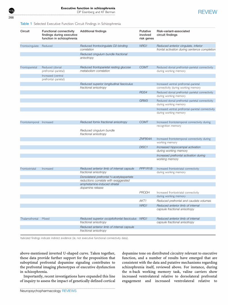
As schizophrenia is highly heritable (Cardno andGottesman, 2000), and healthy relatives of patients showexecutive task impairments and associated neuroimagingphenotypes, which are qualitatively similar to their affectedfamily member but attenuated (Callicott et al, 2003a;Macdonald et al, 2008), and given a core role for executivedysfunction in schizophrenia, it is likely that functionalvariation in specific schizophrenia risk genes will impactaspects of the above-reviewed neurocircuit dynamics inpredictable ways. Building on the endophenotype approachoriginally proposed by Gottesman and Sheilds (1972),recent advances in imaging genetics have begun to provideremarkably convergent evidence supporting this hypoth-esis, as delineated below (see Table 1). Such advances arecrucial, in part, because among the multitude of molecularpathways impacting the interacting neural systems relevantto schizophrenia, any one candidate risk gene variant islikely to contribute only a nominal effect to the complexbehavioral phenotype that establishes the clinical diagnosis,and the gene variants discussed here are no exception.
However, the experiments reviewed below have nonethelessbeen able to detect robust genetic effects by usingneuroimaging techniques to assay ‘intermediate’ pheno-types at the neural systems levelFa level of organizationthat is closer to the actual impact of a single genevariationFrather than measuring diagnosis itself (Mieret al, 2009). One particular strength of this approach is theability to examine risk gene effects in healthy individualsthat do not possess many of the confounds inherent instudying patients, such as medication exposure andpsychotic symptoms, which has resulted in the majority ofstudies employing healthy populations; but by the sametoken, much work is still needed to better understand theeffects of these genetic variants in the complex clinical andgenetic context of schizophrenia.
COMT
Variation in the gene coding for catechol-O-methyltransfer-ase (COMT), an enzyme central to cortical synapticdopamine catabolism modestly influences risk of illnessand has garnered significant attention for providing insightinto the biological underpinnings of the imaging phenotypeof schizophrenia. The rs4680 single nucleotide polymorph-ism (SNP) has been best studied, and the valine risk alleleconfers thermostability, permitting greater enzymatic activ-ity and thereby reduced dopaminergic tone in corticalsynapses. In a seminal paper by Egan et al and subsequentreplications, the valine risk allele reliably predicts worseperformance but increased dorsolateral prefrontal andanterior cingulate physiological response to the n-back taskin both schizophrenic individuals and their unaffectedsiblings (Egan et al, 2001). This work has been extended toshow that predicted prefrontal dopaminergic tone bycombined genotype and pharmacological condition followan inverted U-shaped response during working memory,such that risk allele homozygotes have improved andprotective allele homozygotes have worse prefrontal effi-ciency in response to amphetamine (Mattay et al, 2003).Notably, functional variation in the COMT gene is notlimited to the rs4680 SNP, but rather includes otherpolymorphisms, including a P2 promoter region SNP anda 30 region SNP. These three SNPs show nonlinearinteracting effects on prefrontal efficiency during workingmemory task performance, in agreement with predictions ofresultant cortical dopaminergic catabolic rates, and high-light the complexity of genetic contributions to functionalneuroimaging phenotypes, even within a single gene(Meyer-Lindenberg et al, 2006). To add to this complexity,the ability of COMT to regulate cortical dopamine relies onother genetically determined cellular resources, as suggestedby studies of MTHFR by Roffman et al (2008). Variation inMTHFR (rs1801133), which also shows association withschizophrenia risk (Gilbody et al, 2007), regulates theavailability of methyl groups for use by COMT and, incombination with rs4680, predicts DLPFC activationduring working memory in a manner consistent with the
Executive function in schizophreniaDP Eisenberg and KF Berman...............................................................................................................................................................
265
REVIEW
..............................................................................................................................................
Neuropsychopharmacology REVIEWS
above-mentioned inverted U-shaped curve. Taken together,these data provide further support for the proposition thatsuboptimal prefrontal dopamine signaling contributes tothe prefrontal imaging phenotypes of executive dysfunctionin schizophrenia.
Importantly, recent investigations have expanded this lineof inquiry to assess the impact of genetically defined cortical
dopamine tone on distributed circuitry relevant to executivefunction, and a number of results have emerged that areconsistent with the data and putative mechanisms regardingschizophrenia itself, reviewed above. For instance, duringthe n-back working memory task, valine carriers showincreased ventrolateral relative to dorsolateral prefrontalengagement and increased ventrolateral relative to
Executive function in schizophreniaDP Eisenberg and KF Berman
...............................................................................................................................................................
266
REVIEW
..............................................................................................................................................
Neuropsychopharmacology REVIEWS
dorsolateral connectivity with parietal regions, as had beenseen in schizophrenia earlier (Tan et al, 2007). Additionally,just as inappropriate prefrontal–hippocampal couplingpersists during working memory in schizophrenia patients(Meyer-Lindenberg et al, 2005b), during a recognitionmemory task that activates the hippocampus, carriers ofCOMT rs4680 valine alleles show disadvantageous increasedprefrontal–hippocampal connectivity (Bertolino et al, 2006).Finally, in agreement with the above-highlighted frontos-triatal circuit abnormalities in schizophrenia, particularlydisinhibited presynaptic striatal dopaminergic signaling inassociation with DLPFC hypofunction, postmortem datashow that COMT valine alleles predict increased tyrosinehydroxylase mRNA expression in the midbrain (Akil et al,2003), origin of dopaminergic projections to the striatum.Corroborating this effect are in vivo data describing COMTgenotype effects on the relationship between midbraindopamine storage and prefrontal activation during then-back task: in met homozygotes, this relationshipwas negative, but in val carriers, it was positive (Meyer-Lindenberg et al, 2005a). This has been interpreted as adownstream effect of genetically conferred variation ofprefrontal dopaminergic neurotransmission, as midbrainrelative to cortical COMT expression is weak (Kastner et al,1994), such that suboptimal prefrontal output to mesence-phalic inhibitory cells results in exaggerated activity ofdopamine neurons projecting to the striatum.
RGS4
RGS4 is an important modulator of central dopamine,glutamate, and neuregulin G-protein receptor systems, andtranscript expression in the DLPFC of schizophreniapatients has been shown to be reduced (Mirnics et al,2001). An SNP (rs951436 C-A) in the gene coding for thisprotein is associated with both schizophrenia (Chowdariet al, 2002) and reductions in DLPFC volumes (Prasad et al,2005). Buckholtz et al (2007a, b) studied this risk SNP in alarge group of healthy individuals undergoing functionalMRI scans during the n-back task and found thatindividuals carrying more risk alleles evidenced greateractivation in the left ventrolateral PFC, but less activation inthe right lateral PFC, temporal cortex, and caudate(Buckholtz et al, 2007a). Similar to investigations in COMT(Tan et al, 2007) and schizophrenia itself (Tan et al, 2006),examination of functional connectivity between thesedifferentially activated nodes showed that risk allelesimpaired cooperativity between right hemispheric nodesactivated by the task (eg, DLPFC, PPC) but exaggeratedcooperativity between VLPFC and nodes deactivated by task(eg, mPFC, superior temporal cortex, posterior cingulate,and parahippocampal gyrus) (Buckholtz et al, 2007a).Notably, when regional brain activations during the n-backtask are examined with consideration of both COMT andRGS4 genotypes, there exists an epistatic interaction, suchthat RGS4 risk allele-associated greater DLPFC andmidbrain activation occurs only in the context of COMT
risk allele carriers (Buckholtz et al, 2007b). Regardless ofwhether this interaction occurs biologically at the molecular(eg, COMT regulating RGS4 gene expression, Lipska et al,2006a) or systems level (eg, inefficient executive circuitsbeing more susceptible to RGS4 effects) (Buckholtz et al,2007b), these data highlight the complex contribution ofschizophrenia risk gene networks to executive processing.
GRM3
An SNP in the gene coding for the metabotropic type IIglutamate receptor mGluR3, GRM3 (rs6465084), results inweakly increased risk for schizophrenia, reduced prefrontalexcitatory amino-acid transporter 2 mRNA expression(EEAT2), worse verbal fluency performance, and reducedDLPFC neuronal integrity as measured by magneticresonance spectroscopy (Egan et al, 2004; Marenco et al,2006). As in COMT, during the n-back working memorytask, greater DLPFC BOLD signal activation for the sameperformance level (‘prefrontal inefficiency’) is seen incarriers of the risk SNP (Egan et al, 2004). However, thisfinding of GRM3 risk allele-associated prefrontal ineffi-ciency during working memory, as in RGS4, has beenreplicated in COMT rs4680 risk allele carriers but not inmethionine homozygotes, suggesting an epistatic interac-tion between these two risk genes. Furthermore, carriers ofboth COMT and GRM3 risk alleles show disproportionatelygreater VLPFC over DLPFC connectivity with parietalregions activated by this task (Tan et al, 2007), similar tothe schizophrenia phenotype (Tan et al, 2006).
PPP1R1B
Dopamine- and cAMP-regulated phosphoprotein of mole-cular weight 32 kDa (DARPP-32) is abundant in thestriatum and has a key function in modulating dopaminer-gic postsynaptic intracellular signaling through multifacetedeffects on protein kinases (Svenningsson et al, 2004). Onecommon haplotype in the PPP1R1B gene coding forDARPP-32 shows an association with schizophrenia, withworse IQ, verbal fluency, working memory, and WisconsinCard Sorting performance, with reduced striatal volumes,with reduced striatal BOLD activation during the n-back,and with increased frontostriatal connectivity. Notably,both the activation and connectivity findings were repli-cated in a separate cohort during performance of anemotional face-matching task (Meyer-Lindenberg et al,2007).
PRODH
A functional haplotype (rs4819756 and rs2870983 andrs450046 minor alleles) in the proline oxydase gene,PRODH, shows increased enzymatic activity, risk forschizophrenia, diminished striatal volumes, reduced striatalBOLD activation, and increased frontostriatal connectivityduring the n-back task (Kempf et al, 2008). Despite
Executive function in schizophreniaDP Eisenberg and KF Berman...............................................................................................................................................................
267
REVIEW
..............................................................................................................................................
Neuropsychopharmacology REVIEWS
significant differences between the functions of prolineoxydase and DARPP-32, these results are remarkablysimilar to those of PPP1R1B and converge on circuitry(prefrontal-neostriatal) that is dysregulated in schizophre-nia (Meyer-Lindenberg et al, 2002).
AKT-1
AKT-1 is an intracellular signaling protein that has animportant function in dopamine-mediated neurotransmis-sion (Beaulieu et al, 2005; Wei et al, 2007) and has shownreduced expression in schizophrenic brains (Emamian et al,2004) and lymphocytes (Tan et al, 2008). Further, severalreports have found an association between a functionalAKT-1 genetic variations and schizophrenia (Emamianet al, 2004; Tan et al, 2008). One such variation, an SNP,rs1130233, additionally shows a relationship with neurop-sychological assessments of executive function as well asn-back-related prefrontal activation. The risk allele alsoimparts reduced prefrontal and caudate volumes, inagreement with its hypothesized impact on frontostriatalcircuitry, though formal testing of functional connectivityhas not been performed at this date (Tan et al, 2008).
DISC-1
The disrupted in schizophrenia (DISC-1) gene codes for aprotein abundant in the hippocampus, which partners withNudel and other dynein complex proteins to impactcentrosomal function, neurite outgrowth, and neuronalmigration (Kamiya et al, 2005). Variations in DISC-1 areassociated with schizophrenia (Callicott et al, 2005; Ekelundet al, 2004; Hennah et al, 2003; Hodgkinson et al, 2004), andrecent multimodal imaging data have evidenced an effect ofthe DISC-1 Ser704Cys polymorphism on hippocampalstructure and function in healthy adults (Callicott et al,2005). Specifically, serine homozygotes showed reducedhippocampal gray matter volume, lower hippocampal N-acetyl aspartate, and during the n-back working memorytask, abnormally greater hippocampal activation (Callicottet al, 2005). These results align well with the impairedsuppression of medial temporal lobe activity duringexecutive processing seen in schizophrenia (Meyer-Linden-berg et al, 2001). Furthermore, during verbal fluency taskperformance, serine homozygotes show increased prefron-tal activation (Prata et al, 2008), though to what degreefrontotemporal connectivity is directly influenced by thispolymorphism remains to be tested.
ZNF804A
In a recent genome-wide association study, an SNP(rs1344706) in ZNF804A, a gene coding for a protein ofunclear function but potential gene regulatory ability,showed independent, significant association with schizo-phrenia (O’Donovan et al, 2008). Comparing healthyindividuals with either no, one, or two risk alleles, Esslinger
et al (2009) have found that the number of risk allelespredicted greater prefrontal–hippocampal functional con-nectivity during the n-back working memory task, just ashad been described earlier in patients (Meyer-Lindenberget al, 2005b), reinforcing the fact that greater functionalconnectivity (especially with a dysfunctional prefrontalcortex, as in schizophrenia), not only less, can be the riskphenotype. Better understanding of the biology of ZNF804Ais needed to clarify the nature of this observation, but it isnonetheless remarkable that a risk gene without a priorievidence for either prefrontal or hippocampal involvementcan so clearly show a predicted illness circuit phenotype inthis way.
NRG-1
Neuregulin1 (NRG-1) isoforms and its receptor ErbB4 haveimportant functions in potentially illness-relevant neuralprocesses, including neuronal migration, axonal guidanceand myelination, synaptic plasticity, and glutamatergicdendritic spine maturation (Barros et al, 2009; Mei andXiong, 2008). Variation in the NRG-1 gene has shownassociation with schizophrenia diagnosis, behavioral ab-normalities in mouse models responsive to antipsychoticmedication (Li et al, 2006; Stefansson et al, 2002), andaltered neuregulin isoform expression (Law et al, 2006).
In a group of individuals at high risk of developingschizophrenia by virtue of strong family history, carrierstatus of an NRG1 risk allele (SNP8NRG243177 polymorph-ism, which influences neuregulin transcript expression, Lawet al, 2006) predicted development of psychotic symptomsas well as reduced activation in medial prefrontal andtemporo-occipital regions during a sentence completiontask (Hall et al, 2006).
The number of NRG-1 risk alleles carried in healthy adultscorrelates with reduced semantic verbal fluency perfor-mance and reduced anterior cingulate, inferior frontal, andmiddle temporal activation measured by fMRI BOLD signal(Kircher et al, 2009). Disrupted microstructural connectiv-ity in association with the risk allele of this samepolymorphism is evidence by reduced white matter densityand fractional anisotropy in the anterior limb of the internalcapsule (Mcintosh et al, 2007), which contains importantaxonal fibers linking the prefrontal cortex with other nodesin the extended executive network. Future work is needed toconfirm these findings and determine to what degree theseabnormalities explain functional differences in individualswith different allelic risk loads.
SUMMARY AND FUTURE DIRECTIONS
Key brain regions that show postmortem and in vivoevidence for disarray in schizophrenia are important inexecutive functioning, and are physiologically abnormalduring executive challenge in patients, evidence character-istically aberrant interactions and remarkable susceptibilityto variation in putative schizophrenia risk genes.
Executive function in schizophreniaDP Eisenberg and KF Berman
...............................................................................................................................................................
268
REVIEW
..............................................................................................................................................
Neuropsychopharmacology REVIEWS
DLPFC dysfunction and aberrant functional connectivity,relatively increased VLPFC involvement in executivecircuitry, ACC, and IPL dysfunction and reduced couplingwith DLPFC, impairment in suppression of medial temporalactivity during certain executive challenges, prefrontaldisinhibition of mesostriatal dopaminergic signaling, andreduced thalamofrontal cooperativity not only form acomplex landscape of circuit changes in schizophrenia,but also, in selected subsets of these, create quantifiablelinks to emerging molecular footprints of genetic predis-position to psychosis. Systematic work is needed to bettercharacterize the dynamics of these systems-level abnorm-alities in response to particular executive task demands,pharmacological interventions, and genetic environments.
Specifically, several avenues of research promise toprovide invaluable insights into pathophysiology andultimately targeted treatment of this devastating illness.
To address accumulating evidence of genetic heterogene-ity underlying the disorder and concomitant variability inpsychopathological and neuropsychological profiles, all ofwhich may have contributed to apparent inconsistencies inthe literature, more extensive genetically, clinically, andcognitively stratified studies are necessary. Likewise, long-itudinal studies directed at understanding both naturalisticand pharmacologically induced fluctuations in executivenetwork function are essential to assess the stability ofcircuit perturbations in schizophrenia over the course ofillness and treatment. Additionally, developing advancedmethodologies to bridge molecular and physiological dataand fuel both candidate risk gene discovery and biologicalvalidation has become increasingly important. One suchapproach is to use neurocircuit risk phenotypes asquantitative trait variables to identify genetic factorscontributing to executive dysfunction in psychotic dis-orders. Potkin et al (2008) have begun to implement thisstrategy with DLPFC activation alone as the quantitativetrait variable, yielding novel results. As efforts to charac-terize and quantify the above-outlined systems-level circui-try disruptions in schizophrenia advance, bringing greaterpredictive power for diagnosis and treatment response tonuanced functional imaging phenotypes, this reversemappingFfrom imaging to genesFmay become increas-ingly valuable for understanding illness pathophysiologyand for developing pharmacogenetic models. Similarly,development of robust data-driven analytical techniques,such as parallel independent components analysis (Liu et al,2009) to meaningfully combine highly dimensional geneticand imaging datasets in a coordinated and comprehensivefashion may eventually help shed light on the underlyingstructures of each. Finally, because inherited variation inDNA sequences, though incredibly useful for identifyingkey molecular pathways to schizophrenia as illustratedabove, is likely only a partial contributor to illness brainphenotypes, it will be progressively more important toexplore connections between executive circuit dynamicsand de novo mutations (Stefansson et al, 2008), epigenetics
(Huang and Akbarian, 2007), and gene–environmentinteractions (Caspi et al, 2005; Nicodemus et al, 2008)associated with schizophrenia.
In summary, schizophrenia patients show a remarkablenumber of characteristic abnormalities of executive circui-try, evident in vivo with functional neuroimaging techni-ques, the topography of which corresponds well to otherpathological findings in postmortem tissue and in vivoneurochemical (magnetic resonance spectroscopy, neuror-eceptor mapping) assays. A growing list of candidateschizophrenia risk genes show variation in executive circuitdynamics, akin to that in illness, suggesting that increasingattention to genetic and genetic–environmental interactionsyields promise for better understanding the biology ofexecutive dysfunction in schizophrenia.
ACKNOWLEDGEMENTS
This work was supported by the National Institute of MentalHealth Intramural Research Program.
DISCLOSURE
The authors declared no conflict of interest.
REFERENCES
Aalto S, Bruck A, Laine M, Nagren K, Rinne JO (2005). Frontal and temporal
dopamine release during working memory and attention tasks in healthy
humans: a positron emission tomography study using the high-affinity dopamine
d2 receptor ligand [11c]flb 457. J Neurosci 25: 2471–2477.
Abbott C, Bustillo J (2006). What have we learned from proton magnetic resonance
spectroscopy about schizophrenia? A critical update. Curr Opin Psychiatry 19:
135–139.
Abi-Dargham A, Gil R, Krystal J, Baldwin RM, Seibyl JP, Bowers M et al (1998).
Increased striatal dopamine transmission in schizophrenia: confirmation in a
second cohort. Am J Psychiatry 155: 761–767.
Abi-Dargham A, Mawlawi O, Lombardo I, Gil R, Martinez D, Huang Y et al (2002).
Prefrontal dopamine d1 receptors and working memory in schizophrenia.
J Neurosci 22: 3708–3719. Evidence of upregulated D1 receptors in the
DLPFC in schizophrenia, potentially in response to reduced dopaminergic
input previously shown.
Akbarian S, Kim JJ, Potkin SG, Hagman JO, Tafazzoli A, Bunney Jr WE et al (1995).
Gene expression for glutamic acid decarboxylase is reduced without loss of
neurons in prefrontal cortex of schizophrenics. Arch Gen Psychiatry 52: 258–
266.
Akbarian S, Sucher N, Bradley D, Tafazzoli A, Trinh D, Hetrick W et al (1996).
Selective alterations in gene expression for nmda receptor subunits in prefrontal
cortex of schizophrenics. J Neurosci 16: 19–30.
Akil M, Kolachana BS, Rothmond DA, Hyde TM, Weinberger DR, Kleinman JE
(2003). Catechol-o-methyltransferase genotype and dopamine regulation in the
human brain. J Neurosci 23: 2008–2013.
Akil M, Pierri JN, Whitehead RE, Edgar CL, Mohila C, Sampson AR et al (1999).
Lamina-specific alterations in the dopamine innervation of the prefrontal cortex in
schizophrenic subjects. Am J Psychiatry 156: 1580–1589.
Albert KA, Hemmings Jr HC, Adamo AIB, Potkin SG, Akbarian S, Sandman CA et al
(2002). Evidence for decreased darpp-32 in the prefrontal cortex of patients with
schizophrenia. Arch Gen Psychiatry 59: 705–712. Demonstration of abnormal
DARPP-32 concentrations in the schizophrenic DLPFC, in line with
prefrontal imaging genetics results.
Andreasen NC, Arndt S, Swayze V, Cizadlo T, Flaum M, O0Leary D et al (1994a).
Thalamic abnormalities in schizophrenia visualized through magnetic resonance
image averaging. Science 266: 294–298. First significant investigation of
thalamic size by MRI technology, fueling subsequent experiments on
sensory-gating abnormalities in schizophrenia.
Executive function in schizophreniaDP Eisenberg and KF Berman...............................................................................................................................................................
269
REVIEW
..............................................................................................................................................
Neuropsychopharmacology REVIEWS
Andreasen NC, Flashman L, Flaum M, Arndt S, Swayze 2nd V, O’Leary DS et al
(1994b). Regional brain abnormalities in schizophrenia measured with magnetic
resonance imaging. JAMA 272: 1763–1769.
Andreasen NC, Rezai K, Alliger R, Swayze II VW, Flaum M, Kirchner P et al (1992).
Hypofrontality in neuroleptic-naive patients and in patients with chronic
schizophrenia: assessment with xenon 133 single-photon emission computed
tomography and the tower of london. Arch Gen Psychiatry 49: 943–958.
Andrews J, Wang L, Csernansky JG, Gado MH, Barch DM (2006). Abnormalities
of thalamic activation and cognition in schizophrenia. Am J Psychiatry 163:
463–469.
Arnold SE, Franz BR, Gur RC, Gur RE, Shapiro RM, Moberg PJ et al (1995). Smaller
neuron size in schizophrenia in hippocampal subfields that mediate cortical-
hippocampal interactions. Am J Psychiatry 152: 738–748.
Arnold SE, Lee VM, Gur RE, Trojanowski JQ (1991). Abnormal expression of
two microtubule-associated proteins (map2 and map5) in specific subfields of
the hippocampal formation in schizophrenia. Proc Natl Acad Sci USA 88:
10850–10854.
Ashtari M, Cottone J, Ardekani BA, Cervellione K, Szeszko PR, Wu J et al (2007).
Disruption of white matter integrity in the inferior longitudinal fasciculus in
adolescents with schizophrenia as revealed by fiber tractography. Arch Gen
Psychiatry 64: 1270–1280.
Auer DP, Wilke M, Grabner A, Heidenreich JO, Bronisch T, Wetter TC (2001).
Reduced naa in the thalamus and altered membrane and glial metabolism in
schizophrenic patients detected by 1h-mrs and tissue segmentation. Schizophr
Res 52: 87–99.
Badcock JC, Michie PT, Rock D (2005). Spatial working memory and planning
ability: contrasts between schizophrenia and bipolar i disorder. Cortex 41:
753–763.
Baiano M, David A, Versace A, Churchill R, Balestrieri M, Brambilla P (2007).
Anterior cingulate volumes in schizophrenia: a systematic review and a meta-
analysis of mri studies. Schizophr Res 93: 1–12.
Barbalat G, Chambon V, Franck N, Koechlin E, Farrer C (2009). Organization of
cognitive control within the lateral prefrontal cortex in schizophrenia. Arch Gen
Psychiatry 66: 377–386.
Barch DM, Carter CS, Braver TS, Sabb FW, MacDonald III A, Noll DC et al (2001).
Selective deficits in prefrontal cortex function in medication-naive patients with
schizophrenia. Arch Gen Psychiatry 58: 280–288.
Barch DM, Csernansky JG (2007). Abnormal parietal cortex activation during
working memory in schizophrenia: verbal phonological coding disturbances
versus domain-general executive dysfunction. Am J Psychiatry 164: 1090–1098.
Barch DM, Sheline YI, Csernansky JG, Snyder AZ (2003). Working memory and
prefrontal cortex dysfunction: specificity to schizophrenia compared with major
depression. Biol Psychiatry 53: 376–384.
Barros CS, Calabrese B, Chamero P, Roberts AJ, Korzus E, Lloyd K et al (2009).
Impaired maturation of dendritic spines without disorganization of cortical cell
layers in mice lacking nrg1/erbb signaling in the central nervous system. Proc
Natl Acad Sci 106: 4507–4512.
Basho S, Palmer ED, Rubio MA, Wulfeck B, Muller R-A (2007). Effects of generation
mode in fmri adaptations of semantic fluency: paced production and overt
speech. Neuropsychologia 45: 1697–1706.
Bassett DS, Bullmore E, Verchinski BA, Mattay VS, Weinberger DR, Meyer-
Lindenberg A (2008). Hierarchical organization of human cortical networks in
health and schizophrenia. J Neurosci 28: 9239–9248. Support for distributed
neural network disturbances in schizophrenia using novel, comprehensive
methodologies.
Beasley CL, Zhang ZJ, Patten I, Reynolds GP (2002). Selective deficits in prefrontal
cortical gabaergic neurons in schizophrenia defined by the presence of calcium-
binding proteins. Biol Psychiatry 52: 708–715.
Beaulieu J-M, Sotnikova TD, Marion S, Lefkowitz RJ, Gainetdinov RR, Caron MG
(2005). An akt/[beta]-arrestin 2/pp2a signaling complex mediates dopaminergic
neurotransmission and behavior. Cell 122: 261–273.
Becker TM, Kerns JG, MacDonald III AW, Carter CS (2008). Prefrontal dysfunction
in first-degree relatives of schizophrenia patients during a stroop task.
Neuropsychopharmacology 33: 2619–2625.
Benes F, Vincent S, Alsterberg G, Bird E, SanGiovanni J (1992a). Increased gabaa
receptor binding in superficial layers of cingulate cortex in schizophrenics.
J Neurosci 12: 924–929.
Benes FM, Sorensen I, Bird ED (1991). Reduced neuronal size in posterior
hippocampus of schizophrenic patients. Schizophr Bull 17: 597–608.
Benes FM, Sorensen I, Vincent SL, Bird ED, Sathi M (1992b). Increased density of
glutamate-immunoreactive vertical processes in superficial laminae in cingulate
cortex of schizophrenic brain. Cereb Cortex 2: 503–512.
Benes FM, Vincent SL, Todtenkopf M (2001). The density of pyramidal and
nonpyramidal neurons in anterior cingulate cortex of schizophrenic and bipolar
subjects. Biol Psychiatry 50: 395–406.
Berman K, Doran A, Pickar D, Weinberger D (1993). Is the mechanism of prefrontal
hypofunction in depression the same as in schizophrenia? Regional cerebral
blood flow during cognitive activation. Br J Psychiatry 162: 183–192. First
evidence for diagnostic specificity of prefrontal hypofunction in schizo-
phrenia.
Berman KF, Illowsky BP, Weinberger DR (1988). Physiological dysfunction of
dorsolateral prefrontal cortex in schizophrenia: IV. Further evidence for regional
and behavioral specificity. Arch Gen Psychiatry 45: 616–622. First demonstra-
tion of task specificity of prefrontal hypofunction in schizophrenia.
Berman KF, Ostrem JL, Randolph C, Gold J, Goldberg TE, Coppola R et al (1995).
Physiological activation of a cortical network during performance of the
wisconsin card sorting test: a positron emission tomography study. Neuro-
psychologia 33: 1027–1046.
Berman KF, Torrey EF, Daniel DG, Weinberger DR (1992). Regional cerebral blood
flow in monozygotic twins discordant and concordant for schizophrenia. Arch
Gen Psychiatry 49: 927–934. Discrimination of non-genetic contribution to
hypofrontal phenotype in schizophrenia via twin studies.
Berman KF, Zec RF, Weinberger DR (1986). Physiologic dysfunction of dorsolateral
prefrontal cortex in schizophrenia: Ii. Role of neuroleptic treatment, attention, and
mental effort. Arch Gen Psychiatry 43: 126–135.
Bertolino A, Breier A, Callicott JH, Adler C, Mattay VS, Shapiro M et al (2000a). The
relationship between dorsolateral prefrontal neuronal n-acetylaspartate and
evoked release of striatal dopamine in schizophrenia. Neuropsychopharmaco-
logy 22: 125–132. First demonstration that neuronal integrity of DLPFC is
associated with exaggerated striatal dopaminergic neurotransmission.
Bertolino A, Esposito G, Callicott JH, Mattay VS, Van Horn JD, Frank JA et al
(2000b). Specific relationship between prefrontal neuronal n-acetylaspartate and
activation of the working memory cortical network in schizophrenia. Am J
Psychiatry 157: 26–33. First demonstration that neuronal integrity, as
measured by NAA, of DLPFC is associated with its function during working
memory.
Bertolino A, Roffman JL, Lipska BK, van Gelderen P, Olson A, Weinberger DR
(2002). Reduced n-acetylaspartate in prefrontal cortex of adult rats with neonatal
hippocampal damage. Cereb Cortex 12: 983–990.
Bertolino A, Rubino V, Sambataro F, Blasi G, Latorre V, Fazio L et al (2006).
Prefrontal-hippocampal coupling during memory processing is modulated by
comt val158met genotype. Biol Psychiatry 60: 1250–1258.
Bogerts B, Ashtari M, Degreef G, Alvir JMJ, Bilder RM, Lieberman JA (1990).
Reduced temporal limbic structure volumes on magnetic resonance images in
first episode schizophrenia. Psychiatry Res 35: 1–13.
Boksman K, Theberge J, Williamson P, Drost DJ, Malla A, Densmore M et al (2005).
A 4.0-t fmri study of brain connectivity during word fluency in first-episode
schizophrenia. Schizophr Res 75: 247–263.
Bozikas VP, Kosmidis MH, Kiosseoglou G, Karavatos A (2006). Neuropsychological
profile of cognitively impaired patients with schizophrenia. Compr Psychiatry 47:
136–143.
Breier A, Su T-P, Saunders R, Carson RE, Kolachana BS, de Bartolomeis A
et al (1997). Schizophrenia is associated with elevated amphetamine-induced
synaptic dopamine concentrations: evidence from a novel positron emission
tomography method. Proc Natl Acad Sci USA 94: 2569–2574. First use of
raclopride PET technique to demonstrate exaggerated striatal dopamine
release in schizophrenia in response to amphetamine challenge.
Broome MR, Matthiasson P, Fusar-Poli P, Woolley JB, Johns LC, Tabraham P et al
(2009). Neural correlates of executive function and working memory in the 0at-risk
mental state0. BrJ Psychiatry 194: 25–33.
Browning MD, Dudek EM, Rapier JL, Leonard S, Freedman R (1993). Significant
reductions in synapsin but not synaptophysin specific activity in the brains of
some schizophrenics. Biol Psychiatry 34: 529–535.
Buchanan RW, Francis A, Arango C, Miller K, Lefkowitz DM, McMahon RP et al
(2004). Morphometric assessment of the heteromodal association cortex in
schizophrenia. Am J Psychiatry 161: 322–331.
Buchsbaum BR, Greer S, Chang W-L, Berman KF (2005). Meta-analysis of
neuroimaging studies of the wisconsin card-sorting task and component
processes. Hum Brain Mapp 25: 35–45.
Buchsbaum MS, Christian BT, Lehrer DS, Narayanan TK, Shi B, Mantil J et al
(2006). D2/d3 dopamine receptor binding with [f-18]fallypride in thalamus and
cortex of patients with schizophrenia. Schizophr Res 85: 232–244.
Buckholtz JW, Meyer-Lindenberg A, Honea RA, Straub RE, Pezawas L, Egan MF
et al (2007a). Allelic variation in rgs4 impacts functional and structural
connectivity in the human brain. J Neurosci 27: 1584–1593. First report
to show association between RGS4 and imaging schizophrenia pheno-
types.
Buckholtz JW, Sust S, Tan HY, Mattay VS, Straub RE, Meyer-Lindenberg A et al
(2007b). Fmri evidence for functional epistasis between comt and rgs4. Mol
Psychiatry 12: 893–895.
Executive function in schizophreniaDP Eisenberg and KF Berman
...............................................................................................................................................................
270
REVIEW
..............................................................................................................................................
Neuropsychopharmacology REVIEWS
Byne W, Buchsbaum MS, Mattiace LA, Hazlett EA, Kemether E, Elhakem SL et al
(2002). Postmortem assessment of thalamic nuclear volumes in subjects with
schizophrenia. Am J Psychiatry 159: 59–65.
Callicott JH, Egan MF, Mattay VS, Bertolino A, Bone AD, Verchinksi B et al
(2003a). Abnormal fmri response of the dorsolateral prefrontal cortex in
cognitively intact siblings of patients with schizophrenia. Am J Psychiatry 160:
709–719.
Callicott JH, Mattay VS, Bertolino A, Finn K, Coppola R, Frank JA et al (1999).
Physiological characteristics of capacity constraints in working memory as
revealed by functional mri. Cereb Cortex 9: 20–26.
Callicott JH, Mattay VS, Verchinski BA, Marenco S, Egan MF, Weinberger DR
(2003b). Complexity of prefrontal cortical dysfunction in schizophrenia: more
than up or down. Am J Psychiatry 160: 2209–2215. Influential paper
introducing the concept of prefrontal inefficiency in schizophrenia subjects
performing the n-back at near-normal performance in contrast to
hypofrontality in lower scoring subjects.
Callicott JH, Ramsey NF, Tallent K, Bertolino A, Knable MB, Coppola R et al (1998).
Functional magnetic resonance imaging brain mapping in psychiatry: methodo-
logical issues illustrated in a study of working memory in schizophrenia.
Neuropsychopharmacology 18: 186–196.
Callicott JH, Straub RE, Pezawas L, Egan MF, Mattay VS, Hariri AR et al (2005).
Variation in disc1 affects hippocampal structure and function and increases risk
for schizophrenia. Proc Natl Acad Sci USA 102: 8627–8632. First demonstra-
tion of influence of DISC1 risk allele carrier status on schizophrenia imaging
phenotypes of the medial temporal lobe.
Camchong J, Dyckman KA, Chapman CE, Yanasak NE, McDowell JE (2006). Basal
ganglia-thalamocortical circuitry disruptions in schizophrenia during delayed
response tasks. Biol Psychiatry 60: 235–241.
Cannon TD, Glahn DC, Kim J, Van Erp TGM, Karlsgodt K, Cohen MS et al (2005).
Dorsolateral prefrontal cortex activity during maintenance and manipulation of
information in working memory in patients with schizophrenia. Arch Gen
Psychiatry 62: 1071–1080.
Cannon TD, Thompson PM, van Erp TGM, Toga AW, Poutanen V-P, Huttunen M
et al (2002). Cortex mapping reveals regionally specific patterns of genetic and
disease-specific gray-matter deficits in twins discordant for schizophrenia. Proc
Natl Acad Sci USA 99: 3228–3233. Twin study clarifying genetic and disease-
related alterations in DLPFC gray matter concentrations.
Cantor-Graae E, Warkentin S, Franzen G, Risberg J, Ingvar DH (1991). Aspects of
stability of regional cerebral blood flow in chronic schizophrenia: an 18-year
followup study. Psychiatry Res 40: 253–266. Only study to document stability
of hypofrontality over many years time.
Cardno AG, Gottesman (2000). Twin studies of schizophrenia: from bow-and-arrow
concordances to star wars mx and functional genomics. Am J Med Genet 97:
12–17.
Carter CS, MacDonald III AW, Ross LL, Stenger VA (2001). Anterior cingulate cortex
activity and impaired self-monitoring of performance in patients with schizo-
phrenia: an event-related fmri study. Am J Psychiatry 158: 1423–1428.
Carter CS, Mintun M, Nichols T, Cohen JD (1997). Anterior cingulate gyrus
dysfunction and selective attention deficits in schizophrenia: [15o]h2o pet study
during single-trial stroop task performance. Am J Psychiatry 154: 1670–1675.
Carter CS, Perlstein W, Ganguli R, Brar J, Mintun M, Cohen JD (1998). Functional
hypofrontality and working memory dysfunction in schizophrenia. Am J
Psychiatry 155: 1285–1287.
Caspi A, Moffitt TE, Cannon M, McClay J, Murray R, Harrington H et al (2005).
Moderation of the effect of adolescent-onset cannabis use on adult psychosis by
a functional polymorphism in the catechol-o-methyltransferase gene: longitudinal
evidence of a gene x environment interaction. Biol Psychiatry 57: 1117–1127.
Evidence for gene-environment interactions predisposing to schizophrenia.
Catafau AM, Parellada E, Lomena FJ, Bernardo M, Pavia J, Ros D et al (1994).
Prefrontal and temporal blood flow in schizophrenia: resting and activation
technetium-99m-hmpao spect patterns in young neuroleptic-naive patients with
acute disease. J Nucl Med 35: 935–941.
Chang C, Crottaz-Herbette S, Menon V (2007). Temporal dynamics of basal ganglia
response and connectivity during verbal working memory. Neuroimage 34:
1253–1269.
Chowdari KV, Mirnics K, Semwal P, Wood J, Lawrence E, Bhatia T et al (2002).
Association and linkage analyses of rgs4 polymorphisms in schizophrenia.
Hum Mol Genet 11: 1373–1380.
Clark D, Dedova I, Cordwell S, Matsumoto I (2006). A proteome analysis of
the anterior cingulate cortex gray matter in schizophrenia. Mol Psychiatry 11:
459–470.
Cohen JD, Perlstein WM, Braver TS, Nystrom LE, Noll DC, Jonides J et al (1997).
Temporal dynamics of brain activation during a working memory task. Nature
386: 604–608. Support for differential functional topography during
different stages of working memory task performance.
Curtis VA, Bullmore ET, Brammer MJ, Wright IC, Williams SC, Morris RG et al
(1998). Attenuated frontal activation during a verbal fluency task in patients with
schizophrenia. Am J Psychiatry 155: 1056–1063.
Curtis VA, Bullmore ET, Morris RG, Brammer MJ, Williams SCR, Simmons A et al
(1999). Attenuated frontal activation in schizophrenia may be task dependent.
Schizophr Res 37: 35–44.
Daniel D, Weinberger D, Jones D, Zigun J, Coppola R, Handel S et al (1991). The
effect of amphetamine on regional cerebral blood flow during cognitive activation
in schizophrenia. J Neurosci 11: 1907–1917.
Dao-Castellana M-H, Paillere-Martinot M-L, Hantraye P, Attar-Levy D, Remy P,
Crouzel C et al (1997). Presynaptic dopaminergic function in the striatum of
schizophrenic patients. Schizophr Res 23: 167–174.
Daviss SR, Lewis DA (1995). Local circuit neurons of the prefrontal cortex in
schizophrenia: selective increase in the density of calbindin-immunoreactive
neurons. Psychiatry Res 59: 81–96.
Deicken RF, Johnson C, Eliaz Y, Schuff N (2000). Reduced concentrations of
thalamic n-acetylaspartate in male patients with schizophrenia. Am J Psychiatry
157: 644–647.
Dolan RJ, Fletcher P, Frith CD, Friston KJ, Frackowiak RS, Grasby PM (1995).
Dopaminergic modulation of impaired cognitive activation in the anterior cingulate
cortex in schizophrenia. Nature 378: 180–182. First evidence in live humans
showing that dopaminergic agents can modify cognitive physiology.
Drevets WC, Raichle ME (1998). Suppression of regional cerebral blood during
emotional versus higher cognitive implications for interactions between emotion
and cognition. Cogn Emot 12: 353–385.
Driesen NR, Leung H-C, Calhoun VD, Constable RT, Gueorguieva R, Hoffman R
et al (2008). Impairment of working memory maintenance and response in
schizophrenia: functional magnetic resonance imaging evidence. Biol Psychiatry
64: 1026–1034.
Dunnett SB, Meldrum A, Muir JL (2005). Frontal-striatal disconnection disrupts
cognitive performance of the frontal-type in the rat. Neuroscience 135:
1055–1065.
Durany N, Michel T, Zochling R, Boissl KW, Cruz-Sanchez FF, Riederer P et al
(2001). Brain-derived neurotrophic factor and neurotrophin 3 in schizophrenic
psychoses. Schizophr Res 52: 79–86.
Eack SM, George MM, Prasad KMR, Keshavan MS (2008). Neuroanatomical
substrates of foresight in schizophrenia. Schizophr Res 103: 62–70.
Egan MF, Goldberg TE, Kolachana BS, Callicott JH, Mazzanti CM, Straub RE et al
(2001). Effect of comt val108/158 met genotype on frontal lobe function and risk
for schizophrenia. Proc Natl Acad Sci USA 98: 6917–6922. Seminal paper
showing effect of COMT allelic load on prefrontal physiology and n-back
task performance.
Egan MF, Straub RE, Goldberg TE, Yakub I, Callicott JH, Hariri AR et al (2004).
Variation in grm3 affects cognition, prefrontal glutamate, and risk for schizo-
phrenia. Proc Natl Acad Sci USA 101: 12604–12609. First demonstration of
an association between GRM3 polymorphism and neuropsychological,
spectroscopic, and diagnostic measures.
Ekelund J, Hennah W, Hiekkalinna T, Parker A, Meyer J, Lonnqvist J et al (2004).
Replication of 1q42 linkage in finnish schizophrenia pedigrees. Mol Psychiatry 9:
1037–1041.
Elkashef AM, Doudet D, Bryant T, Cohen RM, Li S-H, Wyatt RJ (2000). 6–18f-dopa
pet study in patients with schizophrenia. Psychiatry Res 100: 1–11.
Elvevag B, Goldberg TE (2000). Cognitive impairment in schizophrenia is the core of
the disorder. Crit Rev Neurobiol 14: 1–21.
Emamian ES, Hall D, Birnbaum MJ, Karayiorgou M, Gogos JA (2004). Convergent
evidence for impaired akt1-gsk3beta signaling in schizophrenia. Nat Genetics
36: 131–137.
Ende G, Braus DF, Walter S, Henn FA (2001). Lower concentration of thalamic
n-acetylaspartate in patients with schizophrenia: a replication study. Am J
Psychiatry 158: 1314–1316.
Eslinger PJ, Grattan LM (1993). Frontal lobe and frontal-striatal substrates for
different forms of human cognitive flexibility. Neuropsychologia 31: 17–28.
Esslinger C, Walter H, Kirsch P, Erk S, Schnell K, Arnold C et al (2009). Neural
mechanisms of a genome-wide supported psychosis variant. Science 324: 605.
First paper to show that a SNP discovered via genome-wide association
study can demonstrate schizophrenia imaging phenotypes.
Fitzsimmons J, Kubicki M, Smith K, Bushell G, Estepar RSJ, Westin C-F et al
(2009). Diffusion tractography of the fornix in schizophrenia. Schizophr Res 107:
39–46.
Fletcher PC, Frith CD, Grasby PM, Friston KJ, Dolan RJ (1996). Local and
distributed effects of apomorphine on fronto-temporal function in acute
unmedicated schizophrenia. J Neurosci 16: 7055–7062.
Fletcher PC, McKenna PJ, Frith CD, Grasby PM, Friston KJ, Dolan RJ (1998). Brain
activations in schizophrenia during a graded memory task studied with functional
neuroimaging. Arch Gen Psychiatry 55: 1001–1008.
Executive function in schizophreniaDP Eisenberg and KF Berman...............................................................................................................................................................
271
REVIEW
..............................................................................................................................................
Neuropsychopharmacology REVIEWS
Flores G, Wood GK, Liang JJ, Quirion R, Srivastava LK (1996). Enhanced
amphetamine sensitivity and increased expression of dopamine d2 receptors
in postpubertal rats after neonatal excitotoxic lesions of the medial prefrontal
cortex. J Neurosci 16: 7366–7375.
Ford JM, Gray M, Whitfield SL, Turken AU, Glover G, Faustman WO et al (2004).
Acquiring and inhibiting prepotent responses in schizophrenia: event-related
brain potentials and functional magnetic resonance imaging. Arch Gen
Psychiatry 61: 119–129.
Fornito A, Yucel M, Patti J, Wood SJ, Pantelis C (2009). Mapping grey matter
reductions in schizophrenia: an anatomical likelihood estimation analysis of
voxel-based morphometry studies. Schizophr Res 108: 104–113.
Frank MJ, Loughry B, O’reilly RC (2001). Interactions between frontal cortex and
basal ganglia in working memory: a computational model. Cogn Affec Behav
Neurosci 1: 137–160.
Frederikse M, Lu A, Aylward E, Barta P, Sharma T, Pearlson G (2000). Sex
differences in inferior parietal lobule volume in schizophrenia. Am J Psychiatry
157: 422–427.
Frith CD, Friston KJ, Liddle PF, Frackowiak RSJ (1991). A pet study of word finding.
Neuropsychologia 29: 1137–1148.
Fu CHY, Suckling J, Williams SCR, Andrew CM, Vythelingum GN, McGuire PK
(2005). Effects of psychotic state and task demand on prefrontal function
in schizophrenia: an fmri study of overt verbal fluency. Am J Psychiatry 162:
485–494.
Gao X-M, Sakai K, Roberts RC, Conley RR, Dean B, Tamminga CA (2000).
Ionotropic glutamate receptors and expression of n-methyl-d-aspartate receptor
subunits in subregions of human hippocampus: effects of schizophrenia. Am J
Psychiatry 157: 1141–1149.
Gilbody S, Lewis S, Lightfoot T (2007). Methylenetetrahydrofolate reductase (mthfr)
genetic polymorphisms and psychiatric disorders: a huge review. Am J Epidemiol
165: 1–13.
Giuliani NR, Calhoun VD, Pearlson GD, Francis A, Buchanan RW (2005). Voxel-
based morphometry versus region of interest: a comparison of two methods
for analyzing gray matter differences in schizophrenia. Schizophr Res 74:
135–147.
Glahn DC, Ragland JD, Abramoff A, Barrett J, Laird AR, Bearden CE et al (2005).
Beyond hypofrontality: a quantitative meta-analysis of functional neuroimaging
studies of working memory in schizophrenia. Hum Brain Mapp 25: 60–69. An
excellent meta-analysis of working memory functional imaging.
Glantz LA, Lewis DA (2000). Decreased dendritic spine density on prefrontal cortical
pyramidal neurons in schizophrenia. Arch Gen Psychiatry 57: 65–73.
Goldberg TE, Berman KF, Fleming K, Ostrem J, Van Horn JD, Esposito G et al
(1998). Uncoupling cognitive workload and prefrontal cortical physiology: a pet
rcbf study. Neuroimage 7: 296–303.
Goldberg TE, Berman KF, Mohr E, Weinberger DR (1990). Regional cerebral blood
flow and cognitive function in Huntington’s disease and schizophrenia:
a comparison of patients matched for performance on a prefrontal-type task.
Arch Neurol 47: 418–422.
Goldman PS, Rosvold HE (1972). The effects of selective caudate lesions in infant
and juvenile rhesus monkeys. Brain Res 43: 53–66.
Goldstein JM, Goodman JM, Seidman LJ, Kennedy DN, Makris N, Lee H et al
(1999). Cortical abnormalities in schizophrenia identified by structural magnetic
resonance imaging. Arch Gen Psychiatry 56: 537–547.
Gottesman II, Sheilds J (1972). Schizophrenia and Genetics: A Twin Study Vantage
Point. Academic Press: New York.
Gourovitch ML, Kirkby BS, Goldberg TE, Weinberger DR, Gold JM, Esposito G et al
(2000). A comparison of rcbf patterns during letter and semantic fluency.
Neuropsychology 14: 353–360.
Graham S, Phua E, Soon CS, Oh T, Au C, Shuter B et al (2009). Role of medial
cortical, hippocampal and striatal interactions during cognitive set-shifting.
Neuroimage 45: 1359–1367.
Guo N, Hwang D-R, Lo E-S, Huang Y-Y, Laruelle M, Abi-Dargham A (2003).
Dopamine depletion and in vivo binding of pet d1 receptor radioligands:
implications for imaging studies in schizophrenia. Neuropsychopharmacology
28: 1703–1711.
Gur RE, Maany V, Mozley PD, Swanson C, Bilker W, Gur RC (1998). Subcortical
mri volumes in neuroleptic-naive and treated patients with schizophrenia. Am J
Psychiatry 155: 1711–1717.
Hall J, Whalley HC, Job DE, Baig BJ, McIntosh AM, Evans KL et al (2006). A
neuregulin 1 variant associated with abnormal cortical function and psychotic
symptoms. Nat Neurosci 9: 1477–1478.
Harrison PJ, McLaughlin D, Kerwin RW (1991). Decreased hippocampal expression
of a glutamate receptor gene in schizophrenia. Lancet 337: 450–452.
Hazlett EA, Buchsbaum MS, Kemether E, Bloom R, Platholi J, Brickman AM et al
(2004). Abnormal glucose metabolism in the mediodorsal nucleus of the
thalamus in schizophrenia. Am J Psychiatry 161: 305–314.
Hennah W, Varilo T, Kestila M, Paunio T, Arajarvi R, Haukka J et al (2003). Haplotype
transmission analysis provides evidence of association for disc1 to schizophrenia
and suggests sex-dependent effects. Hum Mol Genet 12: 3151–3159.
Hietala J, Syvalahti E, Kuoppamaki M, Hietala J, Syvalahti E, Haaparanta M et al
(1995). Presynaptic dopamine function in striatum of neuroleptic-naive schizo-
phrenic patients. Lancet 346: 1130–1131.
Hietala J, Syvalahti E, Vilkman H, Vuorio K, Rakkolainen V, Bergman J et al (1999).
Depressive symptoms and presynaptic dopamine function in neuroleptic-naive
schizophrenia. Schizophr Res 35: 41–50.
Highley JR, Walker MA, McDonald B, Crow TJ, Esiri MM (2003). Size of
hippocampal pyramidal neurons in schizophrenia. Br J Psychiatry 183: 414–417.
Hodgkinson CA, Goldman D, Jaeger J, Persaud S, Kane JM, Lipsky RH et al
(2004). Disrupted in schizophrenia 1 (disc1): association with schizophrenia,
schizoaffective disorder, and bipolar disorder. Am J Hum Genet 75: 862–872.
Holmes AJ, MacDonald III A, Carter CS, Barch DM, Andrew Stenger V, Cohen JD
(2005). Prefrontal functioning during context processing in schizophrenia and
major depression: an event-related fmri study. Schizophr Res 76: 199–206.
Holt DJ, Herman MM, Hyde TM, Kleinman JE, Sinton CM, German DC et al (1999).
Evidence for a deficit in cholinergic interneurons in the striatum in schizophrenia.
Neuroscience 94: 21–31.
Honea R, Crow TJ, Passingham D, Mackay CE (2005). Regional deficits in brain
volume in schizophrenia: a meta-analysis of voxel-based morphometry studies.
Am J Psychiatry 162: 2233–2245.
Honey GD, Pomarol-Clotet E, Corlett PR, Honey RAE, Mckenna PJ, Bullmore ET
et al (2005). Functional dysconnectivity in schizophrenia associated with
attentional modulation of motor function. Brain 128: 2597–2611.
Howes OD, Montgomery AJ, Asselin M-C, Murray RM, Valli I, Tabraham P et al
(2009). Elevated striatal dopamine function linked to prodromal signs of
schizophrenia. Arch Gen Psychiatry 66: 13–20.
Huang H-S, Akbarian S (2007). Gad1 mrna expression and DNA methylation in
prefrontal cortex of subjects with schizophrenia. PLoS ONE 2: e809.
Hulshoff Pol HE, Schnack HG, Mandl RCW, van Haren NEM, Koning H, Collins DL
et al (2001). Focal gray matter density changes in schizophrenia. Arch Gen
Psychiatry 58: 1118–1125.
Ibrahim HM, Hogg Jr AJ, Healy DJ, Haroutunian V, Davis KL, Meador-Woodruff JH
(2000). Ionotropic glutamate receptor binding and subunit mrna expression in
thalamic nuclei in schizophrenia. Am J Psychiatry 157: 1811–1823.
Ingvar D, Franzen G (1974). Distribution of cerebral activity in chronic schizophrenia.
Lancet 304: 1484–1486. Seminal paper first describing in vivo hypofrontality
in schizophrenia.
Ishikawa M, Mizukami K, Iwakiri M, Asada T (2007). Immunohistochemical and
immunoblot analysis of dopamine and cyclic amp-regulated phosphoprotein,
relative molecular mass 32 000 (darpp-32) in the prefrontal cortex of subjects
with schizophrenia and bipolar disorder. Prog Neuropsychopharmacol Biol
Psychiatry 31: 1177–1181.
Jansma JM, Ramsey NF, van der Wee NJA, Kahn RS (2004). Working
memory capacity in schizophrenia: a parametric fmri study. Schizophr Res 68:
159–171.
Jaskiw GE, Karoum F, Freed WJ, Phillips I, Kleinman JE, Weinberger DR (1990a).
Effect of ibotenic acid lesions of the medial prefrontal cortex on amphetamine-
induced locomotion and regional brain catecholamine concentrations in the rat.
Brain Res 534: 263–272.
Jaskiw GE, Karoum FK, Weinberger DR (1990b). Persistent elevations in dopamine
and its metabolites in the nucleus accumbens after mild subchronic stress in
rats with ibotenic acid lesions of the medial prefrontal cortex. Brain Res 534:
321–323.
Jaskiw GE, Weinberger DR, Crawley JN (1991). Microinjection of apomorphine into
the prefrontal cortex of the rat reduces dopamine metabolite concentrations in
microdialysate from the caudate nucleus. Biol Psychiatry 29: 703–706.
Johnson MR, Morris NA, Astur RS, Calhoun VD, Mathalon DH, Kiehl KA et al
(2006). A functional magnetic resonance imaging study of working memory
abnormalities in schizophrenia. Biol Psychiatry 60: 11–21.
Jonides J, Schumacher EH, Smith EE, Koeppe RA, Awh E, Reuter-Lorenz PA et al
(1998). The role of parietal cortex in verbal working memory. J Neurosci 18:
5026–5034.
Kaladjian A, Jeanningros R, Azorin J-M, Grimault S, Anton J-L, Mazzola-Pomietto P
(2007). Blunted activation in right ventrolateral prefrontal cortex during motor
response inhibition in schizophrenia. Schizophr Res 97: 184–193.
Kamiya A, Kubo K-i, Tomoda T, Takaki M, Youn R, Ozeki Y et al (2005). A
schizophrenia-associated mutation of disc1 perturbs cerebral cortex develop-
ment. Nat Cell Biol 7: 1167–1178.
Karlsgodt KH, Glahn DC, van Erp TGM, Therman S, Huttunen M, Manninen M et al
(2007). The relationship between performance and fmri signal during working
memory in patients with schizophrenia, unaffected co-twins, and control
subjects. Schizophr Res 89: 191–197.
Executive function in schizophreniaDP Eisenberg and KF Berman
...............................................................................................................................................................
272
REVIEW
..............................................................................................................................................
Neuropsychopharmacology REVIEWS
Karlsgodt KH, Sanz J, van Erp TGM, Bearden CE, Nuechterlein KH, Cannon TD
(2009). Re-evaluating dorsolateral prefrontal cortex activation during working
memory in schizophrenia. Schizophr Res 108: 143–150.
Kastner A, Anglade P, Bounaix C, Damier P, Javoyagid F, Bromet N et al (1994).
Immunohistochemical study of catechol-o-methyltransferase in the human
mesostriatal system. Neuroscience 62: 449–457.
Kawasaki Y, Suzuki M, Maeda Y, Urata K, Yamaguchi N, Matsuda H et al (1992).
Regional cerebral blood flow in patients with schizophrenia. A preliminary report.
Eur Arch Psychiatry Clin Neurosci 241: 195–200.
Kempf L, Nicodemus KK, Kolachana B, Vakkalanka R, Verchinski BA, Egan MF et al
(2008). Functional polymorphisms in prodh are associated with risk and
protection for schizophrenia and fronto-striatal structure and function. PLoS
Genet 4: e1000252. Demonstration of association between genetic variation
in PRODH and both risk of schizophrenia and associated neuroimaging
phenotypes.
Kerns JG, Cohen JD, MacDonald III AW, Johnson MK, Stenger VA, Aizenstein H
et al (2005). Decreased conflict- and error-related activity in the anterior cingulate
cortex in subjects with schizophrenia. Am J Psychiatry 162: 1833–1839.
Kestler LP, Walker E, Vega EM (2001). Dopamine receptors in the brains of
schizophrenia patients: a meta-analysis of the findings. Behav Pharmacol 12:
355–371.
Kim J-J, Kwon JS, Park HJ, Youn T, Kang DH, Kim MS et al (2003). Functional
disconnection between the prefrontal and parietal cortices during working
memory processing in schizophrenia: a [15o]h2o pet study. Am J Psychiatry 160:
919–923. A demonstration of frontoparietal disconnection during working
memory.
Kindermann SS, Brown GG, Zorrilla LE, Olsen RK, Jeste DV (2004). Spatial working
memory among middle-aged and older patients with schizophrenia and
volunteers using fmri. Schizophr Res 68: 203–216.
Kircher T, Krug A, Markov V, Whitney C, Krach S, Zerres K et al (2009). Genetic
variation in the schizophrenia-risk gene neuregulin 1 correlates with brain
activation and impaired speech production in a verbal fluency task in healthy
individuals. Hum Brain Mapp 9999: NA.
Ko JH, Ptito A, Monchi O, Cho SS, Van Eimeren T, Pellecchia G et al (2009).
Increased dopamine release in the right anterior cingulate cortex during
the performance of a sorting task: a [11c]flb 457 pet study. Neuroimage 46:
516–521.
Koch K, Wagner G, Nenadic I, Schachtzabel C, Schultz C, Roebel M et al (2008).
Fronto-striatal hypoactivation during correct information retrieval in patients with
schizophrenia: an fmri study. Neuroscience 153: 54–62.
Kolachana BS, Saunders RC, Weinberger DR (1995). Augmentation of prefrontal
cortical monoaminergic activity inhibits dopamine release in the caudate nucleus:
an in vivo neurochemical assessment in the rhesus monkey. Neuroscience 69:
859–868.
Kolomeets NS, Orlovskaya DD, Uranova NA (2007). Decreased numerical density
of ca3 hippocampal mossy fiber synapses in schizophrenia. Synapse 61:
615–621.
Konick LC, Friedman L (2001). Meta-analysis of thalamic size in schizophrenia.
Biol Psychiatry 49: 28–38.
Krabbendam L, O’Daly O, Morley LA, van Os J, Murray RM, Shergill SS (2009).
Using the stroop task to investigate the neural correlates of symptom change in
schizophrenia. Br J Psychiatry 194: 373–374.
Kraepelin E (1909–1913). Psychiatrie Vol 2. J.A. Barth: Leipzig.
Kubicki M, Shenton ME, Salisbury DF, Hirayasu Y, Kasai K, Kikinis R et al (2002).
Voxel-based morphometric analysis of gray matter in first episode schizophrenia.
Neuroimage 17: 1711–1719.
Kubicki M, Westin C-F, Nestor PG, Wible CG, Frumin M, Maier SE et al (2003).
Cingulate fasciculus integrity disruption in schizophrenia: a magnetic resonance
diffusion tensor imaging study. Biol Psychiatry 54: 1171–1180.
Kung L, Conley R, Chute DJ, Smialek J, Roberts RC (1998). Synaptic changes in
the striatum of schizophrenic cases: a controlled postmortem ultrastructural
study. Synapse 28: 125–139.
Kung L, Roberts RC (1999). Mitochondrial pathology in human schizophrenic
striatum: a postmortem ultrastructural study. Synapse 31: 67–75.
Kunimatsu N, Aoki S, Kunimatsu A, Yoshida M, Abe O, Yamada H et al (2008).
Tract-specific analysis of the superior occipitofrontal fasciculus in schizophrenia.
Psychiatry Res 164: 198–205.
Kuperberg GR, West WC, Lakshmanan BM, Goff D (2008). Functional magnetic
resonance imaging reveals neuroanatomical dissociations during semantic
integration in schizophrenia. Biol Psychiatry 64: 407–418.
Kurtz MM, Moberg PJ, Ragland JD, Gur RC, Gur RE (2005). Symptoms versus
neurocognitive test performance as predictors of psychosocial status in
schizophrenia: a 1- and 4-year prospective study. Schizophr Bull 31: 167–174.
Landau SM, Lal R, O’Neil JP, Baker S, Jagust WJ (2009). Striatal dopamine and
working memory. Cereb Cortex 19: 445–454.
Laruelle M (1998). Imaging dopamine transmission in schizophrenia. A review and
meta-analysis. Q J Nucl Med 42: 211–221.
Laruelle M, Abi-Dargham A, van Dyck CH, Gil R, D’Souza CD, Erdos J et al (1996).
Single photon emission computerized tomography imaging of amphetamine-
induced dopamine release in drug-free schizophrenic subjects. Proc Natl Acad
Sci USA 93: 9235–9240. First in vivo evidence of exaggerated amphetamine-
induced striatal dopamine release in schizophrenia.
Laurens KR, Ngan ETC, Bates AT, Kiehl KA, Liddle PF (2003). Rostral anterior
cingulate cortex dysfunction during error processing in schizophrenia. Brain 126:
610–622.
Law AJ, Lipska BK, Weickert CS, Hyde TM, Straub RE, Hashimoto R et al (2006).
Neuregulin 1 transcripts are differentially expressed in schizophrenia and
regulated by 50 snps associated with the disease. Proc Natl Acad Sci USA
103: 6747–6752.
Law AJ, Weickert CS, Hyde TM, Kleinman JE, Harrison PJ (2004). Reduced
spinophilin but not microtubule-associated protein 2 expression in the
hippocampal formation in schizophrenia and mood disorders: molecular
evidence for a pathology of dendritic spines. Am J Psychiatry 161: 1848–1855.
Lee J, Folley BS, Gore J, Park S (2008). Origins of spatial working memory deficits
in schizophrenia: an event-related fmri and near-infrared spectroscopy study.
PLoS ONE 3: e1760.
Lewis SJG, Dove A, Robbins TW, Barker RA, Owen AM (2004). Striatal
contributions to working memory: a functional magnetic resonance imaging
study in humans. Eur J Neurosci 19: 755–760.
Li D, Collier DA, He L (2006). Meta-analysis shows strong positive association of the
neuregulin 1 (nrg1) gene with schizophrenia. Hum Mol Genet 15: 1995–2002.
Liddle PF (2000). Cognitive impairment in schizophrenia: its impact on social
functioning. Acta Psychiatr Scand Suppl 400: 11–16.
Lindstrom LH, Gefvert O, Hagberg G, Lundberg T, Bergstrom M, Hartvig P et al
(1999). Increased dopamine synthesis rate in medial prefrontal cortex and
striatum in schizophrenia indicated by -([beta]-11c) dopa and pet. Biol Psychiatry
46: 681–688.
Lipska BK, Aultman JM, Verma A, Weinberger DR, Moghaddam B (2002). Neonatal
damage of the ventral hippocampus impairs working memory in the rat.
Neuropsychopharmacology 27: 47–54. Neurodevelopmental model of pre-
frontal dysfunction via neonatal ventral hippocampal damage.
Lipska BK, Mitkus S, Caruso M, Hyde TM, Chen J, Vakkalanka R et al (2006a).
Rgs4 mrna expression in postmortem human cortex is associated with comt
val158met genotype and comt enzyme activity. Hum Mol Genet 15: 2804–2812.
Lipska BK, Peters T, Hyde TM, Halim N, Horowitz C, Mitkus S et al (2006b).
Expression of disc1 binding partners is reduced in schizophrenia and associated
with disc1 snps. Hum Mol Genet 15: 1245–1258.
Lipska BK, Weinberger DR (2000). To model a psychiatric disorder in animals:
schizophrenia as a reality test. Neuropsychopharmacology 23: 223–239.
Liu J, Pearlson G, Windemuth A, Ruano G, Perrone-Bizzozero NI, Calhoun V
(2009). Combining fmri and snp data to investigate connections between brain
function and genetics using parallel ica. Hum Brain Mapp 30: 241–255.
Liu Z, Tam WC, Xie Y, Zhao J (2002). The relationship between regional cerebral
blood flow and the wisconsin card sorting test in negative schizophrenia.
Psychiatry Clin Neurosci 56: 3–7.
Lumme V, Aalto S, Ilonen T, Nagren K, Hietala J (2007). Dopamine d2/d3 receptor
binding in the anterior cingulate cortex and executive functioning. Psychiatry Res
156: 69–74.
MacDonald AW, Cohen JD, Stenger VA, Carter CS (2000). Dissociating the role of
the dorsolateral prefrontal and anterior cingulate cortex in cognitive control.
Science 288: 1835–1838.
MacDonald III AW, Carter CS, Kerns JG, Ursu S, Barch DM, Holmes AJ et al (2005).
Specificity of prefrontal dysfunction and context processing deficits to schizo-
phrenia in never-medicated patients with first-episode psychosis. Am J
Psychiatry 162: 475–484.
MacDonald III AW, Thermenos HW, Barch DM, Seidman LJ (2008). Imaging genetic
liability to schizophrenia: systematic review of fmri studies of patients0
nonpsychotic relatives. Schizophr Bull 053.
Mackay AVP, Iversen LL, Rossor M, Spokes E, Bird E, Arregui A et al (1982).
Increased brain dopamine and dopamine receptors in schizophrenia. Arch Gen
Psychiatry 39: 991–997.
Mallet L, Mazoyer B, Martinot J-L (1998). Functional connectivity in depressive,
obsessive-compulsive, and schizophrenic disorders: an explorative correlational
analysis of regional cerebral metabolism. Psychiatry Res 82: 83–93.
Manoach DS (2003). Prefrontal cortex dysfunction during working memory
performance in schizophrenia: reconciling discrepant findings. Schizophr Res
60: 285–298.
Manoach DS, Gollub RL, Benson ES, Searl MM, Goff DC, Halpern E et al (2000).
Schizophrenic subjects show aberrant fmri activation of dorsolateral prefrontal
cortex and basal ganglia during working memory performance. Biol Psychiatry
Executive function in schizophreniaDP Eisenberg and KF Berman...............................................................................................................................................................
273
REVIEW
..............................................................................................................................................
Neuropsychopharmacology REVIEWS
48: 99–109. In vivo evidence for frontostriatal dysfunction during working
memory in schizophrenia.
Manoach DS, Press DZ, Thangaraj V, Searl MM, Goff DC, Halpern E et al (1999).
Schizophrenic subjects activate dorsolateral prefrontal cortex during a working
memory task, as measured by fmri. Biol Psychiatry 45: 1128–1137.
Marenco S, Steele SU, Egan MF, Goldberg TE, Straub RE, Sharrief AZ et al (2006).
Effect of metabotropic glutamate receptor 3 genotype on n-acetylaspartate
measures in the dorsolateral prefrontal cortex. Am J Psychiatry 163: 740–742.
Maruff P, Wood SJ, Velakoulis D, Smith DJ, Soulsby B, Suckling J et al (2005).
Reduced volume of parietal and frontal association areas in patients with
schizophrenia characterized by passivity delusions. Psychol Med 35: 783–789.
Mattay VS, Goldberg TE, Fera F, Hariri AR, Tessitore A, Egan MF et al (2003).
Catechol o-methyltransferase val158-met genotype and individual variation in the
brain response to amphetamine. Proc Natl Acad Sci USA 100: 6186–6191. First
demonstration of hypothesized inverted U-shaped prefrontal dopamine-
BOLD activation curve using both genetic information and pharmacological
manipulation.
McDowell JE, Brown GG, Paulus M, Martinez A, Stewart SE, Dubowitz DJ et al
(2002). Neural correlates of refixation saccades and antisaccades in normal and
schizophrenia subjects. Biol Psychiatry 51: 216–223.
McGowan S, Lawrence AD, Sales T, Quested D, Grasby P (2004). Presynaptic
dopaminergic dysfunction in schizophrenia: a positron emission tomographic
[18f]fluorodopa study. Arch Gen Psychiatry 61: 134–142.
McIntosh AM, Moorhead TWJ, Job D, Lymer GKS, Munoz Maniega S, McKirdy J
et al (2007). The effects of a neuregulin 1 variant on white matter density and
integrity. Mol Psychiatry 13: 1054–1059. First paper to report an association
between NRG1 and white matter integrity, in line with neuregulin0s
proposed roles in neurodevelopment, axonal guidance, and myelination.
Meda SA, Giuliani NR, Calhoun VD, Jagannathan K, Schretlen DJ, Pulver A et al
(2008). A large scale (n¼ 400) investigation of gray matter differences in
schizophrenia using optimized voxel-based morphometry. Schizophr Res 101:
95–105.
Medalla M, Barbas H (2009). Synapses with inhibitory neurons differentiate anterior
cingulate from dorsolateral prefrontal pathways associated with cognitive control.
Neuron 61: 609–620.
Mei L, Xiong W-C (2008). Neuregulin 1 in neural development, synaptic plasticity
and schizophrenia. Nat Rev Neurosci 9: 437–452.
Mendrek A, Laurens KR, Kiehl KA, Ngan ETC, Stip E, Liddle PF (2004). Changes in
distributed neural circuitry function in patients with first-episode schizophrenia.
Br J Psychiatry 185: 205–214.
Meyer-Lindenberg A, Kohn PD, Kolachana B, Kippenhan S, McInerney-Leo A,
Nussbaum R et al (2005a). Midbrain dopamine and prefrontal function
in humans: interaction and modulation by comt genotype. Nat Neurosci 8:
594–596.
Meyer-Lindenberg A, Miletich RS, Kohn PD, Esposito G, Carson RE, Quarantelli M
et al (2002). Reduced prefrontal activity predicts exaggerated striatal dopami-
nergic function in schizophrenia. Nat Neurosci 5: 267–271. First paper to show
a link between hypoactivation in prefrontal cortex and abnormally high
presynaptic dopamine measured in the striatum.
Meyer-Lindenberg A, Nichols T, Callicott JH, Ding J, Kolachana B, Buckholtz J et al
(2006). Impact of complex genetic variation in COMT on human brain function.
Mol Psychiatry 11: 867–877.
Meyer-Lindenberg A, Poline J-B, Kohn PD, Holt JL, Egan MF, Weinberger DR et al
(2001). Evidence for abnormal cortical functional connectivity during working
memory in schizophrenia. Am J Psychiatry 158: 1809–1817.
Meyer-Lindenberg A, Straub RE, Lipska BK, Verchinski BA, Goldberg T, Callicott JH
et al (2007). Genetic evidence implicating darpp-32 in human frontostriatal structure,
function, and cognition. J Clin Invest 117: 672–682. First report demonstrating
structural, functional, and cognitive correlates of PPP1R1B variation that are
remarkably similar to schizophrenia neuroimaging phenotypes.
Meyer-Lindenberg AS, Olsen RK, Kohn PD, Brown T, Egan MF, Weinberger DR et al
(2005b). Regionally specific disturbance of dorsolateral prefrontal-hippocampal
functional connectivity in schizophrenia. Arch Gen Psychiatry 62: 379–386. First
demonstration of inappropriately persistent DLPFC-hippocampal func-
tional connectivity.
Middleton FA, Strick PL (2002). Basal-ganglia ‘projections’ to the prefrontal cortex
of the primate. Cereb Cortex 12: 926–935.
Mier D, Kirsch P, Meyer-Lindenberg A (2009). Neural substrates of pleiotropic action
of genetic variation in comt: a meta-analysis. Mol Psychiatry advance online
publication (doi:10.1038/mp.2009.36).
Mirnics K, Middleton FA, Stanwood GD, Lewis DA, Levitt P (2001). Disease-specific
changes in regulator of g-protein signaling 4 (rgs4) expression in schizophrenia.
Mol Psychiatry 6: 293–301.
Mitelman SA, Byne W, Kemether EM, Hazlett EA, Buchsbaum MS (2005).
Metabolic disconnection between the mediodorsal nucleus of the thalamus
and cortical brodmann’s areas of the left hemisphere in schizophrenia. Am J
Psychiatry 162: 1733–1735.
Monchi O, Petrides M, Petre V, Worsley K, Dagher A (2001). Wisconsin card sorting
revisited: distinct neural circuits participating in different stages of the task
identified by event-related functional magnetic resonance imaging. J Neurosci
21: 7733–7741.
Murray A, Hyde T, Knable M, Herman M, Bigelow L, Carter J et al (1995).
Distribution of putative d4 dopamine receptors in postmortem striatum from
patients with schizophrenia. J Neurosci 15: 2186–2191.
Nelson MD, Saykin AJ, Flashman LA, Riordan HJ (1998). Hippocampal volume
reduction in schizophrenia as assessed by magnetic resonance imaging: a meta-
analytic study. Arch Gen Psychiatry 55: 433–440.
Nicodemus KK, Marenco S, Batten AJ, Vakkalanka R, Egan MF, Straub RE et al
(2008). Serious obstetric complications interact with hypoxia-regulated//vascu-
lar-expression genes to influence schizophrenia risk. Mol Psychiatry 13:
873–877.
Nierenberg J, Salisbury DF, Levitt JJ, David EA, McCarley RW, Shenton ME (2005).
Reduced left angular gyrus volume in first-episode schizophrenia. Am J
Psychiatry 162: 1539–1541.
Niznikiewicz M, Donnino R, McCarley RW, Nestor PG, Iosifescu DV, O’Donnell B
et al (2000). Abnormal angular gyrus asymmetry in schizophrenia. Am J
Psychiatry 157: 428–437.
Nopoulos P, Torres I, Flaum M, Andreasen N, Ehrhardt J, Yuh W (1995). Brain
morphology in first-episode schizophrenia. Am J Psychiatry 152: 1721–1723.
Nozaki S, Kato M, Takano H, Ito H, Takahashi H, Arakawa R et al (2009). Regional
dopamine synthesis in patients with schizophrenia using l-[[beta]-11c]dopa pet.
Schizophr Res 108: 78–84.
O’Donovan MC, Craddock N, Norton N, Williams H, Peirce T, Moskvina V et al
(2008). Identification of loci associated with schizophrenia by genome-wide
association and follow-up. Nat Genet 40: 1053–1055.
Ohrmann P, Kugel H, Bauer J, Siegmund A, Kolkebeck K, Suslow T et al (2008).
Learning potential on the wcst in schizophrenia is related to the neuronal integrity
of the anterior cingulate cortex as measured by proton magnetic resonance
spectroscopy. Schizophr Res 106: 156–163.
Okubo Y, Suhara T, Suzuki K, Kobayashi K, Inoue O, Terasaki O et al (1997).
Decreased prefrontal dopamine d1 receptors in schizophrenia revealed by pet.
Nature 385: 634–636.
O’Leary DS, Flaum M, Kesler ML, Flashman LA, Arndt S, Andreasen NC (2000).
Cognitive correlates of the negative, disorganized, and psychotic symptom
dimensions of schizophrenia. J Neuropsychiatry Clin Neurosci 12: 4–15.
Owen AM, Doyon J, Petrides M, Evans AC (1996). Planning and spatial working
memory: a positron emission tomography study in humans. Eur J Neurosci 8:
353–364.
Pakkenberg B (1990). Pronounced reduction of total neuron number in mediodorsal
thalamic nucleus and nucleus accumbens in schizophrenics. Arch Gen
Psychiatry 47: 1023–1028.
Pantelis C, Barnes T, Nelson H, Tanner S, Weatherley L, Owen A et al (1997).
Frontal-striatal cognitive deficits in patients with chronic schizophrenia. Brain
120: 1823–1843.
Pantelis C, Stuart GW, Nelson HE, Robbins TW, Barnes TRE (2001). Spatial working
memory deficits in schizophrenia: relationship with tardive dyskinesia and
negative symptoms. Am J Psychiatry 158: 1276–1285.
Pardo JV, Pardo PJ, Janer KW, Raichle ME (1990). The anterior cingulate cortex
mediates processing selection in the stroop attentional conflict paradigm. Proc
Natl Acad Sci USA 87: 256–259.
Parellada E, Catafau AM, Bernardo M, Lomena F, Catarineu S, Gonzalez-Monclus E
(1998). The resting and activation issue of hypofrontality: a single photon
emission computed tomography study in neuroleptic-naive and neuroleptic-free
schizophrenic female patients. Biol Psychiatry 44: 787–790.
Parellada E, Catafau AM, Bernardo M, Lomena F, Gonzalez-Monclus E,
Setoain J (1994). Prefrontal dysfunction in young acute neuroleptic-naive
schizophrenic patients: a resting and activation spect study. Psychiatry Res
55: 131–139.
Park H-J, Levitt J, Shenton ME, Salisbury DF, Kubicki M, Kikinis R et al (2004). An
mri study of spatial probability brain map differences between first-episode
schizophrenia and normal controls. Neuroimage 22: 1231–1246.
Perlstein WM, Carter CS, Noll DC, Cohen JD (2001). Relation of prefrontal cortex
dysfunction to working memory and symptoms in schizophrenia. Am J
Psychiatry 158: 1105–1113.
Perlstein WM, Dixit NK, Carter CS, Noll DC, Cohen JD (2003). Prefrontal cortex
dysfunction mediates deficits in working memory and prepotent responding in
schizophrenia. Biol Psychiatry 53: 25–38.
Polli FE, Barton JJS, Thakkar KN, Greve DN, Goff DC, Rauch SL et al (2008).
Reduced error-related activation in two anterior cingulate circuits is related to
impaired performance in schizophrenia. Brain 131: 971–986.
Executive function in schizophreniaDP Eisenberg and KF Berman
...............................................................................................................................................................
274
REVIEW
..............................................................................................................................................
Neuropsychopharmacology REVIEWS
Popken GJ, Bunney WE, Potkin SG, Jones EG (2000). Subnucleus-specific loss of
neurons in medial thalamus of schizophrenics. Proc Natl Acad Sci USA 97:
9276–9280.
Postle BR, D’Esposito M (1999). Dissociation of human caudate nucleus activity in
spatial and nonspatial working memory: an event-related fmri study. Cogn Brain
Res 8: 107–115.
Potkin SG, Turner JA, Brown GG, McCarthy G, Greve DN, Glover GH et al (2009).
Working memory and dlpfc inefficiency in schizophrenia: the fbirn study.
Schizophr Bull 35: 19–31.
Potkin SG, Turner JA, Fallon JA, Lakatos A, Keator DB, Guffanti G et al (2008).
Gene discovery through imaging genetics: identification of two novel genes
associated with schizophrenia. Mol Psychiatry 14: 416–428.
Prasad KMR, Chowdari KV, Nimgaonkar VL, Talkowski ME, Lewis DA, Keshavan
MS (2005). Genetic polymorphisms of the rgs4 and dorsolateral prefrontal cortex
morphometry among first episode schizophrenia patients. Mol Psychiatry 10:
213–219.
Prata DP, Mechelli A, Fu CHY, Picchioni M, Kane F, Kalidindi S et al (2008). Effect of
disrupted-in-schizophrenia-1 on pre-frontal cortical function. Mol Psychiatry 13:
915–917.
Pycock CJ, Kerwin RW, Carter CJ (1980). Effect of lesion of cortical dopamine
terminals on subcortical dopamine receptors in rats. Nature 286: 74–77.
Quintana J, Wong T, Ortiz-Portillo E, Kovalik E, Davidson T, Marder SR et al (2003).
Prefrontal-posterior parietal networks in schizophrenia: primary dysfunctions and
secondary compensations. Biol Psychiatry 53: 12–24.
Ragland JD, Gur RC, Glahn DC, Censits DM, Smith RJ, Lazarev MG et al (1998).
Frontotemporal cerebral blood flow change during executive and declarative
memory tasks in schizophrenia: a positron emission tomography study.
Neuropsychology 12: 399–413.
Ragland JD, Moelter ST, Bhati MT, Valdez JN, Kohler CG, Siegel SJ et al (2008).
Effect of retrieval effort and switching demand on fmri activation during semantic
word generation in schizophrenia. Schizophr Res 99: 312–323.
Reith J, Benkelfat C, Sherwin A, Yasuhara Y, Kuwabara H, Andermann F et al
(1994). Elevated dopa decarboxylase activity in living brain of patients with
psychosis. Proc Natl Acad Sci USA 91: 11651–11654.
Roberts RC, Roche JK, Conley RR (2005). Synaptic differences in the postmortem
striatum of subjects with schizophrenia: a stereological ultrastructural analysis.
Synapse 56: 185–197.
Roberts RC, Roche JK, Conley RR (2008). Differential synaptic changes in the
striatum of subjects with undifferentiated versus paranoid schizophrenia.
Synapse 62: 616–627.
Roffman JL, Gollub RL, Calhoun VD, Wassink TH, Weiss AP, Ho BC, et al (2008).
MTHFR 677C-T genotype disrupts prefrontal function in schizophrenia through
an interaction with COMT 158Val-Met. Proc Natl Acad Sci 105: 17573–17578.
Rosoklija G, Toomayan G, Ellis SP, Keilp J, Mann JJ, Latov N et al (2000). Structural
abnormalities of subicular dendrites in subjects with schizophrenia and mood
disorders: preliminary findings. Arch Gen Psychiatry 57: 349–356.
Rubia K, Russell T, Bullmore ET, Soni W, Brammer MJ, Simmons A et al (2001). An
fmri study of reduced left prefrontal activation in schizophrenia during normal
inhibitory function. Schizophr Res 52: 47–55.
Rubin P, Holm S, Friberg L, Videbech P, Andersen HS, Bendsen BB et al (1991).
Altered modulation of prefrontal and subcortical brain activity in newly diagnosed
schizophrenia and schizophreniform disorder: a regional cerebral blood flow
study. Arch Gen Psychiatry 48: 987–995.
Rubin P, Holm S, Madsen PL, Friberg L, Videbech P, Andersen HS et al (1994).
Regional cerebral blood flow distribution in newly diagnosed schizophrenia and
schizophreniform disorder. Psychiatry Res 53: 57–75.
Rusch N, Spoletini I, Wilke M, Bria P, Di Paola M, Di Iulio F et al (2007). Prefrontal-
thalamic-cerebellar gray matter networks and executive functioning in schizo-
phrenia. Schizophr Res 93: 79–89.
Rypma B, Prabhakaran V, Desmond JE, Glover GH, Gabrieli JDE (1999). Load-
dependent roles of frontal brain regions in the maintenance of working memory.
Neuroimage 9: 216–226.
Sakai T, Oshima A, Nozaki Y, Ida I, Haga C, Akiyama H et al (2008). Changes in
density of calcium-binding-protein-immunoreactive gabaergic neurons in pre-
frontal cortex in schizophrenia and bipolar disorder. Neuropathology 28: 143–150.
Salgado-Pineda P, Junque C, Vendrell P, Baeza I, Bargallo N, Falcon C et al (2004).
Decreased cerebral activation during cpt performance: structural and functional
deficits in schizophrenic patients. Neuroimage 21: 840–847.
Saunders RC, Kolachana BS, Bachevalier J, Weinberger DR (1998). Neonatal
lesions of the medial temporal lobe disrupt prefrontal cortical regulation of striatal
dopamine. Nature 393: 169–171.
Sawada K, Barr AM, Nakamura M, Arima K, Young CE, Dwork AJ et al (2005).
Hippocampal complexin proteins and cognitive dysfunction in schizophrenia.
Arch Gen Psychiatry 62: 263–272.
Scheuerecker J, Ufer S, Zipse M, Frodl T, Koutsouleris N, Zetzsche T et al (2008).
Cerebral changes and cognitive dysfunctions in medication-free schizophrenia
F an fmri study. J Psychiatr Res 42: 469–476.
Schlaepfer T, Harris G, Tien A, Peng L, Lee S, Federman E et al (1994). Decreased
regional cortical gray matter volume in schizophrenia. Am J Psychiatry 151:
842–848.
Schlagenhauf F, Wustenberg T, Schmack K, Dinges M, Wrase J, Koslowski M et al
(2008). Switching schizophrenia patients from typical neuroleptics to olanzapine:
effects on bold response during attention and working memory. Eur Neuro-
psychopharmacol 18: 589–599.
Schlosser R, Gesierich T, Kaufmann B, Vucurevic G, Hunsche S, Gawehn J et al
(2003). Altered effective connectivity during working memory performance in
schizophrenia: a study with fmri and structural equation modeling. Neuroimage
19: 751–763.
Schlosser R, Gesierich T, Kaufmann B, Vucurevic G, Stoeter P (2003). Altered
effective connectivity in drug free schizophrenic patients. Neuroreport 14:
2233–2237.
Schlosser RGM, Koch K, Wagner G, Nenadic I, Roebel M, Schachtzabel C et al
(2008). Inefficient executive cognitive control in schizophrenia is preceded by
altered functional activation during information encoding: an fmri study.
Neuropsychologia 46: 336–347.
Schlosser RGM, Nenadic I, Wagner G, Gullmar D, von Consbruch K, Kohler S et al
(2007). White matter abnormalities and brain activation in schizophrenia: a
combined dti and fmri study. Schizophr Res 89: 1–11.
Schneider F, Habel U, Reske M, Kellermann T, Stocker T, Shah NJ et al (2007).
Neural correlates of working memory dysfunction in first-episode schizophrenia
patients: an fmri multi-center study. Schizophr Res 89: 198–210.
Seeman P, Guan HC, Van Tol HH (1993). Dopamine d4 receptors elevated in
schizophrenia. Nature 365: 441–445. Influential paper implicating striatal D4
receptor upregulation in schizophrenia.
Selemon LD, Mrzljak J, Kleinman JE, Herman MM, Goldman-Rakic PS (2003).
Regional specificity in the neuropathologic substrates of schizophrenia: a
morphometric analysis of broca0s area 44 and area 9. Arch Gen Psychiatry 60:
69–77.
Selemon LD, Rajkowska G, Goldman-Rakic PS (1995). Abnormally high neuronal
density in the schizophrenic cortex. A morphometric analysis of prefrontal area 9
and occipital area 17. Arch Gen Psychiatry 52: 805–818; discussion 819–820.
Selemon LD, Rajkowska G, Goldman-Rakic PS (1998). Elevated neuronal density in
prefrontal area 46 in brains from schizophrenic patients: application of a three-
dimensional, stereologic counting method. J Comp Neurol 392: 402–412.
Shergill SS, Kanaan RA, Chitnis XA, O0Daly O, Jones DK, Frangou S et al (2007).
A diffusion tensor imaging study of fasciculi in schizophrenia. Am J Psychiatry
164: 467–473.
Simpson MDC, Slater P, Royston MC, Deakin JFW (1992). Regionally selective
deficits in uptake sites for glutamate and gamma-aminobutyric acid in the basal
ganglia in schizophrenia. Psychiatry Res 42: 273–282.
Smith EE, Jonides J (1999). Storage and executive processes in the frontal lobes.
Science 283: 1657–1661.
Smith RE, Haroutunian V, Davis KL, Meador-Woodruff JH (2001a). Expression of
excitatory amino acid transporter transcripts in the thalamus of subjects with
schizophrenia. Am J Psychiatry 158: 1393–1399.
Smith RE, Haroutunian V, Davis KL, Meador-Woodruff JH (2001b). Vesicular
glutamate transporter transcript expression in the thalamus in schizophrenia.
Neuroreport 12: 2885–2887.
Spence SA, Grasby PM, Liddle PF, Stefan MD, Sharma T, Murray RM et al (2000).
Functional anatomy of verbal fluency in people with schizophrenia and those at
genetic risk: focal dysfunction and distributed disconnectivity reappraised. Br J
Psychiatry 176: 52–60. Demonstration of ACC-DLPFC dysconnectivity in
schizophrenia.
Stefansson H, Rujescu D, Cichon S, Pietilainen OPH, Ingason A, Steinberg S et al
(2008). Large recurrent microdeletions associated with schizophrenia. Nature
455: 232–236.
Stefansson H, Sigurdsson E, Steinthorsdottir V, Bjornsdottir S, Sigmundsson T,
Ghosh S et al (2002). Neuregulin 1 and susceptibility to schizophrenia. Am J Hum
Genet 71: 877–892.
Steinberg JL, Devous Sr MD, Paulman RG (1996). Wisconsin card sorting activated
regional cerebral blood flow in first break and chronic schizophrenic patients and
normal controls. Schizophr Res 19: 177–187.
Stevens AA, Goldman-Rakic PS, Gore JC, Fulbright RK, Wexler BE (1998). Cortical
dysfunction in schizophrenia during auditory word and tone working memory
demonstrated by functional magnetic resonance imaging. Arch Gen Psychiatry
55: 1097–1103.
Stirling J, Hellewell J, Blakey A, Deakin W (2006). Thought disorder in schizophrenia
is associated with both executive dysfunction and circumscribed impairments in
semantic function. Psychol Med 36: 475–484.
Executive function in schizophreniaDP Eisenberg and KF Berman...............................................................................................................................................................
275
REVIEW
..............................................................................................................................................
Neuropsychopharmacology REVIEWS
Suddath R, Christison G, Torrey E, Casanova M, Weinberger D (1990). Anatomical
abnormalities in the brains of monozygotic twins discordant for schizophrenia. N
Engl J Med 322: 789–794.
Suhara T, Okubo Y, Yasuno F, Sudo Y, Inoue M, Ichimiya T et al (2002). Decreased
dopamine d2 receptor binding in the anterior cingulate cortex in schizophrenia.
Arch Gen Psychiatry 59: 25–30.
Svenningsson P, Nishi A, Fisone G, Girault J-A, Nairn AC, Greengard P (2004).
Darpp-32: an integrator of neurotransmission. Annu Rev Pharmacol Toxicol 44:
269–296.
Szeszko PR, Bilder RM, Lencz T, Ashtari M, Goldman RS, Reiter G et al
(2000). Reduced anterior cingulate gyrus volume correlates with executive
dysfunction in men with first-episode schizophrenia. Schizophr Res 43:
97–108.
Talvik M, Nordström A-L, Olsson H, Halldin C, Farde L (2003). Decreased
thalamic d2/d3 receptor binding in drug-naive patients with schizophrenia: a pet
study with [11c]flb 457. Int J Neuropsychopharmacol 6: 361–370.
Tan H-Y, Chen Q, Sust S, Buckholtz JW, Meyers JD, Egan MF et al (2007). Epistasis
between catechol-o-methyltransferase and type ii metabotropic glutamate
receptor 3 genes on working memory brain function. Proc Natl Acad Sci 104:
12536–12541.
Tan H-Y, Choo W-C, Fones CSL, Chee MWL (2005). Fmri study of maintenance
and manipulation processes within working memory in first-episode schizo-
phrenia. Am J Psychiatry 162: 1849–1858.
Tan H-Y, Nicodemus KK, Chen Q, Li Z, Brooke JK, Honea R et al (2008). Genetic
variation in akt1 is linked to dopamine-associated prefrontal cortical structure
and function in humans. J Clin Invest 118: 2200–2208. First demonstration
of AKT1-associated differences in frontostriatal circuit structure and
function.
Tan H-Y, Sust S, Buckholtz JW, Mattay VS, Meyer-Lindenberg A, Egan MF et al
(2006). Dysfunctional prefrontal regional specialization and compensation
in schizophrenia. Am J Psychiatry 163: 1969–1977. Key report showing
increased ventrolateral versus dorsolateral prefrontal involvement in
working memory in schizophrenia.
Tanibuchi I, Goldman-Rakic PS (2003). Dissociation of spatial-, object-, and sound-
coding neurons in the mediodorsal nucleus of the primate thalamus.
J Neurophysiol 89: 1067–1077.
Theberge J, Bartha R, Drost DJ, Menon RS, Malla A, Takhar J et al (2002).
Glutamate and glutamine measured with 4.0 t proton mrs in never-treated
patients with schizophrenia and healthy volunteers. Am J Psychiatry 159:
1944–1946.
Thermenos HW, Goldstein JM, Buka SL, Poldrack RA, Koch JK, Tsuang MT et al
(2005). The effect of working memory performance on functional mri in
schizophrenia. Schizophr Res 74: 179–194.
Thompson PM, Vidal C, Giedd JN, Gochman P, Blumenthal J, Nicolson R et al
(2001). Mapping adolescent brain change reveals dynamic wave of accelerated
gray matter loss in very early-onset schizophrenia. Proc Natl Acad Sci USA 98:
11650–11655. Depiction of longitudinal gray matter loss in early-onset
schizophrenia.
Tooney PA, Chahl LA (2004). Neurons expressing calcium-binding proteins in the
prefrontal cortex in schizophrenia. Prog Neuropsychopharmacol Biol Psychiatry
28: 273–278.
Torrey EF, Barci BM, Webster MJ, Bartko JJ, Meador-Woodruff JH, Knable MB
(2005). Neurochemical markers for schizophrenia, bipolar disorder, and major
depression in postmortem brains. Biol Psychiatry 57: 252–260. An extensive,
cross-diagnostic, cross-regional postmortem study of major psychiatric
illness.
Tsuchida A, Fellows LK (2009). Lesion evidence that two distinct regions within
prefrontal cortex are critical for n-back performance in humans. J Cogn Neurosci
advance online publication (doi:10.1162/jocn.2008.21172).
Verhoeff NPLG, Soares JC, D’Souza CD, Gil R, Degen K, Abi-Dargham A et al
(1999). [123i]iomazenil spect benzodiazepine receptor imaging in schizophrenia.
Psychiatry Res 91: 163–173.
Vernaleken I, Buchholz H-G, Kumakura Y, Siessmeier T, Stoeter P, Bartenstein P
et al (2007). Prefrontal cognitive performance of healthy subjects positively
correlates with cerebral fdopa influx: an exploratory 18[f]-fluoro-l-dopa-pet
investigation. Hum Brain Mapp 28: 931–939.
Volk DW, Austin MC, Pierri JN, Sampson AR, Lewis DA (2000). Decreased glutamic
acid decarboxylase67 messenger rna expression in a subset of prefrontal cortical
\{gamma\}-aminobutyric acid neurons in subjects with sch. Arch Gen Psychiatry
57: 237–245.
Volz HP, Gaser C, Hager F, Rzanny R, Mentzel HJ, Kreitschmann-Andermahr I et al
(1997). Brain activation during cognitive stimulation with the wisconsin card
sorting test–a functional mri study on healthy volunteers and schizophrenics.
Psychiatry Res 75: 145–157.
Volz H-P, Gaser C, Hager F, Rzanny R, Ponisch J, Mentzel H-J et al (1999).
Decreased frontal activation in schizophrenics during stimulation with the
continuous performance testFa functional magnetic resonance imaging study.
Eur Psychiatry 14: 17–24.
Wager TD, Smith EE (2003). Neuroimaging studies of working memory: a meta-
analysis. Cogn Affect Behav Neurosci 3: 255–274.
Wang H-D, Deutch AY (2007). Dopamine depletion of the prefrontal cortex induces
dendritic spine loss: reversal by atypical antipsychotic drug treatment.
Neuropsychopharmacology 33: 1276–1286. Evidence that disruption of
ventral tegmental afferents to the prefrontal cortex adversely impacts
prefrontal dendritic spine availability, which is ameliorated by atypical but
not typical antipsychotic treatment.
Wang X-J, TegnAr J, Constantinidis C, Goldman-Rakic PS (2004). Division of labor
among distinct subtypes of inhibitory neurons in a cortical microcircuit of working
memory. Proc Natl Acad Sci USA 101: 1368–1373.
Wei Y, Williams JM, Dipace C, Sung U, Javitch JA, Galli A et al (2007). Dopamine
transporter activity mediates amphetamine-induced inhibition of akt through a
ca2+/calmodulin-dependent kinase ii-dependent mechanism. Mol Pharmacol
71: 835–842.
Weinberger D, Berman K, Suddath R, Torrey E (1992). Evidence of dysfunction of a
prefrontal-limbic network in schizophrenia: a magnetic resonance imaging and
regional cerebral blood flow study of discordant monozygotic twins. Am J
Psychiatry 149: 890–897. First in vivo evidence of link between hippocampal
and prefrontal abnormalities in schizophrenia.
Weinberger DR, Aloia MS, Goldberg TE, Berman KF (1994). The frontal lobes and
schizophrenia. J Neuropsychiatry Clin Neurosci 6: 419–427.
Weinberger DR, Berman KF, Zec RF (1986). Physiologic dysfunction of dorsolateral
prefrontal cortex in schizophrenia. I. Regional cerebral blood flow evidence. Arch
Gen Psychiatry 43: 114–124. Key paper showing reliable hypoactivation of
DLPFC during executive task performance in schizophrenia.
Weiss AP, DeWitt I, Goff D, Ditman T, Heckers S (2005). Anterior and posterior
hippocampal volumes in schizophrenia. Schizophr Res 73: 103–112.
Weiss EM, Golaszewski S, Mottaghy FM, Hofer A, Hausmann A, Kemmler G et al
(2003). Brain activation patterns during a selective attention test–a functional mri
study in healthy volunteers and patients with schizophrenia. Psychiatry Res 123:
1–15.
Weiss EM, Siedentopf C, Golaszewski S, Mottaghy FM, Hofer A, Kremser C et al
(2007). Brain activation patterns during a selective attention testFa functional
mri study in healthy volunteers and unmedicated patients during an acute
episode of schizophrenia. Psychiatry Res 154: 31–40.
Whitfield-Gabrieli S, Thermenos HW, Milanovic S, Tsuang MT, Faraone SV,
McCarley RW et al (2009). Hyperactivity and hyperconnectivity of the default
network in schizophrenia and in first-degree relatives of persons with
schizophrenia. Proc Natl Acad Sci 106: 1279–1284. Demonstration of
reduced task-related suppression of midline ‘default network’ structures
in conjunction with reduced medial and lateral prefrontal ‘anticorrelations’.
Wible CG, Lee K, Molina I, Hashimoto R, Preus AP, Roach BJ et al (2009).
Fmri activity correlated with auditory hallucinations during performance of a
working memory task: data from the fbirn consortium study. Schizophr Bull 35:
47–57.
Winterer G, Coppola R, Goldberg TE, Egan MF, Jones DW, Sanchez CE et al
(2004). Prefrontal broadband noise, working memory, and genetic risk for
schizophrenia. Am J Psychiatry 161: 490–500.
Wolf DH, Gur RC, Valdez JN, Loughead J, Elliott MA, Gur RE et al (2007).
Alterations of fronto-temporal connectivity during word encoding in schizo-
phrenia. Psychiatry Res 154: 221–232.
Wolf RC, Hose A, Frasch K, Walter H, Vasic N (2008). Volumetric abnormalities
associated with cognitive deficits in patients with schizophrenia. Eur Psychiatry
23: 541–548.
Wong DF, Wagner HN, Tune LE, Dannals RF, Pearlson GD, Links JM et al (1986).
Positron emission tomography reveals elevated d2 dopamine receptors in drug-
naive schizophrenics. Science 234: 1558–1563. Influential paper documenting
increased D2 receptors in medication nauml;ve schizophrenia patients, in
line with the dopamine hypothesis of psychosis.
Woo T-UW, Walsh JP, Benes FM (2004). Density of glutamic acid decarboxylase 67
messenger rna-containing neurons that express the n-methyl-d-aspartate
receptor subunit nr2a in the anterior cingulate cortex in schizophrenia and
bipolar disorder. Arch Gen Psychiatry 61: 649–657.
Wood SJ, Yucel M, Wellard RM, Harrison BJ, Clarke K, Fornito A et al (2007).
Evidence for neuronal dysfunction in the anterior cingulate of patients with
schizophrenia: a proton magnetic resonance spectroscopy study at 3 t.
Schizophr Res 94: 328–331.
Woodward ND, Waldie B, Rogers B, Tibbo P, Seres P, Purdon SE (2009). Abnormal
prefrontal cortical activity and connectivity during response selection in first
Executive function in schizophreniaDP Eisenberg and KF Berman
...............................................................................................................................................................
276
REVIEW
..............................................................................................................................................
Neuropsychopharmacology REVIEWS
episode psychosis, chronic schizophrenia, and unaffected siblings of individuals
with schizophrenia. Schizophr Res 109: 182–190.
Wright IC, Rabe-Hesketh S, Woodruff PWR, David AS, Murray RM, Bullmore ET
(2000). Meta-analysis of regional brain volumes in schizophrenia. Am J
Psychiatry 157: 16–25.
Yasuno F, Suhara T, Okubo Y, Ichimiya T, Takano A, Sudo Y et al (2005). Abnormal
effective connectivity of dopamine d2 receptor binding in schizophrenia.
Psychiatry Res 138: 197–207.
Yoon JH, Minzenberg MJ, Ursu S, Walters R, Wendelken C, Ragland JD et al (2008).
Association of dorsolateral prefrontal cortex dysfunction with disrupted coordi-
nated brain activity in schizophrenia: relationship with impaired cognition,
behavioral disorganization, and global function. Am J Psychiatry 165: 1006–1014.
Yoon U, Lee J-M, Kwon JS, Kim H-P, Shin Y-W, Ha TH et al (2006). An mri study
of structural variations in schizophrenia using deformation field morphometry.
Psychiatry Res 146: 171–177.
Young C, Arima K, Xie J, Hu L, Beach T, Falkai P et al (1998). Snap-25 deficit and
hippocampal connectivity in schizophrenia. Cereb Cortex 8: 261–268.
Yucel M, Pantelis C, Stuart GW, Wood SJ, Maruff P, Velakoulis D et al (2002).
Anterior cingulate activation during stroop task performance: a pet to mri
coregistration study of individual patients with schizophrenia. Am J Psychiatry
159: 251–254.
Yurgelun-Todd D, Waternaux C, Cohen B, Gruber S, English C, Renshaw P
(1996). Functional magnetic resonance imaging of schizophrenic patients
and comparison subjects during word production. Am J Psychiatry 153:
200–205.
Zaidel D, Esiri M, Harrison P (1997). Size, shape, and orientation of neurons in the
left and right hippocampus: investigation of normal asymmetries and alterations
in schizophrenia. Am J Psychiatry 154: 812–818.
Zetzsche T, Preuss U, Frodl T, Leinsinger G, Born C, Reiser M et al (2008). White
matter alterations in schizophrenic patients with pronounced negative sympto-
matology and with positive family history for schizophrenia. Eur Arch Psychiatry
Clin Neurosci 258: 278–284.
Zhou S-Y, Suzuki M, Takahashi T, Hagino H, Kawasaki Y, Matsui M et al (2007).
Parietal lobe volume deficits in schizophrenia spectrum disorders. Schizophr Res
89: 35–48.
Zou L-Q, Yuan H-S, Pei X-L, Dong W-T, Liu P-C, Xie J-X (2008). Diffusion tensor
imaging study of the anterior limb of internal capsules in neuroleptic-naive
schizophrenia. Acad Radiol 15: 285–289.
Executive function in schizophreniaDP Eisenberg and KF Berman...............................................................................................................................................................
277
REVIEW
..............................................................................................................................................
Neuropsychopharmacology REVIEWS
Related Documents