Novel Therapy for Acute Pulmonary Embolism EKOS ® and EkoSonic ® are registered trademarks and Acoustic Pulse Thrombolysis™ is a trademark of EKOS Corporation, a BTG International group company. BTG and the BTG roundel logo are registered trademarks and “Imagine where we can go” is a trademark of BTG International Ltd. Joe Adams, MD Cardiology Associates of North Mississippi

Welcome message from author
This document is posted to help you gain knowledge. Please leave a comment to let me know what you think about it! Share it to your friends and learn new things together.
Transcript

Novel Therapy for Acute Pulmonary Embolism
EKOS® and EkoSonic® are registered trademarks and Acoustic Pulse Thrombolysis™ is a trademark of EKOS Corporation, a BTG International group
company. BTG and the BTG roundel logo are registered trademarks and “Imagine where we can go” is a trademark of BTG International Ltd.
Joe Adams, MD
Cardiology Associates of North Mississippi

Annual incidence
– United States: 69 per 100,000/year1
– Over 600,000 cases annually2
– 1-2 PE episodes per 1000 people, up to 10 per 1000 in the elderly population3-6
Venous thromboembolism3
– PE commonly originates from lower limb deep vein thrombosis (DVT)
– 79% of patients presenting with PE have evidence of DVT
– PE occurs in up to 50% of patients with proximal DVT
Pulmonary Embolism (PE)
2
1. Silverstein et al. Arch intern Med 1998;158:585-93. 2. Wood et al. Chest 2002;121:877-905.
3. Tapson. N Engl J Med 2008;358(10):1037-1052. 4. Geering et al. CMAJ 2012; 184(3):305-310
5. Chunilal et al. JAMA 2003;290:2849–58 6. Siccama et al. Ageing Res Rev 2011;10:304–13

– PE causes or contributes to 15% of all hospital deaths1,2
– More people die each year from PE than highway fatalities, breast cancer and AIDS combined3
PE: A silent and fatal epidemic
3
1. Kasper et al. J Am Coll Cardiol. 1997;30:1165-1171 2. According to http://www.sirweb.org/patients/deep-vein-thrombosis/ 3. Goldhaber. Deep-vein thrombosis: Advancing awareness to protect patient lives. American Public Health Association White Paper. 2003. 4. Anderson et al. Arch Intern Med. 1991;151:933-938. 5. Silverstein et al. Arch Internal Med. 1998;158:585-593. 6. National Highway and Traffic Safety Association. Fatality Analysis Reporting System (FARS) Web-Based Encyclopedia. Accessed January 31, 2002. 7. American Cancer Society. Breast cancer facts and figures, 2001-2002. Accessed January 31, 2002. 8. Centers for Disease Control Report. HIV/AIDS Surveillance Report 2001. Volume 13, Number 2.
Cause of Death # of deaths/yr
PE4,5 Up to 200,000
Highway fatalities6 42,116
Breast Cancer7 40,200
AIDS8 14,499
– Most patients who die from PE are not diagnosed at pre-mortem, and are not even suspected pre-mortem1
PE: A silent and fatal epidemic
4
Study Autopsies PE
present
PE suspected
pre-mortem
Rubenstein2 1,276 44 14 (32%)
Stein3 404 59 6 (30%)
Lau4 11,044 116 27 (23%)
Morganthaler5 2,427 92 45 (49%)
Pulido6 1,032 231 42 (18%)
1. Tapson. Emerging Management Options for PE: What the Vascular Specialist Must Know. VEITHsymposium 2012
2. Rubenstein et al. Arch Intern Med. 1988 Jun;148(6):1425-6
3. Stein and Henry. Chest 1995 Oct;108(4):978-81
4. Lau. Ann Acad Med Singapore. 1995 May;24(3):356-65
5. Morganthaler et al. Mayo Clin Proc 1995;70:417-24
6. Pulido et al. Chest. 2006 May;129(5):1282-7.
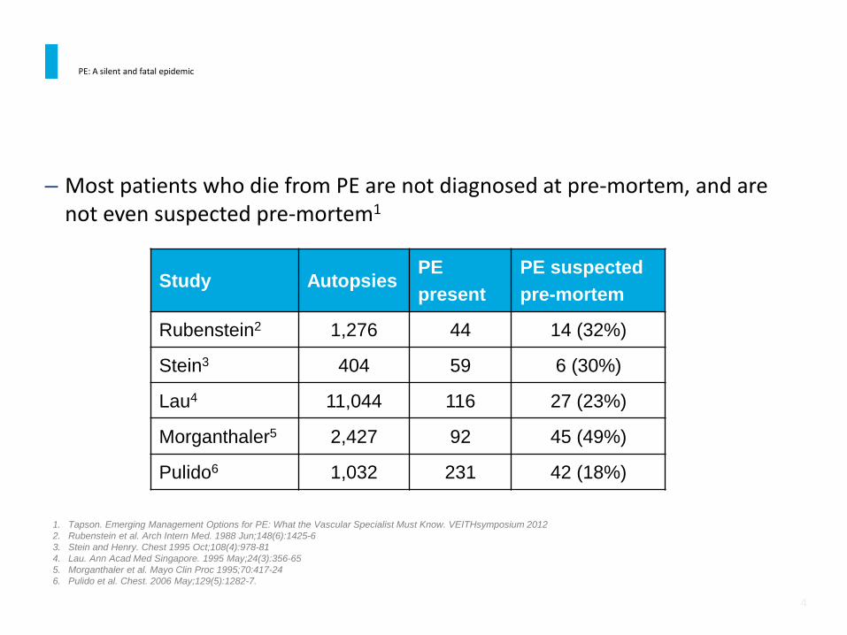
If not treated, there is 30 percent mortality with pulmonary embolism, usually within the first few hours after the episode.1,2
Patients with massive PE have a > 50% in hospital mortality rate.3
Patients with submassive PE have a 25% in hospital rate of death or significant clinical deterioration.4
Impact of PE
5
1. Horlander K et al. Pulmonary embolism mortality in the United States, 1979-1998: an analysis using multiple-cause mortality data. Arch Intern Med 2003; 163(14):1711.
2. Carson J et al. The clinical course of pulmonary embolism. N Engl J Med 1992; 326(19): 1240-1245.
3. Stulz P et al. Decision making in the surgical treatment of massive pulmonary embolism. Eur J Cardiothorac Surg 1994;8(4): 188-93.
4. Konstantinides S et al. Heparin plus alteplase compared with heparin alone in patients with submassive pulmonary embolism. N Engl J Med 2002; 347: 1143-1150
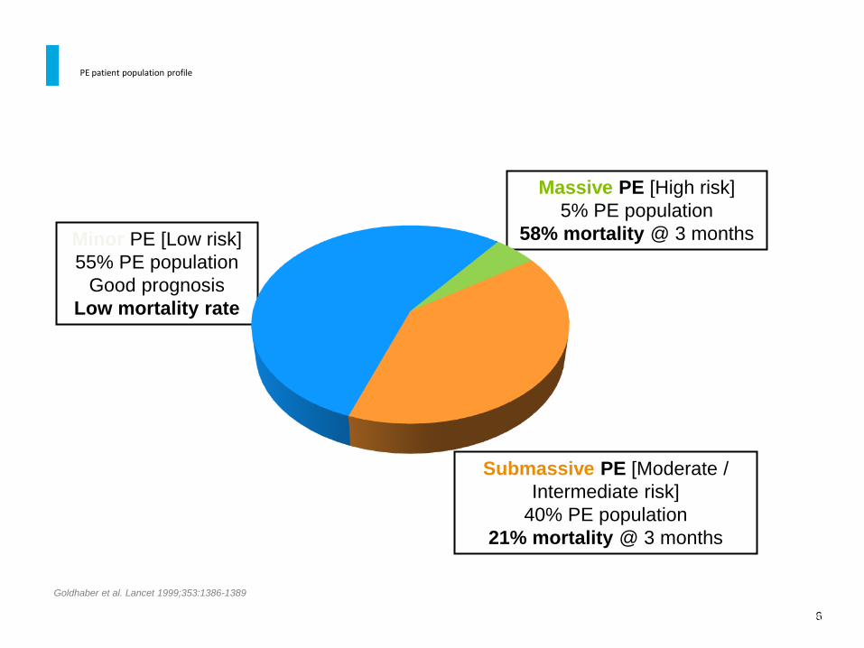
PE patient population profile
6 Goldhaber SZ, Visani L, De Rosa M, et al. for ICOPER. Acute pulmonary embolism; clinical outcomes in the International Cooperative Pulmonary Embolism
Registry. Lancet 1999;353:1386-1389
Massive PE [High risk]
5% PE population
58% mortality @ 3 months
Submassive PE [Moderate /
Intermediate risk]
40% PE population
21% mortality @ 3 months
Minor PE [Low risk]
55% PE population
Good prognosis
Low mortality rate
Goldhaber et al. Lancet 1999;353:1386-1389
PE risk stratification
7
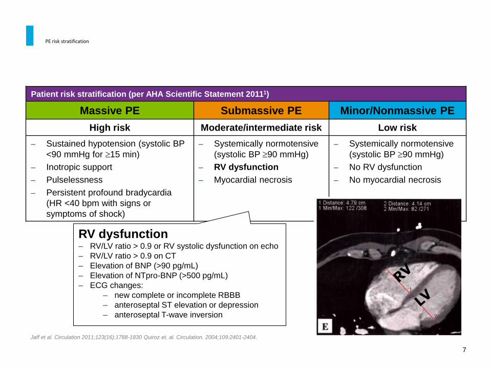
Patient risk stratification (per AHA Scientific Statement 20111)
Massive PE Submassive PE Minor/Nonmassive PE
High risk Moderate/intermediate risk Low risk
– Sustained hypotension (systolic BP
<90 mmHg for 15 min)
– Inotropic support
– Pulselessness
– Persistent profound bradycardia
(HR <40 bpm with signs or
symptoms of shock)
– Systemically normotensive
(systolic BP 90 mmHg)
– RV dysfunction
– Myocardial necrosis
– Systemically normotensive
(systolic BP 90 mmHg)
– No RV dysfunction
– No myocardial necrosis
RV dysfunction – RV/LV ratio > 0.9 or RV systolic dysfunction on echo
– RV/LV ratio > 0.9 on CT
– Elevation of BNP (>90 pg/mL)
– Elevation of NTpro-BNP (>500 pg/mL)
– ECG changes:
– new complete or incomplete RBBB
– anteroseptal ST elevation or depression
– anteroseptal T-wave inversion
Jaff et al. Circulation 2011;123(16):1788-1830.
Jaff et al. Circulation 2011;123(16):1788-1830 Quiroz et. al. Circulation. 2004;109:2401-2404.
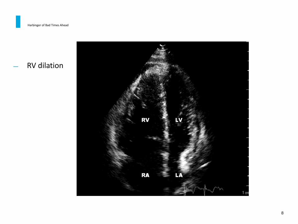
RV dilation
Harbinger of Bad Times Ahead
8

Echocardiographic RV/LV ratio ≥ 0.9 shown to be independent predictive factor of hospital mortality
Adverse outcomes associated with RVD
9
Registry of 1,416 patients
Mortality rate:
1.9% if RV/LV ratio < 0.9
6.6% if RV/LV ratio ≥ 0.9
Fremont et al. CHEST 2008;133:358-362

PE-related mortality risk increases with stepwise increase in RV/LV Ratio
Adverse outcomes associated with RVD
10
− Retrospective analysis of 120
patients with hemodynamically
stable PE based on chest CT
− PE-related mortality at 3 months:
17% if RV/LV ≥ 1.5
8% if 1.0 ≤ RV/LV < 1.5
0% if RV/LV < 1.0
Van der Meer et al. Radiology 2005; 235:798-803.
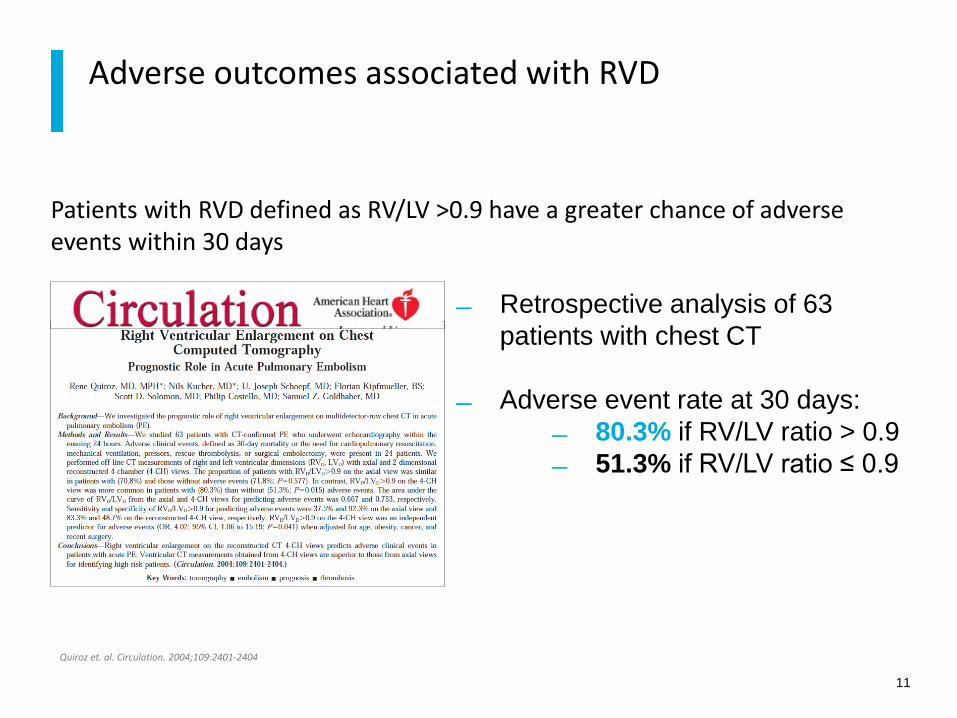
Patients with RVD defined as RV/LV >0.9 have a greater chance of adverse events within 30 days
Adverse outcomes associated with RVD
11
Retrospective analysis of 63
patients with chest CT
Adverse event rate at 30 days:
80.3% if RV/LV ratio > 0.9
51.3% if RV/LV ratio ≤ 0.9
Quiroz et. al. Circulation. 2004;109:2401-2404
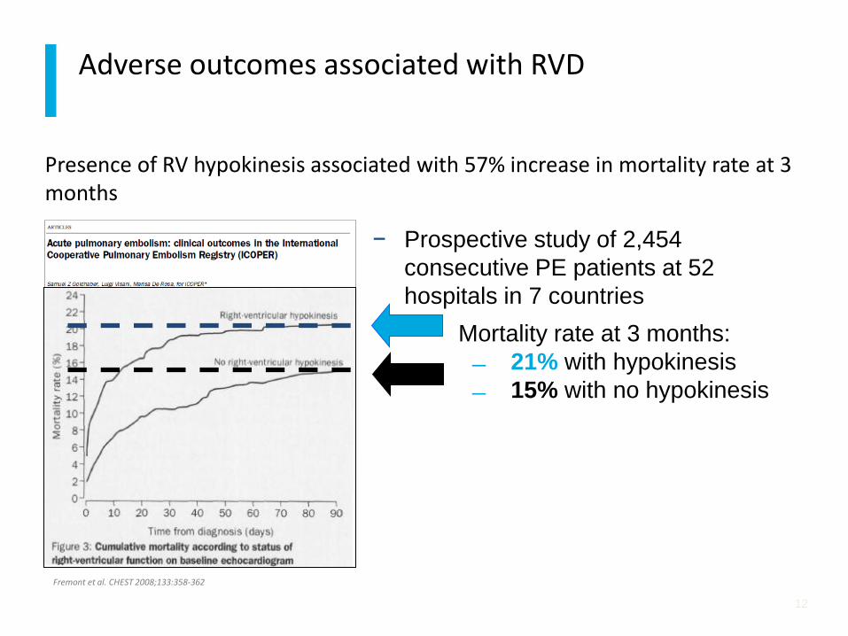
Presence of RV hypokinesis associated with 57% increase in mortality rate at 3 months
Adverse outcomes associated with RVD
12
Fremont et al. CHEST 2008;133:358-362
− Prospective study of 2,454
consecutive PE patients at 52
hospitals in 7 countries
Mortality rate at 3 months:
21% with hypokinesis
15% with no hypokinesis

PE patients with RVD unresolved exhibit 4x increased incidence of mortality compared to those with RVD resolved at discharge
Adverse outcomes with unresolved RVD
13
− Retrospective analysis of 301
patients with first episode PE
with mean f/u at 3.1 years
− Mortality rate at f/u:
10.2% if RVD unresolved
at d/c
2.3% if RVD resolved at
d/c
Grifoni et al. Arch Intern Med 2006; 166:2151-2156

Grifoni et al. Association of Persistent Right Ventricular Dysfunction at Hospital Discharge After Acute Pulmonary Embolism with Recurrent Thromboembolic Events. Arch Intern Med 2006; 166:2151-2156
PE patients with RVD unresolved exhibit 8x increased incidence of recurrent VTE compared to those with RVD resolved at discharge
Adverse outcomes with unresolved RVD
14
Incidence of VTE at 4 years: 0.4 if RVD unresolved 0.05 if RVD resolved
Retrospective analysis of 301 patients with
first episode PE with mean f/u at 3.1 years
ANTICOAGULATION (AC) – HEPARIN
– AC therapy prevents further clot growth
– Studies1-3 found:
– LMWH as effective as UFH in reducing recurrent PE
– LMWH carries reduced bleeding risk compared to UFH
STANDARD OF CARE: usually UFH or LMWH, followed by oral warfarin
– However, AC therapy relies on endogenous t-PA to dissolve occluding clot4
– a process that typically occurs over several weeks or months
– endogenous fibrinolysis may often be incomplete at the end
Standard PE therapy
15
1. Simonneau et al. N Engl J Med 1997;337:657-662.
2. Buller et al. N Engl J Med 2003;349:1695-17023.
3. Meyer et al. Thromb Heamost 1995;74:1432-1435
4. Arcasoy et al. Clin Chest Med 24 (2003) 73– 91.
IV thrombolysis with t-PA
16
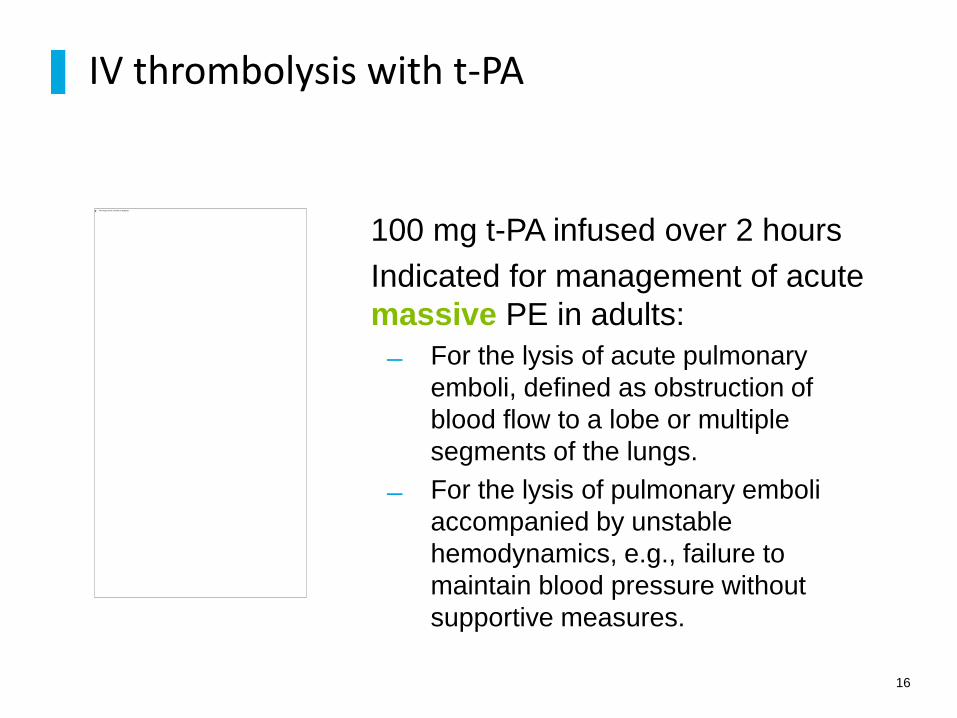
100 mg t-PA infused over 2 hours
Indicated for management of acute
massive PE in adults:
For the lysis of acute pulmonary
emboli, defined as obstruction of
blood flow to a lobe or multiple
segments of the lungs.
For the lysis of pulmonary emboli
accompanied by unstable
hemodynamics, e.g., failure to
maintain blood pressure without
supportive measures.

In randomized trials, systemic thrombolysis for PE is associated with a 13% risk of major bleeding and 1.8% risk of intracranial bleed.1
In clinical practice, these complications rise to 20% and 3%, respectively.2
In clinical practice, systemic thrombolysis is NOT given to up to 2/3 of patients who may qualify based on the PE itself.3
Rationale for A New Therapy
17
1Eur Heart J 2008: 29:2276-2315 2Am J Cardiol. 2006;97:127-9
3Circulation 2006;113:577-82
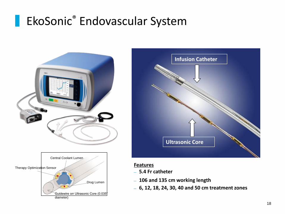
EkoSonic® Endovascular System
18
Features 5.4 Fr catheter
106 and 135 cm working length
6, 12, 18, 24, 30, 40 and 50 cm treatment zones
Infusion Catheter
Ultrasonic Core
Central Coolant Lumen
Therapy Optimization Sensor
Drug Lumen
Guidewire orr Ultrasonic Core (0.035”
diameter)
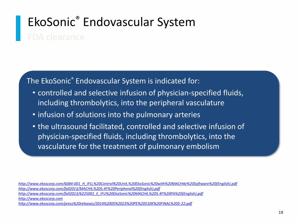
EkoSonic® Endovascular System FDA clearance
19
The EkoSonic® Endovascular System is indicated for:
• controlled and selective infusion of physician-specified fluids, including thrombolytics, into the peripheral vasculature
• infusion of solutions into the pulmonary arteries
• the ultrasound facilitated, controlled and selective infusion of physician-specified fluids, including thrombolytics, into the vasculature for the treatment of pulmonary embolism
http://www.ekoscorp.com/6084-001_H_IFU,%20Control%20Unit,%20EkoSonic%20with%20MACH4e%20Software%20(English).pdf http://www.ekoscorp.com/fall2013/MACH4,%205.4F%20Peripheral%20(English).pdf http://www.ekoscorp.com/fall2013/6225001_E_IFU%20EkoSonic%20MACH4,%205.4F%20PA%20(English).pdf http://www.ekoscorp.com http://www.ekoscorp.com/press%20releases/2014%2005%2023%20PE%20510K%20FINAL%205-22.pdf

Acoustic Pulse Thrombolysis™
Mechanism of action
20
Braatan et al. Thrmob Haemost 1997;78:1063-8. Francis et al. Ultrasound in Medicine and Biology, 1995;21(5):419-24. Soltani et al. Physics in Medicine and Biology, 2008; 53:6837-47.
Fibrin Separation Non-cavitational ultrasound separates fibrin
without fragmentation of emboli
Active Drug Delivery Drug is actively driven into clot by
“Acoustic Streaming”
Fibrin without Ultrasound
Fibrin With Ultrasound Acoustic streaming drives lytic into clot
EKOS® Acoustic Pulse Thrombolysis™ is a minimally invasive system for dissolving thrombus.

EkoSonic® Endovascular System Mechanism of action
21
WITH ULTRASOUND ENERGY
WITHOUT ULTRASOUND ENERGY
How ultrasonic energy unlocks the clot
Ultrasonic energy causes fibrin strands to thin, exposing plasminogen receptor sites and fibrin strands to loosen
Thrombus permeability and lytic penetration are dramatically increased
Ultrasound pressure waves force lytic agent deep into the clot and keep it there
ULTRASOUND ENERGY & THROMBOLYTIC
Braatan et al. Thrmob Haemost 1997;78:1063-8. Francis et al. Ultrasound in Medicine and Biology, 1995;21(5):419-24. Soltani et al. Physics in Medicine and Biology, 2008; 53:6837-47.
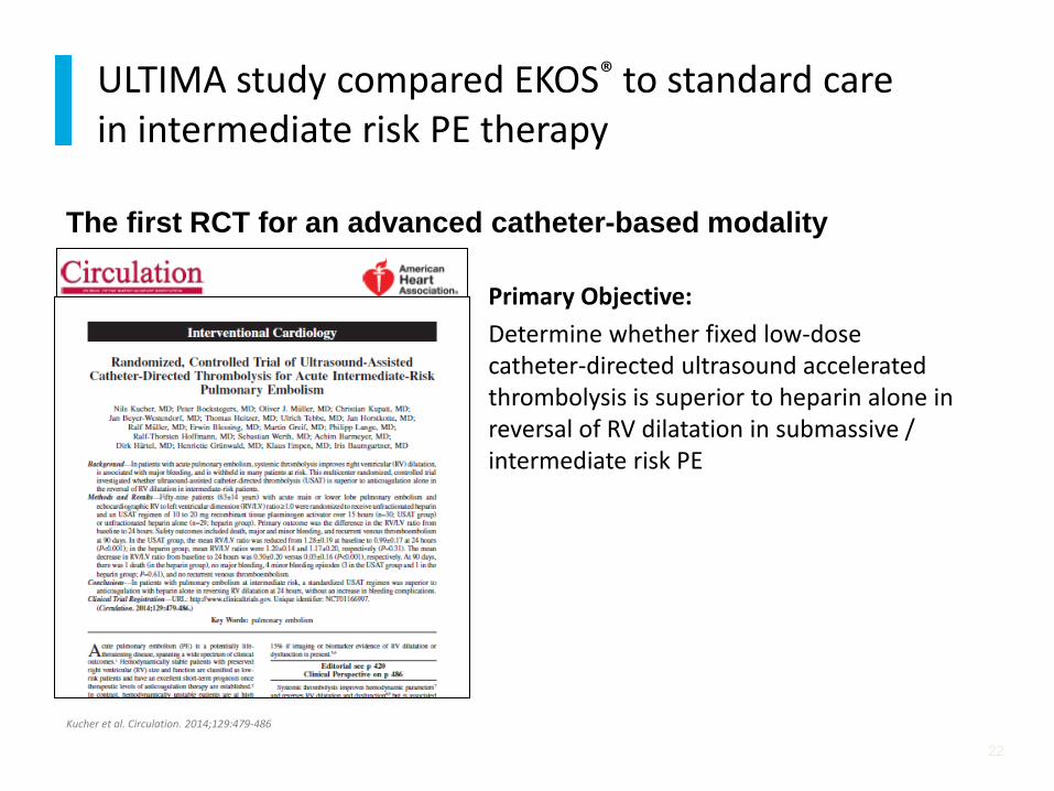
Primary Objective:
Determine whether fixed low-dose catheter-directed ultrasound accelerated thrombolysis is superior to heparin alone in reversal of RV dilatation in submassive / intermediate risk PE
ULTIMA study compared EKOS® to standard care in intermediate risk PE therapy
22
Kucher et al. Circulation. 2014;129:479-486
The first RCT for an advanced catheter-based modality
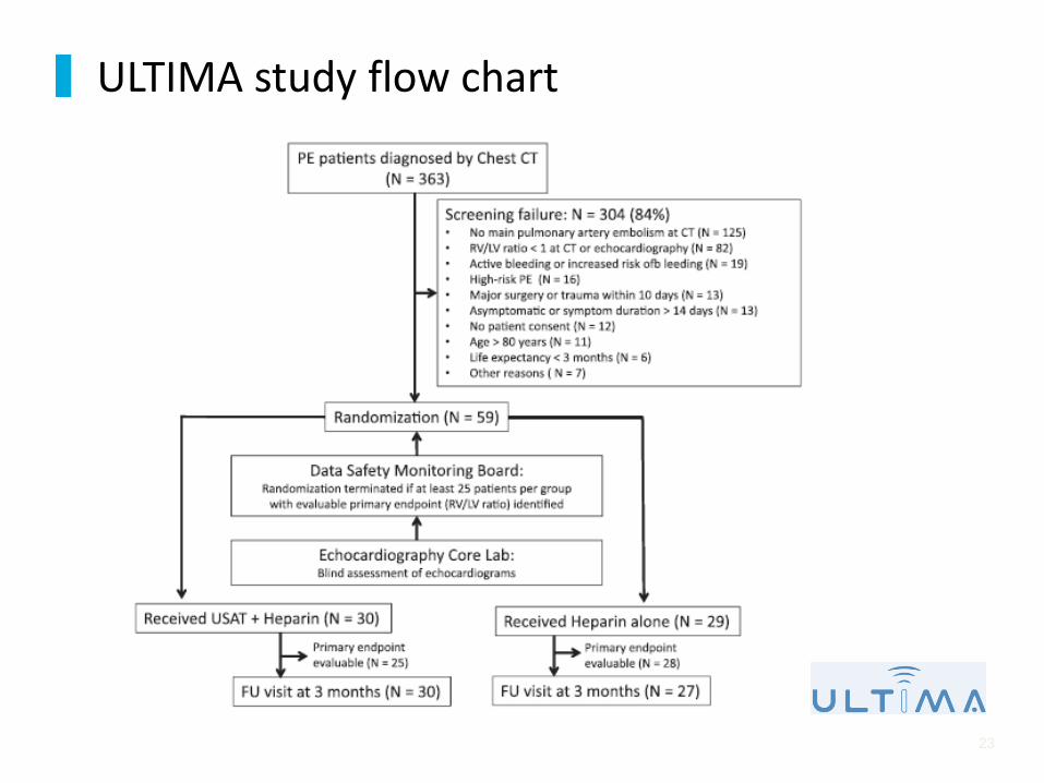
ULTIMA study flow chart
23
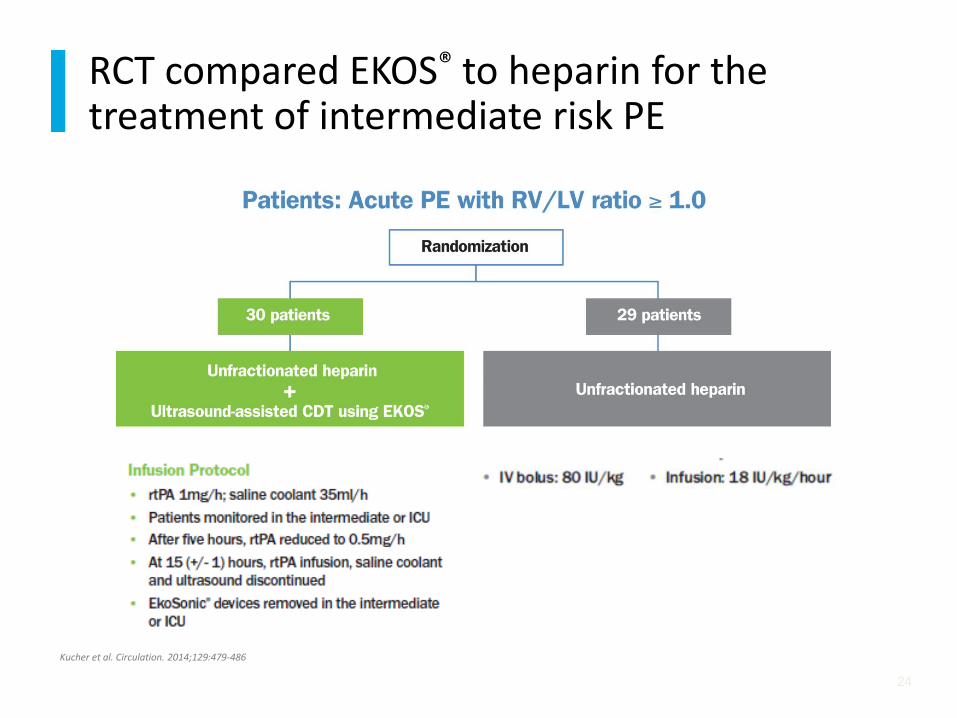
RCT compared EKOS® to heparin for the treatment of intermediate risk PE
24
Kucher et al. Circulation. 2014;129:479-486
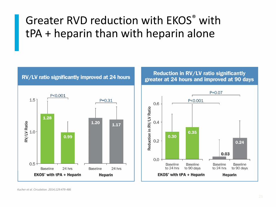
Greater RVD reduction with EKOS® with tPA + heparin than with heparin alone
25
Kucher et al. Circulation. 2014;129:479-486
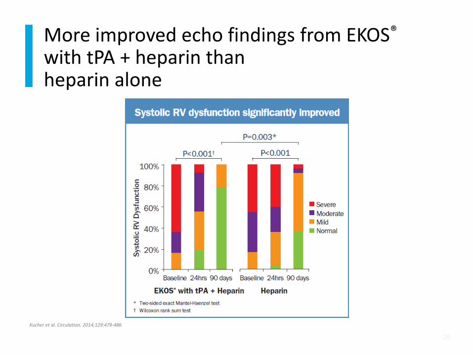
More improved echo findings from EKOS® with tPA + heparin than heparin alone
26
Kucher et al. Circulation. 2014;129:479-486

No statistical difference in safety outcomes with EKOS® with tPA + heparin than heparin alone
27
Kucher et al. Circulation. 2014;129:479-486

CONCLUSION
ULTIMA confirmed that a fixed-dose, ultrasound-assisted catheter-directed thrombolysis EKOS® regimen was superior to anticoagulation alone in improving RV dysfunction at 24 hours without an increase in bleeding
complications.
ULTIMA study
28
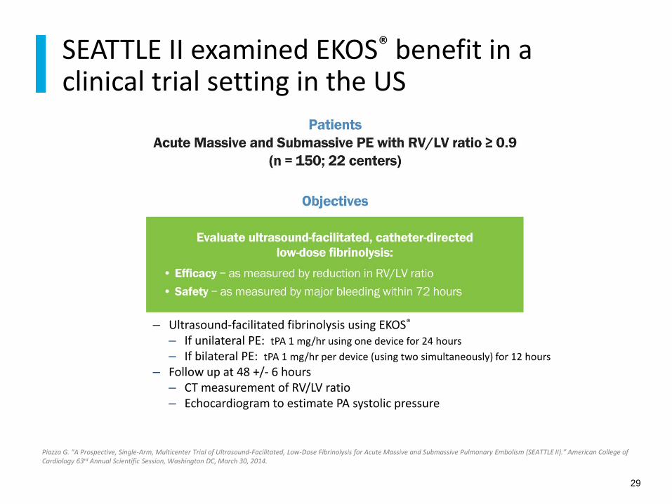
– Ultrasound-facilitated fibrinolysis using EKOS®
– If unilateral PE: tPA 1 mg/hr using one device for 24 hours
– If bilateral PE: tPA 1 mg/hr per device (using two simultaneously) for 12 hours
– Follow up at 48 +/- 6 hours – CT measurement of RV/LV ratio – Echocardiogram to estimate PA systolic pressure
SEATTLE II examined EKOS® benefit in a clinical trial setting in the US
29
Piazza G. “A Prospective, Single-Arm, Multicenter Trial of Ultrasound-Facilitated, Low-Dose Fibrinolysis for Acute Massive and Submassive Pulmonary Embolism (SEATTLE II).” American College of Cardiology 63rd Annual Scientific Session, Washington DC, March 30, 2014.
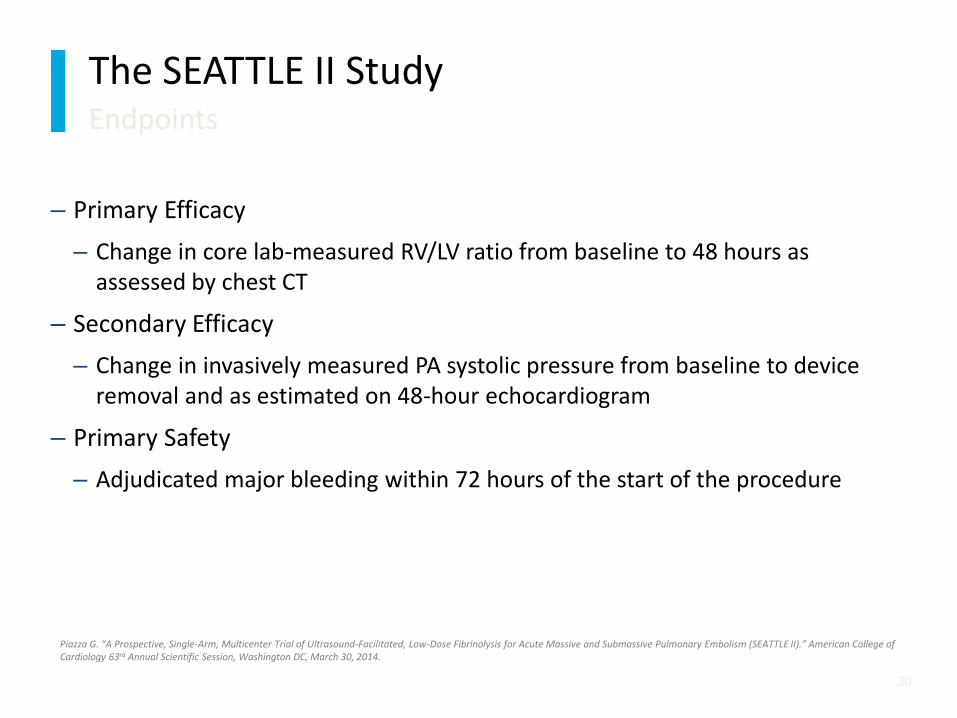
– Primary Efficacy
– Change in core lab-measured RV/LV ratio from baseline to 48 hours as assessed by chest CT
– Secondary Efficacy
– Change in invasively measured PA systolic pressure from baseline to device removal and as estimated on 48-hour echocardiogram
– Primary Safety
– Adjudicated major bleeding within 72 hours of the start of the procedure
The SEATTLE II Study Endpoints
30
Piazza G. “A Prospective, Single-Arm, Multicenter Trial of Ultrasound-Facilitated, Low-Dose Fibrinolysis for Acute Massive and Submassive Pulmonary Embolism (SEATTLE II).” American College of Cardiology 63rd Annual Scientific Session, Washington DC, March 30, 2014.
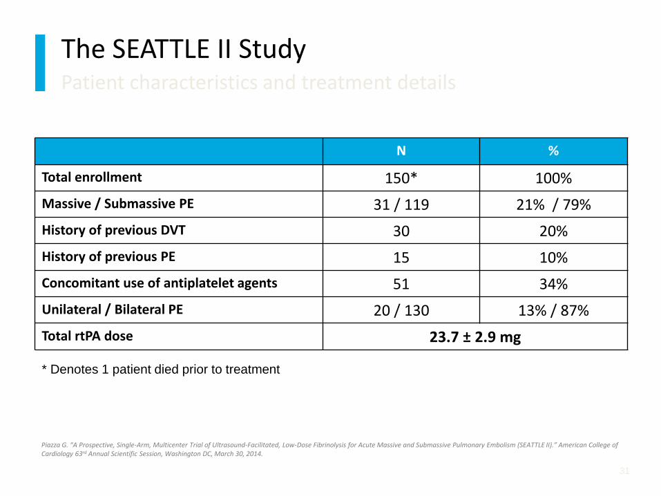
The SEATTLE II Study
Patient characteristics and treatment details
31
N %
Total enrollment 150* 100%
Massive / Submassive PE 31 / 119 21% / 79%
History of previous DVT 30 20%
History of previous PE 15 10%
Concomitant use of antiplatelet agents 51 34%
Unilateral / Bilateral PE 20 / 130 13% / 87%
Total rtPA dose 23.7 ± 2.9 mg
* Denotes 1 patient died prior to treatment
Piazza G. “A Prospective, Single-Arm, Multicenter Trial of Ultrasound-Facilitated, Low-Dose Fibrinolysis for Acute Massive and Submassive Pulmonary Embolism (SEATTLE II).” American College of Cardiology 63rd Annual Scientific Session, Washington DC, March 30, 2014.

Reduced RV/LV ratio and Modified Miller Score at 48 hours post-EKOS®
32
Piazza G. “A Prospective, Single-Arm, Multicenter Trial of Ultrasound-Facilitated, Low-Dose Fibrinolysis for Acute Massive and Submassive Pulmonary Embolism (SEATTLE II).” American College of Cardiology 63rd Annual Scientific Session, Washington DC, March 30, 2014.

Reduced pulmonary artery pressure immediately post-procedure
33
Piazza G. “A Prospective, Single-Arm, Multicenter Trial of Ultrasound-Facilitated, Low-Dose Fibrinolysis for Acute Massive and Submassive Pulmonary Embolism (SEATTLE II).” American College of Cardiology 63rd Annual Scientific Session, Washington DC, March 30, 2014.
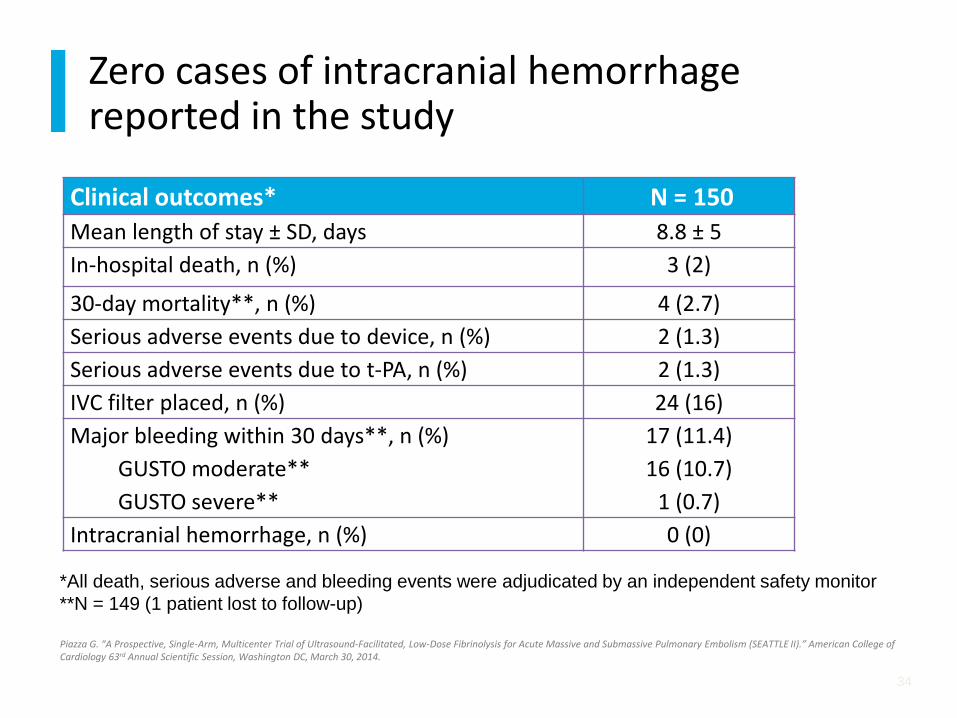
Zero cases of intracranial hemorrhage reported in the study
34
Clinical outcomes* N = 150
Mean length of stay ± SD, days 8.8 ± 5
In-hospital death, n (%) 3 (2)
30-day mortality**, n (%) 4 (2.7)
Serious adverse events due to device, n (%) 2 (1.3)
Serious adverse events due to t-PA, n (%) 2 (1.3)
IVC filter placed, n (%) 24 (16)
Major bleeding within 30 days**, n (%)
GUSTO moderate**
GUSTO severe**
17 (11.4)
16 (10.7)
1 (0.7)
Intracranial hemorrhage, n (%) 0 (0)
*All death, serious adverse and bleeding events were adjudicated by an independent safety monitor
**N = 149 (1 patient lost to follow-up)
Piazza G. “A Prospective, Single-Arm, Multicenter Trial of Ultrasound-Facilitated, Low-Dose Fibrinolysis for Acute Massive and Submassive Pulmonary Embolism (SEATTLE II).” American College of Cardiology 63rd Annual Scientific Session, Washington DC, March 30, 2014.
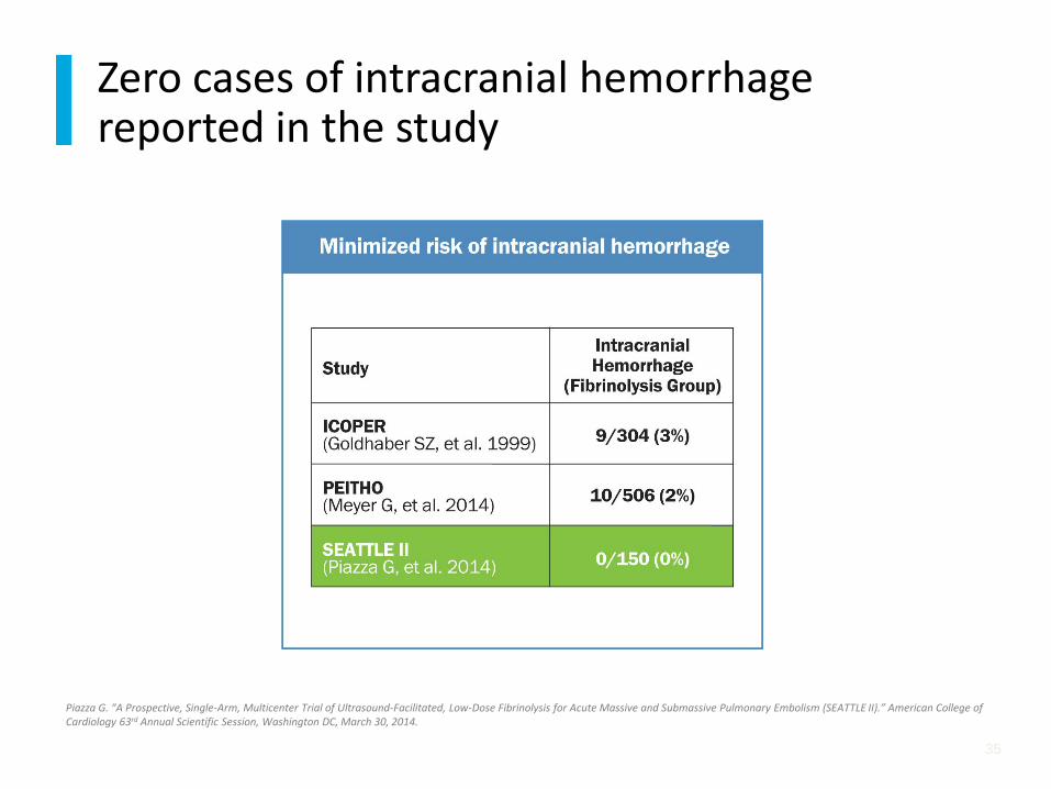
Zero cases of intracranial hemorrhage reported in the study
35
Piazza G. “A Prospective, Single-Arm, Multicenter Trial of Ultrasound-Facilitated, Low-Dose Fibrinolysis for Acute Massive and Submassive Pulmonary Embolism (SEATTLE II).” American College of Cardiology 63rd Annual Scientific Session, Washington DC, March 30, 2014.
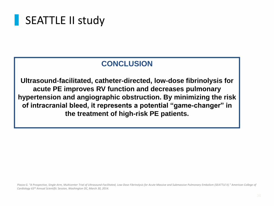
SEATTLE II study
36
CONCLUSION
Ultrasound-facilitated, catheter-directed, low-dose fibrinolysis for
acute PE improves RV function and decreases pulmonary
hypertension and angiographic obstruction. By minimizing the risk
of intracranial bleed, it represents a potential “game-changer” in
the treatment of high-risk PE patients.
Piazza G. “A Prospective, Single-Arm, Multicenter Trial of Ultrasound-Facilitated, Low-Dose Fibrinolysis for Acute Massive and Submassive Pulmonary Embolism (SEATTLE II).” American College of Cardiology 63rd Annual Scientific Session, Washington DC, March 30, 2014.
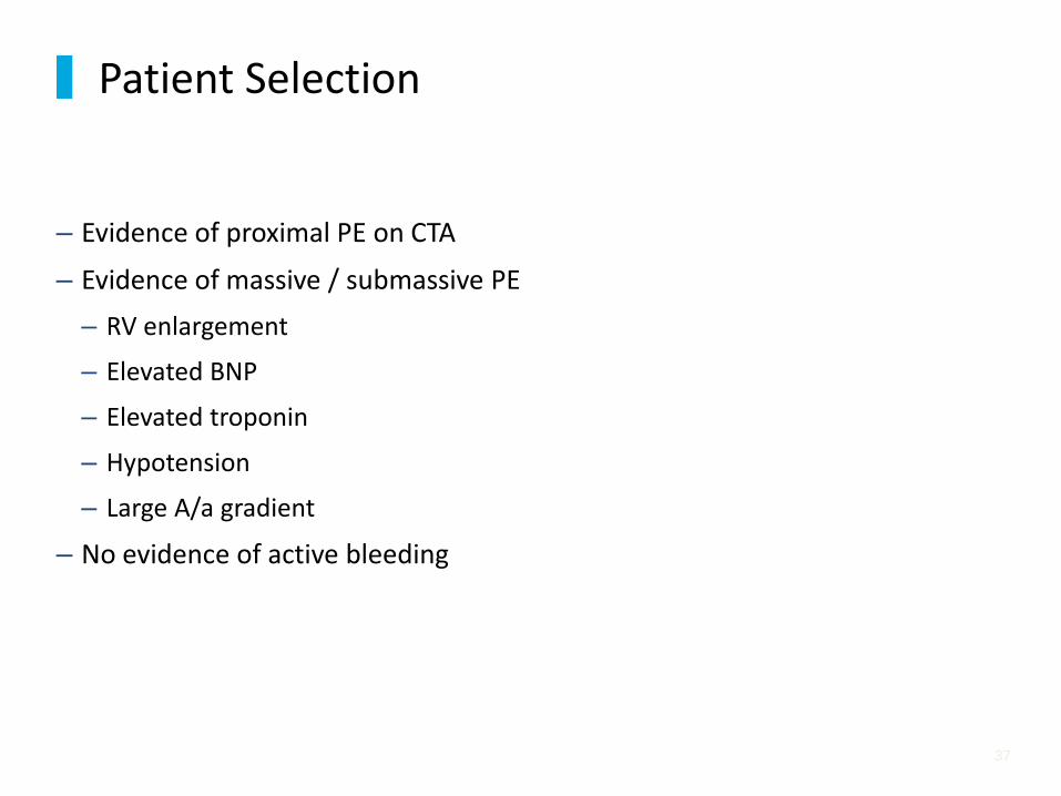
– Evidence of proximal PE on CTA
– Evidence of massive / submassive PE
– RV enlargement
– Elevated BNP
– Elevated troponin
– Hypotension
– Large A/a gradient
– No evidence of active bleeding
Patient Selection
37
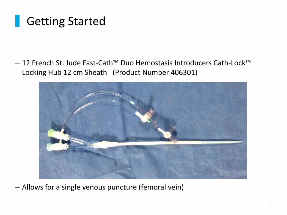
– 12 French St. Jude Fast-Cath™ Duo Hemostasis Introducers Cath-Lock™ Locking Hub 12 cm Sheath (Product Number 406301)
– Allows for a single venous puncture (femoral vein)
Getting Started
38

Getting Started
39

– Use an angled pigtail and exchange-length 0.035 J-wire to selectively engage each pulmonary artery.
– Then exchange over the J-wire for the EKOS® catheter.
Getting Started
40
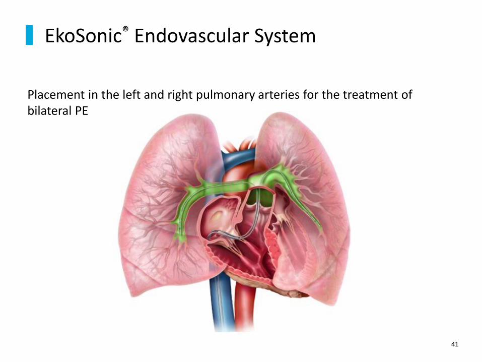
Placement in the left and right pulmonary arteries for the treatment of bilateral PE
EkoSonic® Endovascular System
41
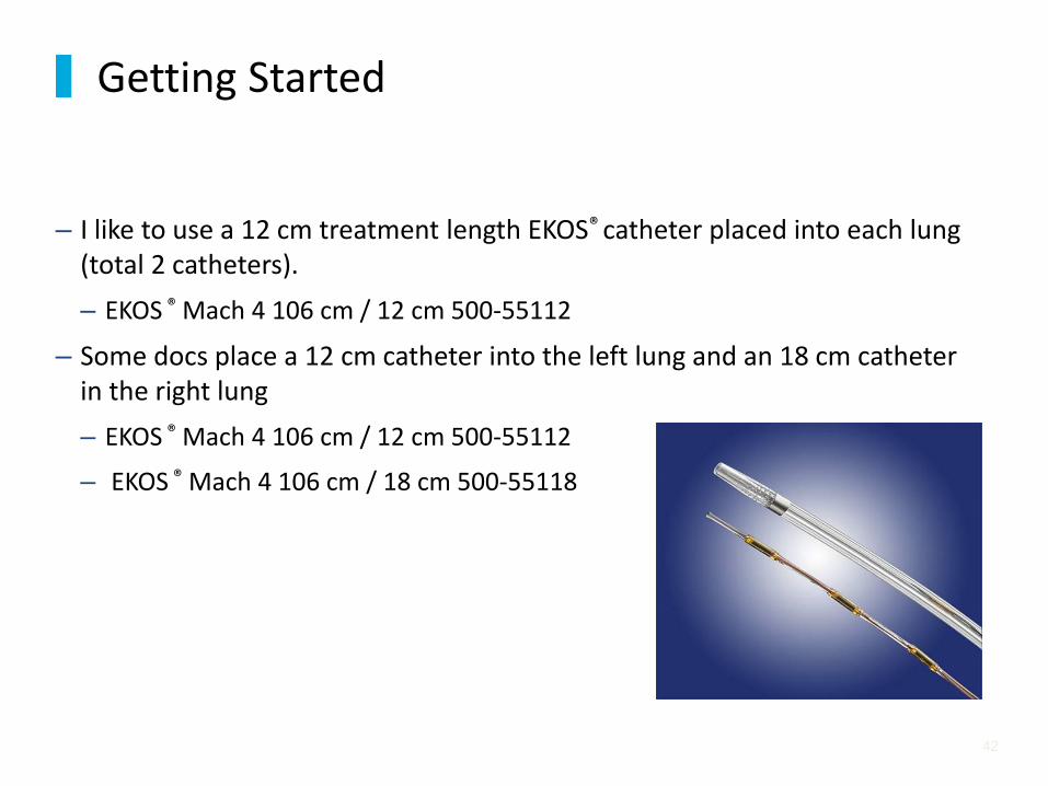
– I like to use a 12 cm treatment length EKOS® catheter placed into each lung (total 2 catheters).
– EKOS ® Mach 4 106 cm / 12 cm 500-55112
– Some docs place a 12 cm catheter into the left lung and an 18 cm catheter in the right lung
– EKOS ® Mach 4 106 cm / 12 cm 500-55112
– EKOS ® Mach 4 106 cm / 18 cm 500-55118
Getting Started
42
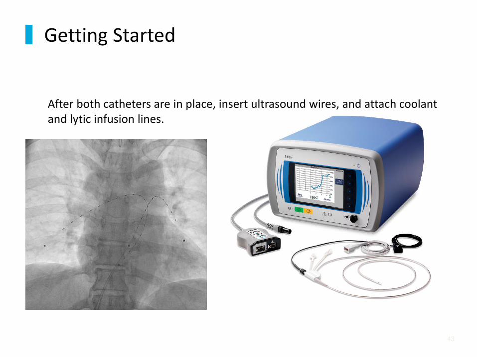
After both catheters are in place, insert ultrasound wires, and attach coolant and lytic infusion lines.
Getting Started
43
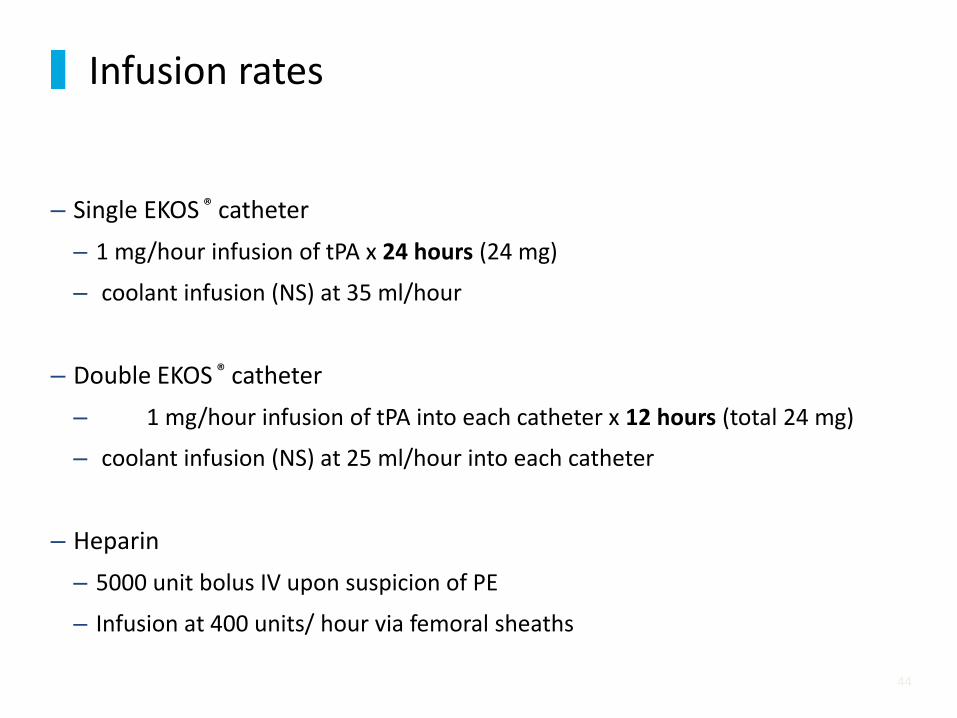
– Single EKOS ® catheter
– 1 mg/hour infusion of tPA x 24 hours (24 mg)
– coolant infusion (NS) at 35 ml/hour
– Double EKOS ® catheter
– 1 mg/hour infusion of tPA into each catheter x 12 hours (total 24 mg)
– coolant infusion (NS) at 25 ml/hour into each catheter
– Heparin
– 5000 unit bolus IV upon suspicion of PE
– Infusion at 400 units/ hour via femoral sheaths
Infusion rates
44
– Remove sheaths at bedside
– Consider repeat echo to look at RV size
– After 4 hours to allow for hemostasis - begin Xarelto 15 mg po twice a day x 3 weeks, then change to 20 mg daily
– Usually can d/c on Day 3
Post Infusion
45

68 year old man who is two days post-CABG suddenly develops chest pain and shortness of breath.
ECG shows new RBBB and there was concern for acute graft closure.
Cath lab notified for “Code STEMI” and stat echo obtained.
Case Study
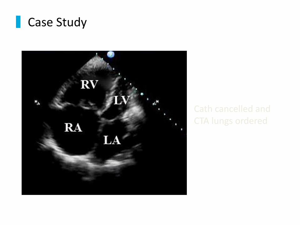
Cath cancelled and CTA lungs ordered
Case Study
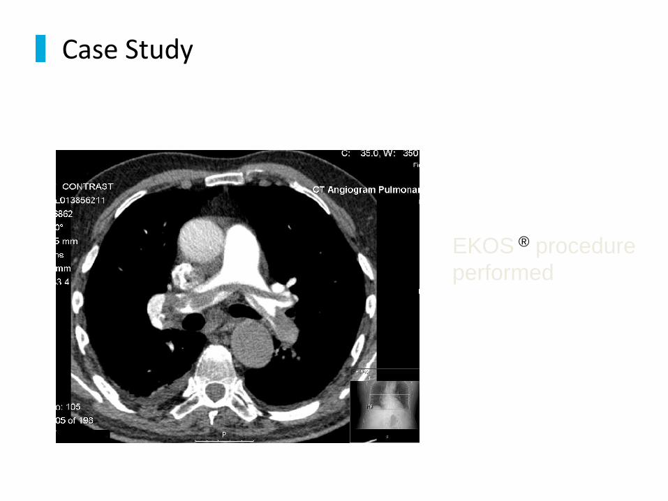
EKOS ® procedure
performed
Case Study
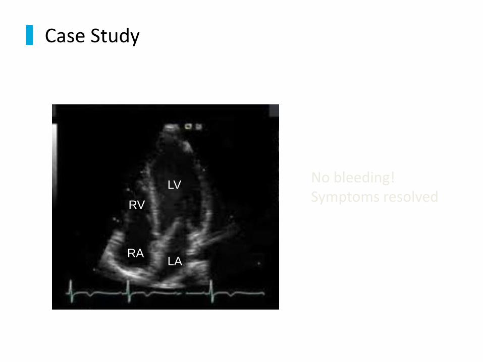
No bleeding! Symptoms resolved
Case Study
LV
RA LA
RV
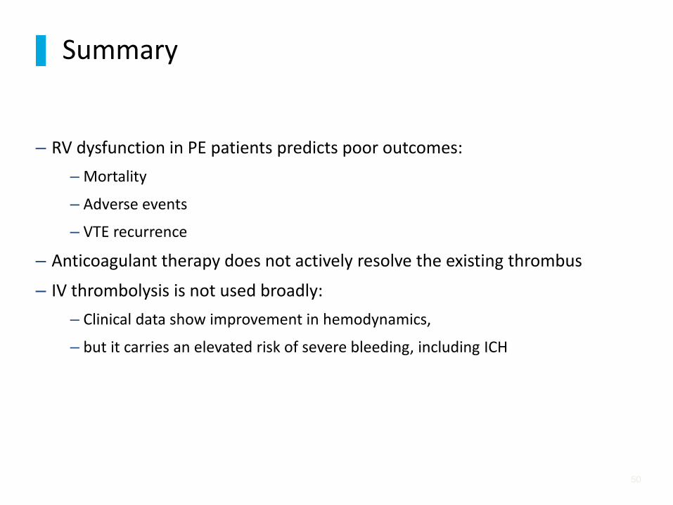
– RV dysfunction in PE patients predicts poor outcomes:
– Mortality
– Adverse events
– VTE recurrence
– Anticoagulant therapy does not actively resolve the existing thrombus
– IV thrombolysis is not used broadly:
– Clinical data show improvement in hemodynamics,
– but it carries an elevated risk of severe bleeding, including ICH
Summary
50
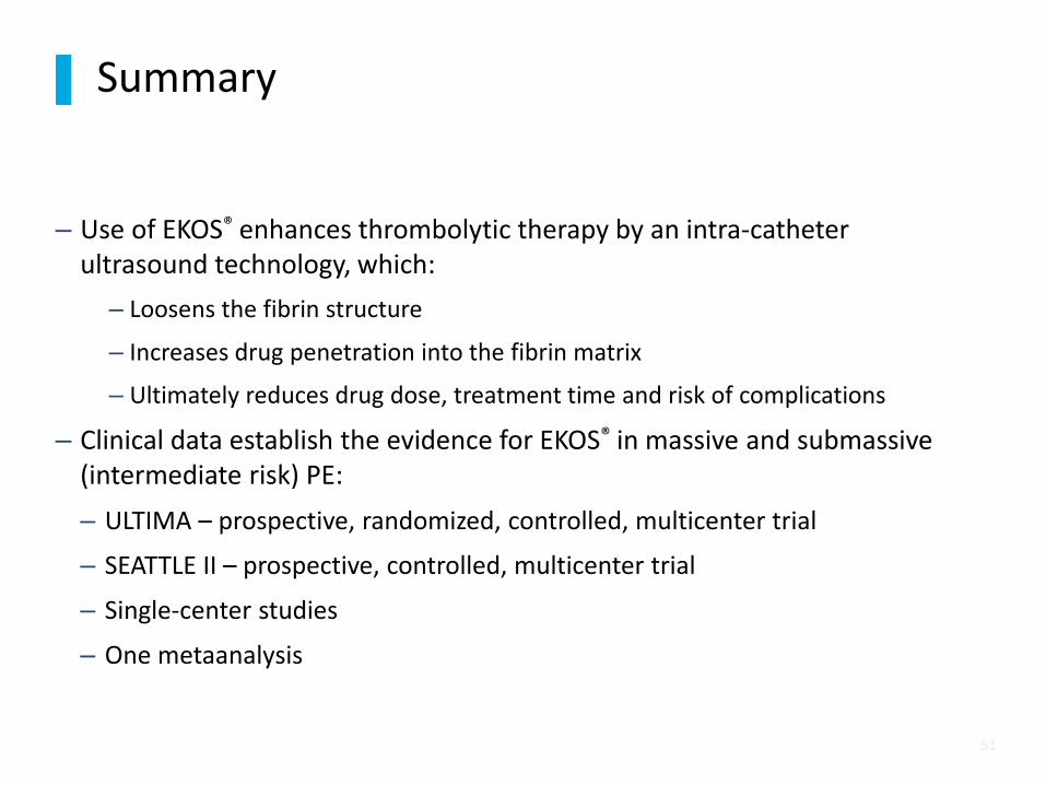
– Use of EKOS® enhances thrombolytic therapy by an intra-catheter ultrasound technology, which:
– Loosens the fibrin structure
– Increases drug penetration into the fibrin matrix
– Ultimately reduces drug dose, treatment time and risk of complications
– Clinical data establish the evidence for EKOS® in massive and submassive (intermediate risk) PE:
– ULTIMA – prospective, randomized, controlled, multicenter trial
– SEATTLE II – prospective, controlled, multicenter trial
– Single-center studies
– One metaanalysis
Summary
51
– Consistent EKOS® results among the various published studies:
– Restoration of hemodynamics as evidenced by a reduced RV/LV ratio and decreased PA pressure
– Resolution of pulmonary artery obstruction
– Favorable outcomes with low dose thrombolysis (20-24 mg tPA based on the clinical trials)
– No reports of intracranial hemorrhage in published clinical studies
Summary
52

53
Acoustic Pulse Thrombolysis™ system
For Pulmonary Embolus
The New “ Code Blue”
EKOS® Acoustic Pulse Thrombolysis™ is a minimally invasive system for dissolving thrombus
Related Documents