1 Novel Techniques and Their Applications to Agricultural Biotechnology, Health Foods and Medical Biotechnology I.C. Baianu*, P.R. Lozano, V.I. Prisecaru and H.C. Lin University of Illinois at Urbana, ACES College, email: [email protected] FSHN Dept., Agricultural & Food Chemistry NMR & NIR Microspectroscopy Facility, 286 Bevier Hall, 905 S. Goodwin Ave, Urbana, IL. 61801, USA ABSTRACT Selected applications of novel techniques in Agricultural Biotechnology, Health Food formulations and Medical Biotechnology are being reviewed with the aim of unraveling future developments and policy changes that are likely to open new markets for Biotechnology and prevent the shrinking or closing of existing ones. Amongst the selected novel techniques with applications in both Agricultural and Medical Biotechnology are: immobilized bacterial cells and enzymes, microencapsulation and liposome production, genetic manipulation of microorganisms, development of novel vaccines from plants, epigenomics of mammalian cells and organisms, and biocomputational tools for molecular modeling related to disease and Bioinformatics. Both fundamental and applied aspects of the emerging new techniques are being discussed in relation to their anticipated, marked impact on future markets and present policy changes that are needed for success in either Agricultural or Medical Biotechnology. The novel techniques are illustrated with figures that attempt to convey-- albeit in a simplified manner-- the most important features of representative and powerful tools that are currently being developed for both immediate and long- term applications in Agriculture, Health Food formulation and production, pharmaceuticals and Medicine. The research aspects are naturally emphasized in our review as they are key to further developments in Biotechnology; however, the course adopted for the implementation of biotechnological applications, and the policies associated with biotechnological applications in the market place, are clearly the determining factors for future Biotechnology successes in the world markets, be they pharmaceutical, medical or agricultural. * Corresponding Author.

Welcome message from author
This document is posted to help you gain knowledge. Please leave a comment to let me know what you think about it! Share it to your friends and learn new things together.
Transcript

1
Novel Techniques and Their Applications to Agricultural Biotechnology,Health Foods and Medical Biotechnology
I.C. Baianu*, P.R. Lozano, V.I. Prisecaru and H.C. LinUniversity of Illinois at Urbana, ACES College, email: [email protected] Dept., Agricultural & Food Chemistry NMR & NIR Microspectroscopy Facility,286 Bevier Hall, 905 S. Goodwin Ave, Urbana, IL. 61801, USA
ABSTRACT
Selected applications of novel techniques in Agricultural Biotechnology, Health Food formulations
and Medical Biotechnology are being reviewed with the aim of unraveling future developments
and policy changes that are likely to open new markets for Biotechnology and prevent the
shrinking or closing of existing ones. Amongst the selected novel techniques with applications in
both Agricultural and Medical Biotechnology are: immobilized bacterial cells and enzymes,
microencapsulation and liposome production, genetic manipulation of microorganisms,
development of novel vaccines from plants, epigenomics of mammalian cells and organisms, and
biocomputational tools for molecular modeling related to disease and Bioinformatics. Both
fundamental and applied aspects of the emerging new techniques are being discussed in relation to
their anticipated, marked impact on future markets and present policy changes that are needed for
success in either Agricultural or Medical Biotechnology. The novel techniques are illustrated with
figures that attempt to convey-- albeit in a simplified manner-- the most important features of
representative and powerful tools that are currently being developed for both immediate and long-
term applications in Agriculture, Health Food formulation and production, pharmaceuticals and
Medicine. The research aspects are naturally emphasized in our review as they are key to further
developments in Biotechnology; however, the course adopted for the implementation of
biotechnological applications, and the policies associated with biotechnological applications in the
market place, are clearly the determining factors for future Biotechnology successes in the world
markets, be they pharmaceutical, medical or agricultural.
* Corresponding Author.

2
KEY WORDS:
Applications in Agricultural and Medical Biotechnology;
immobilized bacterial cells and enzymes;
microencapsulation and liposome production;
genetic manipulation of microorganisms;
genetic therapy in medicine;
development of novel vaccines from plants;
epigenomics of mammalian cells and organisms;
biocomputational tools for molecular modeling related to disease;
Bioinformatics;
Suggested policy changes for future and immediate success of Biotechnology in the world
markets;
Health Food Applications;
Novel Vaccines from Plants;
Medical Applications of Lectins for diagnostics and treatments;
Nuclear Magnetic Resonance (NMR) and MRI/ MRM;
Fluorescence Correlation Spectroscopy and applications to Single Molecule Detection, Single
Virus Particle Detection;
Single virus particle, HIV, HPV, Hepatitis B and C, detection for early, successful treatments;
Ultra-sensitive, Selective Detection and Early Diagnostics of Cancers.

3
1. INTRODUCTION
Biotechnology has been traditionally thought to be associated with biomedical, and
especially, pharmaceutical applications. Recently, agricultural biotechnology promises to yield
even greater and wider gains through the enhancement of crop productivity or the use of
transgenic crops than those that medical biotechnology has already achieved by exploiting
combinations of cellular and molecular biology techniques. Novel biotechnology research is also
rapidly expanding with a view to the application of environment friendly bioprocesses for
chemical, pharmaceutical, and other areas, including food bioengineering and safety. The
tremendous growth of the two areas of modern biotechnology (i.e., agricultural and medical) may
have recently created the impression of technological isolation through specialization of both
agricultural and medical biotechnology. Such an impression-- if not checked from spreading--
might raise artificial barriers between these two important and closely related domains. Caused
mainly by over-specialization, an increasingly ‘narrow’ approach to biotechnology might affect
the evolution and sharing of existing biotechnological tools that were initially developed for
different types of applications. For example, the current adoption of certain genetically modified
foods and the administration of vaccines based on genetically modified organisms (GMO’ s) in
developing countries, EU and Japan raise distribution problems for which adequate solutions
have yet to be found. Potential loss of certain crop markets (such as corn) that may have included
‘GMO-mixed’ crops could be very substantial (>1 billion US $). Therefore, solutions to such
problems must, and can be, found both through crop growing policy changes and also by utilizing
novel, ultra-sensitive, as well as less expensive, techniques for GMO detection and crop quality
control.
Unfortunately, about one billion people in the world remain deficient in several vitamins and
minerals intake (Toenniessen et al 2003). Such undernourished populations live mostly in
developing countries that often have inhospitable climates which favor low efficiency in food
engineering and production, and also where the primary services for storage or administration of
GMO-based vaccines are minimal. The utilization -- in combination with agronomical
techniques-- of biotechnological tools that were originally produced for the medical area, may
help resolve such difficult problems. As an example, the production of vaccines from plants and
microorganism with subsequent addition in foods could reduce the cost of vaccination in such
areas. The modification of microorganisms for the bio-production of fuels (Linko 1985) is also
important, and would also be important in under-developed, or developing, countries that have a

4
large surplus of under-utilized complex carbohydrate sources that could be used for bio-
production of valuable fuels with existing technologies.
Our review focuses on novel techniques and tools employed in biotechnology that are being
developed for chemical, medical, health food production and agricultural applications worldwide.
However, no attempt or claim at extensive coverage of the subject is here being made.
2. IMMOBILIZED MICROBIAL CELLS
Immobilization of whole cells has been defined as the physical confinement or localization of
intact cells to a certain defined region of space with preservation of some (Karel et al 1985), or
most, catalytic activity. The increased stability under extreme conditions of pH and temperature,
as well as the re-use and applicability in continuous processing systems that enclose immobilized
cells instead of soluble enzymes make the cells a preferred, versatile tool in both food industry
and medicine. There are several different approaches to the classification of immobilized
biocatalysts, but the most frequently employed classification is based upon the method of
immobilization selected for a specific application. The selection of immobilization method
depends, therefore, upon the application, the nature of the microorganism being immobilized, as
well as the resources available (Witter 1996). Table 1 shows several, possible immobilization
methods that available for whole microbial cells. Most immobilization methods can be applied
either to whole cells or to enzymes. Some of the advantages of whole-cell immobilization in
comparison with enzyme immobilization are: the higher stability and enzyme activity,
multivariate enzyme applications, and the lower cost (Witter 1996). On the other hand,
disadvantages of using whole cell immobilization in comparison with enzyme immobilization are
linked to the increased diffusional barriers caused by the much larger sizes of cells in comparison
with enzymes.

5
FlocculationAdsorptionWeak bondsIonicCovalent
Binding
Strong bondsCross-linkingThermal GelationIonotropic GelationEntrapmentPolymerizationDialysis Culture
Physical retention
Membrane RetentionUltrafilters
Table 1. Immobilization Methods for Whole Microbial Cells.
(SOURCE : Witter 1996, Ch. 10 in : “ Physical Chemistry of Foods” Vol.2, Baianu et al Eds.
1996).
Adsorption is the least expensive and mildest immobilization method. It uses weak interaction
forces such as hydrogen bonds, hydrophobic interactions and Van der Waal forces to immobilize
cells or enzymes. However, the sensitivity of this interaction to pH makes the leakage of cells
immobilized by this technique quite common. Important applications of this technique are
related to the production of fructose and vinegar, and also waste water treatments.
Ionic binding uses the properties of negatively charged microbial cells to interact with
positively charged ion exchangers (Witter 1996). The results obtained with this technique are also
sensitive to extreme pH values, and the binding strength is greater in comparison with adsorption.
However, the mild conditions employed by this technique make it suitable for use for
immobilization of both enzymes and whole cells.
Covalent binding and cross-linking offer better strength than the previous techniques,
however, there is an encountered toxicity in the reagents (Chibata 1979; Kolot 1981; Babu and
Panda 1991) that are used to produce immobilization.
Entrapment techniques are, however, the most commonly encountered in the industry and
they are based on the formation of thermally reversible gels, ionotropic gels and polymerization.
2.1. Application of Cell Immobilization in the Food Industry and Human Nutrition
Entrapment of cells in a gel–like matrix by ionotropic gelation using alginates and K-
carrageenans is certainly the most useful method for industrial purposes. The alginates form a

6
gelatin matrix in the presence of polyvalent cations by binding the cation to guluronic acid units
(Witter 1996; Nedovic et al 2003) whereas ?- carrageenan solidifies in the presence of potassium
ions. The properties of the gel-like matrix allow the cell to remain viable and with its catalytic
ability for a long period of time. For example, an increment in the yeast concentration obtained
through immobilization techniques has helped the brewing industry to reduce fermentation
process times and the size of their storage facilities. Unfortunately, because of the high
concentration of diacetyl, and the low concentration of higher alcohols and esters, the flavor of
the fast fermented beer has been compromised. Even the amino acid profile has been altered
(Masschelein et al 1994). The main factor causing this uncommon imbalance is the insufficient
mass transfer in the older designs of fermentation reactors; thus, the use of new reactor designs
(Andries et al 1996; Nevodic et al 1996), and combining technologies (Inoue 1995) could
improve the quality of the products obtained through such fermentation reactions.
Nutraceuticals are defined as food components that have health benefits beyond traditional
nutritional value. Novel biotechnology tools like immobilization were also applied for the
isolation and incorporation of such food components in ordinary foods. The synthesis of
nutraceuticals was reported to be successful by empoloying immobilized lipases, such as those
from Candida antartica and Lactobacillus ruteri (Hill et al 1999, and Garcia 2001). The
introduction of conjugated linoleic acid (CLA) in dairy foods has been made possible through the
immobilization of lipases (Hill et al 1999).
2.2. Medical Applications of Molecular and Cellular Immobilization Techniques
There is a quite extensive list of immobilization technique applications in medicine.
A very important group of such applications is concerned with the regulation of equilibrium
between coagulation and dissolution of coagulated blood (fibrinolysis) through the use of
immobilized enzymes. The high probability of death caused by thrombosis (involving the
formation of clots in the blood vessels), has committed physicians to the use of fibrinolytic
therapy for the treatment of occlusions in those parts of the body where a surgical intervention
would be too risky. Amongst the most important enzymes that have been immobilized for use in
such therapy are Plasmin and Heparin (Wolf and Ransberger 1972). The use of biotechnology as
well as microscopic techniques has helped refine and greatly improve such therapeutical means.
2.3. Other Applications of Novel Immobilization Techniques

7
Current regulations for the disposal of toxic chemicals in the environment as well as the
detoxification of water used in any agricultural and industrial process brings the need for novel
biotechnology tools to be developed in order to solve such problems in a cost-efficient manner.
Enzymes have been isolated from genetically manipulated microorganism strains with the
purpose of accelerating the rate of degradation of organic and some inorganic compounds in
wastewater as well as in soils (Head and Bailey 2003).
3. GENETIC MANIPULATION OF MICROORGANISMS FOR BIOTECHNOLOGY
APPLICATIONS
Genetic manipulation techniques have played an important role in enhancing the performance
of microorganisms that are significant in the industrial, pharmaceutical and medical application
areas. Such techniques are discussed briefly in this section together with their selected
applications. It is important to highlight the fact that they are generic, and also that they can be
employed to improve the performance of any cell or microorganism for specific applications.
3.1. Mutagenesis-based Techniques
Mutagenesis can be defined as the group of techniques or processes that produce changes in a
DNA sequence which modifies either the expression of genes or the structure of the gene
products. Two subcategories of mutagenesis are usually applied in the genetic manipulation of
microorganisms: classical mutagenesis and transposon-directed mutagenesis.
Classical Mutagenesis involves the use of chemical mutagens to regulate a process of interest
in the target microorganism. The main purpose of this ‘classical’ approach is to find the
mechanism of action of the targetted microorganism.
Transposon-directed Mutagenesis consists in the genetic alteration of a few sites on the target
chromosome in order to determine the function of this specific site of the chromosome. By
comparison with the previous category transponson- directed mutagenesis it is more specific, and
is therefore preferred in specific applications that require higher selectivity.
3.2. Gene Transfer

8
Electroporation involves the production of transient pores in a bacterial cell membrane
following the application of high-voltage electric field (DC) pulses of short duration . Such pores
allow the introduction of DNA into the cell under certain, favorable conditions. Such methods
have been used with various microorganisms and even plasmids that are of significant use in the
industry.
Another technique that is employed for the transference of genes involves Shuttle Vectors.
Shuttle vectors are DNA constructs that are able to replicate and deliver DNA to widely divergent
types of bacteria (Blaschek 1996). DNA transference is obtained after the use of a microorganism
whose mechanisms of action have been thoroughly studied, as for example, is the case of various
E. coli strains.
3.3. Gene Cloning
Gene cloning and genomic maps to clone the genes of industrially-significant microorganisms
have also been reported to be subsequently introduced in native strains of the microorganism of
interest (Zappe et al 1986; Cary et al 1990; Verhasselt 1989).
3.4. DNA Analysis
DNA analysis involves the study of a particular region of DNA by either physical or chemical
techniques. It is usually carried out in order to obtain insights into the genetic organization, the
mechanisms of gene expression , or to obtain information that may be useful for the construction
of shuttle vectors (Blaschek 1996)
3.5. Applications in the Food Industry
Pioneering studies of genetic manipulation of microorganisms with significant applications to
foods were initially based on Escherichia coli as a microbial model because of its ease of
growing and manageability. Subsequent studies were carried out in the production of dairy
starters, as well as the amino acid and yeast production for brewing purposes, and more recently
attempts are being made to produce butanol through fermentation .
4. EPIGENOMICS IN MAMMALIAN CELLS AND ORGANISMS

9
Upon completion of the US Human Genome Mapping Project and related studies, it became
increasingly evident that a sequence of 30,000 or so ‘active’ genes that encode and direct the
biosynthesis of specific proteins could not possibly exhaust the control mechanisms present in
either normal or abnormal cells (such as, for example, cancer cells). This is even more obvious in
the case of developing embryos or regenerating organs. Recently, specific control mechanisms of
cellular phenotypes and processes were proposed that involve epigenetic controls, such as the
specific acetylation<—> de-acetylation reactions of DNA-bound histones (see, for example,
Scientific American 2003, December issue, for a recent review article on epigenomics).
Epigenomic tools and novel techniques begin to address the complex and varied needs of
epigenetic studies and their applications to controlling cell division and growth. Such tools are,
therefore, potentially very important in medical areas such as cancer research and therapy,
as well as for improving ‘domestic’ animal phenotypes without involving genomic modifications
of the organism. This raises the interesting question if ‘epigenomically controlled-growth
organisms (ECGOs) -- to be produced in the future-- would be still objected against by the same
groups of people that currently object to GMOs , even though genetic modifications would be
neither present nor traceable in such organisms ?
5. MICROENCAPSULATION TECHNIQUES AND THEIR APPLICATIONS
Microencapsulation is now being extensively used in the industry and medicine to
enhance the stability and shelf life of products, to improve the nutritional value of foods, and to
produce enzyme, or immuno-protective, capsules for the production of vaccines and for the
transplant of organs. However, the prospects of microencapsulation techniques are not limited to
these areas because it can be used in combination with other novel biotechnology tools to
broaden the spectrum of its applications.
The concept behind microencapsulation involves surrounding a component of interest
(such as vitamins, proteins, flavor, etc.) that need protecting with a (µm-thick) thin layer of either
polysaccharide or lipid material in order to prevent, or significantly reduce, the deterioration of
the encapsulated component. As a result, liquid droplets, solid particles or gaseous materials are
packaged with continuous shells that can release their contents at controlled rates under desired
conditions (Rosenberg and Kopelman 1983).

10
Fig 1. Photomicrograph of Soy Lecithin microcapsules of about one micron in size.(SOURCE: Baianu et al 1993).
There are several, ‘classical’ techniques that are used for microencapsulation such as:
spray drying, molecular inclusion and extrusion. On the other hand, liposome microencapsulation
is a novel technique that is being developed to the point that it can introduce any substance of
interest in an organism regardless of the substance’s solubility, electrical charge, molecular size
or structure (Gregoriadis 1984 ).
5.1. Liposomes and Liposome Microencapsulation
A liposome can be defined as an artificial lipid vesicle that has a bilayer phospholipid
arrangement with the head groups oriented towards the interior of the bilayer and the acyl group
towards the exterior of the membrane facing water (Fig.1). Liposomes are usully made of
phosphatidylcholine (lipid) molecules although mixtures of phospholipids can also be employed
to make liposomes. The main difference between liposomes and cell membranes is the lack of
bilayer asymmetry in the case of liposomes. On the other hand, liposomes do exhibit barrier
properties very similar to those of cellular lipid membranes, and therefore, they are frequently
used as a model system for the study of the transport activity of the cell membrane boundary
(Smith 1996).

11
Fig 2. Simplified representation of the molecular organization of a liposome microcapsule inwater.
Since the invention of liposomes in the early 1960’s as ‘membrane-like’ structures, they
have been extensively used in the biotechnology field. The ‘membrane-like’ characteristics, their
colloidal size (from ~30 nm to 10 ? m) as well as their solubility properties for various molecules
have made liposomes useful tools for various biotechnology applications, from foods applications
(such as in enzyme microencapsulation), to fermentation and several medical/ pharmaceutical
applications.
A few possible mechanisms for liposome absorption into cells are illustrated in Fig 3.
Fusion and endocytosis are the most common ones being utilized for the production of liposomes
with industrial or medical purposes. During fusion, the soluble material carried in the liposome is
released into the cytoplasm of the target cell through the joining of the liposome with the cell
membrane lipids. Such a mechanism is useful for introducing hydrophobic material into the cell,
and it has been already employed by the petroleum industry (Hofe et al 1986).
(a) (b)aa ©

12
Fig 3. Possible interactions of a liposome with a cell membrane: (a) Fusion; (b) Endocytosis; (c) Adsorption. (SOURCE: Ch .11 in “Physical Chemistry of Food Processes”, vol 2, Baianu et al, eds. 1996).
In the case of endocytosis, the liposome enters the cell surrounded by an endocytotic
vacuole that is derived from the membrane lipids. The contents of the liposomes are then released
by the action of liposomes that were attached to the produced vacuole. Low molecular weight
compounds (that are not charged at low pH values) will thus be able to diffuse into the
cytoplasm.
5.1.1. Classification and Production of Liposomes
Liposomes are classified according to their size and structure, the latter also depends on
the preparation method (Deamer and Uster 1983; Gregoriadis 1984). There are three classes of
liposomes: multilamellar vesicles (MLV’s), small unilamellar vesicles (SUV’ s) and unilamellar
vesicles (LUV’s; Gregoriadis 1984). The type of liposome being utilized in a specific application
depends on the hydrophobicity of the molecule being encapsulated.
MLV’s consist of a series of multiple lipid bilayers that are obtained from a phospolipid
solution in chloroform, which is then evaporated to produce a thin film and subsequently
hydrated in an aqueous solution to form heterogeneous vesicles with diameter sizes from ~0.3 to
2.0 µm. The main advantage of the MLV’s is that both the lipids and the aqueous solution to be
encapsulated are not subjected to harsh treatment (Kim and Baianu 1991).
The use of high-intensity ultrasonication, ethanol injection and pressure applied to
multillamelar vesicles allows the production of single bilayer vesicles SUV’s (Deamer and Uster
1983); this physical treatment reducesthe liposome size. Unfortunately, the smaller size of such a
vesicle also results in a higher curvature that limits its capture volume.
LUV’s are prepared commonly by infusion, reverse–phase evaporation and detergent
dilution methods. They vary in size range from 100 -500 nm and they are the most widely
employed liposomes (Kim and Baianu 1991) because they have less variable size and higher
entrapment volumes ( >~2.7 L / mol of lipid ) than SUV’s (Smith 1996) .

13
Fig 4. The effect of Soy Lecithin Concentration on the Storage Life of Liposomes. Release ofMn+2 during storage is indicated by the marked increase of the relaxation rates (T1 and T2)measured by NMR relaxation techniques: (?)immediately after Mn+2 microencapsulation, (? )after one day of storage , (? ) after two days , and finally (?) after treatment with the Triton X –100 detergent. (SOURCE: Kim and Baianu 1993).
5.1.2. Liposome Formation
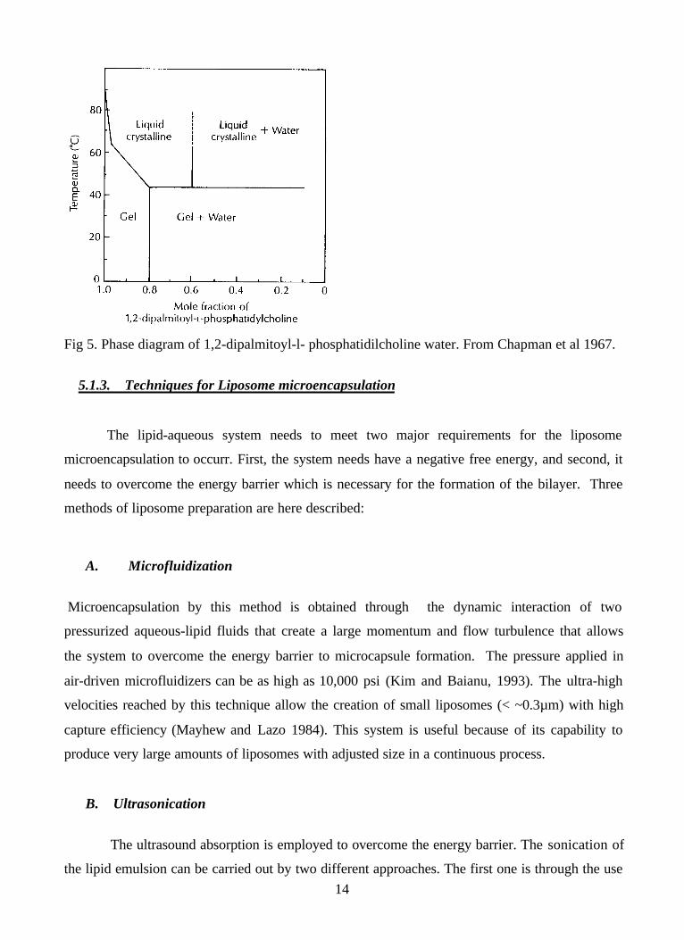
The phase diagram in Figure 5 illustrates the phase changes around the transition
temperature (Tc) of a phospholipid- water system. This phase transition temperature is defined as
the minimum temperature required for the water to break through the lipid membrane. When the
system is cooled to temperatures below Tc, the hydrocarbon chains adopt an ordered packing
phase, thus creating a lamellar structure (Chapman et al. 1967). In the bilayer structure, the
hydrophobic tails are lined up together through hydrophobic interactions, whereas the hydrophilic
part of the lipid faces towards the aqueous phase.
14
Fig 5. Phase diagram of 1,2-dipalmitoyl-l- phosphatidilcholine water. From Chapman et al 1967.
5.1.3. Techniques for Liposome microencapsulation
The lipid-aqueous system needs to meet two major requirements for the liposome
microencapsulation to occurr. First, the system needs have a negative free energy, and second, it
needs to overcome the energy barrier which is necessary for the formation of the bilayer. Three
methods of liposome preparation are here described:
A. Microfluidization
Microencapsulation by this method is obtained through the dynamic interaction of two
pressurized aqueous-lipid fluids that create a large momentum and flow turbulence that allows
the system to overcome the energy barrier to microcapsule formation. The pressure applied in
air-driven microfluidizers can be as high as 10,000 psi (Kim and Baianu, 1993). The ultra-high
velocities reached by this technique allow the creation of small liposomes (< ~0.3µm) with high
capture efficiency (Mayhew and Lazo 1984). This system is useful because of its capability to
produce very large amounts of liposomes with adjusted size in a continuous process.
B. Ultrasonication
The ultrasound absorption is employed to overcome the energy barrier. The sonication of
the lipid emulsion can be carried out by two different approaches. The first one is through the use

15
of a sonication probe placed directly into the suspension of liposomes. The second method is
slower than the first one and employs a sonication bath , such as a sealed container filled with
Nitrogen or Argon gas. Both methods have been extensively applied for the formation of SUV’
s; however, the use of a sonication probe has been found to cause contamination of the liposomes
(Taylor 1983).
C. Reverse phase evaporation
Reverse phase evaporation is used for the preparation of LUV’s (Szoka and
Papahadjopoulos 1978) and it is based upon the extraction of a nonpolar solvent from an
aqueous–nonpolar inverted micelle by rotatory evaporation under vacuum. This withdrawal of
the nonpolar phase changes the intermediate gel-like phase of the micelle into uni-lamellar and
oligo-lamellar vesicles (Kim and Bainu 1993). The advantage of this technique is the uniformity
of the vesicles formed (from about 0.2 to1.0 µm) as well as their high encapsulation efficiency.
On the other hand, the exposure of the components to organic solvents and sonication is likely to
result in protein denaturation (Szoka and Papahadjopoulos 1980).
5.1.4. Characterization of Liposomes
Several techniques may be employed to characterize liposomes. Table 3 compares some
of the current techniques and novel approaches to the study of liposomes and their interactions.

16
Liposome Size distributionAverage size of Liposomes
Liposome stability vsus time and temperatureEncapsulated Molecule Retention rates (Mayhew et al. 1983)
Liposome stability Liposome-cell interactions
Ultrasonic Absorption Temperature induced transitions Limitations of Liposome uses in the presence of hydrophobic proteins
Permeability of liposomesMolecular dynamics of Spin- Labeled Lipids in Liposomes and Encapsulates
Molecular dynamics of Lipids in Liposomes and EncapsulatesWater exchange in phospholipid vesicles (Haran and Sporer 1976)Stability using metal cations (Baianu and Kim 1993) Storage of Liposomes
DNA interactions with molecules encapsulated within liposomesHybridization of DNA for Liposomes Vaccines
Nuclear Magnetic Resonance
Fluorescence Correlation Spectroscopy
Electron Microscopy
Radiactive Tracers
Fluorescence Quenching
Electron Spin Spectroscopy
Table 3. Characterization of Liposome Properties by Various Techniques.
5.1.5. Applications of Liposomes in the Food Industry
Lecithin-based liposomes offer great flexibility and shelf life improvements in the food
industry for introducing water–soluble substances such as flavors and micronutrients. Such
modified liposomes are also being used at present in the beverage and bakery industries to deliver
both flavors and oils that are trapped inside the liposomes; such flavors and oils are released then
into the mouth; these are also employed to incorporate flavor oils. The major impact of such
techniques has been achieved through the use of microencapsules that can be made through a
continuous process on an industrial scale (Fig 6).

17
Fig 6. Microfluidizer employed to microencapsulate enzymes and food proteins in food products.
In the dairy industry, liposomes containing enzymes have been reported to reduce the
ripening time by 30 -50% (Kirby 1990 and Law and King 1991), as well as improve texture and
flavor. The latter was caused by a decrease in the action of proteolytic enzymes in the early
phases of cheese fermentation. (Alkhalaf et al 1988 ).
Because liposomes have the ability to carry fat-based flavors in their bilayer, as well as
water- soluble flavors in the core of the vesicle, they protect the flavor from degradation and also
increase the longevity of the flavor in the system where they are being employed. Therefore, their
use in the beverage industry has become widespread. The rate of diffusion through/from the
bilayer depends on the liposome composition as well as physical properties of the flavor.
Bakery is another area where liposomes have been applied and it is based on the
characteristic of the liposomes of not being destroyed during the processes of mixing or
extrusion; therefore, they can release encapsulated flavorings, fragrances or food additives. When
a flavor is encapsulated, the release occurs after the enzymatic degradation of the liposome, and
thus the rate of release depends on the physical properties of the material of which the liposome
is made. In the case of lecithin, the pH value or range, as well as temperature, are important
factors.

18
5.1.6. Liposome Applications in Medical Biotechnology
Traditionally, liposomes have been used in the bioengineering field to over-produce
certain proteins through the genetic modification of cells. They have thus solved the
inconvenience of transferring high molecular weight molecules through cell membranes (Nicolau
and Cudd 1989).
The use of unilamelar vesicules made of cationic lipids (Rose et al 1991) has improved
the transfection efficiencies and prevented interactions with DNA molecules. Therefore, the
project of introducing genes to cure diseases (genetic therapy) is not far from becoming a reality
if the patient being treated were found not to suffer from severe side effects. Vaccine
formulations based on liposomes have been successfully tested in animal immunization, and such
studies are currently in the clinical testing phase. The benefits and limitiations of liposomes as
drug carriers in a system as complex as the human body depend basically on the interactions of
liposomes with the cells (Lasic, 1993), as well as their immuno-compatibility, or their ability to
escape detection by the human immune system.
5.1.7. Other Applications of Liposomes
Cosmetics is another area where the liposomes have been extensively employed. They
are being utilized as humectants, as well as carriers of formulations containing extracts, vitamins,
moisturizers, antibiotics and proteins. Such applications are mostly directed towards preventing,
or delaying, the aging of the skin. Through their surfactant action liposomes also improve the
coagulation and sinking of oil spreading at a water interface, a methodology which has been
under study for some time by EPA for cleaning up oil spills (Gatt et al 1991; Dutton 1993).
6. LECTIN APPLICATIONS TO CANCER DETECTION AND TREATMENT
Lectins are proteins, or glycoproteins, that agglutinate erythrocytes of some or all blood
groups in vitro (Sharon 1998). They are an important group of bioactive proteins and
glycoproteins found in most organisms, including plants, vertebrates, invertebrates, bacteria and
viruses, and have several important applications to the fields of health food and medical
biotechnology. Lectins are used as tools in the fields of biochemistry, cell biology and

19
immunology, as well as for diagnostic and therapeutic purposes in cancer research (Sharon and
Lis 2002). Lectin aggregation can be employed on a large-scale basis for the commercial
production of biologically active proteins (Takamatsu et al 1999). Lectins have been used in
glycoprotein purification, oligosaccharide analysis, as well as in cell-selection processes. Lectins
can bind reversibly with free sugars or with sugar residues of polysaccharides, glycoproteins or
glycolipids (Goldstein and Poretz 1986). There has been increasing demand for novel diagnostic
and medical cancer therapies that utilize non-traditional sources. Epidemiological studies indicate
that the consumption of a plant-based diet is strongly associated with a reduced risk of
developing several types of cancer (Block et al 1992). Plants contain numerous phytochemicals
that can alter cancer-associated biochemical pathways. One such group being intensively
examined for its role in cancer chemoprevention is lectins (Abdullaev and Gonzalez de Mejia
1997). A review of plant lectins and anticancer properties can be found in a review article (de
Mejia and Prisecaru 2003), and soybean lectins are specifically discussed in another publication
(Gonzalez de Mejia et al 2003).
Lectins are currently being considered for use as cancer therapeutic agents. Lectins were
reported to bind preferentially to cancer cell membranes, or their receptors, causing cytotoxicity,
apoptosis and/or tumor growth inhibition. Lectins were thought to become internalized into cells,
and some lectins were claimed to cause cancer cell agglutination and/or aggregation. Present in
common foods, some lectins resist acid and/or enzymatic digestion and also were reported to
enter the bloodstream in an intact, and biologically active, form. Lectins possess a spectrum of
beneficial, as well as harmful, effects both in vitro and in vivo. Ingestion of lectins also sequesters
the available body pool of polyamines, thereby claimed to thwart cancer cell growth. They have
also been reported to affect the immune system by altering the production of various interleukins,
or by activating certain protein kinases. Lectins were also reported to bind to ribosomes and thus
inhibit protein synthesis. Lectins may also modify the cell cycle by inducing non-apoptotic G1-
phase accumulation mechanisms, G2/M phase cell cycle arrest, and apoptosis, and might activate
the caspase cascade. Lectins were also reported to down-regulate telomerase activity and inhibit
angiogenesis. Lectins could inhibit cell adhesion, proliferation, colony formation and
hemagglutination, and were reported to have cytotoxic effects on human tumor cells. Lectins
could function as surface markers for tumor cell recognition, cell adhesion, signal transduction
across the membrane, mitogenic cytotoxicity and apoptosis. Also, lectins were reported to
modulate the growth, proliferation and apoptosis of premalignant and malignant cells both in

20
vitro and in vivo. Most of these effects are thought to be mediated by specific cell surface
receptors (Gonzalez de Mejia and Prisecaru 2003).
For many years, lectins have been considered toxic substances to both cells and animals,
mainly because of the observed agglutination of erythrocytes and other cells in vitro. On the other
hand, it has also been reported that lectins have an inhibitory effect on the growth of tumors.
Their potential for clinical applications has been investigated only in recent years. Lectins are
now being considered for use both in the diagnostics and therapeutics of cancers. Thus, lectins
are quite versatile biomarkers and have been utilized in a variety of studies involving
histochemical, biochemical and functional techniques for cancer cell characterization (Munoz et
al 2001). Lectins may also be very useful tools for the identification of cancers and the degree of
metastasis, or cancer development stage. Recently, there has been a tendency to shift lectins use
from cancer detection to actual use in combating cancer. The reason for this shift is mainly
caused by recent research that indicated the cytotoxic and apoptosis/necrosis-inducing effects of
certain lectins, combined with the hypothesis that dietary lectins enter the systemic circulation
intact (Wang et al 1998).
One important feature appears to be that lectins stimulate the human immune system. Lectins
were thus reported to exhibit antitumor and anticarcinogenic activities that could be of substantial
benefit in cancer treatment. Extracts of Viscum album (mistletoe) are widely used as
complementary cancer therapies in Europe. Mistletoe has been used parenterally for more than 80
years as an anticancer agent with strong immuno-modulating action. The quality of life of
patients with late-stage pancreatic cancer was reported to be improved on account of exposure to
mistletoe lectin (Friess et al 1996). Immuno-modulation using recombinant ML was reported to
influence tumor growth in breast cancer patients (Stein et al 1998). Bladder carcinoma was
reported to be significantly reduced, and survival times were reported to be prolonged in mice as
the concentration of ML was increased from 3 to 30 ng. ML increased the life span, decreased the
tumor growth and decreased hyperplasia of mice and rats with lymphoma and lung cancer
(Kuttan et al 1997).
A lectin purified from mesquite seed was reported to have an anti-proliferative effect on the
cervical human tumor (HeLa) cells and on cell adhesion. Interestingly, mesquite lectin modulated
the growth, proliferation and apoptosis of HeLa cells, while having no effect on normal cells in
vitro (Gonzalez de Mejia et al 2002; Abdullaev and Gonzalez de Mejia, 1996). Vicia faba

21
agglutinin (VFA), a lectin from broad beans was reported to aggregate, stimulate the
morphological differentiation of, and reduce the malignant phenotype of colon cancer cells
(Jordinson et al 1999). Wheat germ agglutinin (WGA) proved to be highly toxic to human
pancreatic carcinoma cells in vitro. WGA exposure induced chromatin condensation, nuclear
fragmentation and DNA release, consistent with apoptosis. The binding of the snail lectin Helix
pomatia agglutinin (HPA), which recognizes N-acetylgalactosamine and N-acetylglucosamine
sugars, is considered to be a strong predictor of metastasis and unfavorable prognosis in a number
of human adenocarcinomas, including breast cancer (Brooks and Carter 2001). Because of their
carbohydrate bio-recognition properties, lectins may also be used as carriers for targeted drug
delivery, in a manner similar to liposomes (Wroblewski et al 2001), provided the possible side
effects of such treatments could be minimized.
It has been observed that mucosal expression of terminal unsubstituted galactose is increased
in colon pre-cancerous conditions and cancer, and that it allows interaction with mitogenic
galactose-binding lectins of dietary or microbial origin. Based on this observation, an interesting
hypothesis was postulated whereby galactose might be able to prevent cancer by binding and
inhibiting such lectins from interacting with colon cancer cells (Evans et al 2002). D-galactose
treatment was reported to be effective in liver lectin blocking to prevent hepatic metastases in
colorectal carcinoma patients (Isenberg et al 1997). Epithelial cancer cells showed an increased
cell surface expression of mucin antigens with aberrant O-glycosylation, notably Thomsen-
Friedenreich Antigens (TFA). TFA is a carbohydrate antigen with a proven link to malignancy
(Irazoqui et al 2001). Immunoassays could be utilized for antigens such as TFA in order to
determine the metastatic potential of breast and colon cancer cells. Molecular changes in the
membrane surface in the case of both stomach and colon cancer cells occur during the
progression to carcinogenesis. Carbohydrate patterns displayed on the cellular membrane exterior
are molecular signatures with unique biological characteristics related to oncogenesis and
metastasis, and could be used to determine the appropriate chemotherapeutic and surgical
procedures for each specific cancer.
Lectins have already a demonstrated potential for the treatment, prevention and diagnosis of
chronic diseases such as cancer. Further research is, however, required to further elucidate the
effects of purified and dietary lectins and their potential for defense against tumors.

22
7. COMPUTATIONAL BIOLOGY, MOLECULAR MODELING AND
SOME OF THEIR BIOTECHNOLOGY RELATED APPLICATIONS
Computational Biology has a very wide range of applications currently thought of ‘belonging’ to
Biotechnology (Baianu 1986), such as Bioinformatics, even though the development of such
computations has preceeded modern Biotechnology by many decades. Instead of attempting the
hopeless task of covering superficially just a few of the applications of Computational Biology to
either Medical or Agricultural Biotechnology , we decided in favor of an approach that focuses
on a few selected examples in greater depth, by considering molecular modeling techniques that
have very wide applications, ranging from ‘pure’ chemistry, to biochemistry, molecular biology,
biotechnology, medicine, foods and industrial manufacturing.
7.1. Molecular Modeling Techniques
Molecular modeling is a group of techniques that employ computer-generated images of
chemical structures that show the relative positioning of all the atoms present in the molecule
being studied, and/or the simulated dynamics of such molecules together with their ordering
through space-time. Such techniques are of considerable help for understanding many
physicochemical properties of molecules, and may also provide clues about their possible role(s),
that is, their function, in the organism. They can be thus especially valuable tools for
investigating structure-function relationships. Proteins --within a given protein family-- have, in
theory, similar sequences and generally share the same basic structure. Thus, once the structure
for one member of the protein family is determined, molecular modeling computations can help
determine the structure for other members of the same protein family. Such a homology
technique when applied to protein structure may allow scientists to gain additional insight into
protein structure, especially for those proteins for which the available experimental data is scarce.
7.1.1 Tasks in Molecular Modeling
In order to obtain optimal results, the National Center by Biotechnology (NCBI)
suggested that protein sequences should be organized in protein families. Such readily
searchable databases (Table 4) are currently available for many proteins (NCBI 2003).

23
Table 4. Databases for Molecular modelingProtein Data Bank (PDB);
The molecular modeling database (MMDB) at NCBI
Clusters of Orthologous Group of proteins (COGs) with COGNITOR program
The Basic Local Alignment Search Tool (BLAST)Vector Alignment Search Tools (VAST)The Conserved Domain Database(CDD)Domain Architecture Retrieval Tool.
(Source: NCBI 2003: “A science primer” ).
Secondly, a target must be selected. A target is a protein structure that has been
determined via experiments. Thirdly, one must generate a purified protein for analysis of the
chosen target and then determine the protein structure by analytical techniques such as X-ray
crystallography and/or
2D NMR.
The experiments described next, for example, studied apolipoprotein A-1 by employing
molecular modeling techniques in order to understand the interaction of proteins in food systems
and complex organisms.
7.1.2. Apolipoprotein Structures
Lipoprotein in mammals have evolved as the primary transport vehicles for lipids. This
role leads to the importance of lipoproteins in several diseases, such as atherosclerosis and
cardiovascular disease. Lipoprotein particles consist of a core of neutral lipids, stabilized by a
surface monolayer of polar lipids complexed with one or more proteins.
Apolipoprotein A-I and apo B are respectively, the major protein components of high-
density (HDL) and very low density (VLDL) lipoproteins. Thus, understanding apolipoproteins
is very important for medical and health-related fields, such as medical biotechnology, as well as
food science and human nutrition.
24
7.1.3. Methods of Structure Determination applied to Apolipoprotein Molecules
The process of biosynthesis, the physical characteristics and the metabolism of
apolipoproteins have been intensely studied. However, because of the noncrystalline structure of
many apolipoproteins, it has been difficult to obtain structural data at the molecular, or atomic,
level. Therefore, methods combining the amino acid sequence with molecular methods are now
being introduced. Thus, overall structures may be derived from correlations of global secondary
structures determined from polarized light studies combined with local structures predicted from
amino acid sequences. The known amino acid sequence of apolipoprotein from the position 7 to
156 of apo Lp-III was first used to design an Apo A-I template, that could be then approached by
‘standard’ molecular modeling techniques.
7.1.4. Molecular Modeling of Apolipoproteins A-I
Target: Apolipoprophorin III is chosen as a target for apolipoprotein A-I.(apo Lp A-I) .
The structure of Apolipophorin III has been determined in a crystal at 2.5 Å resolution for the
18-kDa apo Lp III from the African migratory locust, Locusta migratoria and the 22-kDa N-
terminal, receptor-binding domain of human apo E.
Template for Apo A-I: Lp IIIa is designed by using molecular software IALIGN from Lp
III by inserting alanine for template of sequences of apo A-I ( using the program SYBYL, V5.5).
Alanine residues are inserted at each of the gap position identified by IALIGN, an interactive
alignment program distributed with the Protein Identification Resource (PIR).(Eleanor M.B,
1994) This model was compared with DgA-I, HuA-I and ChA-I resprsenting canine, human and
chicken Apo A-I respectively. Results were then compared using a “strip of the helix” template
(Vazquez et al 1992) by scoring 1 or 0 for residues that did , or did not, fit into the template.
Modeling Results:
A. Sequence Comparison. The five long a-helices connected by short loops in amino acid
residues 7-156 of apo Lp-III is used as template for LpIIIa, Dig A-I, DgA-I, HuA-I. Amphipathic
potential (AP) is used to detect if the predict structure is suitable for a lipid-aqueous interface in
its stable condition. The results are shown in Table 5.

25
Table 5: Amphipathic potentials of predicted helical segments in apolipoprotein models.
Model
# res AP # res AP # res AP # res AP # res APLp-III 19 0.79 25 0.8 22 0.77 27 0.7 21 0.67Lp-IIIa 19 0.79 25 0.8 24 0.67 33 0.61 22 0.64DgA-I 19 0.58 25 0.84 24 0.75 33 0.48 22 0.68HuA-I 19 0.53 25 0.88 24 0.71 33 0.45 22 0.73ChA-I 19 0.53 25 0.88 24 0.58 33 0.52 22 0.82
Amphipathic potentials (AP) are the best average value for a helical segment of ( # res)residues measured with the “strip of helix”.(Vazquez, et al. 1992)
B. Energy minimized models. In this model, electrostatic interactions contributes the most to
favorable energies. Alanine was chosen as the spacer residue in building apo Lp-IIIa because of
its function of small, non-ionic side chain and serves as a helix- stabilizing residue. Although it
has a high probability of being found in helical structures, it does not participate through
electrostatic interactions. Results of this model are discussed for potential energetic evaluation
and amphipathic analysis of energy refined helices (see Table 6 and Table 7). The structure
obtained through this molecular modeling is illustrated in Figure 6.
Figure 6. Energy Refined models of apo-Lp III and the template apo IIIa constructed byinserting alanine residues into gap positions identified when the sequences were aligned with thecanine, human, and chicken apo A-1 sequences. Backbone structures for this complete modelsare in panel (a). The effects of inserted alanine residues on H3, H4, H5 are displayed in panels(b), (c), (d) respectively. In each display, the apo Lp III is shown to the left of the apo Lp-IIIa.(SOURCE: Brown et al 1994).

26
Table 6. Energetic evaluation of the refined modelsLp-in Lp-lUa DgbjA-I HuA-I ChA-I
Number of residues 150 165 165 165 165
Energy, kcal
Bond stretching 20.7 23.1 28.9 29.2 31.8
angle bending 123.4 154.0 189.6 208.5 212.2
torsional 198.1 235.7 308.3 324.6 340.9
out of plane 26.1 35.8 37.7 41.5 44.5
1-4 van der Waals 218.7 235.6 247.2 260.2 270.3
van der Waals -993.0 -1094.3 -1074.8 -1131.8 -1155.6
1-4 electrostatics 1684.0 1813.3 1497.1 1566.7 1524.1
electrostatic -4497.8 -4567.3 -5214.7 -5182.6 -5208.4
H-bond -69.3 -70.2 -64.5 -69.8 -76.3
Total Energy, kcal -3289.0 -3234.3 -4045.2 -3953.6 -4016.4
kcal/mol/residue -21.9 -19.6 -24.5 -24.0 -24.3
H1 (residues) (7-25) (72-90) (73-88) (72-90)
kcal/mol/residue -16.1 -18.5 -16.6 -17.4
H2 (residues) (35-59) (100-124) (101-125) (100-124)
kcal/mol/residue -16.1 -20.1 -19.4 -19.2
H3 (residues) (70-91) (70-91) (135-158) (136-159) (135-158)
kcal/mol/residue -17.6 -20.7 -18.4 -16.2 -17.6
H4 (residues) (96-121) (96-121) (166-196) (167-197) (166-196)
kcal/mol/residue -15.5 -13.8 -16.7 -16.9 -17.6
H5 (residues) (136-155) (136-155) (207-235) (208-236) (207-235)
kcal/mol/residue -15.8 -15.4 -14.0 -13.4 -14.9
These energy calculations, based on the sequence segments initially assigned to HI -H5 (seeTables 6 and 7), are given to illustrate the stability of the structures. Sequences for helices 3 to5 (H3, H4, H5) of apo Lp-IIIa contain inserted alanine residues. (SOURCE: Brown 1994).
27
Table 7. Amphipathic analysis of energy refined helicesModel Lp-III Lp-IIIa gA-I HuA-I ChA-IHI N7-E25 N7-E25 D72-E90 D73-K88 E72-E90Hb sector 140° 140° 100° 60° 100°Av. Hb 0.98 0.98 0.78 0.7 0.81H2 P35-S59 P35-S59 L100-E124 L101-E125 L101-E124Hb sector 100° 100° 100° 100° 100°
Av. Hb 0.8 0.8 0.7 0.89 0.95H3 S70-T91 S70-S85 Q137-R150 E136-R149 L135-L151
Hb sector 140° 120° 100° 100° 120°Av. Hb 0.93 0.79 0.95 0.88 0.82
H4H4A A96-S121 Q98-T107 D168-K181 Q172-E179 D167-R181Hb sector 100° 140° 80° 100° 80°
Av. Hb 0.56 0.91 1.1 0.94 1.18H4B Q117-al21d S187-S196 A190-A194 P186-V195Hb sector 60° 100° 60°Av. Hb 0.45 0.45 0.66H5 E136-V156 L134-A155 L213-I232 L214-T237 E212-L235Hb sector 120° 80° 100° 100° 100°Av. Hb 0.74 0.53 1.05 0.96 0.86
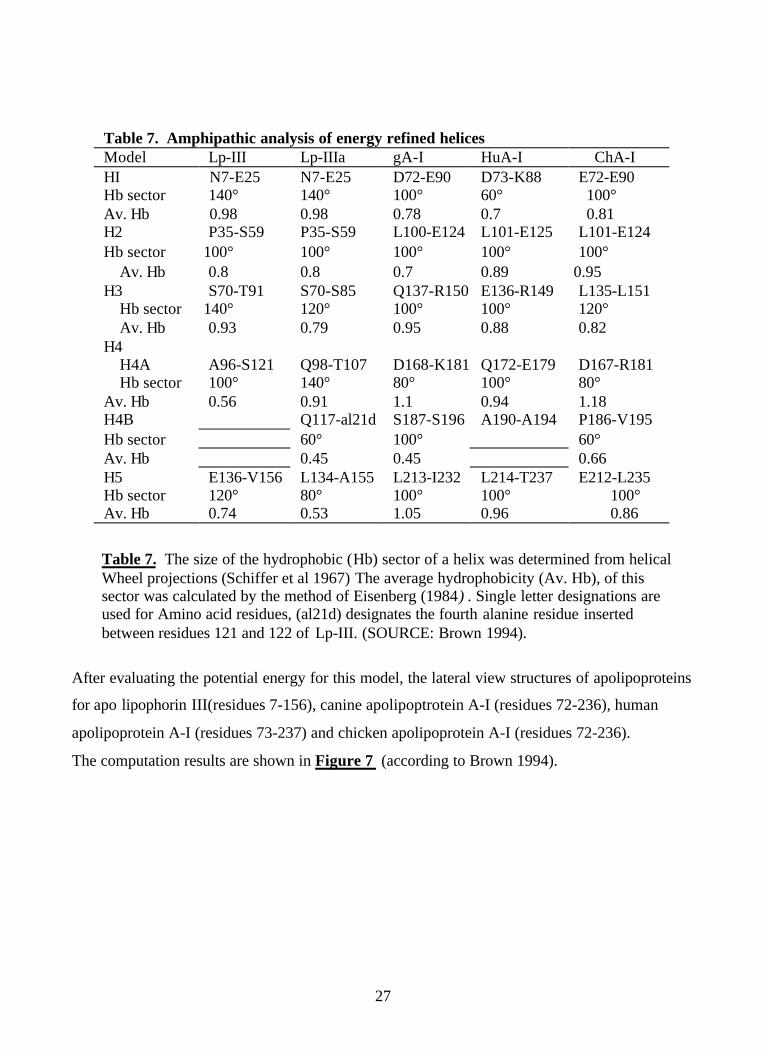
Table 7. The size of the hydrophobic (Hb) sector of a helix was determined from helicalWheel projections (Schiffer et al 1967) The average hydrophobicity (Av. Hb), of thissector was calculated by the method of Eisenberg (1984) . Single letter designations areused for Amino acid residues, (al21d) designates the fourth alanine residue insertedbetween residues 121 and 122 of Lp-III. (SOURCE: Brown 1994).
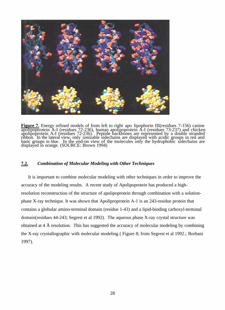
After evaluating the potential energy for this model, the lateral view structures of apolipoproteins
for apo lipophorin III(residues 7-156), canine apolipoptrotein A-I (residues 72-236), human
apolipoprotein A-I (residues 73-237) and chicken apolipoprotein A-I (residues 72-236).
The computation results are shown in Figure 7 (according to Brown 1994).
28
Figure 7. Energy refined models of from left to right apo lipophorin III(residues 7-156) canineapolipoptrotein A-I (residues 72-236), human apolipoprotein A-I (residues 73-237) and chickenapolipoprotein A-I (residues 72-236). Peptide backbones are represented by a double strandedribbon. In the lateral view, only ionizable sidechains are displayed with acidic groups in red andbasic groups in blue. In the end-on view of the molecules only the hydrophobic sidechains aredisplayed in orange. (SOURCE: Brown 1994)
7.2. Combination of Molecular Modeling with Other Techniques
It is important to combine molecular modeling with other techniques in order to improve the
accuracy of the modeling results. A recent study of Apolipoprotein has produced a high-
resolution reconstruction of the structure of apolipoprotein through combination with a solution-
phase X-ray technique. It was shown that Apoliproprotein A-1 is an 243-residue protein that
contains a globular amino-terminal domain (residue 1-43) and a lipid-binding carboxyl-terminal
domain(residues 44-243; Segrest et al 1992). The aqueous phase X-ray crystal structure was
obtained at 4 Å resolution. This has suggested the accuracy of molecular modeling by combining
the X-ray crystallographic with molecular modeling ( Figure 8; from Segrest et al 1992.; Borhani
1997).

29
Figure 8. X-ray Crystal structure of the Apolipoprotein A-I: ? ( 1-43) dimer in solution.
(Source: Borhani et al. 1997).
7.3. An In-depth Analysis of Molecular Structure
The determination of apolipoprotein A-I can be then further associated with lipid
containing domains by employing other molecular modeling techniques. The ‘belt model’ is used
to show the possible orientations of lipoprotein with its apolipoprotein inserted (Figure 9). The
suggested structure can then serve as a template in other high density lipoproteins for their
structure determination and also help in understanding the biological interaction.
Helix 1A
Helix 2 BHelix 7 A
Helix 8A
Helix 9 A
Helix 1BHelix 10A
Helix10 B
Helix 9BHelix 6 A
Helix 1AHelix 2A
Helix 3 A
Helix 4 AHelix 5A
Helix 3 B
Helix 4B
Helix 7B Helix5B
Helix 8B
Helix 1A
Helix 2 BHelix 7 A
Helix 8A
Helix 9 A
Helix 1BHelix 10A
Helix10 B
Helix 9BHelix 6 A
Helix 1AHelix 2A
Helix 3 A
Helix 4 AHelix 5A
Helix 3 B
Helix 4B
Helix 7B Helix5B
Helix 8B

30
7.4. Applications of Molecular Modeling
Molecular modeling has been introduced for more than two decades ago. Increasingly,
modeling software is available for a variety of industrial applications. Global markets for
molecular modeling, in general, now exceed 2 billion US $ annually (Fuji-Keizai 2003)
7.4.1. Applications in the Food Industry and Human Nutrition
Molecular modeling has been suggested by several professionals in food industry as a
new tool for food research. (Hegenbart 1992) Such tools can assist food scientists in problem
solving, as well as save time and money. Examples of utilization of molecular modeling are the
uses of high intensity sweeteners and taste receptors to predict the sweetening potential of new
molecules by using molecular modeling as developed by and E.W. Taylor and S. Wilson at the
University of Georgia. (Hegenbart 1992) Such models can be used in food industry for product
development and also for faster results in sensory evaluation.
7.4.2. Examples of Medical Applications of Molecular Modeling
Molecular modeling is especially helpful in medical fields, such as in development of new
drugs on a nanoscale. Recent studies have shown the importance of using molecular modeling in
both medical and food sciences (Food Ingredient first, 2003). The molecular modeling of
Epigallocatechin Gallate (EGCG), and the HIV cell was undertaken by Shearer (2003). His
report has inspired scientists in Japan who discovered the potential of green tea as an anti-HIV
Fig 9a. Detailed Belt model displayedas a helical ribbon. C(NH2)3, blue,oxygen atom, red; phosphorous atom,yellow all other atom, black.( Source: Borhani 1997)
Fig 9b. Detailed Belt model displayedas all atom model, oriented in 9a.nitrogen atom, blue, oxygen atom, red;carbon atom, cyan, polar hydrogenatom, white. ( Source: Borhani 1997)
31
drug. The chemical compound that is found abundantly in the green tea called Epigallocatechin
Gallate (EGCG) is reported to stop the HIV virus from binding to CD4 molecules and human T-
cells.
7.4.3. Other Applications of Molecular Modeling
Other applications of molecular modeling to manufacturing, life sciences and chemistry
greatly benefit from such molecular modeling programs. Nanotechnology has developed to a 30
to 40 million US $ market, and it also has the potential to grow to a 60 to 70 million US $ market
within the next five years ( Fuji-Keizai 2003).
8. CONCLUSIONS
A simplified overview of the selected applications of biotechnology in the areas of foods,
human nutrition and health, as well as the potential, large-scale applications in the chemical
industry that were discussed in our review is presented in Table 8.
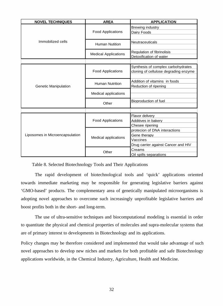
32
NOVEL TECHNIQUES AREA APPLICATION Brewing industry Dairy Foods
Neutraceuticals
Regulation of fibrinolisisDetoxification of water
Synthesis of complex carbohydratescloning of cellulose degrading enzyme
Addition of vitamins in foods Reduction of ripening
Bioproduction of fuel
Flavor delivery Additives in bakeryChesee ripeningprotecion of DNA interactions Gene therapy VaccinesDrug carrier against Cancer and HIV Creams Oil spills separations
Food Applications
Medical applications
Other
Liposomes in Microencapsulation
Food Applications
Human Nutrition
Other
Genetic Manipulation
Medical applications
Food Applications
Immobilized cells Human Nutition
Medical Applications
Table 8. Selected Biotechnology Tools and Their Applications
The rapid development of biotechnological tools and ‘quick’ applications oriented
towards immediate marketing may be responsible for generating legislative barriers against
‘GMO-based’ products. The complementary area of genetically manipulated microorganisms is
adopting novel approaches to overcome such increasingly unprofitable legislative barriers and
boost profits both in the short- and long-term.
The use of ultra-sensitive techniques and biocomputational modeling is essential in order
to quantitate the physical and chemical properties of molecules and supra-molecular systems that
are of primary interest to developments in Biotechnology and its applications.
Policy changes may be therefore considered and implemented that would take advantage of such
novel approaches to develop new niches and markets for both profitable and safe Biotechnology
applications worldwide, in the Chemical Industry, Agriculture, Health and Medicine.

33
9. REFERENCES
1. Abdullaev FI, de Mejía EG. 1997. Antitumor effect of plant lectins. Natural Toxins. 5:157-
163.
2. Alkhalaf W, Piard JC, Soda MC, Gripon JC, Desmezeaud M and Vassa IL. 1988.
Liposomes as proteinase carriers for the accelerated ripening of St. Paulin type cheese.
Journal of Food Science. 53: 1674–1679.
3. Anderson RA, Paquette S, Lovrien R. 2002. Lectin-erythrocyte interaction with external
transmembrane glycohphorin saccharides controlling membrane internal cytoskeleta. J.
Agric. Food Chem. 50 (22): 6599-604.
4. Andrews AL, Atkinson D, Barratt MD, Finer EG, Hauser H, Henry R, Leslie RB, Owens
NL, Phillips MC and Robertson RN. 1976. Eur. J. Biochem. 64, 549-563.
5. Andries M, van Beveren PC, Goffin O, and Masschelein CA.1996. European Brewing.
Convention. p.134-144
6. Babu PS and Panda T.1991. Studies on improvement techniques for immobilizing and
stabilizing penicillin amidase associated with E. coli cells. Enzyme Microbial Technology.
13: 676-682.
7. Baianu IC. 1986. Computer models and automata theory in biology and medicine.
Mathematical Modelling. 7: 1513-1577.
8. Baianu IC, Magin R and Ozu E. 1993. Microencapsulation adds value to Illinois crops.
Illinois Research. 35(1):19-21
9. Baianu IC, Kumosinski TF, Bechtel PJ, Myers-Betts PA, Kakalis LT, Mora-Guttierrez A,
Yakubu P and Wei TC. 1988. NMR Studies of Chemical Activity and Protein Interactions in
Solutions and Hydrated Powders. In: Proceed.196th National Meeting of the American
Chemical Society- Division of Agriculture and Food Chemistry. American Chemical Society
p.156.
10. Baianu IC, Ozu EM, Wei TC and Kumosinski TF. 1993. Molecular Dynamics and NMR
Studies of Concentrated Electrolytes and Dipoles in Water. In: Molecular Modeling. ACS
Symp. Ser. 576. Kumosinski TF and Liebman M, eds. ACS: Washington, D.C. p. 269-324.
11. Baianu IC, Ozu EM, Wei TC and Kumosinski TF. 1993. In: Molecular Modeling. ACS
Symp. Ser. 576. Kumosinski TF and Liebman M, eds. ACS: Washington, D.C. p. 325-341.

34
12. Baianu, I.C. , H. Pessen and T.F. Kumosinski. 1995. Physical Chemistry of Food
Processes . Vol.2., New York: Van Nostrand-Reinhold., pp.602.
13. Banka CL, Bonnet DJ, Black AS, Smith RS, Curtiss LK.1991. J. Biol. Chem. 266:23886-
23892.
14. Blaschek HP and White B. 1993 Renewed interest in renewable fuels. Illinois Research.
35(1): 12-13.
15. Blaschek HP. 1996. Recent Develpoments in the Genetic Manipulation of Microorganims
for biotechnology applications. In: Baianu IC, Pessen H and Kumosinski TF, eds. Physical
Chemistry of Food Processes. Vol 2. New York: Van Nostrand Reinhold. p. 459-474.
16. Block G, Patterson B, Subar A.1992. Fruit, vegetables, and cancer prevention: a review of
the epidemiological evidence. Nutr. Cancer. 18(1): 1-29.
17. Blundell TL and Johnson LN. 1976. Protein Crystallography. Academic Press: New York,
NY.
18. Blue M-L, Ostapchuk P, Gordon JS, Williams DL. 1982. J. Biol. Chem. 257: 11151-11159
19. Boguski MS, Freeman M, Elshourbagy NA, Taylor JM, Gordon JI. J. Lipid Res. 1986, 27,
1011-1034.
20. Borhani DW et al. 1997. Crystal Structure of truncated human apolipoprotein A-I
suggests a lipid-bound conformation. Proc Natl. Acad. Sci. USA. 94: 12291-12296
21. Brooks SA and Carter TM. 2001. N-acetylglucosamine and sialic acid expression in
primary breast cancers. Acta. Histochem. 103(1): 37-51.
22. Brown EM.1989. Poultry Science. 68: 399-407.
23. Brasseur R. J. Biol. Chem. 1991, 266, 16120-16127.
24. Brasseur R, Lins L, Vanloo B, Ruysschaert J-M, Rosseneu M.1992. Proteins. 13: 246-
257.
25. Breiter DR, Kanost MR, Benning MM, Wesenberg G, Law JH, Wells MA, Rayment I and
Holden HM. 1991. Biochemistry. 30: 603-608.
26. Cary JW, Petersen DJ, Papoutsakis ET, and Bennett GN.1988. Cloning expression of
Clostridium acetobutylicum phosphotrans-butyrylase and butyrate kinase genes in
Escherichia coli. J. Bacteriol. 170:4613-4618.
27. Chapman MJ.1986. Meth. Enzymol. 128: 70-143.
28. Chassy BM, and Flinkinger JL.1987. Transformation of Lactobacillus casei by electro-
poration. FEMS Microbiol. Lett. 44: 173-177.

35
29. Chibata I. 1979. Immobilized microbial cells with polyacrylamide gel and carrageenan and
their industrial applications. In Immobilized Microbial Cells, ACS Symposium Series 106.
Venkatsubramanian K ed. Washington DC: American Chemical Society. p. 187-202.
30. Chou PY and Fasman GD. 1978. Adv. Enzymol. 47: 45-148.
31. Cole KD, Fernando-Warnaku1asuriya GJP, Boguski M, Freeman M, Gordon JI, Clark
WA, Law JH, Wells MA. 1987. J. Biol. Chem., 262: 11794-11800.
32. Deamer DW and Uster PS. 1983. Preparation of Liposomes. In: Ostro M.J. ed. Liposomes.
Marcel Dekker. p. 1.
33. De Loof H. 1988. Ann. Biol. Clin.46, 10-15.
34. Dutton G. 1993. The promise of liposomes. Gen. Engin. News. 13: 6–9.
35. Eisenberg, D. 1984. Annu. Rev. Biochem. 53: 595-623.
36. El-Aassar SA, el-Badry HM, Abdel-Fattah. 1990. The biosynthesis of proteases with
fibrinolytic activity in immobilized cultures of Penicillium chrysogenum H9. Microbiol
Biotechnol. 33(1): 26-30.
37. Eleanor MB.1994. Molecular modeling of Apoliprotein A-I. Using Template derived
from crystal structure of Apolipophorin III. In: Molecular Modeling. ACS Symp. Ser.
576.
Kumosinski TF and Liebman M, eds. ACS: Washington, D.C. p. 100-112.
38. Evans RC, Fear S, Ashby D, Hackett A, Williams E, Van Der Vliet M, Dunstan FD and
Rhodes JM.2002. Diet and colorectal cancer: An investigation of the lectin/galactose
hypothesis, Gastroenterology, 122 (7): 1784-1792.
39. Friess H, Beger HG, Kunz J, Funk N, Schilling M, Buchler MW.1996. Treatment of
advanced pancreatic cancer with mistletoe: results of a pilot trial. Anticancer Res, 16(2):
915-920.
40. Garnier J, Osguthorpe DJ, Robson B. 1978. J. Mol. Biol. 120: 97-120.
41. Goldstein IJ, and Poretz, RD.1986. Isolation, physicochemical characterization, and
carbohydrate-binding specificity of lectins. In: Liener, IE, Saron N, and Goldstein IJ, ed.
The Lectins: Properties, Functions, and Applications in Biology and Medicine. London:
Academic, p. 33-237.
42. Gatt S, Bercovier JH and Barenholz Y.1991. Use of liposomes to combat oil spills and
their potential application to bioreclamation. In: Hinchee RE and Olfenbutte RF, eds. On
Site Bioreclamation. Butterworth, Stoneham p. 293–312.
43. Glomset JA. 1968. Lipid Res. 9: 155-167.

36
44. Gonzalez de Mejia E, Bradford T and Hasler C. 2003. The Anticarcinogenic Potential of
Soybean Lectin and Lunasin. Nutr. Rev. (in press).
45. Gonzalez de Mejia E, and Prisecaru VI, Lectins As Bioactive Plant Proteins: New
Frontiers in Cancer Treatment. 2003.Crit. Rev. Food Sci. & Nutr. (in press).
46. Gonzalez de Mejia E, Rocha N, Winter HC and Goldstein IJ. 2002. Differential Effect of a
Lectin from Mesquite (Prosopis juliflora) on HeLa and Normal Human Keratinocyte
Cells. The FASEB J. Experimental Biology.15 (4): 128.
47. Green Tea may prevent HIV: Study. 2003. Food Ingredient First.
48. Gregoriadis G, ed.1984. Liposome Technology. New York: CRC Press.
49. Gregoriadis G and Allison AC. 1990. Liposomes in Biological Systems. New York: John
Wiley & Sons.
50. Haran N and Shporer M. 1976. Biochem. Biophys. Acta 426: 638-646.
51. Head IM, Bailey MJ. 2003. Environmental biotechnology. Methodological advances
spawn new concepts in environmental biotechnology. Current Opinion in Biotech 14 (3):
245-247.
52. Hofe EH, Billings PC, Heidelberger C. and Landolph JR. 1986. In vitro genotoxicity
studies using complex hydrocarbon mixtures. Environ. Mutagen. 8: 589-609.
53. Inoue T. 1995. European brewing: proceedings of the twenty fifth convention. p.25-36.
54. Irazoqui FJ, Jansson B, Lopez PH, and Nores GA. 2001. Correlative fine specificity of
several Thomsen-Friedenreich disaccharide-binding proteins with an effect on tumor cell
proliferation. J. Biochem. (Tokyo).130(1): 33-7.
55. Isenberg J, Stoffel B, Stutzer H, Otte K, and Beuth J.1997. Liver lectin blocking with D-
galactose to prevent hepatic metastases in colorectal carcinoma patients. Anticancer Res.
17(5B): 3767-72.
56. Jordinson M, El- Hariry I, Calnan D, Calam J, and Pignatelli M.1999. Vicia faba
agglutinin, the lectin present in broad beans, stimulates differentiation of undifferentiated
colon cancer cells. Gut. 44:709-714.
57. Karel SF, Libicki SB and Robertson CR. 1985. Immobilization of whole cells Chemical
Engineering Science, 40:1321-1354.
58. Kim H and Baianu IC. 1991. Novel liposomes microencapsulation techniques for food
applications. Food Science and Technology. 2(3): 49-77
59. Kirby CJ and Law B. 1987. In: Andrews, AT, ed. Chemical Aspects of Food Enzymes. Royal
Society of Chemistry. p. 106-119.

37
60. Kolot FB. 1981. Microbial carriers: Strategy for selection. Proc. Biochem istry, 16: 30-46.
61. Kyte J, Doolittle RF. 1982. J. Mol. Biol.157: 105-132.
62. Kollman P. 1987. Ann. Rev. Phys. Chem. 38: 303-316.
63. Kumosinski TF, Brown EM, Farrell HM Jr. 1991. J. Dairy Sci. 74: 2879-2888.
64. Kuttan G. 1997. Anticarcinogenic and antimetastatic activity of Iscador. Anti-Cancer
Drugs. 8:15-16.
65. Lasic DD. 1993. Liposomes: From Physics to Applications. Amsterdam: Elsevier.
66. Li WH, Tanimura M, Luo CC, Datta S and Chan L.1988. J. Lipid Res. 29: 245-271.
67. Linko P.1985. Fuel and industrial chemical through biotechnology. Biotechnology
Advances. 3(1): 39-63.
68. Lou C-C, Li WH, Moore MN, Chan L. 1986. J. Mol. Biol. 187:325-340.
69. Lux SE, Hirz R, Shrager RI. and Gotto AM.1972. J. Biol. Chem., 247: 2598-2606.
70. McLachlan AD. 1977. Nature, 267: 465-466.
71. Maddox IS.1980. Production of n-butanol from whey filtrate using Clostridium
acetabutylicum NCIB 2951. Biotechnol. Lett. 2: 493-498.
72. Maddox, IS and Murray AE.1983. Production of n-butanol by fermentation of wood
hydrolysate. Biotech. Lett. 5:175-178.
73. Mahley RW, Innerarity TL; Rall SC. Jr.; Weisgraber, KW. 1984. J. Lipid Res. 25: 1277-
1294.
74. Marcel YL, Provost PR, Koa H, Raffai E, Dac NV, Fruchart J-C, Rassart E. 1991. J.
Biol. Chem. 266: 3644-3653.
75. Masschellein CA, Ryder DS and Simon JP. 1994. Critical Reviews in Biotechnology 14
(2): 155-177.
76. Mayhew E and Lazo AM. 1984. Biochem. Biophys. Acta. 775: 169-174.
77. Mayhew E and Papahadjopoulos D. 1983. In: Ostro MJ, ed. Liposomes. Marcel Dekker. p.
289.
78. Munoz R, Arias Y, Ferreras JM, Jimenez P, Rojo MA, and Girbes T. 2001. Sensitivity of
cancer cell lines to the novel non-toxic type 2 ribosome-inactivating protein nigrin b.
Cancer Lett. 167(2): 163-169.
79. National Biomedical Research Foundation, Georgetown University Medical
Center.1991.Washington DC. Release 28.0. March 31
80. Nedovic V, Leskosek-Cukalovic I and Vunjak-Novakovic G. 1999. Immobilized cell
technology (ict) in beer fermentation – a possibility for environmentally sustainable and

38
cost-effective process. University of Belgrade. www.rcub.bg.ac.yu.
81. Nedovic VA, Leskosek-Cukalovic I and Vunjak-Novakovic G. 1996. Harvey J, ed.
Winetitles: Adelaide. p.245.
82. Nicolau C. and Cudd A. 1989. Liposomes as carriers of DNA. Crit. Rev. Therap. Drug
Carrier Systems. 6: 239–271.
83. Nolte RT and Atkinson D. 1992.Biophys. 63:1221-1239.
84. Osborne JC and Brewer Jr. 1992. Ann. N.Y. Acad. Sci. 348: 104-121
85. Papahadjopoulos D, Post G, Vail WJ and Biedler JL. 1976. Use of lipid vesicles as carriers
to introduce Actinomycin D into resistant tumor cells. Cancer Res. 36: 2988–2994.
86. Pinker RJ, Lin L, Rose GD and Kallenbach NR. 1990. Protein Science. 2: 1099.
87. Rose JK, Buoncore L and Whitt MA.1991. A new cationic liposome reagent mediating
nearly quantitative transfection of animal cells. Biotechniques.10: 520–525.
88. Rosenberg M and Kopelman IJ. 1983. Food Science Technology. 2: 142-143
89. Segrest JP, Jackson RL and Morrisett JD. 1974. FEBS Lett. 38: 247-253.
90. Sharon N.Glycoproteins now and then: a personal account.1998. Acta Anat (Basel). 161(1-
4): 7-17.
91. Segrest JP, Garber DW, Brouillette CG, Harvey SC and Anantharamaiah GM. 1994. Adv.
Protein Chem. 45: 303-369.
92. Sharon N and Lis H. 2002. How proteins bind carbohydrates: lessons from legume lectins.
J Agric Food Chem. 50(22): 6586-6591.
93. Schiffer M, Edmunson AB. 1967. Biophys. 7:121-125
94. Szoka F. and Papahadjopoulos D. 1978. Proc. Natl. Acad. Sci. USA 75:4194-4198
95. Smith LC, Pownall HJ and Gotto AM. Jr. 1978. Annu. Rev. Biochem. 47:751-777.
96. Sou1ages JL, Wells MA. 1994. Adv. Protein Chem. 45: 371-415.
97. Szoka F and Papahadjopoulos D. 1980. Annual Review of Biophysics and Bioengineering. 9:
467-508.
98. Smith T .1996. Liposomes in Cell Culture. In: Baianu IC, Pessen H and Kumosinski TF, eds.
Physical Chemistry of Food Processes. Vol 2. New York: Van Nostrand Reinhold. p. 554-
587.
99. Taylor AH. 1983. Food Flavour. Ingredients Packag. Process. 5: 48-52
100. Toenniessen GH, Toole J and DeVires J. 2003. Advances in plant biotechnology and
its adoption in developing countries. Current Opinion in Plant Biology. 6:191-198.
100. Vazquez S R, Kuo D Z, Botsitis CM, Hardy LW, Lew RA and Humphreys RE. 1992. J.

39
Biol. Chem. 267: 7406-7410.
101.Verhasselt P., Poncelet F, Vits K, Van Gool, A. and Vanderleyden, J.1989. Cloning and
expression of a Clostridium alpha-amylase gene in Escherichia coli. FEMS Microbiol. Lett.
59: 135-140.
102. Weiner SJ, Kollman PA, Case DA, Singh U C, Ghio C, Alagona G, Profeta S, Weiner
PK. 1984. J. American. Chemical Society.106: 765-784.
103.Weiner SJ, Kollman PA, Nguyen DT, Case DA. 1986. J. Comput. Chem. 7: 230-252.
104.Weisgraber KH.1994. Adv. Protein Chem. 45: 249-302.
105.Wilson C, Wardell MR, Weisgraber KH, Mah1ey RW and Agard DA.1991. Science. 252:
1817-1822.
106.Witter L.1996. Immobilized Microbial Cells. In: Baianu IC, Pessen H and Kumosinski
TF, eds. Physical Chemistry of Food Processes. Vol 2. New York: Van Nostrand Reinhold. p.
475-486.
107.Wuthrich K. 1986. NMR of Proteins and Nucleic Acids. John Wiley & Sons, Inc.: New
York.
108.Zappe H, Jones WA and Woods DR.1987. Cloning and expression of xylanase gene from
Clostridium acetobutylicum P262 in Escherichia coli. Appl. Microbiol. Biotechnol. 27: 57-63.
Related Documents