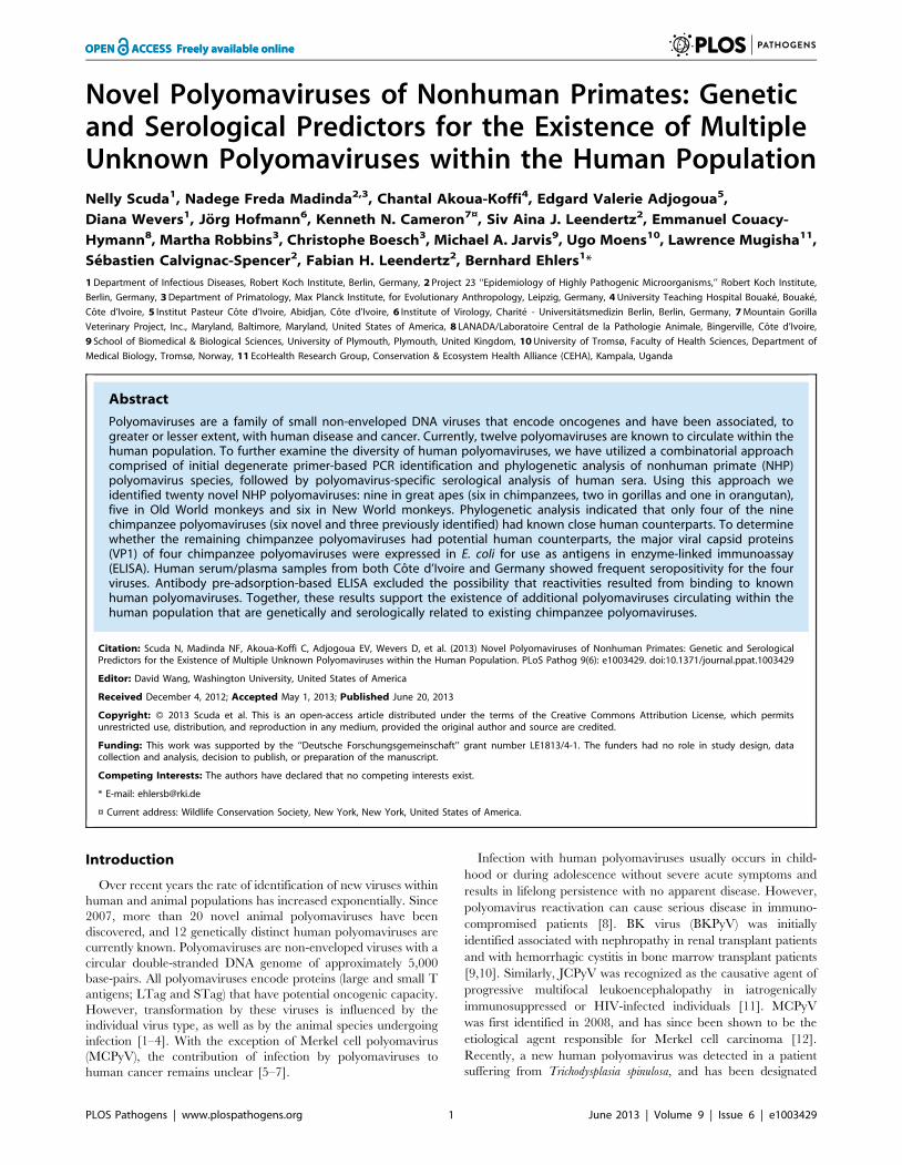
Novel Polyomaviruses of Nonhuman Primates: Genetic and Serological Predictors for the Existence of Multiple Unknown Polyomaviruses within the Human Population Nelly Scuda 1 , Nadege Freda Madinda 2,3 , Chantal Akoua-Koffi 4 , Edgard Valerie Adjogoua 5 , Diana Wevers 1 , Jo ¨ rg Hofmann 6 , Kenneth N. Cameron 7¤ , Siv Aina J. Leendertz 2 , Emmanuel Couacy- Hymann 8 , Martha Robbins 3 , Christophe Boesch 3 , Michael A. Jarvis 9 , Ugo Moens 10 , Lawrence Mugisha 11 , Se ´ bastien Calvignac-Spencer 2 , Fabian H. Leendertz 2 , Bernhard Ehlers 1 * 1 Department of Infectious Diseases, Robert Koch Institute, Berlin, Germany, 2 Project 23 ‘‘Epidemiology of Highly Pathogenic Microorganisms,’’ Robert Koch Institute, Berlin, Germany, 3 Department of Primatology, Max Planck Institute, for Evolutionary Anthropology, Leipzig, Germany, 4 University Teaching Hospital Bouake ´, Bouake ´, Co ˆ te d’Ivoire, 5 Institut Pasteur Co ˆ te d’Ivoire, Abidjan, Co ˆ te d’Ivoire, 6 Institute of Virology, Charite ´ - Universita ¨tsmedizin Berlin, Berlin, Germany, 7 Mountain Gorilla Veterinary Project, Inc., Maryland, Baltimore, Maryland, United States of America, 8 LANADA/Laboratoire Central de la Pathologie Animale, Bingerville, Co ˆ te d’Ivoire, 9 School of Biomedical & Biological Sciences, University of Plymouth, Plymouth, United Kingdom, 10 University of Tromsø, Faculty of Health Sciences, Department of Medical Biology, Tromsø, Norway, 11 EcoHealth Research Group, Conservation & Ecosystem Health Alliance (CEHA), Kampala, Uganda Abstract Polyomaviruses are a family of small non-enveloped DNA viruses that encode oncogenes and have been associated, to greater or lesser extent, with human disease and cancer. Currently, twelve polyomaviruses are known to circulate within the human population. To further examine the diversity of human polyomaviruses, we have utilized a combinatorial approach comprised of initial degenerate primer-based PCR identification and phylogenetic analysis of nonhuman primate (NHP) polyomavirus species, followed by polyomavirus-specific serological analysis of human sera. Using this approach we identified twenty novel NHP polyomaviruses: nine in great apes (six in chimpanzees, two in gorillas and one in orangutan), five in Old World monkeys and six in New World monkeys. Phylogenetic analysis indicated that only four of the nine chimpanzee polyomaviruses (six novel and three previously identified) had known close human counterparts. To determine whether the remaining chimpanzee polyomaviruses had potential human counterparts, the major viral capsid proteins (VP1) of four chimpanzee polyomaviruses were expressed in E. coli for use as antigens in enzyme-linked immunoassay (ELISA). Human serum/plasma samples from both Co ˆ te d’Ivoire and Germany showed frequent seropositivity for the four viruses. Antibody pre-adsorption-based ELISA excluded the possibility that reactivities resulted from binding to known human polyomaviruses. Together, these results support the existence of additional polyomaviruses circulating within the human population that are genetically and serologically related to existing chimpanzee polyomaviruses. Citation: Scuda N, Madinda NF, Akoua-Koffi C, Adjogoua EV, Wevers D, et al. (2013) Novel Polyomaviruses of Nonhuman Primates: Genetic and Serological Predictors for the Existence of Multiple Unknown Polyomaviruses within the Human Population. PLoS Pathog 9(6): e1003429. doi:10.1371/journal.ppat.1003429 Editor: David Wang, Washington University, United States of America Received December 4, 2012; Accepted May 1, 2013; Published June 20, 2013 Copyright: ß 2013 Scuda et al. This is an open-access article distributed under the terms of the Creative Commons Attribution License, which permits unrestricted use, distribution, and reproduction in any medium, provided the original author and source are credited. Funding: This work was supported by the ‘‘Deutsche Forschungsgemeinschaft’’ grant number LE1813/4-1. The funders had no role in study design, data collection and analysis, decision to publish, or preparation of the manuscript. Competing Interests: The authors have declared that no competing interests exist. * E-mail: [email protected] ¤ Current address: Wildlife Conservation Society, New York, New York, United States of America. Introduction Over recent years the rate of identification of new viruses within human and animal populations has increased exponentially. Since 2007, more than 20 novel animal polyomaviruses have been discovered, and 12 genetically distinct human polyomaviruses are currently known. Polyomaviruses are non-enveloped viruses with a circular double-stranded DNA genome of approximately 5,000 base-pairs. All polyomaviruses encode proteins (large and small T antigens; LTag and STag) that have potential oncogenic capacity. However, transformation by these viruses is influenced by the individual virus type, as well as by the animal species undergoing infection [1–4]. With the exception of Merkel cell polyomavirus (MCPyV), the contribution of infection by polyomaviruses to human cancer remains unclear [5–7]. Infection with human polyomaviruses usually occurs in child- hood or during adolescence without severe acute symptoms and results in lifelong persistence with no apparent disease. However, polyomavirus reactivation can cause serious disease in immuno- compromised patients [8]. BK virus (BKPyV) was initially identified associated with nephropathy in renal transplant patients and with hemorrhagic cystitis in bone marrow transplant patients [9,10]. Similarly, JCPyV was recognized as the causative agent of progressive multifocal leukoencephalopathy in iatrogenically immunosuppressed or HIV-infected individuals [11]. MCPyV was first identified in 2008, and has since been shown to be the etiological agent responsible for Merkel cell carcinoma [12]. Recently, a new human polyomavirus was detected in a patient suffering from Trichodysplasia spinulosa, and has been designated PLOS Pathogens | www.plospathogens.org 1 June 2013 | Volume 9 | Issue 6 | e1003429

Welcome message from author
This document is posted to help you gain knowledge. Please leave a comment to let me know what you think about it! Share it to your friends and learn new things together.
Transcript
Novel Polyomaviruses of Nonhuman Primates: Geneticand Serological Predictors for the Existence of MultipleUnknown Polyomaviruses within the Human PopulationNelly Scuda1, Nadege Freda Madinda2,3, Chantal Akoua-Koffi4, Edgard Valerie Adjogoua5,
Diana Wevers1, Jorg Hofmann6, Kenneth N. Cameron7¤, Siv Aina J. Leendertz2, Emmanuel Couacy-
Hymann8, Martha Robbins3, Christophe Boesch3, Michael A. Jarvis9, Ugo Moens10, Lawrence Mugisha11,
Sebastien Calvignac-Spencer2, Fabian H. Leendertz2, Bernhard Ehlers1*
1 Department of Infectious Diseases, Robert Koch Institute, Berlin, Germany, 2 Project 23 ‘‘Epidemiology of Highly Pathogenic Microorganisms,’’ Robert Koch Institute,
Berlin, Germany, 3 Department of Primatology, Max Planck Institute, for Evolutionary Anthropology, Leipzig, Germany, 4 University Teaching Hospital Bouake, Bouake,
Cote d’Ivoire, 5 Institut Pasteur Cote d’Ivoire, Abidjan, Cote d’Ivoire, 6 Institute of Virology, Charite - Universitatsmedizin Berlin, Berlin, Germany, 7 Mountain Gorilla
Veterinary Project, Inc., Maryland, Baltimore, Maryland, United States of America, 8 LANADA/Laboratoire Central de la Pathologie Animale, Bingerville, Cote d’Ivoire,
9 School of Biomedical & Biological Sciences, University of Plymouth, Plymouth, United Kingdom, 10 University of Tromsø, Faculty of Health Sciences, Department of
Medical Biology, Tromsø, Norway, 11 EcoHealth Research Group, Conservation & Ecosystem Health Alliance (CEHA), Kampala, Uganda
Abstract
Polyomaviruses are a family of small non-enveloped DNA viruses that encode oncogenes and have been associated, togreater or lesser extent, with human disease and cancer. Currently, twelve polyomaviruses are known to circulate within thehuman population. To further examine the diversity of human polyomaviruses, we have utilized a combinatorial approachcomprised of initial degenerate primer-based PCR identification and phylogenetic analysis of nonhuman primate (NHP)polyomavirus species, followed by polyomavirus-specific serological analysis of human sera. Using this approach weidentified twenty novel NHP polyomaviruses: nine in great apes (six in chimpanzees, two in gorillas and one in orangutan),five in Old World monkeys and six in New World monkeys. Phylogenetic analysis indicated that only four of the ninechimpanzee polyomaviruses (six novel and three previously identified) had known close human counterparts. To determinewhether the remaining chimpanzee polyomaviruses had potential human counterparts, the major viral capsid proteins(VP1) of four chimpanzee polyomaviruses were expressed in E. coli for use as antigens in enzyme-linked immunoassay(ELISA). Human serum/plasma samples from both Cote d’Ivoire and Germany showed frequent seropositivity for the fourviruses. Antibody pre-adsorption-based ELISA excluded the possibility that reactivities resulted from binding to knownhuman polyomaviruses. Together, these results support the existence of additional polyomaviruses circulating within thehuman population that are genetically and serologically related to existing chimpanzee polyomaviruses.
Citation: Scuda N, Madinda NF, Akoua-Koffi C, Adjogoua EV, Wevers D, et al. (2013) Novel Polyomaviruses of Nonhuman Primates: Genetic and SerologicalPredictors for the Existence of Multiple Unknown Polyomaviruses within the Human Population. PLoS Pathog 9(6): e1003429. doi:10.1371/journal.ppat.1003429
Editor: David Wang, Washington University, United States of America
Received December 4, 2012; Accepted May 1, 2013; Published June 20, 2013
Copyright: � 2013 Scuda et al. This is an open-access article distributed under the terms of the Creative Commons Attribution License, which permitsunrestricted use, distribution, and reproduction in any medium, provided the original author and source are credited.
Funding: This work was supported by the ‘‘Deutsche Forschungsgemeinschaft’’ grant number LE1813/4-1. The funders had no role in study design, datacollection and analysis, decision to publish, or preparation of the manuscript.
Competing Interests: The authors have declared that no competing interests exist.
* E-mail: [email protected]
¤ Current address: Wildlife Conservation Society, New York, New York, United States of America.
Introduction
Over recent years the rate of identification of new viruses within
human and animal populations has increased exponentially. Since
2007, more than 20 novel animal polyomaviruses have been
discovered, and 12 genetically distinct human polyomaviruses are
currently known. Polyomaviruses are non-enveloped viruses with a
circular double-stranded DNA genome of approximately 5,000
base-pairs. All polyomaviruses encode proteins (large and small T
antigens; LTag and STag) that have potential oncogenic capacity.
However, transformation by these viruses is influenced by the
individual virus type, as well as by the animal species undergoing
infection [1–4]. With the exception of Merkel cell polyomavirus
(MCPyV), the contribution of infection by polyomaviruses to
human cancer remains unclear [5–7].
Infection with human polyomaviruses usually occurs in child-
hood or during adolescence without severe acute symptoms and
results in lifelong persistence with no apparent disease. However,
polyomavirus reactivation can cause serious disease in immuno-
compromised patients [8]. BK virus (BKPyV) was initially
identified associated with nephropathy in renal transplant patients
and with hemorrhagic cystitis in bone marrow transplant patients
[9,10]. Similarly, JCPyV was recognized as the causative agent of
progressive multifocal leukoencephalopathy in iatrogenically
immunosuppressed or HIV-infected individuals [11]. MCPyV
was first identified in 2008, and has since been shown to be the
etiological agent responsible for Merkel cell carcinoma [12].
Recently, a new human polyomavirus was detected in a patient
suffering from Trichodysplasia spinulosa, and has been designated
PLOS Pathogens | www.plospathogens.org 1 June 2013 | Volume 9 | Issue 6 | e1003429

Trichodysplasia spinulosa-associated polyomavirus (TSPyV) [13].
Seven additional human polyomaviruses have been identified,
but these viruses have not been linked to any disease [14–20].
Serological evidence indicates that most human adults have been
exposed to many, if not all, of the known human polyomaviruses
[20–25].
Human sera have been observed to serologically cross-react
with polyomaviruses of nonhuman primates (NHPs) that have
closely related human counterparts. For example, human sera
reactive against BKPyV and JCPyV are cross-reactive with the
closely related Old World monkey (OWM) polyomavirus simian
virus 40 (SV40), and sera reactive against human polyomavirus 9
(HPyV9) have been shown to cross-react with the closely related
OWM lymphotropic polyomavirus (LPyV) [21,23,25–27]. We
propose that this cross-reactivity between human and closely
related NHP polyomavirus counterparts may be used as an
indicator for presence of unknown human polyomaviruses
circulating within the human population.
In the present study, we have performed a comprehensive
search for unknown NHP polyomaviruses by using degenerate
primer-based PCR. Identified novel polyomaviruses were then
sequenced to determine phylogenetic position within the poly-
omavirus family, followed by the use of serological assays of
human sera to assess for the presence of reactivity against these
newly identified NHP polyomaviruses. Our main focus was placed
on chimpanzees, since they are our closest phylogenetic relatives
and might therefore harbor polyomaviruses closely related to those
found in humans [28]. We report on the discovery of 20 new NHP
polyomaviruses (6 in chimpanzees), and the sequencing of 10
viruses at the complete genome level. Serological assays identify
reactivity in human sera for a number of evolutionary distinct
chimpanzee polyomaviruses, supporting the existence of currently
unknown human polyomaviruses circulating within the human
population.
Results
Identification and characterization of polyomaviruses inNHP
Degenerate primer PCR-based analysis was performed to
ascertain the diversity of polyomaviruses in wild NHPs. For this
analysis, blood, tissue and fecal samples (n = 792) collected from
live or deceased great apes, OWMs, NWMs and prosimians (44
different species; Table S1) were analysed by using two generic
polyomavirus PCRs (PCR1 and PCR2; Table S2). Both PCRs
target highly conserved regions of the gene encoding for the major
structural protein VP1, and had previously been successful in
identifying multiple chimpanzee polyomaviruses [29,30]. Testing
of 359 samples with PCR1, and 433 samples with PCR2 identified
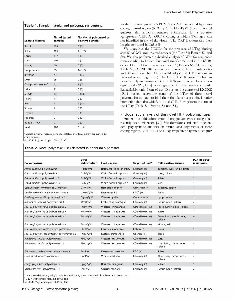
61/792 (8%) positive samples. Among the organs for which more
than 20 samples were available, spleen, lymph node, intestine, and
lung revealed the highest detection rates (20%, 16%, 15% and 7%,
respectively). In addition, 3/7 skin samples (43%) were PCR-
positive. In contrast, less than 4% of feces, blood, urine and kidney
samples were PCR-positive (Table 1). The amplified VP1
sequences were shown by BLAST analysis to originate from 24
distinct polyomaviruses, all exhibiting less than 90% nucleic acid
identity to each other, or to the corresponding region of known
polyomaviruses. Novel polyomaviruses were detected in eight
catarrhine (OWM and great apes) and four platyrrhine (NWM)
species, and were provisionally named according to their host
species as described in the Methods section.
We discovered 9 polyomaviruses in great ape species (six in
chimpanzees; two in the gorilla; one in orangutan). These viruses
were further characterized in the present study and are listed in
Table 2. Four additional great ape polyomaviruses (three of
chimpanzee; one of gorilla) showed a high similarity to the human
MCPyV. Their full-genome sequence was previously published
[30] (Table S3). Six chimpanzees were co-infected with combina-
tions of multiple chimpanzee polyomaviruses. In five OWM and
four NWM species we detected five and six novel polyomaviruses,
respectively (Table 2). In one NWM (white-fronted capuchin;
Cebus albifrons) co-infection was observed. Prosimian (strepsirrhine)
polyomaviruses were not detected, which may be a reflection of
the small (n = 20) sample size.
To characterize the complete genomes of the nine great ape and
11 monkey polyomaviruses, specific nested primers in an inverse
orientation were used to target the partial VP1 sequences obtained
from the initial degenerate primer PCR for long-distance PCR
amplification of the remaining virus genome. Complete genome
sequences of ten polyomaviruses were amplified and sequenced:
four from chimpanzees [Western chimpanzee (Pan troglodytes verus)
and Eastern chimpanzee (Pan troglodytes schweinfurthii)], three from
OWMs [Eastern red colobus (Piliocolobus rufomitratus), red-eared
guenon (Cercopithecus erythrotis) and crab-eating macaque (Macaca
fascicularis)] and three from NWMs [black spider monkey (Ateles
paniscus), white-fronted capuchin (Cebus albifrons) and common
squirrel monkey (Saimiri sciureus)]. Repeated amplification attempts
from the remaining ten polyomaviruses were unsuccessful, most
likely due to low genome copy numbers. The sequence informa-
tion of these ten complete genomes and ten partial VP1 sequences
has been deposited in the GenBank database. The accession
numbers are listed in Table S3.
The full-length genomes have a length of 4970 bp to 5349 bp
and exhibit the typical set of polyomavirus open reading frames
(ORFs). The early regions are comprised of two ORFs encoding
the non-structural proteins LTag and STag. The late regions code
Author Summary
Polyomaviruses are able to cause severe disease inimmunocompromised individuals. The discovery of Merkelcell polyomavirus and its association with Merkel cellcarcinoma has increased interest in these viruses, resultingin the identification of several novel human polyoma-viruses in recent years. The existence of one of theserecently identified viruses, human polyomavirus 9 (HPyV9),had been predicted nearly 30 years prior due to the abilityof human sera to neutralize infection of an African greenmonkey polyomavirus (Lymphotropic polyomavirus; LPyV).HPyV9 and LPyV are now known to be antigenically andphylogenetically closely related. We hypothesized thatnonhuman primate (NHP) polyomaviruses, in particularthose of the closely related chimpanzee, may serve asgenetic and immunological predictors for the existence ofyet unknown human polyomaviruses. In the present study,we discovered 20 novel NHP polyomaviruses, six of whichwere isolated from chimpanzees. Of the 9 chimpanzeepolyomaviruses now known, 5 do not presently have aclosely related human counterpart. Serologic reactivityagainst these novel chimpanzee viruses was observed inhumans from European and African populations. Fromthese data we predict that additional human polyoma-viruses exist which are genetically and serologically relatedto the novel chimpanzee polyomaviruses.
Predictors of Human Polyomaviruses
PLOS Pathogens | www.plospathogens.org 2 June 2013 | Volume 9 | Issue 6 | e1003429
for the structural proteins VP1, VP2 and VP3, separated by a non-
coding control region (NCCR). Only CeryPyV1 (from red-eared
guenon) also harbors sequence information for a putative
agnoprotein ORF. An ORF encoding a middle T-antigen was
not identified in any of the viruses. The ORF locations and their
lengths are listed in Table S4.
We examined the NCCRs for the presence of LTag binding
sites (GAGGC) and inverted repeats (see Text S1; Figures S1 and
S2). We also performed a detailed analysis of LTag for sequences
corresponding to known functional motifs described in the SV40-
derived form of the protein (see Text S2; Figures S3, S4, and S5;
Table S5). All NCCRs possess one or several LTag binding sites
and AT-rich stretches. Only the MfasPyV1 NCCR contains an
inverted repeat (Figure S1). The LTag of all 10 novel nonhuman
primate polyomaviruses contain a K/R-rich nuclear localization
signal and CR1, DnaJ, Zn-finger and ATPase consensus motifs.
Remarkably, only 6 out of the 10 possess the conserved LXCXE
pRb1 pocket, suggesting some of the LTag of these novel
polyomaviruses may not bind the retinoblastoma protein. Putative
interaction domains with Bub-1 and CUL-7 are present in some of
the LTag (Table S5; Figures S3 and S4).
Phylogenetic analysis of the novel NHP polyomavirusesAncient recombination events among polyomavirus lineages has
recently been evidenced [31]. We therefore conducted indepen-
dent phylogenetic analyses on amino acid alignments of three
coding regions, VP1, VP2 and LTag (respective alignment lengths:
Table 1. Sample material and polyomavirus content.
Sample materialNo. of testedsamples
No. (%) of polyomavirus-positive samples
Blood 139 2 (1)
Spleen 126 25 (20)
Feces 117 3 (3)
Lung 106 7 (7)
Kidney 70 0 (0)
Lymph node 63 10 (16)
Intestine 41 6 (15)
Liver 35 2 (6)
Chimp meal remaina 22 1 (5)
Urine 21 0 (0)
Muscle 17 3 (18)
Heart 11 0 (0)
Skin 7 3 (43)
Stomach 7 0 (0)
Thymus 5 0 (0)
Pancreas 3 0 (0)
Bone marrow 2 0 (0)
total 792 61 (8)
aMuscle or other tissues from red colobus monkeys partly consumed bychimpanzees.doi:10.1371/journal.ppat.1003429.t001
Table 2. Novel polyomaviruses detected in nonhuman primates.
PolyomavirusVirusabbreviation Host species Origin of hosta PCR-positive tissue(s)
PCR-positiveindividuals
Ateles paniscus polyomavirus 1 ApanPyV1 Red-faced spider monkey Germany (c) Intestine, liver, lung, spleen 1
Cebus albifrons polyomavirus 1 CalbPyV1 White-fronted capuchin Germany (c) Lung, spleen 1
Cebus albifrons polyomavirus 2 CalbPyV2 White-fronted capuchin Germany (c) Spleen 1
Cebus albifrons polyomavirus 3 CalbPyV3 White-fronted capuchin Germany (c) Skin 1
Cercopithecus erythrotis polyomavirus 1 CeryPyV1 Red-eared guenon Cameroon (w) Intestine, spleen 1
Gorilla beringei graueri polyomavirus 1 GbergPyV1 Eastern gorilla DRCb (w) Feces 1
Gorilla gorilla gorilla polyomavirus 2 GgorgPyV2 Western gorilla Cameroon (w) Lymph node 1
Macaca fascicularis polyomavirus 1 MfasPyV1 Crab-eating macaque Germany (c) Lymph node, spleen 2
Pan troglodytes verus polyomavirus 3 PtrovPyV3 Western chimpanzee Cote d’Ivoire (w) Feces, lymph node, spleen 1
Pan troglodytes verus polyomavirus 4 PtrovPyV4 Western chimpanzee Cote d’Ivoire (w) Spleen 1
Pan troglodytes verus polyomavirus 5 PtrovPyV5 Western chimpanzee Cote d’Ivoire (w) Feces, lung, lymph node,spleen
4
Pan troglodytes verus polyomavirus 6 PtrovPyV6 Western chimpanzee Cote d’Ivoire (w) Muscle, skin 1
Pan troglodytes troglodytes polyomavirus 1 PtrotPyV1 Central chimpanzee Gabon (s) Feces 1
Pan troglodytes schweinfurthii polyomavirus 2 PtrosPyV2 Eastern chimpanzee Uganda (s) Blood 1
Piliocolobus badius polyomavirus 1 PbadPyV1 Western red colobus Cote d’Ivoire (w) Lung 1
Piliocolobus badius polyomavirus 2 PbadPyV2 Western red colobus Cote d’Ivoire (w) Liver, lung, lymph node,spleen
4
Piliocolobus rufomitratus polyomavirus 1 PrufPyV1 Eastern red colobus DRC (w) Spleen 1
Pithecia pithecia polyomavirus 1 PpitPyV1 White-faced saki Germany (c) Blood, lung, lymph node,spleen
2
Pongo pygmaeus polyomavirus 1 PpygPyV1 Bornean orangutan Germany (c) Spleen 1
Saimiri sciureus polyomavirus 1 SsciPyV1 Squirrel monkey Germany (c) Lymph node, spleen 2
aLiving conditions: w, wild; c, held in captivity; s, born in the wild but kept in a sanctuary.bDRC = Democratic Republic of Congo.doi:10.1371/journal.ppat.1003429.t002
Predictors of Human Polyomaviruses
PLOS Pathogens | www.plospathogens.org 3 June 2013 | Volume 9 | Issue 6 | e1003429

244, 90 and 443 amino acids). All alignments were comprised from
the novel polyomaviruses and those currently available in
GenBank, including all known human polyomaviruses (as of
February 2013; Table S3). Maximum likelihood and Bayesian
analyses of these alignments were performed. This confirmed the
likely recombinant nature of some polyomaviruses and notably of
those belonging to the Wukipolyomavirus genus (Figure 1; Figures S6
and S7). In addition, it also revealed that primate polyomaviruses
were scattered over the entire polyomavirus tree, whether
considering VP1, VP2 or large T phylogenetic trees (Figure 1;
Figures S6 and S7). We identified 7 well-supported clades relevant
to the novel polyomaviruses described in this study (Figure 1;
Supplemental Figures S6 and S7; Table 3):
– Clade (a) comprised four NWM polyomaviruses, CalbPyV1
and CalbPyV2 from white-fronted capuchin (Cebus albifrons),
and SsciPyV1 and SqPyV1 from squirrel monkey (Saimiri
sciureus).
– Clade (b) consisted of three novel chimpanzee viruses
[PtrovPyV5 and 6 from Western chimpanzees (Pan troglodytes
verus) and PtrosPyV2 from Eastern chimpanzee (Pan troglodytes
schweinfurthii)] and two novel viruses from cercopithecids
[PbadPyV1 from Western red colobus (Piliocolobus badius),
MfasPyV1 from crab-eating macaques (Macaca fascicularis)]
which were associated to HPyV9 and LPyV.
– Clade (c) included two orangutan viruses (PpygPyV1 and
OraPyV1), a NWM polyomavirus [ApanPyV1 from red-faced
spider monkey (Ateles paniscus)] and the human-infecting
TSPyV.
– Clade (d) comprised a chimpanzee and a monkey polyomavirus
[PtrotPyV1 from Central chimpanzee (Pan troglodytes troglodytes)
and CeryPyV1 from red-eared guenon (Cercopithecus erythrotis)]
which formed a cluster with JCPyV, BKPyV, SV40 (from
rhesus monkey) and SA12 (from baboon).
– Clade (e) was constituted of four great ape polyomaviruses,
PtrovPyV3 and PtrovPyV4 from Western chimpanzees,
GgorgPyV2 from Western lowland gorilla (Gorilla gorilla gorilla)
and OraPyV2 from Sumatran orangutan.
– Clade (f) included two colobus viruses [PrufPyV1 from Eastern
red colobus (Piliocolobus rufomitratus) and PbadPyV2 from
Western red colobus] and the chimpanzee virus ChPyV.
– In clade (g) finally, GbergPyV1 from Eastern lowland gorilla
(Gorilla gorilla beringei), grouped with MCPyV and MCPyV-
related great ape polyomaviruses. These viruses were associ-
ated to two polyomaviruses detected in NWMs [CalbPyV3
from white-fronted capuchin and PpitPyV1 from white-faced
saki (Pithecia pithecia)], as well as to some bat polyomaviruses.
Reactivity of human sera against VP1 of chimpanzeepolyomaviruses
To study the reactivity of human sera against the NHP
polyomaviruses, VP1 proteins from four completely sequenced
Figure 1. Bayesian chronogram deduced from the analysis of a 244 amino acid alignment of VP1 sequences. Polyomaviruses wereidentified in humans (red), apes (blue), other primates (green), and other mammals and birds (black). Novel polyomaviruses identified in this study aremarked with a star and relevant clades to which they belong are highlighted by lettered circles. Viruses from which VP1 was used in serological assaysare highlighted by colored rectangles. The human polyomavirus MXPyV has the same phylogenetic position as HPyV10 and is not shown. Supportvalues are given above branches where posterior probability (pp) .0.95 and bootstrap values (Bp) .50. The tree presented is the maximum cladecredibility tree. The scale axis is indicated in amino acid substitutions per site.doi:10.1371/journal.ppat.1003429.g001
Predictors of Human Polyomaviruses
PLOS Pathogens | www.plospathogens.org 4 June 2013 | Volume 9 | Issue 6 | e1003429

chimpanzee polyomaviruses (ChPyV, PtrovPyV3, PtrovPyV4,
PtrosPyV2) with no close counterparts in humans were selected
for use in indirect ELISA [clades (b), (e) and (f) in Figure 1]. For
these studies, VP1 from JCPyV and HPyV9 were selected as
positive control proteins, and an avian polyomavirus [APyV, also
known as Budgerigar fledgling disease virus (BFDV)] [32] was chosen
as negative control. VP1 proteins expressed in E. coli are known to
form pentameric capsomer structures [33], and have proved
effective for analysis of polyomavirus serology [21,25,34,35]. To
serologically assess the level of ChPyV, PtrovPyV3, PtrovPyV4
and PtrosPyV2 circulating in chimpanzees, ELISA was performed
on plasma samples of 40 chimpanzees. A high seroprevalence was
shown for each virus (ChPyV, 100%; PtroPyV3; 73%; PtrosPyV4,
90%; PtrosPyV2, 88%). These results indicate that all 4
polyomaviruses are hosted by chimpanzees, with ChPyV being
the most prevalent (Figure 2). A serum panel from German
individuals and a plasma panel from individuals from Cote
d’Ivoire were then evaluated for their reactivity to the 4
chimpanzee polyomaviruses and to JCPyV and HPyV9. For the
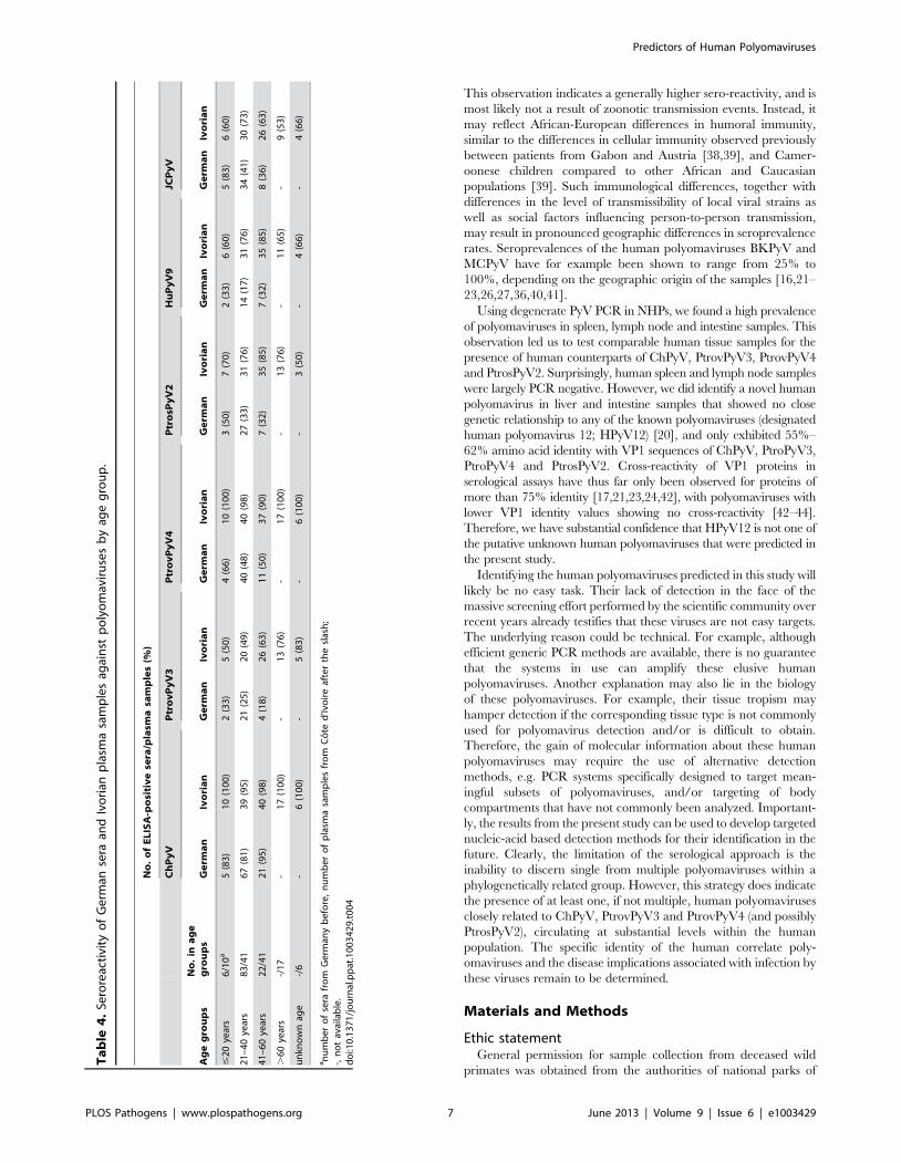
German sera (n = 111), the following seroreactivities were deter-
mined: ChPyV, 84%; PtrovPyV3, 24%; PtrovPyV4, 50%;
PtrosPyV2, 33%; HPyV9, 21%; JCPyV, 42% (Table 4). Fourteen
German sera (13%) exhibited seroreactivity against all four
chimpanzee polyomaviruses, and 14 samples (13%) were com-
pletely negative (Figure S8A). The Cote d’Ivoire plasma samples
(n = 115) showed more frequent reactivity: ChPyV, 97%; Ptrov-
PyV3, 60%; PtrovPyV4, 96%; PtrosPyV2, 77%; HPyV9, 76%;
JCPyV, 65% (Table 4). Each plasma reacted with at least one
chimpanzee polyomavirus, with fifty-three (46%) samples being
reactive against all four tested chimpanzee polyomaviruses (Figure
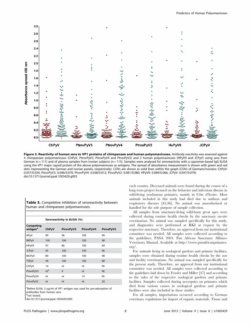
S8B). Comparison of German and Cote d’Ivoire samples revealed
that the seroprevalences were lower in the German samples
(P,0.001 for all viruses; Table 4; Figure 3). This difference was
also observed when analysis was age-restricted to individuals
between 20 and 60 years (P = 0.004 for ChPyV, P,0.001 for all
other viruses). Mean absorbance values were significantly lower in
German samples for all viruses (P,0.001 for all viruses) (Figure 3).
Age had no significant effect on sera/plasma absorbance values
against any virus in either Germans or individuals from Cote
d’Ivoire (P.0.05 for all viruses; Figure S9). To visualize possible
correlations of seroreactivity against the tested VP1 antigens,
OD450 values were plotted against each other, and correlation
analysis was performed to check for statistical evidence of cross-
reaction. Rank correlation showed only slight to moderate
correlations (0.178 to 0.62), and for none of the antigen pairs
was a correlation .0.5 measured for any serum/plasma panel
(Table S6), indicating the absence of marked cross-reactions.
Cross-reactivity between chimpanzee and humanpolyomaviruses
To assess possible antigenic cross-reactivity between the four
chimpanzee polyomaviruses and known human polyomaviruses,
competitive inhibition of seroreactivity was tested. Serum and
plasma samples (n = 5–7) reactive against VP1 of a particular
chimpanzee polyomavirus (ChPyV, PtrovPyV3, PtrovPyV4 or
PtrosPyV2) were tested by ELISA using the respective chimpanzee
polyomavirus VP1 as the antigen. Prior to use in the assay, all sera
were pre-adsorbed with soluble VP1 antigen from BKPyV,
HPyV9, JCPyV, MCPyV or TSPyV. Incubation with the soluble
homologous chimpanzee polyomavirus VP1, and soluble APyV
VP1 served as positive and negative controls, respectively. Pre-
adsorption with the homologous chimpanzee polyomavirus
antigens reduced the ELISA reactivity by approximately 80% or
more in all cases (Table 5), and pre-incubation with APyV VP1
had no effect on reactivity (data not shown). This showed efficacy
and specificity of the pre-adsorption procedure. With one
exception, pre-incubation with VP1 from human polyomaviruses
did not reduce reactivity of sera for VP1 of ChPyV, PtrovPyV3,
PtrovPyV4 or PtrosPyV2. In the one exception, pre-incubation of
PtrosPyV2-reactive human sera with soluble HPyV9-VP1 reduced
the PtrosPyV2-specific ELISA reactivity by 31%, indicating a
potential weak cross-reactivity (Table 5). This cross-reactivity was
consistent with presence of these two viruses in sister phylogenetic
clades (Figure 1), with their VP1 proteins showing 75% identity.
For this relatively high level of identity cross-reactive antibodies
have been detected [21,25]. However, the non-adsorbable
reactivity between PtrosPyV2 and HPyV9 (Table 5) implied –
beside HPyV9 – the involvement of PtrosPyV2 and/or another
unknown polyomavirus in the reactivity of human sera against
PtrosPyV2. In summary, the presence of reactivity in human sera
against VP1 from multiple NHP polyomaviruses with no currently
known human homologue supports the presence of one or more
unidentified human polyomaviruses phylogenetically related to
each of these novel NHP viruses.
Discussion
In the present study, multiple, hitherto unknown, highly diverse
polyomaviruses were detected in great apes and monkeys. These
viruses were localized mainly to lymphoid organs, lungs and
intestinal tissue (Tables 1 and 2; Table S1). In phylogenetic
analysis using VP1, VP2 and LTag antigen protein sequences, four
chimpanzee polyomaviruses (ChPyV, PtrovPyV3, PtrovPyV4,
PtrosPyV2) showed no close relationship to any of the known
human polyomaviruses, including the most recently discovered
human polyomaviruses HPyV10, MWPyV, MXPyV, STLPyV
and HPyV12 (Figure 1 and Figures S6 and S7, respectively).
Positive ELISA reactivities against the VP1 structural proteins of
these four chimpanzee polyomaviruses were observed in panels of
human sera/plasma samples. Experiments involving competitive
inhibition of seroreactivities with a panel of VP1 proteins from five
human polyomaviruses ruled out the presence of cross-reactivity
between the chimpanzee polyomaviruses and human polyoma-
viruses (except for a weak cross-reactivity between HPyV9 and
PtrosPyV2) (Table 5). This was confirmed by the lack of any
significant correlation of seroreactivity against the different
Table 3. Branch support values for selected clades in VP1,VP2 and large T phylogenetic analyses.
Clade VP1 VP2 Large T
aa 1/100b -/-c 1/100
B 1/86 1/98 1/100
C 1/74 1/97 1/100
D 1/98 1/89 1/100
E 1/97 1/92 1/100
F 1/85 1/90 1/94
G 1/95 nad na
aClades are designated by the same letter code as used in Figure 1.bBranch support values are given as posterior probabilities/bootstrap values.The corresponding phylogenetic trees are available as Figure 1 (VP1), Figure S6(VP2) and Figure S7 (large T).c-: not a clade in the corresponding analysis.dna: not applicable, i.e., none of the novel polyomaviruses included in group gallowed for whole genome recovery.doi:10.1371/journal.ppat.1003429.t003
Predictors of Human Polyomaviruses
PLOS Pathogens | www.plospathogens.org 5 June 2013 | Volume 9 | Issue 6 | e1003429

polyomavirus VP1 proteins for any of the sera/plasma samples
tested. Therefore, the reactivity of human sera against the four
chimpanzee polyomaviruses suggests that the majority of human
subjects tested have been exposed to as yet unknown polyoma-
viruses. The use of serology for the detection of unknown
polyomaviruses circulating within the human population is not
without precedent. Several research groups had observed that up
to 30% of human sera react against the monkey polyomavirus
LPyV [21,36,37]. About 30 years after the first observation, it was
discovered that human seroreactivity against LPyV was due to
infection by HPyV9 [25], a human polyomavirus closely related to
LPyV.
Ivorian plasma samples consistently showed higher levels of
VP1 reactivity compared to samples from German individuals
(Figure 3). One possible interpretation of this stronger reactivity is
that it reflects increased ‘spillover’ of NHP polyomaviruses into
humans, perhaps due to the possibility for closer interaction
between humans and NHP species. However, the Ivorian samples
reacted more strongly with all polyomaviruses investigated,
including VP1 from the two human viruses, JCPyV and HPyV9.
Figure 2. Reactivity of chimpanzee plasma samples to VP1 proteins of chimpanzee polyomaviruses. Antibody reactivity was assessedagainst the 4 chimpanzee polyomaviruses ChPyV, PtrovPyV3, PtrovPyV4 and PtrosPyV2 using plasma of 40 chimpanzees. Samples were analysed forseroreactivity with a capsomer-based IgG ELISA using the VP1 major capsid protein of the above polyomaviruses as antigens. The spread ofabsorbance measurement is shown with black dots, and cut-off values (COVs) are depicted with solid lines (PtrovPyV3: 0.028; PtrovPyV4: 0.023;PtrosPyV2: 0.013). A COV for ChPyV could not be calculated because all OD450 values were .0.3.doi:10.1371/journal.ppat.1003429.g002
Predictors of Human Polyomaviruses
PLOS Pathogens | www.plospathogens.org 6 June 2013 | Volume 9 | Issue 6 | e1003429
This observation indicates a generally higher sero-reactivity, and is
most likely not a result of zoonotic transmission events. Instead, it
may reflect African-European differences in humoral immunity,
similar to the differences in cellular immunity observed previously
between patients from Gabon and Austria [38,39], and Camer-
oonese children compared to other African and Caucasian
populations [39]. Such immunological differences, together with
differences in the level of transmissibility of local viral strains as
well as social factors influencing person-to-person transmission,
may result in pronounced geographic differences in seroprevalence
rates. Seroprevalences of the human polyomaviruses BKPyV and
MCPyV have for example been shown to range from 25% to
100%, depending on the geographic origin of the samples [16,21–
23,26,27,36,40,41].
Using degenerate PyV PCR in NHPs, we found a high prevalence
of polyomaviruses in spleen, lymph node and intestine samples. This
observation led us to test comparable human tissue samples for the
presence of human counterparts of ChPyV, PtrovPyV3, PtrovPyV4
and PtrosPyV2. Surprisingly, human spleen and lymph node samples
were largely PCR negative. However, we did identify a novel human
polyomavirus in liver and intestine samples that showed no close
genetic relationship to any of the known polyomaviruses (designated
human polyomavirus 12; HPyV12) [20], and only exhibited 55%–
62% amino acid identity with VP1 sequences of ChPyV, PtroPyV3,
PtroPyV4 and PtrosPyV2. Cross-reactivity of VP1 proteins in
serological assays have thus far only been observed for proteins of
more than 75% identity [17,21,23,24,42], with polyomaviruses with
lower VP1 identity values showing no cross-reactivity [42–44].
Therefore, we have substantial confidence that HPyV12 is not one of
the putative unknown human polyomaviruses that were predicted in
the present study.
Identifying the human polyomaviruses predicted in this study will
likely be no easy task. Their lack of detection in the face of the
massive screening effort performed by the scientific community over
recent years already testifies that these viruses are not easy targets.
The underlying reason could be technical. For example, although
efficient generic PCR methods are available, there is no guarantee
that the systems in use can amplify these elusive human
polyomaviruses. Another explanation may also lie in the biology
of these polyomaviruses. For example, their tissue tropism may
hamper detection if the corresponding tissue type is not commonly
used for polyomavirus detection and/or is difficult to obtain.
Therefore, the gain of molecular information about these human
polyomaviruses may require the use of alternative detection
methods, e.g. PCR systems specifically designed to target mean-
ingful subsets of polyomaviruses, and/or targeting of body
compartments that have not commonly been analyzed. Important-
ly, the results from the present study can be used to develop targeted
nucleic-acid based detection methods for their identification in the
future. Clearly, the limitation of the serological approach is the
inability to discern single from multiple polyomaviruses within a
phylogenetically related group. However, this strategy does indicate
the presence of at least one, if not multiple, human polyomaviruses
closely related to ChPyV, PtrovPyV3 and PtrovPyV4 (and possibly
PtrosPyV2), circulating at substantial levels within the human
population. The specific identity of the human correlate poly-
omaviruses and the disease implications associated with infection by
these viruses remain to be determined.
Materials and Methods
Ethic statementGeneral permission for sample collection from deceased wild
primates was obtained from the authorities of national parks of
Ta
ble
4.
Sero
reac
tivi
tyo
fG
erm
anse
raan
dIv
ori
anp
lasm
asa
mp
les
agai
nst
po
lyo
mav
iru
ses
by
age
gro
up
.
No
.o
fE
LIS
A-p
osi
tiv
ese
ra/p
lasm
asa
mp
les
(%)
Ch
Py
VP
tro
vP
yV
3P
tro
vP
yV
4P
tro
sPy
V2
Hu
Py
V9
JCP
yV
Ag
eg
rou
ps
No
.in
ag
eg
rou
ps
Ge
rma
nIv
ori
an
Ge
rma
nIv
ori
an
Ge
rma
nIv
ori
an
Ge
rma
nIv
ori
an
Ge
rma
nIv
ori
an
Ge
rma
nIv
ori
an
#2
0ye
ars
6/1
0a
5(8
3)
10
(10
0)
2(3
3)
5(5
0)
4(6
6)
10
(10
0)
3(5
0)
7(7
0)
2(3
3)
6(6
0)
5(8
3)
6(6
0)
21
–4
0ye
ars
83
/41
67
(81
)3
9(9
5)
21
(25
)2
0(4
9)
40
(48
)4
0(9
8)
27
(33
)3
1(7
6)
14
(17
)3
1(7
6)
34
(41
)3
0(7
3)
41
–6
0ye
ars
22
/41
21
(95
)4
0(9
8)
4(1
8)
26
(63
)1
1(5
0)
37
(90
)7
(32
)3
5(8
5)
7(3
2)
35
(85
)8
(36
)2
6(6
3)
.6
0ye
ars
-/1
7-
17
(10
0)
-1
3(7
6)
-1
7(1
00
)-
13
(76
)-
11
(65
)-
9(5
3)
un
kno
wn
age
-/6
-6
(10
0)
-5
(83
)-
6(1
00
)-
3(5
0)
-4
(66
)-
4(6
6)
an
um
be
ro
fse
rafr
om
Ge
rman
yb
efo
re,
nu
mb
er
of
pla
sma
sam
ple
sfr
om
Co
ted
’Ivo
ire
afte
rth
esl
ash
;-,
no
tav
aila
ble
.d
oi:1
0.1
37
1/j
ou
rnal
.pp
at.1
00
34
29
.t0
04
Predictors of Human Polyomaviruses
PLOS Pathogens | www.plospathogens.org 7 June 2013 | Volume 9 | Issue 6 | e1003429
each country. Deceased animals were found during the course of a
long term project focused on the behavior and infectious disease in
wild-living nonhuman primates, mainly in Cote d’Ivoire. Most
animals included in this study had died due to anthrax and
respiratory diseases [45,46]. No animal was anaesthetised or
handled for the sole purpose of sample collection.
All samples from sanctuary-living wild-born great apes were
collected during routine health checks by the sanctuary on-site
veterinarian. No animal was sampled specifically for this study,
and diagnostics were performed at RKI at request by the
respective sanctuary. Therefore, no approval from our institutional
committee was needed. All samples were collected according to
the guidelines: PASA 2004. Pan African Sanctuary Alliance
Veterinary Manual. Available at http://www.panafricanprimates.
org/.
For animals living in zoological gardens and primate facilities,
samples were obtained during routine health checks by the zoo
and facility veterinarians. No animal was sampled specifically for
the present study. Therefore, no approval from our institutional
committee was needed. All samples were collected according to
the guidelines laid down by Fowler and Miller [47] and according
to the rules of the respective zoological gardens and primate
facilities. Samples collected during necropsies on primates which
died from various causes in zoological gardens and primate
facilities were also included in these studies.
For all samples, importations occurred according to German
veterinary regulations for import of organic materials. Tissue and
Table 5. Competitive inhibition of seroreactivity betweenhuman and chimpanzee polyomaviruses.
Seroreactivity in ELISA (%)
Competingantigena ChPyV PtrovPyV3 PtrovPyV4 PtrosPyV2
APyV 99 96 100 99
BKPyV 100 100 100 98
HPyV9 97 86 100 69
JCPyV 95 100 100 96
MCPyV 89 100 100 98
TSPyV 94 100 100 88
ChPyV 14 nt nt Nt
PtrovPyV3 ntb 9 nt Nt
PtrovPyV4 nt nt 14 Nt
PtrosPyV2 nt nt nt 20
aBefore ELISA, 2 mg/ml of VP1 antigen was used for pre-adsorption ofantibodies from human sera.bnot tested.doi:10.1371/journal.ppat.1003429.t005
Figure 3. Reactivity of human sera to VP1 proteins of chimpanzee and human polyomaviruses. Antibody reactivity was assessed against4 chimpanzee polyomaviruses (ChPyV, PtrovPyV3, PtrovPyV4 and PtrosPyV2) and 2 human polyomaviruses (HPyV9 and JCPyV) using sera fromGerman (n = 111) and of plasma samples from Ivorian subjects (n = 115). Samples were analysed for seroreactivity with a capsomer-based IgG ELISAusing the VP1 major capsid protein of the above polyomaviruses as antigens. The spread of absorbance measurement is shown with green and reddots (representing the German and Ivorian panels, respectively). COVs are shown as solid lines within the graph (COVs of Germans/Ivorians: ChPyV:0.057/0.034; PtrovPyV3: 0.046/0.070; PtrovPyV4: 0.038/0.012; PtrosPyV2: 0.081/0.080; HPyV9: 0.089/0.066; JCPyV: 0.047/0.079).doi:10.1371/journal.ppat.1003429.g003
Predictors of Human Polyomaviruses
PLOS Pathogens | www.plospathogens.org 8 June 2013 | Volume 9 | Issue 6 | e1003429
blood samples were exported with the appropriate CITES
permissions from the respective country and Germany.
Plasma samples of human volunteers in Cote d’Ivoire were
sampled under the permission of the ministry of health of Cote
d’Ivoire and the Institute Pasteur Cote d’Ivoire. Written informed
consent was obtained from all participants of the study. The study
was performed in cooperation with local health professionals. The
aim of the study (specifically, to study broadly zoonotic diseases in
the region) was explained to the local population during various
educational campaigns. German serum samples were anonymous-
ly collected ‘residual materials’, and the collection was approved
by the ethics committee of the Charite - Universitatsmedizin
Berlin. All samples were collected according to the declaration of
Helsinki.
Human serum and plasma collectionPlasma samples (n = 115) from Cote d’Ivoire were collected
from 57 women and 58 men, (age range: 9–79 years; mean: 42
years; six samples without age information) participating in a
broad study to investigate zoonotic diseases at the human – wildlife
interface in Cote d’Ivoire. Serum samples (n = 111) were collected
from healthy German adults (55 female/56 male, age range: 20–
60 years; mean: 32 years) at the Charite University Hospital,
Berlin, Germany.
NHP sample collection and processingA total of 792 blood, fecal and tissue samples were collected
from live or deceased individuals of 44 primate species (apes,
OWMs, NWMs and prosimians) [30,48]. 316 samples originated
from wild primates in Africa (n = 313) and South America (n = 3),
54 samples from wild-born great apes housed in wildlife
sanctuaries in West and East Africa (n = 49) and Asia (n = 5).
422 samples derived from captive primates held in several
zoological gardens and primate facilities in Europe. Protection
measures for the collection of fecal samples and autopsies and
extraction of DNA of blood and tissue samples as well as fecal
samples were carried out as described previously [30]. Blood of 40
chimpanzees was collected in EDTA tubes living on Ngamba
Island Chimpanzee Sanctuary, Uganda, between 2001 and 2008
during the annual routine health checks under anaesthesia. Plasma
was separated by centrifugation at 3000 rpm for 10 minutes at
room temperature.
Amplification and sequencing of polyomavirus genomesequences
Two generic PCRs for polyomavirus identification [29,30] and
long-distance PCR for genome amplification, as well as PCR
product purification and sequencing were carried out as described
previously [30]. For each novel polyomavirus nested specific
primers for long-distance PCR were derived from the sequences
amplified with the generic PCR. The primer pairs are listed with
their annealing temperatures in Table S2.
Sequence analysisComplete/partial VP1, VP2 and LTag protein coding sequenc-
es generated for this study were translated into amino acids using
SeaView [49] before being assembled with representative
sequences of all polyomaviruses currently recognized as species
by the International Committee on Taxonomy of Viruses (ICTV
[32] or possibly qualifying as new species according to recent
publications.
The three sets of sequences were aligned with SeaView using
Muscle [50] and on the T-Coffee webserver using T-Coffee
[51,52]. CORE indices were computed for all alignments using
the T-Coffee webserver and the following command line: t_coffee
-infile = filename -output = html -score. Average scores were
comparable for the three protein alignments; Muscle alignments
were used in the following. Well-aligned blocks were selected using
Gblocks v0.91b [53] as implemented in SeaView, which resulted
in retaining 90, 244 and 443 positions from the initial VP2, VP1
and LTag alignments.
Best-fit models of amino acid evolution were determined using
ProtTest v3 [54]. The seven empirical matrices of substitution
rates implemented in BEAST v1.7.4 [55] were assessed in
combination with empirical or dataset-borne (+F) amino acid
frequencies and various hypotheses of rate variation along
sequences (rate heterogeneity, +G and/or proportion of invariant
sites, +I). Likelihoods were computed for all resulting 56 models
using the slow optimization option of ProtTest (parameter values,
branch lengths and topology were optimized). Best-fit models were
determined using a combination of statistics: Akaike information
criterion (AIC), corrected AIC and three Bayesian information
criteria (BIC). CpREV+ G was selected for VP2, WAG+I+G for
VP1, WAG+I+G+F for LTag.
Phylogenetic analyses were then performed under the given
models of amino acid evolution in ML and Bayesian frameworks.
ML analyses were performed with PhyML v3.0 [56] as
implemented on the PhyML webserver [57]. All analyses were
performed using the BEST RANDOM option, meaning that one
nearest-neighbor interchange (NNI) and one subtree pruning and
re-grafting (SPR) search were started using a BIONJ tree while five
additional SPR searches used random starting trees, the best of the
seven resulting trees being chosen as the output. Where applicable,
site-specific rate heterogeneity was modeled using a four-category
gamma law (+G4). Branch lengths and topologies were optimized.
Branch support was estimated by performing non-parametrical
bootstrapping (Bp; 500 pseudo-replicates).
Bayesian analyses were performed using BEAST v1.7.4 and the
associated suite of softwares [55]. For all analyses, a relaxed clock
model was implemented so as to account for among lineage rate
variation and a speciation model (birth-death model) was chosen
as depicting the shape of the trees. Two Markov chain Monte
Carlo (MCMC) runs of 10,000,000 generations were run under
these conditions for each alignment, sampling trees and numerical
values of model parameters every 1000 generations. Convergence
of the runs was checked with Tracer v1.5 (available at http://tree.
bio.ed.ac.uk/software/tracer/). Visual confirmation that the
stationary distribution had effectively been reached was obtained
for both runs (a plateau was observed). In addition, model
parameters apparently converged to undistinguishable distribu-
tions for both runs. Finally, combined effective sample sizes (ESS)
were above 200 for all parameters. Trees sampled after a visually
conservative burn-in of 1,000,000 generations were assembled into
a single file using LogCombiner v1.7.1 before the information that
this tree sample (in total 20000 trees) contained was summarized
onto the maximum clade credibility (MCC) tree with TreeAnno-
tator v1.7.4. Posterior probabilities (pp) were taken as branch
support values.
All trees presented in this article were made up with FigTree
v1.3.1 (available at http://tree.bio.ed.ac.uk/software/figtree/).
Expression and purification of recombinant proteinsThe sequences of the major capsid proteins VP1 of HPyV9,
BKPyV, JCPyV, MCPyV, TSPyV, APyV, ChPyV, PtroPyV3,
PtroPyV4 and PtrosPyV2 were codon-optimized, commercially
synthesized (MrGene GmbH, Regensburg, Germany) and
Predictors of Human Polyomaviruses
PLOS Pathogens | www.plospathogens.org 9 June 2013 | Volume 9 | Issue 6 | e1003429

expressed in E.coli K12 as pentameric structures as described
previously [25].
Serological analysisIgG ELISAs, including use of APyV VP1 as a negative control
to exclude non-specific seroreactivity (due to binding of antibodies
to conserved VP1 epitopes or due to unspecific binding),
estimation of cut-off values, calculation of the correlation of
antibody reactivity using the Spearman rank correlation test, and
adsorption assays with soluble VP1 capsomers were performed
essentially as described [25]. The only exceptions from the earlier
cited protocol were dilution of serum and plasma samples 1:100;
and, in adsorption assays, serum and plasma samples were
preincubated with 2 mg/ml of antigen.
Statistical analysesThe database was established in Excel for Windows before
being transferred into Stata (Stata/SE 10.0 for Windows, Stata
Corp, College Station, TX) for statistical analyses. Absorbance
values and prevalence of the individual viruses and the effect of age
and gender on absorbance values were analyzed using regression
models and Fischer exact test.
Provisional nomenclature, abbreviations and nucleotidesequence accession numbers of novel nonhumanprimate polyomaviruses
For the purpose of this paper, tentative names and abbreviations
for the novel NHP polyomaviruses were derived from species and
subspecies name of the host in which the virus was detected (for
example Pan troglodytes verus polyomavirus, PtrovPyV) and listed in
Table 2. Using this naming rationale, the MCPyV-related
polyomaviruses of Pan troglodytes verus, Pan troglodytes schweinfurthii
and Gorilla gorilla gorilla, published in our earlier study [30], were
renamed for consistency. Old names: GggPyV, PtvPyV, PtsPyV;
new names: GgorgPyV, PtrovPyV, PtrosPyV. Nucleotide se-
quence accession numbers of the novel NHP polyomaviruses are
listed in Table S3.
Supporting Information
Figure S1 Large T antigen-binding sites in NCCRs ofnovel NHP polyomaviruses. Sites are boxed.
(TIF)
Figure S2 Sequence homology between NCCRs. The
NCCRs of (a) MfasPyV1 and PtrovPyV5 and (b) PtrosPyV2 and
PtrovPyV5 were aligned. Identical nucleic acids are marked by
vertical lines, gaps by hyphens.
(TIF)
Figure S3 Alignment of the primary sequence of large Tantigens and their functional motifs from novel NHPpolyomaviruses. The LTag proteins of all novel NHP
polyomaviruses (with complete genomes amplified and sequenced)
were aligned with the LTAg of SV40. Functional motifs are
highlighted with different colors. The color code is shown below
the alignment.
(TIF)
Figure S4 Location of functional motifs in large Tantigen. LTag is represented by an open bar.
(TIF)
Figure S5 Amino acid sequence identity between thehost range domain of SV40 large T antigen and the C-
terminal region of CeryPyV1 large T antigen. Identical
amino acids are highlighted in yellow.
(TIF)
Figure S6 Bayesian chronogram deduced from theanalysis of a 90 amino acid alignment of VP2 sequences.Polyomaviruses were identified in humans (red), apes (blue), other
primates (green), and other mammals and birds (black). Novel
polyomaviruses identified in this study are marked with a star.
Viruses from which VP1 was used in serological assays are
highlighted by colored rectangles. Clades ‘a’ and ‘g’ (highlighted in
Figure 1) are not highlighted in this figure as a consequence of the
disruption of clade ‘a’ monophyly by BoPyV and the lack of
sequence for any of the novel polyomaviruses associated to
published ones within clade ‘g’. Support values are given above
branches where posterior probability (pp) .0,95 and bootstrap
values (Bp) .50. The tree presented is the maximum clade
credibility tree. The scale axis is presented as amino acid
substitutions per site.
(TIF)
Figure S7 Bayesian chronogram deduced from theanalysis of a 443 amino acid alignment of large Tsequences. Polyomaviruses were identified in humans (red), apes
(blue), other primates (green), and other mammals and birds (black).
Novel polyomaviruses identified in this study are marked with a star.
Viruses from which VP1 was used in serological assays are
highlighted by colored rectangles. Clade ‘g’ (highlighted in Figure 1)
is not highlighted in this figure as a consequence of the lack of
sequence for any of the novel polyomaviruses associated to published
ones within clade ‘g’. Support values are given above branches where
posterior probability (pp) .0.95 and bootstrap values (Bp) .50. The
tree presented is the maximum clade credibility tree. The scale axis is
presented as amino acid substitutions per site.
(TIF)
Figure S8 Multiple seroreactivities against chimpanzeepolyomaviruses in humans. German sera (A) and Ivorian
plasma samples (B) were tested for seroreactivity against ChPyV,
PtrovPyV3, PtrovPyV4 and PtrovPyV10. The graph displays
percentages of single and multiple reactivities.
(TIF)
Figure S9 Age-stratified reactivity of human sera to VP1proteins of chimpanzees and human polyomaviruses.Antibody reactivity against 2 human polyomaviruses (HPyV9 and
JCPyV) and 4 chimpanzee polyomaviruses (ChPyV, PtrovPyV3,
PtrovPyV4 and PtrosPyV2) of sera from German (n = 111) and of
plasma samples from Ivorian subjects (n = 115). Samples were
analysed for seroreactivity with a capsomer-based IgG ELISA
using the VP1 major capsid protein of the above polyomaviruses
as antigens. Absorbance spread measurements are shown as blue
dots, representing the German (left) and Ivorian panels (right),
respectively. The COV is shown as dashed line (values are given in
legend of Figure 3). Solid line within the graph: age trendline.
(TIF)
Table S1 Primate species and tissues tested withgeneric polyomavirus PCR.(DOC)
Table S2 Primers used for amplification of nonhumanprimate polyomaviruses.(DOC)
Table S3 Known and novel polyomaviruses used inphylogenetic analysis.(DOC)
Predictors of Human Polyomaviruses
PLOS Pathogens | www.plospathogens.org 10 June 2013 | Volume 9 | Issue 6 | e1003429
Table S4 Genomes and encoded proteins of the novelnonhuman primate polyomaviruses.(DOC)
Table S5 Putative functional motifs in the large T-antigens of the novel NHP polyomaviruses.(DOCX)
Table S6 Correlation of seroreactivities against VP1antigens of polyomaviruses.(DOC)
Text S1 LT-ag binding motifs in NCCR of novel NHPpolyomaviruses.(DOCX)
Text S2 Motifs in large T antigens of novel NHPpolyomaviruse.(DOCX)
Acknowledgments
This study was only possible with the international co-operation of multiple
countries, organizations and individuals. We wish to acknowledge the
Ivorian authorities for their long-term support, especially The Ministry of
the Environment and Forests, The Ministry of Research, The Directorship
of the Taı National Park, as well as The Swiss Research Center in Abidjan
and the Ethics Committee. For sample collection we are grateful to S.
Schenk, S. Metzger and the field assistants of the Taı Chimpanzee Project.
We thank The Uganda Wildlife Authority and The Uganda National
Council for Science and Technology for granting us permission to conduct
this research, and K. Zuberbuhler, F. Babweteera and R. Wittig for their
support. In Gabon, we acknowledge The Agence Nationale des Parcs
Nationaux (ANPN) and The Centre National de la Recherche Scientifique
et Technologique (CENAREST) for permission to conduct research in
Loango National Park and The Societe pour la Conservation et le
Developpement (SCD) and Wildlife Conservation Society (WCS) for
financial and logistical support. For assistance in the field we are thankful to
J. Head, L. Rabanal, L. Makaga, U. Bora, I. Bollini, Y. Nkombe, and the
other field assistants of The Loango Ape Project. We also thank The
Centre International de Recherches Medicales de Franceville (CIRMF),
especially E. Leroy and the Ministere des Eaux et Forets du Gabon. We
appreciate the help of the CITES authorities of Cote d’Ivoire, Uganda,
Republic of Congo and Germany in obtaining the necessary permission.
Last, but not least, we thank S. Liebmann, C. Walter, N. Yasmum, S.
Kondgen, A. Blasse, and C. Hedemann for their support in the laboratory.
Author Contributions
Conceived and designed the experiments: NS NFM CAK EVA DW JH
SAJL ECH CB MAJ SCS FHL BE. Performed the experiments: NS NFM
DW SCS BE. Analyzed the data: NS NFM CAK EVA DW JH KNC SAJL
ECH MR CB MAJ LM UM SCS FHL BE. Contributed reagents/
materials/analysis tools: NFM CAK EVA DW JH KNC SAJL LM ECH
MR CB SCS FHL BE. Wrote the paper: BE NS MAJ FHL SCS SAJL
UM.
References
1. Eddy BE (1969) Polyoma virus. In: Gard CHSM, K.F., editor. Virology
Monographs. New York: Springer-Verlag. pp 1–114.
2. Gross L (1983) Oncogenic viruses. Oxford: Pergamon Press.
3. Major EO (1996) Human polyomaviruses. In: Fields BNH, P.M.; Griffin, D.E.,
editors. fields Virology: Lippincott Williams & Wilkins.
4. Pipas JM (2009) SV40: Cell transformation and tumorigenesis. Virology 384:
294–303.
5. Damania B (2007) DNA tumor viruses and human cancer. Trends Microbiol 15:
38–44.
6. McLaughlin-Drubin ME, Munger K (2008) Viruses associated with human
cancer. Biochim Biophys Acta 1782: 127–150.
7. Abend JR, Jiang M, Imperiale MJ (2009) BK virus and human cancer: innocent
until proven guilty. Semin Cancer Biol 19: 252–260.
8. Jiang M, Abend JR, Johnson SF, Imperiale MJ (2009) The role of
polyomaviruses in human disease. Virology 384: 266–273.
9. Padgett BL, Walker DL, ZuRhein GM, Eckroade RJ, Dessel BH (1971)
Cultivation of papova-like virus from human brain with progressive multifocal
leucoencephalopathy. Lancet 1: 1257–1260.
10. Gardner SD, Field AM, Coleman DV, Hulme B (1971) New human
papovavirus (B.K.) isolated from urine after renal transplantation. Lancet 1:
1253–1257.
11. Brew BJ, Davies NW, Cinque P, Clifford DB, Nath A (2010) Progressive
multifocal leukoencephalopathy and other forms of JC virus disease. Nat Rev
Neurol 6: 667–679.
12. Feng H, Shuda M, Chang Y, Moore PS (2008) Clonal integration of a
polyomavirus in human Merkel cell carcinoma. Science 319: 1096–1100.
13. van der Meijden E, Janssens RW, Lauber C, Bouwes Bavinck JN, Gorbalenya
AE, et al. (2010) Discovery of a new human polyomavirus associated with
trichodysplasia spinulosa in an immunocompromized patient. PLoS Pathog 6:
e1001024.
14. Allander T, Andreasson K, Gupta S, Bjerkner A, Bogdanovic G, et al. (2007)
Identification of a third human polyomavirus. J Virol 81: 4130–4136.
15. Gaynor AM, Nissen MD, Whiley DM, Mackay IM, Lambert SB, et al. (2007)
Identification of a novel polyomavirus from patients with acute respiratory tract
infections. PLoS Pathog 3: e64.
16. Schowalter RM, Pastrana DV, Pumphrey KA, Moyer AL, Buck CB (2010)
Merkel cell polyomavirus and two previously unknown polyomaviruses are
chronically shed from human skin. Cell Host Microbe 7: 509–515.
17. Scuda N, Hofmann J, Calvignac-Spencer S, Ruprecht K, Liman P, et al. (2011)
A novel human polyomavirus closely related to the african green monkey-
derived lymphotropic polyomavirus. J Virol 85: 4586–4590.
18. Siebrasse EA, Reyes A, Lim ES, Zhao G, Mkakosya RS, et al. (2012)
Identification of MW polyomavirus, a novel polyomavirus in human stool. J Virol
86(19):10321–6.
19. Lim ES, Reyes A, Antonio M, Saha D, Ikumapayi UN, et al. (2012) Discovery of
STL polyomavirus, a polyomavirus of ancestral recombinant origin that encodes
a unique T antigen by alternative splicing. Virology 436(2):295–303.
20. Korup S, Rietscher J, Calvignac-Spencer S, Trusch F, Hofmann J, et al. (2013)
Identification of a novel human polyomavirus in organs of the gastrointestinal
tract. PLoS One 8: e58021.
21. Kean JM, Rao S, Wang M, Garcea RL (2009) Seroepidemiology of human
polyomaviruses. PLoS Pathog 5: e1000363.
22. Knowles WA, Pipkin P, Andrews N, Vyse A, Minor P, et al. (2003) Population-
based study of antibody to the human polyomaviruses BKV and JCV and the
simian polyomavirus SV40. J Med Virol 71: 115–123.
23. Stolt A, Sasnauskas K, Koskela P, Lehtinen M, Dillner J (2003) Seroepidemi-
ology of the human polyomaviruses. J Gen Virol 84: 1499–1504.
24. Viscidi RP, Rollison DE, Sondak VK, Silver B, Messina JL, et al. (2011) Age-
specific seroprevalence of Merkel cell polyomavirus, BK virus, and JC virus. Clin
Vaccine Immunol 18: 1737–1743.
25. Trusch F, Klein M, Finsterbusch T, Kuhn J, Hofmann J, et al. (2012)
Seroprevalence of human polyomavirus 9 and cross-reactivity to African green
monkey-derived lymphotropic polyomavirus. J Gen Virol 93: 698–705.
26. Brown P, Tsai T, Gajdusek DC (1975) Seroepidemiology of human
papovaviruses. Discovery of virgin populations and some unusual patterns of
antibody prevalence among remote peoples of the world. Am J Epidemiol 102:
331–340.
27. Viscidi RP, Clayman B (2006) Serological cross reactivity between polyomavirus
capsids. Adv Exp Med Biol 577: 73–84.
28. Calvignac-Spencer S, Leendertz SA, Gillespie TR, Leendertz FH (2012) Wild
great apes as sentinels and sources of infectious disease. Clin Microbiol Infect 18:
521–527.
29. Johne R, Enderlein D, Nieper H, Muller H (2005) Novel polyomavirus detected
in the feces of a chimpanzee by nested broad-spectrum PCR. J Virol 79: 3883–
3887.
30. Leendertz FH, Scuda N, Cameron KN, Kidega T, Zuberbuhler K, et al. (2011)
African great apes are naturally infected with polyomaviruses closely related to
Merkel cell polyomavirus. J Virol 85: 916–924.
31. Tao Y, Mang S, Conrardy C, Kuzmin IV, Recuenco S, et al. (2012) Discovery
of Diverse Polyomaviruses in Bats and the Evolutionary History of the
Polyomaviridae. J Gen Virol 94: 738–48.
32. Johne R, Buck CB, Allander T, Atwood WJ, Garcea RL, et al. (2011)
Taxonomical developments in the family Polyomaviridae. Arch Virol 156:
1627–1634.
33. Salunke DM, Caspar DL, Garcea RL (1989) Polymorphism in the assembly of
polyomavirus capsid protein VP1. Biophys J 56: 887–900.
34. Liu M, Zhang T, Zhang Y, Meng F, Li X, et al. (2010) Development and
evaluation of a VP1-ELISA for detection of antibodies to duck hepatitis type 1
virus. J Virol Methods 169: 66–69.
35. van der Meijden E, Kazem S, Burgers MM, Janssens R, Bouwes Bavinck JN,
et al. (2011) Seroprevalence of trichodysplasia spinulosa-associated polyoma-
virus. Emerg Infect Dis 17: 1355–1363.
36. Brade L, Muller-Lantzsch N, zur Hausen H (1981) B-lymphotropic papovavirus
and possibility of infections in humans. J Med Virol 6: 301–308.
Predictors of Human Polyomaviruses
PLOS Pathogens | www.plospathogens.org 11 June 2013 | Volume 9 | Issue 6 | e1003429

37. Takemoto KK, Segawa K (1983) A new monkey lymphotropic papovavirus:
characterization of the virus and evidence of a related virus in humans. ProgClin Biol Res 105: 87–96.
38. Wilfing A, Winkler S, Schrattbauer K, Willheim M, Baier K, et al. (2001)
African-European differences in the capacity of T-cell cytokine production.Am J Trop Med Hyg 65: 504–509.
39. Sagnia B, Ateba Ndongo F, Ndiang Moyo Tetang S, Ndongo Torimiro J, CairoC, et al. (2011) Reference values of lymphocyte subsets in healthy, HIV-negative
children in Cameroon. Clin Vaccine Immunol 18: 790–795.
40. Carter JJ, Paulson KG, Wipf GC, Miranda D, Madeleine MM, et al. (2009)Association of Merkel cell polyomavirus-specific antibodies with Merkel cell
carcinoma. J Natl Cancer Inst 101: 1510–1522.41. Pastrana DV, Tolstov YL, Becker JC, Moore PS, Chang Y, et al. (2009)
Quantitation of human seroresponsiveness to Merkel cell polyomavirus. PLoSPathog 5: e1000578.
42. Chen T, Mattila PS, Jartti T, Ruuskanen O, Soderlund-Venermo M, et al.
(2011) Seroepidemiology of the newly found trichodysplasia spinulosa-associatedpolyomavirus. J Infect Dis 204: 1523–1526.
43. Touze A, Gaitan J, Arnold F, Cazal R, Fleury MJ, et al. (2010) Generation ofMerkel cell polyomavirus (MCV)-like particles and their application to detection
of MCV antibodies. J Clin Microbiol 48: 1767–1770.
44. Tolstov YL, Pastrana DV, Feng H, Becker JC, Jenkins FJ, et al. (2009) HumanMerkel cell polyomavirus infection II. MCV is a common human infection that
can be detected by conformational capsid epitope immunoassays. Int J Cancer125: 1250–1256.
45. Koendgen S, Kuhl H, N’Goran PK, Walsh PD, Schenk S, et al. (2008)Pandemic human viruses cause decline of endangered great apes. Curr Biol 18:
260–264.
46. Leendertz FH, Ellerbrok H, Boesch C, Couacy-Hymann E, Matz-Rensing K,et al. (2004) Anthrax kills wild chimpanzees in a tropical rainforest. Nature 430:
451–452.
47. Fowler ME, Miller RE (2008) Zoo and Wildlife Medicine. St. Louis: Saunders.
48. Wevers D, Metzger S, Babweteera F, Bieberbach M, Boesch C, et al. (2011)
Novel adenoviruses in wild primates: a high level of genetic diversity and
evidence of zoonotic transmissions. J Virol 85: 10774–10784.
49. Gouy M, Guindon S, Gascuel O (2010) SeaView version 4: A multiplatform
graphical user interface for sequence alignment and phylogenetic tree building.
Mol Biol Evol 27: 221–224.
50. Edgar RC (2004) MUSCLE: multiple sequence alignment with high accuracy
and high throughput. Nucleic Acids Res 32: 1792–1797.
51. Notredame C, Higgins DG, Heringa J (2000) T-Coffee: A novel method for fast
and accurate multiple sequence alignment. J Mol Biol 302: 205–217.
52. Di Tommaso P, Moretti S, Xenarios I, Orobitg M, Montanyola A, et al. (2011)
T-Coffee: a web server for the multiple sequence alignment of protein and RNA
sequences using structural information and homology extension. Nucleic Acids
Res 39: W13–17.
53. Talavera G, Castresana J (2007) Improvement of phylogenies after removing
divergent and ambiguously aligned blocks from protein sequence alignments.
Syst Biol 56: 564–577.
54. Abascal F, Zardoya R, Posada D (2005) ProtTest: selection of best-fit models of
protein evolution. Bioinformatics 21: 2104–2105.
55. Drummond AJ, Suchard MA, Xie D, Rambaut A (2012) Bayesian phylogenetics
with BEAUti and the BEAST 1.7. Mol Biol Evol 29: 1969–1973.
56. Guindon S, Dufayard JF, Lefort V, Anisimova M, Hordijk W, et al. (2010) New
algorithms and methods to estimate maximum-likelihood phylogenies: assessing
the performance of PhyML 3.0. Syst Biol 59: 307–321.
57. Guindon S, Lethiec F, Duroux P, Gascuel O (2005) PHYML Online–a web
server for fast maximum likelihood-based phylogenetic inference. Nucleic Acids
Res 33: W557–559.
Predictors of Human Polyomaviruses
PLOS Pathogens | www.plospathogens.org 12 June 2013 | Volume 9 | Issue 6 | e1003429
Related Documents