Molecules 2010, 15, 2187-2202; doi:10.3390/molecules15042187 molecules ISSN 1420-3049 www.mdpi.com/journal/molecules Article Novel Indole-Based Analogs of Melatonin: Synthesis and in Vitro Antioxidant Activity Studies Hanif Shirinzadeh 1 , Burcu Eren 2 , Hande Gurer-Orhan 2 , Sibel Suzen 1, * and Seçkin Özden 1 1 Department of Pharmaceutical Chemistry, Faculty of Pharmacy, Ankara University, 06100 Tandogan, Ankara, Turkey 2 Department of Pharmaceutical Toxicology, Faculty of Pharmacy, Ege University, 35100 Bornova, Izmir, Turkey * Author to whom correspondence should be addressed; E-Mail: [email protected]; Tel.: +90 312 2033074; Fax: +90 312 2131081. Received: 19 January 2010; in revised form: 15 March 2010 / Accepted: 23 March 2010 / Published: 29 March 2010 Abstract: The aim of this study was to synthesize and examine possible in vitro antioxidant effects of indole-based melatonin analogue compounds. As a part of our ongoing study nineteen indole hydrazide/hydrazone derivatives were synthesized, characterized and their in vitro antioxidant activity was investigated by three different assays: by evaluating their reducing effect against oxidation of a redox sensitive fluorescent probe, by examining their protective effect against H 2 O 2 -induced membrane lipid peroxidation and by determining their inhibitory effect on AAPH–induced hemolysis of human erythrocytes. The results indicated significant strong antioxidant activity for most of the compounds, when compared to melatonin. Keywords: indole; hydrazone; hydrazide; melatonin; synthesis; antioxidant activity 1. Introduction Antioxidants are molecules which can interact with free radicals and stop their chain reactions before important and essential molecules are damaged. As oxidative stress is an important part of many diseases, the use of antioxidants is intensively studied in medicinal chemistry, particularly as treatments for vital diseases such as stroke, cancer and neurodegenerative diseases. Free radicals are produced basically during cellular metabolism and some functional activities and have essential roles OPEN ACCESS

Welcome message from author
This document is posted to help you gain knowledge. Please leave a comment to let me know what you think about it! Share it to your friends and learn new things together.
Transcript
Molecules 2010, 15, 2187-2202; doi:10.3390/molecules15042187
molecules ISSN 1420-3049
www.mdpi.com/journal/molecules
Article
Novel Indole-Based Analogs of Melatonin: Synthesis and in Vitro Antioxidant Activity Studies
Hanif Shirinzadeh 1, Burcu Eren 2, Hande Gurer-Orhan 2, Sibel Suzen 1,* and Seçkin Özden 1
1 Department of Pharmaceutical Chemistry, Faculty of Pharmacy, Ankara University, 06100
Tandogan, Ankara, Turkey 2 Department of Pharmaceutical Toxicology, Faculty of Pharmacy, Ege University, 35100 Bornova,
Izmir, Turkey
* Author to whom correspondence should be addressed; E-Mail: [email protected];
Tel.: +90 312 2033074; Fax: +90 312 2131081.
Received: 19 January 2010; in revised form: 15 March 2010 / Accepted: 23 March 2010 /
Published: 29 March 2010
Abstract: The aim of this study was to synthesize and examine possible in vitro
antioxidant effects of indole-based melatonin analogue compounds. As a part of our
ongoing study nineteen indole hydrazide/hydrazone derivatives were synthesized,
characterized and their in vitro antioxidant activity was investigated by three different
assays: by evaluating their reducing effect against oxidation of a redox sensitive
fluorescent probe, by examining their protective effect against H2O2-induced membrane
lipid peroxidation and by determining their inhibitory effect on AAPH–induced hemolysis
of human erythrocytes. The results indicated significant strong antioxidant activity for
most of the compounds, when compared to melatonin.
Keywords: indole; hydrazone; hydrazide; melatonin; synthesis; antioxidant activity
1. Introduction
Antioxidants are molecules which can interact with free radicals and stop their chain reactions
before important and essential molecules are damaged. As oxidative stress is an important part of
many diseases, the use of antioxidants is intensively studied in medicinal chemistry, particularly as
treatments for vital diseases such as stroke, cancer and neurodegenerative diseases. Free radicals are
produced basically during cellular metabolism and some functional activities and have essential roles
OPEN ACCESS
Molecules 2010, 15
2188
in cell signaling, apoptosis and gene expression. On the other hand, excessive free radical attack can
damage DNA, proteins and lipids, resulting very important diseases. Antioxidants can decrease the
oxidative damage by reacting with free radicals or by inhibiting their activity.
It is well known that indole derivatives, extensively present in natural compounds, are very
important substances for their medicinal and biological aspects. Antioxidant effects of the indole ring-
containing substance melatonin (MLT) have been well described and evaluated by Tan et al. [1]. It is a
highly conserved molecule that it acts as a free radical scavenger and a broad-spectrum antioxidant [2].
Studies also showed the role of MLT and its derivatives in many physiological processes and
therapeutic functions, such as the regulation of circadian rhythm and immune functions [3–6].
Melatonin is known to be a potent in vitro antioxidant as well as powerful in vivo radical scavenger. In
in vitro conditions melatonin exhibited potent antioxidant activity in a linoleic acid emulsion system. It
also showed potent superoxide radical scavenging activity, higher than either quercetin or BHT [7].
Recent research has proved that the indole ring in the MLT molecule is the reactive center dealing
with oxidants due to its high resonance stability and very low activation energy barrier towards free
radical reactions [8–10]. Indole is found to reduce cisplatin-induced reactive oxygen species formation
[11] and scavenge hydroxyl radical directly [12]. It was shown that alkyl-substituted indole derivatives
can trap ABTS+• and DPPH indicating that the alkyl group attached to indole is of importance for the
antioxidant activity [13]. The hydrogen peroxide scavenging activity of melatonin showed that the
scavenging activity augmented with increasing concentrations of melatonin. This result may be
illustrative of melatonin's ability to inhibit lipid peroxidation [14]. A series of dihydroindenoindole
derivatives containing methoxy, halogen, and hydroxyl groups was synthesized and showed effective
inhibition against DPPH , ABTS +, DMPD +, and superoxide anion radicals compared to standard
antioxidants [15]. 2-Phenylindole derivatives significantly inhibited lipid peroxidation at 10-3 M
concentration [16]. Indole-3-propionamide derivatives also exhibited important antioxidant activity
compared to melatonin [17]. A large variety of synthetic compounds have been identified as potent in
vitro antioxidants, yet many of these compounds have not provided great clinical benefits, and some
produced side effects. In our earlier study [18] two sets of indole derivatives, with changes in the
5-methoxy and 2-acylaminoethyl groups of MLT were synthesized and tested for their in vitro
antioxidant potency in the DPPH, superoxide dismutase and lipid peroxidation (LP) assays. With a few
exceptions most of the compounds tested showed significant antioxidant activity at concentrations
comparable with or much higher than that of MLT. These results prompted us to synthesize more
indol-3-aldehyde hydrazone and hydrazide derivatives.
As a part of our ongoing study nineteen indole-based MLT analogue hydrazide/hydrazone
derivatives were now synthesized and their antioxidant activity was investigated in vitro by three
different assays: by evaluating their reducing effect against oxidation of a redox sensitive fluorescent
probe, DCFH-DA, by investigating their protective effect against H2O2-induced membrane lipid
peroxidation and by determining their inhibitory effect on 2,2’-azobis(2-amidinopropane
hydrochloride) (AAPH) –induced hemolysis of human erythrocytes. Human erythrocytes were chosen
as a biological model because they are readily available cells that are sensitive to oxidative damage.
The results were compared with MLT. All the indole-based MLT analogue compounds except those
previously synthesized (1a [19,20], 1j [21] and 1r [22]) were characterized on the basis of 1H- and 13C-NMR, mass and FT-IR spectra and elemental analysis.
Molecules 2010, 15
2189
2. Results and Discussion
The present work aimed to synthesize, characterize and investigate the potential antioxidant effects
of indole-based MLT analogue hydrazide/hydrazone derivatives by several in vitro test models. Based
on MLT, N-acetyl-5-methoxytryptamine, a well-known antioxidant, free radical scavenger, and
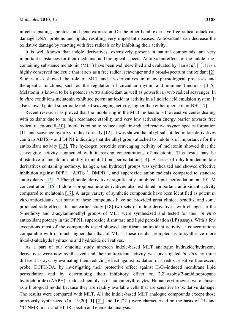
neuroprotectant, new indole imines were developed. Three parts of the MLT molecule were modified
to develop new indole-based MLT analogue compounds. These modifications were done mainly on the
acylamino group (Figure 1).
Figure 1. Parts of the MLT molecule modified to develop new indole-based MLT
analogue compounds.
These chemically significant modulations of the lead structure were made at three different points:
the methoxy group replaced with hydrogen at the 5-position of the indole ring (modification I) and
acetylaminoethyl side chain modified by formation of imine (modification II) and hydrogen replaced
with methyl on nitrogen (modification III). Particular attention was dedicated to the role of the 5-
methoxy group, which was eliminated. These modifications resulted in a set of compounds having
different physical properties, lipophilicity and different substitution patterns on the indole nucleus.
This helped to investigate the effect of substituents with different electronic and lipophilic properties
on the antioxidant activity of new indole derivatives (Scheme 1).
Molecules 2010, 15
2190
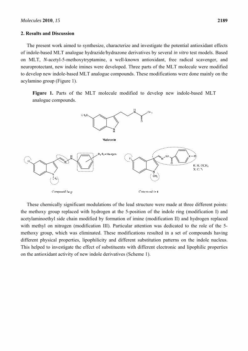
Scheme 1. Synthetic route to indole-based melatonin analogue hydrazide/hydrazone derivatives.
2.1. Effects of synthesized indole derivatives on cellular ROS
The protective effect of newly synthesized indole-based MLT analogue against DCFH-DA
oxidation was determined in human erythrocytes that were preloaded with the fluorescent probe. In
cells, DCFH-DA locates in the cytosol and reflects cellular ROS formation. Oxidation of the probe
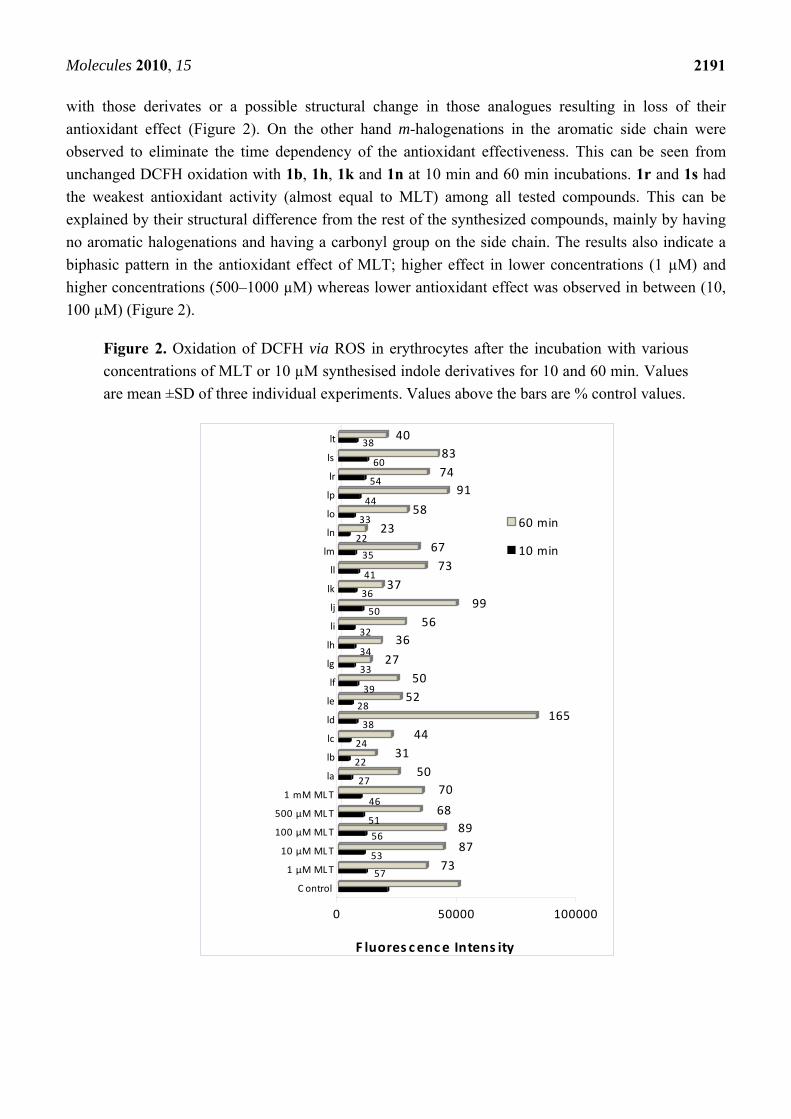
was screened at various time intervals up to 60 min. At 10 min incubation all the synthesized indole
derivatives except 1r and 1s were found to have potent antioxidant activity, even higher than MLT
itself. Among those synthesized indoles with changes in the aromatic side chain, p-halogenations were
found to decrease antioxidant activity compared to o- and m- substitution of the same halogen atom
(Figure 2). A significant further decrease in the antioxidant activity of p-halogenated compounds was
observed at 60 min incubation indicating a possible oxido-reductive reaction took place in incubations
Molecules 2010, 15
2191
with those derivates or a possible structural change in those analogues resulting in loss of their
antioxidant effect (Figure 2). On the other hand m-halogenations in the aromatic side chain were
observed to eliminate the time dependency of the antioxidant effectiveness. This can be seen from
unchanged DCFH oxidation with 1b, 1h, 1k and 1n at 10 min and 60 min incubations. 1r and 1s had
the weakest antioxidant activity (almost equal to MLT) among all tested compounds. This can be
explained by their structural difference from the rest of the synthesized compounds, mainly by having
no aromatic halogenations and having a carbonyl group on the side chain. The results also indicate a
biphasic pattern in the antioxidant effect of MLT; higher effect in lower concentrations (1 µM) and
higher concentrations (500–1000 µM) whereas lower antioxidant effect was observed in between (10,
100 µM) (Figure 2).
Figure 2. Oxidation of DCFH via ROS in erythrocytes after the incubation with various
concentrations of MLT or 10 µM synthesised indole derivatives for 10 and 60 min. Values
are mean ±SD of three individual experiments. Values above the bars are % control values.
5773
5387
5689
5168
4670
2750
2231
2444
38165
2852
3950
3327
3436
3256
5099
3637
4173
3567
2223
3358
4491
5474
6083
3840
0 50000 100000
C ontrol
1 μM MLT
10 μM MLT
100 μM MLT
500 μM MLT
1 mM MLT
la
lb
lc
ld
le
lf
lg
lh
li
lj
lk
ll
lm
ln
lo
lp
lr
ls
lt
60 min
10 min
F luores cence Intens ity
Molecules 2010, 15
2192
2.2. Inhibitory effect of synthesized indole derivatives on hydrogen peroxide-induced peroxidation of
human erythrocyte membranes
Once ROS are formed, one of the subsequent detrimental outcomes is peroxidation and oxidative
destruction of polyunsaturated fatty acids (PUFA) in cell membranes. The process is initiated by an
oxidizing radical that is capable of abstracting one hydrogen atom from PUFA. After several
rearrangement and oxidation reactions, generated lipid hydroperoxides decompose to a wide range of
products, mainly small molecule alkanes and aldehydes. Among those aldehydes, malondialdehyde
(MDA), assayed by the thiobarbituric acid (TBA) assay, is the most widely used biomarker of
oxidative damage to lipids. In the present study we investigated protective effect of synthesized indole
derivatives on H2O2-induced MDA formation in erythrocyte membranes. Figure 3 shows the inhibitory
effect of MLT and the synthesized indole derivatives on H2O2-induced peroxidation of human
erythrocytes in vitro.
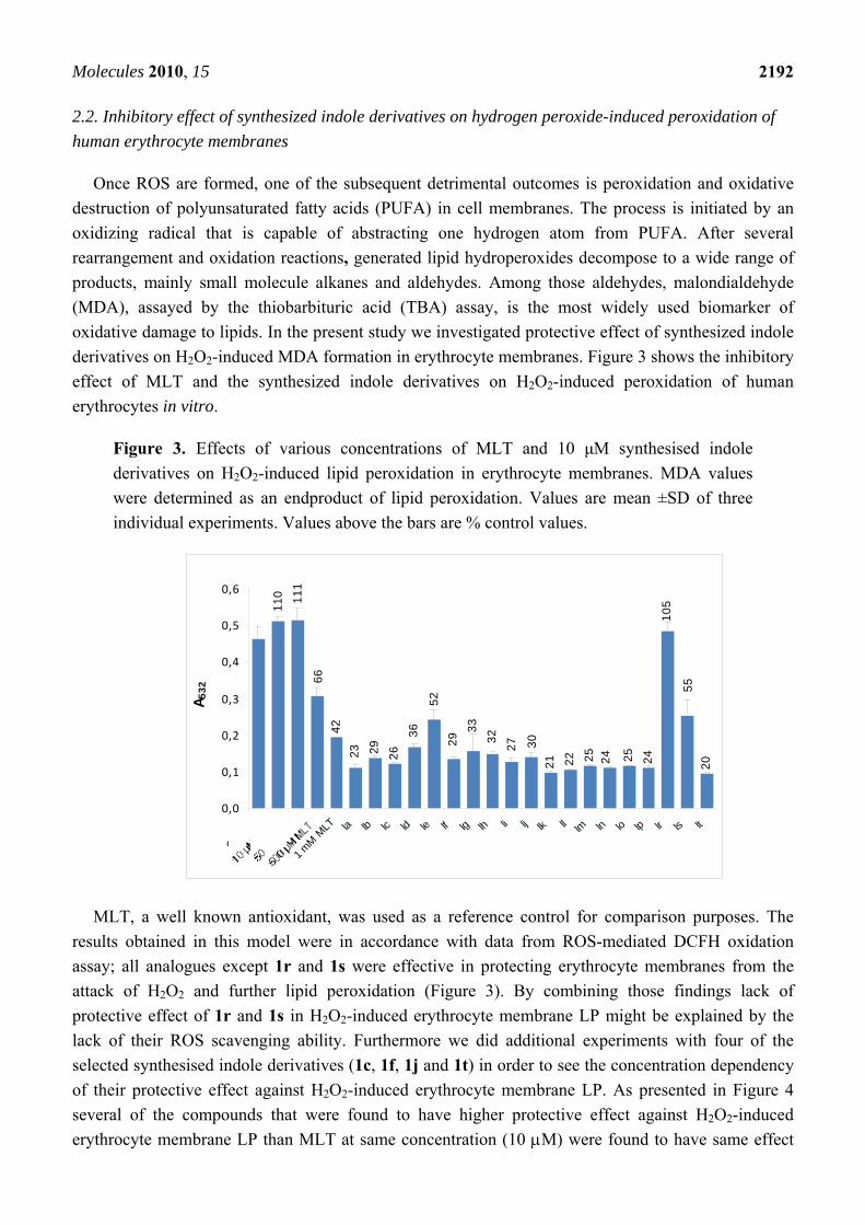
Figure 3. Effects of various concentrations of MLT and 10 μM synthesised indole
derivatives on H2O2-induced lipid peroxidation in erythrocyte membranes. MDA values
were determined as an endproduct of lipid peroxidation. Values are mean ±SD of three
individual experiments. Values above the bars are % control values.
11
0 11
1
66
42
23 29
26
36
52
29
33
32
27 30
21 22 25
24 25
24
10
5
55
20
0,0
0,1
0,2
0,3
0,4
0,5
0,6
Contro
l
1 m
M M
LT la lb lc ld le lf lg lh li lj lk ll lm ln lo lp lr ls lt
A5
32
MLT, a well known antioxidant, was used as a reference control for comparison purposes. The
results obtained in this model were in accordance with data from ROS-mediated DCFH oxidation
assay; all analogues except 1r and 1s were effective in protecting erythrocyte membranes from the
attack of H2O2 and further lipid peroxidation (Figure 3). By combining those findings lack of
protective effect of 1r and 1s in H2O2-induced erythrocyte membrane LP might be explained by the
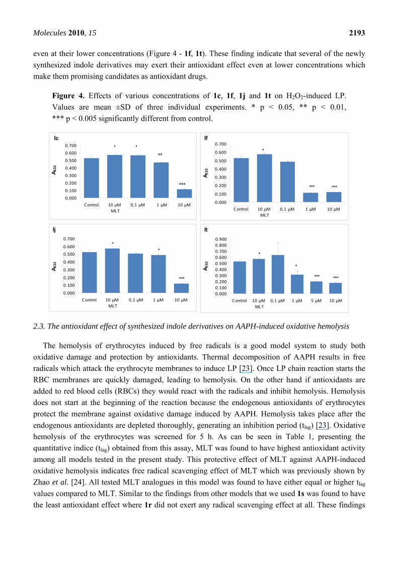
lack of their ROS scavenging ability. Furthermore we did additional experiments with four of the
selected synthesised indole derivatives (1c, 1f, 1j and 1t) in order to see the concentration dependency
of their protective effect against H2O2-induced erythrocyte membrane LP. As presented in Figure 4
several of the compounds that were found to have higher protective effect against H2O2-induced
erythrocyte membrane LP than MLT at same concentration (10 M) were found to have same effect
Molecules 2010, 15
2193
even at their lower concentrations (Figure 4 - 1f, 1t). These finding indicate that several of the newly
synthesized indole derivatives may exert their antioxidant effect even at lower concentrations which
make them promising candidates as antioxidant drugs.
Figure 4. Effects of various concentrations of 1c, 1f, 1j and 1t on H2O2-induced LP.
Values are mean ±SD of three individual experiments. * p < 0.05, ** p < 0.01,
*** p < 0.005 significantly different from control.
2.3. The antioxidant effect of synthesized indole derivatives on AAPH-induced oxidative hemolysis
The hemolysis of erythrocytes induced by free radicals is a good model system to study both
oxidative damage and protection by antioxidants. Thermal decomposition of AAPH results in free
radicals which attack the erythrocyte membranes to induce LP [23]. Once LP chain reaction starts the
RBC membranes are quickly damaged, leading to hemolysis. On the other hand if antioxidants are
added to red blood cells (RBCs) they would react with the radicals and inhibit hemolysis. Hemolysis
does not start at the beginning of the reaction because the endogenous antioxidants of erythrocytes
protect the membrane against oxidative damage induced by AAPH. Hemolysis takes place after the
endogenous antioxidants are depleted thoroughly, generating an inhibition period (tlag) [23]. Oxidative
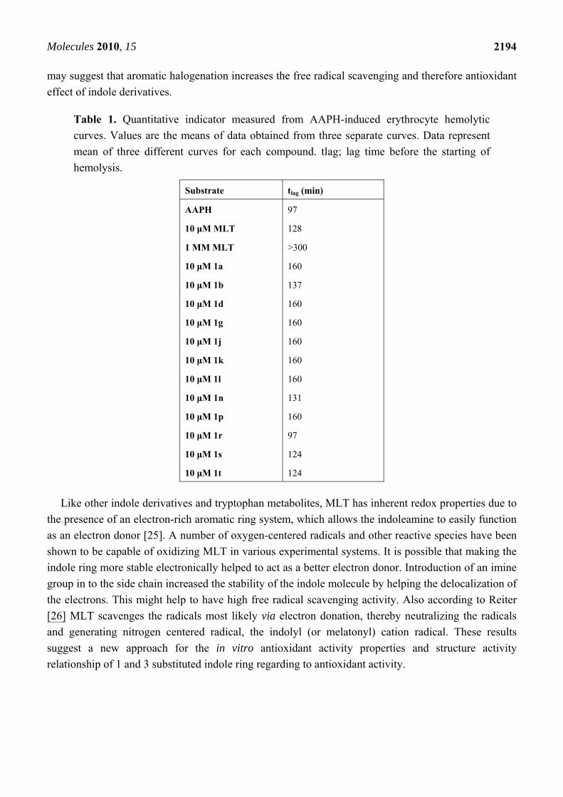
hemolysis of the erythrocytes was screened for 5 h. As can be seen in Table 1, presenting the
quantitative indice (tlag) obtained from this assay, MLT was found to have highest antioxidant activity
among all models tested in the present study. This protective effect of MLT against AAPH-induced
oxidative hemolysis indicates free radical scavenging effect of MLT which was previously shown by
Zhao et al. [24]. All tested MLT analogues in this model was found to have either equal or higher tlag
values compared to MLT. Similar to the findings from other models that we used 1s was found to have
the least antioxidant effect where 1r did not exert any radical scavenging effect at all. These findings
0.000
0.100
0.200
0.300
0.400
0.500
0.600
0.700
C ontrol 10 μM
MLT
0,1 μM 1 μM 10 μM
A5
32
lc
***
**
**
******
0.000
0.100
0.200
0.300
0.400
0.500
0.600
0.700
C ontrol 10 μM
MLT
0,1 μM 1 μM 10 μM A
53
2
lf
*
***
*
*
0.000
0.100
0.200
0.300
0.400
0.500
0.600
0.700
C ontrol 10 μM
MLT
0,1 μM 1 μM 10 μM
A5
32
lj
******
*
*
0.000
0.100
0.200
0.300
0.400
0.500
0.600
0.700
0.800
0.900
C ontrol 10 μM
MLT
0,1 μM 1 μM 5 μM 10 μM
A5
32
lt
Molecules 2010, 15
2194
may suggest that aromatic halogenation increases the free radical scavenging and therefore antioxidant
effect of indole derivatives.
Table 1. Quantitative indicator measured from AAPH-induced erythrocyte hemolytic
curves. Values are the means of data obtained from three separate curves. Data represent
mean of three different curves for each compound. tlag; lag time before the starting of
hemolysis.
Substrate tlag (min)
AAPH 97
10 μM MLT 128
1 MM MLT >300
10 μM 1a 160
10 μM 1b 137
10 μM 1d 160
10 μM 1g 160
10 μM 1j 160
10 μM 1k 160
10 μM 1l 160
10 μM 1n 131
10 μM 1p 160
10 μM 1r 97
10 μM 1s 124
10 μM 1t 124
Like other indole derivatives and tryptophan metabolites, MLT has inherent redox properties due to
the presence of an electron-rich aromatic ring system, which allows the indoleamine to easily function
as an electron donor [25]. A number of oxygen-centered radicals and other reactive species have been
shown to be capable of oxidizing MLT in various experimental systems. It is possible that making the
indole ring more stable electronically helped to act as a better electron donor. Introduction of an imine
group in to the side chain increased the stability of the indole molecule by helping the delocalization of
the electrons. This might help to have high free radical scavenging activity. Also according to Reiter
[26] MLT scavenges the radicals most likely via electron donation, thereby neutralizing the radicals
and generating nitrogen centered radical, the indolyl (or melatonyl) cation radical. These results
suggest a new approach for the in vitro antioxidant activity properties and structure activity
relationship of 1 and 3 substituted indole ring regarding to antioxidant activity.
Molecules 2010, 15
2195
3. Experimental
3.1. Material and methods
Uncorrected melting points were determined with a Büchi SMP-20 apparatus. The 1H- and 13C- NMR spectra were measured with a Varian 400 MHz instrument using TMS internal standard and
DMSO-d6 as solvent. ESI Mass spectra were determined on a Waters Micromass ZQ. FT-IR spectra
were recorded on a Jasco 420 Fourier Transform apparatus. Elemental analyses were performed using
a CHNS-932 instrument (LECO). All spectral analysis was performed at the Central Laboratory of the
Faculty of Pharmacy, Ankara University. Chromatography was carried out using Merck silica gel 60
(230–400 mesh ASTM). The chemical reagents used in synthesis were purchased from Sigma
(Germany) and Aldrich (USA).
3.2. Chemistry
The target imines were derived from 1-methyl-1H-indole-3-carboxaldehyde and appropriate
hydrazine or hydrazide derivatives using simple reaction strategies. For the synthesis of compounds
1a–p a methodology similar to that of Kidwai et al. [27] has been adopted. The hydrazones 1r and 1s
were also prepared from the reaction of equimolar amounts of hydrazide with 1-methyl-1H-indole-3-
carboxaldehyde in the presence of ethanol. Finally N,N’-bis-(1-methylindole-3-ylmethylene)-hydrazine
derivatives were synthesized using hydrazine hydrate with 1-methyl-1H-indole-3-carboxaldehyde in
the presence of ethanol. All the new compounds (except 1a [19,20], 1j [21] and 1r [22]) were
characterized on the basis of their spectral and analytical data.
3.3. General procedure for the synthesis of compounds 1a–p
1-Methyl-1H-indole-3-carboxaldehyde (1 mmol) and phenyl hydrazine or its derivatives (1.3 mmol)
in EtOH (10 mL) was heated for 30 min on the hot water bath in the presence of CH3COONa (0.4 g).
On cooling, the precipitate was collected washed with cold EtOH and recristallized from EtOH to give
1a–p with 44 to 82% yield.
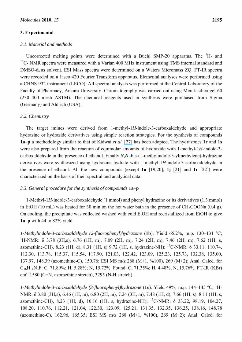
1-Methylindole-3-carboxaldehyde (2-fluorophenyl)hydrazone (1b). Yield 65.2%, m.p. 130–131 ºC; 1H-NMR: δ 3.78 (3H,s), 6.76 (1H, m), 7.09 (2H, m), 7.24 (2H, m), 7.46 (2H, m), 7.62 (1H, s,
azomethine-CH), 8.23 (1H, d), 8.31 (1H, s) 9.72 (1H, s, hydrazine-NH); 13C-NMR: δ 33.11, 110.74,
112.30, 113.78, 115.37, 115.54, 117.90, 121.03, 122.42, 123.09, 125.23, 125.73, 132.38, 135.00,
137.97, 148.39 (azomethine-C), 150.76; ESI MS m/z 268 (M+1, %100), 269 (M+2); Anal. Calcd. for
C16H14N3F: C, 71.89%; H, 5.28%; N, 15.72%. Found: C, 71.35%; H, 4.48%; N, 15.76%. FT-IR (KBr)
cm-1 1580 (C=N, azomethine stretch), 3295 (N-H stretch).
1-Methylindole-3-carboxaldehyde (3-fluorophenyl)hydrazone (1c). Yield 49%, m.p. 144–145 ºC; 1H-
NMR: δ 3.80 (3H,s), 6.46 (1H, m), 6.80 (2H, m), 7.24 (3H, m), 7.48 (1H, d), 7.66 (1H, s), 8.11 (1H, s,
azomethine-CH), 8.23 (1H, d), 10.16 (1H, s, hydrazine-NH); 13C-NMR: δ 33.22, 98.19, 104.27,
108.20, 110.76, 112.21, 121.04, 122.30, 123.09, 125.21, 131.35, 132.35, 136.25, 138.16, 148.78
(azomethine-C), 162.96, 165.35; ESI MS m/z 268 (M+1, %100), 269 (M+2); Anal. Calcd. for
Molecules 2010, 15
2196
C16H14N3F: C, 71.89%; H, 5.27%; N, 15.72%. Found: C, 69.30%; H, 5.05%; N, 15.13%. FT-IR (KBr)
cm-1 1582 (C=N, azomethine stretch), 3318 (N-H stretch).
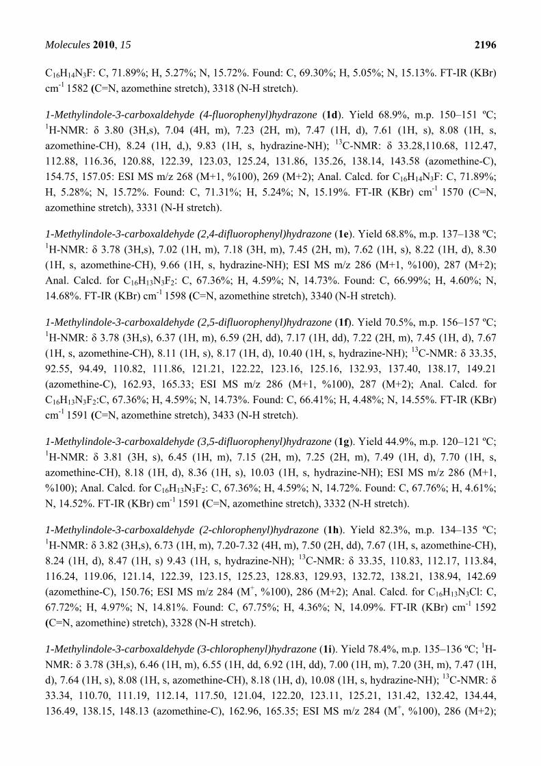
1-Methylindole-3-carboxaldehyde (4-fluorophenyl)hydrazone (1d). Yield 68.9%, m.p. 150–151 ºC; 1H-NMR: δ 3.80 (3H,s), 7.04 (4H, m), 7.23 (2H, m), 7.47 (1H, d), 7.61 (1H, s), 8.08 (1H, s,
azomethine-CH), 8.24 (1H, d,), 9.83 (1H, s, hydrazine-NH); 13C-NMR: δ 33.28,110.68, 112.47,
112.88, 116.36, 120.88, 122.39, 123.03, 125.24, 131.86, 135.26, 138.14, 143.58 (azomethine-C),
154.75, 157.05: ESI MS m/z 268 (M+1, %100), 269 (M+2); Anal. Calcd. for C16H14N3F: C, 71.89%;
H, 5.28%; N, 15.72%. Found: C, 71.31%; H, 5.24%; N, 15.19%. FT-IR (KBr) cm-1 1570 (C=N,
azomethine stretch), 3331 (N-H stretch).
1-Methylindole-3-carboxaldehyde (2,4-difluorophenyl)hydrazone (1e). Yield 68.8%, m.p. 137–138 ºC; 1H-NMR: δ 3.78 (3H,s), 7.02 (1H, m), 7.18 (3H, m), 7.45 (2H, m), 7.62 (1H, s), 8.22 (1H, d), 8.30
(1H, s, azomethine-CH), 9.66 (1H, s, hydrazine-NH); ESI MS m/z 286 (M+1, %100), 287 (M+2);
Anal. Calcd. for C16H13N3F2: C, 67.36%; H, 4.59%; N, 14.73%. Found: C, 66.99%; H, 4.60%; N,
14.68%. FT-IR (KBr) cm-1 1598 (C=N, azomethine stretch), 3340 (N-H stretch).
1-Methylindole-3-carboxaldehyde (2,5-difluorophenyl)hydrazone (1f). Yield 70.5%, m.p. 156–157 ºC; 1H-NMR: δ 3.78 (3H,s), 6.37 (1H, m), 6.59 (2H, dd), 7.17 (1H, dd), 7.22 (2H, m), 7.45 (1H, d), 7.67
(1H, s, azomethine-CH), 8.11 (1H, s), 8.17 (1H, d), 10.40 (1H, s, hydrazine-NH); 13C-NMR: δ 33.35,
92.55, 94.49, 110.82, 111.86, 121.21, 122.22, 123.16, 125.16, 132.93, 137.40, 138.17, 149.21
(azomethine-C), 162.93, 165.33; ESI MS m/z 286 (M+1, %100), 287 (M+2); Anal. Calcd. for
C16H13N3F2:C, 67.36%; H, 4.59%; N, 14.73%. Found: C, 66.41%; H, 4.48%; N, 14.55%. FT-IR (KBr)
cm-1 1591 (C=N, azomethine stretch), 3433 (N-H stretch).
1-Methylindole-3-carboxaldehyde (3,5-difluorophenyl)hydrazone (1g). Yield 44.9%, m.p. 120–121 ºC; 1H-NMR: δ 3.81 (3H, s), 6.45 (1H, m), 7.15 (2H, m), 7.25 (2H, m), 7.49 (1H, d), 7.70 (1H, s,
azomethine-CH), 8.18 (1H, d), 8.36 (1H, s), 10.03 (1H, s, hydrazine-NH); ESI MS m/z 286 (M+1,
%100); Anal. Calcd. for C16H13N3F2: C, 67.36%; H, 4.59%; N, 14.72%. Found: C, 67.76%; H, 4.61%;
N, 14.52%. FT-IR (KBr) cm-1 1591 (C=N, azomethine stretch), 3332 (N-H stretch).
1-Methylindole-3-carboxaldehyde (2-chlorophenyl)hydrazone (1h). Yield 82.3%, m.p. 134–135 ºC; 1H-NMR: δ 3.82 (3H,s), 6.73 (1H, m), 7.20-7.32 (4H, m), 7.50 (2H, dd), 7.67 (1H, s, azomethine-CH),
8.24 (1H, d), 8.47 (1H, s) 9.43 (1H, s, hydrazine-NH); 13C-NMR: δ 33.35, 110.83, 112.17, 113.84,
116.24, 119.06, 121.14, 122.39, 123.15, 125.23, 128.83, 129.93, 132.72, 138.21, 138.94, 142.69
(azomethine-C), 150.76; ESI MS m/z 284 (M+, %100), 286 (M+2); Anal. Calcd. for C16H13N3Cl: C,
67.72%; H, 4.97%; N, 14.81%. Found: C, 67.75%; H, 4.36%; N, 14.09%. FT-IR (KBr) cm-1 1592
(C=N, azomethine) stretch), 3328 (N-H stretch).
1-Methylindole-3-carboxaldehyde (3-chlorophenyl)hydrazone (1i). Yield 78.4%, m.p. 135–136 ºC; 1H-
NMR: δ 3.78 (3H,s), 6.46 (1H, m), 6.55 (1H, dd, 6.92 (1H, dd), 7.00 (1H, m), 7.20 (3H, m), 7.47 (1H,
d), 7.64 (1H, s), 8.08 (1H, s, azomethine-CH), 8.18 (1H, d), 10.08 (1H, s, hydrazine-NH); 13C-NMR: δ
33.34, 110.70, 111.19, 112.14, 117.50, 121.04, 122.20, 123.11, 125.21, 131.42, 132.42, 134.44,
136.49, 138.15, 148.13 (azomethine-C), 162.96, 165.35; ESI MS m/z 284 (M+, %100), 286 (M+2);
Molecules 2010, 15
2197
Anal. Calcd. for C16H14N3Cl: C, 67.72%; H, 4.97%; N, 14.81%. Found: C, 67.57%; H, 4.81%; N,
14.77%. FT-IR (KBr) cm-1 1592 (C=N, azomethine stretch), 3302 (N-H stretch).
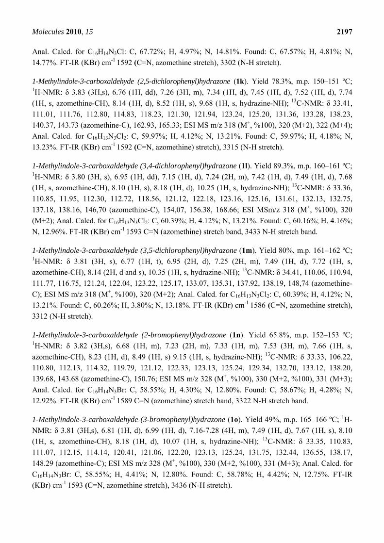
1-Methylindole-3-carboxaldehyde (2,5-dichlorophenyl)hydrazone (1k). Yield 78.3%, m.p. 150–151 ºC; 1H-NMR: δ 3.83 (3H,s), 6.76 (1H, dd), 7.26 (3H, m), 7.34 (1H, d), 7.45 (1H, d), 7.52 (1H, d), 7.74
(1H, s, azomethine-CH), 8.14 (1H, d), 8.52 (1H, s), 9.68 (1H, s, hydrazine-NH); 13C-NMR: δ 33.41,
111.01, 111.76, 112.80, 114.83, 118.23, 121.30, 121.94, 123.24, 125.20, 131.36, 133.28, 138.23,
140.37, 143.73 (azomethine-C), 162.93, 165.33; ESI MS m/z 318 (M+, %100), 320 (M+2), 322 (M+4);
Anal. Calcd. for C16H13N3Cl2: C, 59.97%; H, 4.12%; N, 13.21%. Found: C, 59.97%; H, 4.18%; N,
13.23%. FT-IR (KBr) cm-1 1592 (C=N, azomethine) stretch), 3315 (N-H stretch).
1-Methylindole-3-carboxaldehyde (3,4-dichlorophenyl)hydrazone (1l). Yield 89.3%, m.p. 160–161 ºC; 1H-NMR: δ 3.80 (3H, s), 6.95 (1H, dd), 7.15 (1H, d), 7.24 (2H, m), 7.42 (1H, d), 7.49 (1H, d), 7.68
(1H, s, azomethine-CH), 8.10 (1H, s), 8.18 (1H, d), 10.25 (1H, s, hydrazine-NH); 13C-NMR: δ 33.36,
110.85, 11.95, 112.30, 112.72, 118.56, 121.12, 122.18, 123.16, 125.16, 131.61, 132.13, 132.75,
137.18, 138.16, 146,70 (azomethine-C), 154,07, 156.38, 168.66; ESI MSm/z 318 (M+, %100), 320
(M+2); Anal. Calcd. for C16H13N3Cl2: C, 60.39%; H, 4.12%; N, 13.21%. Found: C, 60.16%; H, 4.16%;
N, 12.96%. FT-IR (KBr) cm-1 1593 C=N (azomethine) stretch band, 3433 N-H stretch band.
1-Methylindole-3-carboxaldehyde (3,5-dichlorophenyl)hydrazone (1m). Yield 80%, m.p. 161–162 ºC; 1H-NMR: δ 3.81 (3H, s), 6.77 (1H, t), 6.95 (2H, d), 7.25 (2H, m), 7.49 (1H, d), 7.72 (1H, s,
azomethine-CH), 8.14 (2H, d and s), 10.35 (1H, s, hydrazine-NH); 13C-NMR: δ 34.41, 110.06, 110.94,
111.77, 116.75, 121.24, 122.04, 123.22, 125.17, 133.07, 135.31, 137.92, 138.19, 148,74 (azomethine-
C); ESI MS m/z 318 (M+, %100), 320 (M+2); Anal. Calcd. for C16H13N3Cl2: C, 60.39%; H, 4.12%; N,
13.21%. Found: C, 60.26%; H, 3.80%; N, 13.18%. FT-IR (KBr) cm-1 1586 (C=N, azomethine stretch),
3312 (N-H stretch).
1-Methylindole-3-carboxaldehyde (2-bromophenyl)hydrazone (1n). Yield 65.8%, m.p. 152–153 ºC; 1H-NMR: δ 3.82 (3H,s), 6.68 (1H, m), 7.23 (2H, m), 7.33 (1H, m), 7.53 (3H, m), 7.66 (1H, s,
azomethine-CH), 8.23 (1H, d), 8.49 (1H, s) 9.15 (1H, s, hydrazine-NH); 13C-NMR: δ 33.33, 106.22,
110.80, 112.13, 114.32, 119.79, 121.12, 122.33, 123.13, 125.24, 129.34, 132.70, 133.12, 138.20,
139.68, 143.68 (azomethine-C), 150.76; ESI MS m/z 328 (M+, %100), 330 (M+2, %100), 331 (M+3);
Anal. Calcd. for C16H14N3Br: C, 58.55%; H, 4.30%; N, 12.80%. Found: C, 58.67%; H, 4.28%; N,
12.92%. FT-IR (KBr) cm-1 1589 C=N (azomethine) stretch band, 3322 N-H stretch band.
1-Methylindole-3-carboxaldehyde (3-bromophenyl)hydrazone (1o). Yield 49%, m.p. 165–166 ºC; 1H-
NMR: δ 3.81 (3H,s), 6.81 (1H, d), 6.99 (1H, d), 7.16-7.28 (4H, m), 7.49 (1H, d), 7.67 (1H, s), 8.10
(1H, s, azomethine-CH), 8.18 (1H, d), 10.07 (1H, s, hydrazine-NH); 13C-NMR: δ 33.35, 110.83,
111.07, 112.15, 114.14, 120.41, 121.06, 122.20, 123.13, 125.24, 131.75, 132.44, 136.55, 138.17,
148.29 (azomethine-C); ESI MS m/z 328 (M+, %100), 330 (M+2, %100), 331 (M+3); Anal. Calcd. for
C16H14N3Br: C, 58.55%; H, 4.41%; N, 12.80%. Found: C, 58.78%; H, 4.42%; N, 12.75%. FT-IR
(KBr) cm-1 1593 (C=N, azomethine stretch), 3436 (N-H stretch).
Molecules 2010, 15
2198
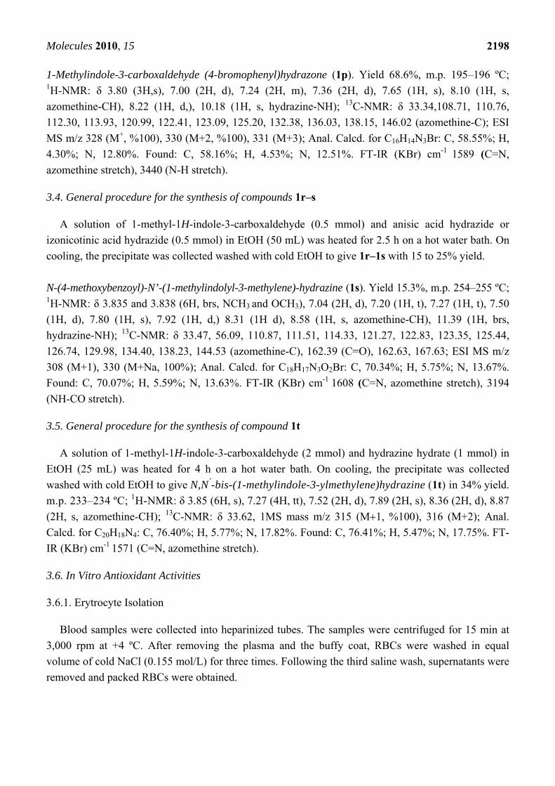
1-Methylindole-3-carboxaldehyde (4-bromophenyl)hydrazone (1p). Yield 68.6%, m.p. 195–196 ºC; 1H-NMR: δ 3.80 (3H,s), 7.00 (2H, d), 7.24 (2H, m), 7.36 (2H, d), 7.65 (1H, s), 8.10 (1H, s,
azomethine-CH), 8.22 (1H, d,), 10.18 (1H, s, hydrazine-NH); 13C-NMR: δ 33.34,108.71, 110.76,
112.30, 113.93, 120.99, 122.41, 123.09, 125.20, 132.38, 136.03, 138.15, 146.02 (azomethine-C); ESI
MS m/z 328 (M+, %100), 330 (M+2, %100), 331 (M+3); Anal. Calcd. for C16H14N3Br: C, 58.55%; H,
4.30%; N, 12.80%. Found: C, 58.16%; H, 4.53%; N, 12.51%. FT-IR (KBr) cm-1 1589 (C=N,
azomethine stretch), 3440 (N-H stretch).
3.4. General procedure for the synthesis of compounds 1r–s
A solution of 1-methyl-1H-indole-3-carboxaldehyde (0.5 mmol) and anisic acid hydrazide or
izonicotinic acid hydrazide (0.5 mmol) in EtOH (50 mL) was heated for 2.5 h on a hot water bath. On
cooling, the precipitate was collected washed with cold EtOH to give 1r–1s with 15 to 25% yield.
N-(4-methoxybenzoyl)-N’-(1-methylindolyl-3-methylene)-hydrazine (1s). Yield 15.3%, m.p. 254–255 ºC; 1H-NMR: δ 3.835 and 3.838 (6H, brs, NCH3 and OCH3), 7.04 (2H, d), 7.20 (1H, t), 7.27 (1H, t), 7.50
(1H, d), 7.80 (1H, s), 7.92 (1H, d,) 8.31 (1H d), 8.58 (1H, s, azomethine-CH), 11.39 (1H, brs,
hydrazine-NH); 13C-NMR: δ 33.47, 56.09, 110.87, 111.51, 114.33, 121.27, 122.83, 123.35, 125.44,
126.74, 129.98, 134.40, 138.23, 144.53 (azomethine-C), 162.39 (C=O), 162.63, 167.63; ESI MS m/z
308 (M+1), 330 (M+Na, 100%); Anal. Calcd. for C18H17N3O2Br: C, 70.34%; H, 5.75%; N, 13.67%.
Found: C, 70.07%; H, 5.59%; N, 13.63%. FT-IR (KBr) cm-1 1608 (C=N, azomethine stretch), 3194
(NH-CO stretch).
3.5. General procedure for the synthesis of compound 1t
A solution of 1-methyl-1H-indole-3-carboxaldehyde (2 mmol) and hydrazine hydrate (1 mmol) in
EtOH (25 mL) was heated for 4 h on a hot water bath. On cooling, the precipitate was collected
washed with cold EtOH to give N,N’-bis-(1-methylindole-3-ylmethylene)hydrazine (1t) in 34% yield.
m.p. 233–234 ºC; 1H-NMR: δ 3.85 (6H, s), 7.27 (4H, tt), 7.52 (2H, d), 7.89 (2H, s), 8.36 (2H, d), 8.87
(2H, s, azomethine-CH); 13C-NMR: δ 33.62, 1MS mass m/z 315 (M1, %100), 316 (M+2); Anal.
Calcd. for C20H18N4: C, 76.40%; H, 5.77%; N, 17.82%. Found: C, 76.41%; H, 5.47%; N, 17.75%. FT-
IR (KBr) cm-1 1571 (C=N, azomethine stretch).
3.6. In Vitro Antioxidant Activities
3.6.1. Erytrocyte Isolation
Blood samples were collected into heparinized tubes. The samples were centrifuged for 15 min at
3,000 rpm at +4 ºC. After removing the plasma and the buffy coat, RBCs were washed in equal
volume of cold NaCl (0.155 mol/L) for three times. Following the third saline wash, supernatants were
removed and packed RBCs were obtained.
Molecules 2010, 15
2199
3.6.2. Estimation of Reactive Oxygen Species by DCFH-DA
For estimation of ROS inside erythrocytes DCFH-DA was used as a probe. In cellular systems non
fluorescent probe DCFH-DA readily crosses the cell membrane and undergoes hydrolysis by
intracellular estrases to nonfluorescent 2',7'-dichlorofluorescin (DCFH). DCFH is then rapidly
oxidized in the presence of reactive oxygen species to highly fluorescent 2′,7′- dichlorofluorescein
(DCF) [28]. In our study, 1% erythrocyte suspension was incubated at 37 ºC in phosphate buffer (50 mM,
pH 7.4) and DCFH-DA (20 μM) for an hour. At the end of incubation period, the erythrocyte
suspension was washed with PBS three times, resuspended in PBS, pipetted onto a black 96-well plate
and various concentrations of melatonin or its derivates were added into the wells. The production of
fluorescent DCF was measured using a multiplate spectrofluorometer (excitation wavelength = 488 nm,
emission wavelength = 525 nm) [29].
3.6.3. Measurement of H2O2-induced lipid peroxidation levels
Lipid peroxidation was assesed by the determination of malondialdehyde (MDA) levels using the
method of Gutteridge et al. [30] and Quinlan et al. [31] based on the reaction of MDA with
thiobarbituric acid (TBA) at 95 ºC. In the TBA test reaction, MDA and TBA react to form a pink
adduct with an absorption maximum at 532 nm. The reaction was performed at pH 2–3 at 95 ºC for
15 min. Erythrocytes were resuspended in phosphate buffer (50 mM, pH 7.4) with 7.8 mM azide and
different concentrations of melatonin or its derivates were added. The samples were preincubated for
30 min at 37 ºC, added 5 mM H2O2 and incubated for 2 h at 37 ºC. The sample was mixed with 28%
(w/v) trichloroacetic acid to precipitate the protein. The precipitate was pelleted by centrifigation and
an aliquot of supernatant was reacted with 1% (w/v) TBA in a boiling water-bath for 15 min. After
cooling, the absorbance was read at 532 nm.
3.6.4. Determination of Erythrocyte Hemolysis
Erythrocytes were resuspended in phosphate saline (PBS: 150 mM NaCl, 1 mM Na2PO4 and
1.9 mM NaH2PO4, pH 7.4) at a 20% (v/v) suspension. Erythrocytes at 5% (v/v) suspension in PBS
were incubated with 75 mM AAPH in the presence of different concentrations of melatonin or its
derivates for 5 h at 37 ºC in a gently shaking water bath. Aliquots were taken out from this mixture at
appropriate intervals and centrifuged 2000 rpm for 5 min to obtain supernatant. The absorbance of the
supernatant was determined spectrophotometrically at 540 nm [32]. The percentage of hemolysis at
different incubation intervals was compared with that of complete hemolysis. For reference,
erythrocytes were treated with distilled water and the absorbance for the hemolysate at 540 nm was
used as 100% hemolysis. Every experiment was repeated three times and the lag time of hemolysis
(tlag) was determined.
3.6.5. Statistical analysis
Unpaired t test was performed to evaluate the significance of the differences between groups. p < 0.05
was accepted as significant.
Molecules 2010, 15
2200
4. Conclusions
In general all the synthesized indole derivatives were found to have potent antioxidant activity, even
higher than MLT itself, according to the results of three in vitro antioxidant experiments revealing
differences in their relative potencies probably related to electronic distribution. No significant
antioxidant activity was observed in two compounds 1r, 1s. These compounds were the isonicotinic
(1r) and anisic acid (1s) hydrazides of indole 3-aldehydes and they have no halogen atoms in their
structure that makes them different from the rest of the synthesized compounds. Structural
investigation of the rest of the active compounds showed that having o- and m- halogenated aromatic
side chain increase the antioxidant activity (such as compounds 1b, 1c, 1m, 1k and 1l). These are the
most promising compounds that should be kept in mind for designing new MLT-based indole
derivatives for our ongoing study. These results suggest a new approach for the in vitro antioxidant
activity properties and structure activity relationships of 1,3-disubstituted indole rings. Lack of a
methoxy group in the 5 position did not affect the antioxidant capacity of the new indole derivatives.
In fact, the in vitro assays showed that lack of a methoxy group, introduction of a methyl group at the
nitrogen in the indole ring and a halogenated aromatic side chain resulted in much more active
compounds than MLT itself. This may be due to increased stability of the indole ring and
delocalization of the electrons to help to scavenge free radicals by forming stable indolyl
cation radicals.
Acknowledgements
This work was supported by The Scientific and Technological Research Council of Turkey
(TÜBİTAK) Research and Development Grant 109S099.
References
1. Tan, D.X.; Chen, L.D.; Poeggeler, B.; Manchester, L.C.; Reiter, R.J. Melatonin: a potent
endogenous hydroxyl radical scavenger. Endocr. J. 1993, 1, 57–60.
2. Sreejith, P.; Beyo, R.S.; Divya, L.; Vijayasree, A.S.; Manju, M.; Oommen, O.V. Triiodothyronine
and melatonin influence antioxidant defense mechanism in a teleost Anabas testudineus (Bloch):
in vitro study. Indian J. Biochem. Biophys. 2007, 44,164–168.
3. Guerrero, J.M.; Reiter, R.J. Melatonin-immune system relationships. Curr. Top. Med. Chem.
2002, 2, 167–179.
4. Ates-Alagoz, Z.; Coban, T.; Suzen, S. A Comparative Study: Evaluation of Antioxidant Activity
of Melatonin and Some Indole Derivatives. Med. Chem. Res. 2005, 14, 169–179.
5. Suzen, S.; Buyukbingol, E. Anti-Cancer Activity Studies of Indolalthiohydantoin (PIT) on certain
cancer cell lines. Farmaco 2000, 55, 246–248.
6. Suzen, S.; Buyukbingol, E. Evaluation of Anti-HIV Activity of 5-(2-phenyl-3’- Indolyl)-2-
thiohydantoin. Farmaco 1998, 53, 525–527.
7. Gulcin, I.; Buyukokuroglu, M.E.; Oktay, M.; Kufrevioglu, O.I. On the in vitro antioxidative
properties of melatonin. J. Pineal Res. 2002, 33, 167–171.
Molecules 2010, 15
2201
8. Tan, D.X.; Reiter, R.J.; Manchester, L.C.; Yan, M.T.; El-sawi, M.; Sainz, R.M.; Mayo, J.C.;
Kohen, R.; Allegra, M.; Hardeland, R. Chemical and physical properties and potential
mechanisms: melatonin as a broad spectrum antioxidant and free radical scavenger. Curr. Top.
Med. Chem. 2002, 2, 181–197.
9. Bozkaya, P.; Dogan, B.; Suzen, S.; Nebioglu D.; Ozkan S.A. Determination and investigation of
electrochemical behaviour of 2-phenylindole derivatives: discussion on possible mechanistic
pathways. Can. J. Anal. Sci. Spec. 2006, 51, 125–139.
10. Suzen, S.; Demircigil, T.; Buyukbingol, E.; Ozkan, S.A. Electroanalitical Evaluation and
Determination of 5-(3’-indolyl)-2-thiohydantoin Derivatives by Voltammetric studies: possible
relevance to in vitro metabolism. New J. Chem. 2003, 27, 1007–1011.
11. Suzen, S., Ateş-Alagoz, Z.; Demircigil, T.; Ozkan, S.A. Synthesis and Analytical Evaluation by
Voltammetric Studies of Some New Indole-3-propionic acid Derivatives. Farmaco 2001, 56,
835–840.
12. Kruk, I.; Aboul-Enein, H.Y.; Michalska, T.; Lichszteld, K.; Kubasik-Kladna, K.; Olgen, S. In
vitro scavenging activity for reactive oxygen species by N-substituted indole-2-carboxylic acid
esters. Luminescence 2007, 22, 379–386.
13. Zhao, F.; Zai-Qun, L. Indole and its alkyl-substituted derivatives protect erythrocyte and DNA
against radical-induced oxidation J. Biochem. Mol. Toxicol. 2009, 23, 273–279.
14. Gulcin, I.; Buyukokuroglu, M.E.; Kufrevioglu, O.I. Metal chelating and hydrogen peroxide
scavenging effects of melatonin. J. Pineal Res. 2003, 34, 278–281.
15. Talaz, O.; Gulcin, İ.; Goksu, S.; Saracoglu, N. Antioxidant activity of 5,10-dihydroindeno[1,2-
b]indoles containing substituents on dihydroindeno part. Bioorg. Med. Chem. 2009, 17,
6583–6589.
16. Suzen, S.; Bozkaya P.; Coban, T.; Nebioglu, D. Investigation of in vitro antioxidant behaviour of
some 2-phenylindole derivatives: discussion on possible antioxidant mechanisms and comparison
with melatonin. J. Enzyme Inh. Med. Chem. 2006, 21, 405–411.
17. Ates-Alagoz, Z.; Coban, T.; Suzen, S. A comparative study: evaluation of antioxidant activity of
melatonin and some indole derivatives. Med. Chem. Res. 2005, 14, 169–179.
18. Gurkok, G.; Coban, T.; Suzen S. Melatonin analogue new indole hydrazide/hydrazone derivatives
with antioxidant behavior: Synthesis and discussion on structure activity relationships. J. Enzym.
Inh. Med. Chem. 2009, 24, 506–515.
19. Wieland, H.; Konz, W.; Mittasch, H. Toad positions VII. Constitution of bufotenin and
bufotenidine. Justus Liebig Ann. Chem. 1934, 513, 1–25.
20. Bulatova, N.N.; Suvorov, N.N. Indole derivatives XXXVI. Reaction of 3-beta-nitrovinylindoles
with nucleophilic reagents. Khim. Geterosikli. Soedin. 1969, 5, 813–17.
21. Baker, J.W.; Happold, F.C.; Walker, N. The tryptophanase-tryptophan reaction: 7. Further
evidence regarding the mechanism of the enzymic degradation of tryptophan to indole: criticism
of the theory that β-o-aminophenylacetaldehyde is the indole-forming intermediate. Biochem. J.
1946, 40, 420–426.
22. Song, L.; Xinhua, H.; Zhibing, Z.; Yan, L.; Beifen, S. Preparation of aryl hydrazide compounds as
immunosupressives. PCT Int. Appl. WO/2007/036083, 2007.
Molecules 2010, 15
2202
23. Niki, E.; Komuro, E.; Takahashi, M.; Urano, S.; Ito, E.; Terao, K.J. Oxidative hemolysis of
erythrocytes and its inhibition by free radical scavengers. Biol. Chem. 1988, 263, 19809–19814.
24. Zhao, F.; Liu, Z.-Q.; Wu, D. Antioxidative effect of melatonin on DNA and erythrocytes against
free radical-induced oxidation. Chem. Phys. Lipids 2008, 151, 77–84.
25. Suzen, S. Antioxidant Activities of Synthetic Indole Derivatives and Possible Activity
Mechanisms. In Topics in Heterocyclic Chemistry; Khan, M.T.H., Ed.; Springer: Berlin,
Heidelberg, Germany, 2007; Volume 11, pp. 145–178.
26. Allegra, M.; Reiter, R.J.; Tan, D.X.; Gentile, C.; Tesoriere, L.; Livrea, M.A. The chemistry of
melatonin's interaction with reactive species. J. Pineal Res. 2003, 34, 1–10.
27. Kidwai, M.; Negi, N.; Gupta, S.D. Synthesis and antifertility activity of 1,5-diaryl-3 (3'-
indolyl)formazans. Chem. Pharm. Bull. (Tokyo) 1994, 42, 2363–2364.
28. Lautraite, S.; Bigot-Lasserre, D.; Bars, R.; Carmichael, N. Optimisation of cell-based assays for
medium throughput screening of oxidative stress. Toxicol. In Vitro 2003, 17, 207–220.
29. Puntarulo, S.; Cederbaum, A.I. Production of Reactive Oxygen Species by Microsomes Enriched
in Specific Human Cytochrome P450 Enzymes. Free Radic. Biol. Med. 1998, 24, 1324–1330.
30. Gutteridge, J.M.; Quinlan, G.J.; Clark, I.; Halliwell, B. Aluminium salts accelerate peroxidation of
membrane lipids stimulated by iron salts. Biochim. Biophys. Acta 1985, 835, 441–447.
31. Quinlan, G.J.; Halliwell, B.; Moorhouse, C.P.; Gutteridge, J.M. Action of lead(II) and aluminium
(III) ions on iron-stimulated lipid peroxidation in liposomes, erythrocytes and rat liver microsomal
fractions. Biochim. Biophys. Acta 1988, 962, 196–200.
32. Liu, Z.Q.; Luo, X.Y.; Sun, Y.X.; Chen, Y.P.; Wang, Z.C. Can ginsenosides protect human
erythrocytes against free-radical-induced hemolysis? Biochim. Biophys. Acta 2002, 1572, 58–66.
Sample Availability: Samples of the compounds are available from the authors.
© 2010 by the authors; licensee Molecular Diversity Preservation International, Basel, Switzerland.
This article is an open-access article distributed under the terms and conditions of the Creative
Commons Attribution license (http://creativecommons.org/licenses/by/3.0/).
Related Documents