Novel Hydroxycinnamoyl-Coenzyme A Quinate Transferase Genes from Artichoke Are Involved in the Synthesis of Chlorogenic Acid 1[W] Gabriella Sonnante*, Rosalinda D’Amore, Emanuela Blanco, Ciro L. Pierri, Monica De Palma, Jie Luo, Marina Tucci, and Cathie Martin Institute of Plant Genetics, National Research Council, 70126 Bari, Italy (G.S., R.D., E.B.); Pharmaco-Biology Department, Laboratory of Biochemistry and Molecular Biology, University of Bari, 70126 Bari, Italy (C.L.P.); Institute of Plant Genetics, Consiglio Nazionale delle Ricerche, 80055 Portici, Italy (M.D.P., M.T.); and John Innes Centre, Norwich NR4 7UH, United Kingdom (J.L., C.M.) Artichoke (Cynara cardunculus subsp. scolymus) extracts have high antioxidant capacity, due primarily to flavonoids and phenolic acids, particularly chlorogenic acid (5-caffeoylquinic acid [CGA]), dicaffeoylquinic acids, and caffeic acid, which are abundant in flower bracts and bioavailable to humans in the diet. The synthesis of CGA can occur following different routes in plant species, and hydroxycinnamoyl-coenzyme A transferases are important enzymes in these pathways. Here, we report on the isolation and characterization of two novel genes both encoding hydroxycinnamoyl-coenzyme A quinate transferases (HQT) from artichoke. The recombinant proteins (HQT1 and HQT2) were assayed after expression in Escherichia coli, and both showed higher affinity for quinate over shikimate. Their preferences for acyl donors, caffeoyl-coenzyme A or p-coumaroyl- coenzyme A, were examined. Modeling and docking analyses were used to propose possible pockets and residues involved in determining substrate specificities in the HQT enzyme family. Quantitative real-time polymerase chain reaction analysis of gene expression indicated that HQT1 might be more directly associated with CGA content. Transient and stable expression of HQT1 in Nicotiana resulted in a higher production of CGA and cynarin (1,3-dicaffeoylquinic acid). These findings suggest that several isoforms of HQT contribute to the synthesis of CGA in artichoke according to physiological needs and possibly following various metabolic routes. The incidence of many chronic disorders, such as cardiovascular diseases and certain cancers, could be reduced by means of improved nutrition, particularly through increased consumption of diets rich in fruit and vegetables. Most of the health-promoting prop- erties of edible plants are related to the presence of secondary metabolites, known as phytonutrients (Segasothy and Phillips, 1999). Artichoke (Cynara cardunculus subsp. scolymus or Cynara cardunculus var scolymus), which originated in the Mediterranean basin (Sonnante et al., 2007), is an edible plant used in medicine since ancient times. Artichoke extracts possess many medicinal properties, including anticarcinogenic, anti-Human immunodeficiency virus, antioxidative, cholesterol-lowering, bile-expelling, hepatoprotective, and diuretic activities, as well as antifungal and antibacterial properties (Agarwal and Mukhtar, 1996; Gebhardt, 1997; Kraft, 1997; Brown and Rice-Evans, 1998; McDougall et al., 1998; Shimoda et al., 2003; Zhua et al., 2005). The high antioxidant capacity of artichoke is due primarily to flavonoids and phenolic acids, particularly 5-caffeoylquinic acid, also known as chlorogenic acid (CGA), 1,3-dicaffeoyl- quinic acid (cynarin), 1,5-dicaffeoylquinic acid, and caffeic acid, which are abundant in artichoke and bioavailable to humans through their diet (Azzini et al., 2007). CGA can be absorbed directly by the small intestine or hydrolyzed by the microflora of the large intestine to release caffeic acid (Plumb et al., 1999; Nardini et al., 2002; Stalmach et al., 2010), which shows the same antioxidant activity as CGA (Rice- Evans et al., 1997). CGA is synthesized by many plants such as solana- ceous species (e.g. tomato [Solanum lycopersicum], to- bacco [Nicotiana tabacum], and eggplant [Solanum melongena]), apple (Malus domestica), pear (Pyrus com- munis), plum (Prunus domestica), coffee (Coffea arabica), and artichoke (Tamagnone et al., 1998a; Clifford, 1999; Wang et al., 2003; Schu ¨ tz et al., 2004) and is believed to play important roles in free radical scavenging (Chen and Ho, 1997; Tamagnone et al., 1998b), enzymatic 1 This work was supported by the European Union Cynares project AgriGenRes063, by the Consiglio Nazionale delle Ricerche (grant no. AG.P02.004, Banca del DNA Vegetale e Sviluppo di Una Piattaforma per l’Analisi di Genomi Vegetali), by the Italian Ministry of Education, University, and Research (grant no. 161, call 1105), by the National Science Foundation of China (grant no. 30500038 to J.L.), and by a core strategic grant from the Biotechnology and Biological Sciences Research Council to the John Innes Centre. * Corresponding author; e-mail [email protected]. The author responsible for distribution of materials integral to the findings presented in this article in accordance with the policy described in the Instructions for Authors (www.plantphysiol.org) is: Gabriella Sonnante ([email protected]). [W] The online version of this article contains Web-only data. www.plantphysiol.org/cgi/doi/10.1104/pp.109.150144 1224 Plant Physiology Ò , July 2010, Vol. 153, pp. 1224–1238, www.plantphysiol.org Ó 2010 American Society of Plant Biologists

Welcome message from author
This document is posted to help you gain knowledge. Please leave a comment to let me know what you think about it! Share it to your friends and learn new things together.
Transcript
Novel Hydroxycinnamoyl-Coenzyme A QuinateTransferase Genes from Artichoke Are Involvedin the Synthesis of Chlorogenic Acid1[W]
Gabriella Sonnante*, Rosalinda D’Amore, Emanuela Blanco, Ciro L. Pierri, Monica De Palma, Jie Luo,Marina Tucci, and Cathie Martin
Institute of Plant Genetics, National Research Council, 70126 Bari, Italy (G.S., R.D., E.B.); Pharmaco-BiologyDepartment, Laboratory of Biochemistry and Molecular Biology, University of Bari, 70126 Bari, Italy (C.L.P.);Institute of Plant Genetics, Consiglio Nazionale delle Ricerche, 80055 Portici, Italy (M.D.P., M.T.); and JohnInnes Centre, Norwich NR4 7UH, United Kingdom (J.L., C.M.)
Artichoke (Cynara cardunculus subsp. scolymus) extracts have high antioxidant capacity, due primarily to flavonoids andphenolic acids, particularly chlorogenic acid (5-caffeoylquinic acid [CGA]), dicaffeoylquinic acids, and caffeic acid, which areabundant in flower bracts and bioavailable to humans in the diet. The synthesis of CGA can occur following different routes inplant species, and hydroxycinnamoyl-coenzyme A transferases are important enzymes in these pathways. Here, we report onthe isolation and characterization of two novel genes both encoding hydroxycinnamoyl-coenzyme A quinate transferases(HQT) from artichoke. The recombinant proteins (HQT1 and HQT2) were assayed after expression in Escherichia coli, and bothshowed higher affinity for quinate over shikimate. Their preferences for acyl donors, caffeoyl-coenzyme A or p-coumaroyl-coenzyme A, were examined. Modeling and docking analyses were used to propose possible pockets and residues involved indetermining substrate specificities in the HQT enzyme family. Quantitative real-time polymerase chain reaction analysis ofgene expression indicated that HQT1 might be more directly associated with CGA content. Transient and stable expression ofHQT1 in Nicotiana resulted in a higher production of CGA and cynarin (1,3-dicaffeoylquinic acid). These findings suggest thatseveral isoforms of HQT contribute to the synthesis of CGA in artichoke according to physiological needs and possiblyfollowing various metabolic routes.
The incidence of many chronic disorders, such ascardiovascular diseases and certain cancers, could bereduced by means of improved nutrition, particularlythrough increased consumption of diets rich in fruitand vegetables. Most of the health-promoting prop-erties of edible plants are related to the presenceof secondary metabolites, known as phytonutrients(Segasothy and Phillips, 1999). Artichoke (Cynaracardunculus subsp. scolymus or Cynara cardunculus varscolymus), which originated in the Mediterraneanbasin (Sonnante et al., 2007), is an edible plant usedin medicine since ancient times. Artichoke extractspossess many medicinal properties, including
anticarcinogenic, anti-Human immunodeficiency virus,antioxidative, cholesterol-lowering, bile-expelling,hepatoprotective, and diuretic activities, as well asantifungal and antibacterial properties (Agarwal andMukhtar, 1996; Gebhardt, 1997; Kraft, 1997; Brown andRice-Evans, 1998; McDougall et al., 1998; Shimodaet al., 2003; Zhua et al., 2005). The high antioxidantcapacity of artichoke is due primarily to flavonoidsand phenolic acids, particularly 5-caffeoylquinic acid,also known as chlorogenic acid (CGA), 1,3-dicaffeoyl-quinic acid (cynarin), 1,5-dicaffeoylquinic acid, andcaffeic acid, which are abundant in artichoke andbioavailable to humans through their diet (Azziniet al., 2007). CGA can be absorbed directly by thesmall intestine or hydrolyzed by the microflora of thelarge intestine to release caffeic acid (Plumb et al.,1999; Nardini et al., 2002; Stalmach et al., 2010), whichshows the same antioxidant activity as CGA (Rice-Evans et al., 1997).
CGA is synthesized by many plants such as solana-ceous species (e.g. tomato [Solanum lycopersicum], to-bacco [Nicotiana tabacum], and eggplant [Solanummelongena]), apple (Malus domestica), pear (Pyrus com-munis), plum (Prunus domestica), coffee (Coffea arabica),and artichoke (Tamagnone et al., 1998a; Clifford, 1999;Wang et al., 2003; Schutz et al., 2004) and is believed toplay important roles in free radical scavenging (Chenand Ho, 1997; Tamagnone et al., 1998b), enzymatic
1 This work was supported by the European Union Cynaresproject AgriGenRes063, by the Consiglio Nazionale delle Ricerche(grant no. AG.P02.004, Banca del DNA Vegetale e Sviluppo di UnaPiattaforma per l’Analisi di Genomi Vegetali), by the ItalianMinistryof Education, University, and Research (grant no. 161, call 1105), bythe National Science Foundation of China (grant no. 30500038 toJ.L.), and by a core strategic grant from the Biotechnology andBiological Sciences Research Council to the John Innes Centre.
* Corresponding author; e-mail [email protected] author responsible for distribution of materials integral to the
findings presented in this article in accordance with the policydescribed in the Instructions for Authors (www.plantphysiol.org) is:Gabriella Sonnante ([email protected]).
[W] The online version of this article contains Web-only data.www.plantphysiol.org/cgi/doi/10.1104/pp.109.150144
1224 Plant Physiology�, July 2010, Vol. 153, pp. 1224–1238, www.plantphysiol.org � 2010 American Society of Plant Biologists

browning of fruits and vegetables (Walker, 1995),defense against fungal pathogens (Maher et al., 1994),and resistance to pathogenic insects (Dowd and Vega,1996; Bushman et al., 2002). The biosynthetic routesleading to CGA in plants are not completely definedyet, and three possible paths have been proposed. Inthe first, hydroxycinnamoyl-CoA quinate hydroxycin-namoyl transferase (HQT) catalyzes the formation ofCGA from caffeoyl-CoA and quinic acid (route 1;Niggeweg et al., 2004). Caffeoyl-CoA is supplied bythe combined activities of hydroxycinnamoyl-CoAshikimate/quinate hydroxycinnamoyl transferase(HCT) and p-coumaroyl ester 3# hydroxylase (C3#H)via a p-coumaroylshikimate intermediate (route 1b;Mahesh et al., 2007). The second proposed route isbased on the synthesis of p-coumaroylquinate by HCT,followed by hydroxylation by C3#H (Ulbrich andZenk, 1979, Hoffmann et al., 2003). The importanceof this route has been questioned because HCT andC3#H are both active in Arabidopsis (Arabidopsisthaliana) and yet no accumulation of CGA occurs andbecause HCT does not show high affinity for quinateas an acyl acceptor (Hoffmann et al., 2003; Niggeweget al., 2004). HCT could be supplemented by HQT inthis pathway in some plants. In the third suggestedroute, caffeoyl glucoside serves as an activated inter-mediate (Villegas and Kojima, 1986).Since CGA biosynthesis does not occur in all spe-
cies, it is possible that different biosynthetic routeshave evolved in diverged species, or indeed, that thedifferent pathways have assumed various degrees ofimportance in distinct species. The cDNA encodingHQT has been isolated from tobacco and tomato; bothtransient expression and stable transformation estab-lished that HQT is the enzyme of greatest importancein the synthesis of chlorogenic acid in tomato (Niggeweget al., 2004). In coffee, the expression of HQT is moreclosely correlated with CGA synthesis and accumula-tion than HCT (Lepelley et al., 2007).In artichoke, a cDNA coding for HCT has been
isolated (Comino et al., 2007). Biochemical character-ization suggested that HCT could act either before orafter the 3#-hydroxylation step and may contribute toCGA biosynthesis. Recently, after the initiation of thiswork, the isolation of a gene encoding HQT fromartichoke was also reported (Comino et al., 2009).A better understanding of CGA and cynarin pro-
duction in artichoke is important to develop agro-nomic, genetic, or biotechnological tools for higherproduction of biologically active compounds. In thispaper, we report on the isolation and characterizationof two full-length cDNA sequences encoding HQT inartichoke, belonging to the BAHD transferase family(D’Auria, 2006). HQT1 and HQT2 have in vitro activ-ities distinct from each other in terms of their speci-ficities. Modeling and docking analyses based on thestructures of two BAHD members from Dendranthemamorifolium (2e1t; Unno et al., 2007) and Rauvolfiaserpentina (2bgh; Ma et al., 2005) were undertaken toidentify possible structural features defining catalytic
specificities. Hqt transcript levels in different bractorders and the receptacle of the edible flower head,and in leaves in the vegetative and productive periods,were compared with CGA content in the same tissues.Transient and stable expression of hqt1 in Nicotianainduced a higher production of CGA and cynarin.
RESULTS
Isolation and Analysis of hqt1/hqt2 Genomic Sequences
and Full-Length cDNAs
The hqt cDNA sequences from tobacco (AJ582651)and tomato (AJ582652) were used to search the lettuce(Lactuca sativa) and sunflower (Helianthus annuus) ESTsin The Gene Index database (http://compbio.dfci.harvard.edu/tgi/plant.html) because these species,like artichoke, are members of the Asteraceae familyand were likely to have genes closely related structur-ally to HQT in artichoke, as has been reported forother genes (De Paolis et al., 2008). Moreover, no ESTfrom artichoke was available when this work wasstarted. The best hits obtained from the BLAST analy-sis grouped in two clusters and after alignment,TC22135, TC23302, TC25546, and TC10357 sequenceswere used to design the primers Hel-F3 and Hqt-R2(Supplemental Table S1), which produced a 600-bpputative hqt sequence (named hqt1) from reverse tran-scription (RT)-PCR of artichoke leaf cDNA. PrimersHel_HQT_F6 and Hel_HQT_R4 were designed on thebasis of the second group of sequences (TC16408,TC16209, and TC30763) and were used to isolate a1,000-bp sequence named hqt2. Both fragments showedsequence similarity to tobacco and tomato hqt.
Partial sequences from artichoke were extendedtoward both the 3# and 5# ends by RACE-PCR. Newsets of primers were designed around the start andstop codons for both hqt1 and hqt2. A 1,413-bp se-quence composed of a 1,350-bp open reading frameencoding a 449-residue protein (estimated molecularmass of 49.59 kD) was obtained for hqt1, whereas thehqt2 sequence was 1,332 bp, encoding a 443-residueprotein, with an estimated molecular mass of 48.89 kD.
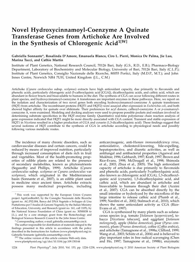
The artichoke hqt cDNA-encoded proteins were80.4% identical and possessed the two conservedsequence motifs, HXXXDG and DFGWG (St-Pierreand De Luca, 2000; D’Auria, 2006), observed in otherplant acyltransferases, for HQT1, HTLAD (170–174amino acids) and DFGWG (396–400 amino acids), andfor HQT2, HTLSD (162–166 amino acids) and DFGWG(390–394 amino acids), as shown in Figure 1. HQT1showed the greatest levels of identity (71%) to therecently isolated HQT from artichoke and cardoon(Cynara cardunculus var altilis; Comino et al., 2009), totobacco, tomato, coffee, and potato (Solanum tuberosum)HQTs (71%, 70%, 68%, and 68%, respectively), and tosweet potato (Ipomoea batatas) N-hydroxycinnamoylbenzoyltransferase (HCBT; 70%). HQT2 showed thegreatest identity to HQT from artichoke (71%; Comino
Novel Artichoke hqt Genes for Chlorogenic Acid Synthesis
Plant Physiol. Vol. 153, 2010 1225
et al., 2009) and from cardoon (70%), to HCBT fromsweet potato (70%), and to tobacco, tomato, and coffeeHQTs (69%, 68%, and 66%, respectively). Both se-quences displayed a lower level of identity to arti-choke HCT (59% for HQT1 and 57% for HQT2).
The genomic sequences of the hqt1 and hqt2 genesestablished that both possess the conserved “Q intron”found in many members of the BAHD acyltransferasefamily (St-Pierre and De Luca, 2000), inserted betweentriplets encoding a Gln residue and a Val residue, inhqt1 at position 459 of the coding sequence and in hqt2at position 438 of the coding sequence. The introns are1,516 and 1,398 bp long for hqt1 and hqt2, respectively.
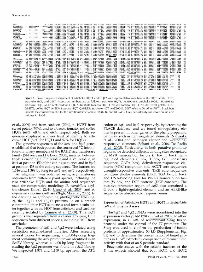
An alignment was obtained using acyltransferasesequences from different plant species, including thetwo artichoke HQTs and the amino acid sequencesused for comparative modeling: D. morifolium acyl-transferase DmAT (2e1t; Unno et al., 2007) and R.serpentina vinorine synthase (2bgh; Ma et al., 2005). Inthe deriving neighbor-joining phylogenetic tree (Fig.2), the HQT1 and HQT2 proteins lie on a branchcontaining other HQT sequences and form a subclus-ter together with the HQT from artichoke and cardoonrecently isolated by Comino et al. (2009). This HQTgroup is well separated from a cluster grouping HCTsequences from different species, including HCT fromartichoke.
The promoters of hqt1 and hqt2 were isolated usingrestriction enzyme-based libraries. After screeningseveral clones by sequencing, a 1,900-bp-long frag-ment containing the hqt1 promoter was identified in anEcoRV library, whereas a 1,400-bp-long fragment in-cluding the hqt2 promoter was found in a StuI library.We inspected 1,874 and 1,159 bp upstream the ATG
codon of hqt1 and hqt2 respectively, by screening thePLACE database, and we found cis-regulatory ele-ments present in other genes of the phenylpropanoidpathway, such as light-regulated elements (Narusakaet al., 2004) and pathogen elicitor and wounding-responsive elements (Soltani et al., 2006; De Paoliset al., 2008). Particularly, in both putative promoterregions, we detected different binding sites recognizedby MYB transcription factors (P box, L box), light-regulated elements (I box, T box, GT1 consensussequence, GATA box), dehydration-responsive ele-ments (MYC recognition site, ACGT core sequence),drought-responsive elements (DRE core sequence),pathogen elicitor elements (ElRE, TGA box, E box),and DNA-binding sites for WRKY transcription fac-tors (W box) and DOF proteins (DOF core site). Theputative promoter region of hqt2 also contained aG box, a light-regulated element, and an ABRE-likesequence for abscisic acid responsiveness.
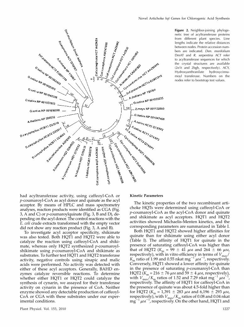
Expression of Artichoke HQT1 and HQT2 in Escherichiacoli and Enzyme Assays
The hqt1 and hqt2 cDNAs were recombined into theexpression vector pJAM1784 (Luo et al., 2007) to allowexpression, in E. coli, of recombinant S-tag fusionproteins under the control of the T7 promoter. TheS-tag was used to confirm the production of fusionproteins of approximately 50 kD (Supplemental Fig.S1) and to determine the concentration of target pro-teins in E. coli extracts by comparison of reconstitutedactivity with that of an S-peptide standard.
Enzymatic assays with the soluble fractions of theE. coli extracts showed that both HQT1 and HQT2
Figure 1. Protein sequence alignment of artichoke HQT1 and HQT2 with representative members of the HQT family, HCBT,artichoke HCT, and 2E1T. Accession numbers are as follows: artichoke HQT1, AM690438; artichoke HQT2, EU839580;artichoke HQT, ABK79689; cardoon HQT, ABK79690; tobacco HQT, Q70G33; tomato HQT, Q70G32; sweet potato HCBT,Q9SST8; coffee HQT, A4ZKM4; potato HQT, Q3HRZ5; artichoke HCT, AAZ80046. 2E1T refers to DmAT A4PHY4. Black barsindicate the conserved motifs for the acyl transferase family: HXXXDG and DFGWG. Gray bars identify conserved amino acidresidues for HQT.
Sonnante et al.
1226 Plant Physiol. Vol. 153, 2010
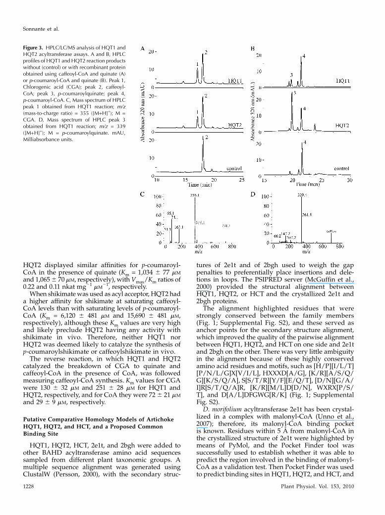
had acyltransferase activity, using caffeoyl-CoA orp-coumaroyl-CoA as acyl donor and quinate as the acylacceptor. By means of HPLC and mass spectrometryanalyses, reaction products were identified as CGA (Fig.3, A and C) or p-coumaroylquinate (Fig. 3, B and D), de-pending on the acyl donor. The control reactions with theE. coli crude extracts transformed with the empty vectordid not show any reaction product (Fig. 3, A and B).To investigate acyl acceptor specificity, shikimate
was also tested. Both HQT1 and HQT2 were able tocatalyze the reaction using caffeoyl-CoA and shiki-mate, whereas only HQT2 synthesized p-coumaroyl-shikimate using p-coumaroyl-CoA and shikimate assubstrates. To further test HQT1 and HQT2 transferaseactivity, negative controls using sinapic and malicacids were performed. No activity was detected witheither of these acyl acceptors. Generally, BAHD en-zymes catalyze reversible reactions. To determinewhether either HQT1 or HQT2 could catalyze thesynthesis of cynarin, we assayed for their transferaseactivity on cynarin in the presence of CoA. Neitherenzyme showed any detectable production of caffeoyl-CoA or CGA with these substrates under our exper-imental conditions.
Kinetic Parameters
The kinetic properties of the two recombinant arti-choke HQTs were determined using caffeoyl-CoA orp-coumaroyl-CoA as the acyl-CoA donor and quinateand shikimate as acyl acceptors. HQT1 and HQT2activities showed Michaelis-Menten kinetics, and thecorresponding parameters are summarized in Table I.
Both HQT1 and HQT2 showed higher affinities forquinate than for shikimate using either acyl donor(Table I). The affinity of HQT1 for quinate in thepresence of saturating caffeoyl-CoA was higher thanthat of HQT2 (Km = 99 6 41 mM and 264 6 66 mM,respectively), with in vitro efficiency in terms of Vmax/Km ratio of 1.99 and 0.55 nkat mg21 mM
21, respectively.Conversely, HQT1 showed a lower affinity for quinatein the presence of saturating p-coumaroyl-CoA thanHQT2 (Km = 2166 76 mM and 596 4 mM, respectively),with Vmax/Km ratios of 1.52 and 7.29 nkat mg21 mM
21,respectively. The affinity of HQT1 for caffeoyl-CoA inthe presence of quinate was about 4.5-fold higher thanfor HQT2 (Km = 1,391 6 283 mM and 6,196 6 293 mM,respectively), withVmax/Km ratios of 0.08 and 0.04 nkatmg21 mM
21, respectively. On the other hand, HQT1 and
Figure 2. Neighbor-joining phyloge-netic tree of acyltransferase proteinsfrom different plant species. Linelengths indicate the relative distancesbetween nodes. Protein accession num-bers are indicated. Den. morifoliumDmAT and R. serpentina ACT referto acyltransferase sequences for whichthe crystal structures are available(2e1t and 2bgh, respectively). ACY,Hydroxyanthranilate hydroxycinna-moyl transferase. Numbers on thenodes refer to bootstrap test values.
Novel Artichoke hqt Genes for Chlorogenic Acid Synthesis
Plant Physiol. Vol. 153, 2010 1227
HQT2 displayed similar affinities for p-coumaroyl-CoA in the presence of quinate (Km = 1,034 6 77 mM
and 1,0656 70mM, respectively), withVmax/Km ratios of0.22 and 0.11 nkat mg21 mM
21, respectively.When shikimatewas used as acyl acceptor, HQT2 had
a higher affinity for shikimate at saturating caffeoyl-CoA levels than with saturating levels of p-coumaroyl-CoA (Km = 6,120 6 481 mM and 15,690 6 481 mM,respectively), although these Km values are very highand likely preclude HQT2 having any activity withshikimate in vivo. Therefore, neither HQT1 norHQT2 was deemed likely to catalyze the synthesis ofp-coumaroylshikimate or caffeoylshikimate in vivo.
The reverse reaction, in which HQT1 and HQT2catalyzed the breakdown of CGA to quinate andcaffeoyl-CoA in the presence of CoA, was followedmeasuring caffeoyl-CoA synthesis. Km values for CGAwere 130 6 32 mM and 251 6 28 mM for HQT1 andHQT2, respectively, and for CoA they were 726 21 mM
and 29 6 9 mM, respectively.
Putative Comparative Homology Models of ArtichokeHQT1, HQT2, and HCT, and a Proposed CommonBinding Site
HQT1, HQT2, HCT, 2e1t, and 2bgh were added toother BAHD acyltransferase amino acid sequencessampled from different plant taxonomic groups. Amultiple sequence alignment was generated usingClustalW (Persson, 2000), with the secondary struc-
tures of 2e1t and of 2bgh used to weigh the gappenalties to preferentially place insertions and dele-tions in loops. The PSIPRED server (McGuffin et al.,2000) provided the structural alignment betweenHQT1, HQT2, or HCT and the crystallized 2e1t and2bgh proteins.
The alignment highlighted residues that werestrongly conserved between the family members(Fig. 1; Supplemental Fig. S2), and these served asanchor points for the secondary structure alignment,which improved the quality of the pairwise alignmentbetween HQT1, HQT2, and HCT on one side and 2e1tand 2bgh on the other. There was very little ambiguityin the alignment because of these highly conservedamino acid residues and motifs, such as [H/P][I/L/T][P/N/L/G]X[V/I/L], HXXXD[A/G], [K/R][A/S/Q/G][K/S/Q/A], S[S/T/R][Y/F][E/Q/T], [D/N][G/A/I]R[S/T/Q/A]R, [K/R][M/L]D[D/N], WXRX[P/S/T], and D[A/L]DFGWG[R/K] (Fig. 1; SupplementalFig. S2).
D. morifolium acyltransferase 2e1t has been crystal-lized in a complex with malonyl-CoA (Unno et al.,2007); therefore, its malonyl-CoA binding pocketis known. Residues within 5 A from malonyl-CoA inthe crystallized structure of 2e1t were highlighted bymeans of PyMol, and the Pocket Finder tool wassuccessfully used to establish whether it was able topredict the region involved in the binding of malonyl-CoA as a validation test. Then Pocket Finder was usedto predict binding sites in HQT1, HQT2, and HCT, and
Figure 3. HPLC/LC/MS analysis of HQT1 andHQT2 acyltransferase assays. A and B, HPLCprofiles of HQT1 and HQT2 reaction productswithout (control) or with recombinant proteinobtained using caffeoyl-CoA and quinate (A)or p-coumaroyl-CoA and quinate (B). Peak 1,Chlorogenic acid (CGA); peak 2, caffeoyl-CoA; peak 3, p-coumaroylquinate; peak 4,p-coumaroyl-CoA. C, Mass spectrum of HPLCpeak 1 obtained from HQT1 reaction; m/z(mass-to-charge ratio) = 355 ([M+H]+); M =CGA. D, Mass spectrum of HPLC peak 3obtained from HQT1 reaction; m/z = 339([M+H]+); M = p-coumaroylquinate. mAU,Milliabsorbance units.
Sonnante et al.
1228 Plant Physiol. Vol. 153, 2010

it was found that they overlapped with the 2e1tmalonyl-CoA binding region. Differences in aminoacid composition of the predicted binding regionsshould be responsible for the experimentally observeddifferent substrate specificities of HQT1, HQT2, andHCT (Supplemental Fig. S2).For docking analyses, a list of potential substrates
(caffeoylquinate, caffeoylshikimate, p-coumaroylqui-nate, p-coumaroylshikimate, sinapate, quinate, shiki-mate, and tetracaffeoylquinate) were docked in theproposed binding site using Autodock 1.5.2 (Goodsellet al., 1996) to investigate and support the identifiedsubstrate specificity found in the experimental tests.Autodock was chosen for its ability to carry out avirtual screening of chemical libraries and to output alist of the ligands ordered by docked energy fromlowest to highest. Autodock suggested the prefer-ence of HQT1 for caffeoylquinate and of HQT2 forp-coumaroylquinate (shown experimentally) and pro-posed a preference of HCT for caffeoylshikimate.It was possible to highlight two distinct adjacent
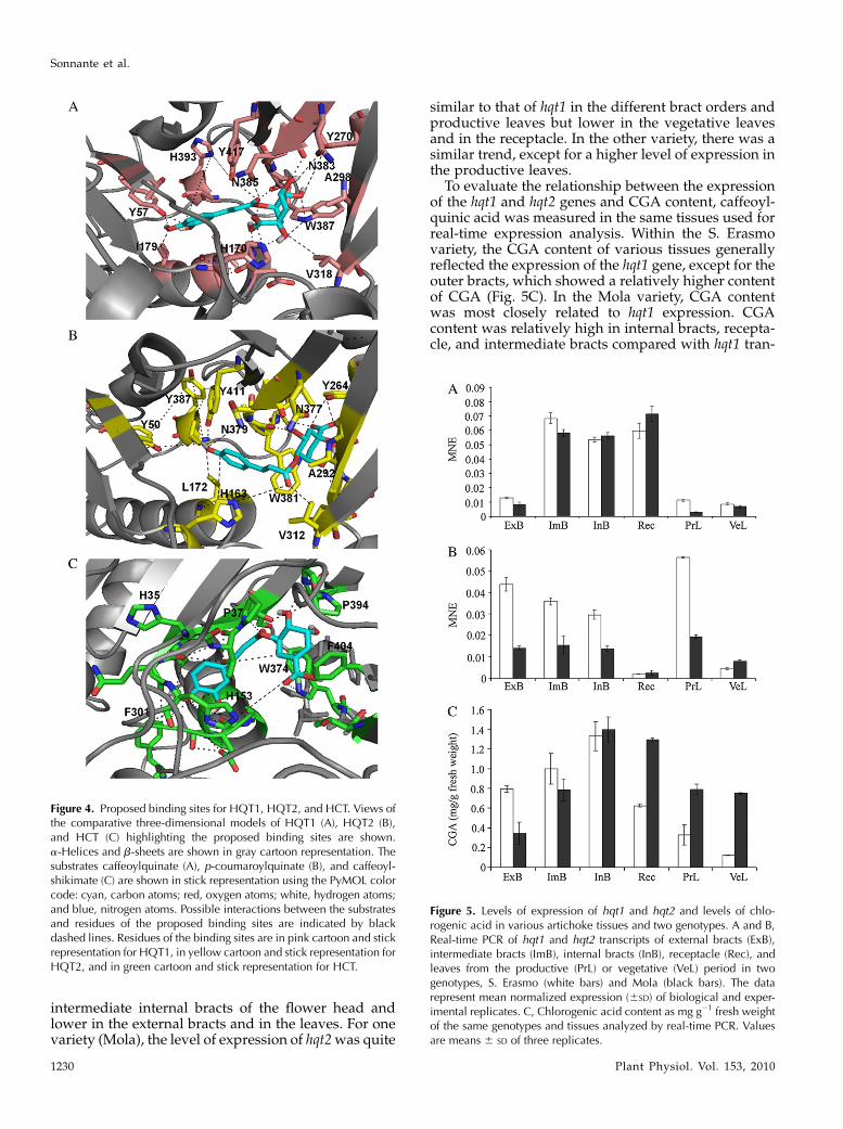
binding pockets in the proposed HQT1 and HQT2binding regions that should be able to interact with thearomatic moiety of caffeoylquinate and p-coumaroyl-quinate, respectively. In HQT1, the first aromaticpocket is composed of residues His-393, Tyr-417, Tyr-57, Ile-179, and His-170, which together may bind thecaffeoyl portion of caffeoylquinate, whereas the sec-ond binding pocket is composed of residues Tyr-270,Trp-387, Val-318, Asn-383, Ala-298, and Asn-385,which, together with His-170, should be able to bindthe quinate portion (Fig. 4A). A set of similar residuessurround p-coumaroylquinate in HQT2, where Tyr-387, Tyr-411, Tyr-50, Leu-172, Asn-379, and His-163may bind the p-coumaroyl portion while Tyr-264,Ala-292, Val-312, Trp-381, Asn-377, and His-163 likelybind the quinate portion (Fig. 4B). Residues of theproposed binding sites are well conserved throughoutthe BAHD family members that accept hydroxycinna-moyl-CoAs as acyl donors and correspond or are veryclose to the sequence motifs highlighted in themultiple sequence alignment of Figure 1 (see also Sup-plemental Fig. S1): WXRXP (for HQT1387–391 andHQT2381–385), S[S/T]YE (for HQT1268–271 and HQT2262–265),
DGRXR (forHQT1300–304 andHQT2294–298), andKX[K/S/A/Q]X[K/E/N/P/G] (for HQT1255–259 and HQT2250–254).
Two equivalent binding pockets can be also identi-fied in the proposed HCT binding region. In HCT, asmight be expected, the binding pocket, which ispredicted to prefer shikimate, is quite different fromthe quinate binding pockets in HQT1 and HQT2 andconsists of residues Pro-394, Phe-404, and Trp-374supported by Pro-37 and His-153 (Fig. 4C). Somedifferences also occur in the caffeoyl binding pocketof HCT that is composed of residues His-35, Phe-301,and His-153 compared with those of HQT1 and HQT2.However, these differences are less specific than thoseobserved for the shikimate/quinate pockets betweenHCT and HQT1/2.
Expression of hqt1 and hqt2 Relative to CGA Content ofDifferent Organs in Artichoke
A real-time PCR assay was developed using gene-specific primers that amplified exclusively hqt1 or hqt2cDNAs. Very recently, another hqt sequence has beenidentified in artichoke (Comino et al., 2009), so we alsoconfirmed the specificity of the hqt1 and hqt2 primerswith respect to artichoke hct and hqt cDNAs. Real-timePCR was used to assay the expression of hqt1 and hqt2in different bract orders (external, intermediate, andinternal), the receptacle, and leaves at two develop-mental stages (vegetative and productive periods) intwo artichoke genotypes. The cycle treshhold valueused for gene expression analysis for each gene, tissue,and genotype was obtained by averaging the cycletreshhold values derived from all the biological andexperimental replicates. The different expressionlevels of the target genes (hqt1 and hqt2) relative tothe reference gene (elf1a) were calculated usingQ-Gene software (Perikles, 2003). Normalization ofthe results against the expression of the constitutiveelf1a and control of similar amplification efficiencieswere performed to allow comparisons among tissuesand between the genotypes.
The expression profiles for hqt1 and hqt2 are shownin Figure 5, A and B. In both artichoke genotypes, theexpression of hqt1 was higher in the receptacle and
Table I. Kinetic data of recombinant HQT1 and HQT2 from E. coli extracts
The data represent means 6 SD of at least three different experiments. 1 nkat = 1 nmol substrate s21. NA, No activity.
Varying Substrate Saturating SubstrateHQT1 HQT2
Km 6 SD Vmax 6 SD Km 6 SD Vmax 6 SD
mM nkat mg21 mM nkat mg21
Quinate Caffeoyl-CoA 99 6 41 197 6 54 264 6 66 145 6 14Caffeoyl-CoA Quinate 1,391 6 283 108 6 28 6,196 6 293 258 6 83Quinate p-Coumaroyl-CoA 216 6 76 329 6 99 59 6 4 430 6 24p-Coumaroyl-CoA Quinate 1,034 6 77 227 6 65 1,065 6 70 113 6 7Shikimate Caffeoyl-CoA 5,088 6 328 19 6 4 6,120 6 481 35 6 8Shikimate p-Coumaroyl-CoA NA NA 15,690 6 2,200 14 6 1CGA CoA 130 6 32 108 6 5 251 6 28 316 6 79CoA CGA 72 6 21 195 6 18 29 6 9 5,361 6 375
Novel Artichoke hqt Genes for Chlorogenic Acid Synthesis
Plant Physiol. Vol. 153, 2010 1229
intermediate internal bracts of the flower head andlower in the external bracts and in the leaves. For onevariety (Mola), the level of expression of hqt2was quite
similar to that of hqt1 in the different bract orders andproductive leaves but lower in the vegetative leavesand in the receptacle. In the other variety, there was asimilar trend, except for a higher level of expression inthe productive leaves.
To evaluate the relationship between the expressionof the hqt1 and hqt2 genes and CGA content, caffeoyl-quinic acid was measured in the same tissues used forreal-time expression analysis. Within the S. Erasmovariety, the CGA content of various tissues generallyreflected the expression of the hqt1 gene, except for theouter bracts, which showed a relatively higher contentof CGA (Fig. 5C). In the Mola variety, CGA contentwas most closely related to hqt1 expression. CGAcontent was relatively high in internal bracts, recepta-cle, and intermediate bracts compared with hqt1 tran-
Figure 4. Proposed binding sites for HQT1, HQT2, and HCT. Views ofthe comparative three-dimensional models of HQT1 (A), HQT2 (B),and HCT (C) highlighting the proposed binding sites are shown.a-Helices and b-sheets are shown in gray cartoon representation. Thesubstrates caffeoylquinate (A), p-coumaroylquinate (B), and caffeoyl-shikimate (C) are shown in stick representation using the PyMOL colorcode: cyan, carbon atoms; red, oxygen atoms; white, hydrogen atoms;and blue, nitrogen atoms. Possible interactions between the substratesand residues of the proposed binding sites are indicated by blackdashed lines. Residues of the binding sites are in pink cartoon and stickrepresentation for HQT1, in yellow cartoon and stick representation forHQT2, and in green cartoon and stick representation for HCT.
Figure 5. Levels of expression of hqt1 and hqt2 and levels of chlo-rogenic acid in various artichoke tissues and two genotypes. A and B,Real-time PCR of hqt1 and hqt2 transcripts of external bracts (ExB),intermediate bracts (ImB), internal bracts (InB), receptacle (Rec), andleaves from the productive (PrL) or vegetative (VeL) period in twogenotypes, S. Erasmo (white bars) and Mola (black bars). The datarepresent mean normalized expression (6SD) of biological and exper-imental replicates. C, Chlorogenic acid content as mg g21 fresh weightof the same genotypes and tissues analyzed by real-time PCR. Valuesare means 6 SD of three replicates.
Sonnante et al.
1230 Plant Physiol. Vol. 153, 2010
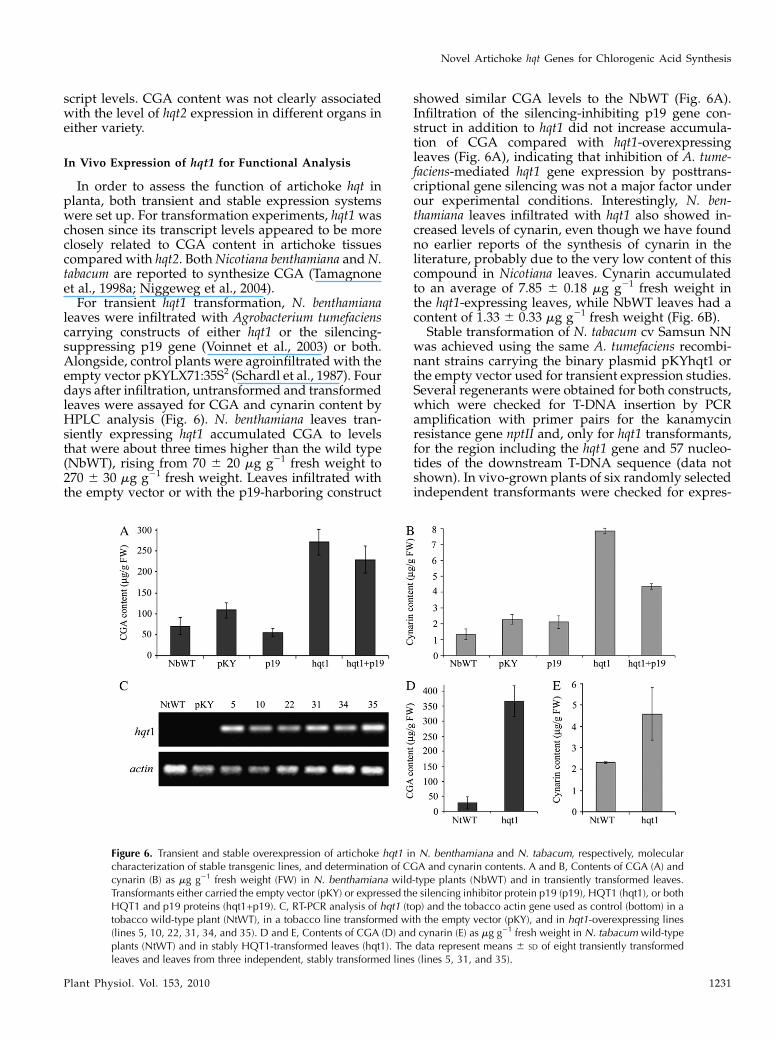
script levels. CGA content was not clearly associatedwith the level of hqt2 expression in different organs ineither variety.
In Vivo Expression of hqt1 for Functional Analysis
In order to assess the function of artichoke hqt inplanta, both transient and stable expression systemswere set up. For transformation experiments, hqt1waschosen since its transcript levels appeared to be moreclosely related to CGA content in artichoke tissuescomparedwith hqt2. BothNicotiana benthamiana andN.tabacum are reported to synthesize CGA (Tamagnoneet al., 1998a; Niggeweg et al., 2004).For transient hqt1 transformation, N. benthamiana
leaves were infiltrated with Agrobacterium tumefacienscarrying constructs of either hqt1 or the silencing-suppressing p19 gene (Voinnet et al., 2003) or both.Alongside, control plants were agroinfiltrated with theempty vector pKYLX71:35S2 (Schardl et al., 1987). Fourdays after infiltration, untransformed and transformedleaves were assayed for CGA and cynarin content byHPLC analysis (Fig. 6). N. benthamiana leaves tran-siently expressing hqt1 accumulated CGA to levelsthat were about three times higher than the wild type(NbWT), rising from 70 6 20 mg g21 fresh weight to270 6 30 mg g21 fresh weight. Leaves infiltrated withthe empty vector or with the p19-harboring construct
showed similar CGA levels to the NbWT (Fig. 6A).Infiltration of the silencing-inhibiting p19 gene con-struct in addition to hqt1 did not increase accumula-tion of CGA compared with hqt1-overexpressingleaves (Fig. 6A), indicating that inhibition of A. tume-faciens-mediated hqt1 gene expression by posttrans-criptional gene silencing was not a major factor underour experimental conditions. Interestingly, N. ben-thamiana leaves infiltrated with hqt1 also showed in-creased levels of cynarin, even though we have foundno earlier reports of the synthesis of cynarin in theliterature, probably due to the very low content of thiscompound in Nicotiana leaves. Cynarin accumulatedto an average of 7.85 6 0.18 mg g21 fresh weight inthe hqt1-expressing leaves, while NbWT leaves had acontent of 1.33 6 0.33 mg g21 fresh weight (Fig. 6B).
Stable transformation of N. tabacum cv Samsun NNwas achieved using the same A. tumefaciens recombi-nant strains carrying the binary plasmid pKYhqt1 orthe empty vector used for transient expression studies.Several regenerants were obtained for both constructs,which were checked for T-DNA insertion by PCRamplification with primer pairs for the kanamycinresistance gene nptII and, only for hqt1 transformants,for the region including the hqt1 gene and 57 nucleo-tides of the downstream T-DNA sequence (data notshown). In vivo-grown plants of six randomly selectedindependent transformants were checked for expres-
Figure 6. Transient and stable overexpression of artichoke hqt1 in N. benthamiana and N. tabacum, respectively, molecularcharacterization of stable transgenic lines, and determination of CGA and cynarin contents. A and B, Contents of CGA (A) andcynarin (B) as mg g21 fresh weight (FW) in N. benthamiana wild-type plants (NbWT) and in transiently transformed leaves.Transformants either carried the empty vector (pKY) or expressed the silencing inhibitor protein p19 (p19), HQT1 (hqt1), or bothHQT1 and p19 proteins (hqt1+p19). C, RT-PCR analysis of hqt1 (top) and the tobacco actin gene used as control (bottom) in atobacco wild-type plant (NtWT), in a tobacco line transformed with the empty vector (pKY), and in hqt1-overexpressing lines(lines 5, 10, 22, 31, 34, and 35). D and E, Contents of CGA (D) and cynarin (E) as mg g21 fresh weight in N. tabacum wild-typeplants (NtWT) and in stably HQT1-transformed leaves (hqt1). The data represent means 6 SD of eight transiently transformedleaves and leaves from three independent, stably transformed lines (lines 5, 31, and 35).
Novel Artichoke hqt Genes for Chlorogenic Acid Synthesis
Plant Physiol. Vol. 153, 2010 1231
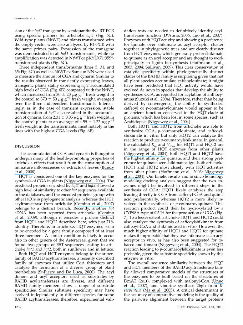
sion of the hqt1 transgene by semiquantitative RT-PCRusing specific primers for artichoke hqt1 (Fig. 6C).Wild-type plants (NtWT) and plants transformed withthe empty vector were also analyzed by RT-PCR withthe same primer pairs. Expression of the transgenewas demonstrated in all hqt1 transformants, while noamplification was detected in NtWTor pKYLX71:35S2-transformed plants (Fig. 6C).
Three independent transformants (lines 5, 31, and35; Fig. 6C) as well as NtWTcv Samsun NNwere usedto measure the amount of CGA and cynarin. Similar tothe results observed in transiently expressing leaves,transgenic plants stably expressing hqt1 accumulatedhigh levels of CGA (Fig. 6D) compared with the NtWT,which increased from 30 6 20 mg g21 fresh weight inthe control to 370 6 50 mg g21 fresh weight, averagedover the three independent transformants. Interest-ingly, as in the case of transient expression, stabletransformation of hqt1 also resulted in the accumula-tion of cynarin, from 2.316 0.05 mg g21 fresh weight inthe control plants to an average of 4.59 6 1.22 mg g21
fresh weight in the transformants, most notably in thelines with the highest CGA levels (Fig. 6E).
DISCUSSION
The accumulation of CGA and cynarin is thought tounderpin many of the health-promoting properties ofartichoke, effects that result from the consumption ofimmature inflorescences or extracts of leaves (Bundyet al., 2008).
HQT is considered one of the key enzymes for thesynthesis of CGA in plants (Niggeweg et al., 2004). Thepredicted proteins encoded by hqt1 and hqt2 showed ahigh level of similarity to other hqt sequences availablein the databases, and the encoded proteins group withother HQTs in phylogenetic analysis, whereas the HCTacyltransferase from artichoke (Comino et al., 2007)belongs to a distinct branch. Recently, another hqtcDNA has been reported from artichoke (Cominoet al., 2009), although it encodes a protein distinctfrom HQT1 and HQT2 described here, with just 71%identity. Therefore, in artichoke, HQT enzymes seemto be encoded by a gene family composed of at leastthree members. A similar condition is likely to occuralso in other genera of the Asteraceae, given that wefound two groups of EST sequences leading to arti-choke hqt1 and hqt2, both in sunflower and in lettuce.
Both HQT and HCT enzymes belong to the super-family of BAHD acyltransferases, a recently describedfamily of enzymes that utilize CoA thioesters andcatalyze the formation of a diverse group of plantmetabolites (St-Pierre and De Luca, 2000). The acyldonors and acyl acceptors used as substrates byBAHD acyltransferases are diverse, and differentBAHD family members show a range of substratespecificities. Similar substrate specificity may haveevolved independently in different species for someBAHD acyltransferases; therefore, experimental vali-
dation tests are needed to definitively identify acyl-transferase function (D’Auria, 2006; Luo et al., 2007).Enzymes with HQT activity and showing a preferencefor quinate over shikimate as acyl acceptor clustertogether in phylogenetic trees and are clearly distinctfrom HCT enzymes, which generally prefer shikimateto quinate as an acyl acceptor and are thought to workprincipally in lignin biosynthesis (Hoffmann et al.,2003, 2004; Sullivan, 2009). This clear conservation ofcatalytic specificity within phylogenetically distinctclades of the BAHD family is surprising given that notall plant species accumulate caffeoylquinate; it mighthave been predicted that HQT activity would haveevolved de novo in species that develop the ability tosynthesize CGA, as reported for acylation of anthocy-anins (Suzuki et al., 2004). Therefore, rather than beingderived by convergence, the ability to synthesizecaffeoyl or p-coumaroylquinate would appear to bean ancient function conserved in the HQT clade ofproteins, which has been lost in some species, such asArabidopsis (Niggeweg et al., 2004).
Both HQT1 and HQT2 from artichoke are able tosynthesize CGA, p-coumaroylquinate, and caffeoyl-shikimate in vitro, but only HQT2 can catalyze thereaction to produce p-coumaroylshikimate. In general,the calculated Km and Vmax for HQT1 and HQT2 arein the range of HQT enzymes from other plants(Niggeweg et al., 2004). Both HQT1 and HQT2 havethe highest affinity for quinate, and their strong pref-erence for quinate over shikimate aligns both artichokeHQT1 and HQT2 most closely with HQT activitiesfrom other plants (Hoffmann et al., 2003; Niggeweget al., 2004). Our kinetic results and in silico homologymodeling docking analyses suggest that the two en-zymes might be involved in different steps in thesynthesis of CGA: HQT1 likely catalyzes the stepleading directly to CGA from caffeoyl-CoA and quinicacid preferentially, whereas HQT2 is more likely in-volved in the synthesis of p-coumaroylquinate. Thisreaction product could then be hydroxylated by aCYP98A type of C3#H for the production of CGA (Fig.7). To a lesser extent, artichoke HQT1 and HQT2 couldalso catalyze the synthesis of caffeoylshikimate fromcaffeoyl-CoA and shikimic acid in vitro. However, themuch higher affinity of HQT1 and HQT2 for quinatemakes it improbable that they use shikimate as an acylacceptor in vivo, as has also been suggested for to-bacco and tomato (Niggeweg et al., 2004). The HQT2reaction leading to p-coumaroylshikimate is even lessprobable, given the substrate specificity shown by thisenzyme in vitro.
The overall sequence similarity between the HQTand HCT members of the BAHD acyltransferase fam-ily allowed comparative models of the structures ofthe enzymes to be built based on the structures ofDmAT (2e1t), complexed with malonyl-CoA (Unnoet al., 2007), and vinorine synthase 2bgh from R.serpentina (Ma et al., 2005). A critical determinant inthe accuracy of comparative modeling is the quality ofthe pairwise alignment between the target proteins
Sonnante et al.
1232 Plant Physiol. Vol. 153, 2010
and the crystallized templates. As the similarity be-tween HQT1, HQT2, or HCT and the crystallizedtemplates is lower than normally considered appro-priate for comparative modeling (24%–26%), a multi-ple sequence alignment approach was used toimprove pairwise alignment.A binding site is speculated for HQT1 and HQT2,
using Pocket Finder predictions and multiple se-quence alignment comparisons. Docking analysissuggested that the best substrate for the proposedbinding site of HQT1 is caffeoylquinate (CGA), whilethe best one for HQT2 is p-coumaroylquinate. Withineach enzyme-binding site, two aromatic pockets havebeen detected that should be able to bind the twoaromatic moieties of CGA and/or p-coumaroylqui-nate.In the putative first aromatic pocket, HQT1 has His-
393 and Ile-179, which are substituted by Tyr-387 andLeu-172 in HQT2. These differences may be not suf-ficient to explain the differing substrate specificities ofthe two enzymes. However, in the alignment, someother hydrophobic/hydrophilic or basic/hydrophilicdifferences were observed close to the first aromaticpocket: the Ala of peptide 170HTLA173 in HQT1 issubstituted with a Ser in the HQT2 peptide 163HTLS166,and an important hydrophilic/basic difference isobserved in a cavity that leads to the binding sitewhere the Ser of the HQT1 peptide 255KASAK259
is substituted with a Lys in the HQT2 peptide250KAKAK254. These residues, together with neighbor-ing residues, as well as the observed amino acidvariability in these portions could be responsible forsubstrate specificity and for redirecting the substratetoward the binding pockets and so might influencesubstrate specificity and enzyme efficiency. Unfortu-nately, at present, it is not possible to transform arti-choke; therefore, experiments using transgenic lines toprovide further evidence on substrate specificities arenot yet feasible.The amino acid composition of the putative second
aromatic pocket is identical in HQT1 and HQT2, and itis predicted that this pocket is able to bind the quinateportion of CGAwith analogous efficiency. The prefer-
ence of both HQT1 and HQT2 for quinate overshikimate was also evident from the docking analysis.Very recently, another HQT coding sequence has beenisolated from artichoke (DQ915589; Comino et al.,2009). This represents a fourth gene coding for ahydroxycinnamoyl transferase in artichoke: its cDNAand the encoded protein are different from those ofHQT1 and HQT2. For alignment purposes, we usedthe sequence DQ915589 retrieved from the NationalCenter for Biotechnology Information (NCBI) data-base, even though the authors show a 10-amino acidlonger sequence in their paper. Comino et al. (2009)reported that their recombinant HQT could use eithercaffeoyl-CoA or p-coumaroyl-CoA as acyl donor in thepresence of quinate, whereas it was inactive withshikimate. However, our results cannot be compareddirectly with artichoke HQT identified by Cominoet al. (2009), since the affinity of their HQT for caffeoyl-CoA or p-coumaroyl-CoA was not investigated. AcDNA encoding HCT has also been isolated fromartichoke. Enzyme assays of recombinant HCT sug-gested it to be more similar to HQT from tobacco andtomato than to HCT from tobacco or Arabidopsis,despite it aligning in the HCT clade. It was claimedthat HCT showed a preference for quinate over shiki-mate (Comino et al., 2007), but this claim was based onKm values for p-coumaroyl-CoA at saturating levels ofquinate or shikimate. Km values for HCT for quinateand shikimate need to be established to verify thisclaim. Our modeling and docking analyses suggest apreference of artichoke HCT for shikimate, in agree-ment with the predicted binding site pocket for HCT,which is clearly different from that of HQT.
To clarify the respective roles of hqt1 and hqt2 inCGA accumulation in artichoke, transcript levels ofthe two genes were analyzed in flower heads andleaves. hqt1 and hqt2 showed different patterns ofexpression, the former being more highly expressed inyounger bracts and receptacle, while the latter wasexpressed almost equally in the different bract ordersand productive leaves.
To gain further insight into the relationship betweenexpression of hqt1 and CGA synthesis, in vivo func-
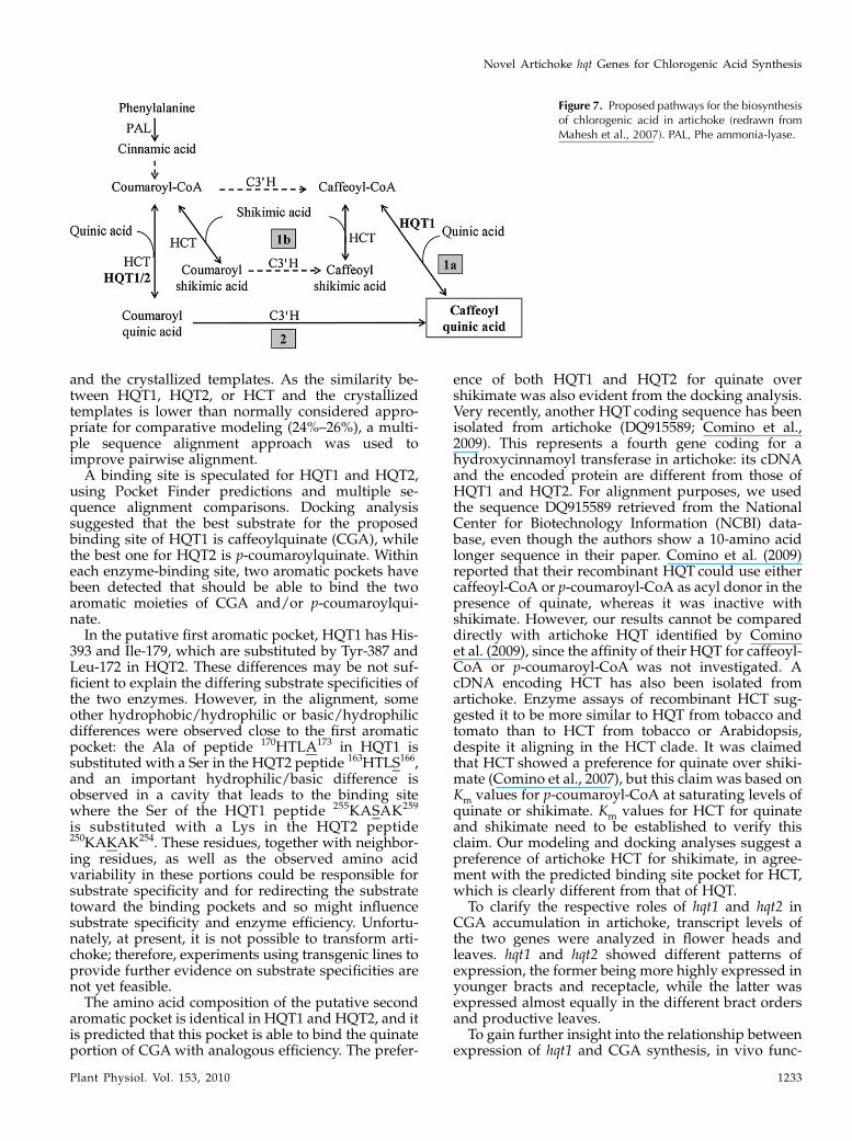
Figure 7. Proposed pathways for the biosynthesisof chlorogenic acid in artichoke (redrawn fromMahesh et al., 2007). PAL, Phe ammonia-lyase.
Novel Artichoke hqt Genes for Chlorogenic Acid Synthesis
Plant Physiol. Vol. 153, 2010 1233

tional analysis was carried out through transient andstable overexpression in N. benthamiana and N. taba-cum, respectively, since artichoke is recalcitrant togenetic transformation. The results demonstratedthat HQT1 is directly involved in the synthesis ofCGA, given that the synthesis of this compoundincreased substantially in both transiently and stablytransformed leaf tissues. In addition, increased accu-mulation of the health-promoting compound cynarinindicated that it is likely synthesized from CGA.Indeed, synthesis of 3,5-dicaffeoylquinic acid hasbeen proposed to proceed from CGA in sweet potato,although the catalyzing enzyme was able to synthesizeonly 3,5-dicaffeoylquinic acid out of the three possibledicaffeoylquinic acid isomers (Kojima and Kondo,1985).
The synthesis of CGA can theoretically occurthrough different routes operating in a metabolicgrid (Fig. 7; Mahesh et al., 2007). Our data suggestthat the route of CGA synthesis in artichoke is likelymodulated according to substrate availability, andprobably in vivo, CGA synthesis occurs preferentiallyvia route 1, involving HCT, C3#H, and HQT (Fig. 7).This is also suggested by the evidence that a recentlyisolated C3#H enzyme from artichoke proved to bemore efficient, within route 1b, in the conversion ofp-coumaroylshikimate into caffeoylshikimate (Mogliaet al., 2009). However, other possible isoforms of C3#Hcould be involved in route 2, as in coffee (Maheshet al., 2007).
The route taken for CGA biosynthesis in any par-ticular cell will depend on the substrate specificities ofthe acyltransferases and the C3#H and on the avail-ability of cytoplasmic pools of the alternative acyldonors and acceptors. In artichoke, we have identifiedtwo new HQT enzymes with different specificities,which suggest that they may operate in different partsof the grid (Fig. 7). HCT is likely most important inroute 1, section 1b (Fig. 7), and its relative contributionto CGA synthesis in different organs will depend onthe cells in which it is active. If, like HCT in tobaccoand Arabidopsis, it is expressed predominantly invascular tissues in artichoke, then its contribution toCGA synthesis may be restricted to these tissues. Thespecificity of HQT1 suggests that it may operate inroute 1, specifically section 1a, although it may alsofunction in route 2 (Fig. 7). The substrate specificity ofHQT2 suggests that it probably operates in route 2.The specificity of different C3#H enzymes operating indifferent cells may also influence the primary route forCGA synthesis, as reported for developing coffeebeans (Mahesh et al., 2007). In addition, the role ofthe newly reported HQT enzyme (Comino et al., 2009)can be better clarified only after experimental deter-mination of its catalytic specificities.
In conclusion, we have demonstrated that HQT1and HQT2 are both involved in the synthesis of CGAin artichoke. The recent report of an additional HQTfrom artichoke shows that several HQT isoforms existin this plant and that they likely operate in a metabolic
grid to ensure high levels of CGA synthesis, probablyby multiple metabolic routes. Indeed, artichoke mayhave evolved multiple HQT isoforms with differingspecificities to enhance CGA accumulation by opti-mizing flux down the different biosynthetic routes.Putative homology modeling and docking analyseshave allowed us to predict, to our knowledge for thefirst time, the binding sites of HQTand HCTenzymes,which could be of general interest for further studies ofthese enzymes in plants.
The exciting observation that elevated expression ofhqt1 in leaves of Nicotiana species not only increasesthe levels of CGA but also those of cynarin lendssupport to previous suggestions that cynarin is syn-thesized from CGA and also offers a means of increas-ing production of this important plant phytonutrient.
MATERIALS AND METHODS
Plant Materials
Plants of artichoke (Cynara cardunculus subsp. scolymus ‘Mola’ and ‘S.
Erasmo’) were grown in the experimental field of the Institute of Plant
Genetics, Policoro, Italy. For each variety, material was collected from three
independent plants; young leaves were collected during the vegetative (mid
October) and productive (mid March) phases; flower heads were harvested at
the commercial stage and separated into external, intermediate, and internal
bracts and receptacle (De Paolis et al., 2008).
Nicotiana benthamiana and tobacco (Nicotiana tabacum ‘Samsun NN’) plants
were grown in soil in a growth chamber at 23�C and 16/8-h photoperiod.
Transgenic N. tabacum plants were grown in vitro on Murashige and Skoog
solidmedium supplementedwith 3% (w/v) Suc and 100mg L21 kanamycin at
23�C and 16/8-h light/dark photoperiod for rooting. In vitro-grown plants
were then transferred to soil and acclimated to in vivo conditions in a growth
chamber with the same temperature and photoperiod.
Isolation and Cloning of Artichoke hqt cDNAs
The hqt nucleotide sequences from tobacco (AJ582651) and tomato (Sola-
num lycopersicum; AJ582652) were used to search the lettuce (Lactuca sativa)
and sunflower (Helianthus annuus) The Gene Index databases (http://compbio.
dfci.harvard.edu/tgi/plant.html). After alignment of the best tentative con-
sensus sequences, primers Hel-F3 and Hqt-R2 for hqt1 and HEL_HQT_F6
and HEL_HQT_R4 for hqt2 (Supplemental Table S1) were designed to amplify
expected fragments of about 600 and 1,000 bp, respectively. Amplification
reactions contained 1 mL of cDNA from leaves, 13 PCR buffer, 0.4 mM
deoxyribonucleotide triphosphates (dNTPs), 400 nM each primer, and 1 unit of
JumpStart (Sigma-Aldrich) Taq DNA polymerase, in a final volume of 25 mL.
The thermal cycling program included a step at 94�C for 3 min, followed by 35
cycles of 94�C for 30 s, 60�C for 30 s, and 72�C for 1min, and a final extension at
72�C for 10 min. PCR fragments were cloned using the GENEJet PCR Cloning
kit (Fermentas) and subsequently sequenced. Sequences were subjected to a
BLAST search against the GenBank plant database (http://www.ebi.ac.uk/
blastall/index.html).
Full-Length cDNA and Genomic Sequences, and
Phylogenetic Analysis
New primers were synthesized for amplification of the 5# and 3# ends of
hqt1 and hqt2 transcripts using 5# and 3# RACE (Invitrogen), respectively. To
identify the 5# ends, 5RACE_HQT1 primer for hqt1 or 5RACE_HQTa1 for hqt2
(Supplemental Table S1) and total RNA from cvMola leaf were used for cDNA
synthesis. After treatment with RNAse H and cDNA column purification, a
homopolymeric tail was added to the cDNA 3# end. A first round of PCR was
performed using the nested antisense primer 5RACE_HQT2 for hqt1 and
5RACE_HQTa2 for hqt2 (Supplemental Table S1) along with the RACE-
specific primer furnished by the manufacturer. Amplification reactions con-
Sonnante et al.
1234 Plant Physiol. Vol. 153, 2010
tained the tailed cDNA, 13 Tuning PCR buffer, 0.2 mM dNTPs, 400 nM each
primer, and 2.5 units of PCR Extender Polymerase (5 PRIME) in a final volume
of 50 mL. The thermal cycling program included a step at 94�C for 3 min,
followed by 35 cycles of 94�C for 30 s, 60�C (hqt1) or 50�C (hqt2) for 30 s, and
72�C for 2min, and a final extension step at 72�C for 10min. A second round of
PCR was performed, under the same conditions except for the annealing
temperature (60�C for hqt1 or 56�C for hqt2), using a 1:1,000 dilution of the first
amplificate and the nested primer 5RACE_HQT3 for hqt1 or 5RACE_HQTa3
for hqt2 (Supplemental Table S1) along with the universal RACE-specific
primer. Amplified cDNA fragments were cloned and sequenced.
For the completion of the 3# ends of each cDNA, total RNA and the adapter
primer (AP) from the kit were used for cDNA synthesis. A first PCR was
performed using primer HQT-F2 for hqt1 or 3RACE_HQTa1 for hqt2 (Supple-
mental Table S1) along with the anchored RACE primer. Reactions contained 2
mL of cDNA, 13 Tuning PCR buffer, 0.2 mM dNTPs, 400 nM each primer, and 2
units of PCR Extender Polymerase (5 PRIME) in a final volume of 50 mL. The
amplification program started with a step at 94�C for 3 min, followed by 35
cycles of 94�C for 30 s, 54�C for 30 s, and 72�C for 3min, and a final extension at
72�C for 10 min. A second round of PCR was performed, under the same
conditions, with the exception of the annealing temperature (60�C for hqt1 or
54�C for hqt2), using a 1:500 dilution of the first amplification and the nested
primers 3RACE_HQT1 for hqt1 or 3RACE_HQTa2 for hqt2 (Supplemental
Table S1). Amplified cDNA fragments were cloned and sequenced.
Full-length cDNA clones were generated using end-specific primers for
hqt1 and hqt2 (HQT_ATG_F/HQT_STOP_R and Hqta_ATG_F/Hqta_
STOP_R, respectively; Supplemental Table S1). Amplification reactions con-
tained 2 mL of cDNA from leaves, 13 PCR buffer, 0.4 mM dNTPs, 400 nM each
primer, and 2.5 units of recombinant Taq DNA polymerase (Invitrogen) in a
final volume of 50 mL. Amplification conditions were 94�C for 3 min and 30
cycles at 94�C for 45 s, 55�C (hqt1) or 62�C (hqt2) for 30 s, and 72�C for 90 s; the
reaction mixture was then incubated at 72�C for 10 min. Fragments were
cloned and five clones were sequenced.
The same primers were used to amplify artichoke genomic DNA by
prolonging the extension step to 4 min. Genomic DNA fragments were cloned
and sequenced.
Full-length cDNA and genomic sequences of hqt1 and hqt2 were aligned
and separately subjected to BLAST search (http://www.ncbi. nlm.nih.gov/
BLAST/). The hqt cDNA sequences were translated in silico using the
program TRANSEQ (http://bioweb.pasteur.fr/seqanal/interfaces/transeq.
html), and the deduced protein sequences were aligned to protein sequences
from other species. Phylogenetic analysis was performed using the program
MEGA4 (Tamura et al., 2007). The tree was constructed using the neighbor-
joining method of Saitou and Nei (1987), with 10,000 bootstrap replicates.
For the isolation of hqt1 and hqt2 promoters, the GenomeWalker Universal
kit (Clontech) was used, following the manufacturer’s instructions. After
artichoke genomic DNA digestion with DraI, EcoRV, PvuII, and StuI and
ligation to adaptors as indicated in the manufacturer’s instructions, a first
round of PCRwas performed for each digested-ligated DNA library, using the
AP1 primer from the kit and the primer specific for hqt1 or hqt2 (GWhqtGSP1
or GWhqt2GSP1, respectively; Supplemental Table S1). Amplification reac-
tions contained 1 mL of ligated DNA, 13 PCR buffer, 0.5 mM dNTPs, 400 nM
each primer, and 2 units of PCR Extender Polymerase (5 PRIME) in a final
volume of 50 mL. The thermal cycling program included seven cycles of 94�Cfor 25 s and 69�C (hqt1) or 68�C (hqt2) for 3 min, followed by 37 cycles of 94�Cfor 25 s and 64�C for 3 min. After the 37 cycles, the reaction mixture was
incubated at 67�C for 7 min. A 1:50 dilution of the amplificates was used as a
template for a nested PCR, using primers GWhqtGSP2 and GWhqt2GSP2
(Supplemental Table S1), and the following thermal cycling program: five
cycles of 94�C for 25 s and 72�C (hqt1) or 66�C (hqt2) for 3 min, followed by 20
cycles of 94�C for 25 s and 67�C (hqt1) or 62�C (hqt2) for 3 min, followed by an
incubation step at 67�C for 7 min. Amplicons were resolved on a low-melting-
point agarose gel, and fragments longer than 1 kb were excised from the gel,
purified, cloned, and sequenced.
The analysis of promoter sequences for the two genes was performed using
the database PLACE (http://www.dna.affrc.go.jp/PLACE/; Higo et al., 1999).
Heterologous Expression in Escherichia coli andImmunoblot Analysis
Full-length cDNAs of hqt1 and hqt2 were introduced into the Gateway
system (Invitrogen) as follows. The cDNAs were amplified using the primer
pairs A-HQT-GW_F/A-HQT-GW_R and A_HQTa_GW_F/A_HQTa_GW_R,
respectively (Supplemental Table S1). The entry clones (pDONR207-hqt1 and
pDONR207-hqt2) were obtained through recombination of the PCR products
with pDONR207 (Invitrogen). Error-free clones were then introduced into the
Gateway-compatible S-tag vector pJAM1784 (Luo et al., 2007) to produce the
expression vectors pJAM1784-hqt1 and pJAM1784-hqt2. Recombinant pro-
teins with an N-terminal S-tag were expressed in E. coli BL-21 Rosetta cells.
Cells were grown at 37�C with shaking at 250 rpm until the optical density at
600 nm was 0.6. Isopropyl b-D-1-thiogalactopyranoside was added to 0.4 mM.
Cells were cultured at 20�C with shaking at 250 rpm for 16 h. Cells were
harvested, resuspended in 100 mM potassium phosphate, pH 7.0, with 1 mM
EDTA, broken using a French press, and centrifuged at 14,000 rpm for 30 min
at 4�C. Total protein was assayed from crude extracts. Samples containing 10
mg of proteins were separated by SDS-PAGE, and proteins were electropho-
retically transferred to nitrocellulose. S-tag fusion peptides were immunode-
tected with 1/5,000 AP-conjugated S-Protein (Novagen). Negative controls
used comparable extracts of E. coli harboring an empty vector.
Transient and Stable Heterologous Expression of hqt1in Nicotiana
Full-length cDNA of hqt1 was amplified with the primers f-HQT-HindIII
and r-HQT-XbaI (Supplemental Table S1) to introduce HindIII and XbaI
restriction sites and cloned into the HindIII-XbaI polylinker sites of the binary
vector pKYLX71:35S2 under the transcriptional control of the cauliflower
mosaic virus 35S promoter with double enhancer (Schardl et al., 1987). Correct
insertion was verified by sequencing. The resulting recombinant pKYhqt1
plasmid was introduced into the Agrobacterium tumefaciens strain LBA4404 by
the freeze-thaw method (Hofgen and Willmitzer, 1988).
For transient transformation, A. tumefaciens LBA4404 cultures carrying the
plasmid pKYhqt1, or the 35S:p19 plasmid harboring the gene encoding the
silencing inhibitor protein p19 (Voinnet et al., 2003), or the empty vector
pKYLX71:35S2 were grown overnight at 28�C with vigorous shaking. Pelleted
cells were resuspended in 10 mMMgCl2 and 150 mgmL21 acetosyringone to an
optical density at 600 nm of 1.0 and incubated at room temperature for 3 h
(Voinnet et al., 2003). For agroinfiltration, equal amounts of the cultures
containing pKYhqt1, or 35S:p19, or pKYhqt1 plus 35S:p19, or the empty
pKYLX71:35S2 vector were used. Each construct or construct combination was
infiltrated into the abaxial air spaces of four leaves of each of two N.
benthamiana plants. After 4 d, the infiltrated leaf material was collected.
The recombinant A. tumefaciens LBA4404 strain harboring either the
pKYhqt1 plasmid or the empty vector was also used to stably transform N.
tabacum cv Samsun NN plants according to standard methods (Horsh et al.,
1987). Kanamycin-resistant regenerated shoots were excised and transferred
to kanamycin-supplemented solid medium for rooting and then transferred to
soil and acclimated to in vivo conditions in a growth chamber. Three-week-old
transgenic and control plants were used for molecular characterization and
HPLC analysis (see below).
Stable insertion of the exogenous expression cassette was checked in
kanamycin-resistant plants by PCR amplification with primers for the nptII
gene (TransF and TransR; Supplemental Table S1). The presence of the hqt1
transgene was also assessed by amplifying DNA of kanamycin-resistant
plants with specific primers for hqt1 (f-HQT-Hind III) and for pKYLX71:35S2
polylinker (pKYLX_R; Supplemental Table S1).
Enzyme Assays and HPLC/Liquid Chromatography/MassSpectrometry Analyses
S-tag fusion peptides for HQT1 and HQT2 were measured using the S-tag
rapid assay kit (Novagen) by comparison with the S-tag peptide standard.
For CGA enzyme assays, the standard reaction mixture (100 mL) consisted
of 50 mM potassium phosphate buffer, pH 7.0, 60 mM caffeoyl-CoA or
p-coumaroyl-CoA, 120 mM quinate or shikimate, 1 mM EDTA, and enzyme as
1 to 5 mL of the crude E. coli extract. For cynarin assay, 1 mM cynarin and 500 mM
CoAwere used. Each reaction was started by addition of the acyl acceptor. After
incubation at 30�C for 10 min, the reaction was terminated by adding 100 mL
of ice-cold 0.5% (v/v) trifluoroacetic acid.
The reaction mixture was analyzed by liquid chromatography/mass
spectrometry (LC/MS) using an Agilent 1100 LC system equipped with diode
array UV light and single quadrupole 1100 MSD detectors. Ten microliters of
the reaction was analyzed on a 100- 3 2-mm 3m Luna C18(2) column
(Phenomenex) using a gradient of methanol (B) versus 0.1% formic acid (A)
in water run at 25�C and 0.25 mL min21: 0 min, 98% A, 2% B; 30 min, 30% A,
70% B; 30.5 min, 98% A, 2% B; 38 min, 98% A, 2% B. Spray chamber conditions
Novel Artichoke hqt Genes for Chlorogenic Acid Synthesis
Plant Physiol. Vol. 153, 2010 1235
were 10 L min21 drying gas at 350�C, 25-p.s.i. nebulizer pressure, and 4,000-V
(positive mode) or 3,500-V (negative mode) spray voltage. Masses were scanned
from 50 to 1,500 atomic mass units, and the fragmentor voltage was 75 V.
Determination of Kinetic Parameters
To evaluate the substrate specificity of HQT1 and HQT2, caffeoyl-CoA and
p-coumaroyl-CoA were tested as acyl donors, while quinate, shikimate,
sinapate, and malate were used as acyl acceptors. The standard reaction
mixture (100 mL) consisted of 50 mM potassium phosphate buffer, pH 7.0, 1 mM
to 24 mM of the different substrates, and 0.6 to 1.4 mg of S-tag fusion proteins
from crude extracts of E. coli. Enzyme activity was measured as the decrease in
time of acyl donor A340 or A360 for p-coumaroyl-CoA or caffeoyl-CoA reaction,
respectively, using a FlexStation3 Microplate Reader (Molecular Devices).
These wavelengths were chosen because they correspond to the maxima of the
difference spectra between the thioesters and the corresponding quinic or
shikimic esters (Lotfy et al., 1992; Niggeweg et al., 2004). The following
concentrations of enzymes and substrates were used. Km for quinate: 57 and 25
ng mL21 for HQT1 and HQT2, respectively, 2.5 mM caffeoyl-CoA, 0.05 to 1 mM
quinate or 2.5 mM p-coumaroyl-CoA, and 0.001 to 0.6 mM quinate; Km for shi-
kimate: 140 ng mL21 for HQT1 or HQT2, 2.5 mM caffeoyl-CoA/p-coumaroyl-
CoA, and 1 to 24 mM shikimate; Km for caffeoyl-CoA: 140 ng mL21 for HQT1
or HQT2, 10 mM quinate, and 0.1 to 4.5 mM caffeoyl-CoA; Km for p-coumaroyl-
CoA: 71 ng mL21 for HQT1 or HQT2, 10 mM quinate, and 0.1 to 3 mM
p-coumaroyl-CoA. For reverse reactions, the Km for CoA was determined
using 10 ng mL21 HQT1 or 6 ng mL21 HQT2, 0.5 mM CGA, and 0.01 to 0.3 mM
CoA. The Km for CGA was determined using 10 ng mL21 HQT1 or 6 ng mL21
HQT2, 0.4 mM CoA, and 0.1 to 0.6 mM CGA.
No activity was detected with sinapic acid or malic acid as acyl acceptor,
using 140 ng mL21 HQT1 or HQT2, and 10 mM sinapic acid or malic acid. Km
and Vmax values were calculated from Lineweaver-Burk double reciprocal
plots. CGA, CoA, quinate, shikimate, sinapate, and malate were from Sigma-
Aldrich. Caffeoyl-CoA and p-coumaroyl-CoAwere purchased from TransMIT.
Comparative Modeling and Docking Investigations
Protein sequences of plant BAHD acyltransferases were obtained from the
NCBI database (http://www.ncbi.nlm.nih.gov/). The homologs of the arti-
choke proteins HQT1 and HQT2 were aligned using ClustalW with the
secondary structure of the anthocyanin acyltransferase from Dendranthema
morifolium (DmAT), complexed with malonyl-CoA, and vinorine synthase
from Rauvolfia serpentina. These crystallized structures were used to weigh gap
penalties. The three-dimensional structures of the two cited proteins are
available at the Protein Data Bank (Berman et al., 2002) under the code names
2e1t (Unno et al., 2007) and 2bgh (Ma et al., 2005), respectively. The pairwise
alignment of each artichoke protein (HQT1 and HQT2) with DmATwas taken
from the multiple sequence alignment and improved by comparison with the
PSIPRED secondary structure alignment prediction output. Then, Modeller
(Sanchez and Sali, 2000) was used to calculate structural models of artichoke
HQT1 and HQT2 proteins. For comparative purposes, the artichoke HCT
protein (Comino et al., 2007) was also analyzed. Five hundred steps of energy
minimization were repeated to generate 100 optimized HQT1, HQT2, and
HCT structures. The structural properties of the HQT1, HQT2, and HCT
models with the best energy function were evaluated using the biochemical/
computational tools of the WHAT IF Web server (Vriend, 1990).
Final models were examined in PyMOL (http://www.pymol.org/) and
SwissPDBViewer (http://spdbv.vital-it.ch/), and where side chain packing
led to clashes or gaps in the local secondary structure, alternative side chain
rotamers were evaluated.
Pocket Finder was used to predict the potential binding sites of the HQT1,
HQT2, and HCT best models. For docking analysis, potential substrates were
docked in the proposed binding site using Autodock 1.5.2 (Goodsell et al., 1996),
which allows selection of a three-dimensional region within which the software
will search for the best interactions between all the possible substrate confor-
mations and the selected three-dimensional region proposed as the binding site.
Expression Analysis in Artichoke and inTransgenic N. tabacum
Levels of hqt1 and hqt2 transcripts in different artichoke tissues and
physiological stages were analyzed using real-time PCR in a Rotor-Gene 6000
(Corbett Research). Artichoke elongation factor 1a (Elf1a; GenBank accession
no. EU442190) was used as a housekeeping gene. Each reaction contained 1.2
mL of a 1:6 cDNA dilution, 5 mL of Power SybrGreen mix (Applied Biosys-
tems), with 300 nM gene-specific primers RT-HQT-F2 and RT-HQT-R2 for hqt1,
RT-HQT2-F and RT-HQT2-R for hqt2, or EL-RT-F and EL-RT-R for Elf1a
(Supplemental Table S1), in a total volume of 10 mL. To reduce pipetting errors,
a CAS-1200 liquid-handling system (Corbett) was used. In all experiments,
appropriate negative controls containing no template were subjected to the
same procedure. Analysis was performed on three independent plants for
each genotype, on three replicates for each sample, and reactions were
repeated twice to verify reproducibility. Real-time PCR cycles were 95�C for
10 min, followed by 40 cycles of three-step reactions (95�C for 15 s, 60�C for 30
s, and 72�C for 30 s). The specificity of products was verified by melting curve
analysis (60�C–95�C). Standard curves for the genes of interest and the
housekeeping gene were generated on the basis of a six-point cDNA dilution
series by plotting the threshold cycle versus relative template concentration.
Mean normalized expression of hqt1 and hqt2 was calculated using Q-Gene
software (Perikles, 2003) by averaging the normalized expression values
derived from biological and experimental replicates.
For the semiquantitative RT-PCR analysis of expression of the hqt1 trans-
gene in stably transformed tobacco plants, cDNAs were synthesized from 1.0
mg of total RNA from leaves of six hqt1 transformants, one plant transformed
with the empty pKYLX71:35S2 vector and one wild-type plant, using Super-
Script II reverse transcriptase (Invitrogen) according to the manufacturer’s
instructions. Semiquantitative RT-PCR amplifications were performed using
specific primers for artichoke hqt1 (RT-HQT-F2 and RT-HQT-R2; Supplemen-
tal Table S1) and tobacco actin as a housekeeping gene (f-ACT and r-ACT;
Supplemental Table S1). Reactions contained 13 PCR buffer, 0.2 mM dNTPs,
1.5 mM MgCl2, 1.5 units of Taq DNA polymerase (Invitrogen), 500 nM each
primer, and 1 mL of cDNA template in a final volume of 50 mL. The
amplification program started with a step at 95�C for 5 min, followed by 30
cycles of three steps (95�C for 15 s, 60�C for 30 s, and 72�C for 10 s), followed by
an elongation step at 72�C for 5 min. Amplified products were separated and
visualized on a 2% agarose gel.
Determination of CGA Content in Artichoke andNicotiana Tissues
The same artichoke plant parts used for real-time PCR experiments were
analyzed for their CGA content, following Fratianni et al. (2007), with some
modifications. Frozen plant tissues were weighed, ground, and incubated at
4�C for 2 h with 5% (w/v) of an extraction mixture composed of acetone:
methanol:ethanol (70:15:15). The supernatant was collected and stored at 4�C,while the pellet was reextracted in ethyl acetate. The two supernatants were
mixed and dried in a Rotavapor at 25�C, resuspended in methanol, and 10 mL
was injected for HPLC analysis using a Luna C18 Phenomenon column in a
Gold System chromatograph equipped with a UV light detector (Beckman).
The mobile phase included HPLC-grade water (containing 0.01% TCA;
solvent A) and 95% acetonitrile (containing 0.01% TCA; solvent B) in the
following gradient system: initial, 0% B; at 2 min, 36% B for 33 min; at 35 min,
36% B for 10 min; at 45 min, 10% B for 2 min. The flow rate was 1 mL min21,
and the detection wavelength was set at 310 nm.
Soluble phenolics were extracted from transiently transformed and wild-
type N. benthamiana leaves and from N. tabacum leaves of three independent
stable transformants. Freshly harvested tissues were homogenized on ice in
80% aqueous methanol solution (1:3, w/v) and then sonicated three times for
1 min. Soluble phenolics were separated by centrifuging at 13,000 rpm for 5
min and stored at 220�C until use. HPLC analysis was performed as follows.
Twenty microliters of each extract was analyzed by HPLC (LC 10; Shimadzu)
with a diode array detector and a Prodigy column (5 mm ODS3 100A, 250 34.60 mm; Phenomenex) at a flow rate of 0.8 mLmin21. The mobile phase was a
mixture of water:formic acid (99.8:0.2, v/v; A) and methanol:acetonitrile
(40:60, v/v; B). Phenolic compound elution was achieved using the following
linear gradient: starting condition, 85%A, 15% B; 6 min, 75%A, 25% B; 16 min,
70% A, 30% B; 20 min, 60% A, 40% B; 24 min, 50% A, 50% B; 32 min, 40% A,
60% B; 35 min, 20% A, 80% B; 38 min, 75% A, 15% B. Chromatograms were
recorded at 365 nm for phenolic acids.
Standard for cynarin (1,3-dicaffeoylquinic acid) was from Carl Roth, and
CGA was from Sigma-Aldrich.
Sequence data from this article can be found in the GenBank/EMBL data
libraries under accession numbers AM690438 (hqt1 cDNA), EU839580 (hqt2
cDNA), EU697935 (hqt1 genomic DNA), FM244907 (hqt2 genomic DNA), and
EU442190 (Elf1a).
Sonnante et al.
1236 Plant Physiol. Vol. 153, 2010
Supplemental Data
The following materials are available in the online version of this article.
Supplemental Figure S1. Expression of recombinant artichoke HQT1 and
HQT2 proteins in E. coli.
Supplemental Figure S2. Multiple sequence alignment of several plant
acyltransferases.
Supplemental Table S1. List of primer names and sequences.
ACKNOWLEDGMENTS
We thank A. De Lisi, D. Nigro, and A. Visconti for HPLC analyses of CGA
and cynarin content; A. Morgese for DNA sequencing; and L. Hill for LC/MS
analyses.
Received October 28, 2009; accepted April 27, 2010; published April 29, 2010.
LITERATURE CITED
Agarwal R, Mukhtar H (1996) Cancer chemoprevention by polyphenols in
green tea and artichoke. Adv Exp Med Biol 401: 35–50
Azzini E, Bugianesi R, Romano F, Di Venere D, Miccadei S, Durazzo A,
Foddai MS, Catasta G, Linsalata V, Maiani G (2007) Absorption and
metabolism of bioactive molecules after oral consumption of cooked
edible heads of Cynara scolymus L. (cultivar Violetto di Provenza) in
human subjects: a pilot study. Br J Nutr 97: 963–969
Berman HM, Battistuz T, Bhat TN, Bluhm WF, Bourne PE, Burkhardt K,
Feng Z, Gilliland GL, Iype L, Jain S, et al (2002) The Protein Data Bank.
Acta Crystallogr D Biol Crystallogr 58: 899–907
Brown JE, Rice-Evans CA (1998) Luteolin-rich artichoke extract protects
low density lipoprotein from oxidation in vitro. Free Radic Res 29:
247–255
Bundy R, Walker AF, Middleton RW, Wallis C, Simpson HC (2008)
Artichoke leaf extract (Cynara scolymus) reduces plasma cholesterol in
otherwise healthy hypercholesterolemic adults: a randomized, double
blind placebo controlled trial. Phytomedicine 15: 668–675
Bushman BS, SnookME, Gerke JP, Szalma SJ, BerhowMA, Houchins KE,
McMullen MD (2002) Two loci exert major effects on chlorogenic acid
synthesis in maize silks. Crop Sci 42: 1669–1678
Chen JH, Ho CT (1997) Antioxidant activities of caffeic acid and its related
hydroxycinnamic acid compounds. J Agric Food Chem 45: 2374–2378
Clifford MN (1999) Chlorogenic acids and other cinnamates: nature,
occurrence, and dietary burden. J Sci Food Agric 79: 362–372
Comino C, Hehn A, Moglia A, Menin B, Bourgaud F, Lanteri S, Portis E
(2009) The isolation and mapping of a novel hydroxycinnamoyltrans-
ferase in the globe artichoke chlorogenic acid pathway. BMC Plant
Biol 9: 30
Comino C, Lanteri S, Portis E, Acquadro A, Romani A, Hehn A, Larbat R,
Bourgaud F (2007) Isolation and functional characterization of a cDNA
coding a hydroxycinnamoyltransferase involved in phenylpropanoid
biosynthesis in Cynara cardunculus L. BMC Plant Biol 7: 14
D’Auria JC (2006) Acyltransferases in plants: a good time to be BAHD.
Curr Opin Plant Biol 9: 331–340
De Paolis A, Pignone D, Morgese A, Sonnante G (2008) Characteriza-
tion and differential expression analysis of artichoke phenylalanine
ammonia-lyase coding sequences. Physiol Plant 132: 33–43
Dowd PF, Vega FE (1996) Enzymatic oxidation products of allelochemicals
as a basis for resistance against insects: effects on the corn leafhopper
Dalbulus maidis. Nat Toxins 4: 85–91
Fratianni F, Tucci M, De Palma M, Pepe R, Nazzaro F (2007) Polyphenolic
composition in different parts of some cultivars of globe artichoke:
Cynara cardunculus L. var. scolymus (L.) Fiori. Food Chem 104: 1282–1286
Gebhardt R (1997) Antioxidative and protective properties of extracts from
leaves of artichoke (Cynara scolymus L.) against hydroperoxide-induced
oxidative stress in cultured rat hepatocytes. Toxicol Appl Pharmacol
144: 279–286
Goodsell DS, Morris GM, Olson AJ (1996) Automated docking of flexible
ligands: applications of AutoDock. J Mol Recognit 9: 1–5
Higo K, Ugawa Y, Iwamoto M, Korenaga T (1999) Plant cis-acting regu-
latory DNA elements (PLACE) database: 1999. Nucleic Acids Res 27:
297–300
Hoffmann L, Besseau S, Geoffroy P, Ritzenthaler C, Meyer D, Lapierre C,
Pollet B, LegrandM (2004) Silencing of hydroxycinnamoyl-coenzyme A
shikimate/quinate hydroxyl cinnamoyltransferase affects phenylpro-
panoid biosynthesis. Plant Cell 16: 1446–1465
Hoffmann L, Maury S, Martz F, Geoffroy P, LegrandM (2003) Purification,
cloning, and properties of an acyltransferase controlling shikimate and
quinate ester intermediates in phenylpropanoid metabolism. J Biol
Chem 278: 95–103
Hofgen R, Willmitzer L (1988) Storage of competent cells for Agrobacterium
transformation. Nucleic Acids Res 16: 9877
Horsh RB, Fry JE, Hoffmann NL, Eicholtz D, Rogers SH, Fraley RT (1987)
A simple and general method for transferring genes in plants. Science
227: 1229–1231
Kojima M, Kondo T (1985) An enzyme in sweet potato root which
catalyzes the conversion of chlorogenic acid, 3-caffeoylquinic acid, to
isochlorogenic acid, 3,5-dicaffeoylquinic acid. Agric Biol Chem 49:
2467–2469
Kraft K (1997) Artichoke leaf extract: recent findings reflecting effects on
lipid metabolism, liver and gastrointestinal tracts. Phytomedicine 4:
369–378
Lepelley M, Cheminade G, Tremillon N, Simkin A, Caillet V, McCarthy J
(2007) Chlorogenic acid synthesis in coffee: an analysis of CGA content
and real-time RT-PCR expression of HCT, HQT, C3H1, and CCoAOMT1
genes during grain development in C. canephora. Plant Sci 172: 978–996
Lotfy S, Fleuriet A, Macheix JJ (1992) Partial purification and character-
ization of hydroxycinnamoyl-CoA: transferases from apple and date
fruits. Phytochemistry 31: 767–772
Luo J, Nishiyama Y, Fuell C, Taguchi G, Elliott K, Hill L, Tanaka Y,
Kitayama M, Yamazaki M, Bailey P, et al (2007) Convergent evolution
in the BAHD family of acyltransferases: identification and characteri-
zation of anthocyanin acyltransferases from Arabidopsis thaliana. Plant J
50: 678–695
Ma X, Koepke J, Panjikar S, Fritzsch G, Stockigt J (2005) Crystal structure
of vinorine synthase, the first representative of the BAHD superfamily.
J Biol Chem 280: 13576–13583
Maher EA, Bate NJ, Ni W, Elkind Y, Dixon RA, Lamb CJ (1994) Increased
disease susceptibility of transgenic tobacco plants with suppressed
levels of preformed phenylpropanoid products. Proc Natl Acad Sci USA
91: 7802–7806
Mahesh V, Million-Rousseau R, Ullmann P, Chabrillange N, Bustamante
J, Mondolot L, Morant M, Noirot M, Hamon S, de Kochko A, et al
(2007) Functional characterization of two p-coumaroyl ester 3#-hydrox-ylase genes from coffee tree: evidence of a candidate for chlorogenic acid
biosynthesis. Plant Mol Biol 64: 145–159
McDougall B, King PJ, Wu BW, Hostomsky Z, Manfred G, Robinson WE
Jr (1998) Dicaffeoylquinic acid and dicaffeoyltartaric acid are selective
inhibitors of human immunodeficiency virus type 1 integrase. Antimi-
crob Agents Chemother 42: 140–146
McGuffin LJ, Bryson K, Jones DT (2000) The PSIPRED protein structure
prediction server. Bioinformatics 16: 404–405
Moglia A, Comino C, Portis E, Acquadro A, De Vos RC, Beekwilder J,
Lanteri S (2009) Isolation and mapping of a C3’H gene (CYP98A49)
from globe artichoke, and its expression upon UV-C stress. Plant Cell
Rep 28: 963–974
Nardini M, Cirillo E, Natella F, Scaccini F (2002) Absorption of phenolic
acids in humans after coffee consumption. J Agric Food Chem 50:
5735–5741
Narusaka Y, Narusaka M, Seki M, Umezawa T, Ishida J, Nakajima M,
Enju A, Shinozaki K (2004) Crosstalk in the responses to abiotic and
biotic stresses in Arabidopsis: analysis of gene expression in cytochrome
P450 gene superfamily by cDNA microarray. Plant Mol Biol 55: 327–342
Niggeweg R, Michael AJ, Martin C (2004) Engineering plants with in-
creased levels of the antioxidant chlorogenic acid. Nat Biotechnol 2:
746–754
Perikles S (2003) Q-Gene processing quantitative real-time RT-PCR data.
Bioinformatics 19: 1439–1440
Persson B (2000) Bioinformatics in protein analysis. EXS 88: 215–231
Plumb GW, Garcia-Conesa MT, Kroon PA, Rhodes M, Ridley S,
Williamson G (1999) Metabolism of chlorogenic acid by human plasma,
liver, intestine and gut microflora. J Sci Food Agric 79: 390–392
Novel Artichoke hqt Genes for Chlorogenic Acid Synthesis
Plant Physiol. Vol. 153, 2010 1237
Rice-Evans CA, Miller NJ, Paganga G (1997) Antioxidant properties of
phenolic compounds. Trends Plant Sci 2: 152–159
Saitou N, Nei M (1987) The neighbor-joining method: a new method for
reconstructing phylogenetic trees. Mol Biol Evol 4: 406–425
Sanchez R, Sali A (2000) Comparative protein structure modeling: intro-
duction and practical examples with Modeller. Methods Mol Biol 143:
97–129
Schardl C, Byrd AD, Benzion GB, Altschuler MA, Hildebrand DF, Hunt
AG (1987) Design and construction of a versatile system for the expres-
sion of foreign genes in plants. Gene 61: 1–11
Schutz K, Kammerer D, Carle R, Schieber A (2004) Identification and
quantification of caffeoylquinic acids and flavonoids from artichoke
(Cynara scolymus L.) heads, juice, and pomace by HPLC-DAD-ESI/MS.
J Agric Food Chem 52: 4090–4096
Segasothy M, Phillips PA (1999) Vegetarian diet: panacea for modern
lifestyle diseases? Q J Med 92: 531–544
Shimoda H, Ninomiya K, Nishida N, Yoshino T, Morikawa T, Matsuda H,
Yoshikawa M (2003) Anti-hyperlipidemic sesquiterpenes and new
sesquiterpene glycosides from the leaves of artichoke (Cynara scolymus
L.): structure requirement and mode of action. Bioorg Med Chem Lett
13: 223–228
Soltani BM, Ehlting J, Hamberger B, Douglas CJ (2006) Multiple cis-
regulatory elements regulate distinct and complex patterns of develop-
mental and wound-induced expression of Arabidopsis thaliana 4CL gene
family members. Planta 224: 1226–1238
Sonnante G, Pignone D, Hammer K (2007) The domestication of artichoke
and cardoon: from Roman times to the genomic age. Ann Bot (Lond)
100: 1095–1100
Stalmach A, Steiling H, Williamson G, Crozier A (2010) Bioavailability of
chlorogenic acids following acute ingestion of coffee by humans with an
ileostomy. Arch Biochem Biophys (in press)
St-Pierre B, De Luca V (2000) Evolution of acyltransferase genes: origin
and diversification of the BAHD superfamily of acyltransferases in-
volved in secondary metabolism. Recent Adv Phytochem 34: 285–315
Sullivan ML (2009) A novel red clover hydroxycinnamoyl transferase has
enzymatic activities consistent with a role in phaselic acid biosynthesis.
Plant Physiol 150: 1866–1879
Suzuki H, Sawada S, Watanabe K, Nagae S, Yamaguchi MA, Nakayama T,
Nishino T (2004) Identification and characterisation of a novel antho-
cyanin malonyltransferase from scarlet sage (Salvia splendens) flowers:
an enzyme that is phylogenetically separated from other anthocyanin
acyltransferases. Plant J 38: 994–1003
Tamagnone L, Merida A, Parr A, Mackay S, Culianez-Macia FA, Roberts
K, Martin C (1998a) The AmMYB308 and AmMYB330 transcription
factors from Antirrhinum regulate phenylpropanoid and lignin biosyn-
thesis in transgenic tobacco. Plant Cell 10: 135–154
Tamagnone L, Merida A, Stacey N, Plaskitt K, Parr A, Chang C, Lynn D,
Dow JM, Roberts K, Martin C (1998b) Inhibition of phenolic acid
metabolism results in precocious cell death and altered cell morphology
in leaves of transgenic tobacco plants. Plant Cell 10: 1801–1816
Tamura K, Dudley J, Nei M, Kumar S (2007) MEGA4: Molecular Evolu-
tionary Genetics Analysis (MEGA) software version 4.0. Mol Biol Evol
24: 1596–1599
Ulbrich B, Zenk MH (1979) Partial purification and properties of hydroxy-
cinnamoyl-CoA:quinate hydroxycinnamoyl transferase from higher
plants. Phytochemistry 18: 929–933
Unno H, Ichimaida F, Suzuki H, Takahashi S, Tanaka Y, Saito A, Nishino
T, Kusunoki M, Nakayama T (2007) Structural and mutational studies
of anthocyanin malonyltransferases establish the features of BAHD
enzyme catalysis. J Biol Chem 282: 15812–15822
Villegas RJA, Kojima M (1986) Purification and characterization of
hydroxycinnamoyl D-glucose: quinate hydroxycinnamoyl transferase
in the root of sweet potato, Ipomoea batatas Lam. J Biol Chem 261:
8729–8733
Voinnet O, Rivas S, Mestre P, Baulcombe D (2003) An enhanced transient
expression system in plants based on suppression of gene silencing by
the p19 protein of tomato bushy stunt virus. Plant J 33: 949–956
Vriend G (1990) WHAT IF: a molecular modeling and drug design pro-
gram. J Mol Graph 8: 52–56
Walker JRL (1995) Enzymatic browning in fruits: its biochemistry and
control. In CY Lee, JR Whitaker, eds, Enzymatic Browning and Its
Prevention: ACS Symposium Series 600. American Chemical Society,
Washington, DC, pp 8–22
Wang M, Simon JE, Aviles IF, He K, Zheng QY, Tadmor Y (2003) Analysis
of antioxidative phenolic compounds in artichoke (Cynara scolymus L.).
J Agric Food Chem 51: 601–608
Zhua XF, Zhanga HX, Lob R (2005) Antifungal activity of Cynara scolymus
L. extracts. Fitoterapia 76: 108–111
Sonnante et al.
1238 Plant Physiol. Vol. 153, 2010
Related Documents


![For Review Only - University of Toronto T-Space · AHCT; anthranilate-N-hydroxycinnamoyl/benzoyltransferase, HCBT; deacetylvindoline 4-O-acetyltransferase, DAT) ... Oct. 2016]. It](https://static.cupdf.com/doc/110x72/5b59eb607f8b9a655d8dd796/for-review-only-university-of-toronto-t-space-ahct-anthranilate-n-hydroxycinnamoylbenzoyltransferase.jpg)