Novel host defence mechanisms during bacterial infections Elvén, Malin 2020 Document Version: Publisher's PDF, also known as Version of record Link to publication Citation for published version (APA): Elvén, M. (2020). Novel host defence mechanisms during bacterial infections. Lund University, Faculty of Medicine. Total number of authors: 1 General rights Unless other specific re-use rights are stated the following general rights apply: Copyright and moral rights for the publications made accessible in the public portal are retained by the authors and/or other copyright owners and it is a condition of accessing publications that users recognise and abide by the legal requirements associated with these rights. • Users may download and print one copy of any publication from the public portal for the purpose of private study or research. • You may not further distribute the material or use it for any profit-making activity or commercial gain • You may freely distribute the URL identifying the publication in the public portal Read more about Creative commons licenses: https://creativecommons.org/licenses/ Take down policy If you believe that this document breaches copyright please contact us providing details, and we will remove access to the work immediately and investigate your claim.

Welcome message from author
This document is posted to help you gain knowledge. Please leave a comment to let me know what you think about it! Share it to your friends and learn new things together.
Transcript

LUND UNIVERSITY
PO Box 117221 00 Lund+46 46-222 00 00
Novel host defence mechanisms during bacterial infections
Elvén, Malin
2020
Document Version:Publisher's PDF, also known as Version of record
Link to publication
Citation for published version (APA):Elvén, M. (2020). Novel host defence mechanisms during bacterial infections. Lund University, Faculty ofMedicine.
Total number of authors:1
General rightsUnless other specific re-use rights are stated the following general rights apply:Copyright and moral rights for the publications made accessible in the public portal are retained by the authorsand/or other copyright owners and it is a condition of accessing publications that users recognise and abide by thelegal requirements associated with these rights. • Users may download and print one copy of any publication from the public portal for the purpose of private studyor research. • You may not further distribute the material or use it for any profit-making activity or commercial gain • You may freely distribute the URL identifying the publication in the public portal
Read more about Creative commons licenses: https://creativecommons.org/licenses/Take down policyIf you believe that this document breaches copyright please contact us providing details, and we will removeaccess to the work immediately and investigate your claim.
1
Novel host defence mechanisms during bacterial infections

2
3
Novel host defence mechanisms during bacterial
infections
Malin Elvén
LICENTIATE DISSERTATION by due permission of the Faculty of Medicine, Lund University, Sweden.
To be defended at the Biomedical Centre, Segerfalksalen (A9) on the 29th of April 2020 at 09.00
Faculty opponent Professor Susanna Törnroth Horsefield
Faculty of Science
Lund University

4

5
Organization LUND UNIVERSITY
Document name LICENTIATE THESIS
Division of Dermatology & Venereology Department of Clinical Sciences Lund, Sweden
Date of issue 2020-04-29
Author: MALIN ELVÉN Sponsoring organization
Title and subtitle NOVEL HOST DEFENCE MECHANISMS DURING BACTERIAL INFECTIONS
Abstract
The immune system has evolved through thousands of years and its architecture has challenged the medical field since the first hieroglyphs and will likely continue do so. With our co-evolution with millions of other species, the complexity to treat infectious diseases has been a race in increasing speed ever since. The establishment of penicillin shortened the length of that battle against a broad spectrum of bacteria. Till this day, penicillin and other types of anti-biotics have saved an enormous number of lives. Unfortunately, pathogenic bacteria don’t cease to develop. Many share similarities, which may be an advantage in attempts to cure infection. In other aspects- some bacteria, partly due to their very sophisticated biology develop resistance. This is one of several reasons that antibiotic resistance is one of the largest threats to survival of mankind today. Chronic wounds and pulmonary diseases are examples among many other conditions that have become difficult to treat by standard anti-biotic schemes. The need for novel, more specific, treatments for bacterial infections is urgent. Fortunately, the immune system carries a range of endogenous anti-bacterial proteins and peptides with many immunological features left to unravel. With a deeper understanding of their structure and function, these studies can hopefully aid in the search for alternatives to anti-biotics as well as bridge understanding between infection and other disease mechanisms.
This thesis provides novel mechanistic insights to a group of endogenous proteins and their role during bacterial infections. Tissue Factor Pathway Inhibitor- 2, primarily known as a serine protease, highly expressed in placenta and additionally proposed to own function as a tumor- suppressor among others, was found to be important for survival during pulmonary P.aeruginosa infection in mice. Novel roles of complement components during persistent, intra-cellularly, infected skin by S.aureus was uncovered, a sophisticated mechanism facilitated by these bacteria to hide from the immune system. Lastly, plasma apoE was found anti-bacterial against E.coli and P.aeruginosa. Additionally, a mechanism on how apoE neutralize their endotoxins is suggested. Our findings suggest the role(s) for these host defence molecules to be re-considered, depending on site of expression as well as the bacteria they face.
Key words Escheria coli, Pseudomonas aeruginosa, Staphylococcus aureus, endotoxins, immunolgobulins, AMPs, TFPI-2, COMPLEMENT, APOE, Classification system and/or index terms (if any)
Supplementary bibliographical information Language ENGLISH
ISSN and key title ISBN 978-91-7619-931-2
Recipient’s notes Number of pages: 69 Price
Security classification
I, the undersigned, being the copyright owner of the abstract of the above-mentioned dissertation, hereby grant to all reference sources permission to publish and disseminate the abstract of the above-mentioned dissertation.
Signature Date

6
Novel host defence mechanisms during bacterial infections
Malin Elvén
7
Cover photo “Three of a kind” by © Andrea Wan (Copyright for personal print)
Copyright pp 1- 69 Malin Elvén
Paper 1 © Frontiers of Immunology
Paper 2 © Frontiers of Immunology
Paper 3 © The Authors (Manuscript unpublished)
Faculty of Medicine Department of Dermatology & Venereology ISBN 978-91-7619-931-2 Printed in Sweden by Media-Tryck, Lund University Lund 2020

8
To my beloved family
“If one may stay healthy, age does not matter and if one is curious one does not grow old”
-Hédi Fried

9
10
Contents
Contents ..................................................................................................................10 Abstract ..................................................................................................................12 Original papers .......................................................................................................13 Abbreviations .........................................................................................................15 1. Introduction to the immune system ..............................................................17
i) The innate immune system .......................................................................19 Pathogen recognition and TLRs ..........................................................19 Leucine Rich Repeat (LRR) domains .................................................20 Immunodeficiencies linked to TLRs ...................................................21 NLRs ...................................................................................................21 Other PRRs ..........................................................................................22 Anti-microbial peptides (AMPs)/ Host- defence peptides (HDPs) .....22 Commensal bacteria (microbiota) and PRRs .......................................24
ii) The adaptive immune system .................................................................26 iii) Inflammation ..........................................................................................26
2. Bacterial infections under investigation .............................................................29 i) Escheria coli (E.coli) ................................................................................29 ii) Pseudomonas aeruginosa (P.aeruginosa) ...............................................30 iii) Staphylococcus aureus (S.aureus) ..........................................................31
3. Conventional antibiotics .....................................................................................32 i) Antibiotic-resistance mechanisms ............................................................33
4.) Host defence molecules .....................................................................................35 i) Tissue Factor Pathway Inhibitor- 2 (TFPI-2) ............................................35 ii) The complement system ..........................................................................38 iii) apolipoprotein E (apoE) ..........................................................................41
Lipoproteins .........................................................................................41 5.) Present investigation .........................................................................................47
i) Paper I .......................................................................................................47

11
ii) Paper II ....................................................................................................48 iii) Paper III ..................................................................................................49
Discussion and future directions.............................................................................51 Concluding remarks ................................................................................................54 Populärvetenskaplig sammanfattning .....................................................................55 Acknlowledgements ...............................................................................................58 Bibliography ...........................................................................................................61
12
Abstract
The immune system has evolved through millions of years and its composition has challenged the medical field since the first hieroglyphs and will likely continue do so. With our co-evolution along with surrounding microbes, the complexity to treat infectious diseases has increased ever since. The discovery of penicillin enabled treatment of a broad spectrum of bacteria. To this day, penicillin and other types of anti-biotics have saved an enormous number of lives. However, pathogenic bacteria don’t cease to develop with many sharing similar features, which may be advantageous in attempts to cure infection. In other aspects- some bacteria, partly due to their sophisticated biology are able to develop resistance. This is one of several reasons that antibiotic resistance is one of the largest threats to the survival of mankind today. Chronic wounds and pulmonary diseases are examples of conditions that have become difficult to treat by standard anti-biotic schemes. The need for novel, more specific, treatments for bacterial infections is urgent. Fortunately, the immune system carries a range of endogenous anti-bacterial proteins and peptides with many immunological features. With a deeper understanding of their structure and function, these studies can hopefully aid in the search for alternatives to antibiotic therapies as well as bridge understanding between infection and other disease mechanisms.
This thesis provides novel mechanistic insights to a group of endogenous proteins and their role during bacterial infections. Tissue Factor Pathway Inhibitor- 2, primarily known as a serine protease inhibitor, highly expressed in placental tissue and additionally proposed to own function as a tumor- suppressor among others, was found to be important for survival during pulmonary P.aeruginosa infection in mice. Novel roles of complement components during persistent, intra-cellularly, infected skin by S.aureus was uncovered, a sophisticated mechanism facilitated by these bacteria to hide from the immune system. Lastly, plasma apoE was found anti-bacterial against E.coli and P.aeruginosa. Additionally, a mechanism on how apoE neutralize their endotoxins is suggested. Our findings motivate for the role(s) of these host defence molecules to be re-considered.

13
Original papers
I. Ali MN, Kasetty G, Elvén M, Alyafei S, Jovic S, Egesten A, Herwald H, Schmidtchen A, Papareddy P.
TFPI-2 Protects Against Gram-Negative Bacterial Infection.
Frontiers in Immunology 9:2072 (2018)
II. Abu-Humaidan AH, Elvén M, Sonesson A, Garred P, Sørensen OE.
Persistent Intracellular Staphylococcus aureus in Keratinocytes Lead to Activation of the Complement System with Subsequent Reduction in the Intracellular Bacterial Load
Frontiers in Immunology 9:396 (2018)
III. Malin Elvén, Ganna Petruk, M. Davoudi, A. Schmidtchen, J. Petrlova
A role for plasma apolipoprotein E in the clearance of Gram- negative bacteria and their endotoxins
Manuscript
Apart from thesis
Helander S, Montecchio M, Pilstål R, Su Y, Kuruvilla J, Elvén M, Ziauddin JME, Anandapadamanaban M, Cristobal S, Lundström P, Sears RC, Wallner B, Sunnerhagen M.
Pre-Anchoring of Pin1 to Unphosphorylated c-Myc in a Fuzzy Complex Regulates c-Myc Activity.
Structure 12:23 (2015)

14

15
Abbreviations
AMP: anti-microbial peptide
apo E: apolipoprotein E
C1q: Complement Factor C1q
C3: Complement Factor C3
CD: Circular Dichroism
ERK: extracellular signal-regulated kinase
FPLC: Fast Pressure Liquid Chromatography
HDP: host defence peptide
Ig: Immunoglobulin
i.n: intra nasal
i.p: intra peritoneal
LPS: lipopolysaccaride
MAC: Membrane attack complex
MBL: Mannan Binding Lectin
NHEK: Normal Human Epithelial Keratinocytes
NFκβ: Nuclear factor kappa beta
PRR: Patter Recognition Receptor
TCC: Terminal Complement Complex
TFPI: Tissue Factor Pathway Inhibitor
PCR: Polymerase Chain Reaction
VCA: Viable Count Assay
WB: Western Blot

16
17
1. Introduction to the immune system
The immune system is collectively made up by aggregation of immune cells, DNA, RNA, proteins and peptides. They coordinate responses to invading microorganisms with the most critical mechanisms involving the prevention and elimination of acquired infections in order to keep inflammation at bay. The immune system offers resistance to infectious disease- termed immunity. The many ways the immune system orchestrates this protection against infections, tumours and the elimination of necrotic cells involves two main arms of the immune system. The innate immune response serves as the primary defence against infections, involving epithelial tissues such as the skin, gut and the respiratory tract in addition to saliva, tears and bile acids. This part of the immune system is also called native or natural immunity and is present in all healthy individuals. In contrast, adaptive immunity delivers a more specialized immune response that builds up for days, often a week at least, while the innate immunity in general acts immediately upon encounter of infections. Ultimately, the adaptive immune system remembers former confrontations with specific pathogens in order to defend against them upon re-encounter. These are the basic differences between the two main building blocks of immunity, also characterized by the different cell types responsible for each task over time, illustrated in Fig 1, and described in further detail below.

18
Figure 1. General mechanisms of the innate and adaptive immune responses. Innate immunity protects against pathogens during the initial process of infection. Epithelial barriers of the skin, respiratory, gastrointestinal and genitourinary tracts provide an important protective first line of defence while macrophages, neutrophils, natural killer cells, natural killer T cells, mast cells, eosinophils and dendritic cells eradicate bacteria that manage to pass the epithelial lining. The complement system is an important part of the immune system that aids elimination of bacteria on all levels. Lipoproteins and their apolipoproteins constituents encompass a broad range of protein complexes with varying function in relation to immunity. Their distinct roles in relation to innate and adaptive immunity aswell as type of infection remains to be further elucidated and is discussed below. The adaptive immunity takes longer to develop and is coordinated by lymphocytes (a group of T-cells with different speciality in order to remove intracellular pathogenic bacteria, B-cells that produce antibodies to block and later eliminate bacteria). The timeline in the figure is approximate and different infections can differ in this respect. The figure is adapted from [1], [2] and findings based on the work presented in this thesis. The free online software BioRender was utilized to make the illustration.

19
i) The innate immune system
The responses of the innate immune system constitutes the initial defence against dangerous substances, often infecting microbes. These mechanisms are also vital for adaptive immune reactions to take place. The innate immune system was previously referred to as “non-specific immunity”, mainly based on being the opposite of acquired, antibody-mediated immunity that the adaptive immunity represents. Today the knowledge about innate immunity reaches beyond that, recognizing the importance of a broad range of microbial antigens such as bacterial and viral nucleic acids, membrane components of Gram- negative and Gram- positive bacteria, parasites, yeast and fungi as ligands to a wide range of receptors and sensors.
Pathogen recognition and TLRs
Toll-like receptors (TLRs), represent one of the families belonging to the receptor group Pattern Recognition Receptors (PRRs). TLRs are widely expressed by both professional immune cells and other cell types such as B and T cells, macrophages, dendritic cells as well as endothelial and epithelial cells [3]. TLRs are type I transmembrane glycoproteins and can be located at the plasma membrane (TLR1, 2, 3, 4, 5, 6 and 10) or in endosomes (TLR3, 7, 8 and 9) where each receptor type or combination (such as TLR1/TLR2, TLR2/TLR6 or TLR7/8) recognizes different molecules of microbial origin, such as nucleic acids, proteins and glycans. TLR4 recognizes LPS and TLR 2 can bind lipoprotein and lipopeptides when complexed with TLR 1 or TLR6. TLR3 recognizes viral double-stranded RNA, TLR5 recognizes flagellin, single- stranded RNA are recognized by TLR/ or TLR8 and TLR9 recognizes microbial DNA [4]. Their ectodomains consist of leucine-rich repeat (LRRs) motifs while the cytosolic part, responsible for signalling, is called Toll/interleukin-1 receptor (TIR) domain[5]. TLRs are dimeric proteins- either forming heterodimers or homodimers. Dimerization induced by ligands is thought to activate recruitment of adaptor proteins to the TIR domains to start signalling [4].
Together, the entire family of TLR receptors can identify most, possibly all, types of infections. Ligand attachment and subsequent activation induces a cascade of responses involving gene expression of transcription factors responsible for initiation of inflammatory responses. These are nuclear factor kappa-B (NF-κB), activator protein-1 (AP-1) and interferon- regulatory factor (IRF-3), expression of antimicrobial peptides, cytokines and ultimately eradication of the pathogen [2].
Ole Erik Soerensen
Give examples e.g. TLR4 recognizes LPS
20
The differential ways the TLRs function both as part of the innate immune system and influence adaptive immunity have been extensively reviewed by Medzhitov [6].
Leucine Rich Repeat (LRR) domains
Leucine Rich Repeat (LRR) domains have been found to play crucial roles in recognizing ligands, though the exact mechanism of their abilities as of PRRs remains to be elucidated. TLRs comprise between 16-28 LRRs [7], yet their variety of functions of LRRs may be represented by the vast number of proteins this ´clan´ (LRR (CL0022)) includes. In total 12 protein families are categorized to the LRR clan and the total number of LRR domains in the clan is 358399 according to the EMBL-EBI database (http://pfam.xfam.org/clan/LRR).
As their name reveals, their signature motif represents an arrangement of the hydrophobic leucine sequences in tandem repeats, where β strands from a parallel β sheet and all α helices are located lateral to the β sheet. All strands and helices are connected by loops, arranged in a right-handed, parallel manner in relation towards a shared axis. Their tertiary structure resembles a horseshoe where the helices are located to the outside and the β sheet in the inner part of the horseshoe. A distinguishing feature of this particular α/ β structure is that one side of the sheets is exposed to the solvent and the other contributes to a hydrophobic core which the leucine residues are responsible for, located between the β sheet and the α helices[4] [8].
Between 19-25 tandem LRR motifs make up the LRR domains, where each motif consists of between 24 and 29 amino acids. These motifs contain the signature sequence XLXXLXLXX together with other conserved residues (XꝊXXꝊXXXXFXXLX where L and F are commonly replaced by other hydrophobic residues, X refers to any amino acid and Ꝋ equals any hydrophobic residue) [9].
LRR motifs are not exclusive for PRRs. They are also critical elements of the structures of more than 60 unique proteins with functions including DNA repair, RNA splicing, cell adhesion and bacterial virulence factors. [8].
The basis for the specificity of different ligand owned by TLRs is indicated by current evidence suggesting the irregular sequences between the different LRRs. This has been exemplified by certain mutations found in human donors, where the D299G mutation in TLR4 was found to inactivate TLR4 and resulting in hyporesponsiveness to LPS. Additionally, residues 285-366 in TLR4 mediate the capacity to discriminate between different types of LPS in mice and human. Interestingly, the very same region of the TLR4-LRR contains a homologous
Ole Erik Soerensen
Shouldn’t this be in section with TLRs?
Ole Erik Soerensen
You have to suggest some specific function of LRRs
Ole Erik Soerensen
Please define/spell out LRRs
21
sequence to a known LPS-binding peptide from serum amyloid P component (SAP) [10].
Immunodeficiencies linked to TLRs
Human disease caused by spontaneous genetic mutations resulting in inadequate TLR signalling causes severe immunodeficiency phenotypes. Monogenic immunodeficiencies related to these diseases explored until this point consist of myeloid differentiation primary response gene 88 (MYD88) deficiency (OMIM #612260) [11], a gene which is utilized by all TLRs with the exception of TLR3 and certain nodes of TLR4-dependent signalling- TIR domain-containing adaptor- inducing interferon-β (TRIF) as well as the TRIF-related adaptor molecule (TRAM). MyD88 and IRAK-4 are both interacting partners downstream of most TLR signalling cascades. Consequently, individuals deficient in MyD88 and Interleukin-1 receptor associated kinase 4 (IRAK4) deficiency (OMIM #607676) phenotypically manifest identical laboratory and clinical symptoms. These include recurrent non-invasive, pyogenic bacterial infections, especially by pneumococci, Pseudomonas aeruginosa and Staphylococcus aureus[12].
NLRs
Nucleotide- binding oligomerization domain- like receptors, also called NOD-like receptors (NLRs) is a receptor family that is responsible for detecting bacteria reaching the intracellular compartment. NLRs are multidomain proteins with a N-terminal effector domain, a nucleotide domain (NACHT, also termed the NOD domain) and a C-terminal part, similar to TLRs in terms of consisting of LRRs motifs. NLRs are grouped based on of four structurally different subfamilies based on the N-terminal domains. Together they encompass, at least, 23 different receptors in humans that serve to detect a range of microbial patterns or components in addition to stress from metabolites which in turn drives an inflammatory response through development of the signalling complexes inflammasomes and NOD signalosomes. These are sizable complexes of the cytoplasm that triggers formation of inflammatory caspases and subsequent expression of the pro-inflammatory cytokines IL-1β and IL-18 [13].
Ole Erik Soerensen
Diseases caused by
Ole Erik Soerensen
I think for at least inflammasomes it is a question of secretion of IL-1 and processing of pro-IL-1 to IL-1

22
Other PRRs
The most crucial family of receptors and their structural motifs, deemed appropriate for a basic understanding of the innate immune system and its relation to this thesis have been mentioned above. In addition, there are a plethora of additional signalling complexes and pathways where proteins such as C-type lectin receptors (CLRs) and RIG-1 like receptors (RLRs) that too represent another very important part of innate immunity. Other proteins in this category are AIM2-like receptors (ALRs) and intracellular DNA sensors as cGAS, together making up the remaining PRRs. Their function and disease linked to the malfunction of these pathways have been thoroughly reviewed by others [5] [14] [3].
Considering the previously ascribed naming system based of non-specificity, it is important to shed light on the extraordinary protecting capability of the innate immunity in terms of evolution. Given that only vertebrates possess the advantages of an adaptive immune system, most organisms on Earth continue to exist solely with innate immunity [2].
Anti-microbial peptides (AMPs)/ Host- defence peptides (HDPs)
There is an additional, extensive pool of molecules expressed, secreted or proteolytically generated upon infections that aid the arsenal of receptor-mediated defence mechanisms that may encompass anti-microbial and/or anti-inflammatory properties. An important group of the defence molecules are represented by peptides (anti-microbial peptides; AMPs, also named host defence peptides; HDPs) that may be genetically encoded or generated through enzymatic processing of a precursor protein [15]. In addition to expression and direct release from a tissue site upon encounter with a microbial pathogen, AMPs may be circulating in their active form or be present in granules in hematopoietic cells or body fluids. Enzymes generating AMPs are often of host origin, and the list of proteases responsible for such cleavage processing is long. Human neutrophil elastase is a common protease known to generate these peptides [16].
AMPs consist in general of 10-150 amino acids, often amphipathic molecules. Their net charge ranges from -3 to +20, resulting in a broad-spectrum of antimicrobial activity. The ability to produce AMPs is evolutionary a highly conserved element of the immune system. Being a feature that exists in species from bacteria to mammals demonstrates not only its importance for survival but also its efficiency in pathogen eradication. AMPs have highly divergent sequences between species, which may indicate the level of immunological fitness each species has to adapt to in terms of the varying bacterial ecosystems occupying them[17, 18].

23
Over 100 human AMPs have been identified, stemming from skin, ear tract, eye, mouth, gut, urinary, nervous and professional immune cells (Fig 1, adaptive immune system). Although they often share general similarities in terms of biophysical characteristics, often cationic and possessing a hydrophobic content of less than 60%. They may be categorized in several ways, with sequence or structure homology the most common. Although variances exist between them, five groups have been suggested for all AMPs from the animal kingdom by Boman in 1995 [17] based on their secondary structure:
Linear, mostly helical, without Cys, with or without a hinge (small group of AMPs, Bactenecins found first- same activity against E.coli and S.aureus)
Linear without Cys and a high proportion of certain residues (Pro, Arg and Trp and Gly)
AMPs with one disulphide bond AMPs with two or more disulphide bonds; mainly or only β-sheet structures
(most found in granules of phagocytotic cells from blood; Defensins with three disulphide bonds most common group- possess wide antibiotic range)
AMPs derived from larger polypeptides with other known functions This classification system has been revised slightly as knowledge about the structures of AMPs increased. The work by Hancock and colleagues has been vital in this respect. Still with secondary structure as main category distinguisher, AMPs may be classified based on the following four groups: α-helical peptides (including LL-37, cecropins or magainins) β-sheet peptides owing stability from two-four disulphide bridges
(including human alpha and beta defensin, plectasin or protegrins) Proline-rich peptides (extended helix): often additionally high number of
tryptophan/ arginine and/or histidine (such as indolicin)/glycine loop-shaped peptides shaped by one disulphide bridge (bactenecin)
The first two classes represent the most commonly found [19] [20] [21].
The question: What are the risks of bacteria developing resistance to peptide antibiotics? is highly relevant. As of Boman´s report in 1995, there was one AMP resistant mutant reported, which had not yet been characterized. Today´s knowledge spans a range of bacterial strategies to circumvent the toxic actions of AMPs where altering net charge and permeability of the bacterial cell wall are two strategies developed by both Gram-positive and Gram- negative bacteria. There are Gram-negative bacteria that prevents electrostatic binding of AMPs to their cell surface by integrating amines (from amino sugars, glycine or phosphoethanolamine) into the

24
lipid A moiety of LPS. This increases the positive charge of the anionic lipid A yet prevents binding by the often positively charged AMPs [22].
Bacterial enzymes such as P.aeruginosa elastase have been found to mimic the action of host proteases such as neutrophil elastase. By inducing release of a thrombin derived AMP, resulting in anti-inflammatory responses otherwise conducted by the host, this is a mechanism hypothesised that could potentially support P.aeruginosa to avoid host responses [23].
Although some bacteria manage to adjust their biophysical features of the environment in order to circumvent the antimicrobial effect of AMPs, these peptides show many promising features such as very low induction of resistance mechanisms in bacteria yet still display broad activity ranges and synergy with many antibiotics, features collectively making AMPs of many origins promising therapeutics during many infection scenarios and inflammatory diseases [21].
Notably, the AMPs is large and diverse group of molecules and much more could be noted about their different antimicrobial actions, their folding mechanisms and evolutionary conserved traits which has been well described in greater detail by others previously, such as the comprehensive review by Paspulpeti et al [18].
Commensal bacteria (microbiota) and PRRs
The type of bacteria that our immune system does not detect as danger, but instead aid immune function is represented by a group named the microbiota or commensal bacteria. These bacteria have collectively given name to a phenomenon called ´colonialization resistance´, due to their protective function. Commensal bacteria was found to participate in host defence against pathogens, an effect described already in 1971 by van der Waaij and colleagues [24].
How these specific groups of bacteria serve as protection is still under intense investigation and studies have managed to outline not only genomes but also proteomes and detailed serotype characterization of great benefit in order to understand why these bacteria do not trigger PRRs or other immunosurveillance mechanisms. An evolutionary discussion on how the mammalian immune system manages to live in symbiosis with certain bacteria is offered by the comprehensive reviews of the field by Chu and Mazmanian [25], Kamada et al [26] and Belkaid and Hand [27]. Deficiency or lack of commensal bacteria as a result of broad- spectra antibiotic schemes often becomes evident in otherwise healthy individuals, an imbalance in gut microbiota especially affects many causing upset stomach, diarrhoea, nausea and loss of appetite. Others might experience mild skin rash and allergic reactions. Microbes do not only keep in balance with the mammalian cells of the host but also keep pathogens at bay and prevent fungal colonization. Thus, a
Ole Erik Soerensen
One doesn’t understand what colonization resistance is from this paragraph

25
common result of antibiotic treatments are yeast infections (such as vaginal or oral thrush). In addition to these abovementioned usually short- term effects, the development of resistant strains and the spread of them is one of the largest issues with antibiotic use, especially when administered for prophylaxis [28]. In the case of individuals with a normally functioning immune system, a temporary skewed balance in the microbiota is commonly recovered by eating a varied diet. Supplementation with probiotics during antibiotic treatments have been speculated to aid recovery of the microbiota diminished not only by antibiotics but has also been suggested as treatment of inflammatory syndromes such as colitis, asthma and Salmonella infection from studies in mice and atopic eczema (infants) where induction or expansion of specific T-regulatory cells (Tregs) have been shown to reduce disease burden [29-32]. Yet evidence for an effect of such treatment remains to be evaluated in vivo in humans.
There are no strictly favourable actions executed by certain bacteria belonging to the community of commensal microorganisms during homeostasis though. Some can promote, trigger and/or transform into pathobionts and promote infection. This has mainly been discovered to be the case of enteric infections such as the case of Clostridium difficile spores whose germination thrive of bile salts produced by commensal bacteria[33]. Such promiscuous behaviour may also be the case for E.coli, S.aureus and P.aeruginosa, commonly existing as pathogens, being the bacteria covered in greater detail in this thesis and discussed in Chapter 2.
A vital feature of the immune system is the critical distinction between the resident microbiota and invading pathogens, yet exactly how this discrimination is performed is still not clear. Likely, this may differ depending on the tissue site where bacteria are homing or invading. Signalling through PRRs has been suggested to play a role on the process of determining commensals from pathogens where the microbiota is able to protect the host from pathogens. A symbiotic relationship between certain bacteria and the site where they are residing has been suggested [25]. Novel mechanisms on how bacterial pathogens colonize their hosts and invade deeper tissues has also been investigated by Ribet and Cossart [34] Future studies will have to explore the possible relationships in greater detail to distinguish what promotes a pathogenic from a commensal bacterial phenotype. There is no doubt our microbiota is a crucial part of the immune system, but the need of more detailed knowledge is needed.
Ole Erik Soerensen
It is antibiotic not antibiotic

26
ii) The adaptive immune system
This section offers a brief introduction to adaptive immunity as this thesis mainly investigates the roles of the proteins and peptides from the perspectives of innate immune responses.
The activation of the adaptive immune system takes place within days in general, in contrast to innate immune responses, that are activated readily and within hours at most. The adaptive immune system complements innate immunity both by engaging a pool of other cell types and subsequently other downstream pathways in order to eradicate infection, inflammation and attempts to establish immunological memory to foreign substrates. These cells are mainly a group of T lymphocytes with different specialties (´cell-mediated´ immunity) together with B lymphocytes (´humoral´ immunity). Innate and adaptive immunity is joined by PRRs in detecting an infection via PRRs, the adaptive immune system becomes activated. Certain PRRs can also active the latter ´directly´, yet this involves several checkpoints that regulate the start of the signaling, what kind of response, magnitude and for what period of time that the response should exist, in order to produce as specific memory as possible of the intruder.
iii) Inflammation
Inflammation is an essential reply to disordered homeostasis and is activated upon infections and cancers of different sorts among other maladies. The word ´inflammation´ originates from the Latin word inflammare, meaning “to set on fire”. The four hallmarks of inflammation and are distinguished by redness, warmth, swelling, and pain. Inflammatory responses may engage both innate and adaptive immune responses and whether chronicity becomes a result of cell types involved rather than amount of repeated acute or unresolved inflammation. Very few disease states are non-inflammatory, yet not all inflammations are diseases. Initiation of inflammation is important yet crucial that chronicity does not develop. Unresolved inflammation may inhibit macrophages and lymphocytes to eradicate infections, decrease chances of wound sites to heal, for NKT cells to eradicate tumours, for obesity to be resolved in addition to common co-morbidities such as diabetes type II and dangerously high cholesterol burden [35, 36]. Many chronic inflammatory disease have been found to be of of autoimmune origin such as rheumatoid arthritis, systemic lupus erythematosus (SLE), IBD, multiple sclerosis (MS), type I diabetes and psoriasis to name a few.
Ole Erik Soerensen
Shorten down!
27
In summary, inflammatory pathways are very much tissue- specific, yet the biologically suitable type of immune response depends on the microenvironment and tissue types much to the same extent as the molecular types of stress the cells are exposed to. The categorization used in this thesis, as in most textbooks and additional literature on the topic available till this day on the immune systems, where discrimination between innate and adaptive immunity is used are largely descriptions that might help to gain theoretical understanding at first. Offering a legitimate overview based on how our immune responses have evolved and still affect the functions of cells and their receptors at large, it is necessary to stress that these pathways collaborate often in multiple and intersecting pathways.
The lungs for instance, make up an especially large (almost the size of a tennis court) and thin (two cells thick) organ if unfolded in total, to make it ideally suitable for gas exchange. Being under constant threat in terms of inhaled or accidentally swallowed pathogens, the lungs can become damaged very fast through the harmful capacity of inflammatory counterstrikes. The lung environment is sterile during normal conditions through presence of an array of constantly active innate immune mechanisms, that maintain homeostasis in absence of inflammation. This is due to tightly mechanisms controlling the inflammatory responses [37]. The skin, brain and gastrointestinal tract on the other hand are in need of completely other types of regulatory mechanisms due to the different cell types, locations and constant presence of microbes.
Ole Erik Soerensen
Please clarify

28

29
2. Bacterial infections under investigation
i) Escheria coli (E.coli)
E.coli is a Gram-negative bacterium, having a cell membrane of LPS and represents one of the most adaptable types of microorganisms. It is also commonly affiliated with the group of bacteria participating in normal intestinal microflora of both humans and additional mammals. Furthermore, E.coli has to a large extent enabled development of recombinant DNA technology serving as cloning host [38].
Nonetheless, it can also be a pathogen. On the list of 2020- Distribution and rank order of the most frequently reported pathogens across all types of adult healthcare associated infections (HAIs), E.coli is ranked at 1st place, representing 17,5% [39].
Certain clones have acquired enhanced virulence to fit new environments, causing a wider range of diseases. These clones of bacteria are termed pathotypes, being disease causing in healthy individuals. E.coli pathotypes may cause the following infections: enteric/diarrhoeal disease, urinary tract infections (UTIs) and sepsis/meningitis. The group of intestinal pathogens includes six well-documented types of E.coli: entheropathogenic (EPEC), entherohaemorrhagic (EHEC), enterotoxigenic (ETEC), enteroaggregative (EAEC), enteroinvasive (EIEC) and diffusely adherent (DAEC) [38].
E.coli have different features depending on host tissue preference which commonly relates to their morphological features. A combination of three different antigens defines the three serotypes E.coli may have- O (LPS), H (flagellar), and at times K (capsular). Each serotype varies by its antigenically different serogroup. Serogroup is based on O antigens solely [38].
The strain of E.coli facilitated during the sepsis model to characterize the TFPI-2 (-/-) mouse (Paper I) as well as the antibacterial killing tests for apoE (in Paper III) is named ATCC 25922 (FDA strain Seattle 1946) is of O6 origin (serogroups are not stated) and is a control strain for antimicrobial susceptibility testing, belonging to the Biosafety Level 1[40]. The O6 strain has proven a broad spectrum of virulence, especially urinary tract infections, although some are considered non-
Ole Erik Soerensen
Delete or shorten!

30
pathogenic [41] such as this particular E.coli strain, its inability to secrete verotoxin (Shiga toxin) likely being one of the major reasons.
Through secretion of virulence factors that impact numerous cellular processes, several E.coli strains induce various diseases within and outside the intestinal system. The ways many strains of E.coli often declares this propitious symbiotic relationship have yet much left to discover albeit the vast amount of literature published on the genetics and physiological functions of this bacterial species.
ii) Pseudomonas aeruginosa (P.aeruginosa)
Pseudomonas aeruginosa (P.aeruginosa) is also an opportunistic, Gram-negative pathogen. It is non-spore forming, motile and a very versatile bacterial species. It is known to cause and to be associated with a range of different infections, commonly found in nosocomial patients with a high mortality rate. It frequently colonizes individuals with burn wounds, neutropenia and patients receiving intensive care. Patients suffering from pulmonary disease such as bronchiectasis and cystic fibrosis are especially vulnerable to become infected by P.aeruginosa. Hospitalization is the most common situation to develop bacteraemia, urosepsis, pneumonia together with infected wounds, sites from which the most serious P.aeruginosa infections are found [42]. P.aeruginosa ranked as number 4, representing 8% of all adult HAIs and number 2 on the list of most frequently reported adult possible ventilator- associates pneumonia (PVAP) pathogens; 12,9% and 21,8% for intensive care units (ICUs) and regular wards respectively as of 2020.
P.aeruginosa have become widely known for its multifaceted ways of developing resistance mechanisms [43]. The plethora of virulence factors that this bacterium can secrete is believed to be one reason, such as elastases, proteases, and rhamnolipids as well as secondary metabolites as phenazine pigments (cyanide and pyocyanin among other). In addition, the feature of quorum sensing is aiding P.aeruginosa to infect efficiently in several ways [44-46]. A virulence factor considered a key determinant of pathogenicity and possible drug target are the rhamnolipids, levels of which positively correlate with worsening of outcome of ventilator-associated pneumonia (VAP). Furthermore, patients that develop VAP are more often colonized with quorum- sensing proficient isolates [47]. Rhamnolipids have also been found responsible for promoting and preservation of infection in patients with cystic fibrosis (CF) through disruption of the bronchial epithelium. [48]. The ability to use quorum sensing seems to be one of the main reasons P.aeruginosa can persist on surfaces such as lungs of CF patients, where it assembles as a biofilm, facilitated by the microaerobic or anaerobic environment provided [49, 50].
Ole Erik Soerensen
Reduce to half!
Ole Erik Soerensen
Shorten by 50%
31
iii) Staphylococcus aureus (S.aureus)
Staphylococcus aureus (S.aureus) is like E.coli and P.aeruginosa both being commensal bacteria and pathogens to humans. S.aureus is dissimilarly a Gram- positive bacteria, with a cell wall built up by polyglycan (PGN) solely and lacking LPS. It colonizes the nasal cavity of roughly 50 % of mankind. Approximately 20% are persistent carriers and 30% are intermittent carriers [51]. As this bacteria preferably colonizes skin and the surface of the lungs [34], S.aureus has been reported to be the main pathogen responsible for causing bacteraemia, infective endocarditis, skin and soft tissue, osteoarticular, pleuropulmonary and device- associated infections [52-54]. S.aureus infections spread through five phases if not hindered, starting with colonization, local infection, systemic dissemination and/or sepsis, metastatic infections and lastly toxinosis at worst.
The pathogenicity of S.aureus mirrors its variety of infection mechanisms, represented by a broad collection of virulence factors. It has historically been and still is one of the most common bacteria in nosocomial infections as well as a leading potential cause of bacteremia acquired in hospital settings [39, 55]. The introduction of β- lactam antibiotics strikingly decreased mortality rates, as depicted from the first reports. In 1941, mortality associated with S.aureus bacteremia was 82% in Boston City Hospital [56]. Compared to a report published in 1965, mortality was 27% of patients admitted to John Hopkins Hospital [57]. Regardless of this large decrease in mortality rates throughout two decades, the remaining high percentage of untreatable cases were found to be caused by penicillin resistant S.aureus. In 1960, the main reason for resistance to penicillin G was discovered to be the capability of staphylococci to secrete penicillinase [58] and semi-synthetic drugs were developed: methicillin, oxacillin, cloxacillin and nafacillin. Their chemical structure differs from penicillin by side-chain substitutions, yet still targets penicillinase. These drugs were effective, but for a very short time. Already in 1963 Jevons et al published their findings on the occurrence of methicillin resistant staphylococci [59]. These specific S.aureus isolates have developed another sophisticated mechanism enabling survival of not only methicillin, namely by producing a penicillin-binding protein: PBP2a. Through binding most β- lactams with low affinity, PBP2s has been able to cause cross-resistance between the whole group of this antibiotic [55]. From 1975 to 1991 methicillin- resistant S.aureus (MRSA) incidents were reported to have increased from 2 to 30% in the U.S. Treatment schemes for MRSA bacteremia as of 2017 have been recommended to Vancomycin or daptomycin (among others, depending on sites of infection)-targeting cell wall synthesis of the bacteria [60]. History have indeed provided an illustrative scheme of how evolutionary pressure rapidly affects microorganisms such as S.aureus to adapt to new drugs.
Ole Erik Soerensen
Shorten!

32
3. Conventional antibiotics
Relevance and demands for novel, endogenous, plausible alternatives to conventional antibiotics mandates a brief background of the field.
The different groups of antibiotics are, just as AMPs, of varying range both mechanistically and in their chemical composition, with molecular weights ranging from 70- 12000 [Da]. In comparison, the rather unexplored effects of endogenous antibacterials/AMPs on bacteria both genetically as well as long-term effects make it challenging to compare them at this stage, other than selectivity and target site.
Antibiotics are commonly categorized in the four different groups, based on their mode of action:
Inhibition of cell wall synthesis (e.g: β- lactams; penicillins and cephalosphorins, imipenem, meropenem, aztreonam, vancomycin)
Penicillins is more efficient against Gram-positive bacteria, due to expression of penicillin binding proteins (PBPs) on the bacterial surface. Cephalosporins have very low affinity to PBPs, yet more effective against Gram- negative bacteria and instead enters via porin channels.
Inhibition of bacterial protein synthesis (e.g: aminoglycosides; gentamicin, streptomycin and tobramycin, chloramphenicol, macrolides, tetracycline, streptogermins, linezolid)
Targets the ribosome. The different ribosomal structures between prokaryotes and eukaryotes is responsible for the selective toxicity. Aminoglycosides irreversibly bind to the 30S subunit and ultimately lead to mRNA misreading. Tetracyclines binds to the same subunit but reversibly. They block binding of tRNA to the ribosome which prevents further protein synthesis. They are known to be effective against both Gram-positive and Gram-negative bacteria. Chloramphenicol binds to the 50S subunit and through blocking formation of peptide bonds it blocks protein synthesis. Macrolides binds 50S subunit reversibly. By preventing ribosome to translocate, protein synthesis is inhibited. Mostly effective against Gram-positive bacteria and infections causing atypical pneumonia.
33
Inhibition of nucleic acid synthesis (e.g: flouroquinolones, rifampin)
Binds to two nuclear enzymes: DNA gyrase or RNA polymerase which leads to inhibition of DNA replication.
Inhibition of folic acid synthesis (e.g: sulphonamides, trimethoporim, pyrimethamine)
Inhibits bacterial protein synthesis by inhibiting enzymes responsible for folic acid synthesis (folate synthase and dihydrofolate reductase) which is essential for synthesis of amino acids.
[61-66]
i) Antibiotic-resistance mechanisms
In short, bacteria exemplify resistance in the following ways:
Altered membrane characteristics of bacteria Decreased uptake due to alterations in porin proteins may prevent some drugs from entering. Bacteria may also start to express efflux pumps which increase the capacity to remove the antibiotic.
Modification of antibiotic enzymatically Some bacteria produce inactivating enzymes towards the antibiotic. Pensillinase is an example, which breaks β- lactam rings of penicillins.
Mutation of ribosomal binding site Bacteria can also have their target molecule altered which prevents antibiotics from binding. This may be caused by changes in ribosomal RNA, hence preventing macrolides from binding subunits.
Mutations affecting nucleic acid synthesis Chromosomal alterations affecting the alpha subunit of DNA gyrase and the beta subunit of RNA polymerase are known to influence the effect of the antibiotics targeting these sites.
[66]

34

35
4.) Host defence molecules
This thesis has aimed to unravel novel anti-bacterial functions of proteins involved in coagulation as well as cancer and placental biology (Paper I), the complement system (Paper II) and lipoproteins (Paper III). In order to do so, investigation and characterization of the following proteins have been under test from different perspectives: TFPI-2 and C-terminal peptides (Paper I), complement components (Paper II) and apoE (Paper III). In Paper I and III investigation of proteins and peptides from them in relation to bacterial killing abilities are presented and Paper II discuss novel findings on the role of several proteins of the complement system and their involvement during intra-cellular infection. This chapter will offer a brief overview of these proteins in terms of their structure and functions explored together with diseases caused by dysfunction of them.
i) Tissue Factor Pathway Inhibitor- 2 (TFPI-2)
Coagulation results from a rapid cascade of molecular events initiated typically upon wound injury. By restriction of bleeding through formation of a clot, this pathway serves as an effective protection against invading microorganisms both by the physical barrier but also thanks to numerous proteins involved in the process of doing so in a timely manner. Coagulation and inflammatory reactions have been found to share molecular mechanisms. Indeed innate immunity, homeostasis and inflammation are linked by numerous, complex interactions [67]. The main starting protein of the coagulation cascade is Tissue Factor (TF) which upon damage of vascular endothelial cells (where it is expressed mainly), forms complex with factor VII/VIIa from the circulation which in turn activates factor IX and X. The following downstream mechanisms are best viewed by an illustration, such as the one offered by Mackman, Fig 1. [68].
There are two proteins in the group called TFPI´s known till this day. TFPI-1, also named lipoprotein associated coagulation inhibitor (LACI). It was initially observed to inhibit factor VIIa, the very start of the coagulation cascade [69] and is still mainly known for its function as an anti-coagulant [70]. TFPI-2 was discovered later- in 1993 by Sprecher et al and published the initial biochemical characterization in 1994
Ole Erik Soerensen
Technically it may be correct – but could give the wrong associations – please limit to clotting and leave out thrombi/thrombosis.
36
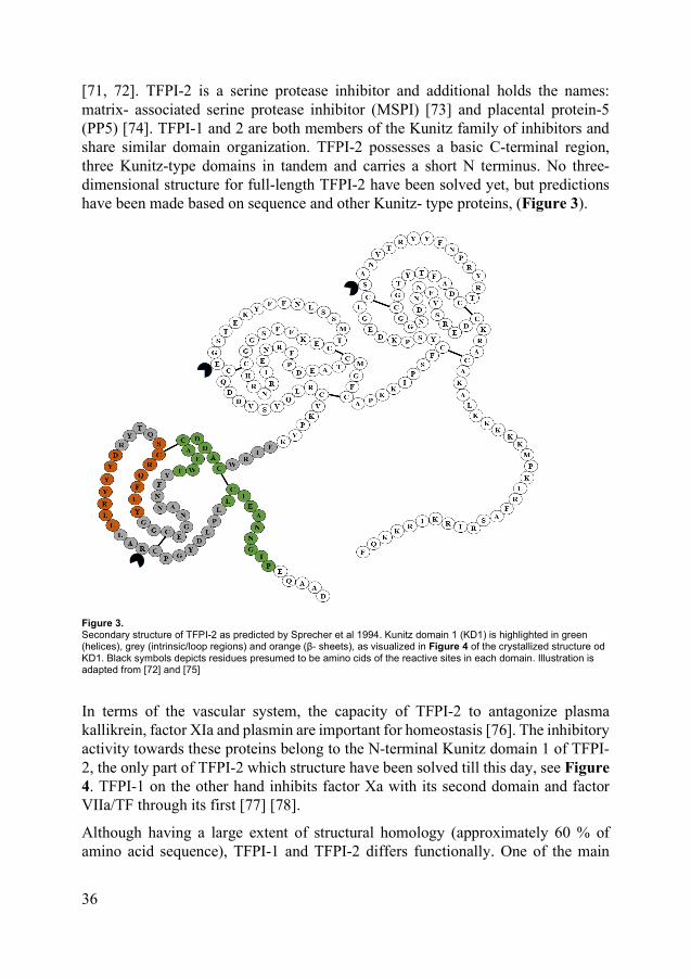
[71, 72]. TFPI-2 is a serine protease inhibitor and additional holds the names: matrix- associated serine protease inhibitor (MSPI) [73] and placental protein-5 (PP5) [74]. TFPI-1 and 2 are both members of the Kunitz family of inhibitors and share similar domain organization. TFPI-2 possesses a basic C-terminal region, three Kunitz-type domains in tandem and carries a short N terminus. No three-dimensional structure for full-length TFPI-2 have been solved yet, but predictions have been made based on sequence and other Kunitz- type proteins, (Figure 3).
Figure 3. Secondary structure of TFPI-2 as predicted by Sprecher et al 1994. Kunitz domain 1 (KD1) is highlighted in green (helices), grey (intrinsic/loop regions) and orange (β- sheets), as visualized in Figure 4 of the crystallized structure od KD1. Black symbols depicts residues presumed to be amino cids of the reactive sites in each domain. Illustration is adapted from [72] and [75]
In terms of the vascular system, the capacity of TFPI-2 to antagonize plasma kallikrein, factor XIa and plasmin are important for homeostasis [76]. The inhibitory activity towards these proteins belong to the N-terminal Kunitz domain 1 of TFPI-2, the only part of TFPI-2 which structure have been solved till this day, see Figure 4. TFPI-1 on the other hand inhibits factor Xa with its second domain and factor VIIa/TF through its first [77] [78].
Although having a large extent of structural homology (approximately 60 % of amino acid sequence), TFPI-1 and TFPI-2 differs functionally. One of the main

37
differences may be their role in coagulation, where TFPI-2 have been suggested to exhibit amidolytic properties on the surface of syncytiotrophoblasts, possibly to improve inhibition of fibrinolysis during pregnancy [79]. Yet this local, tissue bound effect is the only study showing any similarity to TFPI-1 in this respect. The ability to constrain the actions of the certain members of coagulation cascade seems to be local homeostasis rather than as systemic anticoagulant as normal plasma concentrations of TFPI-2 are too low (≤20pM) to inhibit clotting or fibrinolysis. TFPI-2 residing in platelets have been found to regulate coagulation and tPA-induced fibrinolysis and being able to promote clot stabilization at the same time as mitigating intrinsic clotting [76].
Figure 4. Kunitz domain 1 of TFPI-2 illustrated as cartoon, adapted from PDB file: 1ZR0 (Solved ro resolution if 1.8 Å
by co-crystallization with bovine trypsine by Scmidt et al [75])
Furthermore, TFPI-2 expressed from endothelial cells have been shown to regulate thrombosis in arteries but was not found to be necessary for development of these cells or homeostasis [80]. There is no known TFPI-2 mutation in humans causing abolishment of its expression, however certain diseases have been shown to correlate with low levels of TFPI-2. One example of this is bladder cancer, where TFPI-2 levels have been found to inversely correlate with tumor progression suggesting that enhancement of TFPI-2 levels during these circumstances could potentially serve therapeutically and for TFPI-2 to be a biomarker for bladder cancer [81]. Collectively with studies on (inhibition of) cancer progression of nasopharynx, cervix, stromal tumors of gastric origin as well as non-small-cell lung cancer, a role as tumor suppressor has been proposed for TFPI-2 [82] [83] [84] [85].
38
Being discovered for its high expression in placenta, little is known whether TFPI-2 holds additional mechanistic roles other than the suggested function as a local anti-coagulant of syncytiotrophoblasts. A study on pre-eclampsia was published in 2017, where the authors found over-expression and hypomethylation of the promotor expressing placental TFPI-2 to correlate with disease. Due to methylation of promotor, a general feature considered to be a hallmark for epigenetic changes in the genome, pathogenesis is suggested to be under epigenetic regulation involving TFPI-2[86]. Whether for protective or harmful reasons is yet to be discovered.
As a protein highly expressed in vascular cells and found to be involved in matrix remodeling, TFPI-2 have been linked to atherosclerosis [87]. In 2016 Hong et al published their findings from characterizing a conditional, monoallelic knockout of TFPI-2 targeted to vascular endothelial cells and found that decreased levels of TFPI-2 (40,68%) induced atherosclerotic plaques in mice. Reduction of TFPI-2 resulted in increased levels of MMP-2 and MMP-9 along with decreased phosphorylation of PPAR-alpha and PPAR- gamma which is hypothesized to participate in the disease [88].
Furthermore, TFPI2 was recently discovered to be one of the top candidates of the most highly expressed genes in a rare cell niche of skeletal muscle stem cells (satellite cells) from a young individual carrying a homozygous PAX7 null mutation, a gene that have been found to be essential for muscle regeneration in mice. Similarly, this mutation caused severe developmental global muscle hypotrophy along with other severe symptoms affecting breathing among other in this patient. TFPI-2 have not been associated with myogenic progenitor cells in humans previously [89]. In addition, a study in Danio rerio have provided evidence for a prominent role of TFPI-2 in heart development [90]. Collectively, these studies may suggest a role for the TFPI2 gene and /or protein during development. The regenerative potential of inducible pluripotent stem cells has yet not been successful as of transplantable muscle tissue. Whether TFPI2 is a gene directly involved in the transformation of cells to make up muscle tissue remains to be investigated
ii) The complement system
Complement is a group of soluble as well as membrane-associated proteins that are strictly regulated in order to aid defence against pathogens and to resolve inflammation [1] [91] [92]. When activated and depending on by what stimuli, complement cascade(s) results in effective initiation of proteolytic cascades that ultimately leads to opsonization and lysis of the antagonist. This process results in inflammatory responses by expression of proinflammatory molecules [91]. The pathways that coordinate the responses executed by complement are divided in
Ole Erik Soerensen
Please rewrite and make more clear.
39
three main branches called Classical pathway, the Lectin pathway and the Alternative pathway. The Classical pathway is most often triggered by immune complexes (antibody-antigen), apoptotic cells, certain viral envelopes, cell walls of Gram-negative bacteria, C-reactive protein bound to ligand, intermediate filaments of the cytoskeleton and myelin from the central nervous system can activate this pathway too which makes it a part of the adaptive immune system [92] [93] [1]. It is started by complex formation between C1q and the C1 complex (serine proteases C1r and C1s) which binds to the Fc part of IgG1 and IgM. These antibodies stabilize and mediate part of the response. A series of proteolytically generated complexes follow resulting in assembly of components on the surface of the bacteria where C3 is a central complement component connecting the three pathways. The cascade results in formation of a lipophilic membrane attack complex (MAC; C5b-9). By penetrating the bacterial membrane, cytolysis can be achieved by MAC, ultimately inducing inflammation [91-93].
The Alternative pathway can be initiated by lipids, carbohydrates and proteins on microbes but also tumour cells, viral envelopes, plastic surfaces, myelin originating from peripheral nerves and different intracellular organelles [94] [93]. Upon complexing with antigen, serine protease Factor B and Factor D becomes recruited, Proteolytic products of Factor B is generating C3 convertase (also called C3bBb). The stability of C3 convertase is increased thanks to Properdin, a protein released by activated neutrophils (as well as T cells and macrophages), enabling the consecutive proteolysis of C5 and . Activation by any of these ligands, protects C3 convertase from being cleaved by factor H and factor I, mainly inducing opsonization of the antigen as result [94].
The Lectin pathway is activated upon discovery of PAMPs. Either through a carbohydrate-binding plasma protein, mannose- binding lectin (MBL), or Ficolin, terminal mannose residue on glycoproteins of the surface of bacteria, yeast, parasites and viruses can be detected. Ficolin and MBL circulate in seru, comlexed with MBL-associated proteins (MASPs). Four MASPs known to be structurally related are known (MASP, 1,2,3 and 19 which is shorter variant of MASP 2).When MASP 2 binds a pathogen it undergoes conformational changes, resulting in activation of MASP 2 that is able to cleave C4, forming C4a and C4b. The latter binds the surface of the pathogen and recruitment of C2. MASP2 cleaves C2 and C2a and C2b is formed. C4b and C2a co-operatively gains enzymatic activity and this is how the lectin pathway creates its C3 convertase (C4bC2a) [94]. MBL induces proteins of the classical pathway but since it is triggered by a pathogen in the absence of an antibody, it is considered to be a part of the innate immunity. The lectin pathway mainly results in lysis of the pathogen [94].
The complement system has for a long time and still by many categorized to be a well-established and an essential part of the innate immunity against pathogenic
40
organisms. Although, already in the 1970s the ability or necessity of separating the functions of the innate and the adaptive immune systems was started to be discussed. As knowledge has grown, the complement system is one of the central mechanisms adding to the discussion how the communication between these two branches of immunity is connected. The aptitude owned by complement to engage vigorous innate immune reactions in addition to exist in juxtaposition with and ability to affect T- and B- cell biology and other adaptive immune system mechanisms has become ever more appreciated. This may partly be due to the disease mechanisms discovered and thought to be a result of dysregulated complement activation [91, 94]. Systemic lupus erythematosus (SLE) exemplifies results of complement component deficiencies, although this is not always the case, demonstrated by its very complex pathogenesis [95]. This is also the case for (atypical and) hemolytic-uremic syndrome ((a)HUS) in several ways with specific implications during certain E.coli and N.meningitidis infections [96]. On the contrary, an over-active complement cascade or the lack of inhibitors have been found in multiple sclerosis [97], Alzheimer´s disease from several ways- complement may be activated from amyloid plaques binding to C1q, inducing bystander cells which induces nerve cell death and neurodegeneration [98], chronic obstructive pulmonary disease (COPD) [99], allergic asthma [100], in sepsis where C5a and C5aR have been emphasised to direct disease progress in an adverse manner [101] and hyperacute organ rejection [102] as well as in prion disease mechanisms [103].
From an infection point of view, in addition to developing resistance mechanisms, many pathogens have evolved strategies to thwart or disrupt complement for prolonged survival or to increase their virulence. Certain pathogens can facilitate several approaches simultaneously to avoid the potent and instant action of the complement attacks, which is of utmost importance for the bacteria to be able to stick and thrive. In fact, bacteria can hinder complement in almost every step of the cascade, reviewed by Rooijakkers and van Strijp [104]. S.aureus express a membrane protein, Staphylococcal protein A (SpA) that can bind IgG by the Fc region, a function which of the current knowledge, serves the main role for this protein. It does so very efficiently by blocking Fc-receptor-mediated phagocytosis in addition to bind C1q very proficiently[105]. Several other pathogens do also express protein able to bind immunoglobulin, such as S.aureus protein A [106], group A streptococcus and E.coli´s protein G and protein H for instance- findings which revolutionized affinity purification of immunoglobulins, a development that became one of the most frequently used techniques in biochemistry labs for this purpose [107, 108].
Ole Erik Soerensen
Shorten. To many specific detail. This should be a very broad introduction to the complement system mentioning the role in host defense and tissue homeostasis since this is what important in the paper.

41
iii) apolipoprotein E (apoE)
Introducing the characteristics, structure, function, disease mechanisms and possible therapeutic potential of apoE requires an introduction to the family of lipid-protein-complexes lipoproteins that apoE is a part of during many scenarios. Just as the name of this protein reveals, it does not always act on a unaccompanied basis. To what extent apoE is attached to lipoprotein particles differs largely based on the genetic pre-disposition, disease(s), lifestyle and age[109].
Lipoproteins
Lipoproteins are a family of multifaceted particles both structurally and mechanistically. They exist as vesicles with proteins in or on their membrane, serving to transport lipids via their proteins as cholesterol and triglycerides are insoluble in the aqueous environment within and between cells. In short, they exist in seven different sizes with varying function. Chylomicrons are the largest followed by chylomicron remnants, Very Low-Density Lipoprotein (VLDL), intermediate density lipoprotein (IDL), Lipoprotein (a) (Lp(a)) which is a low-density lipoprotein variant, High-Density Lipoprotein (HDL) representing the smallest [110]. They share a common structural arrangement featuring a core of cholesterol esters and triglycerides which is capsulated by phospholipids, free cholesterol and apolipoproteins [110]. Apolipoproteins aid the formation and function of the vesicles by decorating the surface, either as transmembrane proteins or transiently linked, i.e ´exchangeable´. The latter have proven an important role in metabolism of plasma lipoproteins by not only contribute to the stabilization of lipoprotein structure, they can activate enzymes and proteins as well as bind cell surface receptors [111]. ApoE represents the exchangeable pool of this protein family.
apolipoprotein E (apoE)
Human apolipoprotein E (apoE) is a 299 amino acid, amphipathic, glycoprotein. apoE is a high-affinity ligand for the LDL-receptor, LDL receptor-related protein 1 (LRP1), the VLDL receptor and apoE receptor 2 (apoER2) [112-116]. It was initially designated the name “arginine- rich protein” (ARP) due to its high arginine content compared to the previously explored apolipoproteins [117]. Humans express mainly three isoforms of apoE, proteins that have a molecular mass of 34kDa when monomeric [118]. These isoforms are named apoE2, 3 and 4, and differ by one amino acid at position 112 and 158 where apoE3 has an arginine and a cysteine respectively. apoE2 contains cysteines at both sites, whereas apoE4 has arginines instead [119]. Different affinities for the LDL receptor are results of these
Ole Erik Soerensen
This can – in my opinion – be deleted. Just be absolutely sure you can explain the difference between a lipoprotein and apolipoprotein if asked at the dissertation!
Ole Erik Soerensen
Please shorten and focus on the parts relevant for your work!
42
mutations. apoE3 represent the most abundant form in healthy individuals and is a heterodimeric protein consisting of an amine terminus of a 22kDa lipid binding domain and a 10 kDa LDL-receptor binding domain in the carboxyl end, connected by a proteolysis prone hinge region (residues 192-215) [118]. Residues 244-272 of apoE2, 3 and 4 represent the lipid binding moiety [120]. apoE4 is a stablished as risk factor for development of Alzheimer’s disease, poor recovery from stroke and being linked to fast progression of MS [121-126]. Individuals carrying ε2 on two alleles involves an increased risk of developing hyperlipidaemia type III [127].
The full length structure of full-length apoE3 is the only that have been determined till this day out the three isoforms and it was provided by Chen et al after solving it from NMR measurements and was published in 2011 [128]. The authors discovered for the first time a mechanism for how apoE3 adapt an open fold in a two-step process involving a hinge-lock in a lipid- free environment. The N-terminal domain in all three isomers have been determined. Based on this and the work of others, Hatters et al have suggested how this mechanism is affecting the folding of apoE4, where the domains are in proximity, in a closed fold, on the contrary to E3, as illustrated in Figure 5. If true in vivo, this could explain differences in lipid binding preferences, including infectious ligands.

43
Figure 5. Model of apoE3 and apoE4 in lipid-free state as suggested by Hatter et al (2006) [119]. Re-published with permission of Elsevier*.
*Reprinted from Apolipoprotein E structure: insights into function, Vol 31, Issue 8 (2006) p.445-454, D.M Hatters, C.A.Peters-Libeu, K.H.Weisgraber, Copyright (2020) with permission from Elsevier.

44
In the peripheral tissues, the cells expressing most apoE are hepatocytes, macrophages and adipocytes. In the vascular regions of the CNS, astrocytes, microglia and mural cells produces the most of apoE. It also regulates macrophages by promoting production of nitric oxide, reducing T-cell proliferation, decrease growth of smooth muscle cells and induce nitric oxide production in platelets. In adipocytes, apoE aids in maintenance of triglyceride balance and is part of securing integrity of blood-brain-barrier as well as blood-nerve-barrier. apoE has also been discovered to enable lipid antigen presentation by CD1 proteins to natural killer cells [129].
apoE is a lipid transport protein, carrying lipid between tissues. It circulates in plasma to clear the blood from cholesterol, preferentially accompanying VLDL particles and subsequent binding to cell surface receptors. A study of apoE(-/-) mice were studied in addition to mice transgenic expressing the human variant of apoE3 and apoE4 in order to investigate whether apoE impact lipid content in the brain revealed that neither of these mice strains had differences in cholesterol, sphingolipids or multiple phospholipids. The authors did find a large difference in sulfatide content however, both in cerebrospinal fluid and in brain tissue [130]. Sulfatide is a molecule with several functions in the nervous system, regulation of haemostasis/thrombosis, immune system, bacterial and viral infections as well as insulin secretion [131]. Knock-out mice exhibited an increase and brain tissue from (h)apoE4 transgenic mice had much less compared to wild type mice. When compared to human CSF, sulfatide rich complexes were discovered to be specifically associated with HDL particles holding apoE, findings that suggest sulfatide in the CNS and apoE levels may be produced or modified through same pathways. This stipulates another reason for apoE to be of importance in brain injury recovery and Alzheimer’s disease [130].
Several different roles for apoE during infections have been proposed where the protein initially was discovered to convey LPS from Kupffer cells to parenchymal cells in the liver[132]. It has also been reported to be upregulated under control of LDL receptors in the liver, proposing a negative feedback mechanisms [133]. Studies in mice in 2001 by van Oosten et al progressed the understanding of apoE during LPS induced endotoxemia and described a mechanism for apoE including binding and re-directing LPS molecules from macrophages to hepatocytes in vivo[134]. This was not only repeated in mice but also translated in 2014 when Fu et al published a cohort study of paediatric patients, reporting that apoE is elevated in serum of individuals suffering from sepsis, bacterial meningitis and bacterial pneumonia. In contrast, aseptic meningitis and pneumonia caused by mycoplasma did not correlate with serum apoE levels [135].
The last decade several studies have presented evidence that apoE holds regions with both antibacterial as well as antiviral activities. The antibacterial studies, most
45
efforts, from in silico to in vitro, have emphasized not only but especially the receptor binding region of apoE [136-140]
The sequence representing residues 130-162 was discovered through studies on a peptide named COG-133 [141, 142]. Interestingly, this peptide has proven to sustain the biological activities of the full-length protein in terms of its anti-inflammatory and neuroprotective accomplishments under test in an Alzheimer´s disease model in Drosophila melanogaster [143].
On another mechanistic note, recombinantly expressed apoE (from RAW264 cells) has been found to encompass antioxidant activity in an isomer-dependent fashion and was reported to protect against oxidative stress from β- amyloid peptides in the order of E2>E3>E4. Furthermore, the study report cytotoxic to neurons in vitro at 62nM reported by Miyata and Smith [144] which may reflect the difference in sensitivity of cells during cell culture conditions compared to in vivo, and/or the vast contrast between cell types of cells partaking in the blood stream and neurons in addition to the way this protein was prepared as plasma concentrations are normally close to 1µM in healthy individuals[109].
Being aware of the vastly differential concentrations between organs and cell types is likely of great importance for future studies, especially with advancements of more complex in vitro studies such as co-cultures and 3D cultures for increased translational efforts.
In summary, therapeutic efforts facilitating apoE have till this day presented evidence as a potential biomarker during systemic bacterial infections in addition to its relevance for disease mechanisms involving oxidative stress and lipids in addition to the ones discovered so far. Whether polymorphism in the APOE gene affects or have a role during infection and what the exact role(s) of the apoE proteins are during infections remains to be elucidated.
Ole Erik Soerensen
This is the interesting part!

46

47
5.) Present investigation
i) Paper I
Aim:
Characterization of the role of TFPI-2 during endotoxin induced systemic inflammation and bacterial sepsis. Since a separate project on the C-terminal TFPI-2 peptide EDC34 revealed binding to immunoglobulin, the aim was expanded to examine the role of TFPI-2 binding to immunoglobulin.
Methods:
In vivo studies TFPI-2(-/-) mice. qPCR, Western blot, coagulation assays, Radial Diffusion Assay (RDA), Viable Count Assay (VCA), Immunoglobulin depletion, Surface Plasmon Resonance (SPR), cytokine assays (by Fluorescent-Activated Cell Sorting, FACS), Reactive Oxygen Species (ROS) production assay
Results:
TFPI-2 was highly expressed in especially spleen, brain and increased expression was found in the lungs upon in a systemic inflammation model with i.p injection of LPS from E.coli. A similar expression pattern was also observed in a sepsis model (live E.coli, i.p). The surprising finding of increased brain and lung expression of TFPI-2 from initial experiments on wt mice led to a characterization of TFPI-2 focused on pulmonary infections. TFPI-2(-/-) showed higher sensitivity to both LPS and E.coli administered i.p as well as i.n administrated P.aeruginosa compared to wt mice. Bands for hTFPI-2 was also discovered in sputum from four patient samples suffering from COPD. In addition to IgG, TFPI-2 was found to be able to bind IgE, IgM and IgA.
Ole Erik Soerensen
Your result should always be described in past tense!

48
ii) Paper II
Aim:
Investigation of whether complement is activated at the keratinocyte surface upon intracellular infection with S.aureus to.
Methods:
Normal human epidermal keratinocytes (NHEK) cultures. Intracellular infection models in NHEK. Complement activation assays using detecting complement deposition by immunofluorescence (IF) microscopy. Complement activation assays with depleted and reconstituted sera (C1q, MBL, Factor B, C3, and C6) IF-based assays for apoptosis and necrosis of NHEK. Western blot was performed on cell media against C3d and C4c. Viable count of bacteria were performed after each stimulation. Skin biopsies from atopic dermatitis patients were evaluated for bacterial load and TCC deposition by immunofluorescence.
Results:
Persisting intracellular S.aureus induced deposition of complement fragments C3, C4 and TCC on NHEKs. Both the classical and lectin pathway complement pathway was activated upon persistent intracellular infection of primary keratinocytes- since both diminished complement deposition was found with both C1q and factor B deficient sera. Reconstitution of C3, C1q and factor B depleted led to reduction in number intracellular viable bacteria upon complement activation while this was not observed with MBL deficient serum. The complement mediated effect on bacterial killing was furthermore found to be regulated by Mitogen-Activated Protein Kinase (MAPK) pathway. Lastly, examination of skin biopsies from patient samples revealed that complement activation is activated in skin from atopic dermatitis patients colonized with S.aureus.
49
iii) Paper III
Aim:
This study was designed to investigate whether apoE from plasma has anti-bacterial effect against Gram-negative bacteria as previously shown by capacity of several apoE-derived antimicrobial peptides and to investigate whether the structure of apoE is affected by interaction with LPS and Lipid A.
Methods:
Viable count assay (VCA) with Escheria coli (ATCC 25922) and Pseudominas aeruginosa (PA01) with plasma apoE. Blue Native-PAGE and Western Blot for evaluation of complex formation with LPS and Lipid A. Circular dichroism for analysis of secondary structure +/- LPS, TEM was used to visualize bacteria after apoE stimulation. Propidium iodine / SYTO®9 stain for viable and dead bacteria after apoE treatment.
Results:
Plasma-derived apoE derived reduced bacterial growth of P.aeruginosa and E.coli. apoE was shown to decrease bacterial counts of P.aeruginosa at physiological plasma concentrations to similar degree as the previously characterized AMP derived from thrombin (GKY25). The results from viable count assays were confirmed by TEM and fluorescence microscopy. Furthermore, apoE killed bacteria resulting in aggregation of bacteria. Binding of LPS, but also LipidA from E.coli increased α-helical content of apoE.
Ole Erik Soerensen
I would prefer the term LPS rather then endotoxin since it is more molecular specific and refers to the actual experiments you have done!

50
51
Discussion and future directions
Our findings have for the first time showed that TFPI-2 is protective against Gram- negative bacteria, exemplified by systemic E.coli infection and P.aeruginosa infection of the lung. Apparently TFPI-2 limit bacterial growth by binding IgG and subsequently activates complement. Additionally, new knowledge about the complement and its function during persistent, intracellular infection were discovered where both the alternative and classical pathway were found to be involved. In the last study, we found that plasma-derived apoE derived from plasma exhibits high anti-bacterial activity against P.aeruginosa and E.coli in vitro. It binds to both full-length LPS and LipidA.
The methodology of these studies could be further developed. Perhaps most importantly, the results of Paper I are mainly studies performed in mice in vivo, and studies on binding of the TFPI-2 derived antimicrobial peptide EDC34 were done on human material in vitro. There is a great difference in tolerance to endotoxins between mice and humans where mice may tolerate up to 10 000 times more LPS than humans (relative to body weight) a fact to keep in mind in when interpreting the translational relevance of these findings. Although the differences between mouse and human are markedly different in many aspects, the immune system seen in rodents seem largely to resemble the one in humans during infection.
Our study on primary keratinocytes and the involvement of complement during intracellular, long-lasting infection by S.aureus was mainly involving cell culture experiments. As additional skin from two patients and compared with healthy volunteers was investigated, this study would benefit from being compared with a larger group of samples as well as compared with skin from individuals suffering from other diseases too.
Lastly, the investigation of plasma apoE and its potential role during infection of the bacteria mentioned was enabled by using purchased protein. Lacking information of exact molecular weight first and foremost, in addition to posttranslational modification status leaves many questions unanswered. Attempts to express, purify and measure recombinant apoE2, 3 and 4 similarly as presented in paper III was the start of the work were unfortunately hindered by low yields and with LPS contamination as a major problem.

52
The choice of bacterial strains used in the present work deserves a discussion too. The P.aeruginosa strain used was strain PA01, commonly reported as a wild type strain (i.e not genetically modified for laboratory purposes). S.aureus tested in Paper II is from a clinical isolate, E.coli (Escherichia coli (ATCC® 25922™) used in Paper I and III is [40] was too originally isolated from a clinical sample (in 1946) and is commonly used in quality control testing, and the genome of ATCC 25922 has been sequenced [145]. While being a common cause of urinary tract infections, it can also be nonpathogenic (Blum et al, 1995), indeed a pathobiont.
Taking the transient behavior of E.coli, P.aeruginosa and S.aureus discussed above into account, existing both as commensal and pathogen, the results presented in paper I and III might benefit from comparisons with a panel of varying serotypes for deeper understanding of if TFPI-2 can protect against more than the two bacteria species and serotypes under test. Such studies could also bring new insights of the way apoE binds and kills Gram-negative bacteria. Initial tests of the bactericidal effect of apoE also included S.aureus (ATCC 29213). Unfortunately, no clear conclusions were possible to be drawn and more experiments is needed in order to understand if apoE is able to kill Gram- positive bacteria too.
Looking at the microbiome as an organ, especially in light of the transplantations as treatment against infections and inflammation of the gut, it has become evident that bacteria can form colonies that seem to serve purposes in many ways similar to the eukaryotic cells they live in symbiosis with. This might provide clues to new treatments for additional diseases.
If a more long-lasting treatment scheme with healthy, donor-acceptor compatible microbiota would be possible. Together with a deeper knowledge of how pathogens differ from commensal bacteria, maybe genetically modified versions of both together with advanced cell therapies targeting specific pathogens is a therapeutic of the future against infections. Whether this would aid in other maladies as well remains to be investigated. A suggested approach to combat pathogens based on findings presented in this thesis include genetically engineered pathogens relevant for the specific infection or commensals, where cell wall structures are intact but with improved toxic secretory systems including expression of for instance AMPs or antibacterial proteins. Ability to create such clones without harming the “spy” itself would likely be a great challenge. This is not very different from the approach that some pathogenic bacteria already use to affect the microbiota as well as the biology of bacteriophages. The latter may be more efficient as a gene tailoring tool of pathogens for novel antibacterial means looking to what evolution has already figured out for us. This is likely not the first time this approach is suggested on treating infections and it is certainly not the first to suggest facility of bacteria for delivery of therapeutic genes. Both wild type bacteria such as Clostridium and recombinant bacteria are currently under test for targeted cancer therapy.
Ole Erik Soerensen
Was it meant by this – is it a ATCC strain. The term wild type is mostly used in relation to mutant strains!
Ole Erik Soerensen
Please rephrase as I have some difficulties to follow.
53
Such engineered bacteria would possibly answer the question of whether a apoE secreting, commensal P.aeruginosa for instance be able to outsmart and kill its seemingly fellow, pathogenic P.aeruginosa, if such bacteria would survive at all and without harming the host. If so, the test of co-existence of other genetically modified species such as yeast perhaps would benefit treatment strategies or be a more suitable delivery host than bacteria of post translationally modified host defence molecules. These are questions therapeutic approaches of host defence molecules possibly would benefit from testing. Yet, the problem with intracellular, persistent bacteria would likely need a different approach.
In fact, these masquerading manners are a sophisticated strategy quite a few bacteria have developed already, somewhat as discussed briefly in Paper II, although from a host perspective rather than bacterial structures and remodeling abilities. The extensive article covering how especially capsular bacteria manage to trick host barriers in order to be able to reside in tissue and cavities by Cress and co-workers [146] shed light on the risks with strictly decoupling CPSs due to the synergistic and multifunctional properties of these bacteria. Yet, targeting capsules by manipulation and to interfere with the synthesis of capsular envelope may be the most promising approach in addition to phage therapy. Considering the rapidly developing techniques such as -omics, as well as nano science to name a few, perhaps nanobots may serve a purpose for data collection in vivo in order to improve our battle against pathogens and disease in the future.
54
Concluding remarks
Our defence mechanisms against homeostatic imbalances, including eradication of invading pathogens are made up of uncountable favours of the molecules expressed and by numerous cell types collectively. Some of the proteins involved in these processes have been found to take part not only in what historically have been considered immunity but also in other disease/health retaining processes. Our findings shed new light on two proteins and a pathway group of proteins; TFPI-2, apoE and the complement cascades as their previously described roles as tumour suppressor, lipid binding protein and host defence molecules that previously mainly have been considered for their involvement at the very initiation of infection respectively in infection mechanisms, collectively covers a broad spectrum of diseases.
Given the many connections between other disease mechanisms than infections and the involvement of the ones characterized belonging to immune reactions, the question of if there is such thing as The immune system or if all connective pathways making our bodies function rather as One entire system of immune functions came to mind writing this thesis. Sincerely, our traditionally named Immune system is well known to always be engaged but seldom of notice on a level we feel while asymptomatic or healthy. In other words, the question is if there is any part of the body that would not be capable of involvement in immune regulation if challenged. Resolving infections seems to involve many proteins that previously have not been assigned an ́ immune- function´. In the light of our current findings along with many made by others and development of high-throughput and specific techniques such as single-cell genomics and novel variants of mass spectrometry methods providing incredible amounts of information will likely help to define not only new targets in disease but also their connective patterns. Advancements in the field of structural biology, both for large molecules and complexes, such as cryoEM, together with better and faster algorithms to aid data handling, prediction and interpretations provide promising tools for new explorations and assignment of new host defence molecules. Cooperatively, they provide promising means for further advancements in targeting infectious diseases.
55
Populärvetenskaplig sammanfattning
Immunförsvaret håller oss friska största delen av tiden tack vare alla molekyler i och på våra kroppar som ingår i det. Immunförsvaret indelas ofta i två större grenar- det ´medfödda´ som skyddar oss mot en mängd olika infektioner och främmande partiklar med en gång då de möts. Den andra viktiga delen av det här artilleriet är det ´adaptiva´ immunförsvaret som lär sig och tränas efter varje typ av bakterie som kommer i vår väg för att bli mer motståndskraftig till en eventuell nästa gång av. Kroppen är uppbyggt av en rad olika typer av celler som är specialiserade på olika saker och det medfödda och det adaptiva immunförsvaret har till stor del olika celltyper som gör att de kan skydda och läka kroppen om vi får infektioner och inflammationer. Den här indelningen är inte alltid helt strikt, och vissa celltyper tjänar både det medfödda och det adaptiva försvaret. Det som gör att kroppen ”minns” när den haft en speciell infektion är för att antikroppar, s.k. immunglobuliner, bildas när kroppen möter en ny typ av infektion. De produceras för att kunna binda specifikt till en unik del av det som kan vara en fara för kroppen. Tack vare vaccin kan kroppen lära sig vad som är farligt innan den blir sjuk och på så sätt undkomma en allvarlig infektion. Det finns fyra olika sorters grupper av immunoglobuliner och i den här avhandlingen har främst vi studerat den vanligaste: IgG. I studie I (Paper I) testade vi hypotesen att en specifik peptid, EDC34, kunde binda till IgG. Efter att funnit att den gjorde det undersökte vi vilken del av IgG den binder till. Intressant nog var det samma ställe som ett annat väldigt viktigt protein binder till just IgG- det proteinet heter C1q. Detta kan vara av vikt för att snabba på immunförsvaret, m.h.a en process som involverar en domino-lik kaskad av förlopp: Komplementsystemet, men det återstår fler försök för att utröna vad detta har för exakt effekt. EDC34 har tidigare visat sig ha stor antibakteriell effekt på Gram- negativa bakterier. Av den anledningen undersökte vi vad som händer i möss som saknar genen som uttrycker det protein som EDC kommer ifrån. Resultatet var att mössen utan genen (TFPI2) var mycket känsligare mot E.coli och P.aeruginosa infektioner samt uppvisade en mängd molekylära tecken på ökad inflammation. När möss som saknade TFPI-2 först blev infekterade och sedan behandlade med en peptidvariant av TFPI-2 som motsvarar EDC34 i möss uppvisade de högre grad av resistens mot infektion. Dessa resultat föreslår dels en viktig roll för TFPI-2 i immunförsvaret samt öppnar nya potentiella terapier mot dessa typer av infektioner. I studie II (Paper II) undersökte vi vilken roll komplementsystemet har under långvarig infektion i huden av S.aureus, närmare bestämt om komplementproteiner
56
är aktiva då bakterierna dödas inuti cellerna i huden. Dessa bakterier har nämligen listat ut strategier för att inte bara ta sig in i celler, främst i hudens översta skikt, i som heter keratinocyter. De har utvecklat en speciell överlevnadsstrategi- genom att kolonialisera hud hos patienter med atopisk dermatit (a.d) är immunförsvaret hos dessa patienter inte förmöget att göra sig av med S.aureus. Vi fann, både i cellkulturer av keratinocyter, samt i hudbiopsier från patienter med a.d att komplementsystemet var upp-reglerat på keratinocyter i samband med denna typ av ihärdiga infektioner. I cellkulturer såg vi aktiverade proteiner som troligen involverar inte bara en utan två av de tre olika förgreningar i denna kaskad som verkar för att eliminera infekterande bakterier. Detta är viktig kunskap för att få ökad förståelse för vilka delar av immunförsvaret som eventuellt behöver förstärkas, eller bromsas, när infektioner kvarvarar och därmed blir mycket svåra att behandla. Studie III (Paper III) behandlar studier av ett protein tidigare främst känt för sin fett- (lipid)bindande och -transporterande förmåga, apolipoprotein E (apoE). apoE studerades med avseende på bakteriedödande förmåga av de två Gram-negativa bakterierna E.coli och P.aeruginosa samt om apoE kunde binda till olika fettmolekyler (LPS och LipidA) som härstammar ifrån dessa bakteriers membran. LPS och LipidA är båda mycket toxiska om de släpps fria ifrån bakterieväggen och kan orsakar s.k. endotoxemi. Vi fann att apoE från blodplasma mycket effektivt dödar P.aeruginosa i fysiologisk koncentration och även är antibakteriellt emot E.coli. apoE kunde även binda LPS från båda bakterietyperna och LipidA från E.coli. Ytterligare försök behövs för att pröva hur detta förhållande ser ut i samspel med celler från mänsklig vävnad och inte minst in vivo för att utröna hur väl dessa resultat överensstämmer med våra fynd.

57
58
Acknlowledgements
Artur, som huvudhandledare för största delen av mina forskarstudier vill jag börja med att tacka dig för att du anställde mig som doktorand med stort förtroende för mina ambitioner, för att du fick mig att känna mig varmt välkommen i gruppen och på B/C14 med tålamod för att både infektionsmedicin och dermatologi var nya områden för mig. Tack också för tips om löprundor i Lund, för stödet jag fått och chansen att utvecklas som aspirerande forskare. Vi har inte alltid tyckt likadant och jag är inte mindre tacksam för att det har gett mig nyttiga lärdomar. Sent kommer jag glömma hur du med humor lyckades avväpna min lätta och obefogade oro som ny doktorand, när jag vid ett tillfälle inventerade antisera, med att säga ”Kaninen är död” med en timing som bara kan göra sig bra på skånska. Många tack.
Ole, tack för att du med klarsynthet, ett aldrig sinande intresse och tydlighet bistått mina forskarstudier med lärorika diskussioner och idéer som bihandledare. Ditt stöd har varit ovärderligt. Din kompetens och hjälp att göra det bästa av vad jag lyckats åstadkomma är jag lyckligt lottad för att du bistått med. Jag är mycket tacksam för vad det lärt mig. Tack!
Jitka, I have been very lucky to have you as a co-supervisor and supervisor the second half of my studies, in addition to a great colleague before that. Your interest and eagerness to aid me in my scientific development and the progress of the projects means more than words can say. I am very grateful for have had you by my side through this time.
Ann-Charlotte- Din outsinliga energi och källa av koll på allt som håller labbet flytande- vad ASN lab hade varit utan dig vill jag inte veta. Så glad jag är för att haft turen att jobba i samma grupp som dig och dela kontor en stor del av min tid i gruppen dig. Tack för all ovärderlig hjälp, alla skratt och gott snack!
Malgorzata- tack för allt du hjälpt till med sedan starten jag början, särskilt i starten med odling av keratinocyter men också från protokoll till allt möjligt. Kommer också sakna dina goda kakor! Tack.
Thank you all previous group members: Finja- for being a great friend and colleague, thank you for all laughter, good chats and your wise input. Alexandra- så glad att jag haft turen att lära känna dig. Ser framemot många fler år av vänskap. Mina- Tack för att du är du, så omtänksam och rak. Thank you Marta, Julia, Nataliya, Lech, Karim, Praveen, Gopi and Ravi.
59
Thank you all present fellow ASN lab members: Andreas, Sigrid, Jeremy, Madelene, Ganna, Manoj, Sven and Ana. It´s been a pleasure working with you!
Thank you Asia, Elias and Royce for your curiosity and your hard work during your Master´s projects on recombinant apoE.
Thank you to everyone at B14 and C14 that together make it such a nice and fun workplace in a collaborative atmosphere. I will forever be grateful for have met all of you.
Special thanks to Laura (Thanks for being in my life. You mean the world to me), Suvi (lets hit the sauna soon), Tirthankar (finer than the hardest metal, lets catch up soon), Clement (miss you loads, come back from Norway please), Anele (thanks for all the good times, no one owns a Saturday ritual like you), Anas (thanks for the fun science chats, the house experience and arranging the Jordan trip, it was lit), Kristoffer W, Eleni, Jana, Jesper, Amanda, Sandra P, Hilger, Anupam, Lloyd, Andrew, Sue, Ariane, Frida, Mohammad and everyone I have forgotten in this moment
for all your support, discussions, laughs, for being great company around the fika/lunch table/on a hike/ a dancefloor and elsewhere. Thank you for so many nice memories!
Tack Lars för den välkomnande och lagom prestigelösa atmosfär du varit stor del av att grunda för det som är B14. Tack för samtal och feedback.
Tack Inga-Maria, för att du alltid är så hjälpsam- vare sig det gäller att få låna en kolonn eller en pratstund. Det har betytt och hjälpt mig mycket.
Maria S, om du bara visste hur tacksam jag är att jag hamnade i Ditt labb och den träning jag fick under tiden hos dig på IFM under exjobbstiden, men inte minst för det stöd jag fått efter utbildningen. Tack.
Sara H, för handledningen som exjobbare hos Maria. Tack för att du gav mig en så lärorik och fin start in på den akademiska banan.
Madhan- thanks for all the laughter and good times! Hope we meet soon.
Amelie- som jag kan sakna vår tid i Sunnerhagen labb. Hoppas vi ses snart snäckan!
To the Boström/Andersson lab at CMB- Xioachuan, Mathilda, Olga, Madeleen, Anastasia, Tilo, Evan- thank you for a short but very fun and fruitful time.
Kristina, Liza, Mikaela, Petra, Tove och resten på Klostergatan- Linköping var fint, med er blev det jättefint. Tack för att ni finns.
Ida, Anna, Lise-Lotte, Sara, Emma och Annie- om vi inte får till en uppföljare på Falks Grav så hoppas jag innerligt vi får till den där re-unionen igen snart. Tack för den bästa tiden <18år.

60
Paula- Många äventyr har det blivit och hoppas på många fler! Tack för att du är Du.
Raad- Tack för att du finns
Barrikaden AIF Dam- Bästa laget. Så mycket kärlek och sköjj. Tack för att ni finns!
Henke- bättre brorsa kan ingen få. Tack för att du är du och för att du alltid finns där. Gamla nyheter men jag är inte bara den stoltaste systern men också den stoltaste fastern i hela världen. Tack för det också.
Mamma- tack för din ovillkorliga kärlek, för ditt ändlösa tålamod och din humor som får mig att skratta som ingen annan. Min tacksamhet för att du är Du och finns i mitt liv förtjänar en egen avhandling.
Pappa- tack för ditt stöd, för att jag fått ärva en dos av din envisa och nyfikenhet, och för att du lärt mig lite bonnförnuft
Farmor- tack för all kärlek och kloka ord
Carina- så tacksam att du finns i mitt liv
61
Bibliography
1. Abul K. Abbas, A.H.L.a.S.P., Basic Immunology: Functions and Disorders of the Immune system. Vol. 4th Ed. 2014: Saunders, Elsevier Inc. 320.
2. Turvey, S.E. and D.H. Broide, Innate immunity. Journal of Allergy and Clinical Immunology, 2010. 125(2): p. S24-S32.
3. Maglione, P.J., N. Simchoni, and C. Cunningham-Rundles, Toll-like receptor signaling in primary immune deficiencies. Year in Immunology, 2015. 1356: p. 1-21.
4. Jin, M.S. and J.O. Lee, Structures of the toll-like receptor family and its ligand complexes. Immunity, 2008. 29(2): p. 182-191.
5. Mogensen, T.H., Pathogen Recognition and Inflammatory Signaling in Innate Immune Defenses. Clinical Microbiology Reviews, 2009. 22(2): p. 240-+.
6. Medzhitov, R., Toll-like receptors and innate immunity. Nature Reviews Immunology, 2001. 1(2): p. 135-145.
7. Matsushima, N., et al., Comparative sequence analysis of leucine-rich repeats (LRRs) within vertebrate toll-like receptors. Bmc Genomics, 2007. 8.
8. Tooze, B., Introduction to Protein Structure, 2nd edn. (1999), New York: Garland Publishing. 409.
9. Akira, S., S. Uematsu, and O. Takeuchi, Pathogen recognition and innate immunity. Cell, 2006. 124(4): p. 783-801.
10. Bell, J.K., et al., Leucine-rich repeats and pathogen recognition in Toll-like receptors. Trends in Immunology, 2003. 24(10): p. 528-533.
11. Von Bernuth, H., et al., Pyogenic bacterial infections in humans with MyD88 deficiency. Clinical and Experimental Immunology, 2008. 154: p. 213-213.
12. Picard, C., et al., Pyogenic bacterial infections in humans with IRAK-4 deficiency. Science, 2003. 299(5615): p. 2076-2079.
13. Martinon, F., A. Mayor, and J. Tschopp, The Inflammasomes: Guardians of the Body. Annual Review of Immunology, 2009. 27: p. 229-265.
14. Brubaker, S.W., et al., Innate Immune Pattern Recognition: A Cell Biological Perspective. Annual Review of Immunology Vol 33, 2015. 33: p. 257-290.
15. Zanetti, M., et al., Bactenecins, Defense Polypeptides of Bovine Neutrophils, Are Generated from Precursor Molecules Stored in the Large Granules. Journal of Cell Biology, 1990. 111(4): p. 1363-1371.
16. Hansen, F.C., et al., The Thrombin-Derived Host Defense Peptide GKY25 Inhibits Endotoxin-Induced Responses through Interactions with Lipopolysaccharide and Macrophages/Monocytes. J Immunol, 2015. 194(11): p. 5397-406.
62
17. Boman, H.G., Peptide Antibiotics and Their Role in Innate Immunity. Annual Review of Immunology, 1995. 13: p. 61-92.
18. Pasupuleti, M., A. Schmidtchen, and M. Malmsten, Antimicrobial peptides: key components of the innate immune system. Critical Reviews in Biotechnology, 2012. 32(2): p. 143-171.
19. Hancock, R.E.W. and R. Lehrer, Cationic peptides: a new source of antibiotics. Trends in Biotechnology, 1998. 16(2): p. 82-88.
20. Hancock, R.E.W. and H.G. Sahl, Antimicrobial and host-defense peptides as new anti-infective therapeutic strategies. Nature Biotechnology, 2006. 24(12): p. 1551-1557.
21. Alves, D. and M.O. Pereira, Mini-review: Antimicrobial peptides and enzymes as promising candidates to functionalize biomaterial surfaces. Biofouling, 2014. 30(4): p. 483-499.
22. Joo, H.S., C.I. Fu, and M. Otto, Bacterial strategies of resistance to antimicrobial peptides. Philosophical Transactions of the Royal Society B-Biological Sciences, 2016. 371(1695).
23. van der Plas, M.J., et al., Pseudomonas aeruginosa elastase cleaves a C-terminal peptide from human thrombin that inhibits host inflammatory responses. Nat Commun, 2016. 7: p. 11567.
24. van der Waaij D Fau - Berghuis-de Vries, J.M., J.M.F.A.U.L.L.-v. Berghuis-de Vries, and L.-v. Lekkerkerk, Colonization resistance of the digestive tract in conventional and antibiotic-treated mice. J Hyg (Lond). 1971. Sep; 69(3):405-11.(0022-1724 (Print)).
25. Chu, H.T. and S.K. Mazmanian, Innate immune recognition of the microbiota promotes host-microbial symbiosis. Nature Immunology, 2013. 14(7): p. 668-675.
26. Kamada, N., et al., Control of pathogens and pathobionts by the gut microbiota. Nature Immunology, 2013. 14(7): p. 685-690.
27. Belkaid, Y. and T.W. Hand, Role of the Microbiota in Immunity and Inflammation. Cell, 2014. 157(1): p. 121-141.
28. Rafii, F., C.E. Sutherland Jb Fau - Cerniglia, and C.E. Cerniglia, Effects of treatment with antimicrobial agents on the human colonic microflora. (1176-6336 (Print)).
29. Di Giacinto, C., et al., Probiotics ameliorate recurrent Th1-mediated murine colitis by inducing IL-10 and IL-10-dependent TGF-beta-bearing regulatory cells. Journal of Immunology, 2005. 174(6): p. 3237-3246.
30. Feleszko, W., et al., Probiotic-induced suppression of allergic sensitization and airway inflammation is associated with an increase of T regulatory-dependent mechanisms in a murine model of asthma. Clinical and Experimental Allergy, 2007. 37(4): p. 498-505.
31. Karimi, K., et al., Lactobacillus reuteri-induced Regulatory T cells Protect against an Allergic Airway Response in Mice. American Journal of Respiratory and Critical Care Medicine, 2009. 179(3): p. 186-193.
32. Zournpopoulou, G., et al., Lactobacillus fermentum ACA-DC 179 displays probiotic potential in vitro and protects against trinitrobenzene sulfonic acid (TNBS)-induced
63
colitis and Salmonella infection in murine models. International Journal of Food Microbiology, 2008. 121(1): p. 18-26.
33. Giel, J.L., et al., Metabolism of Bile Salts in Mice Influences Spore Germination in Clostridium difficile. Plos One, 2010. 5(1).
34. Ribet, D. and P. Cossart, How bacterial pathogens colonize their hosts and invade deeper tissues. Microbes and Infection, 2015. 17(3): p. 173-183.
35. Mierke, C.T., The fundamental role of mechanical properties in the progression of cancer disease and inflammation. Reports on Progress in Physics, 2014. 77(7).
36. Winer, S. and D.A. Winer, The adaptive immune system as a fundamental regulator of adipose tissue inflammation and insulin resistance. Immunology and Cell Biology, 2012. 90(8): p. 755-762.
37. Charles N. Serhan, P.A.W., and Derek W. Gilroy, Fundamentals of Inflammation. 2010, Cambridge and New York: Cambridge University Press. xiv + 473 p.
38. Kaper, J.B., J.P. Nataro, and H.L.T. Mobley, Pathogenic Escherichia coli. Nature Reviews Microbiology, 2004. 2(2): p. 123-140.
39. Weiner-Lastinger, L.M., et al., Antimicrobial-resistant pathogens associated with adult healthcare-associated infections: Summary of data reported to the National Healthcare Safety Network, 2015-2017. Infection Control and Hospital Epidemiology, 2020. 41(1): p. 1-18.
40. ATCC. Escherichia coli (Migula) Castellani and Chalmers (ATCC® 25922™) 2016; Strain information (Product sheet)]. Available from: https://www.lgcstandards-atcc.org/en/Global/Products/F/F/7/8/25922.aspx.
41. Blum, G., R. Marre, and J. Hacker, Properties of Escherichia-Coli Strains of Serotype O6. Infection, 1995. 23(4): p. 234-236.
42. Kerr, K.G. and A.M. Snelling, Pseudomonas aeruginosa: a formidable and ever-present adversary. Journal of Hospital Infection, 2009. 73(4): p. 338-344.
43. Poole, K., Pseudomonas aeruginosa: resistance to the max. Frontiers in Microbiology, 2011. 2.
44. Schuster, M. and E.P. Greenberg, A network of networks: quorum-sensing gene regulation in Pseudomonas aeruginosa. Int J Med Microbiol, 2006. 296(2-3): p. 73-81.
45. Rumbaugh, K.P., J.A. Griswold, and A.N. Hamood, The role of quorum sensing in the in vivo virulence of Pseudomonas aeruginosa. Microbes and Infection, 2000. 2(14): p. 1721-1731.
46. Singh, P.K., et al., Quorum-sensing signals indicate that cystic fibrosis lungs are infected with bacterial biofilms. Nature, 2000. 407(6805): p. 762-764.
47. Kohler, T., et al., Quorum sensing-dependent virulence during Pseudomonas aeruginosa colonisation and pneumonia in mechanically ventilated patients. Thorax, 2010. 65(8): p. 703-710.
48. Zulianello, L., et al., Rhamnolipids are virulence factors that promote early infiltration of primary human airway epithelia by Pseudomonas aeruginosa. Infection and Immunity, 2006. 74(6): p. 3134-3147.
64
49. Wagner, V.E. and B.H. Iglewski, P. aeruginosa Biofilms in CF Infection. Clinical Reviews in Allergy & Immunology, 2008. 35(3): p. 124-134.
50. Winstanley, C. and J.L. Fothergill, The role of quorum sensing in chronic cystic fibrosis Pseudomonas aeruginosa infections. Fems Microbiology Letters, 2009. 290(1): p. 1-9.
51. Wertheim, H.F.L., et al., The role of nasal carriage in Staphylococcus aureus infections. Lancet Infectious Diseases, 2005. 5(12): p. 751-762.
52. Stryjewski, M.E. and G.R. Corey, Methicillin-Resistant Staphylococcus aureus: An Evolving Pathogen. Clinical Infectious Diseases, 2014. 58: p. S10-S19.
53. Verhoeven, P.O., et al., Detection and clinical relevance of Staphylococcus aureus nasal carriage: an update. Expert Review of Anti-Infective Therapy, 2014. 12(1): p. 75-89.
54. Coates, R., J. Moran, and M.J. Horsburgh, Staphylococci: colonizers and pathogens of human skin. Future Microbiology, 2014. 9(1): p. 75-91.
55. Archer, G.L., Staphylococcus aureus: A well-armed pathogen. Clinical Infectious Diseases, 1998. 26(5): p. 1179-1181.
56. Skinner, D. and C.S. Keefer, Significance of bacteremia caused by Staphylococcus aureus - A study of one hundred and twenty-two cases and a review of the literature concerned wiih experimental infection in animals. Archives of Internal Medicine, 1941. 68(5): p. 851-875.
57. Cluff, L.E. and R.J. Reynolds, Management of Staphylococcal Infections. American Journal of Medicine, 1965. 39(5): p. 812-&.
58. English, A.R., T.J. Mcbride, and H.T. Huang, Microbial Resistance to Penicillin as Related to Penicillinase or Penicillin Acylase Activity. Proceedings of the Society for Experimental Biology and Medicine, 1960. 104(4): p. 547-549.
59. Jevons, M.P., A.W. Coe, and M.T. Parker, Methicillin Resistance in Staphylococci. Lancet, 1963. 1(728): p. 904-&.
60. Hassoun, A., P.K. Linden, and B. Friedman, Incidence, prevalence, and management of MRSA bacteremia across patient populations-a review of recent developments in MRSA management and treatment. Critical Care, 2017. 21.
61. R.J Fowler, H.P.R., M.M Dale, J.M Ritter, Rang and Dale´s Pharmacology (6th Edition). 2008.
62. B.G Katzung, S.B.M., A.J Trevor, Basic & Clinical pharmacology, 11th Ed. 2009, McGraw-Hill Medical. p. 1070-1076.
63. Goodman, L.S., Goodman and Gilman's The Pharmacological Basis of Therapeutics, 10th Edition, in Goodman and Gilman's The Pharmacological Basis of Therapeutics. 10th Edition, L.E.L. J. G. Hardman, and A. G. Gilman, Editor. 2001, McGraw Hill: New York. p. 1565-1578.
64. Harvey, R., Lippincott´s Illustrated Reviews Pharmacology, 4th Ed. 2008, Lippincott Williams & Wilkins; Int edition. p. 529-535.
65. Tripathi, K.D., Essentials of Medical Pharmacology, 6th Ed. 2008, Jaypee Brothers Medical Publishers. p. 727-738.

65
66. Munita, J.M. and C.A. Arias, Mechanisms of Antibiotic Resistance. Microbiology Spectrum, 2016. 4(2).
67. Delvaeye, M. and E.M. Conway, Coagulation and innate immune responses: can we view them separately? Blood, 2009. 114(12): p. 2367-74.
68. Mackman, N., The Role of Tissue Factor and Factor VIIa in Hemostasis. Anesthesia and Analgesia, 2009. 108(5): p. 1447-1452.
69. Girard, T.J., et al., Functional-Significance of the Kunitz-Type Inhibitory Domains of Lipoprotein-Associated Coagulation Inhibitor. Nature, 1989. 338(6215): p. 518-520.
70. Abdel Gader, A.G.M., Tissue Factor Pathway Inhibitor [Tfpi]: A Natural Coagulation Inhibitor and Potential Therapeutic Agent – A Review. Journal of Taibah University Medical Sciences, 2009. 4(1): p. 1-15.
71. Sprecher, C.A., et al., Molecular cloning of the cDNA for a human amyloid precursor protein homolog: evidence for a multigene family. Biochemistry, 1993. 32(17): p. 4481-6.
72. Sprecher, C.A., et al., Molecular cloning, expression, and partial characterization of a second human tissue-factor-pathway inhibitor. Proc Natl Acad Sci U S A, 1994. 91(8): p. 3353-7.
73. Rao, C.N., et al., Extracellular matrix-associated serine protease inhibitors (M(r) 33,000, 31,000, and 27,000) are single-gene products with differential glycosylation: cDNA cloning of the 33-kDa inhibitor reveals its identity to tissue factor pathway inhibitor-2. Archives of Biochemistry and Biophysics, 1996. 335(1): p. 82-92.
74. Miyagi, Y., et al., Cdna Cloning and Messenger-Rna Expression of a Serine Proteinase-Inhibitor Secreted by Cancer-Cells - Identification as Placental Protein-5 and Tissue Factor Pathway Inhibitor-2. Journal of Biochemistry, 1994. 116(5): p. 939-942.
75. Schmidt, A.E., et al., Crystal structure of Kunitz domain 1 (KD1) of tissue factor pathway inhibitor-2 in complex with trypsin - Implications for KD1 specificity of inhibition. Journal of Biological Chemistry, 2005. 280(30): p. 27832-27838.
76. Vadivel, K., et al., Platelets Contain Tissue Factor Pathway Inhibitor-2 Derived from Megakaryocytes and Inhibits Fibrinolysis. Journal of Biological Chemistry, 2014. 289(45): p. 31647-31661.
77. Broze, G.J., T.J. Girard, and W.F. Novotny, Regulation of Coagulation by a Multivalent Kunitz-Type Inhibitor. Biochemistry, 1990. 29(33): p. 7539-7546.
78. Broze, G.J. and T.J. Girard, Tissue factor pathway inhibitor: structure-function. Frontiers in Bioscience-Landmark, 2012. 17: p. 262-280.
79. Udagawa, K., et al., Subcellular localization of PP5/TFPI-2 in human placenta: a possible role of PP5/TFPI-2 as an anti-coagulant on the surface of syncytiotrophoblasts. Placenta, 2002. 23(2-3): p. 145-53.
80. White, T.A., et al., Endothelial-derived tissue factor pathway inhibitor regulates arterial thrombosis but is not required for development or hemostasis. Blood, 2010. 116(10): p. 1787-94.
81. Feng, C.C., et al., TFPI-2 expression is decreased in bladder cancer and is related to apoptosis. Journal of Buon, 2016. 21(6): p. 1518-1523.

66
82. Wang, S.M., et al., TFPI-2 is a putative tumor suppressor gene frequently inactivated by promoter hypermethylation in nasopharyngeal carcinoma. Bmc Cancer, 2010. 10.
83. Dong, Y.L., et al., Hypermethylation of TFPI2 correlates with cervical cancer incidence in the Uygur and Han populations of Xinjiang, China. International Journal of Clinical and Experimental Pathology, 2015. 8(2): p. 1844-U2856.
84. Wang, G.J., et al., Expression of tissue factor pathway inhibitor-2 in gastric stromal tumor and its clinical significance. Experimental and Therapeutic Medicine, 2014. 7(2): p. 513-517.
85. Rollin, J., et al., Expression and methylation status of tissue factor pathway inhibitor-2 gene in non-small-cell lung cancer. British Journal of Cancer, 2005. 92(4): p. 775-783.
86. Xiao, X.R., et al., Hypomethylation of tissue factor pathway inhibitor 2 in human placenta of preeclampsia. Thrombosis Research, 2017. 152: p. 7-13.
87. Crawley, J.T., et al., Expression and localization of tissue factor pathway inhibitor-2 in normal and atherosclerotic human vessels. Arterioscler Thromb Vasc Biol, 2002. 22(2): p. 218-24.
88. Hong, J., et al., Conditional knockout of tissue factor pathway inhibitor 2 in vascular endothelial cells accelerates atherosclerotic plaque development in mice. Thromb Res, 2016. 137: p. 148-56.
89. Marg, A., et al., Human muscle-derived CLEC14A-positive cells regenerate muscle independent of PAX7. Nature Communications, 2019. 10.
90. Zhang, Y.L., et al., Tissue factor pathway inhibitor-2 is critical in zebrafish cardiogenesis. Biochemical and Biophysical Research Communications, 2015. 456(3): p. 827-833.
91. Dunkelberger, J.R. and W.C. Song, Complement and its role in innate and adaptive immune responses. Cell Research, 2010. 20(1): p. 34-50.
92. Walport, M.J., Advances in immunology: Complement (First of two parts). New England Journal of Medicine, 2001. 344(14): p. 1058-1066.
93. Rus, H., C. Cudrici, and F. Niculescu, The role of the complement system in innate immunity. Immunologic Research, 2005. 33(2): p. 103-112.
94. Sarma, J.V. and P.A. Ward, The complement system. Cell and Tissue Research, 2011. 343(1): p. 227-235.
95. Walport, M.J., Complement and systemic lupus erythematosus. Arthritis Research & Therapy, 2002. 4: p. S279-S293.
96. Conway, E.M., HUS and the case for complement. Blood, 2015. 126(18): p. 2085-2090.
97. Ingram, G., et al., Complement in multiple sclerosis: its role in disease and potential as a biomarker. Clinical and Experimental Immunology, 2009. 155(2): p. 128-139.
98. Kolev, M.V., et al., Implication of Complement System and its Regulators in Alzheimer's Disease. Current Neuropharmacology, 2009. 7(1): p. 1-8.
99. Sarma, V.J., M. Huber-Lang, and P.A. Ward, Complement in lung disease. Autoimmunity, 2006. 39(5): p. 387-394.
67
100. Zhang, X., et al., Opposing roles for C5aR- and C3aR signaling in the development of maladaptive immunity in allergic asthma. Molecular Immunology, 2007. 44(16): p. 3912-3912.
101. Ward, P.A., Sepsis, apoptosis and complement. Biochemical Pharmacology, 2008. 76(11): p. 1383-1388.
102. Wasowska, B.A., Mechanisms involved in antibody- and complement-mediated allograft rejection. Immunologic Research, 2010. 47(1-3): p. 25-44.
103. Klein, M.A., et al., Complement facilitates early prion pathogenesis. Nature Medicine, 2001. 7(4): p. 488-492.
104. Rooijakkers, S.H.M. and J.A.G. van Strijp, Bacterial complement evasion. Molecular Immunology, 2007. 44(1-3): p. 23-32.
105. Silverman, G.J., C.S. Goodyear, and D.L. Siegel, On the mechanism of staphylococcal protein A immunomodulation. Transfusion, 2005. 45(2): p. 274-280.
106. Forsgren, A. and J. Sjöquist, “Protein A” from <em>S. Aureus</em>. The Journal of Immunology, 1966. 97(6): p. 822.
107. Akerstrom, B., et al., Protein-G - a Powerful Tool for Binding and Detection of Monoclonal and Polyclonal Antibodies. Journal of Immunology, 1985. 135(4): p. 2589-2592.
108. Akesson, P., et al., Protein-H - a Novel Igg Binding Bacterial Protein. Molecular Immunology, 1990. 27(6): p. 523-531.
109. Rasmussen, K.L., Plasma levels of apolipoprotein E, APOE genotype and risk of dementia and ischemic heart disease: A review. Atherosclerosis, 2016. 255: p. 145-155.
110. Feingold KR, G.C., Introduction to Lipids and Lipoproteins. , A.B. Feingold KR, Boyce A, et al., Editor. 2000 [Updated 2018 Feb 2], Endotext [Internet]. MDText.com, Inc: South Dartmouth (MA).
111. Narayanaswami, V. and R.O. Ryan, Molecular basis of exchangeable apolipoprotein function. Biochimica Et Biophysica Acta-Molecular and Cell Biology of Lipids, 2000. 1483(1): p. 15-36.
112. Mahley, R.W., Apolipoprotein-E - Cholesterol Transport Protein with Expanding Role in Cell Biology. Science, 1988. 240(4852): p. 622-630.
113. Kowal, R.C., et al., Low-Density Lipoprotein Receptor-Related Protein Mediates Uptake of Cholesteryl Esters Derived from Apoprotein-E-Enriched Lipoproteins. Proceedings of the National Academy of Sciences of the United States of America, 1989. 86(15): p. 5810-5814.
114. Takahashi, S., et al., Rabbit Very Low-Density-Lipoprotein Receptor - a Low-Density-Lipoprotein Receptor-Like Protein with Distinct Ligand Specificity. Proceedings of the National Academy of Sciences of the United States of America, 1992. 89(19): p. 9252-9256.
115. Ruiz, J., et al., The apoE isoform binding properties of the VLDL receptor reveal marked differences from LRP and the LDL receptor. Journal of Lipid Research, 2005. 46(8): p. 1721-1731.
68
116. Hauser, P.S., V. Narayanaswami, and R.O. Ryan, Apolipoprotein E: From lipid transport to neurobiology. Progress in Lipid Research, 2011. 50(1): p. 62-74.
117. Utermann, G., Isolation and Partial Characterization of an Arginine-Rich Apolipoprotein from Human Plasma Very-Low-Density Lipoproteins - Apolipoprotein-E. Hoppe-Seylers Zeitschrift Fur Physiologische Chemie, 1975. 356(7): p. 1113-1121.
118. Weisgraber, K.H., Apolipoprotein E: structure-function relationships. Adv Protein Chem, 1994. 45: p. 249-302.
119. Hatters, D.M., C.A. Peters-Libeu, and K.H. Weisgraber, Apolipoprotein E structure: insights into function. Trends in Biochemical Sciences, 2006. 31(8): p. 445-454.
120. Dong, L.M., et al., Human Apolipoprotein-E - Role of Arginine-61 in Mediating the Lipoprotein Preferences of the E3-Isoform and E4-Isoform. Journal of Biological Chemistry, 1994. 269(35): p. 22358-22365.
121. Roses, A.D., et al., Association of Apolipoprotein-E Allele-E4 with Late-Onset Familial and Sporadic Alzheimers-Disease. Neurology, 1993. 43(4): p. A192-A192.
122. Corder, E.H., et al., Gene Dose of Apolipoprotein-E Type-4 Allele and the Risk of Alzheimers-Disease in Late-Onset Families. Science, 1993. 261(5123): p. 921-923.
123. Frieden, C., H.L. Wang, and C.M.W. Ho, A mechanism for lipid binding to apoE and the role of intrinsically disordered regions coupled to domain-domain interactions. Proceedings of the National Academy of Sciences of the United States of America, 2017. 114(24): p. 6292-6297.
124. McCarron, M.O., D. Delong, and M.J. Alberts, APOE genotype as a risk factor for ischemic cerebrovascular disease - A meta-analysis. Neurology, 1999. 53(6): p. 1308-1311.
125. Alberts, M.J., et al., Apoe Genotype and Survival from Intracerebral Hemorrhage. Lancet, 1995. 346(8974): p. 575-575.
126. Fazekas, F., et al., Apolipoprotein E epsilon 4 is associated with rapid progression of multiple sclerosis. Neurology, 2001. 57(5): p. 853-857.
127. Sijbrands, E.J., et al., Severe hyperlipidemia in apolipoprotein E2 homozygotes due to a combined effect of hyperinsulinemia and an SstI polymorphism. Arteriosclerosis Thrombosis and Vascular Biology, 1999. 19(11): p. 2722-2729.
128. Chen, J.L., Q.Q. Li, and J.J. Wang, Topology of human apolipoprotein E3 uniquely regulates its diverse biological functions. Proceedings of the National Academy of Sciences of the United States of America, 2011. 108(36): p. 14813-14818.
129. Zhang, H.L., L.M. Wu, and J. Wu, Cross-Talk between Apolipoprotein E and Cytokines. Mediators of Inflammation, 2011.
130. Han, X.L., et al., Novel role for apolipoprotein E in the central nervous system - Modulation of sulfatide content. Journal of Biological Chemistry, 2003. 278(10): p. 8043-8051.
131. Takahashi, T. and T. Suzuki, Role of sulfatide in normal and pathological cells and tissues. Journal of Lipid Research, 2012. 53(8): p. 1437-1450.
69
132. Rensen, P.C.N., et al., Human recombinant apolipoprotein E redirects lipopolysaccharide from Kupffer cells to liver parenchymal cells in rats in vivo. Journal of Clinical Investigation, 1997. 99(10): p. 2438-2445.
133. Li, L., P.A. Thompson, and R.L. Kitchens, Infection induces a positive acute phase apolipoprotein E response from a negative acute phase gene: role of hepatic LDL receptors. Journal of Lipid Research, 2008. 49(8): p. 1782-1793.
134. Van Oosten, M., et al., Apolipoprotein E protects against bacterial lipopolysaccharide-induced lethality. A new therapeutic approach to treat gram-negative sepsis. J Biol Chem, 2001. 276(12): p. 8820-4.
135. Fu, P., et al., Elevated serum ApoE levels are associated with bacterial infections in pediatric patients. J Microbiol Immunol Infect, 2014. 47(2): p. 122-9.
136. Dobson, C.B., et al., The receptor-binding region of human apolipoprotein E has direct anti-infective activity. Journal of Infectious Diseases, 2006. 193(3): p. 442-450.
137. Hsieh, Y.H. and C.Y. Chou, Structural and functional characterization of human apolipoprotein E 72-166 peptides in both aqueous and lipid environments. J Biomed Sci, 2011. 18: p. 4.
138. Wang, C.Q., et al., An apolipoprotein E mimetic peptide with activities against multidrug-resistant bacteria and immunomodulatory effects. Journal of Peptide Science, 2013. 19(12): p. 745-750.
139. Pane, K., et al., A new cryptic cationic antimicrobial peptide from human apolipoprotein E with antibacterial activity and immunomodulatory effects on human cells. 2016. 283(11): p. 2115-2131.
140. Forbes, S., et al., Comparative surface antimicrobial properties of synthetic biocides and novel human apolipoprotein E derived antimicrobial peptides. Biomaterials, 2013. 34(22): p. 5453-5464.
141. Azevedo, O.G.R., et al., Apolipoprotein E COG 133 mimetic peptide improves 5-fluorouracil-induced intestinal mucositis. Bmc Gastroenterology, 2012. 12.
142. Zanfardino, A., et al., Human apolipoprotein E as a reservoir of cryptic bioactive peptides: The case of ApoE 133-167. Journal of Peptide Science, 2018. 24(7).
143. Sarantseva, S., et al., Apolipoprotein E-Mimetics Inhibit Neurodegeneration and Restore Cognitive Functions in a Transgenic Drosophila Model of Alzheimer's Disease. Plos One, 2009. 4(12).
144. Miyata, M. and J.D. Smith, Apolipoprotein E allele-specific antioxidant activity and effects on cytotoxicity by oxidative insults and beta-amyloid peptides. Nature Genetics, 1996. 14(1): p. 55-61.
145. Minogue, T.D., et al., Complete Genome Assembly of Escherichia coli ATCC 25922, a Serotype O6 Reference Strain. Genome announcements, 2014. 2(5): p. e00969-14.
146. Cress, B.F., et al., Masquerading microbial pathogens: capsular polysaccharides mimic host-tissue molecules. Fems Microbiology Reviews, 2014. 38(4): p. 660-697.
Related Documents