Novel assay to measure the plasmid mobilizing potential of mixed microbial communities Uli Klümper, Ariadni Droumpali, Arnaud Dechesne and Barth F Smets Journal Name: Frontiers in Microbiology ISSN: 1664-302X Article type: Methods Article Received on: 30 Sep 2014 Accepted on: 04 Dec 2014 Provisional PDF published on: 04 Dec 2014 www.frontiersin.org: www.frontiersin.org Citation: Klümper U, Droumpali A, Dechesne A and Smets BF(2014) Novel assay to measure the plasmid mobilizing potential of mixed microbial communities. Front. Microbiol. 5:730. doi:10.3389/fmicb.2014.00730 Copyright statement: © 2014 Klümper, Droumpali, Dechesne and Smets. This is an open-access article distributed under the terms of the Creative Commons Attribution License (CC BY). The use, distribution and reproduction in other forums is permitted, provided the original author(s) or licensor are credited and that the original publication in this journal is cited, in accordance with accepted academic practice. No use, distribution or reproduction is permitted which does not comply with these terms. This Provisional PDF corresponds to the article as it appeared upon acceptance, after rigorous peer-review. Fully formatted PDF and full text (HTML) versions will be made available soon. Evolutionary and Genomic Microbiology

Welcome message from author
This document is posted to help you gain knowledge. Please leave a comment to let me know what you think about it! Share it to your friends and learn new things together.
Transcript
Novel assay to measure the plasmid mobilizing potential of mixedmicrobial communities
Uli Klümper, Ariadni Droumpali, Arnaud Dechesne and Barth F Smets
Journal Name: Frontiers in Microbiology
ISSN: 1664-302X
Article type: Methods Article
Received on: 30 Sep 2014
Accepted on: 04 Dec 2014
Provisional PDF published on: 04 Dec 2014
www.frontiersin.org: www.frontiersin.org
Citation: Klümper U, Droumpali A, Dechesne A and Smets BF(2014) Novelassay to measure the plasmid mobilizing potential of mixedmicrobial communities. Front. Microbiol. 5:730.doi:10.3389/fmicb.2014.00730
Copyright statement: © 2014 Klümper, Droumpali, Dechesne and Smets. This is anopen-access article distributed under the terms of the CreativeCommons Attribution License (CC BY). The use, distribution andreproduction in other forums is permitted, provided the originalauthor(s) or licensor are credited and that the originalpublication in this journal is cited, in accordance with acceptedacademic practice. No use, distribution or reproduction ispermitted which does not comply with these terms.
This Provisional PDF corresponds to the article as it appeared upon acceptance, after rigorous
peer-review. Fully formatted PDF and full text (HTML) versions will be made available soon.
Evolutionary and Genomic Microbiology
Frontiers in Journal Methods 25.11.2014
Novel assay to measure the plasmid mobilizing potential of mixed 1
microbial communities 2
3
Uli Klümper1, Ariadni Droumpali1, Arnaud Dechesne1, Barth F. Smets1* 4
1 Department of Environmental Engineering, Technical University of Denmark, 2800 Kgs. Lyngby, Denmark 5
* Correspondence: Barth F. Smets, Department of Environmental Engineering, Technical University of Denmark, 6 Miljøvej 113, Kgs. Lyngby, 2800, Denmark. 7 [email protected] 8
Keywords: plasmid mobilization, permissiveness, RSF1010, RP4, plasmid transfer, conjugation, horizontal gene 9 transfer. 10
11
Abstract 12
Mobilizable plasmids lack necessary genes for complete conjugation and are therefore non-self-13
transmissible. Instead, they rely on the conjugation system of conjugal plasmids to be horizontally 14 transferred to new recipients. While community permissiveness, the fraction of a mixed microbial 15
community that can receive self-transmissible conjugal plasmids, has been studied, the intrinsic 16 ability of a community to mobilize plasmids that lack conjugation systems is unexplored. Here, we 17 present a novel framework and experimental method to estimate the mobilization potential of mixed 18
communities. We compare the transfer frequency of a mobilizable plasmid to that of a mobilizing 19 and conjugal plasmid measured for a model strain and for the assayed community. With 20
Pseudomonas putida carrying the gfp-tagged mobilizable IncQ plasmid RSF1010 as donor strain, we 21 conducted solid surface mating experiments with either a P. putida strain carrying the mobilizing 22
IncP-1α plasmid RP4 or a model bacterial community that was extracted from the inner walls of a 23 domestic shower conduit. Additionally, we estimated the permissiveness of the same community for 24 RP4 using P. putida as donor strain. The permissiveness of the model community for RP4 (at 25
1.16x10⁻⁴ transconjugants per recipient (T/R)) was similar to that previously measured for soil 26 microbial communities. RSF1010 was mobilized by the model community at a frequency of 1.16x10⁻²⁷ 5 T/R, only one order of magnitude lower than its permissiveness to RP4. This mobilization 28 frequency is unexpectedly high considering that (i) mobilization requires the presence of mobilizing 29 conjugal plasmids within the permissive fraction of the recipients; (ii) in pure culture experiments 30
with P. putida retromobilization of RSF1010 through RP4 only took place in approximately half of 31
the donors receiving the conjugal plasmid in the first step. Further work is needed to establish how 32 plasmid mobilization potential varies within and across microbial communities. This method has the 33 potential to provide such insights; in addition it allows for the direct isolation of in-situ relevant 34
mobilizing plasmids together with their endogenous hosts. 35
1. Introduction 36
Plasmid transfer is believed to be a main mechanism in rapid bacterial adaption to environmental 37 changes (Heuer and Smalla, 2012; Grohmann, 2011; Sørensen et al., 2005). Plasmids can be 38
Klümper et al. Measuring plasmid mobilizing potential
2 This is a provisional file, not the final typeset article
classified into two main groups based on the presence of genes associated with the transfer 39
phenotype (Smillie et al., 2010). Conjugal plasmids encode a complete set of transfer genes needed to 40 be self-transmissible. Mobilizable plasmids, on the other hand, lack some of the genes encoded in the 41 transfer operon (tra), which encodes most of the functions involved in mating pair formation 42
(Thomas and Nielsen, 2005). 43 Conjugal plasmids possess an origin of transfer (oriT), a relaxase (R), type IV coupling proteins 44 (T4CP) and a type IV secretion system (T4SS). The relaxase is a key protein of the conjugal 45 machinery, common to all conjugal and mobilizable plasmids. Conjugal transfer of self-transmissible 46 plasmids like the IncP-1α plasmid RP4 is based on pilus establishment between donor and recipient 47
cells coded by the T4SS. The plasmid then transfers through the pilus into the recipient (Figure 1). 48 Mobilizable plasmids encode only a MOB module (with or without the T4CP) and need the Mating 49 Pair Formation (MPF) apparatus of a co-resident (i.e. located within the same cell) conjugal plasmid 50 to be transmissible by conjugation (Smillie et al., 2010). To be transferred, they take advantage of a 51 conjugal plasmid that initiates replication through expression of its rep genes. These genes are 52
involved in pilus formation and connection of the relaxosome with proteins enabling passage of the 53 DNA across the membranes (Yano et al., 2013). Direct mobilization involves a presently co-resident 54
conjugal plasmid; in retromobilization the donor cells (harboring the mobilizable plasmid) must first 55 receive a mobilizing conjugal plasmid from the recipient, which thereafter mobilizes the mobilizable 56
plasmid towards the recipient (Figure 1).Therefore, microbial communities need a high intrinsic 57 conjugal plasmid content to allow mobilization of mobilizable plasmids with potentially useful 58
genetic content, when no co-resident conjugal plasmids are present in the newly introduced donor 59 strain. 60 The most well-studied non-self-transmissible, mobilizable plasmids belong to the IncQ group. 61
Compared to the broad host range IncP-1 conjugal plasmids, they are relatively small (5.1–14.2-kb) 62 (Loftie-Eaton and Rawlings, 2012). Thanks to their host independent replication system, these 63
plasmids have a broader host-range than any other known replicating components in bacteria (Meyer, 64 2009). They can be conjugally mobilized by a variety of different plasmid encoded type IV 65
transporters (Meyer, 2009) as well as through integrative and conjugative elements (ICEs) (Lee et al., 66 2012) both often at high frequencies (Gregory et al., 2008; Meyer, 2009). 67
Mobilization by the IncP-1 plasmids has contributed extensively to the dissemination of IncQ 68 plasmids (Meyer, 2009) and the coupling of the transfer machinery of the IncP-1 RP4 plasmid to 69 mobilize the IncQ RSF1010 plasmid has been well studied (Haase et al., 1995; Lessl et al., 1993). 70
In order to assess a conjugal plasmid’s potential contribution to horizontal gene transfer in a 71
microbial community, the permissiveness of the community towards the plasmid is a main parameter. 72 We have defined permissiveness as the fraction of a community able to receive and maintain a target 73 exogenous plasmid (Musovic et al., 2010, Klümper et al. 2014). Different factors such as 74 phylogenetic diversity, cell density, and various environmental stress factors may affect community 75 permissiveness (Heuer et al., 2011; Musovic et al., 2010). While some bacteria are known to exude 76
signal molecules in order to obtain plasmids (Hirt, 2002), permissiveness towards a self-77
transmissible, conjugal plasmid is probably a passive trait of the bacterial community. The ability of 78
a community to receive genes located on mobilizable non-self-transmissible plasmids, on the other 79 hand, would rely on the community’s own content of conjugal plasmids. While the spread and 80 contribution of conjugal plasmids to gene exchange has been intensely studied (Shintani et al., 2014; 81 Heuer et al., 2012; Zhang et al., 2014), the mobilization potential of microbial communities and the 82 contribution of mobilizable plasmids to horizontal gene flow have been comparably poorly studied 83
(Top et al., 1995). Exogenous isolation techniques to capture mobilizing and mobilizable plasmids 84 from natural communities have been developed (Top et al., 1994; Smalla et al., 2000; van Elsas et al., 85 1998). However, the characterization of communities based on their mobilization potential has 86
Klümper et al. Measuring plasmid mobilizing potential
Uli Klümper 3
mainly been carried out using indirect measures through triparental matings where both, the donor 87
and the terminal recipient were artificially introduced to the communities (Hill et al., 1992). For 88 example, manure addition was shown to increase a soil microbial community’s ability to support 89 mobilization of a mobilizable plasmid between two introduced strains through an increased intrinsic 90
plasmid content (Götz and Smalla, 1997). Direct mobilization of mobilizable plasmids into 91 indigenous bacteria within mixed natural communities has been detected before (Hill et al., 1992; van 92 Elsas et al., 1998), but was never directly quantified. 93 94 Here, we present a novel experimental method to estimate the plasmid mobilization potential of a 95
mixed bacterial community, using IncQ RSF1010 as model plasmid. We quantify the mobilization 96 potential of a model community extracted from a domestic shower conduit. We evaluated the 97 transfer frequency by comparing it to the community’s permissiveness towards the mobilizing, 98 conjugal plasmid RP4. We finally related the observed transfer frequencies to those measured in 99 transfer between isogenic strains. We additionally aimed to isolate transconjugants that mobilized the 100
RSF1010 plasmid, assuming that retromobilization is the main mobilization process. 101
102
2. Material and methods 103
2.1. Principle of plasmid transfer detection 104
The recipient community was challenged with various plasmid combinations introduced through P. 105
putida in solid surface filter matings (Figure 2). All strains used or constructed for this study can be 106 found in Table 1. The plasmids (Table 2) were marked with a genetic tag encoding a conditionally 107 expressible fluorescent marker. The used entranceposon (Bahl et al., 2009) carries a lacIq repressible 108
promoter upstream of the gfpmut3 gene, encoding for the green fluorescent protein (gfp). The 109
plasmid donor strain was chromosomally tagged with a gene cassette encoding constitutive red 110
fluorescence and constitutive lacIq production. As a result, there is no gfp expression in the donor 111 strain, but upon plasmid transfer to the recipient bacteria, gfp expression is possible, resulting in 112
green fluorescent cells or microcolonies, which can be detected and quantified by fluorescence 113 microscopy or sorted by fluorescent activated cell sorting (FACS), respectively. Pseudomonas putida 114 KT2440 served as the donor strain in all the experiments, and was tagged through electroporation 115
with plasmid pGRG36-lacIq-KmR-Lpp-mCherry carrying both the transposase genes and the Tn7 116 lacIq-Lpp-mCherry-KmR region for specific integration of the lacIq-Lpp-mCherry-KmR gene cassette 117
into the chromosomal attTn7 site as described earlier (Bahl et al., 2009). 118
The 8.7 kbp IncQ plasmid, RSF1010, originally isolated from Escherichia coli (Scholz et al., 1989), 119 harbors streptomycin and sulphonamides resistance determinants and genes for the degradation of 120 arginine and ornithine. For gfp-tagging the pA10403-gfpmut3-KmR section of entranceposon [KmR, 121 PA10403-gfpmut3] was amplified by PCR, subjected to subsequent enzyme digestion and ligated to 122 the RSF1010 vector cut with the same enzyme. The correct insert location at the enzyme cut site of 123 [KmR, PA10403-gfpmut3] in plasmid RSF1010 was confirmed by sequencing from the inserted 124 fragment in one direction using primer Seq_Bw_Ent_gfp: 5’-GCCAGAACCGTTATGATGTCGG-125
3’. The selected gfpmut3-tagged RSF1010 (abbreviated as RSF1010::gfp) plasmid was finally 126 introduced by transformation into the donor strain, P. putida KT2440::KmR-Lpp-mCherry. 127 A donor P. putida KT2440::KmR-Lpp-mCherry with both RSF1010::gfp and the wild type conjugal 128 plasmid RP4 was also constructed. The previously created donor strain P. putida KT2440::KmR-Lpp-129 mCherry carrying the RSF1010::gfp plasmid was mated with E.coli J5 harboring an untagged version 130
Klümper et al. Measuring plasmid mobilizing potential
4 This is a provisional file, not the final typeset article
of the RP4. Mating was carried out on microfiber filters (GF/C Whatman filter, 24mm). Cells were 131
detached from the mating filters and P. putida donor strains hosting both plasmids were selected for 132 on 10 mM Citrate medium supplemented with streptomycin and tetracycline and checked for red and 133
green fluorescence after IPTG induction of gfp. 134
2.2. Donor and recipient strain growth and preparation 135
The P. putida recipient and donor strains were grown overnight on R2A medium supplemented with 136 the plasmid specific antibiotics (Table 2), harvested by centrifugation at 10,000 x g for 10 minutes. 137
Harvested cells were resuspended and washed twice with sterile 0.9% NaCl solution to remove 138 residual antibiotics and thereafter adjusted to a bacterial density of 3x10⁶ bacteria/mL using Thoma 139
chamber counts and sterile 0.9% NaCl solution for dilutions. 140
2.3. Recipient community extraction and preparation 141
As model recipient microbial communities, we extracted biofilms that colonized the inner walls of a 142 domestic shower PVC hose from a private residence. The shower hose was first drained in a sterile 143
50 mL Falcon tube. The emptied hose was then incised with a sterilized steel scalpel blade and the 144
biofilm at its inner surface removed by scraping. The removed biofilm was transferred to the same 50 145 mL Falcon tube. The suspension was centrifuged for 8 min at 10.000x g. The pellet was resuspended 146 in 5 mL TTSP (tetrasodium pyrophosphate [50 mM], Tween 80® [0.05%]), vortexed at maximum 147
speed for 5 min, and sonicated 60 s in a Branson Sonifier 250 (Branson, USA) at 40% power at 200 148 W to disrupt cell aggregates. The bacterial suspension was then filtered through a sterile 20 μm pore-149
size filter. This filtrate was used as the recipient community in mating assays after adjusting the 150
bacterial density to approximately 3x10⁶ bacteria/mL, as confirmed by Thoma chamber counts. 151
2.4. Solid surface filter mating assay 152
The recipient communities were challenged with the plasmids introduced through the constructed 153 donor via solid-surface filter matings (Musovic et al., 2010) at a 1:1 initial donor to recipient cell 154
ratios and initial density of approximately 30,000 bacteria/mm2 filter surface area, with 10-fold 155 diluted R2A as solid 1.5% agar mating medium. Conjugation was verified by epifluorescence 156 stereomicroscopy after 48 hour incubation at room temperature and the transfer events quantified 157
(Musovic et al., 2010). R2A was chosen as filter mating medium as it is presumed optimal for water 158
borne organisms (Reasoner et al., 1979). However, to simulate low nutrient conditions typical of 159 drinking water distribution systems (Boe-Hansen et al., 2002), the R2A medium was diluted to the 160 maximum extent possible, while maintaining high enough bacterial activity for growth of 161 microcolonies, for establishing donor to recipient cell contact during the mating, and for expression 162 of the plasmid encoded gfp-gene after plasmid transfer. Five different dilutions of R2A (1:5, 1:10, 163
1:50, 1:100, 1:1000) were tested and the 10-fold diluted R2A was finally chosen, as it was the highest 164
dilution at which transconjugants were still observed for all tested plasmids. 165
2.5. Visualization and quantification of transfer events by stereomicroscopy and image 166
analysis 167
Successful plasmid transfer was visualized in situ by stereomicroscopy and quantified by automated 168 image analysis (Image Pro Plus 7.1; Media Cybernetics, Silver Spring, MD) as previously described 169
(Musovic et al., 2010), using a Leica MZ16 FA fluorescence stereomicroscope equipped with a 10x 170 plan apochromatic objective, a 10x eyepiece (10x/21B), a 40x magnification zoom. Conditions for 171 gfp- and mCherry-based fluorescence detection were 480/20 nm with emission at 525/40 nm and 172
Klümper et al. Measuring plasmid mobilizing potential
Uli Klümper 5
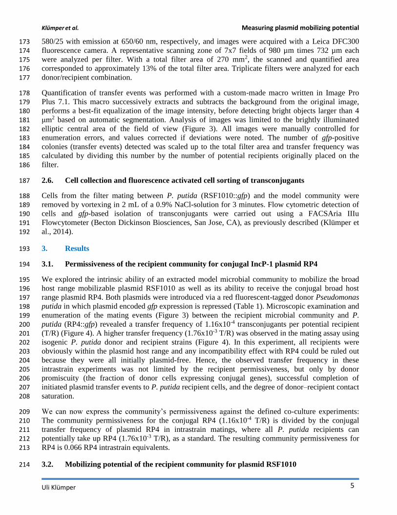
580/25 with emission at 650/60 nm, respectively, and images were acquired with a Leica DFC300 173
fluorescence camera. A representative scanning zone of 7x7 fields of 980 µm times 732 µm each 174 were analyzed per filter. With a total filter area of 270 mm2, the scanned and quantified area 175 corresponded to approximately 13% of the total filter area. Triplicate filters were analyzed for each 176
donor/recipient combination. 177
Quantification of transfer events was performed with a custom-made macro written in Image Pro 178
Plus 7.1. This macro successively extracts and subtracts the background from the original image, 179 performs a best-fit equalization of the image intensity, before detecting bright objects larger than 4 180 μm2 based on automatic segmentation. Analysis of images was limited to the brightly illuminated 181 elliptic central area of the field of view (Figure 3). All images were manually controlled for 182 enumeration errors, and values corrected if deviations were noted. The number of gfp-positive 183
colonies (transfer events) detected was scaled up to the total filter area and transfer frequency was 184 calculated by dividing this number by the number of potential recipients originally placed on the 185
filter. 186
2.6. Cell collection and fluorescence activated cell sorting of transconjugants 187
Cells from the filter mating between P. putida (RSF1010::gfp) and the model community were 188 removed by vortexing in 2 mL of a 0.9% NaCl-solution for 3 minutes. Flow cytometric detection of 189 cells and gfp-based isolation of transconjugants were carried out using a FACSAria IIIu 190
Flowcytometer (Becton Dickinson Biosciences, San Jose, CA), as previously described (Klümper et 191
al., 2014). 192
3. Results 193
3.1. Permissiveness of the recipient community for conjugal IncP-1 plasmid RP4 194
We explored the intrinsic ability of an extracted model microbial community to mobilize the broad 195 host range mobilizable plasmid RSF1010 as well as its ability to receive the conjugal broad host 196 range plasmid RP4. Both plasmids were introduced via a red fluorescent-tagged donor Pseudomonas 197
putida in which plasmid encoded gfp expression is repressed (Table 1). Microscopic examination and 198 enumeration of the mating events (Figure 3) between the recipient microbial community and P. 199
putida (RP4::gfp) revealed a transfer frequency of 1.16x10⁻⁴ transconjugants per potential recipient 200 (T/R) (Figure 4). A higher transfer frequency (1.76x10⁻³ T/R) was observed in the mating assay using 201 isogenic P. putida donor and recipient strains (Figure 4). In this experiment, all recipients were 202 obviously within the plasmid host range and any incompatibility effect with RP4 could be ruled out 203 because they were all initially plasmid-free. Hence, the observed transfer frequency in these 204
intrastrain experiments was not limited by the recipient permissiveness, but only by donor 205 promiscuity (the fraction of donor cells expressing conjugal genes), successful completion of 206 initiated plasmid transfer events to P. putida recipient cells, and the degree of donor–recipient contact 207
saturation. 208
We can now express the community’s permissiveness against the defined co-culture experiments: 209 The community permissiveness for the conjugal RP4 (1.16x10⁻⁴ T/R) is divided by the conjugal 210 transfer frequency of plasmid RP4 in intrastrain matings, where all P. putida recipients can 211 potentially take up RP4 (1.76x10⁻³ T/R), as a standard. The resulting community permissiveness for 212
RP4 is 0.066 RP4 intrastrain equivalents. 213
3.2. Mobilizing potential of the recipient community for plasmid RSF1010 214

Klümper et al. Measuring plasmid mobilizing potential
6 This is a provisional file, not the final typeset article
When the model community was challenged with P. putida (RSF1010::gfp), a transfer frequency of 215
1.16x10⁻⁵ T/R was measured. This value is one order of magnitude lower than the community’s 216 measured permissiveness for the conjugal plasmid RP4 (Figure 4). 217
In these experiments RSF1010 must have been retromobilized into the recipient community by cells 218
carrying IncQ compatible mobilizing conjugal plasmids (Figure 1). In order to explore the 219 retrotransfer frequency of RSF1010 further, isogenic P. putida strains were used to execute two 220 different intrastrain matings, taking advantage of all P.putida recipient cells being potential RSF1010 221 hosts. In the first experiment, a plasmid-free, non-mCherry-tagged P. putida strain served as 222 recipient. In the second experiment, a non-mCherry-tagged P. putida strain hosting the untagged 223
wild-type of the conjugal, mobilizing RP4 plasmid served as recipient. In the first experiment no 224 RSF1010 transfer was observed, consistent with RSF1010’s non-self-transmissible nature. In the 225 second experiment with P. putida (RP4) as recipient, retrotransfer was observed, with a measured 226 frequency of 8.34x10⁻⁴ T/R. Successful RSF1010 retrotransfer requires initial conjugal plasmid 227
transfer from recipients to RSF1010 donors, before RSF1010 is mobilized and retransferred to the 228 recipients (Top et al., 1992) (Figure 1C). 229
This measured RSF1010 retrotransfer frequency by P. putida (RP4) results from a combination of the 230 RP4 transfer process from the recipient to the donor (Figure 1C Step 1&2) and the subsequent 231 mobilization of RSF1010 through the now co-resident RP4 plasmid (Figure 1C Step 3&4). It can be 232
contrasted with the measured RP4 intraspecies transfer frequency of 1.76x10⁻³ T/R. RP4 intrastrain 233 transfer corresponds to the first two steps in RSF1010 retrotransfer (Figure 1A). Hence, the 234
probability of a cell that recently acquired RP4 via conjugal transfer to mobilize RSF1010 can be 235 estimated at 47.4% (8.34x10⁻⁴ T/R for P. putida (RSF1010::gfp) to P. putida (RP4) divided by 236 1.76x10⁻³ (T/R) for P. putida (RP4::gfp) to P. putida). For this specific pair of mobilizing and 237
mobilizable plasmid, retrotransfer is high (Figure 4). 238
The retrotransfer of RSF1010 to the recipient community occurs at a frequency of 10% compared to 239 its permissiveness for the RP4 plasmid. Still, as shown above, mobilization of RSF1010 is realized 240 only approximately every second time a conjugal plasmid is transferred from the recipient 241
community into the donor strain, based on mobilization through RP4. If all these potential 242 mobilization events were realized, the maximal mobilization potential of the recipient community is 243
reached. The theoretical maximal mobilization potential towards RSF1010 can be quantitatively 244 assessed as 2.45x10⁻⁵ T/R by dividing its transfer frequency towards the community (1.16x10⁻⁵ T/R) 245 by the now established 47.4% probability of retrotransfer. When subsequently dividing 2.45x10⁻⁵ T/R 246 through the community’s permissiveness towards RP4 (1.16x10⁻⁴ T/R) as a standard, this results in 247
0.211 RP4 permissiveness equivalents as the maximal mobilization potential. 248
3.3. Potential community permissiveness towards mobilizable plasmid RSF1010 249
In a final experiment, we quantified the intrinsic permissiveness of the model community for 250 RSF1010. To do so, we augmented the community’s own RSF1010 mobilizing potential by adding 251 an exogenous RSF1010 mobilizing strain. Hence, the recipient community was challenged with P. 252 putida hosting both the RSF1010::gfp and the wild-type RP4, which can directly mobilize RSF1010 253 (Figure 1B). The observed transfer frequency of RSF1010 in this mating was 3.14x10⁻³ T/R. This 254
frequency is, surprisingly, higher (~30-fold) than the community’s permissiveness for RP4. As 255 expected, this value is also substantially higher (~2 orders of magnitude) than the RSF1010 256 mobilization frequency (Figure 4) relying on the community’s inherent retromobilization potential 257
only. 258
Klümper et al. Measuring plasmid mobilizing potential
Uli Klümper 7
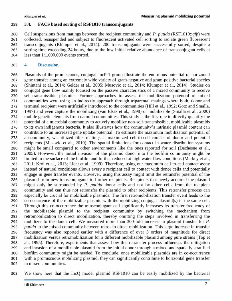
3.4. FACS based sorting of RSF1010 transconjugants 259
Cell suspensions from matings between the recipient community and P. putida (RSF1010::gfp) were 260 collected, resuspended and subject to fluorescent activated cell sorting to isolate green fluorescent 261 transconjugants (Klümper et al., 2014). 200 transconjugants were successfully sorted, despite a 262
sorting time exceeding 24 hours, due to the low initial relative abundance of transconjugant cells at 263 less than 1:1,000,000 events sorted. 264
4. Discussion 265
Plasmids of the promiscuous, conjugal IncP-1 group illustrate the enormous potential of horizontal 266 gene transfer among an extremely wide variety of gram-negative and gram-positive bacterial species 267
(Shintani et al., 2014; Gelder et al., 2005; Musovic et al., 2014; Klümper et al., 2014). Studies on 268 conjugal gene flow mainly focused on the passive characteristics of a mixed community to receive 269
self-transmissible plasmids. Former approaches to assess the mobilization potential of mixed 270 communities were using an indirectly approach through triparental matings where both, donor and 271 terminal recipient were artificially introduced to the communities (Hill et al., 1992; Götz and Smalla, 272 1997) and even capture the mobilizing (van Elsas et al., 1998) or mobilizable (Smalla et al., 2000) 273
mobile genetic elements from natural communities. This study is the first one to directly quantify the 274 potential of a microbial community to actively mobilize non-self-transmissible, mobilizable plasmids 275 to its own indigenous bacteria. It also illustrates how the community’s intrinsic plasmid content can 276
contribute to an increased gene uptake potential. To estimate the maximum mobilization potential of 277 a community, we utilized filter matings at maximized cell-to-cell contact of donor and potential 278
recipients (Musovic et al., 2010). The spatial limitations for contact in water distribution systems 279 might be small compared to other environments like the ones reported for soil (Dechesne et al., 280 2005). However, the initial invasion of the plasmid donor into the biofilm community might be 281
limited to the surface of the biofilm and further reduced at high water flow conditions (Merkey et al., 282
2011; Król et al., 2013; Licht et al., 1999). Therefore, using our maximum cell-to-cell contact assay 283 instead of natural conditions allows every s recipient cell to contact with donor cells and potentially 284 engage in gene transfer events. However, using this assay might limit the retransfer potential of the 285
plasmid from new transconjugants to further recipients. Recipients that newly acquired the plasmid 286 might only be surrounded by P. putida donor cells and not by other cells from the recipient 287
community and can thus not retransfer the plasmid to other recipients. This retransfer process can 288 especially be crucial for mobilizable plasmids. The first retromobilization transfer event leads to the 289 co-occurrence of the mobilizable plasmid with the mobilizing conjugal plasmid(s) in the same cell. 290 Through this co-occurrence the transconjugant cell significantly increases its transfer frequency of 291
the mobilizable plasmid to the recipient community by switching the mechanism from 292 retromobilization to direct mobilization, thereby omitting the steps involved in transferring the 293 mobilizer to the donor cell. We measured more than 300-fold increase in plasmid transfer for P. 294
putida to the mixed community between retro- to direct mobilization. This large increase in transfer 295 frequency was also reported earlier with a difference of over 3 orders of magnitude for direct 296 mobilization versus retromobilization for a different mobilizable plasmid among pure strains (Top et 297 al., 1995). Therefore, experiments that assess how this retransfer process influences the mitigation 298
and invasion of a mobilizable plasmid from the initial donor through a mixed and spatially stratified 299 biofilm community might be needed. To conclude, once mobilizable plasmids are in co-occurrence 300 with a promiscuous mobilizing plasmid, they can significantly contribute to horizontal gene transfer 301 in mixed communities. 302
We show here that the IncQ model plasmid RSF1010 can be easily mobilized by the bacterial 303
Klümper et al. Measuring plasmid mobilizing potential
8 This is a provisional file, not the final typeset article
community extracted from a household water distribution system. The permissiveness of this 304
microbial community towards the conjugal plasmid RP4 is comparable in magnitude with that 305 measured in diverse soil communities (Musovic et al., 2014). The lower permissiveness towards RP4 306 measured for mixed recipient communities compared to P. putida intraspecies transfer results 307
primarily from the inability of a fraction of the bacterial community to either receive, transiently 308 maintain, or express plasmid encoded genes. 309
The community’s potential to retromobilize and subsequently receive RSF1010 is only one order of 310 magnitude lower than its permissiveness towards RP4. This surprisingly high transfer rate may result 311 from the fact that IncQ plasmids have a broader host-range than any other known replicating 312
component in bacteria (Meyer, 2009) combined with an extremely efficient transfer mechanism 313 (Gregory et al., 2008; Meyer, 2009). The numbers appear even higher, taking into account that in 314 pure culture experiments with P. putida, only half of the microcolonies that recently received RP4 315 retromobilized RSF1010. Earlier retrotransfer experiments between two E.coli strains (Top et al., 316
1992) showed T/R ratios within the same orders of magnitude (10-3 – 10-4) as our intrastrain matings. 317 But, they suggested that retrotransfer of the mobilizable plasmid appears at rates lower than 1% once 318
the first step of acquiring a conjugal plasmid is realized. In that work transfer was quantified based 319 on single cells and after 2.33 hours. Our far higher numbers (~50%) might therefore result from 320 quantifying transfer on microcolony basis after 48 hours. Only one retrotransfer event within a 321
microcolony is needed for quantification as a successful transfer event and due to increased 322 incubation time retrotransfer can happen not only through the initial, but also through newly 323
established conjugal pili. Nonetheless, the observed retromobilization requires the presence of 324 mobilizing, conjugal plasmids within the permissive fraction of the recipients. Other mobilization 325 possibilities involve conjugation-independent transfer of plasmids through the formation of 326
nanotubes from members of the complex community towards the donor cells (Dubey and Ben-327 Yehuda, 2011), but are only realized if nanotubes from the recipient to the P. putida donor are 328
established. Therefore, a high intrinsic conjugal plasmid content of the model recipient community in 329
combination with RSF1010’s efficient transfer mechanism is the most likely reason for the observed 330
high mobilization potential. 331
IncP type IV secretion systems can conjugally connect a huge variety of organisms (Grahn et al., 332
2000; Thomas and Nielsen, 2005; Klümper et al., 2014). But like the plasmids encoding them, they 333 are evolutionary adapted to connect their mainly Gram-negative hosts. These self-transmissible 334 plasmids might easily reach dead ends after being transferred, if the secretion system is not encoded 335 efficiently for retransfer in the new host. Contrarily, mobilizable plasmids might less frequently reach 336
dead ends once acquired, since they can utilize the conjugal connections build through adapted 337 resident plasmids in their new host (Meyer, 2009) or through integrative and conjugative elements 338 (ICEs) (Lee et al., 2012). Additionally, mobilizable plasmids are relatively stable, as their nature as 339 high copy number plasmids (Meyer, 2009) increases their sustainability in a host until new transfer 340
becomes possible. These two facts in connection with their strictly host-independent initiation of 341 replication helps them to sustain in a very broad host range, including Pseudomonas sp., related 342 species in the Proteobacteria, as well as phylogenetically distant species within the Firmicutes, 343
Actinomycetes and even Cyanobacteria (Meyer et al. 2009) or plants (Buchanan-Wollaston et al., 344 1987). Consequently, RSF1010, as a mobilizable plasmid, has a far higher replication host range than 345 RP4. RSF1010 can even spread to a mixed community at a more than 30 fold higher transfer 346 frequency when directly mobilized through co-occurring plasmid RP4 in the same donor cell 347 compared to RP4 itself. Therefore, mobilizable plasmids might contribute to long term gene spread 348 and acquisition to a so far underestimated extent, especially in environments with high intrinsic 349
Klümper et al. Measuring plasmid mobilizing potential
Uli Klümper 9
mobilizing plasmid content. In our current experiment, we use a simplified system and are able to 350
deliver insights into the mobilization potential of a community at the first acquisition event of a 351 newly introduced mobilizable plasmid. The wide variety of mobilization systems possibly involved 352 might not resemble the one encoded by RP4 in efficiency. Still, equivalents based on the 353
community’s permissiveness towards RP4 can be used here, since long time maintenance and 354 retransfer are not taken into account. For more complex natural systems and experiments that allow 355 extensive retransfer we recommend assessing the intrinsic mobilization potential of microbial 356 communities based on absolute transfer frequencies, as the transfer and maintenance processes of 357 RSF1010 and RP4 differ too much in the long term. 358
Apart from quantification of the mobilization potential, the method presented here provides several 359 possibilities to study plasmid ecology and mobilization mechanisms. FACS based sorting of 360 RSF1010 carrying transconjugants from the recipient community was possible. Studying the 361 diversity of transconjugants might provide insights into the enormous host range of mobilizable 362
plasmids, compared to those of broad host range conjugal plasmids (Klümper et al., 2014). But the 363 high amount of sorting time prohibits intensive studies at this point. However, taking advantage of 364
FACS sorting, even at low speed, new possibilities for plasmid isolation emerge. The mobilizing, 365 conjugal plasmid can, now, after retromobilization, co-occurring with RSF1010 in the 366 transconjugant, be subsequently isolated within its original environmental host. Compared to 367
common exogenous plasmid isolation techniques our method has the potential to also capture 368 plasmids that are only transiently hosted and therefore quickly lost in the introduced capturing 369
strains. Since these plasmids remain stable in their original hosts, we gain the ability to isolate them 370 with our method. Isolated plasmids need therefore only stable maintenance in their natural hosts 371 rather than in an artificially introduced strain. This increases the range of obtainable plasmids and 372
immediately supplies information on where they naturally occur. This method reverses the 373 exogenous isolation technique for mobilizable plasmids (Top et al., 1994) and is cultivation 374
independent. Additionally using the tools presented here in combination with FACS sorting, single 375
cell observations to better understand the exact mechanisms proposed for retromobilization (Top et 376
al., 1992, 1995) might become possible. 377
In conclusion, this method is the first one to assess the plasmid mobilization potential of a microbial 378
community on a quantitative level by estimating its transfer frequency through fluorescent 379 microscopy. Using the new method, we discovered that a mixed microbial community has the 380 potential to easily mobilize a newly introduced mobilizable plasmid at high rates compared to a 381 conjugal plasmid. We also proved that the mobilizable plasmid is spread at far increased frequencies 382
once directly mobilized by a co-occurring conjugal plasmid from within the same cell. 383
5. Conflict of interest statement 384
The authors declare that the research was conducted in the absence of any commercial or financial 385
relationships that could be construed as a potential conflict of interest. 386
6. Acknowledgement 387
We thank L. Riber & S.J. Sørensen for access to the tagged RSF1010 plasmid, L.K. Jensen for 388 technical assistance in the laboratory and S. M. Milani for assistance in FACS sorting. This work was 389
funded by the Villum Kann Rasmussen Foundation Center of Excellence CREAM (Center for 390 Environmental and Agricultural Microbiology). 391
Klümper et al. Measuring plasmid mobilizing potential
10 This is a provisional file, not the final typeset article
7. References 392
Bahl, M. I., Oregaard, G., Sørensen, S. J., and Hansen, L. H. (2009). Construction and use of flow 393
cytometry optimized plasmid-sensor strains. Methods Mol. Biol. 532, 257–68. 394
Barth, P. T., and Grinter, N. J. (1977). Map of plasmid RP4 derived by insertion of transposon C. J. 395 Mol. Biol. 113, 455–474. 396
Boe-Hansen, R., Albrechtsen, H.-J., Arvin, E., and Jørgensen, C. (2002). Bulk water phase and 397 biofilm growth in drinking water at low nutrient conditions. Water Res. 36, 4477–4486. 398
Buchanan-Wollaston, V., Passiatore, J. E., and Cannon, F. (1987). The mob and oriT mobilization 399 functions of a bacterial plasmid promote its transfer to plants. Nature 328, 172–175. 400
Dechesne, A., Pallud, C. C., Bertolla, F., Grundmann, G. L., and Grundmann, L. G. (2005). Impact of 401 the microscale distribution of a Pseudomonas strain introduced into soil on potential contacts 402 with indigenous bacteria. Appl. Environ. Microbiol. 71, 8123–31. 403
Dubey, G. P., and Ben-Yehuda, S. (2011). Intercellular nanotubes mediate bacterial communication. 404
Cell 144, 590–600. 405
Van Elsas, J. D., Gardener, B. B., Wolters, A. C., and Smit, E. (1998). Isolation, characterization, and 406 transfer of cryptic gene-mobilizing plasmids in the wheat rhizosphere. Appl. Environ. Microbiol. 407
64, 880–9. 408
Gelder, L. De, Vandecasteele, F. P. J., Celeste, J., Forney, L. J., Top, E. M., Brown, C. J., and De 409
Gelder, L. (2005). Plasmid donor affects host range of promiscuous IncP-1beta plasmid pB10 in 410
an activated-sludge microbial community. Appl. Environ. Microbiol. 71, 5309–17. 411
Grahn, A. M., Haase, J., Bamford, D. H., and Lanka, E. (2000). Components of the RP4 conjugative 412 transfer apparatus form an envelope structure bridging inner and outer membranes of donor 413
cells: implications for related macromolecule transport systems. J. Bacteriol. 182, 1564–1574. 414
Gregory, R., Saunders, J. R., and Saunders, V. (2008). Rule-based modelling of conjugative plasmid 415 transfer and incompatibility. Biosystems. 91, 201–15. 416
Grohmann, E. (2011). Microbes and Microbial Technology. , eds. I. Ahmad, F. Ahmad, and J. Pichtel 417 New York, NY: Springer New York. 418
Götz, A., and Smalla, K. (1997). Manure enhances plasmid mobilization and survival of 419 Pseudomonas putida introduced into field soil. Appl. Environ. Microbiol. 63, 1980–1986. 420
Heuer, H., Binh, C. T. T., Jechalke, S., Kopmann, C., Zimmerling, U., Krögerrecklenfort, E., Ledger, 421 T., González, B., Top, E., and Smalla, K. (2012). IncP-1ε plasmids are important vectors of 422 antibiotic resistance genes in agricultural systems: Diversification driven by Class 1 integron 423 gene cassettes. Front. Microbiol. 3, 2. 424
Heuer, H., Schmitt, H., and Smalla, K. (2011). Antibiotic resistance gene spread due to manure 425
Klümper et al. Measuring plasmid mobilizing potential
Uli Klümper 11
application on agricultural fields. Curr. Opin. Microbiol. 14, 236–43. 426
Heuer, H., and Smalla, K. (2012). Plasmids foster diversification and adaptation of bacterial 427
populations in soil. FEMS Microbiol. Rev. 36, 1083–104. 428
Hill, K. E., Weightman, A. J., and Fry, J. C. (1992). Isolation and screening of plasmids from the 429 epilithon which mobilize recombinant plasmid pD10. Appl. Envir. Microbiol. 58, 1292–1300. 430
Hirt, H. (2002). In vivo induction of virulence and antibiotic resistance transfer in Enterococcus 431 faecalis mediated by the sex pheromone-sensing system of pCF10. Infect. Immun. 70, 716–723. 432
Honda, Y., Sakai, H., Hiasa, H., Tanaka, K., Komano, T., and Bagdasarian, M. (1991). Functional 433 division and reconstruction of a plasmid replication origin: molecular dissection of the oriV of 434 the broad-host-range plasmid RSF1010. Proc. Natl. Acad. Sci. 88 , 179–183. 435
Haase, J., Lurz, R., Grahn, A. M., Bamford, D. H., and Lanka, E. (1995). Bacterial conjugation 436 mediated by plasmid RP4: RSF1010 mobilization, donor-specific phage propagation, and pilus 437
production require the same Tra2 core components of a proposed DNA transport complex. J. 438 Bacteriol. 177, 4779–4791. 439
Klümper, U., Riber, L., Dechesne, A., Sannazzarro, A., Hansen, L. H., Sørensen, S. J., and Smets, B. 440
F. (2014). Broad host range plasmids can invade an unexpectedly diverse fraction of a soil 441 bacterial community. ISME J. 442
Król, J. E., Wojtowicz, A. J., Rogers, L. M., Heuer, H., Smalla, K., Krone, S. M., and Top, E. M. 443
(2013). Invasion of E. coli biofilms by antibiotic resistance plasmids. Plasmid 70, 110–9. 444
Lee, C. A., Thomas, J., and Grossman, A. D. (2012). The Bacillus subtilis conjugative transposon 445 ICEBs1 mobilizes plasmids lacking dedicated mobilization functions. J. Bacteriol. 194, 3165–446
72. 447
Lessl, M., Balzer, D., Weyrauch, K., and Lanka, E. (1993). The mating pair formation system of 448 plasmid RP4 defined by RSF1010 mobilization and donor-specific phage propagation. J. 449
Bacteriol. 175, 6415–25. 450
Licht, T. R., Christensen, B. B., Krogfelt, K. A., and Molin, S. (1999). Plasmid transfer in the animal 451 intestine and other dynamic bacterial populations: the role of community structure and 452
environment. Microbiology 145, 2615–2622. 453
Loftie-Eaton, W., and Rawlings, D. E. (2012). Diversity, biology and evolution of IncQ-family 454 plasmids. Plasmid 67, 15–34. 455
Merkey, B. V, Lardon, L. A., Seoane, J. M., Kreft, J.-U., and Smets, B. F. (2011). Growth 456 dependence of conjugation explains limited plasmid invasion in biofilms: an individual-based 457 modelling study. Environ. Microbiol. 13, 2435–52. 458
Meyer, R. (2009). Replication and conjugative mobilization of broad host-range IncQ plasmids. 459
Klümper et al. Measuring plasmid mobilizing potential
12 This is a provisional file, not the final typeset article
Plasmid 62, 57–70. 460
Musovic, S., Dechesne, A., Sørensen, J., and Smets, B. F. (2010). Novel assay to assess 461
permissiveness of a soil microbial community toward receipt of mobile genetic elements. Appl. 462 Environ. Microbiol. 76, 4813–8. 463
Musovic, S., Klümper, U., Dechesne, A., Magid, J., and Smets, B. F. (2014). Long-term manure 464 exposure increases soil bacterial community potential for plasmid uptake. Environ. Microbiol. 465
Rep. 6, 125–30. 466
Nelson, K. E., Weinel, C., Paulsen, I. T., Dodson, R. J., Hilbert, H., Martins dos Santos, V. A. P., 467 Fouts, D. E., Gill, S. R., Pop, M., Holmes, M., et al. (2002). Complete genome sequence and 468
comparative analysis of the metabolically versatile Pseudomonas putida KT2440. Environ. 469 Microbiol. 4, 799–808. 470
Scholz, P., Haring, V., Wittmann-Liebold, B., Ashman, K., Bagdasarian, M., and Scherzinger, E. 471 (1989). Complete nucleotide sequence and gene organization of the broad-host-range plasmid 472
RSF1010. Gene 75, 271–288. 473
Shintani, M., Matsui, K., Inoue, J.-I., Hosoyama, A., Ohji, S., Yamazoe, A., Nojiri, H., Kimbara, K., 474 and Ohkuma, M. (2014). Single-cell analyses revealed transfer ranges of IncP-1, IncP-7, and 475 IncP-9 plasmids in a soil bacterial community. Appl. Environ. Microbiol. 80, 138–45. 476
Smalla, K., Heuer, H., Götz, A., Niemeyer, D., Krogerrecklenfort, E., and Tietze, E. (2000). 477
Exogenous isolation of antibiotic resistance plasmids from piggery manure slurries reveals a 478 high prevalence and diversity of IncQ-like plasmids. Appl. Environ. Microbiol. 66, 4854–4862. 479
Smillie, C., Garcillán-Barcia, M. P., Francia, M. V., Rocha, E. P. C., and de la Cruz, F. (2010). 480 Mobility of plasmids. Microbiol. Mol. Biol. Rev. 74, 434–52. 481
Sørensen, S. J., Bailey, M., Hansen, L. H., Kroer, N., and Wuertz, S. (2005). Studying plasmid 482 horizontal transfer in situ: a critical review. Nat. Rev. Microbiol. 3, 700–10. 483
Thomas, C. M., and Nielsen, K. M. (2005). Mechanisms of, and barriers to, horizontal gene transfer 484 between bacteria. Nat. Rev. Microbiol. 3, 711–21. 485
Top, E. M. E., Rore, H., Collard, J.-M. J., Gellens, V., Slobodkina, G., Verstraete, W., and Mergeay, 486
M. (1995). Retromobilization of heavy metal resistance genes in unpolluted and heavy metal 487 polluted soil. FEMS Microbiol. Ecol. 18, 191–203. 488
Top, E., De Smet, I., Verstraete, W., Dijkmans, R., and Mergeay, M. (1994). Exogenous isolation of 489 mobilizing plasmids from polluted soils and sludges. Appl. Envir. Microbiol. 60, 831–839. 490
Top, E., Vanrolleghem, P., Mergeay, M., and Verstraete, W. (1992). Determination of the 491 mechanism of retrotransfer by mechanistic mathematical modeling. J. Bacteriol. 174, 5953–492 5960. 493
Yano, H., Rogers, L. M., Knox, M. G., Heuer, H., Smalla, K., Brown, C. J., and Top, E. M. (2013). 494
Klümper et al. Measuring plasmid mobilizing potential
Uli Klümper 13
Host range diversification within the IncP-1 plasmid group. Microbiology 159, 2303–15. 495
Zhang, M., Visser, S., Pereira E Silva, M. C., and van Elsas, J. D. (2014). IncP-1 and PromA group 496
plasmids are major providers of horizontal gene transfer capacities across bacteria in the 497 mycosphere of different doil fungi. Microb. Ecol. 498
499
Klümper et al. Measuring plasmid mobilizing potential
14 This is a provisional file, not the final typeset article
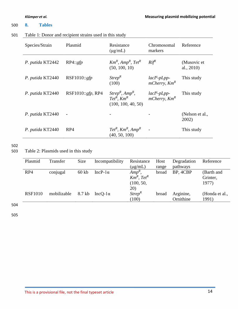
8. Tables 500
Table 1: Donor and recipient strains used in this study 501
Species/Strain Plasmid Resistance
(μg/mL)
Chromosomal
markers
Reference
P. putida KT2442 RP4::gfp KmR, AmpR, TetR
(50, 100, 10)
RifR (Musovic et
al., 2010)
P. putida KT2440 RSF1010::gfp StrepR
(100)
lacIq-pLpp-
mCherry, KmR
This study
P. putida KT2440 RSF1010::gfp, RP4 StrepR, AmpR,
TetR, KmR
(100, 100, 40, 50)
lacIq-pLpp-
mCherry, KmR
This study
P. putida KT2440 - - - (Nelson et al.,
2002)
P. putida KT2440 RP4 TetR, KmR, AmpR
(40, 50, 100)
- This study
502
Table 2: Plasmids used in this study 503
Plasmid Transfer Size Incompatibility Resistance
(μg/mL)
Host
range
Degradation
pathways
Reference
RP4 conjugal 60 kb IncP-1α AmpR,
KmR, TetR
(100, 50,
20)
broad BP, 4CBP (Barth and
Grinter,
1977)
RSF1010 mobilizable 8.7 kb IncQ-1α StrepR
(100)
broad Arginine,
Ornithine
(Honda et al.,
1991)
504
505
Klümper et al. Measuring plasmid mobilizing potential
Uli Klümper 15
506
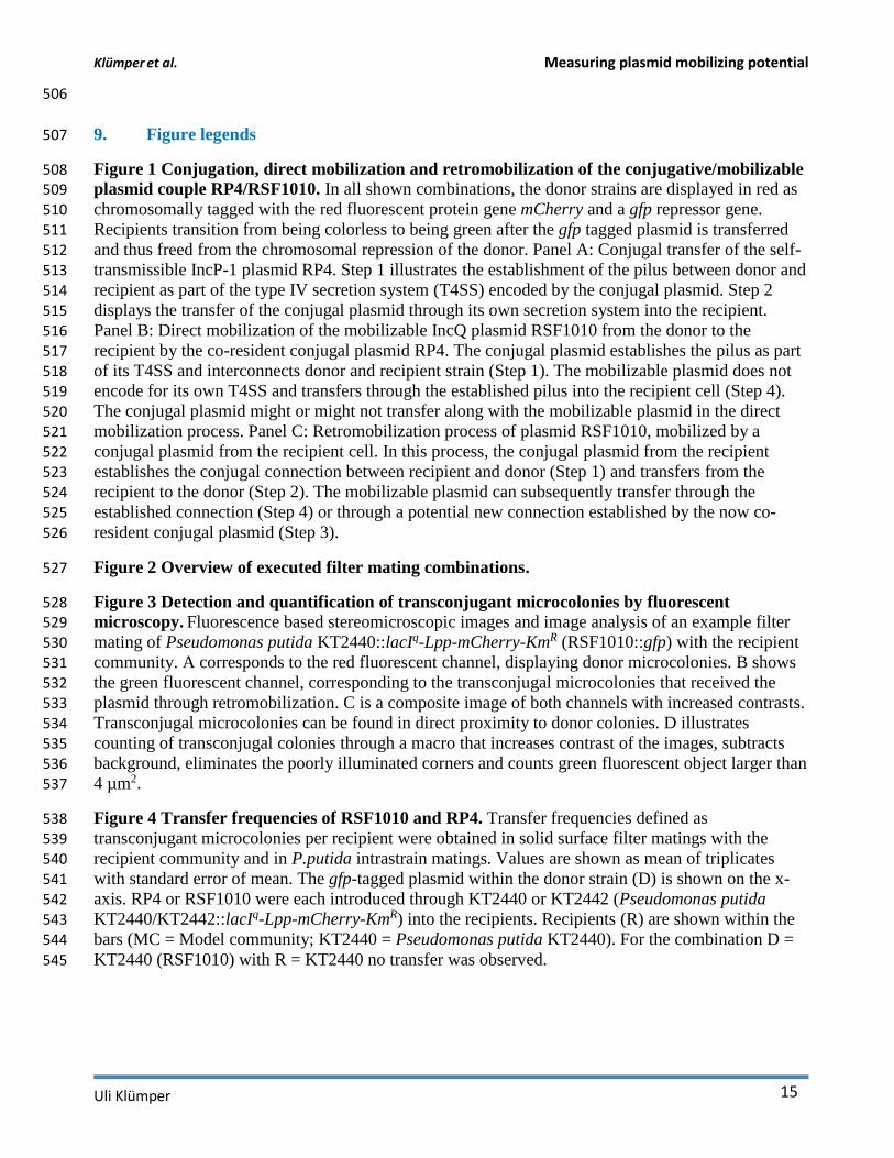
9. Figure legends 507
Figure 1 Conjugation, direct mobilization and retromobilization of the conjugative/mobilizable 508 plasmid couple RP4/RSF1010. In all shown combinations, the donor strains are displayed in red as 509
chromosomally tagged with the red fluorescent protein gene mCherry and a gfp repressor gene. 510 Recipients transition from being colorless to being green after the gfp tagged plasmid is transferred 511 and thus freed from the chromosomal repression of the donor. Panel A: Conjugal transfer of the self-512 transmissible IncP-1 plasmid RP4. Step 1 illustrates the establishment of the pilus between donor and 513 recipient as part of the type IV secretion system (T4SS) encoded by the conjugal plasmid. Step 2 514
displays the transfer of the conjugal plasmid through its own secretion system into the recipient. 515 Panel B: Direct mobilization of the mobilizable IncQ plasmid RSF1010 from the donor to the 516 recipient by the co-resident conjugal plasmid RP4. The conjugal plasmid establishes the pilus as part 517
of its T4SS and interconnects donor and recipient strain (Step 1). The mobilizable plasmid does not 518 encode for its own T4SS and transfers through the established pilus into the recipient cell (Step 4). 519 The conjugal plasmid might or might not transfer along with the mobilizable plasmid in the direct 520
mobilization process. Panel C: Retromobilization process of plasmid RSF1010, mobilized by a 521 conjugal plasmid from the recipient cell. In this process, the conjugal plasmid from the recipient 522
establishes the conjugal connection between recipient and donor (Step 1) and transfers from the 523 recipient to the donor (Step 2). The mobilizable plasmid can subsequently transfer through the 524 established connection (Step 4) or through a potential new connection established by the now co-525
resident conjugal plasmid (Step 3). 526
Figure 2 Overview of executed filter mating combinations. 527
Figure 3 Detection and quantification of transconjugant microcolonies by fluorescent 528 microscopy. Fluorescence based stereomicroscopic images and image analysis of an example filter 529 mating of Pseudomonas putida KT2440::lacIq-Lpp-mCherry-KmR (RSF1010::gfp) with the recipient 530
community. A corresponds to the red fluorescent channel, displaying donor microcolonies. B shows 531 the green fluorescent channel, corresponding to the transconjugal microcolonies that received the 532
plasmid through retromobilization. C is a composite image of both channels with increased contrasts. 533 Transconjugal microcolonies can be found in direct proximity to donor colonies. D illustrates 534 counting of transconjugal colonies through a macro that increases contrast of the images, subtracts 535
background, eliminates the poorly illuminated corners and counts green fluorescent object larger than 536
4 µm2. 537
Figure 4 Transfer frequencies of RSF1010 and RP4. Transfer frequencies defined as 538
transconjugant microcolonies per recipient were obtained in solid surface filter matings with the 539 recipient community and in P.putida intrastrain matings. Values are shown as mean of triplicates 540
with standard error of mean. The gfp-tagged plasmid within the donor strain (D) is shown on the x-541 axis. RP4 or RSF1010 were each introduced through KT2440 or KT2442 (Pseudomonas putida 542 KT2440/KT2442::lacIq-Lpp-mCherry-KmR) into the recipients. Recipients (R) are shown within the 543 bars (MC = Model community; KT2440 = Pseudomonas putida KT2440). For the combination D = 544
KT2440 (RSF1010) with R = KT2440 no transfer was observed. 545
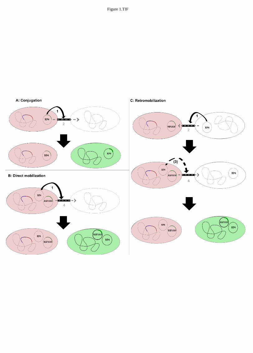
Figure 1.TIF
Figure 2.TIF
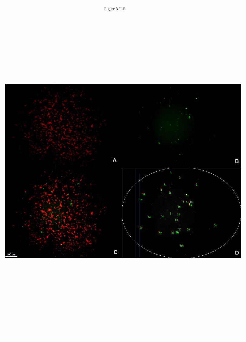
Figure 3.TIF
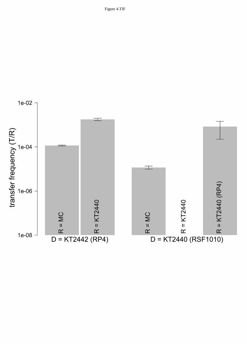
Figure 4.TIF
Related Documents