ANTIMICROBIAL AGENTS AND CHEMOTHERAPY, Jan. 1982, p. 110-118 0066-4804/82/010110-09$02.00/0 Vol. 21, No. 1 Novel Antibiotic Resistance Transfer in Bacteroides TOM D. MAYS,t C. JEFFREY SMITH,t RODNEY A. WELCH,§ CLAUDIO DELFINI,II AND FRANCIS L. MACRINA* Department of Microbiology, Virginia Commonwealth University, Richmond, Virginia 23298 Received 28 September 1981/Accepted 20 October 1981 Resistance to tetracycline and lincosamide antibiotics was transferred en bloc from a strain of Bacteroidesfragilis (V503) to a plasmidless strain of Bacteroides uniformis (V528) during in vitro filter matings. Resistance transfer was detected at frequencies of 1i-5 to 10-6 drug-resistant progeny per input donor cell and was dependent on cell-to-cell contact of donors and recipients. Transfer was insensi- tive to DNase and was not mediated by chloroform- or filter-sterilized donor broth cultures. A determinant for resistance to cefoxitin in V503 was not transferred in this system. V503 contained a 3.7 x 10⁶-dalton plasmid (pVA503). Drug-resistant progeny of V503 x V528 matings usually contained pVA503, but up to 20o of the total progeny of such crosses were plasmid free. Filter blot DNA hybridization studies (Southern method) confirmed that pVA503 was not integrated into the host chromosome of the plasmidless progeny. Drug-resistant progeny from V503 x V528 matings (with or without pVA503) conjugally transferred clindamycin resistance and tetracycline resistance to a suitable recipient strain. None of the drug resistance determinants of V503 were affected by treatment with standard plasmid curing regimens, and methods designed to detect very large plasmid molecules failed to suggest the involvement of extrachromosomal DNA in this resistance transfer system. The well-characterized Bacteroides R plasmid, pBF4 (conferring clindamycin resistance), was found to share hybridizing sequences with bulk cellular V503 DNA when examined by filter blot hybridization. Similarly sized sequences were found in drug-resistant progeny recovered from matings. Neither of the two pBF4 derivatives carrying deletions that abolished clindamycin resistance hybridized with V503 DNA. In the last 3 years there have been several reports describing self-transferable resistance (R) plasmids isolated from anaerobic bacteria. Brefort et al. (2) described a 36 x 106-dalton (36- Mdal) plasmid (pIP401) isolated from a strain of Clostridium perfringens that was associated with the expression of resistance to tetracycline (Tcr) and chloramphenicol (Cmr). pIP401 was transferable among C. perfringens strains via a conjugation-like process. Tally et al. (22) report- ed transferable resistance to clindamycin (Ccr)- erythromycin (Emr) after intraspecies filter mat- ings between strains of Bacteroides fragilis and Bacteroides thetaiotaomicron, and recently they have implicated the involvement of a 10-Mdal plasmid (pBFTM10) in Ccr and conjugal transfer (21). Privitera et al. (14, 15) have described t Present address: Viral Science Laboratory, Electro-Nu- cleonics, Inc., Silver Spring, MD 20904. t Present address: Department of Genetics, Bethesda Re- search Laboratories, Gaithersburg, MD 20768. § Present address: Department of Medical Microbiology, Stanford University, Stanford, CA 94305. 11 Present address: Instituto Sperimentale per L'Enologia, Asti, Italy. transferable Ccr in Bacteroides mediated by a 28-Mdal plasmid. Transferable resistance to tet- racycline in B. fragilis has also been described by Privitera et al. (14, 15), but the plasmid linkage of this marker is not clear at present. Workers in our laboratory (25) have described a 27-Mdal plasmid species (pBF4) isolated from a strain of B. fragilis which is self-transferable in interspecies matings and mediates the expres- sion and transfer of resistance to clindamycin and erythromycin. A restriction enzyme site map of pBF4 has been prepared, and two pairs of inverted repeat sequences have been located on this plasmid (27). In this communication, we describe a B. fragi- lis donor strain (V503) that transfers resistance to clindamycin and tetracycline without the in- volvement of detectable plasmid DNA. The successive en bloc transfer of these resistance determinants to additional recipients suggests a modular self-transferable genetic sequence which may be transposon-like in nature. Evi- dence that the Ccr determinant of B. fragilis V503 shares homology with the analogous deter- minant of pBF4 also is presented. 110 on August 22, 2018 by guest http://aac.asm.org/ Downloaded from

Welcome message from author
This document is posted to help you gain knowledge. Please leave a comment to let me know what you think about it! Share it to your friends and learn new things together.
Transcript

ANTIMICROBIAL AGENTS AND CHEMOTHERAPY, Jan. 1982, p. 110-1180066-4804/82/010110-09$02.00/0
Vol. 21, No. 1
Novel Antibiotic Resistance Transfer in BacteroidesTOM D. MAYS,t C. JEFFREY SMITH,t RODNEY A. WELCH,§ CLAUDIO DELFINI,II AND FRANCIS
L. MACRINA*Department of Microbiology, Virginia Commonwealth University, Richmond, Virginia 23298
Received 28 September 1981/Accepted 20 October 1981
Resistance to tetracycline and lincosamide antibiotics was transferred en blocfrom a strain of Bacteroidesfragilis (V503) to a plasmidless strain of Bacteroidesuniformis (V528) during in vitro filter matings. Resistance transfer was detected atfrequencies of 1i-5 to 10-6 drug-resistant progeny per input donor cell and wasdependent on cell-to-cell contact of donors and recipients. Transfer was insensi-tive to DNase and was not mediated by chloroform- or filter-sterilized donor brothcultures. A determinant for resistance to cefoxitin in V503 was not transferred inthis system. V503 contained a 3.7 x 10⁶-dalton plasmid (pVA503). Drug-resistantprogeny of V503 x V528 matings usually contained pVA503, but up to 20o of thetotal progeny of such crosses were plasmid free. Filter blot DNA hybridizationstudies (Southern method) confirmed that pVA503 was not integrated into thehost chromosome of the plasmidless progeny. Drug-resistant progeny from V503x V528 matings (with or without pVA503) conjugally transferred clindamycinresistance and tetracycline resistance to a suitable recipient strain. None of thedrug resistance determinants of V503 were affected by treatment with standardplasmid curing regimens, and methods designed to detect very large plasmidmolecules failed to suggest the involvement of extrachromosomal DNA in thisresistance transfer system. The well-characterized Bacteroides R plasmid, pBF4(conferring clindamycin resistance), was found to share hybridizing sequenceswith bulk cellular V503 DNA when examined by filter blot hybridization.Similarly sized sequences were found in drug-resistant progeny recovered frommatings. Neither of the two pBF4 derivatives carrying deletions that abolishedclindamycin resistance hybridized with V503 DNA.
In the last 3 years there have been severalreports describing self-transferable resistance(R) plasmids isolated from anaerobic bacteria.Brefort et al. (2) described a 36 x 106-dalton (36-Mdal) plasmid (pIP401) isolated from a strain ofClostridium perfringens that was associatedwith the expression of resistance to tetracycline(Tcr) and chloramphenicol (Cmr). pIP401 wastransferable among C. perfringens strains via aconjugation-like process. Tally et al. (22) report-ed transferable resistance to clindamycin (Ccr)-erythromycin (Emr) after intraspecies filter mat-ings between strains of Bacteroides fragilis andBacteroides thetaiotaomicron, and recently theyhave implicated the involvement of a 10-Mdalplasmid (pBFTM10) in Ccr and conjugal transfer(21). Privitera et al. (14, 15) have described
t Present address: Viral Science Laboratory, Electro-Nu-cleonics, Inc., Silver Spring, MD 20904.
t Present address: Department of Genetics, Bethesda Re-search Laboratories, Gaithersburg, MD 20768.
§ Present address: Department of Medical Microbiology,Stanford University, Stanford, CA 94305.
11 Present address: Instituto Sperimentale per L'Enologia,Asti, Italy.
transferable Ccr in Bacteroides mediated by a28-Mdal plasmid. Transferable resistance to tet-racycline in B. fragilis has also been describedby Privitera et al. (14, 15), but the plasmidlinkage of this marker is not clear at present.Workers in our laboratory (25) have described a27-Mdal plasmid species (pBF4) isolated from astrain of B. fragilis which is self-transferable ininterspecies matings and mediates the expres-sion and transfer of resistance to clindamycinand erythromycin. A restriction enzyme sitemap of pBF4 has been prepared, and two pairsof inverted repeat sequences have been locatedon this plasmid (27).
In this communication, we describe a B. fragi-lis donor strain (V503) that transfers resistanceto clindamycin and tetracycline without the in-volvement of detectable plasmid DNA. Thesuccessive en bloc transfer of these resistancedeterminants to additional recipients suggests amodular self-transferable genetic sequencewhich may be transposon-like in nature. Evi-dence that the Ccr determinant of B. fragilisV503 shares homology with the analogous deter-minant of pBF4 also is presented.
110
on August 22, 2018 by guest
http://aac.asm.org/
Dow
nloaded from
BACTEROIDES RESISTANCE TRANSFER 111
TABLE 1. Principal bacterial strainsPlasmid Source or
Species Strain Selected phenotypic traits' DNA size reference(Mdal)
B. fragilis V503 Tcr Ccr Emr Lnr RfP Cfr ind- cat' ara- 3.7 T.D.W.bB. uniformis V528 Tcs Cc' Ems Ln' RfrCfS ind+ cat- rha- ara+ None (25)
presentB. ovatus V211 Tcs Cc' Ems Ln' Rf CfS ind+ cat- rha+ ara+ None (25)
presenta TcT, Growth on medium containing 20 ,ug of tetracycline per ml; Tc5, no growth on medium containing 5 ,ug of
tetracycline per ml; Ccr, growth on medium containing 200 ,ug of clindamycin per ml; Emr, and Lnr, growth onmedium containing 200 ,ug of erythromycin and lincomycin, respectively, per ml; Em5, Cc' and Lns, no growthon medium containing 5 tag of erythromycin, clindamycin, and lincomycin, respectively, per ml; Rfr, growth onmedium containing 40 Lg per ml of rifampicin per ml; Rf', no growth on medium containing 5 Fig of rifampicin perml; Cfr, growth on medium containing 25 ,ug of cefoxitin per ml; Cf', no growth on medium containing S Fg ofcefoxitin per ml. Indole production (ind+ or ind-) and catalase production (cat' and cat-) were determined asdescribed by Holdeman et al. (7); arabinose fermentation (ara+ or ara-) was determined by the addition ofcolorimetric pH indicator; rha+, ability to grow on rhamnose as the sole source of carbon; rha-, inability to userhamnose as the carbon source.
b Obtained as a clinical isolate (in October 1977) at the University of Illinois Hospital, Chicago Medical Center,and characterized at the Virginia Polytechnic Institute (VPI) Anaerobe Laboratory, Blacksburg (VPI no. 12256).Obtained from T. D. Watkins at VPI.
MATERIALS AND METHODSBacterial strains and media. The bacterial strains
used in this study are described in Table 1. Stockcultures were maintained in chopped meat medium (7).Cells were cultivated in brain heart infusion (BHI)broth (Difco Laboratories, Detroit, Mich.) which wassupplemented with tryptone (final concentration, 1%),cysteine hydrochloride (1%), hemin (5 Fag/ml), andvitamin K (menadione) (1 p.g/ml). The broth wasprereduced anaerobically and sterilized as described inthe Anaerobe Laboratory Manual (7). Solid mediumwas prepared by adding agar (1.5%) to the supplement-ed BHI broth. The defined medium of Varel andBryant (24) supplemented with 0.1% Caseamino Ac-ids, 0.05% yeast extract, and 0.05% tryptone was usedin some instances as selective medium. Rhamnose(0.3%) was used as the sole source of carbon in thismedium.
Broth cultures and test tube matings (see below)were inoculated under a stream of oxygen-free gas(nitrogen-carbon dioxide, 9:1). Agar plates were incu-bated anaerobically either in jars using the Gas-Paksystem (BBL Microbiology Systems, Cockeysville,Md.) or in vented, gas evacuation-replacement jars (7).All incubations were at 37°C. Antibiotic sensitivityassays were performed by using the agar dilutionmethod described previously (11).Mating procedures. Filter matings were performed
with cells that were grown in supplemented BHI brothto the midexponential growth phase (5 x 10⁸ cells perml). Donor and recipient cultures (0.5 and 1.0 ml,respectively) were placed together in sterile (1.5-ml)Eppendorf polypropylene centrifuge tubes (Agonics,Brooklyn, N.Y.). The cells were pelleted and thensuspended in approximately 0.1 ml of broth. Thesemating mixtures were transferred by pipette ontosterile membrane filters (type HA, 25-mm diameter,0.45-,um pore size; Millipore Corp., Bedford, Mass.).The filters were placed on supplemented BHI agarplates, and the plates were incubated under anaerobic
conditions for 16 h unless otherwise noted. The filtersthen were transferred aseptically to sterile 40-ml poly-propylene centrifuge tubes. Sterile prereduced buff-ered salts solution (1 ml) (7) was added to each tube.The cells were washed from the membrane filters byvigorous agitation on a Vortex mixer for 1 min at highspeed. Appropriate dilutions of the cell suspensionwere plated on supplemented BHI agar plates contain-ing appropriate concentrations of antibiotics. The lev-els of donor and recipient cells on the filter aftermating were monitored to ensure that both members ofthe mating mixture had grown on the filter disk (25).Drug-resistant progeny were always reisolated two orthree times on selective media. Reisolated coloniesthen were examined for unselected phenotypic traits.
Test tube matings were performed by asepticallyadding donor and recipient cells (in the proportiondescribed above for filter matings) to a rubber-stop-pered tube containing 2 ml of prereduced and anaero-bically sterilized BHI-supplemented agar. The cellswere pelleted on the surface of the agar butt by brieflysubjecting the tube to centrifugation in a clinicalcentrifuge. The supernatant was aseptically decanted,the tube was flushed with Nz-CO2 (9:1), and themating cell mixture was allowed to incubate at 37°C forthe desired period of time. After the mating period, thecells were washed from the surface of the agar buttwith 1 ml of sterile prereduced buffered salts solutionand transferred to a sterile (1.5-ml) Eppendorf poly-propylene centrifuge tube and disrupted by agitationon a Vortex mixer for 1 min at high speed. The cellsuspension was then plated as described above.Plasmid DNA Isolation and analysis. Plasmid DNA
content was determined by subjecting early stationarygrowth phase cultures to the lysis and cleared-chromo-some technique of Guerry et al. (5). These lysateswere extracted with an equal volume of phenol previ-ously equilibrated with TE buffer (50 mM Tris, and 5mM disodium EDTA, pH 8) followed by extractionwith an equal volume of chloroform-isopentanol(24:1). The aqueous phase was concentrated by the
VOL. 21, 1982
on August 22, 2018 by guest
http://aac.asm.org/
Dow
nloaded from

ANTIMICROB. AGENTS CHEMOTHER.
addition of 5 M NaCi (to a final concentration of 0.15M) and 2 volumes of cold 95% ethanol. This prepara-tion was held at -20°C overnight (16 h) or at -70°C for1 h. It was then subjected to centrifugation at 12,000 xg at -20°C for 20 min. The ethanol solution wasdecanted, and the precipitated DNA was redissolvedin 60 ,ul of TE buffer. Appropriate amounts (10 to 50,u.l) of each preparation were placed on agarose gelsand subjected to electrophoresis for size determina-tions (9). Plasmid DNA from Bacteroides or Esche-richia coli used for endonuclease restriction digestion,molecular cloning, and hybridization studies was iso-lated by a modification of the same procedure. Theculture volume was increased to 800 ml, and theplasmid DNA was further purified during ultracentri-fugation in cesium chloride-ethidium bromide gradi-ents (25). Bulk cellular DNA for filter blot hybridiza-tion studies was isolated by the procedure of Marmur(12). Contour length measurements of purified plasmidDNA were performed as described previously (10).Endonuclease restriction enzymes were obtained fromBethesda Research Laboratories (Rockville, Md.),and digestions were performed according to the manu-facturer's instructions.
Molecular cloning on the EcoRI-D fragment of pBF4into pBR325 (1) was accomplished by using standardrecombinant DNA methodology as previously pub-lished from this laboratory (8).
Filter blot hybridizations. Bulk cellular DNA wastransferred to nitrocellulose filters by the method ofSouthern (20). Radiolabeling of plasmid DNA wasperformed by the in vitro nick translation method with32p in the alpha position of deoxycytidine triphosphate(16). The materials and protocol were supplied by NewEngland Nuclear Corp. (Beverly, Mass.). The 32p_labeled probe DNA (specific radioactivity, 107 cpm/,ug) was denatured in 90 mM Tris buffer with 1 MNaOH at pH 12.1 to 12.3 for 10 min. The probe DNAwas adjusted to pH 8.0 by the addition of 1 M sodiumacetate (pH 4.0). Hybridization of the 32P-labeledprobe DNA with the DNA transferred to the nitrocel-lulose and subsequent autoradiography were per-formed as described by Thayer (23).Ethidium bromide or coumermycin treatment. Cul-
tures of V503 were grown to the early exponentialphase of growth in defined broth medium (24). Ap-proximately 105 cells were transferred to each of aseries of tubes containing 10 ml of the minimal brothand increasing concentrations of ethidium bromide(Sigma Chemical Co., St. Louis, Mo.) or coumermy-cin (Bristol-Meyers, Syracuse, N.Y.). After overnightincubation, the broth culture containing the highestconcentration of drug in which growth was not inhibit-ed was spread on nonselective BHI agar medium. Atleast 500 colonies then were scored for Ccr and Tcr bydirect replication to drug-containing media.
RESULTSPlasmid content of B. fragilis V503. The high
level of Ccr (>200 ,ug/ml) and Tcr (20 ,ug/ml)displayed by B. fragilis V503 prompted us toexamine it for plasmid DNA. Agarose gel elec-trophoretic analysis of cleared lysate prepara-tions of V503 revealed a single plasmid species(pVA503). The size of pVA503 was 3.55 + 0.23
Mdal based on comparative migration in agarosegels (9) (nine determinations with four differentpreparations of pVA503). A size estimate of 3.85± 0.19 Mdal was obtained from contour lengthmeasurements of 16 molecules of pVA503 pho-tographed in the electron microscope. pSC101(6.02 Mdal) was used as a size reference in thesedeterminations (10).
Electrophoretic analysis of cleared, concen-trated cell lysates often revealed a plasmid DNAcomponent that migrated between the 3.7-Mdalcovalently closed circular form of pVA503 andthe host chromosome. This plasmid species mi-grated to a position similar to that of a 7.5-Mdalcovalently closed circular plasmid or a 3.7-Mdalopen circular species. Endonuclease restrictiondigests of purified pVA503 demonstrated thisspecies to be open circular plasmid DNA. Spe-cifically, HaeIII was found to cleave pVA503into two fragments: 2.9 and 0.8 Mdal. Limiteddigestion with this enzyme allowed us to followthe formation of circular and then linear pVA503forms. Under standard conditions of electropho-resis (0.7% agarose), open circular pVA503 mi-grated in the 7.5-Mdal range (covalently closedcircular), whereas linear pVA503 migrated in the6.8-Mdal range (covalently closed circular) (datanot shown).To ensure that a large R plasmid in V503 had
not gone undetected, cell lysates were subjectedto the plasmid isolation procedure of Hansenand Olsen (6). This method can be used reliablyto demonstrate extraordinarily large plasmids.Such analyses still revealed pVA503 to be theonly detectable plasmid species in B. fragilisV503. Control experiments with the Hansen andOlsen method were performed in which plas-mids as large as 220 Mdal could be successfullyisolated from Pseudomonas putida strains (28).Drug resistance transfer. B. fragilis V503 was
examined for its ability to transfer Ccr and Tcrdespite the absence of large candidate conjuga-tive plasmids in this strain. Using the Bacter-oides uniformis V528 recipient, progeny wereselected for resistance to clindamycin and tetra-cycline alone or in combination (Table 2). Trans-fer of the Ccr and Tcr markers was readilydetected in such matings (Table 2, matings 1through 3), but the cefoxitin marker present inV503 was never observed to be transferred inthis system (Table 2, mating 4). Progeny alwayswere verified by scoring the unselected chromo-somal traits of arabinose fermentation and theproduction of catalase and indol. Additionally,we found that B. fragilis V503 and Bacteroidesuniformis V528 could be readily differentiatedon the basis of the fragment pattern of theirchromosomal DNAs after digestion with HindJIIrestriction endonuclease (data not shown). Thephenotypic traits and the HindIII restriction
112 MAYS ET AL.
on August 22, 2018 by guest
http://aac.asm.org/
Dow
nloaded from

BACTEROIDES RESISTANCE TRANSFER 113
TABLE 2. Drug resistance transfer frequencies
No. Mating Donor Recipient Selected ona: Frequency of Representativetransferb progeny
1 Primary B. fragilis V503 B. uniformis V528 Rf Tc Cc 1.0 X 10⁻⁶ V619, V6222 Primary B. fragilis V503 B. uniformis V528 Rf Cc 7.6 x 10-63 Primary B. fragilis V503 B. uniformis V528 Rf Tc 1.2 X 10-64 Primary B. fragilis V503 B. uniformis V528 Rf Cf <1.0 x 10-85 Secondary B. uniformis V619 B. ovatus V211 Rham Tc Cc 1.1 x 10-66 Secondary B. uniformis V619 B. ovatus V211 Rham Cc 1.2 x 10-67 Secondary B. uniformis V619 B. ovatus V211 Rham Tc 3.7 x 10-68 Secondary B. uniformis V622 B. ovatus V211 Rham Tc Cc 2.0 X 10-69 Secondary B. uniformis V622 B. ovatus V211 Rham Cc 1.8 x 10-610 Secondary B. uniformis V622 B. ovatus V211 Rham Tc 5.5 x 10⁻⁶a Selective medium (BHI supplemented as described in the text) for matings 1 through 4 contained rifampicin
(Rf), 10 ,ug/ml; clindamycin (Cc), 5 j,g/ml; tetracycline (Tc), 5 jig/ml; and cefoxitin (Cf), 5 ,ug/ml. Progeny wereidentified as B. uniformis by indole production, lack of catalase production, fermentation of arabinose, andHindlIl endonuclease restriction digest profile of bulk cellular DNA. Tetracycline was used at a concentration of2.5 Fg/ml in the secondary matings. Rhamnose (Rham) was used in defined medium at a concentration of 0.3%.
b Frequency of transfer = number of resistant progeny/viable input donor cell.
fragment patterns of the progeny obtained inthese crosses always conformed to those of therecipient strain (V528). Attempts to obtain prog-eny containing only the Ccr and Tcr markeralone were unsuccessful. Progeny selected asCcr Rf' always inherited Tcr (>200 progenycolonies scored per experiment), and those se-lected as Tcr Rffr were always Ccr (>200 proge-ny colonies scored per experiment).The progeny from eight independently per-
formed matings also were examined for plasmidDNA content (data not shown). Approximately80o of the progeny (10 to 25 clones examinedper experiment) examined contained thepVA503 plasmid, regardless of whether theywere selected on medium containing both tetra-cycline and clindamycin or clindamycin alone.Two independently obtained progeny from
primary mating no. 1 (Table 2) were examinedfor conjugal proficiency. One strain, B. unifor-mis V619 (Table 2), contained the pVA503 plas-mid, whereas B. uniformis V622 was devoid ofdetectable extrachromosomal DNA. B. ovatusV211 was used as a recipient in these matings,and the results are seen in Table 2 (matings 5through 10). Both V619 and V622 were able totransfer their resistance en bloc; the Ccr and Tcrmarkers were never seen to segregate from oneanother in these matings (see above discussion).Using previously published methods (25) we
examined the V503 x V528 mating system withrespect to mode of genetic exchange. The resultsof these experiments revealed that drug resist-ance was not transferred when the donor cul-tures were filter sterilized, treated with chloro-form, or physically separated from the recipientstrain by a membrane filter. The observed trans-fer was insensitive to DNase I. Further, when
broth cultures of V503 and V528 were anaerobi-cally mixed and suitably incubated, no geneticexchange of drug resistance was observed. Pre-liminary test tube matings were performed inwhich V503 and V528 were allowed to incubateon the surface of a 2-ml plug of supplementedBHI agar in a tube containing an anaerobicatmosphere. This enabled us to control the dura-tion of mating under strict anaerobic conditionsin contrast to filter matings incubated in Gas-Pakjars which were exposed to a decreasing concen-tration of oxygen until anaerobiosis wasachieved (1 to 2 h). The minimum time requiredfor the transfer of drug resistance between V503and V528 in test tube matings was 45 min. Tcrand Ccr appeared to transfer as a unit. Noprogeny were found after 45 min of mating thatwere Tcr and Ccr alone (based on the screeningof 100 progeny per experiment in two experi-ments).
Characterization of drug resistance pheno-types. The donor V503 and primary resistantprogeny (V619, V622, and others tested from thesecondary matings) expressed the same levels ofresistance to erythromycin, lincomycin, clinda-mycin, and tetracycline (Table 1). Resistance totetracycline in V503 was expressed inducibly.Cells cultivated in the presence of a sub-inhibi-tory concentration of tetracycline (0.1 ,ug/ml) didnot demonstrate a lag in the growth rate duringthe early exponential phase after challenge withan inhibitory concentration (5 ,g/ml) of tetracy-cline (F. L. Macrina, T. D. Mays, C. J. Smith,and R. A. Welch, J. Antimicrob. Chemother., inpress). Cells cultivated in the absence of thedrug showed a significant lag in growth whenchallenged with tetracycline. The same growthpattern was observed for V619 and V622 (Ma-
VOL. 21, 1982
on August 22, 2018 by guest
http://aac.asm.org/
Dow
nloaded from

ANTIMICROB. AGENTS CHEMOTHER.
crina et al., in press). The inducible expressionof Tcr was supported by agar dilution suscepti-bility assays (data not shown). V503, V619, andV622 (Table 2) cultivated in a sub-inhibitory(0.1-,ug/ml) concentration of tetracycline dis-played 75% of the colony-forming activity on 20,ug of tetracycline per ml as compared withgrowth on drug-free medium. Colony-formingactivity of noninduced cells was <5% on 20 ,ugof tetracycline per ml as compared with growthon drug-free medium. Clindamycin resistancewas expressed constitutively in strain V503 andin V619 and V622 based on similarly performedgrowth curve experiments and viability determi-nations (data not shown).
B. fragilis V503 was grown in low concentra-tions (1 ,ug/ml) of tetracycline and tested forconjugal transfer of its Ccr and Tcr. No inductiveeffect on conjugal proficiency was seen, howev-er, with transfer remaining at levels comparableto that seen in Table 2.The Ccr and Tcr markers in V503 were stable
in cells grown at 37 or 42°C. In addition thesemarkers were stable in cells grown in the pres-ence, of the plasmid curing agents coumermycinor ethidium bromide. A minimum of 500 colo-nies were screened per curing experiment with-out detection of drug-sensitive clones.
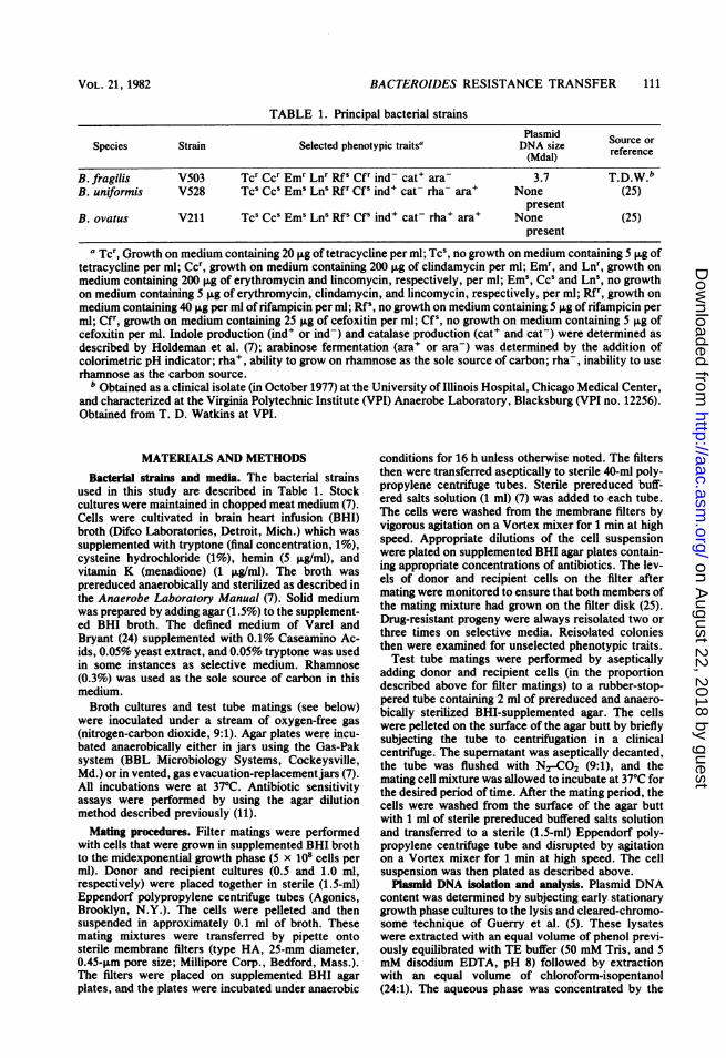
Molecular analysis of V503, V528, V619, andV622. The presence of pVA503 as an autono-mous plasmid species did not correlate with theexpression of the drug resistance phenotypes inthe primary resistant progeny or their ability toact as donors in subsequent matings (Table 2).To investigate whether pVA503 was present asan integrated segment of the host chromosomein the plasmidless progeny, filter blot hybridiza-tions were carried out with a radiolabelledpVA503 probe (Fig. 1). From these hybridiza-tion studies, no sequences of pVA503 werefound that hybridized to V528 (lane B) or aprimary plasmidless progeny isolate (V622, laneD). pVA503 did hybridize to the 3.7-Mdal plas-mid present in V503 (lane A) and in a plasmid-containing progeny (V619, lane C). It should benoted that the blotted DNA in lanes A through Drepresented material prepared by the method ofMarmur (12) and digested with HindlII. pVA503does not have any HindlII cleavage sites (un-published data) and thus appears as covalentlyclosed circular, open circular, and linear compo-nents in lanes A and C. These data enabled us toconclude that pVA503 is not present in an inte-grated state in a resistant progeny cell.As a probe of the molecular basis of Ccr in
V503 we used the well-characterized B. fragilisR plasmid pBF4 (27). This 27-Mdal conjugativeplasmid confers constitutively expressed Ccr.Two deletion-bearing derivatives of pBF4 thatare Ccs were also used. A restriction endonucle-
A B C D Eorin
oc _ 4i f
lin mm _ I
cc m maFIG. 1. Autoradiograph of filter-blotted DNA hy-
bridized with 32P-radiolabeled pVA503. Bulk cellularDNA was isolated by the method of Marmur (12).Purified pVA503 was prepared as described in thetext. HindlIl digestion of the bulk cellular DNA prepa-rations (lanes A through D) was performed, and frag-ments were separated on electrophoretic agarose gels(0.7%) as described in the text. The DNA was trans-ferred to nitrocellulose paper by the method of South-ern (20). pVA503 was radiolabeled with 32P by the nicktranslation method (16). Radiolabeled pVA503 wasallowed to hybridize with the filter-blotted DNA, andautoradiographs were prepared as described byThayer (23). The identity of the DNA preparations thatappear above are as follows: (A) B. fragilis (donor)V503; (B) B. uniformis (recipient) V528; (C) B. unifor-mis V619; (D) B. uniformis V622; (E) uncleavedpVA503 purified from CsCl-ethidium bromide ultra-centrifugation (approximately 1.5 ,ug). The origin (ori)of migration in the agarose gel appears at the top of theautoradiograph. The open circular (oc), linear (lin),and covalently closed circular (cc) forms of pVA503appear in the lanes containing V503 (A), V619 (C), andpurified pVA503 (E). The intermediate bands in lane Eprobably represent topoisomers present in this CsCl-ethidium bromide preparation of pVA503.
ase cleavage site map of pBF4 and the locationof the two above-mentioned deletions (Al andA2) are shown in Fig. 2A. Based on overlap ofthe Al and A2 deletions we selected the EcoRI-Dfragment (kilobase coordinates -1 to 4 on map)as an additional molecular probe in these stud-ies, and, accordingly, constructed a pBR325:E-coRI-D chimeric plasmid using recombinantDNA methodologies in an EK 1 host system.EcoRI-cleaved pBF4 A2 (lane A), pBR325:E-coRI-D (lane B), and pBF4 (lane C) analyzed by
114 MAYS ET AL.
on August 22, 2018 by guest
http://aac.asm.org/
Dow
nloaded from

BACTEROIDES RESISTANCE TRANSFER 115
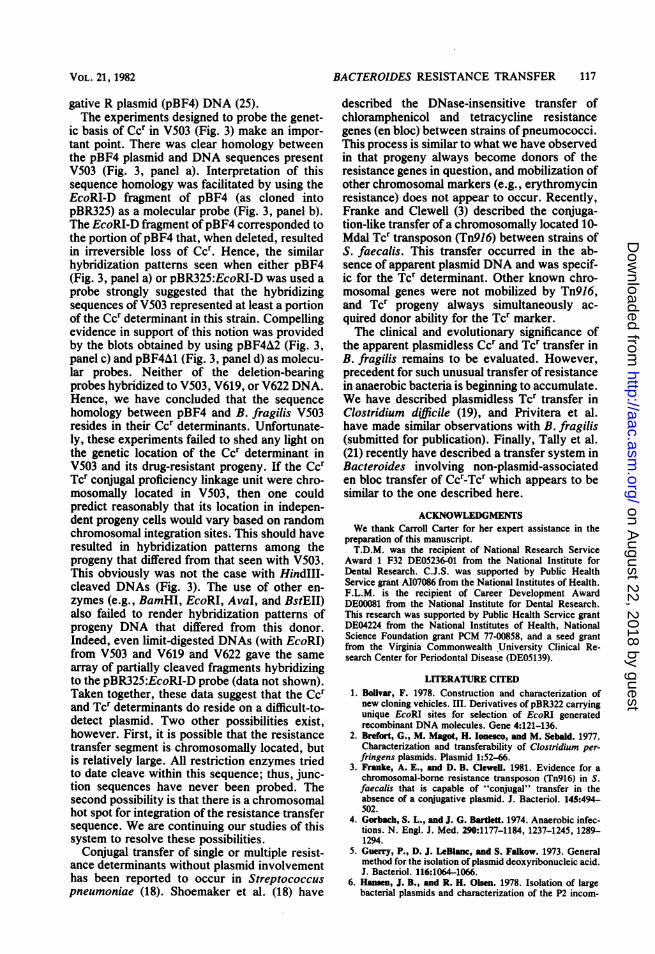
aFIG. 2. pBF4 map and analyses of pBR325:EcoRI-D chimera. a, Restriction endonuclease cleavage site map
of pBF4; the areas covered by the deletions carried by pBF4A1 and pBF4A2 are noted by the thick lines on theinside of the circle. b, Agarose gel analyses of the pBR325:EcoRI-D chimera. Lane A, EcoRI-cleaved pBF4A2;fragment sizes, top to bottom, are: 19.5, 8.1, 4.3, 3.2, and 2.6 kilobases. Lane B: EcoRI-cleaved pBR325:EcoRI-D recombinant plasmid; the uppermost component is linear pBR325, the lowermost component is the 3.7-kilobase EcoRI-D fragment of pBF4. Lane C, EcoRI-cleaved pBF4 DNA; components between the 19.5 and 8.1-kilobase bands and the single faint band below the 8.1-kilobase band are partially digested fragments.
agarose gel electrophoresis are seen in Fig. 2B.Our purpose in these studies was twofold.
First we wanted to determine if the Ccr determi-nant of V503 (isolated in the United States)shared homology with the Ccr determinant car-ried by pBF4 (isolated from a French B. fragilisclinical strain). Second, we wanted to test thehypothesis that the Ccr Tcr determinants werechromosomally located. The results of theseexperiments are presented in the compositeautoradiogram in Fig. 3. The blotted DNA in allfour panels of this figure are the same; all areHindIII-cleaved, Marmur-prepared (12) DNAsfrom each of the following strains: A, B. fragilisV479-1 (contains pBF4); B, V528 (recipient); C,B. fragilis V503 (donor); D and E, V619 andV622, respectively (independently obtainedprogeny; Table 2 and Fig. 1). Panel a representsthe radiographic pattern seen when pBF4 wasused as a molecular probe. All of the sevenHindIII components of pBF4 present in theV479-1 DNA were readily observed (lane A),whereas no pBF4 hybridizing sequences to therecipient V528 strain were detected (lane B).Two pBF4 hybridizing components were seen inthe V503 strain (lane C), however, and thesesame components were also present in the twoindependent progeny strains V619 (lane D) andV622 (lane E). The pBR322:EcoRI-D probe hy-bridized to the V479-1 DNA at a position corre-sponding to the HindIII-D component of pBF4
(lane A, panel b; HindIII-D of pBF4 is a doubletband [unpublished data]). No hybridizing activi-ty to V528 DNA was observed (lane B, panel b),but the same two pBR325:EcoRI-D hybridizingcomponents were seen in V503, V619, and V622(panel b, lanes C, D, and E, respectively). Thesehybridizing components were found to be identi-cal to those seen when pBF4 was used as probeDNA (Fig. 3, panel a) Panels c and d of Fig. 3show the results obtained by using pBF4A2(panel c) and pBF4A1 (panel d) as molecularprobes in this system. Hybridization to thepBF4-containing V479-1 strain (lane A in bothcases) was evident, with the lessened intensityof the HindIII-D component (panel c) and Hind-III-A and -D components (panel d) owing to thelocation of the deletions carried by the probe(27). No hybridizing activity of either of thedeletion-bearing pBF4 derivatives to the V528recipient (lane B), V503 donor (lane C), or theresistant progeny (lanes D and E) was observed,however. It should be noted that lanes B throughE of panels c and d contained DNA in fivefoldexcess over that contained in lane A.
DISCUSSIONThe existence of conjugative R plasmids in
anaerobic bacteria (2, 14, 15, 21, 22, 25-27) isunderscored by the overwhelming predomi-nance of these microorganisms in the humanindigenous microflora (13). Indeed, intestinal B.
VOL. 21, 1982
on August 22, 2018 by guest
http://aac.asm.org/
Dow
nloaded from

ANTIMICROB. AGENTS CHEMOTHER.
A B I
b
FIG. 3. Molecular probe analyses cdonors, recipients, and progeny. Condilisolation, preparation, and 32P-labeling (and Southern blot methodology (20) werfor Fig. 1. The HindIll-cleaved blotte'four panels were as follows: A, B. fi(contains pBF4); B, B. uniformis V528B. fragilis V503 (donor); D, B. uniformitant progeny from V503 x V528 mating)mis V622 (resistant progeny from V503ing). 32p probe DNA was as follows: ppanel b, pBF325:EcoRI-D chimera; panpanel d, pBF4A1. In panel a about 1 j±qapplied to each lane; about 0.5 p.g of ]
was used in panel b. In panels c and d aDNA was applied to lane A, whereas 2was applied to each of lanes B through I
the HindIII components of pBF4 seen inbottom) were: 12.1, 9.9, 5.7, 4.8 (doutand 1.8 kilobases.
fragilis strains are the most commianaerobes in soft tissue infections (4cin is the drug of choice for us(fragilis, a species which usually posent resistance to P-lactam and amantibiotics. The high level of clindaance in B. fragilis V503 was unustthat the Ccr and Tcr of V503 were trthe absence of detectable plasmicnovel finding in Bacteroides. Re(inherit the Ccr and the Tcr determi2) do so in a process that fits th
conjugation-like genetic exchange. It should beC 0 E noted, however, that our data do not rigorously
exclude specialized transduction by some diffi-cult-to-detect (perhaps defective) prophage as ameans of transfer.The Ccr and Tcr of V503 determinants did not
segregate from one another at detectable fre-quencies (Table 2) indicating their genetic link-age. Although B. fragilis V503 was resistant tocefoxitin (presumably via production of a cepha-losporinase), this resistance was never observedto be transferable (Table 2, mating 4). Thesecondary matings (Table 2) clearly suggestedthat the genetic information for the transferprocess was inherited with the resistance deter-minants. Ccr Tcr progeny isolated from primarycrosses always were able to act as conjugaldonors of that resistance.The issue of plasmid involvement in the trans-
fer system described here cannot be fully re-solved at present. Our data do allow us toconclude that the 3.7-Mdal pVA503 plasmid isnot involved in the transfer process or theexpression of resistance. Progeny from V503 xV528 matings were able to transfer Ccr and Tcrdespite the absence of pVA503 as monitored bystandard plasmid DNA detection methods (Ta-Af Bacteroides ble 2). Indeed, such plasmidless strains (e.g.,tions for DNA V622) have been shown to be devoid of any
re as descnbed detectable pVA503 sequences when examinedd DNAs in all by filter blot hybridization (Fig. 1). This rulesragilis V479-1 out the possibility ofpVA503 integration into the(recipient); C, V528 genome after transfer. Our reliable use ofis V619 (resis- the Hansen and Olsen (6) technique arguesE, B. unifor- against the presence of a large conjugative plas-x V528 mat- mid in V503. In control experiments, we could
)anel a, pBF4; successfully demonstrate plasmids in the 200-iel c, pBF4A2; Mdal range from Pseudomonas (28). However,DNA per lane it is possible that a plasmid (or plasmids) that isbout 05 lngof difficult to isolate by conventional methods is!.5 p.g of DNA responsible for the conjugative drug resistanceE. The sizes of we have observed. On the other hand, the failurelane A (top to to obtain drug-susceptible segregants ofB. fragi-
blet), 2.6, 2.3, lis V503 after treatment with plasmid curingagents such as coumermycin or ethidium bro-mide supports the notion that the Ccr and Tcrdeterminants are not extrachromosomally locat-ed. Roberts and Smith (17) recently have report-
only isolated ed that seemingly plasmidless drug-resistant1). Clindamy- strains of Haemophilus influenzae can transfere against B. their resistances by conjugation and that R plas-,sesses inher- mids can be subsequently recovered in resistantinoglycoside progeny. This strain-dependent isolation of plas-mycin resist- mid DNA does not seem likely in our Bacter-aal. The fact oides system. We have failed to detect plasmidsansferable in in three different genetic backgrounds (V528,d DNA is a V211, and V531 [Macrina et al., in press]) aftercipients that transfer from the V503 donor. Further, it shouldinants (Table be noted that derivatives of both V528 and V531ie criteria of have been used as hosts to reliably isolate conju-
116 MAYS ET AL.
on August 22, 2018 by guest
http://aac.asm.org/
Dow
nloaded from
BACTEROIDES RESISTANCE TRANSFER 117
gative R plasmid (pBF4) DNA (25).The experiments designed to probe the genet-
ic basis of Ccr in V503 (Fig. 3) make an impor-tant point. There was clear homology betweenthe pBF4 plasmid and DNA sequences presentV503 (Fig. 3, panel a). Interpretation of thissequence homology was facilitated by using theEcoRI-D fragment of pBF4 (as cloned intopBR325) as a molecular probe (Fig. 3, panel b).The EcoRI-D fragment ofpBF4 corresponded tothe portion ofpBF4 that, when deleted, resultedin irreversible loss of Ccr. Hence, the similarhybridization patterns seen when either pBF4(Fig. 3, panel a) or pBR325:EcoRI-D was used aprobe strongly suggested that the hybridizingsequences of V503 represented at least a portionof the Ccr determinant in this strain. Compellingevidence in support of this notion was providedby the blots obtained by using pBF4A2 (Fig. 3,panel c) and pBF4A1 (Fig. 3, panel d) as molecu-lar probes. Neither of the deletion-bearingprobes hybridized to V503, V619, or V622 DNA.Hence, we have concluded that the sequencehomology between pBF4 and B. fragilis V503resides in their Ccr determinants. Unfortunate-ly, these experiments failed to shed any light onthe genetic location of the Ccr determinant inV503 and its drug-resistant progeny. If the CcrTcr conjugal proficiency linkage unit were chro-mosomally located in V503, then one couldpredict reasonably that its location in indepen-dent progeny cells would vary based on randomchromosomal integration sites. This should haveresulted in hybridization patterns among theprogeny that differed from that seen with V503.This obviously was not the case with HindIII-cleaved DNAs (Fig. 3). The use of other en-zymes (e.g., BamHI, EcoRI, AvaI, and BstEII)also failed to render hybridization patterns ofprogeny DNA that differed from this donor.Indeed, even limit-digested DNAs (with EcoRI)from V503 and V619 and V622 gave the samearray of partially cleaved fragments hybridizingto the pBR325:EcoRI-D probe (data not shown).Taken together, these data suggest that the Ccrand Tcr determinants do reside on a difficult-to-detect plasmid. Two other possibilities exist,however. First, it is possible that the resistancetransfer segment is chromosomally located, butis relatively large. All restriction enzymes triedto date cleave within this sequence; thus, junc-tion sequences have never been probed. Thesecond possibility is that there is a chromosomalhot spot for integration of the resistance transfersequence. We are continuing our studies of thissystem to resolve these possibilities.
Conjugal transfer of single or multiple resist-ance determinants without plasmid involvementhas been reported to occur in Streptococcuspneumoniae (18). Shoemaker et al. (18) have
described the DNase-insensitive transfer ofchloramphenicol and tetracycline resistancegenes (en bloc) between strains of pneumococci.This process is similar to what we have observedin that progeny always become donors of theresistance genes in question, and mobilization ofother chromosomal markers (e.g., erythromycinresistance) does not appear to occur. Recently,Franke and Clewell (3) described the conjuga-tion-like transfer of a chromosomally located 10-Mdal Tcr transposon (Tn916) between strains ofS. faecalis. This transfer occurred in the ab-sence of apparent plasmid DNA and was specif-ic for the Tcr determinant. Other known chro-mosomal genes were not mobilized by Tn916,and Tcr progeny always simultaneously ac-quired donor ability for the Tcr marker.The clinical and evolutionary significance of
the apparent plasmidless Ccr and Tcr transfer inB. fragilis remains to be evaluated. However,precedent for such unusual transfer of resistancein anaerobic bacteria is beginning to accumulate.We have described plasmidless Tcr transfer inClostridium difficile (19), and Privitera et al.have made similar observations with B. fragilis(submitted for publication). Finally, Tally et al.(21) recently have described a transfer system inBacteroides involving non-plasmid-associateden bloc transfer of Ccr-Tcr which appears to besimilar to the one described here.
ACKNOWLEDGMENTSWe thank Carroll Carter for her expert assistance in the
preparation of this manuscript.T.D.M. was the recipient of National Research Service
Award 1 F32 DE05236-01 from the National Institute forDental Research. C.J.S. was supported by Public HealthService grant AI07086 from the National Institutes of Health.F.L.M. is the recipient of Career Development AwardDE00081 from the National Institute for Dental Research.This research was supported by Public Health Service grantDE04224 from the National Institutes of Health, NationalScience Foundation grant PCM 77-00858, and a seed grantfrom the Virginia Commonwealth University Clinical Re-search Center for Periodontal Disease (DE05139).
LITERATURE CITED1. Bolivar, F. 1978. Construction and characterization of
new cloning vehicles. III. Derivatives of pBR322 carryingunique EcoRI sites for selection of EcoRI generatedrecombinant DNA molecules. Gene 4:121-136.
2. Brefort, G., M. Magot, H. Ionesco, and M. Sebald. 1977.Characterization and transferability of Clostridium per-fringens plasmids. Plasmid 1:52-66.
3. Franke, A. E., and D. B. Clewell. 1981. Evidence for achromosomal-borne resistance transposon (Tn916) in S.faecalis that is capable of "conjugal" transfer in theabsence of a conjugative plasmid. J. Bacteriol. 145:494-502.
4. Gorbach, S. L., and J. G. Bartlett. 1974. Anaerobic infec-tions. N. Engl. J. Med. 290:1177-1184, 1237-1245, 1289-1294.
5. Guerry, P., D. J. LeBlanc, and S. Falkow. 1973. Generalmethod for the isolation of plasmid deoxyribonucleic acid.J. Bacteriol. 116:1064-1066.
6. Hansen, J. B., and R. H. Olsen. 1978. Isolation of largebacterial plasmids and characterization of the P2 incom-
VOL. 21, 1982
on August 22, 2018 by guest
http://aac.asm.org/
Dow
nloaded from

ANTIMICROB. AGENTS CHEMOTHER.
patability group plasmids pMG1 and pMG5. J. Bacteriol.135:227-238.
7. Holdeman, L. V., E. P. Cato, and W. E. C. Moore. 1977.Anaerobe laboratory manual. Virginia Polytechnic Insti-tute and State University Anaerobe Laboratory, Blacks-burg.
8. Macrina, F. L., K. R. Jones, and P. H. Wood. 1980.Chimeric streptococcal plasmids and their use as molecu-lar cloning vehicles in Streptococcus sanguis (Challis). J.Bacteriol. 143:1425-1435.
9. Macrlna, F. L., D. J. Kopecko, K. R. Jones, D. J. Ayers,and S. M. McCowen. 1978. A multiple plasmid-containingEscherichia coli strain: convenient source of size refer-ence plasmid molecules. Plasmid 1:417-420.
10. Macrlna, F. L., J. L. Reider, S. S. VIrgIli, and D. J.Kopecko. 1977. Survey of the extrachromosomal genepool of Streptococcus mutans. Infect. Immun. 17:215-226.
11. Macrina, F. L., G. G. Weatherly, and R. Curtiss III. 1974.R6K plasmid replication: influence of chromosomal geno-type in mini- cell-producing strains of Escherichia coli K-12. J. Bacteriol. 120:1387-1400.
12. Marmur, J. 1961. A procedure for the isolation of deoxy-ribonucleic acid from microorganisms. J. Mol. Biol.3:208-218.
13. Moore, W. E. C., E. P. Cato, and L. V. Holdeman. 1978.Some current concepts in intestinal bacteriology. Am. J.Clin. Nutr. 31:S33-S42.
14. Pr{vitera, G., A. Dublanchet, and M. Sebald. 1979. Trans-fer of multiple antibiotic resistance between subspecies ofBacteroidesfragilis. J. Infect. Dis. 139:83-87.
15. Prhivtera, G., M. Sebaid, and F. Fayolle. 1979. Commonregulatory mechanism of expression and conjugative abili-ty of a tetracycline resistance plasmid in Bacteroidesfragilis. Nature (London) 278:657-659.
16. Rlgby, P. W. J., M. Deckmann, C. Rhodes, and P. Berg.1977. Labelling deoxyribonucleic acid to high specificactivity in vitro by nick translation with DNA polymeraseI. J. Mol. Biol. 113:237-251.
17. Roberts, M. C., and A. L. Sndth. 1980. Molecular Charac-terization of "plasmid-free" antibiotic resistant Haemo-
philus influenzae. J. Bacteriol. 144:476-479.18. Shoemaker, N. B., M. D. Smith, and W. R. Guild. 1980.
DNase-resistant transfer of chromosomal cat and tetinsertions by filter mating in Pneumococcus. Plasmid3:80-87.
19. Smith, C. J., S. M. Markowitz, and F. L. Macrina. 1981.Transferable tetracycline resistance in Clostridium diffi-cile. Antimicrob. Agents Chemother. 19:997-1003.
20. Southern, E. M. 1975. Detection of specific sequencesamong DNA fragments separated by gel electrophoresis.J. Mol. Biol. 98:503-517.
21. Tafly, F. P., M. J. Shimell, G. R. Carson, and M. H.Malamy. 1981. Chromosomal and plasmid mediated trans-fer of clindamycin resistance in Bact. fragilis, p. 51-59. InS. B. Levy, R. C. Clowes, and E. L. Koenig (ed.), Mo-lecular biology, pathogenicity and ecology of bacterialplasmids. Plenum Publishing Corp., New York.
22. Tafly, F. P., D. R. Snydman, S. L. Gorbach, and M. H.Malamy. 1979. Plasmid-mediated, transferable resistanceto Clindamycin and Erythromycin in Bacteroidesfragilis.J. Infect. Dis. 139:83-87.
23. Thayer, R. E. 1979. An improved method for detectingforeign DNA in plasmids of Escherichia coli. Anal. Bio-chem. 98:60-63.
24. Varel, V. H., and M. P. Bryant. 1974. Nutritional featuresof Bacteroides fragilis subsp. fragilis. AppI. Microbiol.18:251-257.
25. Welch, R. A., K. R. Jones, and F. L. Macdna. 1979.Transferable lincosamide-macrolide resistance in Bacter-oides. Plasmid 2:261-268.
26. Wekh, R. A., and F. L. Macrina. 1980. Deletion analysisof a transferable Bacteroides R plasmid, p. 137-144. In C.Stuttard and K. Rozee (ed.), Plasmids and transposons.Academic Press, Inc., New York.
27. Welch, R. A., and F. L. Macrna. 1981. Physical charac-terization of the Bacteroides R Plasmid pBF4. J. Bacter-iol. 145:867-872.
28. Zunig, M. C., D. R. Durham, and R. A. Welch. 1981.Plasmid- and chromosome-mediated dissimilation ofnaphthalene and salicylate in Pseudomonas putida PMD-1. J. Bacteriol. 147:836-843.
118 MAYS ET AL.
on August 22, 2018 by guest
http://aac.asm.org/
Dow
nloaded from
Related Documents