UNIVERSITÉ DE LILLE École Doctorale Biologie-Santé THÈSE Pour l’obtention du grade de DOCTEUR DE L’UNIVERSITÉ DE LILLE Spécialité: Neurosciences Elevated prenatal anti-Müllerian hormone reprograms the fetus and induces polycystic ovary syndrome (PCOS) in adulthood Nour El Houda MIMOUNI Thèse présentée et soutenue à Lille, le 25 octobre 2019 Composition du Jury: Pr. Joëlle COHEN-TANNOUDJI Professeur, CNRS UMR 8251, Paris Présidente Dr. Philippe CIOFI Chargé de Recherche, INSERM U862, Bordeaux Rapporteur Pr. Terhi PILTONEN Clinical Researcher for the Academy of Finland Rapportrice Pr. Sophie CATTEAU-JONARD PU-PH, Hôpital Jeanne de Flandre, Lille Examinatrice Dr. Vincent PREVOT Directeur de Recherche, INSERM U1172, Lille Examinateur Dr. Paolo GIACOBINI Directeur de Recherche, INSERM U1172, Lille Directeur de thèse

Welcome message from author
This document is posted to help you gain knowledge. Please leave a comment to let me know what you think about it! Share it to your friends and learn new things together.
Transcript
UNIVERSITÉ DE LILLE
École Doctorale Biologie-Santé
THÈSE
Pour l’obtention du grade de
DOCTEUR DE L’UNIVERSITÉ DE LILLE
Spécialité: Neurosciences
Elevated prenatal anti-Müllerian hormone
reprograms the fetus and induces polycystic ovary
syndrome (PCOS) in adulthood
Nour El Houda MIMOUNI
Thèse présentée et soutenue à Lille, le 25 octobre 2019
Composition du Jury:
Pr. Joëlle COHEN-TANNOUDJI Professeur, CNRS UMR 8251, Paris Présidente Dr. Philippe CIOFI Chargé de Recherche, INSERM U862, Bordeaux Rapporteur Pr. Terhi PILTONEN Clinical Researcher for the Academy of Finland Rapportrice Pr. Sophie CATTEAU-JONARD PU-PH, Hôpital Jeanne de Flandre, Lille Examinatrice Dr. Vincent PREVOT Directeur de Recherche, INSERM U1172, Lille Examinateur Dr. Paolo GIACOBINI Directeur de Recherche, INSERM U1172, Lille Directeur de thèse

1
À mes chers parents,
À mon rayon de soleil, ma mamie,
Claudine Besson
2
Acknowledgements
As PhD stands for Dr of Philosophy, it seems like quite a proper time for me to “philosophically” reflect on this PhD journey, which has challenged me both scientifically and personally during these years.
I am convinced that this work would have never been achieved without the support of many people whose generosity, kindness and interest regarding the project, greatly contributed to the realization of this modest work.
I will naturally start with my thesis director: Dear Paolo, I could never find the words to express my gratitude towards you, you have been an amazing mentor to me during these years, where you guided me and supported me, I have never met a researcher more hardworking and passionate than you. I truly admire your enthusiasm for research and science. Oh mon Dieu! Time flies! I remember coming for the PhD concours scared and you were so kind, helpful and positive. So thank you for believing in me since that day and for the trust that you have put in me for this ambitious project
Thank you for listening to my ideas, and always keeping your door open even for my meaningless reactions (when I come back disappointed from the animal facility after we lost litters from our subfertile mice, or excited when the treatment worked and they are cycling again :’) I can go on for many lines about your qualities as a mentor but most of all you know how to bring out the best from each student with your high degree of rigor. I am excited for the next chapter of the AMH- PCOS story.
Dear Vincent, thank you for welcoming me in your lab few years ago, which I am honored to be a part of, I would like to express my gratitude for your constant words of encouragements, never-ending exciting ideas and helpful advice. I think that I speak on behalf of all the PhDs and Post-Docs by saying that with your high expectations and work ethics you push us to become better scientists and finally, thank you for accepting to examine my work.
Professeur Cohen-Tannoudji, vous me faites l’honneur aujourd’hui de présider mon jury de thèse et de juger mon travail. Dès les premiers cours que j’ai suivis avec vous en neuroendocrinologie dans le cadre du Master « Reproduction et Développement » vous avez suscité en moi une grande admiration envers votre maitrise de la neuroendocrinologie et votre pédagogie et a renforcé ma curiosité scientifique ainsi que mon désir de poursuivre une thèse en neuroendocrinologie. Je vous adresse donc, ma sincère reconnaissance et de mon plus profond respect.
Dr. Philippe Ciofi, je vous remercie pour l’honneur que vous me faites en acceptant d’examiner ce travail de thèse. Depuis le début de cette thèse, vous m’avez apporté de nombreux conseils et avez fait progresser ma réflexion avec nos échanges scientifiques que je qualifierai de

3
scientifiquement exotiques (Je fais allusion, entre autres, à nos discussions sur la différenciation sexuelle chez hyènes !). Je vous adresse mes sincères remerciements ainsi que ma profonde gratitude.
Pr. Terhi Piltonen, I would like to express my deep gratitude to you, for accepting to be a part of my thesis committee, thank you for taking the time to evaluate my work and I’m looking forward for your feedback and precious advice.
Pr. Sophie Catteau-Jonard, je vous remercie pour l’honneur que vous me faites en acceptant d’examiner ce travail de thèse. Ainsi que d’avoir changé votre planning de consultations pour pouvoir siéger parmi les membres de ce jury. Je vous remercie pour l’intérêt et la considération que vous avez portés à ce travail.
4
Ariane, Bénédicte et Virginie, je tiens à vous remercier pour votre bienveillance envers moi ainsi que pour vos précieux conseils tout au long de ces années. Merci d’avoir répondu à toutes mes questions et de m’avoir apporté votre soutien. Nos échanges scientifiques furent très bénéfiques et constructifs.
I gratefully acknowledge the hospital university center (CHU Lille) for providing me with a generous PhD fellowship after the doctoral school concours admission.
Pr. Sablonnière and François Delcroix je vous remercie pour toute votre aide et vous exprime ma profonde reconnaissance.
Moreover, I would like to thank all the people who contributed to the data presented in this work: Especially the animal Core Facility and Meryem Tardivel and Antonino of the Imaging Core Facility of the University of Lille for their help and technical assistance.
Céline, Sophie, Michèle et Nathalie, Thank you for your maternal behaviour twoards us in the research center, you have provided us with a genenours help not only at an adminstrative level. It was great to have you by our side daily.
A special thanks goes to you Céline, always worrying about when I leave the lab and asking me to not to stay too late ! Your support meant alot to me.
Dr. Ahmed Ziyaat, mon cher Ahmed, tu as remporté le trophé du meilleur directeur/chef pour les bonnes raisons, j’éprouve beaucoup d’admiration pour toi sur le plan scientifique et personnel, tes qualités humaines sont toutes à ton honneur. Ce fut un réel plaisir d’effectuer mon projet de recherche M2 sous ta direction à l’institut Cochin. Je te remercie infiniment, et remercie l’excellente équipe de Cochin dirigée par le Dr. Daniel Vaiman et le Pr. Jean Philippe Wolf pour leur acceuil chaleureux et leurs précieux conseils qui m’ont fait avancer dans ma reflexion scientifique. Tu m’as tant appris Ahmed, et je te suis reconnaissante pour toutes les connaissances scientifiques et techniques que tu m’as transmises pendant mon master et ma thèse aussi. Tu as été et tu resteras encore l’une des figures scientifiques qui m’a le plus inspirée et soutenue durant mon parcours.
Pr. Nicolas De roux, et Dr. Fabien Guimiot, vous avez été les premiers a réelement me faire découvrir le monde recherche, et les neurones GnRH qui me fascinent encore au jour d’aujourd’hui. Je vous remercie de m’avoir fait confiance et de m’avoir acceuillie et initiée à l’expermimentation animale et l’embryologie et surtout de m’avoir apporté votre aide et soutien pendant la réalisation de mon projet de M2 à l’hôpital Robert Debré. Veuillez trouvez dans ces remerciements l’expression de mon profond respect.

5
Because Family rhymes with priority in life <3
I would like to dedicate a very special gratitude to my loved ones who have shaped who I have become today:
Les larmes aux yeux et un coeur rempli d’émotions je commence par mes chers parents, qui comme une graine, m’ont arosé d’amour et de soutien, jusqu’à ce que je m’épanouisse et je fleurisse. Vous avez été les premiers à croire en moi, depuis mes premiers pas à l’école jusqu’à l’université. Je n’arrive pas à trouver les mots adéquats pour exprimer ma reconnaissance et mon amour envers vous, sans vous je ne serai pas là aujourd’hui, même noyés dans vos livres vous avez toujours prioritisé la vie de famille et trouvé du temps pour m’encourager.
Maman chérie, j’espère qu’un jour je serai aussi courageuse et forte que toi, te voir combattre ce cancer devatstateur avec le sourire et l’optimisme fut un grand tournant de ma vie et a vraiment redéfini ma façon de voir les choses, tu es la femme la plus intelligente, douce et forte que je connaisse, et ta bonté et gentillesse n’ont pas de limites.
Papounet adoré, je sais que la majorité des filles admirent leurs papas, je fais partie de cette catégorie mais pas pour les mêmes raisons, tes qualités humaines et intellectuelles ne cessent de m’impressionner, tu es une encyclopédie vivante, et un papa formidable, tu m’inspire de jour en jour.
Mes petits frères adorés, Minou et Minouche, j’ai de la chance d’être la grande sœur de deux frères uniques de part leur énergie et leurs personnalités différentes mais complémentaires. Vous avez toujours été là pour moi dans les moments difficiles comme dans les moments de joie. Sachez que je suis déjà très fière de vous et crois très fort en vous et en ce que vous allez accomplir bientôt Je vous aime <3 <3 Votre petite Minouchette.
Habibi, I have known you since the age of 16, and ever since you became my special person, my best friend and my partner in crime, even if our lives took a different path we found each other again and start this new exciting adventure. Words cannot express my gratitude for your unconditional love and patience. This year has been by far one of the most stressful years in my life, organizing two weddings in two countries, working late every day and every week-end and then came the manuscript preparation. Thank youuuuuu for putting up with all of this exhausting yet passionate life-style of a researcher, I love you to the moon and back, and I will forever be grateful for your support. Your butterfly girl forever.. Ich Ich PS: Now we can finally take the time to go on our “postponed” Honeymoon :p !!
Mamie Claudine, tu trouves toujours les bons mots ou les bons plats réconfortants pour me réchauffer le coeur, tu mon rayon de soleil, et je t’aime de tout mon cœur, sans toi je n’en serai pas là aujourd’hui, je te promets que je ne te ferai plus répéter ta phrase : Tu habites à 1 heure de Paris en train et tu rends pas assez souvent visite à ta grand-mère, tu es l’une des personnes les plus chères à mes yeux et je te suis très reconnaissante pour ta douceur et gentiellesse mon petit bijou.
6
Mamie Lala, mon ange guardien, il y’a déjà plusieurs années que tu nous as quitté, mais je sais que tu veilles sur nous de là haut. Tu m’as toujours appelée docteur alors que je commençais tout juste mon parcours universitaire, je te remercie d’avoir cru en moi et de m’avoir tant appris, surtout d’essayer d’apprécier les choses importantes de la vie et d’être toujours reconnaissante, je finis ce paragraphe avec une phrase que tu disais souvent : Wa hada min fadli Rabbi <3
A ma chère tante Jamie, qui depuis mon jeune âge a développé en moi une curiosité scientifique immense grâce à son instinct de journaliste. Je me souviens, de toutes les fois où tu m’emenais à la cité des sciences à Paris, et tu m’attendais des heures et heures avec beaucoup de patience, pendant que je passais mon temps à regarder le parcours de petites fourmies ou à découvrir les mystères du corps humain ! Merci pour ton soutien et ta générosité envers moi, tu as été la première personne à m’offrir un microscope et je n’en m’en suis jamais séparée.
A tous les membres de ma famille, grands et petits que je ne cite pas ici mais à qui je pense très fort.
Mina ma Brooke <3 Ma vie ne serait juste pas pareille sans toi, tu es une personne tellement sensible et adorable, je te remercie pour tout ce que tu as fait pour moi, et je chéri chaque moment passé ensemble, tu es ma sœur de cœur et je vous aime toi et Lydia ma petite princssse plus que tout !
Mon adorable Mimi, inséparables depuis nos 5 ans, nous avons tellement passé de moments forts en émotions ensemble, je suis fière de toi, tu as poursuivi tes rêves jusqu’à ce qu’ils se réalisent. Merci d’avoir toujours été là pour moi, Ich liebe dich.
Saly, Narimène, mes copines chéries et mes binômes qui n’ont jamais cessé de me surprendre depuis notre première dissection de lapin ensemble, je vous aime fort.

7
It would not have been possible to accomplish this work without the support, guidance and kindness that I’ve received from many special people. Indeed, when I moved from Paris to Lille, I did not just meet new colleagues but also amazing friends:
Monica, I’ll start with you, my “lab wife”, thank you for being an extraordinary friend, a shoulder to cry on and a positive soul pushing me forward. Your kindness has no limit, so just thank you for everything you taught me, for our endless passionate discussions and laughter moments while mounting brains and listening to Rosalia. I cannot imagine how I would have survived without you around amor mio, meeting you was a blessing.
Samuel, the “golden boy”, you are one of the smartest and funniest guys that I have ever met, you continue to amaze me every day. You have been a big brother and an adorable friend to me all those years, I will never forget your love and support since I arrived.. I don’t want this paragraph to end because I have so much to say but I’m already crying, so just know that I hope that you are happy in Oxford,but the lab is not the same without you and that even if you left an empty desk few months ago your place in my heart is only getting bigggeeeeer (I bet that Moni is laughing reading this: jajaja) Thank you always having my back and for all the souvenirs that we created together my dear.
Mary, Meri Jaan, my twinsie, I feel so lucky to have you in my life, you are the Christina to my Meredith or vis versa, you have lightened up my life with your tenderness and support, this work would have not been accomplished without you. Thank you for everything, from our sleepless nights discussing “life” with a hot chocolate and a blanket, our midnight walks in Paris, our 10pm western blots revelation to our crepes on that bench in Luxembourg. This year have been the toughest year for both of us which means that after the rain there is always sunshine. You’re an extraordinary bestie and I love you dearly. Bisous to my dear friend Morgan who have always been there.
Valerie or should I say future Dr. Leysen? 😃😃 we have started this PhD journey together, you are one of the sweetest people I know, your heart is pure gold. You are always there to help whom ever needs help. I know that you will have a bright future ahead of you and I believe in you. I will never forget the moments that we have spent together laughing, crying or both at the same time. I couldn’t end this paragraph without sending some bisous to Bompa and Bomma wishing them a long healthy life <3
Giuli, my dear Giuili, you were one of the first people I have met in Lille and were so welcoming and kind (you even introduce me to the famous Quai du Wault) and you have been a great friend ever since, thank you for all our moments in and outside the lab, our shared passion for art had brought us even closer and I will treasure all those memories forever, “ This is not a spoon sweetie!” “Not the eye”! I miss you so much already, I miss your singing in the lab and our incredible evenings.
Dani, my sweet Dani, I want to thank you for the bottom of my heart for your love, support and one of a kind hugs! You are a brilliant scientist and an amazing friend. Your positive energy is always

8
there, even when we leave the lab at crazy hours and on weekends, completely destroyed. Thank you to Chechito as well, I am happy to count you both among my dearest friends and I love you both so much.
Anne-laure, mon « Jean-Mi », je n’arrive même pas à imaginer comment j’aurais survécu toutes ces années de thèse sans ta présence. Rien que d’avoir nos bureaux côte à côte me remontait le moral en arrivant le matin, car je savais que nous allions travailler et rire ensemble. Tu es une personne formidable et tu comptes beaucoup pour moi. J’ai hâte de continuer à travailler avec toi et d’enchainer nos débats scientifiques.
Oh Maria, thank you, for your constant support since the very beginning and your kindness towards me, you are a very sensitive girl with a big heart, and I’m so happy that we have shared so many memories together from sharing the bench to your beautiful wedding in Spain. Much love and Besitos to Carlos as well.
Nadia, my adorable greek friend, Dr.No, Thank you for your constant support. I’m soooo thrilled that you are coming back to the lab soon, you brought so much love and laughter to my days and among all memories our crazy Eurovision nights are priceless and I’m looking forward to our new adventures, let’s see how many stroke faces you are going to pull out this year Hahaha..
Mégane, My friend, qui a toujours été présente pour moi, je te remercie du fond du cœur pour ton soutien continu, tous nos moments de folie et nos karaokés en voiture Hahahaha ! Tu ne cesses de me faire rire et de m’étonner. Je te souhaite beaucoup de succès.
Mauro, I’m happy that you have joined the lab and to be working with you. The more I get to know you the more I feel close to you. You became one of my closest friends here in Lille and I cannot wait for our trip to Brazil together. PS: Maybe we can make our own version of the horror movie “ça” and call it the “unborn fetus”, what do you think?! Allez, I laaave you puppy! Bisous Bisous
Maëliss, je te remercie pour tes précieux conseils et pour tous les moments passés ensemble, je suis si heureuse que tu fasses partie de l’équipe et j’ai l’impression de te connaitre depuis longtemps. Même si je ne sais pas comment tu peux rester Zen malgré ton emploi du temps de folie, mais je sais que tu es une amie formidable avec qui je peux discuter de sciences comme de philosophie de la vie et j’ai hâte de continuer à travailler ensemble en restant motivées grâce à nos Ted talk. Team Placenta !! Gros bisous à Tristan et notre petit Gaspard, la vedette du labo !
Mon Gaëtano, saches que je t’ai toujours considéré comme mon petit frère et je serai toujours là pour toi. Merci pour ton soutien, c’est toujours vers toi que je me tourne quand j’ai besoin d’aide, et tu trouves toujours le temps pour m’apporter ton aide (même si la solution est parfois simple, comme fermer toutes les fenêtres de Firefox.. ).
Manon, future Dr. Duquenne, Je te remercie pour ton soutien et nos moments nostalgiques à chanter du Cabrel ou de l’Aznavour, alors je termine ce paragraphe en chantant : ♫ ♫ La thèse c’est fini, et

9
dire que c’était la ville de mon premier amour ♫ ♫ ♫ Allez, plus que quelques semaines et nous chanterons ce remix ensemble, lol ! je te souhaite énormément de succès et de réussite.
Sara, Che carina ! I will never forget all the tears and laughter we have shared, nor our “creative” time in behavior testing trying to figure out how to place the cameras and synchronize the tasks. Grazie Mille for always believing in me and supporting me and most importantly thank you for always having chocolate to share. You are a very sweet person and I hope that you are enjoying Japan and I wish you all the best.
Brooke, I hope that you are happy no matter where you are, and that you are following your dreams. You were the one starting this ambitious project with Paolo, and I’m grateful for everything you taught me.
Sarah et Emilie, je vous remercie sincèrement pour votre précieuse aide et vos conseils bienveillants, vous êtes un exemple d’équilibre de vie professionnelle et personnelle, je vous adore!
Marion, tu es une fille géniale, et je te souhaite beaucoup de réussite, je te remercie pour tous tes gestes attentionnés et ta gentillesse. Et surtout pour la merveilleuse collection de sucre que tu m’apportes à chaque fois, tu sais à quel point ça me rend heureuse 😝😝
Vicky, C’est fantastiiiiiiiique ! you are a sweetheart and I love you.
Elenoraaaaaa, my twin, I’m thrilled that your are staying for your PhD, I cannot wait for our adventures to come. Never change you are special !
Anne Loyens et Daniele Mazur (Mère Noël et Dani-Chou) merci mille fois pour vos conseils, vos encouragements et votre soutien pendants ces années. Kévin et Mégane, la thèse ça crée des liens.. Et oui ! nos chemins se sont croisés il y’a trois ans maintenant, et je tenais à vous remercier pour votre immense soutien, j’espère que les moments de stress vont bientôt passer et que nous allons finalement goûter à un peu de tranquillité, du moins pour quelques semaines.. Je vous souhaite à tous les deux beaucoup de succès.
Cécile, Pallavi, Sreekala, Romain, Ines, Virginia, Florent, Tori thank you for your indescribable kindness and companionship.
Dr. Sébastien Annicotte, Cyril, Chacha et Fred, je suis si heureuse de collaborer avec une équipe aussi dynamique et agréable que la votre et j’ai vraiment hâte de continuer avec vous.
Ahmed et Daniel, je te remerce chaleureusement pour les moments agréables que j’ai passé au sein de votre équipe à l’institut Cochin où j’ai beaucoup appris. Je vous suis reconnaissante pour tout !
Isabel, my sweet Isabel, thank you for your precious help and kindness, you are adorable <3
10
I. Publications • “Prenatal exposure to anti-müllerian hormone reprograms the fetus and induces polycystic ovary
syndrome (PCOS) in adulthood”
Brooke Tata*, Nour El Houda Mimouni *, Anne-Laure Barbotin, Samuel A. Malone, Anne Loyens, Pascal Pigny, Didier Dewailly, Sophie Catteau-Jonard, Inger Sundström-Poromaa, Terhi T. Piltonen, Federica Dal Bello, Claudio Medana, Vincent Prevot, Jerome Clasadonte and Paolo Giacobini. (Nature Medicine 2018). • “Defective Anti-Müllerian Hormone Signalling Disrupts GnRH Neuron Development and
Underlies Congenital Hypogonadotropic Hypogonadism” Samuel A. Malone, Georgios E. Papadakis, Andrea Messina, Nour El Houda Mimouni, Sara Trova, Monica Imbernon Cécile Allet, Irene Cimino, James S. Acierno, Daniele Cassatella, Cheng Xu, Richard Quinton, Gabor Szinnai, Pascal Pigny, Lur Alonso-Cotchico, Laura Masgrau, Jean-Didier Maréchal, Vincent Prevot, Nelly Pitteloud and Paolo Giacobini (eLife 2019). • “Development of Metabolic Disturbances in a Mouse Model of Polycystic Ovary Syndrome” Nour El Houda Mimouni, Emilie Caron, Monica Imbernon ,Cyril Bourouh, Mauro Silva, Gaëtan Ternier, Jean Sébastien Annicotte, Vincent Prévot and Paolo Giacobini (In preparation). • “Prenatal AMH exposure induces a transgenerational transmission of the PCOS-like phenotype
across multiple generations in rodents” Nour El Houda Mimouni, Anne-Laure Barbotin, Isabel Paiva, Anne-Laurence Boutiller, Vincent Prévot and Paolo Giacobini (In preparation).
• “Plasticity of the hypothalamus in PCOS” AL. Barbotin, G. Kuchinski, NEH Mimouni, S. Jonard, D. Mazur, PV. Simon Jissendi , V. Prevot, V. Mitchell, D. Dewailly, P. Giacobini (In preparation). • “Developmental programing with Anti-Müllerian Hormone (AMH) promotes sexual
dysfunction in a preclinical mouse model of PCOS” Silva. MSB, Trova. S, Mimouni. NEH, Boehm. U, Prevot. V. & Giacobini. P. ( In preparation).
11
II. Meetings:
• NeuroFrance colloque, May 2019, Marseille. “ Prenatal exposure to Anti-Mullerian Hormone reprograms the fetal brain and induces a complex neuroendocrine and metabolic PCOS-like phenotype in rodents”
• Journée thématique Society of Neuroendocrinolgy (SNE) « Intercellular Communication via Extracellular Vesicle » September 2018- Paris “Prenatal exposure to anti-müllerian hormone reprograms the fetus and induces polycystic ovary syndrome (PCOS) in adulthood”
• Journée André Verbert, September 2018- Lille “Postnatal treatment with GnRH antagonist in a new PCOS mouse model rescues the neuroendocrine phenotype”
• 1st EUCRE conference, March 2018- Prato, Italy “Polycystic ovary syndrome neuroendocrine defects are transferred forward to multiple generations”
• JPARC PhD Day 2018, Jean Pierre Aubert Research Center, March 2018-Lille “Exploring the role of the anti-Müllerian hormone in the development and function of GnRH neurons: Implication in the Polycystic ovary syndrom (PCOS) ?”
• Colloque Société de Neuroendocrinologie (SNE) Septembre 2017- Dijon. “ Prenatal exposure to Anti-Mullerian Hormone reprograms the fetal brain and induces PCOS-like traits in adulthood”
• 21st Annuel LARC- Neuroscience Meeting, Octobre 2017- Lille. “Exploring the role of the anti-Müllerian hormone in the development and function of GnRH neurons: Implication in the Polycystic ovary syndrome (PCOS)?”
• Hellenic Society for Neuroscience (HSfN) Meeting, December 2017, Athens-Greece. “Postnatal treatment with GnRH antagonist in a new PCOS mouse model rescues the neuroendocrine phenotype”
• NeuroFrance colloque, May 2017, Bordeaux. “ Prenatal exposure to Anti-Mullerian Hormone reprograms the fetal brain and induces PCOS-like traits in adulthood”
• JPARC PhD Day 2017, Jean Pierre Aubert Research Center, March 2017- “Cartography of Anti-Müllerian Hormone expression in mice ”
III. Trainings
- Training in animal experimentation.
- Specific training for surgical experimental procedures.
- Lifeguard rescuer at workplace.
12
Table of contents
Acknowledgements……………………………………………………………………. 2
Publications………………………………………………………….…………………. 10
Meetings………………………………………………………………………………... 11
Trainings……………………………………………………………….………………. 11
Table of contents………………………………………………………………..……… 12
Figures & Tables………………………………………………………………….……. 16
Abstract…………………………………………………………………………………. 18
Abstract in French…………………………………………………………………..… 19
Abbreviations……………………………………………………………………….….. 21
Chapter 1……………………………………………………………………………… 24
Hypothamalic-Pituitary-Gonadal axis……………………………………………… 24
1.1 Introduction……………………….…………………………………………… 25
1.1.1 Hypothalamus……………………………………………………………… 25
1.1.2 GnRH neurons……………………………………………………………… 25
1.1.3 Embryonic development and anatomical distribution of GnRH neurons 26
1.2 GnRH pulsatile secretion……………………………………………….............. 28
1.3 GnRH receptors………………………………………………………….............. 29
1.4 Gonadotropins…………………………………………………………..………. 30
1.5 Establishment of GnRH afferent neuronal connections - the creation of
GnRH network ……………………............................................................................
30
1.6 Positive and negative feedbacks into the GnRH system …………………… 33
13
Chapter 2……………………………………………………………………………….. 35
Polycystic Ovarian Syndrome: A complex disorder with multiple faces……… 35
2.1 History of PCOS…………………………………………………………………. 36
2.2 Physiopathology of PCOS……………………………………………………… 37
2.2.1 Oligoovulation or chronic anovulation………………………………..…… 38
2.2.2 Hyperandrogenism………………………………………………………… 38
A. Clinical signs of hyperandrogenism…………………………………………. 40
B. Biological signs hyperandrogenism …………………………………………. 42
2.2.3 Polycystic appearing ovaries………………………………………………. 42
2.3 The other faces of PCOS……………………………………………………… 43
2.3.1 Hormonal imbalance ……………………………………………………… 44
2.3.2 Alterations in gonadotropin secretion…………………………………… 44
2.3.3 Increased AMH levels ………………………………………………………. 45
2.4 Metabolic complications……………………………………………………… 45
2.4.1 Metabolic syndrome ……………………………………………………… 45
2.4.1 Obesity……………………………………………………………………….. 46
2.4.2 Hyperinsulinemia…………………………………………………………... 46
2.4.3 Insulin resistance……………………………………………………………. 47
2.5 Cardiovascular pathology…………………………………………………….. 47
2.6 Pregnancy complications……………………………………………………….. 48
2.7 Gynecological complications ………………………………………………….. 48
2.8 Psychological disorders………………………………………………………… 48
Chapter 3 ……………………………………………………………………………….. 49
Progress towards understanding PCOS etiology ………………………………… 49
3.1 Genetic determinants and the genesis of PCOS……………………………… 50
3.3 Animal models: a strong tool to study PCOS………………………………… 59
A. Non-human primates…………………………………………………………… 60
14
B. Sheep…………………………………………………………………………….. 61
C. Rodents…………………………………………………………………………. 61
Chapter 4……………………………………………………………………………… 63
Anti Müllerian Hormone…………………………………………………………… 63
4.1 How was AMH discovered?................................................................................ 64
4.2 AMH gene and protein………………………………………………….……… 64
4.3 Regulation of AMH promoter ………………………………………………… 65
4.4 AMH signaling pathway…………………………………………………… 67
4.4.1 Transforming Growth Factor β family………………………………… 67
4.4.2 TGF receptors…………………………………………………………….…… 67
4.5 AMH receptors ………………………………………………………………...... 68
4.5.1 AMH type 1 receptor………………………………………………………… 69
4.5.1.1 Alk6 …………………………………………………………………........... 69
4.5.1.2 Alk2………………………………………………………………………… 70
4.5.1.3 Alk3………………………………………………………………………… 70
4.6 AMH actions on gonadal development………………………………….…… 70
4.6.1 Development of embryonic gonad…………………………………………. 70
4.6.2 Sexual differentiation of male gonad………………………………………. 71
4.6.3 Persistent Müllerian Duct Syndrome (PMDS)…………………………... 71
4.6.4 Postnatal roles of AMH……………………………………………….…… 72
4.6.4.1 AMH actions in female reproductive function……………………… 72
4.6.4.2 AMH actions in male reproductive function……………………… 74
4.7 Emerging extra-gonadal roles of AMH……………………………………… 74
References of Chapters 1-2-3-4…………………………………………………… 77
15
Chapter 5 ……………………………..………………………………………………… 103
Elevated prenatal Anti-Müllerian Hormone reprograms the fetus and
induces polycystic ovary syndrome in adulthood………………...………………
104
Abstract…………………………………………………………………………………. 105
Intro ….…………………………………………………………………………………. 106
Results………………………………………………………………………………….. 107
Material & Methods…………………………………………..……….……………… 118
Discussion …………………………………………………………………………….. 133
References of Chapter 5………………………………………………………............. 162
Chapter 6……………………………………………………………………………….. 167
Prenatal AMH exposure induces a transgenerational transmission of the
PCOS-like phenotype across multiple generations……………………………….
168
Context………………………………………………………………………………….. 169
Results………………………………………………………………………………….. 170
Material & Methods…………………………………………..……….……………… 188
Discussion………………………………………………………………………………. 194
References of Chapter 6……………………………………………………………. 202
ANNEX………………………………………………………………………………… 208
16
Figures & Tables
Chapter 1 Figure 1. Hypothalamic-Pituitary-Gonadal axis………………………………..…… 26 Figure 2. GnRH distribution in the hypothalamus …………………………………. 32 Chapter 2 Table 1 World Health Organization classification of ovarian dysfunctions in
1973…………………………………………………………………………….
38 Figure 3. Biosynthesis of steroids in the ovary………………………………………. 40 Figure 4. Ferriman and Gallwey score used to assess the degree of hirsutism…... 41 Figure 5. Illustration and ultrasound images of healthy ovaries in comparison
with of polycystic ovaries……………………………………………………
43 Figure 6. The close association in PCOS, between early age reproductive
disorders and long-term metabolic disturbances………………………....
45 Chapter 4 Figure 7. AMH gene and protein……………………………………………………… 65 Figure 8. Model of AMH-AMHR2 signaling………………..……………………….. 69 Figure 9. Mammalian sex differentiation…………………..…………………..…….. 71 Figure 10. AMH actions in the ovary…………………………………………………... 73 Figure11. AMHR2 is expressed in mouse and human GnRH neurons……………. 76 Chapter 5 Figure 1. AMH levels during the second trimester of gestation are higher in
PCOS women than control women………………………………………...
138 Figure 2. Prenatal AMH treatment disrupts estrous cyclicity, ovarian
morphology and fertility in adult offspring……………………………….
140 Figure 3. Prenatal AMH treatment leads to hyperandrogenism and elevation in
LH secretion/pulsatility……………………………………………………. 142
Figure 4. Prenatal AMH treatment increases perinatal T levels in females and masculinizes their brain……………………………………………………..
144
Figure 5. PAMH/GnRH-GFP mice exhibit higher GnRH dendritic spine density, increased GABAergic appositions to GnRH neurons and elevated firing frequency of GnRH neurons in adulthood…………………………
146
17
Figure 6. Postnatal GnRH antagonist treatment of PAMH mice restores the PCOS-like neuroendocrine phenotype…………………………………….
148
Supplementary data
Table 1 Demographic table of pregnant control and PCOS patients 151 Figure 1
Prenatal proprotein AMH (proAMH) treatment induces PCOS-like phenotype in the offspring…………………………………………………..
152
Figure 2 Passage into the brain and action of peripherally-injected AMH………. 154 Figure 3 Prenatal AMH treatment increases LH receptor expression in the
placenta……………………………………………………………………….. 156
Figure 4 Prenatal AMH treatment does not alter body weight of the dams and pups at birth but increases litter loss……………………………………….
157
Figure 5 Early androgenization of PAMH female mice obliterates sexually dimorphic Kisspeptin expression in the RP3V……………………………
158
Figure 6 The glutamatergic appositions onto GnRH neurons do not differ between control and PAMH mice…………………………………………..
159
Figure 7 Schematic representation of the proposed mechanism of action of AMH in the prenatal programming of PCOS……………………………..
160
Chapter 6 Figure 1 Prenatal AMH exposure induces a transgenerational transmission of
the PCOS-like phenotype across multiple generations………………….. 170
Figure 2 A severe decrease in mRNA expression levels of key genes involved in ovarian steroidogenesis is maintained across the subsequent generations of the PAMH PCOS-like mouse model……………………...
176
Figure 3 Prenatal exposition to AMH : description of a male phenotype………... 178 Figure 4 PAMH male offspring exhibit a feminization of the brain……………… 183 Figure 5 PAMH males exhibit a drastic decrease in testosterone levels and
testicular weight, an altered testicular histology, and high prevalence of cryptorchidism which are maintained across 3 generations………….
186
18
Abstract
Polycystic ovary syndrome (PCOS) is the main cause of female infertility worldwide with high comorbidity and economic burden. It is mainly characterized by hyperandrogenism, oligo/anovulation and polycystic appearing ovaries. Moreover, most women with PCOS exhibit higher levels of circulating luteinizing hormone (LH), suggestive of heightened gonadotropin-releasing hormone (GnRH) release. Additionally, PCOS patients also exhibit 2-3x higher levels of Anti-Müllerian Hormone (AMH) as compared to healthy controls. While the exact origin of PCOS is unknown, familiar clustering and twin studies of PCOS patients and their relatives suggest a strong heritable component in PCOS. However, the candidate genes identified account for only <10% of the estimated 70% heritability of PCOS, implying that it may originate during intrauterine development and that environmental factors, such as hormonal imbalances during fetal life, could be involved in the onset of PCOS.
In this study, we first measured AMH levels in a cohort of pregnant women with PCOS and control women which revealed that AMH is significantly more elevated in the former group versus the latter, we then modelized our clinical findings by exposing pregnant mice to high concentration of AMH during a specific temporal window and showed that this fetal exposure leads to a cascade of alterations impacting the maternal brain, the ovaries, and the placenta, which consequently reprogram the fetal brain and induce the acquisition of the major PCOS cardinal neuroendocrine reproductive features, namely hyperandrogenism, elevation in LH pulse frequency and oligo-anovulation, and a persistent rise in the GnRH neuronal firing activity in adulthood. Moreover, our results show that the long-term consequences of a short exposure to elevated AMH levels during gestation expand beyond the first generation exposed and that PCOS-like manifestations seem to be transmitted across subsequent generations of females. Intrestingly, using a pharmacological approach, we demonstrate that tempering GnRH signaling pathway rescues the neuroendocrine phenotype of PCOS-like animals, restoring their normal hormonal levels, estrus cyclicity and ovarian morphology.
Lastly, we sought to understand how early exposure to AMH excess would affect the neuroendocrine and reproductive features of the male offspring. Here, we demonstrate that prenatal AMH treatment profoundly impacts the Hypothalamic-Pituitary-Gonadal (HPG) axis function in males, which fail to engage the testosterone surge at birth observed in control newborns, leading to a feminization of sexually dimorphic circuitries of their brains, an increase in LH, a drastic decrease in testosterone levels, severe alterations in the testicular steroidogenesis and morphology as well as a higher risk of developing cryptorchidism in adulthood. Thus, it could be of clinical interest to relate findings from this study to the reproductive phenotype of sons of PCOS women, who are exposed during gestation but not systematically investigated in adulthood.
Collectively, our results challenge the concept of PCOS originating in utero and appear to consolidate the role of AMH as a trigger of the pathogenesis, suggesting that an altered hormonal milieu during early life associated with PCOS may not only affect the female fetus but also the male fetus exposed and that these alterations could be transmitted across multiple generations. These findings point to PAMH mouse model as an excellent preclinical tool to investigate both neuroendocrine disturbances of PCOS and how developmental programming effects are transmitted, while offering a therapeutic avenue for the treatment of the disease.
Key words: PCOS, Fetal programming, AMH, GnRH, transgenerational transmission.
19
Résumé
Le syndrome des ovaires polykystiques (SOPK) est la principale cause d’infertilité feminine à travers le monde, associé à un risqué élevé de comorbidités avec des conséquences économiques non négligeables. Ce syndrome est caractérisé par une oligo-anovulation, une hyperandrogénie, et un aspect échographique d’ovaires polykystiques. De plus, la plupart des femmes atteintes de SOPK présentent des concentrations élevées de LH suggérant une libération accrue de GnRH. De plus, les patientes SOPK ont habituellement des concentrations en Hormone Anti Müllerienne (AMH) 2 à 3 fois plus élevés que les femmes non atteintes.
Alors que l’origine exacte du SOPK demeure inconnue, des études de clustering familial et portant sur des jumeaux ou des ascendants de femmes atteintes du SOPK ont mis en évidence une forte composante héréditaire. Cependant, les gènes candidats identifiés n’expliquent qu’à peine 10% des cas de SOPK suggérant qu’une origine développementale et que des facteurs environnementaux tels que des modifications hormonales durant la vie fœtale pourrait être à l’origine du SOPK.
Dans cette étude, nous avons d'abord comparé les concentrations d'AMH dans un groupe de femmes atteintes de SOPK et chez des femmes témoins pendant la grossesse. Les concentrations d’AMH se sont révélées significativement plus élevées chez les SOPK par rapport aux témoins. Nous avons ensuite utilisé ces résultats cliniques pour développer un modèle animal murin de SOPK en exposant les souris gestantes à une concentration élevée d’AMH au cours d'une fenêtre temporelle spécifique. Nous avons montré que cette exposition fœtale conduisait à une cascade d'altérations affectant le cerveau maternel, les ovaires et le placenta, entrainant une reprogrammation du cerveau fœtal et induisant l'acquisition des principaux critères diagnostiques retrouvés dans le SOPK, à savoir l'hyperandrogénie, l'augmentation de la pulsalitié de la LH et de l'oligo-anovulation, ainsi qu’une augmentation persistante de l'activité électrique de la GnRH à l'âge adulte. De plus, nos résultats montrent que les conséquences à long terme d'une exposition courte à des niveaux élevés d'AMH pendant la gestation s'étendent au-delà de la première génération exposée et que les manifestations de type SOPK semblent être transmises d’une génération à l’autre chez les femelles.
De manière intéressante, en utilisant une approche pharmacologique, nous avons démontré que l’inhibition partielle de la voie de signalisation de la GnRH permettait de restaurer chez les animaux SOPK un phénotype neuroendocrinien normal, en rétablissant des concentrations hormonales normales, la cyclicité œstrale et leur morphologie ovarienne.
Enfin, nous avons cherché à comprendre comment une exposition précoce à un excès d'AMH affecterait les caractéristiques neuroendocriennes et reproductives de la progéniture mâle. Ici, nous avons démontré que le traitement par AMH en période prénatale modifiait la fonction de l'axe hypothalamo-hypophyso-gonadique (HPG) chez les mâles, qui ne parviennent pas à engager le pic de testostérone néonatal normalement observé chez les nouveau-nés mâles témoins, conduisant à une féminisation des circuits sexuellement dimorphiques cérébraux, à une augmentation de la LH, et finalement à une diminution drastique des niveaux de testostérone à
20
l’âge adulte, à des altérations sévères de la stéroïdogenèse et de la spermatogenèse ainsi qu'à un risque plus élevé de développer une cryptorchidie à l'âge adulte. Ainsi, il pourrait être intéressant de relier les résultats de cette étude au phénotype reproductif des garçons de femmes atteintes du SOPK, qui ont été exposés pendant la grossesse mais qui ne sont habituellement pas suivis plus tard à l'âge adulte.
Ensemble, ces résultats mettent en lumière l’origine in utero du de SOPK et semblent consolider le rôle prépondérant de l'AMH dans la physiopathologie du SOPK en tant qu’élément déclencheur. Il suggère également que des modifications hormonales survenant précocement Durant la vie intra-utérine peuvent affecter non seulement le fœtus féminin mais aussi le fœtus mâle et que ces altérations pourraient être transmises sur plusieurs générations.
Ces résultats indiquent que le modèle préclinique de souris PAMH est un excellent outil pour étudier à la fois les perturbations neuroendocriniennes du SOPK et la reprogrammation fœtale tout en offrant une piste thérapeutique afin d’améliorer la prise en charge des patientes atteintes.
Mots clés: SOPK, reprogrammation fœtale, AMH, GnRH, transmission transgénérationnelle.
21
Abbreviations
AC: Anterior Commissure
ACTH: adrenocorticotrophic hormone.
AGD: Ano-genital distance
AHA: Anterior hypothalamic area.
AMH: Anti-Müllerian Hormone
AVPV: Anteroventral periventricular nucleus
BMI: Body Mass Index.
BMP: Bone Morphogenic Protein
CAH: Congenital Adrenal Hyperplasia.
CC: Corpus Callosum.
CHH: Congenital hypogonadotropic hypogonadism.
CNS: Central Nervous System
DAX1: Dosage sensitive sex reversal, adrenal hypoplasia critical region, on chromosome X, gene 1
DHEAS: dehydroepiandrosterone sulfate
DHT: dihydrotestetrone
EGFR: epidermal growth factor receptor
E2: Estradiol
FAI: Free Androgen Index
FSH: Follicle stimulating hormone
FSH-R: Follicle stimulating hormone Receptor
FX: Fornix
GABA: Gamma aminobutyric acid
GC: Granulosa cells
GDF: Growth and Differentiation Factors
GnRH: Gonadotropin Releasing Hormone
GPCR: G protein-coupled receptor superfamily
GWAS: Genome-wide association study
HA: Hyperandrogenism
HCG: Human chorionic gonadotropin
22
HH: hypogonadotropic hypogonadism
HPG: Hypothalamic-Pituitary-Gonadal axis
IGFBP-1: insulin-like growth factor bindingprotein-1
INSL3: Insulin Like factor 3
KS: Kallmann syndrome
LH: Luteinizing hormone
LHR: Luteinizing hormone Receptor
ME: Median Eminence
MIS: Müllerian inhibiting substance
MS: Medial septum
NIH: National Institute of Health
NKB: Neurokinin B
nNOS: Neuronal nitric oxide synthase
OVLT: Organum Vasculosum of the Lamina Terminalis
PAMH: Prenatal AMH treatement
PCOS: Polycystic Ovary Syndrome
SCN: Suprahiasmatic Nucleus
SF-1: Steroidogenic Factor-1
SHBG: Sex Hormone-Binding Globulin
SOX9: SRY-related homolog box protein 9
SRY: Sex determining region on Y
StAR: Steroidogenic acute regulatory protein
TGFβ: Transforming growth factor beta
THADA: Thyroid adenoma associated
WT1: Wilms’ tumour suppressor-1

23
“I am among those who think that science has great beauty. A scientist in his
laboratory is not only a technician: he is also a child placed before natural
phenomena which impress him like a fairy tale..”
- Marie Curie
24
Chapter 1 Hypothalamic- Pituitary- Gonadal axis
Hypothalamus - Pituitary - Gonads
Chapter 1: Hypothalamic-Pituitary-Gonadal axis
25
Chapter One
1.1 Introduction
In 1973, Geoffrey Harris, a medical student, was the first to discover that the pituitary-
gonadal axis is controlled by the central nervous system (CNS) (Fink 2015). Gonadotropin-releasing
hormone (GnRH), a decapeptide, was isolated as the hypothalamic substance that regulates the
synthesis and secretion of gonadotropins: Luteinizing Hormone (LH) and Follicle Stimulating
Hormone (FSH), through the hypophysial portal circulation (Schally, Arimura et al. 1971).
1.1.1 Hypothalamus
“Les apparences sont trompeuses” is a French expression that I would apply to describe
the hypothalamus, which, despite its small size in the brain, it is the most important integrator of
vegetative and endocrine systems of the body. It is located under the thalamus and above the
pituitary, which is connected to it by a stem, the pituitary stalk.
The hypothalamus controls diverse processes such as: Energy homeostasis, reproduction, sleep and
circadian rhythms through various populations of specialized neurons are present in the
hypothalamus. (Burbridge, Stewart et al. 2016), (Elmquist, Coppari et al. 2005). Among the
hypothalamic neuronal populations are the Gonadotropin Releasing Hormone neurons which
meticulously control the reproductive function in many species.
1.1.2 GnRH neurons
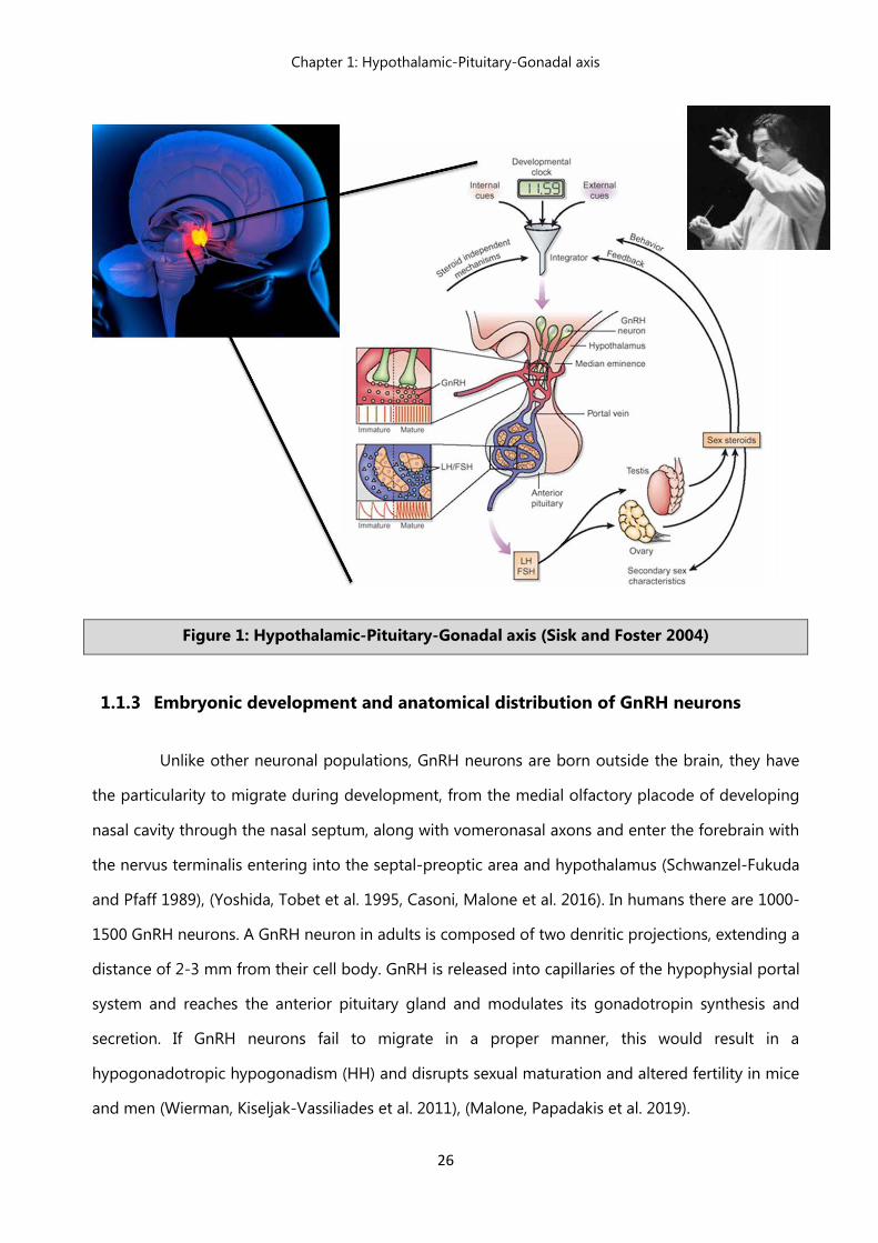
GnRH cells act as integrators of metabolic and reproductive signals, which come from both
the central and the peripheral nervous system. They are considered the main orchestrators of the
physiological events initiated from the hypothalamic region (Figure 1). Their proper development,
expression, and signaling is essential for sexual maturation and the normal functioning of the HPG
axis in mammals (Cattanach, Iddon et al. 1977), (Mason, Hayflick et al. 1986), (Prevot 2011).
Chapter 1: Hypothalamic-Pituitary-Gonadal axis
26
Figure 1: Hypothalamic-Pituitary-Gonadal axis (Sisk and Foster 2004)
1.1.3 Embryonic development and anatomical distribution of GnRH neurons
Unlike other neuronal populations, GnRH neurons are born outside the brain, they have
the particularity to migrate during development, from the medial olfactory placode of developing
nasal cavity through the nasal septum, along with vomeronasal axons and enter the forebrain with
the nervus terminalis entering into the septal-preoptic area and hypothalamus (Schwanzel-Fukuda
and Pfaff 1989), (Yoshida, Tobet et al. 1995, Casoni, Malone et al. 2016). In humans there are 1000-
1500 GnRH neurons. A GnRH neuron in adults is composed of two denritic projections, extending a
distance of 2-3 mm from their cell body. GnRH is released into capillaries of the hypophysial portal
system and reaches the anterior pituitary gland and modulates its gonadotropin synthesis and
secretion. If GnRH neurons fail to migrate in a proper manner, this would result in a
hypogonadotropic hypogonadism (HH) and disrupts sexual maturation and altered fertility in mice
and men (Wierman, Kiseljak-Vassiliades et al. 2011), (Malone, Papadakis et al. 2019).
Chapter 1: Hypothalamic-Pituitary-Gonadal axis
27
It is well established that GnRH migration is modulated by various molecules. Indeed, following
guidance cues given by extracellular molecules, receptor tyrosine kinases, chemokines and a series
of neurotransmitters among others (Wray 2010), GnRH neurons migrate toward the forebrain (Tobet
and Schwarting 2006, Wierman, Kiseljak-Vassiliades et al. 2011, Casoni, Malone et al. 2016) and
subsequently continue the migration ventrally, along a branch of the vomeronasal nerve to reach
the basal forebrain. Eventually, at postnatal day 0, GnRH neurons reach the hypothalamus, their final
destination.
During postnatal development, GnRH neurons are subjected to a series of complex maturational
events involving their morphological characteristics, neurosecretory pattern, and overall neuronal
activity.
In females, during neonatal period, primordial follicles, which are present at postnatal day 7, develop
into secondary follicles. At this stage, it is improbable that LH and FSH would exert direct actions on
primordial follicles as functional gonadotropin receptors haven’t developed yet (O'Shaughnessy,
McLelland et al. 1997). Furthermore, in 1992 Rajah et al., showed that P7 GnRH deficient
hypogonadal mice appear to have normal follicular development, thus, folliculogenesis does not
require GnRH or LH/FSH development at this stage.
During the infantile period, a series of endocrine events stimulate the development of the pool of
secondary follicles into preantral and antral follicles (McGee, Perlas et al. 1997) now responsive to
FSH (O'Shaughnessy, McLelland et al. 1997).The surge of FSH at P12 is the first activation of the
GnRH system and is required for proper maturation of the preantral follicles into antral follicles
(McGee, Perlas et al. 1997); (McGee and Hsueh 2000)Small bursts of secretion of LH are noticed;
however, they remain low and do not participate in the ovarian development during this stage
(Dohler and Wuttke 1974, Zhang, Poutanen et al. 2001).
As the juvenile stage begin and infantile period comes to an end, FSH levels decrease while LH levels
remain low. When juvenile stage nears its end, preovulatory levels of estradiol (E2) significantly
increase to reach a peak around postnatal day 30 (Andrews, Mizejewski et al. 1981, Devillers, Petit
et al. 2019) This increase in estradiol levels stimulates LH secretion, which acquires a particularly
accelerated pulsatile pattern (Kimura and Kawakami 1982). This phenomenon is believed to involve
Chapter 1: Hypothalamic-Pituitary-Gonadal axis
28
GnRH activation, evident by the increased GnRH pulse frequency occurring during the juvenile
period (Sarkar and Fink 1979).
1.2 GnRH pulsatile secretion
Once the GnRH neurons have completed their migration to the hypothalamus during
embryogenisis, secretion of GnRH by the hypothalamus increases in early postnatal life, leading to
temporary activation of gonadal steroidogenesis, especially in males. Before the onset of puberty,
the pulsatile GnRH release is suppressed which results in gonads remaining quiescent until the onset
of puberty. This is when they respond to exogenous gonadotropin stimulation after re-awakening
of pulsalite GnRH release.
GnRH secretion follows two modes: pulsatile and surge (Maeda, Ohkura et al. 2010)The
surge mode occurs only in females. Pulsatile mode was first demonstrated in ovariectomised rhesus
monkeys (Antunes, Carmel et al. 1978). This mode is the episodic release of GnRH, showing distinct
pulses of GnRH secretion into circulation, with undetectable GnRH concentrations during inter-pulse
intervals (Herbison 2018).
The activity of GnRH neurons is tightly coordinated, as they possess intrinsic electrical activity,
dependent on intracellular signaling and mechanisms. The “GnRH pulse generator” is suggested to
be located at the medial basal hypothalamus as the episodic multi-unit electric activity generates
from there (Wilson, Kesner et al. 1984) and it relies on complex relations between neurons containing
norepinephrine, dopamine, gamma aminobutyric acid (GABA) to state some. Glutamate and
norepinephrine are stimulators of the reproductive axis, while GABA is an inhibitor. Regulation of
GnRH pulsality is partly mediated by the Kisspeptin- Neurokinin B (NKB) -opioid pathway, by a
mechanism whereby NKB and dynorphin act autosynaptically on kisspeptin neurons in the arcuate
nucleus to synchronize and shape the pulsatile secretion of kisspeptin and drive the release of GnRH
from fibers in the median eminence.(Navarro, Gottsch et al. 2009), (Ruka, Burger et al. 2013, Ezzat,
Pereira et al. 2015) with which they colocalize (Kallo, Vida et al. 2012). The population of neuronal
nitric oxide synthase (nNOS) neurons has also been shown to play a crucial role in the regulation of
the GnRH system (Chachlaki, Malone et al. 2017)
Chapter 1: Hypothalamic-Pituitary-Gonadal axis
29
1.3 GnRH receptors
The GnRH receptor is a member of the rhodopsin-like G protein-coupled receptor
superfamily (GPCR). It is characterized by seven-transmembrane domain structure. In mammalian
GnRH receptor, the cytoplasmic C-terminal tail is absent (Cheng and Leung 2005) which is present
in non-mammalian GnRH receptors (Lethimonier, Madigou et al. 2004) . The human GnRH receptor
gene is found on chromosome 4 and it consists of three exons separated by two introns (Kakar
1997). GnRH receptors and splice variants have been discovered in human pituitary and pituitary
adenomas (Grosse, Schoneberg et al. 1997). The GnRH receptor has also been identified in the
ovarian cancer cell lines, endometrial carcinoma cell lines, liver, heart, muscle, kidney, peripheral
mononuclear cells, human corpus luteum, luteinized granulosa cells, melanoma, breast carcinoma
and syncytiotrophoblast cell layers of human placenta (Cheng and Leung 2002). Another study has
shown that GnRH and GnRH receptor mRNA are present in human testicular tissue (Bahk, Hyun et
al. 1995).
GnRH regulates the expression of GnRH receptor in the pituitary. Decline in GnRH
stimulation results in decline of GnRH receptors on pituitary gonadotrophs but subsequent exposure
of the pituitary to GnRH restores the levels of receptors. This upregulation is differentially controlled
by varying GnRH pulse frequencies and varies among species (Tsutsumi, Laws et al. 1995). In human
ovarian cancer and peripheral blood mononuclear cells, GnRH performs a biphasic effect on GnRH
receptor expression (Chen, Jeung et al. 1999, Kang, Choi et al. 2000). In contrast, GnRH receptor
expression is downregulated with continuous exposure to GnRH, resulting in inhibition of LH and
FSH synthesis and secretion.
GnRH receptor is suppressed by estradiol in the pituitary and extrapituitary tissues (Quinones-Jenab,
Jenab et al. 1996, Cheng, Chow et al. 2003). Likewise, progesterone inhibits GnRH receptor
expression in the pituitary (Laws, Beggs et al. 1990). On the other hand, activin stimulates GnRH
receptor synthesis (Fernandez-Vazquez, Kaiser et al. 1996). Additionally, it was also discovered in rat
testes that human chorionic gonadotropin (HCG) downregulates the GnRH receptor (Botte, Lerrant
et al. 1999)but increases GnRH receptor gene transcription in choriocarcinoma JEG-3 cells (Cheng
and Leung 2002); Thus, indicating a tissue specific effect.
Chapter 1: Hypothalamic-Pituitary-Gonadal axis
30
It is important to highlight that the mutations of the GnRH receptor gene lead to idiopathic
hypogonadotropic hypogonadism (HH) which is a rare disorder of sexual maturation characterized
by gonadotropin deficiency with low sex steroid levels associated with low levels of FSH and LH
(Topaloglu and Kotan 2016).
1.4 Gonadotropins
As mentioned above GnRH stimulates the synthesis and secretion of FSH and LH. These
gonadotropins are heterodimeric glycoprotein hormones which act on gonads, i.e., ovaries and
testes to control ovarian and testicular functions, respectively. In males, a high GnRH pulse induces
secretion of LH (Constantin 2011)which in turn stimulates testosterone production of Leydig cells
and plays a role in the maintenance of spermatogenesis through its paracrine action on Sertoli cells.
Low pulse frequency promotes secretion of FSH. Before the onset of puberty, FSH stimulates Sertoli
cell proliferation, defines the size of their population, the size of the testes and the quantity of
spermatogenesis (Sharpe, McKinnell et al. 2003). Hence, the main action of FSH is, alongside
testosterone, the augmentation of the sperm production (Huhtaniemi 2015). During the same time
frame (prior to puberty), a plethora of inhibitory input is sent to GnRH neurons, allowing them to
release very low concentrations of the GnRH peptide in the pituitary. Later during development, the
increase in the GnRH pulsatile release from the hypothalamus is stimulated by a combination of
excitatory and inhibitory inputs. This process is critical for the initiation of puberty (Plant 2015).
1.5 Establishment of GnRH afferent neuronal connections – the creation of a GnRH
network
GnRH neuronal projections are already in place at birth, however, a mature neuronal
network has yet to be formed. During the first 3 weeks of postnatal development, GnRH neuronal
projections seem to continue to develop (Buchanan and Yellon 1993), (Heywood and Yellon 1997).
By the second week of postnatal development, the axons of neurons that reside in the ARH have
been evidenced to reach GnRH neuronal soma and they continue to expand throughout the infantile
period (Bouret, Draper et al. 2004), (Caron, Ciofi et al. 2012). The maturation of ARH neuronal
Chapter 1: Hypothalamic-Pituitary-Gonadal axis
31
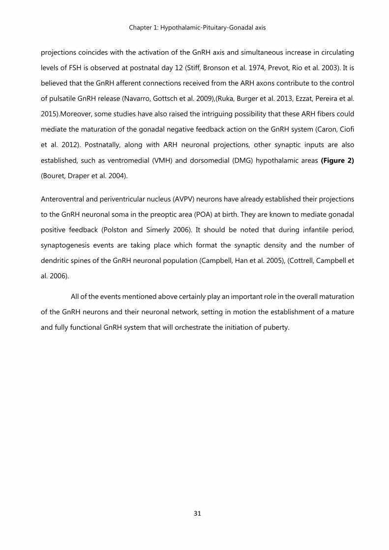
projections coincides with the activation of the GnRH axis and simultaneous increase in circulating
levels of FSH is observed at postnatal day 12 (Stiff, Bronson et al. 1974, Prevot, Rio et al. 2003). It is
believed that the GnRH afferent connections received from the ARH axons contribute to the control
of pulsatile GnRH release (Navarro, Gottsch et al. 2009),(Ruka, Burger et al. 2013, Ezzat, Pereira et al.
2015).Moreover, some studies have also raised the intriguing possibility that these ARH fibers could
mediate the maturation of the gonadal negative feedback action on the GnRH system (Caron, Ciofi
et al. 2012). Postnatally, along with ARH neuronal projections, other synaptic inputs are also
established, such as ventromedial (VMH) and dorsomedial (DMG) hypothalamic areas (Figure 2)
(Bouret, Draper et al. 2004).
Anteroventral and periventricular nucleus (AVPV) neurons have already established their projections
to the GnRH neuronal soma in the preoptic area (POA) at birth. They are known to mediate gonadal
positive feedback (Polston and Simerly 2006). It should be noted that during infantile period,
synaptogenesis events are taking place which format the synaptic density and the number of
dendritic spines of the GnRH neuronal population (Campbell, Han et al. 2005), (Cottrell, Campbell et
al. 2006).
All of the events mentioned above certainly play an important role in the overall maturation
of the GnRH neurons and their neuronal network, setting in motion the establishment of a mature
and fully functional GnRH system that will orchestrate the initiation of puberty.
Chapter 1: Hypothalamic-Pituitary-Gonadal axis
32
(i)
(ii)
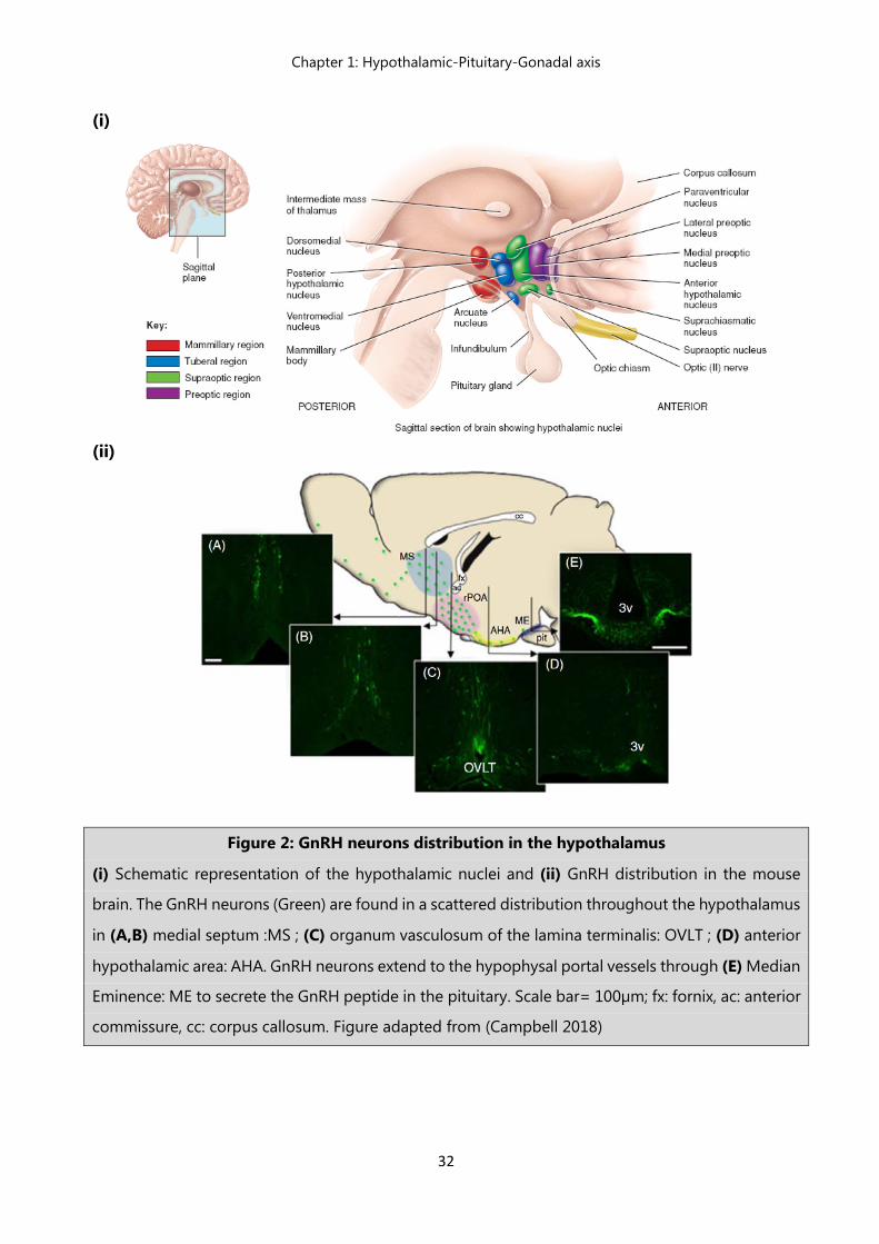
Figure 2: GnRH neurons distribution in the hypothalamus
(i) Schematic representation of the hypothalamic nuclei and (ii) GnRH distribution in the mouse
brain. The GnRH neurons (Green) are found in a scattered distribution throughout the hypothalamus
in (A,B) medial septum :MS ; (C) organum vasculosum of the lamina terminalis: OVLT ; (D) anterior
hypothalamic area: AHA. GnRH neurons extend to the hypophysal portal vessels through (E) Median
Eminence: ME to secrete the GnRH peptide in the pituitary. Scale bar= 100µm; fx: fornix, ac: anterior
commissure, cc: corpus callosum. Figure adapted from (Campbell 2018)
Chapter 1: Hypothalamic-Pituitary-Gonadal axis
33
1.6 Positive and negative feedbacks into the GnRH system
The secretion of estrogen occurs in the gonads during steroidogenesis, estrogen exert a
dual effect on the hypothalamic GnRH secretion. During follicular phase, rising plasma estradiol
levels result in a positive feedback action upon the GnRH network to stimulate the gonadotropin
surge. However, during later stages of the cycle, estradiol suppresses GnRH secretion by exerting its
negative feedback effect on the hypothalamic axis (Radovick, Levine et al. 2012) The surge of LH is
induced when significantly high estrogen levels arrive in the hypothalamus during the preovulatory
period (Christian and Moenter 2010). These high estradiol signals are responsible for the activation
of GnRH neurons and consequently the secretion of GnRH peptide. This initiates a chain reaction
that will eventually trigger ovulation (Moenter, Caraty et al. 1991). This stimulatory action of estrogen
consists the “positive gonadal feedback”, occurring during the late follicular phase in humans and
in the afternoon of proestrus in rodents and (Herbison 1998), (Simerly 2002). The regions where this
positive feedback takes place are following: anteroventral AVPV, preoptic area, and suprachiasmatic
nucleus (SCN) (Radovick, Levine et al. 2012). A number of studies suggest that neurons that express
the estrogen Receptor (ER-α) of the above region send direct inputs onto GnRH neurons which do
not possess the ER-α to mediate the estrogenic positive feedback action. Kisspeptin neurons are
believed to be one of these neuronal populations that are able to integrate positive estrogenic
signals (Adachi, Yamada et al. 2007), (Clarkson, d'Anglemont de Tassigny et al. 2008, Mayer, Acosta-
Martinez et al. 2010). Estrogen plasma levels, during the estrous cycle, inhibit GnRH secretion and
maintain GnRH neuronal pulse in low frequency levels (Levine 1997); (Herbison 1998). It is still
unclear through which mechanisms estradiol promotes its “negative feedback” action to retain GnRH
pulse frequency and amplitude but there are propositions that it involves ER-α- containing neurons
of the arcuate nucleus (AHN), as well as the ER-β-expressing neurons (Wersinger, Haisenleder et al.
1999), (Kwakowsky, Herbison et al. 2012).
Chapter 1: Hypothalamic-Pituitary-Gonadal axis
34
The GnRH system is a complex, yet elegantly coordinated network. Hence, alterations in the
migratory process or in the peptide secretion evoke heavy consequences on the reproductive
function and eventually lead to some of the major reproductive disorders in humans:
- Congenital hypogonadotropic hypogonadism (CHH), a condition characterized by failure of
sexual competence.
- Kallmann syndrome (KS), characterized by a combination of hypogonadotropic hypogonadism
and a deficiency of sense of smell.
- And finally, yet importantly, the Polycystic Ovary Syndrome (PCOS), a reproductive disorder in
we which an alteration of GnRH secretion and pulsatility is reported among other symptoms. In
contrast with the first two disorders characterized by GnRH deficiency, PCOS is characterized by
accelerated GnRH/LH secretion.
The next chapter of this thesis will be thus dedicated to the pathophysiology of PCOS and its main
clinical and biological manifestations.

35
Chapter 2 Polycystic ovary syndrome:
A complex disorder with multiple faces
Chapter 2: Polycystic Ovary Syndrome (PCOS)
36
Chapter Two
2 Polycystic ovary syndrome
2.1 History of the polycystic ovary syndrome
“Giovane rustica, maritata, modicamente pingue, et infeconda, con due ovaie più
grandi del normale, come uova di colomba, bernoccolute, lucenti et biancastre…“
Antonio Vallisneri, 1721
The first case description of the Polycystic Ovary Syndrome (PCOS) was provided by the Italian
scientist Antonio Vallisneri who reported in 1721: “ A young peasant woman, married, moderately
lump and infertile, with ovaries larger than normal, like doves´ eggs, lumpy, shiny and whitish… “
Many years later, Stein and Leventhal published in 1935 the case of seven women suffering from
amenorrhea, hirsutism and obesity along with a particular ovarian aspect, with a thick, white, pearly
cortex and small follicles, they probably did not suspect that they were describing for the first time
a disorder which is now known as one the most complex and prevalent reproductive disorder
affecting women worldwide.
PCOS is heterogeneous endocrinopathy with widely varying clinical manifestations. There is no
single criterion for the diagnosis of this syndrome. Traditionally, the PCOS diagnosis was based on
the histological examination of bilateral ovaries in women presenting anovulation, hirsutism or both
(Goldzieher and Green 1962). However, other co-morbidities such as a metabolic syndrome
including diabetes mellitus type 2, and depression are also associated with PCOS (Azziz, Carmina et
al. 2016).
Due to the variability of clinical manifestations (both the phenotypic and metabolic spectrum of
PCOS patients), a concise and consensual definition has long been lacking and is still very much at
the heart of the debate today.
Chapter 2: Polycystic Ovary Syndrome (PCOS)
37
The first formal attempt to consolidate a clinical definition of PCOS emerged in 1990, during a
conference sponsored by the National Institute of Health (NIH), when the majority of practitioners
recommended that diagnostic criteria should include evidence of hyperandrogenism (clinical or
biochemical) and ovulatory dysfunction in the absence of non-classic congenital adrenal hyperplasia
(CAH).
The second definition (Rotterdam) was presented in 2003 by the Fertility and Embryology
Association of Europe and America Fertility Society in Rotterdam conference in 2003 and has
considered two criteria from the following three criteria as parameters for diagnosis of PCOS after
having dismissed the other causes of hyperandrogenism (Rotterdam ESHRE/ASRM-
Sponsored PCOS consensus workshop group, 2004):
1. Oligo-ovulation or chronic anovulation;
2. Biological and/or clinical hyperandrogenism;
3. Presence of polycystic ovaries on pelvic ultrasound (more than 12 follicles measuring 2 to 9 mm
and ovarian volume greater than 10 mm).
Since 2003, no group of experts has been able to redefine the diagnostic criteria of this pathology
despite great efforts of experts and the evolution of biological assay or imaging techniques.
However, thanks to international clinical expertise and considerable joint efforts, international
guidelines and recommendations have been established to improve the management and
assessment of PCOS patients (Teede, Misso et al. 2018).
2.2 Physiopathology of PCOS
PCOS is a heterogeneous of symptoms which, gathered together, form a spectrum of a
disorder with a mild presentation in some cases and a severe disturbance of reproductive, endocrine
and metabolic function in other cases. In this introduction we will first highlight the major features
of PCOS endocrinopathy before reporting the additional heterogeneous manifestations of the
disorder which are not included in the diagnostic criteria but can typically be seen in PCOS patients.
Chapter 2: Polycystic Ovary Syndrome (PCOS)
38
2.2.1 Oligo-ovulation or chronic anovulation
The total number of follicles is determined early in life. Normal follicle development
comprises initial recruitment, by which primordial follicles start to mature, and cyclic recruitment,
which leads to the growth of a cohort of small antral follicles from which the dominant follicle,
destined to ovulate, is subsequently selected.
Disorders of ovulation account for about 30% of infertility and often present with irregular periods
(oligomenorrhoea) or an absence of periods (amenorrhoea). Polycystic Ovary Syndrome (PCOS) is
the most common cause of chorionic anovulation and anovulatory infertility (Wood, Dumesic et al.
2007, Dumont, Robin et al. 2015) and ovarian dysfunction continues to be the main feature which
makes this syndrome the major cause of anovulatory associated with infertility (Hamilton-Fairley and
Taylor 2003);
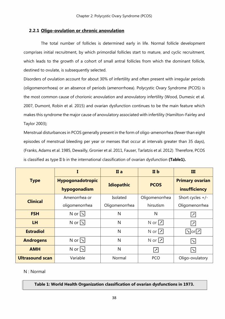
Menstrual disturbances in PCOS generally present in the form of oligo-amenorrhea (fewer than eight
episodes of menstrual bleeding per year or menses that occur at intervals greater than 35 days),
(Franks, Adams et al. 1985, Dewailly, Gronier et al. 2011, Fauser, Tarlatzis et al. 2012). Therefore, PCOS
is classified as type II b in the international classification of ovarian dysfunction (Table1).
N : Normal
Table 1: World Health Organization classification of ovarian dysfunctions in 1973.
Type
I II a II b III
Hypogonadotropic
hypogonadism Idiopathic PCOS
Primary ovarian
insufficiency
Clinical Amenorrhea or
oligomenorrhea
Isolated
Oligomenorrhea
Oligomenorrhea
hirsutism
Short cycles +/-
Oligomenorrhea
FSH N or ↘ N N ↗
LH N or ↘ N N or ↗ ↗
Estradiol N N or ↗ ↘or↗
Androgens N or ↘ N N or ↗ ↘
AMH N or ↘ N ↗ ↘
Ultrasound scan Variable Normal PCO Oligo-ovulatory
Chapter 2: Polycystic Ovary Syndrome (PCOS)
39
2.2.2 Hyperandrogenism
Hyperandrogenism is defined as an excessive secretion of the masculine sex steroids:
testosterone, dihydrotestosterone, and their prohormones dehydroepiandrosterone sulfate
(DHEAS), and androstenedione, this massive production of androgens induces a series of serious
clinical and biologicals manifestations.
It is well established that ovaries and adrenal glands are under control of luteinizing hormone and
adrenocorticotrophic hormone (ACTH), respectively, and contribute to the synthesis of sex steroids
(Handa and Weiser 2014).
The adrenal cortex synthesizes all the three major androgens; dehydroepiandrosterone sulfate,
androstenedione and testosterone, and this is the other major site of male androgen production,
besides the ovaries.
At puberty, the ovaries start to be active by synthesizing hormones (especially androgens)
on one hand and proceed with folliculogenesis on the other hand.
Excessive production of ovarian androgens is a hallmark of polycystic ovarian syndrome and
increased levels of circulating androgens appear in most series studying the syndrome.
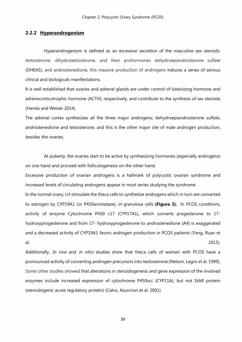
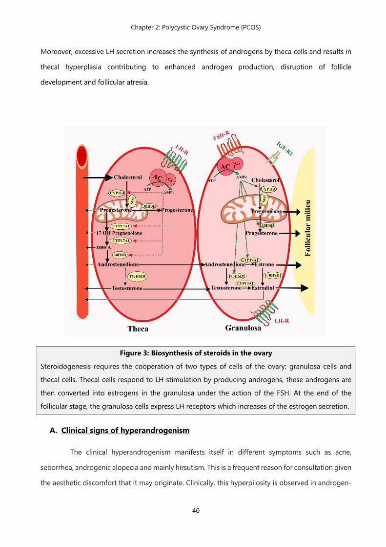
In the normal ovary, LH stimulate the theca cells to synthetize androgens which in turn are converted
to estrogen by CYP19A1 (or P450aromatase), in granulosa cells (Figure 3). In PCOS conditions,
activity of enzyme Cytochrome P450 c17 (CYP17A1), which converts progesterone to 17-
hydroxyprogesterone and from 17- hydroxyprogesterone to androstenedione (A4) is exaggerated
and a decreased activity of CYP19A1 favors androgen production in PCOS patients (Yang, Ruan et
al. 2015).
Additionally, In vivo and in vitro studies show that theca cells of women with PCOS have a
pronounced activity of converting androgen precursors into testosterone.(Nelson, Legro et al. 1999),
Some other studies showed that alterations in steroidogenesis and gene expression of the involved
enzymes include increased expression of cytochrome P450scc (CYP11A), but not StAR protein
(steroidogenic acute regulatory protein) (Calvo, Asuncion et al. 2001).
Chapter 2: Polycystic Ovary Syndrome (PCOS)
40
Moreover, excessive LH secretion increases the synthesis of androgens by theca cells and results in
thecal hyperplasia contributing to enhanced androgen production, disruption of follicle
development and follicular atresia.
Figure 3: Biosynthesis of steroids in the ovary
Steroidogenesis requires the cooperation of two types of cells of the ovary: granulosa cells and
thecal cells. Thecal cells respond to LH stimulation by producing androgens, these androgens are
then converted into estrogens in the granulosa under the action of the FSH. At the end of the
follicular stage, the granulosa cells express LH receptors which increases of the estrogen secretion.
A. Clinical signs of hyperandrogenism
The clinical hyperandrogenism manifests itself in different symptoms such as acne,
seborrhea, androgenic alopecia and mainly hirsutism. This is a frequent reason for consultation given
the aesthetic discomfort that it may originate. Clinically, this hyperpilosity is observed in androgen-
Chapter 2: Polycystic Ovary Syndrome (PCOS)
41
dependent zones: mainly in the face, thorax, back, inguinal hollows, inner and posterior faces of the
thighs.
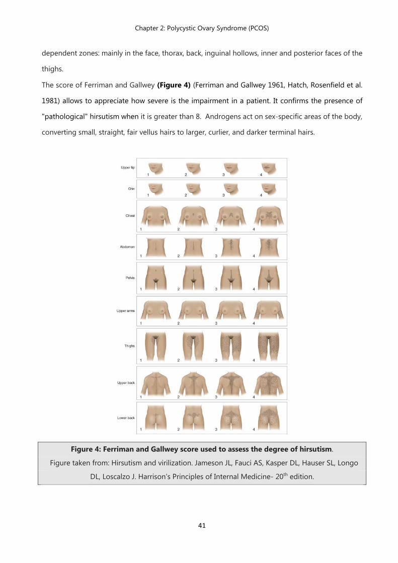
The score of Ferriman and Gallwey (Figure 4) (Ferriman and Gallwey 1961, Hatch, Rosenfield et al.
1981) allows to appreciate how severe is the impairment in a patient. It confirms the presence of
"pathological" hirsutism when it is greater than 8. Androgens act on sex-specific areas of the body,
converting small, straight, fair vellus hairs to larger, curlier, and darker terminal hairs.
Figure 4: Ferriman and Gallwey score used to assess the degree of hirsutism.
Figure taken from: Hirsutism and virilization. Jameson JL, Fauci AS, Kasper DL, Hauser SL, Longo
DL, Loscalzo J. Harrison’s Principles of Internal Medicine- 20th edition.
Chapter 2: Polycystic Ovary Syndrome (PCOS)
42
B. Biological signs of hyperandrogenism
Biological signs of hyperandrogenism typically include an increase of free and total testosterone:
testosterone circulates associated with Sex Hormone-Binding Globulin (SHBG) and other proteins
such as albumin.
Moderate elevation of total testosterone (<1.2 ng/mL) is the most commonly used confirm the
excess circulating androgens in PCOS patients. Nevertheless, it has been reported that the free
testosterone dosage (Azziz, Carmina et al. 2009) or the use of free Androgen Index FAI) [Total
Testosterone Ratio / SHBG x100] were more sensitive markers for highlighting biological
hyperandrogenism (Cussons, Stuckey et al. 2005, Azziz, Carmina et al. 2009).
The lack of a reproducible and reliable dosing method for free testosterone however limits the use
of this parameter to better appreciate the androgenic impregnation.
2.2.3 Polycystic appearing ovaries
Ultrasound scan has an important position in the diagnosis of PCOS as the same level as
the precedent criteria. Transabdominal and transvaginal ultrasound examination methods correlate
well, and both are acceptable tools for the diagnosis.
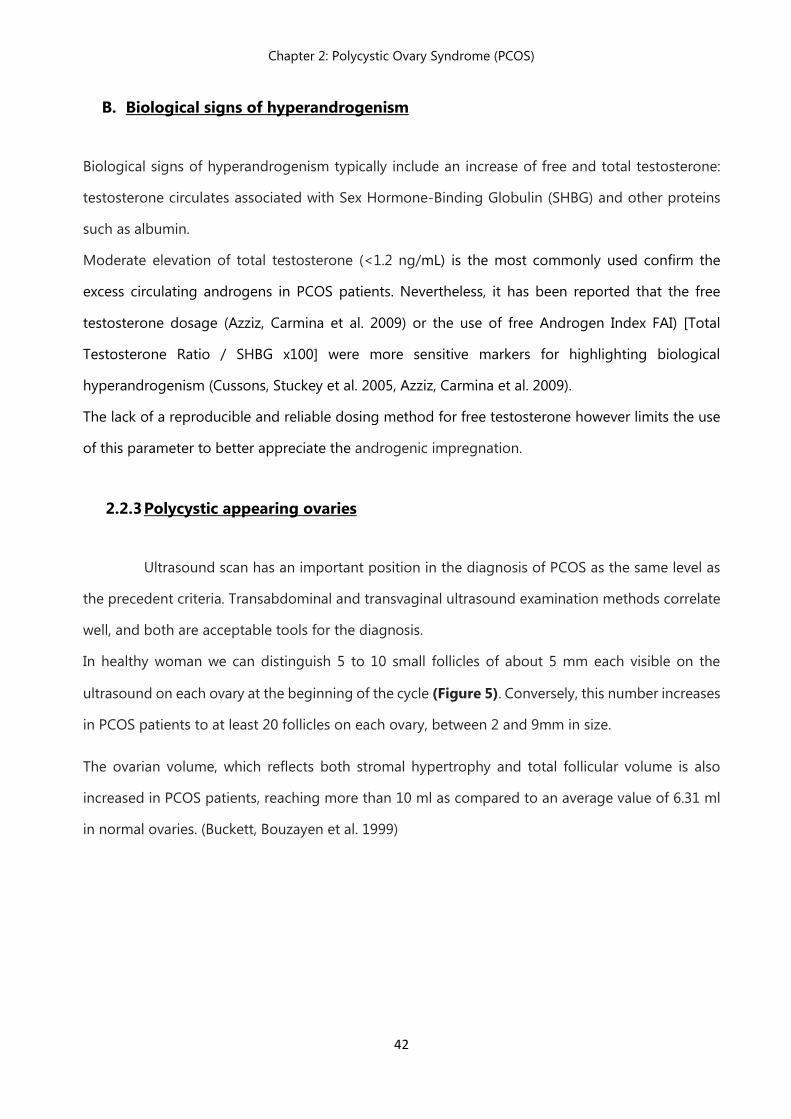
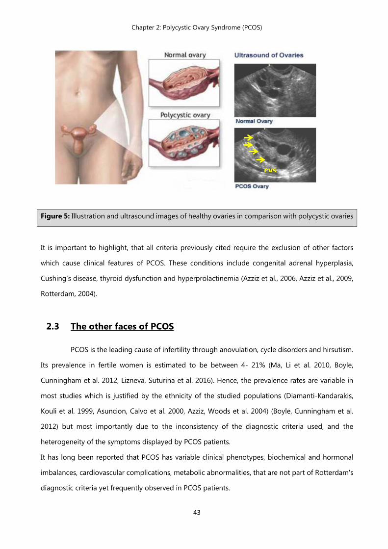
In healthy woman we can distinguish 5 to 10 small follicles of about 5 mm each visible on the
ultrasound on each ovary at the beginning of the cycle (Figure 5). Conversely, this number increases
in PCOS patients to at least 20 follicles on each ovary, between 2 and 9mm in size.
The ovarian volume, which reflects both stromal hypertrophy and total follicular volume is also
increased in PCOS patients, reaching more than 10 ml as compared to an average value of 6.31 ml
in normal ovaries. (Buckett, Bouzayen et al. 1999)
Chapter 2: Polycystic Ovary Syndrome (PCOS)
43
Figure 5: Illustration and ultrasound images of healthy ovaries in comparison with polycystic ovaries
It is important to highlight, that all criteria previously cited require the exclusion of other factors
which cause clinical features of PCOS. These conditions include congenital adrenal hyperplasia,
Cushing’s disease, thyroid dysfunction and hyperprolactinemia (Azziz et al., 2006, Azziz et al., 2009,
Rotterdam, 2004).
2.3 The other faces of PCOS
PCOS is the leading cause of infertility through anovulation, cycle disorders and hirsutism.
Its prevalence in fertile women is estimated to be between 4- 21% (Ma, Li et al. 2010, Boyle,
Cunningham et al. 2012, Lizneva, Suturina et al. 2016). Hence, the prevalence rates are variable in
most studies which is justified by the ethnicity of the studied populations (Diamanti-Kandarakis,
Kouli et al. 1999, Asuncion, Calvo et al. 2000, Azziz, Woods et al. 2004) (Boyle, Cunningham et al.
2012) but most importantly due to the inconsistency of the diagnostic criteria used, and the
heterogeneity of the symptoms displayed by PCOS patients.
It has long been reported that PCOS has variable clinical phenotypes, biochemical and hormonal
imbalances, cardiovascular complications, metabolic abnormalities, that are not part of Rotterdam's
diagnostic criteria yet frequently observed in PCOS patients.
cys
Chapter 2: Polycystic Ovary Syndrome (PCOS)
44
2.3.1 Hormonal imbalance
2.3.1.1 Alterations in gonadotropin secretion
A key characteristic of PCOS is inappropriate gonadotropin secretion. When compared with
the follicular phase of the normal menstrual cycle, women with PCOS exhibit a disproportionately
high LH secretion with relatively constant low FSH secretion. 40-60% PCOS women display an
increase in LH plasma levels (Balen, Conway et al. 1995). In PCOS, the increase in LH is explained by
an increased pulse frequency of the hypothalamic GnRH which may favor the production of the β-
subunit of LH over the β-subunit of FSH and/or by increased pituitary sensitivity to GnRH stimulation
which results in an elevated LH/FSH ratio increased in more than 75% of PCOS patients (McCartney,
Eagleson et al. 2002) (Waldstreicher, Santoro et al. 1988, Taylor, McCourt et al. 1997). The increase
in LH causes the ovaries to favor the production of androgens from the theca cells carrying LH
receptors. The dosage of LH may vary depending on the time of the cycle, the body mass index
(BMI) of the patient or the affinity of dosing kit used, which is why despite its informative value, its
elevation is not essential for a diagnosis of PCOS (Dewailly, Hieronimus et al. 2010, Conway, Dewailly
et al. 2014).
2.3.1.2 Increased AMH levels
Serum anti-müllerian hormone is synthesized by small antral follicles, which are precisely
the ones seen in ultrasound (Jeppesen, Anderson et al. 2013). Most studies report an increase in
circulating levels of AMH in PCOS patients, be 2 to 4 times higher in PCOS patients (Pigny, Merlen
et al. 2003, Laven, Mulders et al. 2004, Park, Lawson et al. 2010). Interestingly, the circulating AMH
levels correlate with the severity of the syndrome, through its direct action to control follicular
development, blocking the follicles’ maturation and assisting in the selection of a “dominant
follicle”, additionally, AMH behaves by its paracrine action on granulosa cells (GC) as a local
inhibitor of FSH action (Jonard and Dewailly 2004).Some clinicians have concluded that AMH can
be utilized as a diagnostic marker for ovarian hyperandrogenism (Dewailly et al., 2010).
Overall, most researchers agree that AMH should be considered as a marker for increased ovarian
reserve (Rosenfield, Wroblewski et al. 2012). Some recent studies have reported that AMH levels are

Chapter 2: Polycystic Ovary Syndrome (PCOS)
45
not only increased during adulthood in PCOS women, but that this elevation in AMH levels is also
observed during pregnancy in PCOS patients, whereas, AMH levels in pregnant healthy women
(Tata, Mimouni et al. 2018), (Valdimarsdottir, Valgeirsdottir et al. 2019).
2.3.2 Metabolic complications
The association between abdominal obesity, hyperinsulinaemia and insulin resistance
play a key role in the pathophysiology of PCOS and worsen both reproductive and long-term
health of patients suffering from PCOS. Burghen et al. were first to highlight a link between PCOS
and insulin resistance. They demonstrated that hyperandrogenism correlates with
hyperinsulinemia in obese PCOS women (Burghen, Givens et al. 1980). Interestingly, it seems that
there is chronological link between the onset of hyperandrogenism as well as dysovulation and the
inception of the metabolic disturbances (Figure 6), not only in PCOS patients but also first-degree
family members, (Yildiz, Yarali et al. 2003), this indicates that PCOS may be the presentation of a
complex genetic trait disorder.
Figure 6: The close association in PCOS, between early age reproductive disorders and long-
term metabolic disturbances.
Chapter 2: Polycystic Ovary Syndrome (PCOS)
46
2.3.2.1 Obesity
Evaluation of height and weight with calculation of Body Mass Index (BMI) along with the
measurement of the waist circumference, are essential to look for overweight or obesity. Obesity is
defined as excessive fat accumulation. According to BMI assessment we can distinguish the major
classifications: Healthy: BMI of 18.5 to <25 kg/m2; Overweight: BMI of 25 to 29 kg/m2; Obese: BMI
greater than 30 kg/m2.
Nearly 50% of PCOS patients are clinically obese, although this varies with ethnicity (Carmina, Legro
et al. 2003). However, the exact causative mechanism of this association remains unclear.
PCOS woman are predisposed to weight gain and in turn overweight seems to exacerbate the clinical
manifestations of PCOS (Teede, Joham et al. 2013, Shorakae, Boyle et al. 2014).
Clinical investigations have reported that oligo-ovulation and anovulatory infertility are more
frequent in obese PCOS patients in comparison with the lean ones (Teede, Joham et al. 2013). A
correlation between the severity of the ovulatory impairment and increased BMI is observed in this
syndrome (Teede, Joham et al. 2013).
Moreover, increased insulin resistance, glucose intolerance, hyperandrogenism and decreased SHBG
levels are commonly observed in obese PCOS patients as compared to lean PCOS patients (Norman,
Masters et al. 2001), (Rich-Edwards, Spiegelman et al. 2002). Likewise, complications during
pregnancy and risk of miscarriages are also emphasized with obesity in PCOS conditions (Pasquali
2014).
2.3.2.2 Hyperinsulinemia
Increased secretion of insulin is an extra-ovarian factor aggravating hyperandrogenism in
PCOS. Several studies have explored the mechanisms that could be implicated:
- At a gonadal level: during steroidogenesis, insulin has a direct stimulation action on androgen
production by thecal cells by stimulating synthesis and/or activity of enzymes involved in ovarian
steroidogenesis (Li, Chen et al. 2013).
- At a hepatic level: through its inhibitory properties on the synthesis of sex hormone binding
globulin (SHBG) in the liver, favoring free circulating bioactive androgens in the circulation.
Chapter 2: Polycystic Ovary Syndrome (PCOS)
47
Hyperinsulinemia also decreases the synthesis of insulin-like growth factor bindingprotein-1 (IGFBP-
1), allowing more IGF-1 to be available both locally and peripherally to stimulate ovarian
steroidogenesis (Janssen, Stolk et al. 1998).
- At pituitary level: insulin seems to influence LH hypersecretion from pituitary by increasing LH pulse
amplitude or potentiate the effects of LH on ovarian steroidogenesis, inducing hyperandrogenism
(Allon, Leach et al. 2005), (Adashi, Hsueh et al. 1981).
2.3.2.3 Insulin resistance
Insulin resistance is defined as an impaired stimulation of glycogen formation in all major
target tissues (skeletal muscle, adipose tissue, liver, kidney), and is frequently associated with PCOS.
In fact, up to 90% of women with PCOS display an acute insulin resistance, surpassing the prediction
obtained by their BMI (Dunaif, Segal et al. 1989). Even if, one would think that lean women would
be spared from these metabolic complications, yet PCOS women with a lean phenotype also exhibit
severe insulin resistance compared with BMI-matched control women (Dunaif 1999).
Insulin resistance causes compensatory hyperinsulinemia and putative origins of this IR in PCOS
include abnormalities in the insulin receptor signaling and enhanced cytokine actions(Escobar-
Morreale, Luque-Ramirez et al. 2011, Diamanti-Kandarakis and Dunaif 2012). Moreover, Andrisse et
al. reported an enhanced insulin resistance of reproductive tissues (ovaries and pituitary) in presence
of androgens in female rodents (Andrisse, Billings et al. 2018).
2.3.3 Cardiovascular pathology
While an association between PCOS and increased cardiovascular risk is still at the heart
of the debate (Rizzo, Berneis et al. 2009, Papadakis, Kandaraki et al. 2017), strong findings show that
postmenopausal PCOS women with a personal history of clinical hyperandrogenism or menstrual
disorders have more cerebrovascular events as well as increased cardiovascular morbidity as
compared to healthy women (Shaw, Bairey Merz et al. 2008) (Krentz, von Muhlen et al. 2007).
Chapter 2: Polycystic Ovary Syndrome (PCOS)
48
2.3.4 Pregnancy complications
Most PCOS women struggle to become pregnant and once they succeed to conceive, they
often encounter complications during pregnancy and delivery (Legro, Castracane et al. 2004), (Wild
2002). Women with PCOS have a three times higher risk to miscarriage in the early months
of pregnancy as compared to women without PCOS (Palomba, de Wilde et al. 2015).
A meta-analysis involving 720 PCOS women and 4505 controls, highlighted increased prevalence in
PCOS women of gestational diabetes, gestational hypertension, preeclampsia and preterm births. In
addition, the infants of PCOS women were more often admitted to a neonatal intensive care unit
and the perinatal mortality was higher (Boomsma, Eijkemans et al. 2006).
2.3.5 Gynecological malignancies
Growing evidence show that endometrial cancer is considerably higher in women with
PCOS (Wild, Pierpoint et al. 2000), (Haoula, Salman et al. 2012), (Dumesic and Lobo 2013). The
chronic anovulation observed in PCOS conditions, could be involved in the onset of endometrial
cancer, due to prolonged exposure to unopposed estrogens, leading to endometrial hyperplasia.
However, other co-founding factors such as: progesterone dependent gene dysregulation,
hyperandrogenism, hyperinsulinemia and/or LH hypersecretion could also contribute to a higher
risk of endometrial cancer (Dumesic and Lobo 2013).
Nevertheless, there is insufficient evidence to implicate PCOS in the development of breast, vaginal,
cervical or ovarian cancers.
2.3.6 Psychological disorders
PCOS is a life-long disease, not exclusively associated to reproductive and metabolic
disturbances as it may also impact the quality of life and mental health of affected women.
An increased prevalence of depressive and anxiety symptoms has been reported in several studies
(Barry, Kuczmierczyk et al. 2011, Dokras, Clifton et al. 2012) (Veltman-Verhulst, Boivin et al. 2012).
Moreover, other studies indicate that women with PCOS and their children have a greater risk of
autism (Cherskov, Pohl et al. 2018).
49
Chapter 3 Progress towards understanding PCOS etiology
Chapter 3: Progress towards understanding PCOS etiology
50
Chapter Three
The etiology of PCOS is not fully understood and the mechanisms underpinning the onset
of disease in women remain elusive, however some studies suggest a genetic basis involved in the
onset of the disease.
3.1 Genetic determinants & the genesis of PCOS
Phenotypic presentation of PCOS may be driven by heritable alleles and their association
with environmental factors. In 1988 Hague et al., suggested PCOS as an autosomal dominant
inheritance disorder with prevalence of 40-50% between first-degree family relatives (Hague, Adams
et al. 1988). A study performed in an Australian monozygotic and dizygotic twin cohort led to
question the autosomal dominant inheritance of PCOS as there was stark difference between
hyperandrogenic and non-hyperandrogenic PCOS women (Jahanfar, Eden et al. 1995). A larger twin
study using quantitative estimation of genetic influence showed a strong genetic contribution with
a heritability rate of 79% for PCOS prevalence according to the Rotterdam criteria (Vink, Sadrzadeh
et al. 2006). Therefore, it is unlikely that PCOS results from a single autosomal genetic determinant.
Genome-wide association studies (GWAS) may provide mechanistic values to understand
the development of PCOS by attempting to give genetic associations. Eleven candidate genes were
elucidated in chinese population by two initial large GWAS which analyzed risk loci for developing
PCOS (Chen, Zhao et al. 2011, Shi and Vine 2012). PCOS association signals were found in DENND1A
loci, which is involved in trafficking of endosomes and androgen production (Tee, Speek et al. 2016);
Thyroid adenoma associated (THADA) gene intronic region, a gene linked with thyroid cancers,
energy balance and risk for type 2 diabetes (Kloth, Belge et al. 2011); (Moraru, Cakan-Akdogan et al.
2017) and variants in FSH gene receptor (FSH-R), critical for folliculogenesis and fertility (Simoni,
Tempfer et al. 2008).
In 2015, Day et al., published a comprehensive GWAS that further elucidated genetic genesis of
PCOS and its neuroendocrine correlates. In this study, more than 5,000 Caucasian European women
were evaluated and dense imputation of genotypes was used to identify association signals and
draw mechanistic interpretations from the genetic data. Among the six genetic loci which were
Chapter 3: Progress towards understanding PCOS etiology
51
discovered to be associated with PCOS were the epidermal growth factor receptor (EGFR) family
members; and FSHB (beta subunit of FSH). Furthermore, this study shed a light on polymorphism
which could lead to an alteration of FSH secretion and signaling, thus participating in
neuroendocrine genesis of PCOS. It is important to note that EGFR family members are involved, not
only, in LH-mediated steroidogenesis (Park, Su et al. 2004) and ovulation but also in brain circuitry
development (Namba, Nagano et al. 2017). With the help of Mendelian randomization analysis, it
was identified that body weight, insulin resistance and low levels of sex-hormone-binding globulin
(SHBG) are among the risk factors of PCOS. The study also demonstrated a strong and positive
association between the risk of having PCOS and delayed menopause, which suggested an
attenuation of ovarian ageing in these women (Day, Hinds et al. 2015). This data linked with the
hypotheses proposed earlier that a genetic predisposition to delayed reproductive senescence in
PCOS would support the evolutionary paradox of being highly prevalent disease that impairs female
reproductive competence (Abbott, Dumesic et al. 2002); (Casarini and Brigante 2014).
Despite a consensus on PCOS not being caused by one single determinant, there exists a
strong association between exposure to environmental factors (such as: excess prenatal androgen)
and the presentation of some of features of the disease after the onset of puberty. In human, in
utero excess of androgen caused by congenital adrenal hyperplasia (CAH) and maternal Cushing’s
disease leads to clinical manifestation of PCOS in adulthood. Similarly, female offspring’s of PCOS
women, presenting higher levels of circulating testosterone during pregnancy than control subjects,
show signs of ovarian and metabolic dysfunction during pubertal development and poses a higher
risk of developing PCOS in adulthood (Crisosto, Codner et al. 2007), (Sir-Petermann, Codner et al.
2009) Furthermore, higher steroidogenic activity is exhibited in placental tissue of mothers with
PCOS which in turn increases androstenedione levels and lowers P450 aromatase activity, which is
critical in converting androgens into estrogens (Maliqueo, Lara et al. 2013). As a result, the altered
placental steroidogenesis in PCOS women could sustain elevated intrauterine androgen levels
during pregnancy and affect the development of female offspring. Taken altogether, the data leads
to a prenatal androgen programming as potential mechanism to drive PCOS features later.
Two hypotheses exist on how the developmental programming generates the reproductive
impairment of PCOS if the syndrome is thought to have in utero origin. The first one explains that
Chapter 3: Progress towards understanding PCOS etiology
52
ovarian dysfunction as the initiator of the disease with the androgen imprinting in the ovaries
triggering an abnormal secretion of testosterone with the onset of puberty (Gilling-Smith, Willis et
al. 1994), (Rosenfield and Bordini 2010). High levels of testosterone may lead to modification of HPG
axis to PCOS symptoms. The second explanation sets the brain as the primary site of PCOS. During
perinatal life, hypothalamic circuits are highly responsive to sex steroid hormone
modulation(McCarthy, Nugent et al. 2017). Consequently, prenatal androgen may act to alter the
organization of components within GnRH the neuronal network and ultimately lead to abnormal
GNRH/LH pulsality and ovulatory dysfunction (Blank, McCartney et al. 2006), (Moore and Campbell
2016). It is important to note that one hypothesis does not exclude the possibility of the other and
that both could happen concomitantly in PCOS.
Various studies have shown that PCOS is hereditable (Azziz and Kashar-Miller 2000); and
have underlined targeted genes, dividing them into three categories: genes encoding androgen
production and metabolism, genes responsible for the secretion and action of insulin, and genes
related to folliculogenesis. The first category involves genes that are linked to androgen secretion,
such as LH and its receptor or genes that linked to androgen production like CYP19. The second
group of genes is related to insulin and its receptor. Lastly, the third group of genes is linked to
ovaries (Franks, Gharani et al. 2001). Other polymorphisms have been found in AMH and its receptor
AMH type 2 in connection with follicle development (Georgopoulos, Karagiannidou et al. 2013).
The evidence of a genetic component is based on familial clustering (Legro, Driscoll et al. 1998),
(Diamanti-Kandarakis, Kandarakis et al. 2006) and twin studies have shown two-fold increase in
concordance of PCOS in genetically identical twins compared to non-identical twins (Vink,
Sadrzadeh et al. 2006).The way PCOS is inherited remains unclear, despite numerous association
studies (focusing on genes associated with synthesis and metabolism of androgens and insulin.
Modern mapping techniques have managed to make some progress to identify targeting genes,
two of which are: locus on chromosome 19p13.2, associated with high susceptibility to PCOS
(Urbanek, Woodroffe et al. 2005) and fat-mass and obesity associated gene, whose polymorphism
has have been found to be associated with PCOS (Barber, Bennett et al. 2008). However, these two
candidate genes need to be studied further to be confirmed in larger studies and various
populations.
Chapter 3: Progress towards understanding PCOS etiology
53
There is a strong genetic component linked to PCOS which is evidenced by clustering PCOS in
families as well as PCOS-like feature in male and female relatives of affected women (Goodarzi,
Dumesic et al. 2011).Twin studies and linkage studies have been used to decipher the contribution
of heritability in this multifactorial syndrome. Linkage studies have been carried out for 37 candidate
genes and the results predicted strong association of follistatin and nominal association of CYP11A1
gene in affected siblings with hyperandrogenemia and PCOS related traits. Furthermore, it was
established by transmission disequilibrium test, by this same study, that there existed a strong
genetic association of D19S884 allelic marker near INSR gene with PCOS (Urbanek, Legro et al. 1999).
Numerous association studies were also carried out on candidate genes that are involved
in pathways related to the etiology of the syndrome. The associated anomalies to this syndrome
have pushed the research community to try and pinpoint the importance of genetic predisposition
in manifestation of PCOS (Prapas, Karkanaki et al. 2009). Although, polymorphisms of genes involved
in pathways such as gonadotropin regulation, insulin signaling, chronic inflammation, and energy
homeostasis has been studied (Prapas, Karkanaki et al. 2009); however, the exact role of these
susceptible genes has not yet been established. Single nucleotide polymorphisms (SNPs) have been
able to uncover functional changes caused by modulation of gene expression or amino acid
variation. Other approaches such as searching for candidate gene are helpful in reveal the impact of
differential frequency distribution in healthy and diseased population. Although, this is possible in
relatively smaller populations, thus, genome-wide association studies (GWAS) have revolutionized
the study of PCOS genetics. Hyperandrogenism is a hallmark of the PCOS condition, therefore,
various polymorphisms in genes involved in androgen synthesis, action, and bioavailability have
been and are being studied.
CYP11A1 Gene. CYP11A1 encodes the enzyme P450 cholesterol side-chain cleavage. The
main function of this enzyme is that it catalyzes the rate limiting step of ovarian steroidogenesis: the
conversion of cholesterol to pregnenolone (Miller 2002), (Pusalkar, Meherji et al. 2009) Two studies
showed that theca cells derived from PCOS ovaries and propagated in long-term culture
demonstrate increased CYP11A expression in comparison to normal theca cells (Nelson, Legro et al.
1999) (Wickenheisser, Biegler et al. 2012) Twenty families were chosen to perform a linkage study
which showed involvement of CYP11A locus in PCOS development and consequently association of
Chapter 3: Progress towards understanding PCOS etiology
54
5’UTR (TTTTA) pentanucleotide repeats in hirsute PCOS women (Gharani, Waterworth et al. 1997).
This was further confirmed by another study from South India (Reddy, Deepika et al. 2014) United
States (Daneshmand, Weitsman et al. 2002), Greece (Diamanti-Kandarakis, Bartzis et al. 2000) and
nominally in women from United Kingdom (Gaasenbeek, Powell et al. 2004). In Chinese women, it
was discovered that different allele combinations may increase or decrease the risk of PCOS (Wang,
Wu et al. 2006).
However, there are studies that found no association between pentanucleotide repeat alleles with
PCOS susceptibility in Spanish (San Millan, Sancho et al. 2001), Chinese (Li and Guijin 2005), 2005;
(Hao, Bao et al. 2009) Argentinian (Perez, Cerrone et al. 2008), Indian (Pusalkar, Meherji et al. 2009)
and Czech (Prazakova, Vankova et al. 2010) women with PCOS. A recent meta-analysis confirmed
strong association of this (TTTTA) repeat polymorphism of CYP11A with increased risk of PCOS in
Caucasian population (Shen, Li et al. 2014).
There are conflicting reports in regards to the association of these pentanucleotide repeats with
PCOS related traits. Carriers of short alleles in women with PCOS have been reported to possess
increased testosterone levels (Pusalkar, Meherji et al. 2009); (Diamanti-Kandarakis, Bartzis et al. 2000)
while no effect of allele dose was seen on CYP11A transcription (Daneshmand, Weitsman et al. 2002)
or serum androgen levels in another studies (Daneshmand, Weitsman et al. 2002); (Gaasenbeek,
Powell et al. 2004); (Li and Guijin 2005).
A significant association was shown in Chinese women with PCOS with the polymorphism rs4077582
(Gao, Cao et al. 2010); (Zhang, Zhu et al. 2009) as well as altered testosterone and LH levels (Zhang,
Zhu et al. 2009). The data from these studies implies CYP11A to be a promising genetic biomarker
for PCOS.
CYP17 Gene. The CYP17 gene encodes cytochrome P450 enzyme with 17-hydroxylase
activity. This enzyme converts pregnenolone and progesterone into 17-hydroxypregnenolone and
17-hydroxyprogesterone, respectively. The lyase activity converts these steroids to
dehydroepiandrosterone (DHEA) and 4-androstenedione (Prapas, Karkanaki et al. 2009).
The majority of studies have focused on a widely studied polymorphism in the promotor
at −34 position (−34 T/C), which creates an additional Sp1 transcription factor binding site, thus
Chapter 3: Progress towards understanding PCOS etiology
55
regulating expression of CYP17 and consequently androgen levels (Sharp, Cardy et al. 2004). A study
showed significant association of this polymorphism with PCOS and male pattern baldness in a
family-based study (Carey, Waterworth et al. 1994); however, these findings were not persistent
when they increased the sample size (Gharani, Waterworth et al. 1996)), nor was this polymorphism
found to be a significant factor for PCOS development in British (Techatraisak, Conway et al. 1997),
Polish (Marszalek, Lacinski et al. 2001) American (Kahsar-Miller, Boots et al. 2004), Korean (Park, Lee
et al. 2008) Chilean (Echiburu, Perez-Bravo et al. 2008) Chinese (Li, Gu et al. 2015), Thai (Techatraisak,
Chayachinda et al. 2015) and Indian (Banerjee, Dasgupta et al. 2016) women with PCOS or even in
Turkish adolescents (Unsal, Konac et al. 2009). A meta-analysis that took all studies into
consideration revealed that this variant was not associated with risk of PCOS development when any
genetic model or stratification by country or ethnicity was considered.
CYP19 Gene. This gene encodes aromatase P450 enzyme which is essential for synthesis
of estrogen from androgens (Bulun, Takayama et al. 2004). In both lean and obese women with
PCOS, reduced aromatase activity has been seen (Chen, Shen et al. 2015) which is further inhibited
by hyperandrogenemia (Medeiros, Barbosa et al. 2015) Yang et al. have shown that decreased
aromatase expression is associated with increased levels of testosterone in follicular fluid derived
from PCOS women (Yang, Ruan et al. 2015).PCOS ovaries present promoter hypermethylation and
reduced CYP19A1 mRNA and protein levels, which suggests repressed aromatase expression (Yu,
Sun et al. 2013) Moreover, a study in Han Chinese women demonstrated that an intronic variant
rs2414096 was significantly associated with increased risk of PCOS development and with increased
estradiol to testosterone ratio (E2/T), FSH levels, and age of menarche (Jin, Sun et al. 2009). In
adolescent girls in the UK carrying this polymorphism, it was observed that they possessed raised
PCOS symptom score and changes in circulating estradiol and testosterone concentrations (Petry,
Ong et al. 2005). There was no association of the rs2414096 polymorphism with PCOS or with
alterations in hormonal and metabolic variables after a 6-month treatment regime of oral
contraceptives in both ovulatory and anovulatory PCOS women (Maier and Spritzer 2012).The
tetranucleotide repeat polymorphism (TTTA) in the fourth intron (Xita, Chatzikyriakidou et al. 2010)
has been investigated. Results indicate that short allele repeats, predominantly consisting of seven
repeats, are prevalent in Greek (Xita, Lazaros et al. 2010) (Xita, Georgiou et al. 2008) and Han Chinese
Chapter 3: Progress towards understanding PCOS etiology
56
(Hao, Zhang et al. 2010) women with PCOS compared to healthy women without PCOS. These short
repeat alleles were associated with hormonal parameters such as increased testosterone levels, high
LH:FSH ratios (Xita, Chatzikyriakidou et al. 2010).
Chinese women with PCOS were shown to be carriers of 11 repeat alleles (Hao, Zhang et al. 2010);
(Xu, Zhang et al. 2013) which in turn influence lipid metabolism (Hao, Zhang et al. 2010).Although
the polymorphism, rs2470152, did not affect PCOS risk but the heterozygous TC genotype was found
to be significantly associated with increased testosterone levels with decreased E2/T ratio, which
suggested a role of this polymorphism in regulating aromatase activity (Zhang, Zhang et al. 2012).
Arg264Cys, a missense polymorphism, increases aromatase activity and affects PCOS susceptibility
(Wang, Li et al. 2011). However, the above findings indicate a definite role of this gene in PCOS
outcome.
AR Gene. Located on chromosome X, the androgen receptor (AR) gene encodes the AR,
which consists of a poorly conserved N terminal domain containing highly polymorphic CAG repeats
(Peng, Xie et al. 2014). In a case study, it was reported that the enfant of a woman carrying a
heterozygous AR gene mutation was diagnosed with androgen insensitivity syndrome suggesting
plausible repercussions on reproductive outcomes associated with AR gene mutations (Nam, Kim et
al. 2015) and not only with repeat lengths. The primary location of AR has been in the granulosa
cells of preantral and antral follicles, theca interna cells of preantral follicles, and both theca and
granulosa cells of dominant follicles (Walters, Allan et al. 2008). In Chinese and Caucasian
populations, it has been ascertained that short AR CAG repeats are more frequent in PCOS cases
and may possibly be linked to PCOS onset (Wang, Li et al. 2011); (Lin, Baracat et al. 2013)e; (Schuring,
Welp et al. 2012) ; (Shah, Antoine et al. 2008) (Xia, Che et al. 2012) and perhaps contributing to the
inherent hyperandrogenic phenotype commonly seen in women with PCOS by enhancing androgen
sensitivity to even low circulating levels of testosterone and increasing AR activity, which promotes
hirsutism, acne, and irregular cycles (Schuring, Welp et al. 2012); Anovulatory normoandrogenic
PCOS women showed a significant trend towards short CAG repeat length indicating increased
intrinsic androgen sensitivity (Mifsud, Ramirez et al. 2000).Moreover, it was found that Indian women
showed comparatively shorter repeat lengths compared to Chinese women, implying that ethnic
variation might play a possible role. No association of AR CAG repeat lengths with PCOS was
Chapter 3: Progress towards understanding PCOS etiology
57
reported in Indian (Rajender, Carlus et al. 2013), Slovene (Ferk, Perme et al. 2008) Korean (Kim (Kim,
Choung et al. 2008), and Croatian women (Skrgatic, Baldani et al. 2012). Some studies have indicated
that CAG repeat lengths may also modify both testosterone and insulin resistance parameters in
women with PCOS although, they have failed to show association with PCOS risk. It is interesting to
note that this CAG repeat polymorphism was a significant predictor of serum circulating
testosterone levels in Croatian (Skrgatic, Baldani et al. 2012), Brazilian (Ramos Cirilo, Rosa et al. 2012),
Chinese (Peng, Xie et al. 2014) and Korean (Kim, Choung et al. 2008) women with PCOS. German
women carrying short CAG repeats possessed increased testosterone which aggravated insulin
resistance in these women suggesting a putative effect of CAG repeats as an underlying mechanism
of hyperandrogenism induced insulin resistance (Mohlig, Jurgens et al. 2006). Meta-analyses, which
have examined the relationship between CAG repeat lengths at AR and PCOS risk, have concluded
that they may not be major determining factors in PCOS etiology (Rajender, Carlus et al. 2013),
(Wang, Goodarzi et al. 2012). Collectively, the data from these studies suggest that AR
polymorphisms may exacerbate the hyperandrogenic phenotype of women with PCOS.
SHBG Gene. Sex hormone binding globulin (SHBG), primarily synthesized in the liver, binds
androgens and estrogen. This lowers the circulating steroid hormones and renders them biologically
unavailable to target tissues (Hammond 2016). Chromosome 17 contains several polymorphisms in
the SHBG and they have been shown to alter hepatic biosynthesis, plasma levels, and plasma
clearance efficiency of SHBG, thus regulating the distribution of sex steroid hormones (Hammond
2016).
A woman showing severe SHBG deficiency helped uncover two novel coding region mutations. One
of them resulted in truncated SHBG synthesis and the other to abnormal glycosylation; leading to
extremely low levels of SHBG with elevated circulating free testosterone concentrations (Hogeveen,
Cousin et al. 2002) . Xita et al., provided evidence for the hypothetical genetic contribution of SHBG
polymorphisms which demonstrated an association between longer TAAAA repeats with late onset
of menarche and decreased SHBG levels in hirsute French women (Cousin, Calemard-Michel et al.
2004). It was further shown that long TAAAA repeat alleles failed to be associated with PCOS risk in
Croatian (Baldani, Skrgatic et al. 2015) Slovenian (Ferk, Teran et al. 2007), French (Cousin et al., 2004),
and Chinese (Zhao, Chen et al. 2005) women; whereas, Greek women with PCOS had significantly

Chapter 3: Progress towards understanding PCOS etiology
58
greater frequency of long repeat alleles compared to controls (Xita, Tsatsoulis et al. 2003). It has
been established that there exists an inverse association between TAAAA repeat polymorphism
alone (Cousin, Calemard-Michel et al. 2004), (Baldani, Skrgatic et al. 2015), (Ferk, Teran et al. 2007),
(Xita, Tsatsoulis et al. 2003) or coupled with short AR CAG repeats (Xita, Georgiou et al. 2008) and
SHBG serum levels. Greek women with PCOS having long SHBG alleles coupled with short CYP19
alleles resulted in low SHBG levels and increased testosterone levels with raised FAI, DHEAS and T/E2
ratios (Xita, Georgiou et al. 2008).
A recent study discovered that in Bahraini women, haplotypes spanning six polymorphisms were
associated with either increased or decreased PCOS susceptibility (Abu-Hijleh, Gammoh et al. 2016).
This study rekindled interest in SHBG gene polymorphisms in PCOS susceptibility.
StAR Gene. Located on chromosome 8, it encodes the steroidogenic acute regulatory
protein which binds to and facilitates uptake of cholesterol into mitochondria of cells for
steroidogenesis. Despite its function, a pilot study carried out in Iranian women investigated seven
known polymorphisms but showed no conclusive association with PCOS risk (Nazouri, Khosravifar
et al. 2015).
HSD17B5 Gene. In theca cells and adrenal glands, the enzyme type 17𝛽𝛽-hydroxysteroid
dehydrogenase type 5 (HSD17B5) is vital in converting androstenedione to testosterone (Ju, Wu et
al. 2015). Revealed for the first time by Qin et al., the −71A/G polymorphism in the promoter region
was studied for its prevalence in a population of ethnically diverse PCOS women. It was found that
this polymorphism is associated with PCOS susceptibility in Caucasian but not in African American
women with PCOS (Qin, Ehrmann et al. 2006). Furthermore, this variant modulated testosterone
biosynthesis and thus plasma testosterone levels (Qin, Ehrmann et al. 2006). However, subsequent
studies failed to find this association (Marioli, Saltamavros et al. 2009) in women with PCOS. Another
intronic polymorphism rs12529 in Chinese women affected testosterone levels but PCOS risk
remained unchanged.
INSL3 Gene. Insulin-like factor 3 (INSL3) is localized in the thecal cells and corpus luteum
of the ovary. The role of INSL3-RXFP2 signaling in maintaining androgen production by theca cells
was established by Glister et al. (Glister, Satchell et al. 2013) It has been reported recently that women
Chapter 3: Progress towards understanding PCOS etiology
59
with PCOS have increased serum INSL3 levels (Anand-Ivell, Tremellen et al. 2013), (Gambineri, Patton
et al. 2011) (Szydlarska, Grzesiuk et al. 2012) implicating INSL3 polymorphisms may have an
important role in modulating ovarian steroidogenesis and hence contribute to the pathogenesis of
PCOS. Additionally, Shaikh et al. led a first a case-control association study, investigated the link
between INSL3 polymorphisms and its haplotypes with PCOS susceptibility and its related traits in a
well characterized cohort of Indian women with PCOS (Shaikh, Dadachanji et al. 2016). In this study,
they showed that the A/G polymorphism present in exon 1 of INSL3: rs6523 was significantly
associated with PCOS susceptibility.
However, despite intensive investigations , the genes identified only account for 10% of
the 70% estimated heritability implying that environmental factors such as androgen excess could
be involved in the onset of PCOS pathology.
3.2 Animal models: A strong tool to study PCOS
Throughout the history of modern medicine, from the rabies vaccine to gene therapy
scientists have relied on animal models to understand diseases and allowed preventive and
therapeutic strategies to be tested. Prenatal life and early childhood are critical periods marked by
the influence of environmental factors in mammals. During these phases of development, the brain
undergoes significant growth and remodeling, and has a particular vulnerability to external
conditions and interference.
As mentioned above the pathogenesis of PCOS is multifactorial and mechanisms underlying its
occurrence remain unclear. In humans, higher testosterone levels, have been found in the umbilical
vein in female infants born to mothers with PCOS (Barry, Kay et al. 2010)
Therefore, this part will be dedicated to reviewing the current animal models used to study
the physiopathology of PCOS and which mainly rely on the prenatal administration of
dihydrotestosterone (DHT) to induce the emergence of PCOS-like traits.

Chapter 3: Progress towards understanding PCOS etiology
60
3.2.1 Developmental programming: role of intrauterine androgenization in the
pathogenesis of PCOS
A- Non-human primates:
Rhesus monkey or Macaca mulatta is an ideal model to PCOS due to the shared physiological
characteristics between primates and the well-conserved genetics (Abbott, Nicol et al., 2013)
(Abbott, Dumesic et al., 1998).
Monkeys are precocious and mono-ovulatory species just as humans, presenting complete
intrauterine ovarian development. When evidence arose of elevated intrauterine androgen levels in
CAH induced ovulatory dysfunction, HA and LH hyper-secretion in female offspring, it was
suggested that that non-human primates also be studied (Abbott, Dumesic et al. 1998). When
pregnant rhesus monkeys are exposed to testosterone from gestation day (GD) 40 to GD 60, the
female offspring presented PCOS features such as anovulation, increased LH secretion, polycystic
ovaries and HA (Abbott, Dumesic et al. 1998). (Dumesic, Abbott et al. 1997); (Eisner, Barnett et al.
2002)et al. 2002). It is important to note that rhesus monkeys can naturally be hyperandrogenic and
develop PCOS-like features. Abbott et al., revealed that prevalence of female monkeys who present
PCOS-like features in a laboratory population was close to findings in humans (~15%). Furthermore,
they showed that these hyperandrogenic rhesus monkeys exhibit high serum concentration of LH
and AMH and they are positively associated with infertility (Abbott, Rayome et al. 2017). The results
from this study lead us to conclude that neuroendcroine, metabolic and ovarian features of PCOS
can be recapitulated in non-primate models. Alas, the generation of these animal models is time
consuming, costly and provides poor availability of transgenic lines, thus, these models have limited
use in research.
B- Sheep
Similar to humans, sheep are monoovulatory animals and are a valuable model for the
study of PCOS. Anovulation, cystic-like ovarian morphology and HA can be recapitulated by the
early (30-90 GD) or mid (69-90 GD) gestation prenatal testosterone exposure (Birch, Padmanabhan
et al. 2003) Elevated LH pulse frequency was seen in PNA sheep model, along with increased
response to intermittent treatment of exogenous GnRH (Manikkam, Thompson et al. 2008) which
Chapter 3: Progress towards understanding PCOS etiology
61
recapitulates the neuroendocrine impairment of PCOS. PNA sheep model has also shown to display
disruption of LH surge and impaired progesterone and estradiol negative feedback on GnRH/LH
release (Padmanabhan and Veiga-Lopez 2013)
This model gives ample opportunity to study the cardinal features of PCOS, however, it can be
remarked that a high degree of virilization of the external genitalia in PNA female sheep is observed
(Roland, Nunemaker et al. 2010), raising the question whether this is modeling a masculinization of
PCOS or the brain. For the sheep model as well, the costs, lack of specific transgenic animals, long
gestational length and seasonal aspects of breeding are few of the hurdles which make the use of
this model limited.
C- Rodents
In rats and mice, during late pregnancy, prenatal exposure of testosterone or
dihydrotestetrone (DHT) evokes some PCOS-like features. Foecking et al. demonstrated that in rats,
1 mg/day of testosterone or DHT during GD 21-23 is able to increase LH secretion frequency,
eliminate the LH surge and attenuate progesterone receptor expression in the POA and in adulthood
(Foecking, Szabo et al. 2005). An increase to 3 mg/day of prenatal testosterone or DHT during the
same period results in higher testosterone levels, irregular estrous cyclicity, ovarian dysfunction, and
high LH frequency and amplitude (Wu, Li et al. 2010). Furthermore, a prenatal dose of 5 mg/day of
testosterone during the same gestational period induces PCOS characteristics such as increased
body weight, hyperinsulinemia and dyslipidemia in adults (Demissie, Lazic et al. 2008). The data from
these studies suggest that differential intrauterine androgen environment might be the key to the
heterogeneous manifestation of PCOS. Anxiety-like behavior was further seen in PNA female rats,
which is associated with disrupted AR signaling in the amygdale (Hu, Richard et al. 2015) This is
evidence that PNA rats are suitable models for PCOS associated neurological disease investigation
such as depression and anxiety.
Similarly, mice and transgenic mice technology has been a vital genetic tool used to understand and
define PCOS pathogenesis. Mice prenatally exposed to DHT with a dose of 250 μg/day during late
pregnancy (GD 16-18) generates female off-springs who as adult female mice display ovarian
dysfunction, increased testosterone plasma levels, and impaired steroid-mediated negative
Chapter 3: Progress towards understanding PCOS etiology
62
feedback mechanisms, similar to the women with PCOS (Sullivan and Moenter 2004) (Moore,
Prescott et al. 2013). This neuroendocrine disruption further causes problems during adulthood as
it is associated with increase of dendritic spine density in GnRH neurons (Moore, Prescott et al. 2013),
suggestive of enhanced afferent synaptic inputs onto the GnRH neurons. Aberrations within the
hypothalamic circuits controlling GnRH/LH secretion were also observed in PNA mice (Moore,
Prescott et al. 2013),(Moore, Prescott et al. 2015). The mild metabolic phenotype of PNA mice and
their leanness demonstrates increased fasting glucose levels, early form of islet dysfunction in the
pancreas and adipocyte hypertrophy (Roland, Nunemaker et al. 2010); (Caldwell, Middleton et al.
2014). This particular lean, PCOS modeling provides a unique opportunity to eliminate specific
factors that might contribute to the reproductive impairment in PCOS without the secondary
influence of hyperinsulinemia or increased body weight.

63
Chapter 4 Anti-Müllerian Hormone (AMH)
Chapter 4: Anti-Müllerian Hormone
64
Chapter Four
4 Anti-Müllerian Hormone (AMH)
4.1 How was AMH discovered?
In 1947, Alfred Jost suggested that a testicular factor regulated the sexual differentiation
of the gonads. He observed that Müllerian ducts of the rabbit would independently differentiate
into the female reproductive system (Jost 1947) He performed gonadectomy on sexually
undifferentiated rabbits and he grafted either ovarian or testicular tissue inside rabbits and observed
the development of the undifferentiated rabbit into correct female or male genitalia, respectively. It
was, however, with the implantation of testosterone proprionate crystal alone following
gonadectomy; that he noticed that Wolffian ducts developed but there was no regression of the
Müllerian ducts. This led him to conclude that testosterone alone was responsible for gonadal
differentiation. Jost called this factor “hormone inhibitrice” or “inhibiteur müllerien”, today known
as, Anti-Müllerian Hormone (AMH) or Müllerian inhibiting substance (MIS).
4.2 AMH gene and protein
The AMH gene is highly conserved through evolution. It is present in almost all mammals.
The human AMH gene is positioned on chromosome 19, band p13.3 (Cohen-Haguenauer, Picard et
al. 1987), the mouse AMH gene on chromosome 10 (King, Lee et al. 1991) and in cattle on
chromosome 7 (Gao and Womack 1997). The AMH gene is 4Kb long and characterised by a high GC
content. Among the 5 exons forming the AMH gene, studies show that 2-5 exons are recognized as
the highest degree of homogeneity in all species.
In humans, the AMH gene encodes a pre-protein of 560 amino acids. The N-terminal domain is
devoid of intrinsic activity, whereas the C-terminal domain possesses the biological activity which
causes the regression of Müllerian ducts (Wilson, di Clemente et al. 1993). Once the first 40 amino
acids are cleaved, the result is a 140 kDa glycoprotein, composed of two monomers of 70 kDa,
belonging to the TGFβ superfamily ( Figure 7). It was discovered in 1978 by several investigators
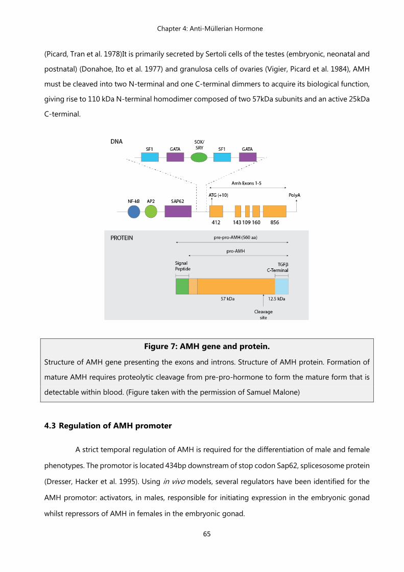
Chapter 4: Anti-Müllerian Hormone
65
(Picard, Tran et al. 1978)It is primarily secreted by Sertoli cells of the testes (embryonic, neonatal and
postnatal) (Donahoe, Ito et al. 1977) and granulosa cells of ovaries (Vigier, Picard et al. 1984), AMH
must be cleaved into two N-terminal and one C-terminal dimmers to acquire its biological function,
giving rise to 110 kDa N-terminal homodimer composed of two 57kDa subunits and an active 25kDa
C-terminal.
Figure 7: AMH gene and protein.
Structure of AMH gene presenting the exons and introns. Structure of AMH protein. Formation of
mature AMH requires proteolytic cleavage from pre-pro-hormone to form the mature form that is
detectable within blood. (Figure taken with the permission of Samuel Malone)
4.3 Regulation of AMH promoter
A strict temporal regulation of AMH is required for the differentiation of male and female
phenotypes. The promotor is located 434bp downstream of stop codon Sap62, splicesosome protein
(Dresser, Hacker et al. 1995). Using in vivo models, several regulators have been identified for the
AMH promotor: activators, in males, responsible for initiating expression in the embryonic gonad
whilst repressors of AMH in females in the embryonic gonad.
Chapter 4: Anti-Müllerian Hormone
66
There are several activators of AMH promoter:
SRY. Haqq et al., unveiled the role of SRY (Sex determining region on Y) in inducing AMH expression.
AMH expression is visible in mice 20h after the initial expression of SRY. However, they showed that
SRY acts indirectly to activate the AMH expression. (Haqq, King et al. 1994)
SOX9. SRY-related homolog box protein 9 or SOX9, belongs to the SOXE family of transcription
factors, expressed in the testes during mammalian differentiation (Schepers, Wilson et al. 2003). It is
expressed in both male and female embryos at embryonic day10. It is upregulated in the embryonic
male gonad and falls to undetectable levels in the female gonad. This increase in SOX9 expression
initiates Sertoli cells to express AMH and sexual development, thus being an activator of AMH
expression.
SF-1. Steroidogenic Factor-1 or SF-1, is expressed in mice in the genital ridges at embryonic day 9.
The expression is upregulated in foetal testes and downregulated in the ovaries as sexual
differentiation begins, thus, suggesting it might play a role in regulating AMH activity. The AMH
promoter contains a conserved 20 bp motif, capable of binding to SF-1 in vitro (Arango, Lovell-
Badge et al. 1999) and SF-1 is required for the upregulation of AMH transcription.
GATA4. Prenatal Sertoli cells and granulose cells express GATA4. It transactivates the AMH
promotor in granulosa cells (Anttonen, Ketola et al. 2003). The GATA binding site on AMH
promotor suggests that GATA4 directly regulates AMH expression (Tremblay and Viger 1999).
WT1. Wilms’ tumour suppressor-1 or WT1, is a transcription factor containing zinc fingers and
DNA binding domain. The synergistic action of WT1 and SF-1 are required for normal gonad
formation (Kreidberg, Sariola et al. 1993, Luo, Ikeda et al. 1994). In vitro, it’s been shown that an
isoform of WT1 interacts with SF-1 to upregulate AMH promoter (Nachtigal, Hirokawa et al. 1998)
Dax1. Dosage sensitive sex reversal, adrenal hypoplasia critical region, on chromosome X, gene 1
or Dax1, is one of the repressors of AMH promotor which is present in the ovaries and absent in the
testes in developing gonads and acts as a dominant-negative regulator of SF-1 (Burris, Guo et al.
1995),(Clipsham and McCabe 2003).
Chapter 4: Anti-Müllerian Hormone
67
4.4 AMH signalling pathway
4.4.1 Transforming Growth Factor β (TGFβ) family
AMH belongs to the divergent superfamily of TGF. In humans, there are more than 30
signalling proteins which include activins, TGFβ, bone morphogenic proteins (BMPs), nodals, or
growth and differentiation factors (GDFs). Their functions during development are indispensible and
heterogeneous as seen in mutant mice that display embryonic mortality or show abnormal
phenotype when lacking different TGF family members (Kaartinen, Voncken et al. 1995, Sanford,
Ormsby et al. 1997).It is implicated in different and varied physiological processes such as:
development, proliferation and cellular differentiation (Watabe and Miyazono 2009).
The superfamily of ligands can be phylogenetically divided into two main groups: the TGF-β/Activin
and BMP/growth and differentiation factor (GDF) branches (de Caestecker, 2004). The TGF-β
members are synthesized as a dimeric complex containing a preproprotein comprising N-terminal
signal peptide, a large proregion, and a smaller biologically active mature region, the C-terminal
(Massague 1990). After dimerization this complex is directed by the signal peptide in the RE/Golgi
where the pro-region undergo posttranslational processing for activation (Kingsley 1994, Massague
1998), is cleaved by furin-like endoproteinase, but remains attached by non-covalent bounds.
4.4.2 TGF receptors
TGF receptors are transmembrane serine-threonine kinase receptors which require
association of two different types of receptor class, resulting in a complex formation. Type I receptors
are the Activin-like Kinases (ALKs), whereas, the type II receptors are named after the ligands they
bind. The number of known ligands in the TGFβ superfamily is greater than the number of known
receptors. This signifies that numerous receptors are common and implicated in different signalling
pathways. The type II receptor kinases are constitutively phosphorylated (Luo and Lodish 1997)and
the ligands that bind to the receptor form the tetrameric complex. The signal is then propagated
downstream by Smad proteins.
Chapter 4: Anti-Müllerian Hormone
68
AMH binds exclusively to the type II receptor, AMHR2 (Mishina, Rey et al. 1996). AMHR2 interacts
with three type I receptors of Activin like kinase (ALK) family – AcvR1 (Activin receptor 1: ALK2),
BMPR1a (Bone morphogenic protein receptor 1A: ALK3) and BMPR1b (Bone morphogenic protein
receptor 1B: ALK6).
4.5 AMH receptors
AMH action is mediated by a heterodimeric receptor formed by AMH type 2 receptor
(AMHR2) and AMH type 1 receptor (Jamin, Arango et al. 2003). The binding of AMH to AMHR2 leads
to the recruitment of AMH type 1 receptor to form a complex which phosphorylates its serine-
threonine kinase domain. This activated AMH type 1 receptor phosphorylates a receptor-regulated
SMAD (R-Smad), associating with Smad4 and moving into the nucleus where they control
transcription (Massague 1998).
In 1994, AMHR2 gene was located on chromosome 12 in humans and on chromosome 15 in mice.
The genetic sequence comprises of 11 exons in which exons 1-3 encode the extracellular domain,
exon 4 the transmembrane domain and exons 5-11 encode the intracellular domain. The mRNA
encodes a protein of 82 kDa comprising of 573 amino acids.
It is most likely that the mechanism through which AMH interacts with AMHR2 is distinct from other
members of the TGFβ family (di Clemente, Jamin et al. 2010). It has been anticipated that once the
full-length AMH is cleaved, it results in a conformational change in the C-terminal domain which
facilitates the receptor binding. The binding of AMHR2 induces the dissociation of the pro-region
by a negative allosteric interaction between the receptor and the pro-region binding sites on C-
terminal dimer (Figure 8). Prenatally, AMHR2 expression pattern reflects AMH profile. It is expressed
by the mesenchymal cells surrounding the Müllerian ducts during embryonic
development(Baarends, van Helmond et al. 1994, di Clemente, Wilson et al. 1994), (Teixeira, He et
al. 1996). It has also been found in foetal testes (Baarends, van Helmond et al. 1994). Postnatally,
AMHR2 is expressed in the endometrium (Wang, Protheroe et al. 2009)breast (Segev, Hoshiya et al.
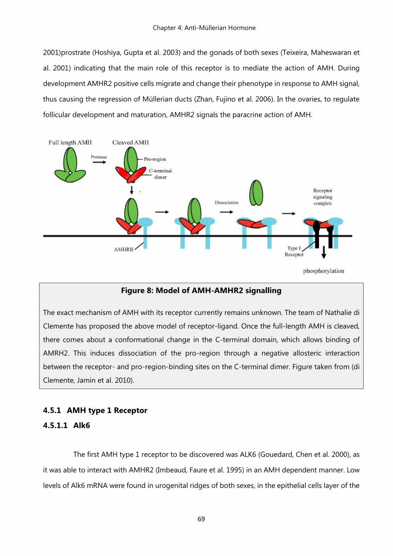
Chapter 4: Anti-Müllerian Hormone
69
2001)prostrate (Hoshiya, Gupta et al. 2003) and the gonads of both sexes (Teixeira, Maheswaran et
al. 2001) indicating that the main role of this receptor is to mediate the action of AMH. During
development AMHR2 positive cells migrate and change their phenotype in response to AMH signal,
thus causing the regression of Müllerian ducts (Zhan, Fujino et al. 2006). In the ovaries, to regulate
follicular development and maturation, AMHR2 signals the paracrine action of AMH.
Figure 8: Model of AMH-AMHR2 signalling
The exact mechanism of AMH with its receptor currently remains unknown. The team of Nathalie di
Clemente has proposed the above model of receptor-ligand. Once the full-length AMH is cleaved,
there comes about a conformational change in the C-terminal domain, which allows binding of
AMRH2. This induces dissociation of the pro-region through a negative allosteric interaction
between the receptor- and pro-region-binding sites on the C-terminal dimer. Figure taken from (di
Clemente, Jamin et al. 2010).
4.5.1 AMH type 1 Receptor
4.5.1.1 Alk6
The first AMH type 1 receptor to be discovered was ALK6 (Gouedard, Chen et al. 2000), as
it was able to interact with AMHR2 (Imbeaud, Faure et al. 1995) in an AMH dependent manner. Low
levels of Alk6 mRNA were found in urogenital ridges of both sexes, in the epithelial cells layer of the
Chapter 4: Anti-Müllerian Hormone
70
Müllerian duct and in adult gonads. It has been shown that Alk6 is important for AMH action in the
ovary but not required for Müllerian duct regression during male sexual differentiation (Monsivais,
Matzuk et al. 2017).
4.5.1.2 Alk2
Alk2 expression is more stringent than Alk6 because it overlaps with AMHR2 perfectly. In fact, Alk2
is expressed in all AMH target tissues early in development, whereas Alk6 is expressed only at the
level of the epithelium of the Müllerian duct(Zhan, Fujino et al. 2006), like the urogenital ridge (Wang
et al., 2005), gonads at different embryonic stages (Visser, Olaso et al. 2001)and mesenchymal cells
adjacent to the Müllerian duct.
4.5.1.3 Alk3
Alk3 interacts weakly with AMHR2 (Clarke, Hoshiya et al. 2001)and it has been reported
that lack of expression of Alk3 in the developing gonads leads to PMDS in mice (Jamin, Arango et
al. 2002). Moreover, AMH seems to spatiotemporally regulate Alk3 because its expression appears
later when Alk2 decreases. Furthermore, it is restricted to the mesenchyme, suggesting sequential
role in Müllerian duct regression (Zhan, Fujino et al. 2006).
4.6 AMH actions on gonadal development
4.6.1 Development of the embryonic gonad
During embryonic development, each individual possesses both female and male
reproductive ducts. These undifferentiated ducts have the potential to differentiate into male or
female reproductive systems. For normal female development, the Wolffian ducts must regress and
Müllerian (paramesonephric) ducts must develop. These will give rise to the uterus, upper part of
vagina and oviducts. Whereas, for normal male development, Müllerian ducts must regress and
Wolffian (mesonephric) ducts must develop; giving rise to seminal vesicles, epididymus, vas deferens
and ejaculatory ducts(Kobayashi and Behringer 2003, Cunha, Robboy et al. 2018).
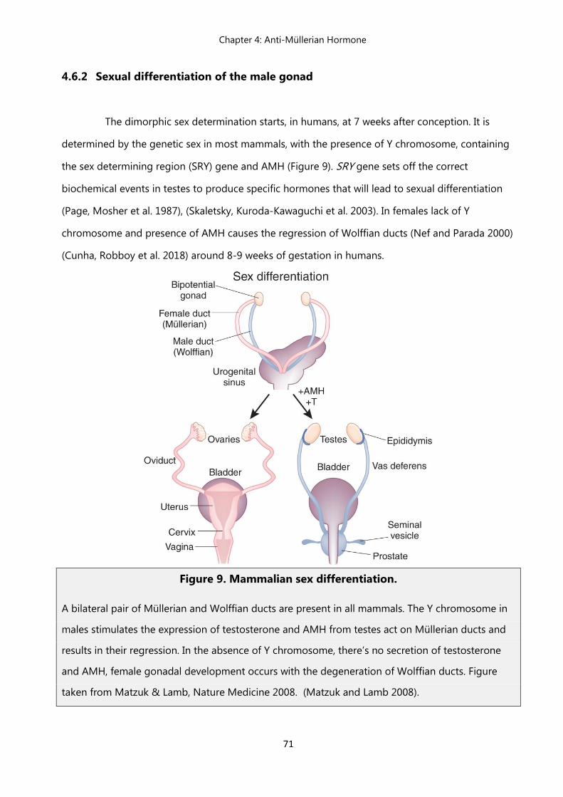
Chapter 4: Anti-Müllerian Hormone
71
4.6.2 Sexual differentiation of the male gonad
The dimorphic sex determination starts, in humans, at 7 weeks after conception. It is
determined by the genetic sex in most mammals, with the presence of Y chromosome, containing
the sex determining region (SRY) gene and AMH (Figure 9). SRY gene sets off the correct
biochemical events in testes to produce specific hormones that will lead to sexual differentiation
(Page, Mosher et al. 1987), (Skaletsky, Kuroda-Kawaguchi et al. 2003). In females lack of Y
chromosome and presence of AMH causes the regression of Wolffian ducts (Nef and Parada 2000)
(Cunha, Robboy et al. 2018) around 8-9 weeks of gestation in humans.
Figure 9. Mammalian sex differentiation.
A bilateral pair of Müllerian and Wolffian ducts are present in all mammals. The Y chromosome in
males stimulates the expression of testosterone and AMH from testes act on Müllerian ducts and
results in their regression. In the absence of Y chromosome, there’s no secretion of testosterone
and AMH, female gonadal development occurs with the degeneration of Wolffian ducts. Figure
taken from Matzuk & Lamb, Nature Medicine 2008. (Matzuk and Lamb 2008).
Chapter 4: Anti-Müllerian Hormone
72
4.6.3 Persistent Müllerian Duct Syndrome (PMDS)
AMH signalling is central to the development of correct male genitalia. This is
demonstrated by mutations affecting the function of AMH or AMH receptors, known to cause
Persistent Müllerian Duct Syndrome (PMDS). Approximately, 45% of the cases of PMDS are caused
by mutations of AMH gene (type 1) and 40% are caused by mutations of AMHR2 gene type 2. This
syndrome is characterised by the retention of Müllerian duct derivatives (the uterus, fallopian tubes
and upper part of the vagina) phenotypically and karyotypically (46XY) in male patients (Josso and
Clemente, 2003;Behringer et al., 1994; Mishina et al., 1996; Belville et al., 1999; Belville et al., 2004;
Orvis et al., 2008). On the surface, PMDS patients do not exhibit any anomalies as they have normal
development of external genitalia and secondary sexual characteristics (Renu et al., 2010).
4.6.4 Postnatal roles of AMH
4.6.4.1 AMH actions in female reproductive function
AMH is secreted by the granulosa cells of small antral and pre-antral follicles to regulate
early follicular development (Sahmay, Aydin et al. 2014). AMH continues to be expressed in the
growing follicles in the ovary until they have reached the size and differentiation state at which they
are to be selected for dominance by the action of pituitary FSH (van Houten, Themmen et al. 2010)
and gradually decreases at the following stages of follicular development. Hence, serum level of
AMH reflects the size of the primordial follicle pool and is considered as a marker of the ovarian
reserve.
AMH regulates ovarian function in cyclic females by preventing premature stimulating hormone
(FSH)-mediated follicular growth and steroidogenesis (Figure 10) By experimentally manipulating
FSH and E2 levels in infantile mice, Devillier et al., have shown that high FSH concentrations lower
Amh expression in early antral follicles, while E2 has no effect. The team noticed decrease in FSH
mediated expression of Cyp19a1 aromatase when they treated infantile ovaries in organotypic
cultures with AMH. Thus, infatile elevation in FSH levels suppresses Amh expression in early antral
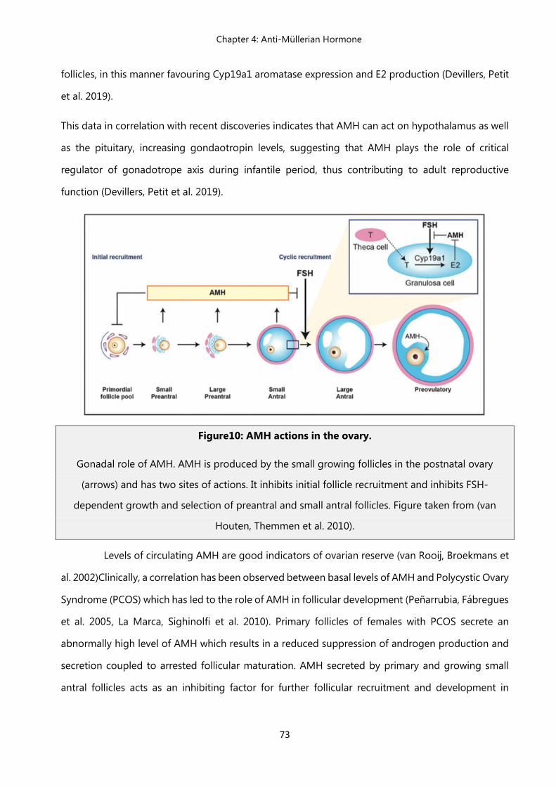
Chapter 4: Anti-Müllerian Hormone
73
follicles, in this manner favouring Cyp19a1 aromatase expression and E2 production (Devillers, Petit
et al. 2019).
This data in correlation with recent discoveries indicates that AMH can act on hypothalamus as well
as the pituitary, increasing gondaotropin levels, suggesting that AMH plays the role of critical
regulator of gonadotrope axis during infantile period, thus contributing to adult reproductive
function (Devillers, Petit et al. 2019).
Figure10: AMH actions in the ovary.
Gonadal role of AMH. AMH is produced by the small growing follicles in the postnatal ovary
(arrows) and has two sites of actions. It inhibits initial follicle recruitment and inhibits FSH-
dependent growth and selection of preantral and small antral follicles. Figure taken from (van
Houten, Themmen et al. 2010).
Levels of circulating AMH are good indicators of ovarian reserve (van Rooij, Broekmans et
al. 2002)Clinically, a correlation has been observed between basal levels of AMH and Polycystic Ovary
Syndrome (PCOS) which has led to the role of AMH in follicular development (Peñarrubia, Fábregues
et al. 2005, La Marca, Sighinolfi et al. 2010). Primary follicles of females with PCOS secrete an
abnormally high level of AMH which results in a reduced suppression of androgen production and
secretion coupled to arrested follicular maturation. AMH secreted by primary and growing small
antral follicles acts as an inhibiting factor for further follicular recruitment and development in
Chapter 4: Anti-Müllerian Hormone
74
humans and rodents. Furthermore, a recent study showed that AMH levels parallel fluctuations in
antral follicle count through menstrual cycle (Depmann, van Disseldorp et al. 2016).
4.6.4.2 AMH actions in male reproductive function
In males, AMH is produced by differentiated Sertoli cells, causing the regression of
Müllerian ducts (Tran, Muesy-Dessole et al. 1977). AMHR2, secreted by Sertoli and Leydig cells
indicates a potential paracrine role in regulating male gonadal function (Arango, Kobayashi et al.
2008)Indeed, high levels of AMHR2 result in decreased expression of testosterone secretion (Racine,
Rey et al. 1998). Along with this, AMH triggers a decrease in luteinizing hormone Receptor (LHR)
expression, further decreasing testosterone secretion. Physiologically, at puberty in males, AMH
levels reduce whereas testosterone levels increase and spermatogenesis is initiated (Rey 2005).
However, it is important to remember that this is only true for postnatal life due to androgen
mediated decrease in AMH levels (Chemes, Rey et al. 2008, Boukari, Meduri et al. 2009).
4.7 Emerging extra-gonadal roles of AMH
Gonadotropin releasing hormone plays a key role for the onset of puberty and
reproduction. It is released into the pituitary blood vessel from where it travels to the anterior
pituitary. The production and release of gonadotropins LH and FSH is under its control. These
gonadotropins, in turn, stimulate gametogenesis and production of sex steroids in the gonads
(Christian and Moenter 2010).
GnRH-secreting neurons are neuroendocrine cells originating in the nasal placode outside of the
nervous system during embryonic development and migrate to the hypothalamus (Wray, Grant et
al. 1989), (Schwanzel-Fukuda and Pfaff 1989). This process is evolutionarily conserved in
mammals(Wray, Grant et al. 1989),(Schwanzel-Fukuda and Pfaff 1989) including humans (Schwanzel-
Fukuda, Crossin et al. 1996), (Casoni, Malone et al. 2016). A disruption of the migration of GnRH
neurons or a defect in GnRH synthesis and secretion results in a rare endocrine disorder congenital
Chapter 4: Anti-Müllerian Hormone
75
hypogonadotropic hypogonadisms (CHH). CHH is characterised by absence of incompleteness of
puberty, which leads to infertility (Boehm, Bouloux et al. 2015).
Strictly in females, another AMH action is observed, occurring only before puberty. This AMH action
is essentially a preferential secretion and adenohypophyseal gene expression of gonadotropin FSH
in vivo in rats (Garrel, Racine et al. 2016). Garrel et al., additionally, demonstrate that GnRH is a
regulator of AMHR2 expression in humans, highlighting the importance of AMH signalling in the
regulation of gonadotrope function. Using perifused murine LbetaT2 gonadotrope cells, the team
showed that Amhr2 expression is differentially regulated by GnRH pulse frequency with an induction
under high GnRH pulsatility (Garrel, Denoyelle et al. 2019).
Erg1 (short proximal region of the promotor) was identified as a new transcription factor controlling
hAMHR2 expression as it mediates basal and GnRH-dependent activity of the promoter. This was
achieved by using site-directed mutagenesis of Erg1 motif and siRNA mediated-knockdown of Egr1.
Furthermore, FOXO1 was recognized as FOXO1 as a negative regulator of basal and GnRH-
dependent AMHR2 expression in gonadotrope cells, using a constitutively active mutant of FOXO1
(Garrel, Denoyelle et al. 2019)
Mature neurons in the adult brain express high levels of AMH receptors in males and females(Wang,
Protheroe et al. 2009). In 2016, Cimino et al. discovered a novel neuroendocrine action of AMH. They
reported a direct action of AMH on GnRH neuronal activity and demonstrated that AMHR2 is
expressed in a part of the hypothalamic gonadotropin-releasing hormone neurons in mice and
humans (Figure 11) and responds to exogenous AMH by induces neuronal activity and GnRH
secretion.(Cimino, Casoni et al. 2016) This, further, results in an increase in LH secretions, equivalent
to levels required to produce an ovulatory surge. In 80% of PCOS females, a notable rise in LH levels
is observed (Taylor, McCourt et al. 1997) indicating that the pulse frequency of GnRH is persistently
accelerated in this syndrome.
It’s been shown that GnRH neurons express AMHR2 since early foetal development to adulthood
and that AMH stimulates GnRH neuronal activity and hormone secretion in mature GnRH cells
(Cimino, Casoni et al. 2016).
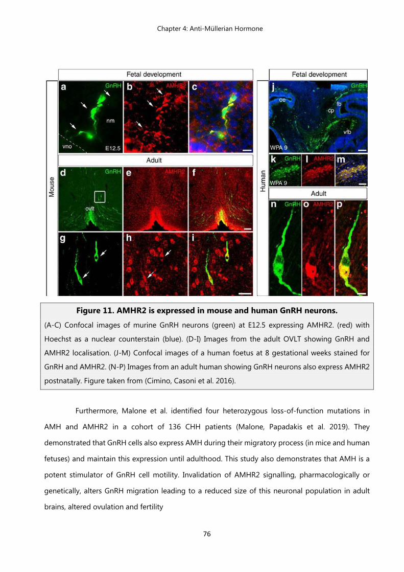
Chapter 4: Anti-Müllerian Hormone
76
Figure 11. AMHR2 is expressed in mouse and human GnRH neurons.
(A-C) Confocal images of murine GnRH neurons (green) at E12.5 expressing AMHR2. (red) with
Hoechst as a nuclear counterstain (blue). (D-I) Images from the adult OVLT showing GnRH and
AMHR2 localisation. (J-M) Confocal images of a human foetus at 8 gestational weeks stained for
GnRH and AMHR2. (N-P) Images from an adult human showing GnRH neurons also express AMHR2
postnatally. Figure taken from (Cimino, Casoni et al. 2016).
Furthermore, Malone et al. identified four heterozygous loss-of-function mutations in
AMH and AMHR2 in a cohort of 136 CHH patients (Malone, Papadakis et al. 2019). They
demonstrated that GnRH cells also express AMH during their migratory process (in mice and human
fetuses) and maintain this expression until adulthood. This study also demonstrates that AMH is a
potent stimulator of GnRH cell motility. Invalidation of AMHR2 signalling, pharmacologically or
genetically, alters GnRH migration leading to a reduced size of this neuronal population in adult
brains, altered ovulation and fertility
References
77
References
Abbott, D. H., D. A. Dumesic, J. R. Eisner, R. J. Colman and J. W. Kemnitz (1998). "Insights into the development
of polycystic ovary syndrome (PCOS) from studies of prenatally androgenized female rhesus
monkeys." Trends Endocrinol Metab 9(2): 62-67.
Abbott, D. H., D. A. Dumesic and S. Franks (2002). "Developmental origin of polycystic ovary syndrome - a
hypothesis." J Endocrinol 174(1): 1-5.
Abbott, D. H., B. H. Rayome, D. A. Dumesic, K. C. Lewis, A. K. Edwards, K. Wallen, M. E. Wilson, S. E. Appt and J.
E. Levine (2017). "Clustering of PCOS-like traits in naturally hyperandrogenic female rhesus monkeys."
Hum Reprod 32(4): 923-936.
Abu-Hijleh, T. M., E. Gammoh, A. S. Al-Busaidi, Z. H. Malalla, S. Madan, N. Mahmood and W. Y. Almawi (2016).
"Common Variants in the Sex Hormone-Binding Globulin (SHBG) Gene Influence SHBG Levels in
Women with Polycystic Ovary Syndrome." Ann Nutr Metab 68(1): 66-74.
Adachi, S., S. Yamada, Y. Takatsu, H. Matsui, M. Kinoshita, K. Takase, H. Sugiura, T. Ohtaki, H. Matsumoto, Y.
Uenoyama, H. Tsukamura, K. Inoue and K. Maeda (2007). "Involvement of anteroventral periventricular
metastin/kisspeptin neurons in estrogen positive feedback action on luteinizing hormone release in
female rats." J Reprod Dev 53(2): 367-378.
Adashi, E. Y., A. J. Hsueh and S. S. Yen (1981). "Insulin enhancement of luteinizing hormone and follicle-
stimulating hormone release by cultured pituitary cells." Endocrinology 108(4): 1441-1449.
Allon, M. A., R. E. Leach, J. Dunbar and M. P. Diamond (2005). "Effects of chronic hyperandrogenism and/or
administered central nervous system insulin on ovarian manifestation and gonadotropin and steroid
secretion." Fertility and Sterility 83(4): 1319-1326.
Anand-Ivell, R., K. Tremellen, Y. Dai, K. Heng, M. Yoshida, P. G. Knight, G. E. Hale and R. Ivell (2013). "Circulating
insulin-like factor 3 (INSL3) in healthy and infertile women." Hum Reprod 28(11): 3093-3102.
Andrews, W. W., G. J. Mizejewski and S. R. Ojeda (1981). "Development of estradiol-positive feedback on
luteinizing hormone release in the female rat: a quantitative study." Endocrinology 109(5): 1404-1413.
Andrisse, S., K. Billings, P. Xue and S. Wu (2018). "Insulin signaling displayed a differential tissue-specific
response to low-dose dihydrotestosterone in female mice." Am J Physiol Endocrinol Metab 314(4):
E353-e365.
Anttonen, M., I. Ketola, H. Parviainen, A. K. Pusa and M. Heikinheimo (2003). "FOG-2 and GATA-4 Are
coexpressed in the mouse ovary and can modulate mullerian-inhibiting substance expression." Biol
Reprod 68(4): 1333-1340.
Antunes, J. L., P. W. Carmel, E. M. Housepian and M. Ferin (1978). "Luteinizing hormone-releasing hormone in
human pituitary blood." J Neurosurg 49(3): 382-386.
Arango, N. A., A. Kobayashi, Y. Wang, S. P. Jamin, H. H. Lee, G. D. Orvis and R. R. Behringer (2008). "A
mesenchymal perspective of Mullerian duct differentiation and regression in Amhr2-lacZ mice." Mol
Reprod Dev 75(7): 1154-1162.
References
78
Arango, N. A., R. Lovell-Badge and R. R. Behringer (1999). "Targeted mutagenesis of the endogenous mouse
Mis gene promoter: in vivo definition of genetic pathways of vertebrate sexual development." Cell
99(4): 409-419.
Asuncion, M., R. M. Calvo, J. L. San Millan, J. Sancho, S. Avila and H. F. Escobar-Morreale (2000). "A prospective
study of the prevalence of the polycystic ovary syndrome in unselected Caucasian women from Spain."
J Clin Endocrinol Metab 85(7): 2434-2438.
Azziz, R., E. Carmina, Z. Chen, A. Dunaif, J. S. Laven, R. S. Legro, D. Lizneva, B. Natterson-Horowtiz, H. J. Teede
and B. O. Yildiz (2016). "Polycystic ovary syndrome." Nat Rev Dis Primers 2: 16057.
Azziz, R., E. Carmina, D. Dewailly, E. Diamanti-Kandarakis, H. F. Escobar-Morreale, W. Futterweit, O. E. Janssen,
R. S. Legro, R. J. Norman, A. E. Taylor and S. F. Witchel (2009). "The Androgen Excess and PCOS Society
criteria for the polycystic ovary syndrome: the complete task force report." Fertil Steril 91(2): 456-488.
Azziz, R. and M. D. Kashar-Miller (2000). "Family history as a risk factor for the polycystic ovary syndrome." J
Pediatr Endocrinol Metab 13 Suppl 5: 1303-1306.
Azziz, R., K. S. Woods, R. Reyna, T. J. Key, E. S. Knochenhauer and B. O. Yildiz (2004). "The prevalence and
features of the polycystic ovary syndrome in an unselected population." J Clin Endocrinol Metab 89(6):
2745-2749.
Baarends, W. M., M. J. van Helmond, M. Post, P. J. van der Schoot, J. W. Hoogerbrugge, J. P. de Winter, J. T.
Uilenbroek, B. Karels, L. G. Wilming, J. H. Meijers and et al. (1994). "A novel member of the
transmembrane serine/threonine kinase receptor family is specifically expressed in the gonads and in
mesenchymal cells adjacent to the mullerian duct." Development 120(1): 189-197.
Bahk, J. Y., J. S. Hyun, S. H. Chung, H. Lee, M. O. Kim, B. H. Lee and W. S. Choi (1995). "Stage specific identification
of the expression of GnRH mRNA and localization of the GnRH receptor in mature rat and adult human
testis." J Urol 154(5): 1958-1961.
Baldani, D. P., L. Skrgatic, J. Z. Cerne, S. K. Oguic, B. M. Gersak and K. Gersak (2015). "Association between
serum levels and pentanucleotide polymorphism in the sex hormone binding globulin gene and
acardiovascular risk factors in females with polycystic ovary syndrome." Mol Med Rep 11(5): 3941-
3947.
Balen, A. H., G. S. Conway, G. Kaltsas, K. Techatrasak, P. J. Manning, C. West and H. S. Jacobs (1995). "Polycystic
ovary syndrome: the spectrum of the disorder in 1741 patients." Hum Reprod 10(8): 2107-2111.
Banerjee, U., A. Dasgupta, A. Khan, M. K. Ghosh, P. Roy, J. K. Rout, P. Roy and S. Dhara (2016). "A cross-sectional
study to assess any possible linkage of C/T polymorphism in CYP17A1 gene with insulin resistance in
non-obese women with polycystic ovarian syndrome." Indian J Med Res 143(6): 739-747.
Barber, T. M., A. J. Bennett, C. J. Groves, U. Sovio, A. Ruokonen, H. Martikainen, A. Pouta, A. L. Hartikainen, P.
Elliott, C. M. Lindgren, R. M. Freathy, K. Koch, W. H. Ouwehand, F. Karpe, G. S. Conway, J. A. Wass, M.
R. Jarvelin, S. Franks and M. I. McCarthy (2008). "Association of variants in the fat mass and obesity
associated (FTO) gene with polycystic ovary syndrome." Diabetologia 51(7): 1153-1158.
References
79
Barry, J. A., A. R. Kay, R. Navaratnarajah, S. Iqbal, J. E. Bamfo, A. L. David, M. Hines and P. J. Hardiman (2010).
"Umbilical vein testosterone in female infants born to mothers with polycystic ovary syndrome is
elevated to male levels." J Obstet Gynaecol 30(5): 444-446.
Barry, J. A., A. R. Kuczmierczyk and P. J. Hardiman (2011). "Anxiety and depression in polycystic ovary syndrome:
a systematic review and meta-analysis." Hum Reprod 26(9): 2442-2451.
Birch, R. A., V. Padmanabhan, D. L. Foster, W. P. Unsworth and J. E. Robinson (2003). "Prenatal programming
of reproductive neuroendocrine function: fetal androgen exposure produces progressive disruption
of reproductive cycles in sheep." Endocrinology 144(4): 1426-1434.
Blank, S. K., C. R. McCartney and J. C. Marshall (2006). "The origins and sequelae of abnormal neuroendocrine
function in polycystic ovary syndrome." Hum Reprod Update 12(4): 351-361.
Boehm, U., P. M. Bouloux, M. T. Dattani, N. de Roux, C. Dode, L. Dunkel, A. A. Dwyer, P. Giacobini, J. P. Hardelin,
A. Juul, M. Maghnie, N. Pitteloud, V. Prevot, T. Raivio, M. Tena-Sempere, R. Quinton and J. Young
(2015). "Expert consensus document: European Consensus Statement on congenital
hypogonadotropic hypogonadism--pathogenesis, diagnosis and treatment." Nat Rev Endocrinol
11(9): 547-564.
Boomsma, C. M., M. J. Eijkemans, E. G. Hughes, G. H. Visser, B. C. Fauser and N. S. Macklon (2006). "A meta-
analysis of pregnancy outcomes in women with polycystic ovary syndrome." Hum Reprod Update
12(6): 673-683.
Botte, M. C., Y. Lerrant, A. Lozach, A. Berault, R. Counis and M. L. Kottler (1999). "LH down-regulates
gonadotropin-releasing hormone (GnRH) receptor, but not GnRH, mRNA levels in the rat testis." J
Endocrinol 162(3): 409-415.
Boukari, K., G. Meduri, S. Brailly-Tabard, J. Guibourdenche, M. L. Ciampi, N. Massin, L. Martinerie, J. Y. Picard,
R. Rey, M. Lombes and J. Young (2009). "Lack of androgen receptor expression in Sertoli cells accounts
for the absence of anti-Mullerian hormone repression during early human testis development." J Clin
Endocrinol Metab 94(5): 1818-1825.
Bouret, S. G., S. J. Draper and R. B. Simerly (2004). "Formation of projection pathways from the arcuate nucleus
of the hypothalamus to hypothalamic regions implicated in the neural control of feeding behavior in
mice." J Neurosci 24(11): 2797-2805.
Boyle, J. A., J. Cunningham, K. O'Dea, T. Dunbar and R. J. Norman (2012). "Prevalence of polycystic ovary
syndrome in a sample of Indigenous women in Darwin, Australia." Med J Aust 196(1): 62-66.
Buchanan, K. L. and S. M. Yellon (1993). "Developmental study of GnRH neuronal projections to the medial
basal hypothalamus of the male Djungarian hamster." J Comp Neurol 333(2): 236-245.
Buckett, W. M., R. Bouzayen, K. L. Watkin, T. Tulandi and S. L. Tan (1999). "Ovarian stromal echogenicity in
women with normal and polycystic ovaries." Hum Reprod 14(3): 618-621.
Bulun, S. E., K. Takayama, T. Suzuki, H. Sasano, B. Yilmaz and S. Sebastian (2004). "Organization of the human
aromatase p450 (CYP19) gene." Semin Reprod Med 22(1): 5-9.
References
80
Burbridge, S., I. Stewart and M. Placzek (2016). "Development of the Neuroendocrine Hypothalamus." Compr
Physiol 6(2): 623-643.
Burghen, G. A., J. R. Givens and A. E. Kitabchi (1980). "Correlation of hyperandrogenism with hyperinsulinism
in polycystic ovarian disease." J Clin Endocrinol Metab 50(1): 113-116.
Burris, T. P., W. Guo, T. Le and E. R. McCabe (1995). "Identification of a putative steroidogenic factor-1 response
element in the DAX-1 promoter." Biochem Biophys Res Commun 214(2): 576-581.
Caldwell, A. S., L. J. Middleton, M. Jimenez, R. Desai, A. C. McMahon, C. M. Allan, D. J. Handelsman and K. A.
Walters (2014). "Characterization of reproductive, metabolic, and endocrine features of polycystic
ovary syndrome in female hyperandrogenic mouse models." Endocrinology 155(8): 3146-3159.
Calvo, R. M., M. Asuncion, D. Telleria, J. Sancho, J. L. San Millan and H. F. Escobar-Morreale (2001). "Screening
for mutations in the steroidogenic acute regulatory protein and steroidogenic factor-1 genes, and in
CYP11A and dosage-sensitive sex reversal-adrenal hypoplasia gene on the X chromosome, gene-1
(DAX-1), in hyperandrogenic hirsute women." J Clin Endocrinol Metab 86(4): 1746-1749.
Campbell, R. E. (2018). Morphology of the Adult GnRH Neuron. The GnRH Neuron and its Control: 121-148.
Campbell, R. E., S. K. Han and A. E. Herbison (2005). "Biocytin filling of adult gonadotropin-releasing hormone
neurons in situ reveals extensive, spiny, dendritic processes." Endocrinology 146(3): 1163-1169.
Carey, A. H., D. Waterworth, K. Patel, D. White, J. Little, P. Novelli, S. Franks and R. Williamson (1994). "Polycystic
ovaries and premature male pattern baldness are associated with one allele of the steroid metabolism
gene CYP17." Hum Mol Genet 3(10): 1873-1876.
Carmina, E., R. S. Legro, K. Stamets, J. Lowell and R. A. Lobo (2003). "Difference in body weight between
American and Italian women with polycystic ovary syndrome: influence of the diet." Hum Reprod
18(11): 2289-2293.
Caron, E., P. Ciofi, V. Prevot and S. G. Bouret (2012). "Alteration in neonatal nutrition causes perturbations in
hypothalamic neural circuits controlling reproductive function." J Neurosci 32(33): 11486-11494.
Casarini, L. and G. Brigante (2014). "The polycystic ovary syndrome evolutionary paradox: a genome-wide
association studies-based, in silico, evolutionary explanation." J Clin Endocrinol Metab 99(11): E2412-
2420.
Casoni, F., S. A. Malone, M. Belle, F. Luzzati, F. Collier, C. Allet, E. Hrabovszky, S. Rasika, V. Prevot, A. Chedotal
and P. Giacobini (2016). "Development of the neurons controlling fertility in humans: new insights
from 3D imaging and transparent fetal brains." Development 143(21): 3969-3981.
Cattanach, B. M., C. A. Iddon, H. M. Charlton, S. A. Chiappa and G. Fink (1977). "Gonadotrophin-releasing
hormone deficiency in a mutant mouse with hypogonadism." Nature 269(5626): 338-340.
Chachlaki, K., S. A. Malone, E. Qualls-Creekmore, E. Hrabovszky, H. Munzberg, P. Giacobini, F. Ango and V.
Prevot (2017). "Phenotyping of nNOS neurons in the postnatal and adult female mouse
hypothalamus." J Comp Neurol 525(15): 3177-3189.
Chemes, H. E., R. A. Rey, M. Nistal, J. Regadera, M. Musse, P. Gonzalez-Peramato and A. Serrano (2008).
"Physiological androgen insensitivity of the fetal, neonatal, and early infantile testis is explained by the
References
81
ontogeny of the androgen receptor expression in Sertoli cells." J Clin Endocrinol Metab 93(11): 4408-
4412.
Chen, H. F., E. B. Jeung, M. Stephenson and P. C. Leung (1999). "Human peripheral blood mononuclear cells
express gonadotropin-releasing hormone (GnRH), GnRH receptor, and interleukin-2 receptor gamma-
chain messenger ribonucleic acids that are regulated by GnRH in vitro." J Clin Endocrinol Metab 84(2):
743-750.
Chen, J., S. Shen, Y. Tan, D. Xia, Y. Xia, Y. Cao, W. Wang, X. Wu, H. Wang, L. Yi, Q. Gao and Y. Wang (2015). "The
correlation of aromatase activity and obesity in women with or without polycystic ovary syndrome."
Journal of Ovarian Research 8(1): 11.
Chen, Z. J., H. Zhao, L. He, Y. Shi, Y. Qin, Y. Shi, Z. Li, L. You, J. Zhao, J. Liu, X. Liang, X. Zhao, J. Zhao, Y. Sun, B.
Zhang, H. Jiang, D. Zhao, Y. Bian, X. Gao, L. Geng, Y. Li, D. Zhu, X. Sun, J. E. Xu, C. Hao, C. E. Ren, Y.
Zhang, S. Chen, W. Zhang, A. Yang, J. Yan, Y. Li, J. Ma and Y. Zhao (2011). "Genome-wide association
study identifies susceptibility loci for polycystic ovary syndrome on chromosome 2p16.3, 2p21 and
9q33.3." Nat Genet 43(1): 55-59.
Cheng, C. K., B. K. Chow and P. C. Leung (2003). "An activator protein 1-like motif mediates 17beta-estradiol
repression of gonadotropin-releasing hormone receptor promoter via an estrogen receptor alpha-
dependent mechanism in ovarian and breast cancer cells." Mol Endocrinol 17(12): 2613-2629.
Cheng, C. K. and P. C. Leung (2005). "Molecular biology of gonadotropin-releasing hormone (GnRH)-I, GnRH-
II, and their receptors in humans." Endocr Rev 26(2): 283-306.
Cheng, K. W. and P. C. Leung (2002). "Human chorionic gonadotropin-activated cAMP pathway regulates
human placental GnRH receptor gene transcription in choriocarcinoma JEG-3 cells." J Clin Endocrinol
Metab 87(7): 3291-3299.
Cherskov, A., A. Pohl, C. Allison, H. Zhang, R. A. Payne and S. Baron-Cohen (2018). "Polycystic ovary syndrome
and autism: A test of the prenatal sex steroid theory." Translational Psychiatry 8(1): 136.
Christian, C. A. and S. M. Moenter (2010). "The neurobiology of preovulatory and estradiol-induced
gonadotropin-releasing hormone surges." Endocr Rev 31(4): 544-577.
Cimino, I., F. Casoni, X. Liu, A. Messina, J. Parkash, S. P. Jamin, S. Catteau-Jonard, F. Collier, M. Baroncini, D.
Dewailly, P. Pigny, M. Prescott, R. Campbell, A. E. Herbison, V. Prevot and P. Giacobini (2016). "Novel
role for anti-Mullerian hormone in the regulation of GnRH neuron excitability and hormone secretion."
Nat Commun 7: 10055.
Clarke, T. R., Y. Hoshiya, S. E. Yi, X. Liu, K. M. Lyons and P. K. Donahoe (2001). "Mullerian Inhibiting Substance
Signaling Uses a Bone Morphogenetic Protein (BMP)-Like Pathway Mediated by ALK2 and Induces
Smad6 Expression." Molecular Endocrinology 15(6): 946-959.
Clarkson, J., X. d'Anglemont de Tassigny, A. S. Moreno, W. H. Colledge and A. E. Herbison (2008). "Kisspeptin-
GPR54 signaling is essential for preovulatory gonadotropin-releasing hormone neuron activation and
the luteinizing hormone surge." J Neurosci 28(35): 8691-8697.
References
82
Clipsham, R. and E. R. McCabe (2003). "DAX1 and its network partners: exploring complexity in development."
Mol Genet Metab 80(1-2): 81-120.
Cohen-Haguenauer, O., J. Y. Picard, M. G. Mattéi, S. Serero, N. Van Cong, M. F. de Tand, D. Guerrier, M. C. Hors-
Cayla, N. Josso and J. Frézal (1987). "Mapping of the gene for anti-Müllerian hormone to the short
arm of human chromosome 19." Cytogenetic and Genome Research 44(1): 2-6.
Constantin, S. (2011). "Physiology of the gonadotrophin-releasing hormone (GnRH) neurone: studies from
embryonic GnRH neurones." J Neuroendocrinol 23(6): 542-553.
Conway, G., D. Dewailly, E. Diamanti-Kandarakis, H. F. Escobar-Morreale, S. Franks, A. Gambineri, F. Kelestimur,
D. Macut, D. Micic, R. Pasquali, M. Pfeifer, D. Pignatelli, M. Pugeat and B. O. Yildiz (2014). "The polycystic
ovary syndrome: a position statement from the European Society of Endocrinology." Eur J Endocrinol
171(4): P1-29.
Cottrell, E. C., R. E. Campbell, S. K. Han and A. E. Herbison (2006). "Postnatal remodeling of dendritic structure
and spine density in gonadotropin-releasing hormone neurons." Endocrinology 147(8): 3652-3661.
Cousin, P., L. Calemard-Michel, H. Lejeune, G. Raverot, N. Yessaad, A. Emptoz-Bonneton, Y. Morel and M.
Pugeat (2004). "Influence of SHBG gene pentanucleotide TAAAA repeat and D327N polymorphism on
serum sex hormone-binding globulin concentration in hirsute women." J Clin Endocrinol Metab 89(2):
917-924.
Crisosto, N., E. Codner, M. Maliqueo, B. Echiburu, F. Sanchez, F. Cassorla and T. Sir-Petermann (2007). "Anti-
Mullerian hormone levels in peripubertal daughters of women with polycystic ovary syndrome." J Clin
Endocrinol Metab 92(7): 2739-2743.
Cunha, G. R., S. J. Robboy, T. Kurita, D. Isaacson, J. Shen, M. Cao and L. S. Baskin (2018). "Development of the
human female reproductive tract." Differentiation 103: 46-65.
Cussons, A. J., B. G. Stuckey, J. P. Walsh, V. Burke and R. J. Norman (2005). "Polycystic ovarian syndrome: marked
differences between endocrinologists and gynaecologists in diagnosis and management." Clin
Endocrinol (Oxf) 62(3): 289-295.
Daneshmand, S. d., S. R. Weitsman, A. Navab, A. J. Jakimiuk and D. A. Magoffin (2002). "Overexpression of
theca-cell messenger RNA in polycystic ovary syndrome does not correlate with polymorphisms in
the cholesterol side-chain cleavage and 17α-hydroxylase/C<sub>17-20</sub> lyase
promoters." Fertility and Sterility 77(2): 274-280.
Day, F. R., D. A. Hinds, J. Y. Tung, L. Stolk, U. Styrkarsdottir, R. Saxena, A. Bjonnes, L. Broer, D. B. Dunger, B. V.
Halldorsson, D. A. Lawlor, G. Laval, I. Mathieson, W. L. McCardle, Y. Louwers, C. Meun, S. Ring, R. A.
Scott, P. Sulem, A. G. Uitterlinden, N. J. Wareham, U. Thorsteinsdottir, C. Welt, K. Stefansson, J. S. E.
Laven, K. K. Ong and J. R. B. Perry (2015). "Causal mechanisms and balancing selection inferred from
genetic associations with polycystic ovary syndrome." Nat Commun 6: 8464.
Demissie, M., M. Lazic, E. M. Foecking, F. Aird, A. Dunaif and J. E. Levine (2008). "Transient prenatal androgen
exposure produces metabolic syndrome in adult female rats." Am J Physiol Endocrinol Metab 295(2):
E262-268.
References
83
Depmann, M., J. van Disseldorp, S. L. Broer, M. J. Eijkemans, J. S. Laven, J. A. Visser, Y. B. de Rijke, B. W. Mol and
F. J. Broekmans (2016). "Fluctuations in anti-Mullerian hormone levels throughout the menstrual cycle
parallel fluctuations in the antral follicle count: a cohort study." Acta Obstet Gynecol Scand 95(7): 820-
828.
Devillers, M. M., F. Petit, V. Cluzet, C. M. Francois, F. Giton, G. Garrel, J. Cohen-Tannoudji and C. J. Guigon (2019).
"FSH inhibits AMH to support ovarian estradiol synthesis in infantile mice." J Endocrinol 240(2): 215-
228.
Devillers, M. M., F. Petit, V. Cluzet, C. M. Francois, F. Giton, G. Garrel, J. Cohen-Tannoudji and C. J. Guigon (2019).
"[Ovarian estradiol production during mini-puberty: importance of the cross-talk between FSH and
AMH]." Med Sci (Paris) 35(3): 201-203.
Dewailly, D., H. Gronier, E. Poncelet, G. Robin, M. Leroy, P. Pigny, A. Duhamel and S. Catteau-Jonard (2011).
"Diagnosis of polycystic ovary syndrome (PCOS): revisiting the threshold values of follicle count on
ultrasound and of the serum AMH level for the definition of polycystic ovaries." Hum Reprod 26(11):
3123-3129.
Dewailly, D., S. Hieronimus, P. Mirakian and J. N. Hugues (2010). "Polycystic ovary syndrome (PCOS)." Ann
Endocrinol (Paris) 71(1): 8-13.
di Clemente, N., S. P. Jamin, A. Lugovskoy, P. Carmillo, C. Ehrenfels, J. Y. Picard, A. Whitty, N. Josso, R. B. Pepinsky
and R. L. Cate (2010). "Processing of anti-mullerian hormone regulates receptor activation by a
mechanism distinct from TGF-beta." Mol Endocrinol 24(11): 2193-2206.
di Clemente, N., C. Wilson, E. Faure, L. Boussin, P. Carmillo, R. Tizard, J. Y. Picard, B. Vigier, N. Josso and R. Cate
(1994). "Cloning, expression, and alternative splicing of the receptor for anti-Mullerian hormone." Mol
Endocrinol 8(8): 1006-1020.
Diamanti-Kandarakis, E., M. I. Bartzis, A. T. Bergiele, T. C. Tsianateli and C. R. Kouli (2000). "Microsatellite
polymorphism (tttta)(n) at -528 base pairs of gene CYP11alpha influences hyperandrogenemia in
patients with polycystic ovary syndrome." Fertil Steril 73(4): 735-741.
Diamanti-Kandarakis, E. and A. Dunaif (2012). "Insulin resistance and the polycystic ovary syndrome revisited:
an update on mechanisms and implications." Endocr Rev 33(6): 981-1030.
Diamanti-Kandarakis, E., H. Kandarakis and R. S. Legro (2006). "The role of genes and environment in the
etiology of PCOS." Endocrine 30(1): 19-26.
Diamanti-Kandarakis, E., C. R. Kouli, A. T. Bergiele, F. A. Filandra, T. C. Tsianateli, G. G. Spina, E. D. Zapanti and
M. I. Bartzis (1999). "A survey of the polycystic ovary syndrome in the Greek island of Lesbos: hormonal
and metabolic profile." J Clin Endocrinol Metab 84(11): 4006-4011.
Dohler, K. D. and W. Wuttke (1974). "Serum LH, FSH, prolactin and progesterone from birth to puberty in
female and male rats." Endocrinology 94(4): 1003-1008.
Dokras, A., S. Clifton, W. Futterweit and R. Wild (2012). "Increased prevalence of anxiety symptoms in women
with polycystic ovary syndrome: systematic review and meta-analysis." Fertil Steril 97(1): 225-
230.e222.
References
84
Donahoe, P. K., Y. Ito, J. M. Price and W. H. Hendren, 3rd (1977). "Mullerian inhibiting substance activity in
bovine fetal, newborn and prepubertal testes." Biol Reprod 16(2): 238-243.
Dresser, D. W., A. Hacker, R. Lovell-Badge and D. Guerrier (1995). "The genes for a spliceosome protein (SAP62)
and the anti-Mullerian hormone (AMH) are contiguous." Hum Mol Genet 4(9): 1613-1618.
Dumesic, D. A., D. H. Abbott, J. R. Eisner and R. W. Goy (1997). "Prenatal exposure of female rhesus monkeys
to testosterone propionate increases serum luteinizing hormone levels in adulthood." Fertil Steril
67(1): 155-163.
Dumesic, D. A. and R. A. Lobo (2013). "Cancer risk and PCOS." Steroids 78(8): 782-785.
Dumont, A., G. Robin, S. Catteau-Jonard and D. Dewailly (2015). "Role of Anti-Müllerian Hormone in
pathophysiology, diagnosis and treatment of Polycystic Ovary Syndrome: a review." Reprod Biol
Endocrinol 13: 137.
Dunaif, A. (1999). "Insulin action in the polycystic ovary syndrome." Endocrinol Metab Clin North Am 28(2):
341-359.
Dunaif, A., K. R. Segal, W. Futterweit and A. Dobrjansky (1989). "Profound peripheral insulin resistance,
independent of obesity, in polycystic ovary syndrome." Diabetes 38(9): 1165-1174.
Echiburu, B., F. Perez-Bravo, M. Maliqueo, F. Sanchez, N. Crisosto and T. Sir-Petermann (2008). "Polymorphism
T --> C (-34 base pairs) of gene CYP17 promoter in women with polycystic ovary syndrome is
associated with increased body weight and insulin resistance: a preliminary study." Metabolism 57(12):
1765-1771.
Eisner, J. R., M. A. Barnett, D. A. Dumesic and D. H. Abbott (2002). "Ovarian hyperandrogenism in adult female
rhesus monkeys exposed to prenatal androgen excess." Fertil Steril 77(1): 167-172.
Elmquist, J. K., R. Coppari, N. Balthasar, M. Ichinose and B. B. Lowell (2005). "Identifying hypothalamic pathways
controlling food intake, body weight, and glucose homeostasis." J Comp Neurol 493(1): 63-71.
Escobar-Morreale, H. F., M. Luque-Ramirez and F. Gonzalez (2011). "Circulating inflammatory markers in
polycystic ovary syndrome: a systematic review and metaanalysis." Fertil Steril 95(3): 1048-
1058.e1041-1042.
Ezzat, A., A. Pereira and I. J. Clarke (2015). "Kisspeptin is a component of the pulse generator for GnRH secretion
in female sheep but not the pulse generator." Endocrinology 156(5): 1828-1837.
Fauser, B. C., B. C. Tarlatzis, R. W. Rebar, R. S. Legro, A. H. Balen, R. Lobo, E. Carmina, J. Chang, B. O. Yildiz, J. S.
Laven, J. Boivin, F. Petraglia, C. N. Wijeyeratne, R. J. Norman, A. Dunaif, S. Franks, R. A. Wild, D. Dumesic
and K. Barnhart (2012). "Consensus on women's health aspects of polycystic ovary syndrome (PCOS):
the Amsterdam ESHRE/ASRM-Sponsored 3rd PCOS Consensus Workshop Group." Fertil Steril 97(1):
28-38.e25.
Ferk, P., M. P. Perme, N. Teran and K. Gersak (2008). "Androgen receptor gene (CAG)n polymorphism in patients
with polycystic ovary syndrome." Fertil Steril 90(3): 860-863.

References
85
Ferk, P., N. Teran and K. Gersak (2007). "The (TAAAA)n microsatellite polymorphism in the SHBG gene
influences serum SHBG levels in women with polycystic ovary syndrome." Hum Reprod 22(4): 1031-
1036.
Fernandez-Vazquez, G., U. B. Kaiser, C. T. Albarracin and W. W. Chin (1996). "Transcriptional activation of the
gonadotropin-releasing hormone receptor gene by activin A." Mol Endocrinol 10(4): 356-366.
Ferriman, D. and J. D. Gallwey (1961). "Clinical assessment of body hair growth in women." J Clin Endocrinol
Metab 21: 1440-1447.
Fink, G. (2015). "60 YEARS OF NEUROENDOCRINOLOGY: MEMOIR: Harris' neuroendocrine revolution: of portal
vessels and self-priming." J Endocrinol 226(2): T13-24.
Foecking, E. M., M. Szabo, N. B. Schwartz and J. E. Levine (2005). "Neuroendocrine consequences of prenatal
androgen exposure in the female rat: absence of luteinizing hormone surges, suppression of
progesterone receptor gene expression, and acceleration of the gonadotropin-releasing hormone
pulse generator." Biol Reprod 72(6): 1475-1483.
Franks, S., J. Adams, H. Mason and D. Polson (1985). "Ovulatory disorders in women with polycystic ovary
syndrome." Clin Obstet Gynaecol 12(3): 605-632.
Franks, S., N. Gharani and M. McCarthy (2001). "Candidate genes in polycystic ovary syndrome." Hum Reprod
Update 7(4): 405-410.
Gaasenbeek, M., B. L. Powell, U. Sovio, L. Haddad, N. Gharani, A. Bennett, C. J. Groves, K. Rush, M. J. Goh, G. S.
Conway, A. Ruokonen, H. Martikainen, A. Pouta, S. Taponen, A. L. Hartikainen, S. Halford, M. R. Jarvelin,
S. Franks and M. I. McCarthy (2004). "Large-scale analysis of the relationship between CYP11A
promoter variation, polycystic ovarian syndrome, and serum testosterone." J Clin Endocrinol Metab
89(5): 2408-2413.
Gambineri, A., L. Patton, O. Prontera, F. Fanelli, W. Ciampaglia, G. E. Cognigni, U. Pagotto and R. Pasquali (2011).
"Basal insulin-like factor 3 levels predict functional ovarian hyperandrogenism in the polycystic ovary
syndrome." J Endocrinol Invest 34(9): 685-691.
Gao, G. H., Y. X. Cao, L. Yi, Z. L. Wei, Y. P. Xu and C. Yang (2010). "[Polymorphism of CYP11A1 gene in Chinese
patients with polycystic ovarian syndrome]." Zhonghua Fu Chan Ke Za Zhi 45(3): 191-196.
Gao, Q. and J. E. Womack (1997). "A genetic map of bovine chromosome 7 with an interspecific hybrid
backcross panel." Mamm Genome 8(4): 258-261.
Garrel, G., C. Denoyelle, D. L'Hote, J. Y. Picard, J. Teixeira, U. B. Kaiser, J. N. Laverriere and J. Cohen-Tannoudji
(2019). "GnRH Transactivates Human AMH Receptor Gene via Egr1 and FOXO1 in Gonadotrope Cells."
Neuroendocrinology 108(2): 65-83.
Garrel, G., C. Racine, D. L'Hote, C. Denoyelle, C. J. Guigon, N. di Clemente and J. Cohen-Tannoudji (2016). "[Anti-
Mullerian hormone: a new regulator of pituitary gonadotrope cells. Involvement in sexual dimorphism
of gonadotrope activity before puberty]." Med Sci (Paris) 32(12): 1076-1078.
Georgopoulos, N. A., E. Karagiannidou, V. Koika, N. D. Roupas, A. Armeni, D. Marioli, E. Papadakis, C. K. Welt
and D. Panidis (2013). "Increased frequency of the anti-mullerian-inhibiting hormone receptor 2
References
86
(AMHR2) 482 A>G polymorphism in women with polycystic ovary syndrome: relationship to
luteinizing hormone levels." J Clin Endocrinol Metab 98(11): E1866-1870.
Gharani, N., D. M. Waterworth, S. Batty, D. White, C. Gilling-Smith, G. S. Conway, M. McCarthy, S. Franks and R.
Williamson (1997). "Association of the steroid synthesis gene CYP11a with polycystic ovary syndrome
and hyperandrogenism." Hum Mol Genet 6(3): 397-402.
Gharani, N., D. M. Waterworth, R. Williamson and S. Franks (1996). "5' polymorphism of the CYP17 gene is not
associated with serum testosterone levels in women with polycystic ovaries." J Clin Endocrinol Metab
81(11): 4174.
Gilling-Smith, C., D. S. Willis, R. W. Beard and S. Franks (1994). "Hypersecretion of androstenedione by isolated
thecal cells from polycystic ovaries." J Clin Endocrinol Metab 79(4): 1158-1165.
Glister, C., L. Satchell, R. A. Bathgate, J. D. Wade, Y. Dai, R. Ivell, R. Anand-Ivell, R. J. Rodgers and P. G. Knight
(2013). "Functional link between bone morphogenetic proteins and insulin-like peptide 3 signaling in
modulating ovarian androgen production." Proc Natl Acad Sci U S A 110(15): E1426-1435.
Goldzieher, J. W. and J. A. Green (1962). "The polycystic ovary. I. Clinical and histologic features." J Clin
Endocrinol Metab 22: 325-338.
Goodarzi, M. O., D. A. Dumesic, G. Chazenbalk and R. Azziz (2011). "Polycystic ovary syndrome: etiology,
pathogenesis and diagnosis." Nat Rev Endocrinol 7(4): 219-231.
Gouedard, L., Y. G. Chen, L. Thevenet, C. Racine, S. Borie, I. Lamarre, N. Josso, J. Massague and N. di Clemente
(2000). "Engagement of bone morphogenetic protein type IB receptor and Smad1 signaling by anti-
Mullerian hormone and its type II receptor." J Biol Chem 275(36): 27973-27978.
Grosse, R., T. Schoneberg, G. Schultz and T. Gudermann (1997). "Inhibition of gonadotropin-releasing hormone
receptor signaling by expression of a splice variant of the human receptor." Mol Endocrinol 11(9):
1305-1318.
Hague, W. M., J. Adams, S. T. Reeders, T. E. Peto and H. S. Jacobs (1988). "Familial polycystic ovaries: a genetic
disease?" Clin Endocrinol (Oxf) 29(6): 593-605.
Hamilton-Fairley, D. and A. Taylor (2003). "Anovulation." Bmj 327(7414): 546-549.
Hammond, G. L. (2016). "Plasma steroid-binding proteins: primary gatekeepers of steroid hormone action." J
Endocrinol 230(1): R13-25.
Handa, R. J. and M. J. Weiser (2014). "Gonadal steroid hormones and the hypothalamo-pituitary-adrenal axis."
Front Neuroendocrinol 35(2): 197-220.
Hao, C. F., H. C. Bao, N. Zhang, H. F. Gu and Z. J. Chen (2009). "Evaluation of association between the
CYP11alpha promoter pentannucleotide (TTTTA)n polymorphism and polycystic ovarian syndrome
among Han Chinese women." Neuro Endocrinol Lett 30(1): 56-60.
Hao, C. F., N. Zhang, Q. Qu, X. Wang, H. F. Gu and Z. J. Chen (2010). "Evaluation of the association between the
CYP19 Tetranucleotide (TTTA)n polymorphism and polycystic ovarian syndrome(PCOS) in Han Chinese
women." Neuro Endocrinol Lett 31(3): 370-374.
References
87
Haoula, Z., M. Salman and W. Atiomo (2012). "Evaluating the association between endometrial cancer and
polycystic ovary syndrome." Human Reproduction 27(5): 1327-1331.
Haqq, C. M., C. Y. King, E. Ukiyama, S. Falsafi, T. N. Haqq, P. K. Donahoe and M. A. Weiss (1994). "Molecular
basis of mammalian sexual determination: activation of Mullerian inhibiting substance gene
expression by SRY." Science 266(5190): 1494-1500.
Hatch, R., R. L. Rosenfield, M. H. Kim and D. Tredway (1981). "Hirsutism: implications, etiology, and
management." Am J Obstet Gynecol 140(7): 815-830.
Herbison, A. E. (1998). "Multimodal influence of estrogen upon gonadotropin-releasing hormone neurons."
Endocr Rev 19(3): 302-330.
Herbison, A. E. (2018). "The Gonadotropin-Releasing Hormone Pulse Generator." Endocrinology 159(11): 3723-
3736.
Heywood, S. G. and S. M. Yellon (1997). "Gonadotropin-releasing hormone neural projections to the systemic
vasculature during sexual maturation and delayed puberty in the male Djungarian hamster." Biol
Reprod 57(4): 873-878.
Hogeveen, K. N., P. Cousin, M. Pugeat, D. Dewailly, B. Soudan and G. L. Hammond (2002). "Human sex
hormone-binding globulin variants associated with hyperandrogenism and ovarian dysfunction." J
Clin Invest 109(7): 973-981.
Hoshiya, Y., V. Gupta, D. L. Segev, M. Hoshiya, J. L. Carey, L. M. Sasur, T. T. Tran, T. U. Ha and S. Maheswaran
(2003). "Mullerian Inhibiting Substance induces NFkB signaling in breast and prostate cancer cells."
Mol Cell Endocrinol 211(1-2): 43-49.
Hu, M., J. E. Richard, M. Maliqueo, M. Kokosar, R. Fornes, A. Benrick, T. Jansson, C. Ohlsson, X. Wu, K. P. Skibicka
and E. Stener-Victorin (2015). "Maternal testosterone exposure increases anxiety-like behavior and
impacts the limbic system in the offspring." Proc Natl Acad Sci U S A 112(46): 14348-14353.
Huhtaniemi, I. (2015). "A short evolutionary history of FSH-stimulated spermatogenesis." Hormones (Athens)
14(4): 468-478.
Imbeaud, S., E. Faure, I. Lamarre, M. G. Mattei, N. di Clemente, R. Tizard, D. Carre-Eusebe, C. Belville, L.
Tragethon, C. Tonkin, J. Nelson, M. McAuliffe, J. M. Bidart, A. Lababidi, N. Josso, R. L. Cate and J. Y.
Picard (1995). "Insensitivity to anti-mullerian hormone due to a mutation in the human anti-mullerian
hormone receptor." Nat Genet 11(4): 382-388.
Jahanfar, S., J. A. Eden, P. Warren, M. Seppala and T. V. Nguyen (1995). "A twin study of polycystic ovary
syndrome." Fertil Steril 63(3): 478-486.
Jamin, S. P., N. A. Arango, Y. Mishina, M. C. Hanks and R. R. Behringer (2002). "Requirement of Bmpr1a for
Müllerian duct regression during male sexual development." Nature Genetics 32(3): 408-410.
Jamin, S. P., N. A. Arango, Y. Mishina, M. C. Hanks and R. R. Behringer (2003). "Genetic studies of the AMH/MIS
signaling pathway for Mullerian duct regression." Mol Cell Endocrinol 211(1-2): 15-19.
References
88
Janssen, J. A., R. P. Stolk, H. A. Pols, D. E. Grobbee, F. H. de Jong and S. W. Lamberts (1998). "Serum free IGF-I,
total IGF-I, IGFBP-1 and IGFBP-3 levels in an elderly population: relation to age and sex steroid levels."
Clin Endocrinol (Oxf) 48(4): 471-478.
Jeppesen, J. V., R. A. Anderson, T. W. Kelsey, S. L. Christiansen, S. G. Kristensen, K. Jayaprakasan, N. Raine-
Fenning, B. K. Campbell and C. Yding Andersen (2013). "Which follicles make the most anti-Mullerian
hormone in humans? Evidence for an abrupt decline in AMH production at the time of follicle
selection." Mol Hum Reprod 19(8): 519-527.
Jin, J. L., J. Sun, H. J. Ge, Y. X. Cao, X. K. Wu, F. J. Liang, H. X. Sun, L. Ke, L. Yi, Z. W. Wu and Y. Wang (2009).
"Association between CYP19 gene SNP rs2414096 polymorphism and polycystic ovary syndrome in
Chinese women." BMC Med Genet 10: 139.
Jonard, S. and D. Dewailly (2004). "The follicular excess in polycystic ovaries, due to intra-ovarian
hyperandrogenism, may be the main culprit for the follicular arrest." Hum Reprod Update 10(2): 107-
117.
Jost, A. (1947). "The age factor in the castration of male rabbit fetuses." Proc Soc Exp Biol Med 66(2): 302.
Ju, R., W. Wu, J. Fei, Y. Qin, Q. Tang, D. Wu, Y. Xia, J. Wu and X. Wang (2015). "Association analysis between the
polymorphisms of HSD17B5 and HSD17B6 and risk of polycystic ovary syndrome in Chinese
population." Eur J Endocrinol 172(3): 227-233.
Kaartinen, V., J. W. Voncken, C. Shuler, D. Warburton, D. Bu, N. Heisterkamp and J. Groffen (1995). "Abnormal
lung development and cleft palate in mice lacking TGF-beta 3 indicates defects of epithelial-
mesenchymal interaction." Nat Genet 11(4): 415-421.
Kahsar-Miller, M., L. R. Boots, A. Bartolucci and R. Azziz (2004). "Role of a CYP17 polymorphism in the regulation
of circulating dehydroepiandrosterone sulfate levels in women with polycystic ovary syndrome." Fertil
Steril 82(4): 973-975.
Kakar, S. S. (1997). "Molecular structure of the human gonadotropin-releasing hormone receptor gene." Eur J
Endocrinol 137(2): 183-192.
Kallo, I., B. Vida, L. Deli, C. S. Molnar, E. Hrabovszky, A. Caraty, P. Ciofi, C. W. Coen and Z. Liposits (2012). "Co-
localisation of kisspeptin with galanin or neurokinin B in afferents to mouse GnRH neurones." J
Neuroendocrinol 24(3): 464-476.
Kang, S. K., K. C. Choi, K. W. Cheng, P. S. Nathwani, N. Auersperg and P. C. Leung (2000). "Role of gonadotropin-
releasing hormone as an autocrine growth factor in human ovarian surface epithelium." Endocrinology
141(1): 72-80.
Kim, J. J., S. H. Choung, Y. M. Choi, S. H. Yoon, S. H. Kim and S. Y. Moon (2008). "Androgen receptor gene CAG
repeat polymorphism in women with polycystic ovary syndrome." Fertil Steril 90(6): 2318-2323.
Kimura, F. and M. Kawakami (1982). "Episodic lh secretion in the immature male and female rat as assessed by
sequential blood sampling." Neuroendocrinology 35(2): 128-132.
References
89
King, T. R., B. K. Lee, R. R. Behringer and E. M. Eicher (1991). "Mapping anti-mullerian hormone (Amh) and
related sequences in the mouse: identification of a new region of homology between MMU10 and
HSA19p." Genomics 11(2): 273-283.
Kingsley, D. M. (1994). "The TGF-beta superfamily: new members, new receptors, and new genetic tests of
function in different organisms." Genes Dev 8(2): 133-146.
Kloth, L., G. Belge, K. Burchardt, S. Loeschke, W. Wosniok, X. Fu, R. Nimzyk, S. A. Mohamed, N. Drieschner, V.
Rippe and J. Bullerdiek (2011). "Decrease in thyroid adenoma associated (THADA) expression is a
marker of dedifferentiation of thyroid tissue." BMC Clin Pathol 11: 13.
Kobayashi, A. and R. R. Behringer (2003). "Developmental genetics of the female reproductive tract in
mammals." Nat Rev Genet 4(12): 969-980.
Kreidberg, J. A., H. Sariola, J. M. Loring, M. Maeda, J. Pelletier, D. Housman and R. Jaenisch (1993). "WT-1 is
required for early kidney development." Cell 74(4): 679-691.
Krentz, A. J., D. von Muhlen and E. Barrett-Connor (2007). "Searching for polycystic ovary syndrome in
postmenopausal women: evidence of a dose-effect association with prevalent cardiovascular disease."
Menopause 14(2): 284-292.
Kwakowsky, A., A. E. Herbison and I. M. Abraham (2012). "The role of cAMP response element-binding protein
in estrogen negative feedback control of gonadotropin-releasing hormone neurons." J Neurosci
32(33): 11309-11317.
La Marca, A., G. Sighinolfi, D. Radi, C. Argento, E. Baraldi, A. C. Artenisio, G. Stabile and A. Volpe (2010). "Anti-
Mullerian hormone (AMH) as a predictive marker in assisted reproductive technology (ART)." Hum
Reprod Update 16(2): 113-130.
Laven, J. S., A. G. Mulders, J. A. Visser, A. P. Themmen, F. H. De Jong and B. C. Fauser (2004). "Anti-Mullerian
hormone serum concentrations in normoovulatory and anovulatory women of reproductive age." J
Clin Endocrinol Metab 89(1): 318-323.
Laws, S. C., M. J. Beggs, J. C. Webster and W. L. Miller (1990). "Inhibin increases and progesterone decreases
receptors for gonadotropin-releasing hormone in ovine pituitary culture." Endocrinology 127(1): 373-
380.
Legro, R. S., V. D. Castracane and R. P. Kauffman (2004). "Detecting insulin resistance in polycystic ovary
syndrome: purposes and pitfalls." Obstet Gynecol Surv 59(2): 141-154.
Legro, R. S., D. Driscoll, J. F. Strauss, 3rd, J. Fox and A. Dunaif (1998). "Evidence for a genetic basis for
hyperandrogenemia in polycystic ovary syndrome." Proc Natl Acad Sci U S A 95(25): 14956-14960.
Lethimonier, C., T. Madigou, J. A. Munoz-Cueto, J. J. Lareyre and O. Kah (2004). "Evolutionary aspects of GnRHs,
GnRH neuronal systems and GnRH receptors in teleost fish." Gen Comp Endocrinol 135(1): 1-16.
Levine, J. E. (1997). "New concepts of the neuroendocrine regulation of gonadotropin surges in rats." Biol
Reprod 56(2): 293-302.
Li, H., Y. Chen, L. Y. Yan and J. Qiao (2013). "Increased expression of P450scc and CYP17 in development of
endogenous hyperandrogenism in a rat model of PCOS." Endocrine 43(1): 184-190.

References
90
Li, L., Z. P. Gu, Q. M. Bo, D. Wang, X. S. Yang and G. H. Cai (2015). "Association of CYP17A1 gene -34T/C
polymorphism with polycystic ovary syndrome in Han Chinese population." Gynecol Endocrinol 31(1):
40-43.
Li, T. and Z. Guijin (2005). "Role of the pentanucleotide (tttta)n polymorphisms ofCYP11α gene in the
pathogenesis of hyperandrogenism in chinese women with polycystic ovary syndrome." Journal of
Huazhong University of Science and Technology [Medical Sciences] 25(2): 212-214.
Lin, L. H., M. C. Baracat, G. A. Maciel, J. M. Soares, Jr. and E. C. Baracat (2013). "Androgen receptor gene
polymorphism and polycystic ovary syndrome." Int J Gynaecol Obstet 120(2): 115-118.
Lizneva, D., L. Suturina, W. Walker, S. Brakta, L. Gavrilova-Jordan and R. Azziz (2016). "Criteria, prevalence, and
phenotypes of polycystic ovary syndrome." Fertility and Sterility 106(1): 6-15.
Luo, K. and H. F. Lodish (1997). "Positive and negative regulation of type II TGF-beta receptor signal
transduction by autophosphorylation on multiple serine residues." Embo j 16(8): 1970-1981.
Luo, X., Y. Ikeda and K. L. Parker (1994). "A cell-specific nuclear receptor is essential for adrenal and gonadal
development and sexual differentiation." Cell 77(4): 481-490.
Ma, Y. M., R. Li, J. Qiao, X. W. Zhang, S. Y. Wang, Q. F. Zhang, L. Li, B. B. Tu and X. Zhang (2010). "Characteristics
of abnormal menstrual cycle and polycystic ovary syndrome in community and hospital populations."
Chin Med J (Engl) 123(16): 2185-2189.
Maeda, K., S. Ohkura, Y. Uenoyama, Y. Wakabayashi, Y. Oka, H. Tsukamura and H. Okamura (2010).
"Neurobiological mechanisms underlying GnRH pulse generation by the hypothalamus." Brain Res
1364: 103-115.
Maier, P. S. and P. M. Spritzer (2012). "Aromatase gene polymorphism does not influence clinical phenotype
and response to oral contraceptive pills in polycystic ovary syndrome women." Gynecol Obstet Invest
74(2): 136-142.
Maliqueo, M., H. E. Lara, F. Sanchez, B. Echiburu, N. Crisosto and T. Sir-Petermann (2013). "Placental
steroidogenesis in pregnant women with polycystic ovary syndrome." Eur J Obstet Gynecol Reprod
Biol 166(2): 151-155.
Malone, S. A., G. E. Papadakis, A. Messina, N. E. H. Mimouni, S. Trova, M. Imbernon, C. Allet, I. Cimino, J. Acierno,
D. Cassatella, C. Xu, R. Quinton, G. Szinnai, P. Pigny, L. Alonso-Cotchico, L. Masgrau, J. D. Marechal, V.
Prevot, N. Pitteloud and P. Giacobini (2019). "Defective AMH signaling disrupts GnRH neuron
development and function and contributes to hypogonadotropic hypogonadism." Elife 8.
Manikkam, M., R. C. Thompson, C. Herkimer, K. B. Welch, J. Flak, F. J. Karsch and V. Padmanabhan (2008).
"Developmental programming: impact of prenatal testosterone excess on pre- and postnatal
gonadotropin regulation in sheep." Biol Reprod 78(4): 648-660.
Marioli, D. J., A. D. Saltamavros, V. Vervita, V. Koika, G. Adonakis, G. Decavalas, K. B. Markou and N. A.
Georgopoulos (2009). "Association of the 17-hydroxysteroid dehydrogenase type 5 gene
polymorphism (-71A/G HSD17B5 SNP) with hyperandrogenemia in polycystic ovary syndrome
(PCOS)." Fertil Steril 92(2): 648-652.
References
91
Marszalek, B., M. Lacinski, N. Babych, E. Capla, J. Biernacka-Lukanty, A. Warenik-Szymankiewicz and W. H.
Trzeciak (2001). "Investigations on the genetic polymorphism in the region of CYP17 gene encoding
5'-UTR in patients with polycystic ovarian syndrome." Gynecol Endocrinol 15(2): 123-128.
Mason, A. J., J. S. Hayflick, R. T. Zoeller, W. S. Young, 3rd, H. S. Phillips, K. Nikolics and P. H. Seeburg (1986). "A
deletion truncating the gonadotropin-releasing hormone gene is responsible for hypogonadism in
the hpg mouse." Science 234(4782): 1366-1371.
Massague, J. (1990). "Transforming growth factor-alpha. A model for membrane-anchored growth factors." J
Biol Chem 265(35): 21393-21396.
Massague, J. (1998). "TGF-beta signal transduction." Annu Rev Biochem 67: 753-791.
Matzuk, M. M. and D. J. Lamb (2008). "The biology of infertility: research advances and clinical challenges." Nat
Med 14(11): 1197-1213.
Mayer, C., M. Acosta-Martinez, S. L. Dubois, A. Wolfe, S. Radovick, U. Boehm and J. E. Levine (2010). "Timing
and completion of puberty in female mice depend on estrogen receptor alpha-signaling in kisspeptin
neurons." Proc Natl Acad Sci U S A 107(52): 22693-22698.
McCarthy, M. M., B. M. Nugent and K. M. Lenz (2017). "Neuroimmunology and neuroepigenetics in the
establishment of sex differences in the brain." Nat Rev Neurosci 18(8): 471-484.
McCartney, C. R., C. A. Eagleson and J. C. Marshall (2002). "Regulation of gonadotropin secretion: implications
for polycystic ovary syndrome." Semin Reprod Med 20(4): 317-326.
McGee, E. A. and A. J. Hsueh (2000). "Initial and cyclic recruitment of ovarian follicles." Endocr Rev 21(2): 200-
214.
McGee, E. A., E. Perlas, P. S. LaPolt, A. Tsafriri and A. J. Hsueh (1997). "Follicle-stimulating hormone enhances
the development of preantral follicles in juvenile rats." Biol Reprod 57(5): 990-998.
Medeiros, S. F., J. S. Barbosa and M. M. Yamamoto (2015). "Comparison of steroidogenic pathways among
normoandrogenic and hyperandrogenic polycystic ovary syndrome patients and normal cycling
women." J Obstet Gynaecol Res 41(2): 254-263.
Mifsud, A., S. Ramirez and E. L. Yong (2000). "Androgen receptor gene CAG trinucleotide repeats in anovulatory
infertility and polycystic ovaries." J Clin Endocrinol Metab 85(9): 3484-3488.
Miller, W. L. (2002). "Androgen biosynthesis from cholesterol to DHEA." Molecular and Cellular Endocrinology
198(1): 7-14.
Mishina, Y., R. Rey, M. J. Finegold, M. M. Matzuk, N. Josso, R. L. Cate and R. R. Behringer (1996). "Genetic
analysis of the Mullerian-inhibiting substance signal transduction pathway in mammalian sexual
differentiation." Genes Dev 10(20): 2577-2587.
Moenter, S. M., A. Caraty, A. Locatelli and F. J. Karsch (1991). "Pattern of gonadotropin-releasing hormone
(GnRH) secretion leading up to ovulation in the ewe: existence of a preovulatory GnRH surge."
Endocrinology 129(3): 1175-1182.
Mohlig, M., A. Jurgens, J. Spranger, K. Hoffmann, M. O. Weickert, H. W. Schlosser, T. Schill, G. Brabant, A.
Schuring, A. F. Pfeiffer, J. Gromoll and C. Schofl (2006). "The androgen receptor CAG repeat modifies
References
92
the impact of testosterone on insulin resistance in women with polycystic ovary syndrome." Eur J
Endocrinol 155(1): 127-130.
Monsivais, D., M. M. Matzuk and S. A. Pangas (2017). "The TGF-β Family in the Reproductive Tract." Cold Spring
Harb Perspect Biol 9(10).
Moore, A. M. and R. E. Campbell (2016). "The neuroendocrine genesis of polycystic ovary syndrome: A role for
arcuate nucleus GABA neurons." J Steroid Biochem Mol Biol 160: 106-117.
Moore, A. M., M. Prescott and R. E. Campbell (2013). "Estradiol negative and positive feedback in a prenatal
androgen-induced mouse model of polycystic ovarian syndrome." Endocrinology 154(2): 796-806.
Moore, A. M., M. Prescott, C. J. Marshall, S. H. Yip and R. E. Campbell (2015). "Enhancement of a robust arcuate
GABAergic input to gonadotropin-releasing hormone neurons in a model of polycystic ovarian
syndrome." Proc Natl Acad Sci U S A 112(2): 596-601.
Moraru, A., G. Cakan-Akdogan, K. Strassburger, M. Males, S. Mueller, M. Jabs, M. Muelleder, M. Frejno, B. P.
Braeckman, M. Ralser and A. A. Teleman (2017). "THADA Regulates the Organismal Balance between
Energy Storage and Heat Production." Dev Cell 41(1): 72-81.e76.
Nachtigal, M. W., Y. Hirokawa, D. L. Enyeart-VanHouten, J. N. Flanagan, G. D. Hammer and H. A. Ingraham
(1998). "Wilms' tumor 1 and Dax-1 modulate the orphan nuclear receptor SF-1 in sex-specific gene
expression." Cell 93(3): 445-454.
Nam, H., C. H. Kim, M. Y. Cha, J. M. Kim, B. M. Kang and H. W. Yoo (2015). "Polycystic ovary syndrome woman
with heterozygous androgen receptor gene mutation who gave birth to a child with androgen
insensitivity syndrome." Obstet Gynecol Sci 58(2): 179-182.
Namba, H., T. Nagano, E. Jodo, S. Eifuku, M. Horie, H. Takebayashi, Y. Iwakura, H. Sotoyama, N. Takei and H.
Nawa (2017). "Epidermal growth factor signals attenuate phenotypic and functional development of
neocortical GABA neurons." J Neurochem 142(6): 886-900.
Navarro, V. M., M. L. Gottsch, C. Chavkin, H. Okamura, D. K. Clifton and R. A. Steiner (2009). "Regulation of
gonadotropin-releasing hormone secretion by kisspeptin/dynorphin/neurokinin B neurons in the
arcuate nucleus of the mouse." J Neurosci 29(38): 11859-11866.
Nazouri, A. S., M. Khosravifar, A. A. Akhlaghi, M. Shiva and P. Afsharian (2015). "No relationship between most
polymorphisms of steroidogenic acute regulatory (StAR) gene with polycystic ovarian syndrome." Int
J Reprod Biomed (Yazd) 13(12): 771-778.
Nef, S. and L. F. Parada (2000). "Hormones in male sexual development." Genes Dev 14(24): 3075-3086.
Nelson, V. L., R. S. Legro, J. F. Strauss, 3rd and J. M. McAllister (1999). "Augmented androgen production is a
stable steroidogenic phenotype of propagated theca cells from polycystic ovaries." Mol Endocrinol
13(6): 946-957.
Norman, R. J., L. Masters, C. R. Milner, J. X. Wang and M. J. Davies (2001). "Relative risk of conversion from
normoglycaemia to impaired glucose tolerance or non-insulin dependent diabetes mellitus in
polycystic ovarian syndrome." Hum Reprod 16(9): 1995-1998.
References
93
O'Shaughnessy, P. J., D. McLelland and M. W. McBride (1997). "Regulation of luteinizing hormone-receptor
and follicle-stimulating hormone-receptor messenger ribonucleic acid levels during development in
the neonatal mouse ovary." Biol Reprod 57(3): 602-608.
Padmanabhan, V. and A. Veiga-Lopez (2013). "Sheep models of polycystic ovary syndrome phenotype." Mol
Cell Endocrinol 373(1-2): 8-20.
Page, D. C., R. Mosher, E. M. Simpson, E. M. Fisher, G. Mardon, J. Pollack, B. McGillivray, A. de la Chapelle and
L. G. Brown (1987). "The sex-determining region of the human Y chromosome encodes a finger
protein." Cell 51(6): 1091-1104.
Palomba, S., M. A. de Wilde, A. Falbo, M. P. H. Koster, G. B. La Sala and B. C. J. M. Fauser (2015). "Pregnancy
complications in women with polycystic ovary syndrome." Human Reproduction Update 21(5): 575-
592.
Papadakis, G., E. Kandaraki, O. Papalou, A. Vryonidou and E. Diamanti-Kandarakis (2017). "Is cardiovascular risk
in women with PCOS a real risk? Current insights." Minerva Endocrinol 42(4): 340-355.
Park, A. S., M. A. Lawson, S. S. Chuan, S. E. Oberfield, K. M. Hoeger, S. F. Witchel and R. J. Chang (2010). "Serum
anti-mullerian hormone concentrations are elevated in oligomenorrheic girls without evidence of
hyperandrogenism." J Clin Endocrinol Metab 95(4): 1786-1792.
Park, J. M., E. J. Lee, S. Ramakrishna, D. H. Cha and K. H. Baek (2008). "Association study for single nucleotide
polymorphisms in the CYP17A1 gene and polycystic ovary syndrome." Int J Mol Med 22(2): 249-254.
Park, J. Y., Y. Q. Su, M. Ariga, E. Law, S. L. Jin and M. Conti (2004). "EGF-like growth factors as mediators of LH
action in the ovulatory follicle." Science 303(5658): 682-684.
Pasquali, R. (2014). "Reproductive endocrinology: Maternal and fetal insulin levels at birth in women with
PCOS." Nat Rev Endocrinol 10(7): 382-384.
Peñarrubia, J., F. Fábregues, D. Manau, M. Creus, G. Casals, R. Casamitjana, F. Carmona, J. A. Vanrell and J.
Balasch (2005). "Basal and stimulation day 5 anti-Müllerian hormone serum concentrations as
predictors of ovarian response and pregnancy in assisted reproductive technology cycles stimulated
with gonadotropin-releasing hormone agonist–gonadotropin treatment." Human Reproduction
20(4): 915-922.
Peng, C. Y., X. Y. Long and G. X. Lu (2010). "Association of AR rs6152G/A gene polymorphism with susceptibility
to polycystic ovary syndrome in Chinese women." Reprod Fertil Dev 22(5): 881-885.
Peng, C. Y., H. J. Xie, Z. F. Guo, Y. L. Nie, J. Chen, J. M. Zhou and J. Yin (2014). "The association between androgen
receptor gene CAG polymorphism and polycystic ovary syndrome: a case-control study and meta-
analysis." J Assist Reprod Genet 31(9): 1211-1219.
Perez, M. S., G. E. Cerrone, H. Benencia, N. Marquez, E. De Piano and G. D. Frechtel (2008). "[Polymorphism in
CYP11alpha and CYP17 genes and the etiology of hyperandrogenism in patients with polycystic ovary
syndrome]." Medicina (B Aires) 68(2): 129-134.
References
94
Petry, C. J., K. K. Ong, K. F. Michelmore, S. Artigas, D. L. Wingate, A. H. Balen, F. de Zegher, L. Ibanez and D. B.
Dunger (2005). "Association of aromatase (CYP 19) gene variation with features of hyperandrogenism
in two populations of young women." Hum Reprod 20(7): 1837-1843.
Picard, J. Y., D. Tran and N. Josso (1978). "Biosynthesis of labelled anti-mullerian hormone by fetal testes:
evidence for the glycoprotein nature of the hormone and for its disulfide-bonded structure." Mol Cell
Endocrinol 12(1): 17-30.
Pigny, P., E. Merlen, Y. Robert, C. Cortet-Rudelli, C. Decanter, S. Jonard and D. Dewailly (2003). "Elevated serum
level of anti-mullerian hormone in patients with polycystic ovary syndrome: relationship to the ovarian
follicle excess and to the follicular arrest." J Clin Endocrinol Metab 88(12): 5957-5962.
Plant, T. M. (2015). "Neuroendocrine control of the onset of puberty." Front Neuroendocrinol 38: 73-88.
Polston, E. K. and R. B. Simerly (2006). "Ontogeny of the projections from the anteroventral periventricular
nucleus of the hypothalamus in the female rat." J Comp Neurol 495(1): 122-132.
Prapas, N., A. Karkanaki, I. Prapas, I. Kalogiannidis, I. Katsikis and D. Panidis (2009). "Genetics of polycystic ovary
syndrome." Hippokratia 13(4): 216-223.
Prazakova, S., M. Vankova, O. Bradnova, P. Lukasova, J. Vcelak, K. Dvorakova, K. Vondra, J. Vrbikova and B.
Bendlova (2010). "[(TTTTA), polymorphism in the promoter of the CYP11A1 gene in the pathogenesis
of polycystic ovary syndrome]." Cas Lek Cesk 149(11): 520-525.
Prevot, V. (2011). "GnRH neurons directly listen to the periphery." Endocrinology 152(10): 3589-3591.
Prevot, V., C. Rio, G. J. Cho, A. Lomniczi, S. Heger, C. M. Neville, N. A. Rosenthal, S. R. Ojeda and G. Corfas
(2003). "Normal female sexual development requires neuregulin-erbB receptor signaling in
hypothalamic astrocytes." J Neurosci 23(1): 230-239.
Pusalkar, M., P. Meherji, J. Gokral, S. Chinnaraj and A. Maitra (2009). "CYP11A1 and CYP17 promoter
polymorphisms associate with hyperandrogenemia in polycystic ovary syndrome." Fertility and
Sterility 92(2): 653-659.
Qin, K., D. A. Ehrmann, N. Cox, S. Refetoff and R. L. Rosenfield (2006). "Identification of a functional
polymorphism of the human type 5 17beta-hydroxysteroid dehydrogenase gene associated with
polycystic ovary syndrome." J Clin Endocrinol Metab 91(1): 270-276.
Quinones-Jenab, V., S. Jenab, S. Ogawa, T. Funabashi, G. D. Weesner and D. W. Pfaff (1996). "Estrogen
regulation of gonadotropin-releasing hormone receptor messenger RNA in female rat pituitary
tissue." Brain Res Mol Brain Res 38(2): 243-250.
Racine, C., R. Rey, M. G. Forest, F. Louis, A. Ferre, I. Huhtaniemi, N. Josso and N. di Clemente (1998). "Receptors
for anti-mullerian hormone on Leydig cells are responsible for its effects on steroidogenesis and cell
differentiation." Proc Natl Acad Sci U S A 95(2): 594-599.
Radovick, S., J. E. Levine and A. Wolfe (2012). "Estrogenic regulation of the GnRH neuron." Front Endocrinol
(Lausanne) 3: 52.
References
95
Rajender, S., S. J. Carlus, S. K. Bansal, M. P. Negi, N. Sadasivam, M. N. Sadasivam and K. Thangaraj (2013).
"Androgen receptor CAG repeats length polymorphism and the risk of polycystic ovarian syndrome
(PCOS)." PLoS One 8(10): e75709.
Ramos Cirilo, P. D., F. E. Rosa, M. F. Moreira Ferraz, C. A. Rainho, A. Pontes and S. R. Rogatto (2012). "Genetic
polymorphisms associated with steroids metabolism and insulin action in polycystic ovary syndrome."
Gynecol Endocrinol 28(3): 190-194.
Reddy, K. R., M. L. Deepika, K. Supriya, K. P. Latha, S. S. Rao, V. U. Rani and P. Jahan (2014). "CYP11A1
microsatellite (tttta)n polymorphism in PCOS women from South India." J Assist Reprod Genet 31(7):
857-863.
Rey, R. (2005). "Anti-Mullerian hormone in disorders of sex determination and differentiation." Arq Bras
Endocrinol Metabol 49(1): 26-36.
Rich-Edwards, J. W., D. Spiegelman, M. Garland, E. Hertzmark, D. J. Hunter, G. A. Colditz, W. C. Willett, H. Wand
and J. E. Manson (2002). "Physical activity, body mass index, and ovulatory disorder infertility."
Epidemiology 13(2): 184-190.
Rizzo, M., K. Berneis, G. Spinas, G. B. Rini and E. Carmina (2009). "Long-term consequences of polycystic ovary
syndrome on cardiovascular risk." Fertility and Sterility 91(4): 1563-1567.
Roland, A. V., C. S. Nunemaker, S. R. Keller and S. M. Moenter (2010). "Prenatal androgen exposure programs
metabolic dysfunction in female mice." J Endocrinol 207(2): 213-223.
Rosenfield, R. L. and B. Bordini (2010). "Evidence that obesity and androgens have independent and opposing
effects on gonadotropin production from puberty to maturity." Brain Res 1364: 186-197.
Rosenfield, R. L., K. Wroblewski, V. Padmanabhan, E. Littlejohn, M. Mortensen and D. A. Ehrmann (2012).
"Antimullerian hormone levels are independently related to ovarian hyperandrogenism and polycystic
ovaries." Fertil Steril 98(1): 242-249.
Ruka, K. A., L. L. Burger and S. M. Moenter (2013). "Regulation of arcuate neurons coexpressing kisspeptin,
neurokinin B, and dynorphin by modulators of neurokinin 3 and kappa-opioid receptors in adult male
mice." Endocrinology 154(8): 2761-2771.
Sahmay, S., Y. Aydin, M. Oncul and L. M. Senturk (2014). "Diagnosis of Polycystic Ovary Syndrome: AMH in
combination with clinical symptoms." J Assist Reprod Genet 31(2): 213-220.
San Millan, J. L., J. Sancho, R. M. Calvo and H. F. Escobar-Morreale (2001). "Role of the pentanucleotide (tttta)(n)
polymorphism in the promoter of the CYP11a gene in the pathogenesis of hirsutism." Fertil Steril
75(4): 797-802.
Sanford, L. P., I. Ormsby, A. C. Gittenberger-de Groot, H. Sariola, R. Friedman, G. P. Boivin, E. L. Cardell and T.
Doetschman (1997). "TGFbeta2 knockout mice have multiple developmental defects that are non-
overlapping with other TGFbeta knockout phenotypes." Development 124(13): 2659-2670.
Sarkar, D. K. and G. Fink (1979). "Mechanism of the first spontaneous gonadotrophin surge and that induced
by pregnant mare serum and effects of neonatal androgen in rats." J Endocrinol 83(3): 339-354.
References
96
Schally, A. V., A. Arimura, A. J. Kastin, H. Matsuo, Y. Baba, T. W. Redding, R. M. Nair, L. Debeljuk and W. F. White
(1971). "Gonadotropin-releasing hormone: one polypeptide regulates secretion of luteinizing and
follicle-stimulating hormones." Science 173(4001): 1036-1038.
Schepers, G., M. Wilson, D. Wilhelm and P. Koopman (2003). "SOX8 is expressed during testis differentiation
in mice and synergizes with SF1 to activate the Amh promoter in vitro." J Biol Chem 278(30): 28101-
28108.
Schuring, A. N., A. Welp, J. Gromoll, M. Zitzmann, B. Sonntag, E. Nieschlag, R. R. Greb and L. Kiesel (2012). "Role
of the CAG repeat polymorphism of the androgen receptor gene in polycystic ovary syndrome
(PCOS)." Exp Clin Endocrinol Diabetes 120(2): 73-79.
Schwanzel-Fukuda, M., K. L. Crossin, D. W. Pfaff, P. M. Bouloux, J. P. Hardelin and C. Petit (1996). "Migration of
luteinizing hormone-releasing hormone (LHRH) neurons in early human embryos." J Comp Neurol
366(3): 547-557.
Schwanzel-Fukuda, M. and D. W. Pfaff (1989). "Origin of luteinizing hormone-releasing hormone neurons."
Nature 338(6211): 161-164.
Segev, D. L., Y. Hoshiya, A. E. Stephen, M. Hoshiya, T. T. Tran, D. T. MacLaughlin, P. K. Donahoe and S.
Maheswaran (2001). "Mullerian inhibiting substance regulates NFkappaB signaling and growth of
mammary epithelial cells in vivo." J Biol Chem 276(29): 26799-26806.
Shah, N. A., H. J. Antoine, M. Pall, K. D. Taylor, R. Azziz and M. O. Goodarzi (2008). "Association of androgen
receptor CAG repeat polymorphism and polycystic ovary syndrome." J Clin Endocrinol Metab 93(5):
1939-1945.
Shaikh, N., R. Dadachanji, P. Meherji, N. Shah and S. Mukherjee (2016). "Polymorphisms and haplotypes of
insulin-like factor 3 gene are associated with risk of polycystic ovary syndrome in Indian women."
Gene 577(2): 180-186.
Sharp, L., A. H. Cardy, S. C. Cotton and J. Little (2004). "CYP17 gene polymorphisms: prevalence and associations
with hormone levels and related factors. a HuGE review." Am J Epidemiol 160(8): 729-740.
Sharpe, R. M., C. McKinnell, C. Kivlin and J. S. Fisher (2003). "Proliferation and functional maturation of Sertoli
cells, and their relevance to disorders of testis function in adulthood." Reproduction 125(6): 769-784.
Shaw, L. J., C. N. Bairey Merz, R. Azziz, F. Z. Stanczyk, G. Sopko, G. D. Braunstein, S. F. Kelsey, K. E. Kip, R. M.
Cooper-DeHoff, B. D. Johnson, V. Vaccarino, S. E. Reis, V. Bittner, T. K. Hodgson, W. Rogers and C. J.
Pepine (2008). "Withdrawn: Postmenopausal Women with a History of Irregular Menses and Elevated
Androgen Measurements at High Risk for Worsening Cardiovascular Event-Free Survival: Results from
the National Institutes of Health—National Heart, Lung, and Blood Institute Sponsored Women’s
Ischemia Syndrome Evaluation." The Journal of Clinical Endocrinology & Metabolism 93(4): 1276-
1284.
Shen, W., T. Li, Y. Hu, H. Liu and M. Song (2014). "Common polymorphisms in the CYP1A1 and CYP11A1 genes
and polycystic ovary syndrome risk: a meta-analysis and meta-regression." Arch Gynecol Obstet
289(1): 107-118.
References
97
Shi, D. and D. F. Vine (2012). "Animal models of polycystic ovary syndrome: a focused review of rodent models
in relationship to clinical phenotypes and cardiometabolic risk." Fertil Steril 98(1): 185-193.
Shorakae, S., J. Boyle and H. Teede (2014). "Polycystic ovary syndrome: a common hormonal condition with
major metabolic sequelae that physicians should know about." Intern Med J 44(8): 720-726.
Simerly, R. B. (2002). "Wired for reproduction: organization and development of sexually dimorphic circuits in
the mammalian forebrain." Annu Rev Neurosci 25: 507-536.
Simoni, M., C. B. Tempfer, B. Destenaves and B. C. Fauser (2008). "Functional genetic polymorphisms and
female reproductive disorders: Part I: Polycystic ovary syndrome and ovarian response." Hum Reprod
Update 14(5): 459-484.
Sir-Petermann, T., E. Codner, V. Perez, B. Echiburu, M. Maliqueo, A. Ladron de Guevara, J. Preisler, N. Crisosto,
F. Sanchez, F. Cassorla and S. Bhasin (2009). "Metabolic and reproductive features before and during
puberty in daughters of women with polycystic ovary syndrome." J Clin Endocrinol Metab 94(6): 1923-
1930.
Sisk, C. L. and D. L. Foster (2004). "The neural basis of puberty and adolescence." Nat Neurosci 7(10): 1040-
1047.
Skaletsky, H., T. Kuroda-Kawaguchi, P. J. Minx, H. S. Cordum, L. Hillier, L. G. Brown, S. Repping, T. Pyntikova, J.
Ali, T. Bieri, A. Chinwalla, A. Delehaunty, K. Delehaunty, H. Du, G. Fewell, L. Fulton, R. Fulton, T. Graves,
S. F. Hou, P. Latrielle, S. Leonard, E. Mardis, R. Maupin, J. McPherson, T. Miner, W. Nash, C. Nguyen, P.
Ozersky, K. Pepin, S. Rock, T. Rohlfing, K. Scott, B. Schultz, C. Strong, A. Tin-Wollam, S. P. Yang, R. H.
Waterston, R. K. Wilson, S. Rozen and D. C. Page (2003). "The male-specific region of the human Y
chromosome is a mosaic of discrete sequence classes." Nature 423(6942): 825-837.
Skrgatic, L., D. P. Baldani, J. Z. Cerne, P. Ferk and K. Gersak (2012). "CAG repeat polymorphism in androgen
receptor gene is not directly associated with polycystic ovary syndrome but influences serum
testosterone levels." J Steroid Biochem Mol Biol 128(3-5): 107-112.
Stiff, M. E., F. H. Bronson and M. H. Stetson (1974). "Plasma gonadotropins in prenatal and prepubertal female
mice: disorganization of pubertal cycles in the absence of a male." Endocrinology 94(2): 492-496.
Sullivan, S. D. and S. M. Moenter (2004). "Prenatal androgens alter GABAergic drive to gonadotropin-releasing
hormone neurons: implications for a common fertility disorder." Proc Natl Acad Sci U S A 101(18):
7129-7134.
Szydlarska, D., W. Grzesiuk, A. Trybuch, A. Kondracka, I. Kowalik and E. Bar-Andziak (2012). "Insulin-like factor
3 -- a new hormone related to polycystic ovary syndrome?" Endokrynol Pol 63(5): 356-361.
Tata, B., N. E. H. Mimouni, A. L. Barbotin, S. A. Malone, A. Loyens, P. Pigny, D. Dewailly, S. Catteau-Jonard, I.
Sundstrom-Poromaa, T. T. Piltonen, F. Dal Bello, C. Medana, V. Prevot, J. Clasadonte and P. Giacobini
(2018). "Elevated prenatal anti-Mullerian hormone reprograms the fetus and induces polycystic ovary
syndrome in adulthood." Nat Med 24(6): 834-846.
References
98
Taylor, A. E., B. McCourt, K. A. Martin, E. J. Anderson, J. M. Adams, D. Schoenfeld and J. E. Hall (1997).
"Determinants of abnormal gonadotropin secretion in clinically defined women with polycystic ovary
syndrome." J Clin Endocrinol Metab 82(7): 2248-2256.
Techatraisak, K., C. Chayachinda, T. Wongwananuruk, C. Dangrat, S. Indhavivadhana, M. Rattanachaiyanont
and W. Thongnoppakhun (2015). "No association between CYP17 -34T/C polymorphism and insulin
resistance in Thai polycystic ovary syndrome." J Obstet Gynaecol Res 41(9): 1412-1417.
Techatraisak, K., G. S. Conway and G. Rumsby (1997). "Frequency of a polymorphism in the regulatory region
of the 17 alpha-hydroxylase-17,20-lyase (CYP17) gene in hyperandrogenic states." Clin Endocrinol
(Oxf) 46(2): 131-134.
Tee, M. K., M. Speek, B. Legeza, B. Modi, M. E. Teves, J. M. McAllister, J. F. Strauss, 3rd and W. L. Miller (2016).
"Alternative splicing of DENND1A, a PCOS candidate gene, generates variant 2." Mol Cell Endocrinol
434: 25-35.
Teede, H. J., A. E. Joham, E. Paul, L. J. Moran, D. Loxton, D. Jolley and C. Lombard (2013). "Longitudinal weight
gain in women identified with polycystic ovary syndrome: results of an observational study in young
women." Obesity (Silver Spring) 21(8): 1526-1532.
Teede, H. J., M. L. Misso, M. F. Costello, A. Dokras, J. Laven, L. Moran, T. Piltonen and R. J. Norman (2018).
"Recommendations from the international evidence-based guideline for the assessment and
management of polycystic ovary syndrome." Fertil Steril 110(3): 364-379.
Teixeira, J., W. W. He, P. C. Shah, N. Morikawa, M. M. Lee, E. A. Catlin, P. L. Hudson, J. Wing, D. T. Maclaughlin
and P. K. Donahoe (1996). "Developmental expression of a candidate mullerian inhibiting substance
type II receptor." Endocrinology 137(1): 160-165.
Teixeira, J., S. Maheswaran and P. K. Donahoe (2001). "Mullerian inhibiting substance: an instructive
developmental hormone with diagnostic and possible therapeutic applications." Endocr Rev 22(5):
657-674.
Tobet, S. A. and G. A. Schwarting (2006). "Minireview: Recent Progress in Gonadotropin-Releasing Hormone
Neuronal Migration." Endocrinology 147(3): 1159-1165.
Topaloglu, A. K. and L. D. Kotan (2016). "Genetics of Hypogonadotropic Hypogonadism." Endocr Dev 29: 36-
49.
Tran, D., N. Muesy-Dessole and N. Josso (1977). "Anti-Mullerian hormone is a functional marker of foetal Sertoli
cells." Nature 269(5627): 411-412.
Tremblay, J. J. and R. S. Viger (1999). "Transcription factor GATA-4 enhances Mullerian inhibiting substance
gene transcription through a direct interaction with the nuclear receptor SF-1." Mol Endocrinol 13(8):
1388-1401.
Tsutsumi, M., S. C. Laws, V. Rodic and S. C. Sealfon (1995). "Translational regulation of the gonadotropin-
releasing hormone receptor in alpha T3-1 cells." Endocrinology 136(3): 1128-1136.
References
99
Unsal, T., E. Konac, E. Yesilkaya, A. Yilmaz, A. Bideci, H. Ilke Onen, P. Cinaz and A. Menevse (2009). "Genetic
polymorphisms of FSHR, CYP17, CYP1A1, CAPN10, INSR, SERPINE1 genes in adolescent girls with
polycystic ovary syndrome." J Assist Reprod Genet 26(4): 205-216.
Urbanek, M., R. S. Legro, D. A. Driscoll, R. Azziz, D. A. Ehrmann, R. J. Norman, J. F. Strauss, 3rd, R. S. Spielman
and A. Dunaif (1999). "Thirty-seven candidate genes for polycystic ovary syndrome: strongest evidence
for linkage is with follistatin." Proc Natl Acad Sci U S A 96(15): 8573-8578.
Urbanek, M., A. Woodroffe, K. G. Ewens, E. Diamanti-Kandarakis, R. S. Legro, J. F. Strauss, 3rd, A. Dunaif and R.
S. Spielman (2005). "Candidate gene region for polycystic ovary syndrome on chromosome 19p13.2."
J Clin Endocrinol Metab 90(12): 6623-6629.
Valdimarsdottir, R., H. Valgeirsdottir, A. K. Wikstrom, T. K. Kallak, E. Elenis, O. Axelsson, K. Ubhayasekhera, J.
Bergquist, T. T. Piltonen, P. Pigny, P. Giacobini and I. S. Poromaa (2019). "Pregnancy and neonatal
complications in women with polycystic ovary syndrome in relation to second-trimester anti-Mullerian
hormone levels." Reprod Biomed Online 39(1): 141-148.
van Houten, E. L., A. P. Themmen and J. A. Visser (2010). "Anti-Mullerian hormone (AMH): regulator and marker
of ovarian function." Ann Endocrinol (Paris) 71(3): 191-197.
van Rooij, I. A. J., F. J. M. Broekmans, E. R. te Velde, B. C. J. M. Fauser, L. F. J. M. M. Bancsi, F. H. d. Jong and A.
P. N. Themmen (2002). "Serum anti-Müllerian hormone levels: a novel measure of ovarian reserve."
Human Reproduction 17(12): 3065-3071.
Veltman-Verhulst, S. M., J. Boivin, M. J. Eijkemans and B. J. Fauser (2012). "Emotional distress is a common risk
in women with polycystic ovary syndrome: a systematic review and meta-analysis of 28 studies." Hum
Reprod Update 18(6): 638-651.
Vigier, B., J. Y. Picard, D. Tran, L. Legeai and N. Josso (1984). "Production of anti-Mullerian hormone: another
homology between Sertoli and granulosa cells." Endocrinology 114(4): 1315-1320.
Vink, J. M., S. Sadrzadeh, C. B. Lambalk and D. I. Boomsma (2006). "Heritability of Polycystic Ovary Syndrome
in a Dutch Twin-Family Study." The Journal of Clinical Endocrinology & Metabolism 91(6): 2100-2104.
Visser, J. A., R. Olaso, M. Verhoef-Post, P. Kramer, A. P. Themmen and H. A. Ingraham (2001). "The
serine/threonine transmembrane receptor ALK2 mediates Mullerian inhibiting substance signaling."
Mol Endocrinol 15(6): 936-945.
Waldstreicher, J., N. F. Santoro, J. E. Hall, M. Filicori and W. F. Crowley, Jr. (1988). "Hyperfunction of the
hypothalamic-pituitary axis in women with polycystic ovarian disease: indirect evidence for partial
gonadotroph desensitization." J Clin Endocrinol Metab 66(1): 165-172.
Walters, K. A., C. M. Allan and D. J. Handelsman (2008). "Androgen actions and the ovary." Biol Reprod 78(3):
380-389.
Wang, H., Q. Li, T. Wang, G. Yang, Y. Wang, X. Zhang, Q. Sang, H. Wang, X. Zhao, Q. Xing, J. Shi, L. He and L.
Wang (2011). "A common polymorphism in the human aromatase gene alters the risk for polycystic
ovary syndrome and modifies aromatase activity in vitro." Mol Hum Reprod 17(6): 386-391.
References
100
Wang, P. Y., A. Protheroe, A. N. Clarkson, F. Imhoff, K. Koishi and I. S. McLennan (2009). "Müllerian inhibiting
substance contributes to sex-linked biases in the brain and behavior." Proc Natl Acad Sci U S A 106(17):
7203-7208.
Wang, R., M. O. Goodarzi, T. Xiong, D. Wang, R. Azziz and H. Zhang (2012). "Negative association between
androgen receptor gene CAG repeat polymorphism and polycystic ovary syndrome? A systematic
review and meta-analysis." Mol Hum Reprod 18(10): 498-509.
Wang, Y., X. Wu, Y. Cao, L. Yi and J. Chen (2006). "A microsatellite polymorphism (tttta)n in the promoter of
the CYP11a gene in Chinese women with polycystic ovary syndrome." Fertility and Sterility 86(1): 223-
226.
Watabe, T. and K. Miyazono (2009). "Roles of TGF-beta family signaling in stem cell renewal and
differentiation." Cell Res 19(1): 103-115.
Wersinger, S. R., D. J. Haisenleder, D. B. Lubahn and E. F. Rissman (1999). "Steroid feedback on gonadotropin
release and pituitary gonadotropin subunit mRNA in mice lacking a functional estrogen receptor
alpha." Endocrine 11(2): 137-143.
Wickenheisser, J. K., J. M. Biegler, V. L. Nelson-DeGrave, R. S. Legro, J. F. Strauss, III and J. M. McAllister (2012).
"Cholesterol Side-Chain Cleavage Gene Expression in Theca Cells: Augmented Transcriptional
Regulation and mRNA Stability in Polycystic Ovary Syndrome." PLOS ONE 7(11): e48963.
Wierman, M. E., K. Kiseljak-Vassiliades and S. Tobet (2011). "Gonadotropin-releasing hormone (GnRH) neuron
migration: initiation, maintenance and cessation as critical steps to ensure normal reproductive
function." Front Neuroendocrinol 32(1): 43-52.
Wild, R. A. (2002). "Long-term health consequences of PCOS." Hum Reprod Update 8(3): 231-241.
Wild, S., T. Pierpoint, H. Jacobs and P. McKeigue (2000). "Long-term consequences of polycystic ovary
syndrome: Results of a 31 year follow-up study." Human Fertility 3(2): 101-105.
Wilson, C. A., N. di Clemente, C. Ehrenfels, R. B. Pepinsky, N. Josso, B. Vigier and R. L. Cate (1993). "Mullerian
inhibiting substance requires its N-terminal domain for maintenance of biological activity, a novel
finding within the transforming growth factor-beta superfamily." Mol Endocrinol 7(2): 247-257.
Wilson, R. C., J. S. Kesner, J. M. Kaufman, T. Uemura, T. Akema and E. Knobil (1984). "Central electrophysiologic
correlates of pulsatile luteinizing hormone secretion in the rhesus monkey." Neuroendocrinology
39(3): 256-260.
Wood, J. R., D. A. Dumesic, D. H. Abbott and J. F. Strauss, 3rd (2007). "Molecular abnormalities in oocytes from
women with polycystic ovary syndrome revealed by microarray analysis." J Clin Endocrinol Metab
92(2): 705-713.
Wray, S. (2010). "From nose to brain: development of gonadotrophin-releasing hormone-1 neurones." J
Neuroendocrinol 22(7): 743-753.
Wray, S., P. Grant and H. Gainer (1989). "Evidence that cells expressing luteinizing hormone-releasing hormone
mRNA in the mouse are derived from progenitor cells in the olfactory placode." Proc Natl Acad Sci U
S A 86(20): 8132-8136.
References
101
Wu, X. Y., Z. L. Li, C. Y. Wu, Y. M. Liu, H. Lin, S. H. Wang and W. F. Xiao (2010). "Endocrine traits of polycystic
ovary syndrome in prenatally androgenized female Sprague-Dawley rats." Endocr J 57(3): 201-209.
Xia, Y., Y. Che, X. Zhang, C. Zhang, Y. Cao, W. Wang, P. Xu, X. Wu, L. Yi, Q. Gao and Y. Wang (2012). "Polymorphic
CAG repeat in the androgen receptor gene in polycystic ovary syndrome patients." Mol Med Rep 5(5):
1330-1334.
Xita, N., A. Chatzikyriakidou, I. Stavrou, C. Zois, I. Georgiou and A. Tsatsoulis (2010). "The (TTTA)n polymorphism
of aromatase (CYP19) gene is associated with age at menarche." Hum Reprod 25(12): 3129-3133.
Xita, N., I. Georgiou, L. Lazaros, V. Psofaki, G. Kolios and A. Tsatsoulis (2008). "The synergistic effect of sex
hormone-binding globulin and aromatase genes on polycystic ovary syndrome phenotype." Eur J
Endocrinol 158(6): 861-865.
Xita, N., L. Lazaros, I. Georgiou and A. Tsatsoulis (2010). "CYP19 gene: a genetic modifier of polycystic ovary
syndrome phenotype." Fertil Steril 94(1): 250-254.
Xita, N., A. Tsatsoulis, A. Chatzikyriakidou and I. Georgiou (2003). "Association of the (TAAAA)n repeat
polymorphism in the sex hormone-binding globulin (SHBG) gene with polycystic ovary syndrome and
relation to SHBG serum levels." J Clin Endocrinol Metab 88(12): 5976-5980.
Xu, P., X. L. Zhang, G. B. Xie, C. W. Zhang, S. M. Shen, X. X. Zhang, Y. X. Cao, W. J. Wang, Y. N. Che, Y. J. Xia, X.
K. Wu, L. Yi, Q. Gao and Y. Wang (2013). "The (TTTA)n polymorphism in intron 4 of CYP19 and the
polycystic ovary syndrome risk in a Chinese population." Mol Biol Rep 40(8): 5041-5047.
Yang, F., Y. C. Ruan, Y. J. Yang, K. Wang, S. S. Liang, Y. B. Han, X. M. Teng and J. Z. Yang (2015). "Follicular
hyperandrogenism downregulates aromatase in luteinized granulosa cells in polycystic ovary
syndrome women." Reproduction 150(4): 289-296.
Yildiz, B. O., H. Yarali, H. Oguz and M. Bayraktar (2003). "Glucose intolerance, insulin resistance, and
hyperandrogenemia in first degree relatives of women with polycystic ovary syndrome." J Clin
Endocrinol Metab 88(5): 2031-2036.
Yoshida, K., S. A. Tobet, J. E. Crandall, T. P. Jimenez and G. A. Schwarting (1995). "The migration of luteinizing
hormone-releasing hormone neurons in the developing rat is associated with a transient, caudal
projection of the vomeronasal nerve." J Neurosci 15(12): 7769-7777.
Yu, Y. Y., C. X. Sun, Y. K. Liu, Y. Li, L. Wang and W. Zhang (2013). "Promoter methylation of CYP19A1 gene in
Chinese polycystic ovary syndrome patients." Gynecol Obstet Invest 76(4): 209-213.
Yuan, C., C. Gao, Y. Qian, Y. Liu, S. W. Jiang, Y. Cui and J. Liu (2015). "Polymorphism of CAG and GGN repeats
of androgen receptor gene in women with polycystic ovary syndrome." Reprod Biomed Online 31(6):
790-798.
Zhan, Y., A. Fujino, D. T. MacLaughlin, T. F. Manganaro, P. P. Szotek, N. A. Arango, J. Teixeira and P. K. Donahoe
(2006). "Mullerian inhibiting substance regulates its receptor/SMAD signaling and causes
mesenchymal transition of the coelomic epithelial cells early in Mullerian duct regression."
Development 133(12): 2359-2369.
References
102
Zhang, C. W., X. L. Zhang, Y. J. Xia, Y. X. Cao, W. J. Wang, P. Xu, Y. N. Che, X. K. Wu, L. Yi, Q. Gao and Y. Wang
(2012). "Association between polymorphisms of the CYP11A1 gene and polycystic ovary syndrome in
Chinese women." Mol Biol Rep 39(8): 8379-8385.
Zhang, F. P., M. Poutanen, J. Wilbertz and I. Huhtaniemi (2001). "Normal prenatal but arrested postnatal sexual
development of luteinizing hormone receptor knockout (LuRKO) mice." Mol Endocrinol 15(1): 172-
183.
Zhang, H. Y., F. F. Zhu, J. Xiong, X. B. Shi and S. X. Fu (2009). "Characteristics of different phenotypes of
polycystic ovary syndrome based on the Rotterdam criteria in a large-scale Chinese population." Bjog
116(12): 1633-1639.
Zhao, J. L., Z. J. Chen, Y. R. Zhao, L. X. Zhao, L. C. Wang, Y. Li, R. Tang and Y. H. Shi (2005). "[Study on the
(TAAAA)n repeat polymorphism in sex hormone-binding globulin gene and the SHBG serum levels in
putative association with the glucose metabolic status of Chinese patients suffering from polycystic
ovarian syndrome in Shandong province]." Zhonghua Yi Xue Yi Chuan Xue Za Zhi 22(6): 644-647.
103
Chapter 5:
Elevated prenatal Anti-Müllerian Hormone
reprograms the fetus and induces polycystic ovary
syndrome in adulthood
Chapter 5: Elevated prenatal AMH reprograms the fetus and induces PCOS in adulthood
104
Chapter Five (Thesis on article)
Elevated prenatal Anti-Müllerian Hormone reprograms the fetus and
induces polycystic ovary syndrome in adulthood
Brooke Tata 1,2,§, Nour El Houda Mimouni 1,2,§, Anne-Laure Barbotin 1,3, Samuel A. Malone
1,2, , Anne Loyens1,2, Pascal Pigny 2,4, Didier Dewailly 1,2,5, Sophie Catteau-Jonard 1,2,5, Inger
Sundström-Poromaa 6, Terhi T. Piltonen7, Vincent Prevot 1,2, Jerome Clasadonte 1,2* and
Paolo Giacobini 1,2*
(Tata, Mimouni et al, Nature Medicine, 2018)
https://doi.org: 10.1038/s41591-018-0035-5
1 Jean-Pierre Aubert Research Center (JPArC), Laboratory of Development and Plasticity of the
Neuroendocrine Brain, Inserm, UMR-S 1172, Lille, France
2 University of Lille, FHU 1,000 Days for Health, School of Medicine, Lille, France
3 CHU Lille, Institut de Biologie de la Reproduction-Spermiologie-CECOS, Lille, France
4 Laboratoire de Biochimie & Hormonologie, Centre de Biologie Pathologie, Centre Hospitalier
Régional Universitaire, CHU de Lille, Lille, France
5 Service de Gynécologie Endocrinienne et Médecine de la Reproduction, Hôpital Jeanne de Flandre,
CHU de Lille, Lille, France
6 Department of Women’s and Children’s Health, Uppsala University, Uppsala, Sweden
7 Department of Obstetrics and Gynecology, Oulu University Hospital, Oulu, Finland; University of
Oulu and Medical Research Center Oulu, Oulu, Finland.
Correspondence should be addressed to Paolo Giacobini ([email protected])
§ The authors equally contributed to this work
* The authors co-supervised this work
Chapter 5: Elevated prenatal AMH reprograms the fetus and induces PCOS in adulthood
105
Abstract
Polycystic ovary syndrome (PCOS) is the main cause of female infertility worldwide with high
comorbidity and economic burden. Most women with PCOS exhibit higher levels of circulating
luteinizing hormone, suggestive of heightened gonadotropin-releasing hormone (GnRH) release,
and Anti-Müllerian Hormone (AMH) as compared to healthy women. Although the prevalence of
the syndrome among women is as high as 18%, the origin of PCOS is yet unknown. Herein, we
measured AMH in a cohort of pregnant PCOS and control women revealing that AMH is significantly
more elevated in pregnant women with PCOS than in controls. We then treated dams with AMH and
followed the neuroendocrine phenotype of their female progeny postnatally. This treatment
engages maternal neuroendocrine driven testosterone excess and diminishes placental metabolism
of testosterone to estradiol, leading to a fetal programming of the exposed offspring into exhibiting
a PCOS-like reproductive and neuroendocrine phenotype in adulthood. GnRH neurons are
persistently hyperactivated in these animals and GnRH antagonist treatment completely restored
their neuroendocrine phenotype, highlighting a critical role for GnRH/GnRH receptor signaling in
the neuroendocrine dysfunctions of PCOS and offering new therapeutic perspectives.
Chapter 5: Elevated prenatal AMH reprograms the fetus and induces PCOS in adulthood
106
Introduction
Polycystic ovary syndrome (PCOS) is the most common female reproductive disorder, affecting
10-18% of women worldwide (Norman, Dewailly et al. 2007, March, Moore et al. 2010, Goodarzi,
Dumesic et al. 2011, Jayasena and Franks 2014). The syndrome is underpinned by excessive ovarian
and/or adrenal androgen secretion, oligo-anovulation and many cases also with insulin resistance.
In non-pregnant women with PCOS, serum levels of Anti-Mullerian Hormone (AMH) are 2-3 –fold
higher than in women without polycystic ovaries (PCO) or PCOS (Cook, Siow et al. 2002, Pigny,
Jonard et al. 2006), and the severity of the reproductive dysfunction is positively correlated with
AMH levels (Pigny, Merlen et al. 2003, Pellatt, Hanna et al. 2007).
The pathophysiology of PCOS also extends to hypothalamic neuronal dysregulation and
increased risks for metabolic derangements (Wild, Carmina et al. 2010, Dumesic and Lobo 2013).
Indeed, 50-70% of women with PCOS are overweight or obese and have an increased risk of
developing type 2 diabetes mellitus and cardiovascular disease (Goodarzi, Dumesic et al.
2011). Moreover, most PCOS women exhibit increased luteinizing hormone (LH) suggestive of rapid
hypothalamic gonadotropin-releasing hormone (GnRH) secretion (Goodarzi, Dumesic et al. 2011,
Cimino, Casoni et al. 2016). Whether this defect is primary or secondary to other changes in PCOS
remains unclear but recent evidence has shown that GnRH neurons express AMH receptors and that
exogenous AMH potently increases GnRH neurons firing and GnRH release in murine living tissue
explants (Cimino, Casoni et al. 2016).
Familial clustering and twin studies have shown that PCOS has a strong heritable component
(McAllister, Legro et al. 2015). However, the mutations that have been identified so far do not
account for the high prevalence of all cases, implying that fetal environmental factors likely play
important roles in the onset of this disease (Sir-Petermann, Maliqueo et al. 2007). The lack of a
defined PCOS pathogenic mechanism has hindered the progress toward a cure for this syndrome.
Here, we showed that an early imbalance in the AMH-dependent gonad/brain/placental dialogue
can be a preparatory and aggravating event predisposing the onset of PCOS.
Chapter 5: Elevated prenatal AMH reprograms the fetus and induces PCOS in adulthood
107
AMH levels were previously found to be low during pregnancy in women with normal fertility (La
Marca, Giulini et al. 2005, Koninger, Kauth et al. 2013). We thus sought to examine AMH levels in a
cohort of pregnant PCOS and control women and revealed that AMH concentrations are significantly
higher in PCOS as compared to healthy women. Next, using mouse models, we showed that
exposure to excess AMH during pregnancy engages maternal neuroendocrine driven testosterone
excess and diminishes placental metabolism of testosterone to estradiol, leading to a fetal
programming of the exposed offspring into exhibiting a PCOS-like reproductive and neuroendocrine
phenotype in adulthood. Female offspring recapitulated the major PCOS neuroendocrine
reproductive features, namely elevation in testosterone and LH levels, associated with oligo-
anovulation and impaired fertility. Moreover, prenatal AMH-treated (PAMH) female offspring
exhibited an increase in the excitatory appositions onto GnRH neurons and heightened GnRH
neuronal activity in adulthood. Prenatal GnRH antagonist treatment prevents the transgenerational
transmission of the disease and postnatal GnRH antagonist treatment restored the neuroendocrine
phenotype of PCOS-like animals.
Results
AMH levels during pregnancy are higher in women with PCOS than in controls
The comparisons of serum AMH levels in 63 control women (age 27-39 years old) and 66 patients
with PCOS (age 27-39 years old) at gestational week 16-19, confirmed with ultrasound, (Fig. 1a,
Supplementary table 1) revealed significant differences in AMH median values between these
populations, with AMH concentrations being higher in pregnant PCOS women than in control
women (Fig. 1b). This difference was not attributable to differences in age between the two groups
(Supplementary table 1).
When we analysed this sample stratified by body mass index (BMI), and classified women as lean
or obese (lean BMI ≤ 25 kg/m2, obese BMI ≥30 kg/m2), we found that AMH was still significantly
higher in women with PCOS as compared to the control group, regardless of BMI, even though the
difference was more striking in the lean women than in the obese (Fig. 1c).
Chapter 5: Elevated prenatal AMH reprograms the fetus and induces PCOS in adulthood
108
Interestingly, when we compared women with PCOS, stratified by BMI as well as
hyperandrogenism (normoandrogenic vs. hyperandrogenic, the latter defined as either biochemical
or clinical hyperandrogenism), we detected a significant difference between lean normo- and
hyperandrogenic women with PCOS, with AMH being more elevated in hyperandrogenic lean PCOS
women than in normoandrogenic women (Fig. 1d). No difference between normo- and
hyperandrogenic obese women with PCOS was detected (Fig. 1d).
We then analysed the control and PCOS groups stratified by age (age 27-34 years and age > 34
years), and found higher AMH levels in both PCOS age groups compared with their respective
control women (Fig. 1e).
Our results show that serum AMH levels are higher in PCOS pregnant women than in control
women, regardless of their age and BMI.
Prenatal AMH-treatment triggers the neuroendocrine disturbances of PCOS in the offspring
In order to test whether prenatal exposure to elevated AMH might lead to PCOS later in life, we
treated pregnant female mice with PBS or recombinant AMH at the end of gestation and studied
the neuroendocrine reproductive features of the female offspring at adulthood (Fig. 2a).
We injected daily intraperitoneally (i.p.) pregnant mice with PBS (Control) or with the bioactive
form of AMH (human recombinant AMHC, 0.12 mg/Kg/d; prenatal AMHC-treated, PAMH) (Cimino,
Casoni et al. 2016) during the same temporal window (E16.5-E18.5) that was previously used to
generate prenatal androgen-treated mice, PNA (Sullivan and Moenter 2004, Moore, Prescott et al.
2013, Moore, Prescott et al. 2015) (Fig. 2a). We chose this temporal window for the treatment
because it lies beyond the developmental stages during which gonadal and genital tract
differentiation takes place in mice (E12.5-E14.5), which therefore excludes any morphogenetic
effects that exogenous AMH could have (Orvis and Behringer 2007). PAMH offspring exhibited
delayed vaginal opening (VO), delayed puberty onset (Supplementary Fig. 1a, b), severely
disrupted estrous cyclicity (oligo-anovulation; Fig. 2b, c) and no difference in body weight
(Supplementary Fig. 1d). PAMH mice rarely entered the preovulatory stage of the estrous cycle
and displayed prolonged time in metestrus/diestrus as compared to the control female offspring

Chapter 5: Elevated prenatal AMH reprograms the fetus and induces PCOS in adulthood
109
(Fig. 2b; metestrus/diestrus: M/D, estrus: E, proestrus: P). Ovarian histology of PAMH animals
showed abnormalities consistent with their anovulatory phenotype, with the presence of fewer post-
ovulation corpora lutea and less late antral follicles as compared to control animals (Fig. 2d, e).
Additionally, PAMH adult female mice showed impaired fertility as indicated by a significant delay
in their first litter, fewer litters and fewer pups per litter produced over a 3-month period (Fig. 2f).
In order to test whether this disruptive effect of prenatal AMH treatment is eventually mediated
by GnRH binding to the GnRH receptor, we treated pregnant mice with AMHC along with a GnRH
antagonist, at a concentration previously validated in animal works (Cetrorelix acetate, 0.5
mg/Kg/day) (Cimino, Casoni et al. 2016), from E16.5 to E18.5. Cetrorelix is known to specifically
saturate GnRH receptors at the level of the anterior pituitary (Halmos, Schally et al. 1996, Pinski,
Lamharzi et al. 1996) thus decreasing the GnRH-mediated release of LH and FSH, with an effect more
pronounced on LH secretion (Duijkers, Klipping et al. 1998).
Prenatal GnRH antagonist treatment prevented the appearance of the ovulatory and fertility
defects in postnatal offspring (PAMH + GnRH antag; Fig. 2b, c, f).
To determine whether AMH treatment affects the levels of circulating testosterone in PAMH
animals, we first measured the ano-genital (ano-vaginal) distance, which reflects androgenic
impregnation, from postnatal day 30 (P30) to P60 (Fig. 3a). The PAMH female offspring displayed a
significantly longer ano-genital distance than did the control female offspring (Fig. 3a) and this
phenotype was also completely reversed by the prenatal co-treatment of AMH with the GnRH
antagonist (Fig. 3a). Accordingly, plasma testosterone (T) and LH levels, measured at diestrus, were
markedly elevated in PAMH compared to control and PAMH+GnRH antagonist mice (Fig. 3b, c). We
then evaluated LH pulsatility by serial blood sampling in intact (non-gonadectomized) female mice
at diestrus (Fig. 3d) and found that PAMH animals had a significantly higher LH pulse frequency as
compared to control and AMH+GnRH antagonist treatment groups (Fig. 3e, f). This is suggestive of
an alteration in the hypothalamic network activity of the offspring caused by late-gestational
exposure to AMH excess.
Chapter 5: Elevated prenatal AMH reprograms the fetus and induces PCOS in adulthood
110
These data show that the PAMH mouse model displays the cardinal PCOS neuroendocrine clinical
features, including hyperandrogenism, LH elevation, sporadic ovulation, altered ovarian morphology
and fertility defects. However, PAMH animals did not present weight alterations, indicating that this
model is most representative of the lean PCOS phenotype.
Circulating AMH is presumed to be a mixture of proprotein (proAMH) and a complex of the NH2-
and COOH-terminal peptides (AMHN,C) (Pankhurst and McLennan 2013, Pankhurst, Chong et al.
2016), that can bind to AMHR2 and initiate signalling (MacLaughlin, Hudson et al. 1992, di Clemente,
Jamin et al. 2010). We thus sought to investigate whether prenatal exposure to proAMH (140 kDa)
would recapitulate the same PCOS-like traits observed upon injection of the bioactive form of AMH
(AMHC; 23.4 kDa).
We injected pregnant mice daily with PBS (Control) or with the proAMH (AMH, 0.12 mg/Kg/d)
from E16.5 to E18.5 and studied the neuroendocrine phenotype of the offspring postnatally.
Phenotypic characterization of these animals (Supplementary Fig. 1) show that prenatal exposure
to proAMH mirrors the same PCOS neuroendocrine features observed in PAMH mice.
Peripherally administered AMH enters the adult brain and acts centrally by inducing GnRH
neuronal activation
We speculated that peripherally administered AMH could impinge on the maternal GnRH
neurons, which express AMHR2 (Cimino, Casoni et al. 2016), leading to their activation. In order to
test that, we quantified the number of active GnRH neurons (GnRH+/Fos+) 90 minutes after i.p.
delivery of AMHC (Supplementary Fig. 2a-d). Indeed, a single AMH injection significantly increased
the total number of Fos+ nuclei in the hypothalamic OVLT (organum vasculosum laminae terminalis;
Supplementary Fig. 2 a-c) as well as the proportion of GnRH neurons expressing Fos in the same
regions (Supplementary Fig. 2 a, b, d). These results show that peripheral AMH can act centrally
by inducing GnRH neuronal activation.
Next, we studied whether blood-borne AMH could enter the maternal brain and/or the fetal brain.
For this purpose, we intravenously administered fluorescently-labeled bioactive AMH to E16.5
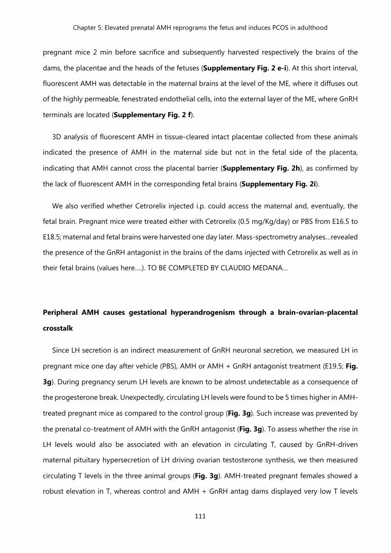
Chapter 5: Elevated prenatal AMH reprograms the fetus and induces PCOS in adulthood
111
pregnant mice 2 min before sacrifice and subsequently harvested respectively the brains of the
dams, the placentae and the heads of the fetuses (Supplementary Fig. 2 e-i). At this short interval,
fluorescent AMH was detectable in the maternal brains at the level of the ME, where it diffuses out
of the highly permeable, fenestrated endothelial cells, into the external layer of the ME, where GnRH
terminals are located (Supplementary Fig. 2 f).
3D analysis of fluorescent AMH in tissue-cleared intact placentae collected from these animals
indicated the presence of AMH in the maternal side but not in the fetal side of the placenta,
indicating that AMH cannot cross the placental barrier (Supplementary Fig. 2h), as confirmed by
the lack of fluorescent AMH in the corresponding fetal brains (Supplementary Fig. 2i).
We also verified whether Cetrorelix injected i.p. could access the maternal and, eventually, the
fetal brain. Pregnant mice were treated either with Cetrorelix (0.5 mg/Kg/day) or PBS from E16.5 to
E18.5; maternal and fetal brains were harvested one day later. Mass-spectrometry analyses…revealed
the presence of the GnRH antagonist in the brains of the dams injected with Cetrorelix as well as in
their fetal brains (values here….). TO BE COMPLETED BY CLAUDIO MEDANA…
Peripheral AMH causes gestational hyperandrogenism through a brain-ovarian-placental
crosstalk
Since LH secretion is an indirect measurement of GnRH neuronal secretion, we measured LH in
pregnant mice one day after vehicle (PBS), AMH or AMH + GnRH antagonist treatment (E19.5; Fig.
3g). During pregnancy serum LH levels are known to be almost undetectable as a consequence of
the progesterone break. Unexpectedly, circulating LH levels were found to be 5 times higher in AMH-
treated pregnant mice as compared to the control group (Fig. 3g). Such increase was prevented by
the prenatal co-treatment of AMH with the GnRH antagonist (Fig. 3g). To assess whether the rise in
LH levels would also be associated with an elevation in circulating T, caused by GnRH-driven
maternal pituitary hypersecretion of LH driving ovarian testosterone synthesis, we then measured
circulating T levels in the three animal groups (Fig. 3g). AMH-treated pregnant females showed a
robust elevation in T, whereas control and AMH + GnRH antag dams displayed very low T levels

Chapter 5: Elevated prenatal AMH reprograms the fetus and induces PCOS in adulthood
112
(Fig. 3g). We next measured circulating estradiol (E2) and progesterone (P) in those treatment
groups and found a robust decrease of both hormones in the AMH-treated dams as compared to
the vehicle-treated and AMH + GnRH antag-treated dams (Fig. 3g). These data indicate that late
gestational exposure to high AMH is sufficient to generate in dams heightened LH and T levels and
diminished E2 and P concentrations in a GnRH-dependent manner.
Since the placenta expresses AMHR2 both in humans (Novembri, Funghi et al. 2015) and in mice
(Fig. 3h), and because we found high T and low E2 levels in AMH-treated dams, we examined if
AMH treatment could alter aromatase expression in the murine placenta, possibly affecting the
conversion of T into E2. We performed qPCR for the enzyme P450 aromatase (CYP19A1) gene in the
placentae harvested from pregnant mice at the end of the treatment period (Fig. 3h). AMH
treatment significantly lowered placental aromatase expression (Fig. 3h). Intriguingly, GnRH antag
treatment prevented the AMH-driven inhibition of CYP19A1 mRNA (Fig. 3h). We thus investigated
the expression of GnRH, GnRH receptor (GnRHR) and LH receptor (LHR) transcripts in the placentae
collected from those animals (Supplementary Fig. 3). All genes were found to be expressed in the
placenta of the different treatment groups. However, while the expression of GnRH and GnRHR was
unchanged among treatment groups, LHR expression was found to be significantly higher in the
AMH-treated dams and to be normalized by the GnRH antag (Supplementary Fig. 3). We next
quantified placental expression of CYP11A1 and HSD3B1, two other key steroidogenic enzymes
involved in onset of steroidogenesis and derivation of progesterone, respectively. We did not find
any difference in the expression of CYP11A1 among treatment groups but we found greatly
diminished HSD3B1 expression in the placentae of AMH-treated- as compared to control dams (Fig.
3h). In addition, GnRH antag-treatment prevented the downregulation of HSD3B1 gene expression,
indicating normalization of the GnRH/LH-driven maternal steroidogenesis.
As T is an anabolic hormone, we monitored whether dams increased their body weight and fat
deposition as a consequence of the AMH treatment (Supplementary Fig. 4a-c). We did not find
any changes in body weight, subcutaneous fat mass and perigonadal fat mass in E19.5 dams injected
for 3 days with vehicle, AMH or AMH + GnRH antag (Supplementary Fig. 4a-c).
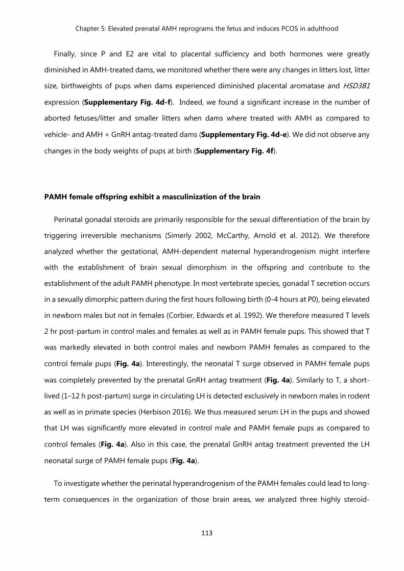
Chapter 5: Elevated prenatal AMH reprograms the fetus and induces PCOS in adulthood
113
Finally, since P and E2 are vital to placental sufficiency and both hormones were greatly
diminished in AMH-treated dams, we monitored whether there were any changes in litters lost, litter
size, birthweights of pups when dams experienced diminished placental aromatase and HSD3B1
expression (Supplementary Fig. 4d-f). Indeed, we found a significant increase in the number of
aborted fetuses/litter and smaller litters when dams where treated with AMH as compared to
vehicle- and AMH + GnRH antag-treated dams (Supplementary Fig. 4d-e). We did not observe any
changes in the body weights of pups at birth (Supplementary Fig. 4f).
PAMH female offspring exhibit a masculinization of the brain
Perinatal gonadal steroids are primarily responsible for the sexual differentiation of the brain by
triggering irreversible mechanisms (Simerly 2002, McCarthy, Arnold et al. 2012). We therefore
analyzed whether the gestational, AMH-dependent maternal hyperandrogenism might interfere
with the establishment of brain sexual dimorphism in the offspring and contribute to the
establishment of the adult PAMH phenotype. In most vertebrate species, gonadal T secretion occurs
in a sexually dimorphic pattern during the first hours following birth (0-4 hours at P0), being elevated
in newborn males but not in females (Corbier, Edwards et al. 1992). We therefore measured T levels
2 hr post-partum in control males and females as well as in PAMH female pups. This showed that T
was markedly elevated in both control males and newborn PAMH females as compared to the
control female pups (Fig. 4a). Interestingly, the neonatal T surge observed in PAMH female pups
was completely prevented by the prenatal GnRH antag treatment (Fig. 4a). Similarly to T, a short-
lived (1–12 h post-partum) surge in circulating LH is detected exclusively in newborn males in rodent
as well as in primate species (Herbison 2016). We thus measured serum LH in the pups and showed
that LH was significantly more elevated in control male and PAMH female pups as compared to
control females (Fig. 4a). Also in this case, the prenatal GnRH antag treatment prevented the LH
neonatal surge of PAMH female pups (Fig. 4a).
To investigate whether the perinatal hyperandrogenism of the PAMH females could lead to long-
term consequences in the organization of those brain areas, we analyzed three highly steroid-
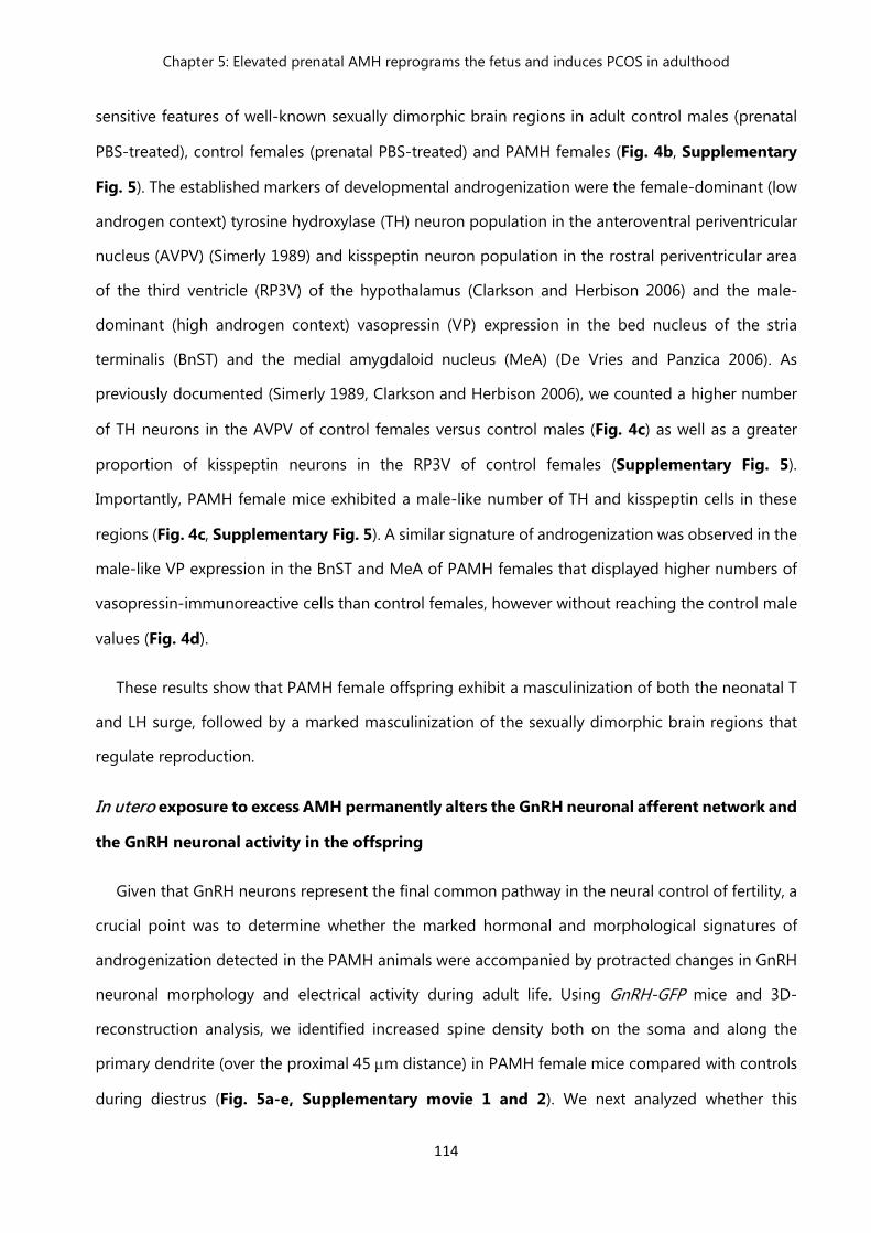
Chapter 5: Elevated prenatal AMH reprograms the fetus and induces PCOS in adulthood
114
sensitive features of well-known sexually dimorphic brain regions in adult control males (prenatal
PBS-treated), control females (prenatal PBS-treated) and PAMH females (Fig. 4b, Supplementary
Fig. 5). The established markers of developmental androgenization were the female-dominant (low
androgen context) tyrosine hydroxylase (TH) neuron population in the anteroventral periventricular
nucleus (AVPV) (Simerly 1989) and kisspeptin neuron population in the rostral periventricular area
of the third ventricle (RP3V) of the hypothalamus (Clarkson and Herbison 2006) and the male-
dominant (high androgen context) vasopressin (VP) expression in the bed nucleus of the stria
terminalis (BnST) and the medial amygdaloid nucleus (MeA) (De Vries and Panzica 2006). As
previously documented (Simerly 1989, Clarkson and Herbison 2006), we counted a higher number
of TH neurons in the AVPV of control females versus control males (Fig. 4c) as well as a greater
proportion of kisspeptin neurons in the RP3V of control females (Supplementary Fig. 5).
Importantly, PAMH female mice exhibited a male-like number of TH and kisspeptin cells in these
regions (Fig. 4c, Supplementary Fig. 5). A similar signature of androgenization was observed in the
male-like VP expression in the BnST and MeA of PAMH females that displayed higher numbers of
vasopressin-immunoreactive cells than control females, however without reaching the control male
values (Fig. 4d).
These results show that PAMH female offspring exhibit a masculinization of both the neonatal T
and LH surge, followed by a marked masculinization of the sexually dimorphic brain regions that
regulate reproduction.
In utero exposure to excess AMH permanently alters the GnRH neuronal afferent network and
the GnRH neuronal activity in the offspring
Given that GnRH neurons represent the final common pathway in the neural control of fertility, a
crucial point was to determine whether the marked hormonal and morphological signatures of
androgenization detected in the PAMH animals were accompanied by protracted changes in GnRH
neuronal morphology and electrical activity during adult life. Using GnRH-GFP mice and 3D-
reconstruction analysis, we identified increased spine density both on the soma and along the
primary dendrite (over the proximal 45 µm distance) in PAMH female mice compared with controls
during diestrus (Fig. 5a-e, Supplementary movie 1 and 2). We next analyzed whether this
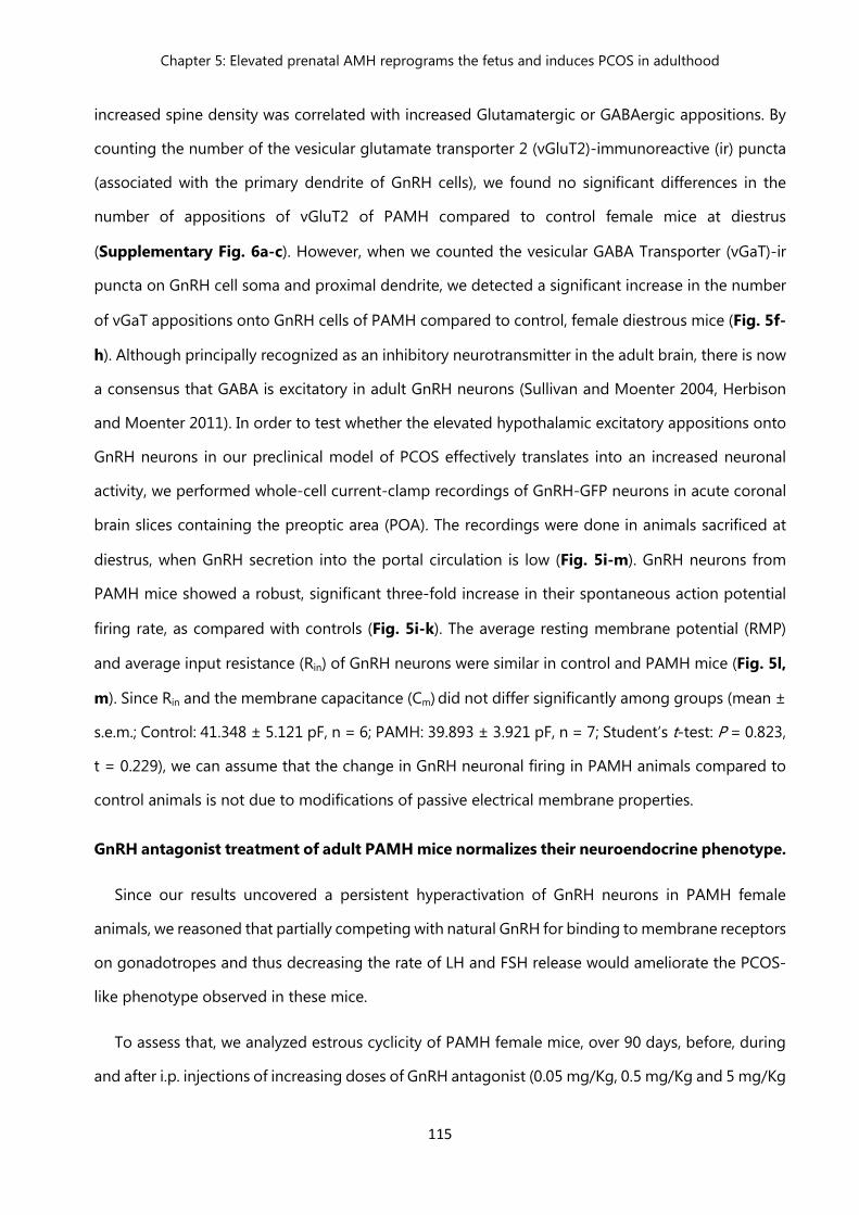
Chapter 5: Elevated prenatal AMH reprograms the fetus and induces PCOS in adulthood
115
increased spine density was correlated with increased Glutamatergic or GABAergic appositions. By
counting the number of the vesicular glutamate transporter 2 (vGluT2)-immunoreactive (ir) puncta
(associated with the primary dendrite of GnRH cells), we found no significant differences in the
number of appositions of vGluT2 of PAMH compared to control female mice at diestrus
(Supplementary Fig. 6a-c). However, when we counted the vesicular GABA Transporter (vGaT)-ir
puncta on GnRH cell soma and proximal dendrite, we detected a significant increase in the number
of vGaT appositions onto GnRH cells of PAMH compared to control, female diestrous mice (Fig. 5f-
h). Although principally recognized as an inhibitory neurotransmitter in the adult brain, there is now
a consensus that GABA is excitatory in adult GnRH neurons (Sullivan and Moenter 2004, Herbison
and Moenter 2011). In order to test whether the elevated hypothalamic excitatory appositions onto
GnRH neurons in our preclinical model of PCOS effectively translates into an increased neuronal
activity, we performed whole-cell current-clamp recordings of GnRH-GFP neurons in acute coronal
brain slices containing the preoptic area (POA). The recordings were done in animals sacrificed at
diestrus, when GnRH secretion into the portal circulation is low (Fig. 5i-m). GnRH neurons from
PAMH mice showed a robust, significant three-fold increase in their spontaneous action potential
firing rate, as compared with controls (Fig. 5i-k). The average resting membrane potential (RMP)
and average input resistance (Rin) of GnRH neurons were similar in control and PAMH mice (Fig. 5l,
m). Since Rin and the membrane capacitance (Cm) did not differ significantly among groups (mean ±
s.e.m.; Control: 41.348 ± 5.121 pF, n = 6; PAMH: 39.893 ± 3.921 pF, n = 7; Student’s t-test: P = 0.823,
t = 0.229), we can assume that the change in GnRH neuronal firing in PAMH animals compared to
control animals is not due to modifications of passive electrical membrane properties.
GnRH antagonist treatment of adult PAMH mice normalizes their neuroendocrine phenotype.
Since our results uncovered a persistent hyperactivation of GnRH neurons in PAMH female
animals, we reasoned that partially competing with natural GnRH for binding to membrane receptors
on gonadotropes and thus decreasing the rate of LH and FSH release would ameliorate the PCOS-
like phenotype observed in these mice.
To assess that, we analyzed estrous cyclicity of PAMH female mice, over 90 days, before, during
and after i.p. injections of increasing doses of GnRH antagonist (0.05 mg/Kg, 0.5 mg/Kg and 5 mg/Kg
Chapter 5: Elevated prenatal AMH reprograms the fetus and induces PCOS in adulthood
116
Cetrorelix acetate/per injection; Fig. 6a). We monitored daily vaginal cytology of PAMH animals
throughout this time: before the beginning of the treatments, during the Cetrorelix administrations
and during the recovery times (discontinuation of treatment; Fig. 6a).
As shown in Figure 2, PAMH mice display prolonged time in metestrus/diestrus as compared to
the control female offspring. Therefore, we initially monitored estrous cyclicity and LH
concentrations in PAMH mice for 12 days before the beginning of Cetrorelix treatment to ascertain
that the animals included in the experimental paradigm displayed the typical PCOS-like
neuroendocrine alterations. Subsequently, in order to normalize the GnRH action on the pituitary
(partial inhibition of GnRH receptor), we injected these mice i.p. every second day, for 12 days, with
Cetrorelix acetate at the dose of 0.05 and 0.5 mg/Kg, respectively (Fig. 6a). Tail-blood samples were
collected for LH measurements twice during the GnRH antagonist treatment and once during the
10 days recovery time (no treatment), that followed each administration period.
To attain a complete blockade of GnRH receptor and suppression of LH secretion, we finally
administered daily 5 mg/Kg Cetrorelix acetate over 12 days (Fig. 6a).
As expected, PAMH mice were oligo-anovulatory, displaying only about 25% of completed
estrous cycles during 12 days as compared to control mice (Fig. 6b). Notably, normal estrous
cyclicity was restored only when animals were injected with 0.5 mg/Kg of GnRH antagonist but not
with the lowest or highest doses of the antagonist (0.05 mg/Kg and 5 mg/Kg Cetrorelix acetate; Fig.
6b).
LH concentrations were also measured in PAMH and Control mice before the beginning of the
treatments (Cetrorelix acetate for the PAMH group and PBS for the Control group), at day 2 and day
6 of each treatment and 4 days after the last injection (recovery time; Fig. 6c). The time-course and
dose-effect experiments showed that the aberrant mean LH values initially observed in PAMH mice
were normalized following the GnRH antagonist treatment at the 0.5 mg/Kg dose only (Fig. 6c, d).
Serum LH levels following the injections with 0.05 mg/Kg Cetrorelix acetate were found to be
significantly greater than in Control animals, whereas they were significantly suppressed when mice
were treated with the highest dose at 5 mg/Kg (Fig. 6c, d). As in clinical practice, the effects of
Chapter 5: Elevated prenatal AMH reprograms the fetus and induces PCOS in adulthood
117
Cetrorelix on LH were reversible few days after discontinuation of treatment, independently by the
dose of the injected drug (Fig. 6c).
After having identified the concentration of the GnRH antagonist which corrected the serum LH
levels and estrous cyclicity in adult PCOS-like animals (0.05 mg/Kg), we performed the rest of the
experiments with this dose (Fig. 6e-i). We monitored the % of time that animals spent in each stage
of the estrous cycle (Metestrus/Diestrus, M/D; Estrus, E; Proestrus, P) and we showed that the GnRH
antagonist restored ovulation of PAMH animals (Fig. 6e).
The aberrant T concentrations and LH pulsatility observed in PAMH mice were also normalized
following the Cetrorelix treatment (Fig. 6g-i). These data show that postnatal GnRH antagonist
treatment can rescue the major PCOS-like traits of PAMH mice: oligo-anovulation,
hyperandrogenism, increased LH concentrations and LH pulsatility.
Chapter 5: Elevated prenatal AMH reprograms the fetus and induces PCOS in adulthood
118
Material and Methods
Study population: human patients
Blood samples were derived from the population-based Uppsala Biobank of Pregnant Women,
where blood samples are collected in conjunction with routine ultrasound screening during
gestational weeks 16-19. Gestational age was determined according to the ultrasound-based
estimated date of birth. Eligible women were 18 years or older and without blood-borne disease. In
Sweden, all pregnant women are invited to a routine ultrasound examination, and approximately
97% of the pregnant population participate, hence, the Biobank subjects likely represent a
population-based sample. Invitation to participate in the Biobank is random, with 70% of
respondents accepting participation, it is estimated that the Biobank covers approximately half of
the pregnant population of Uppsala County {Granfors, 2013 #538} Upon inclusion, brief
demographic data are collected, including ongoing chronic disorders, ongoing medication,
smoking, height and weight. Following written informed consent, a plasma sample is collected. The
sample was centrifuged within two hours and stored at -80°C.
By September 2013, 160 women with PCOS had donated a blood sample to the biobank, and from
these blood samples, 32 normal-weight and 34 obese women with PCOS were selected for the
present study based on their age (27-39 years old). PCOS was diagnosed according to Rotterdam
criteria, i.e. presence of 2 of the following criteria: 1) polycystic ovaries on transvaginal ultrasound,
2) oligoamenorrhea 3) hyperandrogenism (either biochemical or clinical). Hyperandrogenism was
defined as pre-pregnancy hyperandrogenism: either elevated testosterone, or elevated free
androgen index (testosterone/sex hormone binding globulin > 0.05) or pre-pregnancy hirsutism as
judged at the time of diagnosis. All women with PCOS had normal prolactin levels and thyroid
function. Based on the initial, pre-pregnancy, diagnostic work-up, each woman was further
phenotyped as hyperandrogenic PCOS (with signs of biochemical and/or clinical hyperandrogenism)
or normoandrogenic PCOS. The medical records of all women with PCOS were scrutinized to ensure
a correct diagnosis of PCOS, and to obtain information on obstetric and perinatal outcomes.
For each woman with PCOS an age- and BMI matched healthy control was chosen. Each control had
donated blood samples during the same week as the respective case and were healthy according to
the self-report. The medical records of the controls were reviewed to ensure than none had been
Chapter 5: Elevated prenatal AMH reprograms the fetus and induces PCOS in adulthood
119
diagnosed with PCOS or female factor infertility previously. Three blood samples from the controls
could not be retrieved; hence 63 healthy controls were available for analysis. All women gave written
informed consent for inclusion and the study has been approved by the Independent Ethical Review
Board of Uppsala, Sweden.
AMH levels during pregnancy
Serum AMH levels were measured on samples stored at -80°C using the fully automated Acces Dxi
sandwich immunoassay (B13127, Beckman Coulter). This assay measures the proAMH and the
cleaved AMHN,C complex and uses recombinant human AMH as a calibrator. The limit of
quantification of the assay is 0.57 pmol/L. Its intra- and inter-assay imprecision is < 5%. Results are
expressed in pmol/L.
Animals
Timed-pregnant female wild-type C57BL/6J (B6) (Charles River, USA) and transgenic Gnrh-Gfp
{Spergel, 1999 #539} were group-housed under specific pathogen-free conditions in a temperature-
controlled room (21-22°C) with a 12-h light/dark cycle and ad libitum access to food and water.
Standard diet (9.5mm Pelleted RM3, Special Diets Services, France) was given to all mice during
breeding, lactation and growth of young stock. Nutritional profile of the standard diet RM3 is the
following: Protein 22.45%, Fat 4.2%, Fiber 4.42%, Ash 8%, Moisture 10%, Nitrogen free extract 50.4%;
Calories: 3.6 kcal/gr.
Mice were randomly assigned to groups at the time of purchase or weaning to minimize any
potential bias. No data sets were excluded from analyses.
C57BLl/6J Gnrh-Gfp transgenic mice have been previously characterized {Spergel, 1999 #539}.
Animal studies were approved by the Institutional Ethics Committees of Care and Use of
Experimental Animals of the University of Lille (France; Ethical protocol number: APAFIS#2617-
2015110517317420 v5). All experiments were performed in accordance with the guidelines for
animal use specified by the European Council Directive of 22 September 2010 (2010/63/EU). The
sample size, sex and age of the animals used is specified in the text and/or figure legends.

Chapter 5: Elevated prenatal AMH reprograms the fetus and induces PCOS in adulthood
120
Genotyping
Tail biopsies were used to isolate genomic DNA to identify the sex of embryos and mice at birth
using Ube1 primers: Ube1 (forward), 5’CACCTGCACGTTGCCCTT-3’ and Ube1 (reverse),
5’TGGATGGTGTGGCCAATG-3’. Males were identified with two bands marking both X (252 bp) and
Y (334 bp) chromosomes and females with a single band representing only the X chromosome at
252 bp. Gnrh-Gfp transgenic animals were genotyped as previously described {Spergel, 1999 #539}.
Prenatal anti-Müllerian Hormone (PAMH) treatment
Timed-pregnant adult (3-4 months) C57BL6/J (B6) dams were injected daily intraperitoneally (i.p.)
from embryonic day (E) 16.5 to 18.5 with 200 µL of a solution containing respectively: 1) 0.01 M
phosphate buffered saline (PBS, pH 7.4, prenatal control-treated), 2) PBS with 0.12 mgKg-1/d human
anti-Müllerian hormone (AMH) (AMHC, R&D Systems, rhMIS 1737-MS-10, prenatal AMH (PAMH)-
treated), 3) PBS with 0.12 mgKg-1/d AMH and 0.5 mgKg-1 of the GnRH antagonist, cetrorelix acetate
(Sigma, Cat #C5249; PAMH + GnRH antag-treated), 4) 0.5 mgKg-1/d of cetrorelix acetate in PBS
(prenatal GnRH antag-treated), and 5) PBS with 0.12 mgKg-1/d proAMH (Origene Technologies, Cat
#TP308397; prenatal proAMH-treated, PproAMH).
Gnrh-Gfp adult females (P60-P90) were paired with males and checked for copulatory plugs,
indicating day 1 of gestation. Pregnant Gnrh-Gfp dams were given either 200µl i.p. injections of PBS
(control-treated) or 0.12 mgKg-1/d AMHc in PBS from E16.5, E17.5 and E18.5. PAMH-Gnrh-Gfp and
Control-Gnrh-Gfp offspring were weaned, genotyped and phenotyped during postnatal life.
Assessment of phenotype, estrous cycle and fertility
Control, PAMH, PAMH+GnRH antag, GnRH antag, and PproAMH female offspring were weaned at
post-natal day P21 and checked for vaginal opening (VO) and time of first estrus. Anogenital
distance (AGD) and body mass (grams, g) were measured at different ages during post-natal
development (P30, 35, 40, 50 and 60). At VO and in adulthood (P60), vaginal smears were performed
daily for 16 consecutive days (4-cycles) for analysis of age of first estrus and estrous cyclicity. Vaginal
cytology was analyzed under an inverted microscope to identify the specific day of the estrous cycle.
Chapter 5: Elevated prenatal AMH reprograms the fetus and induces PCOS in adulthood
121
The reproductive competency of these animals was determined by pairing the following mice:
Control females mated with untreated C57BL6J (B6) wild-type males, PAMH females mated with
PAMH males, PAMH+GnRH antag females mated with PAMH+GnRH antag males, and GnRH antag
females mated with GnRH antag males for a period of 3 months. Unexperienced males and
primiparous females, selected from at least three different litters, were used for the 90-days mating
protocol test.
Number of pups/litter (number of pups), fertility index (number of litters per females over 3 months),
and time to first litter (number of days to first litter after pairing) were quantified per treatment and
pairing.
Postnatal GnRH antagonist treatment in PAMH mice and assessment of phenotype and
hormone levels.
PAMH adult female offspring were cycled for 12-days before GnRH antagonist treatments. Vaginal
cytology was analyzed under an inverted microscope to record the specific day of the estrous cycle.
Animals were injected intraperitoneally (i.p.) with 200µL of a solution containing 0.01M phosphate
buffered saline (PBS, pH 7.4) with Cetrorelix acetate at 0.05 and 0.5 mg/Kg, every second day, or
daily with Cetrorelix acetate at the dose of 5 mg/Kg. Tail-blood samples were collected for LH
measurements twice before the beginning of the treatments, and at day 2 and 6 of each treatment
as well as 4 days after the last injection (no treatment). Estrous cyclicity was monitored every day at
the same time of day for the 12 days during postnatal GnRH antagonist treatments.
Ovarian histology
Ovaries were collected from 3-4-month-old diestrus mice, immersion-fixed in 4% PFA solution and
stored at 4°C. Paraffin-embedded ovaries were sectioned at a thickness of 5 µm (histology facility,
University of Lille 2, France) and stained with hematoxylin-eosin (Sigma Aldrich, Cat # GHS132,
HT1103128). Sections were examined throughout the ovary. Total numbers of large antral follicles
(containing a single large antrum), atretic and cysts-like follicles (large fluid-filled cysts with an
attenuated granulosa cell layer, dispersed theca cell layer, and an oocyte lacking connection to the
granulosa cells), and corpora lutea (CL) were classified and quantified as previously reported
Chapter 5: Elevated prenatal AMH reprograms the fetus and induces PCOS in adulthood
122
{Caldwell, 2017 #540}. To avoid repetitive counting, each follicle was only counted in the section
where the oocyte’s nucleolus was visible. Using an ocular scale, follicles were classified by diameter
into preantral, large growing (200–400 μm) and cystic follicles (> 400 μm). To avoid repetitive
counting, CL were counted every 100 µm by comparing the section with the preceding and following
sections. CL were characterized by a still present central cavity, filled with blood and follicular fluid
remnants or by prominent polyhedral to round luteal cells.
Perfusions
Adult mice (P60-P90) were anesthetized with i.p. injections of 100 mg/kg of ketamine-HCl and 10
mg/kg xylazine-HCl and perfused transcardially with 20ml of saline, followed by 100 ml of 4% PFA
in 0.1M phosphate buffer (PB) (4% PFA/0.1M PB; pH 7.6). Brains were collected, post-fixed in the
same fixative for 2h at 4 °C, cryoprotected overnight in PB/Sucrose 30% at 4°C, embedded in OCT-
embedding medium (Tissue-Tek), frozen and stored at -80°C until cryosectioning.
Immunohistochemistry
Tissues were cryosectioned coronally (Leica cryostat) at 35µm for free-floating
immunohistochemistry (IHC). Sections were blocked in an incubation solution of Tris-buffered saline
(TBS 0.05M, pH 7.6), 0.25% bovine serum albumin (BSA; Sigma, A9418), 0.3% Triton X-100 (TBS-T;
Sigma, T8787) with 10% normal donkey serum (NDS; D9663; Sigma) for 2h at room temperature
before incubation with the following different primary antisera (depending on experiment) for 72h
at 4°C: guinea pig anti-GnRH (EH#1018; 1:10,000){Hrabovszky, 2010 #349}{Casoni, 2016 #37}, a
generous gift from E. Hrabovszky (Laboratory of Endocrine Neurobiology, Institute of Experimental
Medicine of the Hungarian Academy of Sciences, Budapest, Hungary), rabbit anti-GFP (Abcam
ab6556; 1:1000), mouse monoclonal anti-vesicular GABA Transporter, vGaT) antibody (Synaptic
Systems #131001; 1:500), guinea pig anti-vesicular glutamate transporter 2 (vGluT2) (Synaptic
Systems #135404; 1:500), monoclonal mouse anti-Tyrosine Hydroxylase (TH) antibody (Millipore
MAB318; 1:3000), guinea pig anti-(Arg8)-Vasopressin (Peninsula Laboratories #T-5048; 1:2500),
rabbit anti-cFos (Santa Cruz Biotechnology #sc-52; 1:5000), and a rabbit anti-kisspeptin (A. Caraty
#566 1:10,000, Institut National de la Recherche Agronomique){Clarkson, 2014 #542}. After TBS

Chapter 5: Elevated prenatal AMH reprograms the fetus and induces PCOS in adulthood
123
rinses, immunoreactivity (i.r) was revealed using the corresponding secondary antibodies (Life
Technologies, Molecular Probes, Invitrogen) all 1:400, [Alexa-Fluor 488-conjugated #A21206; Alexa-
Fluor 647-conjugated #A31573, #A31571; Alexa-Fluor 568-conjugated #A10042, #A10037] for 90
min in incubation solution at RT. After TBS washes, sections were incubated with 0.02% Hoechst
(H3569; Invitrogen) in TBS-T for 15-mins at RT and mounted on gelatin-coated slides and
coverslipped with mowiol medium (Sigma #81381).
Sections were examined using an Axio Imager. Z1 ApoTome microscope (Carl Zeiss, Germany)
equipped with a motorized stage and an AxioCam MRm camera (Zeiss). For confocal observation
and analyses, an inverted laser scanning Axio observer microscope (LSM 710, Zeiss) with an EC Plan
NeoFluor ×100/1.4 numerical aperture oil-immersion objective (Zeiss) was used with an argon laser
exciting at 488nm and helium laser exciting at 543nm (Imaging Core Facility of IFR114, of the
University of Lille 2, France).
3D rendering and movies of GnRH neurons were generated using Imaris ×64 software (7.6.1,
Bitplane).
Immunohistochemical analysis
Assessment of Fos-GnRH co-expression
Adult C57BL6/J (B6) females in diestrus were given i.p. injections of a solution containing 200µL of
0.01M PBS, pH 7.4 or PBS with 0.12 mgKg-1/d AMHC at 10:00. 90-min after injection, each mouse
was anaesthetized and perfused transcardially as described above (Perfusions). Brains were
collected, post-fixed in the same fixative for 2h at 4°C, embedded in OCT-embedding medium,
frozen and cryosectioned coronally (Leica cryostat) at 35µm for free-floating IHC. Sections were
processed for dual-label Fos-GnRH IHC.
Analysis of the double-labeled tissue was undertaken by counting the number of single-labeled and
dual-labeled GnRH neurons in the preoptic area (POA: OVLT), with 2-3 brain sections representing
the POA counted per mouse. Sections were scanned using a 20× objective to image Fos-GnRH
double positively labelled cells for quantification. Activated neurons were identified as expressing
Fos in the nucleus of the GnRH i.r. cell. The number of GnRH neurons co-expressing Fos was
quantified and grouped to produce a mean percentage of the co-expression of GnRH-Fos in both

Chapter 5: Elevated prenatal AMH reprograms the fetus and induces PCOS in adulthood
124
PBS- and AMH-treated groups. The data from each mouse were then grouped to provide the median
percentage ± interquartile range (IQR).
For the analysis of the total Fos-i.r cells in the POA, the same sections described above were
examined using a 10 × objective. The total number of Fos positive cells per POA (OVLT) section in
each treatment was quantified using the particle analysis program from Fiji (NIH); the total number
of Fos-positive cells in the OVLT were quantified and averaged across OVLT sections from each
animal and treatment.
Analysis of sexually dimorphic brain nuclei
For single-labeled Tyrosine Hydroxylase (TH) IHC, the number of TH-i.r cell bodies located within
anteroventral periventricular nuclei were counted. For single-labeled Vasopressin (VP) IHC, the
number of VP-i.r cell bodies located within the bed nucleus stria terminalis (BNST) and Medial
Amygdala (MeA) were counted. For both TH and VP single-labeled IHC, each section containing the
AVPV or the BNST and MeA were analyzed in each sex, mouse and treatment (3-5 sections per
mouse). For single-labeled kisspeptin IHC, the number of kisspeptin-i.r cell bodies were counted in
the rostral periventricular area of the third ventricle (RP3V), which comprises the anteroventral
periventricular nucleus (AVPV) and the preoptic periventricular nucleus. Two brain sections at each
level of the RP3V were analyzed per mouse, and the number of kisspeptin positive cells counted and
combined to produce the mean number of kisspeptin neurons per section for the RP3V of each
mouse.
The mean number of TH-, VP- and Kiss-i.r neurons counted per section for the sexually dimorphic
brain regions of each mouse and treatment were then grouped to provide the mean ± s.e.m. values
for the experimental groups.
Assessment of GnRH neuron spine density
Coronal brain sections from adult (P60-90) female Control-Gnrh-Gfp and PAMH-Gnrh-Gfp animals
were analyzed for GnRH neuron spine density.
Ten GFP-i.r GnRH neurons located within the rPOA were randomly chosen from adult (P60-P90)
diestrus Control-Gnrh-Gfp (n=5) and PAMH-Gnrh-Gfp female mice (n=5) and were imaged using a

Chapter 5: Elevated prenatal AMH reprograms the fetus and induces PCOS in adulthood
125
LSM 710, Zeiss upright confocal laser scanning microscope equipped with LSM 710 software. For
each GnRH-GFP cell, a stack of images at 0.25µm intervals were collected using a 100× objective
and 2× digital zoom function throughout the entire depth of the GFP-i.r neuron. Images containing
the cell body and initial portion of the primary dendrite were collected and soma and dendritic spine
density were determined. Spines were identified as protrusions from the somata or dendrite and
defined as being less than 5µm in length from the soma or proximal dendrite (the dendrite with the
greatest circumference extending from the GnRH soma). GnRH neuron soma spine density was
expressed as the number of spines per µm relative to the soma circumference. Spine density was
quantified every 15µm along the entire length of the proximal GnRH dendrite (0-45µm) as 45µm
was the longest length of the proximal dendritic that could be imaged before the dendrite exited
the section. Spine density was expressed as the number of spines/µm.
Analysis of vGaT or vGluT2 appositions on Gnrh-Gfp neurons
Coronal brain sections from adult (P60-90) female Control-Gnrh-Gfp and PAMH-Gnrh-Gfp animals
were analyzed for GnRH neuron spine and vGaT or vGluT2 density. Between 8-10 GFP/vGaT or
GFP/vGluT2 double labeled neurons located within the rPOA from Control;Gnrh-Gfp and
PAMH;Gnrh-Gfp adult (P60-P90) diestrus female mice were imaged using the same criteria above.
A z-series stack of images using a 100× objective was generated to estimate the density of vGaT or
vGluT2 appositions. A contact was defined when there were no black pixels between the primary
Gnrh-Gfp dendritic spine and vGaT or vGluT2 positive terminals. For each image, the number of
vGaT- or vGluT2-labeled puncta directly opposed to GFP-labeled neuronal soma and dendrite (up
to 45µm along the GnRH primary dendrite) were counted and combined to provide the mean values
for each cell. The primary GFP-i.r dendrite could not be followed for more than 45µm from the cell
body, therefore we determined the number of vGaT appositions every 15µm until the dendrite exited
the section. Data are presented as vGaT or vGluT2 appositions/µm.
Fluorescent AMH assays
Recombinant bioactive AMH (AMHC, R&D Systems, rhMIS 1737-MS-10) was tagged with a
fluorescent dye (d2; MW: approx. 800 Da, max light output at 665nm) by Cisbio Bioassays (Codolet,
Chapter 5: Elevated prenatal AMH reprograms the fetus and induces PCOS in adulthood
126
France) as previously described for other hormones{Balland, 2014 #543}, {Schaeffer, 2013 #519}, {Xu,
2017 #545}. Fluorescently-labeled bioactive AMH (2.5 nmoles/animal; Cisbio Bioassays) was injected
into the jugular vein of pregnant mice (E16.5), anesthetized with ketamine/xylazine, and mice
sacrificed 2 min later. Dams’ brains, fetal brains and placentae were collected and immersion-fixed
in 4% PFA overnight at 4 °C. Brains were cryoprotected overnight in PB/Sucrose 30% at 4°C,
embedded in OCT-embedding medium, frozen and stored at -80 °C until cryosectioning. Placentae
were processed for iDisco tissue-clearing as described below.
Cetrorelix measurement in biological samples
Sample preparation for nano-HPLC-HRMS analysis
Timed-pregnant adult (3-4 months) C57BL6/J (B6) dams were injected daily i.p. from embryonic day
(E) 16.5 to 18.5 with 200µL of a solution containing respectively: 1) 0.01M phosphate buffered saline
(PBS, pH 7.4, prenatal control-treated), 2) PBS with 0.5 mgKg-1 of the GnRH antagonist, cetrorelix
acetate (GnRH antag-treated). At E19.5, dams were anesthetized with ketamine/xylazine, and
sacrificed by cervical dislocation. Dams’ brains and fetal brains were collected, snap-frozen in liquid
nitrogen and stored at -80 °C until nano-HPLC-HRMS experiments. Murine maternal and embryo
brain samples were prepared adapting the procedure described. Briefly, brains were weighted,
homogenized with 1% formic acid in cold acetonitrile and incubated for 15 min at -20°C. After
centrifugation, the supernatant was freeze-dried and reconstituted with 3% acetic acid and 1%
trifluoroacetic acid (TFA). all solvents sourced from Sigma-Aldrich Merck. The reconstituted sample
was purified with a solid phase extraction using a Phenomenex polymeric reversed phase cartridge
(Strata-X 33µm Polymeric Reversed Phase, 60 mg/3mL, Phenomenex, Bologna, Italy). The cartridges
were equilibrated with a methanol and water solution of acetic acid and TFA (96:3:1 v/v), loaded with
sample, washed with a 70:30 solution of water solution with acetic acid and TFA (96:3:1 v/v) and
methanol, and eluted with a 90:10 solution of methanol and 3% acetic acid. The eluted solution was
gently dried under stream on nitrogen and reconstituted with 0.1% formic acid. When brains were
homogenized, an internal standard (ganirelix, empirical formula C80H113ClN18O13) was added to
obtain a final concentration of 100ng/ml.
Chapter 5: Elevated prenatal AMH reprograms the fetus and induces PCOS in adulthood
127
Nano-HPLC-HRMS analysis parameters
A Dionex Ultimate 3000 (Thermo Scientific, Milan, Italy) nano-HPLC instrument coupled to a Orbitrap
Fusion (Thermo Scientific, Milan, Italy) high resolution hybrid mass analyzer was used.
Seperation was achieved with an EASY-Spray PepMap® column (C18, 2µm, 100Å, 75µm × 500mm,
Thermo Scientific, Milan, Italy) using formic acid 0.1% in water (solvent A) and in acetonitrile:water
8:2 (solvent B) as mobile phases (ultrapure solvents for nano-LC, Sigma-Aldrich, Milan, Italy). The
gradient conditions were as follow: 5% B for 5 min isocratic, up to 100% B in 30 min and
reconditioned for at least 20 min. The first 5 minutes of isocratic flow was mandatory for the
preconcentration step on a µ-precolumn (C18 PepMap 100, 300µm i.d. × 5 mm, Thermo Scientific,
Milan, Italy), and water with 0.1% formic acid and 0.05% TFA. The injection volume was 1 μL and the
flow rate 300 nL/min.
The column was equipped with a nanoESI source and the capillary voltage was 2000V in positive ion
mode. The ion transfer tube temperature was 275°C and the pressure was standard (0.008 Torr). Full
scan spectra were acquired in the range of m/z 350-2000. MSn spectra were acquired in the range
between the ion trap cut-off and precursor ion m/z values. CID collision energy was selected for
each analyte to allow the survival of 5-10% of the chosen precursor ion. High resolution spectra
were acquired with a resolution of 60000 (FWHM) and the mass accuracy of recorded ions (vs.
calculated) was ± 5ppm units (without internal calibration).
Tissue clearing
We used iDisco clearing protocol, as previously described {Renier, 2014 #547}. Samples were washed
in PBS (twice for 1h), followed by 50% methanol in PBS (1h), 80% methanol (1h) and 100% methanol
(2x 1h). Next, samples were bleached in ice cold 5% H2O2 in 20% DMSO/methanol at 4°C overnight.
Next, samples were washed in methanol (2x 1h), in 20% DMSO/methanol (2x 1h), 80% methanol
(1h), 50% methanol (1h), PBS (2x 1h), and stored in PBS at 4°C until processing. Samples were then
incubated at RT on a rotating wheel at 12 rpm in a solution containing: 20% MeOH, 40% MeOH,
60% MeOH, 80% MethOH, 100% MeOH, 100% MeOH; 1h each. Delipidation was achieved with an
overnight incubation in DCM/Methanol (2:1), followed by a 30-min wash in 100% DCM
(Dichloromethane; Sigma-Aldrich). Samples were cleared in dibenzylether (DBE; Sigma-Aldrich) for
Chapter 5: Elevated prenatal AMH reprograms the fetus and induces PCOS in adulthood
128
2h at RT on constant agitation and in the dark. Finally, samples were moved into fresh DBE and
stored in glass tube in the dark and at RT until imaging. 3D imaging was performed as previously
described 72,73. An ultramicroscope (LaVision BioTec) using InspectorPro software (LaVision BioTec)
was used to perform imaging. The light sheet was generated by a laser (wavelength 488 or 561nm,
Coherent Sapphire Laser, LaVision BioTec) and two cylindrical lenses. A binocular stereomicroscope
(MXV10, Olympus) with a 2× objective (MVPLAPO, Olympus) was used at different magnifications
(1.6×, 4×, 5×, and 6.3×). Samples were placed in an imaging reservoir made of 100% quartz (LaVision
BioTec) filled with DBE and illuminated from the side by the laser light. A PCO Edge SCMOS CCD
camera (2,560 × 2,160 pixel size, LaVision BioTec) was used to acquire images. The step size between
each image was fixed at 2μm.
Pulsatile LH measurements
Diestrus female adult mice were habituated with daily handling for 3–weeks. Blood samples (5µl)
were taken from the tail at 10-min intervals for 2h (between 10:00and 12:00) during diestrus, diluted
in 45µl PBS-Tween (0.05%) and immediately frozen and kept at -80°C. LH levels were determined by
the previously described sensitive LH sandwich ELISA. A 96-well high-affinity binding microplate
(Corning) was coated with 50µl of capture antibody (monoclonal antibody, anti-bovine LHβ subunit,
518B7; L. Sibley; University of California, UC Davis) at a final dilution of 1:1000 (in Na2CO3/NaHCO3,
0.1M, pH 9.6) and incubated overnight at 4°C. Wells were incubated with 200µl blocking buffer (5%
(w/v) skim milk powder in 1 X PBS-T pH 7.4 [0.01M PBS, 0.05% Tween 20 (Sigma #P9416)] for 2hr at
RT. A standard curve was generated using a two-fold serial dilution of mouse LH (reference
preparation, AFP-5306A; National Institute of Diabetes and Digestive and Kidney Diseases National
Hormone and Pituitary Program (NIDDK-NHPP) in 0.25% (w/v) BSA (Sigma, A9418) in PBS-T. The
LH standards and blood samples were incubated with 50µl of detection antibody (rabbit LH
antiserum, AFP240580Rb; NIDDK-NHPP) at a final dilution of 1:10,000 for 1.5h at RT. Each well
containing bound substrate was incubated with 50µl of horseradish peroxidase-conjugated
antibody (goat anti-rabbit; Vector Laboratories, PI-1000) at a final dilution of 1:10,000. After a 1.5h
incubation, 100µl 1-StepTM Ultra TMB-Elisa Substrate Solution (ThermoFisher Scientific, Cat #34028)
was added to each well and left at RT for 10 min. The reaction was halted by the addition of 50µl of
Chapter 5: Elevated prenatal AMH reprograms the fetus and induces PCOS in adulthood
129
3M HCL to each well, and absorbance read at 490nm. Pulses were confirmed using DynPeak {Steyn,
2013 #549}.
LH and T ELISA Assays
LH levels were determined by a sandwich ELISA, as described previously{Vidal, 2012 #550}, using the
mouse LH–RP reference provided by A.F. Parlow (National Hormone and Pituitary Program,
Torrance, CA). Plasma T levels were analyzed using a commercial ELISA (Demeditec Diagnostics,
GmnH, DEV9911) {Moore, 2015 #505} according to the manufacturers’ instructions.
LH and T measurement in pregnant mice
Timed-pregnant female C57BL6/J (B6) dams were treated with i.p. injections of 200µL of PBS or 0.12
mgKg-1/d AMH in PBS from E16.5-E18.5. At E19.5, pregnant-treated dams were decapitated and
trunk blood was harvested in sterile Eppendorf tubes and left on ice until centrifugation. Plasma was
collected after centrifugation of blood samples at 3000 g for 15 mins at 4°C and stored at -80° C
until use. Mean T and LH levels were determined as described above.
LH and T measurement in P0 mice
Timed-pregnant C57BL6/J (B6) dams were given i.p injections of 200 µL PBS or 0.12 mgKg-1/d AMH
in PBS from E16.5-E18.5. Dams were allowed to deliver naturally. Pups were harvested 2h after birth.
Tail biopsies of pups were taken to determine the sex of each animal (see Genotyping). Trunk blood
and was collected from P0 males, control females and PAMH female pups for analysis of circulating
testosterone and LH levels. Plasma was collected after centrifugation of blood samples at 3000g for
15 mins at 4°C and stored at -80° C until use. T and LH levels were determined as described above.
Quantitative RT-PCR analyses
E19.5 placental tissue from female control and PAMH embryos was isolated for total RNA was using
Trizol (ThermoFisher Scientific, Cat #15596026) and the RNeasy Lipid Tissue Mini Kit (Qiagen; Cat #
74804) using the manufacturer’s instructions. For gene expression analyses, mRNAs obtainHSD3ed
from E19.5 placental tissue were reverse transcribed using SuperScript® IV Reverse Transcriptase
Chapter 5: Elevated prenatal AMH reprograms the fetus and induces PCOS in adulthood
130
(Life Technologies). Real-time PCR was carried out on Applied Biosystems 7900HT Fast Real-Time
PCR system using exon-boundary-specific TaqMan® Gene Expression Assays (Applied Biosystems):
Amhr2 (AMHR2- Mm00513847_m1); Cyp19a1 (Cyp19a1-Mm00484049_m1); Cyp11a1 (Cyp11a1-
Mm00490735_m1); Hsd3b1 (Hsd3b1-Mm01261921_mH); Gnrh (Gnrh-Mm01315604_m1); Gnrhr
(Gnrhr-Mm00439143_m1); Lhr (Lhcgr-Mm00442931_m1). Control housekeeping genes: ActB (ActB-
Mm00607939_s1); Gapdh (GAPDH-Mm99999915_g1).
Quantitative RT-PCR was performed using TaqMan® Low-Density Arrays (Applied Biosystems) on an
Applied Biosystems thermocycler using the manufacturer’s recommended cycling conditions. Gene
expression data were analyzed using SDS 2.4.1 and Data Assist 3.01 software (Applied Biosystems),
with ActB and Gapdh as control house-keeping mRNA following a standardized procedure. Values
are expressed relative to control values, as appropriate, set at 1.
Brain slice electrophysiology preparation and recordings
Gnrh-Gfp adult females (3-4 months) were paired with adult Gnrh-Gfp males (3-4 months) and
checked for copulatory plugs. Pregnant Gnrh-Gfp dams were given 200µl i.p. injections of PBS or
0.12 mgKg-1/d AMH in PBS on days 16.5, 17.5 and 18.5 of gestation. Adult (3-4 months) female
diestrus Control;Gnrh-Gfp and PAMH;Gnrh-Gfp offspring were anaesthetized with isoflurane, and,
after decapitation, the brain was rapidly removed and put in ice-cold oxygenated (O2 95% / CO2 5%)
artificial cerebrospinal fluid (ACSF) containing the following (in mM): 120 NaCl, 3.2 KCl, 1 NaH2PO4,
26 NaHCO3, 1 MgCl2, 2 CaCl2, 10 glucose, pH 7.4 (with O2 95% / CO2 5%). After removal of the
cerebellum, the brain was glued and coronal slices (150µm thickness) were cut throughout the
septum and preoptic area using a vibratome (VT1200S; Leica). Before recording, slices were
incubated at 34ºC for a recovery period of 1h. After recovery, slices were placed in a submerged
recording chamber (32.8ºC; Warner Instruments) and continuously perfused (2ml/min) with
oxygenated ACSF. GFP-positive GnRH neurons in the hypothalamic preoptic area were visually
identified with a ×40 objective magnification in an upright Leica DM LFSA microscope under a FITC
filter and their cell body observed by using IR-differential interference contrast. Whole-cell patch-
clamp recordings were performed in current-clamp with bridge mode by using a Multiclamp 700B
amplifier (Molecular Devices). Data were filtered at 1kHz and sampled at 5kHz with Digidata 1440A
Chapter 5: Elevated prenatal AMH reprograms the fetus and induces PCOS in adulthood
131
interface and pClamp 10 software (Molecular Devices). Pipettes (from borosilicate capillaries; World
Precision Instruments) of resistance of 6-8 MΩ when filled with an internal solution containing the
following (in mM): 140 K-gluconate, 10 KCl, 1 EGTA, 2 Mg-ATP and 10 HEPES, pH 7.3 with KOH.
Bridge balance was adjusted to compensate for pipette resistance. All recordings were analyzed with
Clampfit 10 (Molecular Devices). Junction potentials were determined to allow correction of
membrane potential values. Electrical membrane properties were measured in current-clamp mode
by applying a series of current pulses from – 60 to + 60 pA (1s, 10pA increments). Input resistance
(Rin) was determined by measuring the slope of the linear portion of the current-voltage curve.
Recordings were terminated if Rin changed more than 10% between the beginning and the end of
the recording. Membrane capacitance (Cm) was calculated using the equation τ=RC, where τ=time
constant, R=resistance, and C=capacitation. Membrane time constant was estimated by fitting a
single exponential to the charging curve (Clampfit 10; Molecular Devices) generated from a
hyperpolarizing current pulse (1s, 10pA) from rest.
Statistical analyses
All analyses were performed using Prism 7 (Graphpad Software, San Diego, CA) and assessed for
normality (Shapiro–Wilk test) and variance, when appropriate. Sample sizes were chosen according
to standard practice in the field. The investigators were not blinded to the group allocation during
the experiments. However, analyses were performed by two independent investigators in a blinded
fashion. For each experiment, replicates are described in the figure legends.
For AMH measurement during pregnancy, as AMH levels were found not to be normally distributed
in the study population, results are reported as median and 25th-75th percentile range. All
comparisons between PCOS and control patients were performed using the nonparametric Mann-
Whitney U test. The significance level was set at P < 0.05.
For animal studies, data were compared using an unpaired two-tailed Student’s t-test or a one-way
ANOVA for multiple comparisons followed by Tukey’s multiple comparison post-hoc test. Data
analyses for percentages were performed using either a Mann-Whitney U test (comparison between
two experimental groups) or Kruskal-Wallis test (comparison between three or more experimental
groups) followed by a Dunn’s post hoc analysis. The number of biologically independent
Chapter 5: Elevated prenatal AMH reprograms the fetus and induces PCOS in adulthood
132
experiments, sample size, P values, age and sex of the animals are all indicated in the main text or
figure legends. All experimental data are indicated as mean ± s.e.m or as the 25th–75th percentile,
line at median. The significance level was set at P < 0.05. Symbols in figures correspond to the
following significance levels: * P < 0.05, ** P < 0.001, ***P < 0.0001.
Chapter 5: Elevated prenatal AMH reprograms the fetus and induces PCOS in adulthood
133
Discussion
Serum AMH levels have been shown to decrease with age both in healthy women and in women
with PCOS, although they remain always 2- to 3-fold higher and elevated until 40 years of age in
PCOS subjects (Piltonen, Morin-Papunen et al. 2005). On the other hand, AMH levels were previously
found to be very low during pregnancy in women without decreased fertility (La Marca, Giulini et al.
2005, Koninger, Kauth et al. 2013) and it has been assumed that this would be also the case for PCOS
individuals. Herein, we provide compelling evidences showing that pregnant women with PCOS have
significantly higher circulating AMH levels than control pregnant women, independently of their BMI
and age. Our data complement the above findings and add up that AMH levels in women with PCOS
remain high until late reproductive age also during gestation.
Notably, most women with PCOS are hyperandrogenic during pregnancy (Sir-Petermann,
Maliqueo et al. 2002, Sir-Petermann, Maliqueo et al. 2007), yet the cause of this remains enigmatic.
Here, we found a positive relationship between AMH levels and hyperandrogenism in pregnant lean
women with PCOS but not in obese PCOS pregnant patients. We do not know whether a causal-
relationship between AMH and T might exist during gestation in humans. However, our results in
mice demonstrate that AMH has a programming effect leading to gestational and perinatal
hyperandrogenism and subsequent changes in the HPG axis and hormone levels of both the dams
and the progeny.
Our data show for the first time that peripheral bioactive AMH (AMHC) can access the maternal
brain, at the level of the ME, and act centrally by inducing GnRH neuronal activation, as
demonstrated by the increase in GnRH+/Fos+ neurons shortly after peripheral administration of
AMH. Circumventricular organs, like the ME and the OVLT, contain highly permeable, fenestrated
endothelial cells, that allow the free passage of molecules below 35 kDa (Schaeffer, Langlet et al.
2013). Therefore, circulating AMHC can have direct access to GnRH dendrites and terminals, which
express AMHR2 both in mice and humans (Cimino, Casoni et al. 2016), and that extend outside the
blood-brain barrier in the OVLT (Herde, Geist et al. 2011) and in the ME.
The AMH in blood is generally presumed to be the bioactive cleaved AMHC (Ragin, Donahoe et
al. 1992), although recent studies suggested that human blood might contain mainly proAMH and
Chapter 5: Elevated prenatal AMH reprograms the fetus and induces PCOS in adulthood
134
AMHN,C (Pankhurst and McLennan 2013, Pankhurst, Chong et al. 2016). Whether the AMHN,C
undergoes some further cleavage, in proximity of the ME, remains to be fully investigated. However,
the fact that peripheral administration of either proAMH or AMHC leads to overlapping phenotypes
in the offspring supports the latter hypothesis.
Several animal models of PCOS rely on the prenatal administration of dihydrotestosterone (DHT)
to induce the emergence of the PCOS-like traits (Roland and Moenter 2014, Moore and Campbell
2017), even though pregnant women do not have DHT in their circulation. Although there are no
animal models that can perfectly recapitulate all PCOS clinical features, we have now generated one
of the most relevant PCOS mouse model, from a clinical perspective. In fact, our model is based on
an AMH-driven gestational hyperandrogenism, which is then responsible for the pathophysiological
alterations leading to the acquisitions of PCOS cardinal defects in the offspring. This fits well with
previous studies demonstrating that gestational hyperandrogenism in monkeys, either occurring
naturally or induced by T treatment, induces PCOS-like reproductive and metabolic traits in
adulthood (Abbott, Nicol et al. 2013, Abbott, Rayome et al. 2017).
Our findings highlight a novel pathophysiological mechanism whereby exposure to AMH excess
during pregnancy leads to a cascade of alterations touching the maternal brain, the ovaries and the
placenta, and resulting into a strong elevation of maternal LH and T, and by a drop of P and E2. The
maternal androgenization is most likely the result of a dual action of AMH on the dams: 1) central
action and exacerbation of GnRH/LH-driven ovarian steroidogenesis and 2) peripheral action and
inhibition of placental aromatase and HSD3b1 expression, leading respectively to an increase in T
bioavailability and a drop of circulating P levels. The 50% reduction in P levels detected in AMH-
treated dams could significantly lower the break of P on LH secretion and further increase T
production by theca cells (Supplementary Fig. 7).
These findings are in line with clinical investigations, which identified lower placental aromatase
expression in women with PCOS (Maliqueo, Lara et al. 2013), implying that a dysregulation of P450
aromatase activity in pregnant women with PCOS might occur and be partly attributable to elevated
levels of circulating AMH.
Chapter 5: Elevated prenatal AMH reprograms the fetus and induces PCOS in adulthood
135
The robust drop of plasmatic P and E2 detected in AMH-treated pregnant mice most likely
compromises their placental function, thus explaining the higher incidence of aborted fetuses
observed in these animals (Supplementary Fig. 7). Interestingly, numerous clinical reports indicate
that women with PCOS have increased risk of poor pregnancy outcomes (Katulski, Czyzyk et al.
2015), most likely due to placental dysfunctions, and lower maternal E2 levels (Maliqueo, Sundstrom
Poromaa et al. 2015). However, whether women with PCOS also have lower levels of P during
pregnancy is currently unknown and deserve further investigation.
Here, we also showed for the first time that the AMH-driven gestational hyperandrogenism
triggers the masculinization of the brain in female offspring (Supplementary Fig. 7). In the
developing male brain the appropriate exposure of androgens in utero is known to contribute to
establishing sexually dimorphic brain circuitry controlling reproductive physiological processes (Lenz
and McCarthy 2010). Interestingly, PAMH female animals exhibit a masculinization of both the
neonatal T and LH surge, followed by a marked masculinization of the sexually dimorphic brain
regions that regulate reproduction. Notably, the neonatal T and LH surges of PAMH female pups
were corrected by prenatal GnRH antagonist administration, strongly indicating that the neonatal
consequences of the gestational AMH treatments are programmed in utero and not neonatally.
Since GnRH neurons do not express androgen receptor or estrogen receptor alpha (ERα) (Huang
and Harlan 1993), we propose that steroid hormone receptor activation should occur in upstream
neuronal afferents. Indeed, we provided evidences indicating that hyperandrogenism during critical
periods of development leads to increased GABAergic appositions to GnRH neurons and to a
persistent GnRH neuronal hyperactivity in adulthood. The alterations in VGaT, observed in PAMH
animals, phenocopy those of PNA mice (Sullivan and Moenter 2004, Moore, Prescott et al. 2015),
and support a modified afferent synaptic input to GnRH neurons. Since GABAergic inputs are known
to excite GnRH neurons (DeFazio and Moenter 2002, Herbison and Moenter 2011), we can postulate
the hypothesis that elevated excitatory innervation of GnRH neurons might be responsible for the
increase in the GnRH/LH pulse frequency that we described in PAMH adult females. Such
hyperactivation of GnRH neurons (Chang 2007) is most likely responsible for the LH-driven ovarian
androgen production over FSH release, which consequently leads to anovulation. Most women who
Chapter 5: Elevated prenatal AMH reprograms the fetus and induces PCOS in adulthood
136
are diagnosed with PCOS exhibit high-frequency LH secretion, which is suggestive of rapid GnRH
pulsatility (Goodarzi, Dumesic et al. 2011). This is the case for 95% of the metabolically healthy PCOS
women, who present high LH levels, but not for obese PCOS patients (Rebar, Judd et al. 1976, Chang,
Mandel et al. 1982, Pastor, Griffin-Korf et al. 1998, Huang, Tien et al. 2015).
It should be noted that both the PNA mice, described before (Sullivan and Moenter 2004, Roland,
Nunemaker et al. 2010, Moore, Prescott et al. 2013), and the PAMH animals, described in this study,
did not present any weight alterations, indicating that these models are most representative of the
“lean” PCOS condition, characterized by elevated circulating LH. Such findings are highly compatible
with recently improved understanding of a leaner PCOS phenotype per se in the general population.
Indeed, studies in referral PCOS populations (studied in the clinical environment) versus medically
unselected populations have suggested that patients with PCOS diagnosed in the referral setting
had a greater BMI and prevalence of obesity than women with PCOS detected in the medically
unselected population (Ezeh, Yildiz et al. 2013, Azziz 2016, Lizneva, Kirubakaran et al. 2016).
While it is clear that the weight of PAMH mice is unaffected, further study is needed to clarify
whether other metabolic parameters known to be altered in hyperandrogenic lean PCOS women
(Dumesic, Akopians et al. 2016) such as glucose tolerance, insulin resistance or intra-abdominal fat
storage, might be also altered in these animals.
Management strategies aimed at treating PCOS have met only limited success and alternative
and preventive therapies are therefore urgently needed.
Here, we showed that the prenatal co-treatment of AMH with the GnRH antagonist prevented
the appearance of PCOS-like neuroendocrine traits in the offspring, thus demonstrating a critical
role for GnRH in the prenatal programming of the disease. Moreover, we also showed that postnatal
GnRH antagonist treatment of adult PAMH mice, at a concentration that only partially compete with
endogenous GnRH for binding to membrane receptors on gonadotropes, restores their ovulation
and normalizes LH secretion/pulsatility as well as androgen levels. Given that GnRH antagonists are
largely used in the clinic, with no adverse secondary effects, pharmacological antagonism aimed at
tempering GnRH/LH secretion is an attractive therapeutic strategy to restore ovulation and fertility
in PCOS individuals characterized by normal body mass composition and high LH levels.
Chapter 5: Elevated prenatal AMH reprograms the fetus and induces PCOS in adulthood
137
ACKNOWLEDGMENTS
We thank M. Tardivel (microscopy core facility), M.-H. Gevaert (histology core facility), D. Taillieu
and J. Devassine (animal core facility) and the BICeL core facility of the Lille University School of
Medicine for expert technical assistance. We are deeply indebted to Dr. Philippe Ciofi (U1215,
Neurocentre Magendie, Institut National de la Santé et de la Recherche Médicale, Bordeaux, France)
for his helpful feedbacks and discussion of the data.
This work was supported by: the Institut National de la Santé et de la Recherche Médicale
(INSERM), France [grant number U1172]; the Centre Hospitalier Régional Universitaire, CHU de Lille,
France (Bonus H to P.G. and Ph.D. fellowship to N.E.H.M.); ERC-2016-CoG to P.G.
(725149_REPRODAMH); Agence Nationale de la Recherche (ANR), France [ANR-14-CE12-0015-01
RoSes and GnRH to P.G.]; Bourse France L’Oréal-UNESCO Pour les Femmes et la Science to B.K.T;
Horizon 2020 Marie Sklodowska-Curie actions – European Research Fellowship (H2020-MSCA-IF-
2014) to J.C.
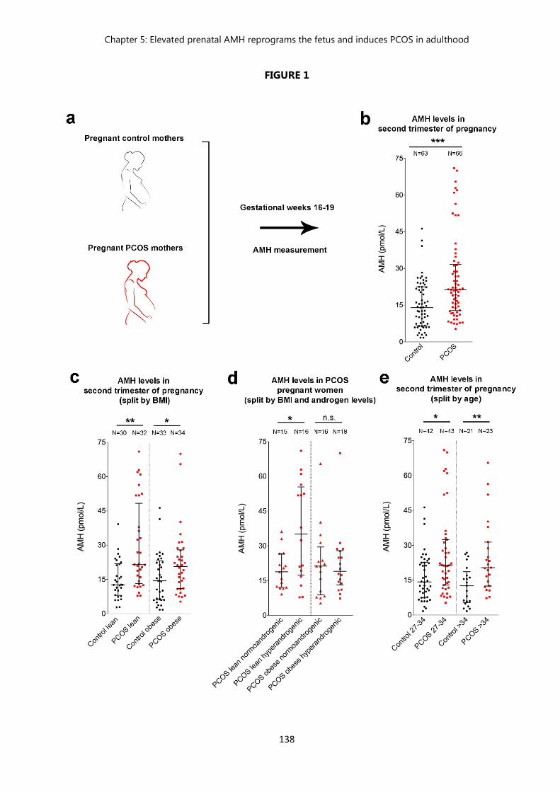
Chapter 5: Elevated prenatal AMH reprograms the fetus and induces PCOS in adulthood
138
FIGURE 1
Chapter 5: Elevated prenatal AMH reprograms the fetus and induces PCOS in adulthood
139
Figure 1. AMH levels during the second trimester of gestation are higher in PCOS women than
control women. (a) Blood samples were derived from 63 control and 66 PCOS pregnant women at
gestational week 16-19 and AMH concentration was measure by ELISA. (b) Scatter-plot showing the
circulating AMH levels in control pregnant women and in PCOS pregnant patients. (c) Scatter-plot
showing the circulating AMH levels in control pregnant women and in PCOS pregnant patients
stratified by the body mass index (BMI) of the patients and classified it into lean and obese subjects.
(d) Scatter-plot showing the circulating AMH levels in PCOS pregnant patients stratified by their BMI
and androgen levels (normoandrogenic or with signs of biochemical and/or clinical
hyperandrogenism). (e) Scatter-plot showing the circulating AMH levels in control pregnant women
and in PCOS pregnant patients stratified by their age. The horizontal line in each plot corresponds
to the median value. The vertical line represents the 25th – 75th percentile range. Comparisons
between groups, matched by BMI, androgen levels and age, were performed using unpaired
nonparametric two-tailed Mann-Whitney U test. * P < 0.05, ** P < 0.005, *** P < 0.0005. Data were
combined from two independent experiments.
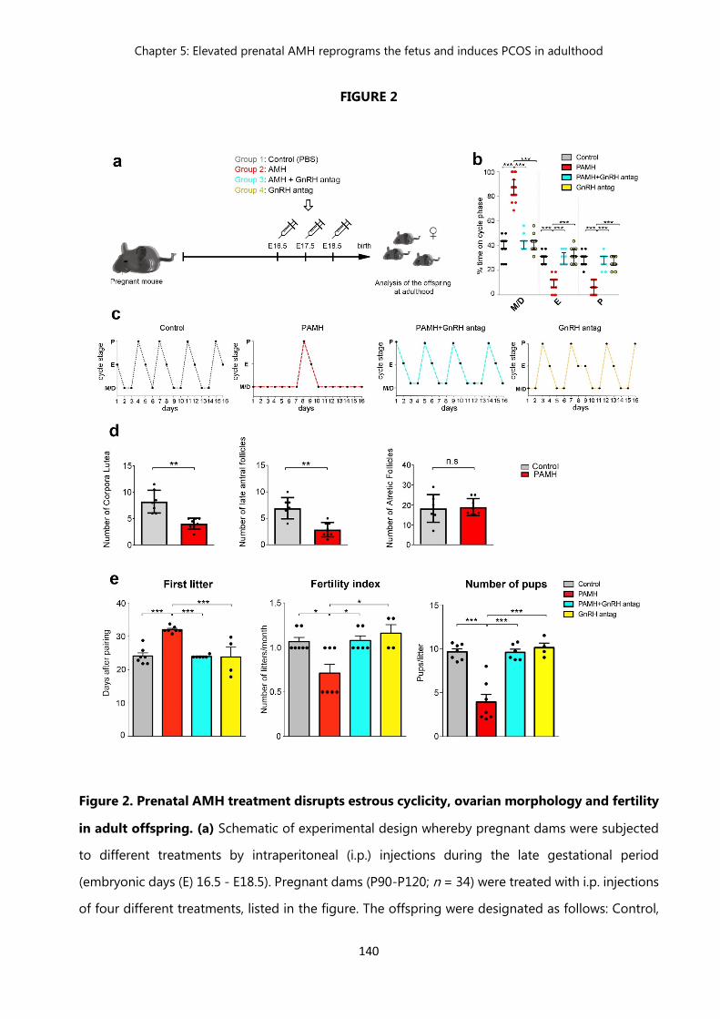
Chapter 5: Elevated prenatal AMH reprograms the fetus and induces PCOS in adulthood
140
FIGURE 2
Figure 2. Prenatal AMH treatment disrupts estrous cyclicity, ovarian morphology and fertility
in adult offspring. (a) Schematic of experimental design whereby pregnant dams were subjected
to different treatments by intraperitoneal (i.p.) injections during the late gestational period
(embryonic days (E) 16.5 - E18.5). Pregnant dams (P90-P120; n = 34) were treated with i.p. injections
of four different treatments, listed in the figure. The offspring were designated as follows: Control,
Chapter 5: Elevated prenatal AMH reprograms the fetus and induces PCOS in adulthood
141
(PBS-treated); PAMH (Prenatal recombinant AMHc-treated); PAMH+GnRH antag, (AMHc plus
Cetrorelix acetate); GnRH antag (Cetrorelix acetate alone). (b) Quantitative analysis of ovarian
cyclicity in adult (P60-P90) offspring mice (control, n = 15; PAMH, n = 19; PAMH+GnRH antag, n =
13; GnRH antag, n = 11). Vaginal cytology was assessed for 16 days. The horizontal line in each
scatter plot corresponds to the median value. The vertical line represents the 25th – 75th percentile
range. Comparisons between treatment groups were performed using Kruskal-Wallis test followed
by Dunn’s post hoc analysis test; *** P < 0.0001. Data were combined from three independent
experiments. (c) Representation of estrous cyclicity in one female mouse of each of the four
treatments during 16 consecutive days (4-cycles). (d) Quantitative analysis of corpora lutea, late
antral follicles and atretic follicles in the ovaries of control (n = 7, age: P90) and PAMH mice (n = 8,
age: P90). Data are represented as mean ± s.e.m (two-tailed Student’s t-test; corpora lutea, t (13) =
4.879, **P < 0.001; late antral follicles, t (13) =4.637, ** P < 0.001; atretic follicles, t (13) =0.226, P =
0.82). Data were combined from two independent experiments. (e) Fertility tests of the adult
offspring mice (P90). Matings were performed for 90 days. Control females were paired with control
males (n = 7), PAMH females were paired with PAMH males (n = 7), PAMH+GnRH antag females
were paired with PAMH+GnRH antag males (n = 6), and GnRH antag females were paired with GnRH
antag males (n = 8). Female PAMH mice exhibit impaired fertility. Data are represented as mean ±
s.e.m. Statistics were computed with one-way ANOVA followed by Tukey’s multiple comparison post
hoc test. *P < 0.05, **P < 0.001 and ***P < 0.0001. Data of fertility tests were combined from two
independent experiments.

Chapter 5: Elevated prenatal AMH reprograms the fetus and induces PCOS in adulthood
142
FIGURE 3
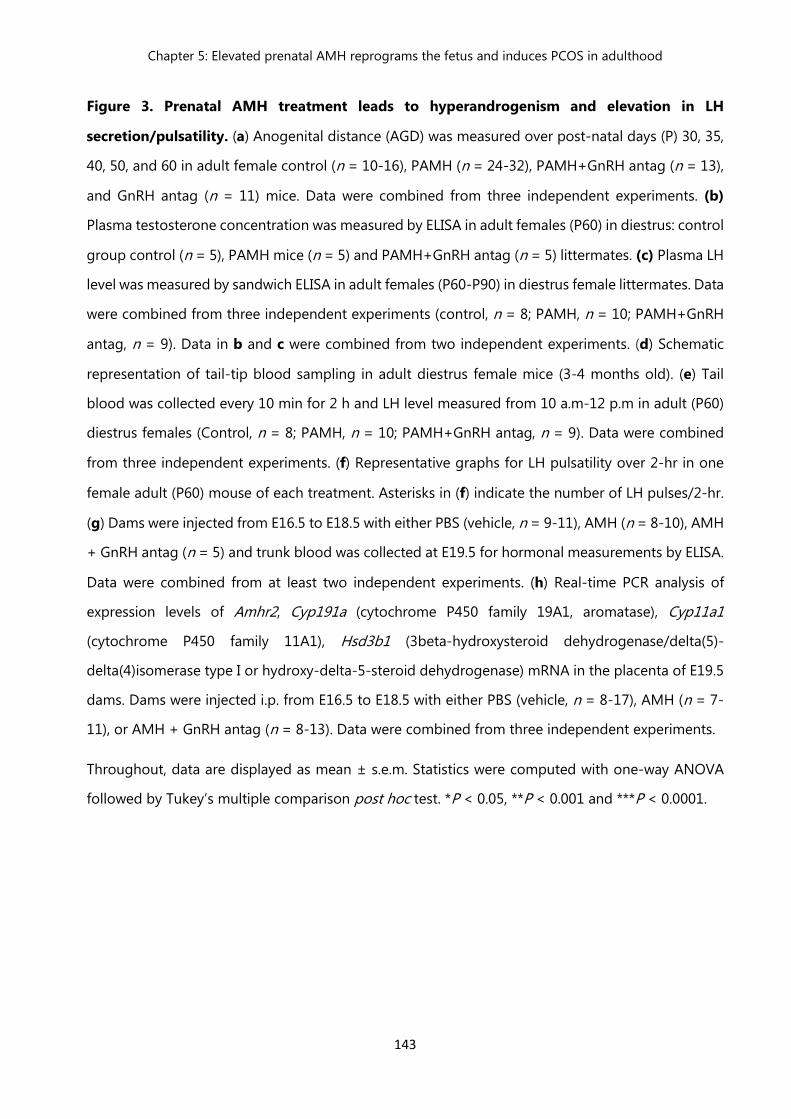
Chapter 5: Elevated prenatal AMH reprograms the fetus and induces PCOS in adulthood
143
Figure 3. Prenatal AMH treatment leads to hyperandrogenism and elevation in LH
secretion/pulsatility. (a) Anogenital distance (AGD) was measured over post-natal days (P) 30, 35,
40, 50, and 60 in adult female control (n = 10-16), PAMH (n = 24-32), PAMH+GnRH antag (n = 13),
and GnRH antag (n = 11) mice. Data were combined from three independent experiments. (b)
Plasma testosterone concentration was measured by ELISA in adult females (P60) in diestrus: control
group control (n = 5), PAMH mice (n = 5) and PAMH+GnRH antag (n = 5) littermates. (c) Plasma LH
level was measured by sandwich ELISA in adult females (P60-P90) in diestrus female littermates. Data
were combined from three independent experiments (control, n = 8; PAMH, n = 10; PAMH+GnRH
antag, n = 9). Data in b and c were combined from two independent experiments. (d) Schematic
representation of tail-tip blood sampling in adult diestrus female mice (3-4 months old). (e) Tail
blood was collected every 10 min for 2 h and LH level measured from 10 a.m-12 p.m in adult (P60)
diestrus females (Control, n = 8; PAMH, n = 10; PAMH+GnRH antag, n = 9). Data were combined
from three independent experiments. (f) Representative graphs for LH pulsatility over 2-hr in one
female adult (P60) mouse of each treatment. Asterisks in (f) indicate the number of LH pulses/2-hr.
(g) Dams were injected from E16.5 to E18.5 with either PBS (vehicle, n = 9-11), AMH (n = 8-10), AMH
+ GnRH antag (n = 5) and trunk blood was collected at E19.5 for hormonal measurements by ELISA.
Data were combined from at least two independent experiments. (h) Real-time PCR analysis of
expression levels of Amhr2, Cyp191a (cytochrome P450 family 19A1, aromatase), Cyp11a1
(cytochrome P450 family 11A1), Hsd3b1 (3beta-hydroxysteroid dehydrogenase/delta(5)-
delta(4)isomerase type I or hydroxy-delta-5-steroid dehydrogenase) mRNA in the placenta of E19.5
dams. Dams were injected i.p. from E16.5 to E18.5 with either PBS (vehicle, n = 8-17), AMH (n = 7-
11), or AMH + GnRH antag (n = 8-13). Data were combined from three independent experiments.
Throughout, data are displayed as mean ± s.e.m. Statistics were computed with one-way ANOVA
followed by Tukey’s multiple comparison post hoc test. *P < 0.05, **P < 0.001 and ***P < 0.0001.
Chapter 5: Elevated prenatal AMH reprograms the fetus and induces PCOS in adulthood
144
FIGURE 4
Figure 4. Prenatal AMH treatment increases perinatal T levels in females and masculinizes
their brain. (a) Plasma T and LH levels were measured in pups 2 hours after birth (males, n = 5;
control females, n = 5; PAMH females, n = 4; PAMH females + GnRH antag, n = 6). (b) Schematic
representation depicting the analyzed sexual dimorphic neuroanatomical regions of the brain
(denoted in red) expressing Vasopressin (VP) and Tyrosine Hydroxylase (TH). (c) Photomicrographs
of TH-immunoreactive neurons in the anteroventral periventricular nucleus (AVPV) of a male, control

Chapter 5: Elevated prenatal AMH reprograms the fetus and induces PCOS in adulthood
145
female and PAMH female at P60. TH immunoreactive (-ir) neurons in the AVPV were quantified in
these animal groups (n = 3 per sex and treatment). (d) Photomicrographs of VP-immunoreactive
neurons in the bed nucleus of the stria terminalis (BNST) and medial amygdala (MeA) of a male,
control female and PAMH female at P60. VP–ir neurons in the BNST and MeA were quantified in
these animal groups (n = 3 per sex and treatment). Throughout, data are displayed as mean ± s.e.m.
Statistics were computed with one-way ANOVA followed by Tukey’s multiple comparison post hoc
test. *P < 0.05, **P < 0.001 and ***P < 0.0001. Experiments were replicated three times with
comparable results.
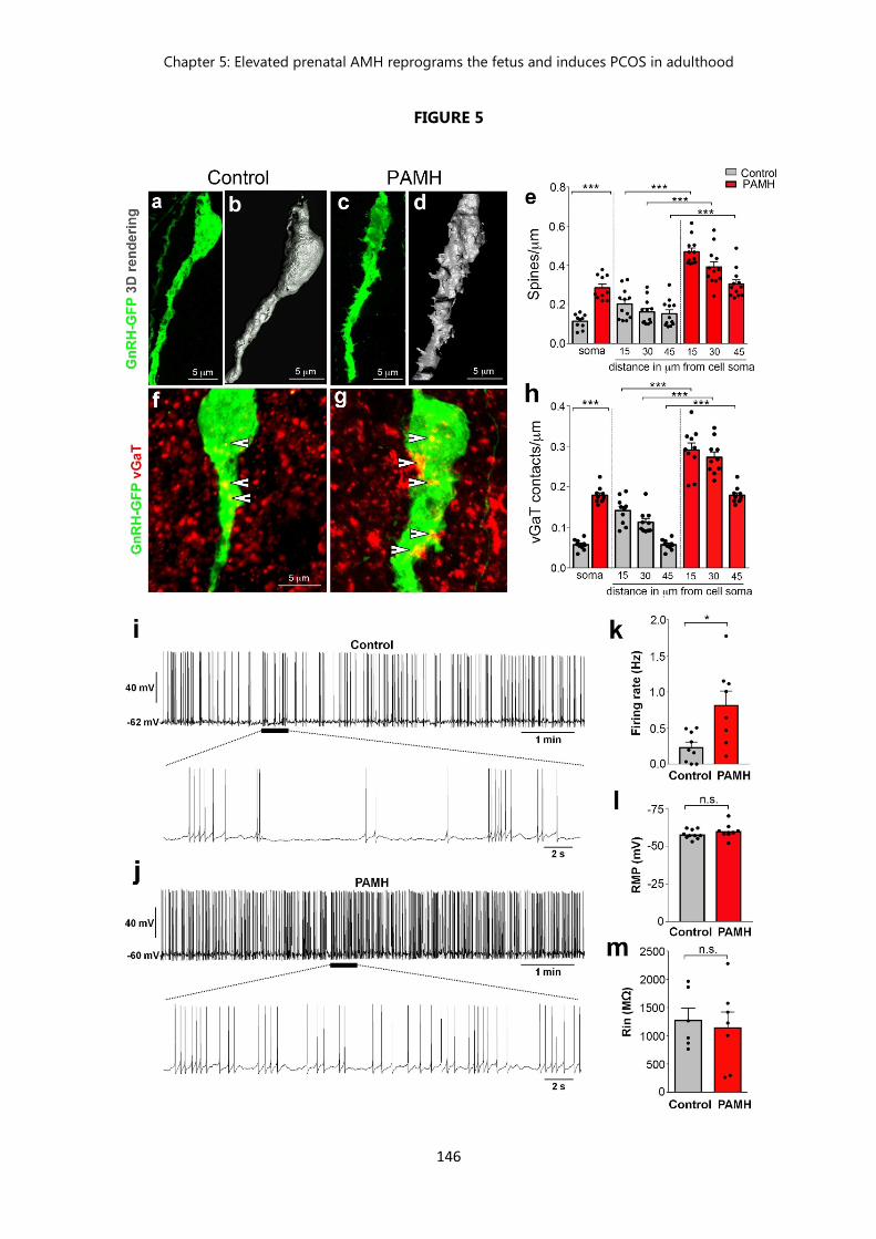
Chapter 5: Elevated prenatal AMH reprograms the fetus and induces PCOS in adulthood
146
FIGURE 5

Chapter 5: Elevated prenatal AMH reprograms the fetus and induces PCOS in adulthood
147
Figure 5. PAMH/GnRH-GFP mice exhibit higher GnRH dendritic spine density, increased
GABAergic appositions to GnRH neurons and elevated firing frequency of GnRH neurons in
adulthood. (a-d) Projected confocal images and 3D reconstruction of GnRH-GFP neurons from an
adult (P60) female diestrus control (a, b) and a PAMH (c, d) female diestrus mouse. (e) The GnRH
dendritic spine density was analyzed in adult (P60) diestrus control and PAMH/GnRH-GFP females.
Two sections with the largest number of GnRH neurons (rPOA containing the OVLT) where chosen
from each animal, where the full extent of GFP-labeled GnRH neurons (n = 10-12 neurons from 3
animals per treatment group) were imaged and analyzed using confocal microscopy. Spine density
was quantified for the soma and each 15 µm portion of the primary GnRH neuronal dendrite. (f, g)
Projected confocal images of a GFP-immunoreactive GnRH neuron (green) surrounded by vGaT-
immunoreactive (red) puncta in an adult diestrus control (f) and PAMH female mouse (g). Points
where the vGaT signal was considered to be immediately adjacent to the GnRH neuron are indicated
by arrowheads. (h) Quantification of the number of vGaT-immunoreactive puncta adjacent to GnRH
neuron soma and along the primary GnRH neuronal dendrite is expressed as vGaT appositions/µm.
vGaT contacts were defined as positive when vGaT immunolabeling was directly apposed onto the
GnRH neuronal dendrites without any black pixels between the GnRH dendritic spine. (i-m)
Spontaneous electrical activity and membrane properties of GnRH neurons recorded in acute brain
slices from control and PAMH GnRH-GFP female diestrus mice. (i) Whole-cell current-clamp
recording showing the typical spontaneous burst firing of a GnRH neuron from control mouse. The
bottom trace shows an expanded time scale of that recording. (j) Same experiment as in (i) but in a
GnRH neuron from PAMH mouse. (k) Average firing rate of GnRH neurons recorded from control (n
= 9 cells from 4 animals) and PAMH mice (n = 8 cells from 4 animals). (l) Average resting membrane
potential (RMP) of GnRH neurons recorded from control (n =10 cells from 4 animals, age P90-P120)
and PAMH mice (n = 8 cells from 4 animals, age P90-P120) was not significantly different between
both groups (two-tailed Student’s t-test, t (16) = 1.143, P = 0.2700, ns). (m) Average input resistance
(Rin) of GnRH neurons recorded from control (n = 6 cells from 4 animals, age P90-P120) and PAMH
mice (n = 7 cells from 4 animals, age P90-P120) was not found significantly different between both
groups (two-tailed Student’s t-test, t (11) = 0.3685, P = 0.7195, ns, not significant). Throughout, data
are displayed as mean ± s.e.m. Statistics were computed with two-tailed Student’s t-test. *P < 0.05,
**P < 0.001 and ***P < 0.0001. Experiments were replicated three times with comparable results.
OVLT, organum vasculosum lamina terminalis; rPOA, rostral Preoptic Area.
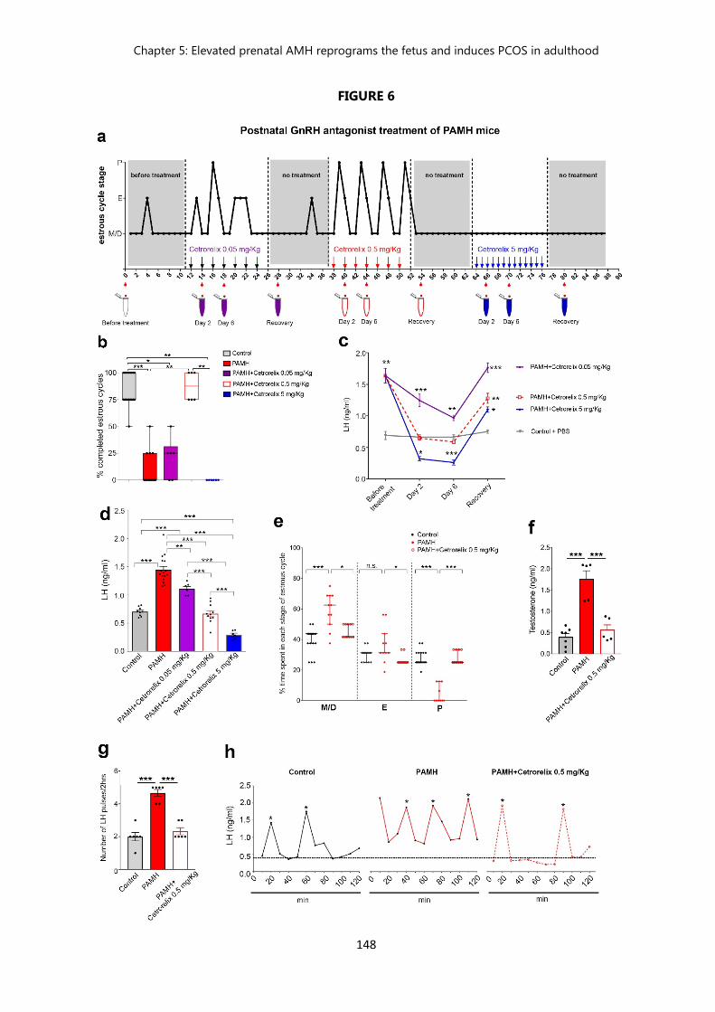
Chapter 5: Elevated prenatal AMH reprograms the fetus and induces PCOS in adulthood
148
FIGURE 6
Chapter 5: Elevated prenatal AMH reprograms the fetus and induces PCOS in adulthood
149
Figure 6. Postnatal GnRH antagonist treatment of PAMH mice restores the PCOS-like
neuroendocrine phenotype. (a) Schematic of experimental design whereby vaginal cytology
(estrous cyclicity) in PAMH adult mice (P90, n = 6) was analyzed during 3 months, before and after
postnatal intraperitoneal (i.p.) injections with 0.05, 0.5 and 5 mg/Kg Cetrorelix acetate. The Y axis
refers to the different stages of the estrous cycle: Metaestrus/Diestrus (M/D), Estrus (E) and Proestrus
(P). The X axis represents the time-course of the experiments (days). Animals were injected i.p. every
second day, for 12 days, with Cetrorelix acetate at the dose of 0.05 and 0.5 mg/Kg, and every day
with Cetrorelix acetate at the dose of 5 mg/Kg. Tail-blood samples were collected for LH
measurements twice before the beginning of the treatments, and at day 2 and 6 of each treatment
as well as 4 days after the last injection (no treatment), that followed each administration period. (b)
Quantitative analysis of the % of completed estrous cycles in control (PBS-treated, n = 19), PAMH
(n = 13), and PAMH mice postnatally treated with the three doses of GnRH antagonist
(PAMH+Cetrorelix 0.05 mg/Kg, PAMH+Cetrorelix 0.5 mg/Kg, PAMH+Cetrorelix 5 mg/Kg; n = 6
each). The horizontal line in each scatter plot corresponds to the median value. The vertical line
represents the 25th – 75th percentile range. Comparisons between treatment groups were performed
using nonparametric Kruskal-Wallis test followed by Dunn’s post hoc analysis test. (c) Time course
of serum LH measured on days 0 (before treatment), 2 and 6 of the treatment and 4 days after
discontinuation of the treatment (recovery time). Treatment groups (n = 6 for each Cetrorelix
treatment group and n = 3 for the Control) are indicated in the figure. Data were combined from
two independent experiments. (d) Mean LH levels were measured in diestrus adult (P60-P120)
control mice (n = 9), PAMH (n = 16), PAMH+Cetrorelix 0.05 mg/Kg (n = 6), PAMH+Cetrorelix 0.5
mg/Kg (n = 11), PAMH+Cetrorelix 5 mg/Kg (n = 6). Data were combined from three independent
experiments. (e) Scatter plot representing the percentage (%) of time spent in each estrous cycle.
The horizontal line in each scatter plot corresponds to the median value. The vertical line represents
the 25th – 75th percentile range. The percentage (%) of time spent in each estrous cycle was quantified
in control (n = 19), PAMH (n = 11) and PAMH+Cetrorelix 0.5 mg/Kg (n = 11) mice (age: P60-P90),
as described above. Comparisons between treatment groups were performed using Kruskal-Wallis
test followed by Dunn’s post hoc analysis test. Data were combined from three independent
experiments. (f) Mean T levels were measured by ELISA in diestrus adult control (n = 7), PAMH (n =
5) and PAMH+Cetrorelix 0.5 mg/Kg (n = 5) animals. (g) Tail blood was collected every 10 min for 2
h and LH level measured from 10 a.m-12 p.m in adult (P90) diestrus females (Control, n = 6; PAMH,
n = 6; PAMH+Cetrorelix 0.5 mg/Kg, n = 6). (h) Representative graphs for LH pulsatility over 2-hr in
Chapter 5: Elevated prenatal AMH reprograms the fetus and induces PCOS in adulthood
150
one female adult (P90) mouse of each treatment. Asterisks in (h) indicate the number of LH pulses/2-
hr. The dotted line refers to the control LH baseline value.
Values in c, d, f, g are represented as the mean ± s.e.m. Statistics in c, d, f, g were computed with
one-way ANOVA followed by Tukey’s multiple comparison post hoc test. *P < 0.05, **P < 0.001 and
***P < 0.0001. Experiments were replicated three times with similar results.

Chapter 5: Elevated prenatal AMH reprograms the fetus and induces PCOS in adulthood
151
SUPPLEMENTARY TABLE 1
Demographic table of pregnant control and PCOS patients. Blood samples were derived from the
population-based Uppsala Biobank of Pregnant Women, where blood samples are collected in
conjunction with the routine ultrasound screening in gestational week 16- 19.
When applicable, the Kruskal-Wallis test followed by Dunn’s post hoc analysis was used to analyze
differences between study groups. The significance level was set at P < 0.05.
Abbreviations: IQR, Interquartile ranges; BMI, body mass index; IVF, in vitro fertilization.
Control lean
(N=30)
PCOS lean
(N=32)
Control obese
(N=33)
PCOS obese
(N=34)
P value
Ethnic group Caucasian
(N=30)
Caucasian
(N=32)
Caucasian
(N=30 of 33)
Caucasian
(N=34)
nd
Gestational age
(weeks)
Age (years) of
pregnant
patients
16-19
32 (IQR 28.75-
35.25)
16-19
31.5 (IQR
29.25-35.00)
16-19
32 (IQR 29.5-
36.00)
16-19
33 (IQR
30.75-37.00)
nd
0.5241
BMI in first
trimester
(Kg/m2)
22.4a (IQR
20.42-23.44)
21.91a (IQR
20.56-23.17)
32.47b (IQR
30.62-33.89)
32.37b (IQR
29.82-34.29)
a vs b
< 0.0001
Gestational
length (days)
Birthweight (g)
280 (IQR 272-
286)
3675 (IQR 3300-
3900)
278 (IQR
270.3-289)
3510 (IQR
3210-3778)
282 (IQR
273.5-289)
3620 (IQR
3285-4125)
279 (IQR
269.8-287)
3670 (IQR
3255-4175)
0.7225
0.3536
IVF 3 out of 31 10 out of 32 0 out of 33 5 out of 34 nd
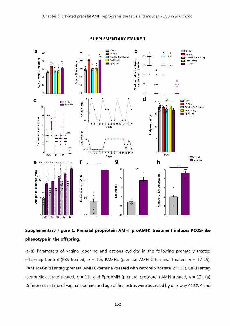
Chapter 5: Elevated prenatal AMH reprograms the fetus and induces PCOS in adulthood
152
SUPPLEMENTARY FIGURE 1
Supplementary Figure 1. Prenatal proprotein AMH (proAMH) treatment induces PCOS-like
phenotype in the offspring.
(a-b) Parameters of vaginal opening and estrous cyclicity in the following prenatally treated
offspring: Control (PBS-treated, n = 19); PAMHc (prenatal AMH C-terminal-treated, n = 17-19),
PAMHc+GnRH antag (prenatal AMH C-terminal-treated with cetrorelix acetate, n = 13), GnRH antag
(cetrorelix acetate-treated, n = 11), and PproAMH (prenatal proprotein AMH-treated, n = 12). (a)
Differences in time of vaginal opening and age of first estrus were assessed by one-way ANOVA and
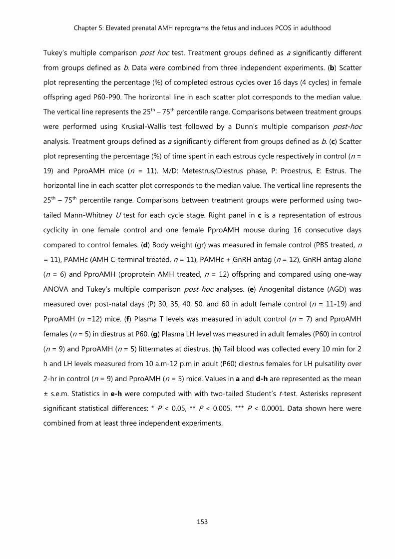
Chapter 5: Elevated prenatal AMH reprograms the fetus and induces PCOS in adulthood
153
Tukey’s multiple comparison post hoc test. Treatment groups defined as a significantly different
from groups defined as b. Data were combined from three independent experiments. (b) Scatter
plot representing the percentage (%) of completed estrous cycles over 16 days (4 cycles) in female
offspring aged P60-P90. The horizontal line in each scatter plot corresponds to the median value.
The vertical line represents the 25th – 75th percentile range. Comparisons between treatment groups
were performed using Kruskal-Wallis test followed by a Dunn’s multiple comparison post-hoc
analysis. Treatment groups defined as a significantly different from groups defined as b. (c) Scatter
plot representing the percentage (%) of time spent in each estrous cycle respectively in control (n =
19) and PproAMH mice (n = 11). M/D: Metestrus/Diestrus phase, P: Proestrus, E: Estrus. The
horizontal line in each scatter plot corresponds to the median value. The vertical line represents the
25th – 75th percentile range. Comparisons between treatment groups were performed using two-
tailed Mann-Whitney U test for each cycle stage. Right panel in c is a representation of estrous
cyclicity in one female control and one female PproAMH mouse during 16 consecutive days
compared to control females. (d) Body weight (gr) was measured in female control (PBS treated, n
= 11), PAMHc (AMH C-terminal treated, n = 11), PAMHc + GnRH antag (n = 12), GnRH antag alone
(n = 6) and PproAMH (proprotein AMH treated, n = 12) offspring and compared using one-way
ANOVA and Tukey’s multiple comparison post hoc analyses. (e) Anogenital distance (AGD) was
measured over post-natal days (P) 30, 35, 40, 50, and 60 in adult female control (n = 11-19) and
PproAMH (n =12) mice. (f) Plasma T levels was measured in adult control (n = 7) and PproAMH
females (n = 5) in diestrus at P60. (g) Plasma LH level was measured in adult females (P60) in control
(n = 9) and PproAMH (n = 5) littermates at diestrus. (h) Tail blood was collected every 10 min for 2
h and LH levels measured from 10 a.m-12 p.m in adult (P60) diestrus females for LH pulsatility over
2-hr in control (n = 9) and PproAMH (n = 5) mice. Values in a and d-h are represented as the mean
± s.e.m. Statistics in e-h were computed with with two-tailed Student’s t-test. Asterisks represent
significant statistical differences: * P < 0.05, ** P < 0.005, *** P < 0.0001. Data shown here were
combined from at least three independent experiments.
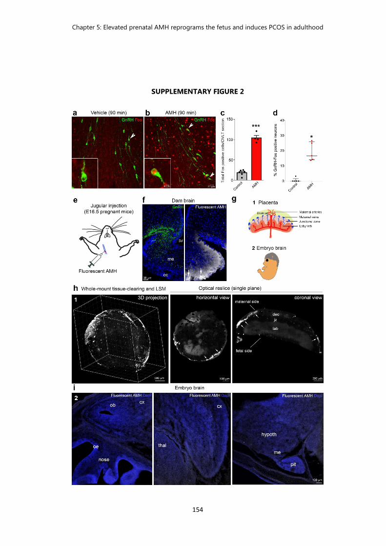
Chapter 5: Elevated prenatal AMH reprograms the fetus and induces PCOS in adulthood
154
SUPPLEMENTARY FIGURE 2
Chapter 5: Elevated prenatal AMH reprograms the fetus and induces PCOS in adulthood
155
Supplementary Figure 2. Passage into the brain and action of peripherally-injected AMH.
Female mice (P60-90) were injected (i.p.) with PBS or AMHC in diestrus and sacrificed 90 min later.
(a, b) Representative confocal images depicting the OVLT after dual-immunolabeling for GnRH
(green) and Fos (red) in PBS-treated (a) and AMH-treated mice (b). Arrows point to double-labeled
Fos-GnRH neurons. (c) The total number of Fos positive cells was quantified in the OVLT sections of
control (n = 6) and AMH-treated animals (n = 4). Control females exhibit less Fos immunoreactive
cells/OVLT section as compared to AMH-treated mice (Unpaired Student’s t-test t (8) = 14.57, *** P
< 0.0001). (d) Scatter plot representing the percentage (%) of GnRH neurons expressing Fos in the
OVLT sections. The horizontal line in each scatter plot corresponds to the median value. The vertical
line represents the 25th – 75th percentile range. Comparisons between treatment groups (n = 5 per
animal group) were performed using Mann-Whitney U test, * P = 0.007. OVLT, organum vasculosum
laminae terminalis. Data shown in a-d were combined from two independent experiments. (e-i)
Fluorescently-labeled bioactive AMH (2.5 nmoles/animal) was injected into the jugular vein of E16.5
pregnant mice (n = 4; e). Animals were sacrificed 2 min later to assess AMH passage into the dam’s
brains (f), placentae (g) and fetal brains (i). (f) Representative photomicrographs showing GnRH-
immunopositive nerve terminals (green, left panel) and fluorescent AMH (white, right panel) in the
median eminence (me), which lies outside the blood-brain-barrier (BBB). Note the diffusion of AMH,
2 mins after jugular administration, from the site of access into the me (arrows; permeable
fenestrated endothelial cells: ec) and spreading in the external layer of the me. (g) Schematic
representation of the other two organs, namely placenta (1) and fetal brain (2), where the passage
of fluorescent AMH was also investigated. (h) 3D analysis of fluorescent AMH in the intact placenta.
Panels are Light-Sheet Microscopy (LSM) images of a representative solvent-cleared placenta. The
displayed 3D-projection, horizontal view and the lateral view, show the presence of fluorescent AMH
(white) in the decidua at the level of the maternal veins (arrows) but not in the fetal side of the
placenta, indicating that AMH cannot pass through the placental barrier. Data shown here were
replicated in three independent experiments. (i) Representative sagittal section of the head of an
E16.5 embryo harvested from a dam injected with fluorescent AMH, which shows the lack of AMH
passage into the fetal brain. Data shown here were replicated in 4 independent experiments. In
Figures f and i cell nuclei are counterstained with DAPI. Dec: decidua; jz: junctional zone; lab:
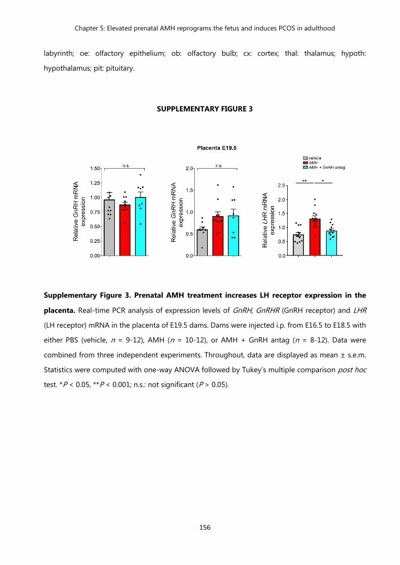
Chapter 5: Elevated prenatal AMH reprograms the fetus and induces PCOS in adulthood
156
labyrinth; oe: olfactory epithelium; ob: olfactory bulb; cx: cortex; thal: thalamus; hypoth:
hypothalamus; pit: pituitary.
SUPPLEMENTARY FIGURE 3
Supplementary Figure 3. Prenatal AMH treatment increases LH receptor expression in the
placenta. Real-time PCR analysis of expression levels of GnRH, GnRHR (GnRH receptor) and LHR
(LH receptor) mRNA in the placenta of E19.5 dams. Dams were injected i.p. from E16.5 to E18.5 with
either PBS (vehicle, n = 9-12), AMH (n = 10-12), or AMH + GnRH antag (n = 8-12). Data were
combined from three independent experiments. Throughout, data are displayed as mean ± s.e.m.
Statistics were computed with one-way ANOVA followed by Tukey’s multiple comparison post hoc
test. *P < 0.05, **P < 0.001; n.s.: not significant (P > 0.05).
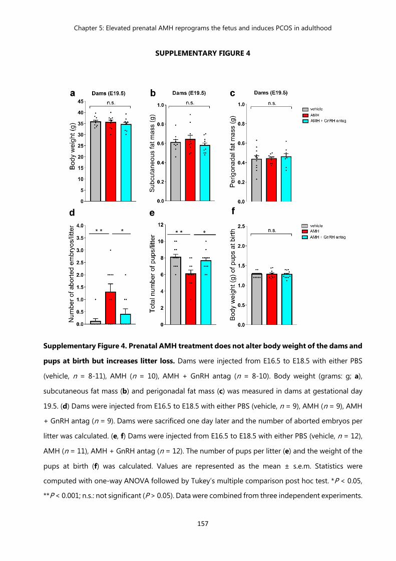
Chapter 5: Elevated prenatal AMH reprograms the fetus and induces PCOS in adulthood
157
SUPPLEMENTARY FIGURE 4
Supplementary Figure 4. Prenatal AMH treatment does not alter body weight of the dams and
pups at birth but increases litter loss. Dams were injected from E16.5 to E18.5 with either PBS
(vehicle, n = 8-11), AMH (n = 10), AMH + GnRH antag (n = 8-10). Body weight (grams: g; a),
subcutaneous fat mass (b) and perigonadal fat mass (c) was measured in dams at gestational day
19.5. (d) Dams were injected from E16.5 to E18.5 with either PBS (vehicle, n = 9), AMH (n = 9), AMH
+ GnRH antag (n = 9). Dams were sacrificed one day later and the number of aborted embryos per
litter was calculated. (e, f) Dams were injected from E16.5 to E18.5 with either PBS (vehicle, n = 12),
AMH (n = 11), AMH + GnRH antag (n = 12). The number of pups per litter (e) and the weight of the
pups at birth (f) was calculated. Values are represented as the mean ± s.e.m. Statistics were
computed with one-way ANOVA followed by Tukey’s multiple comparison post hoc test. *P < 0.05,
**P < 0.001; n.s.: not significant (P > 0.05). Data were combined from three independent experiments.
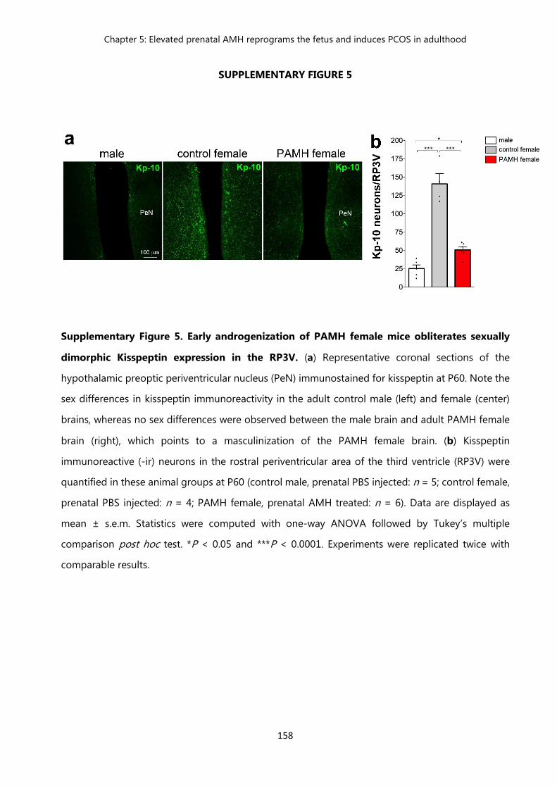
Chapter 5: Elevated prenatal AMH reprograms the fetus and induces PCOS in adulthood
158
SUPPLEMENTARY FIGURE 5
Supplementary Figure 5. Early androgenization of PAMH female mice obliterates sexually
dimorphic Kisspeptin expression in the RP3V. (a) Representative coronal sections of the
hypothalamic preoptic periventricular nucleus (PeN) immunostained for kisspeptin at P60. Note the
sex differences in kisspeptin immunoreactivity in the adult control male (left) and female (center)
brains, whereas no sex differences were observed between the male brain and adult PAMH female
brain (right), which points to a masculinization of the PAMH female brain. (b) Kisspeptin
immunoreactive (-ir) neurons in the rostral periventricular area of the third ventricle (RP3V) were
quantified in these animal groups at P60 (control male, prenatal PBS injected: n = 5; control female,
prenatal PBS injected: n = 4; PAMH female, prenatal AMH treated: n = 6). Data are displayed as
mean ± s.e.m. Statistics were computed with one-way ANOVA followed by Tukey’s multiple
comparison post hoc test. *P < 0.05 and ***P < 0.0001. Experiments were replicated twice with
comparable results.
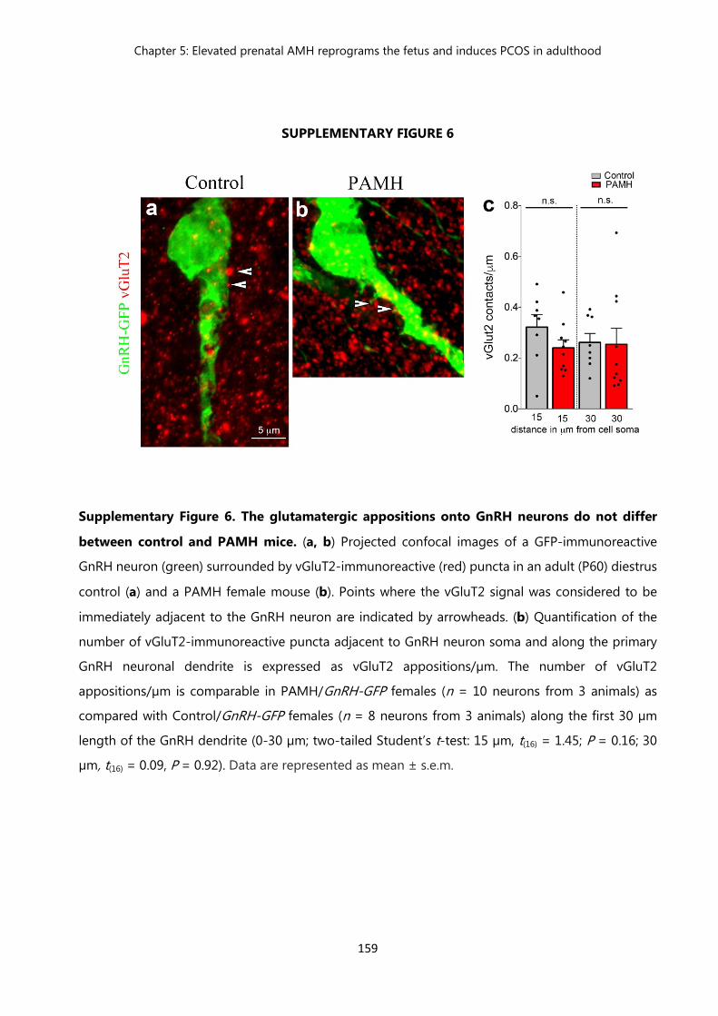
Chapter 5: Elevated prenatal AMH reprograms the fetus and induces PCOS in adulthood
159
SUPPLEMENTARY FIGURE 6
Supplementary Figure 6. The glutamatergic appositions onto GnRH neurons do not differ
between control and PAMH mice. (a, b) Projected confocal images of a GFP-immunoreactive
GnRH neuron (green) surrounded by vGluT2-immunoreactive (red) puncta in an adult (P60) diestrus
control (a) and a PAMH female mouse (b). Points where the vGluT2 signal was considered to be
immediately adjacent to the GnRH neuron are indicated by arrowheads. (b) Quantification of the
number of vGluT2-immunoreactive puncta adjacent to GnRH neuron soma and along the primary
GnRH neuronal dendrite is expressed as vGluT2 appositions/µm. The number of vGluT2
appositions/µm is comparable in PAMH/GnRH-GFP females (n = 10 neurons from 3 animals) as
compared with Control/GnRH-GFP females (n = 8 neurons from 3 animals) along the first 30 µm
length of the GnRH dendrite (0-30 µm; two-tailed Student’s t-test: 15 µm, t(16) = 1.45; P = 0.16; 30
µm, t(16) = 0.09, P = 0.92). Data are represented as mean ± s.e.m.
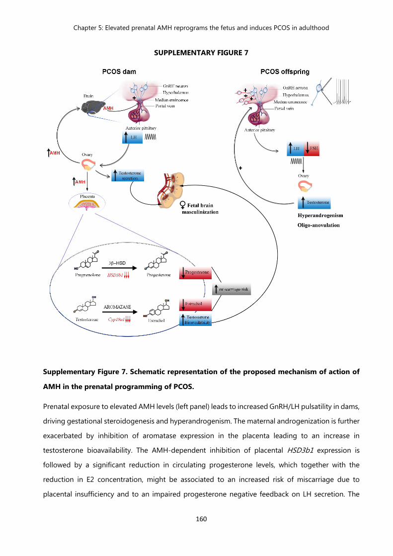
Chapter 5: Elevated prenatal AMH reprograms the fetus and induces PCOS in adulthood
160
SUPPLEMENTARY FIGURE 7
Supplementary Figure 7. Schematic representation of the proposed mechanism of action of
AMH in the prenatal programming of PCOS.
Prenatal exposure to elevated AMH levels (left panel) leads to increased GnRH/LH pulsatility in dams,
driving gestational steroidogenesis and hyperandrogenism. The maternal androgenization is further
exacerbated by inhibition of aromatase expression in the placenta leading to an increase in
testosterone bioavailability. The AMH-dependent inhibition of placental HSD3b1 expression is
followed by a significant reduction in circulating progesterone levels, which together with the
reduction in E2 concentration, might be associated to an increased risk of miscarriage due to
placental insufficiency and to an impaired progesterone negative feedback on LH secretion. The

Chapter 5: Elevated prenatal AMH reprograms the fetus and induces PCOS in adulthood
161
elevated levels of T trigger a cascade of events in the offspring, which converge into the
masculinization of the brain sexually dimorphic nuclei and altered hypothalamic wiring. In adult
female PCOS offspring, the increase in excitatory synaptic input to GnRH drives a persistent rise in
the GnRH neuronal firing activity. Finally, the constitutive hyperactivity of GnRH neurons stimulates
ovarian androgen production and impairs folliculogenesis and ovulation, contributing to the vicious
circle of PCOS.
Chapter 5: Elevated prenatal AMH reprograms the fetus and induces PCOS in adulthood
162
References
Abbott, D. H., L. E. Nicol, J. E. Levine, N. Xu, M. O. Goodarzi and D. A. Dumesic (2013). "Nonhuman primate
models of polycystic ovary syndrome." Mol Cell Endocrinol 373(1-2): 21-28.
Abbott, D. H., B. H. Rayome, D. A. Dumesic, K. C. Lewis, A. K. Edwards, K. Wallen, M. E. Wilson, S. E. Appt and J.
E. Levine (2017). "Clustering of PCOS-like traits in naturally hyperandrogenic female rhesus monkeys."
Hum Reprod 32(4): 923-936.
Azziz, R. (2016). "Introduction: Determinants of polycystic ovary syndrome." Fertil Steril 106(1): 4-5.
Chang, R. J. (2007). "The reproductive phenotype in polycystic ovary syndrome." Nat Clin Pract Endocrinol
Metab 3(10): 688-695.
Chang, R. J., F. P. Mandel, J. K. Lu and H. L. Judd (1982). "Enhanced disparity of gonadotropin secretion by
estrone in women with polycystic ovarian disease." J Clin Endocrinol Metab 54(3): 490-494.
Cimino, I., F. Casoni, X. Liu, A. Messina, J. Parkash, S. P. Jamin, S. Catteau-Jonard, F. Collier, M. Baroncini, D.
Dewailly, P. Pigny, M. Prescott, R. Campbell, A. E. Herbison, V. Prevot and P. Giacobini (2016). "Novel
role for anti-Mullerian hormone in the regulation of GnRH neuron excitability and hormone secretion."
Nat Commun 7: 10055.
Clarkson, J. and A. E. Herbison (2006). "Postnatal development of kisspeptin neurons in mouse hypothalamus;
sexual dimorphism and projections to gonadotropin-releasing hormone neurons." Endocrinology
147(12): 5817-5825.
Cook, C. L., Y. Siow, A. G. Brenner and M. E. Fallat (2002). "Relationship between serum mullerian-inhibiting
substance and other reproductive hormones in untreated women with polycystic ovary syndrome and
normal women." Fertil Steril 77(1): 141-146.
Corbier, P., D. A. Edwards and J. Roffi (1992). "The neonatal testosterone surge: a comparative study." Arch Int
Physiol Biochim Biophys 100(2): 127-131.
De Vries, G. J. and G. C. Panzica (2006). "Sexual differentiation of central vasopressin and vasotocin systems in
vertebrates: different mechanisms, similar endpoints." Neuroscience 138(3): 947-955.
DeFazio, R. A. and S. M. Moenter (2002). "Estradiol feedback alters potassium currents and firing properties of
gonadotropin-releasing hormone neurons." Mol Endocrinol 16(10): 2255-2265.
di Clemente, N., S. P. Jamin, A. Lugovskoy, P. Carmillo, C. Ehrenfels, J. Y. Picard, A. Whitty, N. Josso, R. B. Pepinsky
and R. L. Cate (2010). "Processing of anti-mullerian hormone regulates receptor activation by a
mechanism distinct from TGF-beta." Mol Endocrinol 24(11): 2193-2206.
Duijkers, I. J., C. Klipping, W. N. Willemsen, D. Krone, E. Schneider, G. Niebch and R. Hermann (1998). "Single
and multiple dose pharmacokinetics and pharmacodynamics of the gonadotrophin-releasing
hormone antagonist Cetrorelix in healthy female volunteers." Hum Reprod 13(9): 2392-2398.
Dumesic, D. A., A. L. Akopians, V. K. Madrigal, E. Ramirez, D. J. Margolis, M. K. Sarma, A. M. Thomas, T. R.
Grogan, R. Haykal, T. A. Schooler, B. L. Okeya, D. H. Abbott and G. D. Chazenbalk (2016).
Chapter 5: Elevated prenatal AMH reprograms the fetus and induces PCOS in adulthood
163
"Hyperandrogenism Accompanies Increased Intra-Abdominal Fat Storage in Normal Weight
Polycystic Ovary Syndrome Women." J Clin Endocrinol Metab 101(11): 4178-4188.
Dumesic, D. A. and R. A. Lobo (2013). "Cancer risk and PCOS." Steroids 78(8): 782-785.
Ezeh, U., B. O. Yildiz and R. Azziz (2013). "Referral bias in defining the phenotype and prevalence of obesity in
polycystic ovary syndrome." J Clin Endocrinol Metab 98(6): E1088-1096.
Goodarzi, M. O., D. A. Dumesic, G. Chazenbalk and R. Azziz (2011). "Polycystic ovary syndrome: etiology,
pathogenesis and diagnosis." Nat Rev Endocrinol 7(4): 219-231.
Halmos, G., A. V. Schally, J. Pinski, M. Vadillo-Buenfil and K. Groot (1996). "Down-regulation of pituitary
receptors for luteinizing hormone-releasing hormone (LH-RH) in rats by LH-RH antagonist Cetrorelix."
Proc Natl Acad Sci U S A 93(6): 2398-2402.
Herbison, A. E. (2016). "Control of puberty onset and fertility by gonadotropin-releasing hormone neurons."
Nat Rev Endocrinol 12(8): 452-466.
Herbison, A. E. and S. M. Moenter (2011). "Depolarising and hyperpolarising actions of GABA(A) receptor
activation on gonadotrophin-releasing hormone neurones: towards an emerging consensus." J
Neuroendocrinol 23(7): 557-569.
Herde, M. K., K. Geist, R. E. Campbell and A. E. Herbison (2011). "Gonadotropin-releasing hormone neurons
extend complex highly branched dendritic trees outside the blood-brain barrier." Endocrinology
152(10): 3832-3841.
Huang, C. C., Y. J. Tien, M. J. Chen, C. H. Chen, H. N. Ho and Y. S. Yang (2015). "Symptom patterns and
phenotypic subgrouping of women with polycystic ovary syndrome: association between endocrine
characteristics and metabolic aberrations." Hum Reprod 30(4): 937-946.
Huang, X. and R. E. Harlan (1993). "Absence of androgen receptors in LHRH immunoreactive neurons." Brain
Res 624(1-2): 309-311.
Jayasena, C. N. and S. Franks (2014). "The management of patients with polycystic ovary syndrome." Nat Rev
Endocrinol 10(10): 624-636.
Katulski, K., A. Czyzyk, A. Podfigurna-Stopa, A. R. Genazzani and B. Meczekalski (2015). "Pregnancy
complications in polycystic ovary syndrome patients." Gynecol Endocrinol 31(2): 87-91.
Koninger, A., A. Kauth, B. Schmidt, M. Schmidt, G. Yerlikaya, S. Kasimir-Bauer, R. Kimmig and C. Birdir (2013).
"Anti-Mullerian-hormone levels during pregnancy and postpartum." Reprod Biol Endocrinol 11: 60.
La Marca, A., S. Giulini, R. Orvieto, V. De Leo and A. Volpe (2005). "Anti-Mullerian hormone concentrations in
maternal serum during pregnancy." Hum Reprod 20(6): 1569-1572.
Lenz, K. M. and M. M. McCarthy (2010). "Organized for sex - steroid hormones and the developing
hypothalamus." Eur J Neurosci 32(12): 2096-2104.
Lizneva, D., R. Kirubakaran, K. Mykhalchenko, L. Suturina, G. Chernukha, M. P. Diamond and R. Azziz (2016).
"Phenotypes and body mass in women with polycystic ovary syndrome identified in referral versus
unselected populations: systematic review and meta-analysis." Fertil Steril 106(6): 1510-1520 e1512.
Chapter 5: Elevated prenatal AMH reprograms the fetus and induces PCOS in adulthood
164
MacLaughlin, D. T., P. L. Hudson, A. L. Graciano, M. K. Kenneally, R. C. Ragin, T. F. Manganaro and P. K. Donahoe
(1992). "Mullerian duct regression and antiproliferative bioactivities of mullerian inhibiting substance
reside in its carboxy-terminal domain." Endocrinology 131(1): 291-296.
Maliqueo, M., H. E. Lara, F. Sanchez, B. Echiburu, N. Crisosto and T. Sir-Petermann (2013). "Placental
steroidogenesis in pregnant women with polycystic ovary syndrome." Eur J Obstet Gynecol Reprod
Biol 166(2): 151-155.
Maliqueo, M., I. Sundstrom Poromaa, E. Vanky, R. Fornes, A. Benrick, H. Akerud, S. Stridsklev, F. Labrie, T.
Jansson and E. Stener-Victorin (2015). "Placental STAT3 signaling is activated in women with polycystic
ovary syndrome." Hum Reprod 30(3): 692-700.
March, W. A., V. M. Moore, K. J. Willson, D. I. Phillips, R. J. Norman and M. J. Davies (2010). "The prevalence of
polycystic ovary syndrome in a community sample assessed under contrasting diagnostic criteria."
Hum Reprod 25(2): 544-551.
McAllister, J. M., R. S. Legro, B. P. Modi and J. F. Strauss, 3rd (2015). "Functional genomics of PCOS: from GWAS
to molecular mechanisms." Trends Endocrinol Metab 26(3): 118-124.
McCarthy, M. M., A. P. Arnold, G. F. Ball, J. D. Blaustein and G. J. De Vries (2012). "Sex differences in the brain:
the not so inconvenient truth." J Neurosci 32(7): 2241-2247.
Moore, A. M. and R. E. Campbell (2017). "Polycystic ovary syndrome: Understanding the role of the brain."
Front Neuroendocrinol.
Moore, A. M., M. Prescott and R. E. Campbell (2013). "Estradiol negative and positive feedback in a prenatal
androgen-induced mouse model of polycystic ovarian syndrome." Endocrinology 154(2): 796-806.
Moore, A. M., M. Prescott, C. J. Marshall, S. H. Yip and R. E. Campbell (2015). "Enhancement of a robust arcuate
GABAergic input to gonadotropin-releasing hormone neurons in a model of polycystic ovarian
syndrome." Proc Natl Acad Sci U S A 112(2): 596-601.
Norman, R. J., D. Dewailly, R. S. Legro and T. E. Hickey (2007). "Polycystic ovary syndrome." Lancet 370(9588):
685-697.
Novembri, R., L. Funghi, C. Voltolini, G. Belmonte, S. Vannuccini, M. Torricelli and F. Petraglia (2015). "Placenta
expresses anti-Mullerian hormone and its receptor: Sex-related difference in fetal membranes."
Placenta 36(7): 731-737.
Orvis, G. D. and R. R. Behringer (2007). "Cellular mechanisms of Mullerian duct formation in the mouse." Dev
Biol 306(2): 493-504.
Pankhurst, M. W., Y. H. Chong and I. S. McLennan (2016). "Relative levels of the proprotein and cleavage-
activated form of circulating human anti-Mullerian hormone are sexually dimorphic and variable
during the life cycle." Physiol Rep 4(9).
Pankhurst, M. W. and I. S. McLennan (2013). "Human blood contains both the uncleaved precursor of anti-
Mullerian hormone and a complex of the NH2- and COOH-terminal peptides." Am J Physiol Endocrinol
Metab 305(10): E1241-1247.
Chapter 5: Elevated prenatal AMH reprograms the fetus and induces PCOS in adulthood
165
Pastor, C. L., M. L. Griffin-Korf, J. A. Aloi, W. S. Evans and J. C. Marshall (1998). "Polycystic ovary syndrome:
evidence for reduced sensitivity of the gonadotropin-releasing hormone pulse generator to inhibition
by estradiol and progesterone." J Clin Endocrinol Metab 83(2): 582-590.
Pellatt, L., L. Hanna, M. Brincat, R. Galea, H. Brain, S. Whitehead and H. Mason (2007). "Granulosa cell production
of anti-Mullerian hormone is increased in polycystic ovaries." J Clin Endocrinol Metab 92(1): 240-245.
Pigny, P., S. Jonard, Y. Robert and D. Dewailly (2006). "Serum anti-Mullerian hormone as a surrogate for antral
follicle count for definition of the polycystic ovary syndrome." J Clin Endocrinol Metab 91(3): 941-945.
Pigny, P., E. Merlen, Y. Robert, C. Cortet-Rudelli, C. Decanter, S. Jonard and D. Dewailly (2003). "Elevated serum
level of anti-mullerian hormone in patients with polycystic ovary syndrome: relationship to the ovarian
follicle excess and to the follicular arrest." J Clin Endocrinol Metab 88(12): 5957-5962.
Piltonen, T., L. Morin-Papunen, R. Koivunen, A. Perheentupa, A. Ruokonen and J. S. Tapanainen (2005). "Serum
anti-Mullerian hormone levels remain high until late reproductive age and decrease during metformin
therapy in women with polycystic ovary syndrome." Hum Reprod 20(7): 1820-1826.
Pinski, J., N. Lamharzi, G. Halmos, K. Groot, A. Jungwirth, M. Vadillo-Buenfil, S. S. Kakar and A. V. Schally (1996).
"Chronic administration of the luteinizing hormone-releasing hormone (LHRH) antagonist cetrorelix
decreases gonadotrope responsiveness and pituitary LHRH receptor messenger ribonucleic acid levels
in rats." Endocrinology 137(8): 3430-3436.
Ragin, R. C., P. K. Donahoe, M. K. Kenneally, M. F. Ahmad and D. T. MacLaughlin (1992). "Human mullerian
inhibiting substance: enhanced purification imparts biochemical stability and restores antiproliferative
effects." Protein Expr Purif 3(3): 236-245.
Rebar, R., H. L. Judd, S. S. Yen, J. Rakoff, G. Vandenberg and F. Naftolin (1976). "Characterization of the
inappropriate gonadotropin secretion in polycystic ovary syndrome." J Clin Invest 57(5): 1320-1329.
Roland, A. V. and S. M. Moenter (2014). "Reproductive neuroendocrine dysfunction in polycystic ovary
syndrome: Insight from animal models." Front Neuroendocrinol.
Roland, A. V., C. S. Nunemaker, S. R. Keller and S. M. Moenter (2010). "Prenatal androgen exposure programs
metabolic dysfunction in female mice." J Endocrinol 207(2): 213-223.
Schaeffer, M., F. Langlet, C. Lafont, F. Molino, D. J. Hodson, T. Roux, L. Lamarque, P. Verdie, E. Bourrier, B.
Dehouck, J. L. Baneres, J. Martinez, P. F. Mery, J. Marie, E. Trinquet, J. A. Fehrentz, V. Prevot and P.
Mollard (2013). "Rapid sensing of circulating ghrelin by hypothalamic appetite-modifying neurons."
Proc Natl Acad Sci U S A 110(4): 1512-1517.
Simerly, R. B. (1989). "Hormonal control of the development and regulation of tyrosine hydroxylase expression
within a sexually dimorphic population of dopaminergic cells in the hypothalamus." Brain Res Mol
Brain Res 6(4): 297-310.
Simerly, R. B. (2002). "Wired for reproduction: organization and development of sexually dimorphic circuits in
the mammalian forebrain." Annu Rev Neurosci 25: 507-536.
Chapter 5: Elevated prenatal AMH reprograms the fetus and induces PCOS in adulthood
166
Sir-Petermann, T., M. Maliqueo, B. Angel, H. E. Lara, F. Perez-Bravo and S. E. Recabarren (2002). "Maternal
serum androgens in pregnant women with polycystic ovarian syndrome: possible implications in
prenatal androgenization." Hum Reprod 17(10): 2573-2579.
Sir-Petermann, T., M. Maliqueo, E. Codner, B. Echiburu, N. Crisosto, V. Perez, F. Perez-Bravo and F. Cassorla
(2007). "Early metabolic derangements in daughters of women with polycystic ovary syndrome." J Clin
Endocrinol Metab 92(12): 4637-4642.
Sullivan, S. D. and S. M. Moenter (2004). "Prenatal androgens alter GABAergic drive to gonadotropin-releasing
hormone neurons: implications for a common fertility disorder." Proc Natl Acad Sci U S A 101(18):
7129-7134.
Wild, R. A., E. Carmina, E. Diamanti-Kandarakis, A. Dokras, H. F. Escobar-Morreale, W. Futterweit, R. Lobo, R. J.
Norman, E. Talbott and D. A. Dumesic (2010). "Assessment of cardiovascular risk and prevention of
cardiovascular disease in women with the polycystic ovary syndrome: a consensus statement by the
Androgen Excess and Polycystic Ovary Syndrome (AE-PCOS) Society." J Clin Endocrinol Metab 95(5):
2038-2049.
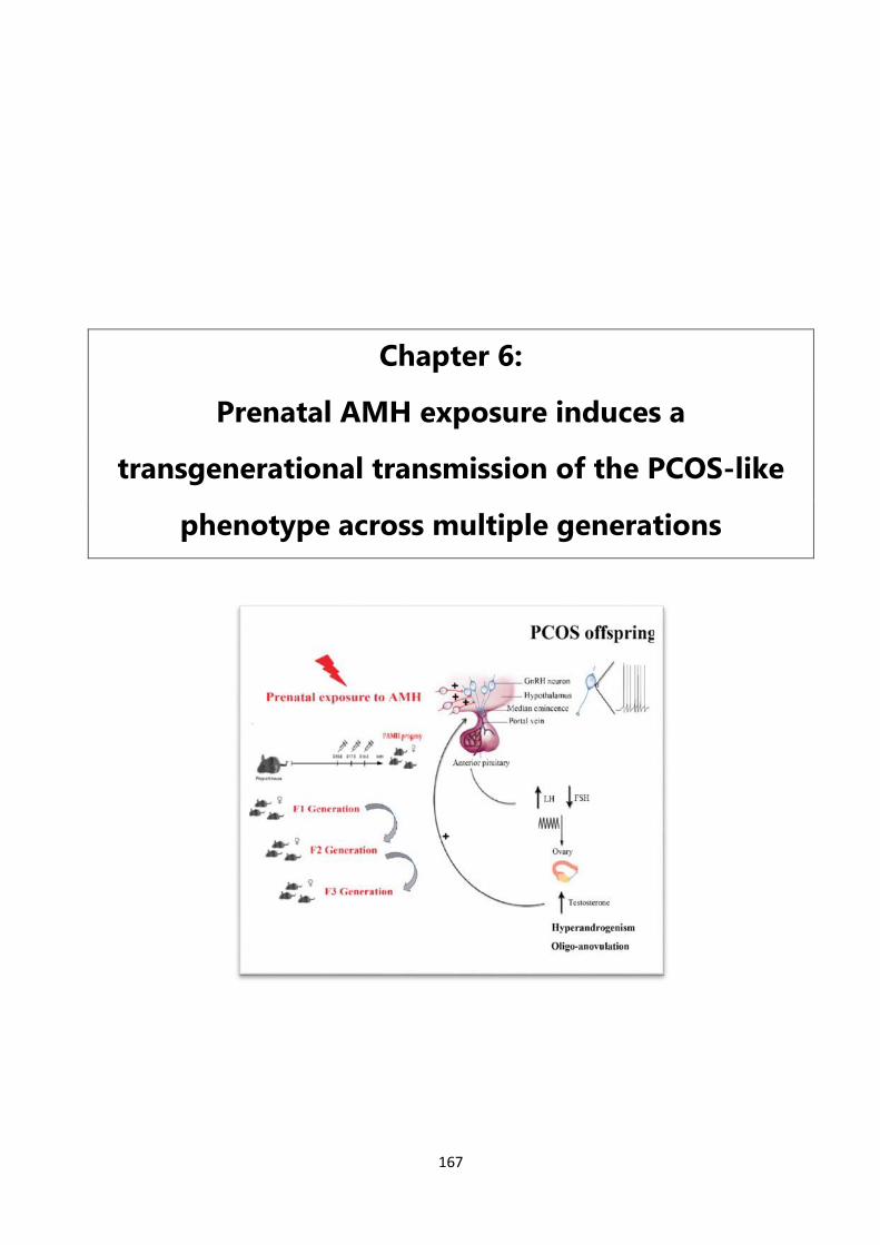
167
Chapter 6:
Prenatal AMH exposure induces a
transgenerational transmission of the PCOS-like
phenotype across multiple generations
Chapter 6: A transgenerational study in a PCOS mouse model
168
Chapter Six
Prenatal AMH exposure induces a transgenerational
transmission of the PCOS-like phenotype across multiple
generations
Nour El Houda Mimouni 1,2, Anne-Laure Barbotin 1,3, Isabel Paiva 4, Vincent Prevot 1,2,
Anne-Laurence Boutiller4 and Paolo Giacobini 1,2
(Mimouni et al, In preparation)
1 Jean-Pierre Aubert Research Center (JPArC), Laboratory of Development and Plasticity of
the Neuroendocrine Brain, Inserm, UMR-S 1172, Lille, France
2 University of Lille, FHU 1,000 Days for Health, School of Medicine, Lille, France
3 CHU Lille, Institut de Biologie de la Reproduction-Spermiologie-CECOS, Lille, France
4 Laboratory of Cognitive and Adaptive Neuroscience, National Centre for Scientific Research,
Strasbourg.
Chapter 6: A transgenerational study in a PCOS mouse model
169
Context
Described in 1935 by Stein and Leventhal, the definition of this syndrome is still at
the core of the debate due to its complex clinical manifestations. According to various
population studies in conducted in geographically distinct populations, the prevalence of
the syndrome is estimated to reach 18% following the Rotterdam criteria (Azziz, Woods et
al. 2004),(March, Moore et al. 2010), (Asuncion, Calvo et al. 2000).
Additionally, clinical observations indicate that the risk of developing PCOS rises to 20-40%
in first-degree relatives of PCOS women (Kahsar-Miller, Nixon et al. 2001), (Legro, Driscoll
et al. 1998, Franks, Webber et al. 2008). However, varying manifestations of reproductive
and metabolic are observed in first-degree relatives of PCOS patients (Sir-Petermann,
Maliqueo et al. 2002, Sir-Petermann, Ladron de Guevara et al. 2012). One portion of the
daughters and sisters of women with PCOS have higher levels of adrenal ovarian androgens
from adolescence into adulthood (Yildiz, Yarali et al. 2003, Azziz, Woods et al. 2004). And
another portion of female first-degree relatives of PCOS women seem to have a
considerable risk of insulin resistance, hyperinsulinemia, type2 diabetes, and/or impaired
glucose tolerance (Sir-Petermann, Maliqueo et al. 2002, Kent, Gnatuk et al. 2008). However,
less is known about the phenotype of sons of PCOS women, who are exposed during
gestation but not systematically investigated in adulthood.
Using our PCOS-like mouse model, this chapter aims to:
- Further explore the role of AMH in the onset of PCOS-like traits in a transgenerational
inheritance context.
- Investigate how early exposure to AMH excess would affect the neuroendocrine and
reproductive features of the male offspring at birth, puberty and in adulthood.
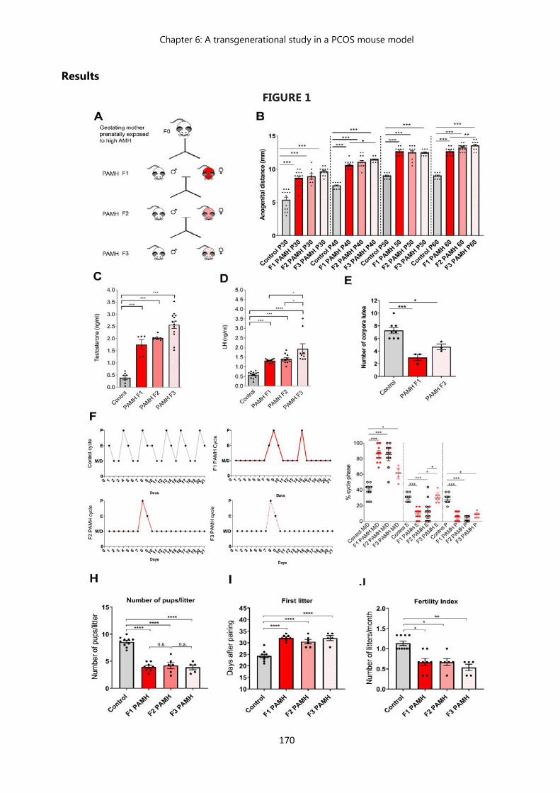
Chapter 6: A transgenerational study in a PCOS mouse model
170
Results FIGURE 1
Chapter 6: A transgenerational study in a PCOS mouse model
171
Prenatal AMH exposure induces a transgenerational transmission of the PCOS-like
phenotype across multiple generations.
A) Schematic illustration of the mating strategy employed to generate the F1, F2, F3 PAMH progeny.
Whereby Pregnant female mice (F0) were treated with PBS (Control) or with the bioactive form of
AMH (PAMH:Prenatal AMH treatement), from embryonic day E16.5 to E18.5, to generate the first
progeny of (F1) PAMH males and females which were then crossed to produce the second (F2) and
third (F3) PAMH generations . F2 and F3 PAMH females were not subjected to further AMH
exposures.
(B) Anogenital distance (AGD) was measured over post-natal days (P) 30, 40,50 and 60 in adult
control females (n = 11-14), F1 PAMH (n = 12-16), F2 PAMH (n = 7-14), F3 PAMH (n =12). AGD
was significantly longer in PAMH females as compared to the control group throughout post-natal
development. Comparisons were performed using One-way ANOVA followed and Tukey’s multiple
comparison post hoc test : ( **** P < 0.0001, P30: F (3, 46) = 36.00; P40: F (3, 47) = 112; P50: F (3,
45) = 92.88; P60: F (3, 45) = 171.8).
(C) Plasma testosterone concentration was measured by ELISA in adult females (P60) in diestrus:
Control (n = 7), F1 PAMH (n = 5), F2 PAMH (n = 10) and F3 PAMH (n = 13).
(D) Plasma LH level was measured by sandwich ELISA in (P60-P90) diestrus female offspring: Control
(n = 13), F1 PAMH females (n = 10), F2 PAMH females (n = 10), F3 PAMH females (n = 9).
comparisons between groups were computed using one-way ANOVA and Tukey’s multiple
comparison post hoc test ( **** P < 0.0001, T: F (3, 31) = 45.95, **** P < 0.0001, LH: F(3,33) = 30.52)
(E) Quantification of the number of corpora lutea (CL) in the ovaries of adult diestrus female mice:
Control (n = 4), F1 PAMH (n = 4) and F3 PAMH (n=3). Data are represented as mean ± s.e.m.
comparisons between groups were computed using one-way ANOVA and Tukey’s multiple
comparison post hoc test. Both groups of PAMH females from the first and third generation
displayed a reduced number of CL as compared to control females (***p= 0.0007; F(2, 11)= 15.03).
(F) Representative estrous cyclicity of 8 mice/treatment group during 16 consecutive days. M/D:
Metestrus/Diestrus phase, P: Proestrus, E: Estrus.
Chapter 6: A transgenerational study in a PCOS mouse model
172
(G) Quantitative analysis of ovarian cyclicity in the adult female (P60-P90) offspring mice from
control and PAMH mice. Scatter plot representing the percentage (%) of time spent in each estrous
cycle respectively in control (n = 19), PAMH F1 females (n = 11), PAMH F2 females (n = 11), PAMH
F3 females (n = 7), M/D: Metestrus/Diestrus phase, P: Proestrus, E: Estrus. Comparisons between
treatment groups were performed using Kruskal-Wallis test followed by Dunn’s multiple comparison
post hoc test: M/D: *** P < 0.0001; P: *** P < 0.0001 ; E: *** P < 0.0001. The horizontal line in each
scatter plot corresponds to the median value. The vertical line represents the 25th – 75th percentile
range.
(G-H-I) Fertility tests of adult offspring mice (P90). Inexperienced males and primiparous females,
selected from at least three different litters, were used for the 90 days mating protocol test. Control
females were paired with control males (n = 10), F1 PAMH (1st generation) females were paired with
F1 PAMH males (n = 8), F2 PAMH ( 2nd generation) females were paired with F2 PAMH males (n =
6), and finally F3 PAMH (3rd generation) females were paired with F3 PAMH males (n=5). (G) Number
of pups/litter (H) Time to first litter (number of days to first litter after pairing) (I) Fertility index
(number of litters per females over 3 months) were quantified per generation and pairing.
PAMH adult female and male mice from all 3 generations showed a persistent impaired fertility as
indicated by a significant delay in their first litter, fewer litters and fewer pups per litter produced
over a 3-month period. Data are represented as mean ± s.e.m. Statistical analysis was performed
using One-way ANOVA and Tukey’s multiple comparison post hoc test: Time to first litter,
F(3,25)=27.97, ***P < 0.0001; Number of pups/litter, F(3,25)= 43.66 *** P < 0.0001; Analysis of fertility
index between groups was assessed using Kruskal Wallis followed by Dunn's multiple comparisons
post hoc test: ***P= 0.0005.
Chapter 6: A transgenerational study in a PCOS mouse model
173
Prenatal AMH exposure induces a transgenerational transmission of the PCOS-like
phenotype towards multiple generations of female progeny
Polycystic ovary syndrome (PCOS) is a heterogeneous disorder characterized by multiple
endocrine and metabolic disturbances. It is the leading cause of infertility, characterized by
anovulation, cycle disorders, hyperandrogenism and severe hormonal imbalance (March, Moore et
al. 2010) (Azziz, Woods et al. 2004, Norman, Dewailly et al. 2007) (Goodarzi, Dumesic et al. 2011)
(Dumesic, Oberfield et al. 2015)
In the past decades, several large series of family-based and twin studies have highlighted a
strong familiar component in PCOS (Cooper, Spellacy et al. 1968, Hague, Adams et al. 1988, Legro,
Driscoll et al. 1998, Rodin, Bano et al. 1998). Thus, only a small portion of candidate genes has been
identified by genome-wide association studies (GWAS) and linkage studies (Chen, Zhao et al. 2011)
(Franks, Gharani et al. 2001, Dadachanji, Shaikh et al. 2018) suggesting that environmental factors
such as high androgen levels during early embryonic life might play role in the onset of the
syndrome in the unborn fetus. (Abbott, Barnett et al. 2005, Abbott and Bacha 2013).
Giving the strong heritable characteristics of PCOS, we next sought to investigate if putative
long-term consequences of prenatal exposure to excessive AMH levels are transmitted across
multiple generations in female mice and would in turn, mirror the transmission of the cardinal
neuroendocrine features observed in first degree relatives of PCOS women. In order to further
delineate if the elevation of AMH levels observed in women with PCOS during pregnancy could be
one of the drivers of the condition and its transmission in the progeny or inversely a consequence
of the neuroendocrine disturbances of the syndrome.
In order to test this hypothesis, we treated pregnant females with PBS (Control) or with the
bioactive form of AMH (human recombinant AMHC, 0.12 mg/Kg/d; prenatal AMH-treated, PAMH)
from embryonic day E16.5 to E18.5, to generate F1 PAMH males and females, that we ultimately
crossed to produce the second (F2) and third (F3) PAMH generations. No further AMH exposure for
any subsequent generations (F2 and F3 PAMH) was thus employed (Figure 1A).
Hyperandrogenism is a hallmark of the syndrome, along with the disproportionately high LH
secretion with relatively constant low FSH secretion observed in PCOS patients (Balen, Conway et al.
1995). This increase is explained by an increased pulse frequency of the hypothalamic gonadotropin-
releasing hormone (GnRH) which may favor the production of the β-subunit of LH over the β-subunit
Chapter 6: A transgenerational study in a PCOS mouse model
174
of FSH and/or by increased pituitary sensitivity to GnRH stimulation which results in an elevated
LH/FSH ratio increased in more than 75% of PCOS patients (McCartney, Eagleson et al. 2002) (Taylor,
McCourt et al. 1997) (Waldstreicher, Santoro et al. 1988).
Interestingly, growing clinical investigations in daughters and first-degree female relatives of PCOS
women report clinical and biological signs of hyperandrogenism as well as an increase in LH levels
in these patients.
Therefore, we sought to evaluate the androgenic impregnation across the 3 PAMH generations of
female offspring by measuring the ano-genital distance (AGD) at postnatal day 30 (P30), (P40), (P50)
and (P60) (Figure 1B). All of the three PAMH generations studied (F1, F2 and F3) displayed a
significantly longer ano-genital distance as compared the control female offspring at the different
time points of postnatal development analyzed, indicating a higher androgenic impregnation in
these mice.
Subsequently, we uncovered a significant and persistent elevation in both circulating levels of
testosterone and LH in the F1, F2 and F3 PAMH females in comparison with the control group.
(Figure 1C, 1D)
Together, these results suggest that a single intrauterine exposure to elevated AMH levels during
embryogenesis could be responsible for the transmission of pcos-like traits of hyperandrogenism
and altered LH levels to the subsequent non-exposed generations of PAMH female offsprings.
Several studies have reported that the incidence of oligomenorrhea and PCO is amplified in first-
degree relatives of PCOS patients as compared with the controls (Cooper, Spellacy et al. 1968),
(Wilroy, Givens et al. 1975, Givens 1988). Another study conducted in a large group of hirsute women
with or without oligomenorrhea and their family members, described a higher prevalence of
hirsutism, oligomenorrhea and infertility in first degree relatives as compared to controls (Ferriman
and Purdie 1979). (Lunde, Magnus et al. 1989).
To evaluate if prenatal AMH treatment would transgenerational alter reproductive phenotype of the
PAMH females in a transgenerational manner, we have monitored the estrus cyclicity in the different
groups during 16 days and the results confirm that the F1 PAMH females exhibit alterations in estrus
cyclicity and ovarian function as previously described in the female PAMH mice (Tata, Mimouni et
al. 2018). We thus report for the first time in this study that these alterations are transmitted to the
F2 and F3 PAMH females, which also rarely entered the preovulatory stage of the estrous cycle and
Chapter 6: A transgenerational study in a PCOS mouse model
175
displayed prolonged time in metestrus/diestrus as compared to the control female offspring (Figure
1F) and dysovulation (Figure 1G) as compared to the control littermates. These results are
consistent with the analysis of the ovarian histology of the PAMH mice, which revealed a persistent
decrease in the number of corpora lutea (CL) in the F1 PAMH and F3 PAMH offspring. (Figure 1E).
Finally, using a 90 days fertility test protocol, we meticulously examined the main reproductive
characteristics of each group, in other terms: Fertility index (number of litters per month), First litter
(days to first litter after pairing) and the number of pups per litter.
A significant fertility impairment was detected in all of the three PAMH generations studied, and
was reflected by a longer interval between litters, a reduced number of pups per litter and a
diminished fertility index as compared to the control groups (Figure 1B). Furthermore, in the F2 and
F3 PAMH breedings, we observed a significantly higher impact on fertility parameters of first litter
and litter size in comparison to the F1 PAMH breeding, suggesting that a unique exposition to AMH
in the late stages of embryonic development induces long term reproductive damage maintained
across generations and which could even result in a worsen phenotype than the one observed in
the first generation directly exposed to AMH treatment during gestation.
Unexpectedly, the F1 and F3 PAMH males breedings with control females also provoke a likewise
decrease in fertility as indicated by a significant delay in their first litter, smaller litters and fewer
pups per litter produced over a 3-month period. Suggesting that the prenatal exposition to elevated
AMH levels impacts the fertility of the male progeny and this defect is transmitted to the subsequent
generations.
Altogether, our results provide compelling evidence that the effects of AMH-dependent
developmental programming in early life environment is not only transmitted from parent to
offspring but extends to generations beyond.

Chapter 6: A transgenerational study in a PCOS mouse model
176
FIGURE 2
Chapter 6: A transgenerational study in a PCOS mouse model
177
A severe decrease in mRNA expression levels of key genes involved in ovarian steroidogenesis
is maintained across the subsequent generations of the PAMH PCOS-like mouse model
(A) Simplified schematic representation of the steroidogenesis in the theca and granulosa cells
of the ovary. Steroidogenesis is a multi-step process in which, cholesterol, the main precursor of
steroids is ultimately converted into androgens and estrogens. (StAR) steroidogenic acute
regulatory protein, (HSD3B1) hydroxyl-Δ-5-steroid dehydrogenase, 3 β-and steroid Δ-isomerase 1,
(CYP17A1) cytochrome P450, family 17, subfamily A polypeptide 1, (CYP11A1) cytochrome P450,
family 11, subfamily A, polypeptide 1, (CYP19A1) cytochrome P450, family 19, subfamily A,
polypeptide 1a, (IGF1) insulin-like growth factor 1, (LHR) Luteinizing hormone receptor, (FSHR)
Follicule stimulating hormone receptor. (DHEA) Dehydroepiandrosterone.
(B) Real time PCR analysis of the most relevant steroidogenic markers in the ovaries of adult
diestrus females. We extracted mRNA from control ovaries (n=5); F1 PAMH(n=6); F2 PAMH (n=6);
F3 PAMH (n=6). Quantitative analysis of real time PCR shows that AMH prenatal treatment severely
impacts the mRNA expression levels of Cyp19a1, Hsd3b1 in the ovaries of F1, F2 and F3 PAMH
females in comparison with the control ovaries. However, no differences were observed in the mRNA
expression levels of Cyp11a1 between the two groups. Data are represented as mean +/- sem.
Differences between groups were assessed using One-way ANOVA and Tukey’s multiple comparison
post hoc test: Cyp19a1: ****P < 0.0001, F (3, 19) = 16.74; Hsd3b1: **P=0.0035, F (3, 19) = 6.424
Cyp11a1: P=0.7214, F(3, 19)= 0.4483.
The most common biochemical abnormality in women with PCOS is excessive production of
ovarian androgens. This feature well correlates with the observation that PCOS women exhibit higher
ovarian steroidogenic activity than healthy patients (secretion of higher levels of testosterone,
androstenedione, of progesterone)(Nelson, Legro et al. 1999, Nelson, Qin et al. 2001).
For this reason, we evaluated the relative expression of the same genes involved in the androgen
biosynthetic pathway in the ovaries (Figure 2A) in control and F1, F2, F3 PAMH ovaries. Our results
show that there are no changes in the relative expression levels of Cyp11a1. Conversely, there is
drastic decrease in the relative expression of Cyp19a1 which is responsible for the conversion of
testosterone into Estradiol, and Hsd3b1 which converts pregnenolone to progesterone. Overall, our
findings strengthen the rationale about the role of AMH in utero as a trigger of PCOS-like
disturbances observed in the adulthood, and most importantly that this could be transmitted to the
generations beyond.
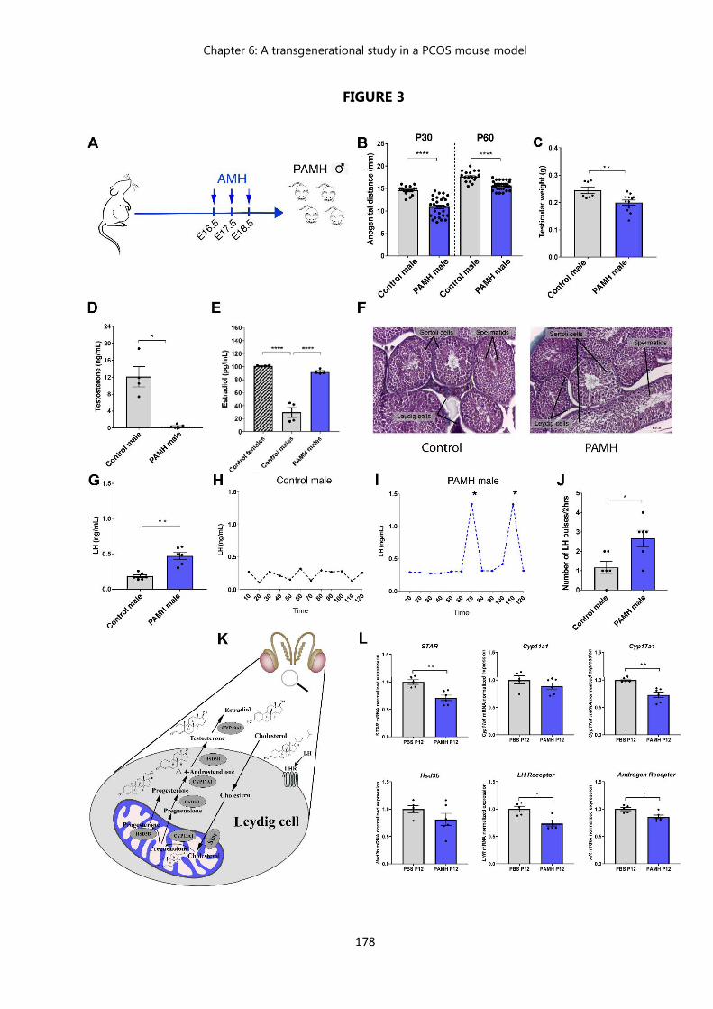
Chapter 6: A transgenerational study in a PCOS mouse model
178
FIGURE 3
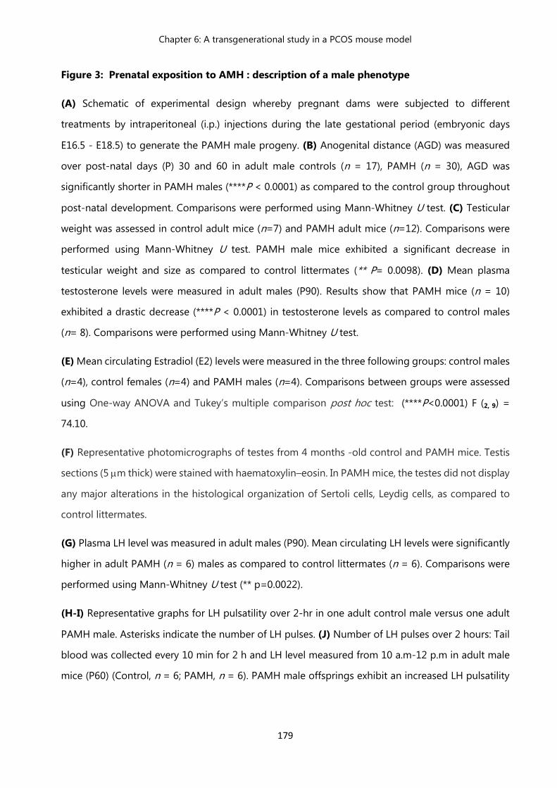
Chapter 6: A transgenerational study in a PCOS mouse model
179
Figure 3: Prenatal exposition to AMH : description of a male phenotype
(A) Schematic of experimental design whereby pregnant dams were subjected to different
treatments by intraperitoneal (i.p.) injections during the late gestational period (embryonic days
E16.5 - E18.5) to generate the PAMH male progeny. (B) Anogenital distance (AGD) was measured
over post-natal days (P) 30 and 60 in adult male controls (n = 17), PAMH (n = 30), AGD was
significantly shorter in PAMH males (****P < 0.0001) as compared to the control group throughout
post-natal development. Comparisons were performed using Mann-Whitney U test. (C) Testicular
weight was assessed in control adult mice (n=7) and PAMH adult mice (n=12). Comparisons were
performed using Mann-Whitney U test. PAMH male mice exhibited a significant decrease in
testicular weight and size as compared to control littermates (** P= 0.0098). (D) Mean plasma
testosterone levels were measured in adult males (P90). Results show that PAMH mice (n = 10)
exhibited a drastic decrease (****P < 0.0001) in testosterone levels as compared to control males
(n= 8). Comparisons were performed using Mann-Whitney U test.
(E) Mean circulating Estradiol (E2) levels were measured in the three following groups: control males
(n=4), control females (n=4) and PAMH males (n=4). Comparisons between groups were assessed
using One-way ANOVA and Tukey’s multiple comparison post hoc test: (****P<0.0001) F (2, 9) =
74.10.
(F) Representative photomicrographs of testes from 4 months -old control and PAMH mice. Testis
sections (5 µm thick) were stained with haematoxylin–eosin. In PAMH mice, the testes did not display
any major alterations in the histological organization of Sertoli cells, Leydig cells, as compared to
control littermates.
(G) Plasma LH level was measured in adult males (P90). Mean circulating LH levels were significantly
higher in adult PAMH (n = 6) males as compared to control littermates (n = 6). Comparisons were
performed using Mann-Whitney U test (** p=0.0022).
(H-I) Representative graphs for LH pulsatility over 2-hr in one adult control male versus one adult
PAMH male. Asterisks indicate the number of LH pulses. (J) Number of LH pulses over 2 hours: Tail
blood was collected every 10 min for 2 h and LH level measured from 10 a.m-12 p.m in adult male
mice (P60) (Control, n = 6; PAMH, n = 6). PAMH male offsprings exhibit an increased LH pulsatility
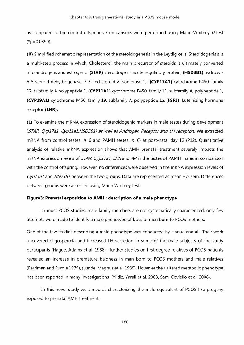
Chapter 6: A transgenerational study in a PCOS mouse model
180
as compared to the control offsprings. Comparisons were performed using Mann-Whitney U test
(*p=0.0390).
(K) Simplified schematic representation of the steroidogenesis in the Leydig cells. Steroidogenisis is
a multi-step process in which, Cholesterol, the main precursor of steroids is ultimately converted
into androgens and estrogens. (StAR) steroidogenic acute regulatory protein, (HSD3B1) hydroxyl-
Δ-5-steroid dehydrogenase, 3 β-and steroid Δ-isomerase 1, (CYP17A1) cytochrome P450, family
17, subfamily A polypeptide 1, (CYP11A1) cytochrome P450, family 11, subfamily A, polypeptide 1,
(CYP19A1) cytochrome P450, family 19, subfamily A, polypeptide 1a, (IGF1) Luteinizing hormone
receptor (LHR).
(L) To examine the mRNA expression of steroidogenic markers in male testes during development
(STAR, Cyp17a1, Cyp11a1,HSD3B1) as well as Androgen Receptor and LH receptor). We extracted
mRNA from control testes, n=6 and PAMH testes, n=6) at post-natal day 12 (P12). Quantitative
analysis of relative mRNA expression shows that AMH prenatal treatment severely impacts the
mRNA expression levels of STAR, Cyp17a1, LHR and AR in the testes of PAMH males in comparison
with the control offspring. However, no differences were observed in the mRNA expression levels of
Cyp11a1 and HSD3B1 between the two groups. Data are represented as mean +/- sem. Differences
between groups were assessed using Mann Whitney test.
Figure3: Prenatal exposition to AMH : description of a male phenotype
In most PCOS studies, male family members are not systematically characterized, only few
attempts were made to identify a male phenotype of boys or men born to PCOS mothers.
One of the few studies describing a male phenotype was conducted by Hague and al. Their work
uncovered oligospermia and increased LH secretion in some of the male subjects of the study
participants (Hague, Adams et al. 1988), further studies on first degree relatives of PCOS patients
revealed an increase in premature baldness in man born to PCOS mothers and male relatives
(Ferriman and Purdie 1979), (Lunde, Magnus et al. 1989). However their altered metabolic phenotype
has been reported in many investigations (Yildiz, Yarali et al. 2003, Sam, Coviello et al. 2008).
In this novel study we aimed at characterizing the male equivalent of PCOS-like progeny
exposed to prenatal AMH treatment.
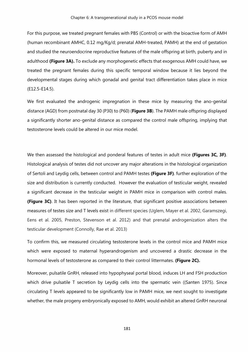
Chapter 6: A transgenerational study in a PCOS mouse model
181
For this purpose, we treated pregnant females with PBS (Control) or with the bioactive form of AMH
(human recombinant AMHC, 0.12 mg/Kg/d; prenatal AMH-treated, PAMH) at the end of gestation
and studied the neuroendocrine reproductive features of the male offspring at birth, puberty and in
adulthood (Figure 3A). To exclude any morphogenetic effects that exogenous AMH could have, we
treated the pregnant females during this specific temporal window because it lies beyond the
developmental stages during which gonadal and genital tract differentiation takes place in mice
(E12.5-E14.5).
We first evaluated the androgenic impregnation in these mice by measuring the ano-genital
distance (AGD) from postnatal day 30 (P30) to (P60) (Figure 3B). The PAMH male offspring displayed
a significantly shorter ano-genital distance as compared the control male offspring, implying that
testosterone levels could be altered in our mice model.
We then assessed the histological and ponderal features of testes in adult mice (Figures 3C, 3F).
Histological analysis of testes did not uncover any major alterations in the histological organization
of Sertoli and Leydig cells, between control and PAMH testes (Figure 3F). further exploration of the
size and distribution is currently conducted. However the evaluation of testicular weight, revealed
a significant decrease in the testicular weight in PAMH mice in comparison with control males.
(Figure 3C). It has been reported in the literature, that significant positive associations between
measures of testes size and T levels exist in different species (Uglem, Mayer et al. 2002, Garamszegi,
Eens et al. 2005, Preston, Stevenson et al. 2012) and that prenatal androgenization alters the
testicular development (Connolly, Rae et al. 2013)
To confirm this, we measured circulating testosterone levels in the control mice and PAMH mice
which were exposed to maternal hyperandrogenism and uncovered a drastic decrease in the
hormonal levels of testosterone as compared to their control littermates. (Figure 2C).
Moreover, pulsatile GnRH, released into hypophyseal portal blood, induces LH and FSH production
which drive pulsatile T secretion by Leydig cells into the spermatic vein ((Santen 1975). Since
circulating T levels appeared to be significantly low in PAMH mice, we next sought to investigate
whether, the male progeny embryonically exposed to AMH, would exhibit an altered GnRH neuronal
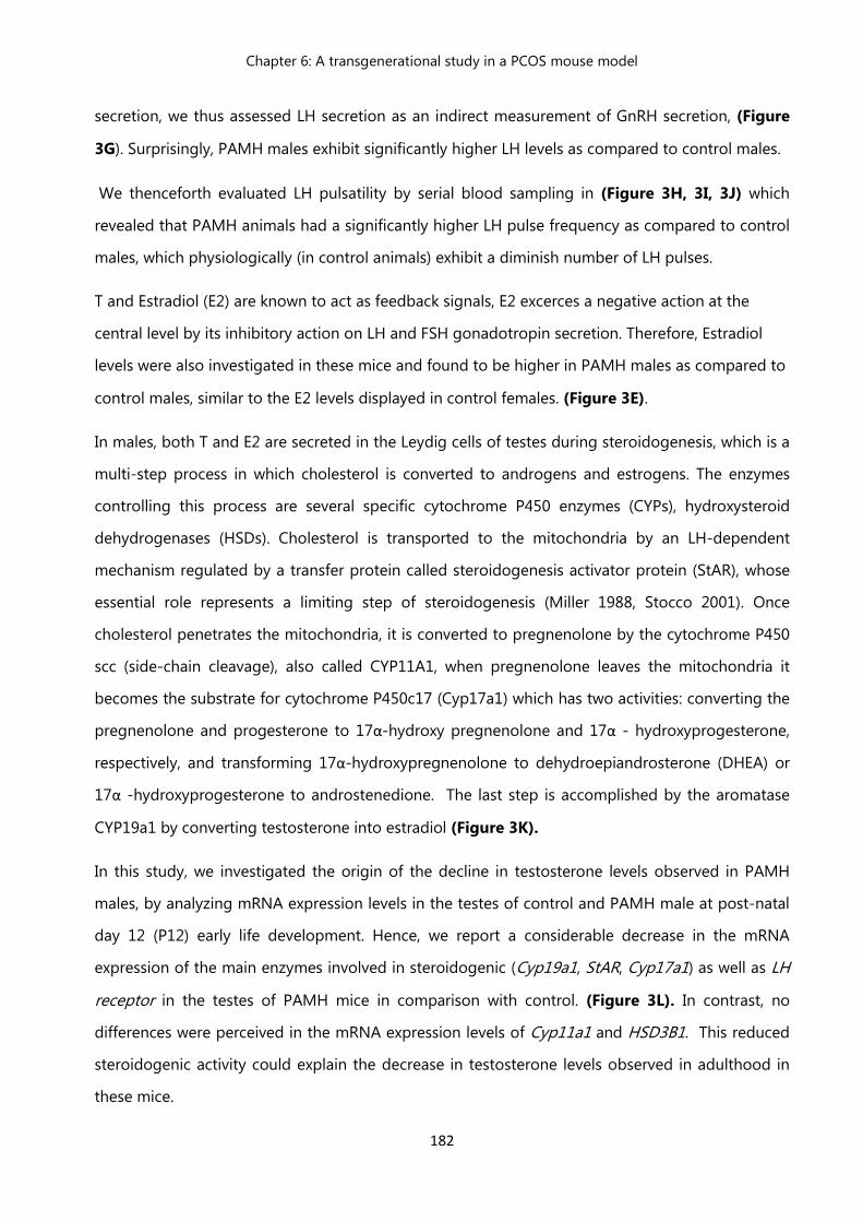
Chapter 6: A transgenerational study in a PCOS mouse model
182
secretion, we thus assessed LH secretion as an indirect measurement of GnRH secretion, (Figure
3G). Surprisingly, PAMH males exhibit significantly higher LH levels as compared to control males.
We thenceforth evaluated LH pulsatility by serial blood sampling in (Figure 3H, 3I, 3J) which
revealed that PAMH animals had a significantly higher LH pulse frequency as compared to control
males, which physiologically (in control animals) exhibit a diminish number of LH pulses.
T and Estradiol (E2) are known to act as feedback signals, E2 excerces a negative action at the
central level by its inhibitory action on LH and FSH gonadotropin secretion. Therefore, Estradiol
levels were also investigated in these mice and found to be higher in PAMH males as compared to
control males, similar to the E2 levels displayed in control females. (Figure 3E).
In males, both T and E2 are secreted in the Leydig cells of testes during steroidogenesis, which is a
multi-step process in which cholesterol is converted to androgens and estrogens. The enzymes
controlling this process are several specific cytochrome P450 enzymes (CYPs), hydroxysteroid
dehydrogenases (HSDs). Cholesterol is transported to the mitochondria by an LH-dependent
mechanism regulated by a transfer protein called steroidogenesis activator protein (StAR), whose
essential role represents a limiting step of steroidogenesis (Miller 1988, Stocco 2001). Once
cholesterol penetrates the mitochondria, it is converted to pregnenolone by the cytochrome P450
scc (side-chain cleavage), also called CYP11A1, when pregnenolone leaves the mitochondria it
becomes the substrate for cytochrome P450c17 (Cyp17a1) which has two activities: converting the
pregnenolone and progesterone to 17α-hydroxy pregnenolone and 17α - hydroxyprogesterone,
respectively, and transforming 17α-hydroxypregnenolone to dehydroepiandrosterone (DHEA) or
17α -hydroxyprogesterone to androstenedione. The last step is accomplished by the aromatase
CYP19a1 by converting testosterone into estradiol (Figure 3K).
In this study, we investigated the origin of the decline in testosterone levels observed in PAMH
males, by analyzing mRNA expression levels in the testes of control and PAMH male at post-natal
day 12 (P12) early life development. Hence, we report a considerable decrease in the mRNA
expression of the main enzymes involved in steroidogenic (Cyp19a1, StAR, Cyp17a1) as well as LH
receptor in the testes of PAMH mice in comparison with control. (Figure 3L). In contrast, no
differences were perceived in the mRNA expression levels of Cyp11a1 and HSD3B1. This reduced
steroidogenic activity could explain the decrease in testosterone levels observed in adulthood in
these mice.
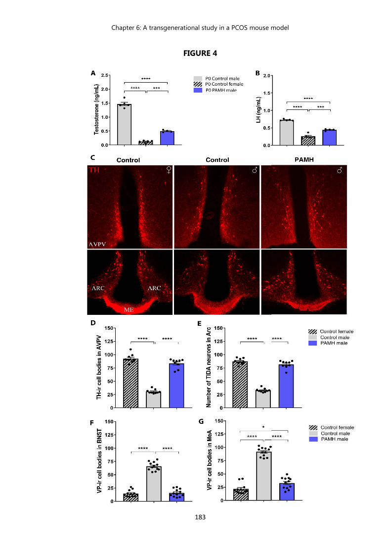
Chapter 6: A transgenerational study in a PCOS mouse model
183
FIGURE 4
Chapter 6: A transgenerational study in a PCOS mouse model
184
Figure 4: PAMH male offspring exhibit a feminization of the brain.
(A-B) Plasma T and LH levels were measured in pups 2 hours after birth (Control males, n = 4; control
females, n = 5; PAMH males, n = 5). Statistics were computed with One-way ANOVA and Tukey’s
multiple comparison post hoc test: Testosterone: F (2, 11) =283.5, ***P < 0.0001; LH: F (2, 11) = 153.2,
***P < 0.0001. (C) Photomicrographs of TH-immunoreactive neurons in the anteroventral
periventricular nucleus (AVPV) and arcuate nucleus (ARC) of a control male, control female and
PAMH male at P60. ME: Median Eminence (D) TH immunoreactive (-ir) neurons in the AVPV were
quantified in these animal groups (n = 9 per sex and treatment). The number of AVPV TH-ir neurons
are represented as the mean ± s.e.m. One-way ANOVA and Tukey’s multiple comparisons post-hoc
test, F(2,24) = 193.1, ***P < 0.0001. (E) Quantification of the tuberoinfundibular dopaminergic (TIDA)
immunoreactive neurons in the ARC; (n = 9 per sex and treatment). Data are represented as the
mean ± s.e.m. One-way ANOVA and Tukey’s multiple comparisons post-hoc test: F (2,24) = 252.5 ;
***P < 0.0001. (F-G) Quantification of the VP-immunoreactive neurons in the bed nucleus of the
stria terminalis (BNST) and medial amygdala (MeA) of a male, control female and PAMH male at P60.
(n = 12 per sex and treatment). Data are represented as the mean ± s.e.m. One-way ANOVA and
Tukey’s multiple comparisons post-hoc test: BNST, F(2,33) = 185.8 ; MeA, F(2,33) = 205.8,* P < 0.05,
***P < 0.0001.
PAMH male offspring exhibit a feminization of the brain.
Prenatal life, early childhood and adolescence are critical periods marked by the influence of
environmental factors in mammals. During these phases of development, the brain undergoes
significant growth and remodeling, and is particularly vulnerable to external signals. During this
temporal window of embryonic development, under the action of the gonadal steroids, the brain
irreversibly engages in a differentiation path of the male or female direction.
The neonatal testosterone surge is responsible for establishing brain sexual dimorphisms that
underpin sexually dimorphic physiology and behavior (Clarkson and Herbison 2016) (Simerly 2002,
McCarthy, Arnold et al. 2012). The masculinizing and defeminizing effects of testosterone are mainly
mediated by its metabolite, estradiol, (McCarthy 2008), tthrough the aromatization of testosterone
by the enzyme P450 aromatase (CYP19A1).
Chapter 6: A transgenerational study in a PCOS mouse model
185
We thus, analyzed whether the late gestational, AMH-dependent maternal hyperandrogenism
previously described (Tata, Mimouni et al. 2018) might interfere with the appropriate establishment
of brain sexual dimorphism in the male offspring.
In most vertebrates, gonadal T secretion occurs in a sexually dimorphic pattern during the first
hours following birth (0-4 hours at P0), being elevated in newborn males but not in females (Corbier,
Edwards et al. 1992). We therefore measured T levels 2 hours post-partum in control males and
females as well as in PAMH male pups. We report that PAMH adult male failed to engage the
testosterone surge at birth observed in control newborns (Figure 4A). Interestingly, as stated above
(Figure 3D), the levels of testosterone remain dramatically low in PAMH males as compared to the
control group, when measured in adulthood.
Few hours after the testosterone surge, most studies indicate the existence of an LH surge shortly
after T in males. Remarkably PAMH male mice display no evidence of a neonatal LH surge, and their
LH levels mirror the levels observed in control females rather than the ones detected in control males
(Figure 4B).
Furthermore, using immunohistochemistry we labeled some of the main sexually dimorphic
neuronal populations in rodents’ brains. (Figure 4C) The evaluation of sexually dimorphic areas of
the brains of control males and females and PAMH males allowed us to uncover a feminization of
the brains of PAMH mice, this has been confirmed by quantitative analysis of immunoreactive
sexually dimorphic neurons (Vasopressin, Tyrosine Hydroxylase and TIDA neurons), these
mentionned neuronal populations were quantified in every group and the results show that the
number of Immunoreactive neurons is closer in numbers to the one observed in control female
brains than in males. ( Figure 4D, 4E, 4F, 4J). Suggesting that the exposition to elevated levels of
AMH during embryogenesis has a feminizing effect on male offspring and conversely, this same
prenatal exposition induces a masculinization of the fetal brain of female offspring.
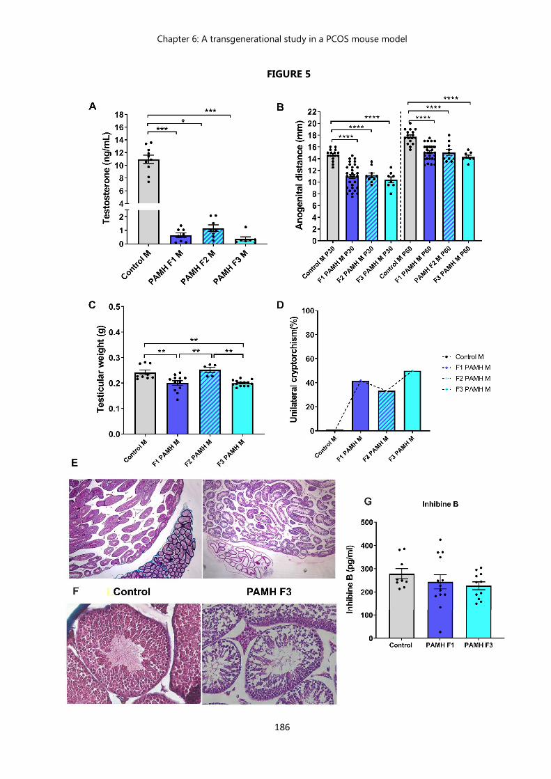
Chapter 6: A transgenerational study in a PCOS mouse model
186
FIGURE 5
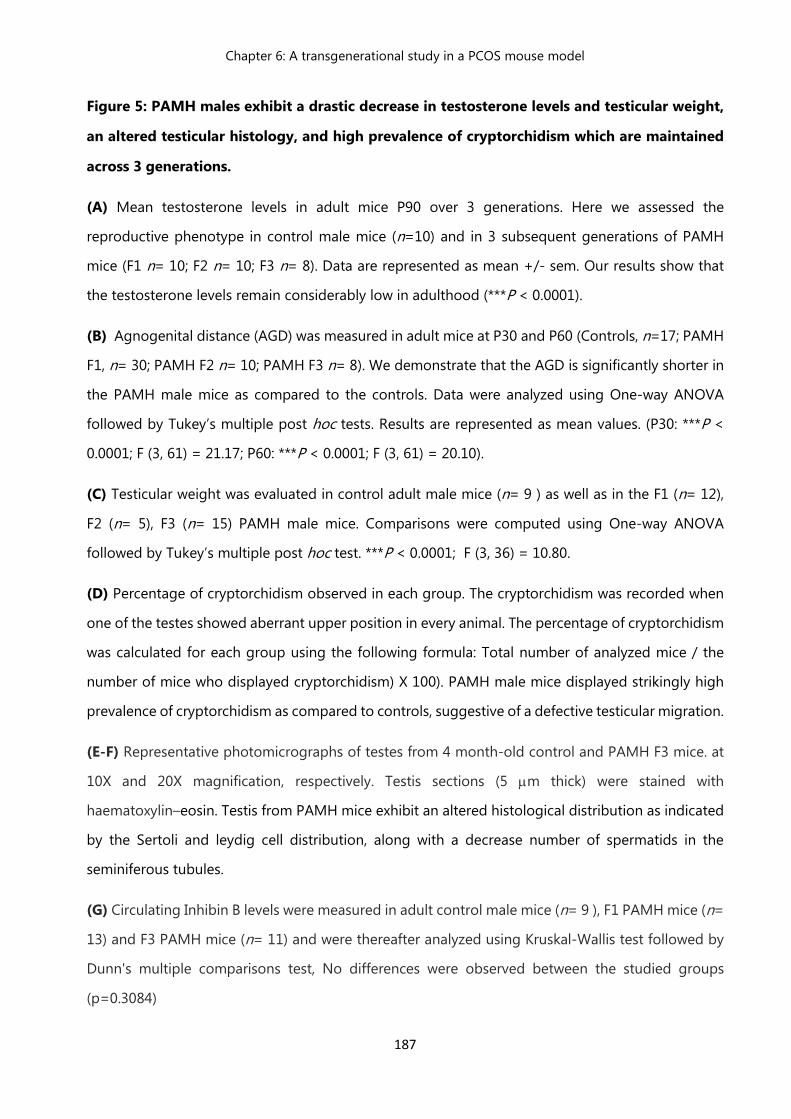
Chapter 6: A transgenerational study in a PCOS mouse model
187
Figure 5: PAMH males exhibit a drastic decrease in testosterone levels and testicular weight,
an altered testicular histology, and high prevalence of cryptorchidism which are maintained
across 3 generations.
(A) Mean testosterone levels in adult mice P90 over 3 generations. Here we assessed the
reproductive phenotype in control male mice (n=10) and in 3 subsequent generations of PAMH
mice (F1 n= 10; F2 n= 10; F3 n= 8). Data are represented as mean +/- sem. Our results show that
the testosterone levels remain considerably low in adulthood (***P < 0.0001).
(B) Agnogenital distance (AGD) was measured in adult mice at P30 and P60 (Controls, n=17; PAMH
F1, n= 30; PAMH F2 n= 10; PAMH F3 n= 8). We demonstrate that the AGD is significantly shorter in
the PAMH male mice as compared to the controls. Data were analyzed using One-way ANOVA
followed by Tukey’s multiple post hoc tests. Results are represented as mean values. (P30: ***P <
0.0001; F (3, 61) = 21.17; P60: ***P < 0.0001; F (3, 61) = 20.10).
(C) Testicular weight was evaluated in control adult male mice (n= 9 ) as well as in the F1 (n= 12),
F2 (n= 5), F3 (n= 15) PAMH male mice. Comparisons were computed using One-way ANOVA
followed by Tukey’s multiple post hoc test. ***P < 0.0001; F (3, 36) = 10.80.
(D) Percentage of cryptorchidism observed in each group. The cryptorchidism was recorded when
one of the testes showed aberrant upper position in every animal. The percentage of cryptorchidism
was calculated for each group using the following formula: Total number of analyzed mice / the
number of mice who displayed cryptorchidism) X 100). PAMH male mice displayed strikingly high
prevalence of cryptorchidism as compared to controls, suggestive of a defective testicular migration.
(E-F) Representative photomicrographs of testes from 4 month-old control and PAMH F3 mice. at
10X and 20X magnification, respectively. Testis sections (5 µm thick) were stained with
haematoxylin–eosin. Testis from PAMH mice exhibit an altered histological distribution as indicated
by the Sertoli and leydig cell distribution, along with a decrease number of spermatids in the
seminiferous tubules.
(G) Circulating Inhibin B levels were measured in adult control male mice (n= 9 ), F1 PAMH mice (n=
13) and F3 PAMH mice (n= 11) and were thereafter analyzed using Kruskal-Wallis test followed by
Dunn's multiple comparisons test, No differences were observed between the studied groups
(p=0.3084)
Chapter 6: A transgenerational study in a PCOS mouse model
188
Material and Methods
Animals
Timed-pregnant female wild-type C57BL/6J (B6) (Charles River, USA) were group-housed under
specific pathogen-free conditions in a temperature-controlled room (21-22°C) with a 12-h light/dark
cycle and ad libitum access to food and water. Standard diet (9.5mm Pelleted RM3, Special Diets
Services, France) was given to all mice during breeding, lactation and growth of young stock.
Nutritional profile of the standard diet RM3 is the following: Protein 22.45%, Fat 4.2%, Fiber 4.42%,
Ash 8%, Moisture 10%, Nitrogen free extract 50.4%; Calories: 3.6 kcal/gr.
Mice were randomly assigned to groups at the time of purchase or weaning to minimize any
potential bias. No data sets were excluded from analyses.
Animal studies were approved by the Institutional Ethics Committees of Care and Use of
Experimental Animals of the University of Lille (France; Ethical protocol number: APAFIS#2617-
2015110517317420 v5). All experiments were performed in accordance with the guidelines for
animal use specified by the European Council Directive of 22 September 2010 (2010/63/EU). The
sample size, sex and age of the animals used is specified in the text and/or figure legends.
Prenatal anti-Müllerian Hormone (PAMH) treatment
Timed-pregnant adult (3-4 months) C57BL6/J (B6) dams were injected daily intraperitoneally (i.p.)
from embryonic day (E) 16.5 to 18.5 with 200 µL of a solution containing respectively: 1) 0.01 M
phosphate buffered saline (PBS, pH 7.4, prenatal control-treated), 2) PBS with 0.12 mgKg-1/d human
anti-Müllerian hormone (AMH) (AMHC, R&D Systems, rhMIS 1737-MS-10, prenatal AMH (PAMH)-
treated).
Assessment of phenotype, estrous cycle and fertility
Control, F1 PAMH, F2 PAMH and F3 PAMH female offspring were weaned at post-natal day P21 and
checked for vaginal opening (VO) and time of first estrus. Anogenital distance (AGD) was measured
at different ages during post-natal development (P30, 35, 40, 50 and 60). At VO and in adulthood
(P60), vaginal smears were performed daily for 16 consecutive days (4-cycles) for analysis of age of
first estrus and estrous cyclicity. Vaginal cytology was analyzed under an inverted microscope to
Chapter 6: A transgenerational study in a PCOS mouse model
189
identify the specific day of the estrous cycle. The reproductive competency of these animals was
determined by pairing the following mice: Control females mated with untreated C57BL6J (B6) wild-
type males, PAMH females mated with PAMH males, PAMH F2 females mated with F2 PAMH males,
and F3 PAMH females mated with F3 PAMH males for a period of 3 months. Unexperienced males
and primiparous females, selected from at least three different litters, were used for the 90-days
mating protocol test.
Number of pups/litter (number of pups), fertility index (number of litters per females over 3 months),
and time to first litter (number of days to first litter after pairing) were quantified per treatment and
pairing.
Assessment of Testicular weight and cryptorchidism
The cryptorchidism was recorded when one of the testes showed aberrant upper position in every
animal. The percentage of cryptorchidism was calculated for each group using the following formula:
Total number of analyzed mice / the number of mice who displayed cryptorchidism) X 100). Ponderal
characteristics of testis from control and PAMH males were routinely measured during each
experiment.
Ovarian and Testicular histology
Ovaries were collected from 3-4-month-old diestrus mice, and testes from 4 months old male mice,
immersion-fixed in 4% PFA solution and stored at 4°C. Paraffin-embedded ovaries and testes were
sectioned at a thickness of 5 µm (histology facility, University of Lille 2, France) and stained with
hematoxylin-eosin (Sigma Aldrich, Cat # GHS132, HT1103128). Sections were examined throughout
the ovary and testis.
Total numbers of corpora lutea (CL) was classified and quantified as previously reported {Caldwell,
2017 #540}. To avoid repetitive counting, each follicle was only counted in the section where the
oocyte’s nucleolus was visible. Using an ocular scale, follicles were classified by diameter into
preantral, large growing (200–400 μm) and cystic follicles (> 400 μm). To avoid repetitive counting,
CL were counted every 100 µm by comparing the section with the preceding and following sections.
Chapter 6: A transgenerational study in a PCOS mouse model
190
CL were characterized by a still present central cavity, filled with blood and follicular fluid remnants
or by prominent polyhedral to round luteal cells.
Perfusions
Adult mice (P60-P90) were anesthetized with i.p. injections of 100 mg/kg of ketamine-HCl and 10
mg/kg xylazine-HCl and perfused transcardially with 20ml of saline, followed by 100 ml of 4% PFA
in 0.1M phosphate buffer (PB) (4% PFA/0.1M PB; pH 7.6). Brains were collected, post-fixed in the
same fixative for 2h at 4 °C, cryoprotected overnight in PB/Sucrose 30% at 4°C, embedded in OCT-
embedding medium (Tissue-Tek), frozen and stored at -80°C until cryosectioning.
Immunohistochemistry
Tissues were cryosectioned coronally (Leica cryostat) at 35µm for free-floating
immunohistochemistry (IHC). Sections were blocked in an incubation solution of Tris-buffered saline
(TBS 0.05M, pH 7.6), 0.25% bovine serum albumin (BSA; Sigma, A9418), 0.3% Triton X-100 (TBS-T;
Sigma, T8787) with 10% normal donkey serum (NDS; D9663; Sigma) for 2h at room temperature
before incubation with the following different primary antisera (depending on experiment) for 72h
at 4°C: Monoclonal mouse anti-Tyrosine Hydroxylase (TH) antibody (Millipore MAB318; 1:3000),
guinea pig anti-(Arg8)-Vasopressin (Peninsula Laboratories #T-5048; 1:2500), rabbit anti-cFos (Santa
Cruz Biotechnology #sc-52; 1:5000), and a rabbit anti-kisspeptin (A. Caraty #566 1:10,000, Institut
National de la Recherche Agronomique){Clarkson, 2014 #542}. After TBS rinses, immunoreactivity
(i.r) was revealed using the corresponding secondary antibodies (Life Technologies, Molecular
Probes, Invitrogen) all 1:400, [Alexa-Fluor 488-conjugated #A21206; Alexa-Fluor 647-conjugated
#A31573, #A31571; Alexa-Fluor 568-conjugated #A10042, #A10037] for 90 min in incubation
solution at RT. After TBS washes, sections were incubated with 0.02% Hoechst (H3569; Invitrogen)
in TBS-T for 15-mins at RT and mounted on gelatin-coated slides and coverslipped with mowiol
medium (Sigma #81381).
For confocal observation and analyses, an inverted laser scanning Axio observer microscope (LSM
710, Zeiss) with an EC Plan NeoFluor ×100/1.4 numerical aperture oil-immersion objective (Zeiss)
Chapter 6: A transgenerational study in a PCOS mouse model
191
was used with an argon laser exciting at 488nm and helium laser exciting at 543nm (Imaging Core
Facility of IFR114, of the University of Lille 2, France).
Immunohistochemical analysis
Analysis of sexually dimorphic brain nuclei
For single-labeled Tyrosine Hydroxylase (TH) IHC, the number of TH-i.r cell bodies located within
anteroventral periventricular nuclei were counted. For single-labeled Vasopressin (VP) IHC, the
number of VP-i.r cell bodies located within the bed nucleus stria terminalis (BNST) and Medial
Amygdala (MeA) were counted. For both TH and VP single-labeled IHC, each section containing the
AVPV or the BNST and MeA were analyzed in each sex, mouse and treatment (3-5 sections per
mouse). For single-labeled kisspeptin IHC, the number of kisspeptin-i.r cell bodies were counted in
the rostral periventricular area of the third ventricle (RP3V), which comprises the anteroventral
periventricular nucleus (AVPV) and the preoptic periventricular nucleus. Two brain sections at each
level of the RP3V were analyzed per mouse, and the number of kisspeptin positive cells counted and
combined to produce the mean number of kisspeptin neurons per section for the RP3V of each
mouse.
The mean number of TH-, VP- and Kiss-i.r neurons counted per section for the sexually dimorphic
brain regions of each mouse and treatment were then grouped to provide the mean ± s.e.m. values
for the experimental groups.
Pulsatile LH measurements
Males and diestrus female adult mice were habituated with daily handling for 3–weeks. Blood
samples (5µl) were taken from the tail at 10-min intervals for 2h (between 10:00and 12:00) during
diestrus, diluted in 45µl PBS-Tween (0.05%) and immediately frozen and kept at -80°C. LH levels
were determined by the previously described sensitive LH sandwich ELISA. A 96-well high-affinity
binding microplate (Corning) was coated with 50µl of capture antibody (monoclonal antibody, anti-
bovine LHβ subunit, 518B7; L. Sibley; University of California, UC Davis) at a final dilution of 1:1000
(in Na2CO3/NaHCO3, 0.1M, pH 9.6) and incubated overnight at 4°C. Wells were incubated with 200µl
blocking buffer (5% (w/v) skim milk powder in 1 X PBS-T pH 7.4 [0.01M PBS, 0.05% Tween 20 (Sigma
Chapter 6: A transgenerational study in a PCOS mouse model
192
#P9416)] for 2hr at RT. A standard curve was generated using a two-fold serial dilution of mouse LH
(reference preparation, AFP-5306A; National Institute of Diabetes and Digestive and Kidney Diseases
National Hormone and Pituitary Program (NIDDK-NHPP) in 0.25% (w/v) BSA (Sigma, A9418) in PBS-
T. The LH standards and blood samples were incubated with 50µl of detection antibody (rabbit LH
antiserum, AFP240580Rb; NIDDK-NHPP) at a final dilution of 1:10,000 for 1.5h at RT. Each well
containing bound substrate was incubated with 50µl of horseradish peroxidase-conjugated
antibody (goat anti-rabbit; Vector Laboratories, PI-1000) at a final dilution of 1:10,000. After a 1.5h
incubation, 100µl 1-StepTM Ultra TMB-Elisa Substrate Solution (ThermoFisher Scientific, Cat #34028)
was added to each well and left at RT for 10 min. The reaction was halted by the addition of 50µl of
3M HCL to each well, and absorbance read at 490nm.
Hormonal ELISA Assays
LH levels were determined by a sandwich ELISA, as described previously {Vidal, 2012 #550}, using
the mouse LH–RP reference provided by A.F. Parlow (National Hormone and Pituitary Program,
Torrance, CA). Plasma T , E2, Inhibin B levels were analyzed using commercial ELISA kits
Testosterone: (Demeditec Diagnostics, GmnH, DEV9911) {Moore, 2015 #505}; E2: ; Inhibin B:
(Anshlab #AL-163), according to the manufacturers’ instructions.
Quantitative RT-PCR analyses
Brain, testes and ovaries tissues were harvest from female and male controls and PAMH were
isolated for total RNA was using Trizol (ThermoFisher Scientific, Cat #15596026) and the RNeasy
Lipid Tissue Mini Kit (Qiagen; Cat # 74804) using the manufacturer’s instructions. For gene
expression analyses, mRNAs obtained were reverse transcribed using SuperScript® IV Reverse
Transcriptase (Life Technologies). Real-time PCR was carried out on Applied Biosystems 7900HT Fast
Real-Time PCR system using exon-boundary-specific TaqMan® Gene Expression Assays (Applied
Biosystems): Cyp19a1 (Cyp19a1-Mm00484049_m1); Cyp11a1 (Cyp11a1- Mm00490735_m1);
Cyp17a1 (Cyp17a1-Mm00484040_m1); Hsd3b1 (Hsd3b1-Mm01261921_mH); Lhr (Lhcgr-
Mm00442931_m1); StAR (StAR-Mm00441556_m1) and AR (AR-Mm00442681_m1) Control
housekeeping genes: ActB (ActB-Mm00607939_s1); Gapdh (GAPDH-Mm99999915_g1).

Chapter 6: A transgenerational study in a PCOS mouse model
193
Quantitative RT-PCR was performed using TaqMan® Low-Density Arrays (Applied Biosystems) on an
Applied Biosystems thermocycler using the manufacturer’s recommended cycling conditions. Gene
expression data were analyzed using SDS 2.4.1 and Data Assist 3.01 software (Applied Biosystems),
with ActB and Gapdh as control house-keeping mRNA following a standardized procedure. Values
are expressed relative to control values, as appropriate, set at 1.
Statistical analyses
All analyses were performed using Prism 8 (Graphpad Software, San Diego, CA) and assessed for
normality (Shapiro–Wilk test) and variance, when appropriate. Sample sizes were chosen according
to standard practice in the field.
For animal studies, data were compared using an unpaired two-tailed Student’s t-test or a one-way
ANOVA for multiple comparisons followed by Tukey’s multiple comparison post-hoc test. Data
analyses for percentages were performed using either a Mann-Whitney U test (comparison between
two experimental groups) or Kruskal-Wallis test (comparison between three or more experimental
groups) followed by a Dunn’s post hoc analysis. The number of biologically independent
experiments, sample size, P values, age and sex of the animals are all indicated in the main text or
figure legends. All experimental data are indicated as mean ± s.e.m or as the 25th–75th percentile,
line at median. The significance level was set at P < 0.05. Symbols in figures correspond to the
following significance levels: * P < 0.05, ** P < 0.001, ***P < 0.0001.
Chapter 6: A transgenerational study in a PCOS mouse model
194
Discussion
Polycystic Ovary Syndrome (PCOS) is the most common cause of anovulatory infertility of women
of reproductive age (Wood et al., 2007; Dumont et al., 2015).
Despite great efforts of clinicians, therapeutic management of PCOS patients is strictly symptomatic
and progress towards a cure has been delayed by its poorly understood etiology.
PCOS has been a matter of scientific interest for many years, which was initially considered as a
polygenic gonadal pathology. Familiar clustering and twin studies conducted in geographically
distinct populations of PCOS patients and their relatives suggest a strong heritable component in
PCOS. (McAllister, Legro et al. 2015). However, the candidate genes identified account for <10% of
the estimated 70% heritability of PCOS, implying a considerable environmental contribution to the
phenotypic expression of PCOS (Hayes, Urbanek et al. 2015) (Azziz, Carmina et al. 2016).
Contemporary understanding of PCOS suggest that it may originate during intrauterine
development, which sheds to light the role of environmental factors such androgen excess during
this critical developmental window.
Regarding the heritability of the syndrome, clinical observations indicate that the risk of developing
PCOS rises to 20-40% in first-degree relatives of PCOS women (Kahsar-Miller, Nixon et al. 2001)
(Legro, Driscoll et al. 1998, Franks, Webber et al. 2008). However, varying manifestations of
reproductive and metabolic are observed in first-degree relatives of PCOS patients (Sir-Petermann,
Maliqueo et al. 2002, Sir-Petermann, Ladron de Guevara et al. 2012). One portion of the daughters
and sisters of women with PCOS have higher levels of adrenal ovarian androgens from adolescence
into adulthood (Yildiz, Yarali et al. 2003, Azziz, Woods et al. 2004). And another portion of female
first-degree relatives of PCOS women seem to have a considerable risk of insulin resistance,
hyperinsulinemia, type2 diabetes, and/or impaired glucose tolerance (Sir-Petermann, Maliqueo et
al. 2002, Kent, Gnatuk et al. 2008).
Recent studies have reported that PCOS women exhibit elevated AMH levels during pregnancy as
compared to healthy women (Tata, Mimouni et al. 2018), (Valdimarsdottir, Valgeirsdottir et al. 2019).
AMH is a member of transforming growth factor-beta (TGF-β) superfamily secreted by the granulosa
cells of small antral and pre-antral follicles to regulate early follicular development (Sahmay, Aydin
et al. 2014). Interestingly, our team previously showed that AMH has an extra-gonadal role by
centrally stimulating GnRH neurons, which express AMHR2 in both humans and rodents (Cimino,
Casoni et al. 2016).
Chapter 6: A transgenerational study in a PCOS mouse model
195
Giving the strong heritable characteristics of PCOS, the present study aimed to further explore the
role of AMH in the onset of PCOS-like traits in a transgenerational inheritance context. Therefore,
we investigated the putative long-term consequences of prenatal exposure to excessive AMH levels
across subsequent generations of F1, F2 and F3 females and we demonstrate that the fertility of
these PAMH females in comparison with control littermates is adversely affected. The females of
each of the three generations of exhibit a decrease in litter size and fertility index, pronounced delay
in time of first litter, disrupted estrus cyclicity, and lower numbers of corpora lutea.
These findings indicate that the previously described dysfunctional ovulation and fertility in the F1
PAMH females are transmitted to the F2 and F3 PAMH female offspring. The findings obtained from
our pre-clinical PCOS model correlate well with a large number of clinical surveys, reporting the
amplified incidence of menstrual irregularities and subfertility/infertility among daughters and
sisters of PCOS women as compared to healthy controls (Cooper, Spellacy et al. 1968), (Wilroy,
Givens et al. 1975, Givens 1988) (Ferriman and Purdie 1979), (Lunde, Magnus et al. 1989).
Hyperandrogenemism is a predominant feature of PCOS and a major contributor to abnormalities
such hirsutism and acne. These PCOS traits were also repeatedly described in first degree relatives
PCOS women (Ferriman and Purdie 1979).
We have previously described that AMH-driven maternal hyperandrogenism could lead to an
increase of testosterone levels in the first generation of PAMH females. Our novel findings show that
the biological hyperandrogenism is transmitted to F2 and F3 progeny of PAMH females, as indicated
by their longer ADG distance across post-natal development suggestive of an increased androgenic
impregnation, along with a significant increase in testosterone levels as compared to control
females.
Nevertheless, ovulatory dysfunction and hyperandrogenism were not the only PCOS-like features
transferred across the PAMH generations studied. Here, we demonstrate that F2 and F3 PAMH
females also exhibit persistent alterations at a central level as indicated by LH circulating levels,
which were significantly elevated in F2 and F3 PAMH offspring, when compared with controls.
Altogether, these findings strengthen our hypothesis about PCOS originating in utero and appear
to consolidate the role of AMH as a trigger of the pathogenesis. Our findings show that a short
prenatal exposure to elevated AMH levels has a profound impact on the programing of fetal brain
circuits inducing PCOS-like manifestations in adulthood. This appears to be inherited by subsequent
Chapter 6: A transgenerational study in a PCOS mouse model
196
generations, which will as well manifest excessive androgen secretion, alterations in gonadotropin
secretion, severely impaired fertility, and disrupted ovulation.
Since, excessive production of ovarian androgens is the most consistently expressed trait of the
disorder and that steroidogenic activity is increased in PCOS patients compared to healthy patients.
(Takayama, Fukaya et al. 1996) (Nelson, Legro et al. 1999, Nelson, Qin et al. 2001) We sought to
dissect the mechanisms that may underpin the transgenerational transmission by checking the
expression of the key genes implicated in the androgen biosynthetic pathway in the ovaries
associated with PCOS: Cyp11a1, Cyp19a1 Hsd3b1.
Although, some studies suggest an implication of the gene Cyp11a1 in PCOS (Wood, Ho et al. 2004)
which catalyzes the first step of steroidogenesis, we report the absence of any changes in the relative
mRNA expression of the Cyp11a1 in our model. Conversely, we uncovered a drastic decrease in the
relative mRNA expression of Cyp19a1, which is responsible for the conversion of testosterone into
estradiol, and Hsd3b1, which converts pregnenolone to progesterone. This well correlates with the
previous studies associating the down regulation of CYP19A1 aromatase gene in luteinized
granulosa cells from PCOS women (Yang, Ruan et al. 2015).
As we confirmed a clear decrease in the expression levels of aromatase in the progeny of PAMH
mice, it seems crucial to delineate subsequently if this decrease could be related to an epigenetic
process involving DNA methylation or histone modification to better understand the
transgenerational inheritance mechanism. It is now largely documented that environmental
influences during the period of developmental plasticity leads to an epigenetic change within the
first-generation offspring’s germline that is then transmitted to F2 offspring and beyond. (Perera
and Herbstman 2011).
A first hint into resolving this enigma was provided by Yu, sun et al, when they analyzed ovaries of
PCOS and control Chinese women and uncovered a hypermethylation of the promoter of CYP19a1
gene which could explain the repression of its expression (Yu, Sun et al. 2013).
In most PCOS studies, phenotypes of daughters and sisters of PCOS women were
investigated but less is known about boys born to PCOS mothers. Male family members are not
systematically characterized, only few attempts were made to identify a male phenotype of first-
degree male relatives of PCOS women.
Chapter 6: A transgenerational study in a PCOS mouse model
197
One of the few studies describing a male reproductive phenotype was conducted by Cohen et al.
Their work uncovered oligospermia and increased LH secretion in some of the male subjects of the
study participants (Cohen, Givens et al. 1975) Some other investigations revealed higher AMH levels
compared to sons born to healthy mothers (Recabarren, Sir-Petermann et al. 2008) . Additional
studies have highlighted the altered metabolic profile observed in first-degree relatives of PCOS
women (Yildiz, Yarali et al. 2003, Sam, Coviello et al. 2008) (Recabarren, Smith et al. 2008). Among
those metabolic alterations, an increase in insulin resistance and glucose intolerance have been
observed.
The second part of this study aimed at investigating how early exposure to AMH excess the
neuroendocrine and reproductive features of the male offspring at birth, puberty and in adulthood.
The PAMH male offspring displayed a significantly diminished testicular weight, shorter ano-genital
distance and lower T levels as compared the control male offspring. It is consistent with previous
investigations, reporting that prenatal androgenization alters the testicular development in several
species (Rojas-Garcia, Recabarren et al. 2010), (Recabarren, Rojas-Garcia et al. 2008), (Connolly, Rae
et al. 2013).
In most vertebrates, gonadal T secretion occurs in a sexually dimorphic pattern during the first hours
following birth (0-4 hours at P0). It is reportedly elevated in newborn males but not in females
(Corbier, Edwards et al. 1992). In this study, we report that PAMH adult male failed to engage the
testosterone surge at birth observed in control newborns. Interestingly, as stated above the levels
of testosterone remain dramatically low in PAMH males as compared to the control group, when
measured in adulthood, suggesting an impact on the HPG axis and decreased steroidogenic activity
at a testicular level.
Few hours after the testosterone surge, most studies indicate the existence of an LH surge shortly
after T in males. Remarkably PAMH male mice display no evidence of a neonatal LH surge, and their
LH levels at P0 mirror the levels observed in control females rather than the ones detected in control
males.
Pulsatile GnRH, released into hypophyseal portal blood, stimulates LH and FSH production which
drive pulsatile T secretion by Leydig cells into the spermatic vein (Santen 1975). In this context, we
attempted to investigate whether, the male progeny embryonically exposed to AMH would have
would exhibit an altered GnRH neuronal secretion, we thus assessed LH secretion as an indirect
Chapter 6: A transgenerational study in a PCOS mouse model
198
measurement of GnRH secretion. Our data show that PAMH males exhibit significantly higher
circulating LH levels and an increase in LH pulses number as compared to control males.
Considering that PAMH males exhibit low testosterone levels, no changes in inhibin B and high LH
pulsatility, there might be an intrinsic defect in hypothalamic circuits controlling negative feedback
in adulthood. Maternal AMH-driven hyperandrogenism could also be responsible of this central
defect, by driving the secretion of GnRH and LH secretion through its receptor AMHR2 present on
GnRH neurons (Cimino, Casoni et al. 2016) in the dams as previously described, leading to a
programmation of the fetal brains resulting in an altered hypothalamic GnRH secretion.
Remarkably, the prenatal administration of GnRH antagonist rescues the testosterone levels,
Anogenital distance and LH and levels in these mice (data not shown) suggesting that the defect is
rather central which highlight the implication of the GnRH system in PCOS. Further
electrophysiological studies are required to confirm the hypothesis.
Dogma holds that during a specific period of embryonic development, under the action of the
gonadal steroids, the brain irreversibly engages in a differentiation path of the male or female path
and that the neonatal testosterone surge is responsible for establishing brain sexual dimorphisms
that underpin sexually dimorphic physiology and behavior, (Simerly 2002, McCarthy, Arnold et al.
2012),(Ciofi, Lapirot et al. 2007),(Clarkson and Herbison 2016). The masculinizing and defeminizing
effects of testosterone are mainly mediated by its metabolite, estradiol, (McCarthy 2008), through
the aromatization of testosterone by the CYP19A1 aromatase.
We thus, analyzed whether the late gestational, AMH-dependent maternal hyperandrogenism
previously described (Tata, Mimouni et al. 2018) might interfere with the appropriate establishment
of sexually dimorphic circuitries in the male offspring brains.
The evaluation of sexually dimorphic areas of the brains of control males, females and PAMH males
allowed us to uncover a feminization of the brains of PAMH mice. This has been confirmed by
quantitative analysis of immunoreactive sexually dimorphic neurons (Vasopressin, Tyrosine
Hydroxylase and TIDA neurons) in each group. Our result show that the number of Immunoreactive
neurons is comparable to the one observed in control female brains than in males. Suggesting that
the exposition to elevated levels of AMH during embryogenesis has a feminizing effect on male
offspring partly through a suppression of the essential T surge at birth.
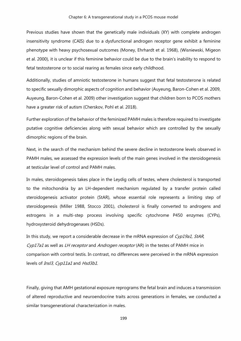
Chapter 6: A transgenerational study in a PCOS mouse model
199
Previous studies have shown that the genetically male individuals (XY) with complete androgen
insensitivity syndrome (CAIS) due to a dysfunctional androgen receptor gene exhibit a feminine
phenotype with heavy psychosexual outcomes (Money, Ehrhardt et al. 1968), (Wisniewski, Migeon
et al. 2000), it is unclear if this feminine behavior could be due to the brain’s inability to respond to
fetal testosterone or to social rearing as females since early childhood.
Additionally, studies of amniotic testosterone in humans suggest that fetal testosterone is related
to specific sexually dimorphic aspects of cognition and behavior (Auyeung, Baron-Cohen et al. 2009,
Auyeung, Baron-Cohen et al. 2009) other investigation suggest that children born to PCOS mothers
have a greater risk of autism (Cherskov, Pohl et al. 2018).
Further exploration of the behavior of the feminized PAMH males is therefore required to investigate
putative cognitive deficiencies along with sexual behavior which are controlled by the sexually
dimorphic regions of the brain.
Next, in the search of the mechanism behind the severe decline in testosterone levels observed in
PAMH males, we assessed the expression levels of the main genes involved in the steroidogenesis
at testicular level of control and PAMH males.
In males, steroidogenesis takes place in the Leydig cells of testes, where cholesterol is transported
to the mitochondria by an LH-dependent mechanism regulated by a transfer protein called
steroidogenesis activator protein (StAR), whose essential role represents a limiting step of
steroidogenesis (Miller 1988, Stocco 2001), cholesterol is finally converted to androgens and
estrogens in a multi-step process involving specific cytochrome P450 enzymes (CYPs),
hydroxysteroid dehydrogenases (HSDs).
In this study, we report a considerable decrease in the mRNA expression of Cyp19a1, StAR,
Cyp17a1 as well as LH receptor and Androgen receptor (AR) in the testes of PAMH mice in
comparison with control testis. In contrast, no differences were perceived in the mRNA expression
levels of Insl3, Cyp11a1 and Hsd3b1.
Finally, giving that AMH gestational exposure reprograms the fetal brain and induces a transmission
of altered reproductive and neuroendocrine traits across generations in females, we conducted a
similar transgenerational characterization in males.
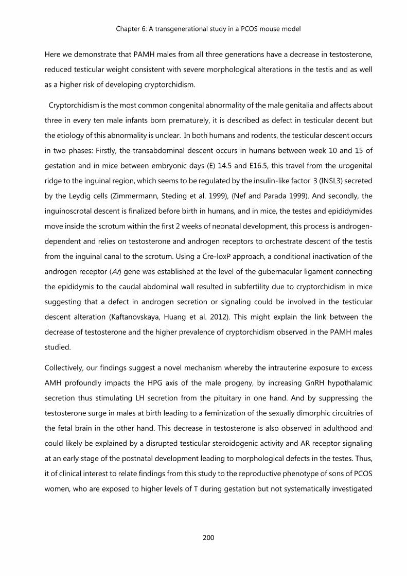
Chapter 6: A transgenerational study in a PCOS mouse model
200
Here we demonstrate that PAMH males from all three generations have a decrease in testosterone,
reduced testicular weight consistent with severe morphological alterations in the testis and as well
as a higher risk of developing cryptorchidism.
Cryptorchidism is the most common congenital abnormality of the male genitalia and affects about
three in every ten male infants born prematurely, it is described as defect in testicular decent but
the etiology of this abnormality is unclear. In both humans and rodents, the testicular descent occurs
in two phases: Firstly, the transabdominal descent occurs in humans between week 10 and 15 of
gestation and in mice between embryonic days (E) 14.5 and E16.5, this travel from the urogenital
ridge to the inguinal region, which seems to be regulated by the insulin-like factor 3 (INSL3) secreted
by the Leydig cells (Zimmermann, Steding et al. 1999), (Nef and Parada 1999). And secondly, the
inguinoscrotal descent is finalized before birth in humans, and in mice, the testes and epididymides
move inside the scrotum within the first 2 weeks of neonatal development, this process is androgen-
dependent and relies on testosterone and androgen receptors to orchestrate descent of the testis
from the inguinal canal to the scrotum. Using a Cre-loxP approach, a conditional inactivation of the
androgen receptor (Ar) gene was established at the level of the gubernacular ligament connecting
the epididymis to the caudal abdominal wall resulted in subfertility due to cryptorchidism in mice
suggesting that a defect in androgen secretion or signaling could be involved in the testicular
descent alteration (Kaftanovskaya, Huang et al. 2012). This might explain the link between the
decrease of testosterone and the higher prevalence of cryptorchidism observed in the PAMH males
studied.
Collectively, our findings suggest a novel mechanism whereby the intrauterine exposure to excess
AMH profoundly impacts the HPG axis of the male progeny, by increasing GnRH hypothalamic
secretion thus stimulating LH secretion from the pituitary in one hand. And by suppressing the
testosterone surge in males at birth leading to a feminization of the sexually dimorphic circuitries of
the fetal brain in the other hand. This decrease in testosterone is also observed in adulthood and
could likely be explained by a disrupted testicular steroidogenic activity and AR receptor signaling
at an early stage of the postnatal development leading to morphological defects in the testes. Thus,
it of clinical interest to relate findings from this study to the reproductive phenotype of sons of PCOS
women, who are exposed to higher levels of T during gestation but not systematically investigated
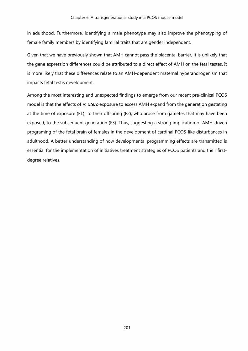
Chapter 6: A transgenerational study in a PCOS mouse model
201
in adulthood. Furthermore, identifying a male phenotype may also improve the phenotyping of
female family members by identifying familial traits that are gender independent.
Given that we have previously shown that AMH cannot pass the placental barrier, it is unlikely that
the gene expression differences could be attributed to a direct effect of AMH on the fetal testes. It
is more likely that these differences relate to an AMH-dependent maternal hyperandrogenism that
impacts fetal testis development.
Among the most interesting and unexpected findings to emerge from our recent pre-clinical PCOS
model is that the effects of in utero exposure to excess AMH expand from the generation gestating
at the time of exposure (F1) to their offspring (F2), who arose from gametes that may have been
exposed, to the subsequent generation (F3). Thus, suggesting a strong implication of AMH-driven
programing of the fetal brain of females in the development of cardinal PCOS-like disturbances in
adulthood. A better understanding of how developmental programming effects are transmitted is
essential for the implementation of initiatives treatment strategies of PCOS patients and their first-
degree relatives.
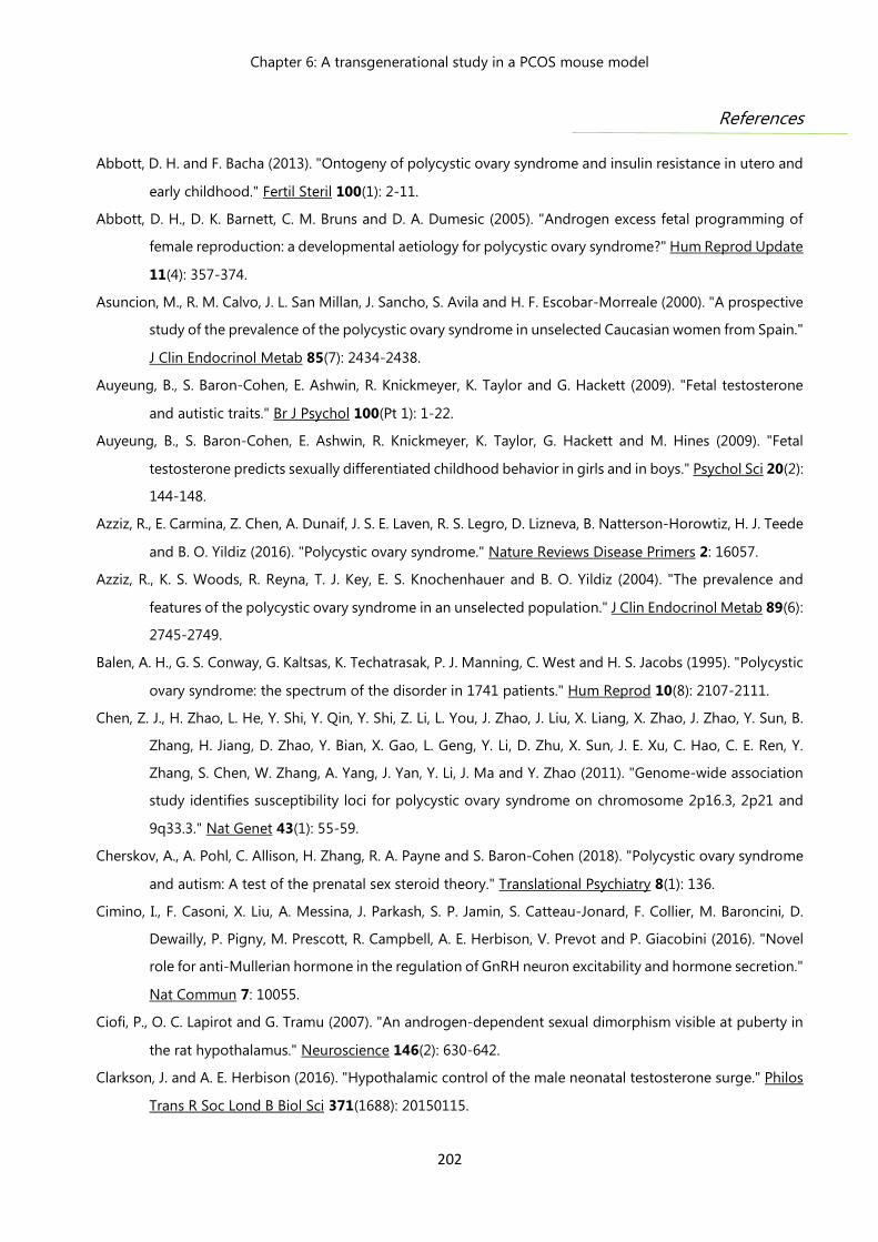
Chapter 6: A transgenerational study in a PCOS mouse model
202
References
Abbott, D. H. and F. Bacha (2013). "Ontogeny of polycystic ovary syndrome and insulin resistance in utero and
early childhood." Fertil Steril 100(1): 2-11.
Abbott, D. H., D. K. Barnett, C. M. Bruns and D. A. Dumesic (2005). "Androgen excess fetal programming of
female reproduction: a developmental aetiology for polycystic ovary syndrome?" Hum Reprod Update
11(4): 357-374.
Asuncion, M., R. M. Calvo, J. L. San Millan, J. Sancho, S. Avila and H. F. Escobar-Morreale (2000). "A prospective
study of the prevalence of the polycystic ovary syndrome in unselected Caucasian women from Spain."
J Clin Endocrinol Metab 85(7): 2434-2438.
Auyeung, B., S. Baron-Cohen, E. Ashwin, R. Knickmeyer, K. Taylor and G. Hackett (2009). "Fetal testosterone
and autistic traits." Br J Psychol 100(Pt 1): 1-22.
Auyeung, B., S. Baron-Cohen, E. Ashwin, R. Knickmeyer, K. Taylor, G. Hackett and M. Hines (2009). "Fetal
testosterone predicts sexually differentiated childhood behavior in girls and in boys." Psychol Sci 20(2):
144-148.
Azziz, R., E. Carmina, Z. Chen, A. Dunaif, J. S. E. Laven, R. S. Legro, D. Lizneva, B. Natterson-Horowtiz, H. J. Teede
and B. O. Yildiz (2016). "Polycystic ovary syndrome." Nature Reviews Disease Primers 2: 16057.
Azziz, R., K. S. Woods, R. Reyna, T. J. Key, E. S. Knochenhauer and B. O. Yildiz (2004). "The prevalence and
features of the polycystic ovary syndrome in an unselected population." J Clin Endocrinol Metab 89(6):
2745-2749.
Balen, A. H., G. S. Conway, G. Kaltsas, K. Techatrasak, P. J. Manning, C. West and H. S. Jacobs (1995). "Polycystic
ovary syndrome: the spectrum of the disorder in 1741 patients." Hum Reprod 10(8): 2107-2111.
Chen, Z. J., H. Zhao, L. He, Y. Shi, Y. Qin, Y. Shi, Z. Li, L. You, J. Zhao, J. Liu, X. Liang, X. Zhao, J. Zhao, Y. Sun, B.
Zhang, H. Jiang, D. Zhao, Y. Bian, X. Gao, L. Geng, Y. Li, D. Zhu, X. Sun, J. E. Xu, C. Hao, C. E. Ren, Y.
Zhang, S. Chen, W. Zhang, A. Yang, J. Yan, Y. Li, J. Ma and Y. Zhao (2011). "Genome-wide association
study identifies susceptibility loci for polycystic ovary syndrome on chromosome 2p16.3, 2p21 and
9q33.3." Nat Genet 43(1): 55-59.
Cherskov, A., A. Pohl, C. Allison, H. Zhang, R. A. Payne and S. Baron-Cohen (2018). "Polycystic ovary syndrome
and autism: A test of the prenatal sex steroid theory." Translational Psychiatry 8(1): 136.
Cimino, I., F. Casoni, X. Liu, A. Messina, J. Parkash, S. P. Jamin, S. Catteau-Jonard, F. Collier, M. Baroncini, D.
Dewailly, P. Pigny, M. Prescott, R. Campbell, A. E. Herbison, V. Prevot and P. Giacobini (2016). "Novel
role for anti-Mullerian hormone in the regulation of GnRH neuron excitability and hormone secretion."
Nat Commun 7: 10055.
Ciofi, P., O. C. Lapirot and G. Tramu (2007). "An androgen-dependent sexual dimorphism visible at puberty in
the rat hypothalamus." Neuroscience 146(2): 630-642.
Clarkson, J. and A. E. Herbison (2016). "Hypothalamic control of the male neonatal testosterone surge." Philos
Trans R Soc Lond B Biol Sci 371(1688): 20150115.
Chapter 6: A transgenerational study in a PCOS mouse model
203
Cohen, P. N., J. R. Givens, W. L. Wiser, R. S. Wilroy, R. L. Summitt, S. A. Coleman and R. N. Andersen (1975).
"Polycystic ovarian disease, maturation arrest of spermiogenesis, and Klinefelter's syndrome in siblings
of a family with familial hirsutism." Fertil Steril 26(12): 1228-1238.
Connolly, F., M. T. Rae, L. Bittner, K. Hogg, A. S. McNeilly and W. C. Duncan (2013). "Excess androgens in utero
alters fetal testis development." Endocrinology 154(5): 1921-1933.
Cooper, H. E., W. N. Spellacy, K. A. Prem and W. D. Cohen (1968). "Hereditary factors in the Stein-Leventhal
syndrome." Am J Obstet Gynecol 100(3): 371-387.
Corbier, P., D. A. Edwards and J. Roffi (1992). "The neonatal testosterone surge: a comparative study." Arch Int
Physiol Biochim Biophys 100(2): 127-131.
Dadachanji, R., N. Shaikh and S. Mukherjee (2018). "Genetic Variants Associated with Hyperandrogenemia in
PCOS Pathophysiology." Genet Res Int 2018: 7624932.
Dumesic, D. A., S. E. Oberfield, E. Stener-Victorin, J. C. Marshall, J. S. Laven and R. S. Legro (2015). "Scientific
Statement on the Diagnostic Criteria, Epidemiology, Pathophysiology, and Molecular Genetics of
Polycystic Ovary Syndrome." Endocr Rev 36(5): 487-525.
Ferriman, D. and A. W. Purdie (1979). "The inheritance of polycystic ovarian disease and a possible relationship
to premature balding." Clin Endocrinol (Oxf) 11(3): 291-300.
Franks, S., N. Gharani and M. McCarthy (2001). "Candidate genes in polycystic ovary syndrome." Hum Reprod
Update 7(4): 405-410.
Franks, S., L. J. Webber, M. Goh, A. Valentine, D. M. White, G. S. Conway, S. Wiltshire and M. I. McCarthy (2008).
"Ovarian morphology is a marker of heritable biochemical traits in sisters with polycystic ovaries." J
Clin Endocrinol Metab 93(9): 3396-3402.
Garamszegi, L. Z., M. Eens, S. Hurtrez-Bousses and A. P. Moller (2005). "Testosterone, testes size, and mating
success in birds: a comparative study." Horm Behav 47(4): 389-409.
Givens, J. R. (1988). "Familial polycystic ovarian disease." Endocrinol Metab Clin North Am 17(4): 771-783.
Goodarzi, M. O., D. A. Dumesic, G. Chazenbalk and R. Azziz (2011). "Polycystic ovary syndrome: etiology,
pathogenesis and diagnosis." Nat Rev Endocrinol 7(4): 219-231.
Hague, W. M., J. Adams, S. T. Reeders, T. E. Peto and H. S. Jacobs (1988). "Familial polycystic ovaries: a genetic
disease?" Clin Endocrinol (Oxf) 29(6): 593-605.
Hayes, M. G., M. Urbanek, D. A. Ehrmann, L. L. Armstrong, J. Y. Lee, R. Sisk, T. Karaderi, T. M. Barber, M. I.
McCarthy, S. Franks, C. M. Lindgren, C. K. Welt, E. Diamanti-Kandarakis, D. Panidis, M. O. Goodarzi, R.
Azziz, Y. Zhang, R. G. James, M. Olivier, A. H. Kissebah, E. Stener-Victorin, R. S. Legro and A. Dunaif
(2015). "Genome-wide association of polycystic ovary syndrome implicates alterations in
gonadotropin secretion in European ancestry populations." Nat Commun 6: 7502.
Kaftanovskaya, E. M., Z. Huang, A. M. Barbara, K. De Gendt, G. Verhoeven, I. P. Gorlov and A. I. Agoulnik (2012).
"Cryptorchidism in mice with an androgen receptor ablation in gubernaculum testis." Mol Endocrinol
26(4): 598-607.
Chapter 6: A transgenerational study in a PCOS mouse model
204
Kahsar-Miller, M. D., C. Nixon, L. R. Boots, R. C. Go and R. Azziz (2001). "Prevalence of polycystic ovary syndrome
(PCOS) in first-degree relatives of patients with PCOS." Fertil Steril 75(1): 53-58.
Kent, S. C., C. L. Gnatuk, A. R. Kunselman, L. M. Demers, P. A. Lee and R. S. Legro (2008). "Hyperandrogenism
and hyperinsulinism in children of women with polycystic ovary syndrome: a controlled study." J Clin
Endocrinol Metab 93(5): 1662-1669.
Legro, R. S., D. Driscoll, J. F. Strauss, 3rd, J. Fox and A. Dunaif (1998). "Evidence for a genetic basis for
hyperandrogenemia in polycystic ovary syndrome." Proc Natl Acad Sci U S A 95(25): 14956-14960.
Lunde, O., P. Magnus, L. Sandvik and S. Hoglo (1989). "Familial clustering in the polycystic ovarian syndrome."
Gynecol Obstet Invest 28(1): 23-30.
March, W. A., V. M. Moore, K. J. Willson, D. I. Phillips, R. J. Norman and M. J. Davies (2010). "The prevalence of
polycystic ovary syndrome in a community sample assessed under contrasting diagnostic criteria."
Hum Reprod 25(2): 544-551.
McAllister, J. M., R. S. Legro, B. P. Modi and J. F. Strauss, 3rd (2015). "Functional genomics of PCOS: from GWAS
to molecular mechanisms." Trends Endocrinol Metab 26(3): 118-124.
McCarthy, M. M. (2008). "Estradiol and the developing brain." Physiol Rev 88(1): 91-124.
McCarthy, M. M., A. P. Arnold, G. F. Ball, J. D. Blaustein and G. J. De Vries (2012). "Sex differences in the brain:
the not so inconvenient truth." J Neurosci 32(7): 2241-2247.
McCartney, C. R., C. A. Eagleson and J. C. Marshall (2002). "Regulation of gonadotropin secretion: implications
for polycystic ovary syndrome." Semin Reprod Med 20(4): 317-326.
Miller, W. L. (1988). "Molecular biology of steroid hormone synthesis." Endocr Rev 9(3): 295-318.
Money, J., A. A. Ehrhardt and D. N. Masica (1968). "Fetal feminization induced by androgen insensitivity in the
testicular feminizing syndrome: effect on marriage and maternalism." Johns Hopkins Med J 123(3):
105-114.
Nef, S. and L. F. J. N. g. Parada (1999). "Cryptorchidism in mice mutant for Insl3." 22(3): 295.
Nelson, V. L., R. S. Legro, J. F. Strauss, 3rd and J. M. McAllister (1999). "Augmented androgen production is a
stable steroidogenic phenotype of propagated theca cells from polycystic ovaries." Mol Endocrinol
13(6): 946-957.
Nelson, V. L., K. N. Qin, R. L. Rosenfield, J. R. Wood, T. M. Penning, R. S. Legro, J. F. Strauss, 3rd and J. M.
McAllister (2001). "The biochemical basis for increased testosterone production in theca cells
propagated from patients with polycystic ovary syndrome." J Clin Endocrinol Metab 86(12): 5925-
5933.
Norman, R. J., D. Dewailly, R. S. Legro and T. E. Hickey (2007). "Polycystic ovary syndrome." Lancet 370(9588):
685-697.
Perera, F. and J. Herbstman (2011). "Prenatal environmental exposures, epigenetics, and disease." Reprod
Toxicol 31(3): 363-373.
Chapter 6: A transgenerational study in a PCOS mouse model
205
Preston, B. T., I. R. Stevenson, G. A. Lincoln, S. L. Monfort, J. G. Pilkington and K. Wilson (2012). "Testes size,
testosterone production and reproductive behaviour in a natural mammalian mating system." 81(1):
296-305.
Recabarren, S. E., P. P. Rojas-Garcia, M. P. Recabarren, V. H. Alfaro, R. Smith, V. Padmanabhan and T. Sir-
Petermann (2008). "Prenatal testosterone excess reduces sperm count and motility." Endocrinology
149(12): 6444-6448.
Recabarren, S. E., T. Sir-Petermann, R. Rios, M. Maliqueo, B. Echiburu, R. Smith, P. Rojas-Garcia, M. Recabarren
and R. A. Rey (2008). "Pituitary and testicular function in sons of women with polycystic ovary
syndrome from infancy to adulthood." J Clin Endocrinol Metab 93(9): 3318-3324.
Recabarren, S. E., R. Smith, R. Rios, M. Maliqueo, B. Echiburu, E. Codner, F. Cassorla, P. Rojas and T. Sir-
Petermann (2008). "Metabolic profile in sons of women with polycystic ovary syndrome." J Clin
Endocrinol Metab 93(5): 1820-1826.
Rodin, D. A., G. Bano, J. M. Bland, K. Taylor and S. S. Nussey (1998). "Polycystic ovaries and associated metabolic
abnormalities in Indian subcontinent Asian women." Clin Endocrinol (Oxf) 49(1): 91-99.
Rojas-Garcia, P. P., M. P. Recabarren, L. Sarabia, J. Schon, C. Gabler, R. Einspanier, M. Maliqueo, T. Sir-
Petermann, R. Rey and S. E. Recabarren (2010). "Prenatal testosterone excess alters Sertoli and germ
cell number and testicular FSH receptor expression in rams." Am J Physiol Endocrinol Metab 299(6):
E998-e1005.
Sahmay, S., Y. Aydin, M. Oncul and L. M. Senturk (2014). "Diagnosis of Polycystic Ovary Syndrome: AMH in
combination with clinical symptoms." J Assist Reprod Genet 31(2): 213-220.
Sam, S., A. D. Coviello, Y. A. Sung, R. S. Legro and A. Dunaif (2008). "Metabolic phenotype in the brothers of
women with polycystic ovary syndrome." Diabetes Care 31(6): 1237-1241.
Santen, R. J. (1975). "Is aromatization of testosterone to estradiol required for inhibition of luteinizing hormone
secretion in men?" J Clin Invest 56(6): 1555-1563.
Simerly, R. B. (2002). "Wired for reproduction: organization and development of sexually dimorphic circuits in
the mammalian forebrain." Annu Rev Neurosci 25: 507-536.
Sir-Petermann, T., A. Ladron de Guevara, E. Codner, J. Preisler, N. Crisosto, B. Echiburu, M. Maliqueo, F. Sanchez,
F. Perez-Bravo and F. Cassorla (2012). "Relationship between anti-Mullerian hormone (AMH) and
insulin levels during different tanner stages in daughters of women with polycystic ovary syndrome."
Reprod Sci 19(4): 383-390.
Sir-Petermann, T., M. Maliqueo, B. Angel, H. E. Lara, F. Perez-Bravo and S. E. Recabarren (2002). "Maternal
serum androgens in pregnant women with polycystic ovarian syndrome: possible implications in
prenatal androgenization." Hum Reprod 17(10): 2573-2579.
Stocco, D. M. (2001). "StAR Protein and the Regulation of Steroid Hormone Biosynthesis." 63(1): 193-213.
Takayama, K., T. Fukaya, H. Sasano, Y. Funayama, T. Suzuki, R. Takaya, Y. Wada and A. Yajima (1996).
"Immunohistochemical study of steroidogenesis and cell proliferation in polycystic ovarian
syndrome." Hum Reprod 11(7): 1387-1392.
Chapter 6: A transgenerational study in a PCOS mouse model
206
Tata, B., N. E. H. Mimouni, A. L. Barbotin, S. A. Malone, A. Loyens, P. Pigny, D. Dewailly, S. Catteau-Jonard, I.
Sundstrom-Poromaa, T. T. Piltonen, F. Dal Bello, C. Medana, V. Prevot, J. Clasadonte and P. Giacobini
(2018). "Elevated prenatal anti-Mullerian hormone reprograms the fetus and induces polycystic ovary
syndrome in adulthood." Nat Med 24(6): 834-846.
Taylor, A. E., B. McCourt, K. A. Martin, E. J. Anderson, J. M. Adams, D. Schoenfeld and J. E. Hall (1997).
"Determinants of abnormal gonadotropin secretion in clinically defined women with polycystic ovary
syndrome." J Clin Endocrinol Metab 82(7): 2248-2256.
Uglem, I., I. Mayer and G. Rosenqvist (2002). "Variation in plasma steroids and reproductive traits in dimorphic
males of corkwing wrasse (Symphodus melops L.)." Horm Behav 41(4): 396-404.
Valdimarsdottir, R., H. Valgeirsdottir, A. K. Wikstrom, T. K. Kallak, E. Elenis, O. Axelsson, K. Ubhayasekhera, J.
Bergquist, T. T. Piltonen, P. Pigny, P. Giacobini and I. S. Poromaa (2019). "Pregnancy and neonatal
complications in women with polycystic ovary syndrome in relation to second-trimester anti-Mullerian
hormone levels." Reprod Biomed Online 39(1): 141-148.
Waldstreicher, J., N. F. Santoro, J. E. Hall, M. Filicori and W. F. Crowley, Jr. (1988). "Hyperfunction of the
hypothalamic-pituitary axis in women with polycystic ovarian disease: indirect evidence for partial
gonadotroph desensitization." J Clin Endocrinol Metab 66(1): 165-172.
Wilroy, R. S., Jr., J. R. Givens, W. L. Wiser, S. A. Coleman, R. N. Andersen and R. L. Summitt (1975). "Hyperthecosis:
an inheritable form of polycystic ovarian disease." Birth Defects Orig Artic Ser 11(4): 81-85.
Wisniewski, A. B., C. J. Migeon, H. F. Meyer-Bahlburg, J. P. Gearhart, G. D. Berkovitz, T. R. Brown and J. Money
(2000). "Complete androgen insensitivity syndrome: long-term medical, surgical, and psychosexual
outcome." J Clin Endocrinol Metab 85(8): 2664-2669.
Wood, J. R., C. K. Ho, V. L. Nelson-Degrave, J. M. McAllister and J. F. Strauss, 3rd (2004). "The molecular
signature of polycystic ovary syndrome (PCOS) theca cells defined by gene expression profiling." J
Reprod Immunol 63(1): 51-60.
Yang, F., Y. C. Ruan, Y. J. Yang, K. Wang, S. S. Liang, Y. B. Han, X. M. Teng and J. Z. Yang (2015). "Follicular
hyperandrogenism downregulates aromatase in luteinized granulosa cells in polycystic ovary
syndrome women." Reproduction 150(4): 289-296.
Yildiz, B. l. O., H. Yarali, H. Oguz and M. Bayraktar (2003). "Glucose Intolerance, Insulin Resistance, and
Hyperandrogenemia in First Degree Relatives of Women with Polycystic Ovary Syndrome." The Journal
of Clinical Endocrinology & Metabolism 88(5): 2031-2036.
Yildiz, B. O., H. Yarali, H. Oguz and M. Bayraktar (2003). "Glucose intolerance, insulin resistance, and
hyperandrogenemia in first degree relatives of women with polycystic ovary syndrome." J Clin
Endocrinol Metab 88(5): 2031-2036.
Yu, Y. Y., C. X. Sun, Y. K. Liu, Y. Li, L. Wang and W. Zhang (2013). "Promoter methylation of CYP19A1 gene in
Chinese polycystic ovary syndrome patients." Gynecol Obstet Invest 76(4): 209-213.

Chapter 6: A transgenerational study in a PCOS mouse model
207
Zimmermann, S., G. Steding, J. M. A. Emmen, A. O. Brinkmann, K. Nayernia, A. F. Holstein, W. Engel and I. M.
Adham (1999). "Targeted Disruption of the Insl3 Gene Causes Bilateral Cryptorchidism." Molecular
Endocrinology 13(5): 681-691.

208
ANNEX
*For correspondence:
[email protected] (NP);
[email protected] (PG)
Competing interests: The
authors declare that no
competing interests exist.
Funding: See page 31
Received: 27 March 2019
Accepted: 28 June 2019
Reviewing editor: Joel K
Elmquist, University of Texas
Southwestern Medical Center,
United States
Copyright Malone et al. This
article is distributed under the
terms of the Creative Commons
Attribution License, which
permits unrestricted use and
redistribution provided that the
original author and source are
credited.
Defective AMH signaling disrupts GnRHneuron development and function andcontributes to hypogonadotropichypogonadismSamuel Andrew Malone1,2, Georgios E Papadakis3, Andrea Messina3,Nour El Houda Mimouni1,2, Sara Trova1,2, Monica Imbernon1,2, Cecile Allet1,2,Irene Cimino1, James Acierno3, Daniele Cassatella3, Cheng Xu3, Richard Quinton4,Gabor Szinnai5, Pascal Pigny6, Lur Alonso-Cotchico7, Laura Masgrau7,8,Jean-Didier Marechal7, Vincent Prevot1,2, Nelly Pitteloud3*, Paolo Giacobini1,2*
1Jean-Pierre Aubert Research Center (JPArc), Laboratory of Development andPlasticity of the Neuroendocrine Brain, Inserm, UMR-S 1172, Lille, France;2University of Lille, FHU 1, 000 Days for Health, Lille, France; 3Faculty of Biologyand Medicine, Service of Endocrinology, Diabetology and Metabolism, UniversityHospital, Lausanne, Switzerland; 4Institute of Genetic Medicine and the RoyalVictoria Infirmary, University of Newcastle-upon-Tyne, Newcastle-upon-Tyne, UnitedKingdom; 5Pediatric Endocrinology and Diabetology, University of Basel Children’sHospital, Basel, Switzerland; 6CHU Lille, Laboratoire de Biochimie et Hormonologie,Centre de Biologie Pathologie, Lille, France; 7Departament de Quımica, UniversitatAutonoma de Barcelona, Bellaterra, Spain; 8Institut de Biotecnologia i deBiomedicina, Universitat Autonoma de Barcelona, Bellaterra, Spain
Abstract Congenital hypogonadotropic hypogonadism (CHH) is a condition characterized by
absent puberty and infertility due to gonadotropin releasing hormone (GnRH) deficiency, which is
often associated with anosmia (Kallmann syndrome, KS). We identified loss-of-function
heterozygous mutations in anti-Mullerian hormone (AMH) and its receptor, AMHR2, in 3% of CHH
probands using whole-genome sequencing. We showed that during embryonic development, AMH
is expressed in migratory GnRH neurons in both mouse and human fetuses and unconvered a novel
function of AMH as a pro-motility factor for GnRH neurons. Pathohistological analysis of Amhr2-
deficient mice showed abnormal development of the peripheral olfactory system and defective
embryonic migration of the neuroendocrine GnRH cells to the basal forebrain, which results in
reduced fertility in adults. Our findings highlight a novel role for AMH in the development and
function of GnRH neurons and indicate that AMH signaling insufficiency contributes to the
pathogenesis of CHH in humans.
DOI: https://doi.org/10.7554/eLife.47198.001
IntroductionGonadotropin releasing hormone (GnRH) is essential for puberty onset and reproduction. GnRH is
released into the pituitary portal blood vessels for delivery to the anterior pituitary. There, GnRH
controls the production and release of the gonadotropins LH (luteinizing hormone) and FSH (follicle
stimulating hormone), which in turn stimulate gametogenesis and sex steroid production in the
gonads (Christian and Moenter, 2010). GnRH–secreting neurons are unusual neuroendocrine cells,
Malone et al. eLife 2019;8:e47198. DOI: https://doi.org/10.7554/eLife.47198 1 of 36
RESEARCH ARTICLE
as they originate in the nasal placode outside the central nervous system during embryonic develop-
ment, and migrate to the hypothalamus along the vomeronasal and terminal nerves (VNN, TN)
(Wray et al., 1989; Schwanzel-Fukuda and Pfaff, 1989). This process is evolutionarily conserved
and follows a similar spatio-temporal pattern in all mammals (Wray et al., 1989; Schwanzel-
Fukuda and Pfaff, 1989), including humans (Schwanzel-Fukuda et al., 1996; Casoni et al., 2016).
Disruption of GnRH neuronal migration and/or defective GnRH synthesis and secretion leads to con-
genital hypogonadotropic hypogonadisms (CHH), a rare endocrine disorder (prevalence: 1 in 4000)
characterized by absent or incomplete puberty resulting in infertility (Boehm et al., 2015). CHH is
clinically and genetically heterogeneous with several causal genes identified to date (Boehm et al.,
2015), and follows various modes of transmission, including oligogenic inheritance (Sykiotis et al.,
2010). However, the mutations identified so far only account for half of clinically reported cases, sug-
gesting that other causal genes remain to be discovered. Unravelling new genetic pathways involved
in the regulation of the development of the GnRH system is relevant for understanding the basis of
pathogenesis leading to CHH in humans.
AMH is a TGF-b family member and it signals by binding to a specific type II receptor (AMHR2)
(di Clemente et al., 1994; Baarends et al., 1994), which heterodimerizes with one of several type I
TGF-b receptors (Acvr1 [Alk2], Bmpr1a [Alk3] and Bmpr1b [Alk6]), to recruit Smad proteins that sub-
sequently undergo nuclear translocation to regulate target gene expression (Josso and Clemente,
2003). Although AMH signaling has been traditionally reported to play a crucial role during sex dif-
ferentiation and gonadal functions (Josso et al., 1998; Behringer et al., 1994), accumulating evi-
dence has started to shed light on unexpected functions of AMH in the central nervous system as
well as in the pituitary (Lebeurrier et al., 2008; Wang et al., 2009; Tata et al., 2018; Cimino et al.,
2016; Garrel et al., 2016). We have previously shown that GnRH neurons express AMHR2 from
early fetal development to adulthood and that AMH stimulates GnRH neuronal activity and hormone
secretion in mature GnRH cells (Cimino et al., 2016). Here, we expand this information by demon-
strating that GnRH cells also express AMH during their migratory process, both in mice and human
fetuses and we describe a novel role of AMH as a potent stimulator of GnRH cell motility. Finally, we
show that pharmacological or genetic invalidation of Amhr2 signaling in vivo alters GnRH migration
and the projections of VNN/TN to the basal forebrain, which results into a reduced size of this neu-
ronal population in adult brains, altered ovulation and fertility. The involvement of the AMH signal-
ing pathway in GnRH ontogeny and secretion led to the identification of four heterozygous loss-of-
function mutations in AMH and AMHR2 among 136 CHH patients. Collectively, this study identified
a novel embryonic role of AMH in the development and function of GnRH neurons and provides
genetic evidence that disturbance of AMH signaling can contribute to CHH phenotype in humans.
Results
Amh is expressed by GnRH migratory cells in mouse and human fetusesWe have recently shown that migratory GnRH neurons and developing vomeronasal/olfactory axons
express Amhr2 in mammals (Cimino et al., 2016). In this study, we investigated whether migratory
GnRH neurons expressed Amh in addition to Amhr2. In order to do so, we first isolated GnRH neu-
rons through fluorescence activated cell sorting (FACS) from Gnrh1 <GFP> embryos (Spergel et al.,
1999) at embryonic day 12.5 (E12.5), coincident with the beginning of the GnRH neuronal migratory
process (Wray et al., 1989; Schwanzel-Fukuda and Pfaff, 1989), at postnatal day 12 (PN12) and at
postnatal day 90 (PN90; Figure 1a,b). These experiments revealed expression of Amh already at
E12.5 both in GnRH neurons and in total head extracts (Figure 1a). Moreover, real-time PCR experi-
ments showed increasing expression of Amh in GnRH neurons from early embryonic development
(E12.5) to adult life (PN90; Figure 1a,b).
Ex vivo cultures of embryonic nasal explants have been used to study factors regulating GnRH
migration by both our group (Giacobini et al., 2004; Giacobini et al., 2007) and others
(Fueshko and Wray, 1994). At 4 days in vitro, olfactory axons, which express bIII�tubulin (TUJ1),
emerge from the nasal explant tissue mass and GnRH neurons begin migrating out from the explant,
tightly associated to those fibers (Figure 1c). We generated nasal explants from Gnrh1 <GFP>
embryos and we immunostained these primary cultures using an antibody directed against the bio-
logically active form of Amh (C-terminal region; Figure 1d–f). These experiments revealed the
Malone et al. eLife 2019;8:e47198. DOI: https://doi.org/10.7554/eLife.47198 2 of 36
Research article Developmental Biology Genetics and Genomics
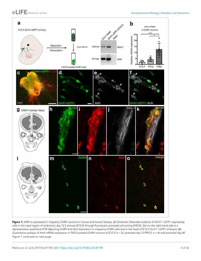
Figure 1. AMH is expressed in migratory GnRH neurons in mouse and human fetuses. (a) Schematic illustrates isolation of Gnrh1 <GFP> expressing
cells in the nasal region of embryonic day 12.5 animals (E12.5) through fluorescent activated cell sorting (FACS). Gel on the right-hand side is a
representative qualitative PCR depicting GnRH and Amh expression in migratory GnRH cells and in the head of E12.5 Gnrh1 <GFP> embryos. (b)
Quantitative analysis of Amh mRNA expression in FACS-isolated GnRH neurons at E12.5 (n = 5), postnatal day 12 (PN12, n = 6) and postnatal day 60
Figure 1 continued on next page
Malone et al. eLife 2019;8:e47198. DOI: https://doi.org/10.7554/eLife.47198 3 of 36
Research article Developmental Biology Genetics and Genomics
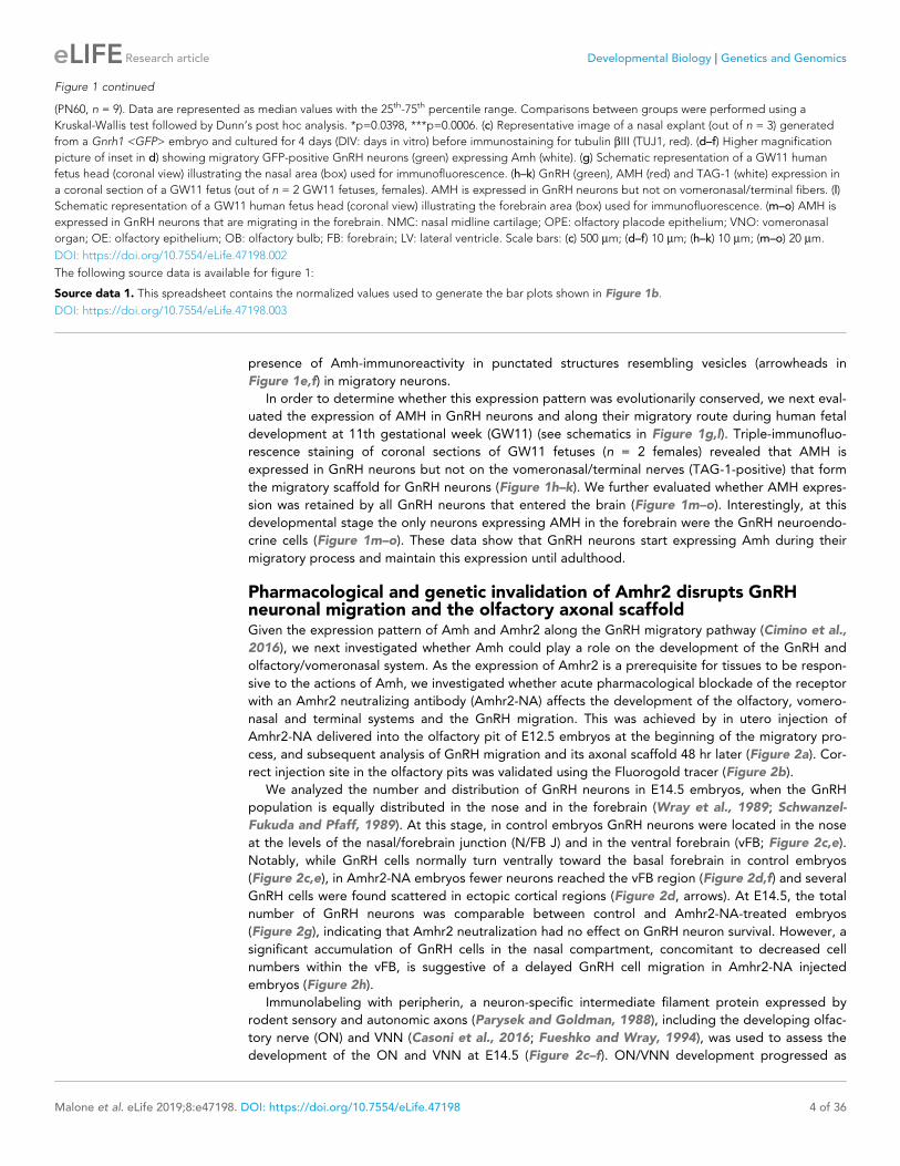
presence of Amh-immunoreactivity in punctated structures resembling vesicles (arrowheads in
Figure 1e,f) in migratory neurons.
In order to determine whether this expression pattern was evolutionarily conserved, we next eval-
uated the expression of AMH in GnRH neurons and along their migratory route during human fetal
development at 11th gestational week (GW11) (see schematics in Figure 1g,l). Triple-immunofluo-
rescence staining of coronal sections of GW11 fetuses (n = 2 females) revealed that AMH is
expressed in GnRH neurons but not on the vomeronasal/terminal nerves (TAG-1-positive) that form
the migratory scaffold for GnRH neurons (Figure 1h–k). We further evaluated whether AMH expres-
sion was retained by all GnRH neurons that entered the brain (Figure 1m–o). Interestingly, at this
developmental stage the only neurons expressing AMH in the forebrain were the GnRH neuroendo-
crine cells (Figure 1m–o). These data show that GnRH neurons start expressing Amh during their
migratory process and maintain this expression until adulthood.
Pharmacological and genetic invalidation of Amhr2 disrupts GnRHneuronal migration and the olfactory axonal scaffoldGiven the expression pattern of Amh and Amhr2 along the GnRH migratory pathway (Cimino et al.,
2016), we next investigated whether Amh could play a role on the development of the GnRH and
olfactory/vomeronasal system. As the expression of Amhr2 is a prerequisite for tissues to be respon-
sive to the actions of Amh, we investigated whether acute pharmacological blockade of the receptor
with an Amhr2 neutralizing antibody (Amhr2-NA) affects the development of the olfactory, vomero-
nasal and terminal systems and the GnRH migration. This was achieved by in utero injection of
Amhr2-NA delivered into the olfactory pit of E12.5 embryos at the beginning of the migratory pro-
cess, and subsequent analysis of GnRH migration and its axonal scaffold 48 hr later (Figure 2a). Cor-
rect injection site in the olfactory pits was validated using the Fluorogold tracer (Figure 2b).
We analyzed the number and distribution of GnRH neurons in E14.5 embryos, when the GnRH
population is equally distributed in the nose and in the forebrain (Wray et al., 1989; Schwanzel-
Fukuda and Pfaff, 1989). At this stage, in control embryos GnRH neurons were located in the nose
at the levels of the nasal/forebrain junction (N/FB J) and in the ventral forebrain (vFB; Figure 2c,e).
Notably, while GnRH cells normally turn ventrally toward the basal forebrain in control embryos
(Figure 2c,e), in Amhr2-NA embryos fewer neurons reached the vFB region (Figure 2d,f) and several
GnRH cells were found scattered in ectopic cortical regions (Figure 2d, arrows). At E14.5, the total
number of GnRH neurons was comparable between control and Amhr2-NA-treated embryos
(Figure 2g), indicating that Amhr2 neutralization had no effect on GnRH neuron survival. However, a
significant accumulation of GnRH cells in the nasal compartment, concomitant to decreased cell
numbers within the vFB, is suggestive of a delayed GnRH cell migration in Amhr2-NA injected
embryos (Figure 2h).
Immunolabeling with peripherin, a neuron-specific intermediate filament protein expressed by
rodent sensory and autonomic axons (Parysek and Goldman, 1988), including the developing olfac-
tory nerve (ON) and VNN (Casoni et al., 2016; Fueshko and Wray, 1994), was used to assess the
development of the ON and VNN at E14.5 (Figure 2c–f). ON/VNN development progressed as
Figure 1 continued
(PN60, n = 9). Data are represented as median values with the 25th-75th percentile range. Comparisons between groups were performed using a
Kruskal-Wallis test followed by Dunn’s post hoc analysis. *p=0.0398, ***p=0.0006. (c) Representative image of a nasal explant (out of n = 3) generated
from a Gnrh1 <GFP> embryo and cultured for 4 days (DIV: days in vitro) before immunostaining for tubulin bIII (TUJ1, red). (d–f) Higher magnification
picture of inset in d) showing migratory GFP-positive GnRH neurons (green) expressing Amh (white). (g) Schematic representation of a GW11 human
fetus head (coronal view) illustrating the nasal area (box) used for immunofluorescence. (h–k) GnRH (green), AMH (red) and TAG-1 (white) expression in
a coronal section of a GW11 fetus (out of n = 2 GW11 fetuses, females). AMH is expressed in GnRH neurons but not on vomeronasal/terminal fibers. (l)
Schematic representation of a GW11 human fetus head (coronal view) illustrating the forebrain area (box) used for immunofluorescence. (m–o) AMH is
expressed in GnRH neurons that are migrating in the forebrain. NMC: nasal midline cartilage; OPE: olfactory placode epithelium; VNO: vomeronasal
organ; OE: olfactory epithelium; OB: olfactory bulb; FB: forebrain; LV: lateral ventricle. Scale bars: (c) 500 mm; (d–f) 10 mm; (h–k) 10 mm; (m–o) 20 mm.
DOI: https://doi.org/10.7554/eLife.47198.002
The following source data is available for figure 1:
Source data 1. This spreadsheet contains the normalized values used to generate the bar plots shown in Figure 1b.
DOI: https://doi.org/10.7554/eLife.47198.003
Malone et al. eLife 2019;8:e47198. DOI: https://doi.org/10.7554/eLife.47198 4 of 36
Research article Developmental Biology Genetics and Genomics
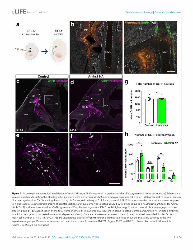
Figure 2. In utero pharmacological invalidation of Amhr2 disrupts GnRH neuronal migration and the olfactory/terminal nerve targeting. (a) Schematic of
in utero injections targeting the olfactory pits. Injections were performed at E12.5 and embryos harvested 48 hr later. (b) Representative coronal section
of an embryo head at E14.5 showing that olfactory pit Fluorogold delivery at E12.5 was successful. GnRH immunoreactive neurons are shown in green.
(c–f) Representative photomicrographs of sagittal sections of mouse embryos injected at E12.5 with either saline or a neutralizing antibody for Amhr2
(Amhr2-NA) and immunostained for GnRH (green) and Peripherin (magenta) at E14.5. (e, f) Higher magnification confocal photomicrograph of boxed
areas in c and d. (g) Quantification of the total number of GnRH immunoreactive neurons in saline-injected (control) and Amhr2-NA injected embryos
(n = 4 for both groups, harvested from two independent dams). Data are represented as mean ± s.e.m (n = 4, unpaired two-tailed Student’s t test:
mean cell number, t6 = 0.3796, p=0.7173). (h) Quantitative analysis of GnRH neuronal distribution throughout the migratory pathway in the two
experimental groups. Data are represented as mean ± s.e.m (n = 4, two-way ANOVA, F3,24 = 15.09, p<0.0001; followed by Holm-Sıdak multiple
Figure 2 continued on next page
Malone et al. eLife 2019;8:e47198. DOI: https://doi.org/10.7554/eLife.47198 5 of 36
Research article Developmental Biology Genetics and Genomics
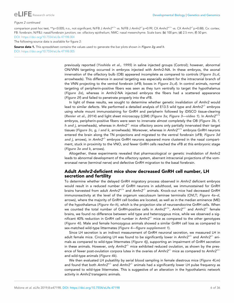
previously reported (Yoshida et al., 1995) in saline injected groups (Control); however, abnormal
ON/VNN targeting occurred in embryos injected with Amhr2-NA. In these embryos, the axonal
innervation of the olfactory bulb (OB) appeared incomplete as compared to controls (Figure 2c,d,
arrowheads). This difference in axonal targeting was especially evident for the intracranial branch of
the VNN projecting to the ventral forebrain (vFB; boxes in Figure 2c,d). In control animals, normal
targeting of peripherin-positive fibers was seen as they turn ventrally to target the hypothalamus
(Figure 2e), whereas in Amhr2-NA injected embryos the fibers had a scattered appearance
(Figure 2f) and failed to penetrate properly into the vFB.
In light of these results, we sought to determine whether genetic invalidation of Amhr2 would
lead to similar defects. We performed a detailed analysis of E13.5 wild type and Amhr2-/- embryos
using whole mount immunostaining for GnRH and peripherin followed by iDISCO tissue-clearing
(Renier et al., 2014) and light sheet microscopy (LSM) (Figure 3a; Figure 3—video 1). In Amhr2+/+
embryos, peripherin-positive fibers were seen to innervate almost completely the OB (Figure 3b, f,
h and j, arrowheads), whereas in Amhr2-/- mice olfactory axons only partially innervated their target
tissues (Figure 3c, g, i and k, arrowheads). Moreover, whereas in Amhr2+/+ embryos GnRH neurons
entered the brain along the TN projections and migrated to the ventral forebrain (vFB; Figure 3d
and j, arrows), in Amhr2-/- embryos GnRH neurons appeared more clustered in the nasal compart-
ment, stuck in proximity to the VNO, and fewer GnRH cells reached the vFB at this embryonic stage
(Figure 3e and k, arrows).
Altogether, these experiments revealed that pharmacological or genetic invalidation of Amhr2
leads to abnormal development of the olfactory system, aberrant intracranial projections of the vom-
eronasal nerve (terminal nerve) and defective GnRH migration to the basal forebrain.
Adult Amhr2-deficient mice show decreased GnRH cell number, LHsecretion and fertilityTo determine whether the delayed GnRH migratory process observed in Amhr2 deficient embryos
would result in a reduced number of GnRH neurons in adulthood, we immunostained for GnRH
brains harvested from adult Amhr2+/+ and Amhr2-/- animals. Knock-out mice had decreased GnRH
immunoreactivity at the level of the organum vasculosum laminae terminalis (OVLT; Figure 4a–d,
arrows), where the majority of GnRH cell bodies are located, as well as in the median eminence (ME)
of the hypothalamus (Figure 4e–h), which is the projection site of neuroendocrine GnRH cells. When
we counted the total number of GnRH-positive cells in Amhr2+/+, Amhr2+/- and Amhr2-/- female
brains, we found no difference between wild type and heterozygous mice, while we observed a sig-
nificant 40% reduction in GnRH cell number in Amhr2-/- mice as compared to the other genotypes
(Figure 4i). Male and female homozygous animals showed a similar GnRH cell loss as compared to
sex-matched wild-type littermates (Figure 4—figure supplement 1).
Since LH secretion is an indirect measurement of GnRH neuronal secretion, we measured LH in
adult female mice. Circulating LH was found to be significantly lower in Amhr2+/- and Amhr2-/- ani-
mals as compared to wild-type littermates (Figure 4j), supporting an impairment of GnRH secretion
in these animals. However, only Amhr2-/- mice exhibited reduced ovulation, as shown by the pres-
ence of fewer post-ovulation corpora lutea in the ovaries of Amhr2-/- mice as compared to Amhr2+/-
and wild-type animals (Figure 4k).
We then evaluated LH pulsatility by serial blood sampling in female diestrous mice (Figure 4l,m)
and found that both Amhr2+/- and Amhr2-/- animals had a significantly lower LH pulse frequency as
compared to wild-type littermates. This is suggestive of an alteration in the hypothalamic network
activity in Amhr2 transgenic animals.
Figure 2 continued
comparison post hoc test, **p<0.005; n.s., not significant; N/FB J Amhr2+/+ vs. N/FB J Amhr2-/-p=0.99, CX Amhr2+/+ vs. CX Amhr2-/-p=0.88). Cx: cortex;
FB: forebrain; N/FBJ: nasal/forebrain junction; oe: olfactory epithelium; NMC: nasal mesenchyme. Scale bars: (b) 100 mm; (d) 2.5 mm; (f) 50 mm.
DOI: https://doi.org/10.7554/eLife.47198.004
The following source data is available for figure 2:
Source data 1. This spreadsheet contains the values used to generate the bar plots shown in Figure 2g and h.
DOI: https://doi.org/10.7554/eLife.47198.005
Malone et al. eLife 2019;8:e47198. DOI: https://doi.org/10.7554/eLife.47198 6 of 36
Research article Developmental Biology Genetics and Genomics
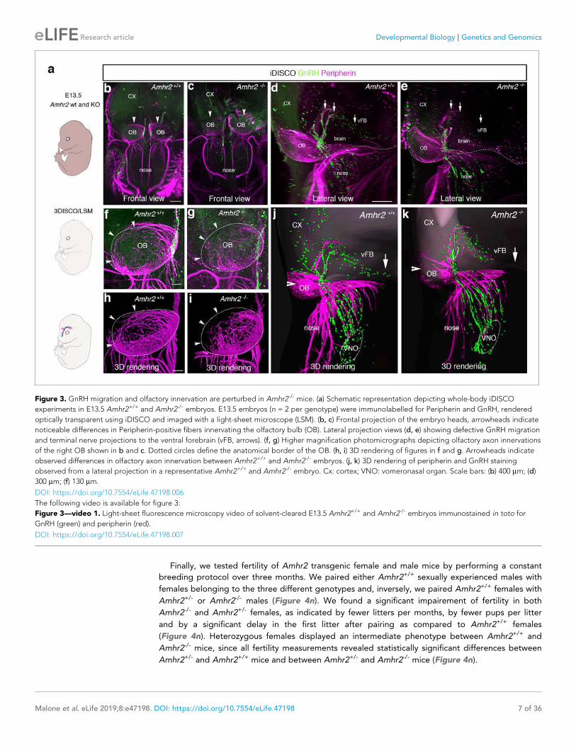
Finally, we tested fertility of Amhr2 transgenic female and male mice by performing a constant
breeding protocol over three months. We paired either Amhr2+/+ sexually experienced males with
females belonging to the three different genotypes and, inversely, we paired Amhr2+/+ females with
Amhr2+/- or Amhr2-/- males (Figure 4n). We found a significant impairement of fertility in both
Amhr2-/- and Amhr2+/- females, as indicated by fewer litters per months, by fewer pups per litter
and by a significant delay in the first litter after pairing as compared to Amhr2+/+ females
(Figure 4n). Heterozygous females displayed an intermediate phenotype between Amhr2+/+ and
Amhr2-/- mice, since all fertility measurements revealed statistically significant differences between
Amhr2+/- and Amhr2+/+ mice and between Amhr2+/- and Amhr2-/- mice (Figure 4n).
Figure 3. GnRH migration and olfactory innervation are perturbed in Amhr2-/- mice. (a) Schematic representation depicting whole-body iDISCO
experiments in E13.5 Amhr2+/+ and Amhr2-/- embryos. E13.5 embryos (n = 2 per genotype) were immunolabelled for Peripherin and GnRH, rendered
optically transparent using iDISCO and imaged with a light-sheet microscope (LSM). (b, c) Frontal projection of the embryo heads, arrowheads indicate
noticeable differences in Peripherin-positive fibers innervating the olfactory bulb (OB). Lateral projection views (d, e) showing defective GnRH migration
and terminal nerve projections to the ventral forebrain (vFB, arrows). (f, g) Higher magnification photomicrographs depicting olfactory axon innervations
of the right OB shown in b and c. Dotted circles define the anatomical border of the OB. (h, i) 3D rendering of figures in f and g. Arrowheads indicate
observed differences in olfactory axon innervation between Amhr2+/+ and Amhr2-/- embryos. (j, k) 3D rendering of peripherin and GnRH staining
observed from a lateral projection in a representative Amhr2+/+ and Amhr2-/- embryo. Cx: cortex; VNO: vomeronasal organ. Scale bars: (b) 400 mm; (d)
300 mm; (f) 130 mm.
DOI: https://doi.org/10.7554/eLife.47198.006
The following video is available for figure 3:
Figure 3—video 1. Light-sheet fluorescence microscopy video of solvent-cleared E13.5 Amhr2+/+ and Amhr2-/- embryos immunostained in toto for
GnRH (green) and peripherin (red).
DOI: https://doi.org/10.7554/eLife.47198.007
Malone et al. eLife 2019;8:e47198. DOI: https://doi.org/10.7554/eLife.47198 7 of 36
Research article Developmental Biology Genetics and Genomics
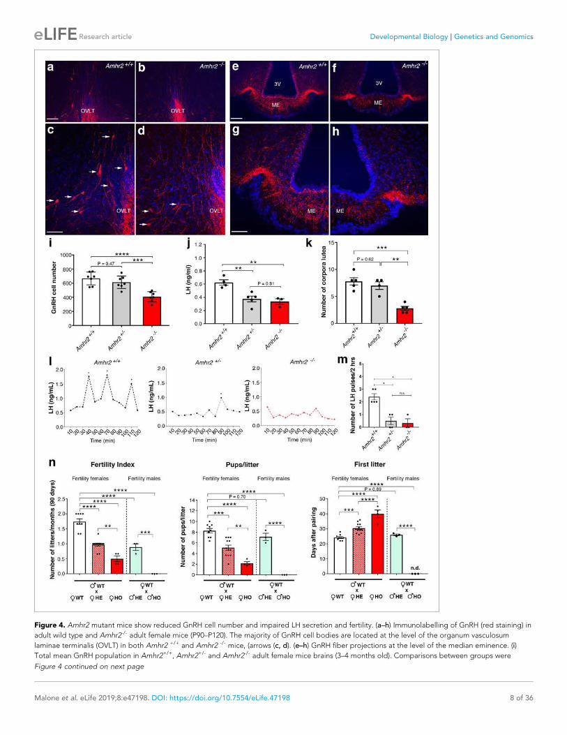
Figure 4. Amhr2 mutant mice show reduced GnRH cell number and impaired LH secretion and fertility. (a–h) Immunolabelling of GnRH (red staining) in
adult wild type and Amhr2-/- adult female mice (P90–P120). The majority of GnRH cell bodies are located at the level of the organum vasculosum
laminae terminalis (OVLT) in both Amhr2 +/+ and Amhr2 -/- mice, (arrows (c, d). (e–h) GnRH fiber projections at the level of the median eminence. (i)
Total mean GnRH population in Amhr2+/+, Amhr2+/- and Amhr2-/- adult female mice brains (3–4 months old). Comparisons between groups were
Figure 4 continued on next page
Malone et al. eLife 2019;8:e47198. DOI: https://doi.org/10.7554/eLife.47198 8 of 36
Research article Developmental Biology Genetics and Genomics
In heterozygous males, only the number of litters/90 days was found to be significantly reduced
as compared to Amhr2+/+ males (Figure 4n). Amhr2-/- males are completely infertile (Figure 4n), in
agreement with previous reports showing that inactivation of Amhr2 or Amh in humans and mice
leads to persistent Mullerian duct syndrome (Imbeaud et al., 1995; Mishina et al., 1996).
Taken together, these data support the physiological relevance of Amh signaling both in the
development and homeostasis of the hypothalamic-pituitary-gonadal axis.
AMH increases GN11 cell migration via the Amhr2/Bmpr1b signalingpathwayThe manipulation of the GnRH migratory system and functional experimentation on these neurons
have been challenging because of their limited number (800 in rodents) and anatomical dispersal
along their migratory route. The generation of immortalized GnRH neurons (GN11 and GT1-7 cell
lines [Radovick et al., 1991; Mellon et al., 1990]) has permitted the study of immature migratory
(GN11 cells), and mature post-migratory (GT1-7 cells) GnRH neurons, respectively.
To assess whether immortilized GnRH cell lines retain expression of Amh and Amh receptors, RT-
PCR analysis was performed (Figure 5a). Our data show that both GN11 and GT1-7 cells express
Amh and Amhr2, even though the transcript levels were significantly higher in GT1-7 cells as com-
pared to GN11 cells (Figure 5a). These data are consistent with our current (Figure 1c) and previous
findings (Cimino et al., 2016) obtained from GnRH sorted cells. As for the Amh-type one receptors,
both cell lines express Acvr1 and Bmpr1a, with GT1-7 cells displaying higher levels of expression
compared to GN1 cells (Figure 5a). Interestingly, GN11 cells, but not GT1-7 cells, express Bmpr1b
(Figure 5a), indicating that Amhr2/Bmpr1b signaling maybe a putative hallmark of migratory GnRH
neurons. These results point to the GN11 cell line as an appropriate in vitro model to test the func-
tional role of Amh on cell motility.
Activation of the MAPK pathway (phosphorylation of ERK1/2) has been previously associated with
increased GN11 cell motility (Messina et al., 2011; Hanchate et al., 2012). Here, we found that
AMH, at concentrations previously reported to be functional in other cell lines (Garrel et al., 2016),
significantly increased the phosphorylation of ERK1/2 in GN11 cells in a dose- and time-dependent
manner (Figure 5b,c). The AMH-dependent activation of MAPK pathway was prevented by the phar-
macological blockage of Amhr2 (AMHR2 neutralizing antibody; AMHR2-NA; Figure 5b).
Figure 4 continued
performed using one-way ANOVA followed by Tukey’s post hoc test (n = 7 for all groups, F2,18 <0.0001; Amhr2+/+ vs Amhr2+/- P = 0.4716; WT vs.
Amhr2-/-p<0.0001, Amhr2+/- vs Amhr2-/-p=0.0007). (j) Plasma LH levels in adult mature (4–6 months old) diestrous females (Amhr2+/+, n = 4; Amhr2+/-,
n = 5; Amhr2-/- n = 3). Statistical analysis was performed by one-way ANOVA (F2,9 = 12.64, p=0.0024) followed by Tukey’s multiple comparison post hoc
test (Amhr2+/+ vs. Amhr2+/- P = 0.005; Amhr2+/+ vs. Amhr2-/-p=0.046, Amhr2+/- vs. Amhr2-/-p=0.8164). (k) Quantitative analyses of the mean number of
corpora lutea (CL) in Amhr2+/+ (n = 5), Amhr2+/- (n = 4) and Amhr2-/- (n = 5) adult ovaries (4–6 months old). Statistical significance between groups was
assessed using one-way ANOVA (F2,11 = 22.11, p=0.0001) followed by Tukey’s multiple comparison post hoc test (Amhr2+/+ vs. Amhr2+/- P = 0.6259;
Amhr2+/+ vs. Amhr2-/-p=0.0002 and Amhr2+/- vs. Amhr2-/-p=0.0012). (l) Representative graphs for LH pulsatility in female dioestrous adult mice of the
corresponding genotype. Asterisks indicate the number of LH pulses per 2 hr interval. (m) Number of LH pulses in adult (P60) diestrous females
(Amhr2+/+, n = 5; Amhr2+/-, n = 4; Amhr2-/- n = 3). Statistical analysis was performed by non-parametric Kruskal-Wallis test p=0.0028 (Amhr2+/+ vs.
Amhr2+/- P = 0.041; Amhr2+/+ vs. Amhr2-/-p=0.038 and Amhr2+/- vs. Amhr2-/-p>0.999). (n) Bar graphs illustrating the results of the constant mating
protocol performed over 90 days on the following groups: (♀Amhr2+/+ x ♂Amhr2+/+, n = 9; ♀Amhr2+/- x ♂Amhr2+/+, n = 12; ♀Amhr2-/- x ♂Amhr2+/+,
n = 4; ♀Amhr2+/+ x ♂Amhr2+/-, n = 3; ♀Amhr2+/+ x ♂Amhr2+/-, n = 3. Female and male mice were 4–6 months-old). Comparisons between groups
were performed using one-way ANOVA (fertility index, F4,26 = 51.47, p<0.0001; first litter, F4,26 = 88.82, p<0.0001; pups per litter, F4,26 = 29.67 P<0.0001)
followed by Tukey’s multiple comparison post hoc test, *p<0.05; **p<0.005; ***p<0.0005; ****p<0.0001. Each cluster of data points represents a
different mouse. Data were combined from three independent experiments. Throughout the figure, data are displayed as mean ± s.e.m. *p<0.05;
**p<0.005; ***p<0.0005; ****p<0.0001. Scale bars: (a, b, e, f) 100 mm; (c, d, g, h) 50 mm.
DOI: https://doi.org/10.7554/eLife.47198.008
The following source data and figure supplements are available for figure 4:
Source data 1. This spreadsheet contains the values used to generate the bar plots shown in Figure 4i j, k, m, n.
DOI: https://doi.org/10.7554/eLife.47198.011
Figure supplement 1. GnRH cell number in Amhr2 wild-type and knock-out animals as a function of sex.
DOI: https://doi.org/10.7554/eLife.47198.009
Figure supplement 1—source data 1. This spreadsheet contains the values used to generate the bar plots shown in Figure 4—figure supplement 1.
DOI: https://doi.org/10.7554/eLife.47198.010
Malone et al. eLife 2019;8:e47198. DOI: https://doi.org/10.7554/eLife.47198 9 of 36
Research article Developmental Biology Genetics and Genomics
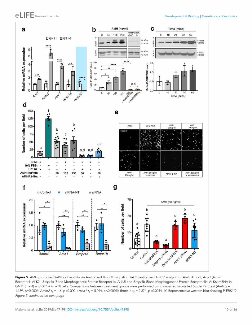
Figure 5. AMH promotes GnRH cell motility via Amhr2 and Bmpr1b signaling. (a) Quantitative RT-PCR analysis for Amh, Amhr2, Acvr1 (Activin
Receptor1; ALK2), Bmpr1a (Bone Morphogenetic Protein Receptor1a; ALK3) and Bmpr1b (Bone Morphogenetic Protein Receptor1b; ALK6) mRNA in
GN11 (n = 4) and GT1-7 (n = 3) cells. Comparisons between treatment groups were performed using unpaired two-tailed Student’s t test (Amh t5 =
1.139, p=0.0004; Amhr2 t5 = 1.6, p<0.0001; Acvr1 t5 = 5.044, p<0.0001); Bmpr1a t5 = 2.374, p<0.0044. (b) Representative western blot showing P-ERK1/2
Figure 5 continued on next page
Malone et al. eLife 2019;8:e47198. DOI: https://doi.org/10.7554/eLife.47198 10 of 36
Research article Developmental Biology Genetics and Genomics
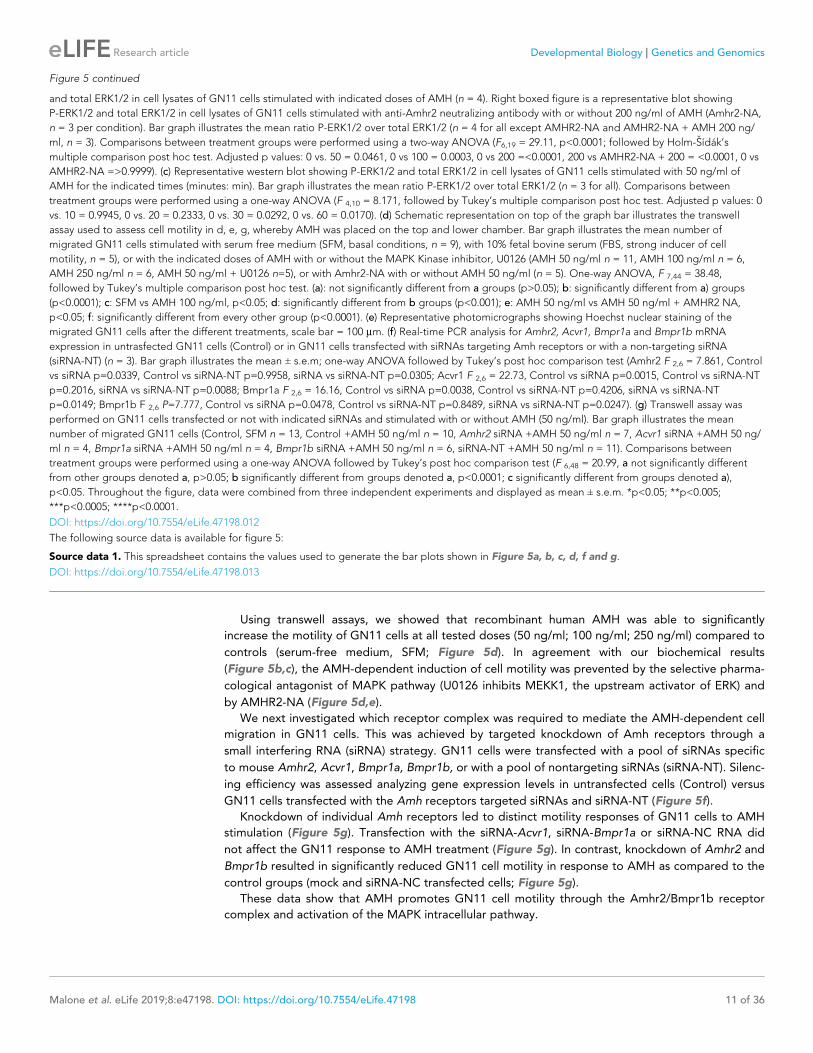
Using transwell assays, we showed that recombinant human AMH was able to significantly
increase the motility of GN11 cells at all tested doses (50 ng/ml; 100 ng/ml; 250 ng/ml) compared to
controls (serum-free medium, SFM; Figure 5d). In agreement with our biochemical results
(Figure 5b,c), the AMH-dependent induction of cell motility was prevented by the selective pharma-
cological antagonist of MAPK pathway (U0126 inhibits MEKK1, the upstream activator of ERK) and
by AMHR2-NA (Figure 5d,e).
We next investigated which receptor complex was required to mediate the AMH-dependent cell
migration in GN11 cells. This was achieved by targeted knockdown of Amh receptors through a
small interfering RNA (siRNA) strategy. GN11 cells were transfected with a pool of siRNAs specific
to mouse Amhr2, Acvr1, Bmpr1a, Bmpr1b, or with a pool of nontargeting siRNAs (siRNA-NT). Silenc-
ing efficiency was assessed analyzing gene expression levels in untransfected cells (Control) versus
GN11 cells transfected with the Amh receptors targeted siRNAs and siRNA-NT (Figure 5f).
Knockdown of individual Amh receptors led to distinct motility responses of GN11 cells to AMH
stimulation (Figure 5g). Transfection with the siRNA-Acvr1, siRNA-Bmpr1a or siRNA-NC RNA did
not affect the GN11 response to AMH treatment (Figure 5g). In contrast, knockdown of Amhr2 and
Bmpr1b resulted in significantly reduced GN11 cell motility in response to AMH as compared to the
control groups (mock and siRNA-NC transfected cells; Figure 5g).
These data show that AMH promotes GN11 cell motility through the Amhr2/Bmpr1b receptor
complex and activation of the MAPK intracellular pathway.
Figure 5 continued
and total ERK1/2 in cell lysates of GN11 cells stimulated with indicated doses of AMH (n = 4). Right boxed figure is a representative blot showing
P-ERK1/2 and total ERK1/2 in cell lysates of GN11 cells stimulated with anti-Amhr2 neutralizing antibody with or without 200 ng/ml of AMH (Amhr2-NA,
n = 3 per condition). Bar graph illustrates the mean ratio P-ERK1/2 over total ERK1/2 (n = 4 for all except AMHR2-NA and AMHR2-NA + AMH 200 ng/
ml, n = 3). Comparisons between treatment groups were performed using a two-way ANOVA (F6,19 = 29.11, p<0.0001; followed by Holm-Sıdak’s
multiple comparison post hoc test. Adjusted p values: 0 vs. 50 = 0.0461, 0 vs 100 = 0.0003, 0 vs 200 =<0.0001, 200 vs AMHR2-NA + 200 = <0.0001, 0 vs
AMHR2-NA =>0.9999). (c) Representative western blot showing P-ERK1/2 and total ERK1/2 in cell lysates of GN11 cells stimulated with 50 ng/ml of
AMH for the indicated times (minutes: min). Bar graph illustrates the mean ratio P-ERK1/2 over total ERK1/2 (n = 3 for all). Comparisons between
treatment groups were performed using a one-way ANOVA (F 4,10 = 8.171, followed by Tukey’s multiple comparison post hoc test. Adjusted p values: 0
vs. 10 = 0.9945, 0 vs. 20 = 0.2333, 0 vs. 30 = 0.0292, 0 vs. 60 = 0.0170). (d) Schematic representation on top of the graph bar illustrates the transwell
assay used to assess cell motility in d, e, g, whereby AMH was placed on the top and lower chamber. Bar graph illustrates the mean number of
migrated GN11 cells stimulated with serum free medium (SFM, basal conditions, n = 9), with 10% fetal bovine serum (FBS, strong inducer of cell
motility, n = 5), or with the indicated doses of AMH with or without the MAPK Kinase inhibitor, U0126 (AMH 50 ng/ml n = 11, AMH 100 ng/ml n = 6,
AMH 250 ng/ml n = 6, AMH 50 ng/ml + U0126 n=5), or with Amhr2-NA with or without AMH 50 ng/ml (n = 5). One-way ANOVA, F 7,44 = 38.48,
followed by Tukey’s multiple comparison post hoc test. (a): not significantly different from a groups (p>0.05); b: significantly different from a) groups
(p<0.0001); c: SFM vs AMH 100 ng/ml, p<0.05; d: significantly different from b groups (p<0.001); e: AMH 50 ng/ml vs AMH 50 ng/ml + AMHR2 NA,
p<0.05; f: significantly different from every other group (p<0.0001). (e) Representative photomicrographs showing Hoechst nuclear staining of the
migrated GN11 cells after the different treatments, scale bar = 100 mm. (f) Real-time PCR analysis for Amhr2, Acvr1, Bmpr1a and Bmpr1b mRNA
expression in untrasfected GN11 cells (Control) or in GN11 cells transfected with siRNAs targeting Amh receptors or with a non-targeting siRNA
(siRNA-NT) (n = 3). Bar graph illustrates the mean ± s.e.m; one-way ANOVA followed by Tukey’s post hoc comparison test (Amhr2 F 2,6 = 7.861, Control
vs siRNA p=0.0339, Control vs siRNA-NT p=0.9958, siRNA vs siRNA-NT p=0.0305; Acvr1 F 2,6 = 22.73, Control vs siRNA p=0.0015, Control vs siRNA-NT
p=0.2016, siRNA vs siRNA-NT p=0.0088; Bmpr1a F 2,6 = 16.16, Control vs siRNA p=0.0038, Control vs siRNA-NT p=0.4206, siRNA vs siRNA-NT
p=0.0149; Bmpr1b F 2,6 P=7.777, Control vs siRNA p=0.0478, Control vs siRNA-NT p=0.8489, siRNA vs siRNA-NT p=0.0247). (g) Transwell assay was
performed on GN11 cells transfected or not with indicated siRNAs and stimulated with or without AMH (50 ng/ml). Bar graph illustrates the mean
number of migrated GN11 cells (Control, SFM n = 13, Control +AMH 50 ng/ml n = 10, Amhr2 siRNA +AMH 50 ng/ml n = 7, Acvr1 siRNA +AMH 50 ng/
ml n = 4, Bmpr1a siRNA +AMH 50 ng/ml n = 4, Bmpr1b siRNA +AMH 50 ng/ml n = 6, siRNA-NT +AMH 50 ng/ml n = 11). Comparisons between
treatment groups were performed using a one-way ANOVA followed by Tukey’s post hoc comparison test (F 6,48 = 20.99, a not significantly different
from other groups denoted a, p>0.05; b significantly different from groups denoted a, p<0.0001; c significantly different from groups denoted a),
p<0.05. Throughout the figure, data were combined from three independent experiments and displayed as mean ± s.e.m. *p<0.05; **p<0.005;
***p<0.0005; ****p<0.0001.
DOI: https://doi.org/10.7554/eLife.47198.012
The following source data is available for figure 5:
Source data 1. This spreadsheet contains the values used to generate the bar plots shown in Figure 5a, b, c, d, f and g.
DOI: https://doi.org/10.7554/eLife.47198.013
Malone et al. eLife 2019;8:e47198. DOI: https://doi.org/10.7554/eLife.47198 11 of 36
Research article Developmental Biology Genetics and Genomics
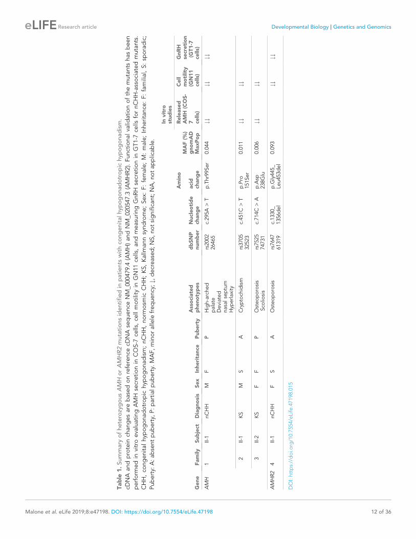
Table
1.Summary
ofheterozygousAMH
orAMHR2mutationsidentifiedin
patients
withco
ngenitalhyp
ogonadotropic
hyp
ogonadism.
cDNA
andprotein
changesare
basedonreference
cDNA
sequence
NM_0
00479.4
(AMH)andNM_0
20547.3
(AMHR2).Functionalvalid
ationofthemutants
hasbeen
perform
edin
vitroevaluatingAMH
secretionin
COS-7
cells,ce
llmotilityin
GN11ce
lls,andmeasuringGnRH
secretionin
GT1-7
cells
fornCHH-associatedmutants.
CHH,co
ngenitalhyp
ogonadotropic
hyp
ogonadism;nCHH,norm
osm
icCHH;KS,Kallm
annsyndrome;Sex:
F:female;M:male;Inheritance
:F:familial,S:sp
oradic;
Puberty:
A:absentpuberty,
P:partialpuberty.
MAF,minoralle
lefrequency;#,decreased;NS,notsignificant;NA,notapplicable.
Gene
Family
Subject
Diagnosis
Sex
Inheritance
Puberty
Associated
phenotypes
dbSNP
number
Nucleotide
change
Amino
acid
change
MAF(%
)gnomAD
MaxPop
Invitro
studies
Release
dAMH
(COS-
7 cells)
Cell
motility
(GN11
cells)
GnRH
secretion
(GT1-7
cells)
AMH
1II-1
nCHH
MF
PHigh-arched
palate
Deviated
nasalseptum
Hyp
erlaxity
rs2002
26465
c.295A
>T
p.Thr99Ser
0.044
##
##
##
2II-1
KS
MS
ACryptoch
idism
rs3705
32523
c.451C
>T
p.Pro
151Ser
0.011
##
##
3II-2
KS
FF
POsteoporosis
Sco
liosis
rs7525
74731
c.714C
>A
p.Asp
238Glu
0.006
##
##
AMHR2
4II-1
nCHH
FS
AOsteoporosis
rs7647
61319
c.1330_
1356del
p.G
ly445_
Leu453del
0.093
##
##
DOI:https://doi.o
rg/10.7554/eLife.47198.015
Malone et al. eLife 2019;8:e47198. DOI: https://doi.org/10.7554/eLife.47198 12 of 36
Research article Developmental Biology Genetics and Genomics
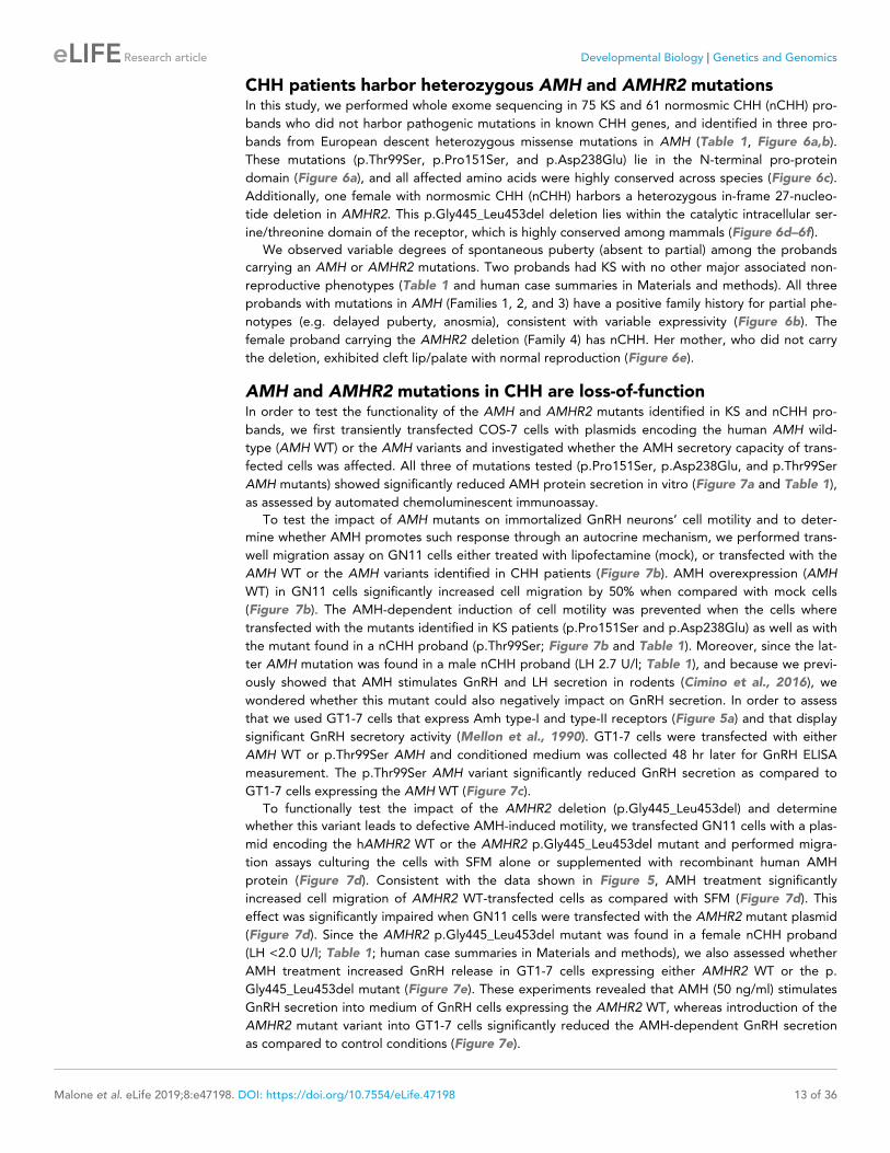
CHH patients harbor heterozygous AMH and AMHR2 mutationsIn this study, we performed whole exome sequencing in 75 KS and 61 normosmic CHH (nCHH) pro-
bands who did not harbor pathogenic mutations in known CHH genes, and identified in three pro-
bands from European descent heterozygous missense mutations in AMH (Table 1, Figure 6a,b).
These mutations (p.Thr99Ser, p.Pro151Ser, and p.Asp238Glu) lie in the N-terminal pro-protein
domain (Figure 6a), and all affected amino acids were highly conserved across species (Figure 6c).
Additionally, one female with normosmic CHH (nCHH) harbors a heterozygous in-frame 27-nucleo-
tide deletion in AMHR2. This p.Gly445_Leu453del deletion lies within the catalytic intracellular ser-
ine/threonine domain of the receptor, which is highly conserved among mammals (Figure 6d–6f).
We observed variable degrees of spontaneous puberty (absent to partial) among the probands
carrying an AMH or AMHR2 mutations. Two probands had KS with no other major associated non-
reproductive phenotypes (Table 1 and human case summaries in Materials and methods). All three
probands with mutations in AMH (Families 1, 2, and 3) have a positive family history for partial phe-
notypes (e.g. delayed puberty, anosmia), consistent with variable expressivity (Figure 6b). The
female proband carrying the AMHR2 deletion (Family 4) has nCHH. Her mother, who did not carry
the deletion, exhibited cleft lip/palate with normal reproduction (Figure 6e).
AMH and AMHR2 mutations in CHH are loss-of-functionIn order to test the functionality of the AMH and AMHR2 mutants identified in KS and nCHH pro-
bands, we first transiently transfected COS-7 cells with plasmids encoding the human AMH wild-
type (AMH WT) or the AMH variants and investigated whether the AMH secretory capacity of trans-
fected cells was affected. All three of mutations tested (p.Pro151Ser, p.Asp238Glu, and p.Thr99Ser
AMH mutants) showed significantly reduced AMH protein secretion in vitro (Figure 7a and Table 1),
as assessed by automated chemoluminescent immunoassay.
To test the impact of AMH mutants on immortalized GnRH neurons’ cell motility and to deter-
mine whether AMH promotes such response through an autocrine mechanism, we performed trans-
well migration assay on GN11 cells either treated with lipofectamine (mock), or transfected with the
AMH WT or the AMH variants identified in CHH patients (Figure 7b). AMH overexpression (AMH
WT) in GN11 cells significantly increased cell migration by 50% when compared with mock cells
(Figure 7b). The AMH-dependent induction of cell motility was prevented when the cells where
transfected with the mutants identified in KS patients (p.Pro151Ser and p.Asp238Glu) as well as with
the mutant found in a nCHH proband (p.Thr99Ser; Figure 7b and Table 1). Moreover, since the lat-
ter AMH mutation was found in a male nCHH proband (LH 2.7 U/l; Table 1), and because we previ-
ously showed that AMH stimulates GnRH and LH secretion in rodents (Cimino et al., 2016), we
wondered whether this mutant could also negatively impact on GnRH secretion. In order to assess
that we used GT1-7 cells that express Amh type-I and type-II receptors (Figure 5a) and that display
significant GnRH secretory activity (Mellon et al., 1990). GT1-7 cells were transfected with either
AMH WT or p.Thr99Ser AMH and conditioned medium was collected 48 hr later for GnRH ELISA
measurement. The p.Thr99Ser AMH variant significantly reduced GnRH secretion as compared to
GT1-7 cells expressing the AMH WT (Figure 7c).
To functionally test the impact of the AMHR2 deletion (p.Gly445_Leu453del) and determine
whether this variant leads to defective AMH-induced motility, we transfected GN11 cells with a plas-
mid encoding the hAMHR2 WT or the AMHR2 p.Gly445_Leu453del mutant and performed migra-
tion assays culturing the cells with SFM alone or supplemented with recombinant human AMH
protein (Figure 7d). Consistent with the data shown in Figure 5, AMH treatment significantly
increased cell migration of AMHR2 WT-transfected cells as compared with SFM (Figure 7d). This
effect was significantly impaired when GN11 cells were transfected with the AMHR2 mutant plasmid
(Figure 7d). Since the AMHR2 p.Gly445_Leu453del mutant was found in a female nCHH proband
(LH <2.0 U/l; Table 1; human case summaries in Materials and methods), we also assessed whether
AMH treatment increased GnRH release in GT1-7 cells expressing either AMHR2 WT or the p.
Gly445_Leu453del mutant (Figure 7e). These experiments revealed that AMH (50 ng/ml) stimulates
GnRH secretion into medium of GnRH cells expressing the AMHR2 WT, whereas introduction of the
AMHR2 mutant variant into GT1-7 cells significantly reduced the AMH-dependent GnRH secretion
as compared to control conditions (Figure 7e).
Malone et al. eLife 2019;8:e47198. DOI: https://doi.org/10.7554/eLife.47198 13 of 36
Research article Developmental Biology Genetics and Genomics
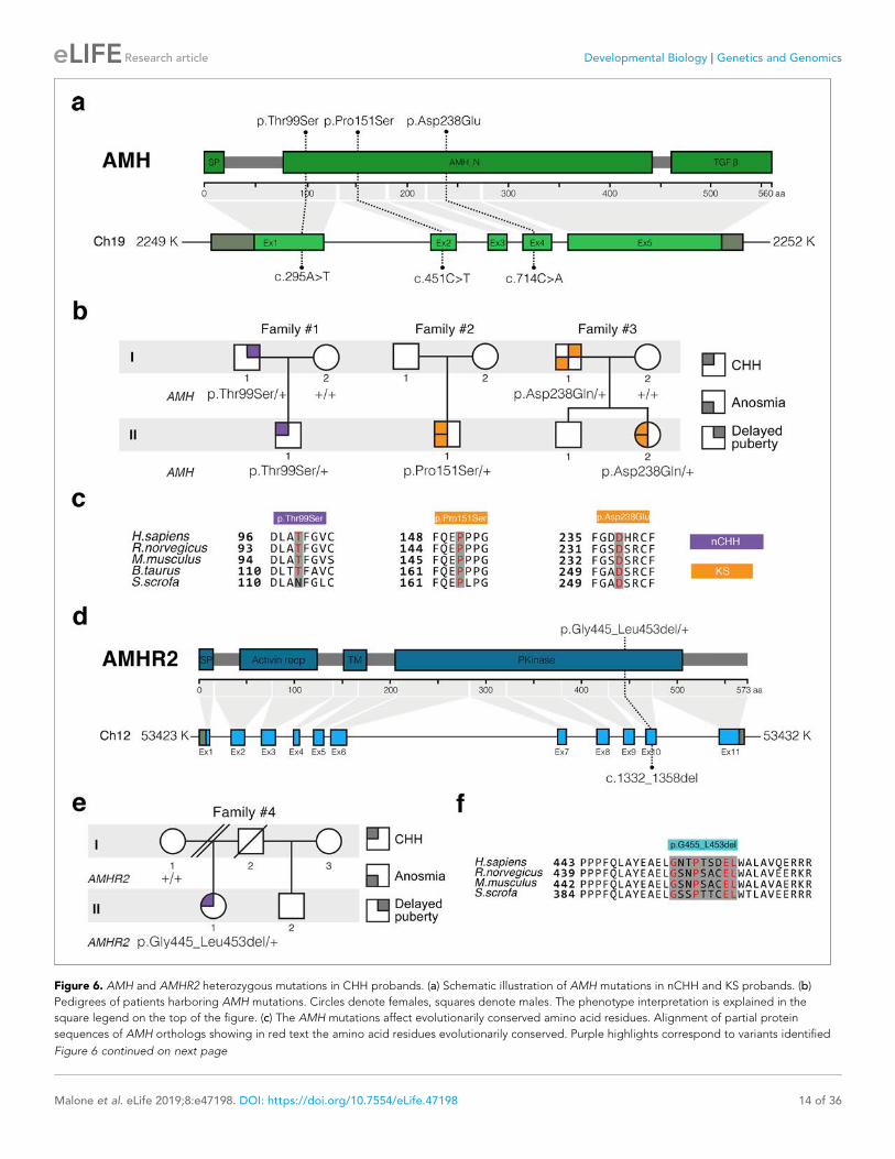
Figure 6. AMH and AMHR2 heterozygous mutations in CHH probands. (a) Schematic illustration of AMH mutations in nCHH and KS probands. (b)
Pedigrees of patients harboring AMH mutations. Circles denote females, squares denote males. The phenotype interpretation is explained in the
square legend on the top of the figure. (c) The AMH mutations affect evolutionarily conserved amino acid residues. Alignment of partial protein
sequences of AMH orthologs showing in red text the amino acid residues evolutionarily conserved. Purple highlights correspond to variants identified
Figure 6 continued on next page
Malone et al. eLife 2019;8:e47198. DOI: https://doi.org/10.7554/eLife.47198 14 of 36
Research article Developmental Biology Genetics and Genomics
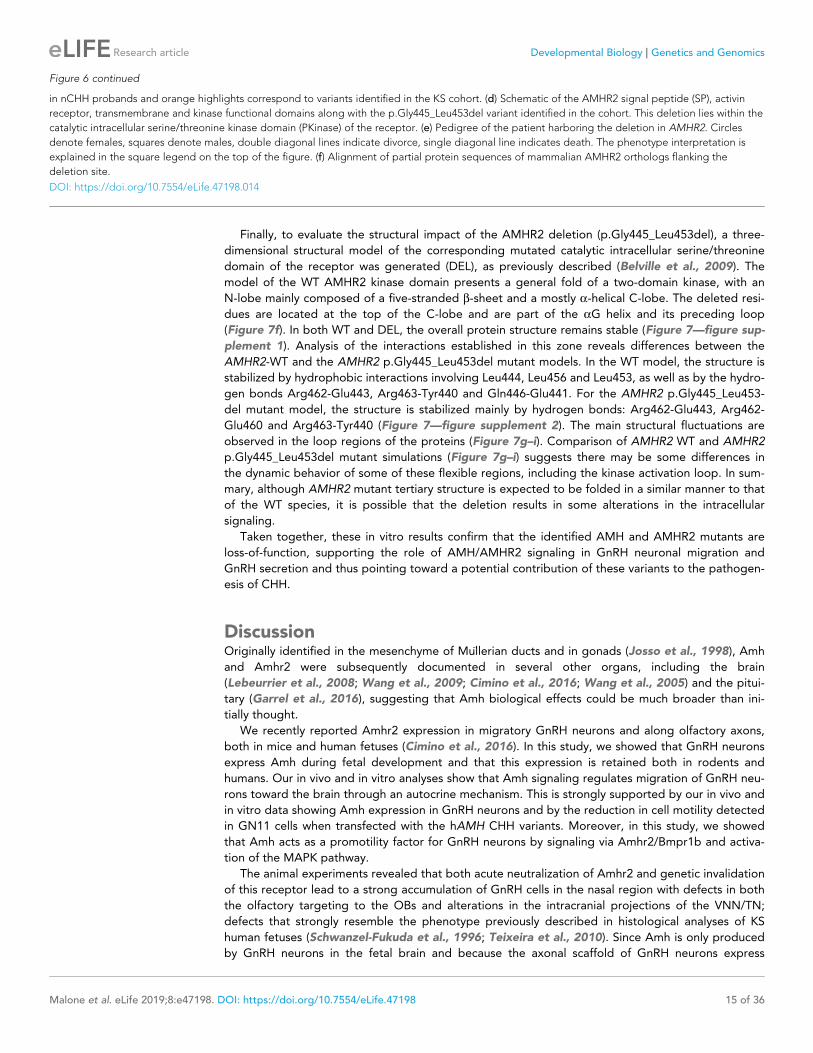
Finally, to evaluate the structural impact of the AMHR2 deletion (p.Gly445_Leu453del), a three-
dimensional structural model of the corresponding mutated catalytic intracellular serine/threonine
domain of the receptor was generated (DEL), as previously described (Belville et al., 2009). The
model of the WT AMHR2 kinase domain presents a general fold of a two-domain kinase, with an
N-lobe mainly composed of a five-stranded b-sheet and a mostly a-helical C-lobe. The deleted resi-
dues are located at the top of the C-lobe and are part of the aG helix and its preceding loop
(Figure 7f). In both WT and DEL, the overall protein structure remains stable (Figure 7—figure sup-
plement 1). Analysis of the interactions established in this zone reveals differences between the
AMHR2-WT and the AMHR2 p.Gly445_Leu453del mutant models. In the WT model, the structure is
stabilized by hydrophobic interactions involving Leu444, Leu456 and Leu453, as well as by the hydro-
gen bonds Arg462-Glu443, Arg463-Tyr440 and Gln446-Glu441. For the AMHR2 p.Gly445_Leu453-
del mutant model, the structure is stabilized mainly by hydrogen bonds: Arg462-Glu443, Arg462-
Glu460 and Arg463-Tyr440 (Figure 7—figure supplement 2). The main structural fluctuations are
observed in the loop regions of the proteins (Figure 7g–i). Comparison of AMHR2 WT and AMHR2
p.Gly445_Leu453del mutant simulations (Figure 7g–i) suggests there may be some differences in
the dynamic behavior of some of these flexible regions, including the kinase activation loop. In sum-
mary, although AMHR2 mutant tertiary structure is expected to be folded in a similar manner to that
of the WT species, it is possible that the deletion results in some alterations in the intracellular
signaling.
Taken together, these in vitro results confirm that the identified AMH and AMHR2 mutants are
loss-of-function, supporting the role of AMH/AMHR2 signaling in GnRH neuronal migration and
GnRH secretion and thus pointing toward a potential contribution of these variants to the pathogen-
esis of CHH.
DiscussionOriginally identified in the mesenchyme of Mu€llerian ducts and in gonads (Josso et al., 1998), Amh
and Amhr2 were subsequently documented in several other organs, including the brain
(Lebeurrier et al., 2008; Wang et al., 2009; Cimino et al., 2016; Wang et al., 2005) and the pitui-
tary (Garrel et al., 2016), suggesting that Amh biological effects could be much broader than ini-
tially thought.
We recently reported Amhr2 expression in migratory GnRH neurons and along olfactory axons,
both in mice and human fetuses (Cimino et al., 2016). In this study, we showed that GnRH neurons
express Amh during fetal development and that this expression is retained both in rodents and
humans. Our in vivo and in vitro analyses show that Amh signaling regulates migration of GnRH neu-
rons toward the brain through an autocrine mechanism. This is strongly supported by our in vivo and
in vitro data showing Amh expression in GnRH neurons and by the reduction in cell motility detected
in GN11 cells when transfected with the hAMH CHH variants. Moreover, in this study, we showed
that Amh acts as a promotility factor for GnRH neurons by signaling via Amhr2/Bmpr1b and activa-
tion of the MAPK pathway.
The animal experiments revealed that both acute neutralization of Amhr2 and genetic invalidation
of this receptor lead to a strong accumulation of GnRH cells in the nasal region with defects in both
the olfactory targeting to the OBs and alterations in the intracranial projections of the VNN/TN;
defects that strongly resemble the phenotype previously described in histological analyses of KS
human fetuses (Schwanzel-Fukuda et al., 1996; Teixeira et al., 2010). Since Amh is only produced
by GnRH neurons in the fetal brain and because the axonal scaffold of GnRH neurons express
Figure 6 continued
in nCHH probands and orange highlights correspond to variants identified in the KS cohort. (d) Schematic of the AMHR2 signal peptide (SP), activin
receptor, transmembrane and kinase functional domains along with the p.Gly445_Leu453del variant identified in the cohort. This deletion lies within the
catalytic intracellular serine/threonine kinase domain (PKinase) of the receptor. (e) Pedigree of the patient harboring the deletion in AMHR2. Circles
denote females, squares denote males, double diagonal lines indicate divorce, single diagonal line indicates death. The phenotype interpretation is
explained in the square legend on the top of the figure. (f) Alignment of partial protein sequences of mammalian AMHR2 orthologs flanking the
deletion site.
DOI: https://doi.org/10.7554/eLife.47198.014
Malone et al. eLife 2019;8:e47198. DOI: https://doi.org/10.7554/eLife.47198 15 of 36
Research article Developmental Biology Genetics and Genomics
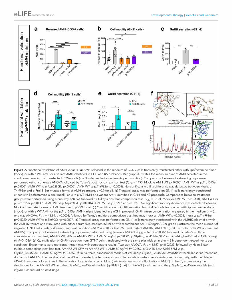
Figure 7. Functional validation of AMH variants. (a) AMH released in the medium of COS-7 cells transiently transfected either with lipofectamine alone
(mock), or with a WT AMH or a variant AMH identified in CHH and KS probands. Bar graph illustrates the mean amount of AMH secreted in the
conditioned medium of transfected COS-7 cells (n = 3 independent experiments per condition). Comparisons between treatment groups were
performed using a one-way ANOVA followed by Tukey’s post hoc comparison test (F4,10 = 1193, Mock vs AMH WT p<0.0001, AMH WT vs p.Pro151Ser
p<0.0001, AMH WT vs p.Asp238Glu p<0.0001, AMH WT vs p.Thr99Ser p<0.0001). No significant motility difference was detected between Mock, p.
Thr99Ser and p.Pro151Ser mutated forms of AMH treatment, p>0.9 for all. (b) Transwell assay was performed on GN11 cells transiently transfected
either with lipofectamine alone (mock), or with a WT AMH or a variant AMH identified in CHH and KS probands. Comparisons between treatment
groups were performed using a one-way ANOVA followed by Tukey’s post hoc comparison test (F4,50 = 13.94, Mock vs AMH WT p<0.0001, AMH WT vs
p.Pro151Ser p<0.0001, AMH WT vs p.Asp238Glu p<0.0014, AMH WT vs p.Thr99Ser p=0.0218. No significant motility difference was detected between
Mock and mutated forms of AMH treatment, p>0.9 for all. (c) Quantification of GnRH secretion from GT1-7 cells transfected with lipofectamine alone
(mock), or with a WT AMH or the p.Pro151Ser AMH variant identified in a nCHH proband. GnRH mean concentration measured in the medium (n = 3,
one-way ANOVA: F 2,6 = 43.84, p=0.0003; followed by Tukey’s multiple comparison post hoc test, mock vs. AMH WT p=0.0003, mock vs p.Thr99Ser
p=0.5220, AMH WT vs p.Thr99Ser p=0.0007. (d) Transwell assay was performed on GN11 cells transiently transfected with the AMHR2 plasmid or with
the AMHR2 variant and stimulated with either serum-free medium (SFM) or with recombinant AMH (50 ng/ml). Bar graph illustrates the mean number of
migrated GN11 cells under different treatment conditions (SFM n = 10 for both WT and mutant AMHR2, AMH 50 ng/ml n = 12 for both WT and mutant
AMHR2). Comparisons between treatment groups were performed using two-way ANOVA (F1,43 = 16.5 P=0.0002; followed by Sidak’s multiple
comparison post hoc test, AMHR2 WT SFM vs AMHR2 WT + AMH 50 ng/ml p<0.0001, p.Gly445_Leu453del SFM vs p.Gly445_Leu453del + AMH 50 ng/
ml P=0.1036). (e) Quantification of GnRH secretion from GT1-7 cells transfected with the same plasmids as in d (n = 3 independent experiments per
condition). Experiments were replicated three times with comparable results. Two-way ANOVA, F1,8 = 1.927, p<0.02025; followed by Holm-Sıdak
multiple comparison post hoc test, AMHR2 WT SFM vs AMHR2 WT + AMH 50 ng/ml P=0.0269, p.Gly445_Leu453del SFM vs p.
Gly445_Leu453del + AMH 50 ng/ml P=0.4652. (f) Initial three-dimensional models of WT and p.Gly445_Leu453del catalytic intracellular serine/threonine
domains of AMHR2. The backbone of the WT and deleted proteins are shown in tan or white cartoon representations, respectively, with the deleted
445–453 residues colored in red. The activation loop is depicted in blue. (g–i) Root-mean-square fluctuations (RMSF) of the Ca atoms along the
simulations for the AMHR2 WT and the p.Gly445_Leu453del models. (g) RMSF (in A) for the WT (black line) and the p.Gly445_Leu453del models (red
Figure 7 continued on next page
Malone et al. eLife 2019;8:e47198. DOI: https://doi.org/10.7554/eLife.47198 16 of 36
Research article Developmental Biology Genetics and Genomics
Amhr2, we hypothesize that Amh signaling contributes to the correct development of the ventral
branch of the vomeronasal/terminal nerves in the brain through a paracrine mechanism. Mono-allelic
inactivation of Amhr2 in mice is not sufficient to significantly alter GnRH neuronal migration, as indi-
cated by the normal number of GnRH neurons observed in adult Amhr2 heterozygous brains. On
the other hand, the presence of only one Amhr2-null allele is sufficient to trigger significant impair-
ments of LH secretion, LH pulsatlity and fertility in adult female mice. In heterozygous males, only
the number of litters/90 days was found to be significantly reduced as compared to Amhr2+/+ males,
suggesting that sexual behavior but not fecundity is likely altered in Amhr2+/- males.
Bi-allelic inactivation of Amhr2 in mice results instead into a significant reduction of the GnRH cell
population in the brains of adult Amhr2-null mice of both sexes as compared to wild-type animals.
Since male and female homozygous adult animals showed a comparable GnRH cell loss, it is likely
that the GnRH migratory defect observed in Amhr2-deficient embryos is independent of the genetic
sex of the animals.
We speculate that Amh/Amhr2 signaling can regulate, respectively, GnRH migration, during
embryonic development, and GnRH/LH secretion postnatally. The latter point is also supported by
our in vitro experiments showing AMH-induced GnRH secretion in GT1-7 cells.
Homozygous Amhr2 female mice have a more pronounced phenotype than heterozygous ani-
mals, as they combine developmental defects in GnRH migration with severely impaired ovulation
and fertility in adulthood. This strong phenotype could be the consequence of a lack of a broad
spectrum of actions of Amh at different prenatal and postnatal stages, impacting the GnRH neuronal
migration and the GnRH secretion, respectively, or gonadotrope function (Garrel et al., 2016;
Garrel et al., 2019). Moreover, since Amh is known to be expressed by granulosa cells in the ovaries
(Vigier et al., 1984) and to regulate folliculogenesis (Durlinger et al., 2001; Durlinger et al., 1999),
it is likely that part of the reproductive phenotype of Amhr2-/- mice is also due in part to the lack of
ovarian Amh. Dissecting the specific contribution of ovarian versus cerebral Amh in the control of
fertility would only be possible using a neuronal specific knockout of Amhr2, which is not available
yet. However, our previous (Cimino et al., 2016) and current findings, showing that Amh directly
increases GnRH and LH secretion, support a role for Amh in the neuroendocrine regulation of fertil-
ity in physiological and pathological conditions.
Our study identified heterozygous mutations in CHH probands that affect highly conserved amino
acids of AMH or its exclusive binding receptor, AMHR2. The AMH mutants display defects in AMH
release. Since the described proAMH cleavage sites (Mamsen et al., 2015; Pankhurst and
McLennan, 2013) are not located in close proximity of these mutations, it is unlikely that they
impinge on the cleavage of the proAMH. The decreased AMH secretion might rather result from
altered protein trafficking leading to accumulation of the mutants AMH in the endoplasmic reticulum
(ER) and defective release. The AMH mutants identified in KS probands also significantly reduced
GN11 migration compared to the wild-type AMH. Interestingly, the p.Thr99Ser AMH mutation found
in a nCHH proband compromises both GnRH cell motility in GN11 cells as well as GnRH secretion in
GT1-7 cells. It is plausible that AMH mutants cause dominant negative effects by forming hetero-
dimers with wild-type AMH, thus reducing AMHR2 activation and the downstream biological
Figure 7 continued
line, being the average over the three 100 ns simulations) are given for each residue of the protein. For a better comparison, residue numbers were
kept the same for both models. Molecular representation of the WT (h) and p.Gly445_Leu453del (i) models colored by RMSF: the blue-green-red scale
corresponds to low-medium-high RMSF values. The yellow spheres indicate the first residues after the p.Gly445_Leu453del deletion. The activation
loop region is highlighted inside a blue frame (arrows).
DOI: https://doi.org/10.7554/eLife.47198.016
The following source data and figure supplements are available for figure 7:
Source data 1. This spreadsheet contains the values used to generate the bar plots shown in Figure 7a–e.
DOI: https://doi.org/10.7554/eLife.47198.019
Figure supplement 1. Root-mean-square deviations (RMSD) of the AMHR2 protein backbone along the simulations.
DOI: https://doi.org/10.7554/eLife.47198.017
Figure supplement 2. Molecular representation of main interactions stabilizing the zone around the AMHR2 deletion.
DOI: https://doi.org/10.7554/eLife.47198.018
Malone et al. eLife 2019;8:e47198. DOI: https://doi.org/10.7554/eLife.47198 17 of 36
Research article Developmental Biology Genetics and Genomics
response. Finally, the AMHR2 p.Gly445_Leu453del mutation identified in a female nCHH proband
was also loss-of-function in in vitro migration and GnRH secretion assays.
The heterozygous mutations in AMH and AMHR2 are found in 3% of CHH probands in our cohort
(4 out of 136). This is consistent with the genetic landscape of CHH as the majority of known CHH
genes have a low mutational prevalence (<5%) (Boehm et al., 2015). Variable expressivity was
observed in family members carrying the same mutation, consistent with the fact that CHH repre-
sents the more severe end of a large spectrum of manifestations. This is a common theme in the
genetics of CHH, and factors such as digenic/oligogenic inheritance (Boehm et al., 2015;
Sykiotis et al., 2010; Pitteloud et al., 2007), epigenetic regulation or non-genetic contributions
likely play important roles. As the current study is limited by the small number of probands harbor-
ing AMH and AMHR2 mutations, confirmation in larger CHH cohorts will be necessary to establish
the specific contributions of these two genes in the pathogenesis of CHH.
Notably, homozygous or compound heterozygous loss-of-function mutations in AMH (OMIM:
600957) or AMHR2 (OMIM: 600956) cause PMDS in both mice and humans (Imbeaud et al., 1995;
Mishina et al., 1996). PMDS is characterized by the retention of Mu€llerian duct derivatives (the
uterus, fallopian tubes, and upper part of the vagina) in males (Josso and Clemente, 2003;
Behringer et al., 1994; Mishina et al., 1996; Belville et al., 1999; Belville et al., 2004; Orvis et al.,
2008). Interestingly, we identified two mutations (AMH p.Pro151Ser and AMHR2 p.Gly445_Leu453-
del) (Picard et al., 2017) in our CHH cohort previously associated with autosomal recessive PMDS.
MRI or pelvic ultrasounds were performed in the two male CHH patients harboring AMH mutations,
including the patient carrying the p.Pro151Ser. No defects in the internal genitalia were identified,
consistent with the fact that monoallelic defects in AMH or AMHR2 do not cause PMDS
(Picard et al., 2017). Notably, parents of PMDS probands carrying heterozygote AMH or AMHR2
mutations are fertile (Picard et al., 2017; Josso et al., 2005); however, detailed reproductive and
olfactory phenotyping in these parents have not been reported. Furthermore, there are few studies
examining the hormonal profile of patients with PMDS although spontaneous puberty is reported
based on clinical observation (Josso et al., 2005). To assess pubertal and reproductive defects in
PMDS patients or family members, detailed reproductive phenotyping will be necessary.
Taken together, these data demonstrate the pleiotropic roles of AMH and AMHR2 in shaping
internal genitalia and GnRH neuron migration. Different mechanistic actions of the mutants (i.e.
recessive vs. dominant negative vs. haploinsufficiency) in combination with tissue-specific signaling
pathway might guide the final phenotype.
AMH and AMHR2 mutations affecting GN11 cell motility were found in both KS and nCHH indi-
viduals. We thus speculate that GnRH migratory defects could also occur in some cases of nCHH.
This hypothesis challenges the current dogma, whereby defects in GnRH cell migration lead to KS
and not nCHH (Boehm et al., 2015). Yet, increasing genetic evidence indicates that CHH genes do
not segregate into 2 (i.e. KS and nCHH) but rather three categories: 1) KS only, 2) nCHH only and 3)
both KS and nCHH. The latter includes genes involved in GnRH neuron migration such as FGFR1,
SEMA7A, AXL (Boehm et al., 2015). We recently showed that GnRH neurons in human fetuses
migrate into the brain in tight associations with vomeronasal and terminal nerves and not olfactory
nerves (Casoni et al., 2016). Another recent study demonstrated that correct development of the
OBs and axonal connection to the forebrain are not required for GnRH neuronal migration
(Taroc et al., 2017), thus implying a stronger contribution of the vomeronasal/terminal nerve as a
scaffold for the GnRH migration. Taken together, this evidence indicates that the vomeronasal and
the terminal nerve play important roles in the ontogenesis and migration of GnRH neurons in verte-
brates, and raises the intriguing possibility that some genetic forms of nCHH might be due to defec-
tive central projections of the vomeronasal/terminal nerves leading to subsequent alterations of the
GnRH migratory process.
In conclusion, this work highlights the role of AMH/AMHR2 signaling in GnRH neuronal migration,
hormone secretion and regulation of fertility, and identifies heterozygous mutations in AMH and
AMHR2 in CHH patients.
Materials and methods
Key resources table
Malone et al. eLife 2019;8:e47198. DOI: https://doi.org/10.7554/eLife.47198 18 of 36
Research article Developmental Biology Genetics and Genomics
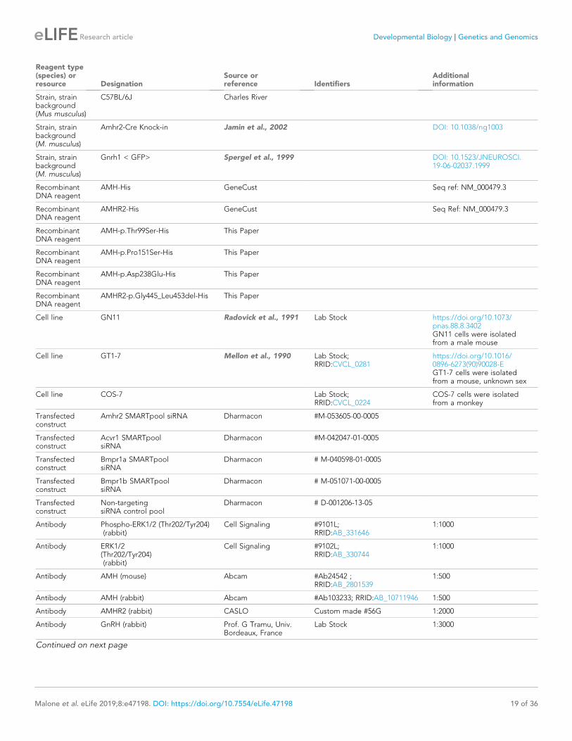
Reagent type(species) orresource Designation
Source orreference Identifiers
Additionalinformation
Strain, strainbackground(Mus musculus)
C57BL/6J Charles River
Strain, strainbackground(M. musculus)
Amhr2-Cre Knock-in Jamin et al., 2002 DOI: 10.1038/ng1003
Strain, strainbackground(M. musculus)
Gnrh1 < GFP> Spergel et al., 1999 DOI: 10.1523/JNEUROSCI.19-06-02037.1999
RecombinantDNA reagent
AMH-His GeneCust Seq ref: NM_000479.3
RecombinantDNA reagent
AMHR2-His GeneCust Seq Ref: NM_000479.3
RecombinantDNA reagent
AMH-p.Thr99Ser-His This Paper
RecombinantDNA reagent
AMH-p.Pro151Ser-His This Paper
RecombinantDNA reagent
AMH-p.Asp238Glu-His This Paper
RecombinantDNA reagent
AMHR2-p.Gly445_Leu453del-His This Paper
Cell line GN11 Radovick et al., 1991 Lab Stock https://doi.org/10.1073/pnas.88.8.3402GN11 cells were isolatedfrom a male mouse
Cell line GT1-7 Mellon et al., 1990 Lab Stock;RRID:CVCL_0281
https://doi.org/10.1016/0896-6273(90)90028-EGT1-7 cells were isolatedfrom a mouse, unknown sex
Cell line COS-7 Lab Stock;RRID:CVCL_0224
COS-7 cells were isolatedfrom a monkey
Transfectedconstruct
Amhr2 SMARTpool siRNA Dharmacon #M-053605-00-0005
Transfectedconstruct
Acvr1 SMARTpoolsiRNA
Dharmacon #M-042047-01-0005
Transfectedconstruct
Bmpr1a SMARTpoolsiRNA
Dharmacon # M-040598-01-0005
Transfectedconstruct
Bmpr1b SMARTpoolsiRNA
Dharmacon # M-051071-00-0005
Transfectedconstruct
Non-targetingsiRNA control pool
Dharmacon # D-001206-13-05
Antibody Phospho-ERK1/2 (Thr202/Tyr204)(rabbit)
Cell Signaling #9101L;RRID:AB_331646
1:1000
Antibody ERK1/2(Thr202/Tyr204)(rabbit)
Cell Signaling #9102L;RRID:AB_330744
1:1000
Antibody AMH (mouse) Abcam #Ab24542 ;RRID:AB_2801539
1:500
Antibody AMH (rabbit) Abcam #Ab103233; RRID:AB_10711946 1:500
Antibody AMHR2 (rabbit) CASLO Custom made #56G 1:2000
Antibody GnRH (rabbit) Prof. G Tramu, Univ.Bordeaux, France
Lab Stock 1:3000
Continued on next page
Malone et al. eLife 2019;8:e47198. DOI: https://doi.org/10.7554/eLife.47198 19 of 36
Research article Developmental Biology Genetics and Genomics
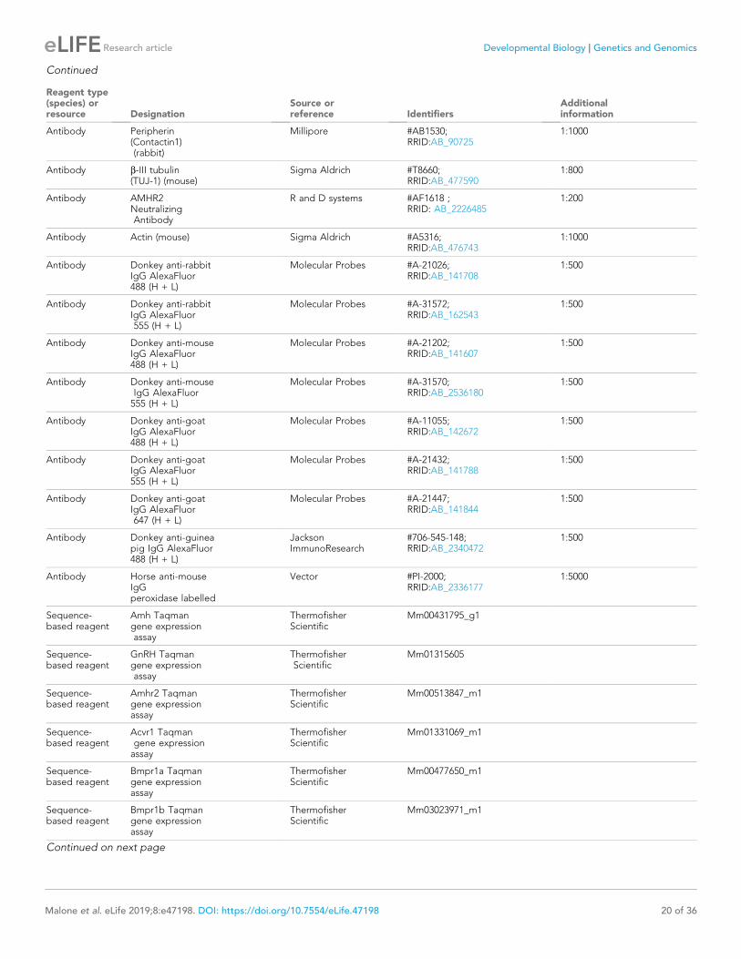
Continued
Reagent type(species) orresource Designation
Source orreference Identifiers
Additionalinformation
Antibody Peripherin(Contactin1)(rabbit)
Millipore #AB1530;RRID:AB_90725
1:1000
Antibody b-III tubulin(TUJ-1) (mouse)
Sigma Aldrich #T8660;RRID:AB_477590
1:800
Antibody AMHR2NeutralizingAntibody
R and D systems #AF1618 ;RRID: AB_2226485
1:200
Antibody Actin (mouse) Sigma Aldrich #A5316;RRID:AB_476743
1:1000
Antibody Donkey anti-rabbitIgG AlexaFluor488 (H + L)
Molecular Probes #A-21026;RRID:AB_141708
1:500
Antibody Donkey anti-rabbitIgG AlexaFluor555 (H + L)
Molecular Probes #A-31572;RRID:AB_162543
1:500
Antibody Donkey anti-mouseIgG AlexaFluor488 (H + L)
Molecular Probes #A-21202;RRID:AB_141607
1:500
Antibody Donkey anti-mouseIgG AlexaFluor555 (H + L)
Molecular Probes #A-31570;RRID:AB_2536180
1:500
Antibody Donkey anti-goatIgG AlexaFluor488 (H + L)
Molecular Probes #A-11055;RRID:AB_142672
1:500
Antibody Donkey anti-goatIgG AlexaFluor555 (H + L)
Molecular Probes #A-21432;RRID:AB_141788
1:500
Antibody Donkey anti-goatIgG AlexaFluor647 (H + L)
Molecular Probes #A-21447;RRID:AB_141844
1:500
Antibody Donkey anti-guineapig IgG AlexaFluor488 (H + L)
JacksonImmunoResearch
#706-545-148;RRID:AB_2340472
1:500
Antibody Horse anti-mouseIgGperoxidase labelled
Vector #PI-2000;RRID:AB_2336177
1:5000
Sequence-based reagent
Amh Taqmangene expressionassay
ThermofisherScientific
Mm00431795_g1
Sequence-based reagent
GnRH Taqmangene expressionassay
ThermofisherScientific
Mm01315605
Sequence-based reagent
Amhr2 Taqmangene expressionassay
ThermofisherScientific
Mm00513847_m1
Sequence-based reagent
Acvr1 Taqmangene expressionassay
ThermofisherScientific
Mm01331069_m1
Sequence-based reagent
Bmpr1a Taqmangene expressionassay
ThermofisherScientific
Mm00477650_m1
Sequence-based reagent
Bmpr1b Taqmangene expressionassay
ThermofisherScientific
Mm03023971_m1
Continued on next page
Malone et al. eLife 2019;8:e47198. DOI: https://doi.org/10.7554/eLife.47198 20 of 36
Research article Developmental Biology Genetics and Genomics
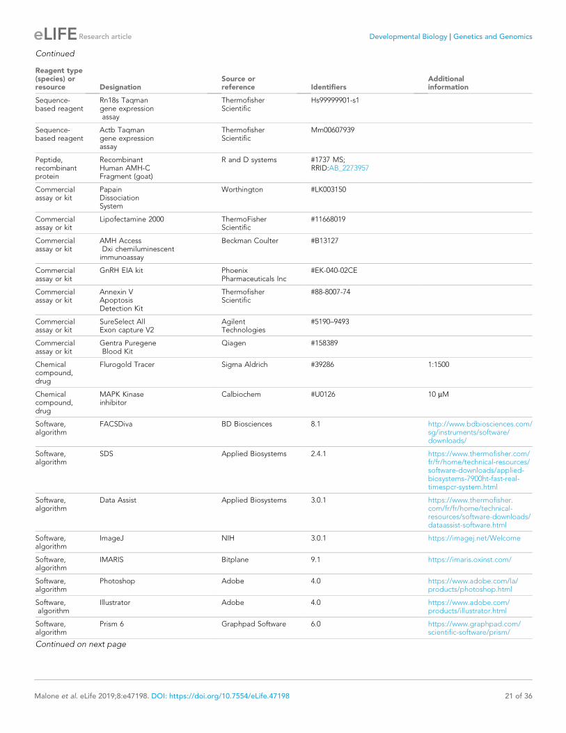
Continued
Reagent type(species) orresource Designation
Source orreference Identifiers
Additionalinformation
Sequence-based reagent
Rn18s Taqmangene expressionassay
ThermofisherScientific
Hs99999901-s1
Sequence-based reagent
Actb Taqmangene expressionassay
ThermofisherScientific
Mm00607939
Peptide,recombinantprotein
RecombinantHuman AMH-CFragment (goat)
R and D systems #1737 MS;RRID:AB_2273957
Commercialassay or kit
PapainDissociationSystem
Worthington #LK003150
Commercialassay or kit
Lipofectamine 2000 ThermoFisherScientific
#11668019
Commercialassay or kit
AMH AccessDxi chemiluminescentimmunoassay
Beckman Coulter #B13127
Commercialassay or kit
GnRH EIA kit PhoenixPharmaceuticals Inc
#EK-040-02CE
Commercialassay or kit
Annexin VApoptosisDetection Kit
ThermofisherScientific
#88-8007-74
Commercialassay or kit
SureSelect AllExon capture V2
AgilentTechnologies
#5190–9493
Commercialassay or kit
Gentra PuregeneBlood Kit
Qiagen #158389
Chemicalcompound,drug
Flurogold Tracer Sigma Aldrich #39286 1:1500
Chemicalcompound,drug
MAPK Kinaseinhibitor
Calbiochem #U0126 10 mM
Software,algorithm
FACSDiva BD Biosciences 8.1 http://www.bdbiosciences.com/sg/instruments/software/downloads/
Software,algorithm
SDS Applied Biosystems 2.4.1 https://www.thermofisher.com/fr/fr/home/technical-resources/software-downloads/applied-biosystems-7900ht-fast-real-timespcr-system.html
Software,algorithm
Data Assist Applied Biosystems 3.0.1 https://www.thermofisher.com/fr/fr/home/technical-resources/software-downloads/dataassist-software.html
Software,algorithm
ImageJ NIH 3.0.1 https://imagej.net/Welcome
Software,algorithm
IMARIS Bitplane 9.1 https://imaris.oxinst.com/
Software,algorithm
Photoshop Adobe 4.0 https://www.adobe.com/la/products/photoshop.html
Software,algorithm
Illustrator Adobe 4.0 https://www.adobe.com/products/illustrator.html
Software,algorithm
Prism 6 Graphpad Software 6.0 https://www.graphpad.com/scientific-software/prism/
Continued on next page
Malone et al. eLife 2019;8:e47198. DOI: https://doi.org/10.7554/eLife.47198 21 of 36
Research article Developmental Biology Genetics and Genomics
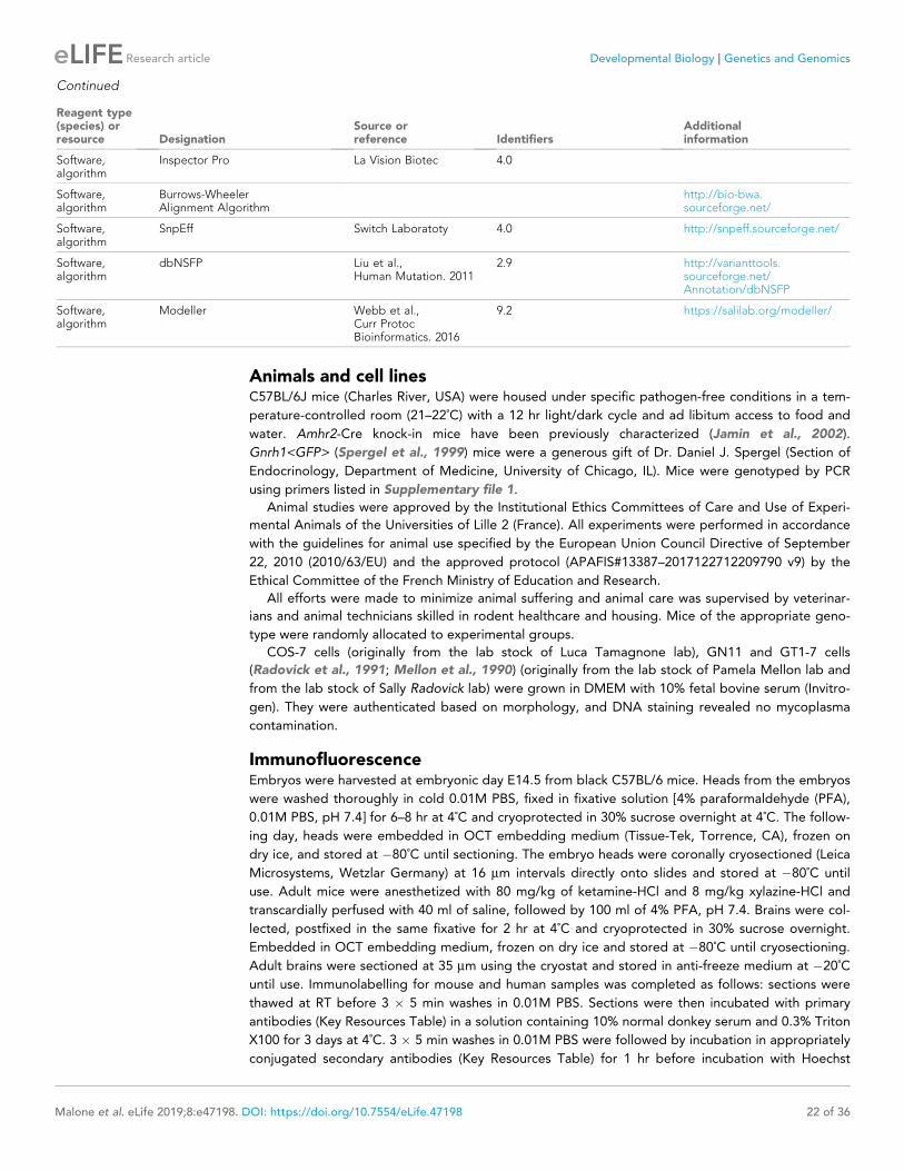
Continued
Reagent type(species) orresource Designation
Source orreference Identifiers
Additionalinformation
Software,algorithm
Inspector Pro La Vision Biotec 4.0
Software,algorithm
Burrows-WheelerAlignment Algorithm
http://bio-bwa.sourceforge.net/
Software,algorithm
SnpEff Switch Laboratoty 4.0 http://snpeff.sourceforge.net/
Software,algorithm
dbNSFP Liu et al.,Human Mutation. 2011
2.9 http://varianttools.sourceforge.net/Annotation/dbNSFP
Software,algorithm
Modeller Webb et al.,Curr ProtocBioinformatics. 2016
9.2 https://salilab.org/modeller/
Animals and cell linesC57BL/6J mice (Charles River, USA) were housed under specific pathogen-free conditions in a tem-
perature-controlled room (21–22˚C) with a 12 hr light/dark cycle and ad libitum access to food and
water. Amhr2-Cre knock-in mice have been previously characterized (Jamin et al., 2002).
Gnrh1<GFP> (Spergel et al., 1999) mice were a generous gift of Dr. Daniel J. Spergel (Section of
Endocrinology, Department of Medicine, University of Chicago, IL). Mice were genotyped by PCR
using primers listed in Supplementary file 1.
Animal studies were approved by the Institutional Ethics Committees of Care and Use of Experi-
mental Animals of the Universities of Lille 2 (France). All experiments were performed in accordance
with the guidelines for animal use specified by the European Union Council Directive of September
22, 2010 (2010/63/EU) and the approved protocol (APAFIS#13387–2017122712209790 v9) by the
Ethical Committee of the French Ministry of Education and Research.
All efforts were made to minimize animal suffering and animal care was supervised by veterinar-
ians and animal technicians skilled in rodent healthcare and housing. Mice of the appropriate geno-
type were randomly allocated to experimental groups.
COS-7 cells (originally from the lab stock of Luca Tamagnone lab), GN11 and GT1-7 cells
(Radovick et al., 1991; Mellon et al., 1990) (originally from the lab stock of Pamela Mellon lab and
from the lab stock of Sally Radovick lab) were grown in DMEM with 10% fetal bovine serum (Invitro-
gen). They were authenticated based on morphology, and DNA staining revealed no mycoplasma
contamination.
ImmunofluorescenceEmbryos were harvested at embryonic day E14.5 from black C57BL/6 mice. Heads from the embryos
were washed thoroughly in cold 0.01M PBS, fixed in fixative solution [4% paraformaldehyde (PFA),
0.01M PBS, pH 7.4] for 6–8 hr at 4˚C and cryoprotected in 30% sucrose overnight at 4˚C. The follow-
ing day, heads were embedded in OCT embedding medium (Tissue-Tek, Torrence, CA), frozen on
dry ice, and stored at �80˚C until sectioning. The embryo heads were coronally cryosectioned (Leica
Microsystems, Wetzlar Germany) at 16 mm intervals directly onto slides and stored at �80˚C until
use. Adult mice were anesthetized with 80 mg/kg of ketamine-HCl and 8 mg/kg xylazine-HCl and
transcardially perfused with 40 ml of saline, followed by 100 ml of 4% PFA, pH 7.4. Brains were col-
lected, postfixed in the same fixative for 2 hr at 4˚C and cryoprotected in 30% sucrose overnight.
Embedded in OCT embedding medium, frozen on dry ice and stored at �80˚C until cryosectioning.
Adult brains were sectioned at 35 mm using the cryostat and stored in anti-freeze medium at �20˚C
until use. Immunolabelling for mouse and human samples was completed as follows: sections were
thawed at RT before 3 � 5 min washes in 0.01M PBS. Sections were then incubated with primary
antibodies (Key Resources Table) in a solution containing 10% normal donkey serum and 0.3% Triton
X100 for 3 days at 4˚C. 3 � 5 min washes in 0.01M PBS were followed by incubation in appropriately
conjugated secondary antibodies (Key Resources Table) for 1 hr before incubation with Hoechst
Malone et al. eLife 2019;8:e47198. DOI: https://doi.org/10.7554/eLife.47198 22 of 36
Research article Developmental Biology Genetics and Genomics
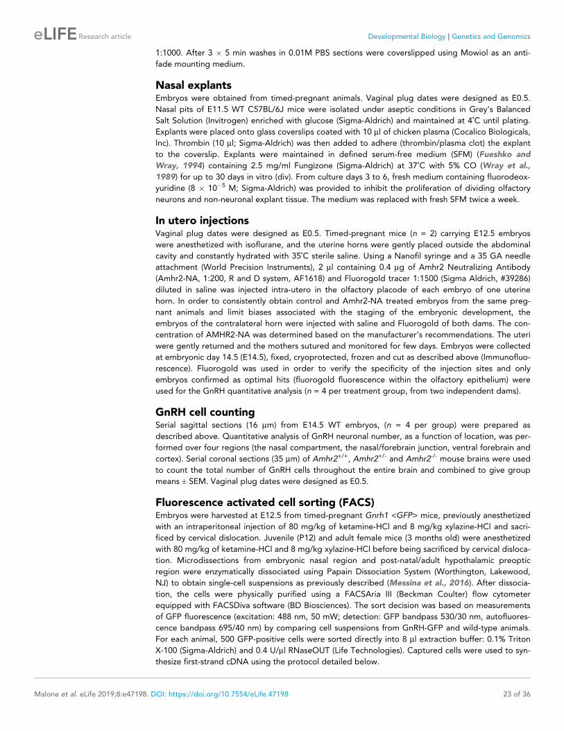
1:1000. After 3 � 5 min washes in 0.01M PBS sections were coverslipped using Mowiol as an anti-
fade mounting medium.
Nasal explantsEmbryos were obtained from timed-pregnant animals. Vaginal plug dates were designed as E0.5.
Nasal pits of E11.5 WT C57BL/6J mice were isolated under aseptic conditions in Grey’s Balanced
Salt Solution (Invitrogen) enriched with glucose (Sigma-Aldrich) and maintained at 4˚C until plating.
Explants were placed onto glass coverslips coated with 10 ml of chicken plasma (Cocalico Biologicals,
Inc). Thrombin (10 ml; Sigma-Aldrich) was then added to adhere (thrombin/plasma clot) the explant
to the coverslip. Explants were maintained in defined serum-free medium (SFM) (Fueshko and
Wray, 1994) containing 2.5 mg/ml Fungizone (Sigma-Aldrich) at 37˚C with 5% CO (Wray et al.,
1989) for up to 30 days in vitro (div). From culture days 3 to 6, fresh medium containing fluorodeox-
yuridine (8 � 10�5 M; Sigma-Aldrich) was provided to inhibit the proliferation of dividing olfactory
neurons and non-neuronal explant tissue. The medium was replaced with fresh SFM twice a week.
In utero injectionsVaginal plug dates were designed as E0.5. Timed-pregnant mice (n = 2) carrying E12.5 embryos
were anesthetized with isoflurane, and the uterine horns were gently placed outside the abdominal
cavity and constantly hydrated with 35˚C sterile saline. Using a Nanofil syringe and a 35 GA needle
attachment (World Precision Instruments), 2 ml containing 0.4 mg of Amhr2 Neutralizing Antibody
(Amhr2-NA, 1:200, R and D system, AF1618) and Fluorogold tracer 1:1500 (Sigma Aldrich, #39286)
diluted in saline was injected intra-utero in the olfactory placode of each embryo of one uterine
horn. In order to consistently obtain control and Amhr2-NA treated embryos from the same preg-
nant animals and limit biases associated with the staging of the embryonic development, the
embryos of the contralateral horn were injected with saline and Fluorogold of both dams. The con-
centration of AMHR2-NA was determined based on the manufacturer’s recommendations. The uteri
were gently returned and the mothers sutured and monitored for few days. Embryos were collected
at embryonic day 14.5 (E14.5), fixed, cryoprotected, frozen and cut as described above (Immunofluo-
rescence). Fluorogold was used in order to verify the specificity of the injection sites and only
embryos confirmed as optimal hits (fluorogold fluorescence within the olfactory epithelium) were
used for the GnRH quantitative analysis (n = 4 per treatment group, from two independent dams).
GnRH cell countingSerial sagittal sections (16 mm) from E14.5 WT embryos, (n = 4 per group) were prepared as
described above. Quantitative analysis of GnRH neuronal number, as a function of location, was per-
formed over four regions (the nasal compartment, the nasal/forebrain junction, ventral forebrain and
cortex). Serial coronal sections (35 mm) of Amhr2+/+, Amhr2+/- and Amhr2-/- mouse brains were used
to count the total number of GnRH cells throughout the entire brain and combined to give group
means ± SEM. Vaginal plug dates were designed as E0.5.
Fluorescence activated cell sorting (FACS)Embryos were harvested at E12.5 from timed-pregnant Gnrh1 <GFP> mice, previously anesthetized
with an intraperitoneal injection of 80 mg/kg of ketamine-HCl and 8 mg/kg xylazine-HCl and sacri-
ficed by cervical dislocation. Juvenile (P12) and adult female mice (3 months old) were anesthetized
with 80 mg/kg of ketamine-HCl and 8 mg/kg xylazine-HCl before being sacrificed by cervical disloca-
tion. Microdissections from embryonic nasal region and post-natal/adult hypothalamic preoptic
region were enzymatically dissociated using Papain Dissociation System (Worthington, Lakewood,
NJ) to obtain single-cell suspensions as previously described (Messina et al., 2016). After dissocia-
tion, the cells were physically purified using a FACSAria III (Beckman Coulter) flow cytometer
equipped with FACSDiva software (BD Biosciences). The sort decision was based on measurements
of GFP fluorescence (excitation: 488 nm, 50 mW; detection: GFP bandpass 530/30 nm, autofluores-
cence bandpass 695/40 nm) by comparing cell suspensions from GnRH-GFP and wild-type animals.
For each animal, 500 GFP-positive cells were sorted directly into 8 ml extraction buffer: 0.1% Triton
X-100 (Sigma-Aldrich) and 0.4 U/ml RNaseOUT (Life Technologies). Captured cells were used to syn-
thesize first-strand cDNA using the protocol detailed below.
Malone et al. eLife 2019;8:e47198. DOI: https://doi.org/10.7554/eLife.47198 23 of 36
Research article Developmental Biology Genetics and Genomics
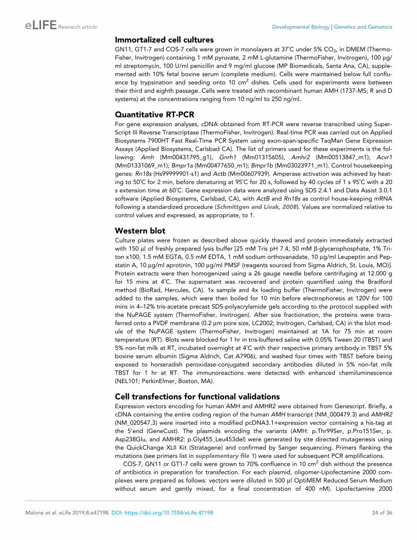
Immortalized cell culturesGN11, GT1-7 and COS-7 cells were grown in monolayers at 37˚C under 5% CO2, in DMEM (Thermo-
Fisher, Invitrogen) containing 1 mM pyruvate, 2 mM L-glutamine (ThermoFisher, Invitrogen), 100 mg/
ml streptomycin, 100 U/ml penicillin and 9 mg/ml glucose (MP Biomedicals, Santa Ana, CA), supple-
mented with 10% fetal bovine serum (complete medium). Cells were maintained below full conflu-
ence by trypsination and seeding onto 10 cm2 dishes. Cells used for experiments were between
their third and eighth passage. Cells were treated with recombinant human AMH (1737-MS; R and D
systems) at the concentrations ranging from 10 ng/ml to 250 ng/ml.
Quantitative RT-PCRFor gene expression analyses, cDNA obtained from RT-PCR were reverse transcribed using Super-
Script III Reverse Transcriptase (ThermoFisher, Invitrogen). Real-time PCR was carried out on Applied
Biosystems 7900HT Fast Real-Time PCR System using exon-span-specific TaqMan Gene Expression
Assays (Applied Biosystems, Carlsbad CA). The list of primers used for these experiments is the fol-
lowing: Amh (Mm00431795_g1), Gnrh1 (Mm01315605), Amhr2 (Mm00513847_m1); Acvr1
(Mm01331069_m1); Bmpr1a (Mm00477650_m1); Bmpr1b (Mm03023971_m1). Control housekeeping
genes: Rn18s (Hs99999901-s1) and Actb (Mm00607939). Amperase activation was achieved by heat-
ing to 50˚C for 2 min, before denaturing at 95˚C for 20 s, followed by 40 cycles of 1 s 95˚C with a 20
s extension time at 60˚C. Gene expression data were analyzed using SDS 2.4.1 and Data Assist 3.0.1
software (Applied Biosystems, Carlsbad, CA), with ActB and Rn18s as control house-keeping mRNA
following a standardized procedure (Schmittgen and Livak, 2008). Values are normalized relative to
control values and expressed, as appropriate, to 1.
Western blotCulture plates were frozen as described above quickly thawed and protein immediately extracted
with 150 ml of freshly prepared lysis buffer [25 mM Tris pH 7.4, 50 mM b-glycerophosphate, 1% Tri-
ton x100, 1.5 mM EGTA, 0.5 mM EDTA, 1 mM sodium orthovanadate, 10 mg/ml Leupeptin and Pep-
statin A, 10 mg/ml aprotinin, 100 mg/ml PMSF (reagents sourced from Sigma Aldrich, St. Louis, MO)].
Protein extracts were then homogenized using a 26 gauge needle before centrifuging at 12.000 g
for 15 mins at 4˚C. The supernatant was recovered and protein quantified using the Bradford
method (BioRad, Hercules, CA). 1x sample and 4x loading buffer (ThermoFisher, Invitrogen) were
added to the samples, which were then boiled for 10 min before electrophoresis at 120V for 100
mins in 4–12% tris-acetate precast SDS-polyacrylamide gels according to the protocol supplied with
the NuPAGE system (ThermoFisher, Invitrogen). After size fractionation, the proteins were trans-
ferred onto a PVDF membrane (0.2 mm pore size, LC2002; Invitrogen, Carlsbad, CA) in the blot mod-
ule of the NuPAGE system (ThermoFisher, Invitrogen) maintained at 1A for 75 min at room
temperature (RT). Blots were blocked for 1 hr in tris-buffered saline with 0.05% Tween 20 (TBST) and
5% non-fat milk at RT, incubated overnight at 4˚C with their respective primary antibody in TBST 5%
bovine serum albumin (Sigma Aldrich, Cat A7906), and washed four times with TBST before being
exposed to horseradish peroxidase-conjugated secondary antibodies diluted in 5% non-fat milk
TBST for 1 hr at RT. The immunoreactions were detected with enhanced chemiluminescence
(NEL101; PerkinElmer, Boston, MA).
Cell transfections for functional validationsExpression vectors encoding for human AMH and AMHR2 were obtained from Genescript. Briefly, a
cDNA containing the entire coding region of the human AMH transcript (NM_000479.3) and AMHR2
(NM_020547.3) were inserted into a modified pcDNA3.1+expression vector containing a his-tag at
the 5’end (GeneCust). The plasmids encoding the variants (AMH: p.Thr99Ser, p.Pro151Ser, p.
Asp238Glu, and AMHR2: p.Gly455_Leu453del) were generated by site directed mutagenesis using
the QuickChange XLII Kit (Stratagene) and confirmed by Sanger sequencing. Primers flanking the
mutations (see primers list in supplementary file 1) were used for subsequent PCR amplifications.
COS-7, GN11 or GT1-7 cells were grown to 70% confluence in 10 cm2 dish without the presence
of antibiotics in preparation for transfection. For each plasmid, oligomer-Lipofectamine 2000 com-
plexes were prepared as follows: vectors were diluted in 500 ml OptiMEM Reduced Serum Medium
without serum and gently mixed, for a final concentration of 400 nM). Lipofectamine 2000
Malone et al. eLife 2019;8:e47198. DOI: https://doi.org/10.7554/eLife.47198 24 of 36
Research article Developmental Biology Genetics and Genomics
(ThermoFisher, Invitrogen) was mixed gently before use, then diluted 10 ml in 500 ml OptiMEM (Ther-
moFisher, Invitrogen). Tubes were gently mixed and incubated for 5 min at room temperature. After
the 5-min incubation, the diluted vector was combined with the diluted Lipofectamine 2000 and
incubated for 20 min at room temperature. During the incubation, cells were trypsinized and dissoci-
ated, then re-suspended in the lipofectamine containing vector mixture. Cells were then incubated
at 37˚C in a 5% CO2 incubator for 48 hr, changing the medium to OptiMEM supplemented with 5%
fetal bovine serum after 6 hr. Conditioned media was collected for AMH or GnRH quantitation and
transfected cells used for either western blotting or transwell migration assays.
AMH quantificationAMH levels in conditioned media were measured by an automatic chemoluminescent immunoassay
on a Dxi system (Beckman Coulter, France) after a 1/10 dilution in the Sample Diluent A. This assay
detects proAMH and the cleaved AMHN,C complex. The limit of quantification of the assay is 0.57
pmol/L with an intra- and inter-assay imprecision less than 5%.
Determination of GnRH secretionGT1-7 cells were transiently transfected in OptiMem with either AMH WT or p.Thr99Ser hAMH var-
iants. 48 hr later, the medium was collected and frozen until EIA measurement. In another set of
experiments, GT1-7 cells were transiently transfected with either AMHR2 WT or with the AMHR2
CHH variants. 48 hr later, the cells were treated for 4 hr with either PBS or recombinant AMH (1737-
MS; R and D systems, 50 ng/ml). Finally, the medium was frozen until EIA measurement. The col-
lected media from these experiments were analyzed for GnRH content following a GnRH EIA proto-
col (EK-040-02CE, Phoenix Pharmaceuticals Inc, CA).
Transwell migration assayTranswell chambers were used according to manufacturer’s instructions (Falcon). In brief, GN11 cells
grown in complete medium until sub-confluence were harvested and re-suspended at a density of 1
� 105 cells/ml in SFM. Cells were seeded on the upper side of 8 mm pore membranes and incubated
for 12 hr with SFM, human recombinant AMH (1737-MS; R and D systems, 50–250 ng/ml) or with
DMEM supplemented with 10% fetal bovine serum. Each factor (serum, AMH, inhibitors and anti-
bodies) was placed on the upper and lower chamber of the transwell. GN11 cells were incubated in
the presence of recombinant AMH (50 ng/ml) together with MAPK inhibitor (UO126; Calbiochem) at
a concentration of 10 mM, as previously described (Balland et al., 2014). In another set of experi-
ments, GN11 cells were treated for 12 hr with Amhr2-NA (R and D system, AF1618), at the same
concentration (1:200) used for the in utero injections experiments (Figure 2), in the presence or
absence of recombinant AMH (50 ng/ml). Cells on the upper side of the filters were mechanically
removed and cells on the lower side fixed in 4% PFA for 30 min before nuclei labelling with DAPI.
Four non-overlapping regions were imaged per membrane using a Zeiss 20x objective (N.A. 0.8)
mounted on a Axio Imager Z2 light microscope (Zeiss), with nuclei counted using an ImageJ plugin
(National Institute of Health, Bethseda) and averaged to produce an average per well. n for each
experiment is detailed in the figure legends.
siRNA transfectionsGN11 cells were grown to 70% confluence in 10 cm2 dish without the presence of antibiotics in
preparation for transfection. For siRNA experiments, GN11 cells were transiently transfected with 75
nM SMARTpool siRNA targeting mouse Amhr2, Acvr1, Bmpr1a, Bmpr1b, or 75 nM nontargeting
SMARTpool siRNA (siRNA NT) as negative control (Dharmacon, Horizon Discovery LTD, Cambridge,
UK). Gene knockdown was assessed by quantitative PCR.
Ovarian histologyOvaries were collected from 6-month-old mice, immersion-fixed in 4% PFA solution and stored at 4˚
C. Paraffin-embedded ovaries were sectioned at a thickness of 5 mm (histology facility, University of
Lille 2, France) and stained with hematoxylin-eosin (Sigma Aldrich, Cat # GHS132, HT1103128). Sec-
tions were examined throughout the ovary. Corpora lutea (CL) were classified and quantified as pre-
viously reported (Caldwell et al., 2017). To avoid repetitive counting, CL were counted every 100
Malone et al. eLife 2019;8:e47198. DOI: https://doi.org/10.7554/eLife.47198 25 of 36
Research article Developmental Biology Genetics and Genomics
mm by comparing the section with the preceding and following sections. CL were characterized by a
still present central cavity, filled with blood and follicular fluid remnants or by prominent polyhedral
to round luteal cells.
Pulsatile LH measurementMice were habituated with daily handling for 3–4 weeks. Blood samples (5 ml) were taken from the
tail in 10 min intervals for 2 hr (between 12h00 hours and 15h00 hours), diluted in PBS-Tween and
immediately frozen. LH levels were determined by sandwich ELISA (Steyn et al., 2013). A 96-well
high-affinity binding microplate (9018; Corning) was coated with 50 ml of capture antibody (monoclo-
nal antibody, anti-bovine LH beta subunit, 518B7; University of California) at a final dilution of
1:1000 (in 1 x PBS, 1.09 g of Na2HPO4 (anhydrous), 0.32 g of NaH2PO4 (anhydrous) and 9 g of
NaCl in 1000 ml of distilled water) and incubated overnight at 4˚ C. Wells were incubated with 200
ml of blocking buffer (5% (w/v) skim milk powder in 1 x PBS-T (1 x PBS with 0.05% Tween 20) for 2
hr at room temperature. A standard curve was generated using a twofold serial dilution of mLH (ref-
erence preparation, AFP-5306A; National Institute of Diabetes and Digestive and Kidney Diseases -
National Hormone and Pituitary Program (NIDDK-NHPP)) in 0.2% (w/v) bovine serum albumin 1 x
PBS-T. The LH standards and blood samples were incubated with 50 ml of detection antibody (poly-
clonal antibody, rabbit LH antiserum, AFP240580Rb; NIDDK-NHPP) at a final dilution of 1:10,000 for
1.5 hr (at RT). Each well containing bound substrate was incubated with 50 ml of horseradish peroxi-
dase-conjugated antibody (polyclonal goat anti-rabbit; Vector) at a final dilution of 1:10,000. After a
1.5 hr incubation, 100 ml of o-phenylenediamine (002003; Invitrogen), substrate containing 0.1%
H2O2 was added to each well and left at RT for 30 min. The reaction was stopped by the addition of
50 ml of 3M HCl to each well, and absorbance of each well was read at a wavelength of 490 nm.
Pulses were confirmed using DynPeak (Vidal et al., 2012).
Fertility testThe reproductive competency of these animals was determined by pairing the following mice:
Amhr2+/+ males mated with Amhr2+/+ females, Amhr2+/- females, or with Amhr2-/- females, or
inversely, for a period of 3 months. One male and one female were housed in each cage during the
constant breeding protocol. Each litter was sacrificed at birth to allow the dams to re-enter estrous
cyclicity within a few days. Number of pups/litter, fertility index (number of litters per female per
month, averaged during the 3 months), and time to first litter (number of days to first litter after pair-
ing) were quantified per pairing.
iDISCOExperiments were performed as previously described (Renier et al., 2014) and detailed below.
Sample pre-treatment with methanolSamples were washed in PBS (twice for 1 hr), followed by 50% methanol in PBS (once for 1 hr), 80%
methanol (once for 1 hr) and 100% methanol (twice for 1 hr). Next, samples were bleached in 5%
H2O2 in 20% DMSO/methanol (2 ml 30% H2O2/2 ml DMSO/8 ml methanol, ice cold) at 4˚C over-
night. Next, samples were washed in methanol (twice for 1 hr), in 20% DMSO/methanol (twice for 1
hr), 80% methanol (once for 1 hr), 50% methanol (once for 1 hr), PBS (twice for 1 hr), and finally,
PBS/0.2% TritonX-100 (twice for 1 hr) before proceeding to the staining procedures.
Whole-mount immunostainingSamples were incubated at 37˚C on an adjustable rotator in 10 ml of a blocking solution (PBSGNaT)
of 1X PBS containing 0.2% gelatin (Sigma), 0.5% Triton X-100 (Sigma-Aldrich) and 0.01% NaAzide
for three nights. Samples were transferred to 10 ml of PBSGNaT containing primary antibodies
(Table S1) and placed at 37˚C in rotation for 7 days This was followed by six washes of 30 min in
PBSGT at RT and a final wash in PBSGT overnight at 4˚C. Next, samples were incubated in secondary
antibodies (1:400, Alexa 568, Alexa 647) diluted in 10 ml PBSGNaT for 2 days at 37˚C in a rotating
tube. After six 30 min washes in PBS at room temperature, the samples were stored in PBS at 4˚C in
the dark until clearing.
Malone et al. eLife 2019;8:e47198. DOI: https://doi.org/10.7554/eLife.47198 26 of 36
Research article Developmental Biology Genetics and Genomics
Tissue clearingAll incubation steps were performed at RT in a fume hood, on a tube rotator at 14 rpm covered with
aluminium foil to avoid contact with light. Samples were dehydrated in a graded series (50%, 80%,
and 100%) of tetrahydrofurane (THF; anhydrous, containing 250 ppm butylated hydroxytoluene
inhibitor, Sigma-Aldrich) diluted in H2O as follow: 1) 50% THF overnight at RT; 2) 80% THF 1 hr at
RT; 3) 100% THF 1h30 at RT; 4) 100% THF 1h30 at RT. This was followed by a delipidation step of
30–40 min in 100% dichloromethane (DCM; Sigma-Aldrich). Samples were cleared in dibenzylether
(DBE; Sigma-Aldrich) for 2 hr at RT on constant agitation and in the dark. Finally, samples were
moved into fresh DBE and stored in glass tube in the dark and at RT until imaging. We could image
samples, as described below, without any significant fluorescence loss for up to 6 months.
Imaging3D imaging was performed as previously described (Belle et al., 2014). An ultramicroscope (LaVision
BioTec) using ImspectorPro software (LaVision BioTec) was used to perform imaging. The light sheet
was generated by a laser (wavelength 488 or 561 nm, Coherent Sapphire Laser, LaVision BioTec)
and two cylindrical lenses. A binocular stereomicroscope (MXV10, Olympus) with a 2 � objective
(MVPLAPO, Olympus) was used at different magnifications (1.6�, 4�, 5�, and 6.3�). Samples were
placed in an imaging reservoir made of 100% quartz (LaVision BioTec) filled with DBE and illumi-
nated from the side by the laser light. A PCO Edge SCMOS CCD camera (2560 � 2160 pixel size,
LaVision BioTec) was used to acquire images. The step size between each image was fixed at 2 mm.
Image analysisFor confocal observations and analyses, an inverted laser scanning Axio observer microscope (LSM
710, Zeiss, Oberkochen, Germany) with EC Plan NeoFluor 10�/0.3 NA, 20�/0.5 NA and 40�/1.3
NA (Zeiss, Oberkochen, Germany) objectives were used (Imaging Core Facility of IFR114 of the Uni-
versity of Lille, France).
Images, 3D volume, and movies were generated using Imaris x64 software (version 7.6.1, Bit-
plane). Stack images were first converted to imaris file (.ims) using ImarisFileConverter and 3D recon-
truction was performed using ‘volume rendering’. Optical slices of samples were obtained using the
‘orthoslicer’ tools. The surface of the samples was created using the ‘surface’ tool by creating a
mask around each volume. 3D pictures were generated using the ‘snapshot’ tool. ImageJ (National
Institute of Health, Bethesda) and Photoshop CS6 (Adobe Systems, San Jose, CA) were used to pro-
cess, adjust and merge the photomontages. Figures were prepared using Adobe Photoshop and
Abode Illustrator CS6.
Human CHH subjectsThe CHH cohort included 180 probands (105 KS and 75 nCHH). The majority of the patients were
male (n = 127). The diagnosis of CHH was made on the basis of: i) absent or incomplete puberty by
17 years of age; ii) low/normal gonadotropin levels in the setting of low serum testosterone/estradiol
levels; and iii) otherwise normal anterior pituitary function and normal imaging of the hypothalamic-
pituitary area (Pitteloud et al., 2002). Olfaction was assessed by self-report and/or formal testing
(Lewkowitz-Shpuntoff et al., 2012). When available, family members were included for genetic
studies. This study was approved by the ethics committee of the University of Lausanne, and all par-
ticipants provided written informed consent prior to study participation.
Human case summariesFamily # 1, Patient II-1AMH p.Thr99Ser (heterozygous)The caucasian male proband consulted our clinic at age 32 for symptomatic hypogonadism accom-
panied by mild anemia and oligospermia. He was previously diagnosed with delayed puberty at age
17 but was never offered testosterone replacement nor was he followed up to ensure pubertal com-
pletion. Physical examination revealed partial but incomplete puberty (testicular volume 12 ml bilat-
erally, pubic hair Tanner IV) as well as eunuchoid proportions (arm span 184 cm for height of 176
cm). Targeted clinical exam detected slight hyperlaxity, high-arched palate and pectum excavatum.
Blood tests confirmed a hypogonadotropic hypogonadism (testosterone 7.5 nmol/l, LH 2.7 U/l, FSH
Malone et al. eLife 2019;8:e47198. DOI: https://doi.org/10.7554/eLife.47198 27 of 36
Research article Developmental Biology Genetics and Genomics
3.6 U/l) without dysfunction of other pituitary axes. Formal olfactory testing confirmed normal sense
of smell. Pituitary MRI and GnRH-stimulation test were normal. Bone density scan typically detected
osteopenia at the lumbar spine. Family history included delayed puberty and growth in his father
while mother’s puberty was normal (menarche at 11 years old). The proband harbors a heterozygous
mutation in AMH inherited by his father who as stated above has a partial phenotype (delayed
puberty). He also harbors a GNRHR variant, which was not considered as pathogenic or likely patho-
genic by our filtering process, due to its presence only at heterozygous state for a gene with autoso-
mal recessive transmission mode. Given his request for fertility, HCG treatment was introduced,
allowing for increase of testosterone and stimulation of spermatogenesis.
Family # 2, Patient II-1AMH p.Pro151Ser (heterozygous)This anosmic male proband was diagnosed at 2 years old for unilateral cryptorchidism, treated by
left orchidopexy. Subsequent follow up showed no signs of puberty at 17.5 years with prepubertal
testes (volume 2 ml bilaterally). Serum testosterone was low while gonadotropines were undetect-
able (LH/FSH < 1.0 U/L). Formal smell testing confirmed anosmia (UPSIT: 11/40, <5th %ile) and he
was diagnosed with Kallmann syndrome. Intramuscular injections of testosterone were initiated with
good effect on virilization. A cranial MRI showed normal pituitary size and absent olfactory bulbs.
Family history was negative for pubertal delay. Parents refused to participate in the genetic study
and undergo smell testing. The patient harbors a rare mutation in AMH with no changes in known
CHH genes. A pelvic MRI was performed in July 2017 and showed no argument in favor of PMDS.
Family # 3, Patient II-2AMG p.Asp238Gln (heterozygous)The female proband originated from Kazakhstan consulted us at age of 27.9 years for infertility. She
presented with primary amenorrhea at age 17. She described onset of breast development at 13
years, which rapidly stalled (Tanner II-III). She was placed on estrogen-progesterone replacement
(estradiol/dihydrogesterone) at age 18. She remained amenorrheic during multiple treatment
pauses. When assessed in our clinic and several months after withdrawal of estrogen pills, hypogona-
dotropic hypogonadism was confirmed (estradiol 0.05 nmol/l, LH 0.5 U/l, FSH 1.4 U/l) with otherwise
normal pituitary function and adequate response to GnRH stimulation (LH baseline 0.4 U/l, peak 6.3
U/l; FSH baseline 1.2 U/l, peak 6.4 U/l). A 10 hr frequent sampling did not detect any LH pulses.
Olfactory assessment by 12-item Sniffin’ Sticks revealed hyposmia (12/16). Her physical status was
notable for eunuchoid proportions and moderate scoliosis without dysmorphic features. Cranial MRI
showed a small pituitary gland, while bone density scan indicated osteoporosis. Polycystic ovaries
syndrome was excluded as well as a non-classic congenital adrenal hyperplasia. We concluded to KS
diagnosis with partial GnRH deficiency and resumed estrogen/dihydrogesterone treatment. Family
history included delayed puberty in the father (first shaving at age 18, continued growing until age
24). The latter was also anosmic, while the mother exhibited normal puberty (menarche at 13 years
old) and olfactive function. The patient harbors an AMH mutation, inherited by the partially affected
father. No changes in known CHH genes were seen.
Family # 4, Patient II-1AMHR2 p.Gly445_Leu453del (heterozygous)The female Caucasian patient presented with primary amenorrhea and absent pubertal development
at age 17. She remained amenorrheic until age 22, and then was offered oral contraceptives (estra-
diol, norgestrel). Endocrinology assessment in her native country (Serbia) led to diagnosis of hypogo-
nadotropic hypogonadism at age 33 (estradiol <0.04 nmol/l, LH <2.0 U/l, FSH 0.1 U/l) with
otherwise normal pituitary function, assessed by an insulin tolerance test. Similarly, LHRH stimulation
test showed adequate pituitary response (LH baseline 2.0 U/l, peak 6.5 U/l; FSH baseline 1.8 U/l,
peak 6.3 U/l). Pituitary MRI was normal as well as formal smell test (Sniffin’ Sticks 14/16, > 25th %ile).
The patient consulted us to discuss ovulation induction by pulsatile GnRH treatment. After with-
drawal of estrogen pills, hypogonadotropic hypogonadism was confirmed (estradiol <0.04 nmol/l,
LH 0.4 U/l, FSH 1.4 U/l). Physical exam did not show any associated phenotypes. Family history was
unremarkable for pubertal timing but her mother exhibited history of cleft lip/palate, corrected
Malone et al. eLife 2019;8:e47198. DOI: https://doi.org/10.7554/eLife.47198 28 of 36
Research article Developmental Biology Genetics and Genomics
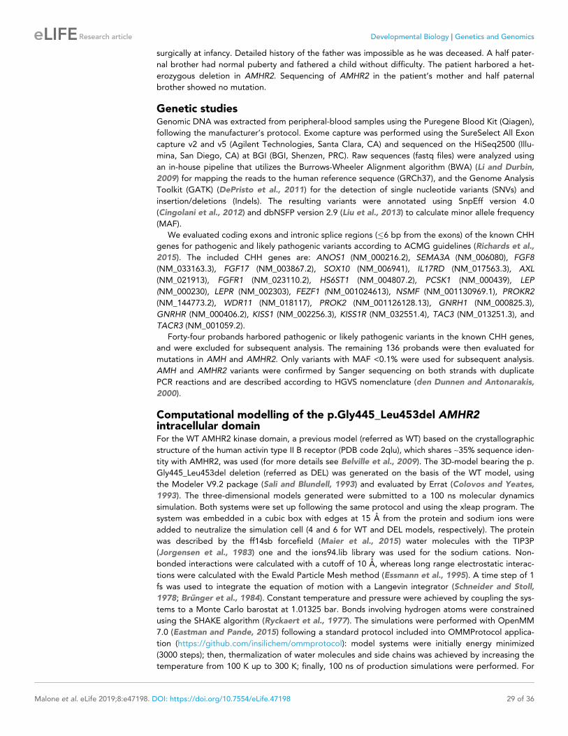
surgically at infancy. Detailed history of the father was impossible as he was deceased. A half pater-
nal brother had normal puberty and fathered a child without difficulty. The patient harbored a het-
erozygous deletion in AMHR2. Sequencing of AMHR2 in the patient’s mother and half paternal
brother showed no mutation.
Genetic studiesGenomic DNA was extracted from peripheral-blood samples using the Puregene Blood Kit (Qiagen),
following the manufacturer’s protocol. Exome capture was performed using the SureSelect All Exon
capture v2 and v5 (Agilent Technologies, Santa Clara, CA) and sequenced on the HiSeq2500 (Illu-
mina, San Diego, CA) at BGI (BGI, Shenzen, PRC). Raw sequences (fastq files) were analyzed using
an in-house pipeline that utilizes the Burrows-Wheeler Alignment algorithm (BWA) (Li and Durbin,
2009) for mapping the reads to the human reference sequence (GRCh37), and the Genome Analysis
Toolkit (GATK) (DePristo et al., 2011) for the detection of single nucleotide variants (SNVs) and
insertion/deletions (Indels). The resulting variants were annotated using SnpEff version 4.0
(Cingolani et al., 2012) and dbNSFP version 2.9 (Liu et al., 2013) to calculate minor allele frequency
(MAF).
We evaluated coding exons and intronic splice regions (�6 bp from the exons) of the known CHH
genes for pathogenic and likely pathogenic variants according to ACMG guidelines (Richards et al.,
2015). The included CHH genes are: ANOS1 (NM_000216.2), SEMA3A (NM_006080), FGF8
(NM_033163.3), FGF17 (NM_003867.2), SOX10 (NM_006941), IL17RD (NM_017563.3), AXL
(NM_021913), FGFR1 (NM_023110.2), HS6ST1 (NM_004807.2), PCSK1 (NM_000439), LEP
(NM_000230), LEPR (NM_002303), FEZF1 (NM_001024613), NSMF (NM_001130969.1), PROKR2
(NM_144773.2), WDR11 (NM_018117), PROK2 (NM_001126128.13), GNRH1 (NM_000825.3),
GNRHR (NM_000406.2), KISS1 (NM_002256.3), KISS1R (NM_032551.4), TAC3 (NM_013251.3), and
TACR3 (NM_001059.2).
Forty-four probands harbored pathogenic or likely pathogenic variants in the known CHH genes,
and were excluded for subsequent analysis. The remaining 136 probands were then evaluated for
mutations in AMH and AMHR2. Only variants with MAF <0.1% were used for subsequent analysis.
AMH and AMHR2 variants were confirmed by Sanger sequencing on both strands with duplicate
PCR reactions and are described according to HGVS nomenclature (den Dunnen and Antonarakis,
2000).
Computational modelling of the p.Gly445_Leu453del AMHR2intracellular domainFor the WT AMHR2 kinase domain, a previous model (referred as WT) based on the crystallographic
structure of the human activin type II B receptor (PDB code 2qlu), which shares ~35% sequence iden-
tity with AMHR2, was used (for more details see Belville et al., 2009). The 3D-model bearing the p.
Gly445_Leu453del deletion (referred as DEL) was generated on the basis of the WT model, using
the Modeler V9.2 package (Sali and Blundell, 1993) and evaluated by Errat (Colovos and Yeates,
1993). The three-dimensional models generated were submitted to a 100 ns molecular dynamics
simulation. Both systems were set up following the same protocol and using the xleap program. The
system was embedded in a cubic box with edges at 15 A from the protein and sodium ions were
added to neutralize the simulation cell (4 and 6 for WT and DEL models, respectively). The protein
was described by the ff14sb forcefield (Maier et al., 2015) water molecules with the TIP3P
(Jorgensen et al., 1983) one and the ions94.lib library was used for the sodium cations. Non-
bonded interactions were calculated with a cutoff of 10 A, whereas long range electrostatic interac-
tions were calculated with the Ewald Particle Mesh method (Essmann et al., 1995). A time step of 1
fs was used to integrate the equation of motion with a Langevin integrator (Schneider and Stoll,
1978; Brunger et al., 1984). Constant temperature and pressure were achieved by coupling the sys-
tems to a Monte Carlo barostat at 1.01325 bar. Bonds involving hydrogen atoms were constrained
using the SHAKE algorithm (Ryckaert et al., 1977). The simulations were performed with OpenMM
7.0 (Eastman and Pande, 2015) following a standard protocol included into OMMProtocol applica-
tion (https://github.com/insilichem/ommprotocol): model systems were initially energy minimized
(3000 steps); then, thermalization of water molecules and side chains was achieved by increasing the
temperature from 100 K up to 300 K; finally, 100 ns of production simulations were performed. For
Malone et al. eLife 2019;8:e47198. DOI: https://doi.org/10.7554/eLife.47198 29 of 36
Research article Developmental Biology Genetics and Genomics
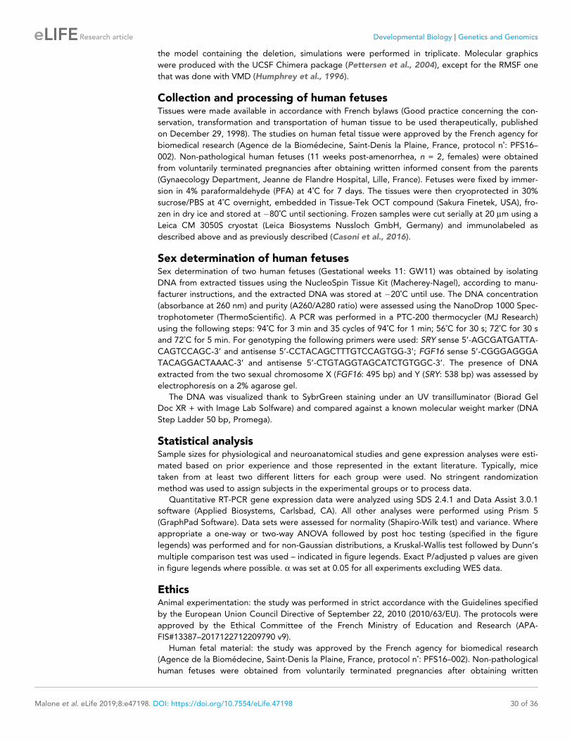
the model containing the deletion, simulations were performed in triplicate. Molecular graphics
were produced with the UCSF Chimera package (Pettersen et al., 2004), except for the RMSF one
that was done with VMD (Humphrey et al., 1996).
Collection and processing of human fetusesTissues were made available in accordance with French bylaws (Good practice concerning the con-
servation, transformation and transportation of human tissue to be used therapeutically, published
on December 29, 1998). The studies on human fetal tissue were approved by the French agency for
biomedical research (Agence de la Biomedecine, Saint-Denis la Plaine, France, protocol n˚: PFS16–
002). Non-pathological human fetuses (11 weeks post-amenorrhea, n = 2, females) were obtained
from voluntarily terminated pregnancies after obtaining written informed consent from the parents
(Gynaecology Department, Jeanne de Flandre Hospital, Lille, France). Fetuses were fixed by immer-
sion in 4% paraformaldehyde (PFA) at 4˚C for 7 days. The tissues were then cryoprotected in 30%
sucrose/PBS at 4˚C overnight, embedded in Tissue-Tek OCT compound (Sakura Finetek, USA), fro-
zen in dry ice and stored at �80˚C until sectioning. Frozen samples were cut serially at 20 mm using a
Leica CM 3050S cryostat (Leica Biosystems Nussloch GmbH, Germany) and immunolabeled as
described above and as previously described (Casoni et al., 2016).
Sex determination of human fetusesSex determination of two human fetuses (Gestational weeks 11: GW11) was obtained by isolating
DNA from extracted tissues using the NucleoSpin Tissue Kit (Macherey-Nagel), according to manu-
facturer instructions, and the extracted DNA was stored at �20˚C until use. The DNA concentration
(absorbance at 260 nm) and purity (A260/A280 ratio) were assessed using the NanoDrop 1000 Spec-
trophotometer (ThermoScientific). A PCR was performed in a PTC-200 thermocycler (MJ Research)
using the following steps: 94˚C for 3 min and 35 cycles of 94˚C for 1 min; 56˚C for 30 s; 72˚C for 30 s
and 72˚C for 5 min. For genotyping the following primers were used: SRY sense 5’-AGCGATGATTA-
CAGTCCAGC-3’ and antisense 5’-CCTACAGCTTTGTCCAGTGG-3’; FGF16 sense 5’-CGGGAGGGA
TACAGGACTAAAC-3’ and antisense 5’-CTGTAGGTAGCATCTGTGGC-3’. The presence of DNA
extracted from the two sexual chromosome X (FGF16: 495 bp) and Y (SRY: 538 bp) was assessed by
electrophoresis on a 2% agarose gel.
The DNA was visualized thank to SybrGreen staining under an UV transilluminator (Biorad Gel
Doc XR + with Image Lab Solfware) and compared against a known molecular weight marker (DNA
Step Ladder 50 bp, Promega).
Statistical analysisSample sizes for physiological and neuroanatomical studies and gene expression analyses were esti-
mated based on prior experience and those represented in the extant literature. Typically, mice
taken from at least two different litters for each group were used. No stringent randomization
method was used to assign subjects in the experimental groups or to process data.
Quantitative RT-PCR gene expression data were analyzed using SDS 2.4.1 and Data Assist 3.0.1
software (Applied Biosystems, Carlsbad, CA). All other analyses were performed using Prism 5
(GraphPad Software). Data sets were assessed for normality (Shapiro-Wilk test) and variance. Where
appropriate a one-way or two-way ANOVA followed by post hoc testing (specified in the figure
legends) was performed and for non-Gaussian distributions, a Kruskal-Wallis test followed by Dunn’s
multiple comparison test was used – indicated in figure legends. Exact P/adjusted p values are given
in figure legends where possible. a was set at 0.05 for all experiments excluding WES data.
EthicsAnimal experimentation: the study was performed in strict accordance with the Guidelines specified
by the European Union Council Directive of September 22, 2010 (2010/63/EU). The protocols were
approved by the Ethical Committee of the French Ministry of Education and Research (APA-
FIS#13387–2017122712209790 v9).
Human fetal material: the study was approved by the French agency for biomedical research
(Agence de la Biomedecine, Saint-Denis la Plaine, France, protocol n˚: PFS16–002). Non-pathological
human fetuses were obtained from voluntarily terminated pregnancies after obtaining written
Malone et al. eLife 2019;8:e47198. DOI: https://doi.org/10.7554/eLife.47198 30 of 36
Research article Developmental Biology Genetics and Genomics
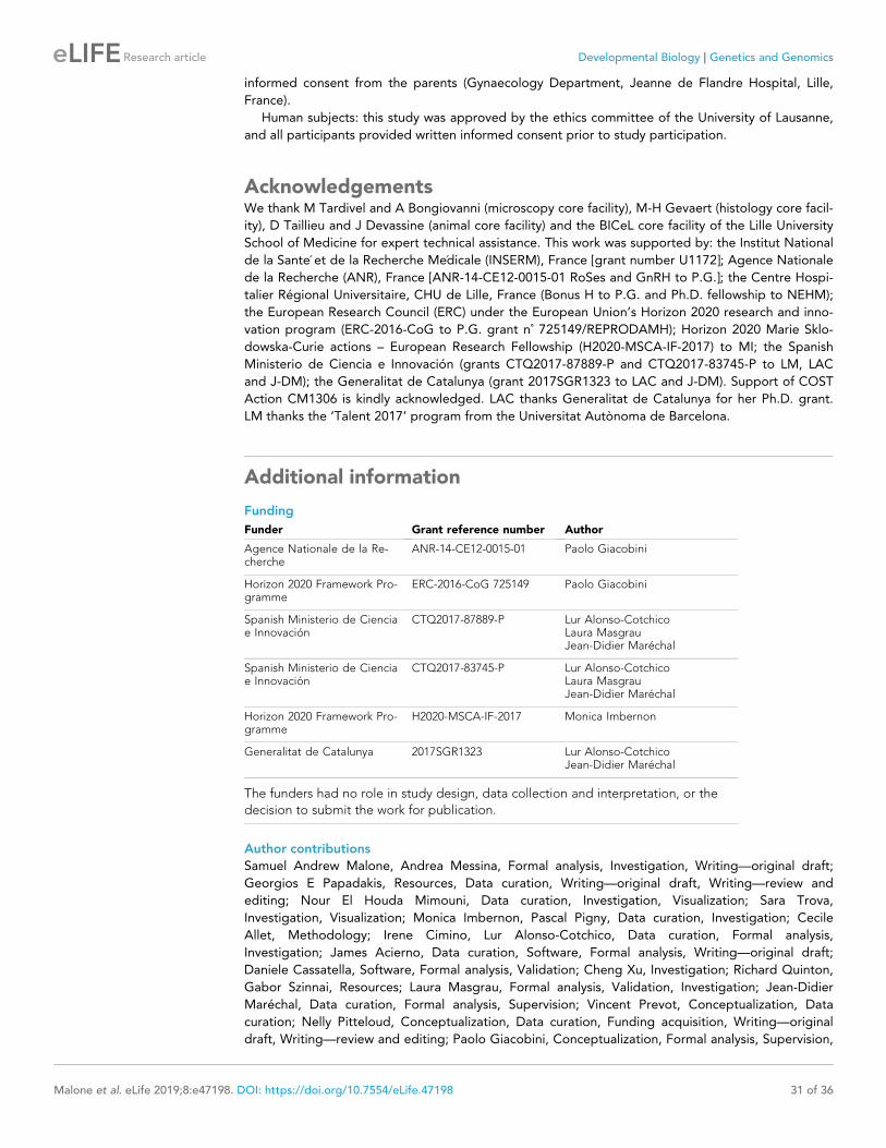
informed consent from the parents (Gynaecology Department, Jeanne de Flandre Hospital, Lille,
France).
Human subjects: this study was approved by the ethics committee of the University of Lausanne,
and all participants provided written informed consent prior to study participation.
AcknowledgementsWe thank M Tardivel and A Bongiovanni (microscopy core facility), M-H Gevaert (histology core facil-
ity), D Taillieu and J Devassine (animal core facility) and the BICeL core facility of the Lille University
School of Medicine for expert technical assistance. This work was supported by: the Institut National
de la Sante�et de la Recherche Me�dicale (INSERM), France [grant number U1172]; Agence Nationale
de la Recherche (ANR), France [ANR-14-CE12-0015-01 RoSes and GnRH to P.G.]; the Centre Hospi-
talier Regional Universitaire, CHU de Lille, France (Bonus H to P.G. and Ph.D. fellowship to NEHM);
the European Research Council (ERC) under the European Union’s Horizon 2020 research and inno-
vation program (ERC-2016-CoG to P.G. grant n˚ 725149/REPRODAMH); Horizon 2020 Marie Sklo-
dowska-Curie actions – European Research Fellowship (H2020-MSCA-IF-2017) to MI; the Spanish
Ministerio de Ciencia e Innovacion (grants CTQ2017-87889-P and CTQ2017-83745-P to LM, LAC
and J-DM); the Generalitat de Catalunya (grant 2017SGR1323 to LAC and J-DM). Support of COST
Action CM1306 is kindly acknowledged. LAC thanks Generalitat de Catalunya for her Ph.D. grant.
LM thanks the ‘Talent 2017’ program from the Universitat Autonoma de Barcelona.
Additional information
Funding
Funder Grant reference number Author
Agence Nationale de la Re-cherche
ANR-14-CE12-0015-01 Paolo Giacobini
Horizon 2020 Framework Pro-gramme
ERC-2016-CoG 725149 Paolo Giacobini
Spanish Ministerio de Cienciae Innovacion
CTQ2017-87889-P Lur Alonso-CotchicoLaura MasgrauJean-Didier Marechal
Spanish Ministerio de Cienciae Innovacion
CTQ2017-83745-P Lur Alonso-CotchicoLaura MasgrauJean-Didier Marechal
Horizon 2020 Framework Pro-gramme
H2020-MSCA-IF-2017 Monica Imbernon
Generalitat de Catalunya 2017SGR1323 Lur Alonso-CotchicoJean-Didier Marechal
The funders had no role in study design, data collection and interpretation, or the
decision to submit the work for publication.
Author contributions
Samuel Andrew Malone, Andrea Messina, Formal analysis, Investigation, Writing—original draft;
Georgios E Papadakis, Resources, Data curation, Writing—original draft, Writing—review and
editing; Nour El Houda Mimouni, Data curation, Investigation, Visualization; Sara Trova,
Investigation, Visualization; Monica Imbernon, Pascal Pigny, Data curation, Investigation; Cecile
Allet, Methodology; Irene Cimino, Lur Alonso-Cotchico, Data curation, Formal analysis,
Investigation; James Acierno, Data curation, Software, Formal analysis, Writing—original draft;
Daniele Cassatella, Software, Formal analysis, Validation; Cheng Xu, Investigation; Richard Quinton,
Gabor Szinnai, Resources; Laura Masgrau, Formal analysis, Validation, Investigation; Jean-Didier
Marechal, Data curation, Formal analysis, Supervision; Vincent Prevot, Conceptualization, Data
curation; Nelly Pitteloud, Conceptualization, Data curation, Funding acquisition, Writing—original
draft, Writing—review and editing; Paolo Giacobini, Conceptualization, Formal analysis, Supervision,
Malone et al. eLife 2019;8:e47198. DOI: https://doi.org/10.7554/eLife.47198 31 of 36
Research article Developmental Biology Genetics and Genomics
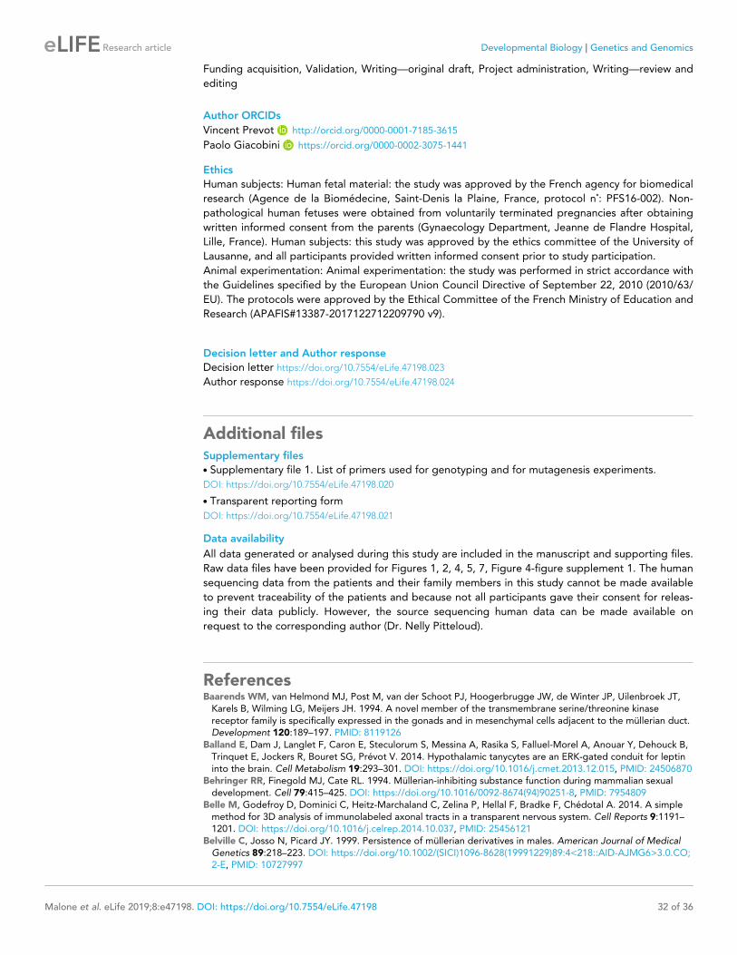
Funding acquisition, Validation, Writing—original draft, Project administration, Writing—review and
editing
Author ORCIDs
Vincent Prevot http://orcid.org/0000-0001-7185-3615
Paolo Giacobini https://orcid.org/0000-0002-3075-1441
Ethics
Human subjects: Human fetal material: the study was approved by the French agency for biomedical
research (Agence de la Biomedecine, Saint-Denis la Plaine, France, protocol n˚: PFS16-002). Non-
pathological human fetuses were obtained from voluntarily terminated pregnancies after obtaining
written informed consent from the parents (Gynaecology Department, Jeanne de Flandre Hospital,
Lille, France). Human subjects: this study was approved by the ethics committee of the University of
Lausanne, and all participants provided written informed consent prior to study participation.
Animal experimentation: Animal experimentation: the study was performed in strict accordance with
the Guidelines specified by the European Union Council Directive of September 22, 2010 (2010/63/
EU). The protocols were approved by the Ethical Committee of the French Ministry of Education and
Research (APAFIS#13387-2017122712209790 v9).
Decision letter and Author response
Decision letter https://doi.org/10.7554/eLife.47198.023
Author response https://doi.org/10.7554/eLife.47198.024
Additional filesSupplementary files. Supplementary file 1. List of primers used for genotyping and for mutagenesis experiments.
DOI: https://doi.org/10.7554/eLife.47198.020
. Transparent reporting form
DOI: https://doi.org/10.7554/eLife.47198.021
Data availability
All data generated or analysed during this study are included in the manuscript and supporting files.
Raw data files have been provided for Figures 1, 2, 4, 5, 7, Figure 4-figure supplement 1. The human
sequencing data from the patients and their family members in this study cannot be made available
to prevent traceability of the patients and because not all participants gave their consent for releas-
ing their data publicly. However, the source sequencing human data can be made available on
request to the corresponding author (Dr. Nelly Pitteloud).
ReferencesBaarends WM, van Helmond MJ, Post M, van der Schoot PJ, Hoogerbrugge JW, de Winter JP, Uilenbroek JT,Karels B, Wilming LG, Meijers JH. 1994. A novel member of the transmembrane serine/threonine kinasereceptor family is specifically expressed in the gonads and in mesenchymal cells adjacent to the mullerian duct.Development 120:189–197. PMID: 8119126
Balland E, Dam J, Langlet F, Caron E, Steculorum S, Messina A, Rasika S, Falluel-Morel A, Anouar Y, Dehouck B,Trinquet E, Jockers R, Bouret SG, Prevot V. 2014. Hypothalamic tanycytes are an ERK-gated conduit for leptininto the brain. Cell Metabolism 19:293–301. DOI: https://doi.org/10.1016/j.cmet.2013.12.015, PMID: 24506870
Behringer RR, Finegold MJ, Cate RL. 1994. Mullerian-inhibiting substance function during mammalian sexualdevelopment. Cell 79:415–425. DOI: https://doi.org/10.1016/0092-8674(94)90251-8, PMID: 7954809
Belle M, Godefroy D, Dominici C, Heitz-Marchaland C, Zelina P, Hellal F, Bradke F, Chedotal A. 2014. A simplemethod for 3D analysis of immunolabeled axonal tracts in a transparent nervous system. Cell Reports 9:1191–1201. DOI: https://doi.org/10.1016/j.celrep.2014.10.037, PMID: 25456121
Belville C, Josso N, Picard JY. 1999. Persistence of mullerian derivatives in males. American Journal of MedicalGenetics 89:218–223. DOI: https://doi.org/10.1002/(SICI)1096-8628(19991229)89:4<218::AID-AJMG6>3.0.CO;2-E, PMID: 10727997
Malone et al. eLife 2019;8:e47198. DOI: https://doi.org/10.7554/eLife.47198 32 of 36
Research article Developmental Biology Genetics and Genomics
Belville C, Van Vlijmen H, Ehrenfels C, Pepinsky B, Rezaie AR, Picard JY, Josso N, di Clemente N, Cate RL. 2004.Mutations of the anti-mullerian hormone gene in patients with persistent mullerian duct syndrome:biosynthesis, secretion, and processing of the abnormal proteins and analysis using a three-dimensional model.Molecular Endocrinology 18:708–721. DOI: https://doi.org/10.1210/me.2003-0358, PMID: 14673134
Belville C, Marechal JD, Pennetier S, Carmillo P, Masgrau L, Messika-Zeitoun L, Galey J, Machado G, Treton D,Gonzales J, Picard JY, Josso N, Cate RL, di Clemente N. 2009. Natural mutations of the anti-Mullerian hormonetype II receptor found in persistent mullerian duct syndrome affect ligand binding, signal transduction andcellular transport. Human Molecular Genetics 18:3002–3013. DOI: https://doi.org/10.1093/hmg/ddp238,PMID: 19457927
Boehm U, Bouloux PM, Dattani MT, de Roux N, Dode C, Dunkel L, Dwyer AA, Giacobini P, Hardelin JP, Juul A,Maghnie M, Pitteloud N, Prevot V, Raivio T, Tena-Sempere M, Quinton R, Young J. 2015. Expert consensusdocument: european consensus statement on congenital hypogonadotropic hypogonadism–pathogenesis,diagnosis and treatment. Nature Reviews Endocrinology 11:547–564. DOI: https://doi.org/10.1038/nrendo.2015.112, PMID: 26194704
Brunger A, Brooks CL, Karplus M. 1984. Stochastic boundary conditions for molecular dynamics simulations ofST2 water. Chemical Physics Letters 105:495–500. DOI: https://doi.org/10.1016/0009-2614(84)80098-6
Caldwell ASL, Edwards MC, Desai R, Jimenez M, Gilchrist RB, Handelsman DJ, Walters KA. 2017.Neuroendocrine androgen action is a key extraovarian mediator in the development of polycystic ovarysyndrome. PNAS 114:E3334–E3343. DOI: https://doi.org/10.1073/pnas.1616467114, PMID: 28320971
Casoni F, Malone SA, Belle M, Luzzati F, Collier F, Allet C, Hrabovszky E, Rasika S, Prevot V, Chedotal A,Giacobini P. 2016. Development of the neurons controlling fertility in humans: new insights from 3D imagingand transparent fetal brains. Development 143:3969–3981. DOI: https://doi.org/10.1242/dev.139444, PMID: 27803058
Christian CA, Moenter SM. 2010. The neurobiology of preovulatory and estradiol-induced gonadotropin-releasing hormone surges. Endocrine Reviews 31:544–577. DOI: https://doi.org/10.1210/er.2009-0023,PMID: 20237240
Cimino I, Casoni F, Liu X, Messina A, Parkash J, Jamin SP, Catteau-Jonard S, Collier F, Baroncini M, Dewailly D,Pigny P, Prescott M, Campbell R, Herbison AE, Prevot V, Giacobini P. 2016. Novel role for anti-Mullerianhormone in the regulation of GnRH neuron excitability and hormone secretion. Nature Communications 7:10055. DOI: https://doi.org/10.1038/ncomms10055, PMID: 26753790
Cingolani P, Platts A, Wang leL, Coon M, Nguyen T, Wang L, Land SJ, Lu X, Ruden DM. 2012. A program forannotating and predicting the effects of single Nucleotide Polymorphisms, SnpEff: snps in the genome ofDrosophila Melanogaster strain w1118; iso-2; iso-3. Fly 6:80–92. DOI: https://doi.org/10.4161/fly.19695,PMID: 22728672
Colovos C, Yeates TO. 1993. Verification of protein structures: patterns of nonbonded atomic interactions.Protein Science 2:1511–1519. DOI: https://doi.org/10.1002/pro.5560020916, PMID: 8401235
den Dunnen JT, Antonarakis SE. 2000. Mutation nomenclature extensions and suggestions to describe complexmutations: a discussion. Human Mutation 15:7–12. DOI: https://doi.org/10.1002/(SICI)1098-1004(200001)15:1<7::AID-HUMU4>3.0.CO;2-N, PMID: 10612815
DePristo MA, Banks E, Poplin R, Garimella KV, Maguire JR, Hartl C, Philippakis AA, del Angel G, Rivas MA,Hanna M, McKenna A, Fennell TJ, Kernytsky AM, Sivachenko AY, Cibulskis K, Gabriel SB, Altshuler D, Daly MJ.2011. A framework for variation discovery and genotyping using next-generation DNA sequencing data.Nature Genetics 43:491–498. DOI: https://doi.org/10.1038/ng.806, PMID: 21478889
di Clemente N, Wilson C, Faure E, Boussin L, Carmillo P, Tizard R, Picard JY, Vigier B, Josso N, Cate R. 1994.Cloning, expression, and alternative splicing of the receptor for anti-Mullerian hormone. MolecularEndocrinology 8:1006–1020. DOI: https://doi.org/10.1210/mend.8.8.7997230, PMID: 7997230
Durlinger AL, Kramer P, Karels B, de Jong FH, Uilenbroek JT, Grootegoed JA, Themmen AP. 1999. Control ofprimordial follicle recruitment by anti-Mullerian hormone in the mouse ovary. Endocrinology 140:5789–5796.DOI: https://doi.org/10.1210/endo.140.12.7204, PMID: 10579345
Durlinger AL, Gruijters MJ, Kramer P, Karels B, Kumar TR, Matzuk MM, Rose UM, de Jong FH, Uilenbroek JT,Grootegoed JA, Themmen AP. 2001. Anti-Mullerian hormone attenuates the effects of FSH on follicledevelopment in the mouse ovary. Endocrinology 142:4891–4899. DOI: https://doi.org/10.1210/endo.142.11.8486, PMID: 11606457
Eastman P, Pande VS. 2015. OpenMM: a hardware independent framework for molecular simulations.Computing in Science & Engineering 12:34–39. DOI: https://doi.org/10.1109/MCSE.2010.27, PMID: 26146490
Essmann U, Perera L, Berkowitz ML, Darden T, Lee H, Pedersen LG. 1995. A smooth particle mesh ewaldmethod. The Journal of Chemical Physics 103:8577–8593. DOI: https://doi.org/10.1063/1.470117
Fueshko S, Wray S. 1994. LHRH cells migrate on peripherin fibers in embryonic olfactory explant cultures: an invitro model for neurophilic neuronal migration. Developmental Biology 166:331–348. DOI: https://doi.org/10.1006/dbio.1994.1319, PMID: 7958456
Garrel G, Racine C, L’Hote D, Denoyelle C, Guigon CJ, di Clemente N, Cohen-Tannoudji J. 2016. Anti-Mullerianhormone: a new actor of sexual dimorphism in pituitary gonadotrope activity before puberty. Scientific Reports6:23790. DOI: https://doi.org/10.1038/srep23790, PMID: 27030385
Garrel G, Denoyelle C, L’Hote D, Picard JY, Teixeira J, Kaiser UB, Laverriere JN, Cohen-Tannoudji J. 2019. GnRHtransactivates human AMH receptor gene via Egr1 and FOXO1 in gonadotrope cells. Neuroendocrinology 108:65–83. DOI: https://doi.org/10.1159/000494890, PMID: 30368511
Malone et al. eLife 2019;8:e47198. DOI: https://doi.org/10.7554/eLife.47198 33 of 36
Research article Developmental Biology Genetics and Genomics
Giacobini P, Kopin AS, Beart PM, Mercer LD, Fasolo A, Wray S. 2004. Cholecystokinin modulates migration ofgonadotropin-releasing hormone-1 neurons. Journal of Neuroscience 24:4737–4748. DOI: https://doi.org/10.1523/JNEUROSCI.0649-04.2004, PMID: 15152034
Giacobini P, Messina A, Wray S, Giampietro C, Crepaldi T, Carmeliet P, Fasolo A. 2007. Hepatocyte growthfactor acts as a motogen and guidance signal for gonadotropin hormone-releasing hormone-1 neuronalmigration. Journal of Neuroscience 27:431–445. DOI: https://doi.org/10.1523/JNEUROSCI.4979-06.2007,PMID: 17215404
Hanchate NK, Giacobini P, Lhuillier P, Parkash J, Espy C, Fouveaut C, Leroy C, Baron S, Campagne C, VanackerC, Collier F, Cruaud C, Meyer V, Garcıa-Pinero A, Dewailly D, Cortet-Rudelli C, Gersak K, Metz C, Chabrier G,Pugeat M, et al. 2012. SEMA3A, a gene involved in Axonal Pathfinding, is mutated in patients with Kallmannsyndrome. PLoS Genetics 8:e1002896. DOI: https://doi.org/10.1371/journal.pgen.1002896, PMID: 22927827
Humphrey W, Dalke A, Schulten K. 1996. VMD: visual molecular dynamics. Journal of Molecular Graphics 14:33–38. DOI: https://doi.org/10.1016/0263-7855(96)00018-5, PMID: 8744570
Imbeaud S, Faure E, Lamarre I, Mattei MG, di Clemente N, Tizard R, Carre-Eusebe D, Belville C, Tragethon L,Tonkin C, Nelson J, McAuliffe M, Bidart JM, Lababidi A, Josso N, Cate RL, Picard JY. 1995. Insensitivity to anti-mullerian hormone due to a mutation in the human anti-mullerian hormone receptor. Nature Genetics 11:382–388. DOI: https://doi.org/10.1038/ng1295-382, PMID: 7493017
Jamin SP, Arango NA, Mishina Y, Hanks MC, Behringer RR. 2002. Requirement of Bmpr1a for mullerian ductregression during male sexual development. Nature Genetics 32:408–410. DOI: https://doi.org/10.1038/ng1003, PMID: 12368913
Jorgensen WL, Chandrasekhar J, Madura JD, Impey RW, Klein ML. 1983. Comparison of simple potentialfunctions for simulating liquid water. The Journal of Chemical Physics 79:926–935. DOI: https://doi.org/10.1063/1.445869
Josso N, Racine C, di Clemente N, Rey R, Xavier F. 1998. The role of anti-Mullerian hormone in gonadaldevelopment. Molecular and Cellular Endocrinology 145:3–7. DOI: https://doi.org/10.1016/S0303-7207(98)00186-5, PMID: 9922092
Josso N, Belville C, di Clemente N, Picard JY. 2005. AMH and AMH receptor defects in persistent mullerian ductsyndrome. Human Reproduction Update 11:351–356. DOI: https://doi.org/10.1093/humupd/dmi014, PMID: 15878900
Josso N, Clemente N. 2003. Transduction pathway of anti-Mullerian hormone, a sex-specific member of the TGF-beta family. Trends in Endocrinology & Metabolism 14:91–97. DOI: https://doi.org/10.1016/S1043-2760(03)00005-5, PMID: 12591180
Lebeurrier N, Launay S, Macrez R, Maubert E, Legros H, Leclerc A, Jamin SP, Picard JY, Marret S, Laudenbach V,Berger P, Sonderegger P, Ali C, di Clemente N, Vivien D. 2008. Anti-Mullerian-hormone-dependent regulationof the brain serine-protease inhibitor neuroserpin. Journal of Cell Science 121:3357–3365. DOI: https://doi.org/10.1242/jcs.031872, PMID: 18796535
Lewkowitz-Shpuntoff HM, Hughes VA, Plummer L, Au MG, Doty RL, Seminara SB, Chan YM, Pitteloud N,Crowley WF, Balasubramanian R. 2012. Olfactory phenotypic spectrum in idiopathic hypogonadotropichypogonadism: pathophysiological and genetic implications. The Journal of Clinical Endocrinology &Metabolism 97:E136–E144. DOI: https://doi.org/10.1210/jc.2011-2041, PMID: 22072740
Li H, Durbin R. 2009. Fast and accurate short read alignment with Burrows-Wheeler transform. Bioinformatics 25:1754–1760. DOI: https://doi.org/10.1093/bioinformatics/btp324, PMID: 19451168
Liu X, Jian X, Boerwinkle E. 2013. dbNSFP v2.0: a database of human non-synonymous SNVs and their functionalpredictions and annotations. Human Mutation 34:E2393–E2402. DOI: https://doi.org/10.1002/humu.22376,PMID: 23843252
Maier JA, Martinez C, Kasavajhala K, Wickstrom L, Hauser KE, Simmerling C. 2015. ff14SB: improving theaccuracy of protein side chain and backbone parameters from ff99SB. Journal of Chemical Theory andComputation 11:3696–3713. DOI: https://doi.org/10.1021/acs.jctc.5b00255, PMID: 26574453
Mamsen LS, Petersen TS, Jeppesen JV, Møllgard K, Grøndahl ML, Larsen A, Ernst E, Oxvig C, Kumar A, Kalra B,Andersen CY. 2015. Proteolytic processing of anti-Mullerian hormone differs between human fetal testes andadult ovaries. Molecular Human Reproduction 21:571–582. DOI: https://doi.org/10.1093/molehr/gav024,PMID: 25920489
Mellon PL, Windle JJ, Goldsmith PC, Padula CA, Roberts JL, Weiner RI. 1990. Immortalization of hypothalamicGnRH neurons by genetically targeted tumorigenesis. Neuron 5:1–10. DOI: https://doi.org/10.1016/0896-6273(90)90028-E, PMID: 2196069
Messina A, Ferraris N, Wray S, Cagnoni G, Donohue DE, Casoni F, Kramer PR, Derijck AA, Adolfs Y, Fasolo A,Pasterkamp RJ, Giacobini P. 2011. Dysregulation of Semaphorin7A/b1-integrin signaling leads to defectiveGnRH-1 cell migration, abnormal gonadal development and altered fertility. Human Molecular Genetics 20:4759–4774. DOI: https://doi.org/10.1093/hmg/ddr403, PMID: 21903667
Messina A, Langlet F, Chachlaki K, Roa J, Rasika S, Jouy N, Gallet S, Gaytan F, Parkash J, Tena-Sempere M,Giacobini P, Prevot V. 2016. A microRNA switch regulates the rise in hypothalamic GnRH production beforepuberty. Nature Neuroscience 19:835–844. DOI: https://doi.org/10.1038/nn.4298, PMID: 27135215
Mishina Y, Rey R, Finegold MJ, Matzuk MM, Josso N, Cate RL, Behringer RR. 1996. Genetic analysis of theMullerian-inhibiting substance signal transduction pathway in mammalian sexual differentiation. Genes &Development 10:2577–2587. DOI: https://doi.org/10.1101/gad.10.20.2577, PMID: 8895659
Orvis GD, Jamin SP, Kwan KM, Mishina Y, Kaartinen VM, Huang S, Roberts AB, Umans L, Huylebroeck D, ZwijsenA, Wang D, Martin JF, Behringer RR. 2008. Functional redundancy of TGF-beta family type I receptors and
Malone et al. eLife 2019;8:e47198. DOI: https://doi.org/10.7554/eLife.47198 34 of 36
Research article Developmental Biology Genetics and Genomics
receptor-Smads in mediating anti-Mullerian hormone-induced mullerian duct regression in the mouse. Biologyof Reproduction 78:994–1001. DOI: https://doi.org/10.1095/biolreprod.107.066605, PMID: 18322278
Pankhurst MW, McLennan IS. 2013. Human blood contains both the uncleaved precursor of anti-Mullerianhormone and a complex of the NH2- and COOH-terminal peptides. American Journal of Physiology-Endocrinology and Metabolism 305:E1241–E1247. DOI: https://doi.org/10.1152/ajpendo.00395.2013,PMID: 24045871
Parysek LM, Goldman RD. 1988. Distribution of a novel 57 kDa intermediate filament (IF) protein in the nervoussystem. The Journal of Neuroscience 8:555–563. DOI: https://doi.org/10.1523/JNEUROSCI.08-02-00555.1988,PMID: 3276833
Pettersen EF, Goddard TD, Huang CC, Couch GS, Greenblatt DM, Meng EC, Ferrin TE. 2004. UCSF chimera–avisualization system for exploratory research and analysis. Journal of Computational Chemistry 25:1605–1612.DOI: https://doi.org/10.1002/jcc.20084, PMID: 15264254
Picard JY, Cate RL, Racine C, Josso N. 2017. The persistent mullerian duct syndrome: an update based upon apersonal experience of 157 cases. Sexual Development 11:109–125. DOI: https://doi.org/10.1159/000475516,PMID: 28528332
Pitteloud N, Hayes FJ, Dwyer A, Boepple PA, Lee H, Crowley WF. 2002. Predictors of outcome of long-termGnRH therapy in men with idiopathic hypogonadotropic hypogonadism. The Journal of Clinical Endocrinology& Metabolism 87:4128–4136. DOI: https://doi.org/10.1210/jc.2002-020518, PMID: 12213860
Pitteloud N, Quinton R, Pearce S, Raivio T, Acierno J, Dwyer A, Plummer L, Hughes V, Seminara S, Cheng YZ, LiWP, Maccoll G, Eliseenkova AV, Olsen SK, Ibrahimi OA, Hayes FJ, Boepple P, Hall JE, Bouloux P, MohammadiM, et al. 2007. Digenic mutations account for variable phenotypes in idiopathic hypogonadotropichypogonadism. Journal of Clinical Investigation 117:457–463. DOI: https://doi.org/10.1172/JCI29884,PMID: 17235395
Radovick S, Wray S, Lee E, Nicols DK, Nakayama Y, Weintraub BD, Westphal H, Cutler GB, Wondisford FE.1991. Migratory arrest of gonadotropin-releasing hormone neurons in transgenic mice. PNAS 88:3402–3406.DOI: https://doi.org/10.1073/pnas.88.8.3402, PMID: 2014260
Renier N, Wu Z, Simon DJ, Yang J, Ariel P, Tessier-Lavigne M. 2014. iDISCO: a simple, rapid method toimmunolabel large tissue samples for volume imaging. Cell 159:896–910. DOI: https://doi.org/10.1016/j.cell.2014.10.010, PMID: 25417164
Richards S, Aziz N, Bale S, Bick D, Das S, Gastier-Foster J, Grody WW, Hegde M, Lyon E, Spector E, VoelkerdingK, Rehm HL, ACMG Laboratory Quality Assurance Committee. 2015. Standards and guidelines for theinterpretation of sequence variants: a joint consensus recommendation of the american college of medicalgenetics and genomics and the association for molecular pathology. Genetics in Medicine 17:405–423.DOI: https://doi.org/10.1038/gim.2015.30, PMID: 25741868
Ryckaert J-P, Ciccotti G, Berendsen HJC. 1977. Numerical integration of the cartesian equations of motion of asystem with constraints: molecular dynamics of n-alkanes. Journal of Computational Physics 23:327–341.DOI: https://doi.org/10.1016/0021-9991(77)90098-5
Sali A, Blundell TL. 1993. Comparative protein modelling by satisfaction of spatial restraints. Journal of MolecularBiology 234:779–815. DOI: https://doi.org/10.1006/jmbi.1993.1626, PMID: 8254673
Schmittgen TD, Livak KJ. 2008. Analyzing real-time PCR data by the comparative C(T) method. Nature Protocols3:1101–1108. DOI: https://doi.org/10.1038/nprot.2008.73, PMID: 18546601
Schneider T, Stoll E. 1978. Molecular-dynamics study of a three-dimensional one-component model for distortivephase transitions. Physical Review B 17:1302–1322. DOI: https://doi.org/10.1103/PhysRevB.17.1302
Schwanzel-Fukuda M, Crossin KL, Pfaff DW, Bouloux PM, Hardelin JP, Petit C. 1996. Migration of luteinizinghormone-releasing hormone (LHRH) neurons in early human embryos. The Journal of Comparative Neurology366:547–557. DOI: https://doi.org/10.1002/(SICI)1096-9861(19960311)366:3<547::AID-CNE12>3.0.CO;2-M,PMID: 8907364
Schwanzel-Fukuda M, Pfaff DW. 1989. Origin of luteinizing hormone-releasing hormone neurons. Nature 338:161–164. DOI: https://doi.org/10.1038/338161a0, PMID: 2645530
Spergel DJ, Kruth U, Hanley DF, Sprengel R, Seeburg PH. 1999. GABA- and glutamate-activated channels ingreen fluorescent protein-tagged gonadotropin-releasing hormone neurons in transgenic mice. The Journal ofNeuroscience 19:2037–2050. DOI: https://doi.org/10.1523/JNEUROSCI.19-06-02037.1999, PMID: 10066257
Steyn FJ, Wan Y, Clarkson J, Veldhuis JD, Herbison AE, Chen C. 2013. Development of a methodology for andassessment of pulsatile luteinizing hormone secretion in juvenile and adult male mice. Endocrinology 154:4939–4945. DOI: https://doi.org/10.1210/en.2013-1502, PMID: 24092638
Sykiotis GP, Plummer L, Hughes VA, Au M, Durrani S, Nayak-Young S, Dwyer AA, Quinton R, Hall JE, Gusella JF,Seminara SB, Crowley WF, Pitteloud N. 2010. Oligogenic basis of isolated gonadotropin-releasing hormonedeficiency. PNAS 107:15140–15144. DOI: https://doi.org/10.1073/pnas.1009622107, PMID: 20696889
Taroc EZM, Prasad A, Lin JM, Forni PE. 2017. The terminal nerve plays a prominent role in GnRH-1 neuronalmigration independent from proper olfactory and vomeronasal connections to the olfactory bulbs. BiologyOpen 6:1552–1568. DOI: https://doi.org/10.1242/bio.029074, PMID: 28970231
Tata B, Mimouni NEH, Barbotin AL, Malone SA, Loyens A, Pigny P, Dewailly D, Catteau-Jonard S, Sundstrom-Poromaa I, Piltonen TT, Dal Bello F, Medana C, Prevot V, Clasadonte J, Giacobini P. 2018. Elevated prenatalanti-Mullerian hormone reprograms the fetus and induces polycystic ovary syndrome in adulthood. NatureMedicine 24:834–846. DOI: https://doi.org/10.1038/s41591-018-0035-5, PMID: 29760445
Malone et al. eLife 2019;8:e47198. DOI: https://doi.org/10.7554/eLife.47198 35 of 36
Research article Developmental Biology Genetics and Genomics
Teixeira L, Guimiot F, Dode C, Fallet-Bianco C, Millar RP, Delezoide AL, Hardelin JP. 2010. Defective migrationof neuroendocrine GnRH cells in human arrhinencephalic conditions. Journal of Clinical Investigation 120:3668–3672. DOI: https://doi.org/10.1172/JCI43699, PMID: 20940512
Vidal A, Zhang Q, Medigue C, Fabre S, Clement F. 2012. DynPeak: an algorithm for pulse detection andfrequency analysis in hormonal time series. PLOS ONE 7:e39001. DOI: https://doi.org/10.1371/journal.pone.0039001, PMID: 22802933
Vigier B, Picard JY, Tran D, Legeai L, Josso N. 1984. Production of anti-Mullerian hormone: another homologybetween sertoli and granulosa cells. Endocrinology 114:1315–1320. DOI: https://doi.org/10.1210/endo-114-4-1315, PMID: 6546716
Wang PY, Koishi K, McGeachie AB, Kimber M, Maclaughlin DT, Donahoe PK, McLennan IS. 2005. Mullerianinhibiting substance acts as a motor neuron survival factor in vitro. PNAS 102:16421–16425. DOI: https://doi.org/10.1073/pnas.0508304102, PMID: 16260730
Wang PY, Protheroe A, Clarkson AN, Imhoff F, Koishi K, McLennan IS. 2009. Mullerian inhibiting substancecontributes to sex-linked biases in the brain and behavior. PNAS 106:7203–7208. DOI: https://doi.org/10.1073/pnas.0902253106, PMID: 19359476
Wray S, Grant P, Gainer H. 1989. Evidence that cells expressing luteinizing hormone-releasing hormone mRNA inthe mouse are derived from progenitor cells in the olfactory placode. PNAS 86:8132–8136. DOI: https://doi.org/10.1073/pnas.86.20.8132, PMID: 2682637
Yoshida K, Tobet SA, Crandall JE, Jimenez TP, Schwarting GA. 1995. The migration of luteinizing hormone-releasing hormone neurons in the developing rat is associated with a transient, caudal projection of thevomeronasal nerve. The Journal of Neuroscience 15:7769–7777. DOI: https://doi.org/10.1523/JNEUROSCI.15-12-07769.1995, PMID: 8613718
Malone et al. eLife 2019;8:e47198. DOI: https://doi.org/10.7554/eLife.47198 36 of 36
Research article Developmental Biology Genetics and Genomics
Related Documents


