ORIGINAL ARTICLE Not on speaking terms: hallucinations and structural network disconnectivity in schizophrenia Branislava C ´ urc ˇic ´-Blake • Luca Nanetti • Lisette van der Meer • Leonardo Cerliani • Remco Renken • Gerdina H. M. Pijnenborg • Andre ´ Aleman Received: 16 July 2013 / Accepted: 15 October 2013 Ó Springer-Verlag Berlin Heidelberg 2013 Abstract Auditory verbal hallucinations (AVH) in schizophrenia have previously been associated with func- tional deficiencies in language networks, specifically with functional disconnectivity in fronto-temporal connections in the left hemisphere and in interhemispheric connections between frontal regions. Here, we investigate whether AVH are accompanied by white matter abnormalities in tracts connecting the frontal, parietal and temporal lobes, also engaged during language tasks. We combined diffu- sion tensor imaging with tract-based spatial statistics and found white matter abnormalities in patients with schizo- phrenia as compared with healthy controls. The patients showed reduced fractional anisotropy bilaterally: in the anterior thalamic radiation (ATR), body of the corpus callosum (forceps minor), cingulum, temporal part of the superior longitudinal fasciculus (SLF) and a small area in the inferior fronto-occipital fasciculus (IFOF); and in the right hemisphere: in the visual cortex, forceps major, body of the corpus callosum (posterior parts) and inferior pari- etal cortex. Compared to patients without current halluci- nations, patients with hallucinations revealed decreased fractional anisotropy in the left IFOF, uncinate fasciculus, arcuate fasciculus with SLF, corpus callosum (posterior parts–forceps major), cingulate, corticospinal tract and ATR. The severity of hallucinations correlated negatively with white matter integrity in tracts connecting the left frontal lobe with temporal regions (uncinate fasciculus, IFOF, cingulum, arcuate fasciculus anterior and long part and superior long fasciculus frontal part) and in inter- hemispheric connections (anterior corona radiata). These findings support the hypothesis that hallucinations in schizophrenia are accompanied by a complex pattern of Electronic supplementary material The online version of this article (doi:10.1007/s00429-013-0663-y) contains supplementary material, which is available to authorized users. B. C ´ urc ˇic ´-Blake (&) Á L. Nanetti Á L. van der Meer Á L. Cerliani Á R. Renken Á G. H. M. Pijnenborg Á A. Aleman Department of Neuroscience, Neuroimaging Center (NIC), University Medical Center Groningen, University of Groningen, A. Deusinglaan 2, 9713AW Groningen, The Netherlands e-mail: [email protected] L. van der Meer Department of Rehabilitation, Lentis, Zuidlaren, The Netherlands L. van der Meer Rob Giel Research Centrum, University of Groningen, University Medical Center Groningen, Groningen, The Netherlands L. Cerliani Netherlands Institute for Neuroscience, Royal Netherlands Academy of Arts and Sciences (KNAW), Amsterdam, The Netherlands G. H. M. Pijnenborg Department of Clinical Psychology & Experimental Psychopathalogy, University of Groningen, Grote Kruisstraat 2/1, 9712TS Groningen, The Netherlands G. H. M. Pijnenborg Department of Psychotic Disorders, GGZ-Drenthe, Dennenweg 9, 9404LA Assen, The Netherlands 123 Brain Struct Funct DOI 10.1007/s00429-013-0663-y

Welcome message from author
This document is posted to help you gain knowledge. Please leave a comment to let me know what you think about it! Share it to your friends and learn new things together.
Transcript
ORIGINAL ARTICLE
Not on speaking terms: hallucinations and structural networkdisconnectivity in schizophrenia
Branislava Curcic-Blake • Luca Nanetti • Lisette van der Meer •
Leonardo Cerliani • Remco Renken • Gerdina H. M. Pijnenborg •
Andre Aleman
Received: 16 July 2013 / Accepted: 15 October 2013
� Springer-Verlag Berlin Heidelberg 2013
Abstract Auditory verbal hallucinations (AVH) in
schizophrenia have previously been associated with func-
tional deficiencies in language networks, specifically with
functional disconnectivity in fronto-temporal connections
in the left hemisphere and in interhemispheric connections
between frontal regions. Here, we investigate whether
AVH are accompanied by white matter abnormalities in
tracts connecting the frontal, parietal and temporal lobes,
also engaged during language tasks. We combined diffu-
sion tensor imaging with tract-based spatial statistics and
found white matter abnormalities in patients with schizo-
phrenia as compared with healthy controls. The patients
showed reduced fractional anisotropy bilaterally: in the
anterior thalamic radiation (ATR), body of the corpus
callosum (forceps minor), cingulum, temporal part of the
superior longitudinal fasciculus (SLF) and a small area in
the inferior fronto-occipital fasciculus (IFOF); and in the
right hemisphere: in the visual cortex, forceps major, body
of the corpus callosum (posterior parts) and inferior pari-
etal cortex. Compared to patients without current halluci-
nations, patients with hallucinations revealed decreased
fractional anisotropy in the left IFOF, uncinate fasciculus,
arcuate fasciculus with SLF, corpus callosum (posterior
parts–forceps major), cingulate, corticospinal tract and
ATR. The severity of hallucinations correlated negatively
with white matter integrity in tracts connecting the left
frontal lobe with temporal regions (uncinate fasciculus,
IFOF, cingulum, arcuate fasciculus anterior and long part
and superior long fasciculus frontal part) and in inter-
hemispheric connections (anterior corona radiata). These
findings support the hypothesis that hallucinations in
schizophrenia are accompanied by a complex pattern of
Electronic supplementary material The online version of thisarticle (doi:10.1007/s00429-013-0663-y) contains supplementarymaterial, which is available to authorized users.
B. Curcic-Blake (&) � L. Nanetti � L. van der Meer �L. Cerliani � R. Renken � G. H. M. Pijnenborg � A. Aleman
Department of Neuroscience, Neuroimaging Center (NIC),
University Medical Center Groningen, University of Groningen,
A. Deusinglaan 2, 9713AW Groningen, The Netherlands
e-mail: [email protected]
L. van der Meer
Department of Rehabilitation, Lentis, Zuidlaren,
The Netherlands
L. van der Meer
Rob Giel Research Centrum, University of Groningen,
University Medical Center Groningen, Groningen,
The Netherlands
L. Cerliani
Netherlands Institute for Neuroscience, Royal Netherlands
Academy of Arts and Sciences (KNAW), Amsterdam,
The Netherlands
G. H. M. Pijnenborg
Department of Clinical Psychology & Experimental
Psychopathalogy, University of Groningen, Grote Kruisstraat
2/1, 9712TS Groningen, The Netherlands
G. H. M. Pijnenborg
Department of Psychotic Disorders, GGZ-Drenthe,
Dennenweg 9, 9404LA Assen, The Netherlands
123
Brain Struct Funct
DOI 10.1007/s00429-013-0663-y
white matter alterations that negatively affect the language,
emotion and attention/perception networks.
Keywords Hallucinations � Anatomical
connectivity � Diffusion tensor imaging (DTI) �Language network � Thalamo-cortical connectivity �Fronto-temporal connectivity
Abbreviations
ACC Anterior cingulate cortex
ACR Anterior corona radiata
AF Anterior fasciculus
ATR Anterior thalamic radiation
AVH Auditory verbal hallucinations
DTI Diffusion tensor imaging
IFG Inferior frontal gyrus
IFOF Inferior fronto-occipital fasciculus
ILF Inferior longitudinal fasciculus
FA Fractional anisotropy
ROI Region of interest
SLF Superior longitudinal fasciculus
TBSS Tract-based spatial statistics
TFCE Threshold-free cluster enhancement
UF Uncinate fasciculus
Introduction
Auditory verbal hallucinations (AVH, or ‘‘hearing voices’’)
are distressing symptoms of schizophrenia that affect the
majority of patients and diminish their quality of life (Al-
eman and Larøi 2008). Recent developments in brain
imaging reveal that AVH in schizophrenia are associated
with a complicated brain connectivity dysfunction (Jardri
et al. 2011; Brown and Thompson 2010) that incorporates
both functional and anatomical connections (Allen et al.
2008). Diffusion tensor imaging (DTI) is a relatively novel
non-invasive technique to investigate the integrity of ana-
tomical connections in the brain. This method uses a spe-
cific sequence of MR imaging to estimate white matter
structural integrity as reflected by regional water diffusivity
in white matter tracts (Beaulieu 2002). Although several
studies have used DTI to investigate anatomical connec-
tivity in schizophrenia, only a handful of them have
extended this research to find the underlying associations
with hallucinations. In addition, previous studies have
chiefly focused on the integrity of only one tract the arcuate
fasciculus (AF), neglecting the full network. Even more,
the variety of techniques used in these studies has led to
inconsistent results (Shergill et al. 2007; Catani et al. 2011;
Hubl et al. 2004; de Weijer et al. 2011).
Dysfunction of the language processing network, consti-
tuted of Broca’s and Wernicke’s areas, their right hemisphere
homologs and the anterior cingulate gyrus (ACC), has been
associated with AVH (Allen et al. 2008; Mechelli et al. 2007;
Jardri et al. 2011; Kuhn and Gallinat 2010). Several theories of
hallucinations rely on the idea of a lack of synchronization
between language areas such as Broca’s and Wernicke’s areas
(Grossberg 2000; Aleman and Larøi 2008; Hoffman 2007).
Indeed, in two previous studies we found decreased functional
connectivity among these regions during rest (Vercammen
et al. 2010) and during a phonological language task (Curcic-
Blake et al. 2012). More specifically, when inner speech is
initiated, the strength of the effective connectivity between
Wernicke’s and Broca’s areas is negatively associated with
hallucinations, and the interhemispheric connections from
Broca’s homolog (the equivalent area in the right hemisphere)
to Broca’s area are weakened (Curcic-Blake et al. 2012). In the
present article, we aim at investigating the anatomical
underpinnings of these findings, focusing on pathways con-
necting the regions involved in language processing, namely
fronto-temporo-parietal, bilateral frontal (Broca’s area and the
right hemisphere homolog) and ACC.
Finding the anatomical pathways that connect specific
functional regions is not straightforward. Most available
methods investigate separately either functional activation
(fMRI) or anatomical connections (DTI), but do not com-
bine the two. However, using intraoperative direct electr-
ostimulation, Duffau (2008) and Mandonnet et al. (2007)
were able to distinguish pathways relevant to language
tasks by selectively disrupting particular connections dur-
ing the execution of a task. They identified the inferior
fronto-occipital fasciculus (IFOF), superior longitudinal
fasciculus (SLF), AF and uncinate fasciculus (UF; relayed
to the inferior longitudinal fasciculus) as pathways that are
both chiefly involved in language and in connecting the
frontal and temporo-parietal language areas. Also impor-
tant are the genu of the corpus callosum and the anterior
corona radiate (ACR) which connect the left and right
frontal cortex (Ranson 1947), and the cingulum which
connects the ACC with the posterior brain regions.
We used DTI combined with tract-based spatial statistics
(TBSS) (Smith et al. 2006) to investigate these white matter
pathways in the context of AVH. DTI-derived measures such
as fractional anisotropy (FA) have been proven to reflect the
integrity of the axonal membrane and myelin sheath
(Beaulieu 2002), as well as the local coherence of myelinated
axons (Cercignani and Horsfield 2001). Focusing on FA in
the framework of TBSS for assessing the relationship
between white matter integrity and the presence of halluci-
nations in patients with schizophrenia puts the present study
in line with previous work on neuropsychiatric conditions
(Benedetti et al. 2011).
Brain Struct Funct
123
Methods
Participants
Patients with a DSM-IV diagnosis of schizophrenia and
healthy controls participating in a study of insight into
psychosis (van der Meer et al. 2012) were included in our
DTI study. We selected only right-handed subjects,
because handedness has previously been proven to influ-
ence the brain lateralization and the thickness of fibers
(Parker et al. 2005), but not consistently for left-handed
people (Knecht et al. 2000). To confirm the diagnosis of
patients and the absence of psychiatric disorders in healthy
controls, all subjects were assessed by the Mini Interna-
tional Neuropsychiatric Interview-Plus (Sheehan et al.
1998). The patients were divided into two groups according
to item M6 of this interview: patients who either never
experienced and/or did not have any AVH in the last month
(NoAVH group), and patients who had AVH in the past
month (AVH group). The severity of symptoms was
determined by the Positive and Negative Syndrome Scale
(PANSS) interview (Kay et al. 1987). Two patients were
excluded because their reported presence of AVH was
inconsistent with the severity [PANSS P3 item score of\3,
indicating the absence of hallucinations (Kay et al. 1987),
together with a MINI interview item M6 indicating the
presence of hallucinations]. To clarify this issue, we aimed
to ensure that the patients in the AVH group had halluci-
nations in the auditory verbal modality, regardless of other
modalities (visual or tactile). Similarly, we aimed to ensure
that the NoAVH group had no AVH in the month prior to
the study and some of them had never experienced AVH
(n = 6). Insight into psychosis was assessed using the
Schedule for the Assessment of Insight-Expanded Version
(SAI-E) (Kemp and David 1997). All but two patients were
taking at least one type of antipsychotic medication.
Medication effects were included in additional analysis
according to a standardized quantitative method for com-
paring dosages of different drugs (Andreasen et al. 2010).
Each medication dose was expressed in equivalent doses of
haloperidol.
This study was approved by the Medical Ethical Com-
mittee of the University Medical Hospital in Groningen.
All the participants signed a written informed consent.
They received a monetary compensation (45 Euros) upon
their participation.
Imaging and data analysis
Diffusion-weighted images were acquired using a 3T Phi-
lips Intera MRI scanner (Philips, Best, The Netherlands)
with an 8-channel SENSE head coil using a single-shot
pulsed gradient spin echo EPI sequence (TR = 7,621 ms,
TE = 113 ms, SENSE factor 3, p-reduction, and POS
factor of 1). Fifty-one slices were acquired with the fol-
lowing parameters: field of view 240 9 240 mm; matrix
size 128 9 128; image resolution 1.875 9 1.875 9 2 mm3
without gap. In total, 67 volumes were acquired per sub-
ject, 7 without diffusion weighting (b0 = 0 s/mm2; aver-
aged automatically by the scanner) and 60 volumes with
diffusion weighting (b = 4,000 s/mm2) along 60 isotropi-
cally distributed directions.
Eddy current and motion artifacts were corrected for,
and all diffusion images were aligned to the reference
volume (first b0 image) using the FSL toolbox (http://
www.fmrib.ox.ac.uk/fsl). Subsequently, voxels related to
the brain were separated from the skull using the FSL Brain
Extraction Tool and the diffusion tensor was fitted to the
images using FSL FDT Diffusion to estimate diffusion
tensor parameters and related indices of white matter
integrity (FA). Maps of the FA parameter were fed to the
TBSS processing pipeline (Smith et al. 2006).
The TBSS method involves the normalization of diffu-
sion-weighted images in such a way that the center of the
main tracts is aligned for each subject, where between-
group differences are most informative and less likely due
to partial volume artifacts. The use of this method
decreases the likelihood of observing differences between
groups that arise from partial volume artifacts. This
increases the statistical power of the analysis, enabling the
detection of medium and small effects, which are more
frequent in white matter studies of neuropsychiatric syn-
dromes in comparison to neurological conditions. At this
point, one subject was excluded from further analysis since
its overall FA (within the white matter skeleton) was more
than three standard deviations lower than the average of the
overall FA value within the patient group. FA is very
sensitive to unspecific noise (Bastin et al. 1998; Alexander
et al. 2007); thus, such extreme value is usually indicative
of some unknown measurement artifacts such as move-
ment. We carried out (1) voxelwise and (2) regions of
interest (ROI) analysis, as described below.
Voxelwise comparison
Within the white matter tract skeleton, we estimated the
voxelwise FA differences between controls and schizo-
phrenia patients, as well as within the patient group.
Inference was carried out using the FSL randomize tool for
nonparametric permutation testing: between-group FA
values were compared with the corresponding null distri-
butions estimated by exchanging subject labels 5,000
times. As a standard procedure, the age of the subjects was
entered as an additional regressor. The additional covari-
ates that were determined significantly by ANCOVA (see
below) were entered in comparisons between two patient
Brain Struct Funct
123
groups. The final p threshold of 0.05 was corrected for
multiple comparisons using threshold-free cluster
enhancement (TFCE) (Smith and Nichols 2009), corre-
sponding to an FWE correction (p = 0.05).
ROI analysis and covariates
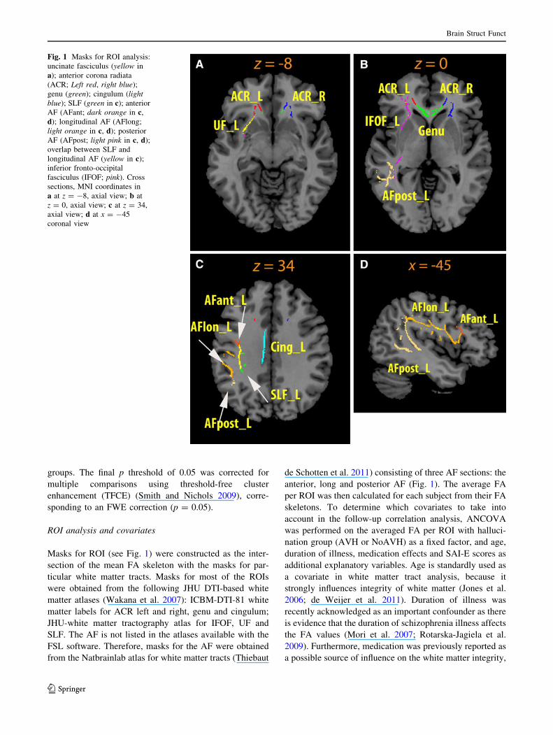
Masks for ROI (see Fig. 1) were constructed as the inter-
section of the mean FA skeleton with the masks for par-
ticular white matter tracts. Masks for most of the ROIs
were obtained from the following JHU DTI-based white
matter atlases (Wakana et al. 2007): ICBM-DTI-81 white
matter labels for ACR left and right, genu and cingulum;
JHU-white matter tractography atlas for IFOF, UF and
SLF. The AF is not listed in the atlases available with the
FSL software. Therefore, masks for the AF were obtained
from the Natbrainlab atlas for white matter tracts (Thiebaut
de Schotten et al. 2011) consisting of three AF sections: the
anterior, long and posterior AF (Fig. 1). The average FA
per ROI was then calculated for each subject from their FA
skeletons. To determine which covariates to take into
account in the follow-up correlation analysis, ANCOVA
was performed on the averaged FA per ROI with halluci-
nation group (AVH or NoAVH) as a fixed factor, and age,
duration of illness, medication effects and SAI-E scores as
additional explanatory variables. Age is standardly used as
a covariate in white matter tract analysis, because it
strongly influences integrity of white matter (Jones et al.
2006; de Weijer et al. 2011). Duration of illness was
recently acknowledged as an important confounder as there
is evidence that the duration of schizophrenia illness affects
the FA values (Mori et al. 2007; Rotarska-Jagiela et al.
2009). Furthermore, medication was previously reported as
a possible source of influence on the white matter integrity,
Fig. 1 Masks for ROI analysis:
uncinate fasciculus (yellow in
a); anterior corona radiata
(ACR; Left red, right blue);
genu (green); cingulum (light
blue); SLF (green in c); anterior
AF (AFant; dark orange in c,
d); longitudinal AF (AFlong;
light orange in c, d); posterior
AF (AFpost; light pink in c, d);
overlap between SLF and
longitudinal AF (yellow in c);
inferior fronto-occipital
fasciculus (IFOF; pink). Cross
sections, MNI coordinates in
a at z = -8, axial view; b at
z = 0, axial view; c at z = 34,
axial view; d at x = -45
coronal view
Brain Struct Funct
123

although this is still under debate (Kubicki et al. 2005). To
include it as a covariate, for each subject we calculated the
medication quantity equivalent dose of haloperidol
according to Andreasens’s formula for antipsychotic
medication (Andreasen et al. 2010). For nine patients, for
one type of medication (often patients used combined
medication) the dosage was unknown. In those cases, we
averaged quantity for this particular medication and then
calculated the haloperidol equivalent. In four cases,
patients also received antipsychotics not listed in the article
(Andreasen et al. 2010), Effexor (venlafaxine; 39) and
Orap (19). For them, we used averaged medication
quantities across all subjects. Finally, because the patients
were selected according to their insight rating—SAI-E
score, we also entered SAI-E scores in ANCOVA.
The average FA values per subject for each region were
subsequently entered into a partial correlation analysis to
investigate the correlation between the p3 score and ROI
value using SPSS package software. An FDR correction for
multiple comparisons was performed on each p value from
the correlation analysis.
Results
Subjects
The three groups of subjects (healthy controls, schizophrenia
patients with and without current hallucinations) did not sig-
nificantly differ in age, gender, education level, negative
PANSS score or general symptom severity (details in
Table 1). However, the two patient groups differed in their
positive PANSS scores. This was expected as this scale
contains the p3 item for hallucinations. In the group of patients
without current hallucinations, six patients had never experi-
enced AVH before.
White matter integrity differences
between schizophrenia patients and healthy subjects
The results of voxelwise TBSS comparison (p \ 0.05
TFCE corrected) were in accordance with previous reports
(Ellison-Wright and Bullmore 2009). The schizophrenia
patients exhibited bilaterally decreased FA values in the
following regions: anterior thalamic radiation (ATR, site of
the IFOF/UF/ACR); body of the corpus callosum (forceps
minor); cingulum (posterior parts); temporal part of the
SLF and a small area in the IFOF. In the right hemisphere,
schizophrenia patients had decreased FA in the visual
cortex-optic radiation, forceps major, body of the corpus
callosum (posterior parts) and inferior parietal lobule/
auditory cortex. The results are summarized in Fig. 2.
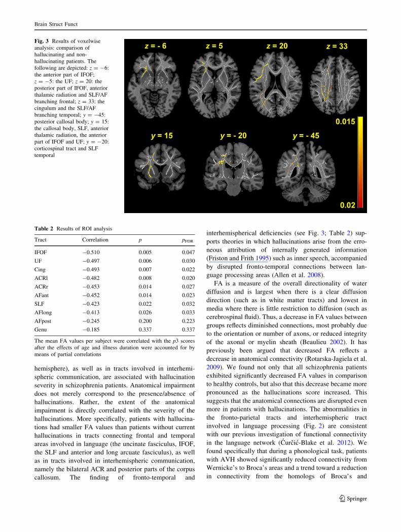
Hallucinations and FA
TBSS tracts of 32 right-handed schizophrenia patients were
entered for the voxelwise and ROI patient comparison. We
first compared patients using the voxelwise TBSS compari-
son (p \ 0.05 TFCE corrected). The confounding effects of
age and duration of illness were accounted for by introducing
their respective scores as covariates. Patients with current
hallucinations had significantly decreased FA on higher
threshold (p \ 0.02 TFCE corrected) in the left hemisphere
(see Fig. 3): anterior IFOF, UF, ACR, AF especially the
anterior and long parts of the AF (branches reaching frontal
regions such as BA44, and temporal regions), callosal body
Table 1 Demographic data of subjects
Mean (SD) Significance
Healthy Schizophrenia
patients no AVH
Schizophrenia
patients AVH
Three groups No AVH vs AVH
(n = 14) (n = 14) (n = 17)
Age in years 34 (11) 34 (12) 35 (11) F(2,42) = 0.02 (0.98)
Gender males 9 11 13 v2(2,42) = 0.87 (0.65)
Education 5.7 (1.0) 5.2 (1.1) 5.2 (1.2) F(2,42) = 0.7 (0.35)
PANSS pos. 11.4 (4.4) 16.9 (4.0) U = 38 (0.001)
PANSS neg. 13.9 (4.5) 12.8 (4.3) U = 102 (0.19)
PANSS gen. 27.7 (4.7) 29.7 (6.4) U = 99 (0.43)
PANSS tot. 53.1 (9.4) 59.4 (12.0) U = 82 (0.14)
Insight 14.4 (5.4) 11.6 (6.5) T(29) = 1.3 (0.20)
Duration of illness in years 12.6 (8.0) 15.3 (10.4) T(29) = -0.8 (0.44)
Medication (mg) haloperidol equivalent 6.9 (5.7) 7.4 (4.8) U = 102.5 (0.51)
The left column lists the demographic variables. The 2nd–4th columns from the left show average values of the variables across the group, with their
standard deviations in brackets. Education level was rated according to a six-point scale defined by Verhage, which ranges from primary school (1) to
university level (6). Nonparametric tests were used to test the group difference for PANSS (Mann–Whitney test) and gender (Chi square)
Brain Struct Funct
123

(medial and posterior part—the forceps major), the cingu-
lum, corticospinal tract and the ATR. In addition, on a lower
threshold (p \ 0.05 TFCE corrected) patients with current
hallucinations had decreased FA in several regions in the
right hemisphere including the anterior part of AF, parts of
SLF, ACR, cingulum and the corticospinal tract.
Covariate analysis revealed that age affected the FA in
IFOF and anterior AF and only marginally in other tracts (see
Online Resource 1). However, the duration of illness was
significant for several tracts: the anterior AF, the IFOF, the
cingulum and posterior and long portions of the AF (trend).
Insight and medication revealed no effect on the FA. There-
fore in addition to the standard age covariate, we also included
the duration of illness in all our analysis. Post hoc contrast of
group differences between patients with and without current
hallucinations revealed a significant decrease in FA in the left
IFOF (F(1,27) = 10.85; pFDR = 0.027), left ACR
(F(1,27) = 847; pFDR = 0.036), left UF (F(1,27) = 7.84;
pFDR = 0.021), left cingulum (F(1,27) = 6.78; pFDR =
0.037), anterior part of AF (F(1,27) = 6.35; pFDR = 0.036),
posterior part of AF (F(1,27) = 5.81; pFDR = 0.038) and SLF
(F(1,27) = 5.79; pFDR = 0.033).
Partial correlation ROI analysis (right-handed hallucina-
tions patients, n = 17; no hallucinations, n = 14) revealed a
negative correlation of FA and P3 in the left IFOF, UF,
cingulum, left and right ACR, anterior and long part of AF
and SLF (see Table 2 for r values and significance). We
found that the association between AF impairment and hal-
lucination severity is most evident in the anterior part of the
AF. Details of partial correlation analysis and FDR correc-
tion calculation are given in Online Resource 2. The distri-
bution of FA values in the ROIs is depicted in Fig. 4.
Discussion
We found that alterations of white matter in tracts that
connect frontal and temporal areas (especially in the left
Fig. 2 Results of voxelwise
analysis: comparison of healthy
controls and schizophrenia
patients. In figure panels the
following are depicted: ATR
left/anterior limb of internal
capsule (z = -3 and z = 5);
IFOF (z = 5 and x = -24);
forceps minor/genu (z = 10 and
z = 17); forceps major
(z = 17); cingulum (z = 17 and
x = -16 and x = -12); corpus
callosum (z = 30 and x = -6
and x = 2 and x = 27); SLF
right (z = 30 and x = -12);
primary auditory cortex (z = 5);
optic radiation (x = 27)
Brain Struct Funct
123
hemisphere), as well as in tracts involved in interhemi-
spheric communication, are associated with hallucination
severity in schizophrenia patients. Anatomical impairment
does not merely correspond to the presence/absence of
hallucinations. Rather, the extent of the anatomical
impairment is directly correlated with the severity of the
hallucinations. More specifically, patients with hallucina-
tions had smaller FA values than patients without current
hallucinations in tracts connecting frontal and temporal
areas involved in language (the uncinate fasciculus, IFOF,
the SLF and anterior and long arcuate fasciculus), as well
as in tracts involved in interhemispheric communication,
namely the bilateral ACR and posterior parts of the corpus
callosum. The finding of fronto-temporal and
interhemispherical deficiencies (see Fig. 3; Table 2) sup-
ports theories in which hallucinations arise from the erro-
neous attribution of internally generated information
(Friston and Frith 1995) such as inner speech, accompanied
by disrupted fronto-temporal connections between lan-
guage processing areas (Allen et al. 2008).
FA is a measure of the overall directionality of water
diffusion and is largest when there is a clear diffusion
direction (such as in white matter tracts) and lowest in
media where there is little restriction to diffusion (such as
cerebrospinal fluid). Thus, a decrease in FA values between
groups reflects diminished connections, most probably due
to the orientation or number of axons, or reduced integrity
of the axonal or myelin sheath (Beaulieu 2002). It has
previously been argued that decreased FA reflects a
decrease in anatomical connectivity (Rotarska-Jagiela et al.
2009). We found not only that all schizophrenia patients
exhibited significantly decreased FA values in comparison
to healthy controls, but also that this decrease became more
pronounced as the hallucinations score increased. This
suggests that the anatomical connections are disrupted even
more in patients with hallucinations. The abnormalities in
the fronto-parietal tracts and interhemispheric tract
involved in language processing (Fig. 2) are consistent
with our previous investigation of functional connectivity
in the language network (Curcic-Blake et al. 2012). We
found specifically that during a phonological task, patients
with AVH showed significantly reduced connectivity from
Wernicke’s to Broca’s areas and a trend toward a reduction
in connectivity from the homologs of Broca’s and
Fig. 3 Results of voxelwise
analysis: comparison of
hallucinating and non-
hallucinating patients. The
following are depicted: z = -6:
the anterior part of IFOF;
z = -5: the UF; z = 20: the
posterior part of IFOF, anterior
thalamic radiation and SLF/AF
branching frontal; z = 33: the
cingulum and the SLF/AF
branching temporal; y = -45:
posterior callosal body; y = 15:
the callosal body, SLF, anterior
thalamic radiation, the anterior
part of IFOF and UF; y = -20:
corticospinal tract and SLF
temporal
Table 2 Results of ROI analysis
Tract Correlation p pFDR
IFOF -0.510 0.005 0.047
UF -0.497 0.006 0.030
Cing -0.493 0.007 0.022
ACRl -0.482 0.008 0.020
ACRr -0.453 0.014 0.027
AFant -0.452 0.014 0.023
SLF -0.423 0.022 0.032
AFlong -0.413 0.026 0.033
AFpost -0.245 0.200 0.223
Genu -0.185 0.337 0.337
The mean FA values per subject were correlated with the p3 scores
after the effects of age and illness duration were accounted for by
means of partial correlations
Brain Struct Funct
123

Wernicke’s areas to Broca’s area in the right hemisphere.
In other words, patients without AVH had intermediate
functional connectivity strengths, similar to the anatomical
connectivity strengths found in the current study.
Our finding of diminished connectivity between frontal
and temporo-parietal regions also yields information that
may complement the so-called corollary discharge
hypothesis (Feinberg and Guazzelli 1999; Frith 2005). This
hypothesis states that the improper source attribution of
voices in patients with hallucinations can be explained in
terms of insufficient top-down feedback and control from
higher cognitive areas (such as Broca’s region) toward the
primary and secondary auditory areas (such as Wernicke’s
region). Indeed, recent experimental studies point toward a
delay in this top-down control (Hoffman et al. 2011;
Whitford et al. 2011). Consequently, our findings may
suggest that abnormal fronto-temporoparietal connections
in patients with hallucinations are responsible for the
decreased functional connectivity between these areas, a
putative underlying mechanism of reduced corollary
discharge.
Two fasciculi of the left hemisphere, the IFOF and the
UF, both belong to the language network and were found
significantly altered in schizophrenia patients. The IFOF
bundle connects the frontal and temporal regions, more
specifically the frontal lobe with the posterior portion of the
occipital gyri via the posterior temporo-basal area (Martino
et al. 2010). The IFOF has only recently been identified as
an important connection for the semantic processing of
language (Duffau 2008). Next to the AF, which is involved
in phonological language processing, Duffau describes the
IFOF as the ventral route for language processing. A clear
anatomical, also DTI-oriented, description of this impor-
tant connection appeared only recently in literature (Forkel
et al. 2012), in which a thorough literature review was
combined with DTI tractography of the IFOF. IFOF also
has an important role in primary visual processing (ffytche
and Catani 2005), visual imagery, such as visual halluci-
nations (Chechlacz et al. 2012) or visualization of music
(Zamm et al. 2013), visual awareness (Amad et al. 2013),
and processing of emotions (Baggio et al. 2012; Xu et al.
2012). Specifically, Forkel and colleagues outlined how
IFOF connects two branches of the frontal lobe, namely the
inferior frontal gyrus (where Broca’s area is placed) and
the medial frontal gyrus. They further described the route
of this fasciculus as ventral, passing the temporal lobe and
ending in the occipital lobe. Although previous studies
found indications that the IFOF plays a role in schizo-
phrenia (Clark et al. 2011), the direct involvement of the
IFOF in AVH is investigated for the first time in our cur-
rent work.
The left UF is also a pathway that connects the temporal
regions and the frontal lobe, in particular the inferior
frontal gyrus. The UF is traditionally considered to be part
of the limbic lobe (Von Der Heide et al. 2013) and its
function is not entirely clear. Recently, it has been found
that the UF is involved in the semantic processing of lan-
guage (Catani et al. 2013), an alternative route to the IFOF
for language processing (Duffau et al. 2009). However, the
role of the UF is still under debate, with evidence
Fig. 4 Block diagram of FA
values in ROI for hallucinating
(dark gray) and non-
hallucinating group (light gray).
Significant differences revealed
by post hoc analysis of
ANCOVA (the age and duration
of illness were accounted for)
are indicated by asterisk
Brain Struct Funct
123
suggesting that it acts as a pathway for creating mnemonic
associations involving the processing of language, visual
and auditory information (Von Der Heide et al. 2013). Our
findings of abnormalities in the left UF in schizophrenia
patients compared to healthy controls are consistent with
the results of Hubl et al. (2004) and de Weijer et al. (2011).
Our finding that the degree of FA reduction in the UF is
associated with the severity of hallucinations is, however,
an original and novel finding of the present study.
We found a negative correlation between FA in both the
SLF and the anterior and long part of AF, and the severity
of hallucinations. The SLF is a complex association tract
that links multiple frontal, temporo-parietal and occipital
regions (Makris et al. 2005). It is considered the most
important fasciculus in language processing because it
provides direct connections between the language regions
Broca’s and Wernicke’s areas. According to Makris et al.
(2005), the AF, which is often investigated in studies of
auditory hallucinations, is part of the SLF originating in the
dorsal prefrontal cortex (Brodmann areas 6 and 46), pass-
ing around the caudal Sylvian fissure and ending in the
caudal area of the superior temporal sulcus.
Our results are in agreement with the findings of Catani
et al. (2011) who performed exhaustive tractography ana-
lysis on the AF and observed that schizophrenia patients
with hallucinations showed reduced FA in the long seg-
ment of the left AF consistent with our findings. They were
able to examine the details of fibers within the AF tract and
to pinpoint the posterior temporal and anterior regions in
the inferior frontal and parietal lobes where the differences
occurred. However, the findings of this study are not
entirely consistent with the previous literature. For exam-
ple, Shergill et al. (2007) reported a positive association of
FA along the AF with hallucinations, where Hubl et al.
(2004) found both increased and decreased FA values in
the left AF when patients with and without hallucinations
were compared [evident from Fig. 5a (1 and 2) in their
article]. On the other hand, de Weijer et al. (2011) did not
find any direct correlation between the severity of hallu-
cinations and the FA values in the left AF after excluding
the influence of age. However, they found that the mag-
netic transfer ratio (MTR) was increased in patients with
hallucinations, while FA was negatively correlated with
MTR, thus supporting our findings.
The reasons for these discrepancies might lie in the
different data acquisition methods used in the above studies
(line-by-line spin echo sampling by Hubl et al.), the dif-
ference in analysis methods and the different question-
naires (BPRS was used by Shergill et al.). Another
important aspect is that the duration of illness may partially
explain the reduced FA in schizophrenia patients (Mori
et al. 2007). Indeed, Rotarska-Jagiela et al. (2009) found
positive correlations of the duration of illness with the FA
values in the bilateral arcuate fasciculus and negative
correlations in the corpus callosum, occipitofrontal fas-
ciculus and other areas.
The ACR and the posterior corpus callosum are also the
loci of some significant differences between hallucinating
and non-hallucinating schizophrenia patients. The ACR
consists of a mixture of various association, projection and
callosal fibers, and partially connects the corpus callosum
with the cortex (Wakana et al. 2004), thus providing an
extension to the interhemispheric connection. It has been
implied that the ACR connects the ACC with other brain
structures (Tang et al. 2010) and is involved in attention
networks (Niogi et al. 2010). We found an association
between the bilateral ACR and hallucination severity. The
majority of the left ACR mask overlapped with the dif-
ferences between schizophrenia patients and healthy con-
trols, suggesting that these differences in the left
hemisphere are not only a characteristic of schizophrenia,
but also to some extent of hallucinations. However, the
differences observed in the right ACR are specific for
hallucinations. Similarly, we found significant differences
in the voxelwise comparison of the two patient groups in
the posterior parts of the corpus callosum. These findings
are consistent with that of Knochel et al. (2012) who
reported that hypoconnectivity in the corpus callosum was
associated with the severity of hallucinations.
Similarly, consideration can be made about the cingu-
lum, which is related to the anterior cingulate (also
involved in attention and sensory decision making, as
mentioned above). An impeded connection along the left
cingulum might contribute to the overall impedance of this
sensory processing. The ACC provides ‘the interface for
emotions’; it is known to be important in schizophrenia
(Barch and Dowd 2010). In line with these findings, Ver-
cammen et al. revealed a reduction in coupling between the
temporo-parietal junction and ACC in patients with AVH.
We found decreased FA values in the left cingulate of
patients in the hallucination group compared to patients
without current hallucinations, a result that is in agreement
with Hubl et al. (2004). These studies are consistent with
theories of decreased attribution of agency, and thus
decreased awareness of one’s own volitional actions (in
this case inner speech production) in AVH patients (Frith
1996).
A number of limitations of our study deserve discussion.
First, besides acting as an indicator of white matter integ-
rity, FA might be influenced by other factors such as the
thickness of fibers (Beaulieu 2002). However, in our TBSS
analysis the FA values were extracted from regions
belonging to a mean estimated skeleton mask. Thus, vari-
ations in fiber thickness beyond the skeleton do not affect
our results. This also implies that our method is unsuitable
for the investigation of fiber thickness. Second, DTI does
Brain Struct Funct
123
not differentiate between efferent and afferent projections,
thus it does not provide any information about the causality
and directionality of the connections. Third, we investi-
gated differences between patients with and without cur-
rent hallucinations according to the PANSS and MINI
interviews. Patients were considered to be without current
hallucinations if none were reported in the month prior to
scanning. However, they may have experienced halluci-
nations earlier in their illness, and thus may still have had
trait characteristics associated with hallucinations. This
implies that our conclusions are limited to the severity of
hallucinations rather than their presence/absence, espe-
cially because schizophrenia patients who have never
experienced hallucinations are difficult to find. Fourth, we
did correct for the dose of medication in schizophrenia
patients, rounding the doses that were missing for nine
patients. However, all these patients were medicated and
we used a standard method for filling in unknown param-
eters (by means of specific mean values). This is a rec-
ommended method because it minimizes bias while
maintaining the df as compared to simply omitting the data
(Rubin et al. 2007). Furthermore, a previous study (Seok
et al. 2007) found that controlling for medication dosage
did not change the correlation between FA values in the
SLF and the hallucination severity, in line with our findings
that medication dosage did not affect the FA values in any
ROI.
All the fasciculi investigated in this paper are considered
to be involved in language processing. However, their
function is often much broader and encompasses the pro-
cessing of emotions (cingulate, ACR), visual information
(IFOF) or episodic memory (UF). Consequently, AVHs are
also associated with broader functional deficiencies, such
as cognitive control and emotional processing (Aleman and
Larøi 2008). Future studies should investigate the role of
these regions in more detail. For example, the AF seems to
be more asymmetric in men (Catani and Mesulam 2008)
and the lateralization of the UF is still under debate
(Highley et al. 2002; Thiebaut de Schotten et al. 2011).
Therefore, an important future strategy will be to combine
the volumetric asymmetries of these regions in relation to
language.
To conclude, our results suggest that hallucinations in
schizophrenia patients are associated with a complex set of
white matter abnormalities that involve at least three dis-
tinct systems: the language network, putatively in the
‘inner speech’ domain; and the central in attention and
perception loop; and the limbic system, providing the
emotional edge. Additionally, we advance the hypothesis
that the severity of the hallucinations experienced by
schizophrenia patients is a direct function of the extent of
the impairment affecting the anatomical connectivity
within these three systems.
Acknowledgments The authors thank G.R. Blake for his comments
on earlier versions of the manuscript. We thank psychiatrists Dr.
R. Bruggeman and Dr. H. Knegtering for their help with patient
inclusion. Our thanks are due especially to A. de Vos and J. van der
Velde for help with data sorting and to S. Chalavi for discussions on
analysis. L. Cerliani is supported by an NWO MaGW Open compe-
tition grant (grant number 400-08-089) and by an NIHC grant (grant
number 056-13-017). This research was supported by a EURYI
Award from the European Science Foundation (No. 044035001)
awarded to A.A.
Conflict of interest The authors declare that they have no com-
peting financial interests in relation to the work described.
References
Aleman A, Larøi F (2008) Hallucinations: the science of idiosyncratic
perception. American Psychological Association, Washington,
DC
Alexander AL, Lee JE, Lazar M, Field AS (2007) Diffusion tensor
imaging of the brain. Neurotherapeutics 4:316–329
Allen P, Larøi F, McGuire PK, Aleman A (2008) The hallucinating
brain: a review of structural and functional neuroimaging studies
of hallucinations. Neurosci Biobehav Rev 32:175–191
Amad A, Cachia A, Gorwood P, Pins D, Delmaire C, Rolland B et al
(2013) The multimodal connectivity of the hippocampal com-
plex in auditory and visual hallucinations. Mol Psychiatry (in
press)
Andreasen NC, Pressler M, Nopoulos P, Miller D, Ho BC (2010)
Antipsychotic dose equivalents and dose-years: a standardized
method for comparing exposure to different drugs. Biol Psychi-
atry 67:255–262
Baggio HC, Segura B, Ibarretxe-Bilbao N, Valldeoriola F, Marti MJ,
Compta Y et al (2012) Structural correlates of facial emotion
recognition deficits in Parkinson’s disease patients. Neuropsych-
ologia 50:2121–2128
Barch DM, Dowd EC (2010) Goal representations and motivational
drive in schizophrenia: the role of prefrontal–striatal interactions.
Schizophr Bull 36:919–934
Bastin ME, Armitage PA, Marshall I (1998) A theoretical study of the
effect of experimental noise on the measurement of anisotropy in
diffusion imaging. Magn Reson Imaging 16:773–785
Beaulieu C (2002) The basis of anisotropic water diffusion in the
nervous system—a technical review. NMR Biomed 15:435–455
Benedetti F, Yeh PH, Bellani M, Radaelli D, Nicoletti MA, Poletti S,
Falini A, Dallaspezia S, Colombo C, Scotti G, Smeraldi E,
Soares JC, Brambilla P (2011) Disruption of white matter
integrity in bipolar depression as a possible structural marker of
illness. Biol Psychiatry 69:309–317
Brown GG, Thompson WK (2010) Functional brain imaging in
schizophrenia: selected results and methods. Curr Top Behav
Neurosci 4:181–214
Catani M, Craig MC, Forkel SJ, Kanaan R, Picchioni M, Toulopoulou
T, Shergill S, Williams S, Murphy DG, McGuire P (2011)
Altered integrity of perisylvian language pathways in schizo-
phrenia: relationship to auditory hallucinations. Biol Psychiatry
70:1143–1150
Catani M, Mesulam M (2008) The arcuate fasciculus and the
disconnection theme in language and aphasia: history and
current state. Cortex 44:953–961
Catani M, Mesulam MM, Jakobsen E, Malik F, Martersteck A,
Wieneke C et al (2013) A novel frontal pathway underlies verbal
fluency in primary progressive aphasia. Brain 136:2619–2628
Brain Struct Funct
123
Cercignani M, Horsfield MA (2001) The physical basis of diffusion-
weighted MRI. J Neurol Sci 186(Suppl 1):S11–S14
Chechlacz M, Rotshtein P, Hansen PC, Riddoch JM, Deb S,
Humphreys GW (2012) The neural underpinnings of simultan-
agnosia: disconnecting the visuospatial attention network.
J Cogn Neurosci 24:718–735
Clark KA, Nuechterlein KH, Asarnow RF, Hamilton LS, Phillips OR,
Hageman NS, Woods RP, Alger JR, Toga AW, Narr KL (2011)
Mean diffusivity and fractional anisotropy as indicators of disease
and genetic liability to schizophrenia. J Psychiatr Res 45:980–988
Curcic-Blake B, Liemburg E, Vercammen A, Swart M, Knegtering H,
Bruggeman R, Aleman A (2012) When Broca goes uninformed:
reduced information flow to Broca’s area in schizophrenia
patients with auditory hallucinations. Schizophr Bull 39(5):
1087–1095
de Weijer AD, Mandl RC, Diederen KM, Neggers SF, Kahn RS,
Hulshoff Pol HE, Sommer IE (2011) Microstructural alterations
of the arcuate fasciculus in schizophrenia patients with frequent
auditory verbal hallucinations. Schizophr Res 130:68–77
Duffau H (2008) The anatomo-functional connectivity of language
revisited. New insights provided by electrostimulation and
tractography. Neuropsychologia 46:927–934
Duffau H, Gatignol P, Moritz-Gasser S, Mandonnet E (2009) Is the
left uncinate fasciculus essential for language? A cerebral
stimulation study. J Neurol 256:382–389
Ellison-Wright I, Bullmore E (2009) Meta-analysis of diffusion tensor
imaging studies in schizophrenia. Schizophr Res 108:3–10
Feinberg I, Guazzelli M (1999) Schizophrenia—a disorder of the
corollary discharge systems that integrate the motor systems of
thought with the sensory systems of consciousness. Br J
Psychiatry 174:196–204
ffytche DH, Catani M (2005) Beyond localization: from hodology to
function. Philos Trans R Soc Lond B Biol Sci 360:767–779
Forkel SJ, Thiebaut de SM, Kawadler JM, Dell’Acqua F, Danek A,
Catani M (2012) The anatomy of fronto-occipital connections
from early blunt dissections to contemporary tractography.
Cortex. doi:10.1016/j.cortex.2012.09.005
Friston KJ, Frith CD (1995) Schizophrenia: a disconnection syn-
drome? Clin Neurosci 3:89–97
Frith C (1996) Neuropsychology of schizophrenia: what are the
implications of intellectual and experimental abnormalities for
the neurobiology of schizophrenia? Br Med Bull 52:618–626
Frith C (2005) The neural basis of hallucinations and delusions. C R
Biol 328:169–175
Grossberg S (2000) How hallucinations may arise from brain
mechanisms of learning, attention, and volition. J Int Neuropsy-
chol Soc 6:583–592
Highley JR, Walker MA, Esiri MM, Crow TJ, Harrison PJ (2002)
Asymmetry of the uncinate fasciculus: a post-mortem study of
normal subjects and patients with schizophrenia. Cereb Cortex
12:1218–1224
Hoffman RE (2007) A social deafferentation hypothesis for induction
of active schizophrenia. Schizophr Bull 33:1066–1070
Hoffman RE, Pittman B, Constable RT, Bhagwagar Z, Hampson M
(2011) Time course of regional brain activity accompanying
auditory verbal hallucinations in schizophrenia. Br J Psychiatry
198:277–283
Hubl D, Koenig T, Strik W, Federspiel A, Kreis R, Boesch C, Maier
SE, Schroth G, Lovblad K, Dierks T (2004) Pathways that make
voices: white matter changes in auditory hallucinations. Arch
Gen Psychiatry 61:658–668
Jardri R, Pouchet A, Pins D, Thomas P (2011) Cortical activations
during auditory verbal hallucinations in schizophrenia: a coordi-
nate-based meta-analysis. Am J Psychiatry 168:73–81
Jones DK, Catani M, Pierpaoli C, Reeves SJ, Shergill SS, O’Sullivan M,
Golesworthy P, McGuire P, Horsfield MA, Simmons A, Williams
SC, Howard RJ (2006) Age effects on diffusion tensor magnetic
resonance imaging tractography measures of frontal cortex
connections in schizophrenia. Hum Brain Mapp 27:230–238
Kay SR, Fiszbein A, Opler LA (1987) The positive and negative
syndrome scale (PANSS) for schizophrenia. Schizophr Bull
13:261–276
Kemp R, David AS (1997) Insight and compliance. In: Blackwell B
(ed) Compliance and the treatment alliance in serious mental
illness. Hardwood Academic Publishers, Amsterdam, pp 61–86
Knecht S, Drager B, Deppe M, Bobe L, Lohmann H, Floel A,
Ringelstein EB, Henningsen H (2000) Handedness and hemi-
spheric language dominance in healthy humans. Brain 123(Pt
12):2512–2518
Knochel C, Oertel-Knochel V, Schonmeyer R, Rotarska-Jagiela A,
van de Ven V, Prvulovic D, Haenschel C, Uhlhaas P, Pantel J,
Hampel H, Linden DE (2012) Interhemispheric hypoconnectiv-
ity in schizophrenia: fiber integrity and volume differences of the
corpus callosum in patients and unaffected relatives. NeuroIm-
age 59:926–934
Kubicki M, Westin CF, McCarley RW, Shenton ME (2005) The
application of DTI to investigate white matter abnormalities in
schizophrenia. Ann N Y Acad Sci 1064:134–148
Kuhn S, Gallinat J (2010) Quantitative meta-analysis on state and
trait aspects of auditory verbal hallucinations in schizophrenia.
Schizophr Bull 32:358–365
Makris N, Kennedy DN, McInerney S, Sorensen AG, Wang R,
Caviness VS Jr, Pandya DN (2005) Segmentation of subcom-
ponents within the superior longitudinal fascicle in humans: a
quantitative, in vivo, DT-MRI study. Cereb Cortex 15:854–869
Mandonnet E, Nouet A, Gatignol P, Capelle L, Duffau H (2007) Does
the left inferior longitudinal fasciculus play a role in language? A
brain stimulation study. Brain 130:623–629
Martino J, Brogna C, Robles SG, Vergani F, Duffau H (2010) Anatomic
dissection of the inferior fronto-occipital fasciculus revisited in the
lights of brain stimulation data. Cortex 46:691–699
Mechelli A, Allen P, Amaro E, Fu CHY, Williams SCR, Brammer
MJ, Johns LC, McGuire PK (2007) Misattribution of speech and
impaired connectivity in patients with auditory verbal halluci-
nations. Hum Brain Mapp 28:1213–1222
Mori T, Ohnishi T, Hashimoto R, Nemoto K, Moriguchi Y, Noguchi
H, Nakabayashi T, Hori H, Harada S, Saitoh O, Matsuda H,
Kunugi H (2007) Progressive changes of white matter integrity
in schizophrenia revealed by diffusion tensor imaging. Psychi-
atry Res 154:133–145
Niogi S, Mukherjee P, Ghajar J, McCandliss BD (2010) Individual
differences in distinct components of attention are linked to
anatomical variations in distinct white matter tracts. Front
Neuroanat 4:2
Parker GJ, Luzzi S, Alexander DC, Wheeler-Kingshott CA, Ciccarelli
O, Lambon Ralph MA (2005) Lateralization of ventral and
dorsal auditory-language pathways in the human brain. Neuro-
image 24:656–666
Ranson SW (1947) The anatomy of the nervous system: its develop-
ment and function. W. B. Saunders company, London
Rotarska-Jagiela A, Oertel-Knoechel V, DeMartino F, van de Ven V,
Formisano E, Roebroeck A, Rami A, Schoenmeyer R, Haenschel
C, Hendler T, Maurer K, Vogeley K, Linden DE (2009)
Anatomical brain connectivity and positive symptoms of
schizophrenia: a diffusion tensor imaging study. Psychiatry
Res 174:9–16
Rubin LH, Witkiewitz K, Andre JS, Reilly S (2007) Methods for
handling missing data in the behavioral neurosciences: don’t
throw the baby rat out with the bath water. J Undergrad Neurosci
Educ 5:A71–A77
Seok JH, Park HJ, Chun JW, Lee SK, Cho HS, Kwon JS, Kim JJ
(2007) White matter abnormalities associated with auditory
Brain Struct Funct
123
hallucinations in schizophrenia: a combined study of voxel-
based analyses of diffusion tensor imaging and structural
magnetic resonance imaging. Psychiatry Res 156:93–104
Sheehan DV, Lecrubier Y, Sheehan KH, Amorim P, Janavs J, Weiller
E, Hergueta T, Baker R, Dunbar GC (1998) The Mini-
International Neuropsychiatric Interview (M.I.N.I.): the devel-
opment and validation of a structured diagnostic psychiatric
interview for DSM-IV and ICD-10. J Clin Psychiatry 59(Suppl
20):22–33
Shergill SS, Kanaan RA, Chitnis XA, O’Daly O, Jones DK, Frangou
S, Williams SC, Howard RJ, Barker GJ, Murray RM, McGuire P
(2007) A diffusion tensor imaging study of fasciculi in
schizophrenia. Am J Psychiatry 164:467–473
Smith SM, Nichols TE (2009) Threshold-free cluster enhancement:
addressing problems of smoothing, threshold dependence and
localisation in cluster inference. NeuroImage 44:83–98
Smith SM, Jenkinson M, Johansen-Berg H, Rueckert D, Nichols TE,
Mackay CE, Watkins KE, Ciccarelli O, Cader MZ, Matthews
PM, Behrens TE (2006) Tract-based spatial statistics: voxelwise
analysis of multi-subject diffusion data. NeuroImage
31:1487–1505
Tang YY, Lu Q, Geng X, Stein EA, Yang Y, Posner MI (2010) Short-
term meditation induces white matter changes in the anterior
cingulate. Proc Natl Acad Sci USA 107:15649–15652
Thiebaut de Schotten M, Ffytche DH, Bizzi A, Dell’Acqua F, Allin
M, Walshe M, Murray R, Williams SC, Murphy DG, Catani M
(2011) Atlasing location, asymmetry and inter-subject variability
of white matter tracts in the human brain with MR diffusion
tractography. Neuroimage 54:49–59
van der Meer L, de Vos AE, Stiekema AP, Pijnenborg GH, van Tol
MJ, Nolen WA, David AS, Aleman A (2012) Insight in
schizophrenia: involvement of self-reflection networks? Schiz-
ophr Bull 39(6):1288–1295. doi:10.1093/schbul/sbs122
Vercammen A, Knegtering H, den Boer JA, Liemburg EJ, Aleman A
(2010) Auditory hallucinations in schizophrenia are associated
with reduced functional connectivity of the temporo-parietal
area. Biol Psychiatry 67:912–918
Von Der Heide RJ, Skipper LM, Klobusicky E, Olson IR (2013)
Dissecting the uncinate fasciculus: disorders, controversies and a
hypothesis. Brain 136:1692–1707
Wakana S, Jiang H, Nagae-Poetscher LM, van Zijl PC, Mori S (2004)
Fiber tract-based atlas of human white matter anatomy. Radi-
ology 230:77–87
Wakana S, Caprihan A, Panzenboeck MM, Fallon JH, Perry M, Gollub
RL, Hua K, Zhang J, Jiang H, Dubey P, Blitz A, van Zijl P, Mori S
(2007) Reproducibility of quantitative tractography methods
applied to cerebral white matter. Neuroimage 36:630–644
Whitford TJ, Mathalon DH, Shenton ME, Roach BJ, Bammer R,
Adcock RA, Bouix S, Kubicki M, De Siebenthal J, Rausch AC,
Schneiderman JS, Ford JM (2011) Electrophysiological and
diffusion tensor imaging evidence of delayed corollary dis-
charges in patients with schizophrenia. Psychol Med 41:959–969
Xu J, Kober H, Carroll KM, Rounsaville BJ, Pearlson GD, Potenza
MN (2012) White matter integrity and behavioral activation in
healthy subjects. Hum Brain Mapp 33:994–1002
Zamm A, Schlaug G, Eagleman DM, Loui P (2013) Pathways to
seeing music: enhanced structural connectivity in colored-music
synesthesia. Neuroimage 74:359–366
Brain Struct Funct
123
Related Documents