Thorax (1954), 9, 313. NORMAL STANDARDS FOR LUNG VOLUMES, INTRAPUL- MONARY GAS-MIXING, AND MAXIMUM BREATHING CAPACITY BY C. D. NEEDHAM, MARY C. ROGAN, AND I. McDONALD From Aberdeen General Hospitals and the University of Aberdeen (RECEIVED FOR PUBLICATION JUNE 20, 1954) An objective assessment of respiratory function is important in the diagnosis and management of patients complaining of dyspnoea or suffering from disease involving the lungs. There are four main subdivisions, excluding blood composition, of the respiratory gas exchange process (ventilatory, dis- tributive, diffusional, and circu!atory), which are disturbed in various proportions in different diseases; therefore no single test of respiratory function can be adequate in all cases (Comroe, 1951 ; Gilson and Oldham, 1952). The ventilatory and distributive (intrapulmonary gas mixing) aspects of respiratory efficiency are commonly studied by measuring the total lung capacity (T.L.C.) and its subdivisions, the maximum breath- ing capacity (M.B.C.), the timed vital capacity (T.V.C.), and some form of "mixing efficiency" test. It is perhaps rather generally assumed that reasonably adequate information is already avail- able concerning these tests, which have been in use for some years. However, Comroe (1951), Fletcher (1953), and Donald (1953) all comment on the inadequacy of control data, and we find that a critical survey of the published reports up- holds their opinion. We intend to make only a very brief survey of the literature, as there are already excellent reviews in this field (Comroe, 1950, 1951 ; Donald, 1953 ; Fowler, 1952). Apart from vital capacity (V.C.), the normal values of which have been well studied (Hutchin- son, 1846; West, 1920; Stewart, 1922; Kelly, 1933; Myers, 1925), we find there are a number of difficulties in determining the normal values for the other testing procedures. Different workers have used various methods to obtain normal values for the several aspects of respiratory function, and the lack of standardized procedure makes the results not always compar- able; this is particularly true for tests of mixing efficiency and maximum breathing capacity. Most of the reported series consist of only small numbers and the few larger groups cover only one or two testing procedures. Females of all ages are poorly represented, and comparatively few of the male subjects have been in the younger (age less than 18 years) or older (age more than 50 years) groups. There has been a tendency for selected types of subjects to be used, e.g., medical students and nurses in the younger age range and hospital patients and doctors in the older. The criteria of normality for selection of the subjects have not always been made clear, though the paper by Whitfield, Waterhouse, and Arnott (1950) is a notable exception. Furthermore, the interpreta- tion of some of the results is made difficult by lack of information on body measurements, by the inclusion of rather large age ranges in single groups, or by incomplete analysis of the data obtained. Estimation of total lung capac ty (T.L.C.) has been carried out in very small numbers of subjects by early workers (Lundsgaard and Van Slyke, 1918; Lundsgaard and Schierbeck, 1923; Binger, 1923; Lindhard, 1925; Anthony, 1930; Christie, 1932), but Table I lists the more recent and larger groups studied. Prediction formulae, based on body measurements, have been calculated for T.L.C. by Hurtado and Fray (1933), by Kaltreider, Fray, and Hyde (1938), by Aslett, D'Arcy Hart, and McMichael (1939) and by Whitfield and others (1950); and for vital capacity (V.C.) by West (1920), Kelly (1933), and Baldwin, Cournand, and Richards (1948). Widely varying values have been given for the ratio of residual volume to total lung capacity (R.V. /T.L.C. ratio) at different ages (Kalt- reider and others, 1938; Robinson, 1938; Bates and Christie, 1950; Greifenstein, King, Latch, and Comroe, 1952). Regardless of this disagreement the ratio has been widely accepted, erroneously we think, as the key to the laboratory diagnosis of emphysema since it was firsL suggested by Hurtado. Fray, Kaltreider, and Brooks (1934). on 7 May 2018 by guest. Protected by copyright. http://thorax.bmj.com/ Thorax: first published as 10.1136/thx.9.4.313 on 1 December 1954. Downloaded from

Welcome message from author
This document is posted to help you gain knowledge. Please leave a comment to let me know what you think about it! Share it to your friends and learn new things together.
Transcript

Thorax (1954), 9, 313.
NORMAL STANDARDS FOR LUNG VOLUMES, INTRAPUL-MONARY GAS-MIXING, AND MAXIMUM
BREATHING CAPACITYBY
C. D. NEEDHAM, MARY C. ROGAN, AND I. McDONALDFrom Aberdeen General Hospitals and the University of Aberdeen
(RECEIVED FOR PUBLICATION JUNE 20, 1954)
An objective assessment of respiratory functionis important in the diagnosis and management ofpatients complaining of dyspnoea or suffering fromdisease involving the lungs. There are four mainsubdivisions, excluding blood composition, of therespiratory gas exchange process (ventilatory, dis-tributive, diffusional, and circu!atory), which aredisturbed in various proportions in differentdiseases; therefore no single test of respiratoryfunction can be adequate in all cases (Comroe,1951 ; Gilson and Oldham, 1952). The ventilatoryand distributive (intrapulmonary gas mixing)aspects of respiratory efficiency are commonlystudied by measuring the total lung capacity(T.L.C.) and its subdivisions, the maximum breath-ing capacity (M.B.C.), the timed vital capacity(T.V.C.), and some form of "mixing efficiency"test.
It is perhaps rather generally assumed thatreasonably adequate information is already avail-able concerning these tests, which have been inuse for some years. However, Comroe (1951),Fletcher (1953), and Donald (1953) all commenton the inadequacy of control data, and we findthat a critical survey of the published reports up-holds their opinion. We intend to make only avery brief survey of the literature, as there arealready excellent reviews in this field (Comroe,1950, 1951 ; Donald, 1953 ; Fowler, 1952).Apart from vital capacity (V.C.), the normal
values of which have been well studied (Hutchin-son, 1846; West, 1920; Stewart, 1922; Kelly,1933; Myers, 1925), we find there are a numberof difficulties in determining the normal values forthe other testing procedures.
Different workers have used various methods toobtain normal values for the several aspects ofrespiratory function, and the lack of standardizedprocedure makes the results not always compar-
able; this is particularly true for tests of mixingefficiency and maximum breathing capacity. Most
of the reported series consist of only small numbersand the few larger groups cover only one or twotesting procedures. Females of all ages are poorlyrepresented, and comparatively few of the malesubjects have been in the younger (age less than18 years) or older (age more than 50 years) groups.There has been a tendency for selected types ofsubjects to be used, e.g., medical students andnurses in the younger age range and hospitalpatients and doctors in the older. The criteriaof normality for selection of the subjects have notalways been made clear, though the paper byWhitfield, Waterhouse, and Arnott (1950) is anotable exception. Furthermore, the interpreta-tion of some of the results is made difficult by lackof information on body measurements, by theinclusion of rather large age ranges in singlegroups, or by incomplete analysis of the dataobtained.
Estimation of total lung capac ty (T.L.C.) hasbeen carried out in very small numbers of subjectsby early workers (Lundsgaard and Van Slyke,1918; Lundsgaard and Schierbeck, 1923; Binger,1923; Lindhard, 1925; Anthony, 1930; Christie,1932), but Table I lists the more recent and largergroups studied. Prediction formulae, based onbody measurements, have been calculated forT.L.C. by Hurtado and Fray (1933), by Kaltreider,Fray, and Hyde (1938), by Aslett, D'Arcy Hart, andMcMichael (1939) and by Whitfield and others(1950); and for vital capacity (V.C.) by West(1920), Kelly (1933), and Baldwin, Cournand, andRichards (1948). Widely varying values have beengiven for the ratio of residual volume to total lungcapacity (R.V. /T.L.C. ratio) at different ages (Kalt-reider and others, 1938; Robinson, 1938; Batesand Christie, 1950; Greifenstein, King, Latch, andComroe, 1952). Regardless of this disagreementthe ratio has been widely accepted, erroneouslywe think, as the key to the laboratory diagnosis ofemphysema since it was firsL suggested by Hurtado.Fray, Kaltreider, and Brooks (1934).
on 7 May 2018 by guest. P
rotected by copyright.http://thorax.bm
j.com/
Thorax: first published as 10.1136/thx.9.4.313 on 1 D
ecember 1954. D
ownloaded from

C. D. NEEDHAM, MARY C. ROGAN, and Z. McDONALD
TABLE ITOTAL LUNG CAPACITY DETERMINATIONS
SubjectsAuthor Method
Total M F Yr. Yr.
Hurtado and Uncorrected 50 50 - _ _Boller (1933) oxygen
dilutionHurtado and ,, ,, 50 - 50 - _
others (1934)Kaltreider and ,, ,. 50 50 _ _ 18
others (1938)Robinson(1938) ,, .. 93 93 - 41 20Lester and Open circuit 15 Not 15 -
others (1942) statedGreifenstein and ,11 - 1 1
others (1952) 26 15 15Aslett and Corrected 64 64 - - 8
others (1939) oxygendilution
Birath (1944) Closed circuit 35 16 19 -
hydrogenWhitfield and Hydrogen or 96 64 10* 20*
others (1950) heliumClosed circuit 32 5* 10*
Gilson and Open circuit 4 4 - -_Hugh-Jones and helium(1949) closed circuit
Bates and Helium closed 27 Not 1 * 10*Christie (1950) circuit statedMeneely and ,, ,, 10 7 3 1 -
Kaltreider(1949)
Total 520 359 119 73 112
* Approximate numbers, extracted trom the total number, age,range, and means, and scatter diagrams.
TABLE I1MIXING
SubjectsAuthor Method
Total M F (Years)
Roelsen (1939) Single breath 14 13 I1 19-37Fractional analysis
Cournand and 7 minutes 17 Not stated I Notothers (1941) Nitrogen washout stated
Darling and 7 minutes 21 18i 3 21-65others (1944) Nitrogen washout
Birath (1944) Fractional analysis 35 16 19 18-39Closed circuit
Bates and Continuous 17 Not stated 17-37Christie (1950) analysis
Closed circuit 10 47-62Briscoe and Continuous 10 15-40
others (1951) analysisClosed circuit 16 2 65-75
4 17-39Comroe and Single breath 14 Not stated 18-38
Fowler (1951) Continuousanalysis
Fowler (1949) Single breath 18 12 6 17-73Continuous
analvsisGreifenstein and 7 minutes 26 11 50-75
others (1952) Nitrogen washout 15 50-77and single breathfractional analysis
The importance of the distributive aspect ofventilation (intrapulmonary mixing of inspiredair) has been recognized for many years, andFowler (1952) has published a valuable review ofthe extensive literature. Though much work has
been done on this subject it has been largely de-voted to the evolution of a multitude of differentand not strictly comparable methods. Table IIsummarizes the reported work on normal subjects.The maximum breathing capacity (M.B.C.) test
devised by Hermannsen (1933) is generally accep-ted as very useful in assessing overall ventilatoryability. Table III summarizes the reported workon normal subjects. Prediction formulae havebeen calculated on the basis of sex, age, and bodysurface area (B.S.A.) by Baldwin, Cournand, andRichards (1948), and, in a purely male group, onage alone by Wright, Yee, Filley, and Stranahan(1949).
TABLE IlMAXIMUM BREATHING CAPACITY
SubjectsAuthor Method
Total M F Age(Years)
Hermannsen Spirometer 23 Both Not stated(1933)
Cournand and 40 20 20 Average 27others (1939)
Wright and Douglas bag 250 250 - Not statedothers (1949) high velo-
city valveGilsonand Hugh-: Spirometer 4 4 - 29-44
Jones (1949)Gray and others 323 283 40 Young adult
(1950) -Gaensler (1951) . 35 "Equally Younger age
Baldwin andothers (1948)
Greifenstein and Tissotothers (1952) spirometer
Bernstein and Spirometerothers (1952) with light
bellTurner and SpirometerMcLean (1951)
divided"92 52
40
26 l15
14 Not stated
50 30 20
group16-69
16-7950-7550-77
Not stated
53-14
Following criticism of the M.B.C. test as beingtoo strenuous for really ill patients and too de-pendent upon co-operation by the subject therehave been attempts to devise a simpler means ofobtaining the same information. Tiffeneau, Bous-ser, and Drutel (1949), Gaensler (1951), and Ken-nedy (1953) all claim that a rather good estimateof the subject's actual (as opposed to predictednormal) M.B.C. can be obtained from the timedvital capacity (T.V.C.). This test is more rapidlyperformed and imposes much less strain on an illpatient. Gaensler began with a three-second testbut later used the one-second test, and providedapparatus is available the shorter time is preferableas it yields a more accurate estimate of the M.B.C.He used an electrically controlled cut-out formeasuring the one-second fraction, but a fast re-
volving kymographic method (Kennedy, 1953) ismore useful as the shape of the whole inspiratoryand expiratory curves can be seen.
314
on 7 May 2018 by guest. P
rotected by copyright.http://thorax.bm
j.com/
Thorax: first published as 10.1136/thx.9.4.313 on 1 D
ecember 1954. D
ownloaded from

NORMAL STANDARDS FOR LUNG VOLUMES
It is the purpose of this communication to pre-sent and analyse further data on the normal valuesfor these respiratory function measurements, i.e.,total lung capacity (T.L.C.) and its subdivisions:intrapulmonary gas-mixing efficiency, maximumbreathing capacity (M.B.C.), and timed vital capa-city (T.V.C.).
MATERIALWe studied 324 subjects, 183 men and 141
women. This included 150 aged 11-19 years (78men, 72 women), 114 aged 20-49 years (72 men,42 women), and 60 aged 50-77 years (33 men, 27women).The subjects of both sexes were distributed fairly
evenly year by year in the age 11-19 group and bydecades up to the age of 70 years.We tried to arrange that subjects from the same
age and sex groups should be drawn from morethan one section of the community so that ourresults might be as representative as possible ofthe general population.
Subjects aged 11-19 years were obtained froma well-run orphanage, boy-scout and girl-guidecompanies, a secondary school, a pre-nursingschool, laboratory technicians, nurses, a churchyouth group, medical students, and army recruits.Subjects aged 20 onwards consisted of nursingstaff, hospital and university staff (graduates andothers), factory workers (both men and women),members of a business women's association, per-sonal friends, a few medical students, and hospitalpatients (suffering from disorders unrelated to thecardio-pulmonary system and not causing generaldebility).
CRITERIA FOR ACCEPTANCE AS NORMAL SUBJECTSThe decision whether or not to include a given
subject was taken before function testing; nonewere subsequently rejected because of failure tocome up to expectations on test procedures.The following were the criteria adopted: (1) No
history of (a) asthma, (b) frequent or habitualwinter cough, (c) being subject to " colds alwaysgoing to the chest," or (d) " smoker's cough " ofmore than a mild degree. (2) No exertionaldyspnoea beyond that appropriate to their years:obviously there may be differing views on whatability for physical exertion may properly beexpected of a person as age advances. Our viewmay be summarized by saying that we expecteda person to be able to keep up without distresswith apparently healthy people of his or her ownsex and age. We tried to determine this by dis-cussing with each subject his or her daily routine.This is not a very high standard, but we wished
to sample a cross-section of an ordinary healthy,and not an exceptionally fit, community. (3) Noobvious obesity. (4) No abnormal findings onclinical examination of the cardio-pulmonarysystem (though a mild hypertension less than180/100 mm. Hg did not, by itself, disqualify).Full physical examination of the heart and lungswas not carried out on most of the subjects agedless than 20 years or on some of the older subjects.(5) Normal chest radiograph, but because of prac-tical difficulties this was not carried out on mostof the subjects aged less than 15 years or on a fewof the others.
It was impossible to carry out full physicaland radiological examinations on every subject,although this would have been desirable, but weconsidered that, in deciding whether to accept themas normal, the history of their actual exertionalability was of more importance. If any subjectswere wrongly accepted, through lack of such ex-amination, the effect would have been to lower ourstandards of " normal " performance, but there isno evidence of this in our results.
NOMENCLATUREWe have followed the nomenclatures recently
adopted (Pappenheimer, 1950). Intrapulmonarymixing efficiency is designated " M.E.%." Timedvital capacity, which we measured over a two-second interval, is simply referred to as T.V.C.,and maximum breathing capacity as M.B.C.
METHODTesting procedures were carried out in the morning,
afternoon, or evening over the period June, 1952, toSeptember, 1953. No difficulty was found in securingco-operation from the subjects, who were seated forall the tests and were in the non-basal state. TheF.R.C. and M.E.% were determined by the closedcircuit helium dilution method of Bates and Christie(1950), to whom we are indebted for the calculatednormal data from which we constructed the theoreticalmixing curves. We followed them in using oxygenrather than air in the circuit, as we wished to use ourresults for comparison with those obtained frompatients, some of whom are more comfortable whenbreathing oxygen. We made some minor modificationsin their method. (1) Rearrangement of the controlswitches enabled the entire operation to be carried outby a single observer. (2) A higher output (80 1. permin.) fan-type pump reduced the mixing time in thespirometer circuit so that our M.E.% values may besystematically slightly greater than theirs. (3) Thefast kymograph speed (5 inches per minute) was used,as the 90% mixing point could then be more accuratelyread off the curve. (4) During the preliminary oxygenrun, two V.C.s were obtained at the slow drum speed,then two on the fast drum when the subject was
315
on 7 May 2018 by guest. P
rotected by copyright.http://thorax.bm
j.com/
Thorax: first published as 10.1136/thx.9.4.313 on 1 D
ecember 1954. D
ownloaded from

C. D. NEEDHAM, MARY C. ROGAN, and 1. McDONALD
urged to breathe out as rapidly as possible. Thelargest of the four attempts was taken as the V.C.,and the better of the two on the fast drum gave theT.V.C. The T.V.C. divided by the best V.C. gave
the T.V.C./V.C. ratio. Even the fast speed of thestandard Palmer kymograph does not enable accuratemeasurements of T.V.C. over less than a two-secondinterval, but this disadvantage is offset by the factthat other workers possessing this standard apparatuscan use our normal values.The M.B.C. was determined, using a standard
Douglas double valve box and 100 litre Dougles bag,the air in the bag being measured through a water-filled gasometer. Although this method of determiningthe M.B.C. has the obvious disadvantages that no trac-ing is obtained and the level at which the breathingis carried out is not shown, yet the apparatus requiredcan easily be duplicated for work in other laboratories.The subject was instructed to breathe as deeply andas quickly as possible, and several trials were givenuntil it was obvious to a trained observer that a
maximal effort was being obtained. On the actualrun two to three seconds were allowed for starting,then the air was collected over a 15-second period,during which encouragement was given to maintainoptimal rate and depth. After a five-minute rest a
second measurement was made. The values thusobtained were usually within 8 litres of each other,but if not then a third attempt was allowed. Thehighest value obtained was taken as the M.B.C. Noset respiratory rate was used, but every encouragementwas given to keep this above 50 per minute. Co-operation by the subjects again presented no real diffi-culty, though certain nervous subjects and some ofthe older women required more preliminary instruc-tion.
Height was recorded without shoes. Weight wastaken in pyjamas or indoor clothes, with appropriatededuction. Body surface area (B.S.A.) was read offfrom a nomogram constructed from the Du Bois (1927)formula.
All gas volumes were measured at ambient pressureand room temperature, the observed range being17-22° C.; the decision not to adjust gas volumes toB.T.P.S. was made for several reasons. It is unlikelythat the large volume of air ventilated during an
M.B.C. run will reach 37' C., fully saturated, and thesame objection holds to some extent with a vital capa-city determination. While a simple B.T.P.S. correc-tion may properly be applied to the F.R.C., furtherinvestigation would be required to work out the differ-ent corrections for the other primary measurements,and the final result would be to add greatly to allroutine work in this field. Since the conditions oftesting do not vary very much, the errors introducedby omitting any correction will not in any case inter-fere with comparisons. The addition of 6% to ourF.R.C. values would allow reasonably accurate com-parisons to be made with data so corrected.Table IV gives the analysis from duplicate experi-
ments. All the duplicate M.B.C.s were done ondifferent days. About half of the F.R.C. duplicate
TABLE IVREPEATABILITY OF MEASUREMENTS
Measurement F.R.C. M.B.C. M.E. %
No. of cases on which repeatswere made.54 40 27
Mean values .2,866 ml. 106 1./min. 79-4Maximum difference between
repeats .260 ,, 16 ,, 13Standard deviation of repeatmeasurements .. 94 ,, 46 ,, 4-1
F.R.C. = functional residual capacity, M.B.C. = maximum breath-ing capacity, M.E.y=mixing efficiency.
determinations were done on different days, but aseparate analysis showed that this had no effect onrepeatability. The number of duplicates is smallerfor M.E.% than for F.R.C., because some of the latterwere from other work not included in the mainanalysis.
NOTES ON STATISTICAL METHODSREGRESSION ANALYSES OF LUNG MEASUREMENTS ON
PHYSICAL CHARACTERISTICS.-Multiple regression ana-lyses were carried out for each of the lung measure-ments for each of the four groups of normal cases,taking age, height, weight, body surface area, andsitting height as the independent variables. The objectwas to obtain regression equations suitable for routineuse in the prediction of normal values of the lungmeasurements. Partial regression coefficients havenot been calculated for all of the five independentvariables, since in every case it was possible to obtainthe same accuracy of prediction from equations in-volving, at the most, three of the variables.
In the first of the alternative sets of regressionequations given here, the most useful variables havebeen picked out progressively, for each separate equa-tion, until these remaining could not account for astatistically significant proportion of the remainingvariation in the lung measurement.
In the second set of regression equations more ofthe variables have been omitted. At the expense of aslight loss in predictive power, shown by increases inthe residual standard deviations, there is a gain insimplicity. Not only have terms been eliminated, butchanges have been made in the actual variables usedin some of the equations so as to obtain the greatestpossible homogeneity in this respect, which is ofadvantage in facilitating comparisons between theequations.ASSUMPTIONS UNDERLYING REGRESSION ANALYSES.
-The basic condition that must be satisfied to justifythe regression analysis is that the discrepancies betweenthe observed and predicted values of the lung measure-ments should be normally distributed, with a varia-bility independent of the values of the physical charac-teristics involved. The standard deviation of thesediscrepancies is, of course, the value quoted underthe heading of residual standard deviation.
Graphical checks have shown that this condition isat least approximately satisfied in each case, althoughthere is a slight tendency in the non-adult groups forthe variances to increase with increasing body size.
316
on 7 May 2018 by guest. P
rotected by copyright.http://thorax.bm
j.com/
Thorax: first published as 10.1136/thx.9.4.313 on 1 D
ecember 1954. D
ownloaded from

NORMAL STANDARDS FOR LUNG VOLUMES
CORRELATION COEFFICIENTS.-We follow the prac-tice in previous papers on this subject of giving totalrather than partial correlation coefficients. Thismeans, for example, that the correlation of vitalcapacity with height is that directly calculated fromthe pairs of values for each case, and is not adjustedto make it applicable to a population of uniform age,uniform weight, uniform body surface area, oruniform sitting height.
DIscuSSIONIn the children up to 12-13 years there is little
difference between boys and girls (though a studyof a younger age group would be necessary toexamine this properly), but from the age ofpuberty the boys' lung volumes and even moretheir M.B.C.s are greater than the girls' (Figs. 1and 2). The women, however, appear to attainadult values about one year earlier than the men(17 years and 18 years respectively). This earliermaturation in girls was also shown in the verydetailed vital capacity studies by Stewart (1922)and Kelly (1933), whose values, both for men andfor women, are in close agreement with those ofthe present study. Male groups with mean age11, 14, and 174 years in the series studied byRobinson (1938) gave values comparable with oursfor T.L.C., V.C., and F.R.C., allowance beingmade for their larger B.S.A., but the R.V./T.L.C.
9000 -
8000 . _ Normals Aged 11-19 YearsReressions of Lung Measurements on Bod
Surface Area7000 Males._ -
-Fema~lesc6000-. . .~~~~~~~~~~~T.L.C.eSOC.- -1 -E -
5;000 ----- IC~~~~~~~~~~~~~~~~C
3000 -
2000 ;;4___? _ fFnR.V.000
I0 -1I1- -A 1- nIIBody Surface Area (Sq. Metres)
FIG. 1
*--1 1 1- I1 HLNormals Aged 11-19 Years
160 eRegressions of M.B.C. and EC.
Males
4120.
°.100
V 1.
E
u.10 1E
>o
01
c-
FIG. 2
E
v,
E
01C
c>
Normal A tusRegressions of T.LC. on Height
7000 Males .. -
6000-iF as---5000--XXF4000U.-
3000 .-
2000..
1000--
6 6654 57 60 63 66
Height (in.)
FIG. 3
69 72 *75
z
317
I.V 1.L
on 7 May 2018 by guest. P
rotected by copyright.http://thorax.bm
j.com/
Thorax: first published as 10.1136/thx.9.4.313 on 1 D
ecember 1954. D
ownloaded from

C. D. NEEDHAM, MARY C. ROGAN, and 1. McDONALD
and F.R.C./T.L.C. ratios are rather lower thanours. The values found by Turner and McLean(1951) for V.C. and M.B.C. in children from 11to 14 years are similar to those obtained by us
for the corresponding age groups. Applying theirprediction formulae to our group gives a reason-
able estimate of both V.C. and M.B.C., althoughwe find B.S.A. to be better than height as a basisfor prediction. The two groups each of fivechildren of mean ages 12 and 141 years reportedby Lester, Cournand, and Riley (1942) gave valuesvery like ours for V.C. and for M.B.C., but theirT.L.C.s were calculated from an assumed R.V./T.L.C. ratio of 20.4%, which is appreciably lowerthan ours at any age.
In the adult group the M.B.C., the V.C., the T.V.C.,and to a much smaller degree the T.L.C. are seen todecrease with advancing age whereas the F.R.C./T.L.C. ratio rises slightly and the R.V./T.L.C.ratio steeply. The M.E.0% is unchanged by age
in the men and shows a barely significant decreasein the older female groups up to the age of 70
8000
7000
60001E
E 5000E
, 4000
Normal Adults
Regressions of M.B.C., F.R.C., R.V. and E.C. on AcMB.C.Male- --
Female
M.B.C.-
F.R.C.3000 _ .
20(
lo
00
-R.E.RV=,= -RN.V
00
v
10 20 30 40 50 60 70Age (Years at Last Birthday)
FIG. 4
TABLE VOVERALL MEANS, STANDARD DEVIATIONS, AND COEFFICIENTS OF VARIATION
AgegrI
Age groNo. cT.L.C
F.R.(R.V.u 1.1
Males FemalesMeasurement
Mean S.D. CV.% Mean S.D. C.V.0,up 11-19:If cases .. . 78 72~. (ml.) .. 4,590 1,300 28 3,880 800 21
C. (ml.) .. .. 2,180 690 32 1.830 420 23C./T.L.C. (%) .. 47-2 4.5 10 47 1 4-9 10(ml.) 1,140 430 38 995 280 28/Tr T 1- o/\ ')A.Q A.7 1Q )<7 A.71R.V. I1.L.(-. (7o)
R.E.R.V. (ml.)V.C. (ml.)T.V.C. (2 sec.) (ml.)M.B.C. (. /min.)R.T.V. (ml.)M.E. (%) ..Age (yr.)Height (in.)Weight (lb.)B.S.A. (sq.m.)Sittinig height (in.)
Age group 20-70:No. of casesT.L.C. (ml.)F.R.C. (ml.)F.R.C./T.L.C. (%)R.V. (ml.)R.V./T.L.C. (',)
R.E.R.V. (ml.)V.C. (ml.)T.V.C. (2 sec.) (ml.)M.B.C. (1. min.)R.T.V. (ml.)M.E. (%)Age (yr.)Height (in.)Weight (lb.)B.S.A. (sq.m.)Sitting height (in.)
24 91,0403,4503,440
10363085 315 363-2112-4
1-51232 8
1026,2303,330
53-42,100
33-81,2404,1304,000
12166078-741-267-8151-2
1 80734.9
39098098032
200149265.7
28 30 26529
830680
7-1520
7-44107508302423011-413 22-52000 1401-6
IY382828313217179
25189
1320132522331821203514323-71384-6
8352,8802,870
8951080 715561-9109 11467
32 5
664,3302,300
53-01,570
36-4730
2,7602,670
8455078 742 163 0128-7
1-59733 1
4-l/2406306202012013 02-64 1
27-50-2262 3
620490
7 5380
7.230054056016
160I l.l
14-4
2-321 30 1401I5
318
160ge
140
120
100-s
a)E
60>
40
20
--i080
29
22222416177
25
157
142114242041202119
14343 7
1794 5,
T.L.C.=total lung capacity, F.R.C.=functional residual capacity, R.V.=residual volume, R.E.R.V.=resting expiratory reserve volumeV.C. = vital capacity, T.V.C. = timed vital capacity, M.B.C. = maximum breathing capacity, R.T.V. = resting tidal volume, M.E. = mixing efficiency.
(i) In a normally distributed population approximately 95% of the individual values lie within the range (mean± 2 x standard deviation).
(ii) The coefficient of variation is the standard deviation expressed as a percentage of the mean, and thereforegives an appreciation of the relative variability of the different measurements.
wU U. ,I.|n(
-j
on 7 May 2018 by guest. P
rotected by copyright.http://thorax.bm
j.com/
Thorax: first published as 10.1136/thx.9.4.313 on 1 D
ecember 1954. D
ownloaded from

NORMAL STANDARDS FOR LUNG VOLUMES
TABLE VIPHYSICAL CHARACTERISTICS
Age Height Weight Body Surface Area Sitting HeightAge (Years) (in.) (lb.) (sq.m.) (in.)Sx
(Years) No.(YMean Min. Max. Mean Min. Max. Mean Min. Max. Mean Min. Max. Mean Min. Max.
11 8 54-8 52 58 77 61 96 1-12 1-00 1-25 29-0 27 3112 13 57-3 53 64 80 62 115 1-22 1-02 1-55 29-6 27 3313 8 59-6 54 67 96 80 120 1*38 1*25 1*62 31*2 29 3414 8 63-1 59 69 108 92 141 1-49 1-33 1-71 32-0 30 35
M 15 8 65-4 62 68 121 100 132 1-60 1-43 1-70 33-6 30 3516 8 66-5 63 70 124 108 142 1*63 1*48 1-82 34-2 32 3617 9 66-7 62 69 130 116 145 1-69 1-54 1*86 35-1 33 3718 8 69-6 66 73 146 120 166 1-82 1-62 1*98 36-1 34 3819 8 69- 1 67 72 146 130 168 1*81 1*68 2-02 35*9 35 39
20-30 27 25-2 20 29 69-1 66 72 155 124 195 1*86 1*62 2-10 35.8 33 3830-40 23 35-0 30 39 68-4 64 72 154 113 197 1*82 1*49 2-08 35*3 32 38
M 40-50 22 44-9 40 48 67-8 63 72 152 118 175 1-81 1-59 2-00 35-1 32 3850-60 20 54-3 50 59 65-6 62 68 142 112 195 1-73 1-46 2-02 33-6 30 3660-70 10 64-2 60 68 67-6 64 72 150 124 182 1-79 1-62 2-05 34-2 33 37
M > 70 3 76-2 75 77 64-2 63 65 127 110 138 1-64 1-59 1-68 32-0 31 33
1 1 8 54-2 50 58 70 60 88 1-10 1-00 1-26 28-5 27 3112 8 58-2 56 62 82 65 101 1*24 1 09 1*42 30-5 29 3213 8 61-1 58 65 86 73 105 1.31 1-16 1-48 31-6 29 3414 8 61-8 59 65 106 94 122 1.45 1-34 1-60 32-6 31 35
F 15 8 63-4 60 67 117 83 151 1-54 1-27 1-80 33-2 32 3516 8 64-9 61 70 127 114 153 1-62 1-52 1-80 34-1 32 3617 8 63-4 60 67 124 97 155 1-58 1-38 1-82 33-1 31 3518 8 64-8 62 67 140 118 172 1-70 1-54 1-91 34-5 33 3719 8 65-2 64 67 132 114 156 1-66 1-56 1-80 34-4 33 35
20-30 18 24-4 20 29 63.6 60 69 127 99 161 1*60 142 1*73 33-7 32 3630-40 13 34-3 30 39 63-5 58 69 121 90 156 1-57 1*32 1*88 33-7 32 37
F 40-50 11 46-2 41 49 63*1 59 66 140 122 158 1-66 1-54 1-80 32-6 30 3450-60 16 54-1 50 59 62-1 56 65 124 94 170 1-56 1-30 1-84 32-5 30 3460-70 8 64-6 60 69 62-5 60 64 138 94 175 1*64 1*41 1-80 32-5 30 35
F > 70 3 75-5 74 77 57-0 55 60 105 86 119 1-37 1-22 1-44 29-0 28 30
(i) Tables VI, VII, and VIII are included as a summary of the observations carried out, since space does not permit a full listof values.
(ii) The values given should not be taken as establishing normal mean values or normal ranges for each age group. Splitdown to this extent, they are each dependent on comparatively few cases and so are subject to comparatively largesampling errors. The most outstanding example of such an effect occurs in the male 50-60 age group, where thesample has a small mean height compared with the other age groups, which results in the mean T.L.C., for example,being correspondingly depressed.
(iii) Values obtained from a few normals aged over 70 have been included in the tables for comparison, but no use has beenmade of these figures in any of the other calculations.
years. It is interesting to note how much morekindly the years treat the women than the menin respect of both V.C. and M.B.C.Our values for lung volumes in men (Table VII,
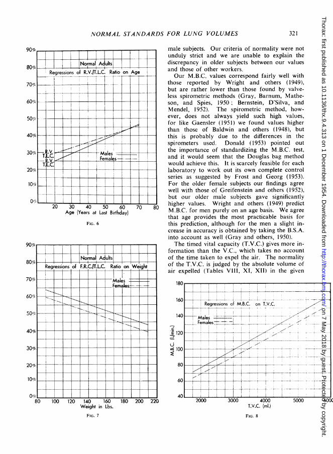
Figs. 3, 4, and 5) are rather lower than those foundby Robinson (1938), even allowing for the B.T.P.S.correction, but they are somewhat higher thanthose found by Baldwin and others (1948). Ourvalues for R.V./T.L.C. and F.R.C./T.L.C. ratios(Table VII, Figs. 6 and 7) are higher than thosefound by earlier workers (Kaltreider and others,1938; Robinson, 1938), but are in close agreementwith those found by Bates and Christie (1950), byWhitfield and others (1950), and by Greifensteinand others (1952). Like these more recent authorswe found a marked increase in the R.V./T.L.C.ratio with advancing age, but the F.R.C./T.L.C.ratio is much less affected (Table X, Fig. 7). Theformer rises because of change in the absolutevalue of both R.V. (increase) and T.L.C. (decrease),whereas the much smaller rise in the F.R.C. /T.L.C.
is due to the decreasing T.L.C., the F.R.C. chang-ing but little (Fig. 4). Our older subjects, whilethey showed a rather high R.V. /T.L.C. ratio,certainly did not suffer from emphysema, as theyshowed no excess dyspnoea on exertion, gave highM.B.C. volumes, had, normal mixing efficiency, andshowed no evidence of air trapping on the spiro-metric record. An R.V./T.L.C. ratio above 36%has often been accepted in itself as evidence ofemphysema (Baldwin and, others, 1949; Motley,1953 ; Galdston, Wolfe, and Steele, 1952; Greifen-stein and others, 1952), but the present results andthose of Bates and Christie (1950) and of Whit-field and others (1950) make this view difficult tomaintain. It may be that an increase in theF.R.C. /T.L.C. ratio will prove to be of moresignificance.The absence of really significant deterioration in
intrapulmonary gas-mixing up to the age of 70 yearsis in marked contrast with what is usually stated(Greifenstein and others, 1952; Bates and Christie,
319
on 7 May 2018 by guest. P
rotected by copyright.http://thorax.bm
j.com/
Thorax: first published as 10.1136/thx.9.4.313 on 1 D
ecember 1954. D
ownloaded from

C. D. NEEDHAM, MARY C. ROGAN, and 1. McDONALD
yIII-T 1950 ; Fowler, 1952). In this laboratory an M.E.
Normal Adults 1 t as low as 25 % is commonly found in the presence- - of pulmonary disease (emphysema, communicatingwns of V.C. on Age and Hleight lung cysts, etc.), but a value below 60% is seldom
found in normal subjects. In the first of these_ ._iP ;_t _papers we notice that the mixing defect is more
marked in the men than in the women, whereaswe find no difference between our own male andfemale groups (Table VIII). The M.B.C. in their
_ -_ female group is nearly the same as in ours, but-l ___ - _ in their men it is lower (78 1./im. as against 95
- - -- - - 1. /m.), which makes it doubtful whether the men
_ - -; - - in their series were as good a normal sample aswere the women. Comroe and Fowler (1951) re-
- - = - _ - - ported a much greater range of M.E.% in oldthan in young subjects, whereas we found it un-affected by age. Fowler (1949) in a series of 18subjects aged up to 73 years obtained normal
-___<lmixing values, and Briscoe, Becklake, and Rose(1951) found M.E.% normal in one older man
_3,0 40 50 60 70 -{ > (aged 75) and only moderately reduced in thekge (Years at Last Birthday) other (aged 65). Bates and Christie (1950) re-
FIC. s ported reduced mixing efficiency in 10 elderly
TABLE VIILUNG MEASUREMENTS
T.L.C. F.R.C.(mI.) (mI.)
Min. Max. Mean Min.
2,310 3,690 1,310 1,0202,600 3,930 1,550 1,1703,390 4,880 1,880 1.3903,240 5,920 1,920 1,2504,040 5,260 2,170 1,8303,850 6,730 2,460 1,4605,120 6,700 2,800 2,3305,140 7,320 2,980 2,5605,170 6,900 2,840 2,1105,000 8,600 3,210 1,9005,700 8,300 3,550 2,6005,100 7,200 3,300 1,9003,800 7,100 3,160 1,8005,100 7,500 3,560 2,700
4,290 5,150 3,120 2,830
2,320 2,860 1,170 1,0002,520 3,320 1,480 1,0002,610 4,280 1,760 1,3203,300 4,980 1,810 1,4003,050 4,850 1,920 1,5904,130 5,730 2,220 1,6903,740 5,380 2,020 1,5203,810 5,210 1,990 1,5304,040 4,940 2,080 1,680
3,500 5,600 2,340 1,7003,700 5,800 2,440 1,6003,400 5,200 2,070 1,5003,400 4,600 2,290 1,4002,900 4,700 2,340 i1,600
2,910 3,680 12,030 1,920
F.R.C.IT.L.C.
Max. Mean Min. Max. I
1,800 444 138 511,850 48-3 40 542,320 47-2 41 562,610 46-1 38 522,500 45.9 41 523,460 47-0 38 543,300 48-3 45 523,660 48-9 44 553,650 47-5 40 54
4,300 49-5 39 574,800 536 40 604,800 530 38 674,400 56-2 36 675,200 59 2 49 71
3,270 66-0 63 69
1,320 45.6 39 531,730 48-4 40 552,480 49-5 44 582,310 46-9 41 532,320 47-8 42 532,710 47-0 41 562,350 46-9 33 532,550 44-9 40 542,520 47 0 41 54
3,500 51-6 43 633,100 527 42 642,900 49-7 43 573,100 5511 42 713,300 57.5 42 70
2,180 64-0 59 68
T.L.C.= total lung capacity, F.R.C.= functional residual capacity, R.V.=-residual volume, R.E.R.V.=resting expiratory reserve volume.
320
R.V. iT.L.C.
Mean Min.1 Max.
RlV R..RR.E.R.V.
(ml.)n r Min. Max.
II
Sex
M
M
M
F
F
F
Age
111213141516171819
20-3030-4040-5050-6060-70
>70
111213141516171819
20-3030-4040-5050-6060-70
>70
6
8138888988
2723222010
3
8
88888
18818
131 1168
3
Mean
2,9603,2103,9604,1404,7305,1605,7706,1305,960
6,5006,6306,1905,6105,970
4,730
2,5703,0603,5303,8604,0404,6804,3304.4404,420
4,5604,6504,1404,1204,000
3,180
R.V.(ml.)
Min.Mm.
570570720750810760
1,0101,120550900
1,5001,2001,3002,200
2,370
480480750700850620840710910
1,000800
1,0001,2001,000
1,380
Mean
770810950980
1,1001,2901,5601,6101,380
1,7502,1102,1702,2002,700
2,420
630840950950
1,0401,2001,0701,0901,190
1,4601,5201,5001,6801,820
1,580
Max.
1,1601,1501,0801,3901,3201,9702,2002,1001,820
2,6003,0003,1002,9003,800
2,470
8901,1001,4801,1501,2302,0301,3201,3701,490
2,2002,0002,5002,1002,700
1,830
26-025-124-124-023124-626-926.223-6
26-632-034-839044.9
513
24-527-027 124-826-125 225-024-627031-932-735 640645*151.0
211819151917191913
1823242641
48
171922212015201922
2222303032
38
Meat
540750930950
1,0701,1701,2301,3701,460
1,4701,4301,140980860
710
540650810860880
1,010950900920
900950570610490
440
333331292835333533
3541444955
55
343634283235313133
3941494960
63
350410500500920700
1,000700950
900800500500300
480
400500500600620680620630510
600600200100000
150
770900
1,2401,7001,2601,9001,5402,0002,300
2,3001,9001,8001,8001,600
860
670830
1,1001,1601,3501,4301,3001,3001,270
1,3001,300900
1,2001,100800
on 7 May 2018 by guest. P
rotected by copyright.http://thorax.bm
j.com/
Thorax: first published as 10.1136/thx.9.4.313 on 1 D
ecember 1954. D
ownloaded from
NORMAL STANDARDS FOR LUNG VOLUMES
FIG. 6
90u7o
80% iI T | |INIalAUISRegressions of F.R.C./T.LC. Ratio on Weight_
70 M-malesFemnales
40%3091jc
30%6-e-__-_
2017c
IS;X0
80 100 120 140 160Weight in Lbs.
FIG. 7
180
male subjects. Our criteria of normality were notunduly strict and we are unable to explain thediscrepancy in older subjects between our valuesand those of other workers.Our M.B.C. values correspond fairly well with
those reported by Wright and others (1949),but are rather lower than those found by valve-less spirometric methods (Gray, Barnum, Mathe-son, and Spies, 1950; Bernstein, D'Silva, andMendel, 1952). The spirometric method, how-ever, does not always yield such high values,for like Gaensler (1951) we found values higherthan those of Baldwin and others (1948), butthis is probably due to the differences in thespirometers used. Donald (1953) pointed outthe importance of standardizing the M.B.C. test,and it would seem that the Douglas bag methodwould achieve this. It is scarcely feasible for eachlaboratory to work out its own complete controlseries as suggested by Frost and Georg (1953).For the older female subjects our findings agreewell with those of Greifenstein and others (1952),but our older male subjects gave significantlyhigher values. Wright and others (1949) predictM.B.C. for men purely on an age basis. We agreethat age provides the most practicable basis forthis prediction, although for the men a slight in-crease in accuracy is obtained by taking the B.S.A.into account as well (Gray and others, 1950).The timed vital capacity (T.V.C.) gives more in-
formation than the V.C., which takes no accountof the time taken to expel the air. The normalityof the T.V.C. is judged by the absolute volume ofair expelled (Tables VIII, XI, XII) in the given
200 220T.V.C. (ml.)
FIG. 8
321
I
on 7 May 2018 by guest. P
rotected by copyright.http://thorax.bm
j.com/
Thorax: first published as 10.1136/thx.9.4.313 on 1 D
ecember 1954. D
ownloaded from

TABLE VIII
LUNG MEASUREMENTS (CONTINUED)
V.C. T.V.C. (2 sec.) M.B.C. R.T.V. M.E.
Sex Age No. (l)(l)(.m. m. ~~Mean Min.~ Max. Mean Min. IMax. Mean Min. Max. Mean Min. Max. Mean Min. Max.
I1 8 2,180 1,740 2,460 2,170 1,740 2,460 57 40 8 1 470 360 670 78-8 54 10512 13 2,420 2,000 3,100 2,410 2,000 3,100 72 49 90 540 400 750 85-9 62 11213 8 3,010 2,500 3,800 3,010 2,500 3,800 87 71 110 510 370 880 80-6 65 10714 8 3,160 2,480 5,010 3,160 2,480 5,010 98 59 121 625 400 900 77-2 60 96
M 15 8 i3,640 3,160 4,020 3,640 3,160 4,020 113 94 133 710 480 960 91-6 70 11516 8 3,60 3,090 5,170 3,860 3,090 5,170 114 88 138 770 520 1,160 84.8 65 12-017 9 4,200 3,820 4,580 4,160 3,820 4,580 124 95 152 690 510 1,030 81-8 70 9518 8 4,520 3,800 5,400 4,520 3,800 5,400 137 105 165 710 560 1,200 94-9 72 10519 8 4,590 3,770 5,550 4,590 3,770 5,550 136 99 169 740 440 1,230 92-1 69 108
20-30 27 4,760 4,000 6,000 4,730 4,000 6,000 138 111 164 570 370 1,140 80-4 60 10630-40 23 4,510 3,600 5,400 4,460 3,600 5,400 129 99 171 660 520 1,180 77.4 61 94
M 40-50 22 4,010 3,300 5,000 3,870 3,300 4,800 123 82 154 700 490 1,100 82-9 65 11550-60 20 3,400 2,000 4,400 3,160 1,900 4,200 104 72 146 710 390 1,100 74-7 56 10160-70 10 3,280 2,900 3,900 2,970 2,300 3,700 89 64 122 780 490 1,400 76-0 63 87
M >70 3 2,320 1,920 2,680 2,130 1,580 2,600 68 40 95 550 430 780 59.0 44 7 1
11I 8 1,940 1,540 2,380 1,940 1,540 2,380 53 43 61 500 360 640 73.9 62 8812 8 2,220 ,1,900 2,520 2,220 1,900 I2-520 69 54 89 420 350 470 78-1 66 911 3 8 2,560 1,790 2,930 2,560 1,790 2,930 83 51 97 580 410 800 77.2 56 10414 8 2,900 2,440 3,830 2:890 2,440 3,830 89 68 110 480 270 650 78-4 56 113
F 1 5 8 3,000 2,200 3,900 3,000 2,200 3,900 90 74 102 560 450 670 77.1 63 10016 8 3,480 3,220 3,800 3,480 3,220 3,800 104 86 114 560 370 900 93*4 8 1 10017 8 3,260 2,800 4,330 3,230 2,800 4,330 110 91 142 460 340 570 87-8 68 10018 8 3,350 2,860 3,960 3,350 2,860 3,960 100 76 120 480 400 670 77-5 70 9719 8 3,200 2,880 3,720 3,200 2,880 3,720 103 90 120 550 450 690 81-6 60 91
20 30 18 3,090 2,500 4,200 3,080 2,500 4,200 96 68 122 570 390 930 82-8 64 , 9630-40 13 3,140 2,200 4,100 3,050 2,100 4,000 91 62 114 590 450 1,300 79-6 68 I100
F 40-50 1 1 2,640 2,300 33600 2,510 1,900 3,500 80 62 106 490 310 750 76-6 58 10050-60 16 2,440 2,100 2,800 .2,310 1,900 2,700 75 53 99 530 280 820 76-0 61 10760 70 8 2,180 1,800 2,700 2,040 1,600 2,500 66 54 78 570 400 860 76-2 58 89
F >70 3 1,600 1,080 2,300 1,490 1,080 2,000 63 48 78 590 320 750 64-0 48 8 1
TABLE IXLUNG MEASUREMENTS AND PHYSICAL CHARACTERISTICS: TOTAL CORRELATION COEFFICIENTS
Age ~~~~~~~~~~~~~LungMeasurementAe PhysicalGop CharacteristicR ... ,,M.E.(Years) Sex T.L.C. F.]R C ... R.V. / R. E. R.V. V.C. TVC M.B.C. R.T.V.
T.L.C. T.L.C. . . (2sec.)()Age . .. M 0-86 0.81 0-1-4 0-66 _0-00 0-71 0-86 0-86 0 83 0-45 0-24
F 0-75 0-62 -0 09 0-54 0-01 -48 0-72 0-72 0.75 0-07 0-21
Height .. M 0-88 0-82 0-10 0-62 --0-09 0-76 0-89 0-89 0-84 0-57 0-32F 0-88 0-80 0-03 0-65 0-01 0-66 0-84 0-84 0-84 0-24 0-30
11-19 Weight .. M 0-91 0 83 0-06 0-67 -0-05 0-74 0-91 0-92 0-89 0-59 0-31F 0-84 t 0 64 -0-22 0-50 -0-17 0-55 0-86 0-86 0-80 0-04 0-25
B.S. A. -. M 0-93 0-85 0-08 0-68 -006 0-76 0-93 10-93 0-90 0-59 0-30F 0-88 0-71 -0-15 0-56 -0-12 0-61 0-88 0-88 0-83 0-10 0-27
Sitting height M 0-92 0-86 0 09 0-73 002 0-71 0-91 0-91 0-88 0-63 0-32F 0-86 0-76 0 01 0-62 -0-01 0-62 0-82 0-82 0-87 0-23 0)28
Age . .. M I -0-36 0-03 0-39 0-49 0-77 - 056 -0-74 -078 -0-66 0.37 -0-16F -0-33 0-03 0-27 0.34 0-66 -0-51 -0-63 -067 -062 0-06 --0-25
Height -- M 0-55 0 28 -0 14 0-05 -0-28 0-38 0-58 0-55 0-41 0.19 0-14F 0-47 0.18 -018 0-00 -0-30 0-31 0-54 0-50 0-30 0-10 0-16
20 70 Weight .. M 0.22 -0-17 -0-48 -0-17 -0-34 -006 0-36 0-35 0-28 0-10 0-06F 0-07 -026 -0-49 -0-08 -0-13 -0-33 0-14 0-10 0-00 0-14 0-03
B.S.A. -. M 0.37 -0-03 -042 -0-10 -0-35 -0-07 0-48 0-46 0-37 0-14 0-10F 0-21 0-15 0-45 -006 -0-20 0-16 0-30 0-24 0-10 0-14 - 0-08
Sitting height M 0-46 0-18 -0-18 -0-03 -0-32 0-33 0-53 0-54 0-46 0.19 0-16F - 0-47 0-17 -0-16 -0-06 -0-37 0-318 0-58 0-56 0-38 0-27 0-23
T.L.C. =total lung capactty, F.R.C. =functional residual capacity, R.V. =residual volume, R.E.R.V. =resting expiratory reservevolume, V.C.=vital capaciiy, T.V.C.-stimed vital capacity, M.B.C. =maximum breathing capacity, R.T.V.=resting tidalvolume, M.E. mixing efficiency, B.S.A.~=body surface area.
For the number of cases from which these correlations have been calculated, the coefficient must be at least 0-2 in magnitudeto establish a significant association.
on 7 May 2018 by guest. P
rotected by copyright.http://thorax.bm
j.com/
Thorax: first published as 10.1136/thx.9.4.313 on 1 D
ecember 1954. D
ownloaded from

NORMAL STANDARDS FOR LUNG VOLUMES 323
TABLE XBEST REGRESSION EQUATIONS FOR EACH LUNG MEASUREMENT
Regression Coefficients (with Standard Errors)Lung
Mentre x< Age x egh x.SA> Sitting Constant Residual Coeffiinment k'(Completed Height (l. (sqhtx . S.n) Height in Standard VaitonYears) (in.) (lb.) (m m (in.) Equation Deviation Vaton
T.L.C. A* 100 (±40) 200 (±60) -6,480 460 10(ml.) B 90 (±30) 2,000 (±800) -3,900 360 9
C -10 (±5) 160 (±30) 1,500 (±500) -4,210 680 11D -10 (±5) 110 (±30) -2,180 540 12
F.R.C. A 80 (±30) 140 (±30) -3,600 340 16(ml.) B 85 (±7) -3,430 260 14
C 10 (±5) 180 (±30) -18 (±4) -6,560 590 18D -40 (±10) 5,000 (±1,400) -530 450 20
F.R.C./ A +47-2 4-5 10T.L.C. B +47-1 4-9 10(%x) C 0-24 (±0 05) 1-2 (±0 3) -0-23 (±0-04) -3-0 5 5 10
D 0-17 (±0 05) -0-19 (±0 04) I +70 4 6-1 11
R.V. A 110 (±12) -2,460 300 26(ml.) B 45 (±6) -1,790 210 21
C 24 (±3) 100 (±20) -10 (±3) -4,150 410 19D 9 (±3) +1,200 360 23
R.V./ A +24-9 4-7 19T.L.C. B +25*7 4-7 18(%) C 0-45 (±0 04) 0-6 (±0 2) -0-12 (±0-03) -7-1 4-4 13
D 0-34 (±0 05) -0 07 (±0 03) +31-3 5-4 15
R.E.R.V. A 1,100 (±130) -630 250 24(mI.) B 40 (±5) -1.640 190 22
C -14 (±3) 80 (±20) -8 (±2) -2,410 310 25D -7 (±2) -26 (±5) 3,500 (±800) -1,220 220 30
V.C. A 3,400 (±150) -1,690 360 10(ml.) B 2,400 (±160) -640 300 10
C -35 (±4) 110 (±20) - 1,910 440 1 1D -20 (±3) 100 (±20) -2,710 360 13
T.V.C. A 3,400 (±150) -1,700 360 10(ml.) B 2,400 (±160) -650 300 11
C -42 (±4) 100 (±20) -1,070 460 1 1D -23 (±3) 90 (±20) -2,040 370 14
M.B.C. A 108 (±6) -60 14 14(I./min.) B 2 (±0.7) 6-4 (±0 8) -149 10 11
C -1t1 (±0 2) 40 (±13) +94 18 14D -0-7 (±0 1) +113 13 16
R.T.V. A 44 (±6) -810 160 25(ml.) B 30 (±9) -500 (±200) -610 110 22
C 10 (±+2) 66 (±14) -2,050 200 30D 28 (±12) -370 154 28
M.E. A 0 8 (±0 3) +34-7 14 17(M) B 1-0 (±04) +18-8 13 16
C +78-7 1 1 14D -0-2 (±O-1) +87-0 I 14
*A=Males 11-19. B=Females 11-19. C=Males 20-70. D=Females 20-70.B.S A.= body surface area, T.L.C.=total lung capacity, F.R.C.=functional residual capacity, R.V.=residual volume,
R.E.R.V.=resting expiratory reserve volume, V.C.=vital capacity, T.V.C.=timed vital capacity, M.B.C.=maximumbreathing capacity, R.T.V. =resting tidal volume, M.E. =mixing efficiency.
(i) As an example of how the equations should be read from the table we give the M.B.C. equation for normal adultmales, as follows: M.B.C. = (- 11 x age) +(40 x B.S.A.) +94, where the units are as given in the table.
(ii) Roughly speaking, the error in prediction should be less than the residual standard deviation in two cases out of three,and less than twice the residual standard deviation in 19 cases out of 20.
(iii) The regression coefficients are not all significantly different between males and females, as may be seen from theirstandard errors, but since some of them are, no attempt has been made to combine the pairs of values.
Similar remarks apply to the table of simplified regression equations (Tables XI, XII, XIII).
on 7 May 2018 by guest. P
rotected by copyright.http://thorax.bm
j.com/
Thorax: first published as 10.1136/thx.9.4.313 on 1 D
ecember 1954. D
ownloaded from

C. D. NEEDHAM, MARY C. ROGAN, and 1. McDONALD
time, by the proportion which this volume repre-sents of the total V.C. (Table XIV), and also bythe shape of the expiratory tracing.
Using even the fast speed of the standard Palmerkymograph we were unable to measure T.V.C.accurately over less than a two-second period. Aone-second T.V.C. would have the advantage ofdiscriminating between those normal subjects withvery fast expiratory rate (complete in one second)and those with medium expiratory rate (requiringone to two seconds). However, the two-secondT.V.C. is usually adequate for clinical purposes,as patients may take more than 10 seconds to expelthe entire V.C. The actual M.B.C. can be fairlywell predicted in normal subjects from the two-second T.V.C. (Table XV, Fig. 8). Kennedy(1953) obtained a closer prediction of the M.B.C.by measuring the 0.75 second T.V.C. on a specialfast kymograph, but he examined a mixed groupof normal subjects and patients. It is possible thate-ven the two-second T.V.C. would correlate betterwith the M.B.C. in patients with prolonged expira-tory time than it does in normal subjects many ofwhom expel all, or nearly all, their V.C. in onesecond. It may be unnecessary to predict M.B.C.from T.V.C. because there seems to be no good
SIMPLIFIED REGRES,MEASUREMENT IN A4
SIONS ON BC
LungMeasurement
T.L.C. (ml.). AB
F.R.C. (ml.) AB
F.R.C. T.L.C. A(%/) . . B
R.V. (ml.) ..AB
RN'.IT.L.C. AMB
R.E.R.V. (ml.) AB
V.C. (ml.) ..AB
T.V.C. (ml.) AB
M.B.C. (I.,/min.) AB
R.T.V. (ml.) AB
M.E. AB
TABLE XII
SIMPLIFIED REGRESSION EQUAT-IONS FOR EACH LUNG
MEASUREMENT IN AGE GROUP 20-70 YEARS: (a) REGRES-
SIONS ON AGE AND/OR HEIGHT
LungMeasure-ment
T.L.C.(ml.)
F.R.C.(Ml.)
R.V.(MI.)
T.L.C.%R.E.R.V.
(ml.)V.C.
(Ml.)T.V.C.
(Ml.)M.B.C.(1./min.)R.T.V.
(MI.)ME(%
Regression
x Age x(Completed Height
Years) (in.)
C 180(±30)D 120(±30)C
C 19 (±3)D 9 (±3)C 0.43 (±0.04)D 0-33 (±0-05)C -17 (±3)D ll1(±2)C -35 (±4) ll0 (±20)D -20(±3) l00 (±20)C 42 (±4) l00 (±20)D -23(±3) 90 (±20)C -12 (±01I)D -07 (±01I)C 6 (±_l15)DCD
Resi-dual
Constant Stan-in dard
Equation Devia-tion
-5,980 690-3,220 550+3,330 680+2,300 490± 1,330 450±1,200 360
+16-3 4-8±22-7 5.5
± 1,930 340+1,190 260
1,910 440-2,710 360-1,070 460-2,040 370±170 18+ 113 13±420 220+550 160+78.7 11-4+78.7 III1
Coeffi-cientof
Varia-tion
I111320212123141527361 1131 114151633291414
C Males. D Females.
TABLE XIIISIMPLIFIED REGRESSION EQUATIONS FOR F.R.C. T.L.C.FOR AGE GROUP 20-70 YEARS: (b) REGRESSION ON
WEIGHT
TABLE XI RegressionLung Residual Coefficient
SION EQUATIONS FOR EACH LUNG Measure-' x Constant Standard ofkGE GROUP 11-19 YEARS: REGRES- ment Weight in Deviation Variation)DY SURFACE AREA ALONE (lb.) Equation
F.R.C. c 0-17 (±0.03) +79.1 6-3 12Regression Coeffi- T.L.C.(', D 0 17 (± 0-04 +74.9 66 1 2
Residual cient _____________________________
x B.S.A. Constant Standard ofsqm) in Deviation Vansa- C =Males. D =-Females.sqm) Equation lion
4,500 (±200) -2,220 500 I113,100 (±200) -670 380 102,200 (±200) -1,150 360 171,300 (±140) -80 300 16 TABLE XIV
+47-2 4-5 10 NORMAL VALUES OF (T.V.C./V.C. x 100): DISTRIBUTION±47-1 4*9 10 BY AGE AND SEX
l,l00 (±130) - 520 320 28 ____ __________ ___________
700 (±ll01) -30 230 23+24.9 4.7 19 Age Distributions of Values of T.V.C. /V.C. x 100+25-7 4*7 18 Group ----I1,100 (±100) -630 250 24 (Years) <5 >5 Total' 8 85'>9 Total
650 (±100) -120 190 23 < 585-95 >9 NO.1 <8 18-5 5 No.3,400 (±150) -1690 360 102,400 (± 160) 640 300 10 No. of Males No. of Females3,400 (±150) 1,700 360 10 11-19 .. 0 1 77 78 0 1 71 722,400 (±160) -650 300 1 1 20-30 .. 0 1 26 27 0 0 18 18
108 (±6) -60 14 14 30-40. 0 1 22 23 0 3 10 1377 (±5) -24 12 13 40-50. 0 7 15 22 2 3 6 1 1450 (±70) -50 170 27 50-60. 3 9 8 20 0 9 7 16
+510 120 24 60-70. 2 7 1 10 0 4 4 8±85-3 15 18 >70. 1 I 1 3 0 1 2 3±80.7 13 16 ____________________________
_________________ ___ __________ Total 6 27 150 183 2 21 1118 141
T.L.C. =total lung capacity, F.R.C. =functional residual capacity,R.V. =residual volume, R.E.R.V. =resting expiratory reserve volume,V.C.=vital capacity, T.V.C.=timed vital capacity, M.B.C.=maxi-mum breathing capacity, R.T.V. =resting tidal volume. M.E. =mixingefficiency.
324
A= Males. B Females.
B.S.A. - body surface area, T.L.C. =total lung capacity, F.R.C.functional residualUcapacity, R.V. =residual volume, R.E.R.V. =restingexpiratory reserve volume, V.C. =vital capacity., T.V.C. =timed vitalcapacity, M.B.C.=-maximum breathing capacity. R.T.V.=restingtidal volume, M.E. =mixing efficiency.
-1-
on 7 May 2018 by guest. P
rotected by copyright.http://thorax.bm
j.com/
Thorax: first published as 10.1136/thx.9.4.313 on 1 D
ecember 1954. D
ownloaded from

NORMAL STANDARDS FOR LUNG VOLUMES
TABLE XVPREDICTION OF M.B.C. FROM T.V.C. (2 SEC.)
Group Regression Equation Residual CoefficientRegreCssion.)Standard ofCases (M.B.C. in 1./min.: T.V.C. in ml.) Deviation Variation______ _____ ~~~~~~~(%)
Males, M.B.C. =(0-028xT.V.C.)+6-3 16 151 1-19 years ±0-002Females, M.B.C. = (0-028 x T.V.C.) + 8-4 12 1311-19 years ±0-002Males, M.B.C. = (0-022 x T.V.C.) + 33*2 16 1320-70 years ±0-002Females, M.B.C. = (0-021 x T.V.C.) +27-6 1 1 1420-70 years ± 0-002
M.B.C. =maximum breathing capacity, T.V.C. = timed vital capacity.(i) Four equations are given, one for each group of cases. These
could not properly be combined, since the residual variation issignificantly higher for males than for females, and the regressioncoefficients themselves are significantly higher for the adults than forthe 11-19 age group.
(ii) It will be noted that in the cases of the adults, these equationsgive better predictions than do the regressions on physical charac-teristics, but this is not true of the 11-19 age group.
reason why the T.V.C. absolute value itself shouldnot be used as a valid measure of ventilatorycapacity. This would not be quite the same thingas the M.B.C., because it takes no account of theexhaustion factor, but, as this is chiefly prominentin patients who are unsuitable for the M.B.C. test,it is not a very weighty objection.
SUMMARYComment is made on the inadequacy of present
standards for the ventilatory and distributiveaspects of pulmonary function.Lung volumes, intrapulmonary gas-mixing effi-
ciency, timed vital capacity, and maximum breath-ing capacity have been measured in a total of 324normal male and female subjects of whom 150were under 20 years and 60 were over 50 yearsof age.The results have been subjected to statistical
analysis. The interrelations of the functions withage and body measurements have been studied, andregression equations have been evolved to allowprediction of expected normal values. A numberof the equations are also presented in graphicalform.The findings in the present study are briefly
discussed in relation to those previously reported.
The expenses of this investigation were covered bya grant from the Aberdeen Royal Infirmary Endow-ment Fund, for which we wish to thank the Board ofManagement. We are much indebted to Dr. A. M.Michie (Medical Superintendent) for his ready help inadministrative matters at all times. Professor H. W.Fullerton kindly arranged for this work to be carriedout in the Department of Medicine, and gave otheradministrative assistance. In the early days when we
were building up and calibrating the apparatus we
received much helpful advice from Professor R. V.Christie (St. Bartholomew's Hospital) and from Mr.H. Griffith (Senior Lecturer in Natural Philosophy,University of Aberdeen). The number of subjects andof people who assisted in marshalling them for testingis too large to allow individual recognition, but we
wish to thank them all for the essential part whichthey played.
REFERENCESAnthony, A. J. (1930). Dtsch. Arch. klin. Med., 167, 129.Aslett, E. A., D'Arcy Hart, P., and McMichael, J. (1939). Proc. roy.
Soc. B, 126, 502.Baldwin, E. DeF., Cournand, A., and Richards, D. W. (1948).
Medicine, Baltimore, 27, 243.-(1949). Ibid., 28, 201.
Bates, D. V., and Christie, R. V. (1950). Clin. Sci., 9, 17.Bernstein, L., D'Silva, J. L., and Mendel, D. (1952). Thorax, 7, 255.Binger, C. A. L. (1923). J. exp. med., 39, 457.Birath, G. (1944). Acta med. scand., Suppl. 154.Briscoe, W. A., Becklake, M. R., and Rose, T. F. (1951). Clin.
Sci., 10, 37.Christie, R. V. (1932). J. clin. Invest., 11, 1099.Comroe, J. H. (Editor) (1950). Meth. med. Res., 2, 74.-(1951). Amer. J. Med., 10, 356.-and Fowler, W. S. (1951). Ibid., 10, 408.Cournand, A., Baldwin, E. DeF., Darling, R. C., and Richards,
D. W. (1941). J. cdin. Invest., 20, 681.Richards, D. W., and Darling, R. C. (1939). Amer. Rev.Tuberc., 40, 487.
Darling, R. C., Cournand, A., and Richards, D. W. (1944). J. clin.Invest., 23, 55.
Donald, K. W. (1953). Brit. med. J., 1, 415, 473.Du Bois, E. F. (1927). Basal Metabolism in Health and Disease,
p. 19, 2nd ed. Philadelphia.Fletcher, C. M. (1953). Proc. roy. Soc. Med., 46, 538.Fowler, W. S. (1949). J. appl. Physiol., 2, 283..7l (1952). Physiol. Rev., 32, 1.Frost, J., and Georg, J. (1953). Acta med. scand., 147, 349.Gaensler, E. A. (1951). Amer. Rev. Tuberc., 64, 256.Galdston, M., Wolfe, W. B., and Steele, J. M. (1952). J. appl.
Physiol., 5, 17.Gilson, J. C., and Hugh-Jones, P. (1949). Clin. Sci., 7, 185.
and Oldham, P. D. (1952). Proc. roy. Soc. Med., 45, 584.Gray, J. S., Barnum, D. R., Matheson, H. W., and Spies, S. N. (1950).
J. clin. Invest., 29, 677.Greifenstein, F. E., King, R. M., Latch, S. S., and Comroe, J. H.
(1952). J. appl. Physiol., 4, 641.Hermannsen, J. (1933). Z. ges. exp. Med., 90, 130.Hurtado, A., and Boiler, C. (1933). J. clin. Invest., 12, 793.
and Fray, W. W. (1933). Ibid., 12, 807.- Kaltreider, N. L., and Brooks, W. D. W. (1934). Ibid.,13, 169.
Hutchinson, J. (1846). Med.-chir. Trans., 29, 137.Kaltreider, N. L., Fray, W. W., and Hyde, H. van Z. (1938). Amer.
Rev. Tuberc., 37, 662.Kelly, H. G. (1933). Univ. Ia Stud. Child Welf., 7, No. 5.Kennedy, M. C. S. (1953). Thorax, 8, 73.Lester, C. W., Cournand, A., and Riley, R. L. (1942). J. thorac.
Surg., 11, 529.Lindhard, J. (1925). Skand. Arch. Physiol., 47, 188.Lundsgaard, C., and Schierbeck, K. (1923). Acta med. scand., 58,
470.and Van Slyke, D. D. (1918). J. exp. Med., 27, 65.
Meneely, G. R., and Kaltreider, N. L. (1949). J. clin. Invest., 28, 129.Motley, H. L. (1953). Dis. Chest. 24, 378.Myers, J. A. (1925). Vital Capacity of the Lungs. Baltimore.Pappenheimer, J. (1950). Fed. Proc., 9, 602.Robinson, S. (1938). Arbeitsphysiologie, 10, 251.Roelsen, E. (1939). Acta med. scand., 98, 141.Stewart, C. A. (1922). Amer. J. Dis. Child., 24, 451.Tiffeneau, R., Bousser, J., and Drutel, P. (1949). Paris med. (partie
mid), 39, 543.Turner, J. A., and McLean, R. L. (1951). Pediatrics, 7, 360.West, H. F. (1920). Arch. intern. Med., 25, 306.Whitfield, A. G. W., Waterhouse, J. A. H., and Arnott, W. Melville
(1950). Brit. J. soc. Med., 4, 1, 86, and 113.Wright, G. W., Yee, L. B., Filley, G. F., and Stranahan, A. (1949).
J. thorac. Surg., 18, 372.
325
on 7 May 2018 by guest. P
rotected by copyright.http://thorax.bm
j.com/
Thorax: first published as 10.1136/thx.9.4.313 on 1 D
ecember 1954. D
ownloaded from
Related Documents


