The Journal of Neuroscience, October 1992, 12(10): 39853991 Noradrenergic Inhibition of Synaptic Transmission between Mitral and Granule Cells in Mammalian Olfactory Bulb Cultures Paul Q. Trombley and Gordon M. Shepherd Section of Neurobiology, Yale University Medical School, New Haven, Connecticut 06510 Noradrenergic modulation of the glutamatergic-GABAergic synapses between mitral/tufted (M/T) and granule cells has been implicated in some forms of olfactory learning (Brennan et al., 1990). Norepinephrine (NE) has been shown to dis- inhibit mitral cells (Jahr and Nicoll, 1982), but its site of action is not well defined. The effects of NE on synaptic transmis- sion between monosynaptically coupled pairs of mitral and granule cells have been examined using primary culture and whole-cell recording techniques. Intracellular stimulation of M/T cells evoked dual-component EPSPs in granule cells consisting of both NMDA and AMPA (a-amino-3-hydroxy-5- methyl-4-isoxaroleproprionic acid) receptor-mediated mechanisms. The EPSPs were reversibly inhibited by ap- proximately 50% during application of 30 PM NE. NE had no effect, however, on the membrane current evoked by ex- ogenous application of glutamate, indicating a presynaptic site of action. The effect of NE on EPSPs was mimicked by the a-adrenergic agonist clonidine but not by the fl-adren- ergic agonist isoproterenol. NE had no significant effect on either accommodation or macroscopic currents in either M/T or granule cells. NE also inhib:+ad spontaneous GABAergic IPSPs recorded in M/T cells, by a presynaptic a-adrenergic- mediated mechanism. These results support previous re- sults suggesting a disinhibitory role for NE in the olfactory bulb. This action, however, is at least in part mediated by a reduction in mitral cell-mediated granule cell excitation. In recent years the olfactory system hasemerged as a model in which well-defined behavioral consequences of learning can be correlated with cellular and molecular mechanisms, including changes in physiology, anatomy, and metabolism(Coopersmith and Leon, 1984; Wilson et al., 1987; Woo et al., 1987). Much of this experience-dependent plasticity takes place in the olfac- tory bulb, where recent evidence suggests that modulation of the synaptic connectionsbetween mitral and granule (GR) cells mediate thesechanges (Brennan et al., 1990). Olfactory learning is critically influenced by interactions be- tweenglutamate, GABA, and norepinephrine (NE) (for reviews, seeLeon, 1987; Brennan et al., 1990), transmitters that have been implicated in synaptic modulation/plasticity in several regions of the mammalian brain, including the hippocampus Received Mar. 2, 1992; revised May 11, 1992; accepted May 13, 1992. This work was supported by NRSA Grant F32 DC 00072 01 to P.Q.T. and by research grants from the NIDCD and NINDS to G.M.S. We thank Dr. Charles Greer for reading earlier versions of the manuscript. Correspondence should be addressed to Paul Q. Trombley, Section of Neuro- biology, Yale Medical School, 333 Cedar Street, New Haven, CT 065 10. Copyright 0 1992 Society for Neuroscience 0270-6474/92/123985-07$05.00/O (Hopkins and Johnston, 1984; Davies et al., 1991)and the visual cortex (Bear and Singer, 1986; Kleinschmidt et al., 1987). Most excitatory synaptic pathways use glutamate as a transmitter (Mayer and Westbrook, 1987). In the olfactory bulb, the excit- atory connections include the mitral/tufted (M/T) cell dendro- dendritic, recurrent axon collateral synapses within the bulb, and the M/T cell output via the lateral olfactory tract to the olfactory cortex (Mori, 1987; Trombley and Westbrook, 1990). The main inhibitory transmitter in the mammalian brain is GABA (Stepheson and Dolphin, 1989). GABAergic intemeu- rons in the olfactory bulb include both internal GR cellsin the GR cell layer and external GR cells(periglomerular cells)in the glomerular layer (Ribak et al., 1977; Jahr and Nicoll, 1980; Nowycky et al., 198 1). These two populations of GABAergic neurons reflect the two levels of local circuit processing that occur in the olfactory bulb, the external plexiform layer and the glomerular layer, respectively. The olfactory bulb also receives a denseprojection of nor- adrenergic fibers from the locus coeruleus that terminate at all levels of the olfactory bulb (Shipley et al., 1985). Recent evi- dence demonstrated that some forms of olfactory learning re- quire the activation of adrenergicreceptors(Kaba and Keveme, 1988; Sullivan et al., 1989). It was further suggested that a locus for learning is the putative glutamatergic-GABAergic synaptic contacts between mitral and GR cells (Brennan et al., 1990). Although detailed studies examining the synaptic effectsof NE are lacking, in the turtle olfactory bulb, NE acts to disinhibit mitral cells (Jahr and Nicoll, 1982). However, the site of NE action and the mechanismare not yet clearly defined. In this report, we use primary olfactory bulb cultures and whole-cell recording techniques to examine the effectsof NE on identified mitral/GR cell pairs. In particular, we sought to ex- amine potential neuromodulatory mechanisms at the level of the single synapse that might be relevant to olfactory learning. We report here that NE acts to reduceexcitatory synaptic trans- mission between monosynaptic pairs of mitral/GR cells. This effect may enhancemitral cell excitability through a reduction in reciprocal inhibition from GR cells. Materials and Methods Cell culture. Primary dissociated cultures of the olfactory bulb were prepared using methods similar to those described in Trombley and Westbrook (1990). The olfactory bulbs were dissected from I-2-d-old rat pups and cut into several pieces after the meninges were removed. The tissue was incubated at 37°C for 45-60 min in a calcium-buffered solution containing 20 U/ml papain (Worthington) and 1 mM cysteine. The tissue was gently triturated using a fire-polished Pasteur pipette, after inactivation of the enzyme with 2.5 mg/ml bovine serum albumin and 2.5 mg/ml trypsin inhibitor (Sigma, type II). The cell suspension

Welcome message from author
This document is posted to help you gain knowledge. Please leave a comment to let me know what you think about it! Share it to your friends and learn new things together.
Transcript

The Journal of Neuroscience, October 1992, 12(10): 39853991
Noradrenergic Inhibition of Synaptic Transmission between Mitral and Granule Cells in Mammalian Olfactory Bulb Cultures
Paul Q. Trombley and Gordon M. Shepherd
Section of Neurobiology, Yale University Medical School, New Haven, Connecticut 06510
Noradrenergic modulation of the glutamatergic-GABAergic synapses between mitral/tufted (M/T) and granule cells has been implicated in some forms of olfactory learning (Brennan et al., 1990). Norepinephrine (NE) has been shown to dis- inhibit mitral cells (Jahr and Nicoll, 1982), but its site of action is not well defined. The effects of NE on synaptic transmis- sion between monosynaptically coupled pairs of mitral and granule cells have been examined using primary culture and whole-cell recording techniques. Intracellular stimulation of M/T cells evoked dual-component EPSPs in granule cells consisting of both NMDA and AMPA (a-amino-3-hydroxy-5- methyl-4-isoxaroleproprionic acid) receptor-mediated mechanisms. The EPSPs were reversibly inhibited by ap- proximately 50% during application of 30 PM NE. NE had no effect, however, on the membrane current evoked by ex- ogenous application of glutamate, indicating a presynaptic site of action. The effect of NE on EPSPs was mimicked by the a-adrenergic agonist clonidine but not by the fl-adren- ergic agonist isoproterenol. NE had no significant effect on either accommodation or macroscopic currents in either M/T or granule cells. NE also inhib:+ad spontaneous GABAergic IPSPs recorded in M/T cells, by a presynaptic a-adrenergic- mediated mechanism. These results support previous re- sults suggesting a disinhibitory role for NE in the olfactory bulb. This action, however, is at least in part mediated by a reduction in mitral cell-mediated granule cell excitation.
In recent years the olfactory system has emerged as a model in which well-defined behavioral consequences of learning can be correlated with cellular and molecular mechanisms, including changes in physiology, anatomy, and metabolism (Coopersmith and Leon, 1984; Wilson et al., 1987; Woo et al., 1987). Much of this experience-dependent plasticity takes place in the olfac- tory bulb, where recent evidence suggests that modulation of the synaptic connections between mitral and granule (GR) cells mediate these changes (Brennan et al., 1990).
Olfactory learning is critically influenced by interactions be- tween glutamate, GABA, and norepinephrine (NE) (for reviews, see Leon, 1987; Brennan et al., 1990), transmitters that have been implicated in synaptic modulation/plasticity in several regions of the mammalian brain, including the hippocampus
Received Mar. 2, 1992; revised May 11, 1992; accepted May 13, 1992.
This work was supported by NRSA Grant F32 DC 00072 01 to P.Q.T. and by research grants from the NIDCD and NINDS to G.M.S. We thank Dr. Charles Greer for reading earlier versions of the manuscript.
Correspondence should be addressed to Paul Q. Trombley, Section of Neuro- biology, Yale Medical School, 333 Cedar Street, New Haven, CT 065 10.
Copyright 0 1992 Society for Neuroscience 0270-6474/92/123985-07$05.00/O
(Hopkins and Johnston, 1984; Davies et al., 199 1) and the visual cortex (Bear and Singer, 1986; Kleinschmidt et al., 1987). Most excitatory synaptic pathways use glutamate as a transmitter (Mayer and Westbrook, 1987). In the olfactory bulb, the excit- atory connections include the mitral/tufted (M/T) cell dendro- dendritic, recurrent axon collateral synapses within the bulb, and the M/T cell output via the lateral olfactory tract to the olfactory cortex (Mori, 1987; Trombley and Westbrook, 1990). The main inhibitory transmitter in the mammalian brain is GABA (Stepheson and Dolphin, 1989). GABAergic intemeu- rons in the olfactory bulb include both internal GR cells in the GR cell layer and external GR cells (periglomerular cells) in the glomerular layer (Ribak et al., 1977; Jahr and Nicoll, 1980; Nowycky et al., 198 1). These two populations of GABAergic neurons reflect the two levels of local circuit processing that occur in the olfactory bulb, the external plexiform layer and the glomerular layer, respectively.
The olfactory bulb also receives a dense projection of nor- adrenergic fibers from the locus coeruleus that terminate at all levels of the olfactory bulb (Shipley et al., 1985). Recent evi- dence demonstrated that some forms of olfactory learning re- quire the activation of adrenergic receptors (Kaba and Keveme, 1988; Sullivan et al., 1989). It was further suggested that a locus for learning is the putative glutamatergic-GABAergic synaptic contacts between mitral and GR cells (Brennan et al., 1990). Although detailed studies examining the synaptic effects of NE are lacking, in the turtle olfactory bulb, NE acts to disinhibit mitral cells (Jahr and Nicoll, 1982). However, the site of NE action and the mechanism are not yet clearly defined.
In this report, we use primary olfactory bulb cultures and whole-cell recording techniques to examine the effects of NE on identified mitral/GR cell pairs. In particular, we sought to ex- amine potential neuromodulatory mechanisms at the level of the single synapse that might be relevant to olfactory learning. We report here that NE acts to reduce excitatory synaptic trans- mission between monosynaptic pairs of mitral/GR cells. This effect may enhance mitral cell excitability through a reduction in reciprocal inhibition from GR cells.
Materials and Methods
Cell culture. Primary dissociated cultures of the olfactory bulb were prepared using methods similar to those described in Trombley and Westbrook (1990). The olfactory bulbs were dissected from I-2-d-old rat pups and cut into several pieces after the meninges were removed. The tissue was incubated at 37°C for 45-60 min in a calcium-buffered solution containing 20 U/ml papain (Worthington) and 1 mM cysteine. The tissue was gently triturated using a fire-polished Pasteur pipette, after inactivation of the enzyme with 2.5 mg/ml bovine serum albumin and 2.5 mg/ml trypsin inhibitor (Sigma, type II). The cell suspension
3966 Trombley and Shepherd - NE Inhibits Synaptic Transmission in the Olfactory Bulb
was plated at low density (26,00O/cm*) in 35 mm culture dishes (Corn- ing) containing a previously prepared confluent layer of olfactory bulb astrocytes. The neuronal growth medium contained 95% Minimal Es- sential Medium (MEM; GIBCO) with 5% horse serum, 6 gm/liter glu- cose, and a nutrient supplement (Serum Extender, Collaborative Re- search, Inc.). Astrocyte feeder layers were prepared by plating a suspension of olfactory bulb cells, prepared as described above, in a 75 cm* flask containing MEM with 10% fetal calf serum and 6 gm/liter glucose. When the astrocytes reached confluence, they were removed with 0.125% tryp- sin, resuspended, and plated onto 35 mm dishes coated with collagen (Vitrogen, Collagen Corp.) and poly-L-lysine (30,000-70,000 MW, 10 p&ml; Sigma). 5’-Fluoro-2-deoxyuridine and uridine (15 and 35 &ml, respectively) were added 1 d after plating neurons to prevent overgrowth of background cells. Electrophysiological recordings were made after 7- 10 d in culture.
Presumptive M/T and GR cells were identified based on the mor- phological, physiological, and immunohistochemical criteria estab- lished in Trombley and Westbrook (1990). Briefly, the cultures con- tained two morphologically distinct populations of neurons: a small number of large-diameter pyramidal-shaped neurons (20-40 Km soma) and a much larger population of small-diameter bipolar neurons (5-10 pm soma). These correlate with M/T and granule/periglomerular cells, respectively. Intracellular stimulation of the neurons with morphology reflecting M/T cells in vivo invariably evoked EPSPs in adjacent neurons. Intracellular stimulation of the small bipolar neurons, however, evoked picrotoxin-sensitive IPSPs. These morphologically distinct populations could also be identified by immunohistochemical markers. The large pyramidal neurons, the presumptive M/T cells, were N-acetylaspartyl- glutamate immunoreactive; in contrast, the small bipolar neurons, pre- sumptive granule/periglomerular neurons, were glutamic acid decar- boxylase immunoreactive.
Electrophysiology. Voltage- and current-clamp recordings were per- formed at room temperature on the stage of an inverted phase-contrast microscope. The recording chamber was perfused at 0.5-2.0 ml/min with a solution containing (in mM) NaCl, 162.5; KCl, 2.5; CaCl,, 2; HEPES, 10; glucose, 10; MgCl,, 0 or 1; and glycine, 0 or 1 PM. The pH was adjusted to 7.3 with NaOH and the final osmolarity was 325 mOsm. Picrotoxin (20 PM) was used in some experiments to block inhibitory synaptic activity. Patch electrodes were pulled from borosilicate glass, fire polished, and filled with solution containing (in mM) KMeSO, or CsCl, 145; MgCl,, 1; HEPES, 10; Mg-ATP, 4; Mg-GTP, 0.5; and EGTA, 1.1 or 11; pH 7.2, osmolarity 3 10. Electrode resistances were 4-6 Ma. Drugs were diluted in the recording solution and applied using a gravity- fed flow pipe perfusion system, consisting of an array of 400~pm-i.d. glass barrels. The flow pipes were aligned with the neuron using a hy- draulic manipulator, and flow was controlled with pinch clamps. Al- though no attempt was made in these experiments to optimize the speed of solution changes, peak drug responses occurred within 200-500 msec. Neurons were always bathed with fast flow from one barrel containing control solution except during application of drugs. Drugs applied were 6-cyano-2,3-dihydroxy-7-nitroquinoxaline (CNQX; Cambridge Re- search Biochemicals); DL-2-amino-5-phosphonovalerate (AP5), nor- epinephrine (NE), clonidine, isoproterenol, glutamate, picrotoxin, and GABA (Sigma).
Experimentalprocedures. To examine excitatory postsynaptic poten- tials (EPSPs), simultaneous whole-cell recordings were made from monosynaptically coupled M/T-GR cell pairs in current-clamp mode with an Axoclamp 2A amplifier (Axon Instruments). Both patch elec- trodes contained KMeSO,-based solutions; membrane potentials were maintained near - 60 mV using small holding currents (? 20 PA). Action potentials were evoked in the M/T neuron by injection of a brief (2 msec) depolarizing current pulse at 0.25-0.5 Hz. EPSPs were considered monosynaptic if there was no intertrial variation in latency and the latency was < 5 msec. The evoked EPSPs were filtered at l-3 kHz with an eight-pole Bessel filter, digitized at 5-10 kHz, and analyzed using pCLAMP software (Axon Instruments) on an IBM-386 computer.
Whole-cell recordings were made from M/T cells in primary cultures of olfactory bulb neurons plated at 50,000-75,00O/cm* to examine the effects of NE, clonidine, and isoproterenol on spontaneous IPSPs. Ex- periments examining the direct effects of NE on M/T and GR cells used patch electrodes containing a KMeSO,-based solution.
To examine responses evoked by glutamate and GABA, whole-cell voltage-clamp recordings were made using the discontinuous single- electrode voltage-clamp mode at a switch frequency of 1 O- 15 kHz. The patch electrodes contained a CsCl-based solution, and tetrodotoxin (300
nM) was added to the extracellular solution. Agonist-evoked currents were filtered at l-3 kHz and digitized at 5-10 kHz.
All data are expressed as me& + SD.
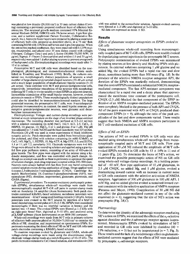
Results Effects of glutamate receptor antagonists on EPSPs evoked in GR cells Using simultaneous whole-cell recording from monosynapti- tally coupled pairs of M/T-GR cells, EPSPs were readily evoked by depolarizing current injection in the presynaptic M/T neuron. Polysynaptic contamination of evoked EPSPs was minimized by plating neurons at low density and blocking IPSPs with pic- rotoxin. In external solutions containing 1 PM glycine and no added Mg, the evoked EPSPs had a rapid rise rate and slow decay, sometimes lasting more than 500 msec (Fig. 1B). In the presence of the selective NMDA receptor antagonist AP5, the duration of the EPSPs was markedly reduced, demonstrating that the control EPSPs contained a substantial NMDA receptor- mediated component. The fast APS-resistant component was characterized by a rapid rise and a decay phase that approxi- mated the membrane time constant of the postsynaptic cell. CNQX (2.5 FM) blocked the fast component of the EPSP in- dicative of an AMPA receptor-mediated potential. The EPSPs were completely blocked in the presence of both AP5 and CNQX. All of the pairs examined with AP5 or CNQX produced dual- component EPSPs (n = 9), although the ratio of the peak am- plitudes of the fast and slow components varied. These results suggest that both NMDA and AMPA receptors participate in M/T cell-mediated excitation of GR cells.
Effects of NE on EPSPs
The actions of NE on evoked EPSPs in GR cells were also studied using simultaneous whole-cell recordings from mono- synaptically coupled pairs of M/T and GR cells. Flow pipe application of 30 PM NE reduced the amplitude of M/T cell- evoked EPSPs recorded in GR cells by 53 + 14% in 11 out of 11 pairs (Fig. 2A). To determine the locus of NE’s action, we examined the possible postsynaptic action of NE on GR cells using whole-cell voltage-clamp recordings. At a holding poten- tial of -60 mV, flow pipe application of 10 PM glutamate, in 2.5 KM CNQX, no added Mg, and 1 PM glycine, evoked a desensitizing inward current with an increase in current noise in GR cells consistent with the selective activation of NMDA receptors. Application of 500 KM glutamate in 100 PM AP5, 1 mM Mg, and no added glycine evoked a sustained inward cur- rent consistent with the selective application of AMPA receptors (Patneau and Mayer, 1990). Coapplication of 30 PM NE did not affect the glutamate-mediated current in any of the cells examined (n = 6), suggesting that the site of NE’s action was presynaptic (Fig. 2&C).
Pharmacology To determine the identity of the adrenergic receptors mediating NE’s action on EPSPs, we examined the effects of the qselective agonist clonidine and the p-adrenergic receptor agonist isopro- terenol. Monosynaptic EPSPs evoked by stimulation of M/T and recorded in GR cells were inhibited by clonidine (40 * 13% reduction; n = 5) but not by isoproterenol (n = 7; Fig. 3). These results and the lack of a noradrenergic effect on glutamate- mediated currents suggest that the effects of NE were mediated by presynaptic cu,-adrenergic receptors.

The Journal of Neuroscience, October 1992, Y2(10) 3987
Control
Mitral Cell
10mV
40 mV L 40 ms 10 ms
Figure 1. Mitral cell-evoked EPSPs in GR cells are mediated by both NMDA and AMPA receptors. A, Phase-contrast photograph of a putative M/T (MT) cell and three putative GR cells from the olfactory bulb of a neonatal rat pup after 7 d in primary culture. Scale bar, 40 cm. B, Simultaneous whole-cell recordings were made from a monosynaptically coupled M/T-GR cell pair. In whole-cell current-clamp mode, a 2 msec depolarizing current pulse generated an action potential in the M/T cell (Lower trace). The M/T cell action potential evoked an EPSP in the GR cell (upper truces). Plow pipe application of 100 PM AP5 blocked a slow component of the response that was resistant to 2.5 hM CNQX. The results were indicative ofa dual postsynaptic receptor mechanism as coapplication of AP5 and CNQX completely blocked the response. Extracellular solution contained 1 pM glycine and no added Mg. KMeSO, patch solution. Membrane potential, - 60 mV.

3988 Trombley and Shepherd - NE inhibits Synaptic Transmission in the Olfactory Bulb
Figure 2. NE reversibly inhibited EPSPs. A, An EPSP in a CR cell was evoked by intracellular stimulation of a monosynaptically coupled M/T cell. Flow pipe application of 30 PM NE re- versibly reduced the EPSPs by approx- imately 50%. KMeSO, patch solution. B, Under voltage clamp, flow pipe ap- plication of 10 pM glutamate results in an inward current through NMDA re- ceptors in a GR cell that was not an- tagonized by coapplication of 30 PM NE. External solution: 2.5 PM CNQX, 0 Mg, 1 PM glycine. C, Flow pipe ap- plication of 500 PM glutamate in the presence of 1 mM Mg, no added gly- tine, and 100 ELM AP5 evoked an in- ward current through AMPA receptors that also was not antagonized by coap- plication of 30 PM NE. A-C, Holding potential, -60 mV. Band C, CsCl patch solution.
A Control/Wash
10mV
100 ms
30 PM NE B __ C
30 NM NE
10 HIM Glutamate
--I
*wxyxr~*l 500 jiM Glutamate r
Direct effects of NE We used both whole-cell current- and voltage-clamp recordings to reveal any direct effects NE might have on either M/T or GR cells. Under voltage clamp, flow pipe application of 30 PM NE
Control
---. -
-
Clonidine Noradrenergic disinhibition
lsoproteronol
NE dramatically reduced spontaneous GABAergic IPSPs re- corded in M/T cells (76 f 17% reduction; n = 4). This effect was mimicked by the a,-adrenergic receptor agonist clonidine (Fig. 54) but not by the P-adrenergic agonist isoproterenol (not shown). To determine if the effect of NE was via inhibition of postsynaptic GABA receptors, we examined the effects of NE on GABA-mediated currents. Under voltage clamp at a holding potential of -60 mV, rapid perfusion with 100 PM GABA evoked a desensitizing inward current. Coapplication of NE during the current was without detectable effect (Fig. 5B). These results suggest that the decrease in IPSPs was not due to nor- adrenergic antagonism of postsynaptic GABA receptors on M/T cells.
20 mV I--
50 msec
Figure 3. The effects of NE on EPSPs are mimicked by cr,-adrenergic receptor agonists. NE (30 PM) reduced mitral to granule EPSPs by 52%. This effect is similar to the effects of 1 pM clonidine, which inhibited the EPSPs by 47%. The amplitude of the EPSPs during application of 1 PM isoproterenol, however, was similar to control. Solutions were the same as for Figure 1.
I
5 seconds
did not evoke any detectable current in either M/T or GR cells (n = 12; data not shown). NE also had no marked effect on membrane currents evoked by 10 mV, 50 msec steps from -60 mV to + 10 mV in either M/T or GR cells (n = 9; Fig. 4B). Similar results were seen using voltage ramps (data not shown). In hippocampal pyramidal neurons, NE acts to reduce accom- modation that results in a higher frequency of firing in response to depolarizing current injection (Madison and Nicoll, 1982). To examine the effects of NE on accommodation in both M/T and GR cells, we made whole-cell recordings in current-clamp mode and induced repetitive firing using low-amplitude 500 msec depolarizing current pulses. Neither M/T (Fig. 4A) nor GR cells (not shown) showed any change in firing frequency in response to 30 FM NE. These results suggest that NE does not have any marked effects on accommodation or membrane cur- rents recorded under our experimental conditions.
Discussion
The results of these experiments demonstrate that mitral cell excitation of GR cells is mediated by a dual postsynaptic re- ceptor mechanism involving the activation of both the NMDA

B Mitral
The Journal of Neuroscience, October 1992, 72(10) 3989
Granule
Control
30 flt.4 NE
50mV / 1 nA:SOO pA
100 ms
and AMPA subtypes of excitatory amino acid receptors. NE inhibited monosynaptic EPSPs recorded in GR cells but did not block currents evoked by flow pipe application of glutamate. The effect of NE on EPSPs was mimicked by the cY-adrenergic receptor agonist clonidine but not by the /3-adrenergic receptor agonist isoproterenol. NE and clonidine, but not isoproterenol, also reduced spontaneous GABAergic IPSPs in mitral cells. NE, however, had no effect on currents mediated by exogenous ap- plication of GABA. We conclude that NE disinhibits mitral cells by reducing mitral cell excitation of GR cells via a presynaptic ol,-adrenergic receptor mechanism.
10 msec
I Wash
500 pA
1OmV
10 Seconds
Figure 4. NE had no direct action on either M/T or GR cells. A, Under cur- rent clamp, a 500 msec depolarizing current pulse results in repetitive firing in an M/T cell. Application of 30 FM NE did not change firing frequency. Similar results were seen for CR cells (not shown). B, Under voltage clamp, 10 mV step depolarizations from -60 mV to + 10 mV evoked a family of in- ward and outward currents. Rapid per- fusion with 30 PM NE did not effect the evoked currents. KMeSO, patch solu- tion.
NMDA receptors participate in excitation of GR cells Trombley and Westbrook (1990) recently provided evidence that M/T cells likely use glutamate as a transmitter. The results presented here demonstrate further that both the NMDA and the AMPA subtypes ofthe excitafory amino acid receptor family are activated during mitral cell-mediated excitation of GR cells. These results are consistent with several earlier reports using extracellular field potential recording techniques. Jacobson and Hamberger (1986) reported that the nonselective excitatory amino acid receptor antagonist kynurenic acid blocked the ex-
A Control
10 Seconds
Figure 5. NE also reduced IPSPs by a presynaptic mechanism. A, Spontane- ous GABAergic IPSPs recorded in an M/T cell are reduced by 30 pM NE. The effects of NE are mimicked by the (r-2- adrenergic receptor agonist clonidine (1 PM) but not by the p-adrenergic recep- tor agonist isoproterenol (1 PM). KMeSO, electrode. B, Under voltage clamp, 100 PM GABA evokes a desen- sitizing inward current that is not an- tagonized by coapplication of 30 PM NE, suggesting a presynaptic site for the action of NE on IPSPs. CsCl patch so- lution. Holding potential, -60 mV.
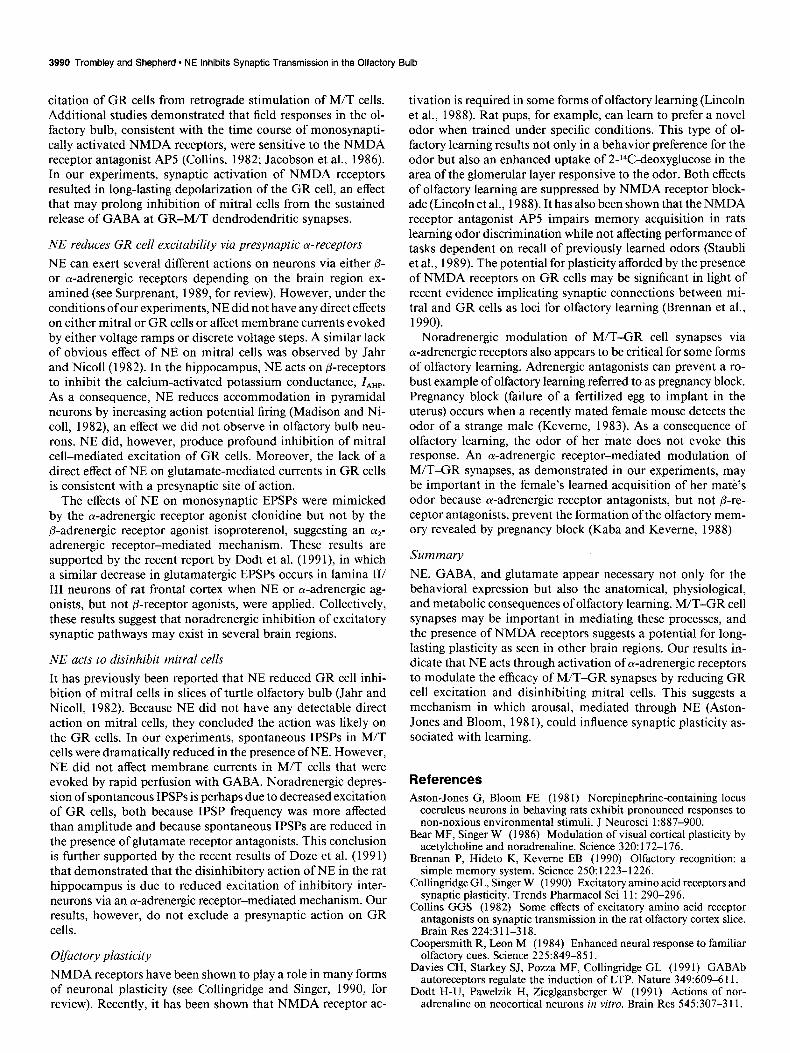
3990 Trombley and Shepherd * NE Inhibits Synaptic Transmission in the Olfactory Bulb
citation of GR cells from retrograde stimulation of M/T cells. Additional studies demonstrated that field responses in the ol- factory bulb, consistent with the time course of monosynapti- tally activated NMDA receptors, were sensitive to the NMDA receptor antagonist AP5 (Collins, 1982; Jacobson et al., 1986). In our experiments, synaptic activation of NMDA receptors resulted in long-lasting depolarization of the GR cell, an effect that may prolong inhibition of mitral cells from the sustained release of GABA at GR-M/T dendrodendritic synapses.
NE reduces GR cell excitability via presynaptic cu-receptors
NE can exert several different actions on neurons via either /3- or a-adrenergic receptors depending on the brain region ex- amined (see Surprenant, 1989, for review). However, under the conditions of our experiments, NE did not have any direct effects on either mitral or GR cells or affect membrane currents evoked by either voltage ramps or discrete voltage steps. A similar lack of obvious effect of NE on mitral cells was observed by Jahr and Nicoll(l982). In the hippocampus, NE acts on P-receptors to inhibit the calcium-activated potassium conductance, Z,,,. As a consequence, NE reduces accommodation in pyramidal neurons by increasing action potential firing (Madison and Ni- ~011, 1982), an effect we did not observe in olfactory bulb neu- rons. NE did, however, produce profound inhibition of mitral cell-mediated excitation of GR cells. Moreover, the lack of a direct effect of NE on glutamate-mediated currents in GR cells is consistent with a presynaptic site of action.
The effects of NE on monosynaptic EPSPs were mimicked by the a-adrenergic receptor agonist clonidine but not by the p-adrenergic receptor agonist isoproterenol, suggesting an o(*- adrenergic receptor-mediated mechanism. These results are supported by the recent report by Dodt et al. (199 l), in which a similar decrease in glutamatergic EPSPs occurs in lamina II/ III neurons of rat frontal cortex when NE or a-adrenergic ag- onists, but not P-receptor agonists, were applied. Collectively, these results suggest that noradrenergic inhibition of excitatory synaptic pathways may exist in several brain regions.
NE acts to disinhibit mitral cells
It has previously been reported that NE reduced GR cell inhi- bition of mitral cells in slices of turtle olfactory bulb (Jahr and Nicoll, 1982). Because NE did not have any detectable direct action on mitral cells, they concluded the action was likely on the GR cells. In our experiments, spontaneous IPSPs in M/T cells were dramatically reduced in the presence of NE. However, NE did not affect membrane currents in M/T cells that were evoked by rapid perfusion with GABA. Noradrenergic depres- sion of spontaneous IPSPs is perhaps due to decreased excitation of GR cells, both because IPSP frequency was more affected than amplitude and because spontaneous IPSPs are reduced in the presence of glutamate receptor antagonists. This conclusion is further supported by the recent results of Doze et al. (199 1) that demonstrated that the disinhibitory action of NE in the rat hippocampus is due to reduced excitation of inhibitory inter- neurons via an cu-adrenergic receptor-mediated mechanism. Our results, however, do not exclude a presynaptic action on GR cells.
Olfactory plasticity NMDA receptors have been shown to play a role in many forms of neuronal plasticity (see Collingridge and Singer, 1990, for review). Recently, it has been shown that NMDA receptor ac-
tivation is required in some forms of olfactory learning (Lincoln et al., 1988). Rat pups, for example, can learn to prefer a novel odor when trained under specific conditions. This type of ol- factory learning results not only in a behavior preference for the odor but also an enhanced uptake of 2-14C-deoxyglucose in the area of the glomerular layer responsive to the odor. Both effects of olfactory learning are suppressed by NMDA receptor block- ade (Lincoln et al., 1988). It has also been shown that the NMDA receptor antagonist AP5 impairs memory acquisition in rats learning odor discrimination while not affecting performance of tasks dependent on recall of previously learned odors (Staubli et al., 1989). The potential for plasticity afforded by the presence of NMDA receptors on GR cells may be significant in light of recent evidence implicating synaptic connections between mi- tral and GR cells as loci for olfactory learning (Brennan et al., 1990).
Noradrenergic modulation of M/T-GR cell synapses via ol-adrenergic receptors also appears to be critical for some forms of olfactory learning. Adrenergic antagonists can prevent a ro- bust example of olfactory learning referred to as pregnancy block. Pregnancy block (failure of a fertilized egg to implant in the uterus) occurs when a recently mated female mouse detects the odor of a strange male (Keveme, 1983). As a consequence of olfactory learning, the odor of her mate does not evoke this response. An a-adrenergic receptor-mediated modulation of M/T-GR synapses, as demonstrated in our experiments, may be important in the female’s learned acquisition of her mate’s odor because a-adrenergic receptor antagonists, but not P-re- ceptor antagonists, prevent the formation of the olfactory mem- ory revealed by pregnancy block (Kaba and Keveme, 1988)
Summary
NE, GABA, and glutamate appear necessary not only for the behavioral expression but also the anatomical, physiological, and metabolic consequences of olfactory learning. M/T-GR cell synapses may be important in mediating these processes, and the presence of NMDA receptors suggests a potential for long- lasting plasticity as seen in other brain regions. Our results in- dicate that NE acts through activation of a-adrenergic receptors to modulate the efficacy of M/T-GR synapses by reducing GR cell excitation and disinhibiting mitral cells. This suggests a mechanism in which arousal, mediated through NE (Aston- Jones and Bloom, 198 I), could influence synaptic plasticity as- sociated with learning.
References Aston-Jones G, Bloom FE (198 1) Norepinephrine-containing locus
coeruleus neurons in behaving rats exhibit pronounced responses to non-noxious environmental stimuli. J Neurosci 1:887-900.
Bear MF, Singer W (1986) Modulation of visual cortical plasticity by acetylcholine and noradrenaline. Science 320: 172-l 76.
Brennan P, Hideto K, Keverne EB (1990) Olfactory recognition: a simple memory system. Science 250: 1223-l 226.
Collingridge CL, Singer W (1990) Excitatory amino acid receptors and synaptic plasticity: Trends Pharmacol Sci -11: 290-296.
Collins GGS (1982) Some effects of excitatorv amino acid recentor antagonists & synaptic transmission in the rai olfactory cortex slice. Brain Res 224:3 1 l-3 18.
Coopersmith R, Leon M (1984) Enhanced neural response to familiar olfactory cues. Science 225:849-85 1.
Davies CH, Starkey SJ, Pozza MF, Collingridge CL (199 1) GABAb autoreceptors regulate the induction of LTP. Nature 349:609-6 11.
Dodt H-U, Pawelzik H, Zieglgansberger W (1991) Actions of nor- adrenaline on neocortical neurons in vitro. Brain Res 545:307-3 11.

The Journal of Neuroscience, October 1992, 72(10) 3991
Doze VA, Cohen GA, Madison DV (199 1) Synaptic localization of adrenergic disinhibition in the rat hippocampus. Neuron 6889-900.
Hopkins WF, Johnston D (1984) Frequency-dependent noradrenergic modulation of long-term potentiation in the hippocampus. Science 226:350-351.
Jacobson I, Hamberger A (1986) Effects of kynurenic acid on evoked extracellular field potentials in the rat olfactory bulb in vivo. Brain Res 386:389-392.
Jacobson I, Butcher S, Hamberger A (1986) An analysis of the effects of excitatory amino acid receptor antagonists on evoked field poten- tials in the olfactory bulb. Neuroscience 19:267-273. -
Jahr CE, Nicoll RA (1980) Dendrodendritic inhibition: demonstration with intracellular recording. Science 207:1473-1475.
Jahr CE, Nicoll RA (1982) Noradrenergic modulation of dendroden- dritic inhibition in the olfactory bulb. Nature 297:227-229.
Kaba H, Keveme EB (1988) The effects of microinfusions of druas into the accessory olfactory’bulb on the olfactory block to pregnancy. Neuroscience 25: 1007-1011.
Keveme EB (1983) Pheromonal influences on the endocrine regulation of reproduction. Trends Neurosci 6:38 l-384.
Kleinschmidt A, Bear MF, Singer W (1987) Blockade of NMDA re- ceptors disrupts experience-dependent plasticity of kitten striate cor- tex. Science 238:355-358.
Leon M (1987) Plasticity of olfactory output circuits related to early olfactory learning. Trends Neurosci 10:434438.
Lincoln J, Coopersmith R, Harris EW, Cotman CW, Leon M (1988) NMDA receptor activation and early olfactory learning. Dev Brain Res 39:309-3 12.
Madison DV, Nicoll RA (1982) Noradrenaline blocks accommodation ofpyramidal cell discharge in the hippocampus. Nature 299:636-638.
Mayer ML, Westbrook GL (1987) The physiology of excitatory amino acids in the vertebrate central nervous system. Prog Neurobiol 28: 197-276.
Mori K (1987) Membrane and synaptic properties of identified neu- rons in the olfactory bulb. Prog Neurobiol 29:275-320.
Nowycky MC, Mori K, Shepherd GM (198 1) GABAergic mechanisms of dendrodendritic synapses in isolated turtle olfactory bulb. J Neu- rophysiol 46:639-648.
Patneau DK, Mayer ML (1990) Structure-activity relationships for amino acid transmitter candidates acting at N-methyl-D-aspartate and quisqualate receptors. J Neurosci 10:2385-2399.
Ribak CE.~Vauahn JE. Saito K. Barber R. Roberts E (1977) Glutamate decarbdxylase localization in neurons of the olfactory bulb. Brain Res 126:1-18.
Shipley MT, Halloran FJ, De La Torre J (1985) Surprisingly rich projection from the locus coeruleus to the olfactory bulb in the rat. Brain Res 329:294-299.
Staubli U, Thibault 0, DiLorenzo M, Lynch G (1989) Antagonism of NMDA receptors impairs acquisition but not retention of olfactory memory. Behav Neurosci 103:54-60.
Stepheson FA, Dolphin AC (1989) GABA and glycine neurotrans- mission. Semin Neurosci 1: 115-l 23.
Sullivan RM, Wilson DA, Leon M (1989) Norepinephrine and leam- ing-induced plasticity in infant rat olfactory system. J Neurosci 9: 39984006.
Surprenant A (1989) The neurotransmitter noradrenaline and its re- ceptors. Semin Neurosci 1: 125-l 36.
Trombley PQ, Westbrook GL (1990) Excitatory synaptic transmission in primary cultures of rat olfactory bulb. J Neurophysiol64:598-606.
Wilson DA, Sullivan RM, Leon M (1987) Single-unit analysis of post- natal olfactory learning: modified olfactory bulb output response pat- terns to learned attractive odors. J Neurosci 7:3 154-3 162.
Woo CC, Coopersmith R, Leon M (1987) Localized changes in ol- factory bulb morphology associated with early olfactory learning. J Comp Neuro1263: 113-125.
Related Documents