Biochem. J. (2004) 382, 597–606 (Printed in Great Britain) 597 Noradrenaline represses PPAR (peroxisome-proliferator-activated receptor) γ 2 gene expression in brown adipocytes: intracellular signalling and effects on PPARγ 2 and PPARγ 1 protein levels Eva M. LINDGREN*, Ronni NIELSEN†, Natasa PETROVIC*, Anders JACOBSSON*, Susanne MANDRUP†, Barbara CANNON* and Jan NEDERGAARD* 1 *The Wenner-Gren Institute, The Arrhenius Laboratories F3, Stockholm University, SE-106 91 Stockholm, Sweden, and †Department of Biochemistry and Molecular Biology, University of Southern Denmark, Odense, Denmark PPAR (peroxisome-proliferator-activated receptor) γ is expressed in brown and white adipose tissues and is involved in the control of differentiation and proliferation. Noradrenaline stimulates brown pre-adipocyte proliferation and brown adipocyte differentiation. The aim of the present study was thus to investigate the influence of noradrenaline on PPARγ gene expression in brown adipocytes. In primary cultures of brown adipocytes, PPARγ 2 mRNA levels were 20-fold higher than PPARγ 1 mRNA levels. PPARγ expres- sion occurred during both the proliferation and the differentiation phases, with the highest mRNA levels being found at the time of transition between the phases. PPARγ 2 mRNA levels were down- regulated by noradrenaline treatment (EC 50 , 0.1 µM) in both pro- liferative and differentiating cells, with a lagtime of 1 h and lasting up to 4 h, after which expression gradually recovered. The down-regulation was β -adrenoceptor-induced and intracellularly mediated via cAMP and protein kinase A; the signalling pathway did not involve phosphoinositide 3-kinase, Src, p38 mitogen- activated protein kinase or extracellular-signal-regulated kinases 1 and 2. Treatment of the cells with the protein synthesis inhibi- tor cycloheximide not only abolished the noradrenaline-induced down-regulation of PPARγ 2 mRNA, but also in itself indu- ced PPARγ 2 hyperexpression. The down-regulation was probably the result of suppression of transcription. The down-regulation of PPARγ 2 mRNA resulted in similar down-regulation of PPARγ 2 and phosphoPPARγ 2 protein levels. Remarkably, the level of PPARγ 1 protein was similar to that of PPARγ 2 (despite almost no PPARγ 1 mRNA), and the down-regulation by noradrenaline demonstrated similar kinetics to that of PPARγ 2; thus PPARγ 1 was apparently translated from the PPARγ 2 template. It is sug- gested that β -adrenergic stimulation via cAMP and protein kinase A represses PPARγ gene expression, leading to reduction of PPARγ 2 mRNA levels, which is then reflected in down-regulated levels of PPARγ 2, phosphoPPARγ 2 and PPARγ 1. Key words: brown adipocyte, cAMP, cycloheximide, noradren- aline, peroxisome-proliferator-activated receptor γ 2 (PPARγ 2), protein kinase A. INTRODUCTION The PPAR (peroxisome-proliferator-activated receptor) γ is a ligand-dependent transcription factor that is predominantly ex- pressed in adipose tissue, both white and brown [1,2]. In mouse, there are two PPARγ isoforms, γ 1 and γ 2, which are derived from the same gene, but are transcribed from different promoters [2a]. The expression of the PPARγ 2 isoform is practically res- tricted to adipose tissue [2]. PPARγ plays an important promoting role in the differentiation of adipocytes [3]. PPARγ heterodi- merizes with the 9-cis-retinoic acid receptor, RXRα, and the het- erodimer binds to the PPRE (PPAR-response element) [4]. PPREs have been characterized in the promoter/enhancer regions of several genes expressed in adipocytes, such as lipoprotein lipase, aP2 (i.e. fatty acid binding protein) and phosphoenolpyruvate carboxykinase (for review see [5]). Also the gene for the brown- adipose-tissue-specific UCP1 (uncoupling protein 1) contains a PPRE site [6,7]. Due to the importance of PPARγ for cell differentiation [8], significant interest has developed for the use of PPARγ activation as an anticancer treatment in different tissues [9]. Activation of PPARγ in cancer cells is expected to promote differentiation and thus to decrease proliferation. Conversely, during physiologically induced cell proliferation, the opposite effect may be necessary, i.e. to ensure a low activity of PPARγ in order to allow for proli- feration instead of differentiation. One such physiologically induced state of cell proliferation occurs during the recruitment process in brown adipose tissue [10]. In brown adipose tissue, noradrenaline promotes both cell proliferation and cell differen- tiation. From studies of cells in culture, it has been concluded that a switch in the responsiveness to noradrenaline occurs: in brown pre-adipocytes, noradrenaline stimulates proliferation, but in brown adipocytes, noradrenaline advances the differentiation pro- cess, both processes being mediated by cAMP [10]. It could thus be proposed that this switch in the ability of noradrenaline to pro- mote these different processes may be associated with a switch in the ability of noradrenaline to influence PPARγ gene expression, from repressing PPARγ gene expression in brown pre-adipocytes to promoting PPARγ gene expression in mature brown adipo- cytes. To investigate whether and how noradrenaline influences the expression of the transcription factor PPARγ in brown adipo- cytes, primary cultures of brown adipocytes were exposed to noradrenaline, the signalling process involved in the control of PPARγ expression was analysed, and the relationship between PPARγ mRNA and protein amounts was examined. Abbreviations used: CREB, cAMP-response-element-binding protein; DMEM, Dulbecco’s modified Eagle’s medium; ERK, extracellular-signal-regulated kinase; MAPK, mitogen-activated protein kinase; MEK, MAPK/ERK kinase; PI3K, phosphoinositide 3-kinase; PKA, protein kinase A; PPAR, peroxisome- proliferator-activated receptor; PPRE, PPAR-response element; TFIIB, transcription factor IIB; UCP1, uncoupling protein 1. 1 To whom correspondence should be addressed (email [email protected]). c 2004 Biochemical Society

Welcome message from author
This document is posted to help you gain knowledge. Please leave a comment to let me know what you think about it! Share it to your friends and learn new things together.
Transcript

Biochem. J. (2004) 382, 597–606 (Printed in Great Britain) 597
Noradrenaline represses PPAR (peroxisome-proliferator-activated receptor)γ 2 gene expression in brown adipocytes: intracellular signalling andeffects on PPARγ 2 and PPARγ 1 protein levelsEva M. LINDGREN*, Ronni NIELSEN†, Natasa PETROVIC*, Anders JACOBSSON*, Susanne MANDRUP†, Barbara CANNON*and Jan NEDERGAARD*1
*The Wenner-Gren Institute, The Arrhenius Laboratories F3, Stockholm University, SE-106 91 Stockholm, Sweden, and †Department of Biochemistry and Molecular Biology,University of Southern Denmark, Odense, Denmark
PPAR (peroxisome-proliferator-activated receptor) γ is expressedin brown and white adipose tissues and is involved in the control ofdifferentiation and proliferation. Noradrenaline stimulates brownpre-adipocyte proliferation and brown adipocyte differentiation.The aim of the present study was thus to investigate the influenceof noradrenaline on PPARγ gene expression in brown adipocytes.In primary cultures of brown adipocytes, PPARγ 2 mRNA levelswere 20-fold higher than PPARγ 1 mRNA levels. PPARγ expres-sion occurred during both the proliferation and the differentiationphases, with the highest mRNA levels being found at the time oftransition between the phases. PPARγ 2 mRNA levels were down-regulated by noradrenaline treatment (EC50, 0.1 µM) in both pro-liferative and differentiating cells, with a lagtime of 1 h andlasting up to 4 h, after which expression gradually recovered. Thedown-regulation was β-adrenoceptor-induced and intracellularlymediated via cAMP and protein kinase A; the signalling pathwaydid not involve phosphoinositide 3-kinase, Src, p38 mitogen-activated protein kinase or extracellular-signal-regulated kinases1 and 2. Treatment of the cells with the protein synthesis inhibi-
tor cycloheximide not only abolished the noradrenaline-induceddown-regulation of PPARγ 2 mRNA, but also in itself indu-ced PPARγ 2 hyperexpression. The down-regulation was probablythe result of suppression of transcription. The down-regulation ofPPARγ 2 mRNA resulted in similar down-regulation of PPARγ 2and phosphoPPARγ 2 protein levels. Remarkably, the level ofPPARγ 1 protein was similar to that of PPARγ 2 (despite almostno PPARγ 1 mRNA), and the down-regulation by noradrenalinedemonstrated similar kinetics to that of PPARγ 2; thus PPARγ 1was apparently translated from the PPARγ 2 template. It is sug-gested that β-adrenergic stimulation via cAMP and protein kinaseA represses PPARγ gene expression, leading to reduction ofPPARγ 2 mRNA levels, which is then reflected in down-regulatedlevels of PPARγ 2, phosphoPPARγ 2 and PPARγ 1.
Key words: brown adipocyte, cAMP, cycloheximide, noradren-aline, peroxisome-proliferator-activated receptor γ 2 (PPARγ 2),protein kinase A.
INTRODUCTION
The PPAR (peroxisome-proliferator-activated receptor) γ is aligand-dependent transcription factor that is predominantly ex-pressed in adipose tissue, both white and brown [1,2]. In mouse,there are two PPARγ isoforms, γ 1 and γ 2, which are derivedfrom the same gene, but are transcribed from different promoters[2a]. The expression of the PPARγ 2 isoform is practically res-tricted to adipose tissue [2]. PPARγ plays an important promotingrole in the differentiation of adipocytes [3]. PPARγ heterodi-merizes with the 9-cis-retinoic acid receptor, RXRα, and the het-erodimer binds to the PPRE (PPAR-response element) [4]. PPREshave been characterized in the promoter/enhancer regions ofseveral genes expressed in adipocytes, such as lipoprotein lipase,aP2 (i.e. fatty acid binding protein) and phosphoenolpyruvatecarboxykinase (for review see [5]). Also the gene for the brown-adipose-tissue-specific UCP1 (uncoupling protein 1) contains aPPRE site [6,7].
Due to the importance of PPARγ for cell differentiation [8],significant interest has developed for the use of PPARγ activationas an anticancer treatment in different tissues [9]. Activation ofPPARγ in cancer cells is expected to promote differentiation andthus to decrease proliferation. Conversely, during physiologically
induced cell proliferation, the opposite effect may be necessary,i.e. to ensure a low activity of PPARγ in order to allow for proli-feration instead of differentiation. One such physiologicallyinduced state of cell proliferation occurs during the recruitmentprocess in brown adipose tissue [10]. In brown adipose tissue,noradrenaline promotes both cell proliferation and cell differen-tiation. From studies of cells in culture, it has been concluded thata switch in the responsiveness to noradrenaline occurs: in brownpre-adipocytes, noradrenaline stimulates proliferation, but inbrown adipocytes, noradrenaline advances the differentiation pro-cess, both processes being mediated by cAMP [10]. It could thusbe proposed that this switch in the ability of noradrenaline to pro-mote these different processes may be associated with a switch inthe ability of noradrenaline to influence PPARγ gene expression,from repressing PPARγ gene expression in brown pre-adipocytesto promoting PPARγ gene expression in mature brown adipo-cytes.
To investigate whether and how noradrenaline influences theexpression of the transcription factor PPARγ in brown adipo-cytes, primary cultures of brown adipocytes were exposed tonoradrenaline, the signalling process involved in the controlof PPARγ expression was analysed, and the relationship betweenPPARγ mRNA and protein amounts was examined.
Abbreviations used: CREB, cAMP-response-element-binding protein; DMEM, Dulbecco’s modified Eagle’s medium; ERK, extracellular-signal-regulatedkinase; MAPK, mitogen-activated protein kinase; MEK, MAPK/ERK kinase; PI3K, phosphoinositide 3-kinase; PKA, protein kinase A; PPAR, peroxisome-proliferator-activated receptor; PPRE, PPAR-response element; TFIIB, transcription factor IIB; UCP1, uncoupling protein 1.
1 To whom correspondence should be addressed (email [email protected]).
c© 2004 Biochemical Society
598 E. M. Lindgren and others
EXPERIMENTAL
Animals, cell isolation and cell culture
Male NMRI mice, purchased from local suppliers (B&K orEklunds, Stockholm, Sweden), were kept at room temperature(approx. 22 ◦C) for at least 24 h after arrival. At the age of4 weeks, the mice were killed by CO2, and the brown adiposetissue was isolated from the interscapular, cervical and axillarydepots, principally as described in [11]. The pooled tissue pieceswere minced in DMEM (Dulbecco’s modified Eagle’s medium)and transferred to a digestion solution with 0.2 % (w/v) collagen-ase (type II; Sigma) in a buffer consisting of 0.1 M Hepes(pH 7.4), 123 mM NaCl, 5 mM KCl, 1 mM CaCl2, 4.5 mM glu-cose and 1.5 % (w/v) BSA. The digestion was performed for30 min at 37 ◦C with continuous vortex-mixing. The suspensionwas filtered through a 250 µm pore-size nylon filter (Sintab, Oxie,Sweden) into sterile 10 ml tubes. The filtered suspension was kepton ice for 30 min to let the mature adipocytes float up. The toplayer of the suspension was removed, and the rest of the suspen-sion was filtered through a 25 µm pore-size nylon filter (Sintab)and re-centrifuged at 700 g for 10 min, to pellet the precursorcells. The pellet was resuspended in 5 ml of DMEM and centri-fuged at 700 g for 10 min. The pellet was then suspended inculture medium (0.5 ml/animal). The cells were cultured in six 10-cm2-well plates (Corning); 1.8 ml of culture medium was addedto each well before 0.2 ml (cells corresponding to 0.4 animals)of cell suspension was added. The culture medium was DMEMwith 10 % (v/v) newborn calf serum (Flow Laboratories, McLean,VA, U.S.A.), 4 nM insulin, 25 µg/ml sodium ascorbate, 10 mMHepes, 4 mM glutamine, 50 units/ml penicillin and 50 µg/mlstreptomycin. The cells were grown at 37 ◦C in an atmosphereof 8 % CO2 in air with 80 % humidity. The medium was changedon day 1 and then every second day. The medium was pre-warmedto 37 ◦C before changing. The medium was not changed on thesame day as the cells were harvested. The experiments were per-formed on different days of culture, as indicated in each individualexperiment.
Analysis of mRNA levels
After the experiments, the medium was discarded and the cellswere harvested from each well with 1 ml of Ultraspec (BiotecxLaboratories, Houston, TX, U.S.A.) as described in the manu-facturer’s protocol. The RNA obtained was examined by PCR orNorthern blotting.
For quantitative real-time PCR, the first-strand cDNA was firstsynthesized as described previously [12]. RNA expression wasthen quantified by real-time quantitative PCR using the ABIPRISM 7700 Sequence Detection System (Applied Biosystems).Each PCR reaction contained, in a final volume of 25 µl, 1.5 µlof first-strand cDNA, 12.5 µl of 2× SYBR Green PCR MasterMix (Applied Biosystems) and 7.5 pmol of each primer [forPPARγ 1, forward, 5′-GGA CTG TGT GAC AGA CAA GATTTG A-3′ and reverse, 5′-CTG AAT ATC AGT GGT TCA CCGC-3′ (GenBank® accession number U01841); for PPARγ 2, for-ward, 5′-CTC TGT TTT ATG CTG TTA TGG GTG A-3′ andreverse, 5′-GGT CAA CAG GAG AAT CTC CCA G-3′ (Gen-Bank® accession number U09138); for TFIIB (transcription factorIIB), forward, 5′-GTT CTG CTC CAA CTT TTG CCT-3′ andreverse, 5′-TGT GTA GCT GCC ATC TGC ACT T-3′ (Gen-Bank® accession number NM_145546); primers from DNA Tech-nology A/S, Aarhus, Denmark]. All reactions were performedusing the following cycling conditions: 50 ◦C for 2 min and 95 ◦Cfor 10 min, followed by 40 cycles at 95 ◦C for 15 s and 60 ◦C for1 min. PCR was carried out in 96-well plates and in triplicate.
The relative amounts of all mRNAs were calculated using thecomparative CT method.
For Northern blotting, total RNA was separated on an agarosegel (1.25 %) containing 20 mM Mops (pH 7.0) and 6.7 % formal-dehyde. Ethidium bromide (1 mg/ml) was added to the gel toexamine the distribution of RNA in UV light. The gel was runfor 2–3 h at 100 V. The RNA was transferred on to a Hybond-Nmembrane (Amersham Biosciences) by capillary blotting over-night. The membrane was prehybridized at 42 ◦C for at least1 h in 10 ml of hybridization solution containing 5× SSC(pH 7.0) (1× SSC is 0.15 M NaCl/0.015 M sodium citrate),5× Denhardt’s (1× Denhardt’s is 0.02 % Ficoll 400/0.02 %polyvinylpyrrolidone/0.02 % BSA), 0.5 % (w/v) SDS, 50 mMsodium phosphate (pH 6.5), 50 % formamide and 100 µg/ml de-graded herring sperm DNA. Hybridization of the membranes wasperformed overnight at 42 ◦C in a fresh hybridization solutionwith the addition of denaturated probe, [32P]dCTP-labelled cDNA,corresponding to mouse PPARγ mRNA, labelled by randompriming (Amersham Biosciences). The PPARγ cDNA contained454 bp corresponding to positions 127–581 in the publishedPPARγ 2 sequence with GenBank® accession number U09138(a gift from Dr Bruce Spiegelman, Dana Faber Cancer ResearchInstitute, Boston, MA, U.S.A.). The identity of the probe wasconfirmed by sequencing. The probe matched the mouse mRNAregions that correspond to exon 1 and exon 2, which are exonscommon for PPARγ 1 and γ 2. The probe was thus not specificfor PPARγ 2, but based on the results in Figure 1(C), the outcomewas referred to as PPARγ 2. After hybridization, the solution wasremoved and the membranes were washed twice for 30 min atroom temperature in a solution of 2× SSC and 0.2 % (w/v)SDS, followed by washing twice for 60 min at 45 ◦C in 0.1×SSC and 0.2 % (w/v) SDS. The membranes were sealed in aplastic envelope and then exposed to a PhosphoImager screen andscanned in a Molecular Dynamic’s PhosphoImager 425 S. SpecificmRNA signals were analysed with the ImageQuant software.For analysis, the curve-fitting option of the KaleidaGraph 3.0application was used as indicated in the legends to the Fig-ures. The uncertainties given are those of the KaleidaGraph es-timates.
Analysis of PPAR protein levels
Brown adipocytes were treated with 1 µM noradrenaline for thespecified times. After the treatment, the cell cultures were washedtwice in PBS and then harvested in a lysis buffer containing62.5 mM Tris/HCl (pH 6.8), 2% (w/v) SDS, 10% (v/v) glyceroland a protease inhibitor cocktail (Complete Mini; Roche). Aftersonication, the concentration of the soluble proteins was deter-mined (using the method of Lowry) in the sample, and 1/10 vol oflysis buffer supplemented with 0.5 M dithiothreitol and 1% (w/v)Bromophenol Blue was added to each sample. Proteins (30 µg)were separated in 12% polyacrylamide gels containing SDS, andtransferred on to a PVDF membrane (Amersham Biosciences)in 48 mM Tris/HCl, 39 mM glycine, 0.037% (w/v) SDS and15% (v/v) methanol using a semi-dry electrophoretic transfer cell(Bio-Rad). Following transfer, the membrane was stained withPonceau S for examination of equal loading of proteins. Afterwashing, the membrane was blocked in 5% (w/v) dried milkovernight at 4 ◦C. The membrane was then incubated in 1:500diluted primary antibody. The antibody against PPARγ (sc-7273)was obtained from Santa Cruz Laboratories, while the antibodyagainst PPARα (MA1-822) was from Affinity BioReagents. Theimmunoblots were visualized with horseradish-peroxidase-conju-gated secondary antibodies (Cell Signaling Technology) andECL® (enhanced chemiluminescence; Amersham Biosciences) in
c© 2004 Biochemical Society

Peroxisome-proliferator-activated receptor γ 2 gene expression in brown adipocytes 599
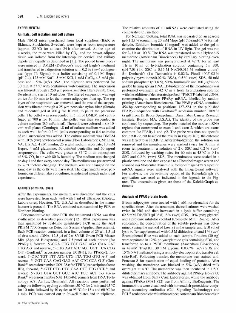
Figure 1 PPARγ 2 mRNA levels in brown adipocytes during differentiation
Primary cultures of brown adipocytes were grown in culture for the indicated number of days. Where indicated, 0.1 µM noradrenaline (NA) had been added 3 h before harvest. Total RNA was isolatedand analysed as described in the Experimental section. (A) Northern blot. Total RNA (5 µg) was used for each lane and the blot was hybridized with the PPARγ probe described in the Experimentalsection. (B) PPARγ mRNA levels during differentiation. Experiments were performed as illustrated in (A). PPARγ values (�) with thick lines are from cultures not treated with noradrenaline andare means +− S.E.M. for four independent experiments, each performed in duplicate. The value at day 5 was in each experiment set to 100 %, and the PPARγ mRNA levels on the other days wereexpressed relative to this value in each individual experiment. Values with broken lines are from an additional experiment in which PPARγ mRNA levels (�) and noradrenaline-induced UCP1 mRNAlevels (�) were determined in parallel (means from duplicate samples, normalized to 18 S RNA); these data were also adjusted to 100 % for the day of highest mRNA level. (C) PPARγ 1 and PPARγ 2mRNA levels in brown adipocytes. Cultures were grown as in (A) and then treated with 1 µM noradrenaline or not for 2 h. RNA was extracted and PPARγ 1, PPARγ 2 and TFIIB mRNA levels weredetermined by real-time PCR as described in the Experimental section. The PPARγ 1 and PPARγ 2 mRNA levels were normalized to the TFIIB mRNA levels in each sample. The mean total amountof PPARγ mRNA in untreated cells was set to 100 %. Results are means +− S.E.M. of triplicate samples; * indicates that the effect of noradrenaline on PPARγ 2 was significant (P � 0.05), whereasthere was no significant effect on PPARγ 1 levels. (D) Relative effect of noradrenaline on the PPARγ 2 mRNA level during cell differentiation. Values were calculated from the data in (A), adjustingthe non-treated PPARγ 2 mRNA to 100 % for each day and indicating the relative effect of noradrenaline for each day.
a CCD (charge-coupled device) camera. The blots were quantifiedusing the Image Gauge V3.45 program (Fuji Film).
Chemicals
Noradrenaline bitartrate (Arterenol), A23187, PMA, pertussistoxin, cycloheximide, actinomycin D, forskolin, isoprenaline,8-bromo-cAMP, CGP-12177, cirazoline, CL-316243 and Ly-294002 were obtained from Sigma/RBI; H89, PP2, PD-98059and SB-202190 from Calbiochem. PMA, Ly-294002, H89, PP2,PD-98059, SB-202190 and forskolin were dissolved in DMSO.
RESULTS
PPARγ mRNA levels in spontaneously differentiatingbrown adipocytes
To examine whether noradrenaline differentially influencesPPARγ gene expression in brown pre-adipocytes compared withdifferentiated brown adipocytes, brown pre-adipocytes weregrown and developed in culture. Under the conditions used, thefibroblast-like pre-adipocytes proliferate until they are confluent
at day 5–6 and then differentiate to mature brown adipocytesin a spontaneous, but highly temporally and qualitatively repro-ducible, way [10,11,13].
Significant levels of PPARγ mRNA were observed already inthe morphologically undifferentiated cells at day 3 (Figure 1A).In a compilation of a series of experiments (Figure 1B), it is seenthat the level of PPARγ mRNA increased somewhat during thecell culture, to reach a maximum on day 5 of culture. The expres-sion of the established brown adipocyte differentiation marker,noradrenaline-induced UCP1 mRNA, is also displayed in Fig-ure 1(B), together with the corresponding PPARγ levels; clearly,the maximal PPARγ expression coincides with the maturationphase of the cells. The presence of PPARγ already in brown pre-adipocytes and the small increase slightly before the acceleratedexpression of differentiation markers is in accordance with earlierobservations [13]. PPARγ expression is thus maximal at the timeof the switch from the brown pre-adipocyte to the mature brownadipocyte stage.
To examine which PPARγ transcripts were expressed in thesecells, RNA samples from the cell cultures with maximal PPARγexpression level were analysed by quantitative real-time PCR(Figure 1C, grey bars). As seen, practically no PPARγ 1 mRNA
c© 2004 Biochemical Society
600 E. M. Lindgren and others
was found in the cell cultures; �95% of the PPARγ transcriptpresent was PPARγ 2 mRNA. For comparison, we also examinedthe PPARγ 1/PPARγ 2 mRNA ratio in some mouse tissues. Simi-larly to the case in the brown adipocytes in culture, PPARγ 2was the dominant isoform both in mouse brown adipose tissue(>70%) and white adipose tissue (>75%), whereas in liver, thePPARγ 1 isoform was dominant (>90%) (results not shown).Based on the results in Figure 1(C), we refer in the following tothe detected species on Northern blots as PPARγ 2.
Effect of noradrenaline on PPARγ 2 mRNA levels
As shown in Figures 1(A) and (D), PPARγ mRNA levels weredecreased markedly upon noradrenaline treatment. Real-timePCR established that the reduction observed was due to a decreasein the levels of PPARγ 2, and was not counteracted by an incre-ase in PPARγ 1 (Figure 1C). The noradrenaline-induced down-regulation of PPARγ 2 mRNA levels was observed in proliferativeas well as in differentiating cells (Figures 1A and 1D); there wasthus no switch in the qualitative response between pre-adipocytesand mature adipocytes concerning adrenergic regulation ofPPARγ 2 mRNA levels. The absence of a switch is in contrastwith the qualitative switch from noradrenaline promoting cellproliferation in brown pre-adipocytes to noradrenaline promotingcell differentiation in mature brown adipocytes, to the switch fromβ1-adrenoceptors being coupled to adenylate cyclase to β3-adrenoceptors being the coupled ones [14], and to the switch fromnoradrenaline decreasing C/EBPα (CCAAT/enhancer-bindingprotein α) gene expression to noradrenaline increasing it [15].
For further investigations of the effect of noradrenaline, cellsat day 5 in culture were used. To examine the time course of thenoradrenaline effect, the cell cultures were treated with noradren-aline, and the levels of PPARγ 2 mRNA were determined aftervarious lengths of time. The combined results from a series of suchexperiments are presented in Figure 2(A). There was a markedlagtime of nearly 1 h before a noradrenaline effect was observable,an indication that a complex mediatory system was involved.The lagtime was followed by a marked successive decrease inmRNA, lasting for about 1 h. The PPARγ 2 mRNA levels thenremained low for about 2 h, and then successively increased. ThemRNA levels regained control values after about 24 h; with ahigher dose of noradrenaline, a doubling of mRNA levels hasbeen reported after 24 h of treatment [16].
To obtain a dose–response curve for the noradrenaline-induced decrease in PPARγ 2 mRNA levels, cultures were treatedfor 3 h with different concentrations of noradrenaline (Figure 2B).The effect of noradrenaline was clearly dose-dependent, mono-phasic and saturable. The PPARγ 2 mRNA levels were reducedmaximally to approx. 50%, even with high concentrations ofnoradrenaline. The calculated EC50 value for noradrenaline was80 +− 11 nM. This is similar to that of the noradrenaline-inducedincrease in the gene expression of VEGF (vascular endothelialgrowth factor) A [17] and ribonucleotide reductase subunit R2[18], but higher than the EC50 (approx. 10 nM) for stimulationof gene expression of UCP1 [11] and the β1-adrenergic receptor[19], and much higher than the EC50 for the noradrenaline-induceddecrease in β3-adrenoceptor mRNA (approx. 1 nM) [19,20].
The effects of noradrenaline-induced repression of PPARγ 2 geneexpression on PPARγ protein levels
To study whether the norepinephrine-induced repression ofPPARγ 2 gene expression resulted in lower levels of the corres-ponding protein, we examined samples of norepinephrine-treatedbrown adipocytes by immunoblotting.
Figure 2 Effect of noradrenaline (NA) on PPARγ 2 mRNA levels
(A) Time curve. Primary cultures of brown adipocytes on day 5 were treated with 0.1 µMnoradrenaline for the indicated lengths of time, and analysed for PPARγ 2 mRNA as in Figure 1(A).The curve is based on two to nine independent experiments (except at 21 h, where only one wasperformed) in duplicate wells. In each experiment, the PPARγ 2 mRNA value from non-treatedcells was set to 100 % and the other values were expressed relative to this. Results are means +−S.E.M. The curve was drawn by the ‘smooth’ function of KaleidaGraph. (B) Dose–response curve.Brown adipocyte cultures (day 5) were treated with the indicated concentrations of noradrenalinefor 3 h and analysed as in Figure 1(A). Results are means +− S.E.M. for five independentexperiments. In each experiment, the control level was set to 100 % and the other values were ex-pressed relative to this. The results were analysed for adherence to Michaelis–Menten kineticswith the formula: mRNANA = 100 % − �mRNAmax · [noradrenaline]/(EC50 + [noradrenaline])(excluding the 100 µM value). The EC50 value thus obtained was 80 +− 11 nM noradrenalineand the maximal decrease (�mRNAmax) was 55 +− 1 %.
Using a commercial antibody against PPARγ (sc-7273), whichcan recognize both PPARγ 2 and PPARγ 1, we observed thatthe brown adipocyte protein extracts displayed three bands (Fig-ure 3A). The upper two bands were identified as the phospho-rylated and non-phosphorylated forms of PPARγ 2. This wasbased on the lower of these two having the expected molecularmass of PPARγ 2 (56 kDa) [21], and in a parallel immunoblot,protein extracts from 3T3-L1 adipocytes and from cells over-expressing PPARγ 2 protein displayed similar bands (results notshown). As a negative control (Figure 3A), we used a protein ex-tract from mouse heart, as heart does not express PPARγ 2 [22].Treatment of brown adipocyte protein extracts with calf intestinalphosphatase before electrophoresis resulted in the disappearanceof the upper of these bands (results not shown), confirming thephosphoprotein nature of this band. The intensities of both thesebands were decreased markedly after noradrenaline stimulation(Figure 3A, and see below).
c© 2004 Biochemical Society

Peroxisome-proliferator-activated receptor γ 2 gene expression in brown adipocytes 601
Figure 3 Effect of noradrenaline (NA) on PPARγ and PPARα protein levels
Primary cultures of brown adipocytes were treated essentially as indicated in Figure 2, andprotein was extracted and analysed by immunoblotting as described in the Experimental section;each lane contained 30 µg of protein from brown adipocytes (b.a.) non-treated (−) or treatedfor 4 h with 1 µM noradrenaline, or from mouse heart homogenate. In (A), an anti-PPARγ
antibody was used; in (B), an anti-PPARα antibody was used. The sizes (in kDa) indicated tothe left are from protein size markers; the arrows to the right indicate the suggested nature of thebands (see text).
The anti-PPARγ antibody also recognized a band of a mol-ecular mass of 52 kDa. The intensity of this band was also de-creased markedly after noradrenaline treatment (Figure 3A). Themolecular mass of both PPARγ 1 and PPARα is approx. 52 kDa,and due to the similarity between PPARγ and PPARα, the pos-sibility existed that the anti-PPARγ antibody also recognizedPPARα. PPARα is also expressed in brown adipocytes [13], butas demonstrated above (Figure 1C), PPARγ 1 mRNA is nearlyabsent from the brown adipocytes examined in the present study,and PPARγ 1 protein would therefore not a priori be expected.Attempts to separate PPARα and PPARγ 1 were made with largergels and utilizing protein from cell lines retrovirally transducedwith PPARα and PPARγ , but a separation was not achieved(results not shown).
Therefore, to examine the possibility that the 52 kDa band re-sulted from non-specific interaction of the anti-PPARγ antibodywith PPARα, we analysed samples using a commercial anti-body (MA1-822) against PPARα. As seen (Figure 3B), thisantibody recognized a band at ≈ 52 kDa in heart preparations,in accordance with the reported high expression of PPARα inthis tissue [22]. Similarly, this anti-PPARα antibody recognized aprotein in the brown adipocyte extracts, principally in agreementwith brown adipocytes expressing the mRNA of PPARα [13].Notably, the band recognized by the anti-PPARα antibody didnot diminish in intensity as an effect of noradrenaline treatment(Figure 3B).
However, since the anti-PPARγ antibody could not recognizeany protein of 52 kDa in the heart preparation (Figure 3A) thatclearly contained very significant amounts of PPARα (Figure 3B),we conclude that the band recognized by the anti-PPARγ anti-body at 52 kDa cannot be PPARα; it is therefore probablyPPARγ 1. This conclusion is unexpected, considering the >20-fold predominance of PPARγ 2 mRNA levels over PPARγ 1mRNA levels in this system (Figure 1C). It is, of course, possiblethat the translation efficiency is 20-fold higher for PPARγ 1 thanfor PPARγ 2 mRNA. However, alternatively, since the mRNA
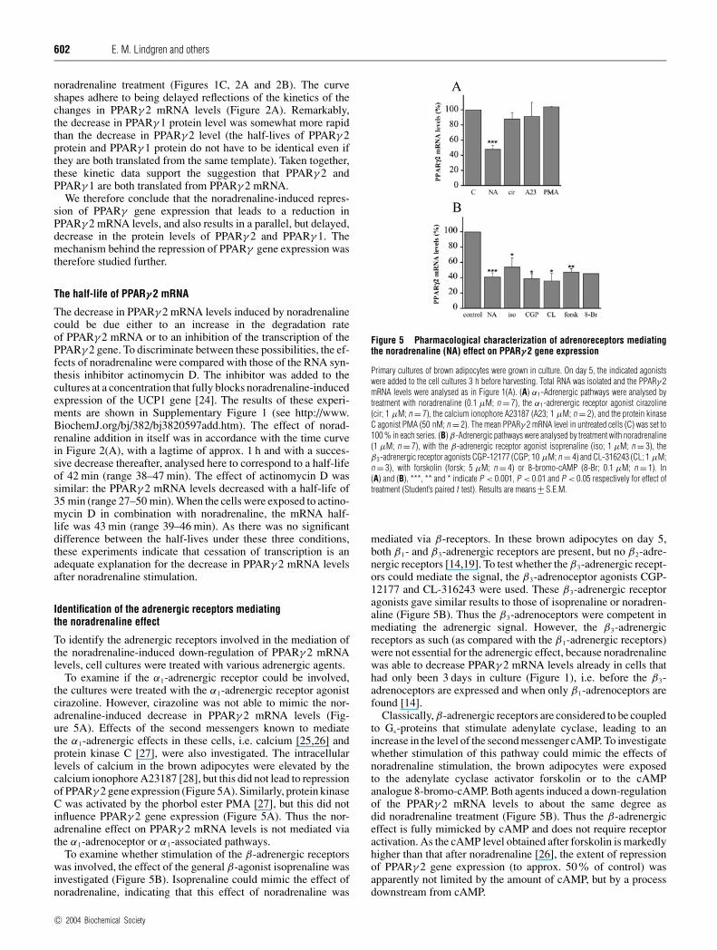
Figure 4 Time course of noradrenaline (NA) effect on PPARγ 1 and PPARγ 2levels
Primary cultures of brown adipocytes were treated essentially as indicated in Figure 2, andprotein was extracted and analysed for PPARγ by immunoblotting as described in Figure 3.(A) Example of time curve. (B) Compilation of results from experiments as those illustratedin (A). For each time point, the level of PPARγ 1 and PPARγ 2 was set to 100 % and the corres-ponding level in NA-treated cultures was calculated; values at each time point are from oneculture or means +− S.E.M. from two cultures, each analysed singly or duplicate. For the PPARγ 2levels, the line connecting the data points has been drawn ignoring the 4 h data point.
template for PPARγ 2 also contains the full template for PPARγ 1,including an undisturbed translation-initiation sequence, we sug-gest that the PPARγ 1 is formed from the PPARγ 2 template. Thelevels of PPARγ 1 and PPARγ 2 are about the same, as judgedfrom the intensities on the immunoblot (Figure 3A). This may beinterpreted that the translation machinery, with equal probability,commences at the PPARγ 2 and PPARγ 1 initiation sites on thePPARγ 2 mRNA; alternatively, each protein could be translated inparallel from both the PPARγ 2 and the PPARγ 1 initiation sites,as has been discussed for other proteins [23].
In Figure 4(A), an example of a time curve of the effect of norad-renaline on PPARγ protein levels is shown, and in Figure 4(B),a compilation of results from several experiments with different,overlapping, time points is displayed. It is seen that both PPARγ 2and PPARγ 1 protein amounts decrease, finally reaching a levelof approx. 50% of the starting value; this level is not statisticallysignificantly different for PPARγ 2 and PPARγ 1 (in the analysis,the phosphoPPARγ 2 and non-phosphorylated PPARγ 2 bandswere combined). A 50% reduction is what would be expectedfrom the 50% reduction in PPARγ 2 mRNA observed after
c© 2004 Biochemical Society
602 E. M. Lindgren and others
noradrenaline treatment (Figures 1C, 2A and 2B). The curveshapes adhere to being delayed reflections of the kinetics of thechanges in PPARγ 2 mRNA levels (Figure 2A). Remarkably,the decrease in PPARγ 1 protein level was somewhat more rapidthan the decrease in PPARγ 2 level (the half-lives of PPARγ 2protein and PPARγ 1 protein do not have to be identical even ifthey are both translated from the same template). Taken together,these kinetic data support the suggestion that PPARγ 2 andPPARγ 1 are both translated from PPARγ 2 mRNA.
We therefore conclude that the noradrenaline-induced repres-sion of PPARγ gene expression that leads to a reduction inPPARγ 2 mRNA levels, and also results in a parallel, but delayed,decrease in the protein levels of PPARγ 2 and PPARγ 1. Themechanism behind the repression of PPARγ gene expression wastherefore studied further.
The half-life of PPARγ 2 mRNA
The decrease in PPARγ 2 mRNA levels induced by noradrenalinecould be due either to an increase in the degradation rateof PPARγ 2 mRNA or to an inhibition of the transcription of thePPARγ 2 gene. To discriminate between these possibilities, the ef-fects of noradrenaline were compared with those of the RNA syn-thesis inhibitor actinomycin D. The inhibitor was added to thecultures at a concentration that fully blocks noradrenaline-inducedexpression of the UCP1 gene [24]. The results of these experi-ments are shown in Supplementary Figure 1 (see http://www.BiochemJ.org/bj/382/bj3820597add.htm). The effect of norad-renaline addition in itself was in accordance with the time curvein Figure 2(A), with a lagtime of approx. 1 h and with a succes-sive decrease thereafter, analysed here to correspond to a half-lifeof 42 min (range 38–47 min). The effect of actinomycin D wassimilar: the PPARγ 2 mRNA levels decreased with a half-life of35 min (range 27–50 min). When the cells were exposed to actino-mycin D in combination with noradrenaline, the mRNA half-life was 43 min (range 39–46 min). As there was no significantdifference between the half-lives under these three conditions,these experiments indicate that cessation of transcription is anadequate explanation for the decrease in PPARγ 2 mRNA levelsafter noradrenaline stimulation.
Identification of the adrenergic receptors mediatingthe noradrenaline effect
To identify the adrenergic receptors involved in the mediation ofthe noradrenaline-induced down-regulation of PPARγ 2 mRNAlevels, cell cultures were treated with various adrenergic agents.
To examine if the α1-adrenergic receptor could be involved,the cultures were treated with the α1-adrenergic receptor agonistcirazoline. However, cirazoline was not able to mimic the nor-adrenaline-induced decrease in PPARγ 2 mRNA levels (Fig-ure 5A). Effects of the second messengers known to mediatethe α1-adrenergic effects in these cells, i.e. calcium [25,26] andprotein kinase C [27], were also investigated. The intracellularlevels of calcium in the brown adipocytes were elevated by thecalcium ionophore A23187 [28], but this did not lead to repressionof PPARγ 2 gene expression (Figure 5A). Similarly, protein kinaseC was activated by the phorbol ester PMA [27], but this did notinfluence PPARγ 2 gene expression (Figure 5A). Thus the nor-adrenaline effect on PPARγ 2 mRNA levels is not mediated viathe α1-adrenoceptor or α1-associated pathways.
To examine whether stimulation of the β-adrenergic receptorswas involved, the effect of the general β-agonist isoprenaline wasinvestigated (Figure 5B). Isoprenaline could mimic the effect ofnoradrenaline, indicating that this effect of noradrenaline was
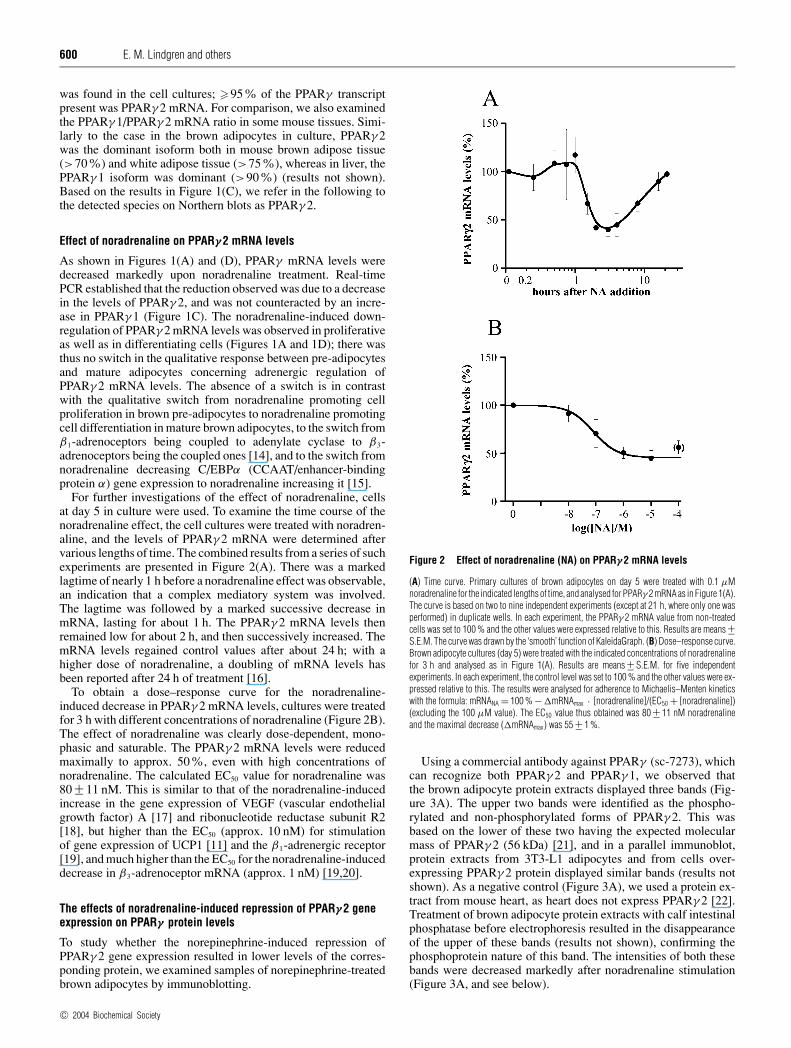
Figure 5 Pharmacological characterization of adrenoreceptors mediatingthe noradrenaline (NA) effect on PPARγ 2 gene expression
Primary cultures of brown adipocytes were grown in culture. On day 5, the indicated agonistswere added to the cell cultures 3 h before harvesting. Total RNA was isolated and the PPARγ 2mRNA levels were analysed as in Figure 1(A). (A) α1-Adrenergic pathways were analysed bytreatment with noradrenaline (0.1 µM; n = 7), the α1-adrenergic receptor agonist cirazoline(cir; 1 µM; n = 7), the calcium ionophore A23187 (A23; 1 µM; n = 2), and the protein kinaseC agonist PMA (50 nM; n = 2). The mean PPARγ 2 mRNA level in untreated cells (C) was set to100 % in each series. (B) β-Adrenergic pathways were analysed by treatment with noradrenaline(1 µM; n = 7), with the β-adrenergic receptor agonist isoprenaline (iso; 1 µM; n = 3), theβ3-adrenergic receptor agonists CGP-12177 (CGP; 10 µM; n = 4) and CL-316243 (CL; 1 µM;n = 3), with forskolin (forsk; 5 µM; n = 4) or 8-bromo-cAMP (8-Br; 0.1 µM; n = 1). In(A) and (B), ***, ** and * indicate P < 0.001, P < 0.01 and P < 0.05 respectively for effect oftreatment (Student’s paired t test). Results are means +− S.E.M.
mediated via β-receptors. In these brown adipocytes on day 5,both β1- and β3-adrenergic receptors are present, but no β2-adre-nergic receptors [14,19]. To test whether the β3-adrenergic recept-ors could mediate the signal, the β3-adrenoceptor agonists CGP-12177 and CL-316243 were used. These β3-adrenergic receptoragonists gave similar results to those of isoprenaline or noradren-aline (Figure 5B). Thus the β3-adrenoceptors were competent inmediating the adrenergic signal. However, the β3-adrenergicreceptors as such (as compared with the β1-adrenergic receptors)were not essential for the adrenergic effect, because noradrenalinewas able to decrease PPARγ 2 mRNA levels already in cells thathad only been 3 days in culture (Figure 1), i.e. before the β3-adrenoceptors are expressed and when only β1-adrenoceptors arefound [14].
Classically, β-adrenergic receptors are considered to be coupledto Gs-proteins that stimulate adenylate cyclase, leading to anincrease in the level of the second messenger cAMP. To investigatewhether stimulation of this pathway could mimic the effects ofnoradrenaline stimulation, the brown adipocytes were exposedto the adenylate cyclase activator forskolin or to the cAMPanalogue 8-bromo-cAMP. Both agents induced a down-regulationof the PPARγ 2 mRNA levels to about the same degree asdid noradrenaline treatment (Figure 5B). Thus the β-adrenergiceffect is fully mimicked by cAMP and does not require receptoractivation. As the cAMP level obtained after forskolin is markedlyhigher than that after noradrenaline [26], the extent of repressionof PPARγ 2 gene expression (to approx. 50% of control) wasapparently not limited by the amount of cAMP, but by a processdownstream from cAMP.
c© 2004 Biochemical Society

Peroxisome-proliferator-activated receptor γ 2 gene expression in brown adipocytes 603
Investigation of possible β-adrenergic-receptor-associatedsignalling pathwaysThe intracellular pathways involved in the noradrenaline-induced down-regulation in PPARγ 2 mRNA levels were investi-gated further by studying the effects of pathway-specific intra-cellular inhibitors. For this, the cells were pre-treated with theseinhibitors 1 h before and during a 3 h exposure to noradrenaline(see Supplementary Figure 2 at http://www.BiochemJ.org/bj/382/bj3820597add.htm). Again, exposure to noradrenaline in other-wise untreated cultures led to a reduction in PPARγ 2 mRNAlevels to about 50% (Supplementary Figure 2A). In Supplement-ary Figure 2(B), the effect of noradrenaline, i.e. the �PPARγ 2mRNA value, is depicted, for easier comparison of this parameterbetween the different inhibitors.
In 3T3-F442A adipocytes, β3-adrenoceptors have been sug-gested to be able to directly stimulate Gi-proteins [29,30]. If aβ3-adrenergic-receptor-coupled Gi-protein pathway was involvedin the mediation of the noradrenaline-induced repression ofPPARγ 2 gene expression in the brown adipocytes studied here,inhibition of Gi-proteins by pertussis toxin should inhibit the nor-adrenaline effect; this was clearly not the case (SupplementaryFigure 2). This postulated β3-adrenoceptor-coupled Gi-proteinpathway has also been proposed to directly stimulate PI3K (phos-phoinositide 3-kinase) in adipocytes [29], and this kinase, stimu-lated either in this way or through the classical β-adrenergicreceptor/Gs/cAMP pathway, could then mediate the repression.However, when the cultured brown adipocytes were treated withthe PI3K inhibitor Ly-294002 at a concentration that inhibitsinsulin-induced glucose uptake in cultured brown adipocytes [31],the noradrenaline effect on PPARγ 2 mRNA levels was unchanged(Supplementary Figure 2).
In cultured brown adipocytes, PKA (protein kinase A) furthermediates the cAMP signal towards UCP1 gene expression [32]. Toinvestigate whether PKA also mediates the noradrenaline effecton PPARγ 2 gene expression, the PKA inhibitor H89 was used ata concentration that abolishes noradrenaline-induced UCP1 geneexpression [32]. H89 abolished the noradrenaline-induced dec-rease in PPARγ 2 mRNA levels (Supplementary Figure 2B).This outcome is thus compatible with the noradrenaline-induceddecrease in PPARγ 2 mRNA levels being PKA-mediated, but non-specific effects of inhibitors can never be totally excluded.
β3-adrenoceptor pathways (as well as α1-adrenoceptor path-ways) activate the cytosolic tyrosine kinase Src in brown adipo-cytes [33]. Therefore the Src inhibitor PP2 was used at aconcentration that fully abolishes noradrenaline-induced andSrc-mediated phosphorylation of ERK1/2 (extracellular-signal-regulated kinase 1 and 2) in cultured brown adipocytes [17].However, PP2 had only a marginal effect on the noradrenaline-induced decrease in PPARγ 2 mRNA levels.
In brown adipocytes, noradrenaline activates the MAPK (mito-gen-associated protein kinase) ERK1/2 through MEK (MAPK/ERK kinase) activation [34]. That activation of ERK1/2 maybe mediatory for the effect of noradrenaline on PPARγ 2 levelsmay be suggested, since PPARγ 2 is phosphorylated by MAPK inadipocyte cell lines, and the phosphorylated PPARγ 2 suppressesthe transcriptional activity of PPARγ 2 and thus adipogenesis [35].However, the MEK inhibitor PD-98059, at a concentration thatinhibits the noradrenaline effect on ERK1/2 phosphorylation [34],had no effect on the noradrenaline-induced decrease on PPARγ 2mRNA levels (Supplementary Figure 2).
In brown adipocytes, noradrenaline also activates p38 MAPK[36]. Therefore the p38 inhibitor SB-202190 was added to thecells (Supplementary Figure 2B), but this did not have a significanteffect on the noradrenaline-induced repression of PPARγ 2 geneexpression (Supplementary Figure 2).
Thus the data observed were compatible with a β-adrenergicpathway, including cAMP and PKA activation, but not involvingGi, PI3K, Src, ERK1/2 or p38 MAPK.
Further mediation of the adrenergic signal requiresprotein synthesis
As a lagtime was observed for the noradrenaline-inducedrepression of PPARγ 2 gene expression (Figure 2A), it is feasiblethat the effect requires the synthesis of a new protein, rather thanbeing due to a direct effect on existing proteins. The cultureswere therefore treated with the protein synthesis inhibitor cyclo-heximide at a concentration which blocks translation in dif-ferentiated brown adipocytes in culture [37]. Cycloheximide fullyblocked the ability of noradrenaline to decrease PPARγ 2 mRNAlevels (Supplementary Figure 3 at http://www.BiochemJ.org/bj/382/bj3820597add.htm). Additionally, after a lagtime of about1 h, it evoked in itself (or equally in the presence of noradrenaline)an increase in PPARγ 2 mRNA levels. The levels were doubledafter a further 1 h and may apparently increase further during thenext 6–12 h [13]. These results indicate that protein synthesis isrequired for mediation of the adrenergic signal and could alsoimply that a continually synthesized protein with a rapid turnoverleads to tonic repression of PPARγ 2 mRNA synthesis.
DISCUSSION
In the present study, we found that the PPARγ mRNA isoformexpressed in brown adipocytes was predominantly PPARγ 2.In primary cultures of brown adipocytes, PPARγ 2 mRNA wasfound already in the undifferentiated brown pre-adipocytes. Theexpression peaked at day 5, just as the cells reached confluence andentered differentiation. Noradrenaline induced a transient de-crease in PPARγ 2 mRNA levels in both brown pre-adipocytesand in brown adipocytes, and this resulted in parallel decreasesin the protein levels of phosphoPPARγ 2, PPARγ 2 and PPARγ 1.A tentative pathway for the intracellular mediation of the nor-adrenaline-induced repression of gene expression may be formu-lated (see Supplementary Figure 4 at http://www.BiochemJ.org/bj/382/bj3820597add.htm).
PPARγ 2 mRNA dominance in brown adipocytes
We found that the PPARγ 2 mRNA levels exceeded the PPARγ 1mRNA levels approx. 20-fold in the cultured brown adipocytes.The relative levels of PPARγ 1 mRNA compared with those ofPPARγ 2 mRNA have not previously been determined in brownadipocytes, but a predominant expression of PPARγ 2 (comparedwith that of PPARγ 1) in mouse adipocyte cell lines has beenmentioned previously [2]. In mouse brown and white adiposetissue, we also observed a clear predominance of PPARγ 2 mRNA(as mentioned in the Results section), and, similarly, in humanwhite adipose tissue, a 20-fold excess of PPARγ 2 mRNA has beenfound [38]. However, there are also reports that roughly equallevels of PPARγ 1 and PPARγ 2 mRNA, or a predominance ofPPARγ 1, are found in mouse [39] and human [40] white adiposetissue. These qualitative differences in the PPARγ 1/PPARγ 2mRNA ratios reported (if not methodological) may be relatedto the prevailing hormonal status.
The noradrenaline effect
When brown pre-adipocytes or adipocytes were treated withnoradrenaline, down-regulation of the PPARγ 2 mRNA levelsoccurred within the first 2–4 h after treatment (Figures 1 and 2).
c© 2004 Biochemical Society

604 E. M. Lindgren and others
The down-regulation was transient. In the brown adipocyte-likecell line HIB-1B, both a noradrenaline-induced decrease [6] anda noradrenaline-induced increase [13] in PPARγ 2 mRNA levelshave been noted (4–5 h after stimulation), perhaps reflectingvariability in the characteristics of the cell line and phenotypicdrift in the cell line. The noradrenaline-induced repression obser-ved here in brown adipocyte primary cultures may be related toreported effects of exposure to cold on PPARγ 2 gene expressionin brown adipose tissue. Cold exposure leads to increased releaseof noradrenaline in situ in brown adipose tissue, and cold exposurehas been reported to induce a decrease in PPARγ 2 mRNA levels[41,42].
Noradrenaline stimulates proliferation in brown pre-adipocytesand differentiation in mature brown adipocytes. Therefore, inaccordance with present formulations concerning a positive role ofPPARγ 2 for cell differentiation and a concurrent inhibitory role incell proliferation, a shift from a noradrenaline-induced repressionof transcription in brown pre-adipocytes to a noradrenaline-indu-ced augmentation of transcription in differentiated brown adipo-cytes would have been expected. A notable observation here wastherefore the lack of a switch in the qualitative response to nor-adrenaline between brown pre-adipocytes and brown adipocytes(Figure 1).
Cellular mediation of the noradrenaline effect
Based on the observations reported here, we suggest a pathway forthe intracellular signalling from adrenergic stimulation to repres-sion of PPARγ 2 gene expression and decreased levels of PPARγ 2and PPARγ 1, as depicted in Supplementary Figure 4. The basisfor this pathway is as follows.
Although α1-, α2-, β1- and β3-adrenoceptors are present inbrown adipocytes, the experiments with adrenergic agonists andinhibitors or activators of the corresponding signalling processes(Figure 5 and Supplementary Figure 2) demonstrated that onlyβ-adrenoceptors (both β1- and β3-adrenoceptors) could mediatethe repression. cAMP levels are increased after noradrenalinestimulation in these cell cultures [26,43]. The repression ofPPARγ 2 mRNA levels is probably mediated via this cAMP, asa repression could also be induced by forskolin or 8-bromo-cAMP (Figure 5B). In contrast, in HIB-1B pre-adipocytes [6]and in rabbit type II pneumonocytes [44], dibutyryl-cAMP in-creased PPARγ 2/γ 1 mRNA levels. Thus no general response ofthe PPARγ 2 gene to cAMP increases can be formulated.
PKA is activated by noradrenaline/cAMP in brown adipocytes[32]. Based on investigation of the effects of different kinaseinhibitors (Supplementary Figure 2), it seems likely that the nor-adrenaline-induced decrease in PPARγ 2 mRNA levels ismediated by PKA, since the effect was totally abolished by theinhibitor H89, but not by any other inhibitor tested.
The response could proceed further via CREB (cAMP-response-element-binding protein) phosphorylation. Noradren-aline, through PKA activation, induces phosphorylation of thetranscription factor CREB also in primary cultures of brownadipocytes [45,46]. However, in other cell types (3T3-L1 cells), ithas been observed that a constitutively active CREB constructinduces (not represses) the expression of PPARγ 2 [47], anddominant-negative CREB constructs inhibit PPARγ 2 geneexpression [47]. As noradrenaline inhibits PPARγ 2 transcriptionin the brown adipocytes used in the present study, it is unlikelythat CREB is a direct activator of PPARγ 2 gene expression.
In this context, it may be noted that the noradrenaline effect hasa lagtime (Figure 2A), suggesting that an activation/inactivationof an existing transcription factor cannot easily explain themediation; instead there could be a requirement for protein
synthesis in the mediation process. Inhibition of protein synthesisindeed abolished the effect of noradrenaline (SupplementaryFigure 3). As inhibition of protein synthesis not only abolished thenoradrenaline effect, but also led to superinduction of PPARγ 2(Supplementary Figure 3) [13], the results can be interpreted asindicating the existence of a mediatory protein. Although therecould be other effects of protein synthesis inhibition [48], the datawould be compatible with a mediatory protein being expressed ata basal level, having a relatively short half-life (<1 h) and actingas a transcriptional repressor on the PPARγ 2 gene. When proteinsynthesis is inhibited, the regulatory protein would successivelydisappear, and so thus would repression, leading to increasedlevels of PPARγ 2 mRNA. On the other hand, noradrenalinestimulation (e.g. through CREB phosphorylation) could enhancethe rate of synthesis of the mediatory repressor protein, leadingto an increased repression of PPARγ 2 transcription.
Both PPARγ 2 and PPARγ 1 protein levels are repressedby noradrenaline
Although noradrenaline stimulation led to a marked reduction inPPARγ 2 mRNA levels, this reduction could be without significanteffects on cellular functions, if it were not reflected in an alteredlevel of PPARγ 2 protein. As the repression is transient (Fig-ure 2A), the protein levels could remain nearly unchanged if thehalf-life of PPARγ 2 was long. However, direct investigation ofPPARγ 2 protein levels clearly demonstrated that the decreasedmRNA level was fully reflected in a decreased PPARγ 2 level(Figures 3 and 4). Thus regulatory effects of the altered expressionmay be envisaged.
Although only PPARγ 2 mRNA is found in significant amountsin the brown adipocytes used in the present study, the immunoblotsrevealed that PPARγ 1 protein was present in equal amounts toPPARγ 2 protein (Figures 3 and 4). The most likely explanationfor this phenomenon is that the PPARγ 2 mRNA is translated withequal probability from the PPARγ 2 initiation site and from thePPARγ 1 initiation site (that is found unperturbed in the PPARγ 2mRNA). Whether this is a specific phenomenon for the brownadipocytes used in the present study is unknown, but the potentialfor the dual outcome of translation of the PPARγ 2 mRNA hasbeen evident since the first characterization of the PPARγ 2 splicevariant [2].
The physiological function of noradrenaline-induced repressionof PPARγ 2 gene expression
The physiological role of the noradrenaline-induced repressionof PPARγ 2 gene expression reported in the present paper, andthe ensuing decreases in PPARγ 2 and PPARγ 1 protein, is onlypartially understandable. A repression of PPARγ 2 gene expres-sion in brown pre-adipocytes is functionally explainable basedon the proliferative effect of noradrenaline [43] and the anti-proliferative role ascribed to PPARγ . However, the persistence ofthe effect in mature brown adipocytes, in which cell proliferationis not induced by noradrenaline [43], makes this explanation forthe noradrenaline-induced repression less probable.
If the differentiation-promoting effect of PPARγ is accepted,the suppressed expression of PPARγ 2 by noradrenaline shouldhave a de-differentiating effect on brown adipocyte characterist-ics. However, a general differentiation-promoting effect of nor-adrenaline is seen in the brown adipocytes. For instance, eventhough there is a PPRE site, e.g. in the UCP1 promoter [6,7],UCP1 gene expression is markedly induced, and not eventransiently repressed during noradrenaline stimulation [11]; theexplanation concerning UCP1 may be that this PPRE site could
c© 2004 Biochemical Society
Peroxisome-proliferator-activated receptor γ 2 gene expression in brown adipocytes 605
be stimulated primarily by PPARα [7]; PPARα is not decreased bynoradrenaline treatment (Figure 3A). Although brown adipocytesshare traits with white adipocytes and adipocyte-like cell lines, thefinal differentiation process of brown adipocytes must necessarilybe distinct from that of white adipocytes. Thus, whereas PPARγis necessary for adipose conversion, the possibility exists thattemporal repression of PPARγ 2 expression is a part of the eventsthat promote acquisition of brown adipose traits (as compared withgeneral adipose traits) during chronic noradrenaline stimulationof maturating brown adipocytes.
This study was supported by grants from The Swedish Cancer Society, The SwedishScience Research Council, the EU programme DLARFID (Dietary Lipids as Risk Factors inDevelopment) and The Danish Health Science Research Council. We thank Birgitta Leksellfor competent technical assistance and Tore Bengtsson for valuable discussions.
REFERENCES
1 Kliewer, S. A., Forman, B. M., Blumberg, B., Ong, E. S., Borgmeyer, U., Mangelsdorf,D. J., Umesono, K. and Evans, R. M. (1994) Differential expression and activation of afamily of murine peroxisome proliferator-activated receptors. Proc. Natl. Acad. Sci. U.S.A.91, 7355–7359
2 Tontonoz, P., Hu, E., Graves, R. A., Budavari, A. I. and Spiegelman, B. M. (1994)mPPARγ 2: tissue-specific regulator of an adipocyte enhancer. Genes Dev. 8, 1224–1234
2a Zhu, Y., Qi, C., Korenberg, J. R., Chen, X. N., Noya, D., Rao, M. S. and Reddy, J. K. (1995)Structural organization of mouse peroxisome proliferator-activated receptor γ (mPPARγ )gene: alternative promoter use and different splicing yield two mPPARγ isoforms.Proc. Natl. Acad. Sci. U.S.A. 92, 7921–7925
3 Tontonoz, P., Hu, E. and Spiegelman, B. M. (1994) Stimulation of adipogenesis infibroblasts by PPARγ 2, a lipid-activated transcription factor. Cell 79, 1147–1156
4 Kliewer, S. A., Umesono, K., Noonan, D. J., Heyman, R. A. and Evans, R. M. (1992)Convergence of 9-cis retinoic acid and peroxisome proliferator signalling pathwaysthrough heterodimer formation of their receptors. Nature (London) 358, 771–774
5 Berger, J. and Moller, D. E. (2002) The mechanisms of action of PPARs. Annu. Rev. Med.53, 409–435
6 Sears, I. B., MacGinnitie, M. A., Kovacs, L. G. and Graves, R. A. (1996)Differentiation-dependent expression of the brown adipocyte uncoupling protein gene:regulation by peroxisome proliferator-activated receptor γ . Mol. Cell. Biol. 16,3410–3419
7 Barbera, M. J., Schluter, A., Pedraza, N., Iglesias, R., Villarroya, F. and Giralt, M. (2001)Peroxisome proliferator-activated receptor α activates transcription of the brown fatuncoupling protein-1 gene: a link between regulation of the thermogenic and lipidoxidation pathways in the brown fat cell. J. Biol. Chem. 276, 1486–1493
8 Rosen, E. D. and Spiegelman, B. M. (2001) PPARγ : a nuclear regulator of metabolism,differentiation, and cell growth. J. Biol. Chem. 276, 37731–37734
9 Koeffler, H. P. (2003) Peroxisome proliferator-activated receptor γ and cancers.Clin. Cancer Res. 9, 1–9
10 Nedergaard, J., Herron, D., Jacobsson, A., Rehnmark, S. and Cannon, B. (1995)Norepinephrine as a morphogen?: its unique interaction with brown adipose tissue.Int. J. Dev. Biol. 39, 827–837
11 Rehnmark, S., Nechad, M., Herron, D., Cannon, B. and Nedergaard, J. (1990) α- andβ-adrenergic induction of the expression of the uncoupling protein thermogenin in brownadipocytes differentiated in culture. J. Biol. Chem. 265, 16464–16471
12 Hansen, J. B., Petersen, R. K., Larsen, B. M., Bartkova, J., Alsner, J. and Kristiansen, K.(1999) Activation of peroxisome proliferator-activated receptor γ bypasses the function ofthe retinoblastoma protein in adipocyte differentiation. J. Biol. Chem. 274, 2386–2393
13 Valmaseda, A., Carmona, M. C., Barbera, M. J., Vinas, O., Mampel, T., Iglesias, R.,Villarroya, F. and Giralt, M. (1999) Opposite regulation of PPAR-α and -γ geneexpression by both their ligands and retinoic acid in brown adipocytes.Mol. Cell. Endocrinol. 154, 101–109
14 Bronnikov, G., Bengtsson, T., Kramarova, L., Golozoubova, V., Cannon, B. andNedergaard, J. (1999) β1 to β3 switch in control of cAMP during brown adipocytedevelopment explains distinct β-adrenoceptor subtype mediation of proliferation anddifferentiation. Endocrinology 140, 4185–4197
15 Rehnmark, S., Antonson, P., Xanthopoulos, K. G. and Jacobsson, A. (1993) Differentialadrenergic regulation of C/EBPα and C/EBPβ in brown adipose tissue. FEBS Lett. 318,235–241
16 Rodriguez, E., Ribot, J. and Palou, A. (2002) Trans-10, cis-12, but not cis-9, trans-11CLA isomer, inhibits brown adipocyte thermogenic capacity. Am. J. Physiol. Regul. Integr.Comp. Physiol. 282, R1789–R1797
17 Fredriksson, J. M., Lindquist, J. M., Bronnikov, G. E. and Nedergaard, J. (2000)Norepinephrine induces vascular endothelial growth factor gene expression in brownadipocytes through a β-adrenoreceptor/cAMP/protein kinase A pathway involving Srcbut independently of Erk1/2. J. Biol. Chem. 275, 13802–13811
18 Fredriksson, J. M. and Nedergaard, J. (2002) Norepinephrine specifically stimulatesribonucleotide reductase subunit R2 gene expression in proliferating brown adipocytes:mediation via a cAMP/PKA pathway involving src and Erk1/2 kinases. Exp. Cell Res. 274,207–215
19 Bengtsson, T., Nedergaard, J. and Cannon, B. (2000) Differential regulation of the geneexpression of β-adrenoceptor subtypes in brown adipocytes. Biochem. J. 347,643–651
20 Bengtsson, T., Redegren, K., Strosberg, A. D., Nedergaard, J. and Cannon, B. (1996)Down-regulation of β3-adrenoreceptor gene expression in brown fat cells is transient andrecovery is dependent upon a short-lived protein factor. J. Biol. Chem. 271,33366–33375
21 Thuillier, P., Baillie, R., Sha, X. and Clarke, S. D. (1998) Cytosolic and nuclear distributionof PPARγ 2 in differentiating 3T3-L1 preadipocytes. J. Lipid Res. 39, 2329–2338
22 Escher, P., Braissant, O., Basu-Modak, S., Michalik, L., Wahli, W. and Desvergne, B.(2001) Rat PPARs: quantitative analysis in adult rat tissues and regulation in fasting andrefeeding. Endocrinology 142, 4195–4202
23 Kozak, M. (1999) Initiation of translation in prokaryotes and eukaryotes. Gene 234,187–208
24 Pico, C., Herron, D., Palou, A., Jacobsson, A., Cannon, B. and Nedergaard, J. (1994)Stabilization of the mRNA for the uncoupling protein thermogenin by transcriptional/translational blockade and by noradrenaline in brown adipocytes differentiated in culture:a degradation factor induced by cessation of stimulation? Biochem. J. 302, 81–86
25 Wilcke, M. and Nedergaard, J. (1989) α1- and β-adrenergic regulation of intracellularCa2+ levels in brown adipocytes. Biochem. Biophys. Res. Commun. 163, 292–300
26 Thonberg, H., Zhang, S.-J., Tvrdik, P., Jacobsson, A. and Nedergaard, J. (1994)Norepinephrine utilizes α1-and β-adrenoreceptors synergistically to maximally inducec-fos expression in brown adipocytes. J. Biol. Chem. 269, 33179–33186
27 Barge, R. M., Mills, I., Silva, E. and Larsen, P. R. (1988) Phorbol esters, protein kinase C,and thyroxine 5′-deiodinase in brown adipocytes. Am. J. Physiol. 254, E323–E327
28 Pappone, P. A. and Lee, S. C. (1995) α-Adrenergic stimulation activates acalcium-sensitive chloride current in brown fat cells. J. Gen. Physiol. 106, 231–258
29 Gerhardt, C. C., Gros, J., Strosberg, A. D. and Issad, T. (1999) Stimulation of theextracellular signal-regulated kinase 1/2 pathway by human β-3 adrenergic receptor:new pharmacological profile and mechanism of activation. Mol. Pharmacol. 55,255–262
30 Soeder, K. J., Snedden, S. K., Cao, W., Della Rocca, G. J., Daniel, K. W., Luttrell, L. M. andCollins, S. (1999) The β3-adrenergic receptor activates mitogen-activated protein kinasein adipocytes through a Gi-dependent mechanism. J. Biol. Chem. 274, 12017–12022
31 Chernogubova, E., Cannon, B. and Bengtsson, T. (2004) Norepinephrine increasesglucose transport in brown adipocytes via β3-adrenoceptors through a cAMP, PKA andPI3-kinase-dependent pathway stimulating conventional and novel PKCs. Endocrinology145, 269–280
32 Fredriksson, J. M., Thonberg, H., Ohlson, K. B. E., Ohba, K., Cannon, B. andNedergaard, J. (2001) Analysis of inhibition by H89 of UCP1 gene expression andthermogenesis indicates protein kinase A mediation of β3-adrenergic signalling ratherthan β3-adrenoceptor antagonism by H89. Biochim. Biophys. Acta 1538, 206–217
33 Lindquist, J. M., Fredriksson, J. M., Rehnmark, S., Cannon, B. and Nedergaard, J. (2000)β3- and α1-adrenergic Erk1/2 activation is Src but not Gi-mediated in brown adipocytes.J. Biol. Chem. 275, 22670–22677
34 Lindquist, J. M. and Rehnmark, S. (1998) Ambient temperature regulation of apoptosis inbrown adipose tissue: Erk 1/2 promotes norepinephrine-dependent cell survival.J. Biol. Chem. 273, 30147–30156
35 Hu, E., Kim, J. B., Sarraf, P. and Spiegelman, B. M. (1996) Inhibition of adipogenesisthrough MAP kinase-mediated phosphorylation of PPARγ . Science 274, 2100–2103
36 Cao, W., Medvedev, A. V., Daniel, K. W. and Collins, S. (2001) β-Adrenergic activation ofp38 MAP kinase in adipocytes: cAMP induction of the uncoupling protein 1 (UCP1) generequires p38 MAP kinase. J. Biol. Chem. 276, 27077–27082
37 Puigserver, P., Herron, D., Gianotti, M., Palou, A., Cannon, B. and Nedergaard, J.(1992) Induction and degradation of the uncoupling protein thermogenin in brownadipocytes in vitro and in vivo: evidence for a rapidly degradable pool. Biochem. J. 284,393–398
38 Giusti, V., Verdumo, C., Suter, M., Gaillard, R. C., Burckhardt, P. and Pralong, F. (2003)Expression of peroxisome proliferator-activated receptor-γ 1 and peroxisome proliferator-activated receptor-γ 2 in visceral and subcutaneous adipose tissue of obese women.Diabetes 52, 1673–1676
c© 2004 Biochemical Society

606 E. M. Lindgren and others
39 Vidal-Puig, A., Jimenez-Linan, M., Lowell, B. B., Hamann, A., Hu, E., Spiegelman, B.,Flier, J. S. and Moller, D. E. (1996) Regulation of PPARγ gene expression by nutritionand obesity in rodents. J. Clin. Invest. 97, 2553–2561
40 Auboeuf, D., Rieusset, J., Fajas, L., Vallier, P., Frering, V., Riou, J. P., Staels, B., Auwerx, J.,Laville, M. and Vidal, H. (1997) Tissue distribution and quantification of the expression ofmRNAs of peroxisome proliferator-activated receptors and liver X receptor-α in humans:no alteration in adipose tissue of obese and NIDDM patients. Diabetes 46, 1319–1327
41 Guardiola-Diaz, H. M., Rehnmark, S., Usuda, N., Albrektsen, T., Feltkamp, D., Gustafsson,J.-A. and Alexson, S. E. H. (1999) Rat peroxisome proliferator-activated receptors andbrown adipose tissue function during cold acclimatization. J. Biol. Chem. 274,23368–23377
42 Yu, X. X., Lewin, D. A., Forrest, W. and Adams, S. H. (2002) Cold elicits the simultaneousinduction of fatty acid synthesis and β-oxidation in murine brown adipose tissue:prediction from differential gene expression and confirmation in vivo. FASEB J. 16,155–168
43 Bronnikov, G., Houstek, J. and Nedergaard, J. (1992) β-Adrenergic, cAMP-mediatedstimulation of proliferation of brown fat cells in primary culture: mediation via β1 but notvia β3 receptors. J. Biol. Chem. 267, 2006–2013
44 Michael, L. F., Lazar, M. A. and Mendelson, C. R. (1997) Peroxisome proliferator-activatedreceptor γ 1 expression is induced during cyclic adenosine monophosphate-stimulateddifferentiation of alveolar type II pneumonocytes. Endocrinology 138, 3695–3703
45 Chaudhry, A. and Granneman, J. G. (1999) Differential regulation of functional responsesby β-adrenergic receptor subtypes in brown adipocytes. Am. J. Physiol. 277, R147–R153
46 Thonberg, H., Nedergaard, J. and Cannon, B. (2002) A novel pathway for adrenergicstimulation of cAMP-response-element-binding protein (CREB) phosphorylation:mediation via α1-adrenoceptors and protein kinase C activation. Biochem. J. 364, 73–79
47 Reusch, J. E. B., Colton, L. A. and Klemm, D. J. (2000) CREB activation inducesadipogenesis in 3T3-L1 cells. Mol. Cell. Biol. 20, 1008–1020
48 Ross, J. (1997) A hypothesis to explain why translation inhibitors stabilize mRNAs inmammalian cells: mRNA stability and mitosis. Bioessays 19, 527–529
Received 24 October 2003/4 June 2004; accepted 14 June 2004Published as BJ Immediate Publication 14 June 2004, DOI 10.1042/BJ20031622
c© 2004 Biochemical Society
Related Documents