Nonsynonymous Mutations within APOB in Human Familial Hypobetalipoproteinemia EVIDENCE FOR FEEDBACK INHIBITION OF LIPOGENESIS AND POSTENDOPLASMIC RETICULUM DEGRADATION OF APOLIPOPROTEIN B * □ S Received for publication, August 27, 2009, and in revised form, December 1, 2009 Published, JBC Papers in Press, December 23, 2009, DOI 10.1074/jbc.M109.060467 Shumei Zhong ‡ , Antonia Lucia Magnolo § , Meenakshi Sundaram ‡ , Hu Zhou ‡ , Erik F. Yao ‡ , Enza Di Leo § , Paola Loria ¶ , Shuai Wang ‡ , Michelle Bamji-Mirza ‡ , Lisheng Wang ‡1 , C. Jamie McKnight , Daniel Figeys ‡2 , Yuwei Wang ‡ , Patrizia Tarugi §3 , and Zemin Yao ‡4 From the ‡ Department of Biochemistry, Microbiology and Immunology, Ottawa Institute of Systems Biology, University of Ottawa, Ottawa, Ontario K1H 8M5, Canada, the § Dipartimento di Scienze Biomediche, Universita ` di Modena e Reggio Emilia, Via Campi 287, I-41100 Modena, Italy, the Department of Physiology and Biophysics, Boston University School of Medicine, Boston, Massachusetts 02118-2526, and the ¶ Dipartimento di Medicina, Endocrinologia, Metabolismo Egeriatria, Universita ` di Modena e Reggio Emilia, I-41100 Modena, Italy Five nontruncating missense APOB mutations, namely A31P, G275S, L324M, G912D, and G945S, were identified in heterozygous carriers of familial hypobetalipoproteinemia (FHBL) in the Italian population. To test that the FHBL phe- notype was a result of impaired hepatic secretion of mutant apoB proteins, we performed transfection studies using McA-RH7777 cells stably expressing wild type or mutant forms of human apolipoprotein B-48 (apoB-48). All mutant proteins displayed varied impairment in secretion, with G912D the least affected and A31P barely secreted. Although some A31P was degraded by proteasomes, a significant pro- portion of it (although inappropriately glycosylated) escaped endoplasmic reticulum (ER) quality control and presented in the Golgi compartment. Degradation of the post-ER A31P was achieved by autophagy. Expression of A31P also de- creased secretion of endogenous apoB and triglycerides, yet the impaired lipoprotein secretion did not lead to lipid accu- mulation in the cells or ER stress. Rather, expression of genes involved in lipogenesis was down-regulated, including liver X receptor , sterol regulator element-binding protein 1c, fatty acid synthase, acetyl-CoA carboxylase 1, stearoyl-CoA desaturase 1, and lipin-1. These results suggest that feedback inhibition of hepatic lipogenesis in conjunction with post-ER degradation of misfolded apoB proteins can contribute to reduce fat accumulation in the FHBL liver. Familial hypobetalipoproteinemia (FHBL) 5 (OMIM 107730) is an autosomal co-dominant disorder characterized by low plasma levels (5th percentile for age and sex) of low density lipoprotein (LDL) cholesterol and apolipoprotein B (apoB) (1). The genetic basis for FHBL is heterogeneous and is often asso- ciated with mutations within the APOB gene (1). The APOB gene encodes both apoB-100 and apoB-48, the latter being a truncated form representing the N-terminal 48% of the full- length apoB-100 (2, 3). In rats and mice, both apoB-100 and apoB-48 are synthesized in the liver (4). The majority of APOB gene mutations in FHBL are nonsense, frameshift, or splicing variants that lead to various C-terminally truncated apoB spe- cies (5). However, nonsynonymous, nontruncating mutations (e.g. L343V and R463W) (6, 7) do occur in FHBL. Transfection studies showed that apoB-100 and apoB-48 bearing the L343V or R463W mutation were secreted inefficiently and exhibited increased endoplasmic reticulum (ER) retention (6, 7). Unlike truncation mutants (ranging from apoB-15 to apoB-94) that are often secreted normally (8, 9), the nontruncation mutants are impaired in their secretion. The nontruncating mutations identified in FHBL to date are all located within the N-terminal region composed of multiple anti-parallel -helices (residues 292–593) (10, 11). This region, together with the preceding -barrel (the first 264 residues), is known as a modeled 1 domain of apoB based on its homol- ogy to lipovitellin (10). This homology model predicts that the 1 domain of apoB consists of -barrel and -helical domain plus two -sheets termed C sheet (residues 611–782) and A sheet (residues 783–930), respectively. The 1 domain is known to be important for effective secretion from transfected cells, because recombinant apoB segments lacking this domain * This work was supported in part by National Institutes of Health Grant HL-26335 (to C. J. M.), Heart and Stroke Foundation of Ontario Grant T-4643 (to Z. Y.), Genome Canada Grant 2008-OGI-TD-01 (to D. F.), Cana- dian Institutes of Health Research (CIHR) Grant MOP-158235 (to L. W.), and Telethon Foundation Onlus, Italy Grant GGP05042 (to P. T.). □ S The on-line version of this article (available at http://www.jbc.org) contains supplemental Tables 1 and 2 and Fig. S1. 1 Recipient of a New Investigator award from CIHR. 2 Canadian Research Chair of Proteomics and Systems Biology. 3 To whom correspondence may be addressed. E-mail: [email protected]. 4 A Carrier Investigator of the Heart and Stroke Foundation of Ontario. To whom correspondence may be addressed: 451 Smyth Rd., Dept. of Bio- chemistry, Microbiology and Immunology, University of Ottawa, Ottawa, Ontario K1H 8M5, Canada. E-mail: [email protected]. 5 The abbreviations used are: FHBL, familial hypobetalipoproteinemia; LDL, low density lipoprotein(s); apo, apolipoprotein; apoB-48wt, wild type apoB-48; VLDL, very low density lipoprotein(s); MTP, microsomal triglycer- ide transfer protein; TAG, triacylglycerol; LDL-C, LDL cholesterol; DMEM, Dulbecco’s modified Eagle’s medium; FBS, fetal bovine serum; ER, endo- plasmic reticulum; Endo H, endoglycosidase H; PC, phosphatidylcholine; ACC, acetyl-CoA carboxylase; FAS, fatty acid synthase; LXR, liver X recep- tor ; MS/MS, tandem mass spectrometry; HPLC, high pressure liquid chro- matography; RT, reverse transcription. THE JOURNAL OF BIOLOGICAL CHEMISTRY VOL. 285, NO. 9, pp. 6453–6464, February 26, 2010 © 2010 by The American Society for Biochemistry and Molecular Biology, Inc. Printed in the U.S.A. FEBRUARY 26, 2010 • VOLUME 285 • NUMBER 9 JOURNAL OF BIOLOGICAL CHEMISTRY 6453 at Univ of Ottawa - OCUL, on February 25, 2010 www.jbc.org Downloaded from http://www.jbc.org/content/suppl/2010/01/08/M109.060467.DC1.html Supplemental Material can be found at:

Welcome message from author
This document is posted to help you gain knowledge. Please leave a comment to let me know what you think about it! Share it to your friends and learn new things together.
Transcript
Nonsynonymous Mutations within APOB in Human FamilialHypobetalipoproteinemiaEVIDENCE FOR FEEDBACK INHIBITION OF LIPOGENESIS AND POSTENDOPLASMICRETICULUM DEGRADATION OF APOLIPOPROTEIN B*□S
Received for publication, August 27, 2009, and in revised form, December 1, 2009 Published, JBC Papers in Press, December 23, 2009, DOI 10.1074/jbc.M109.060467
Shumei Zhong‡, Antonia Lucia Magnolo§, Meenakshi Sundaram‡, Hu Zhou‡, Erik F. Yao‡, Enza Di Leo§, Paola Loria¶,Shuai Wang‡, Michelle Bamji-Mirza‡, Lisheng Wang‡1, C. Jamie McKnight�, Daniel Figeys‡2, Yuwei Wang‡,Patrizia Tarugi§3, and Zemin Yao‡4
From the ‡Department of Biochemistry, Microbiology and Immunology, Ottawa Institute of Systems Biology, University of Ottawa,Ottawa, Ontario K1H 8M5, Canada, the §Dipartimento di Scienze Biomediche, Universita di Modena e Reggio Emilia, Via Campi287, I-41100 Modena, Italy, the �Department of Physiology and Biophysics, Boston University School of Medicine, Boston,Massachusetts 02118-2526, and the ¶Dipartimento di Medicina, Endocrinologia, Metabolismo Egeriatria, Universita diModena e Reggio Emilia, I-41100 Modena, Italy
Five nontruncating missense APOB mutations, namelyA31P, G275S, L324M, G912D, and G945S, were identified inheterozygous carriers of familial hypobetalipoproteinemia(FHBL) in the Italian population. To test that the FHBL phe-notype was a result of impaired hepatic secretion of mutantapoB proteins, we performed transfection studies usingMcA-RH7777 cells stably expressing wild type or mutantforms of human apolipoprotein B-48 (apoB-48). All mutantproteins displayed varied impairment in secretion, withG912D the least affected and A31P barely secreted. Althoughsome A31P was degraded by proteasomes, a significant pro-portion of it (although inappropriately glycosylated) escapedendoplasmic reticulum (ER) quality control and presented inthe Golgi compartment. Degradation of the post-ER A31Pwas achieved by autophagy. Expression of A31P also de-creased secretion of endogenous apoB and triglycerides, yetthe impaired lipoprotein secretion did not lead to lipid accu-mulation in the cells or ER stress. Rather, expression of genesinvolved in lipogenesis was down-regulated, including liverX receptor �, sterol regulator element-binding protein 1c,fatty acid synthase, acetyl-CoA carboxylase 1, stearoyl-CoAdesaturase 1, and lipin-1. These results suggest that feedbackinhibition of hepatic lipogenesis in conjunction with post-ERdegradation of misfolded apoB proteins can contribute toreduce fat accumulation in the FHBL liver.
Familial hypobetalipoproteinemia (FHBL)5 (OMIM 107730)is an autosomal co-dominant disorder characterized by lowplasma levels (�5th percentile for age and sex) of low densitylipoprotein (LDL) cholesterol and apolipoprotein B (apoB) (1).The genetic basis for FHBL is heterogeneous and is often asso-ciated with mutations within the APOB gene (1). The APOBgene encodes both apoB-100 and apoB-48, the latter being atruncated form representing the N-terminal 48% of the full-length apoB-100 (2, 3). In rats and mice, both apoB-100 andapoB-48 are synthesized in the liver (4). The majority of APOBgene mutations in FHBL are nonsense, frameshift, or splicingvariants that lead to various C-terminally truncated apoB spe-cies (5). However, nonsynonymous, nontruncating mutations(e.g. L343V and R463W) (6, 7) do occur in FHBL. Transfectionstudies showed that apoB-100 and apoB-48 bearing the L343Vor R463W mutation were secreted inefficiently and exhibitedincreased endoplasmic reticulum (ER) retention (6, 7). Unliketruncationmutants (ranging fromapoB-15 to apoB-94) that areoften secreted normally (8, 9), the nontruncation mutants areimpaired in their secretion.The nontruncating mutations identified in FHBL to date are
all located within the N-terminal region composed of multipleanti-parallel �-helices (residues 292–593) (10, 11). This region,together with the preceding �-barrel (the first 264 residues), isknown as a modeled ��1 domain of apoB based on its homol-ogy to lipovitellin (10). This homology model predicts that the��1 domain of apoB consists of �-barrel and �-helical domainplus two �-sheets termed C sheet (residues 611–782) and Asheet (residues 783–930), respectively. The ��1 domain isknown to be important for effective secretion from transfectedcells, because recombinant apoB segments lacking this domain
* This work was supported in part by National Institutes of Health GrantHL-26335 (to C. J. M.), Heart and Stroke Foundation of Ontario GrantT-4643 (to Z. Y.), Genome Canada Grant 2008-OGI-TD-01 (to D. F.), Cana-dian Institutes of Health Research (CIHR) Grant MOP-158235 (to L. W.), andTelethon Foundation Onlus, Italy Grant GGP05042 (to P. T.).
□S The on-line version of this article (available at http://www.jbc.org) containssupplemental Tables 1 and 2 and Fig. S1.
1 Recipient of a New Investigator award from CIHR.2 Canadian Research Chair of Proteomics and Systems Biology.3 To whom correspondence may be addressed. E-mail: [email protected] A Carrier Investigator of the Heart and Stroke Foundation of Ontario. To
whom correspondence may be addressed: 451 Smyth Rd., Dept. of Bio-chemistry, Microbiology and Immunology, University of Ottawa, Ottawa,Ontario K1H 8M5, Canada. E-mail: [email protected].
5 The abbreviations used are: FHBL, familial hypobetalipoproteinemia; LDL,low density lipoprotein(s); apo, apolipoprotein; apoB-48wt, wild typeapoB-48; VLDL, very low density lipoprotein(s); MTP, microsomal triglycer-ide transfer protein; TAG, triacylglycerol; LDL-C, LDL cholesterol; DMEM,Dulbecco’s modified Eagle’s medium; FBS, fetal bovine serum; ER, endo-plasmic reticulum; Endo H, endoglycosidase H; PC, phosphatidylcholine;ACC, acetyl-CoA carboxylase; FAS, fatty acid synthase; LXR�, liver X recep-tor �; MS/MS, tandem mass spectrometry; HPLC, high pressure liquid chro-matography; RT, reverse transcription.
THE JOURNAL OF BIOLOGICAL CHEMISTRY VOL. 285, NO. 9, pp. 6453–6464, February 26, 2010© 2010 by The American Society for Biochemistry and Molecular Biology, Inc. Printed in the U.S.A.
FEBRUARY 26, 2010 • VOLUME 285 • NUMBER 9 JOURNAL OF BIOLOGICAL CHEMISTRY 6453
at Univ of O
ttawa - O
CU
L, on February 25, 2010
ww
w.jbc.org
Dow
nloaded from
http://www.jbc.org/content/suppl/2010/01/08/M109.060467.DC1.htmlSupplemental Material can be found at:
were either secreted poorly or not secreted at all (12, 13). The��1 domain can bind to microsomal triglyceride transfer pro-tein (MTP), an apoB-specific molecular chaperone that facili-tates lipid recruitment duringVLDL assembly (14). There are atleast three MTP binding sites within the ��1 domain (residues2–154, 430–570, and 512–721) (10, 15, 16). Although somestudies have shown that secretion of the ��1 domain is inde-pendent ofMTP (see Ref. 17 and references cited within), thereis no evidence showing that apoB-100 or apoB-48 can besecreted without the activity of MTP.VLDL assembly and maturation involve multiple lipidation
steps that occur during trafficking of VLDL precursorsthroughout the ER/Golgi compartments (18). The assemblyprocess is highly dependent on the availability of lipids, mainlytriacylglycerol (TAG). The activities of TAG synthesis en-zymes, includingDGAT1 (diacylglycerol acyltransferase 1) (19)and lipin-1 (20), have been shown to control VLDL assembly/secretion. Increased de novo lipogenesis also contributes sig-nificantly to VLDL assembly/secretion. Regulation of hepaticlipogenesis requires a network of nuclear receptors that coor-dinately govern expression of the lipogenesis enzymes acetyl-CoA carboxylase 1 (ACC1), fatty acid synthase (FAS), andSCD1 (stearoyl-CoA desaturase) (21). Transcription factorsand co-factors regulating lipogenesis gene expression andVLDL production include liver X receptor � (LXR�) (22),SREBP-1 (sterol regulatory element-binding protein 1), andPGC-1 (peroxisomeproliferator-activated receptor-� coactiva-tor) family members PGC-1� and PGC-1� (23), among others.Because of the importance of the ��1 domain in VLDL
assembly/secretion, we have searched for naturally occurringmutations within this domain that could affect apoB secretion.Recently, through molecular diagnosis of 110 Italian subjectswith clinical diagnosis of FHBL, we discovered eight subjectscarrying novel missense mutations, five of which occurredwithin the ��1 domain. The present studies showed thatmutation A31P within the �-barrel severely impaired apoBsecretion, and cells expressing the A31P mutant exhibitedsuppressed lipogenesis.
EXPERIMENTAL PROCEDURES
Materials—Cell culture reagents were purchased fromInvitrogen. Reagents for recombinant DNA experiments wereobtained from New England Biolabs (Pickering, Canada).[2-3H]Glycerol (9.6 Ci/mmol) and [35S]methionine/cysteine(1,000 Ci/mmol) were obtained from GE Healthcare. [3H]Ace-tic acid (0.1 Ci/mmol) was obtained from PerkinElmer LifeSciences. Rabbit anti-mouse apoE antibody (used for immu-noprecipitation) was obtained from BioDesign (Saco, ME).Horseradish peroxidase-linked anti-goat antibody and 3-methyl-adenine were obtained from Sigma. Horseradish peroxidase-conjugated anti-mouse and anti-rabbit IgG antibodies wereobtained from Amersham Biosciences. MG132 was obtainedfrom Calbiochem. Chemiluminescent substrates were pur-chased from Roche Applied Science. Trypsin was obtainedfrom Promega (Madison, WI). Antibodies against FAS andgiantin (Abcam (Cambridge, MA)); ACC, phospho-ACC (atSer79), calnexin, and actin (Millipore (Temecula, CA)); eIF2aand phospho-eIF2� (at Ser52) (Cell SignalingTechnology (Dan-
vers, MA)); C/EBP-homologous protein (CHOP or GADD153)(Santa Cruz Biotechnology, Inc. (Santa Cruz, CA)); and GRP78(glucose-regulated protein 78) (Assay Designs (Ann Arbor,MI)) were purchased from the respective suppliers. Mono-clonal antibody 1D1 against human apoBwas a gift of Drs. YvesMarcel and Ross Milne (University of Ottawa Heart Institute).Anti-MTP antibody was a gift of Dr. Carol Shoulders (MedicalResearch Council Clinical Sciences Centre, HammersmithHospital, London, UK). Polyclonal antiserum against rat VLDLproteins (used for immunoprecipitation of rat apoB-100) wasproduced in our laboratory.The FHBL Subjects—The FHBL subjects were identified as
probable heterozygous carriers during a survey of 110 consec-utive individuals referred to the hospital for the presence offatty liver associated with low plasma levels of LDL cholesterol(LDL-C) and apoB (1). The diagnosis of definite FHBL wasbased on three criteria: (i) LDL-C � 70 mg/dl and apoB � 50mg/dl; (ii) exclusion of secondary hypocholesterolemia; and(iii) vertical transmission of the lipid and apoB phenotype infamily. The diagnosis of probable heterozygous FHBL wasmadewhen reliable family datawere unavailable. Sequencing ofthe APOB gene revealed pathogenic mutations in 54 subjects(1). The majority of the mutations were located in the codingsequence of APOB. Fifty-one subjects were found to carrymutations causing the formation of truncated apoBs of varioussizes (1). Three subjects were carriers of the previously de-scribedmissensemutationR463W(6), known to be the cause ofFHBL. Five subjects (four with definite and one with probableFHBL) were heterozygous for rare non-conserved APOB genemutations, namely G945S, A31P, L324M, G912D, and G275S(1). The present study focused on these five mutations.Analysis of APOB Gene—Genomic DNA was isolated from
peripheral blood (NucleoSpin Blood L, Macherey-Nagel,Duren, Germany). The entire APOB gene coding region,including the 5�-flanking region and at least 50 base pairs ofintronic sequence at each intron-exon boundary (GenBankTMaccession number AY324608.1, GI:32187678) were amplifiedby PCR and sequenced as previously described (24). Sequenceswere detected on an ABI PRISM 3100 DNA sequencer, andresults were analyzed with ABI PRISM SeqScape software(Applied Biosystems,Warrington, UK). Only a fewmembers ofthe five kindred were available for DNA analysis. The APOBgene mutations found in the probands were screened in familymembers available for the study by sequencing the appropriategene regions (25). Sequence variants were confirmed by using asecond independent amplification of the affected DNA regionand resequencing in both directions. One hundred control sub-jects randomly selected among normolipidemic individuals ofboth sexes were screened for the mutations of the APOB genefound in FHBL patients by direct sequencing of the appropriategene regions or by restriction enzyme digestion of PCR prod-ucts. APOB gene mutations are described according to muta-tion nomenclature (available on the HumanGenome VariationSociety Web site) (26, 27). Nucleotide numbers are derivedfrom APOB cDNA sequence (GenBankTM accession numberNM_000384). Amino acid sequence changes in apoB are de-scribed according to the National Centre for BiotechnologyInformation reference sequence (NP_000375.2, GI:105990532).
APOB Missense Mutations in Familial Hypobetalipoproteinemia
6454 JOURNAL OF BIOLOGICAL CHEMISTRY VOLUME 285 • NUMBER 9 • FEBRUARY 26, 2010
at Univ of O
ttawa - O
CU
L, on February 25, 2010
ww
w.jbc.org
Dow
nloaded from
http://www.jbc.org/content/suppl/2010/01/08/M109.060467.DC1.htmlSupplemental Material can be found at:
The apoE genotype was performed as described (28). Informedconsent was obtained from all probands (or in the case of chil-dren from their parents) and their family members as well asfrom control subjects. The study protocol was approved by theethics committee of the University of Modena and ReggioEmilia and is in accordance with ethical standards as formu-lated by the Helsinki Declaration of 1975 (revised in 1983).Analysis of Plasma Lipids and Lipoproteins—Blood samples
were collected after an overnight fast. Plasma total cholesterol,TAG, high density lipoprotein cholesterol, LDL-C, apoA-I, andapoB levels were determined with enzymatic methods (RocheApplied Science) by using a Hitachi 912 autoanalyzer. Plasmalipoproteins were also separated by density gradient ultracen-trifugation (24). Isolated lipoprotein fractions were precipi-tated in 10% trichloroacetic acid and extracted with ethanoldiethyl ether (3:2, v/v), and apolipoproteins were separated bySDS-PAGE (3.5/5–10% gel for apoB and 5–20% gel for otherproteins) (24).Preparation of Expression Plasmids—The expression plas-
mids encoding human apoB-48 or apoB-17 prepared previously(7) were used as templates to prepare mutants using theQuikChange� II XL site-directed mutagenesis kit (Stratagene,La Jolla, CA). The mutagenic primers used to introduce theA31P, G275S, L324M, G912D, and G945S mutations werelisted in supplemental Table 1. The expression plasmids werepurified by the Qiagen plasmid purification kit (Qiagen,Milano, Italy), and the APOB coding regions were authenti-cated by sequencing.Cell Culture, Transfection, and Detection of Recombinant
ApoB—Stable expression of all of the recombinant apoB-48proteins was achieved using McA-RH7777 cells as describedpreviously (29). The cells were routinely cultured in Dulbecco’s
modified Eagle’s medium (DMEM) supplemented with 10%fetal bovine serum (FBS) and 10% horse serum. Stable cloneswere selected with G418 (400 �g/ml), and those that showedsecretion of apoB-48 mutants at similar levels were maintainedin G418 (200 �g/ml) for experiments. Expression of apoB-48and apoB-17 was determined by Western blotting of condi-tioned media using monoclonal antibody 1D1 that has anepitope at amino acids 401–582 of human apoB-100 (8).Lipoprotein Fractionation—Lipoproteins presented in the
microsomal lumenor secreted into themediawere fractionatedinto VLDL1, VLDL2, and other lipoproteins as described previ-ously (30).Subcellular Fractionation of Microsomal Membranes—Sub-
cellular fractionation of ER and Golgi microsome and analysisof endoglycosidase H (Endo H) sensitivity of apoB-48 proteinsassociated with the microsomes were performed as describedpreviously (31).Co-immunoprecipitation—Cells were cultured in DMEM
containing 20% FBS and 0.4 mM oleate for 2 h and harvested in1 ml of lysis buffer (50 mM Tris-HCl, pH 8.0, 150 mM NaCl, 1%Nonidet P-40, 5 mM EDTA, 20% sucrose, protease inhibitormixture). The lysate samples were incubated for 1 h at 4 °C bygentle mixing, and the insoluble materials were removed bycentrifugation (13,000 rpm, 4 °C, 15 min). An aliquot of thesupernatant (750 �g of protein) wasmixed with lysis buffer to afinal volume of 1 ml. The samples were precleared by incuba-tion with protein A-Sepharose CL-4B beads (2 h, 4 °C) prior toincubation with monoclonal antibody 1D1 that had been cou-pled to CNBr-activated Sepharose 4B beads as described previ-ously (30). After overnight incubation at 4 °C, the proteins wereeluted and subjected to SDS-PAGE and Western blot analysisto quantify apoB, MTP, and GRP78.
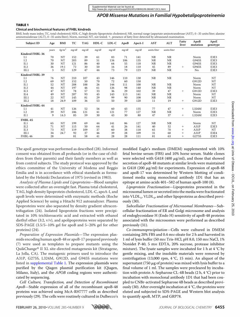
TABLE 1Clinical and biochemical features of FHBL kindredsBMI, body mass index; TC, total cholesterol; HDL-C, high density lipoprotein cholesterol; NR, normal range (aspartate aminotransferase (AST), 0–35 units/liter; alanineaminotransferase (ALT), 0–35 units/liter); Norm, normal; NT, not tested; �, presence of fatty liver detected by ultrasound examination.
Subject ID Age BMI TC TAG HDL-C LDL-C ApoB ApoA-I AST ALT Fattyliver
ApoBmutation
ApoEgenotype
years kg/m2 mg/dl mg/dl mg/dl mg/dl mg/dl mg/dl units/liter units/literKindred FHBL-36I.1 70 NT 152 39 61 83 71 141 NR NR Norm E3E3I.2 70 NT 203 89 51 134 106 135 NR NR G945S E3E3II.1 30 NT 121 86 40 64 55 118 NR NR G945S E3E3II.2 36 19.1 72 30 50 16 18 110 45 89 � G945S E3E3III.1 3 NT 129 47 44 73 79 123 NR NR No. E3E3
Kindred FHBL-39I.1 76 NT 210 107 43 146 110 130 NR NR Norm NTI.2 69 NT 152 50 70 72 60 150 G912D NTII.1 52 NT 208 188 26 144 111 100 NR NR Norm NTII.2 44 NT 197 46 61 126 98 140 NR NR Norm NTII.3 47 NT 78 57 33 36 29 102 39 47 � G912D E3E3II.4 49 NT 207 93 45 143 113 141 NR NR Norm NTIII.1 22 NT 145 81 29 99 90 108 NR NR Norm NTIII.2 18 24.9 109 36 53 50 39 120 11 19 � G912D E3E3
Kindred FHBL-44I.1 41 NT 126 52 56 60 43 135 77 47 � L324M E3E3I.2 39 NT 186 121 41 121 77 127 NR NR Norm E2E3II.1 9 14.3 85 59 30 43 30 80 47 37 � L324M E2E3
FHBL-45II.1 65 NT 199 69 44 141 86 127 NR NR Norm NTII.2 71 NT 115 50 64 41 26 141 40 52 � A31P E3E3II.3 73 NT 119 109 37 60 38 118 65 70 � A31P NTIII.1 34 24.7 92 37 46 39 28 109 31 68 � A31P E3E4
FHBL-46 28 81 58 41 28 28 111 81 63 � G275S E3E3
APOB Missense Mutations in Familial Hypobetalipoproteinemia
FEBRUARY 26, 2010 • VOLUME 285 • NUMBER 9 JOURNAL OF BIOLOGICAL CHEMISTRY 6455
at Univ of O
ttawa - O
CU
L, on February 25, 2010
ww
w.jbc.org
Dow
nloaded from
http://www.jbc.org/content/suppl/2010/01/08/M109.060467.DC1.htmlSupplemental Material can be found at:
Pulse-chase Analysis of ApoB—Post-translational stabilityand secretion efficiency of apoB (recombinant apoB-48 andendogenous apoB-100) were determined by pulse-chase exper-iments with [35S]methionine/cysteine as described previously(7). In some experiments, the post-translational stability andsecretion of apoB-48 mass were determined in the presence ofcycloheximide (10 �M) (32). Cells (in 60-mm dishes) were cul-tured in DMEM containing 20% FBS and 0.4 mM oleate for 0, 1,2, and 4 h. Cycloheximidewas added to themedia at time 0. Thecell and medium samples were collected at the indicated times,and the recombinant apoB-48 proteins were recovered fromcells and media, respectively, by immunoprecipitation. AfterSDS-PAGE and Western blotting, the intensity of apoB-48bands was quantified by scanning densitometry.Metabolic Labeling of Lipids—Synthesis and secretion of
TAG and phosphatidylcholine (PC) were determined by meta-bolic labeling of cells with either [3H]glycerol for 1 and 2 h inDMEM containing 20% FBS and 0.4 mM oleate or [3H]aceticacid for up to 8 h inDMEMcontaining 20%FBS. Extraction andanalysis of lipids by thin layer chromatography were performedas described previously (20).Protein Sample Preparation for Mass Spectrometry—Total
cell lysates were resolved by SDS-PAGE. Proteins were visual-
ized by silver staining, and bands of interest were excised andsubjected to in-gel digestion as described previously (33) withmodifications. The gel pieces (�1 mm3) were washed with 50mM NH4HCO3 and subsequently dehydrated in 50% acetoni-trile (v/v) and 25mMNH4HCO3 (at room temperature, 15min).After aspirating the supernatant, the gel pieces were driedunder vacuum and treated with 10mM dithiothreitol (in 50mM
NH4HCO3) at 56 °C for 15 min to reduce disulfide bonds. Free-SH groups were alkylated with 100 mM iodoacetamide (in 50mM NH4HCO3) for 15 min in darkness at room temperature.The gel pieces were dehydrated again in 50% acetonitrile (v/v)as described above and subjected to trypsin digestion (20 ng oftrypsin/�l in 50 mM NH4HCO3) at 37 °C overnight. Peptideswere extracted from the gel pieceswith 5% formic acid (v/v) and50% acetonitrile (v/v) with sonication in ice-cold aqueous solu-tion and dried under vacuum. The peptide samples were storedat �20 °C prior to mass spectrometric analysis.Liquid Chromatography-MS/MS and Dada Analysis—The
liquid chromatography-MS/MS was performed as describedpreviously (34). The resulting peptides from the gel bands wereresolved in 5% formic acid and loaded on a 200 �m � 50-mmfused silica precolumn packed in house with 5 cm of 5-�mYMC ODS-A C18 beads (Waters Co. (Milford, MA)) using an
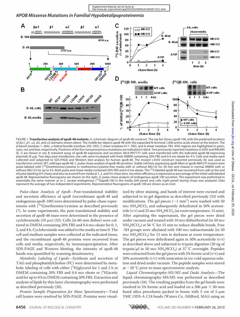
FIGURE 1. Transfection analysis of apoB-48 mutants. A, schematic diagram of apoB-48 construct. The top bar shows apoB-100, with the predicted locationsof ��1, �1, �2, �2, and �3 domains shown above. The middle bar depicts apoB-48 with the expanded N-terminal 1,000 amino acids shown at the bottom. The� barrel (residues 1–264), �-helical bundle (residues 292–593), C-sheet (residues 611–782), and A-sheet (residues 783–930) regions are highlighted in green,cyan, red, and blue, respectively. Positions of the five nonsynonymous mutations are indicated in black. Two previously reported mutations (L343V and R463W)(6, 7) are shown in red. B, transient assay of apoB-48 expression and secretion. McA-RH7777 cells were transfected with the indicated apoB-48-expressingplasmids (8 �g). Two days post-transfection, the cells were incubated with fresh DMEM containing 20% FBS and 0.4 mM oleate for 4 h. Cells and media werecollected and subjected to SDS-PAGE and Western blot analysis for human apoB-48. The mutant L343V construct reported previously (6) was used astransfection control. WT, wild type apoB-48. C, pulse-chase analysis of apoB-48 secretion. Stable cell lines expressing apoB-48wt or apoB-48A31P mutant werepulse-labeled with [35S]methionine/cysteine in methionine/cysteine-free media with or without MG132 for 30 min and chased in normal DMEM with orwithout MG132 for up to 4 h. Both pulse and chase media contained 20% FBS and 0.4 mM oleate. The 35S-labeled apoB-48 was recovered from cells at the endof pulse labeling (0 h chase) and also recovered from media at 1, 2, and 4 h chase time. Secretion efficiency is expressed as percentage of the initial radiolabeledapoB-48. Representative fluorograms are shown on the right. D, pulse-chase analysis of endogenous apoB-100 secretion. The experiment was performed inessentially the same manner as in C, except endogenous [35S]apoB-100 in the media (left panel) and cells (right panel) during chase was analyzed. Datarepresent the average of two independent experiments. Representative fluorograms of apoB-100 are shown as an inset.
APOB Missense Mutations in Familial Hypobetalipoproteinemia
6456 JOURNAL OF BIOLOGICAL CHEMISTRY VOLUME 285 • NUMBER 9 • FEBRUARY 26, 2010
at Univ of O
ttawa - O
CU
L, on February 25, 2010
ww
w.jbc.org
Dow
nloaded from
http://www.jbc.org/content/suppl/2010/01/08/M109.060467.DC1.htmlSupplemental Material can be found at:
1100 micro-HPLC system (Agilent Technologies, Santa Clara,CA). Following a desalting step, the flowwas split, and peptideswere eluted through a second 75 �m � 50-mm column packedwith the same beads at �200 nl/min. The peptides were elutedusing a 2-h gradient (5–80% acetonitrile with 0.1% formic acid)into an ESI LTQ linear ion trap mass spectrometer (ThermoElectron, Waltham, MA). MS/MS spectra were acquired in adata-dependent acquisition mode that automatically selectedand fragmented the 10 most intense peaks from each massspectrum generated. The acquired MS/MS spectra weresearched against themouse International Protein Index proteinsequence data base (version 3.52, 39,906 protein sequences;European Bioinformatics Institute) augmented with the re-versed sequence of each entry in the forward data base. Mascot2.2.02 (Matrix Science)was used to search the protein sequencedata base. The precursor and fragment mass tolerances for theLTQ data were set at 2.0 and 0.8 Da, respectively. Mascot cut-off scores were set to 30. Peptides ranked with a probability-based Mowse (expect) p � 0.05 were accepted. The false posi-tive rate was controlled at �1%. The identified proteins withthe expected molecular weight and at least two non-redundant
peptides were accepted after rigor-ous filtration as described above.Real-time RT-PCR—Isolation of
RNA from cells, reverse transcrip-tion, and relative mRNA concentra-tion determination by real-timeRT-PCR were performed as describedpreviously (20). Cyclophilin A wasused as the real-time RT-PCR con-trol. The primers used for RT-PCRwere obtained from Sigma, and thesequences of the primers are listedin supplemental Table 2.MolecularModeling—Thehomo-
logy model of the N-terminal 930amino acids of apoB-100 (apoB-20.5)was constructed using MODELLER7.7 using standard parameters(35) and the coordinates of lam-prey lipovitellin (36). The se-quences of apoB20.5 and lipov-itellin were aligned using theBlast-2-Sequence program (37).Graphic representation was achievedbyMOLMOL (38).
RESULTS
Clinical Phenotyping and Geno-typing of Five Heterozygous FHBLSubjects—Table 1 summarizes theclinical and biochemical featuresof members of the five FHBL kin-dreds examined. All probandsand some of the respective familymembers had fatty liver docu-mented by abdominal liver ultra-sound examination (as described
in Refs. 24 and 39). Detailed description of the kindreds isavailable as supplemental material. Sequencing of theAPOB gene revealed that each proband was heterozygous forsingle nucleotide substitutions causing novel non-conserva-tive amino acid substitutions in mature apoB. They arec.2914 G3A in exon 19 (G945S) in FHBL-36 (supplementalFig. S1A), c.2816 G 3 A in exon 18 (G912D) in probandFHBL-39 (supplemental Fig. S1B), c.1051 C 3 A in exon 9(L324M) in proband FHBL-44 (supplemental Fig. S1C),c.172 G3 C in exon 3 (A31P) in proband FHBL-45 (supple-mental Fig. S1D), and c.904 G 3 A in exon 8 (G275S) inproband FHBL-46 (not shown in supplemental Fig. S1). Allof the mutations occurred within the ��1 domain (Fig. 1A)(10, 11). These mutations co-segregated with the FHBL lipidphenotype in the families and were not found in a large groupof normolipidemic healthy control subjects. However, thesmall number of family members in the respective kindredprecluded the feasibility of performing genetic linkage anal-ysis. All of the amino acids involved in the mutations, withthe exception of Gly912, are highly conserved in mammalianspecies.
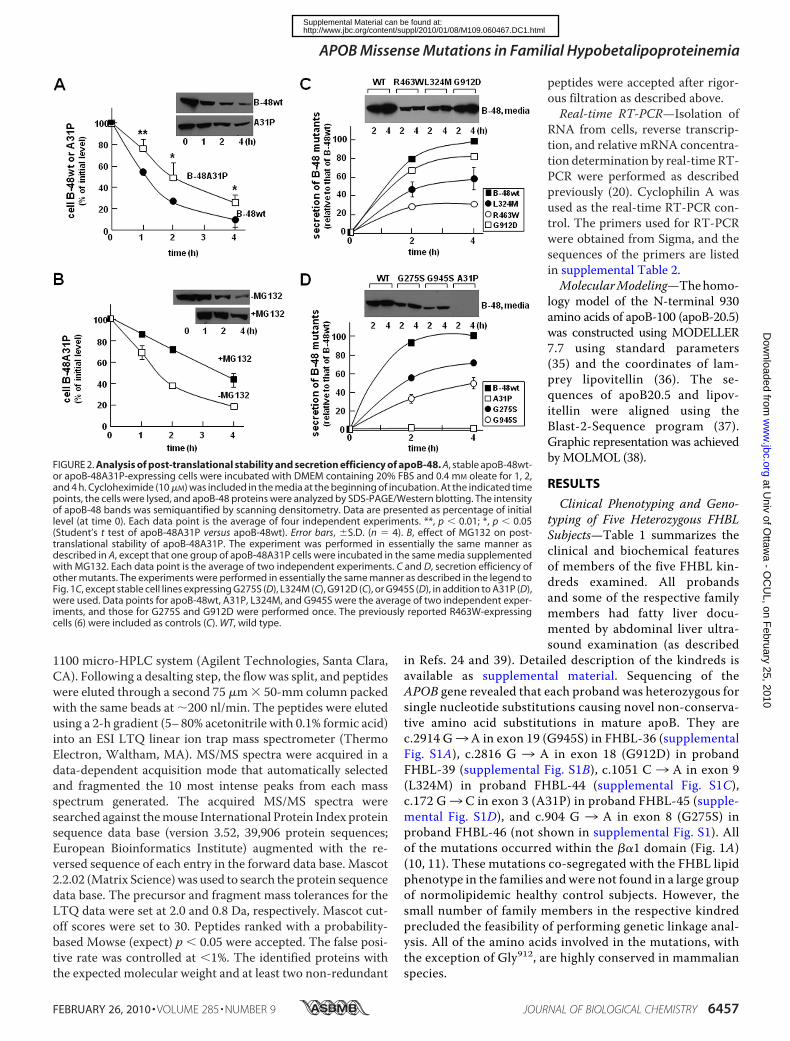
FIGURE 2. Analysis of post-translational stability and secretion efficiency of apoB-48. A, stable apoB-48wt-or apoB-48A31P-expressing cells were incubated with DMEM containing 20% FBS and 0.4 mM oleate for 1, 2,and 4 h. Cycloheximide (10 �M) was included in the media at the beginning of incubation. At the indicated timepoints, the cells were lysed, and apoB-48 proteins were analyzed by SDS-PAGE/Western blotting. The intensityof apoB-48 bands was semiquantified by scanning densitometry. Data are presented as percentage of initiallevel (at time 0). Each data point is the average of four independent experiments. **, p � 0.01; *, p � 0.05(Student’s t test of apoB-48A31P versus apoB-48wt). Error bars, �S.D. (n � 4). B, effect of MG132 on post-translational stability of apoB-48A31P. The experiment was performed in essentially the same manner asdescribed in A, except that one group of apoB-48A31P cells were incubated in the same media supplementedwith MG132. Each data point is the average of two independent experiments. C and D, secretion efficiency ofother mutants. The experiments were performed in essentially the same manner as described in the legend toFig. 1C, except stable cell lines expressing G275S (D), L324M (C), G912D (C), or G945S (D), in addition to A31P (D),were used. Data points for apoB-48wt, A31P, L324M, and G945S were the average of two independent exper-iments, and those for G275S and G912D were performed once. The previously reported R463W-expressingcells (6) were included as controls (C). WT, wild type.
APOB Missense Mutations in Familial Hypobetalipoproteinemia
FEBRUARY 26, 2010 • VOLUME 285 • NUMBER 9 JOURNAL OF BIOLOGICAL CHEMISTRY 6457
at Univ of O
ttawa - O
CU
L, on February 25, 2010
ww
w.jbc.org
Dow
nloaded from
http://www.jbc.org/content/suppl/2010/01/08/M109.060467.DC1.htmlSupplemental Material can be found at:
Transfection Studies of ApoB-48Mutants Revealed ImpairedSecretion fromMcA-RH7777 Cells—We tested the effect of theidentified FHBL mutations on apoB-48 secretion by transfec-tion studies using McA-RH7777 cells that retain the ability toassemble and secrete VLDL (30, 40). Transient assay withapoB-48 bearing the FHBL mutations gave correct expressionin the transfected cells (Fig. 1B, top). Except for A31P, all of theapoB-48 proteins were detected in the media (Fig. 1B, bottom).The effect of the A31P mutation on apoB secretion was alsotested in apoB-17 that had little lipid-binding ability (9). Tran-sient assay with apoB-17A31P showed robust expression in thecells but no secretion (data not shown). Thus, the effect of A31Pmutation on apoB secretion is independent of lipidation.The lack of apoB-48A31P secretion was further confirmed
using stable cell lines. Pulse-chase analysis showed thatalthough apoB48wt was secreted efficiently, the A31P mutantwas barely detectable in the media (Fig. 1C). Including the pro-teasome inhibitorMG132 had little effect on apoB-48 secretionefficiency, although cell-associated A31P protein was increasedat the end of the 30-min pulse labeling (see bands labeled 0 h,cell in Fig. 1C). Expression of A31P severely impaired secretionof endogenous rat apoB-100 (Fig. 1D) but not apoE (data notshown).
Post-translational stability of the A31P mutant was deter-mined in cells cultured with cycloheximide (to block proteinsynthesis). Although cellular apoB-48wt was decreased rapidly(t1⁄2 � 1 h) as a consequence of efficient secretion, A31Pmutantshowed prolonged intracellular retention (t1⁄2 2 h), although itwas not secreted (Fig. 2A). The addition of MG132 partiallyblocked decay of cellular A31P (Fig. 2B) but not apoB-48wt(data not shown). These results suggest that a significant pro-portion of A31P mutant was degraded by proteasomes.Similar experiments were performed with stable cell lines
expressing the other FHBL mutants. It was shown previouslyand confirmed here that the R463W mutant form of apoB-48was secreted poorly as compared with apoB-48wt (Fig. 2C).Likewise, L324M (Fig. 2C), G275S (Fig. 2D), G912D (Fig. 2C),and G945S (Fig. 2D) mutants were also secreted at reducedlevels. Among these, the G912D mutation had the least effecton apoB-48 secretion as compared with A31P. These resultsindicate that the hypobetalipoproteinemia phenotypes of thefive kindreds, such as the reduced plasma cholesterol, TAG,and apoB concentrations (Table 1), are not caused solely byimpaired secretion of the mutant apoB or apoB-containinglipoproteins. The t1⁄2 of cellular apoB formostmutants was sim-ilar to that of apoB-48wt (data not shown), exceptA31P showed
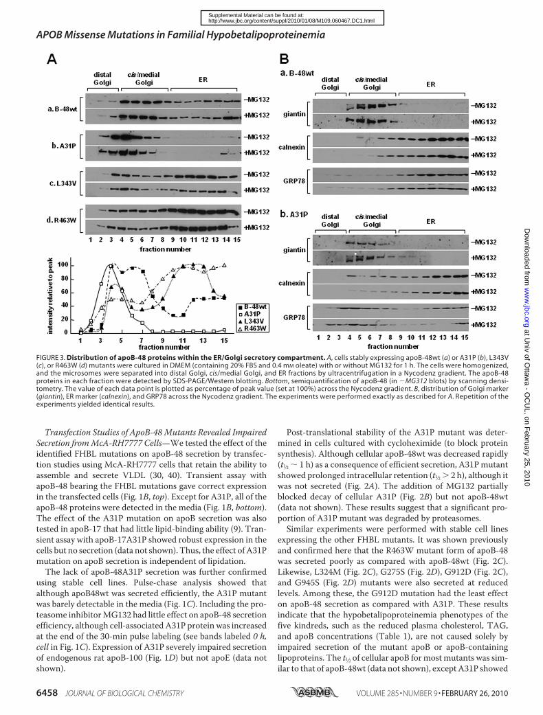
FIGURE 3. Distribution of apoB-48 proteins within the ER/Golgi secretory compartment. A, cells stably expressing apoB-48wt (a) or A31P (b), L343V(c), or R463W (d) mutants were cultured in DMEM (containing 20% FBS and 0.4 mM oleate) with or without MG132 for 1 h. The cells were homogenized,and the microsomes were separated into distal Golgi, cis/medial Golgi, and ER fractions by ultracentrifugation in a Nycodenz gradient. The apoB-48proteins in each fraction were detected by SDS-PAGE/Western blotting. Bottom, semiquantification of apoB-48 (in �MG312 blots) by scanning densi-tometry. The value of each data point is plotted as percentage of peak value (set at 100%) across the Nycodenz gradient. B, distribution of Golgi marker(giantin), ER marker (calnexin), and GRP78 across the Nycodenz gradient. The experiments were performed exactly as described for A. Repetition of theexperiments yielded identical results.
APOB Missense Mutations in Familial Hypobetalipoproteinemia
6458 JOURNAL OF BIOLOGICAL CHEMISTRY VOLUME 285 • NUMBER 9 • FEBRUARY 26, 2010
at Univ of O
ttawa - O
CU
L, on February 25, 2010
ww
w.jbc.org
Dow
nloaded from
http://www.jbc.org/content/suppl/2010/01/08/M109.060467.DC1.htmlSupplemental Material can be found at:
markedly prolonged intracellular retention (Fig. 2A). Thesedata suggested that, unlike mutations within the �-helicaldomain or �-sheets that only moderately affected apoB-48secretion, the A31Pmutation that occurred within the �-barrelalmost entirely blocked apoB-48 secretion. The followingexperiments focused on A31P mutant.Distribution of ApoB-48A31Pwithin ER/Golgi Compartment—
The lack of A31P mutant secretion might be attributable to itsinability to escape ER quality control. To test this, we deter-mined ER/Golgi distribution of A31P mutant. At steady state,apoB-48wtwas distributed across the entire ER/Golgi secretorypathway (Fig. 3A, panel a). In contrast, the A31P mutant waspredominantly located in Golgi compartments (Fig. 3A, panelb), which was different form L343V or R463W mutants thatshowed ER retention (panels c and d). The addition of MG132to the media did not alter distribution of any of the proteins(Fig. 3A, �MG132). Thus, the absence of A31P within the ERwas unlikely to be attributable to rapid ER-associated degrada-tion. Distribution of ER (calnexin) and Golgi (giantin) markerproteins was unchanged between apoB-48wt and A31P cells
(Fig. 3B), indicting that physico-chemical properties of the secretorymachinery were intact. It was notedthat distribution of GRP78 wasskewed toward cis/medial Golgifractions in A31P cells (Fig. 3B,compareGRP78 blots between pan-els a and b). The similar distributionbetween apoB-48A31P and GRP78indicates that they may coexist inthe same compartments.The nature of A31P accumula-
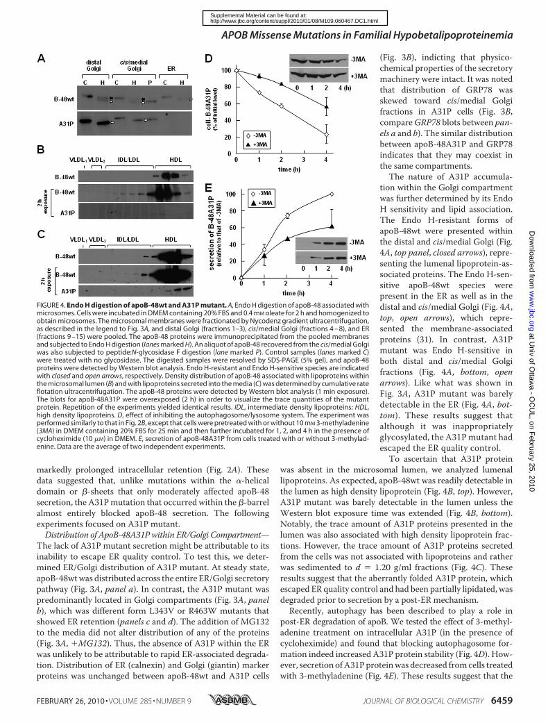
tion within the Golgi compartmentwas further determined by its EndoH sensitivity and lipid association.The Endo H-resistant forms ofapoB-48wt were presented withinthe distal and cis/medial Golgi (Fig.4A, top panel, closed arrows), repre-senting the lumenal lipoprotein-as-sociated proteins. The Endo H-sen-sitive apoB-48wt species werepresent in the ER as well as in thedistal and cis/medial Golgi (Fig. 4A,top, open arrows), which repre-sented the membrane-associatedproteins (31). In contrast, A31Pmutant was Endo H-sensitive inboth distal and cis/medial Golgifractions (Fig. 4A, bottom, openarrows). Like what was shown inFig. 3A, A31P mutant was barelydetectable in the ER (Fig. 4A, bot-tom). These results suggest thatalthough it was inappropriatelyglycosylated, the A31Pmutant hadescaped the ER quality control.To ascertain that A31P protein
was absent in the microsomal lumen, we analyzed lumenallipoproteins. As expected, apoB-48wt was readily detectable inthe lumen as high density lipoprotein (Fig. 4B, top). However,A31P mutant was barely detectable in the lumen unless theWestern blot exposure time was extended (Fig. 4B, bottom).Notably, the trace amount of A31P proteins presented in thelumen was also associated with high density lipoprotein frac-tions. However, the trace amount of A31P proteins secretedfrom the cells was not associated with lipoproteins and ratherwas sedimented to d � 1.20 g/ml fractions (Fig. 4C). Theseresults suggest that the aberrantly folded A31P protein, whichescaped ER quality control and had been partially lipidated, wasdegraded prior to secretion by a post-ER mechanism.Recently, autophagy has been described to play a role in
post-ER degradation of apoB. We tested the effect of 3-methyl-adenine treatment on intracellular A31P (in the presence ofcycloheximide) and found that blocking autophagosome for-mation indeed increased A31P protein stability (Fig. 4D). How-ever, secretion ofA31Pproteinwas decreased fromcells treatedwith 3-methyladenine (Fig. 4E). These results suggest that the
FIGURE 4. Endo H digestion of apoB-48wt and A31P mutant. A, Endo H digestion of apoB-48 associated withmicrosomes. Cells were incubated in DMEM containing 20% FBS and 0.4 mM oleate for 2 h and homogenized toobtain microsomes. The microsomal membranes were fractionated by Nycodenz gradient ultracentrifugation,as described in the legend to Fig. 3A, and distal Golgi (fractions 1–3), cis/medial Golgi (fractions 4 – 8), and ER(fractions 9 –15) were pooled. The apoB-48 proteins were immunoprecipitated from the pooled membranesand subjected to Endo H digestion (lanes marked H). An aliquot of apoB-48 recovered from the cis/medial Golgiwas also subjected to peptide:N-glycosidase F digestion (lane marked P). Control samples (lanes marked C)were treated with no glycosidase. The digested samples were resolved by SDS-PAGE (5% gel), and apoB-48proteins were detected by Western blot analysis. Endo H-resistant and Endo H-sensitive species are indicatedwith closed and open arrows, respectively. Density distribution of apoB-48 associated with lipoproteins withinthe microsomal lumen (B) and with lipoproteins secreted into the media (C) was determined by cumulative rateflotation ultracentrifugation. The apoB-48 proteins were detected by Western blot analysis (1 min exposure).The blots for apoB-48A31P were overexposed (2 h) in order to visualize the trace quantities of the mutantprotein. Repetition of the experiments yielded identical results. IDL, intermediate density lipoproteins; HDL,high density lipoproteins. D, effect of inhibiting the autophagosome/lysosome system. The experiment wasperformed similarly to that in Fig. 2B, except that cells were pretreated with or without 10 mM 3-methyladenine(3MA) in DMEM containing 20% FBS for 25 min and then further incubated for 1, 2, and 4 h in the presence ofcycloheximide (10 �M) in DMEM. E, secretion of apoB-48A31P from cells treated with or without 3-methylad-enine. Data are the average of two independent experiments.
APOB Missense Mutations in Familial Hypobetalipoproteinemia
FEBRUARY 26, 2010 • VOLUME 285 • NUMBER 9 JOURNAL OF BIOLOGICAL CHEMISTRY 6459
at Univ of O
ttawa - O
CU
L, on February 25, 2010
ww
w.jbc.org
Dow
nloaded from
http://www.jbc.org/content/suppl/2010/01/08/M109.060467.DC1.htmlSupplemental Material can be found at:
A31P mutant, either in the membrane-bound or lipoprotein-associated form, might be degraded through the autophago-some pathway.Consideration was given to the possibility that the impaired
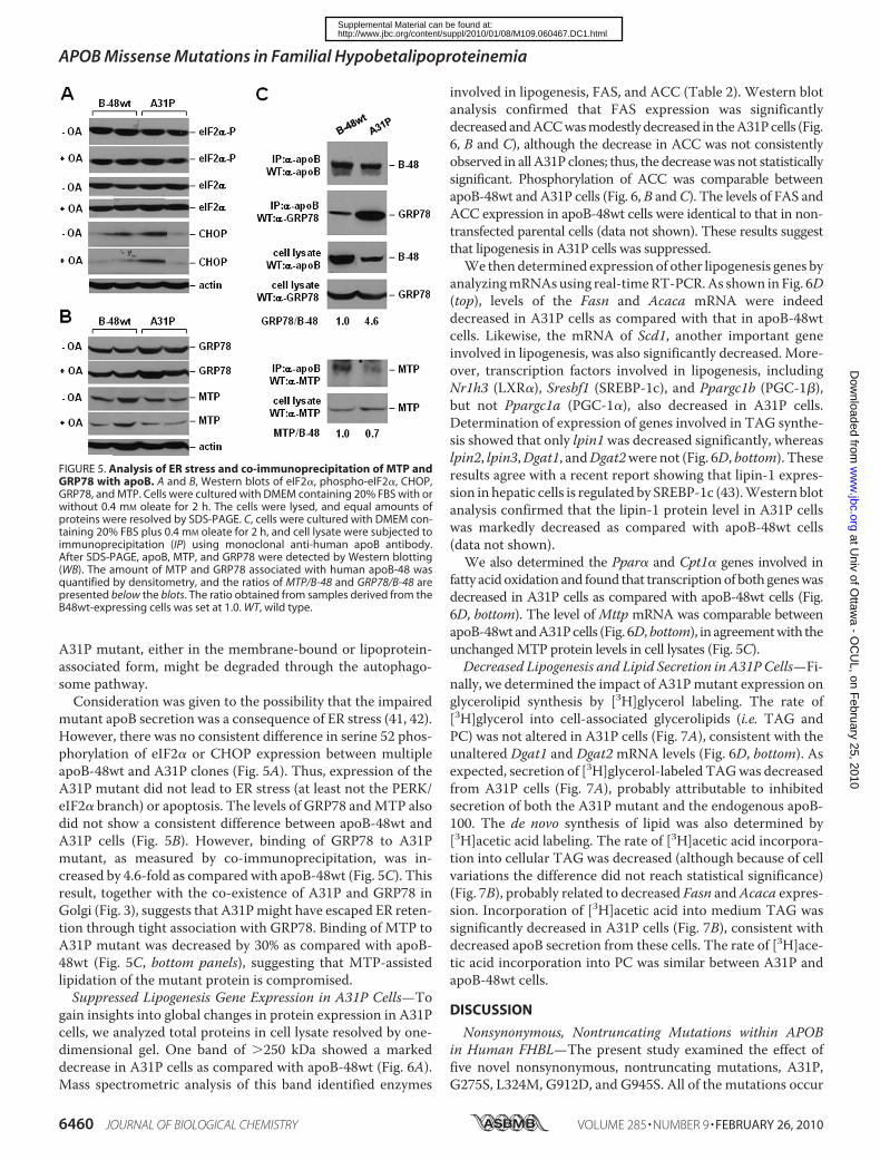
mutant apoB secretion was a consequence of ER stress (41, 42).However, there was no consistent difference in serine 52 phos-phorylation of eIF2� or CHOP expression between multipleapoB-48wt and A31P clones (Fig. 5A). Thus, expression of theA31P mutant did not lead to ER stress (at least not the PERK/eIF2� branch) or apoptosis. The levels of GRP78 andMTP alsodid not show a consistent difference between apoB-48wt andA31P cells (Fig. 5B). However, binding of GRP78 to A31Pmutant, as measured by co-immunoprecipitation, was in-creased by 4.6-fold as compared with apoB-48wt (Fig. 5C). Thisresult, together with the co-existence of A31P and GRP78 inGolgi (Fig. 3), suggests that A31Pmight have escaped ER reten-tion through tight association with GRP78. Binding of MTP toA31P mutant was decreased by 30% as compared with apoB-48wt (Fig. 5C, bottom panels), suggesting that MTP-assistedlipidation of the mutant protein is compromised.Suppressed Lipogenesis Gene Expression in A31P Cells—To
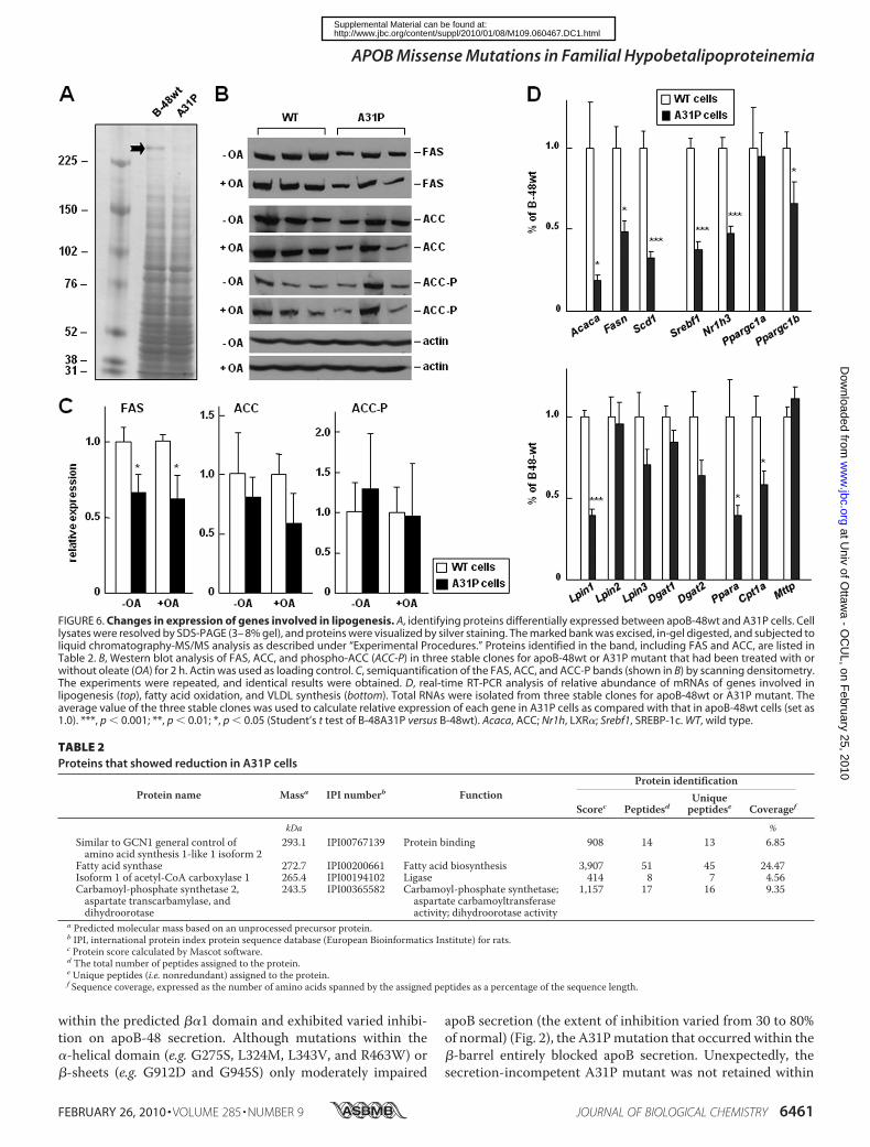
gain insights into global changes in protein expression in A31Pcells, we analyzed total proteins in cell lysate resolved by one-dimensional gel. One band of 250 kDa showed a markeddecrease in A31P cells as compared with apoB-48wt (Fig. 6A).Mass spectrometric analysis of this band identified enzymes
involved in lipogenesis, FAS, and ACC (Table 2). Western blotanalysis confirmed that FAS expression was significantlydecreasedandACCwasmodestlydecreased in theA31Pcells (Fig.6, B and C), although the decrease in ACC was not consistentlyobserved in all A31P clones; thus, the decreasewas not statisticallysignificant. Phosphorylation of ACC was comparable betweenapoB-48wt andA31P cells (Fig. 6, B andC). The levels of FAS andACC expression in apoB-48wt cells were identical to that in non-transfected parental cells (data not shown). These results suggestthat lipogenesis in A31P cells was suppressed.We thendetermined expression of other lipogenesis genes by
analyzingmRNAsusing real-timeRT-PCR.As shown in Fig. 6D(top), levels of the Fasn and Acaca mRNA were indeeddecreased in A31P cells as compared with that in apoB-48wtcells. Likewise, the mRNA of Scd1, another important geneinvolved in lipogenesis, was also significantly decreased. More-over, transcription factors involved in lipogenesis, includingNr1h3 (LXR�), Sresbf1 (SREBP-1c), and Ppargc1b (PGC-1�),but not Ppargc1a (PGC-1�), also decreased in A31P cells.Determination of expression of genes involved in TAG synthe-sis showed that only lpin1was decreased significantly, whereaslpin2, lpin3,Dgat1, andDgat2were not (Fig. 6D, bottom). Theseresults agree with a recent report showing that lipin-1 expres-sion in hepatic cells is regulated by SREBP-1c (43).Western blotanalysis confirmed that the lipin-1 protein level in A31P cellswas markedly decreased as compared with apoB-48wt cells(data not shown).We also determined the Ppar� and Cpt1� genes involved in
fatty acidoxidation and found that transcriptionof bothgeneswasdecreased in A31P cells as compared with apoB-48wt cells (Fig.6D, bottom). The level of Mttp mRNA was comparable betweenapoB-48wtandA31Pcells (Fig. 6D,bottom), in agreementwith theunchangedMTP protein levels in cell lysates (Fig. 5C).Decreased Lipogenesis and Lipid Secretion in A31P Cells—Fi-
nally, we determined the impact of A31Pmutant expression onglycerolipid synthesis by [3H]glycerol labeling. The rate of[3H]glycerol into cell-associated glycerolipids (i.e. TAG andPC) was not altered in A31P cells (Fig. 7A), consistent with theunaltered Dgat1 and Dgat2mRNA levels (Fig. 6D, bottom). Asexpected, secretion of [3H]glycerol-labeled TAGwas decreasedfrom A31P cells (Fig. 7A), probably attributable to inhibitedsecretion of both the A31P mutant and the endogenous apoB-100. The de novo synthesis of lipid was also determined by[3H]acetic acid labeling. The rate of [3H]acetic acid incorpora-tion into cellular TAG was decreased (although because of cellvariations the difference did not reach statistical significance)(Fig. 7B), probably related to decreased Fasn andAcaca expres-sion. Incorporation of [3H]acetic acid into medium TAG wassignificantly decreased in A31P cells (Fig. 7B), consistent withdecreased apoB secretion from these cells. The rate of [3H]ace-tic acid incorporation into PC was similar between A31P andapoB-48wt cells.
DISCUSSION
Nonsynonymous, Nontruncating Mutations within APOBin Human FHBL—The present study examined the effect offive novel nonsynonymous, nontruncating mutations, A31P,G275S, L324M, G912D, and G945S. All of the mutations occur
FIGURE 5. Analysis of ER stress and co-immunoprecipitation of MTP andGRP78 with apoB. A and B, Western blots of eIF2�, phospho-eIF2�, CHOP,GRP78, and MTP. Cells were cultured with DMEM containing 20% FBS with orwithout 0.4 mM oleate for 2 h. The cells were lysed, and equal amounts ofproteins were resolved by SDS-PAGE. C, cells were cultured with DMEM con-taining 20% FBS plus 0.4 mM oleate for 2 h, and cell lysate were subjected toimmunoprecipitation (IP) using monoclonal anti-human apoB antibody.After SDS-PAGE, apoB, MTP, and GRP78 were detected by Western blotting(WB). The amount of MTP and GRP78 associated with human apoB-48 wasquantified by densitometry, and the ratios of MTP/B-48 and GRP78/B-48 arepresented below the blots. The ratio obtained from samples derived from theB48wt-expressing cells was set at 1.0. WT, wild type.
APOB Missense Mutations in Familial Hypobetalipoproteinemia
6460 JOURNAL OF BIOLOGICAL CHEMISTRY VOLUME 285 • NUMBER 9 • FEBRUARY 26, 2010
at Univ of O
ttawa - O
CU
L, on February 25, 2010
ww
w.jbc.org
Dow
nloaded from
http://www.jbc.org/content/suppl/2010/01/08/M109.060467.DC1.htmlSupplemental Material can be found at:
within the predicted ��1 domain and exhibited varied inhibi-tion on apoB-48 secretion. Although mutations within the�-helical domain (e.g. G275S, L324M, L343V, and R463W) or�-sheets (e.g. G912D and G945S) only moderately impaired
apoB secretion (the extent of inhibition varied from 30 to 80%of normal) (Fig. 2), the A31Pmutation that occurred within the�-barrel entirely blocked apoB secretion. Unexpectedly, thesecretion-incompetent A31P mutant was not retained within
FIGURE 6. Changes in expression of genes involved in lipogenesis. A, identifying proteins differentially expressed between apoB-48wt and A31P cells. Celllysates were resolved by SDS-PAGE (3– 8% gel), and proteins were visualized by silver staining. The marked bank was excised, in-gel digested, and subjected toliquid chromatography-MS/MS analysis as described under “Experimental Procedures.” Proteins identified in the band, including FAS and ACC, are listed inTable 2. B, Western blot analysis of FAS, ACC, and phospho-ACC (ACC-P) in three stable clones for apoB-48wt or A31P mutant that had been treated with orwithout oleate (OA) for 2 h. Actin was used as loading control. C, semiquantification of the FAS, ACC, and ACC-P bands (shown in B) by scanning densitometry.The experiments were repeated, and identical results were obtained. D, real-time RT-PCR analysis of relative abundance of mRNAs of genes involved inlipogenesis (top), fatty acid oxidation, and VLDL synthesis (bottom). Total RNAs were isolated from three stable clones for apoB-48wt or A31P mutant. Theaverage value of the three stable clones was used to calculate relative expression of each gene in A31P cells as compared with that in apoB-48wt cells (set as1.0). ***, p � 0.001; **, p � 0.01; *, p � 0.05 (Student’s t test of B-48A31P versus B-48wt). Acaca, ACC; Nr1h, LXR�; Srebf1, SREBP-1c. WT, wild type.
TABLE 2Proteins that showed reduction in A31P cells
Protein name Massa IPI numberb FunctionProtein identification
Scorec PeptidesdUniquepeptidese Coveragef
kDa %Similar to GCN1 general control ofamino acid synthesis 1-like 1 isoform 2
293.1 IPI00767139 Protein binding 908 14 13 6.85
Fatty acid synthase 272.7 IPI00200661 Fatty acid biosynthesis 3,907 51 45 24.47Isoform 1 of acetyl-CoA carboxylase 1 265.4 IPI00194102 Ligase 414 8 7 4.56Carbamoyl-phosphate synthetase 2,aspartate transcarbamylase, anddihydroorotase
243.5 IPI00365582 Carbamoyl-phosphate synthetase;aspartate carbamoyltransferaseactivity; dihydroorotase activity
1,157 17 16 9.35
a Predicted molecular mass based on an unprocessed precursor protein.b IPI, international protein index protein sequence database (European Bioinformatics Institute) for rats.c Protein score calculated by Mascot software.d The total number of peptides assigned to the protein.e Unique peptides (i.e. nonredundant) assigned to the protein.f Sequence coverage, expressed as the number of amino acids spanned by the assigned peptides as a percentage of the sequence length.
APOB Missense Mutations in Familial Hypobetalipoproteinemia
FEBRUARY 26, 2010 • VOLUME 285 • NUMBER 9 JOURNAL OF BIOLOGICAL CHEMISTRY 6461
at Univ of O
ttawa - O
CU
L, on February 25, 2010
ww
w.jbc.org
Dow
nloaded from
http://www.jbc.org/content/suppl/2010/01/08/M109.060467.DC1.htmlSupplemental Material can be found at:
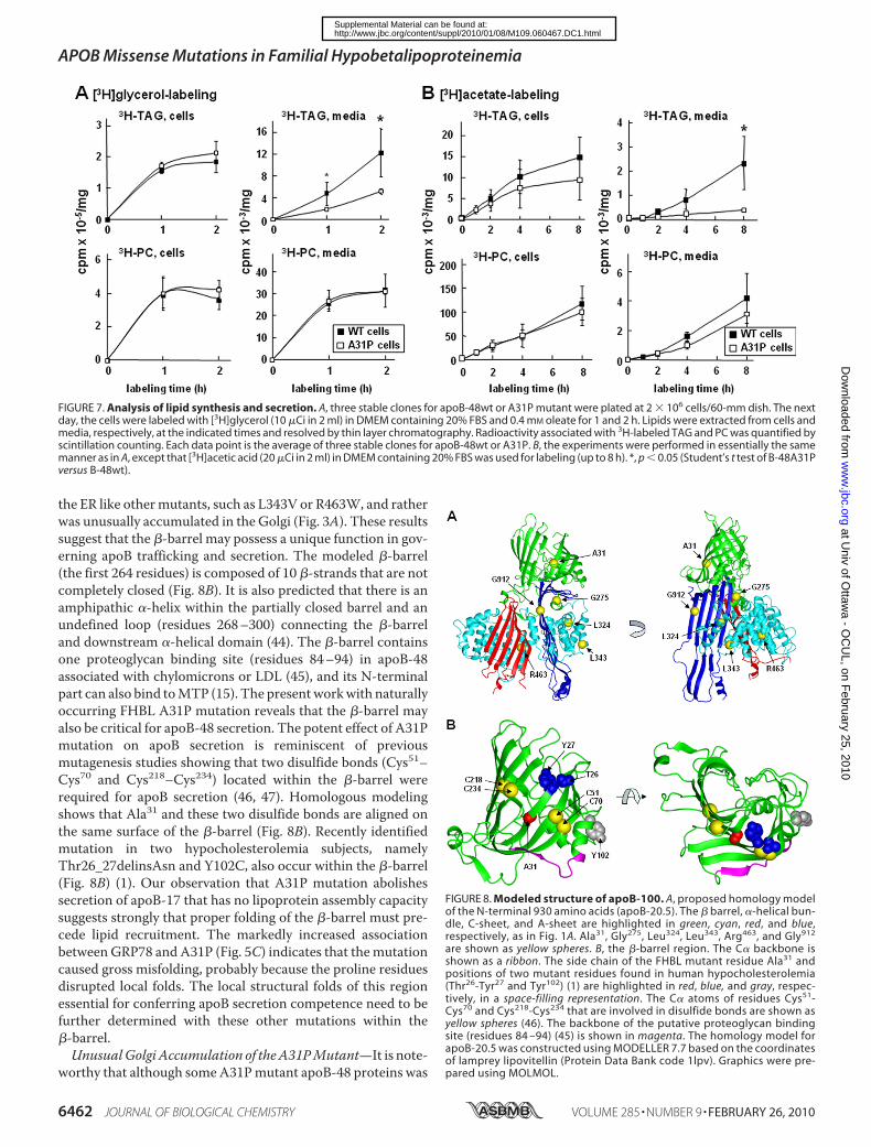
the ER like othermutants, such as L343V or R463W, and ratherwas unusually accumulated in the Golgi (Fig. 3A). These resultssuggest that the �-barrel may possess a unique function in gov-erning apoB trafficking and secretion. The modeled �-barrel(the first 264 residues) is composed of 10 �-strands that are notcompletely closed (Fig. 8B). It is also predicted that there is anamphipathic �-helix within the partially closed barrel and anundefined loop (residues 268–300) connecting the �-barreland downstream �-helical domain (44). The �-barrel containsone proteoglycan binding site (residues 84–94) in apoB-48associated with chylomicrons or LDL (45), and its N-terminalpart can also bind toMTP (15). The presentworkwith naturallyoccurring FHBL A31P mutation reveals that the �-barrel mayalso be critical for apoB-48 secretion. The potent effect of A31Pmutation on apoB secretion is reminiscent of previousmutagenesis studies showing that two disulfide bonds (Cys51–Cys70 and Cys218–Cys234) located within the �-barrel wererequired for apoB secretion (46, 47). Homologous modelingshows that Ala31 and these two disulfide bonds are aligned onthe same surface of the �-barrel (Fig. 8B). Recently identifiedmutation in two hypocholesterolemia subjects, namelyThr26_27delinsAsn and Y102C, also occur within the �-barrel(Fig. 8B) (1). Our observation that A31P mutation abolishessecretion of apoB-17 that has no lipoprotein assembly capacitysuggests strongly that proper folding of the �-barrel must pre-cede lipid recruitment. The markedly increased associationbetweenGRP78 and A31P (Fig. 5C) indicates that themutationcaused gross misfolding, probably because the proline residuesdisrupted local folds. The local structural folds of this regionessential for conferring apoB secretion competence need to befurther determined with these other mutations within the�-barrel.UnusualGolgi Accumulation of theA31PMutant—It is note-
worthy that although someA31Pmutant apoB-48 proteins was
FIGURE 7. Analysis of lipid synthesis and secretion. A, three stable clones for apoB-48wt or A31P mutant were plated at 2 � 106 cells/60-mm dish. The nextday, the cells were labeled with [3H]glycerol (10 �Ci in 2 ml) in DMEM containing 20% FBS and 0.4 mM oleate for 1 and 2 h. Lipids were extracted from cells andmedia, respectively, at the indicated times and resolved by thin layer chromatography. Radioactivity associated with 3H-labeled TAG and PC was quantified byscintillation counting. Each data point is the average of three stable clones for apoB-48wt or A31P. B, the experiments were performed in essentially the samemanner as in A, except that [3H]acetic acid (20 �Ci in 2 ml) in DMEM containing 20% FBS was used for labeling (up to 8 h). *, p � 0.05 (Student’s t test of B-48A31Pversus B-48wt).
FIGURE 8. Modeled structure of apoB-100. A, proposed homology modelof the N-terminal 930 amino acids (apoB-20.5). The � barrel, �-helical bun-dle, C-sheet, and A-sheet are highlighted in green, cyan, red, and blue,respectively, as in Fig. 1A. Ala31, Gly275, Leu324, Leu343, Arg463, and Gly912
are shown as yellow spheres. B, the �-barrel region. The C� backbone isshown as a ribbon. The side chain of the FHBL mutant residue Ala31 andpositions of two mutant residues found in human hypocholesterolemia(Thr26-Tyr27 and Tyr102) (1) are highlighted in red, blue, and gray, respec-tively, in a space-filling representation. The C� atoms of residues Cys51-Cys70 and Cys218-Cys234 that are involved in disulfide bonds are shown asyellow spheres (46). The backbone of the putative proteoglycan bindingsite (residues 84 –94) (45) is shown in magenta. The homology model forapoB-20.5 was constructed using MODELLER 7.7 based on the coordinatesof lamprey lipovitellin (Protein Data Bank code 1lpv). Graphics were pre-pared using MOLMOL.
APOB Missense Mutations in Familial Hypobetalipoproteinemia
6462 JOURNAL OF BIOLOGICAL CHEMISTRY VOLUME 285 • NUMBER 9 • FEBRUARY 26, 2010
at Univ of O
ttawa - O
CU
L, on February 25, 2010
ww
w.jbc.org
Dow
nloaded from
http://www.jbc.org/content/suppl/2010/01/08/M109.060467.DC1.htmlSupplemental Material can be found at:
degraded by the proteasomes (presumably through an ER-as-sociated mechanism), a significant amount of the mutant pro-tein escaped ER retention and presented in the distal compart-ment of the secretory pathway. This is in sharp contrast toFHBL mutations occurring in the �-helical domain, such asL343V and R463W, that showed extended ER retention (Fig.3A). Apparently, the A31P mutant proteins escaped the ERquality control mechanism (48). The observation that only asmall amount of A31P that escaped from the ER was presentedin the lumen (as high density lipoprotein) suggests that themisfolded proteins remained in association with the mem-branes. Currently, whether or not the membrane-associatedA31P mutants had assembled some lipids is unknown; nor is itclear if they were retained within the Golgi by proteins otherthan GRP78. Examination of the minute amount of A31P pro-teins secreted into the media showed they were mostly nonli-poprotein entities (d 1.20 g/ml) (Fig. 4C). These results sug-gest that those A31P mutants containing lipoprotein assemblyintermediates were degraded by a post-ER degradation mech-anism, probably autophagosome (49, 50), prior to secretion.Suppressed Lipogenesis in FHBL A31P Mutant-expressing
Cells—Another intriguing observation made in the presentstudy was markedly suppressed lipogenesis in A31P cells; thus,the expressions of SREBP-1c, together with its target genesFasn, Acaca, Scd1, and lpin1 were all down-regulated (Fig. 6).Suppressed hepatic lipogenesis has also been observed in twomouse FHBLmodels producing truncated apoBproteins equiv-alent to human apoB-38.9 or apoB-27.6 (51–53). Like whatoccurred inA31P cells, the livers expressing apoB-38.9 or apoB-27.6 also exhibited reduced SREBP-1c, FAS, and SCD1 (51).Thus, suppression of lipogenesis may occur in FHBL witheither truncating or nontruncating apoB mutations. Sup-pressed lipogenesis in FHBL hepatocytes may explain theabnormally low secretion efficiency of endogenous rat apoB-100 from the transfected cells resulting from the limited lipidsubstrate availability (Fig. 1D).
How expression of FHBLmutant A31P could result in down-regulation of SREBP-1c (and LXR� as well) is unclear. Onepossibilitymay involve lipin-1. The dual function lipin-1 plays arole as PAP1 (phosphatidate phosphatase 1) (54) as well as atranscription coactivator for PGC-1� andPPAR� (55).Not sur-prisingly, decreased Lpin1 in A31P cells was accompanied withreduced Ppar� and Cpt1� (Fig. 6D). In McA-RH7777 cells,lipin-1 is located in both cytosol and membrane (ER andnuclear) fractions (20). We found that the lipin-1 mass in totalcell lysate was markedly decreased in A31P cells as comparedwith apoB-48wt control (data not shown). Decreased lipin-1(acting as PAP1) would thus be expected to result in low TAGsynthesis. However, a [3H]glycerol labeling experiment sug-gested that TAG synthesis was normal in A31P cells (Fig. 7A).From a transcription coactivator point of view, decreasedlipin-1 in A31P cells could have an impact on PGC-1� action,leading to lowered lipogenesis, although the expression ofPGC-1� or -1�was little or onlymarginally affected (Fig. 6D). Itis reported recently that lipin-1 expression is regulated bySREBP-1c (43). It is likely that expression of A31P mutant hasperturbed the regulatory circuit among SREBP-1c, PPAR�,LXR� and lipin-1, resulting in profound suppression of lipo-
genesis. The effect of A31Pmutation on lipogenesis needs to beconfirmed in knock-in mice harboring the Apobmutation.
Acknowledgments—We thank Dr. Gordon Jiang for helpful discus-sions on structure-function relationships within human apoB-100,Drs. Yves Marcel and Ross Milne for the monoclonal antibody 1D1,and Dr. Carol Shoulders for anti-MTP antibody.
REFERENCES1. Tarugi, P., Averna, M., Di Leo, E., Cefalu, A. B., Noto, D., Magnolo, L.,
Cattin, L., Bertolini, S., and Calandra, S. (2007) Atherosclerosis 195,e19–e27
2. Chan, L. (1992) J. Biol. Chem. 267, 25621–256243. Fisher, E. A., and Ginsberg, H. N. (2002) J. Biol. Chem. 277, 17377–173804. Yao, Z., and McLeod, R. S. (1994) Biochim. Biophys. Acta 1212, 152–1665. Schonfeld, G. (2003) J. Lipid Res. 44, 878–8836. Burnett, J. R., Shan, J., Miskie, B. A., Whitfield, A. J., Yuan, J., Tran, K.,
McKnight, C. J., Hegele, R. A., and Yao, Z. (2003) J. Biol. Chem. 278,13442–13452
7. Burnett, J. R., Zhong, S., Jiang, Z. G., Hooper, A. J., Fisher, E. A., McLeod,R. S., Zhao, Y., Barrett, P. H., Hegele, R. A., van Bockxmeer, F. M., Zhang,H., Vance, D. E., McKnight, C. J., and Yao, Z. (2007) J. Biol. Chem. 282,24270–24283
8. McLeod, R. S., Zhao, Y., Selby, S. L., Westerlund, J., and Yao, Z. (1994)J. Biol. Chem. 269, 2852–2862
9. Yao, Z. M., Blackhart, B. D., Linton, M. F., Taylor, S. M., Young, S. G., andMcCarthy, B. J. (1991) J. Biol. Chem. 266, 3300–3308
10. Mann,C. J., Anderson, T.A., Read, J., Chester, S. A., Harrison,G. B., Kochl,S., Ritchie, P. J., Bradbury, P., Hussain, F. S., Amey, J., Vanloo, B., Rosseneu,M., Infante, R., Hancock, J. M., Levitt, D. G., Banaszak, L. J., Scott, J., andShoulders, C. C. (1999) J. Mol. Biol. 285, 391–408
11. Jiang, Z. G., Gantz, D., Bullitt, E., andMcKnight, C. J. (2006) Biochemistry45, 11799–11808
12. Gretch, D. G., Sturley, S. L., Wang, L., Lipton, B. A., Dunning, A., Grun-wald, K. A., Wetterau, J. R., Yao, Z., Talmud, P., and Attie, A. D. (1996)J. Biol. Chem. 271, 8682–8691
13. McLeod, R. S., Wang, Y., Wang, S., Rusinol, A., Links, P., and Yao, Z.(1996) J. Biol. Chem. 271, 18445–18455
14. Segrest, J. P., Jones,M.K., andDashti, N. (1999) J. Lipid Res. 40, 1401–141615. Bradbury, P., Mann, C. J., Kochl, S., Anderson, T. A., Chester, S. A., Han-
cock, J. M., Ritchie, P. J., Amey, J., Harrison, G. B., Levitt, D. G., Banaszak,L. J., Scott, J., and Shoulders, C. C. (1999) J. Biol. Chem. 274, 3159–3164
16. Hussain, M. M., Bakillah, A., Nayak, N., and Shelness, G. S. (1998) J. Biol.Chem. 273, 25612–25615
17. Dashti, N., Manchekar, M., Liu, Y., Sun, Z., and Segrest, J. P. (2007) J. Biol.Chem. 282, 28597–28608
18. Olofsson, S. O., and Boren, J. (2005) J. Intern. Med. 258, 395–41019. Yamazaki, T., Sasaki, E., Kakinuma, C., Yano, T., Miura, S., and Ezaki, O.
(2005) J. Biol. Chem. 280, 21506–2151420. Bou Khalil, M., Sundaram, M., Zhang, H. Y., Links, P. H., Raven, J. F.,
Manmontri, B., Sariahmetoglu,M., Tran, K., Reue, K., Brindley, D. N., andYao, Z. (2009) J. Lipid Res. 50, 47–58
21. Musso, G., Gambino, R., andCassader,M. (2009) Prog. Lipid Res. 48, 1–2622. Grefhorst, A., Elzinga, B.M., Voshol, P. J., Plosch, T., Kok, T., Bloks, V.W.,
van der Sluijs, F. H., Havekes, L. M., Romijn, J. A., Verkade, H. J., andKuipers, F. (2002) J. Biol. Chem. 277, 34182–34190
23. Lin, J., Yang, R., Tarr, P. T., Wu, P. H., Handschin, C., Li, S., Yang, W., Pei,L., Uldry, M., Tontonoz, P., Newgard, C. B., and Spiegelman, B. M. (2005)Cell 120, 261–273
24. Tarugi, P., Lonardo, A., Gabelli, C., Sala, F., Ballarini, G., Cortella, I., Pre-viato, L., Bertolini, S., Cordera, R., and Calandra, S. (2001) J. Lipid Res. 42,1552–1561
25. Di Leo, E., Magnolo, L., Lancellotti, S., Croce, L., Visintin, L., Tiribelli, C.,Bertolini, S., Calandra, S., andTarugi, P. (2007) J.Med. Genet. 44, 219–224
26. den Dunnen, J. T., and Antonarakis, S. E. (2000) Hum. Mutat. 15, 7–1227. den Dunnen, J. T., and Paalman, M. H. (2003) Hum. Mutat. 22, 181–182
APOB Missense Mutations in Familial Hypobetalipoproteinemia
FEBRUARY 26, 2010 • VOLUME 285 • NUMBER 9 JOURNAL OF BIOLOGICAL CHEMISTRY 6463
at Univ of O
ttawa - O
CU
L, on February 25, 2010
ww
w.jbc.org
Dow
nloaded from
http://www.jbc.org/content/suppl/2010/01/08/M109.060467.DC1.htmlSupplemental Material can be found at:
28. Fasano, T., Cefalu, A. B., Di Leo, E., Noto, D., Pollaccia, D., Bocchi, L.,Valenti, V., Bonardi, R., Guardamagna, O., Averna, M., and Tarugi, P.(2007) Arterioscler. Thromb. Vasc. Biol. 27, 677–681
29. Blackhart, B. D., Yao, Z. M., andMcCarthy, B. J. (1990) J. Biol. Chem. 265,8358–8360
30. Wang, Y., McLeod, R. S., and Yao, Z. (1997) J. Biol. Chem. 272,12272–12278
31. Tran, K., Thorne-Tjomsland, G., DeLong, C. J., Cui, Z., Shan, J., Burton, L.,Jamieson, J. C., and Yao, Z. (2002) J. Biol. Chem. 277, 31187–31200
32. Boren, J., Graham, L., Wettesten, M., Scott, J., White, A., and Olofsson,S. O. (1992) J. Biol. Chem. 267, 9858–9867
33. Abu-Farha, M., Lambert, J. P., Al-Madhoun, A. S., Elisma, F., Skerjanc,I. S., and Figeys, D. (2008)Mol. Cell Proteomics 7, 560–572
34. Zhou, H., Hou,W., Denis, N. J., Zhou, H., Vasilescu, J., Zou, H., and Figeys,D. (2009) J. Proteome Res. 8, 556–566
35. Sali, A., and Blundell, T. L. (1993) J. Mol. Biol. 234, 779–81536. Thompson, J. R., and Banaszak, L. J. (2002) Biochemistry 41, 9398–940937. Tatusova, T. A., and Madden, T. L. (1999) FEMS Microbiol. Lett. 174,
247–25038. Koradi, R., Billeter, M., andWuthrich, K. (1996) J. Mol. Graph. 14, 51–5539. Lancellotti, S., Zaffanello, M., Di Leo, E., Costa, L., Lonardo, A., and Ta-
rugi, P. (2005) J. Hepatol. 43, 188–19140. Wang, Y., Tran, K., and Yao, Z. (1999) J. Biol. Chem. 274, 27793–2780041. Su, Q., Tsai, J., Xu, E., Qiu, W., Bereczki, E., Santha, M., and Adeli, K.
(2009) Hepatology 50, 77–8442. Ota, T., Gayet, C., and Ginsberg, H. N. (2008) J. Clin. Invest. 118, 316–33243. Ishimoto, K., Nakamura, H., Tachibana, K., Yamasaki, D., Ota, A., Hirano,
K., Tanaka, T., Hamakubo, T., Sakai, J., Kodama, T., and Doi, T. (2009)J. Biol. Chem. 284, 22195–22205
44. Richardson, P. E., Manchekar, M., Dashti, N., Jones, M. K., Beigneux, A.,Young, S. G., Harvey, S. C., and Segrest, J. P. (2005) Biophys. J. 88,2789–2800
45. Flood, C., Gustafsson, M., Richardson, P. E., Harvey, S. C., Segrest, J. P.,and Boren, J. (2002) J. Biol. Chem. 277, 32228–32233
46. Huang, X. F., and Shelness, G. S. (1997) J. Biol. Chem. 272, 31872–3187647. Tran, K., Boren, J., Macri, J., Wang, Y., McLeod, R., Avramoglu, R. K.,
Adeli, K., and Yao, Z. (1998) J. Biol. Chem. 273, 7244–725148. Dejgaard, S., Nicolay, J., Taheri, M., Thomas, D. Y., and Bergeron, J. J.
(2004) Curr. Issues Mol. Biol. 6, 29–4249. Pan, M., Maitin, V., Parathath, S., Andreo, U., Lin, S. X., St Germain, C.,
Yao, Z., Maxfield, F. R., Williams, K. J., and Fisher, E. A. (2008) Proc. Natl.Acad. Sci. U.S.A. 105, 5862–5867
50. Ohsaki, Y., Cheng, J., Fujita, A., Tokumoto, T., and Fujimoto, T. (2006)Mol. Biol. Cell 17, 2674–2683
51. Lin, X., Schonfeld, G., Yue, P., and Chen, Z. (2002) Arterioscler. Thromb.Vasc. Biol. 22, 476–482
52. Lin, X., Chen, Z., Yue, P., Averna, M. R., Ostlund, R. E., Jr., Watson, M. A.,and Schonfeld, G. (2006) Am. J. Physiol. Gastrointest. Liver Physiol. 290,G1170–G1176
53. Chen, Z., Fitzgerald, R. L., and Schonfeld, G. (2002) J. Biol. Chem. 277,14135–14145
54. Carman, G. M., and Han, G. S. (2006) Trends Biochem. Sci. 31, 694–69955. Finck, B. N., Gropler, M. C., Chen, Z., Leone, T. C., Croce, M. A., Harris,
T. E., Lawrence, J. C., Jr., and Kelly, D. P. (2006) Cell Metab. 4, 199–210
APOB Missense Mutations in Familial Hypobetalipoproteinemia
6464 JOURNAL OF BIOLOGICAL CHEMISTRY VOLUME 285 • NUMBER 9 • FEBRUARY 26, 2010
at Univ of O
ttawa - O
CU
L, on February 25, 2010
ww
w.jbc.org
Dow
nloaded from
http://www.jbc.org/content/suppl/2010/01/08/M109.060467.DC1.htmlSupplemental Material can be found at:
Related Documents