Nonsurgical management of patients with lumbar spinal stenosis: a literature review and a case series of three patients managed with physical therapy Julie M. Whitman, PT, DSc a,b, * , Timothy W. Flynn, PT, PhD b,c , Julie M. Fritz, PT, PhD d a Wilford Hall Air Force Medical Center, 2200 Bergquist Drive, Suite 1, Lackland Air Force Base, TX 78236-5300, USA b US Army-Baylor University Postprofessional Doctoral Program in Orthopaedic & Manual Physical Therapy, Brooke Army Medical Center, 3851 Roger Brooke Drive, Fort Sam Houston, TX 78234, USA c US Army-Baylor University Graduate Program in Physical Therapy, Academy of Health Sciences, 3151 Scott Road, Fort Sam Houston, TX 78234-6138, USA d Department of Physical Therapy, University of Pittsburgh, 6035 Forbes Tower, Pittsburgh, PA 15260, USA Lumbar spinal stenosis (LSS) is a recognized source of significant disabil- ity among the elderly and a substantial expense to the health care system. Patients with LSS may actually suffer from greater physical burden than those with many other significant medical conditions, such as chronic ob- structive pulmonary disease, systemic lupus erythematosus, and congestive heart failure [1]. LSS is a prevalent condition, with an estimated 13% to 14% of those patients who seek help from a specialty physician and 3% to 4% who see a general practitioner for low back pain (LBP) diagnosed with LSS [1–4]. From 1979 through 1992, data from the National Hospital Dis- charge Survey revealed an eightfold increase in surgery for spinal stenosis [2], and this disorder is noted as the most common diagnosis associated with spinal surgery in patients over 65 years of age [5–7]. Over thirty thousand The opinions and assertions contained herein are the private views of the authors and are not to be construed as official or as reflecting the views of the Department of Defense. * Corresponding author. Physical Therapy Clinic, 377 th Medical Group, Kirtland Air Force Base, NM 87117-5524, USA. E-mail address: [email protected] or [email protected] (J.M. Whitman). 1047-9651/03/$ – see front matter Ó 2003, Elsevier Science (USA). All rights reserved. PII: S 1 0 4 7 - 9 6 5 1 ( 0 2 ) 0 0 0 7 6 - 1 Phys Med Rehabil Clin N Am 14 (2003) 77–101

Welcome message from author
This document is posted to help you gain knowledge. Please leave a comment to let me know what you think about it! Share it to your friends and learn new things together.
Transcript
Nonsurgical management of patientswith lumbar spinal stenosis: a literaturereview and a case series of three patients
managed with physical therapy
Julie M. Whitman, PT, DSca,b,*,Timothy W. Flynn, PT, PhDb,c,
Julie M. Fritz, PT, PhDd
aWilford Hall Air Force Medical Center, 2200 Bergquist Drive, Suite 1,
Lackland Air Force Base, TX 78236-5300, USAbUS Army-Baylor University Postprofessional Doctoral Program in Orthopaedic & Manual
Physical Therapy, Brooke Army Medical Center, 3851 Roger Brooke Drive,
Fort Sam Houston, TX 78234, USAcUS Army-Baylor University Graduate Program in Physical Therapy,
Academy of Health Sciences, 3151 Scott Road, Fort Sam Houston, TX 78234-6138, USAdDepartment of Physical Therapy, University of Pittsburgh, 6035 Forbes Tower,
Pittsburgh, PA 15260, USA
Lumbar spinal stenosis (LSS) is a recognized source of significant disabil-
ity among the elderly and a substantial expense to the health care system.
Patients with LSS may actually suffer from greater physical burden than
those with many other significant medical conditions, such as chronic ob-
structive pulmonary disease, systemic lupus erythematosus, and congestive
heart failure [1]. LSS is a prevalent condition, with an estimated 13% to
14% of those patients who seek help from a specialty physician and 3% to
4% who see a general practitioner for low back pain (LBP) diagnosed withLSS [1–4]. From 1979 through 1992, data from the National Hospital Dis-
charge Survey revealed an eightfold increase in surgery for spinal stenosis
[2], and this disorder is noted as the most common diagnosis associated with
spinal surgery in patients over 65 years of age [5–7]. Over thirty thousand
The opinions and assertions contained herein are the private views of the authors and are
not to be construed as official or as reflecting the views of the Department of Defense.
* Corresponding author. Physical Therapy Clinic, 377th Medical Group, Kirtland Air Force
Base, NM 87117-5524, USA.
E-mail address: [email protected] or [email protected] (J.M.Whitman).
1047-9651/03/$ – see front matter � 2003, Elsevier Science (USA). All rights reserved.
PII: S 1 0 4 7 - 9 6 5 1 ( 0 2 ) 0 0 0 7 6 - 1
Phys Med Rehabil Clin N Am
14 (2003) 77–101
surgical procedures for LSS are performed annually with an inpatient
expense of almost one billion dollars [8,9]. Although the substantial societal
impact of LSS is apparent, there is uncertainty about the natural history ofthe disorder and evidence for both nonsurgical and surgical management
remains unclear [2,5,7,10,11]. As the population continues to age, it will
become increasingly important to determine the appropriate strategies for
management of patients suffering from this spinal disorder. The purpose
of this article is to briefly review the clinical diagnosis and presentation of
LSS, review the status of the literature on nonsurgical management of the
condition, and to present three cases of patients managed with manual phys-
ical therapy.LSS has been defined as a focal narrowing of the spinal canal, nerve root
canals, or intervertebral foramina [12,13]. Classification of the disorder may
be made based on the location of the narrowing, either in the central or lat-
eral canal, or on the etiology of the stenosis, either primary or secondary
[12,14]. Primary stenosis refers to various congenital etiologies whereas sec-
ondary or acquired stenosis is from degenerative, post surgical (iatrogenic),
spondylolisthetic, posttraumatic, or combined etiologies [12,15,16]. The
most frequently reported classification of LSS is that of degenerative etiol-ogy, especially among the elderly [15,17–20]. Although LSS is classified
according to the structural findings from imaging studies, many researchers
report significant radiographic LSS in asymptomatic individuals [21–24],
and no consistent strong relationship has been identified between imaging
findings and results of treatment [2,25–27]. Thus, the magnitude of symp-
toms cannot be directly attributed to the severity of degenerative changes.
Researchers report that in addition to a fixed structural component, LSS has
a dynamic or movement-associated component. Compressive forces (axialloading) and spinal extension have been demonstrated to decrease the
cross-sectional area (CSA) of the central spinal canal and the neuroforamen,
whereas spinal flexion and non–weight-bearing postures have been demon-
strated to increase CSAs [28–34]. In addition, evidence suggests that the
structural and dynamic elements of stenosis may lead to increased epidural
pressures [35] or vascular congestion or insufficiency [36–39] in the spinal
region, thus contributing to the symptoms associated with LSS [40]. This
dynamic component helps to explain the typical clinical presentation of apatient with LSS. Symptoms are increased when standing and walking due
to increased spinal extension and compressive loading, and are decreased
when sitting. Ultimately, the patient’s clinical presentation must corroborate
the imaging findings before a diagnosis of lumbar spinal stenosis can be
made [2,41].
Clinical presentation
Factors from both the history and physical examinations are important
in the diagnosis of LSS. Patients diagnosed with degenerative LSS are typ-
78 J.M. Whitman et al / Phys Med Rehabil Clin N Am 14 (2003) 77–101
ically over 50 years of age with an insidious onset of chronic, progressive
LBP, and more recent onset of lower extremity (LE) symptoms [7,15,
40–43]. Many patients with LSS suffer from neurogenic claudication, a com-bination of pain, tension, and weakness that occurs with walking and is
relieved when sitting [7,40,42,44]. Neurological changes are reported in
20% to 50% of patients [7,41], but cauda equina syndrome is rare [7,16,26,
45–47]. Patients with LSS frequently demonstrate lumbar spine motion
restrictions and experience pain with lumbar extension [15,35,41,48,49].
Activities requiring lumbar extension or weight bearing (such as walking,
standing, or going down stairs) typically increase symptoms associated with
LSS, whereas activities or positions involving decreased weight bearingor lumbar flexion (such as sitting, stooping or bending forward, lying down,
or leaning forward on a shopping cart or walker) ease symptoms [35,40,
42,48]. Diminished walking tolerance is considered to be one of the most sig-
nificant functional limitations associated with LSS with measures or reports
of walking tolerance often used as a functional outcome measure for these
patients with LSS [15,26,48,49].
Several researchers have quantified the value of examination findings for
the diagnosis of LSS. Katz and colleagues [41] identified the absence of painwhile seated as a highly specific finding for the diagnosis of LSS (Specificity
[Sp]¼ 0.93). In other words, a patient report of no pain with sitting is help-
ful to rule in the diagnosis of LSS. These researchers also report the findings
of pain below the buttocks, age over 65, and no pain with flexion as highly
sensitive for the diagnosis of LSS (Sensitivity [Sn]¼ 0.88, Sn¼ 0.77, and
Sn¼ 0.79, respectively) [41]. Fritz et al [50] identified a patient ranking of
sitting as the best posture (Sn¼ 0.89) and standing or walking as the worst
posture as highly sensitive (Sn¼ 0.89). The absence of ranking sitting as thebest posture and standing or walking as the worst posture decreased the
likelihood of the patient having LSS (Negative Likelihood Ratios¼ 0.28 and
0.33, respectively). Additionally, a two-stage treadmill test (TSTT) has been
suggested as a diagnostic tool in determining the presence of LSS [50]. The
TSTT is conducted by comparing the results of a patient’s walking tolerance
with a level and inclined (15%) bout of walking at a self-selected comfortable
pace. The best classification of those with LSS, as determined by a radiolog-
ical reference standard, occurred with the findings of an earlier onset ofsymptoms and prolonged recovery time with level treadmill walking
(Sp¼ 0.947, Positive Likelihood Ratio [PLR]¼ 14.51) [50]. As a single find-
ing, the presence of a longer total walking time during inclined walking is
predictive of LSS (Sp¼ 0.923, PLR¼ 6.49) [50].
Conservative management
Many researchers and reviewers suggest that patients with LSS undergoa period of conservative therapy before considering surgical intervention
[2,16,51–54]. Despite this frequent recommendation, however, the recent
79J.M. Whitman et al / Phys Med Rehabil Clin N Am 14 (2003) 77–101
report by the Agency for Healthcare Research and Quality [2] stated that
only 15 of the 147 surgical management trials they reviewed reported that the
patients received, and showed no improvement after, a course of conserva-tive treatment. The actual amount of prior conservative treatment reported
in these 15 studies was as little as 2 weeks in duration [2]. These facts may
reflect the paucity of evidence available on the natural history of LSS and
the clinical course of patients managed conservatively, and the limitations
in drawing conclusions from the studies currently available. Conservative
management strategies for LSS frequently include combinations of varied
types of interventions such as bed rest, medications, epidural steroid injec-
tions, acupuncture, physical agents, postural or ergonomic advice, corsets,and flexion-based exercise programs. Outcome measures used for both sur-
gical and nonsurgical studies are often not assessed by blinded individuals,
are not objective in nature, or are poorly defined. No studies exist that inves-
tigate the efficacy of a focused, noninvasive intervention program compared
with a control group using a large sample size, well-established outcome
measures, testers blinded to group assignment, and evaluation of long-term
outcomes.
Available evidence does demonstrate that the natural history of LSS doesnot appear to be one of inevitable deterioration, and some patients with LSS
may improve over time. In a study on the natural history of patients with
LSS followed over a 4-year period, Johnsson et al [43] reported that 85% of
subjects demonstrated either improvement or no change in symptoms and
70% reported either increases or no change in walking tolerance with no
treatment administered.
Several studies have documented the outcomes of conservative treatment
regimens or compared surgical with nonsurgical approaches [15,26,51,55–61]. Only one study randomly assigned patients to either surgical or non-
surgical treatment groups. Amundsen and colleagues [26] reported long-term
outcomes for 18 subjects with moderate symptoms who were randomized
into the nonsurgical group and 50 subjects with mild to moderate symp-
tomatic LSS who were nonrandomly assigned to nonsurgical treatment.
Nonsurgical intervention included admission to an inpatient ward for 1
month, use of a hyperextension back brace, back school education, and
encouragement to walk and move as normally as possible. Subjects wereadvised to wear the brace for 2 to 3 more months after discharge, start gen-
eral physical training and ambulation, and keep their backs slightly kypho-
tic. Stabilizing exercises were employed, but according to the authors, no
attempt was made to improve mobility of the spine. Outcomes were estab-
lished from a combination of the opinion of the nonblinded examining phy-
sician and the patients’ self-rating. The researchers reported ‘‘good’’ results
for 47% to 57% of all nonsurgically treated patients at 4 years. Although the
operational definitions for categorization of level of symptoms at baselineand the exact method of classification into the overall treatment result
groups of ‘‘excellent,’’ ‘‘fair,’’ ‘‘unchanged,’’ or ‘‘worse’’ at the follow-up
80 J.M. Whitman et al / Phys Med Rehabil Clin N Am 14 (2003) 77–101
assessments were unclear, the results of this study demonstrate that approx-
imately one half of patients with mild to moderate symptoms of LSS are
able to improve with a nonoperative course of treatment [26].Researchers from the Maine Lumbar Spine Study provided outcome
information on 148 patients with LSS either confirmed by imaging or based
on a strong clinical suspicion of stenosis [51,55]. Subjects in this study were
not randomized into treatment groups and the patients selecting surgery
were worse both clinically and radiographically at baseline than those select-
ing conservative management. A total of 67 patients underwent combina-
tions of assorted nonsurgical treatments, including medications (narcotic
analgesics); epidural steroid injections; bed rest; bracing; and various formsof exercise, manual medicine intervention, or physical modalities [51,55,56].
Well-established outcome measures were used, including pain ratings, qual-
ity of life assessment, functional status assessment (Roland Scale), and ques-
tions regarding patient satisfaction. At the 1-year follow-up, over 80% of the
nonsurgically treated patients’ pain did not worsen, and half of these
patients reported improvement. This status remained almost the same at 4
years, with over 70% of the nonsurgical group reporting that they were not
worse and one half of these patients reporting improvement. Additionally,49% of the nonsurgical patients were satisfied to live the rest of their lives
at their current status. At the 4-year follow-up, 85.7% of the subjects with
moderate symptoms at baseline who were treated nonsurgically were the
same or better. Although this study indicates that many patients may re-
spond positively or at least remain stable over time, it is difficult to conclude
which particular types of nonsurgical interventions were most efficacious.
Several other studies with both surgical and nonsurgical arms also
support the premise that many patients with LSS may do well without sur-gery. Hurri et al [57] provided 12-year data for 18 nonsurgically treated pa-
tients, one third of whom had severe LSS. Improvement was demonstrated in
44% of the nonsurgical group and only 11% worsened. Additionally, both
Johnsson et al [58] and Mariconda et al [59] reported improvements in
small cohorts of patients who either refused surgery or had severe comorbid-
ities that were contraindications for surgery. The nonsurgical interventions
were not well defined in these studies.
Fukusaki et al [60] provided the only well-designed, randomized, con-trolled trial that compared a conservative treatment with a placebo treat-
ment for patients with LSS. In this study, 53 patients were randomized to
one of three treatment groups (epidural saline, epidural anesthetics, or
epidural anesthetics and steroids). A blinded examiner measured walking
tolerance on a treadmill at 1 week, 1 month, and 3 months. All treatment
groups received relief of symptoms at the 1-week and 1-month testing ses-
sions, but no symptomatic relief remained at the 3-month follow-up session.
This study [60] demonstrated that a local anesthetic block could providetemporary improvements in neurogenic claudication for patients with
LSS. Epidural steroids, however, did not offer any additional benefit in the
81J.M. Whitman et al / Phys Med Rehabil Clin N Am 14 (2003) 77–101
short-term outcomes measured. Further discussion of injections or other
forms of invasive nonsurgical interventions are outside of the scope of the
present article.Two large case series have reported the results of specific nonsurgical
treatment regimens for LSS. Onel and colleagues [15] prospectively eval-
uated the efficacy of an inpatient treatment program for 145 subjects suffer-
ing from either central or lateral stenosis. Treatment included heating
modalities, daily flexion and extension exercises, and administration of sal-
mon calcitonin. The 1-month outcome data demonstrated improvements in
lumbar flexibility and reported walking tolerance. Ninety-five percent of the
group demonstrated improvement in global clinical improvement scores,which included measures of pain, spinal functional capacity, neurogenic
findings, and neurogenic claudication distances; only two subjects were
referred for surgical intervention due to neurological deterioration. No
long-term outcomes were reported [15]. Simotas et al [61] retrospectively
evaluated the outcomes of a cohort of 49 patients with central LSS. In
the 3 years evaluated, conservative interventions included combinations of
bed rest, oral corticosteroids, corsets, acupuncture, manipulation, trans-
cutaneous electrical nerve stimulation, epidural steroid injections, and non-steroidal anti-inflammatory (NSAID) medications. Additionally, almost all
of the subjects received postural instruction, gentle lumbopelvic mobiliza-
tion, and flexion-biased lumbar stabilization exercises. After 3 years, one
quarter of the subjects were significantly better, almost one half noted at
least some degree of overall improvement, and approximately one third
reported improved walking tolerance. Nine patients (18%) ultimately opted
for surgery. Although the interventions evaluated by Simotas et al [61] were
too varied to establish the efficacy of any specific treatment, this study dem-onstrated that many patients undergoing nonsurgical intervention do not
face inevitable deterioration, but often remain the same or experience posi-
tive long-term outcomes.
A few small case reports or studies on nonsurgical intervention for LSS
are available in the literature. Kirkaldy-Willis [62] reported on the use of spi-
nal manipulation for 11 patients with central stenosis. Subjects admitted to
the study had not responded to previous conservative measures and were
disabled by pain at the initiation of treatment. Although these patients hadan 11.5-year mean duration of symptoms, improvement was noted in six of
these patients and two were symptom free with no restrictions in work or
other activities after a course of manipulative intervention. Fritz et al [63]
reported on two subjects with LSS who received physical therapy interven-
tion for 6 weeks. The therapy included impairment-specific stretching and
strengthening and body weight supported ambulation. Both patients dem-
onstrated 70% to 100% improvements in pain and disability by discharge
that were maintained 1 month later [63]. DuPriest [64] demonstrated posi-tive changes in pain and physical examination findings for one patient with
LSS after 12 sessions of impairment-specific treatment. This patient sought
82 J.M. Whitman et al / Phys Med Rehabil Clin N Am 14 (2003) 77–101
treatment after a previously unsuccessful 3-month course of other non-
surgical interventions (intramuscular corticosteroids injection, NSAIDs,
physical modalities, massage, and traction treatments). After undergoingtreatment including manipulation, specific stretching and strengthening
exercises, aerobic exercise (cycling and walking), ultrasound, and a heel lift,
the patient was able to return to daily 4-mile walks and no longer had pain.
These gains remained through the 3-month follow-up period. Rendeiro [65]
also described positive outcomes for a patient with LSS after implementa-
tion of a manual physical therapy program. Although definitive conclusions
on efficacy cannot be drawn from these reports due to small sample sizes,
short-term follow-up, and lack of control groups for comparison, they pro-vide insight into possible outcomes after participation in noninvasive and
nonmedically oriented intervention programs.
In general, studies investigating the efficacy of nonsurgical intervention
for LSS or comparing nonsurgical versus surgical management for LSS
suffer from methodological drawbacks similar to those experienced in
studies investigating surgical management for this disorder. The studies are
often observational in nature without randomization or comparison group,
utilize poorly defined management strategies and poor outcome measures, orinclude small sample sizes. Therefore, aside from demonstrating that many
patients suffering from LSS may improve or at least remain status quo over
time, a clinician can only derive limited conclusions from the literature to
assist with designing a specific nonsurgical management strategy for individ-
ual patients. In the absence of strong literature-based evidence, providers
must focus on signs and symptoms from the historical and physical exami-
nation to develop a plan of care. Further, the lack of evidence available
serves as an indication that clinical trials designed to determine the efficacyof different types of nonsurgical intervention for patients suffering from LSS
are indicated [2].
Case series
The purpose of this small case series is to describe a manual physical ther-
apy program and long-term outcomes for three patients with LSS. The
treating therapist utilized information from the historical and physicalexamination to establish an individualized plan of care for the patients. The
treatment program addressed the impairments the treating therapist felt
most contributed to the patients’ low back and LE symptoms. Previous
studies have been criticized for not including data on different dimensions
of outcomes [7]. Therefore, we used a variety of measures to assess outcomes
from multiple dimensions of the Nagi disability model, including the impair-
ment level, the functional limitation level, and disability [63,66]. In addition
to reporting changes in historical and physical examination findings andfunctional limitations, patient reports of global rating of change (GRC), and
overall functional improvement, we included both region-specific and a
83J.M. Whitman et al / Phys Med Rehabil Clin N Am 14 (2003) 77–101
condition-specific self-report questionnaires and a patient report of walking
tolerance.
Instrumentation
The region-specific and condition-specific measures of disability used in
this case series were the Oswestry Disability Index (OSW) and a modified
Spinal Stenosis Scale (SSS), respectively. The OSW is a frequently used
region-specific disability scale for patients with LBP [67]. The OSW used
in this case series was the version studied by Fritz and Irrgang, with specific
questions modified to improve compliance [68]. In a study including 67patients with acute work-related LBP, Fritz and Irrgang [68] found the
modified OSW to be reliable and responsive with a minimum clinically
important difference (MCID) of 6 points over a 4-week period of time. The
SSS is a condition-specific self-report questionnaire for patients with LSS
[69,70]. This questionnaire consists of three scales addressing physical func-
tion status, patient satisfaction, and symptom severity. Each scale includes
five to seven items scored on a Likert response scale, and each scale score
is calculated as the unweighted mean of all answered items. The range ofscores is from 1 to 5, 6, or 7, depending on the subscale, with lower scores
corresponding with less disability, greater satisfaction, or less pain. Our only
modification was to replace the word ‘‘surgery’’ with the word ‘‘treatment’’
on the satisfaction scale of the SSS. In a large prospective observational
study of patients undergoing decompressive surgery for LSS, the difference
in physical function and symptom severity scale scores between the unsatis-
fied and somewhat satisfied patients was 0.52 and 0.48, respectively. These
scores represent the MCID for each scale. Although the SSS questionnairehas been found to be reliable, internally consistent, and more responsive than
was a region-specific measure in studies evaluating surgical outcomes for
patients with LSS [69], the psychometric properties of this modified SSS for
patients undergoing nonsurgical management are unknown.
We used a retrospective GRC as an additional outcome measure, because
no condition-specific measures have been validated for nonsurgically man-
aged patients with LSS. The retrospective GRC as an outcome gold stan-
dard represents a credible option in the absence of an external gold standardand is a common, feasible, and useful method for assessing outcomes [71].
Jaeschke [71] has described a 15-point global self-rating scale whereby
patients may rate their own perception of improvement. The scale ranges
from �7 (‘‘a very great deal worse’’) to 0 (‘‘about the same’’) to +7 (‘‘a very
great deal better’’). Intermittent descriptors of worsening are assigned values
from �1 to �6, and intermittent descriptors of improvement are assigned
values from þ1 to þ6.
Many researchers recommend or use a treadmill test to objectively assesswalking tolerance after surgical and nonsurgical treatment [15,27,60,72–76].
Because the TSTT, as described by Fritz et al [50], was found to have good
84 J.M. Whitman et al / Phys Med Rehabil Clin N Am 14 (2003) 77–101
discriminative ability for patients with LSS, the test was used as a diagnostic
tool only in this case series. Unfortunately, we did not use any form of
treadmill testing to assess maximal walking time or distance either preinter-vention or postintervention, and thus were only able to provide patient
reports of walking tolerance for these three patients.
Each patient’s overall functional status change was determined by com-
paring the individual’s baseline with 18-month functional limitations. After
reviewing the baseline limitations (Table 1), the patient was asked to ‘‘Esti-
mate the overall percentage difference, if any, in your average ability to do
these activities and any other necessary daily activities today versus your
status when you first came to physical therapy.’’
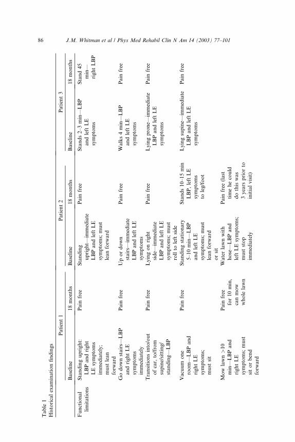
Patient description and historical examination findings
The three subjects were retired military members who were treated in the
Physical Therapy Clinic at Brooke Army Medical Center, San Antonio,
Texas, and followed over an 18-month period. All patients reported LBP
and LE symptoms that were aggravated by standing upright and walking.
In all individuals, lower extremity pain was worse than the LBP and allsymptoms resolved with bending forward or sitting. Table 1 includes more
detailed historical examination findings.
Patient 1
This 81-year-old male suffered from worsening LBP and right LE symp-
toms extending to the posterior calf and plantar surface of the foot. The ini-
tial onset of symptoms started with a fall on to his back and buttocks 5 years
prior. The LBP was an intermittent, deep ache with intermittent, deep, sharp
pain in the right buttock. The right lower calf symptoms were intermittentwith a tired, achy quality. This patient had not exercised regularly since the
fall due to his LBP and LE symptoms and reported a ‘‘very sedentary’’ life-
style with at least 6 to 8 hours of sitting per day. Standard radiographs dem-
onstrated the following: multilevel degenerative disc disease with moderate
disc space narrowing in L4-5 and L5-S1; pars defect of the left L4 vertebral
body, Grade I anterior listhesis of L4 on L5; and transitional vertebra at
the lumbosacral junction. General health issues included aortic valve re-
placement 1 year prior, hypertension, coronary artery disease, angina (onlywhen mowing the lawn), shortness of breath with activity, 3-year history of
vertigo, skin cancer removed in 1950s (unsure what type, not recorded in
medical records), and positive smoking history (stopped in 1962).
Patient 2
This 63-year-old male had episodic, worsening LBP since a ‘‘blow to the
back’’ in high school football 45 years prior. The LBP was described as an
intermittent, deep, sharp pain. His intermittent LE numbness, tingling, andpain extended along the lateral thigh, leg, and foot. LE symptoms began 8 to
10 years prior and had progressively increased in frequency and intensity.
85J.M. Whitman et al / Phys Med Rehabil Clin N Am 14 (2003) 77–101
Table
1
Historicalexaminationfindings
Patient1
Patient2
Patient3
Baseline
18months
Baseline
18months
Baseline
18months
Functional
limitations
Standingupright:
LBPandright
LEsymptoms
immediately;
must
lean
forw
ard
Pain
free
Standing
upright—
immediate
LBPandleftLE
symptoms;must
leanforw
ard
Pain
free
Stands2–3min—
LBP
andleft
LE
symptoms
Stand45
min—
rightLBP
Godownstairs—
LBP
andrightLE
symptoms
immediately
Pain
free
Upordown
stairs—
immediate
LBPandleftLE
symptoms
Pain
free
Walks4min—
LBP
andleft
LE
symptoms
Pain
free
Transitionsinto/out
ofcar,to/from
supine/sitting/
standing—
LBP
Pain
free
Lyingonright
side—
immediate
LBPandleftLE
symptoms;must
rollto
leftside
Pain
free
Lyingprone—
immediate
LBPandleft
LE
symptoms
Pain
free
Vacuum
one
room—
LBPand
rightLE
symptoms;
must
sit
Pain
free
Standingstationary
5–10min—
LBP
andleft
LE
symptoms;must
leanforw
ard
orsit
Stands10–15min
LBP,leftLE
symptoms
toleg/foot
Lyingsupine—
immediate
LBPandleft
LE
symptoms
Pain
free
Mow
lawn>10
min—
LBPand
rightLE
symptoms;must
sitorbend
forw
ard
Pain
free
for10min,
canmow
whole
lawn
Waterlawnwith
hose—
LBPand
leftLEsymptoms;
must
stop
immediately
Pain
free
(last
timehecould
dothiswas
3years
priorto
initialvisit)
86 J.M. Whitman et al / Phys Med Rehabil Clin N Am 14 (2003) 77–101
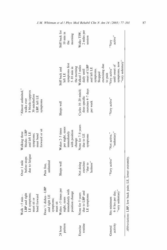
Walk
>5min—
LBPandright
LEsymptoms;
must
sitor
bendforw
ard
Over
1mile
before
stops
dueto
fatigue
Walkingthree
blocks—
LBP
andleft
LE
symptoms;
must
bend
forw
ard
orsit
‘‘Alm
ost
unlimited,’’
walksover
8blocks(approx
30min)before
LBP,left
LE
symptoms
Does
½dishes—
LBP
andrightLE
symptoms;
must
sit
Pain
free,
unlimited
24hour
pattern
Wakes
>5times
per
night,eases
immediately
with
positionchange
Sleepswell
Wakes
2–3times
per
night,eases
immediately
withposition
change
Sleepswell
Stiffback
and
left
LE
symptomsfirst
5–10min
in
themorning
Stiffback
for
10min
in
the
morning
Exercise
routine
Nonefor5years
dueto
LBPand
LEsymptoms
Notdoing
exercises
‘‘dueto
laziness’’
Nonefor7–8years
dueto
LE
symptoms
Cycles
10–20min/d;
does
specific
exercises4–7days
per
week
Walked
3miles
dailyuntil
onsetofLBP
andleftLE
symptoms.
Stopped
exercisingdue
topain
WalksTIW
,
2miles
per
session
General
activity
Sitsminim
um
6–8hrs
per
day,
‘‘verysedentary’’
‘‘Veryactive’’
‘‘Notactive,’’
‘‘sedentary’’
‘‘Veryactive’’
‘‘Veryactive’’
untilonsetof
symptoms,now
‘‘verysedentary’’
‘‘Very
active’’
Abbreviations:LBP,low
back
pain;LE,lower
extrem
ity.
87J.M. Whitman et al / Phys Med Rehabil Clin N Am 14 (2003) 77–101
This patient’s pain and disability from LSS caused him to retire from work 4
years prior and lumbar decompressive surgery was scheduled 1 year prior to
the initial physical therapy (PT) examination. The back surgery was can-celled secondary to the patient needing a prostatectomy and the patient was
referred to PT for nonsurgical management of his LSS. The MRI report
from 1 year prior reported a congenitally small spinal canal with central
canal stenosis at L3-4 and L4-5; canal flattening at L4-5; collapsed degener-
ative discs at L4-5, L5-S1; and mild right foraminal narrowing at L5-S1.
General health issues included a history of prostate cancer (removed 1 year
prior), hypertension, and allergies.
Patient 3
This 71-year-old male reported a history of worsening intermittent dullache in the left buttock and intermittent dull achy symptoms extending
along the lateral left thigh to the proximal one third of the lateral tibia.
These symptoms, of insidious onset 6 weeks prior, had caused this patient
to cease his daily walking program. This patient’s recent MRI report
included mild spinal canal stenosis at L2-3 and L5-S1 and severe spinal
canal stenosis from L3 through L5. General health issues included diabetes
mellitus type II, hypertension, and glaucoma.
Initial physical examination findings
All three patients presented with the following impairments: (1) a flexedstanding posture and decreased passive extension of the hips, (2) stiffness of
the lower lumbar spine and the hips upon manual mobility testing, (3) tight-
ness in the iliopsoas and rectus femoris bilaterally, and (4) active range of
motion limitations with reproduction of lumbar and lower extremity symp-
toms (limited into extension, side bending to the side of LE symptoms, and
quadrant testing to the side of LE symptoms). Patients 2 and 3 tested pos-
itively on the TSTT. Patient 1 underwent a bicycle test to further differenti-
ate between neurogenic and vascular claudication. Based on the patient’srate of perceived exertion, this individual worked harder on the bicycle test
and exercised twice as long on the bicycle than he had during the TSTT, yet
he complained of no LBP or LE pain while cycling. Lastly, all three patients
demonstrated reflex and strength changes in the S1 distribution. Table 2
provides a detailed description of the physical examination findings for each
patient at baseline and 18 months.
Physical therapy intervention
Each patient received five sessions of impairment-specific manual physi-
cal therapy intervention. Treatment to the lumbar and lower thoracic spine
regions included both rotational and posterior to anterior mobilization ormanipulation techniques. Posterior to anterior accessory motion mobiliza-
tion techniques were used to address hip stiffness and to try to increase hip
88 J.M. Whitman et al / Phys Med Rehabil Clin N Am 14 (2003) 77–101
extension mobility. The therapist manually stretched the tight rectus femoris
and iliopsoas muscles (see Fig. 1 [A–F] for pictures of selected manual phys-
ical therapy techniques used to treat these three patients). After improve-ment in spinal and hip mobility, gluteal muscle and lower abdominal
muscle strengthening was initiated. With each visit, the patient was
instructed in specific exercises designed to reinforce the physical therapy
interventions that were found to be efficacious in that patient’s care. The
exercises were continually adjusted until the individual patient felt stretch
or muscle activation in the targeted tissue region. Further sessions with the
therapist (ranging from one to four additional appointments) were used for
reinforcement of the home program. Lastly, all three patients were asked togo on a daily intentional walk. The patients were instructed to stop walking
when symptoms reached the level that would normally make them stop.
Patients 2 and 3 participated in a progressive body-weight supported
treadmill ambulation training program. For this training, a cable and trunk
harness system is used to lift, or unload, a specified amount of body weight
from the patient while the patient walks on the treadmill. Body-weight sup-
port systems have been shown to decrease the downward excursion of the
body’s center of gravity [77] and predictably reduce the vertical componentof the ground reaction force with walking and with running [78]. Theoreti-
cally, this same body weight support system will decrease compression
forces usually placed on the lumbar spine with weight bearing, thus increas-
ing the cross-sectional area of the spinal canal and neuroforamen. Clinically,
it is our experience that many patients with LSS often tolerate walking with
a body-weight supported system for longer periods of time and with less
pain than without the support. Body-weight supported ambulation training
has been advocated for patients with LSS [63], other spinal conditions [79],and various lower extremity disorders [80–82]. For our patients with LSS,
enough body-weight support was used to alleviate LE symptoms and to
allow the patients to walk as normally as possible and as long as possible.
Naturally, the amount of support required fluctuated from session to ses-
sion, but the overall goal was to gradually decrease the amount of unloading
used and gradually increase the walking pace and distance.
Orthotics were prescribed for patient 3 after the efficacy of the manual
intervention had been established. These were used to compensate for bio-mechanical deficiencies of the feet that the therapist determined adversely
affected lumbar spine and pelvic mechanics with gait. The foot and ankle
findings included marked bilateral pes planus (right> left), bilateral na-
vicular drop of approximately 10 mm, no evidence of calcaneal inversion
bilaterally throughout gait, short stride length bilaterally, and an apropul-
sive gait. ‘‘Off-the-shelf’’ orthotics were customized for this patient with 2
degrees medial posting on the left forefoot, left rearfoot, and right rearfoot;
and 4 degrees medial posting to the right forefoot. To determine whether theorthotics would be beneficial for this patient, the time to onset of neurogenic
claudication was measured during three successive trials of walking first
89J.M. Whitman et al / Phys Med Rehabil Clin N Am 14 (2003) 77–101
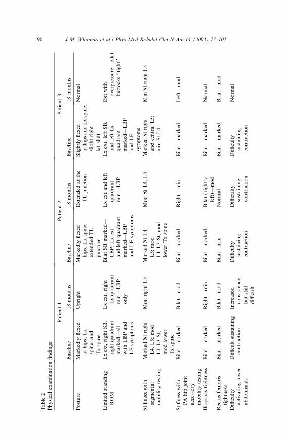
Table
2
Physicalexaminationfindings
Patient1
Patient2
Patient3
Baseline
18months
Baseline
18months
Baseline
18months
Posture
Markedly
flexed
athips,Lx
spine,
and
Txspine
Upright
Markedly
flexed
hips,Lxspine;
extended
TL
junction
Extended
atthe
TLjunction
Slightlyflexed
athipsandLxspine;
slightright
latshift
Norm
al
Lim
ited
standing
ROM
Lxext,rightSB,
rightquadrant
marked—
all
withLBPand
LEsymptoms
Lxext,right
Lxquadrant
min—
LBP
only
BilatSBmarked—
LBP;Lxext
andleft
quadrant
marked—LBP
andLEsymptoms
Lxextandleft
quadrant
min—
LBP
Lxext,leftSB,
andleftLx
quadrant
marked—
LBP
andLE
symptoms
Extwith
overpressure—
bilat
buttocks‘‘tight’’
Stiffnesswith
segmental
mobilitytesting
Marked
Stright
L4,L5;mod
L1–L3St;
modlower
Txspine
ModrightL5
Marked
StL4,
L5;mod
L1–L3St;mod
lower
Txspine
ModStL4,L5
Marked
Stright
andcentralL5;
min
StL4
Min
StrightL5
Stiffnesswith
PA
hip
joint
accessory
mobilitytesting
Bilat—
marked
Bilat—
mod
Bilat—
marked
Right—
min
Bilat—
marked
Left—
mod
Iliopsoastightness
Bilat—
marked
Right—
min
Bilat—
marked
Bilat(right>
left)—
mod
Bilat—
marked
Norm
al
Rectusfemoris
tightness
Bilat—
marked
Bilat—
mod
Bilat—
min
Norm
al
Bilat—
marked
Bilat—
mod
Diffi
culty
activatinglower
abdominals
Diffi
cultsustaining
contraction
Increased
consistency,
butstill
diffi
cult
Diffi
culty
sustaining
contraction
Diffi
culty
sustaining
contraction
Diffi
culty
sustaining
contraction
Norm
al
90 J.M. Whitman et al / Phys Med Rehabil Clin N Am 14 (2003) 77–101
Muscle
weakness
Bilatgluteus
medius
andmaxim
us
Norm
al
Bilatgluteus
mediusand
maxim
us
Norm
al
Bilatgluteus
mediusand
maxim
us
Norm
al
Straightleg
raise
Right—
70�,LBP
andLE
symptoms,
increaseswith
dorsiflexion
Right—
75�,
HSpullonly,
nochangewith
dorsiflexion
Left—
60�,
LBP,
ankle
DF
increasesLBP;
right—
70�,post
thightightness,
nochangewith
dorsiflexion
Bilat—
80�,min
proxpost
thigh
tightness,no
changewith
dorsiflexion
Left—
52�;
right—
68�;
both
withpost
thigh
symptoms,both
increaseswith
dorsiflexion
Bilat—
75�,both
with
proxpost
ipsilat
thighsymptoms,
both
increase
with
dorsiflexion
Neurological
findings
RightAJ1þ,Right
PFweakness
Nochange
Norm
alexcept
bilatabsent
AJ,
bilatPF
weakness,fl
sensationto
the
left
lateralfoot
Nochange
Norm
alexcept:
leftankle
eversion4/5,both
PFweakness(l>r),
bilatabsentAJ,
absentleftKJ,
rightKJ1þ
Nochangeexcept
further
flstrength
withleftankle
PF
Only
positiveorremarkable
findingsnotedhere.
Abbreviations:AJ,anklejerk
reflex;AROM,activerangeofmotion;Bilat,bilateral;deg,degrees;DF,dorsiflexion;fl,
limited
ordecreased;ext,extension;
flex,flexion;glut,gluteus;HS,hamstrings;Lx,lumbar;Lxquadrant,combined
motionincludingextensionandipsilateralside-bendingandrotation;max,
maxim
us;
med,medius;
min,minim
al;mod,moderate;PA,post
toanterior;
PF,plantarflexion/plantarflexor;
post,posterior;
prox,proxim
al;RF,rectus
femoris;SB,sidebending;SLR,straightlegraise;
St,stiff/stiffness;TL,thoraco-lumbar;Tx,thoracic;
tight,tightness.
91J.M. Whitman et al / Phys Med Rehabil Clin N Am 14 (2003) 77–101
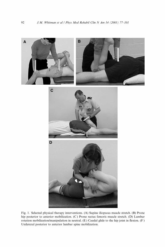
Fig. 1. Selected physical therapy interventions. (A) Supine iliopsoas muscle stretch. (B) Prone
hip posterior to anterior mobilization. (C ) Prone rectus femoris muscle stretch. (D) Lumbar
rotation mobilization/manipulation in neutral. (E ) Caudal glide to the hip joint in flexion. (F )
Unilateral posterior to anterior lumbar spine mobilization.
92 J.M. Whitman et al / Phys Med Rehabil Clin N Am 14 (2003) 77–101
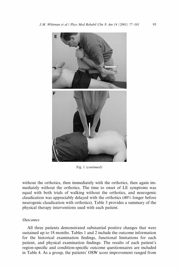
without the orthotics, then immediately with the orthotics, then again im-mediately without the orthotics. The time to onset of LE symptoms was
equal with both trials of walking without the orthotics, and neurogenic
claudication was appreciably delayed with the orthotics (40% longer before
neurogenic claudication with orthotics). Table 3 provides a summary of the
physical therapy interventions used with each patient.
Outcomes
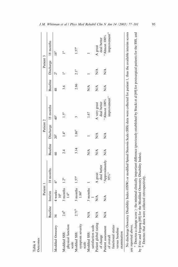
All three patients demonstrated substantial positive changes that were
sustained up to 18 months. Tables 1 and 2 include the outcome information
for the historical examination findings, functional limitations for each
patient, and physical examination findings. The results of each patient’s
region-specific and condition-specific outcome questionnaires are included
in Table 4. As a group, the patients’ OSW score improvement ranged from
Fig. 1 (continued )
93J.M. Whitman et al / Phys Med Rehabil Clin N Am 14 (2003) 77–101
66% to 95% of their baseline scores by discharge and 33% to 82% at 18
months. On the Physical Function Scale of the modified SSS, improvementranged from 1.0 to 2.6 points from baseline to discharge and 0.9 to 2.6 at 18
months. On the Symptom Severity Scale, improvement ranged from 0.76 to
1.85 by discharge and 0.14 to 1.29 at 18 months. Except for patient 2’s 18-
month Symptom Severity Scale score, the magnitude of all of the improve-
ments in outcomes noted by our patients surpassed the MCIDs previously
established by Stucki et al [69] for the Spinal Stenosis Scale and by Fritz and
Irrgang [68] for the modified OSW. Further, all three patients reported
improvements in overall functional status. These results, in addition to thepatients’ responses on the GRC scale, are reported in Table 4. Finally,
Table 3
Summary of physical therapy interventions
Patient 1 Patient 2 Patient 3
Total no. visits
with therapist
9 10 9
Manual therapy
sessions included:
5 5 6
Spinal
manipulation
(rotational,
PA graded)
Spinal
manipulation
(rotational,
PA graded)
Spinal
manipulation
(rotational,
PA graded)
Hip joint
mobilization
(PA)
Hip joint
mobilization
(PA)
Hip joint
mobilization
(PA)
Hip flexor
stretching
Hip flexor
stretching
Hip flexor
stretching
Body weight
supported
ambulation
Body weight
supported
ambulation
Body weight
supported
ambulation
Gluteal muscle
retraining
Gluteal and lower
abdominal
retraining
Visits focused on
correct performance
of home exercise
program:
Spinal mobility
exercises
Flexion and
rotational spinal
mobilization
exercises
Flexion and
rotational spinal
mobilization
exercises
Flexion and
rotational spinal
mobilization
exercises
Muscle stretching Hip flexor muscle
stretching
Hip flexor muscle
stretching
Hip flexor muscle
stretching
Muscle retraining Gluteal and calf
muscle
retraining
Gluteal and calf
muscle retraining
Gluteal and lower
abdominal muscle
retraining
Aerobic exercise Daily walking Daily: alternate
walk and cycle
Daily walking
Abbreviation: PA, posterior to anterior.
94 J.M. Whitman et al / Phys Med Rehabil Clin N Am 14 (2003) 77–101
Table
4
Outcomes
Patient1
Patient2
Patient3
Baseline
Interim
18months
Baseline
Discharge
18months
Baseline
Discharge
18months
Modified
Osw
estry
34
4weeks
10a
6a
60
20a
40a
48
2a
18a
Modified
SSS:
physicalfunction
scale
2.6
b3months
1.6
a
1.2
a2.4
1.4
a1.5
a3.6
1a
1a
Modified
SSS:
symptom
severity
scale
2.71b
3months
1.86a
1.57a
3.14
1.86a
32.86
2.1
a1.57a
Modified
SSS:
satisfactionscale
N/A
3months
1
1N/A
11.67
N/A
11
Patientglobalrating
ofchange
N/A
N/A
Agreat
dealbetter
N/A
N/A
Averygreat
dealbetter
N/A
N/A
Agreat
dealbetter
Patientassessm
ent
ofoverall
functionalstatus
versusbaseline
examination
N/A
N/A
‘‘Approxim
ately
95%’’
N/A
N/A
‘‘Alm
ost
100%
improvem
ent’’
N/A
N/A
‘‘Alm
ost
100%
improvem
ent’’
NodischargeOsw
estryDisabilityIndex
(OSW)ormodified
SpinalStenosisScale(SSS)data
werecollectedforpatient1,thustheavailableinterim
scores
are
reported
above.
aDenotesachangescore
�theminim
alclinicallyim
portantdifference
(previouslyestablished
byStuckiet
al[69]forpostsurgicalpatientsfortheSSS,and
byFritz
andIrrgang[68]fortheModified
Osw
estryDisabilityIndex).
bDenotesthatdata
werecollectedretrospectively.
95J.M. Whitman et al / Phys Med Rehabil Clin N Am 14 (2003) 77–101
although all three patients were taking pain medications for their lower back
and leg pain at the baseline evaluation, the patients reported only occasional
use of pain medication at the 18-month follow-up appointment.
Discussion
This case series is the first to use a well-defined, impairment-based, non-
invasive outpatient treatment program for patients with LSS and to provide
patient-centered long-term outcome information. Most studies investigating
the efficacy of conservative interventions or comparing surgical with non-
surgical outcomes for patients with LSS combine invasive treatments, phar-macological management, physical modalities, and nonspecific exercises
[15,51,55,61,83]. As previously discussed, the use of such unfocused manage-
ment strategies prevents further differentiation of what aspect of care was
the most beneficial for patients with LSS. Additionally, some researchers
have reported on outcomes after prolonged inpatient treatment programs
[15,26]. Although this situation allows for improved experimental control
with human subjects, it is our opinion that a prolonged period of nonsurgi-
cal inpatient care for patients with LSS is not practical in the United Statesat this time. The outpatient treatment program we implemented focuses on
the patients’ individualized, prioritized impairments identified at the initial
examination. The treatments emphasized manual physical therapy tech-
niques targeting each patients’ prioritized impairments, specific exercises to
either reinforce the manual physical therapy treatment or strengthen specific
muscles, and a walking program. Under this physical therapy program, the
patients experienced significant improvements, and the potential adverse
effects of invasive therapies or pharmacological management strategies oftenincluded in other ‘‘nonsurgical’’ treatment programs were avoided.
The use of long-term patient-centered outcome measures that focused on
different levels of the disablement model was an asset of this case series.
Many previous reports on nonsurgical management of patients with LSS
only assessed short-term outcomes [15,63–65], used combinations of clini-
cian opinion and patient-centered outcomes assessment [26], or did not
establish the patient’s baseline status before initiating intervention [84].
We examined changes over time at the impairment, functional limitation,and disability levels of the disability model at baseline and up to 18 months
after initiation of physical therapy. Some areas for improvement in the out-
come assessment tools that were utilized have been identified, however.
Although the condition-specific SSS is a promising health outcome measure-
ment tool for those with LSS, the psychometric properties of the modified
SSS for those receiving nonsurgical intervention needs to be established.
Additionally, even though the SSS asks questions specifically addressing the
patient’s walking tolerance, a more quantitative measure of this prevalentfunctional limitation would be beneficial. We suggest that future research
studies incorporate assessment of walking tolerance with a treadmill test
96 J.M. Whitman et al / Phys Med Rehabil Clin N Am 14 (2003) 77–101
at baseline, at discharge, and at scheduled follow-up sessions. Lastly, our
method of quantifying the patient’s overall functional improvement could
possibly be improved by using the Patient Specific Functional Scale (PSFS)[85–87]. This is a patient-specific questionnaire that requires patients to
identify up to five important activities that they are having difficulty with
as a result of their condition. The patient rates difficulty on an 11-point
numerical scale (0¼ unable to perform the activity; 10¼ able to perform
activity at the same level as before the injury or problem) and the average
score for up to five activities is established as the PSFS score. This scale
has been established as reliable, valid, and responsive in other populations
[85–87], but use with the LSS population has not been established.This small case series demonstrates that patients with LSS can make sig-
nificant gains in disability, symptoms, and function in relatively short peri-
ods of time; and that these gains can be maintained up to 18 months.
Although the outcomes attained in this series seem promising, no cause and
effect relationship can be established from case reports. Additionally, the
treating therapist administered all questionnaires and assessments. Failure
to perform outcome assessments in a blinded fashion is a threat to the val-
idity of our findings. Our intention, however, is for this small case series toserve as a demonstration of the positive outcomes that are possible through
the use of a focused, impairment-specific manual physical therapy manage-
ment program. Additionally, it can provide insight into the types of out-
comes that may be beneficial for assessing the progress of patients with
LSS. The authors are currently involved in a randomized clinical trial com-
paring two conservative management strategies and a randomized clinical
trial comparing conservative management and surgical management of LSS.
References
[1] Fanuele JC, Birkmeyer NJO, Abdu WA, et al. The impact of spinal problems on the health
status of patients: have we underestimated the effect? Spine 2000;25:1509–14.
[2] ECRI Health Technology Assessment Group. Treatment of degenerative lumbar spinal
stenosis. Evidence report/technology assessment no. 32 (Prepared by ECRI under Contract
No. 290-97-0020). Rockville, MD: Agency for Healthcare Research and Quality; June
2001. Publication #AHRQ01-E048.
[3] Hart LG, Deyo RA, Cherkin DC. Physician office visits for low back pain. Frequency,
clinical evaluation, and treatment patterns from a US national survey. Spine 1995;1:
11–9.
[4] Long DM, BenDebba M, Torgerson WS, et al. Persistent back pain and sciatica in the
United States: patient characteristics. J Spinal Disord 1996;9:40–58.
[5] Ciol MA, Deyo RA, Howell E, et al. An assessment of surgery for spinal stenosis: time
trends, geographic variations, complications and reoperations. J Am Geriatr Soc 1996;
44(3):285–90.
[6] Taylor VM, Deyo RA, Cherkin DC, et al. Low back pain hospitalization: recent United
States trends and regional variations. Spine 1994;19:1207–13.
[7] Turner JA, Ersek M, Herron L, et al. Surgery for lumbar spinal stenosis. Attempted meta-
analysis of the literature. Spine 1992;17:1–8.
97J.M. Whitman et al / Phys Med Rehabil Clin N Am 14 (2003) 77–101
[8] Katz JN, Dalgas M, Stucki G, et al. Diagnosis of lumbar spinal stenosis. Rheum Dis Clin
North Am 1994;20:471–83.
[9] Katz JN, Lipson SJ, Chang LC, et al. Seven- to 10-year outcome of decompressive surgery
for degenerative lumbar spinal stenosis. Spine 1996;21:92–8.
[10] Gibson JNA, Grant IC, Waddell G. The cochrane review of surgery for lumbar disc
prolapse and degenerative lumbar spondylosis. Spine 1999;24:1820–32.
[11] Waddell G, Gibson JNA. Scientific evidence on the management of lumbar spinal stenosis.
In: Gunzburg R, Szpalski M, editors. Lumbar spinal stenosis. Philadelphia: Lippincott,
Williams & Wilkins; 2000. p. 367–71.
[12] Arnoldi CC, Brodsky AE, Cauchoix J, et al. Lumbar spinal stenosis and nerve root
entrapment syndromes. Definition and classification. Clin Orthop 1976;Mar–Apr(115):4–5.
[13] Penning L. Functional pathology of lumbar spinal stenosis. Clin Biomech 1992;7:3–17.
[14] Postacchini F. Management of lumbar spinal stenosis. J Bone Joint Surg 1996;78:
154–64.
[15] Onel D, Sari H, Donmez C. Lumbar spinal stenosis: clinical/radiologic therapeutic
evaluation in 145 patients. Conservative treatment or surgical intervention? Spine 1993;
18:291–8.
[16] Spivak JM. Degenerative lumbar spinal stenosis. J Bone Joint Surg 1998;80:1053–66.
[17] Airaksinen O, Herno A, Turunen V, et al. Surgical outcome of 438 patients treated
surgically for lumbar spinal stenosis. Spine 1997;22:2278–82.
[18] Kirkaldy-Willis WH, Wedge JH, Yong-Hing K, et al. Pathology and pathogenesis of
lumbar spondylosis and stenosis. Spine 1978;3:319–28.
[19] Postacchini F, Gumina S, Cinotti G, et al. Ligamenta flava in lumbar disc herniation and
spinal stenosis. Spine 1994;19:917–22.
[20] Rausching W. Normal and pathologic anatomy of the lumbar root canals. Spine 1987;
12:1010–9.
[21] Boden SD, Davis DO, Dina TS, et al. Abnormal magnetic resonance scans of the lumbar
spine in asymptomatic subjects: a prospective investigation. J Bone Joint Surg 1990;72:
403–8.
[22] Hitselberger WE, Witten RM. Abnormal myelograms in asymptomatic patients. J Neuro-
surg 1968;28:204–6.
[23] Jensen MC, Brant-Zawadzki MN, Obuchowski N, et al. Magnetic resonance imaging of
the lumbar spine in people without back pain. N Engl J Med 1994;331:69–73.
[24] Wiesel SW, Tsourmas N, Feffer HL, et al. A study of computer-assisted tomography.
I. The incidence of positive CAT scans in an asymptomatic group of patients. Spine 1984;9:
549–51.
[25] Amundsen T, Weber H, Lilleas F, et al. Lumbar spinal stenosis. Clinical and radiologic
features. Spine 1995;20:1178–86.
[26] Amundsen T, Weber H, Nordal HJ, et al. Lumbar spinal stenosis: conservative or surgical
management? A prospective 10-year study. Spine 2000;25:1424–36.
[27] Herno A, Airaksinen O, Tapani S, et al. Computed tomography findings 4 years after
surgical management of lumbar spinal stenosis. No correlation with clinical outcome. Spine
1999;24:2234–9.
[28] Infusa A, An HS, Lim TH, et al. Anatomic changes of the spinal canal and intervertebral
foramen associated with flexion-extension movement. Spine 1996;21:2412–20.
[29] Liyang D, Yinkan X, Zhang W, et al. The effect of flexion-extension motion of the lumbar
spine on the capacity of the spinal canal: an experimental study. Spine 1989;14:523–5.
[30] Panjabi MM, Takata K, Goel VK. Kinematics of the lumbar intervertebral foramen. Spine
1983;3:348–57.
[31] Penning L, Wilmink JT. Posture-dependent bilateral compression of L4 or L5 nerve root in
facet hypertrophy. A dynamic CT-myelographic study. Spine 1987;12:488–500.
[32] Schonstrom N, Lindahl S, Willen J, et al. Dynamic changes in the dimensions of the
lumbar spinal canal: an experimental study in vitro. J Orthop Res 1989;7:115–21.
98 J.M. Whitman et al / Phys Med Rehabil Clin N Am 14 (2003) 77–101
[33] Sortland O, Magnaes B, Hauge T. Functional myelography with metrizamide in the
diagnosis of lumbar spinal stenosis. Acta Radiol Suppl 1977;355:42–54.
[34] Willen J, Danielson B, Gaulitz A, et al. Dynamic effects on the lumbar spinal canal. Axially
loaded CT-myelography and MRI in patients with sciatica and/or neurogenic claudication.
Spine 1997;22:2968–76.
[35] Takahashi K, Miyazaki T, Takino T, et al. Epidural pressure measurements. Relationship
between epidural pressure and posture in patients with lumbar spinal stenosis. Spine
1995;20:650–3.
[36] Baker AR, Collins TA, Porter RW, et al. Laser Doppler study of porcine cauda equina
blood flow: the effect of electrical stimulation of the rootlets during single and double site,
low pressure compression of the cauda equina. Spine 1995;20:660–4.
[37] Iwamoto H, Matsuda H, Noriage A, et al. Lumbar spinal canal stenosis examined elec-
trophysiologically in a rat model for chronic cauda equina compression. Spine 1997;22:
2636–40.
[38] Jespersen SM, Hansen ES, Hoy K, et al. Two-level spinal stenosis in minipigs.
Hemodynamic effects of exercise. Spine 1995;20:2765–73.
[39] Watanabe R, Parke WW. Vascular and neural pathology of lumbosacral spinal stenosis.
J Neurosurg 1986;64:64–70.
[40] Bridwell KH. Lumbar spinal stenosis. Diagnosis, management, and treatment. Clin Geriatr
Med 1994;10:677–701.
[41] Katz JN, Dalgas M, Stucki G, et al. Degenerative lumbar spinal stenosis. Diagnostic value
of the history and physical examination. Arthritis Rheum 1995;38:1236–41.
[42] Ciric E, Mikhael MA, Tarkington JA, et al. The lateral recess syndrome. J Neurosurg
1980;53:433–43.
[43] Johnsson KE, Rosen I, Uden A. The natural course of lumbar spinal stenosis. Clin Orthop
Rel Res 1992;Jun(279):82–6.
[44] Verbiest H. Lumbar spinal stenosis: morphology, classification, long term results. In:
Weinstein J, Wiesel S, editors. The lumbar spine. Philadelphia: W.B. Saunders; 1990.
p. 546–89.
[45] Johnsson KE. Lumbar spinal stenosis. A retrospective study of 163 cases in southern
Sweden. Acta Orthop Scand 1995;66:403–5.
[46] Tuite GF, Doran SE, Stern JD, et al. Outcome after laminectomy for lumbar spinal
stenosis. Part II: radiographic changes and clinical correlations [see comments]. J Neurosurg
1994;81:707–15.
[47] Tuite GF, Stern JD, Doran SE, et al. Outcome after laminectomy for lumbar spinal
stenosis. Part I: clinical correlations [published erratum appears in J Neurosurg 1995;82(5):
912]. J Neurosurg 1994;81:699–706.
[48] Iversen MD, Katz JN. Examination findings and self-reported walking capacity in patients
with lumbar spinal stenosis. Phys Ther 2001;81:1296–306.
[49] Makan P, Fairbank JLT, Wanders L. Clinical assessment of lumbar spinal stenosis. J Bone
Joint Surg (Britain) 1998;80-B(Suppl II):158.
[50] Fritz JM, Erhard RE, Delitto A, et al. Preliminary results of the use of a two-stage
treadmill test as a clinical diagnostic tool in the differential diagnosis of lumbar spinal
stenosis. J Spinal Disord 1997;10:410–6.
[51] Atlas SJ, Keller RB, Robson D, et al. Surgical and nonsurgical management of lumbar
spinal stenosis: four-year outcomes from the Maine Lumbar Spine Study. Spine 2000;25:
556–62.
[52] Fritz JM, Delitto A, Welch WC, et al. Lumbar spinal stenosis: a review of current concepts
in evaluation, management, and outcome measurements. Arch Phys Med Rehabil 1998;
79:700–8.
[53] Jenis LG, An HS. Spine update. Lumbar foraminal stenosis. Spine 2000;25:389–94.
[54] Nagler W, Hausen HS. Conservative management of lumbar spinal stenosis. Identifying
patients likely to do well without surgery. Postgrad Med 1998;103:69–71.
99J.M. Whitman et al / Phys Med Rehabil Clin N Am 14 (2003) 77–101
[55] Atlas SJ, Deyo RA, Keller RB, et al. The Maine Lumbar Spine Study, part III. 1-year
outcomes of surgical and nonsurgical management of lumbar spinal stenosis. Spine 1996;
21:1787–95.
[56] Keller RB, Atlas SG, Singer DE, et al. The Maine Lumbar Spine Study, I. Background and
concepts. Spine 1996;21:1769–76.
[57] Hurri H, Slatis P, Soini J, et al. Lumbar spinal stenosis: assessment of long-term out-
come 12 years after operative and conservative treatment. J Spinal Disord 1998;11:110–5.
[58] Johnsson KE, Uden A, Rosen I. The effect of decompression on the natural course of
spinal stenosis. A comparison of surgically treated and untreated patients. Spine 1991;
16:615–9.
[59] Mariconda M, Zanforlino G, Celestino GA, et al. Factors influencing the outcome of
degenerative lumbar spinal stenosis. J Spinal Disord 2000;13:131–7.
[60] Fukusaki M, Kobayashi I, Hara T, et al. Symptoms of spinal stenosis do not improve after
epidural steroid injection. Clin J Pain 1998;14:148–51.
[61] Simotas AC, Dorey FJ, Hansraj KK, et al. Nonoperative treatment for lumbar spinal
stenosis. Clinical and outcome results and a 3-year survivorship analysis. Spine 2000;25:
197–204.
[62] Kirkaldy-Willis WH. Manipulation. In: Kirkaldy-Willis W, editor. Managing low back
pain. New York: Churchill Livingstone; 1983. p. 175–83.
[63] Fritz JM, Erhard RE, Vignovic M. A nonsurgical treatment approach for patients with
lumbar spinal stenosis. Phys Ther 1997;77:962–73.
[64] DuPriest CM. Nonoperative management of lumbar spinal stenosis. J Manipulative
Physiol Ther 1993;16:411–4.
[65] Rendeiro DG. Conservative management of lumbar spinal stenosis—a case report. J Man-
ual Manipulative Ther 2000;8:139.
[66] Nagi SZ. Some conceptual issues in disability and rehabilitation. In: Sussman M, editor.
Sociology and rehabilitation. Washington, DC: American Sociological Association; 1965.
p. 100–13.
[67] Fairbank JC, Couper J, Davies JB, et al. The Oswestry low back pain disability
questionnaire. Physiotherapy 1980;66:271–3.
[68] Fritz JM, Irrgang JJ. A comparison of a modified Oswestry Low Back Pain Disability
Questionnaire and the Quebec Back Pain Disability Scale. Phys Ther 2001;81:776–88.
[69] Stucki G, Daltroy L, Liang MH, et al. Measurement properties of a self-administered
outcome measure in lumbar spinal stenosis. Spine 1996;21:796–803.
[70] Stucki G, Liang MH, Fossel AH, et al. Relative responsiveness of condition-specific and
generic health status measures in degenerative lumbar spinal stenosis. J Clin Epidemiol
1995;48:1369–78.
[71] Jaeschke R, Singer J, Guyatt GH. Measurement of health status: ascertaining the
minimally clinically important difference. Control Clin Trials 1989;10:407–15.
[72] Deen HG, Gordon MD, Zimmerman RS, et al. Test-retest reproducibility of the exercise
treadmill examination in lumbar spinal stenosis. Mayo Clin Proc 2000;75:1002–7.
[73] Deen HG, Zimmerman RS, Lyons MK, et al. Use of the exercise treadmill to measure
baseline functional status and surgical outcome in patients with severe lumbar spinal
stenosis. Spine 1998;23:244–8.
[74] Deen HG, Zimmerman RS, Lyons MK, et al. Measurement of exercise tolerance on the
treadmill in patients with symptomatic lumbar spinal stenosis: a useful indicator of
functional status and surgical outcome. J Neurosurg 1995;83:27–30.
[75] Herno A, Partanen K, Talaslahti T, et al. Long-term clinical and magnetic resonance
imaging follow-up assessment of patients with lumbar spinal stenosis after laminectomy.
Spine 1999;24:1533–7.
[76] Herno A, Saari T, Suomalainen O, et al. The degree of decompressive relief and its relation
to clinical outcome in patients undergoing surgery for lumbar spinal stenosis. Spine
1999;24:1010–4.
100 J.M. Whitman et al / Phys Med Rehabil Clin N Am 14 (2003) 77–101
[77] Finch L, Barbeau H, Arsenault B. Influence of body weight support on normal human.
Phys Ther 1991;71:842–56.
[78] Flynn TW, Canavan PK, Cavanagh PR, et al. Plantar pressure reduction in an incremental
weight-bearing system. Phys Ther 1997;77:410–6.
[79] Olsen J, Svendsen B. Medical exercise therapy: an adjunct to orthopaedic manual therapy.
Orthop Prac 1992;4:7–10.
[80] Hunter D, Cole EZ, Murray JM, et al. Energy expenditure of below-knee amputees during
harness-supported treadmill ambulation. J Orthop Sports Phys Ther 1995;21:268–76.
[81] Kelsey DD, Tyson E. A new method of training for the lower extremity using unloading.
J Orthop Sports Phys Ther 1994;19:218–23.
[82] Kline MK, Axen K, Haas F. Mechanical unweighting effects on treadmill exercise and pain
in elderly people with osteoarthritis of the knee. Phys Ther 1996;76:387–94.
[83] Swezey RL. Outcomes for lumbar stenosis: a 5-year follow-up study. J Clin Rheumatol
1996;2:129–34.
[84] Herno A, Airaksinen O, Saari T, et al. Lumbar spinal stenosis: a matched-pair study of
operated and non-operated patients. Br J Neurosurg 1996;10:461–5.
[85] Chatman A, Neel J, Hyams S, et al. The Patient Specific Functional Scale: measurement
properties in patients with knee dysfunction. Phys Ther 1997;77:820–9.
[86] Stratford PW, Gill C, Westaway M, et al. Assessing disability and change on individual
patients: a report of a patient specific measure. Physiother Can 1995;47:258–63.
[87] Westaway M, Stratford PW, Binkley J. The Patient Specific Functional Scale: validation of
its use in persons with neck dysfunction. J Orthop Sports Phys Ther 1998;27:331–8.
101J.M. Whitman et al / Phys Med Rehabil Clin N Am 14 (2003) 77–101
Related Documents