Noninvasive Monitoring of Fecal Cortisol Metabolites in the Eastern Chipmunk (Tamias striatus): Validation and Comparison of Two Enzyme Immunoassays Author(s): Pierre-Olivier Montiglio, Fanie Pelletier, Rupert Palme, Dany Garant, Denis Réale, Rudy Boonstra Reviewed work(s): Source: Physiological and Biochemical Zoology, (-Not available-), p. 000 Published by: The University of Chicago Press Stable URL: http://www.jstor.org/stable/10.1086/664592 . Accessed: 19/02/2012 15:30 Your use of the JSTOR archive indicates your acceptance of the Terms & Conditions of Use, available at . http://www.jstor.org/page/info/about/policies/terms.jsp JSTOR is a not-for-profit service that helps scholars, researchers, and students discover, use, and build upon a wide range of content in a trusted digital archive. We use information technology and tools to increase productivity and facilitate new forms of scholarship. For more information about JSTOR, please contact [email protected]. The University of Chicago Press is collaborating with JSTOR to digitize, preserve and extend access to Physiological and Biochemical Zoology. http://www.jstor.org

Welcome message from author
This document is posted to help you gain knowledge. Please leave a comment to let me know what you think about it! Share it to your friends and learn new things together.
Transcript

Noninvasive Monitoring of Fecal Cortisol Metabolites in the Eastern Chipmunk (Tamiasstriatus): Validation and Comparison of Two Enzyme ImmunoassaysAuthor(s): Pierre-Olivier Montiglio, Fanie Pelletier, Rupert Palme, Dany Garant, Denis Réale,Rudy BoonstraReviewed work(s):Source: Physiological and Biochemical Zoology, (-Not available-), p. 000Published by: The University of Chicago PressStable URL: http://www.jstor.org/stable/10.1086/664592 .Accessed: 19/02/2012 15:30
Your use of the JSTOR archive indicates your acceptance of the Terms & Conditions of Use, available at .http://www.jstor.org/page/info/about/policies/terms.jsp
JSTOR is a not-for-profit service that helps scholars, researchers, and students discover, use, and build upon a wide range ofcontent in a trusted digital archive. We use information technology and tools to increase productivity and facilitate new formsof scholarship. For more information about JSTOR, please contact [email protected].
The University of Chicago Press is collaborating with JSTOR to digitize, preserve and extend access toPhysiological and Biochemical Zoology.
http://www.jstor.org

000
T E C H N I C A L C O M M E N T
Noninvasive Monitoring of Fecal Cortisol Metabolites in the Eastern
Chipmunk (Tamias striatus): Validation and Comparison
of Two Enzyme Immunoassays
* Corresponding author; e-mail: [email protected].† E-mail: [email protected].‡ E-mail: [email protected].§ E-mail: [email protected] E-mail: [email protected].# E-mail: [email protected]
Physiological and Biochemical Zoology 85(2):000–000. 2012. � 2012 by TheUniversity of Chicago. All rights reserved. 1522-2152/2012/8502-1114$15.00.DOI: 10.1086/664592
Pierre-Olivier Montiglio1,*Fanie Pelletier2,†
Rupert Palme3,‡
Dany Garant4,§
Denis Reale1,k
Rudy Boonstra5,#
1Chaire de Recherche du Canada en EcologieComportementale, Departement des Sciences Biologiques,Universite du Quebec a Montreal, CP 8888 SuccursaleCentre-Ville, Montreal, Quebec H3C 3P8, Canada; 2Chairede Recherche du Canada en Demographie Evolutive etConservation, Departement de Biologie, Faculte des Sciences,Universite de Sherbrooke, 2500 Boulevard de l’Universite,Sherbrooke, Quebec J1K 2R1, Canada; 3Department ofBiomedical Sciences/Biochemistry, University of VeterinaryMedicine, Veterinarplatz 1, A-1210 Vienna, Austria;4Departement de Biologie, Faculte des Sciences, Universite deSherbrooke, 2500 Boulevard de l’Universite, Sherbrooke,Quebec J1K 2R1, Canada; 5Center for the Neurobiology ofStress, University of Toronto Scarborough, 1265 MilitaryTrail, Toronto, Ontario M1C 1A4, Canada
Accepted 12/25/2011; Electronically Published 2/7/2012
ABSTRACT
Monitoring fecal glucocorticoid metabolites in wild animals,using enzyme immunoassays, enables the study of endocri-nological patterns relevant to ecology and evolution. Whilesome researchers use antibodies against the parent hormone(which is typically absent from fecal samples), others advocatethe use of antibodies designed to detect glucocorticoid metab-olites. We validated two assays to monitor fecal cortisol me-tabolites in the eastern chipmunk (Tamias striatus). We com-
pared an antibody produced against cortisol and one producedagainst 5a-pregnane-3b, 11b, 21-triol-20-one using a radio-metabolism study and an injection with adrenocorticotropichormone (ACTH). Most cortisol metabolites were excreted inthe urine (∼83%). Peak excretion in the feces occurred 8 hafter injection. Both assays detected an increase in fecal cortisolmetabolite levels after injection of ACTH. Males, but not fe-males, exhibited a circadian variation in metabolite levels. Thesexes did not exhibit any difference over the time course androute of excretion or the relative increase in fecal cortisol me-tabolite levels after ACTH injection. The cortisol assay displayedhigher reactivity to ACTH injection relative to baseline thandid the metabolite assay. While both antibodies gave compa-rable results, the cortisol antibody was more sensitive to changesin plasma cortisol levels in eastern chipmunks.
Introduction
Studies of the hypothalamic-pituitary-adrenal (HPA) axis infree-ranging populations provide insights into how animals ad-just their physiology, behavior, and life history to environmentalperturbations (Boonstra 2005; Reeder and Kramer 2005; Wing-field 2005). A better understanding of this axis sheds light oncentral processes in ecology and evolution, such as the long-term consequences of predation on population dynamics (Sher-iff et al. 2010b, 2011b), adaptive maternal effects (Breuner 2008;Love and Williams 2008), or the impact of human activitieson animal populations (Thiel et al. 2008, 2011; Macbeth et al.2010).
The activity of the HPA axis is classically studied by quan-tifying the secretion of its end products, glucocorticoids, in theblood. The last decades have witnessed important methodo-logical improvements to monitor glucocorticoid levels in a lessinvasive manner, and it is now possible to quantify their me-tabolites in urine and feces (Palme 2005; Sheriff et al. 2011a).Fecal measurements may be preferable to blood measurementsbecause they represent an integration of glucocorticoid levelsin the blood over a few hours rather than the point estimatesobtained from blood samples (Touma et al. 2003; Sheriff et al.2010a). Thus, they are buffered against small fluctuations inglucocorticoid levels that are likely to occur with blood mea-surements and may reflect more accurately the baseline glu-

000 P.-O. Montiglio, F. Pelletier, R. Palme, D. Garant, D. Reale, and R. Boonstra
cocorticoid level of the animal (Goymann 2005). Moreover,recent work has shown that the fecal glucocorticoid metabolitelevel of an animal may be a good index of an animal’s capacityto secrete plasma glucocorticoids in response to an environ-mental perturbation (Sheriff et al. 2010a). Concentrations offecal glucocorticoid metabolites thus enable studying not onlyHPA activity but also potentially its reactivity to environmentalperturbations.
Measuring glucocorticoids in their metabolized form, how-ever, introduces some complexities. First, the time over whichblood levels of glucocorticoids are integrated in the fecal sam-ples will depend on the species’ biology and activity pattern(Palme 2005). Second, the proportion of glucocorticoid me-tabolites excreted via the feces or the urine may vary with thespecies or the sex of the animals (Touma et al. 2003; Palme etal. 2005). It is thus advisable to perform a radiometabolismstudy, where a known quantity of radiolabeled glucocorticoidis injected and traced in the feces as well as in the urine toassess the time lag between the secretion of glucocorticoids intothe blood and the excretion of their metabolites in the fecesand urine. Third, when measuring fecal glucocorticoid metab-olites, one deals with a mixture of compounds displaying dif-ferent molecular structures, in which little or no native (i.e.,unmetabolized) glucocorticoid(s) is present (Mostl et al. 2005;Palme 2005; Bosson et al. 2009). It is thus necessary to ensurethat the technique used to assess glucocorticoid reacts with animportant portion of the glucocorticoid metabolites producedby the animal. It is also important to ensure that glucocorticoidsecretion into the blood is well reflected in the fecal samples(Touma and Palme 2005).
Fecal glucocorticoid metabolites can be quantified with dif-ferent approaches, including enzyme immunoassays (EIAs).Immunoassays originally designed to target native (i.e., non-metabolized) glucocorticoids have been extensively used toquantify fecal glucocorticoid metabolites in an array of species(Harper and Austad 2000; Wasser et al. 2000; Young and Mon-fort 2009). These assays assume that the antibody has sufficientcross-reactivity to recognize (a group of) metabolites of theparent hormone and that an increase in blood glucocorticoidsecretion leads to an increase in metabolite excretion in thefeces. These assays initially were developed for blood mea-surements (Mostl et al. 2005). Although these assays have dif-ferent cross-reactivities and hence different efficacy, they havebeen used with success to study relationships between fecalglucocorticoid metabolite levels and some aspects of the re-production or the ecology of the animals in their natural en-vironment (Jurke et al. 1997; Lynch et al. 2002; Mateo 2007;Young and Monfort 2009). Other studies use antibodies de-signed to recognize a group of fecal glucocorticoid metabolitessharing a common structure (Jurke et al. 1997; Lynch et al.2002; Mostl and Palme 2002; Mostl et al. 2005; Mateo 2007;Young and Monfort 2009). These metabolite antibodies typi-cally yield higher concentrations of measured metabolites anddisplay a higher reactivity with the fecal glucocorticoid metab-olites than antibodies targeting native glucocorticoids and aretherefore more likely to detect small fluctuations in fecal glu-
cocorticoid metabolites (Morrow et al. 2002; Frigerio et al.2004).
In this study, we validate and compare two fecal assays tomonitor HPA axis activity in the eastern chipmunk (Tamiasstriatus): one using an antibody raised against cortisol and an-other using an antibody raised against 5a-pregnane-3b, 11b,21-triol-20-one. Cortisol is the main glucocorticoid secreted byall sciurid species studied to date, including the yellow-belliedmarmot (Marmota flaviventris), California ground squirrel(Spermophilus beecheyi), golden-mantled ground squirrel (Sper-mophilus saturatus), red squirrels (Tamiasciurus hudsonicus),and yellow-pine chipmunks (Tamias amoenus; Boonstra andMcColl 2000; Kenagy and Place 2000; Place and Kenagy 2000;Boonstra et al. 2001). First, we performed a radiometabolismstudy to analyze the time lag between cortisol secretion intothe blood and its excretion in the feces as well as the relativeimportance of the routes of excretion (urine vs. feces). Wemeasured the radioactivity as well as immunoreactivity of eachfraction with a metabolite antibody recognizing steroids witha 5a-3b-11b-diol structure (Touma et al. 2003) as well as witha cortisol antibody validated in a diversity of species (Young etal. 2004). The metabolite antibody enables a reliable monitoringof the HPA axis in related species, such as Columbian groundsquirrels (Spermophilus columbianus; Bosson et al. 2009) andred squirrels (T. hudsonicus; Dantzer et al. 2010). Second, wemonitored baseline fecal cortisol metabolite levels of captiveindividuals over 2 d to determine the pattern of the circadianrhythm excretion. We also monitored fecal cortisol metabolitelevels after stimulation of the adrenal by an ACTH injectionto ensure that our method detects changes in blood cortisollevels. Finally, we compared both assays regarding their suit-ability to noninvasively measure adrenocortical activity in east-ern chipmunks. While assays against the parent hormone andthe assays against the metabolites have successfully been appliedto study glucocorticoid levels in free-ranging populations, fewstudies compare these two types of assays. Consequently, ourstudy will expand our knowledge on the relative advantages ofthese two types of assays applied to wild populations.
Methods
Capture and Housing of Chipmunks
We livetrapped nine chipmunks (five males and four females)on the campus of the University of Toronto at Scarborough(243�47′N, 79�11′W, m) in August 2010. Long-elevation p 116worth traps were baited with sunflower seeds, apples, and car-rots; opened at dawn; inspected every 90 min; and closedaround 15 h. Females showing signs of lactation were imme-diately released at the point of capture. We housed chipmunksindividually in plastic cages (47 cm # 26 cm # 20 cm)equipped with a wire bottom and a water bottle with a stainlesssteel nipple. Cages were mounted within a second same-sizedtray, equipped with a fine metal mesh. This system enabled thefeces and urine to fall through the cage bottom, but the feceswere caught on the fine wire mesh while the urine passedthrough, thus preventing contamination of the feces by the

Technical Comment 000
Table 1: Chronology of the manipulations carried out on nine Eastern chipmunks in August 2010
Date Procedure Treatment Collection time for samples
August 7–8 CaptureAugust 8–11 Acclimation No manipulation Every 4 h from 0800 to 2200 hoursAugust 12–15 Radiometabolism study Injection of radiolabeled cortisol Every 2 h from 0800 to 2200 hoursAugust 16 Baseline monitoring No manipulation Every 4 h from 0800 to 2200 hoursAugust 17–18 ACTH challenge Injection of synthetic ACTH Every 2 h from 0800 to 2200 hoursAugust 19 Baseline monitoring No manipulation Every 4 h from 0800 to 2200 hours
urine. Animals sometimes urinated onto the feces, in whichcase the fecal pellets were discarded. Hence, we were able tocollect the samples while minimizing disturbance of the chip-munks. Each chipmunk was provided with a plastic refuge, apiece of natural wood, and some cotton as bedding material.They had ad lib. access to rodent chow pellets, peanut butter,sunflower seeds, apples, carrots, and occasionally acorns. Chip-munks were kept at 20�C under natural photoperiod for theduration of the experiment. Except during collection of thesamples, chipmunks were not exposed to human presence ornoise. Chipmunks were left to acclimate for at least 5 d beforebeginning the trials. During that phase, feces were collectedevery 4 h between 0800 and 2200 hours to habituate them tothe procedure (see table 1 for the chronology of the tests andthe sampling times). Upon completion of the experiment, weinspected and weighed the chipmunks. Chipmunks did not loseweight or show any sign of deteriorating health related to thetrials. However, we witnessed a decrease in activity during thelast 3 d of the trials that paralleled the decrease in chipmunkaboveground activity frequently observed in nature during thelate summer (Dunford 1970; Elliott 1978). All husbandry andmanipulations were conducted under University of Torontoanimal use protocol 20008380, issued in accordance with theCanadian Council on Animal Care guidelines.
Route and Time Course of Cortisol Excretion
To monitor the route and timing of cortisol excretion, we in-jected the nine chipmunks intraperitoneally with 1,110 kBq ofradiolabeled cortisol (1,2,6,7-[3H]-cortisol; Amersham Biosci-ences; TBq/mmol). Before injection,specific activity p 1.55radiolabeled cortisol was dried down under air and reconsti-tuted in 10% ethanol and 90% sterile physiological saline so-lution in order to inject a volume of 300 mL. Chipmunks wereinjected between 0700 and 0800 hours, and urine and feceswere then collected for 72 h (see table 1 for sampling intervals).We collected all dry (i.e., uncontaminated) feces pellets withforceps and collected all urine using a 1-mL pipette. We rinsedthe collecting trays with 2 mL of methanol to collect the re-maining radioactivity and added these to the urine sample.Feces mixed with urine were also collected and analyzed sep-arately. All samples were immediately put on ice and frozen at�20�C within 20 min of collection. Urine samples were drieddown under air and reconstituted in 1 mL of methanol (80%).We dried the fecal samples at 70�C for 3 d (until they reached
constant mass; dried radioactive samples ranged from 5 to 50mg). We then crushed the feces using a plunger and extractedeach whole sample by vortexing the pulverized feces with 1 mLof methanol (80%). We quantified activity in the samples byadding 2.5 mL of scintillation fluid to 0.5 mL of urine or fecalextract and measured radioactivity with a scintillation counterwith quench correction (Tri-Carb 2900TR, Boston, MA).
Characterization of Fecal Cortisol Metabolites
Six fecal extracts with maximum radioactivity (three samplesfrom males and three from females) were dried down underair and sent to University of Veterinary Medicine, Vienna, Aus-tria. The radioactive cortisol metabolites in these samples wereseparated according to their polarity by reverse-phase high-performance liquid chromatography (HPLC). We measured ra-dioactivity and immunoreactivity in each fraction with the 5a-pregnane-3b, 11b, 21-triol-20-one EIA described below (fordetails see Touma et al. 2003; Lepschy et al. 2007). In addition,we quantified the amount of immunoreactive cortisol presentin the fecal samples by measuring the immunoreactivity of eachfraction with a cortisol EIA (see Palme and Mostl 1997 foradditional details on the assay).
Monitoring Baseline and Stimulated Cortisol Secretion
To validate the ability of the EIAs to detect changes in cortisolsecretion patterns, we injected animals with synthetic adre-nocorticotropic hormone (4 IU/kg, ACTH; Synacthen Depot,CIBA). Fecal samples were collected to measure baseline levelsof fecal cortisol metabolites 24 h before and 48 h after ACTHinjection. Animals were injected between 0700 and 0800 hours.After injection, we collected samples from 0800 to 2200 hoursfor 48 h (see table 1 for frequency of sampling).
Extraction and Analysis of Fecal Cortisol Metabolites
We dried down fecal samples at 70�C for 72 h (samples typicallyreach constant mass after 48 h). Dried samples were then trans-ferred to a 2-mL test tube and ground using a plunger. Weextracted the fecal cortisol metabolite by vortexing mg35 � 5of ground feces in 1 mL of methanol 80% for 30 min at 1,500rpm and centrifuging (2,500 g; 20 min). We then analyzed thesupernatant with a 5a-pregnane-3b, 11b, 21-triol-20-one EIA(Touma et al. 2003). Details about cross-reactivity of the an-

000 P.-O. Montiglio, F. Pelletier, R. Palme, D. Garant, D. Reale, and R. Boonstra
tibody and the assay procedure can be found in Touma et al.(2003). Mean intra- and interassay coefficients of variation oftwo pool samples were 11.5% and 16.5%, respectively.
To compare assays, we also analyzed the samples collectedduring baseline monitoring and after ACTH injection with acortisol EIA. The cortisol EIA used a polyclonal cortisol anti-body (R4866; C. J. Munro, University of California, Davis) witha horseradish peroxidase ligand (see Munro and Lasley 1988for details on cross-reactivity; see Young et al. 2004 for detailson EIA protocol). Mean intra- and interassay coefficients ofvariation were 9.7% and 9.9%, respectively.
Statistical Analysis
We tested for sex differences in time to peak excretion in fecesand urine, percentage of injected radioactivity recovered, andthe proportion of radioactivity excreted in the feces, using linearmodels. Fecal cortisol metabolite levels measured with the 5a-pregnane-3b, 11b, 21-triol-20-one and the cortisol assays wereanalyzed in a two-step manner. First, to explore the factoraffecting cortisol baseline levels, we used a linear mixed modelincluding hour of sample collection (categorical variable), sex,and their interaction as fixed effects. Individual identity wasfitted as a random effect to account for repeated measurementsof the same individuals over time (also known as pseudorep-lication; Crawley 2007). A log-likelihood ratio test comparinga model including the random effect of interest and a modelwithout it while holding the fixed effect structure constant wasused to test for the significance of the random effect (Pinheiroand Bates 2000). For the individual random effect, we alsoreport the repeatability (r), defined as the variance associatedwith the effect divided by the sum of this variance and theresidual variance from the model (Pinheiro and Bates 2000).High r values are indicative of substantial, consistent differencesamong individuals. Second, to evaluate how ACTH injectionaffected fecal cortisol metabolite levels compared to baselinelevels, we analyzed fecal cortisol metabolite levels in all collectedsamples (i.e., during baseline sampling and after ACTH injec-tion) within one linear mixed model. This second model ini-tially included hour of sample collection, time since ACTHinjection, type of sampling (baseline monitoring or ACTH in-jection), sex, and their interactions as fixed explanatory vari-ables. Again, we added chipmunk identity as a random effectto account for pseudoreplication and tested its significance us-ing a log-likelihood ratio test as described above. All modelswere simplified by stepwise deletion of nonsignificant terms(Crawley 2007). To compare the results obtained with bothassays, we computed Pearson’s product-moment correlationcoefficient for all samples analyzed. We also compared the rel-ative increase in fecal cortisol metabolite level detected by eachassay after ACTH injection using a linear model. The responsevariable was the increase in fecal cortisol metabolite level ofeach individual expressed as the percentage of baseline level atthe same time of day (the percentage was then log transformed).The explanatory variables were number of hours since ACTHinjection, assay used (5a-pregnane-3b, 11b, 21-triol-20-one or
cortisol assay), sex of individuals, and their interactions. Allfecal cortisol metabolite levels are expressed as log-transformedvalues (units are ng/g dry feces). Statistical analyses were per-formed using R 2.10.1 (R Development Core Team 2009).Means are reported with �1 SEM.
Results
Route and Time Course of 3H-Cortisol Excretion
During the 72 h after injection of radiolabeled cortisol, wecollected 151 and 180 urine and fecal samples, respectively. Werecovered of the 1,110 kBq injected in each48.33% � 0.13%subject ( ). A total of andrange p 29.00–75.17 83.10% � 6.67%
of the radioactivity recovered was found in16.21% � 6.86%the urine and the feces, respectively. The rest (0.69% �
) was found in the feces contaminated with urine. There0.90%was no difference in the percentage of radioactivity recoveredamong sexes ( , , ) or ineffect p 12.85 � 8.5 t p 1.51 P p 0.177
the percentage of radiolabeled cortisol excreted via the feces( , , ). Median peakeffect p �4.29 � 4.65 t p �0.92 P p 0.397
excretion of radioactivity in the urine occurred at 4 h afterinjection ( , h) and in feces 8range p 2–4 mean p 3.11 � 1.05h after injection ( , ; see fig.range p 8–12 mean p 9.33 � 1.731). Time to peak excretion in urine ( , ) ort p 0.27 P p 0.807
in feces ( , ) was not affected by sex.t p �1.04 P p 0.337
Characterization of Fecal Cortisol Metabolites
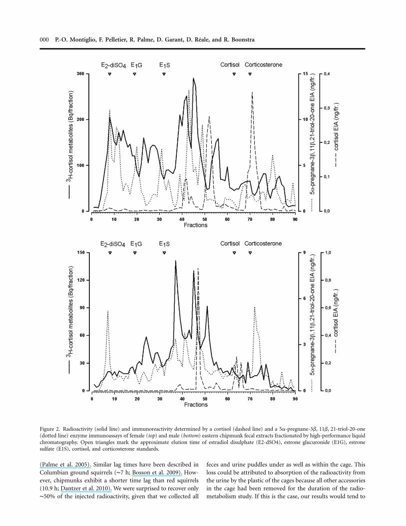
HPLC revealed that cortisol was almost completely metabolizedin both females and males (see fig. 2). The most prominentradioactivity peaks eluted between fractions 35 and 50 for bothsexes. There were differences between sexes regarding theformed metabolites and their immunoreactivity. Females pro-duced more metabolites with higher polarity (fractions 5–20).The 5a-pregnane-3b, 11b, 21-triol-20-one assay detected moremetabolites and had higher peak levels. However, also the cor-tisol EIA was able to react with some of the radioactivemetabolites.
Monitoring Baseline and Stimulated (ACTH Injection)Cortisol Secretion
Baseline fecal cortisol metabolite levels ( samples) an-N p 72alyzed with the 5a-pregnane-3b, 11b, 21-triol-20-one EIAranged from 1,720 to 15,000 ng/g of dry feces (median p
ng/g) and did not vary with sex in the sampling at 08005,390hours ( , , ). How-effect p �0.19 � 0.19 t p �0.96 P p 0.3757
ever, males tended to show higher fecal cortisol metabolite levelsduring the rest of the day ( , all ; seeall effects ! 0.34 P ! 0.04fig. 3, top). Animals also displayed consistent individual dif-ferences regarding their fecal cortisol metabolite level (r p
, , ). The ACTH51.22% log-likelihood ratio p 25.67 P ! 0.001injection ( samples) affected fecal cortisol metaboliteN p 72levels ∼8 h after the injection ( ,effect p 0.51 � 0.15 t p116
, ; see fig. 3, top). Again, we detected important3.38 P p 0.001

Technical Comment 000
Figure 1. Radioactivity ( ) excreted in urine (filled dots, solid line) and feces (open dots, dashed line) in nine eastern chipmunksmean � SEMafter an injection of radiolabeled cortisol.
individual differences in the mean fecal cortisol metabolite lev-els ( , , ).r p 46.72% log-likelihood ratio p 53.16 P ! 0.001
Analysis of the baseline fecal cortisol metabolite levels withthe cortisol EIA ranged from 133 to 2,942 ng/g of dry feces( ng/g) and revealed that males displayed highermedian p 700fecal cortisol metabolite levels than females at 1200 hours( , , ) and 1600 hourseffect p 0.79 � 0.30 t p 2.59 P p 0.01251
( , , ; see fig. 3, bot-effect p 1.01 � 0.31 t p �0.18 P p 0.00251
tom). The cortisol EIA also revealed important individual var-iation ( , , ). Thisr p 72.20% likelihood ratio p 50.04 P ! 0.001EIA also detected an effect of ACTH injection on fecal cortisolmetabolite levels with effects at 1600 hours (effect p 1.92 �
, , ) and 2000 hours (0.46 t p 4.14 P p 0.001 effect p 1.67 �51
, , ). This EIA detected important in-0.50 t p 3.32 P p 0.00151
dividual variation in the mean fecal cortisol metabolite levels( , , ). Con-r p 40.26% log-likelihood ratio p 41.05 P ! 0.001centrations of fecal cortisol metabolites measured with bothEIAs were significantly correlated ( , ,r p 0.45 N p 138 P !
).0.001Analysis of the relative increase in fecal cortisol metabolite
detected by each assay (5a-pregnane-3b, 11b, 21-triol-20-oneassay: , ; cortisol assay:median p 101% range p 36–556
, ) showed that the cortisolmedian p 132% range p 16–1,385EIA measured a higher increase relative to the baseline samples8 and 12 h after ACTH injection compared with the 5a-preg-nane-3b, 11b, 21-triol-20-one EIA (8-h sampling: effect p
, , ; 12-h sampling:1.28 � 0.39 t p 3.25 P p 0.001 effect p106
, , ; one outlier was excluded1.11 � 0.39 t p 2.81 P p 0.005106
from the analysis; see fig. 4). Relative increase in measured fecalcortisol metabolites did not vary between both EIAs during allthe remaining sampling times after ACTH injection (all
, ). We did not find any statisticallyeffects ! 0.50 all P 1 0.20significant difference between the sexes in the relative increase
in fecal cortisol metabolites with either assay (all terms rejectedwith ).P 1 0.18
Discussion
The objective of this study was to assess glucocorticoid levelsof eastern chipmunks noninvasively by validating the mea-surement of fecal cortisol metabolites using two different EIAs.The first used a cortisol antibody, while the second, using a5a-pregnane-3b, 11b, 21-triol-20-one antibody, targeted agroup of its metabolites. Our key conclusion is that both an-tibodies appear to accurately represent fecal metabolite levelsbut that the cortisol EIA is more sensitive. The radiometabolismexperiment showed that only about 16% of the metabolites areexcreted in feces and that the time lag between secretion ofcortisol into the blood and the appearance of its metabolitesin the feces was about 8 h. We found that males displayed acircadian rhythm, with a peak concentration at 1600 hours,whereas females did not. After an ACTH stimulation test, wefound that both assays detected an increase in cortisol metab-olites, which was expressed more strongly in the cortisol EIA.Finally, both assays detected ample individual variation, show-ing that animals consistently differed from each other regardingtheir fecal cortisol metabolite levels. We discuss each of thesepoints below.
Our radiometabolism study showed that chipmunks excretedmost of the injected cortisol in urine (83%) within the first 4h after injection of radiolabeled cortisol. In contrast, we de-tected a peak in excretion in the feces 8 h after the injection.Blood cortisol is metabolized in the liver and excreted into thegut via the bile (Mostl et al. 2005). The time lag between ra-diolabeled cortisol injection and its peak excretion in the fecescan be attributed mostly to the time needed for intestinal transit
000 P.-O. Montiglio, F. Pelletier, R. Palme, D. Garant, D. Reale, and R. Boonstra
Figure 2. Radioactivity (solid line) and immunoreactivity determined by a cortisol (dashed line) and a 5a-pregnane-3b, 11b, 21-triol-20-one(dotted line) enzyme immunoassays of female (top) and male (bottom) eastern chipmunk fecal extracts fractionated by high-performance liquidchromatography. Open triangles mark the approximate elution time of estradiol disulphate (E2-dSO4), estrone glucuronide (E1G), estronesulfate (E1S), cortisol, and corticosterone standards.
(Palme et al. 2005). Similar lag times have been described inColumbian ground squirrels (∼7 h; Bosson et al. 2009). How-ever, chipmunks exhibit a shorter time lag than red squirrels(10.9 h; Dantzer et al. 2010). We were surprised to recover only∼50% of the injected radioactivity, given that we collected all
feces and urine puddles under as well as within the cage. Thisloss could be attributed to absorption of the radioactivity fromthe urine by the plastic of the cages because all other accessoriesin the cage had been removed for the duration of the radio-metabolism study. If this is the case, our results would tend to

Technical Comment 000
Figure 3. Concentrations of fecal cortisol metabolites (FCM) in male ( ) and female ( ) eastern chipmunks during baseline (openN p 5 N p 4dots, dotted line) and after an adrenocorticotropic hormone injection (filled dots, solid line). FCM levels were measured using a 5a-pregnane-3b, 11b, 21-triol-20-one (top) and a cortisol (bottom) enzyme immunoassay.
overestimate the proportion of radiolabeled cortisol excretedvia the feces. A portion of this loss may also be attributed toradioactivity loss during metabolism. Indeed, some of the tri-tium incorporated into the cortisol molecules may be ex-changed for nonradioactive hydrogen during metabolism. Us-ing a more stable radioisotope (i.e., 14C) to label the cortisolwould have avoided this bias. However, 14C-labeled glucocor-ticoids are very expensive because they require complex syn-thesis protocols (Mostl et al. 2005). In accordance with previousradiometabolism studies carried out in other species, our HPLCanalyses of fecal samples show that 3H-cortisol was extensivelymetabolized. Native cortisol was found only in small amounts,if at all, in the feces (Touma et al. 2003; Palme et al. 2005;Lepschy et al. 2007). Nevertheless, a cortisol EIA was able toreact with some of the radioactive metabolites. As a result ofthe cross-reactions of the used antibody (antigen: cortisol-3-carboxymethyloxime linked to bovine serum albumin), thesemetabolites should have a 11a,17ß,21-triol-20-one structure(e.g., tetrahydrocortisol). The 5a-pregnane-3b, 11b, 21-triol-20-one assay detected more metabolites and had higher peaklevels. Proper identification of the metabolites would require
substantial efforts (including mass spectrometry and steroidstandards no longer available), which are beyond the aim ofthis study. In addition, our results show once again that ex-pressed differences in excreted fecal glucocorticoid metabolitesare present even in closely related species (Bosson et al. 2009;Dantzer et al. 2010).
Both assays detected a significant increase in immunoreac-tivity after stimulation of the adrenals (with an injection ofACTH). Thus, our study validates both EIAs. Such a physio-logical validation is critical because it ensures that an assay isable to detect changes in endogenous cortisol production bythe adrenal glands (Mostl et al. 2005; Palme et al. 2005; Toumaand Palme 2005). Moreover, the time course of fecal cortisolmetabolite levels after an ACTH injection was in agreementwith the time course of cortisol excretion evidenced by theradiometabolism study, with a peak excretion occurring in thefeces around 8 h after adrenal stimulation. Thus, both assaysare suited to monitor cortisol production in the eastern chip-munk. We nevertheless detected important differences regard-ing the reactivity of each assay to an increase in blood cortisol.Indeed, whereas the 5a-pregnane-3b, 11b, 21-triol-20-one as-

000 P.-O. Montiglio, F. Pelletier, R. Palme, D. Garant, D. Reale, and R. Boonstra
Figure 4. Fecal cortisol metabolites after an adrenocorticotropic hormone (ACTH) injection for male ( ) and female ( ) easternN p 4 N p 4chipmunks. Levels are expressed as a percentage of baseline levels ( ) measured at the same time of day. Fecal cortisol metabolitesmean � SEMwere measured using a 5a-pregnane-3b, 11b, 21-triol-20-one (open dots) and a cortisol (filled dots) enzyme immunoassay. One outlier malewas excluded from the analysis.
say detected a mean relative increase of ∼100% after ACTHinjection, the cortisol EIA displayed a larger reactivity (relativeincrease of ∼500% when the sexes are pooled). An increasedreactivity is traditionally thought to result in a greater sensitivity(Morrow et al. 2002; Mostl et al. 2005). Therefore, the cortisolassay validated in this study should detect smaller fluctuationsin fecal cortisol metabolite levels (Frigerio et al. 2004). Thesefindings contrast with previous studies reporting a higher re-activity of metabolite assays compared to cortisol/corticoste-rone assays (Morrow et al. 2002; Frigerio et al. 2004). However,others also reported a cortisol EIA being better suited to someprimate species (Heistermann et al. 2006). In addition, an assaytargeting another group of fecal metabolites (11-oxoetiocho-lanolone EIA) turned out to be better suited than the 5a-pregnane-3b, 11b, 21-triol-20-one EIA to other squirrel species(Strauss et al. 2007; Sheriff et al. 2012). Thus, our results oncemore underline the importance of validating each assay in agiven species (Palme 2005; Touma and Palme 2005; Sheriff etal. 2011a).
The assays we validated indicated significant gender differ-ences in circadian patterns of baseline fecal cortisol metabolitelevels. Circadian rhythms in fecal cortisol metabolite levels havebeen detected in Columbian ground squirrels (Bosson et al.2009) but not in red squirrels (Dantzer et al. 2010). While malechipmunks displayed an increase in cortisol metabolite levelsthat peaked around 1600 hours each day, females did not.Circadian patterns in cortisol metabolite levels should reflectcircadian fluctuation in blood levels. Animals typically display
a peak in cortisol secretion into the blood at the beginning oftheir active phase, which is thought to enable increased loco-motion and foraging behavior (Reeder and Kramer 2005). Ac-counting for the 8-h lag time, our results suggest that the peakcortisol secretion into the blood is located around dawn. Thecircadian variation in cortisol metabolites we observed in thisstudy is thus in accordance with what we would predict. Studiesreporting circadian cortisol variation in small mammals typi-cally report more pronounced variation in females than inmales (Touma et al. 2004; Cavigelli et al. 2005; Lepschy et al.2007). In our case, we did not detect circadian variation infemales. Sex differences over the circadian rhythm of fecal cor-tisol metabolites could arise from differences in activity patternsbetween the sexes or because of differences in metabolite com-position between males and females. As suggested by Toumaet al. (2003), activity pattern affects the timing and frequencyof fecal excretion and might thus modify circadian fecal cortisolmetabolite patterns. In our case, however, we did not detectany difference between males and females in the time courseof excretion (see “Route and Time Course of 3H-Cortisol Ex-cretion”), suggesting that the differences we witnessed betweenmales and females cannot be explained by sex differences inactivity patterns. Males and females have already been shownto excrete different glucocorticoid metabolites in other rodentspecies (Touma et al. 2003; Lepschy et al. 2007). In our study,females produced more polar metabolites, as evidenced by theincreased radioactivity eluted in fractions 10–20. In addition,the assays seem to recognize different metabolites in both sexes.

Technical Comment 000
Finally, both assays detected ample individual variation. Suchindividual variation was absent in the radiometabolism study,and thus we cannot invoke individual differences in excretionpatterns to explain individual variation detected by the assays.Consistent differences in fecal glucocorticoid levels among in-dividuals are repeatedly reported in the literature (Touma andPalme 2005; Bosson et al. 2009). In our study, individuals werekept in a standardized environment, and such differences couldreflect individual differences related to components of the HPAaxis (e.g., some individuals could display an increased adrenalbaseline activity). When encountered in the field, individualdifferences in fecal cortisol metabolite levels might also origi-nate from reproductive or social status differences among an-imals and from environmental characteristics such as temper-ature or predation risk (Sapolsky and Ray 1989; Frigerio et al.2004; Travers et al. 2010). Cortisol levels can also be associatedwith resource allocation decisions of animals relative to repro-ductive or survival functions (Ricklefs and Wikelski 2002). In-dividual differences in cortisol levels are related to continuousbehavioral variation over the stress response, as well as indi-vidual variation in reproductive tactics or life history that char-acterize some species (Øverli et al. 2007; Lancaster et al. 2008;Koolhaas et al. 2010; Reale et al. 2010). Disentangling the effectsof these factors on cortisol levels and explaining consistentindividual variation in cortisol production require the moni-toring of cortisol levels over extended periods of time withinand among individuals and in natural environments. The assayswe validated in this study will enable such studies in the chip-munk and will thus contribute to understanding the roles glu-cocorticoids play in the expression of adaptive individual var-iation (Wingfield et al. 2008).
Conclusion
In conclusion, our study investigated the timing and route ofexcretion of cortisol metabolites and validated two enzyme im-munoassays to monitor adrenocortical activity in the easternchipmunk. Both assays provide a valid method of measuringcortisol levels secreted by individuals around 8 h before sam-pling. The cortisol assay displayed higher reactivity. These im-munoassays will enable longitudinal studies of the cortisol levelsin free-ranging individuals, providing a valuable tool to studythe physiological bases of individual variation in easternchipmunks.
Acknowledgments
We thank Helene Presseault-Gauvin for assistance in the lab-oratory, Curtis Bosson for valuable advice on the 5a-pregnane-3b, 11b, 21-triol-20-one EIA, and three anonymous reviewersfor comments on the manuscript. P.-O.M. was supported by aFonds de Recherche du Quebec–Nature et Technologies schol-arship. We acknowledge the Natural Sciences and EngineeringResearch Council of Canada for Discovery grants to D.R., D.G.,F.P., and R.B. and a Canada Research Chair to F.P. and D.R.We declare no conflict of interest.
Literature Cited
Boonstra R. 2005. Equipped for life: the adaptive role of thestress axis in male mammals. J Mammal 86:236–247.
Boonstra R., A.H. Hubbs, E.A. Lacey, and C.J. McColl. 2001.Seasonal changes in glucocorticoid and testosterone concen-trations in free-living arctic ground squirrels from the borealforest of the Yukon. Can J Zool 79:49.
Boonstra R. and C.J. McColl. 2000. Contrasting stress responseof male arctic ground squirrels and red squirrels. J Exp Zool286:390–404.
Bosson C.O., R. Palme, and R. Boonstra. 2009. Assessment ofthe stress response in Columbian ground squirrels: labora-tory and field validation of an enzyme immunoassay for fecalcortisol metabolites. Physiol Biochem Zool 82:291–301.
Breuner C. 2008. Maternal stress, glucocorticoids, and the ma-ternal/fetal match hypothesis. Horm Behav 54:485–487.
Cavigelli S.A., S.L. Monfort, T.K. Whitney, Y.S. Mechref, M.Novotny, and M.K. McClintock. 2005. Frequent serial fecalcorticoid measures from rats reflect circadian and ovariancorticosterone rhythms. J Endocrinol 184:153–163.
Crawley M.J. 2007. The R book. Wiley, Hoboken, NJ.Dantzer B., A.G. McAdam, R. Palme, Q. Fletcher, S. Boutin,
M.M. Humphries, and R. Boonstra. 2010. Fecal cortisol me-tabolite levels in free-ranging North American red squirrels:assay validation and the effects of reproductive condition.Gen Comp Endocrinol 167:279–286.
Dunford C. 1970. Behavioral aspects of spatial organization inthe chipmunk, Tamias striatus. Behaviour 36:215–231.
Elliott L. 1978. Social behavior and foraging ecology of theeastern chipmunk (Tamias striatus) in the AdirondackMountains. Smithsonian Contributions to Zoology, no. 265.Smithsonian Institution, Washington, DC.
Frigerio D., J. Dittami, E. Mostl, and K. Kotrschal. 2004. Ex-creted corticosterone metabolites co-vary with ambient tem-perature and air pressure in male greylag geese (Anser anser).Gen Comp Endocrinol 137:29–36.
Goymann W. 2005. Noninvasive monitoring of hormones inbird droppings: physiological validation, sampling, extrac-tion, sex differences, and the influence of diet on hormonemetabolite levels. Ann NY Acad Sci 1046:35–53.
Harper J.M. and S.N. Austad. 2000. Fecal glucocorticoids: anoninvasive method of measuring adrenal activity in wildand captive rodents. Physiol Biochem Zool 73:12–22.
Heistermann M., R. Palme, and A. Ganswindt. 2006. Com-parison of different enzyme immunoassays for assessment ofadrenocortical activity in primates based on fecal analysis.Am J Primatol 68:257–273.
Jurke M.H., N.M. Czekala, D.G. Lindburg, and S.E. Millard.1997. Fecal corticoid metabolite measurement in the cheetah(Acinonyx jubatus). Zoo Biol 16:133–147.
Kenagy G.J. and N.J. Place. 2000. Seasonal changes in plasmaglucocorticosteroids of free-living female yellow-pine chip-munks: effects of reproduction and capture and handling.Gen Comp Endocrinol 117:189–199.
Koolhaas J.M., S.F. de Boer, C.M. Coppens, and B. Buwalda.

000 P.-O. Montiglio, F. Pelletier, R. Palme, D. Garant, D. Reale, and R. Boonstra
2010. Neuroendocrinology of coping styles: towards under-standing the biology of individual variation. Front Neu-roendocrinol 31:307–321.
Lancaster L.T., L.C. Hazard, J. Clobert, and B.R. Sinervo. 2008.Corticosterone manipulation reveals differences in hierar-chical organization of multidimensional reproductive trade-offs in r-strategist and K-strategist females. J Evol Biol 21:556–565.
Lepschy M., C. Touma, R. Hruby, and R. Palme. 2007. Non-invasive measurement of adrenocortical activity in male andfemale rats. Lab Anim 41:372–387.
Love O.P. and T.D. Williams. 2008. Plasticity in the adreno-cortical response of a free-living vertebrate: the role of pre-and post-natal developmental stress. Horm Behav 54:496–505.
Lynch J.W., T.E. Ziegler, and K.B. Strier. 2002. Individual andseasonal variation in fecal testosterone and cortisol levels ofwild male tufted capuchin monkeys, Cebus apella nigritus.Horm Behav 41:275–287.
Macbeth B., M. Cattet, G. Stenhouse, M. Gibeau, and D. Janz.2010. Hair cortisol concentration as a noninvasive measureof long-term stress in free-ranging grizzly bears (Ursus arc-tos): considerations with implications for other wildlife. CanJ Zool 88:935–949.
Mateo J.M. 2007. Ecological and hormonal correlates of anti-predator behavior in adult Belding’s ground squirrels (Sper-mophilus beldingi). Behav Ecol Sociobiol 62:37–49.
Morrow C.J., E.S. Kolver, G.A. Verkerk, and L.R. Matthews.2002. Fecal glucocorticoid metabolites as a measure of ad-renal activity in dairy cattle. Gen Comp Endocrinol 126:229–241.
Mostl E. and R. Palme. 2002. Hormones as indicators of stress.Dom Anim Endocrinol 23:67–74.
Mostl E., S. Rettenbacher, and R. Palme. 2005. Measurementof corticosterone metabolites in birds’ droppings: an ana-lytical approach. Ann NY Acad Sci 1046:17–34.
Munro C.J. and B.L. Lasley. 1988. Non-radiometric methodsfor immunoassay of steroid hormones. Pp. 289–329 in B.D.Albertson and F.P. Haseltime, eds. Non-radiometric assays:technology and application in polypeptide and steroid hor-mone detection. Liss, New York.
Øverli Ø., C. Sørensen, K.G.T. Pulman, T.G. Pottinger, W. Kor-zan, C.H. Summers, and G.E. Nilsson. 2007. Evolutionarybackground for stress-coping styles: relationships betweenphysiological, behavioral, and cognitive traits in non-mam-malian vertebrates. Neurosci Biobehav Rev 31:396–412.
Palme R. 2005. Measuring fecal steroids: guidelines for practicalapplication. Ann NY Acad Sci 1046:75–80.
Palme R. and E. Mostl. 1997. Measurement of cortisol metab-olites in faeces of sheep as a parameter of cortisol concen-tration in blood. Z Saeugetierkd–Int J Mamm Biol62(suppl.):192–197.
Palme R., S. Rettenbacher, C. Touma, S.M. El-Bahr, and E.Mostl. 2005. Stress hormones in mammals and birds: com-parative aspects regarding metabolism, excretion, and non-
invasive measurement in fecal samples. Trends Comp En-docrinol Neurobiol 1040:162–171.
Pinheiro J.C. and D.M. Bates. 2000. Mixed effects models in Sand S-Plus. Springer, New York.
Place N.J. and G.J. Kenagy. 2000. Seasonal changes in plasmatestosterone and glucocorticosteroids in free-living male yel-low-pine chipmunks and the response to capture and han-dling. J Comp Physiol B 170:245–251.
R Development Core Team. 2009. R: a language and environ-ment for statistical computing. R Foundation for StatisticalComputing, Vienna.
Reale D., D. Garant, M.M. Humphries, V. Careau, P. Bergeron,and P.-O. Montiglio. 2010. The emergence of the pace of lifesyndrome at the population level. Philos Trans R Soc B 365:4051–4063.
Reeder D.M. and K.M. Kramer. 2005. Stress in free-rangingmammals: integrating physiology, ecology, and natural his-tory. J Mammal 86:225–235.
Ricklefs R.E. and M. Wikelski. 2002. The physiology/life-historynexus. Trends Ecol Evol 17:462–468.
Sapolsky R.M. and J. Ray. 1989. Styles of dominance and theirendocrine correlates among wild olive baboons (Papio anu-bis). Am J Primatol 18:1–12.
Sheriff M., B. Dantzer, B. Delhanty, R. Palme, and R. Boonstra.2011a. Measuring stress in wildlife: techniques for quanti-fying glucocorticoids. Oecologia 166:869–887.
Sheriff M.J., S.A. Donker, H. Wheeler, C.J. Krebs, R. Palme,D.S. Hik, and R. Boonstra. 2012. Mountain top and valleybottom experiences: the stress axis as an integrator of en-vironmental variability in arctic ground squirrel populations.J Zool (Lond) (forthcoming).
Sheriff M.J., C. Krebs, and R. Boonstra. 2010a. Assessing stressin animal populations: do fecal and plasma glucocorticoidstell the same story? Gen Comp Endocrinol 166:614–619.
———. 2010b. The ghosts of predators past: population cyclesand the role of maternal programming under fluctuatingpredation risk. Ecology 91:2983–2994.
———. 2011b. From process to pattern: how fluctuating pre-dation risk impacts the stress axis of snowshoe hares duringthe 10-year cycle. Oecologia 166:593–605.
Strauss A., E. Mascher, R. Palme, and E. Millesi. 2007. Sexuallymature and immature yearling male European ground squir-rels: a comparison of behavioral and physiological parame-ters. Horm Behav 52:646–652.
Thiel D., S. Jenni-Eiermann, V. Braunisch, R. Palme, and L.Jenni. 2008. Ski tourism affects habitat use and evokes aphysiological stress response in capercaillie Tetrao urogallus:a new methodological approach. J Appl Ecol 45:845–853.
Thiel D., S. Jenni-Eiermann, R. Palme, and L. Jenni. 2011.Winter tourism increases stress hormone levels in endan-gered capercaillie Tetrao urogallus. Ibis 153:122–133.
Touma C. and R. Palme. 2005. Measuring fecal glucocorticoidmetabolites in mammals and birds: the importance of val-idation. Ann NY Acad Sci 1046:54–74.
Touma C., R. Palme, and N. Sachser. 2004. Analyzing corti-costerone metabolites in fecal samples of mice: a noninvasive

Technical Comment 000
technique to monitor stress hormones. Horm Behav 45:10–22.
Touma C., N. Sachser, E. Mostl, and R. Palme. 2003. Effectsof sex and time of day on metabolism and excretion ofcorticosterone in urine and feces of mice. Gen Comp En-docrinol 130:267–278.
Travers M., M. Clinchy, L. Zanette, R. Boonstra, and T. Wil-liams. 2010. Indirect predator effects on clutch size and thecost of egg production. Ecol Lett 13:980–988.
Wasser S.K., K.E. Hunt, J.L. Brown, K. Cooper, C.M. Crockett,U. Bechert, J.J. Millspaugh, S. Larson, and S.L. Monfort.2000. A generalized fecal glucocorticoid assay for use in adiverse array of nondomestic mammalian and avian species.Gen Comp Endocrinol 120:260–275.
Wingfield J.C. 2005. The concept of allostasis: coping with a
capricious environment. J Mammal 86:248–254.
Wingfield J.C., T.D. Williams, and M.E. Visser. 2008. Intro-
duction: integration of ecology and endocrinology in avian
reproduction: a new synthesis. Philos Trans R Soc B 363:
1581–1588.
Young A.J. and S.L. Monfort. 2009. Stress and the costs of extra-
territorial movement in a social carnivore. Biol Lett 5:439–
441.
Young K.M., S. Walker, C. Lanthier, W. Waddell, S. Monfort,
and J. Brown. 2004. Noninvasive monitoring of adreno-
cortical activity in carnivores by fecal glucocorticoid analyses.
Gen Comp Endocrinol 137:148–165.
Related Documents