Nonconventional Ventilation of Pediatric Patients Mark Ryan, MD Pediatric Surgery Critical Care Fellow 9/10/14

Welcome message from author
This document is posted to help you gain knowledge. Please leave a comment to let me know what you think about it! Share it to your friends and learn new things together.
Transcript

Nonconventional Ventilation of
Pediatric Patients
Mark Ryan, MD
Pediatric Surgery Critical Care Fellow
9/10/14

Unique characteristics of infant lungs
• The smaller the child, the higher the airway resistance
▫ RRS and RAW decrease as height increases
• Possible that lung tissue growth exceeds increases in airway diameter
▫ Infants are prone to air trapping and hyperinflation
Especially with airway narrowing and increased resistance (bronchioloitis)
Lanteri CJ, Sly PD. Changes in respiratory mechanics with age. J Appl Physiol 1993;74:369–78.

Unique characteristics of infant lungs
• Airways exposed to mechanical ventilation are difficult to expand, but easy to collapse
▫ Greater resistance to airflow
▫ Gas trapping
▫ Increased dead space
▫ Increased work of breathing
▫ Increased chest wall compliance
▫ Closing pressure near FRC – more prone to atelectasis
Wolfson MR, Bhutani VK, Shaffer TH, et al. Mechanics and energetics of breathing helium in infants with bronchopulmonary dysplasia. J Pediatr1984;104:752–7.

Unique characteristics of infant lungs
• Ventilation changes airway properties
▫ Increased tracheal diameter
▫ Thinning of cartilage and muscle
▫ Disruption of muscle-cartilage junction
▫ Focal abrasions of the epithelium
Deoras KS, Wolfson MR, Bhutani VK, et al. Structural changes in the tracheae of preterm lambs induced by ventilation. Pediatr Res 1989;26:434–7.

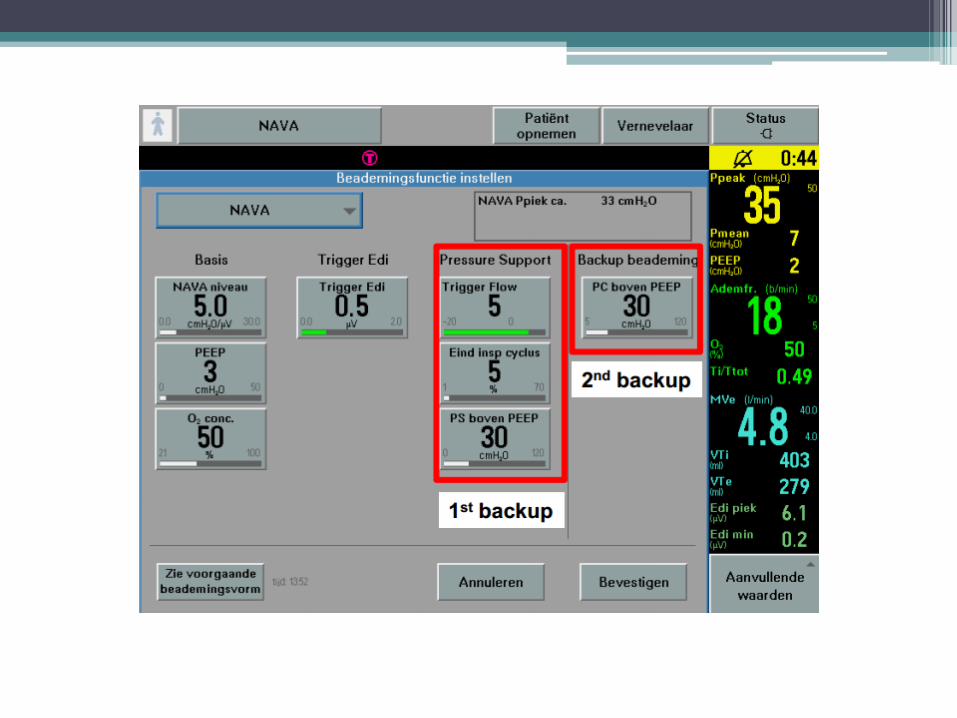
• Neurally Adjusted Ventilatory Assist (NAVA)
• High Frequency Oscillatory Ventilation (HFOV)
• High Frequency Jet Ventilation (HFJV)

Neurally Adjusted Ventilatory
Assist (NAVA)

Steps required to trigger ventilator

Problems with conventional ventilation
• Patient-ventilator asynchrony▫ Found in ¼ of patients on assisted ventilation▫ Mismatch between neural output and ventilator
inspiratory/expiratory times▫ Associated with longer duration of MV▫ Increased use of sedation, muscle relaxants
• Delivery of excess tidal volume and pressures▫ Increased risk of barotrauma and ventilator induced
lung injury (VILI)▫ Can result in ventilator induced diaphragm
dysfunction (VIDD)

NAVA device

Overview
• Assist mode of CMV
▫ Delivers pressure proportional to the integral of the electrical activity of the diaphragm (EAdi)
▫ Reflects neural output of respiratory center
• Vent triggered and cycled off based on EAdi
• PAW = NAVA level x EAdi
▫ PAW (cm H20) = Airway Pressure
▫ NAVA level (cm H2O/mV) – set by clinician


Mechanics
• Increase in EAdi triggers inspiratory effort• Decrease in EAdi terminates assistance▫ Delivery pressure is synchronous with diaphragm
activity▫ Vt is controlled by output of respiratory center
• Decreases inspiratory trigger delay▫ Caused by PEEPi, poor respiratory effort
• Decreases cycling off delay▫ Time from end of neural diaphragmatic input and
end of breath

NAVA monitoring – No synchronization

NAVA-triggered ventilation

Mechanics
• Protects against excess Vt and PAW
▫ Downregulation of EAdi in response to increasing vent assist levels
▫ As you increase EAdi, the patient will downregulate their neural output
• Hering-Breuer reflex▫ Pulmonary stretch receptors trigger action
potential in vagus nerve▫ Inhibits respiratory center in the medulla▫ Prolongs expiration

Drawbacks
• Body position, PEEP, and intra-abdominal pressure can alter position of diaphragm
▫ Requires adjustment after major changes in vent settings, clinical condition, and positioning
• Setting high NAVA levels can result in unstable breathing patterns
▫ High Vt, followed by periods of apnea
• Optimal method for setting EAdi trigger has not been determined

• 16 ventilated infants
▫ Age 2 days – 4 yrs (mean 9.7 mos), Mean wt 6.2 kg
• Given 30 min of PSV mode NAVA x 4 hrs
• NAVA mode:
▫ Improved synchrony
▫ 28% decrease in PIP at 30 min, 31% at 3 hrs
▫ 11% decrease in MAP, 9% at 3 hrs
▫ No significant change in pO2, pCO2

• 5 ventilated neonates, 25-29 wks
• NAVA x 4 hrs PCV x 4 hrs
▫ Cycle repeated 3 times
• Data collected every 30 min
• On NAVA
▫ Lower PIP, FiO2, RR, increased Vt
▫ Decreased pCO2, increased compliance
▫ No difference in MAP

High Frequency Oscillatory
Ventilation (HFOV)

Overview
• Uses pump-driven diaphragm
• Delivers small volumes at frequencies of 3-15 Hz
• Constant mean airway pressure (PMAW)
• High alveolar distention and recruitment
• Limit exposure to high ventilatory pressures
• Exhalation is active
Rouby J.J., Simonneau G., Benhamou D., et al: Factors influencing pulmonary volumes and CO2 elimination during high-frequency jet ventilation. Anesthesiology 1985; 63:473-482.


Mechanisms of Gas Exchange
• Different than conventional ventilation
• Normal RR 10-34 BPM
• Panting RR 240-300 BPM
▫ 5-6 Hz
Meyer M, Hahn G, Buess C, et al. Pulmonary gas exchange in panting dogs. J Appl Physiol 1989;66:1258–63.

Nichols, David G. (2012-07-11). Rogers' Textbook of Pediatric Intensive Care (Rogers Textbook of Pediatric Intensive Care) (Kindle Locations 23869-23870). Lippincot (Wolters Kluwer Health). Kindle Edition.
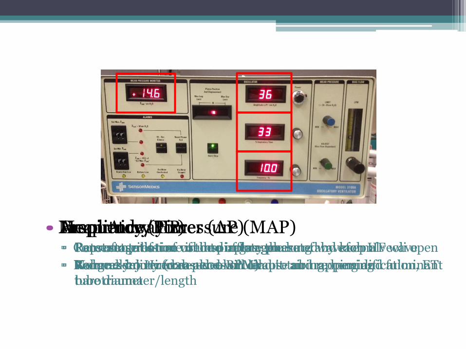
• Mean Airway Pressure (MAP)▫ Constant pressure used to inflate the lung and keep alveoli open
▫ Reduces injury from alveolar collapse and reopening
• Amplitude/Power (ΔP)▫ Representation of volume of gas generated by each HF wave
▫ Volume delivered depends on circuit tubing, humidification, ET tube diameter/length
• Inspiratory Time▫ Percentage of time in inspiratory phase of waveform
▫ Normally not increased – will lead to air trapping and fulminant barotrauma
• Frequency (Hz)▫ Rate of oscillation of the diaphragm
▫ Range 2-20 Hz (120-1200 BPM)

Initial Settings
• Frequency
▫ 15Hz – Premature infant, <2.5 kg
▫ 10Hz – Term infant 2.5-6kg
▫ 8Hz – Children 6-10kg
▫ 6Hz – Children >10kg
• Inspiratory time set to 33% (I:E = 1:2)
▫ Time increases with decreasing frequency
▫ 15 Hz = 22ms, 8Hz = 41ms, 6Hz = 55ms


Initial Settings
• MAP▫ Neonates: 2-4 cm above MAP on CMV▫ Infants/Children: 4-8 cm above MAP on CMV▫ If using HFOV first: MAP 8-10 cm (neonate) and
15-18 cm (infants/children)
• Amplitude (ΔP)▫ Adjust to vigorous chest wall to thigh wiggle (~24-
34 cm)▫ Titrate based on pCO2 (45-60)

Management
• Hypoxia▫ Check CXR to assess lung volume
Diaphragm should be @ ribs 9-10
▫ Increase MAP until adequate SaO2 achieved or lung is over-inflated
▫ Max MAP 40-45 cm▫ If SaO2 is adequate and lungs over inflated
Decrease MAP 1-2 cm every 2-4 hrs until volume normal
▫ If low SaO2 and over-inflated, can decrease frequency (will increase IT)


Management
• Ventilation (CO2)
▫ Alveolar ventilation (Ve) = Vt2 x freq
CMV: Ve = Vt x RR
▫ Primary regulation is through ΔP
▫ If high pCO2 at high ΔP
Decrease frequency
Creating a cuff leak can enhance CO2 clearance
▫ Increase IT to 50% as last resort

Weaning
• Oxygen
▫ Wean FiO2 first until <0.5-0.6 (unless hyperinflated)
▫ If FiO2 <0.5-0.6 (or hyperinflated)
Decrease MAP by 1 cm q4-q8h
If desats, increase MAP by 3-4, then wean more slowly
▫ When MAP is 8-16 with FiO2 < 0.4-0.5, can convert to CMV

Weaning
• Carbon Dioxide
▫ Decrease ΔP by 3cm per change until ΔP = 11-13
▫ Must maintain chest wall vibration
▫ If low PaCO2 on minimal amplitude
Decrease frequency to 10Hz and then 6Hz to decrease alveolar ventilation

• Multicenter RCT• 500 infants randomized to HFOV or SIMV• VLBW (601-1200g), <4 hrs old, 1 dose
surfactant, required ventilation (PEEP 6, FiO2 25%)
• HFOV patients:▫ Extubated earlier▫ 56% survived to 36 wks (vs 47%)▫ No increase in ICH, PVL, other complications

• Compiled RCTs comparing HFOV and CV in preterm or LBW infants with pulmonary dysfunction/RDS
• 17 studies, 3652 infants• No effect on mortality at 28-30d• Subgroup analysis: Reduced CLD with HFOV, no
surfactant, I:E of 1:2 on HFOV• Increased air leaks, grade III/IV IVH in HFOV group• Not recommended as initial vent strategy
HFOV as initial ventilation strategy

Trials in CDH
• Improved survival and lower incidence of CLD
• Better oxygenation and higher MAP with less barotrauma
• May decrease need for ECMO
• Study by CDH registry on pts with initial HFOV
▫ Increased rate of mortality and BPD

• Prospective, multicenter RCT• Enrollment criteria▫ Age > 34 weeks, antenatal diagnosis▫ No genetic/cardiac/renal/skeletal/CNS anomalies▫ Randomized to HFOV or CMV at birth
• Primary endpoints: BPD/death within 28 days• Secondary Endpoints:▫ Overall mortality, severity of BPD, days on
ventilator, VILI, pulmonary HTN, need for ECMO

High Frequency Jet Ventilation
(HFJV)

Overview
• Developed in the 1970s for gas exchange during tracheal procedures
• Flow interrupter – uses a pinch valve to generate a stream of high frequency pulses
• Gas propelled into lungs at high velocity▫ Sends gas via laminar and transitional flow down
core of the bronchial tree ▫ Minimizes effect of dead space
• Requires conventional vent in tandem▫ Generates PEEP and sigh breaths
Engle WA, Yoder MC, Andreoli SP, et al. Controlled, prospective, randomized comparison of high-frequency jet ventilation and conventional ventilation in neonates with respiratory failure and persistent pulmonary hypertension. J Perinatol 1997;17:3–9.

Overview
• Flow mechanics similar to HFOV
• Exhalation is passive

• Peak Inspiratory Pressure
▫ Max pressure of delivered gas by the jet
• PEEP
▫ Set by attached conventional vent
• ΔP
▫ PIP - PEEP
• Rate
▫ Set at intervals of 60
▫ Range 240-660 BPM
4-11Hz
• Inspiratory Time (IT)▫ Set at 20ms (0.02 sec)
▫ Increase can cause gas trapping and pneumothorax
• I:E Ratio
▫ Dependent on frequency
▫ At IT of 20ms
Rate 660 – I:E 1:3.5
Rate 420 – I:E 1:6
Rate 240 – I:E 1:12

Initial Settings
• Rate
▫ 420 BPM (7Hz) usual starting frequency in infants
▫ 360 BPM (6Hz) if air leaks/trapping
• IT
▫ Set at 20ms (0.02 sec)

Initial Settings
• PEEP
▫ Set by conventional vent
▫ 2-4 cm below MAP on CMV/HFOV
▫ 8 cm if starting on HFJV
▫ Titrate PEEP based on ability to oxygenate
• PIP
▫ Start at 2 cm below PIP on CMV
▫ MAP should equal that of CMV

Initial Settings
• Sigh breaths (on conventional ventilator)
▫ For alveolar recruitment
▫ Attach HFJV to conventional vent
▫ Rate of 3-4 BPM with PIP 6 above PEEP

Management
⇧ PEEP, Same PIP⇧ MAP⇩ ΔP
⇩ PIP, Same/⇧ PEEPSame MAP⇩ ΔP
⇩ PIP, Same/⇩ PEEP⇩ MAP⇩ ΔP
⇧ PIP, ⇧ PEEP⇧ MAPSame ΔP
⇩ PIP, ⇩ PEEP⇩ MAPSame ΔP
⇧ PIP, Same/⇧ PEEP⇧ MAP⇧ ΔP
⇧ PIP, Same PEEPSame MAP⇧ ΔP
Same PIP, ⇩ PEEP⇩ MAP⇧ ΔP

Important differences
• Active vs passive exhalation▫ Increased RR on HFJV increases CO2 clearance▫ Increased frequency on HFOV decreases CO2
clearance
• MAP can be controlled by▫ Changing PEEP or PIP
• Oxygenation can be adjusted via▫ PEEP▫ MAP▫ FiO2
• IT is fixed, and I:E changes with RR

• Multicenter RCT of 130 preterm infants
▫ 700 to 1500g
▫ All received surfactant
• No difference in mortality, ROP, air leak, severe IVH
• HFJV group less likely to need O2 at 36 weeks PMA or at discharge
Keszler M, Donn SM, Bucciarelli RL, et al. Multicenter controlled trial
comparing high-frequency jet ventilation and conventional mechanical
ventilation in newborn infants with pulmonary interstitial emphysema. J
Pediatr 1991; 119:85.

• RCT of 73 preterm infants
▫ 500g-2000g, 95% received surfactant
• No difference in air leak, need for O2 at 36 weeks PMA, duration of O2 therapy, LOS
• HFJV group more likely to have grade IV IVH, PVL, death (17 vs 7%)
Wiswell TE, Graziani LJ, Kornhauser MS, et al. High-frequency jet ventilation
in the early management of respiratory distress syndrome is associated with a
greater risk for adverse outcomes. Pediatrics 1996; 98:1035.

Future Advances
• Computer-directed closed loop systems for weaning
▫ Current extubation failure rate 14-24%
▫ NAVA may have a role in decreasing this
Farias JA, Alia I, Retta A, et al. An evaluation of extubation failure predictors in mechanically ventilated infants and children. Intensive Care Med 2002;28(6):752–7.
Strickland JH Jr, Hasson JH. A computer-controlled ventilator weaning system. Chest 1991;100:1096–9.

Questions?
Related Documents