Original article Non-protein-bound iron and 4-hydroxynonenal protein adducts in classic autism Alessandra Pecorelli a,1 , Silvia Leoncini a,1 , Claudio De Felice b,⇑ , Cinzia Signorini a , Cosimina Cerrone c , Giuseppe Valacchi d,e , Lucia Ciccoli a , Joussef Hayek f a Department of Pathophysiology, Experimental Medicine & Public Health, University of Siena, Siena, Italy b Neonatal Intensive Care Unit, University Hospital Azienda Ospedaliera Universitaria Senese (AOUS), Siena, Italy c Research, Training and Rehabilitation Institute (Istituto per la Ricerca, Formazione e Riabilitazione, I.Ri.Fo.R.), Siena, Italy d Department of Evolutionary Biology, University of Ferrara, Ferrara, Italy e Department of Food and Nutrition, Kyung Hee University, Seoul, South Korea f Child Neuropsychiatry Unit, University Hospital AOUS, Siena, Italy Received 6 December 2011; received in revised form 6 March 2012; accepted 21 March 2012 Abstract A link between oxidative stress and autism spectrum disorders (ASDs) remains controversial with opposing views on its role in the pathogenesis of the disease. We investigated for the first time the levels of non-protein-bound iron (NPBI), a pro-oxidant factor, and 4-hydroxynonenal protein adducts (4-HNE PAs), as a marker of lipid peroxidation-induced protein damage, in classic autism. Patients with classic autism (n = 20, mean age 12.0 ± 6.2 years) and healthy controls (n = 18, mean age 11.7 ± 6.5 years) were exam- ined. Intraerythrocyte and plasma NPBI were measured by high performance liquid chromatography (HPLC), and 4-HNE PAs in erythrocyte membranes and plasma were detected by Western blotting. The antioxidant defences were evaluated as erythrocyte glu- tathione (GSH) levels using a spectrophotometric assay. Intraerythrocyte and plasma NPBI levels were significantly increased (1.98- and 3.56-folds) in autistic patients, as compared to controls (p = 0.0019 and p < 0.0001, respectively); likewise, 4-HNE PAs were significantly higher in erythrocyte membranes and in plasma (1.58- and 1.6-folds, respectively) from autistic patients than controls (p = 0.0043 and p = 0.0001, respectively). Erythrocyte GSH was slightly decreased (10.34%) in patients compared to controls (p = 0.0215). Our findings indicate an impairment of the redox status in classic autism patients, with a consequent imbalance between oxidative stress and antioxidant defences. Increased levels of NPBI could contribute to lipid peroxidation and, conse- quently, to increased plasma and erythrocyte membranes 4-HNE PAs thus amplifying the oxidative damage, potentially contribut- ing to the autistic phenotype. Ó 2012 The Japanese Society of Child Neurology. Published by Elsevier B.V. All rights reserved. Keywords: Neurodevelopment disorders; Oxidative stress; Free iron; Lipid peroxidation; 4-hydroxynonenal protein adducts 1. Introduction Autistic spectrum disorders (ASDs) are a complex group of neurodevelopment disorders, still poorly understood and treatment-refractory. Specifically, ASDs refer to five different disorders characterized by delay in the development of multiple basic functions: classic autistic disorder (also referred as “classic autism”), a milder form known as Asperger syndrome, 0387-7604/$ - see front matter Ó 2012 The Japanese Society of Child Neurology. Published by Elsevier B.V. All rights reserved. http://dx.doi.org/10.1016/j.braindev.2012.03.011 ⇑ Corresponding author. Address: Neonatal Intensive Care Unit, University General Hospital, Azienda Ospedaliera Universitaria Senese, Viale M. Bracci, 16, I-53100 Siena, Italy. Tel.: +39 0577 586542; fax: +39 0577 234009. E-mail address: [email protected] (C. De Felice). 1 Equally contributing authors. www.elsevier.com/locate/braindev Brain & Development 35 (2013) 146–154

Welcome message from author
This document is posted to help you gain knowledge. Please leave a comment to let me know what you think about it! Share it to your friends and learn new things together.
Transcript
www.elsevier.com/locate/braindev
Brain & Development 35 (2013) 146–154
Original article
Non-protein-bound iron and 4-hydroxynonenal protein adductsin classic autism
Alessandra Pecorelli a,1, Silvia Leoncini a,1, Claudio De Felice b,⇑, Cinzia Signorini a,Cosimina Cerrone c, Giuseppe Valacchi d,e, Lucia Ciccoli a, Joussef Hayek f
a Department of Pathophysiology, Experimental Medicine & Public Health, University of Siena, Siena, Italyb Neonatal Intensive Care Unit, University Hospital Azienda Ospedaliera Universitaria Senese (AOUS), Siena, Italy
c Research, Training and Rehabilitation Institute (Istituto per la Ricerca, Formazione e Riabilitazione, I.Ri.Fo.R.), Siena, Italyd Department of Evolutionary Biology, University of Ferrara, Ferrara, Italy
e Department of Food and Nutrition, Kyung Hee University, Seoul, South Koreaf Child Neuropsychiatry Unit, University Hospital AOUS, Siena, Italy
Received 6 December 2011; received in revised form 6 March 2012; accepted 21 March 2012
Abstract
A link between oxidative stress and autism spectrum disorders (ASDs) remains controversial with opposing views on its role inthe pathogenesis of the disease. We investigated for the first time the levels of non-protein-bound iron (NPBI), a pro-oxidant factor,and 4-hydroxynonenal protein adducts (4-HNE PAs), as a marker of lipid peroxidation-induced protein damage, in classic autism.Patients with classic autism (n = 20, mean age 12.0 ± 6.2 years) and healthy controls (n = 18, mean age 11.7 ± 6.5 years) were exam-ined. Intraerythrocyte and plasma NPBI were measured by high performance liquid chromatography (HPLC), and 4-HNE PAs inerythrocyte membranes and plasma were detected by Western blotting. The antioxidant defences were evaluated as erythrocyte glu-tathione (GSH) levels using a spectrophotometric assay. Intraerythrocyte and plasma NPBI levels were significantly increased (1.98-and 3.56-folds) in autistic patients, as compared to controls (p = 0.0019 and p < 0.0001, respectively); likewise, 4-HNE PAs weresignificantly higher in erythrocyte membranes and in plasma (1.58- and 1.6-folds, respectively) from autistic patients than controls(p = 0.0043 and p = 0.0001, respectively). Erythrocyte GSH was slightly decreased (�10.34%) in patients compared to controls(p = 0.0215). Our findings indicate an impairment of the redox status in classic autism patients, with a consequent imbalancebetween oxidative stress and antioxidant defences. Increased levels of NPBI could contribute to lipid peroxidation and, conse-quently, to increased plasma and erythrocyte membranes 4-HNE PAs thus amplifying the oxidative damage, potentially contribut-ing to the autistic phenotype.� 2012 The Japanese Society of Child Neurology. Published by Elsevier B.V. All rights reserved.
Keywords: Neurodevelopment disorders; Oxidative stress; Free iron; Lipid peroxidation; 4-hydroxynonenal protein adducts
0387-7604/$ - see front matter � 2012 The Japanese Society of Child Neuro
http://dx.doi.org/10.1016/j.braindev.2012.03.011
⇑ Corresponding author. Address: Neonatal Intensive Care Unit,University General Hospital, Azienda Ospedaliera UniversitariaSenese, Viale M. Bracci, 16, I-53100 Siena, Italy. Tel.: +39 0577586542; fax: +39 0577 234009.
E-mail address: [email protected] (C. De Felice).1 Equally contributing authors.
1. Introduction
Autistic spectrum disorders (ASDs) are a complexgroup of neurodevelopment disorders, still poorlyunderstood and treatment-refractory. Specifically,ASDs refer to five different disorders characterized bydelay in the development of multiple basic functions:classic autistic disorder (also referred as “classicautism”), a milder form known as Asperger syndrome,
logy. Published by Elsevier B.V. All rights reserved.
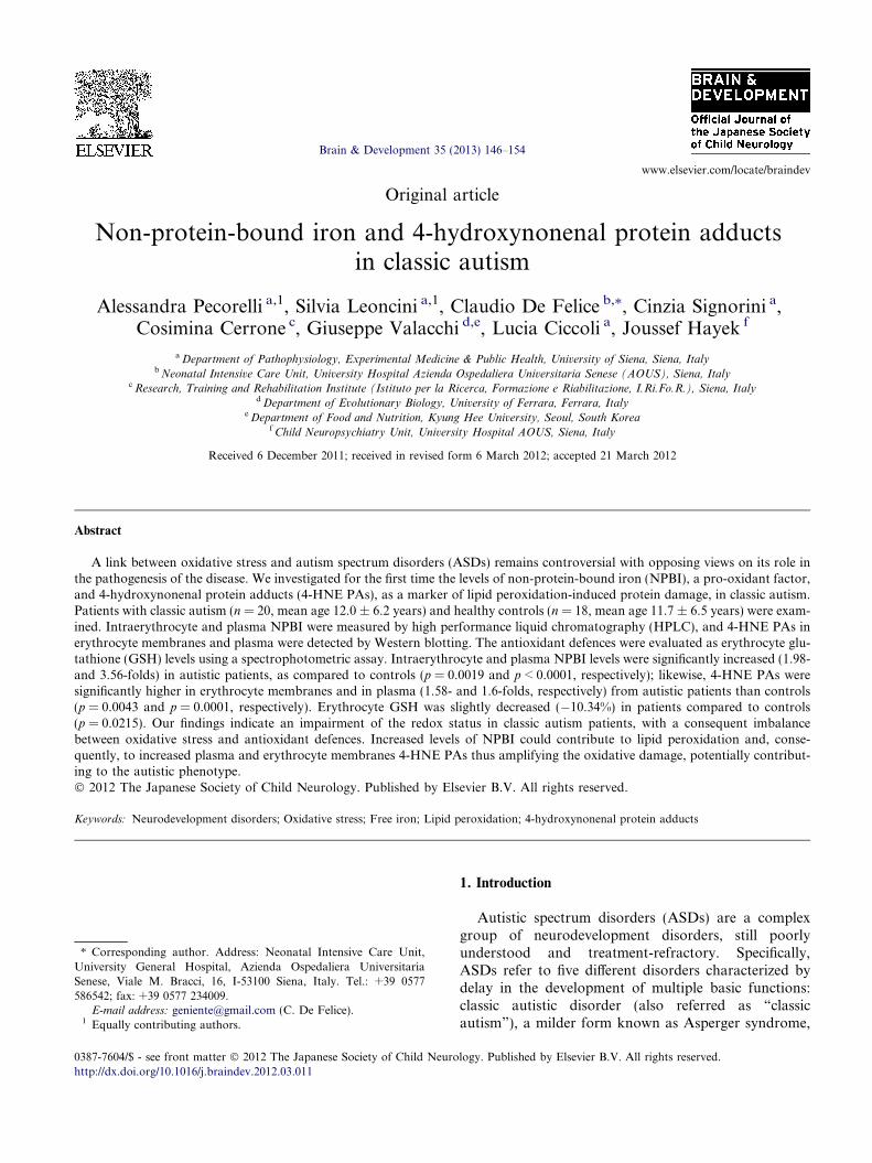
Table 1Oxidative stress markers and antioxidant defenses in ASDs: summary of the current knowledge.
Line missing
Symbols legend: " increased; M unchanged; # decreased. *Including also data from patients with Asperger syndrome and/or Pervasive DevelopmentDisorder not otherwise specified (PDD/NOS); **Review article.Legend: TAOS, total antioxidant status; GSH, reduced glutathione; GSSG, oxidized glutathione; GSH-Px, glutathione peroxidase; SOD, superoxidedismutase; NO, nitric oxide; MDA, malondialdheide; TBARS, thiobarbituric acid reactive substances; 8-OHdG, 8-hydroxy-20-deoxyguanosine.
A. Pecorelli et al. / Brain & Development 35 (2013) 146–154 147
the rare genetic condition known as Rett syndrome(RTT), childhood disintegrative disorder, and pervasivedevelopmental disorder not otherwise specified (PDD-NOS) [1].
The onset of clinical manifestation in ASDs is usuallywithin the 2nd year of life and mainly consists in thetriad (1) impairment in language, (2) social behaviourand (3) stereotyped or repetitive behaviour. ASDs areconsidered to be the result of a complex interactionbetween a genetic background and environmental fac-tors [2], and appear to be steadily increasing in fre-quency, according to some authors. In fact, autismdiagnosis has been reported to be rising by some countsfrom 1 in 5000 in the mid 1970s, to 1 in 110 in 2009 [3]. If
from one side, the rise in autistic patients can beexplained for ca. 50% of the observed growth becauseof increased awareness, diagnosis and social factors,the reasons for about 46% of the increase remainsunexplained.
Oxidative stress (OS) is a well-known pathogenicmechanism involved in several human pathologies. Bydefinition, OS occurs when the antioxidant response isinsufficient to balance the production of reactive oxygenspecies (ROS), potentially causing cell death by apopto-sis or necrosis via an array of signaling pathways. Brainis particularly vulnerable to ROS damage compared toother organs due to its high metabolic rate combinedwith a relatively low concentration of antioxidant
148 A. Pecorelli et al. / Brain & Development 35 (2013) 146–154
proteins. OS and mitochondrial dysfunction have beenimplicated in all major neurodegenerative disorders(i.e., amyotrophic lateral sclerosis, Parkinson’s and Alz-heimer’s disease) [4].
A potential relationship between OS and ASDs hasbeen repeatedly explored [5–23] (Table 1) and generallyfound to be related to either increased OS or alteredantioxidant defences, although this link remains highlycontroversial, with opposing views regarding its role inthe development of the autistic phenotype. In particular,at this time, it is unclear whether OS is a cause or con-sequence of autism [24], and the issue is further compli-cated by the heterogeneity of patients series (oftenmixing classic autism with Asperger syndrome and/orPDD-NOS; see Table 1 legend) and the employed OSbiomarkers.
Recently, we have shown increased OS marker levels,i.e., non-protein-bound iron (NPBI: free redox-activeiron) [25] and 4-hydroxynonenal protein adducts (4-HNE PAs) [26] in RTT, a particular form of ASDs thatbelongs to those small group of the autistic conditionsknown to be the result of a single gene mutation.
To date, no information exists regarding NPBI, apro-oxidant factor, and 4-HNE PAs, a biochemicalmarker of lipid peroxidation-induced protein damage,in classic autism.
NPBI acts as a pro-oxidant, causing release of hydro-xyl radicals as the results of the Fenton reaction [4]. Inturn, the hydroxyl radicals can initiate lipid peroxida-tion of polyunsaturated fatty acids (PUFA), producingseveral compounds of degradation, one of the moststudied is 4-HNE, which can covalently bind proteins,phospholipids, and DNA. This highly reactive aldehydereacts readily with nucleophilic groups of protein aminoacidic side chains, and its covalent attachment to pro-teins, lead to alteration in their structure and biologicalactivity [27]. Thus, 4-HNE, depending on its concentra-tion and location, may be considered as a “second toxicmessenger”, which disseminates and augments initialfree radical events.
In the present study, we evaluated the levels of NPBIand 4-HNE PAs in plasma and erythrocytes (RBCs) ofpatients with classic autistic disorder.
2. Subjects and methods
2.1. Subjects
A total of 20 patients with classic autistic disorder(mean age: 12.0 ± 6.2 years, range 4–30), as well as 18healthy controls of comparable age (mean age:11.7 ± 6.5 years, range 4–30) participated to the study.
All the patients were consecutively admitted to theChild Neuropsychiatry Unit of the University Hospitalof Siena (Head: J.H.). Patients with diagnosed RTT,X-fragile or tuberous sclerosis were excluded from the
present study. In addition, subjects with perinataladverse events and/or brain abnormalities on magneticresonance imaging (MRI) were excluded for the presentstudy. All the subjects were on a typical Mediterraneandiet.
None of the autistic patients had active epilepsy atthe time of blood sampling. A total of 4 patients(16%) experienced seizures early in the course of theirdisease, which were no longer present. In particular,none of the patients exhibited complications of epilepticseizures. Antiepileptic drug (AED) treatment (i.e., eithercarbamazepine or valproic acid), as monotherapy, wasassumed by 8 patients (32%) as mood stabilizers at anaverage dose of 10 mg/b.w./day.
The autistic patients were diagnosed by Diagnosticand Statistical Manual of mental disorders IV (DSM-IV) and evaluated using Autism Diagnostic ObservationSchedule (ADOS), and Autism Behaviour Checklist(ABC). Childhood Autism Rating Scale scores (CARS)[28] were calculated and Intelligence quotient (I.Q.) esti-mated in both the autistic and control group by an expe-rienced child neuropsychiatrist (J.H.). Blood samplingsin the control group were carried out, during routinehealth checks or blood donations, always followed bywritten informed consent. The study was conductedafter the approval by the Institutional Review Boardand all written informed consents were obtained fromeither the parents or the legal tutors of the enrolledpatients.
2.2. Blood sampling
All samplings were carried out around 8 AM afterovernight fasting. Blood was collected in heparinizedtubes and all manipulations were carried out within2 h after sample collection. The blood samples were cen-trifuged at 2400g for 15 min at 4 �C; the platelet poorplasma was saved and the buffy coat was removed byaspiration. RBCs were washed twice with physiologicalsolution (150 mM NaCl). An aliquot of packed erythro-cytes was resuspended in Ringer solution [125 mMNaCl, 5 mM KCl, 1 mM MgSO4, 32 mM N-2-hydroxy-ethylpiperazine-N-2-ethanesulfonic acid (HEPES),5 mM glucose, 1 mM CaCl2], pH 7.4 as a 50% (vol/vol) suspension for the determination of intraerythro-cyte NPBI. The remaining volume of packed erythro-cytes was used for erythrocyte membranespreparations (i.e., haemoglobin-free ghosts) for 4-HNEPAs determinations. Plasma was used for the NPBIassay and Western blotting.
2.3. Intraerythrocyte and plasma NPBI
Intraerythrocyte NPBI was determined as a desferri-oxamine (DFO)–iron complex (ferrioxamine), as previ-ously reported [25]. Briefly, 25 lM DFO was added to

A. Pecorelli et al. / Brain & Development 35 (2013) 146–154 149
the samples (1 ml erythrocyte suspension), the erythro-cytes were then lysed by adding water (1 vol) and freez-ing (�70 �C) and thawing. The hemolysate wasultrafiltered at 3373g for 30 min in centrifugal filterswith a 30-kDa molecular weight cut-off (VIVASPIN 4,Sartorius Stedim Biotech GmbH, Goettingen, Ger-many) and the ultrafiltrate was stored at �20 �C untilanalysis. The DFO excess was removed by silica (Silica-gel; 25–40 lm) column chromatography. The DFO–ironcomplex was determined by HPLC and the detectionwavelength was 229 nm. Plasma NPBI was determinedas reported for intraerythrocyte NPBI. DFO (25 lM)was added to plasma diluted (1:1; vol:vol) with Ringersolution, then the samples were ultrafiltered and storedat �20 �C until analysis as above. The calibration curvecorrelation for intraerythrocyte NPBI and plasma NPBIwas adequate (r2 = 0.994009), the minimum detectionlimit was 0.1 nmol/ml, and mean intra- and inter-obser-ver coefficients of variation of 62.5% and 65%,respectively.
2.4. Erythrocyte membrane preparation
An aliquot (600 ll) of packed RBC was lysed inDodge buffer, and erythrocyte membranes were pre-pared, according to Dodge [29], by repeated washinguntil the “ghosts” were pearly white. Samples were keptfrozen at �70 �C until used for sodium dodecyl sul-phate–polyacrylamide gel electrophoresis (SDS–PAGE).
2.5. Western blot for erythrocyte and plasma 4-HNE
protein adducts
Western blot protocols were performed as previ-ously described [26]. Erythrocyte membrane andplasma proteins (30 lg protein, determined using Bio-Rad protein assay; BioRad, Hercules, CA, USA) wereresolved on 4–20% SDS–PAGE gels (Lonza GroupLtd., Switzerland) and transferred onto a hybondECL nitrocellulose membrane (GE Healthcare EuropeGmbH, Milan, Italy). After blocking in 3% non-fatmilk (BioRad, Hercules, CA, USA), the membraneswere incubated overnight at 4 �C with goat polyclonalanti 4-HNE adduct antibody (cod. AB5605; MilliporeCorporation, Billerica, MA, USA). Following washesin TBS Tween and incubation with specific secondaryantibody (mouse anti-goat horseradish peroxidase-con-jugated, Santa Cruz Biotechnology Inc., CA, USA)for 1 h at RT, the membranes were incubated withECL reagents (BioRad, Hercules, CA, USA) for1 min. The bands were visualized by autoradiography.Quantification of the significant bands was performedby digitally scanning the Amersham Hyperfilme ECL(GE Healthcare Europe GmbH, Milan, Italy) andmeasuring immunoblotting image densities with Ima-geJ software.
2.6. Erythrocyte glutathione
Glutathione (GSH) content determinations in RBCswere carried out according to the spectrophotometricmethod of Beutler et al. [30]. After RBCs lysis and sub-sequent metaphosphoric acid protein precipitation, themethod involved GSH oxidation by sulfhydryl reagent5,50-dithiobis (2-nitrobenzoic acid) (DTNB) to formthe derivative 50-thio-2-nitrobenzoic acid (TNB), mea-surable at 412 nm.
2.7. Statistical analysis
Variables of interest were tested for normal distribu-tion (D’Agostino–Pearson test) and data are presentedeither as means ± SD or medians (inter-quartile range),as appropriate. As the variables tested were continuousnormally distributed, average values from autisticpatients and the control population were compared byusing independent-sample t test. Associations betweencontinuous parametric variables were tested using linearregression analysis. The MedCalc version 12.1.4 statisti-cal software package (MedCalc Software, Mariakerke,Belgium) was used, and two-tailed p values of less than0.05 were considered significant.
3. Results
3.1. CARS and IQ ranges
CARS scores were 50.3 ± 7.6 in the autistic patients vs.19.5 ± 2.4 in the control group (p < 0.0001). Estimated IQswere 30.0 ± 5.1 in the autistic patients and 97.5 ± 5.9 in thecontrol group (p < 0.0001). In the autistic group, CARSscore was positively related with chronological age[r = 0.7695 (0.5902 to 0.8765), p < 0.0001] and inverselycorrelated with IQ [r = �0.6773 (�0.8074 to �0.4843),p < 0.0001]. Likewise, in the autistic patients IQ was inver-sely related to age [r = �0.6195 (�0.7858 to �0.3697),p < 0.0001]. In contrast, no significant correlations betweenCARS or IQ and age were present in the control group.
3.2. Intraerythrocyte and plasma NPBI
The levels of intraerythrocyte NPBI was significantlyhigher in autistic patients as compared with the controlRBCs (p = 0.0019), with a ca. 1.98-fold increase(Fig. 1A). Likewise, an 3.56-fold increase in plasmaNPBI was detected in autistic patients as compared tohealthy controls (p < 0.0001) (Fig. 1B).
3.3. Erythrocyte membrane and plasma 4-HNE protein
adducts
4-HNE PAs were examined by Western blot analysisand the specificity of 4-HNE antibody was assessed by

Fig. 1. Non-protein-bound-iron (NPBI) in erythrocytes and plasma from patients with autism. (A) Intraerythrocyte NPBI in autistic patients(n = 20) and healthy controls (n = 18). Intraerythrocyte NPBI was expressed as nmol/ml packed erythrocytes. (B) Plasma NPBI in patients withautism (n = 20) and healthy controls (n = 18). Statistical significant differences were reported. The values are reported as means ± SD. *p = 0.0019significantly different from controls. **p < 0.0001 significantly different from healthy controls.
150 A. Pecorelli et al. / Brain & Development 35 (2013) 146–154
dot-blot analysis (data not shown), as previouslydescribed [26]. 4-HNE PAs signal was clearly increasedin the erythrocyte membranes of autistic patients respectto the healthy controls (p = 0.0043) (Fig. 2A). Quantifi-cation of the bands (Fig. 2C) shows that erythrocytemembrane 4-HNE PAs were significantly increased of1.58-folds. As illustrated in Fig. 2B and D, the trendshowed in the erythrocyte membrane 4-HNE PAs wasalso observed in the plasma of autistic patients (1.6-foldincrease; p = 0.0001 as compared to healthy controls).In addition, in Fig. 2A and B, it possible to appreciatethe physiological variability between the some represen-tative samples shown, demonstrating a certain grade of4-HNE PAs signal also in the healthy controls.
Fig. 2. Immunochemical detection of 4-hydroxynonenal protein adducts (4autistic patients. Panels A and B depict Western blot for 4-HNE PAs in erautistic patients (n = 20). Shown is a representative of four replicate experimHNE PAs bands, determined by densitometric analysis of the scanned imareported as medians (inter-quartile range). *p = 0.0043 significantly differentcontrols.
3.4. Intraerythrocyte GSH
Autistic patients showed slightly reduced erythrocyteGSH levels compared to those of the healthy controls(�10.34%; p = 0.0215) (Fig. 3).
3.5. Correlations between OS markers, erythrocyte GSH,
CARS and IQ values
We observed a positive correlation between intraery-throcyte NPBI and plasma NPBI levels [r = 0.7289(0.4998 to 0.8626), p < 0.0001]. We also observed a sig-nificant positive correlation between intraerythrocyteNPBI and erythrocyte membrane 4-HNE PAs
-HNE PAs) PAs in erythrocyte membrane and plasma samples fromythrocyte membranes and plasma from healthy controls (n = 18) andents. In the bottom panels (C and D) is shown the quantification of 4-ges. Average values were expressed as arbitrary units. The values arefrom healthy controls. **p = 0.0001 significantly different from healthy

Fig. 3. Erythrocyte glutathione (GSH) levels in patients with autism.The values are reported as means ± SD; *p = 0.0215 significantlydifferent from healthy controls.
A. Pecorelli et al. / Brain & Development 35 (2013) 146–154 151
[r = 0.7862 (0.1829 to 0.9594), p = 0.0207] and a trendfor the correlation between plasma NPBI and 4-HNEPAs [r = 0.5685 (�0.0079 to 0.8614) and p = 0.0537],whereas no significant correlations were observedbetween intraerythrocyte GSH and the examined OSbiomarkers (p P 0.5004, data not shown). No signifi-cant correlations were observed between the examinedOS biomarkers, erythrocyte GSH, CARS and IQ (clas-sic autism: r values range: �0.3185 to 0.2325; p valuesrange: 0.2889 to 0.9508; controls: r values range:�0.2869 to 0.2406; p values range: 0.4761 to 0.9378).
In addition, no statistical significant differences(p P 0.78) in the levels of the examined OS markerswere observed between the AED- treated and AED-untreated sub-populations.
4. Discussion
Our data showed a clear and significant increase ofthe evaluated OS markers along with a decrease oferythrocyte GSH in autistic patients, thus confirmingthat OS can play a role in the pathogenesis of autism.However, it is difficult to determine whether OS is a pri-mary contributor to the development of the disease orwhether it is a secondary consequence of the originalinsults that have been proposed to contribute to pheno-typical features of disorder. As increased NPBI and 4-HNE PAs have also been reported in RTT [25,26], agenetically determined condition in which autistic traitsare invariably present in the regression stage of the nat-ural progression of the disease, it is conceivable that, atleast partially, some common mechanisms are involvedin the development of the autistic phenotype. Theobserved decrease in the erythrocyte GSH content, indi-cating lower antioxidant defences, is in line with priorreports [6–12]. It is interesting to note that the inclusionor exclusion of autistic sub-types different from classicautistic disorder does not significantly change the cur-rent knowledge on the oxidative derangement previously
reported in autism [5–23] (Table 1). Taken together, thedata from the literature and our findings strongly sup-port the notion that OS may be involved in the patho-genesis of autism.
It is well established that redox-active iron is one ofthe most active sources of OS. In particular, iron is inti-mately linked to OS [31]. Several studies have estab-lished an involvement of disruption of iron regulationas an important etiological factor in neurodegenerativedisorders, such as Alzheimer’s disease, Parkinson’s dis-ease, Huntington’s disease and Friedreich’s ataxia [32].We have previously demonstrated that OS induces inRBCs iron release in a free redox active form accompa-nied by methemoglobin (Met-Hb) formation [33]. Ourresults show an increase in the intraerythrocyte NPBIlevels in autistic patients and, as part of free iron coulddiffuse out of RBCs [34], this fact may account also forthe observed increase of plasma NPBI. Indeed, the sig-nificant positive correlation observed between intraery-throcyte and plasma NPBI levels in our patients wouldsuggest this possibility.
NPBI causes a release of hydroxyl radical, an extre-mely powerful oxidizing species, which, in turn, is themain trigger for lipid peroxidation [4].
4-HNE PAs are to be considered not only a reliablemarker of OS in that, once the aldehyde forms theadducts to the proteins, they show a biological impactby likely changing protein function. The presence of 4-HNE PAs represents a consequence of lipid peroxida-tion and therefore plasma proteins can be considered atarget of increased OS status in classic autism. Theseirreversible protein modifications have been extensivelyinvestigated in research on neurological diseases, suchas Alzheimer’s disease, Huntington’s disease, amyotro-phic lateral sclerosis, and Parkinson’s disease [35] andalso on neurodevelopmental disorders, such as RTT[26].
Whether the enhanced OS observed in autisticpatients is exogenous or endogenous is, of course, stillunknown.
Several authors have suggested a link with environ-mental stressors [2]. This idea is supported by the epide-miologic evidence that exposure, near roadways, totraffic-related air pollutants, not only during pregnancybut also in early postnatal life, augments the geneticrisks implicated in autism aetiology [36]. The work byVolk et al. [37] showed higher incidence of autism inthe children living within 309 m of the freeway aroundthe time of birth. This could be a consequence of thepro-oxidant effect that air pollution has on central ner-vous system during developmental process, a period ofparticular vulnerability and susceptibility to any envi-ronmental insult for brain. For example, it is well dem-onstrated that diesel exhaust particles, benzo[a]pyreneand ozone could affect neuro-maturation in animalmodels through their induced oxidative damage [38,39].

152 A. Pecorelli et al. / Brain & Development 35 (2013) 146–154
Alternatively, as previously shown for RTT, it is pos-sible that OS detected in autistic patients derives froman endogenous source. In fact, our group showed thatthe probable origin of oxidant insult in RTT could bean impaired pulmonary gas exchange with a conse-quence chronic hypoxia condition [25]. Although thereare no solid data showing a clear relationship betweensystemic hypoxia and autism, it is possible that the sim-ilar phenomena observed in RTT occurs also in autisticpatients, at least during or after gestation, or locally incerebral tissue. For example, the work of Kolevzonet al. [40], on the basis of epidemiological evidence,has advanced the possibility that neonatal or foetalhypoxia are implicated in autism; while Burstyn et al.[41] has suggested a weak effect of foetal hypoxia on riskof autism. Furthermore, even if only at the brain level, ithas been shown that autism is characterized by a hypo-perfusion condition with the resulting brain hypoxia[42].
Our results show a statistically significant increasedlevels of erythrocyte membrane and plasma 4-HNEPAs in autistic patients as compared to their non-autis-tic siblings. In addition, we found a linear positive cor-relation between 4-HNE PAs and NPBI, although thiscorrelation was significant for RBCs, but not plasma.The reason of this discrepancy would be due to the dilut-ing factor of the high blood volume or to the limitednumber of patients.
Several factors, ultimately leading to enhanced OS inautism, may all be connected to increased levels of 4-HNE PAs, such as alterations in antioxidant enzymes,abnormal iron and copper metabolism, imbalance inhomocysteine and methionine metabolism, increasedxanthine oxidase activity, and mitochondrial dysfunc-tion [7].
Recent evidence indicates that the activity of the mainenzyme involved in the 4-HNE detoxification, which isglutathione-S-transferases (GST), is significantlydecreased in autistic RBCs [43], causing an impairedability to metabolize 4-HNE PAs. This condition mightlead to an increase in the susceptibility of plasma andRBCs membrane proteins to 4-HNE “attack”, explain-ing the increased 4-HNE PAs levels observed in autisticpatients.
Significant alterations in the fatty acid profiles in indi-viduals with ASDs has been reported, in both plasmaand membrane RBCs [44]. It is possible that pro-oxidantenvironment present in autistic patients could contributeto fatty acid deficiencies; particularly, lower arachidonicacid (AA) concentration recorded in children with ASDs[45] is consistent with the increased levels of 4-HNEPAs, as 4-HNE is a oxidation product generated, amongothers, from AA by a free radical initiated process.Moreover, NPBI-induced lipid peroxidation and theconsequent 4-HNE PAs formation could lead to the lossof membrane function and integrity in RBCs as
observed in a recent study, where the fluidity of theerythrocyte membrane of children with autism waslower than the controls [7].
The interest of the role of peroxidation in neurode-generative diseases and also in neurodevelopment disor-ders has been greatly increased due to the particularcomposition of brain. In fact, the brain is highly vulner-able to OS due to its limited antioxidant capacity, higherenergy requirement, and higher amounts of lipids andiron [4,31]. Therefore, neurons are more susceptible toOS since they are the first cells to be affected by theincrease in ROS and shortage of antioxidants. The issueof using biological samples other than the brain tissue orthe cerebrospinal fluid (CSF) for monitoring the redoxstatus of patients with a neurological disease, in whichthe OS is involved, is a long-standing one and a questionnot easy to solve. However, our OS prior studies in arare disease included in the ASDs group, i.e. RTT, indi-cate that blood may be considered a suitable biologicalsample in order to monitor an autistic disorder[23,25,26].
Taken together, these findings suggest that a systemicoxidant status, triggered and possibly amplified byNPBI release, may play a key role in the pathogenicmechanisms of classic autistic disorder. It is possiblethat a vicious cycle is activated where the presence ofNPBI induces peroxidation and subsequently, RBCsexposed to lipoperoxidation products, such as 4-HNE,are damaged and further iron is released fromhaemoglobin.
A critical question regards the issue of whetherincreased OS is a primary contributor or just a second-ary consequence of autism. In the lack of an universallyaccepted experimental model of autism it is currentlyimpossible to solve this puzzle at this time. Future inves-tigations in this animal models are certainly needed inorder to better dissect at the molecular level the relation-ship between OS damage and neurodevelopment. Nev-ertheless, our study, as well as many other reports ofthe literature, indicates a pro-oxidant/antioxidant bal-ance shift toward the pro-oxidant arm in classic autism,thus paving the way towards new therapeutical strate-gies aimed at modulating OS in these patients.
In conclusion, our data suggest that, whatever thesource, an oxidative-driven brain damage could play akey role in the pathogenesis of autism.
Funding
None.
Conflicts of interest
All authors declare that they have no conflicts ofinterest related to the present study.
A. Pecorelli et al. / Brain & Development 35 (2013) 146–154 153
Acknowledgments
This paper is dedicated to professional singer MatteoSetti (Reggio Emilia, Italy, official web site: http://www.matteosetti.it) as his precious collaboration sur-prisingly triggered our studies on hypoxia and oxidativestress in Rett syndrome, a genetic model of autism. Wethank Roberto Faleri (Central Medical Library, Univer-sity of Siena, Siena, Italy) for online bibliographicassistance.
References
[1] Kim YS, Leventhal BL, Koh YJ, Fombonne E, Laska E, Lim EC,et al. Prevalence of autism spectrum disorders in a totalpopulation sample. Am J Psychiatry 2011;168:904–12.
[2] Herbert MR. Contributions of the environment and environmen-tally vulnerable physiology to autism spectrum disorders. CurrOpin Neurol 2010;23:103–10.
[3] Weintraub K. The prevalence puzzle: autism counts. Nature2011;479:22–4.
[4] Halliwell B, Gutteridge JMC. Free radicals in biology andmedicine. 4th ed. Oxford: Oxford University Press; 2007.
[5] Parellada M, Moreno C, Mac-Dowell K, Leza JC, Giraldez M,Bailon C, et al. Plasma antioxidant capacity is reduced inAsperger syndrome. J Psychiatry Res 2011. http://dx.doi.org/10.1016/j.jpsychires.2011.10.004.
[6] Melnyk S, Fuchs GJ, Schulz E, Lopez M, Kahler SG, Fussell JJ,et al. Metabolic imbalance associated with methylation dysregu-lation and oxidative damage in children with autism. J AutismDev Disord 2011. http://dx.doi.org/10.1007/s10803-011-1260-7.
[7] Chauhan A, Chauhan V. Oxidative stress in autism. Pathophys-iology 2006;13:171–81.
[8] James SJ, Melnyk S, Jernigan S, Cleves MA, Halsted CH, WongDH, et al. Metabolic endophenotype and related genotypes areassociated with oxidative stress in children with autism. Am JMed Genet B Neuropsychiatr Genet 2006;141B:947–56.
[9] Al-Gadani Y, El-Ansary A, Attas O, Al-Ayadhi L. Metabolicbiomarkers related to oxidative stress and antioxidant status inSaudi autistic children. Clin Biochem 2009;42:1032–40.
[10] Adams JB, Audhya T, McDonough-Means S, Rubin RA, QuigD, Geis E, et al. Nutritional and metabolic status of children withautism vs. neurotypical children, and the association with autismseverity. Nutr Metab 2011;8:34.
[11] James SJ, Rose S, Melnyk S, Jernigan S, Blossom S, Pavliv O,et al. Cellular and mitochondrial glutathione redox imbalance inlymphoblastoid cells derived from children with autism. FASEB J2009;23:2374–83.
[12] Adams JB, Baral M, Geis E, Mitchell J, Ingram J, Hensley A,et al. The severity of autism is associated with toxic metal bodyburden and red blood cell glutathione levels. J Toxicol2009;2009:532640.
[13] Yorbik O, Sayal A, Akay C, Akbiyik DI, Sohmen T. Investiga-tion of antioxidant enzymes in children with autistic disorder.Prostaglandins Leukot Essent Fatty Acids 2002;67:341–3.
[14] Mostafa GA, El-Hadidi ES, Hewedi DH, Abdou MM. Oxidativestress in Egyptian children with autism: relation to autoimmunity.J Neuroimmunol 2010;219:114–8.
[15] Sogut S, Zoroglu SS, Ozyurt H, Yılmaz HR, Ozugurlu F, SivaslıE, et al. Changes in nitric oxide levels and antioxidant enzymeactivities may have a role in the pathophysiological mechanismsinvolved in autism. Clin Chim Acta 2003;331:111–7.
[16] Zoroglu SS, Armutcu F, Ozen S, Gurel A, Sivasli E, Yetkin O,et al. Increased oxidative stress and altered activities of erythro-
cyte free radical scavenging enzymes in autism. Eur ArchPsychiatry Clin Neurosci 2004;254:143–7.
[17] Bowers K, Li Q, Bressler J, Avramopoulos D, Newschaffer C,Fallin MD. Glutathione pathway gene variation and risk ofautism spectrum disorders. J Neurodev Disord 2011;3:132–43.
[18] Ming X, Johnson WG, Stenroos ES, Mars A, Lambert GH,Buyske S. Genetic variant of glutathione peroxidase 1 in autism.Brain Dev 2010;32:105–9.
[19] McGinnis WR. Oxidative stress in autism. Altern Ther HealthMed 2004;10:22–36.
[20] Essa MM, Guillemin GJ, Waly MI, Al-Sharbati MM, Al-FarsiYM, Hakkim FL, et al. Increased markers of oxidative stress inautistic children of the Sultanate of Oman. Biol Trace Elem Res2011. http://dx.doi.org/10.1007/s12011-011-9280-x.
[21] El-Ansary A, Al-Daihan S, Al-Dbass A, Al-Ayadhi L. Measure-ment of selected ions related to oxidative stress and energymetabolism in Saudi autistic children. Clin Biochem 2010;43:63–70.
[22] Meguid NA, Dardir AA, Abdel-Raouf ER, Hashish A. Evalua-tion of oxidative stress in autism: defective antioxidant enzymesand increased lipid peroxidation. Biol Trace Elem Res2011;143:58–65.
[23] Signorini C, De Felice C, Leoncini S, Giardini A, D’Esposito M,Filosa S, et al. F4-neuroprostanes mediate neurological severity inRett syndrome. Clin Chim Acta 2011;412:1399–406.
[24] Palmieri L, Persico AM. Mitochondrial dysfunction in autismspectrum disorders: cause or effect? Biochim Biophys Acta2010;1797:1130–7.
[25] De Felice C, Ciccoli L, Leoncini S, Signorini C, Rossi M,Vannuccini L, et al. Systemic oxidative stress in classic Rettsyndrome. Free Radic Biol Med 2009;47:440–8.
[26] Pecorelli A, Ciccoli L, Signorini C, Leoncini S, Giardini A,D’Esposito M, et al. Increased levels of 4HNE-protein plasmaadducts in Rett syndrome. Clin Biochem 2011;44:368–71.
[27] Dubinina EE, Dadali VA. Role of 4-hydroxy-trans-2-nonenal incell functions. Biochemistry 2010;75:1069–87.
[28] Schopler E, Reichler RJ, DeVellis RF, Daly K. Toward objectiveclassification of childhood autism: Childhood Autism RatingScale (CARS). J Autism Dev Disord 1980;10:91–103.
[29] Dodge JT, Mitchell C, Hanahan DJ. The preparation andchemical characteristics of hemoglobin-free ghosts of humanerythrocytes. Arch Biochem Biophys 1963;100:119–30.
[30] Beutler M, Duron O, Kelly BM. Improved method for thedetermination of blood glutathione. J Lab Clin Med1963;61:882–8.
[31] Papanikolaou G, Pantopoulos K. Iron metabolism and toxicity.Toxicol Appl Pharmacol 2005;202:199–211.
[32] Jomova K, Vondrakova D, Lawson M, Valko M. Metals,oxidative stress and neurodegenerative disorders. Mol CellBiochem 2010;345:91–104.
[33] Comporti M, Signorini C, Buonocore G, Ciccoli L. Iron release,oxidative stress and erythrocyte ageing. Free Radic Biol Med2002;32:568–76.
[34] Ciccoli L, Rossi V, Leoncini S, Signorini C, Blanco-Garcia J,Aldinucci C, et al. Iron release, superoxide production andbinding of autologous IgG to band 3 dimers in newborn and adulterythrocytes exposed to hypoxia and hypoxia–reoxygenation.Biochim Biophys Acta 2004;1672:203–13.
[35] Gueraud F, Atalay M, Bresgen N, Cipak A, Eckl PM, Huc L,et al. Chemistry and biochemistry of lipid peroxidation products.Free Radic Res 2010;44:1098–124.
[36] Guxens M, Aguilera I, Ballester F, Estarlich M, Fernandez-Somoano A, Lertxundi A, et al. Prenatal exposure to residential air pollu -tion and infant mental development: modulation by antioxidants anddetoxification factors. Environ Health Perspect 2012;120:144–9.
[37] Volk HE, Hertz-Picciotto I, Delwiche L, Lurmann F, McConnellR. Residential proximity to freeways and autism in the CHARGEstudy. Environ Health Perspect 2011;119:873–7.
154 A. Pecorelli et al. / Brain & Development 35 (2013) 146–154
[38] Hougaard KS, Jensen KA, Nordly P, Taxvig C, Vogel U, SaberAT, et al. Effects of prenatal exposure to diesel exhaust particleson postnatal development, behavior, genotoxicity and inflamma-tion in mice. Part Fibre Toxicol 2008;5:3.
[39] Gonzalez-Pina R, Escalante-Membrillo C, Alfaro-Rodriguez A,Gonzalez-Maciel A. Prenatal exposure to ozone disrupts cerebel-lar monoamine contents in newborn rats. Neurochem Res2008;33:912–8.
[40] Kolevzon A, Gross R, Reichenberg A. Prenatal and perinatal riskfactors for autism: a review and integration of findings. ArchPediatr Adolesc Med 2007;161:326–33.
[41] Burstyn I, Wang X, Yasui Y, Sithole F, Zwaigenbaum L. Autismspectrum disorders and fetal hypoxia in a population-based
cohort: accounting for missing exposures via estimation–maximi-zation algorithm. BMC Med Res Methodol 2011;11:2.
[42] Ichim TE, Solano F, Glenn E, Morales F, Smith L, Zabrecky G,et al. Stem cell therapy for autism. J Transl Med 2007;5:30.
[43] Hermawati D, Then SM, Winarni TI, Faradz SMH, Jamal R. Lowererythrocyte GST activity in autism spectrum disorder (ASD) patientscompared to normal controls. Asia-Pacific J Mol Med 2011;1:2.
[44] Brown CM, Austin DW. Autistic disorder and phospholipids: areview. Prostaglandins Leukot Essent Fatty Acids 2011;84:25–30.
[45] El-Ansary AK, Bacha AGB, Al-Ayahdi LY. Impaired plasmaphospholipids and relative amounts of essential polyunsaturatedfatty acids in autistic patients from Saudi Arabia. Lipids HealthDis 2011;10:63.
Related Documents