NMR Structure of Escherichia coli Glutaredoxin 3- Glutathione Mixed Disulfide Complex: Implications for the Enzymatic Mechanism Kerstin Nordstrand 1 , Fredrik A ˚ slund 1,2 , Arne Holmgren 1,2 Gottfried Otting 1 and Kurt D. Berndt 1,2 * 1 Department of Medical Biochemistry and Biophysics and 2 Medical Nobel Institute for Biochemistry, Karolinska Institute, S-171 77, Stockholm Sweden Glutaredoxins (Grxs) catalyze reversible oxidation/reduction of protein disulfide groups and glutathione-containing mixed disulfide groups via an active site Grx-glutathione mixed disulfide (Grx-SG) intermediate. The NMR solution structure of the Escherichia coli Grx3 mixed disulfide with glutathione (Grx3-SG) was determined using a C14S mutant which traps this intermediate in the redox reaction. The structure contains a thio- redoxin fold, with a well-defined binding site for glutathione which involves two intermolecular backbone-backbone hydrogen bonds forming an antiparallel intermolecular b-bridge between the protein and gluta- thione. The solution structure of E. coli Grx3-SG also suggests a binding site for a second glutathione in the reduction of the Grx3-SG intermedi- ate, which is consistent with the specificity of reduction observed in Grxs. Molecular details of the structure in relation to the stability of the intermediate and the activity of Grx3 as a reductant of glutathione mixed disulfide groups are discussed. A comparison of glutathione binding in Grx3-SG and ligand binding in other members of the thioredoxin super- family is presented, which illustrates the highly conserved intermolecular interactions in this protein family. # 1999 Academic Press Keywords: glutaredoxin; glutathione; thioredoxin superfamily; NMR structure; thiol-disulfide oxidoreductase *Corresponding author Introduction Glutaredoxins (Grxs) are ubiquitous redox active proteins present in most organisms from bacteria to man. In cells, Grx can support the synthesis of deoxyribonucleotides by providing the essential enzyme ribonucleotide reductase (RR) with reducing equivalents, in a reaction coupled to oxidation of glutathione (GSH; A ˚ slund et al., 1994; Holmgren, 1976, 1979a). In this reac- tion Grx acts as a dithiol, where the exposed N- terminal cysteine in the active site sequence Cys- Pro-Tyr-Cys initiates attack on the substrate dis- ulfide (Figure 1), followed by an intramolecular Present addresses: F. A ˚ slund, Department of Microbiology and Molecular Genetics, Harvard Medical School, Boston, MA 02115, USA; K. D. Berndt, Center of Structural Biochemistry, Department of Biosciences, Ha ¨ lsova ¨gen 7, Karolinska Institute, NOVUM, S-141 57 Huddinge, Sweden. Abbreviations used: Cys GS , cysteine of glutathione; Gly GS , glycine of glutathione; gGlu GS , g-glutamic acid of glutathione; Grx, glutaredoxin; Grx1, glutaredoxin 1 from E. coli; Grx2, glutaredoxin 2 from E. coli; Grx3, glutaredoxin 3 from E. coli; Grx-SG, glutaredoxin-glutathione mixed disulfide; Grx1-SG, the mixed disulfide between E. coli glutaredoxin 1 (C14S) and glutathione; Grx3-SG, the mixed disulfide between E. coli glutaredoxin 3 (C14S-C65Y) and glutathione; GSH, reduced glutathione; GSSG, glutathione disulfide; GST, glutathione S-transferase; HED, 2-hydroxyethyl disulfide; HSQC, heteronuclear single quantum coherence; NOE, nuclear Overhauser effect; NOESY, NOE spectroscopy; PDI, protein disulfide isomerase; RR, ribonucleotide reductase; RMSD, root-mean-square deviation; TOCSY, total correlation spectroscopy; Trx, thioredoxin; 3 J HaHN , three-bond coupling constant between the a proton and the amide proton; 3 J HaHb , three-bond coupling constant between the a proton and an H b proton; 3 J NHb , three-bond coupling constant between the amide nitrogen atom and an H b proton; E 00 , difference in apparent standard state redox potential; COSY, correlated spectroscopy. E-mail address of the corresponding author: [email protected] Article No. jmbi.1998.2444 available online at http://www.idealibrary.com on J. Mol. Biol. (1999) 286, 541–552 0022-2836/99/070541–12 $30.00/0 # 1999 Academic Press

Welcome message from author
This document is posted to help you gain knowledge. Please leave a comment to let me know what you think about it! Share it to your friends and learn new things together.
Transcript
NMR Structure of Escherichia coli Glutaredoxin 3-Glutathione Mixed Disulfide Complex: Implicationsfor the Enzymatic Mechanism
Kerstin Nordstrand1, Fredrik AÊ slund1,2, Arne Holmgren1,2
Gottfried Otting1 and Kurt D. Berndt1,2*
1Department of MedicalBiochemistry and Biophysicsand 2Medical Nobel Institutefor Biochemistry, KarolinskaInstitute, S-171 77, StockholmSweden
Glutaredoxins (Grxs) catalyze reversible oxidation/reduction of proteindisul®de groups and glutathione-containing mixed disul®de groups viaan active site Grx-glutathione mixed disul®de (Grx-SG) intermediate. TheNMR solution structure of the Escherichia coli Grx3 mixed disul®de withglutathione (Grx3-SG) was determined using a C14S mutant which trapsthis intermediate in the redox reaction. The structure contains a thio-redoxin fold, with a well-de®ned binding site for glutathione whichinvolves two intermolecular backbone-backbone hydrogen bonds formingan antiparallel intermolecular b-bridge between the protein and gluta-thione. The solution structure of E. coli Grx3-SG also suggests a bindingsite for a second glutathione in the reduction of the Grx3-SG intermedi-ate, which is consistent with the speci®city of reduction observed inGrxs. Molecular details of the structure in relation to the stability of theintermediate and the activity of Grx3 as a reductant of glutathione mixeddisul®de groups are discussed. A comparison of glutathione binding inGrx3-SG and ligand binding in other members of the thioredoxin super-family is presented, which illustrates the highly conserved intermolecularinteractions in this protein family.
# 1999 Academic Press
Keywords: glutaredoxin; glutathione; thioredoxin superfamily; NMRstructure; thiol-disul®de oxidoreductase*Corresponding author
Introduction
Glutaredoxins (Grxs) are ubiquitous redoxactive proteins present in most organisms frombacteria to man. In cells, Grx can support thesynthesis of deoxyribonucleotides by providingthe essential enzyme ribonucleotide reductase
(RR) with reducing equivalents, in a reactioncoupled to oxidation of glutathione (GSH; AÊ slundet al., 1994; Holmgren, 1976, 1979a). In this reac-tion Grx acts as a dithiol, where the exposed N-terminal cysteine in the active site sequence Cys-Pro-Tyr-Cys initiates attack on the substrate dis-ul®de (Figure 1), followed by an intramolecular
Present addresses: F. AÊ slund, Department of Microbiology and Molecular Genetics, Harvard Medical School,Boston, MA 02115, USA; K. D. Berndt, Center of Structural Biochemistry, Department of Biosciences, HaÈ lsovaÈgen 7,Karolinska Institute, NOVUM, S-141 57 Huddinge, Sweden.
Abbreviations used: CysGS, cysteine of glutathione; GlyGS, glycine of glutathione; gGluGS, g-glutamic acid ofglutathione; Grx, glutaredoxin; Grx1, glutaredoxin 1 from E. coli; Grx2, glutaredoxin 2 from E. coli; Grx3, glutaredoxin3 from E. coli; Grx-SG, glutaredoxin-glutathione mixed disul®de; Grx1-SG, the mixed disul®de between E. coliglutaredoxin 1 (C14S) and glutathione; Grx3-SG, the mixed disul®de between E. coli glutaredoxin 3 (C14S-C65Y) andglutathione; GSH, reduced glutathione; GSSG, glutathione disul®de; GST, glutathione S-transferase; HED,2-hydroxyethyl disul®de; HSQC, heteronuclear single quantum coherence; NOE, nuclear Overhauser effect; NOESY,NOE spectroscopy; PDI, protein disul®de isomerase; RR, ribonucleotide reductase; RMSD, root-mean-squaredeviation; TOCSY, total correlation spectroscopy; Trx, thioredoxin; 3JHaHN, three-bond coupling constant between thea proton and the amide proton; 3JHaHb, three-bond coupling constant between the a proton and an Hb proton; 3JNHb,three-bond coupling constant between the amide nitrogen atom and an Hb proton; �E00, difference in apparentstandard state redox potential; COSY, correlated spectroscopy.
E-mail address of the corresponding author: [email protected]
Article No. jmbi.1998.2444 available online at http://www.idealibrary.com on J. Mol. Biol. (1999) 286, 541±552
0022-2836/99/070541±12 $30.00/0 # 1999 Academic Press
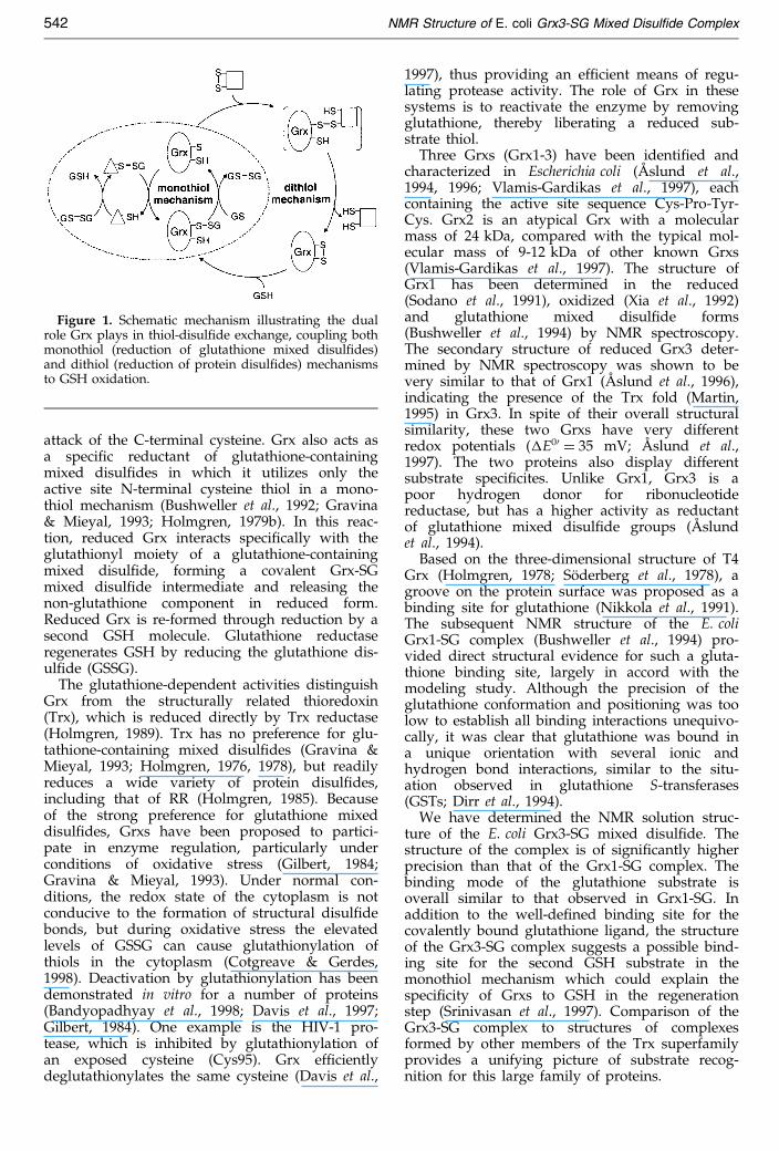
attack of the C-terminal cysteine. Grx also acts asa speci®c reductant of glutathione-containingmixed disul®des in which it utilizes only theactive site N-terminal cysteine thiol in a mono-thiol mechanism (Bushweller et al., 1992; Gravina& Mieyal, 1993; Holmgren, 1979b). In this reac-tion, reduced Grx interacts speci®cally with theglutathionyl moiety of a glutathione-containingmixed disul®de, forming a covalent Grx-SGmixed disul®de intermediate and releasing thenon-glutathione component in reduced form.Reduced Grx is re-formed through reduction by asecond GSH molecule. Glutathione reductaseregenerates GSH by reducing the glutathione dis-ul®de (GSSG).
The glutathione-dependent activities distinguishGrx from the structurally related thioredoxin(Trx), which is reduced directly by Trx reductase(Holmgren, 1989). Trx has no preference for glu-tathione-containing mixed disul®des (Gravina &Mieyal, 1993; Holmgren, 1976, 1978), but readilyreduces a wide variety of protein disul®des,including that of RR (Holmgren, 1985). Becauseof the strong preference for glutathione mixeddisul®des, Grxs have been proposed to partici-pate in enzyme regulation, particularly underconditions of oxidative stress (Gilbert, 1984;Gravina & Mieyal, 1993). Under normal con-ditions, the redox state of the cytoplasm is notconducive to the formation of structural disul®debonds, but during oxidative stress the elevatedlevels of GSSG can cause glutathionylation ofthiols in the cytoplasm (Cotgreave & Gerdes,1998). Deactivation by glutathionylation has beendemonstrated in vitro for a number of proteins(Bandyopadhyay et al., 1998; Davis et al., 1997;Gilbert, 1984). One example is the HIV-1 pro-tease, which is inhibited by glutathionylation ofan exposed cysteine (Cys95). Grx ef®cientlydeglutathionylates the same cysteine (Davis et al.,
1997), thus providing an ef®cient means of regu-lating protease activity. The role of Grx in thesesystems is to reactivate the enzyme by removingglutathione, thereby liberating a reduced sub-strate thiol.
Three Grxs (Grx1-3) have been identi®ed andcharacterized in Escherichia coli (AÊ slund et al.,1994, 1996; Vlamis-Gardikas et al., 1997), eachcontaining the active site sequence Cys-Pro-Tyr-Cys. Grx2 is an atypical Grx with a molecularmass of 24 kDa, compared with the typical mol-ecular mass of 9-12 kDa of other known Grxs(Vlamis-Gardikas et al., 1997). The structure ofGrx1 has been determined in the reduced(Sodano et al., 1991), oxidized (Xia et al., 1992)and glutathione mixed disul®de forms(Bushweller et al., 1994) by NMR spectroscopy.The secondary structure of reduced Grx3 deter-mined by NMR spectroscopy was shown to bevery similar to that of Grx1 (AÊ slund et al., 1996),indicating the presence of the Trx fold (Martin,1995) in Grx3. In spite of their overall structuralsimilarity, these two Grxs have very differentredox potentials (�E00 � 35 mV; AÊ slund et al.,1997). The two proteins also display differentsubstrate speci®cites. Unlike Grx1, Grx3 is apoor hydrogen donor for ribonucleotidereductase, but has a higher activity as reductantof glutathione mixed disul®de groups (AÊ slundet al., 1994).
Based on the three-dimensional structure of T4Grx (Holmgren, 1978; SoÈderberg et al., 1978), agroove on the protein surface was proposed as abinding site for glutathione (Nikkola et al., 1991).The subsequent NMR structure of the E. coliGrx1-SG complex (Bushweller et al., 1994) pro-vided direct structural evidence for such a gluta-thione binding site, largely in accord with themodeling study. Although the precision of theglutathione conformation and positioning was toolow to establish all binding interactions unequivo-cally, it was clear that glutathione was bound ina unique orientation with several ionic andhydrogen bond interactions, similar to the situ-ation observed in glutathione S-transferases(GSTs; Dirr et al., 1994).
We have determined the NMR solution struc-ture of the E. coli Grx3-SG mixed disul®de. Thestructure of the complex is of signi®cantly higherprecision than that of the Grx1-SG complex. Thebinding mode of the glutathione substrate isoverall similar to that observed in Grx1-SG. Inaddition to the well-de®ned binding site for thecovalently bound glutathione ligand, the structureof the Grx3-SG complex suggests a possible bind-ing site for the second GSH substrate in themonothiol mechanism which could explain thespeci®city of Grxs to GSH in the regenerationstep (Srinivasan et al., 1997). Comparison of theGrx3-SG complex to structures of complexesformed by other members of the Trx superfamilyprovides a unifying picture of substrate recog-nition for this large family of proteins.
Figure 1. Schematic mechanism illustrating the dualrole Grx plays in thiol-disul®de exchange, coupling bothmonothiol (reduction of glutathione mixed disul®des)and dithiol (reduction of protein disul®des) mechanismsto GSH oxidation.
542 NMR Structure of E. coli Grx3-SG Mixed Disul®de Complex

Results
Enzymatic activity
Wild-type Grx3 contains a third cysteine (C65) inaddition to the two active site redox active cysteineresidues (Figure 2). Since this additional cysteinecan form either intermolecular disul®de-bondedoligomers or glutathione mixed disul®de bondsupon oxidation with GSSG, it was replaced usingsite-directed mutagenesis. In most Grxs, the aminoacid in the homologous position is either cysteine,tyrosine, or phenylalanine, leading to our choice oftyrosine for replacement. In order to trap the Grx3-SG mixed disul®de quantitatively, the C-terminalactive site cysteine was mutated to serine (C14S).The activity of these mutants as reductants of glu-tathione mixed disul®des and as reductants ofribonucleotide reductase is shown in Table 1. TheC65Y mutation had essentially no effect on gluta-thione mixed disul®de activity as measured by theHED assay (see Materials and Methods). The C14Sand C14S/C65Y mutants retained 36 and 31 % ofthe activity as glutathione mixed disul®de reduc-tants, respectively, as compared with the wild-typeand C65Y mutants. These values are very similarto the 33 % activity reported for the C14S mutantof Grx1 (Bushweller et al., 1992). The activities ofthe Grx3 C14S and C14S/C65Y mutants as reduc-tants of ribonucleotide reductase were dramaticallyreduced, as also found for the analogous C14Smutant of Grx1, indicating that a dithiol mechan-ism is required for this reaction.
Structure determination
The structure of the E. coli Grx3-SG complex wasdetermined using the Grx3 double mutant C14S/
C65Y. Based on virtually complete 1H and 15Nassignments, 1167 distance constraints werederived from NOESY (nuclear Overhauser effectspectroscopy) spectra (Table 2), including 21 inter-molecular distance constraints between Grx3 andglutathione. A total of 63 3JHNHa coupling constantswere measured to derive dihedral angle con-straints. Coupling constants 3JHaHb and 3JNHb pro-vided unambiguous stereospeci®c assignmentsfor 17 b-methylene protons. Additionally, the fol-lowing torsion angles were restricted based on aqualitative evaluation of COSY (correlated spec-troscopy) and NOESY cross-peaks: w2 of Ser14 andg-GluGS, and w3 of Arg49. Stereospeci®c assign-ments for methyl groups of two leucine residues,20 and 31, were adopted from data for the oxi-dized form of Grx3 which has very similar 1H and15N chemical shifts, except for the active site region(A. SandstroÈm, F.AÊ ., A.H., G.O. & K.D.B., unpub-lished results). The chemical shifts for these twoleucine residues were identical in the two datasets. Further stereospeci®c assignments wereobtained during the course of re®nement. In the®nal input, 35 of 47 non-degenerate b-methyleneprotons, eight of 20 non-degenerate g-methyleneprotons, three of seven non-degenerate d-methyl-ene protons, as well as all the valine and six ofseven leucine methyl groups, and all asparagineand glutamine d and e-amino protons, respectively,were stereospeci®cally assigned.
The structure of the Grx3-SG complex was calcu-lated using the program DYANA, starting from100 random conformations. The 20 conformerswith the lowest target functions (<0.58 AÊ 2) wereselected for energy re®nement. The ensemble of 20energy-re®ned conformers satis®es the experimen-
Figure 2. Amino acid sequencealignment of Grx3 and selectedGrxs. Sequences are as describedby aAÊ slund et al. (1996), bHoÈoÈget al. (1983), cSjoÈberg & Holmgren(1972), dYang et al. (1989),ePadilla et al. (1995), fMcFarlanet al., 1992). The amino acidsequences were aligned based onthe three-dimensional structuresof pig Grx (Katti et al., 1995), T4Grx (SoÈderberg et al., 1978),E. coli Grx1 (Bushweller et al.,1994) and E. coli Grx3 (thiswork), and the secondary struc-ture of human Grx (Sun et al.,1997). The residue numberingrefers to Grx3. Conserved activesite residues are highlighted(light). Charged residues impli-cated in glutathione binding arealso highlighted (dark). Segmentsof helix (thin) and strand(thick) identi®ed by PROMOTIF(Hutchinson & Thornton, 1996)are underlined.
NMR Structure of E. coli Grx3-SG Mixed Disul®de Complex 543

tal input data well (Table 2), and is chosen to rep-resent the NMR structure of the Grx3-SG complex.
The root-mean-square deviation (RMSD) of pro-tein backbone atoms (N, Ca, C0) of the ®nal set of20 conformers (Figure 3(a)) is 0.57 AÊ relative to themean structure. The RMSD of the glutathionebackbone atoms (Ca, Cb, Cg, Cd of g-GluGS and N,Ca, C0 of CysGS and GlyGS) is 1.10 AÊ when the pro-tein backbone atoms are superimposed. ThisRMSD decreases to 0.40 AÊ when superimposingthe glutathione backbone atoms (Figure 3(b)). Thisimplies that although the conformation of the glu-tathione is as precisely de®ned as the polypeptidebackbone, there is a signi®cant amount of variabil-ity in the orientation between the protein and theligand. In a Ramachandran plot, 86 % of the non-glycine residues are located within the mostfavored regions, with 13 % in the additionallyallowed regions, and 1 % in the generously allowedregion, using the criteria of the program PRO-CHECK (Laskowski et al., 1996).
Description of the structure
Grx3 forms a three-layer aba sandwich, consist-ing of a bab and a bba motif (b1a1b2, b3b4a3)with a connecting helix (a2). Additionally, the Cterminus forms a short helix (a4) (Figure 4). The
same basic fold is found not only in other thioldisul®de oxidoreductases, Grxs, Trxs, protein dis-ul®de isomerases (PDI) and DsbA, but also in theglutathione-binding domain of the GSTs and theglutathione peroxidases (Martin, 1995). Residuesidenti®ed in a-helical segments in the Grx3-SGcomplex are: 11-24, 37-48, 64-74 and 76-81. Helix3 ends in a classical Gly-based Schellman cappingmotif (Milner-White, 1988) with hydrogen bondsbetween Gly75 HN/Asp72 O0 and Gly76 HN/Leu71 O0, and average values of f and c forGly75 of 80 � and 41 �, respectively. The proteinhas a central four-stranded mixed b-sheet contain-ing residues 2-7, 29-33, 54-58 and 60-62, where b2
runs parallel with b1 and strands b3 and b4 forma b-hairpin motif. The peptide bond precedingPro53 was identi®ed as cis from the large NOEbetween Val52 Ha and Pro53 Ha, and the absenceof NOEs between Val52 Ha and Pro53 Hd2/d3,and was constrained as such in the structure cal-culation. With the exception of the glutathioneperoxidases, cis proline is found in a homologousposition in all known structures of the Trx super-family. In 9 out of the 20 conformers, residues55 and 62-63 are identi®ed as classic b-bulges.A b-bulge opposite the cis proline is also aconserved feature in the superfamily.
Table 1. Activity of wild type E. coli Grx3 and selected mutants
Specific activity in RibonucleotideHED assay reductase activity
Protein (units/mg) (nmol of dCDP/30 minutes)a
Grx3 410 � 21 1.0 � 0.1Grx3 C65Y 416 � 21 1.7 � 0.1Grx3 C14S 145 � 7 0.0 � 0.1Grx3 C14S/C65Y 129 � 6 0.1 � 0.1Grx1 220b � 6 20.3 � 0.1Grx1 C14S 84c � 4 0.1 � 0.1
a See Materials and Methods for details.b (BjoÈrnberg & Holmgren (1991).c (Bushweller et al. (1992).
Table 2. Structural statistics for the Grx3-SG complex
A. Input dataTotal number of meaningful distance constraints 1167
Intraresidual 232Medium range 591Long range 344
Number of distance constraints between protein and ligand 21Total number of angle 197
B. RMSD of the 20 final conformersAverage RMSD to the mean (AÊ ) of
Residues 1-82, superimposing backbone atoms (N, Ca, C0) 0.57Glutathione, superimposing protein backbone atoms 1.10Glutathione, superimposing glutathione atoms, g-Glu: Ca, Cb, Cg, Cd; Cys, Gly: N, Ca, C0 0.40
C. Residual distance constraint violationsAverage values for the 20 final conformers
Number of violations >0.10 AÊ 0.05(�0.22)Sum of violations (AÊ ) 6.62(�0.29)Maximum violation (AÊ ) 0.10(�0.01)
544 NMR Structure of E. coli Grx3-SG Mixed Disul®de Complex

Interactions between glutathione and Grx-3
The cysteine of glutathione (CysGS) is covalentlybound to Cys11 of Grx3 through a disul®de bond.The torsion angle of this disul®de bond (w3) is86(�6) � in 17 of the 20 conformers, and is classi®edas a right-handed spiral (Hutchinson & Thornton,1996). The carbonyl oxygen and the amide hydro-gen atoms of CysGS form hydrogen bonds to the
amide hydrogen (in all conformers) and carbonyloxygen atoms (in 11/20 conformers), respectively,of Val52 which is the residue preceding the con-served cis proline (Figure 5). These two intermole-
Figure 3. (a) The ensemble of 20energy-re®ned conformers of theGrx3-SG complex, superimposedusing all protein backbone heavyatoms (residues 1-82). The proteinbackbone is displayed in green andthe glutathione in dark blue. Allheavy atoms of glutathione are dis-played, together with the side-chain heavy atoms of Cys11, show-ing the covalent bond to the gluta-thione. (b). Stereo picture indicatingthe precision of the glutathioneconformation in the 20 NMR con-formers. The glutathione is shownin an all-atom representation, fol-lowing superposition of the gluta-thione backbone (Table 2). NOEsobserved between the glutathione(dark blue) and Grx3 (green) areidenti®ed by red lines for a singleselected conformer. The same con-former is used to represent theGrx3-SG complex in all Figures.
Figure 4. Cartoon representation of the Grx3-SG com-plex. The glutathione molecule and the side-chain ofCys11, which are covalently linked through a disul®debond, are displayed as sticks with sulfur atoms rep-resented as spheres.
Figure 5. Schematic representation of Grx3-SG inter-actions in a selected NMR conformer. Thin line, poly-peptide backbone; thick line, glutathione and side-chains of selected residues. Atoms Sg of CysGS andCys11, as well as Og of Ser14 are represented as spheres.
NMR Structure of E. coli Grx3-SG Mixed Disul®de Complex 545

cular backbone-backbone hydrogen bonds thusform an antiparallel b-bridge (Kabsch & Sander,1983) between the protein and glutathione.
The a-carboxylate of the C-terminal residue ofglutathione, GlyGS has two possible salt bridgepartners on the protein surface (Figures 5 and 6),at least one of which is found within hydrogen-bonding distance in more than half of the confor-mers (12/20). These interactions involve the aminohydrogen atoms of Lys8 which are within 2.4 AÊ
distance in 7/20 conformers, and the guanidinohydrogen atoms of Arg40, which are also locatedwithin 2.4 AÊ distance in 7/20 conformers.
The 3JHaHb coupling constants (10.9, 4.6 Hz,respectively) and the Hb2/b3-Hg2/g3 COSY andNOESY cross-peak patterns of the N-terminal resi-due of glutathione, g-GluGS, show that this end ofthe glutathione is also conformationally restricted,although the a-carboxylate and the a-amino groupappear disordered in the NMR ensemble due tothe absence of assignable protons for these groups(Figure 3(a) and (b)). It is thus possible that thesecharged terminal groups are in fact at de®ned pos-itions, similar to the situation in the crystal struc-tures of GST-glutathione complexes where the a-carboxylate and the a-amino groups of g-GluGS
form well-de®ned salt bridges with complementaryside-chain charges on the protein. In Grx3-SG theguanidino group of Arg49 and carboxylate ofAsp67 can provide complementary charges for theg-GluGS a-carboxylate and the a-amino groups,respectively. When forming these salt bridges, g-GluGS ®lls a continuation of the binding groove onthe protein surface, comprising residues Gly63,Gly64, Tyr13 and Thr51. Although a fully extendedconformation of the g-GluGS chain is completelyconsistent with the experimental data set, it wasnot sampled by the present 20 conformers due to
the absence of constraints with the a-carboxylateand the a-amino groups.
Both the residues forming the groove and two ofthe residues implicated in favorable ionic inter-actions (Lys8, Asp67) are highly conserved amongthe Grxs (Figure 2) but not in other members of thesuperfamily (Eklund et al., 1984). Residues Arg40and Arg49 in Grx3 are located in a region withcomparatively large structural differences betweenthe different glutaredoxin groups, yet a compari-son with pig Grx (Katti et al., 1995) reveals twoarginine side-chains (Arg67 and Arg71) in roughlyanalogous positions in the structure, which couldprovide the corresponding interactions. Modelingstudies suggest these residues contribute to theglutathione binding site in the mammalian Grxs(Nikkola et al., 1991).
The presence of glutathione in the Grx3-SGmixed disul®de perturbs the chemical shifts of sev-eral residues compared with reduced wild-type(AÊ slund et al., 1996) or oxidized C65Y mutant Grx3(A.S., F.AÊ ., A.H., G.O. & K.D.B., unpublishedresults). Compared with the oxidized Grx3 C65Ymutant, large chemical shift perturbations(>0.5 ppm) at, or close to, the active site loop (resi-dues 8-16 and 33-35), were observed for the amideprotons of Val52, Tyr65 and Asp66. The HN reson-ance of Val52 is shifted down®eld due to thehydrogen bond to the CysGS O0. The CysGS HN res-onance (at 9.6 ppm) is also shifted compared withthe corresponding shift in free GSH (8.2 ppm; Yorket al., 1987), due to the hydrogen bond betweenCysGS HN and Val52 O0. No hydrogen bond accep-tor explaining the 0.9 ppm down®eld shift ofTyr65 HN could be identi®ed. The down®eld shiftof Asp66 HN by 1.4 ppm (compared with the oxi-dized form) can be explained by a hydrogen bondwith the Tyr13 OZ present in 7/20 conformers. Inthe remainder of the conformers this distance isstill short (< 3 AÊ ) in 17/20 conformers. Chemicalshifts of other possible hydrogen bond donors inthis region display little variation between thedifferent oxidation states of Grx3.
Comparison with E. coli Grx-1
A comparison of the NMR structures of E. coliGrx3-SG and the mixed disul®de complex withE. coli Grx1 (Bushweller et al., 1994) shows similarthree-dimensional structures as expected from the33 % sequence identity (Figure 2). Aligning andcomparing the backbone heavy atoms of residues2-5, 11-24, 28-33 and 49-74 in Grx3-SG with resi-dues 2-5, 11-24, 33-38 and 56-81 in Grx1-SG yieldsan average pairwise RMSD of 1.22(�0.53) AÊ . Helix1 is shorter by one turn at the C terminus than thecorresponding helix in Grx1-SG, which results in atighter turn between helix 1 and strand 2. Helix 2in Grx1-SG is longer than in Grx3-SG, but is inter-rupted at residue 43, resulting in two helical seg-ments including residues 37-42 and 44-53. Theb-hairpin motif immediately following helix 2 isvery similar in the two proteins. The Schellman
Figure 6. Electrostatic surface of a selected NMR con-former of the Grx3-SG complex. Negative and positivesurface potential are colored red and blue, respectively.The covalently linked glutathione is displayed in stickrepresentation (yellow). Selected residue positions ofGrx3 are labeled.
546 NMR Structure of E. coli Grx3-SG Mixed Disul®de Complex

motif at the end of helix 3 in Grx3-SG results in asharp bend, bringing helix 4 and the C terminusinto close proximity with the N terminus. In thisrespect, Grx3 is more similar to the mammalianGrxs (Katti et al., 1995; Padilla et al., 1995; Sun et al.,1998) than to Grx1, where this feature is absent.
The glutathione binding modes in Grx1-SG andGrx3-SG are overall very similar, with a notabledifference: the signi®cant down®eld shift of the HN
resonance of Thr73 in Grx1-SG compared with theoxidized form was reported to arise from hydrogenbonding to the a-carboxylate of g-GluGS. The corre-sponding atoms in Grx3-SG are beyond hydrogen-bonding distance in all conformers that representthe solution structure. An explanation for a similardown®eld shift of the amide proton of Asp66 inGrx3-SG is the observed hydrogen bond to thephenolic oxygen of Tyr13 (see above). Althoughthe non-active site mutation (C65Y) precedesAsp66 and is a potential complicating factor in thisinterpretation, the side-chain of Tyr65 extendsaway from the active site in the NMR structureand is unlikely to interfere with the interactiondescribed above. Furthermore, the active site tyro-sine is highly conserved among Grxs, and mayhave an important role in the enzymatic mechan-ism (see Discussion). In this context it is note-worthy that T4 Grx mutants, in which the activesite tyrosine was replaced, were completely inac-tive (Nikkola et al., 1991).
Discussion
Ligand binding in Grx3 and other members ofthe Trx superfamily
Glutathione binding in Grx3 may be comparedwith the mode of substrate recognition in othermembers of the Trx superfamily. Structures areavailable for the complexes of Grx1 and GST withglutathione (Bushweller et al., 1994; Sinning et al.,1993) and Trx with T7 DNA polymerase (DoublieÂet al., 1998) or peptides from NFkB (Qin et al.,1995) or Ref-1 (Qin et al., 1996). Grx, GST and Trxshare a common folding motif at the active site,including the positioning of the active site cysteineresidues at the N terminus of an a-helix and a loopwith a conserved cis proline (residue 53 in Grx3;Martin, 1995), but differ largely in size, functionand speci®city. The conservation of the ligandbinding motif is best illustrated by the alignmentof ®ve residues in the loop containing the con-served cis proline and the backbone atoms of eitherCysGS in Grx1; the S-benzylcysteine of the gluta-thione analogue in the complex with GST; orThr327 of T7 DNA polymerase in the complexwith Trx (Figure 7(a)-(c)). The b-sheet-like arrange-ment of the peptide ligands ensures that they areappropriately positioned for interaction with theenzyme active site. This is not only valid forcysteine-containing peptides, but also for inter-actions which do not involve a redox reaction, asexempli®ed by the complex between Trx and T7
Figure 7. Comparison of ligand binding in Grx3 withdifferent members of the Trx superfamily. In each com-parison, six residues are aligned, including the cysteineof the ligand (Thr in T7 DNA polymerase). In all com-parisons, Grx3-SG is shown in blue, displaying the back-bone of residues 45-60 and all heavy atoms of residue11 and glutathione. Hydrogen bonds are shown as bro-ken lines. Selected positions on the ligand are labeledwith both the name and residue number (subscript).(a) Comparison with Grx1-SG (1GRX; Bushweller et al.,1994; red) displaying the backbone of residues 52-67and all heavy atoms of Cys11 and glutathione. (b) Com-parison with GST complexed with S-benzylglutathione(1GUH; Sinning et al., 1993; yellow) displaying the back-bone of residues 48-63 and all heavy atoms of theligand. (c) Comparison with Trx complexed with bac-teriophage T7 DNA polymerase (1T7P; Doublie et al.,1998; green) displaying the backbone of residues 72-81and all heavy atoms of residue 32 of Trx and 325-329 ofthe polymerase.
NMR Structure of E. coli Grx3-SG Mixed Disul®de Complex 547
DNA polymerase (Doublie et al., 1998). In the pro-cessivity domain (Doublie et al., 1998), the poly-merase complexes reduced E. coli Trx with a highaf®nity, whereas oxidized Trx does not bind to thepolymerase. This speci®city seems to be at leastpartly explained by hydrogen bonding betweenthe side-chains of Thr327 of the polymerase andthe active site Cys32 of reduced Trx (Doublie et al.,1998). It is remarkable how closely the confor-mation of the disul®de bond in Grx3-SG ismatched by the side-chains of Cys32 (Trx) andThr327 (T7 polymerase) in the non-covalent com-plex between the T7 polymerase and Trx(Figure 7(c)).
Substrate speci®city in the different enzymes isotherwise usually determined by residues otherthan the active site cysteine residues. The GSTs,catalyzing the glutathionylation of a variety ofboth endogenous and xenobiotic substances, havea well-de®ned and highly conserved binding sitefor glutathione (Dirr et al., 1994). Similarly, Grx1and Grx3 bind glutathione in a groove on the pro-tein surface with the chain direction guided byinteraction with complementary charged side-chains of the protein. Trx, DsbA and PDI do notform a binding groove able to complement the glu-tathione charges, consistent with their lack of speci-®city for glutathione-containing mixed disul®degroups (Holmgren, 1979a; LundstroÈm-Ljung &Holmgren, 1995). Nonetheless, in Trx, ionic inter-actions appear to be a decisive factor in determin-ing the orientation of the ligand which can beeither parallel (Qin et al., 1995) or antiparallel (Qinet al., 1996). Ligand interactions in the Trx super-family thus combine two general elements of pro-tein-ligand recognition: polypeptide association viab strand-like backbone hydrogen bonds as seen,for example, in the serine proteinases (BraÈndeÂn &Tooze, 1991), and the ability to accommodatedifferent substrates in two opposite orientations viaappropriate complementary surfaces, as observedin interactions of peptides with SH3 domains(Katz, 1997).
Structure/function relationships in Grx 3
Grxs and Trxs are both families of redox-activeproteins with redox potentials between ÿ200 andÿ270 mV (AÊ slund et al., 1997). While Trxs act pri-marily as protein disul®de reductants, Grxs have,in addition, a unique activity as a speci®c reduc-tant of glutathione mixed disul®des (Figure 1).Grx3 has very low activity in the dithiol mechan-ism, as exempli®ed by the reaction with RR, but ishighly active in the HED (2-hyoboxythyl disul®de)assay which samples activity in the monothiolpathway (Table 1). It has been noted that the Grxsare missing a residue corresponding with Asp26 inTrx (Chivers & Raines, 1997) which has a markedimpact on the fast intramolecular reaction of E. coliTrx (Chivers & Raines, 1997; Dyson et al., 1997;LeMaster et al., 1997). However, such residues areperhaps not to be expected in Grxs, since longer
lifetimes of Grx-SG intermediates open a pathwaynot favored in Trxs (Figure 1). The rate of break-down of the Grx-SG intermediate determineswhich of the two pathways will dominate. If theintramolecular reaction (leading to oxidizedenzyme) is comparatively slow, reduction by anexternal nucleophile will be favored (leading toreduced enzyme). Under physiological conditionsthe most abundant external nucleophile in thecytoplasm is reduced glutathione (pKa 8.7-9.2;Benesch & Benesch, 1955; Szajewski & Whitesides,1980).
The side-chain conformation of Ser14 is wellde®ned by the NMR data, since the hydroxyl pro-ton resonance was observed in the NMR spectra,allowing both assignment of NOE cross-peaks tothis atom and restriction of the Ser14 w2 torsionangle. Likewise, in the reduced form of the wild-type protein the thiol proton of Cys14 wasobserved, with similar coupling constants (3JHNHa,3JHaHb, and 3JHbHg) and NOEs (AÊ slund et al., 1996;K.N., F.AÊ ., S. Meunier, A.H., G.O., & K.D.B.,unpublished results).
Assuming that a cysteine at position 14 wouldhave a similar side-chain conformation as serine inthe C14S mutant used in the structure determi-nation of Grx3-SG, one ®nds that the Cys14 thiolwould have an unfavorable conformation forattack on Cys11. The hydroxyl group of Ser14 isturned away from the Cys11 Sg with the b-methyl-ene group of Ser14 between the two (Figure 5),and a 180 � w1 rotation of this side-chain wouldhave to precede an intramolecular reaction. A simi-lar conformation is observed for Ser14 in the Grx1-SG complex. However, the disul®de in Grx1 is notof suf®cient precision to allow a comparison of therelative position to Ser14. It is also worth notingthat the reduction of Grx3-SG is facilitated by thelow pKa of the protein thiolate (<5.5; K.D.B., F.AÊ .,S. Meunier, A.H. & G.O., unpublished results)which enhances the ability of Grx3 to act as a leav-ing group (Szajewski & Whitesides, 1980).
Structural implications for theenzymatic mechanism
Although the reduction of the Grx-SG can beachieved by a number of small thiol groups, it hasbeen shown for human Grx that the reduction byGSH occurs at an approximately 20-fold higherrate than expected from its thiol pKa value(Srinivasan et al., 1997). The reason for this rateenhancement is not understood. The effect couldbe due to the a-carboxylate of g-GluGS acting as abase in the deprotonation of the reacting thiol, ashas been proposed for glutathione S-transferaseA1-1 (Widersten et al., 1996). However, the struc-ture of the Grx3-SG complex reveals another possi-bility in the case of the Grxs. When the Sg of asecond glutathione molecule approaches the boundCysGS Sg atom in the mixed disul®de to within2.0 AÊ , the a-carboxylate and a-amino groups ofg-GluGS of the approaching glutathione molecule
548 NMR Structure of E. coli Grx3-SG Mixed Disul®de Complex

can be oriented such that these charged groups®nd complementary charges in the Arg16 guanidi-no group and Asp66 side-chain carboxylate,respectively (Figure 6). In this orientation, thesecond substrate would extend across the aromaticring of Tyr13, thus utilizing most of the remainderof the hydrophobic interaction surface. Such amechanism could in part explain the observed kin-etic behavior of human Grx (Srinivasan et al.,1997). A movement of the tyrosine side-chain uponreduction of the enzyme could then displace thesubsequently formed GSSG from the active site.Movement of the tyrosine side-chain is suggestedby the observation of a hydrogen bond betweenthe phenolic oxygen of Tyr13 and Asp66 HN in theGrx3-SG complex (see Results), which is not pre-sent in preliminary NMR structures of the reducedand oxidized forms of Grx3 (K.D.B., A.S., F.AÊ .,S. Meunier, A.H. & G.O., unpublished results).
Sequence alignment of a number of Grxs showsthat one of the charged residues in Grx3, Arg16,proposed for speci®c ionic interaction with thesecond glutathione, is largely conserved among theGrxs (Figure 2). The second interacting residue,Asp66, is found to be either Asp, Thr or Ser in thealigned sequences, all of which have similar sizedside-chains with hydrogen-bonding capabilities.A Grx-like protein isolated from the hyperthermo-philic bacterium Methanobacterium thermoautotrophi-cum which appears to contain no glutathione orglutathione-like cytosolic thiol and has no activityas a glutathione mixed disul®de reductant(McFarlan et al., 1992), has been included in thesequence comparison. In this protein, only the Grxconsensus active site sequence Cys-Pro-Tyr-Cysand the non-speci®c substrate binding residuesVal-Pro are the same as in the other Grxs. Theamino acids identi®ed as responsible for gluta-thione-speci®c interactions are not conserved.
Residue 65 of Grx3 is solvent-exposed andmutation of this residue to Tyr did not affect theenzymatic activities (Table 1). However, glutathio-nylation of this cysteine would interfere stericallywith the proposed second interaction surface forGSH, and it seems possible that this singlecysteine, which is present also in the mammalianGrxs, could have a regulatory role.
Materials and Methods
Sample preparation
E. coli BL21(DE3) cells were transformed with a pET-24D-plasmid containing the gene for Grx3 C14S/C65Y(F.AÊ ., G. Spyrou & A.H., unpublished results). The pro-tein was expressed and puri®ed essentially as described(AÊ slund et al., 1994, 1996) with the exception that theaf®nity chromatography step was omitted. Two sampleswere prepared for NMR spectroscopy: one with uniform15N-labeling and one without isotope labeling. Bothsamples were reduced with 10 mM DTT in 100 mMphosphate buffer containing 1 mM EDTA at pH 8.0.Unlabeled glutathione disul®de (GSSG) was sub-sequently added to a concentration of 100 mM. Excess
GSSG was removed by gel ®ltration on a PD-10 column(Pharmacia), followed by extensive ultra®ltration using50 mM phosphate buffer (pH 6.0) and a YM-3 membrane(Millipore;. 2H2O was added to a ®nal concentration of7 %, resulting in ®nal protein concentrations of 5.0 mMand 2.8 mM for the 15N-labeled and unlabeled sample,respectively. The protein concentration was determinedby UV absorbance at 280 nm, using an extinction coef®-cient of 5120 Mÿ1cmÿ1 estimated from the amino acidcomposition of the mutant (Gill & von Hippel, 1989).
Enzymatic assays
Ribonucleotide reductase activity was assayed essen-tially as described (Holmgren, 1979b). Activity wasmeasured by monitoring the conversion of [3H]CDP to[3H]dCDP by 20 mg E. coli ribonucleotide reductase (sub-units R1 and R2 in equal proportions, provided by Pro-fessor B.-M. SjoÈberg, Stockholm University) in a ®nalvolume of 120 ml, using 1 mM DTT as a reductant. Thesamples were incubated at 37 �C for 30 minutes using aconcentration of 3.3 mM Grx3 and mutants thereof.
GSH-disul®de oxidoreductase assays (HED assays)were performed as described by Holmgren (1979a),measuring the Grx-catalyzed reduction of b-hydro-xyethyl disul®de by GSH at the expense of NADPH asmonitored at 340 nm. A standard of puri®ed E. coli Grx1was used in each experiment as a positive control. Oneunit was de®ned as the oxidation of one mmol ofNADPH per minute (Holmgren & AÊ slund, 1995).
NMR data collection
All spectra were recorded at a 1H frequency of600 MHz on a Bruker DMX 600 NMR spectrometer. Thesequence-speci®c resonance assignments were obtainedessentially as described for the reduced form of wt Grx3(AÊ slund et al., 1996), except that the SCUBA scheme(Brown et al., 1988) was used in experiments recordedfor assignment purposes.
Distance constraints were collected from three differ-ent NOESY spectra recorded with 40 ms mixing time:3D NOESY-15N-HSQC (Talluri & Wagner, 1996) with32 � 512 � 2048 data points (resolution in t1 wasimproved by linear prediction to 64 data points), usingt1max � 8 ms, t2max � 40 ms, t3max � 128 ms and a totalrecording time of 44 hours; 2D-NOESY (Rance et al.,1985) with 1200 x 4096 data points, t1max � 75 ms,t2max � 256 ms and a total recording time of 34 hours;and an o1-decoupled NOESY (Otting et al., 1990) with1600 � 4096 data points, using t1max � 200 ms,t2max � 256 ms and a total recording time of ten hours.The 3JHNHa coupling constants were measured from a 2D15N-HSQC spectrum (Szyperski et al., 1992), and fromthe 2D NOESY spectrum by line-®tting using the pro-gram INFIT (Szyperski et al., 1992); 3JHaHb coupling con-stants were measured from a small ¯ip angle COSYspectrum (Aue et al., 1976; Mueller, 1987), and hetero-nuclear 3JNHb coupling constants were qualitativelymeasured from a 2D HNHB spectrum (Archer et al.,1991). Additionally, a few side-chain torsion angles wererestricted based on a qualitative evaluation of the COSYand NOESY cross-peaks. With both Cys14 Hb protonsstereospeci®cally assigned, a small Hb2-Hg COSY cross-peak and a large Hb3-Hg COSY cross-peak, together withlarge Hb2-Hg and small Hb3-Hg NOESY cross-peaks areconsistent with a ÿ60 � rotamer position, and restrictedconservatively to ÿ60(�60) � in the structure calculations.
NMR Structure of E. coli Grx3-SG Mixed Disul®de Complex 549
Using a similar strategy, w2 of g-Glu and w3 of Arg49were each found to be consistent with a 180 � rotamerand were restricted to 180(�60) �.
Slowly exchanging amide protons were identi®edby exchanging the 15N-labeled sample into 2H2O byrapid gel ®ltration on a NAP-5 column (Pharmacia) pre-equilibrated with 2H2O. 15N-HSQC spectra wererecorded 20 minutes, eight hours and 28 hours afterinitiating exchange. The duration of each experimentwas 20 minutes.
Structure calculations
Peak volumes were converted to upper distance con-straints (2.4-5.5 AÊ ) using the program CALIBA (GuÈ ntertet al., 1991). Dihedral angle constraints for f, c and w1,and stereospeci®c assignments for b-methylene hydrogenatoms were obtained with the program HABAS (GuÈ ntertet al., 1991). Additional stereospeci®c assignments wereobtained during the re®nement procedure using the pro-gram GLOMSA (GuÈ ntert et al., 1991). Structures werecalculated with the program DYANA, using torsionangle dynamics and simulated annealing (GuÈ ntert et al.,1997).
The complex was treated as a single molecule in thecalculations, with the C terminus of the protein and theN terminus of the glutathione connected through a gen-eric 20 residue pseudo-atom linker provided by the pro-gram DYANA. These so-called linker residues containthree pseudo-atoms which can penetrate atoms in themolecule without steric penalty, 1 AÊ bond lengths, and90 � bond angles. One rotatable bond separates two lin-ker residues and in an extended conformation, a 20 resi-due linker segment would be approximately 40 AÊ long.This is suf®cient not to arti®cially constrain the protein-peptide interactions. The glutathione molecule was trea-ted as a tripeptide, using a modi®ed library entry for theg-Glu residue. The intermolecular disul®de was enforcedin a standard manner by imposing a range of 2.0-2.1 AÊ
between Sg atoms and a range of 3.0-3.1 AÊ between Cb
and Sg atoms (GuÈ ntert et al., 1997). Starting from 100 ran-dom conformers, each was annealed in 8000 steps. Con-formers with low residual constraint violations wereconverted to the appropriate format, which includesremoval of the pseudo-atom linker, and energy-mini-mized with a 6 AÊ layer of explicit water using the pro-gram OPAL.
Structure analysis
Analysis of structural motifs in Grx3 was performedusing the program PROMOTIF (version 2.0; Hutchinson& Thornton, 1996). A residue was considered to be in acertain secondary structure element, if it was found tobelong to this type in a majority of the 20 energy re®nedconformers. The secondary structures of E. coli Grx1(PDB accession code 1GRX), T4 Grx (1AAZ), and pigGrx (1KTE) were analyzed with the same program.RMSD values were calculated using the program MOL-MOL (version 2.5; Koradi et al., 1996), with which all pic-tures were also created. Ramachandran plots wereanalyzed using the criteria of PROCHECK (Laskowskiet al., 1996) as implemented in the program DYANA(version 1.4; GuÈ ntert et al., 1997).
Protein Data Bank accession number
The coordinates of the 20 ®nal energy-re®nedDYANA conformers of the E. coli Grx3-SG mixed disul-®de complex, together with the complete list of exper-imental NMR constraints and 15N and 1H chemicalshifts, have been deposited with the Brookhaven ProteinDatabank, with the accession code 3GRX.
Acknowledgments
Financial support by grants from the Swedish NaturalScience Research Council (10161 and 11146), the SwedishCancer Society (961) and EU grant BJ04-CT96 0436 isgratefully acknowledged.
References
Archer, S. J., Ikura, M., Torchia, D. A. & Bax, A. (1991).An alternative 3D NMR technique for correlatingbackbone 15N with sidechain Hb resonances in lar-ger proteins. J. Magn. Reson. 95, 636-641.
AÊ slund, F., Ehn, B., Miranda-Vizuete, A., Pueyo, C. &Holmgren, A. (1994). Two additional glutaredoxinsexist in Escherichia coli: glutaredoxin 3 is a hydrogendonor for ribonucleotide reductase in a thiore-doxin/glutaredoxin 1 double mutant. Proc. NatlAcad. Sci. USA, 91, 9813-9817.
AÊ slund, F., Nordstrand, K., Berndt, K. D., Nikkola, M.,Bergman, T., Ponstingl, H., JoÈrnvall, H., Otting, G.& Holmgren, A. (1996). Glutaredoxin-3 from Escher-ichia coli. Amino acid sequence, 1H and 15N NMRassignments, and structural analysis. J. Biol. Chem.271, 6736-6745.
AÊ slund, F., Berndt, K. D. & Holmgren, A. (1997). Redoxpotentials of glutaredoxins and other thiol-disul®deoxidoreductases of the thioredoxin superfamilydetermined by direct protein-protein redox equili-bria. J. Biol. Chem. 272, 30780-30786.
Aue, W. P., Bartholdi, E. & Ernst, R. R. (1976). Two-dimensional spectroscopy. Application to nuclearmagnetic resonance. J. Chem. Phys. 64, 2229-2246.
Bandyopadhyay, S., Starke, D. W., Mieyal, J. J. &Gronostajski, R. M. (1998). Thioltransferase (glutare-doxin) reactivates the DNA-binding activity of oxi-dation-inactivated nuclear factor I. J. Biol. Chem.273, 392-397.
Benesch, R. E. & Benesch, R. (1955). The acid strength ofthe -SH group in cysteine and related compounds.J. Am. Chem. Soc. 77, 5877-5881.
BjoÈrnberg, O. & Holmgren, A. (1991). Characterizationof homogeneous recombinant glutaredoxin fromEscherichia coli: puri®cation from an inducible lPL
expression system and properties of a novelelongated form. Protein Expr. Purif. 2, 287-295.
BraÈndeÂn, C. I. & Tooze, J. (1991). Introduction to ProteinStructure, Garland Publishing, Inc., New York.
Brown, S. C., Weber, P. L. & Mueller, L. (1988). Towardcomplete 1H NMR spectra in proteins. J. Magn.Reson. 77, 166-169.
Bushweller, J. H., AÊ slund, F., WuÈ thrich, K. & Holmgren,A. (1992). Structural and functional characterizationof the mutant Escherichia coli glutaredoxin (C14S)and its mixed disul®de with glutathione. Biochemis-try, 31, 9288-9293.
550 NMR Structure of E. coli Grx3-SG Mixed Disul®de Complex

Bushweller, J. H., Billeter, M., Holmgren, A. &WuÈ thrich, K. (1994). The nuclear magnetic reson-ance solution structure of the mixed disul®debetween Escherichia coli glutaredoxin(C14S) and glu-tathione. J. Mol. Biol. 235, 1585-1597.
Chivers, P. T. & Raines, R. T. (1997). General acid/basecatalysis in the active site of Escherichia coli thiore-doxin. Biochemistry, 36, 15810-15816.
Cotgreave, I. A. & Gerdes, R. G. (1998). Recent trends inglutathione biochemistry - glutathione-protein inter-actions-a molecular link between oxidative stressand cell proliferation. Biochem. Biophys. Res. Com-mun. 242, 1-9.
Davis, D. A., Newcomb, F. M., Starke, D. W., Ott, D. E.,Mieyal, J. J. & Yarchoan, R. (1997). Thioltransferase(glutaredoxin) is detected within HIV-1 and canregulate the activity of glutathionylated HIV-1 pro-tease in vitro. J. Biol. Chem. 272, 25935-25940.
Dirr, H., Reinemer, P. & Huber, R. (1994). X-ray crystalstructures of cytosolic glutathione S-transferases.Implications for protein architecture, substrate rec-ognition and catalytic function. Eur. J. Biochem. 220,645-661.
DoublieÂ, S., Tabor, S., Long, A. M., Richardson, C. C. &Ellenberger, T. (1998). Crystal structure of a bac-teriophage T7 DNA replication complex at 2.2 AÊ
resolution. Nature, 391, 251-258.Dyson, H. J., Jeng, M. F., Tennant, L. L., Slaby, I.,
Lindell, M., Cui, D. S., Kuprin, S. & Holmgren, A.(1997). Effects of buried charged groups on cysteinethiol ionization and reactivity in Escherichia colithioredoxin: structural and functional characteriz-ation of mutants of Asp 26 and Lys 57. Biochemistry,36, 2622-2636.
Eklund, H., Cambillau, C., SjoÈberg, B. M., Holmgren, A.,JoÈrnvall, H., HoÈoÈg, J. O. & BraÈndeÂn, C. I. (1984).Conformational and functional similarities betweenglutaredoxin and thioredoxins. EMBO J. 3, 1443-1449.
Gilbert, H. F. (1984). Redox control of enzyme activitiesby thiol/disul®de exchange. Methods Enzymol. 107,330-351.
Gill, S. C. & von Hippel, P. H. (1989). Calculation ofprotein extinction coef®cients from amino acidsequence data. Anal. Biochem. 182, 319-326.
Gravina, S. A. & Mieyal, J. J. (1993). Thioltransferase is aspeci®c glutathionyl mixed disul®de oxidoreduc-tase. Biochemistry, 32, 3368-3376.
GuÈ ntert, P., Qian, Y. Q., Otting, G., MuÈ ller, M., Gehring,W. & WuÈ thrich, K. (1991). Structure determinationof the Antp(C39-S) homeodomain from nuclearmagnetic resonance data in solution using a novelstrategy for the structure calculation with the pro-grams DIANA, CALIBA, HABAS and GLOMSA.J. Mol. Biol. 217, 531-540.
GuÈ ntert, P., Mumenthaler, C. & WuÈ thrich, K. (1997).Torsion angle dynamics for NMR structure calcu-lation with the new program DYANA. J. Mol. Biol.273, 283-298.
Holmgren, A. (1976). Hydrogen donor system forEscherichia coli ribonucleoside-diphosphate reductasedependent upon glutathione. Proc. Natl Acad. Sci.USA, 73, 2275-2279.
Holmgren, A. (1978). Glutathione-dependent enzymereactions of the phage T4 ribonucleotide reductasesystem. J. Biol. Chem. 253, 7424-7430.
Holmgren, A. (1979a). Glutathione-dependent synthesisof deoxyribonucleotides. Characterization of the
enzymatic mechanism of Escherichia coli glutare-doxin. J. Biol. Chem. 254, 3672-3678.
Holmgren, A. (1979b). Glutathione-dependent synthesisof deoxyribonucleotides. Puri®cation and character-ization of glutaredoxin from Escherichia coli. J. Biol.Chem. 254, 3664-3671.
Holmgren, A. (1985). Thioredoxin. Annu. Rev. Biochem.54, 237-271.
Holmgren, A. (1989). Thioredoxin and glutaredoxin sys-tems. J. Biol. Chem. 264, 13963-13966.
Holmgren, A. & AÊ slund, F. (1995). Glutaredoxin.Methods Enzymol. 252, 283-292.
HoÈoÈg, J. O., JoÈ rnvall, H., Holmgren, A., Carlquist, M. &Persson, M. (1983). The primary structure of Escheri-chia coli glutaredoxin. Distant homology with thior-edoxins in a superfamily of small proteins with aredox-active cystine disul®de/cysteine dithiol. Eur.J. Biochem. 136, 223-232.
Hutchinson, E. G. & Thornton, J. M. (1996). PROMOTIF-a program to identify and analyze structural motifsin proteins. Protein Sci. 5, 212-220.
Kabsch, W. & Sander, C. (1983). Dictionary of proteinsecondary structure: pattern recognition of hydro-gen-bonded and geometrical features. Biopolymers,22, 2577-2637.
Katti, S. K., Robbins, A. H., Yang, Y. & Wells, W. W.(1995). Crystal structure of thioltransferase at 2.2 AÊ
resolution. Protein Sci. 4, 1998-2005.Katz, B. A. (1997). Structural and mechanistic determi-
nants of af®nity and speci®city of ligands discov-ered or engineered by phage display. Annu. Rev.Biophys. Biomol. Struct. 26, 27-45.
Koradi, R., Billeter, M. & WuÈ thrich, K. (1996). MOL-MOL: a program for display and analysis of macro-molecular structures. J. Mol. Graph. Model. 14, 51-55.
Laskowski, R. A., Rullmann, J. A., MacArthur, M. W.,Kaptein, R. & Thornton, J. M. (1996). AQUA andPROCHECK-NMR: programs for checking the qual-ity of protein structures solved by NMR. J. Biomol.NMR, 8, 477-486.
LeMaster, D. M., Springer, P. A. & Unkefer, C. J. (1997).The role of the buried aspartate of Escherichia colithioredoxin in the activation of the mixed disul®deintermediate. J. Biol. Chem. 272, 29998-30001.
LundstroÈm-Ljung, J. & Holmgren, A. (1995). Glutare-doxin accelerates glutathione-dependent folding ofreduced ribonuclease A together with protein disul-®de-isomerase. J. Biol. Chem. 270, 7822-7828.
Martin, J. L. (1995). Thioredoxin: a fold for all reasons.Structure, 3, 245-250.
McFarlan, S. C., Terrell, C. A. & Hogenkamp, H. P.(1992). The puri®cation, characterization, andprimary structure of a small redox protein fromMethanobacterium thermoautotrophicum, an archaebac-terium. J. Biol. Chem. 267, 10561-10569.
Milner-White, E. J. (1988). Recurring loop motif in pro-teins that occurs in right-handed and left-handedforms. Its relationship with alpha-helices and beta-bulge loops. J. Mol. Biol. 199, 503-511.
Mueller, L. (1987). P. E. COSY, a simple alternative to E.COSY. J. Magn. Reson. 72, 191-196.
Nikkola, M., Gleason, F. K., Saarinen, M., Joelson, T.,BjoÈrnberg, O. & Eklund, H. (1991). A putative glu-tathione-binding site in T4 glutaredoxin investi-gated by site-directed mutagenesis. J. Biol. Chem.266, 16105-16112.
Otting, G., Orbons, L. P. M. & WuÈ thrich, K. (1990). Sup-pression of zero-quantum coherence in NOESY andsoft-NOESY. J. Magn. Reson. 89, 423-430.
NMR Structure of E. coli Grx3-SG Mixed Disul®de Complex 551
Padilla, C. A., MartõÂnez-Galisteo, E., BaÂrcena, J. A.,Spyrou, G. & Holmgren, A. (1995). Puri®cationfrom placenta, amino acid sequence, structure com-parisons and cDNA cloning of human glutaredoxin.Eur. J. Biochem. 227, 27-34.
Qin, J., Clore, G. M., Kennedy, W. M., Huth, J. R. &Gronenborn, A. M. (1995). Solution structure ofhuman thioredoxin in a mixed disul®de intermedi-ate complex with its target peptide from the tran-scription factor NFkB. Structure, 3, 289-297.
Qin, J., Clore, G. M., Kennedy, W. P., Kuszewski, J. &Gronenborn, A. M. (1996). The solution structure ofhuman thioredoxin complexed with its target fromRef-1 reveals peptide chain reversal. Structure, 4,613-620.
Rance, M., Bodenhausen, G., Wagner, G., WuÈ thrich, K.& Ernst, R. R. (1985). A systematic approach to thesuppression of J cross-peaks in 2D exchange and2D NOE spectroscopy. J. Magn. Reson. 62, 497-510.
Sinning, I., Kleywegt, G. J., Cowan, S. W., Reinemer, P.,Dirr, H. W., Huber, R., Gilliland, G. L., Armstrong,R. N., Ji, X., Board, P. G., Olin, B., Mannervik, B. &Jones, T. A. (1993). Structure determination andre®nement of human alpha class glutathione trans-ferase A1-1, and a comparison with the Mu and Piclass enzymes. J. Mol. Biol. 232, 192-212.
SjoÈberg, B. M. & Holmgren, A. (1972). Studies on thestructure of T4 thioredoxin. II. Amino acid sequenceof the protein and comparison with thioredoxinfrom Escherichia coli. J. Biol. Chem. 247, 8063-8068.
Sodano, P., Xia, T. H., Bushweller, J. H., BjoÈrnberg, O.,Holmgren, A., Billeter, M. & WuÈ thrich, K. (1991).Sequence-speci®c 1H N.M.R. assignments and deter-mination of the three-dimensional structure ofreduced Escherichia coli glutaredoxin. J. Mol. Biol.221, 1311-1324.
SoÈderberg, B. O., SjoÈberg, B. M., Sonnerstam, U. &BraÈndeÂn, C. I. (1978). Three-dimensional structureof thioredoxin induced by bacteriophage T4. Proc.Natl Acad. Sci. USA, 75, 5827-5830.
Srinivasan, U., Mieyal, P. A. & Mieyal, J. J. (1997). pHpro®les indicative of rate-limiting nucleophilic dis-
placement in thioltransferase catalysis. Biochemistry,36, 3199-3206.
Sun, C., Holmgren, A. & Bushweller, J. H. (1997). Com-plete 1H, 13C, and 15N NMR resonance assignmentsand secondary structure of human glutaredoxin inthe fully reduced form. Protein Sci. 6, 383-390.
Sun, C. H., Berardi, M. J. & Bushweller, J. H. (1998). TheNMR solution structure of human glutaredoxin inthe fully reduced form. J. Mol. Biol. 280, 687-701.
Szajewski, R. P. & Whitesides, G. M. (1980). Rate con-stants and equilibrium constants for thiol-disul®deinterchange reactions involving oxidized gluta-thione. J. Am. Chem. Soc. 102, 2011-2026.
Szyperski, T., GuÈ ntert, P., Otting, G. & WuÈ thrich, K.(1992). Determination of scalar coupling constantsby inverse Fourier transformation of in-phase multi-plets. J. Magn. Reson. 99, 552-560.
Talluri, S. & Wagner, G. (1996). An optimized 3DNOESY-HSQC. J. Magn. Reson. sect. B, 112, 200-205.
Vlamis-Gardikas, A., AÊ slund, F., Spyrou, G., Bergman,T. & Holmgren, A. (1997). Cloning, overexpression,and characterization of glutaredoxin 2, an atypicalglutaredoxin from Escherichia coli. J. Biol. Chem. 272,11236-11243.
Widersten, M., BjoÈrnestedt, R. & Mannervik, B. (1996).Involvement of the carboxyl groups of glutathionein the catalytic mechanism of human glutathionetransferase A1-1. Biochemistry, 35, 7731-7742.
Xia, T. H., Bushweller, J. H., Sodano, P., Billeter, M.,BjoÈrnberg, O., Holmgren, A. & WuÈ thrich, K. (1992).NMR structure of oxidized Escherichia coli glutare-doxin: comparison with reduced E. coli glutaredoxinand functionally related proteins. Protein Sci. 1, 310-321.
Yang, Y. F., Gan, Z. R. & Wells, W. W. (1989). Cloningand sequencing the cDNA encoding pig liver thiol-transferase. Gene, 83, 339-346.
York, M. J., Beilharz, G. R. & Kuchel, P. W. (1987). Con-formation of reduced glutathione in aqueous sol-ution by 1H and 13C N.M.R. Int. J. Pept. Protein Res.29, 638-646.
Edited by P. E. Wright
(Received 19 August 1998; received in revised form 17 November 1998; accepted 20 November 1998)
552 NMR Structure of E. coli Grx3-SG Mixed Disul®de Complex
Related Documents