Structural Brain Abnormalities and Suicidal Behavior in Borderline Personality Disorder Paul H. Soloff, Patrick Pruitt, Mohit Sharma, Jacqueline Radwan, Richard White, and Vaibhav A. Diwadkar Department of Psychiatry, University of Pittsburgh School of Medicine (P.H. Soloff) and, Department of Psychiatry & Behavioral Neurosciences, Wayne State University School of Medicine (P. Pruitt, M. Sharma, J. Radwan, R. White, V.A. Diwadkar) Abstract Background—Structural brain abnormalities have been demonstrated in subjects with BPD in prefrontal and fronto-limbic regions involved in the regulation of emotion and impulsive behavior, executive cognitive function and episodic memory. Impairment in these cognitive functions is associated with increased vulnerability to suicidal behavior. We compared BPD suicide attempters and non-attempters, high and low lethality attempters to healthy controls to identify neural circuits associated with suicidal behavior in BPD. Methods—Structural MRI scans were obtained on 68 BPD subjects (16 male, 52 female), defined by IPDE and DIB/R criteria, and 52 healthy controls (HC: 28 male, 24 female). Groups were compared by diagnosis, attempt status, and attempt lethality. ROIs were defined for areas reported to have structural or metabolic abnormalities in BPD, and included: mid-inf. orbitofrontal cortex, mid-sup temporal cortex, anterior cingulate, insula, hippocampus, amygdala, fusiform, lingual and parahippocampal gyri. Data were analyzed using optimized voxel-based morphometry implemented with DARTEL in SPM5, co-varied for age and gender, corrected for cluster extent (p<.001). Results—Compared to HC, BPD attempters had significantly diminished gray matter concentrations in 8 of 9 ROIs, non-attempters in 5 of 9 ROIs. Within the BPD sample, attempters had diminished gray matter in Lt. insula compared to non-attempters. High lethality attempters had significant decreases in Rt. mid-sup. temporal gyrus, Rt. mid-inf. orbitofrontal gyrus, Rt. insular cortex, Lt. fusiform gyrus, Lt. lingual gyrus and Rt. parahippocampal gyrus compared to low lethality attempters. Conclusions—Specific structural abnormalities discriminate BPD attempters from non- attempters and high from low lethality attempters. Objectives of the Study Suicidal behavior is associated with abnormalities in regulation of emotion, impulsivity, executive cognitive function, and episodic memory. Attempters appear more sensitive to social disapproval, make more high risk decisions, and have reduced ability to envision © 2012 Elsevier Ltd. All rights reserved. Corresponding author: Paul H. Soloff, MD. Western Psychiatric Institute and Clinic, 3811 O’Hara St. Pittsburgh, Pa. 15213. [email protected]. Publisher's Disclaimer: This is a PDF file of an unedited manuscript that has been accepted for publication. As a service to our customers we are providing this early version of the manuscript. The manuscript will undergo copyediting, typesetting, and review of the resulting proof before it is published in its final citable form. Please note that during the production process errors may be discovered which could affect the content, and all legal disclaimers that apply to the journal pertain. NIH Public Access Author Manuscript J Psychiatr Res. Author manuscript; available in PMC 2013 April 1. Published in final edited form as: J Psychiatr Res. 2012 April ; 46(4): 516–525. doi:10.1016/j.jpsychires.2012.01.003. NIH-PA Author Manuscript NIH-PA Author Manuscript NIH-PA Author Manuscript

Welcome message from author
This document is posted to help you gain knowledge. Please leave a comment to let me know what you think about it! Share it to your friends and learn new things together.
Transcript

Structural Brain Abnormalities and Suicidal Behavior inBorderline Personality Disorder
Paul H. Soloff, Patrick Pruitt, Mohit Sharma, Jacqueline Radwan, Richard White, andVaibhav A. DiwadkarDepartment of Psychiatry, University of Pittsburgh School of Medicine (P.H. Soloff) and,Department of Psychiatry & Behavioral Neurosciences, Wayne State University School ofMedicine (P. Pruitt, M. Sharma, J. Radwan, R. White, V.A. Diwadkar)
AbstractBackground—Structural brain abnormalities have been demonstrated in subjects with BPD inprefrontal and fronto-limbic regions involved in the regulation of emotion and impulsive behavior,executive cognitive function and episodic memory. Impairment in these cognitive functions isassociated with increased vulnerability to suicidal behavior. We compared BPD suicide attemptersand non-attempters, high and low lethality attempters to healthy controls to identify neural circuitsassociated with suicidal behavior in BPD.
Methods—Structural MRI scans were obtained on 68 BPD subjects (16 male, 52 female),defined by IPDE and DIB/R criteria, and 52 healthy controls (HC: 28 male, 24 female). Groupswere compared by diagnosis, attempt status, and attempt lethality. ROIs were defined for areasreported to have structural or metabolic abnormalities in BPD, and included: mid-inf. orbitofrontalcortex, mid-sup temporal cortex, anterior cingulate, insula, hippocampus, amygdala, fusiform,lingual and parahippocampal gyri. Data were analyzed using optimized voxel-based morphometryimplemented with DARTEL in SPM5, co-varied for age and gender, corrected for cluster extent(p<.001).
Results—Compared to HC, BPD attempters had significantly diminished gray matterconcentrations in 8 of 9 ROIs, non-attempters in 5 of 9 ROIs. Within the BPD sample, attemptershad diminished gray matter in Lt. insula compared to non-attempters. High lethality attemptershad significant decreases in Rt. mid-sup. temporal gyrus, Rt. mid-inf. orbitofrontal gyrus, Rt.insular cortex, Lt. fusiform gyrus, Lt. lingual gyrus and Rt. parahippocampal gyrus compared tolow lethality attempters.
Conclusions—Specific structural abnormalities discriminate BPD attempters from non-attempters and high from low lethality attempters.
Objectives of the StudySuicidal behavior is associated with abnormalities in regulation of emotion, impulsivity,executive cognitive function, and episodic memory. Attempters appear more sensitive tosocial disapproval, make more high risk decisions, and have reduced ability to envision
© 2012 Elsevier Ltd. All rights reserved.Corresponding author: Paul H. Soloff, MD. Western Psychiatric Institute and Clinic, 3811 O’Hara St. Pittsburgh, Pa. [email protected]'s Disclaimer: This is a PDF file of an unedited manuscript that has been accepted for publication. As a service to ourcustomers we are providing this early version of the manuscript. The manuscript will undergo copyediting, typesetting, and review ofthe resulting proof before it is published in its final citable form. Please note that during the production process errors may bediscovered which could affect the content, and all legal disclaimers that apply to the journal pertain.
NIH Public AccessAuthor ManuscriptJ Psychiatr Res. Author manuscript; available in PMC 2013 April 1.
Published in final edited form as:J Psychiatr Res. 2012 April ; 46(4): 516–525. doi:10.1016/j.jpsychires.2012.01.003.
NIH
-PA Author Manuscript
NIH
-PA Author Manuscript
NIH
-PA Author Manuscript

positive outcomes based on current memory (Van Heeringen et al., 2011; Jollant et al.,2005). High lethality attempters have deficits in executive cognitive functions independentof deficits associated with co-morbid depression (Keilp et al., 2001). Structural andmetabolic abnormalities in areas which mediate these psychological functions maycontribute a neurobiologic diathesis to suicidal behavior at times of stress, independent ofdiagnoses (Mann, 2003).
There is a surprising paucity of imaging studies directly contrasting suicide attempters andnon-attempters, or correlating imaging results with suicidal behavior. MRI studies of suicideattempters with major depression describe diminished brain volumes in very diverse areas ofgrey and white matter, including: the orbital frontal cortex, basal ganglia, cingulate cortex,insular cortex, amygdala, parahippocampus and hippocampus (Van Heeringen et al., 2011;Monkul et al., 2007; Ahearn et al., 2001; Hwang et al., 2010). PET studies demonstratediminished metabolism in ventromedial and lateral areas of prefrontal cortex in highlethality depressed attempters. (Oquendo et al., 2003). Among schizophrenic patients,attempters have diminished grey matter density in left orbital frontal and left superiortemporal lobe compared to non-attempters (Aguilar et al., 2008). Given the diversity in riskfactors for suicide, it is not surprising that the neurobiologic mediation of suicidal behaviorwould involve multiple brain regions, or differ across diagnoses. In this study, weinvestigated structural brain differences between suicide attempters and non-attempters, highand low lethality attempters with Borderline Personality Disorder (BPD).
Why study suicidal behavior in BPD?Recurrent suicidal behavior is a diagnostic criterion for BPD, a highly prevalent disorderfound in approximately 1% of the population (Paris, J. 2010, for review). As a group, BPDpatients are characterized by a high incidence of suicide attempts (over 70%), a highfrequency of attempts within individuals (on average 3 lifetime attempts), and a suicidecompletion rate of 3–10% (Soloff et al., 2000, Paris & Zweig-Frank, 2001). Impulsiveaggression and emotion dysregulation, risk factors for suicidal behavior across diagnoses,are defining characteristics of this disorder. Impairment in executive cognitive function andepisodic memory have been demonstrated in neuropsychological studies in BPD (Fertuck etal., 2006; Ruocco, 2005). These deficits are associated with increased vulnerability tosuicidal behavior at times of emotional stress.
Imaging Studies in BPDMRI studies in BPD subjects compared to healthy controls report volume loss anddiminished grey matter concentrations in areas of the frontal lobes, including orbital frontalcortex, and anterior cingulate cortex, in areas of the medial temporal lobes, includinghippocampus, and amygdala (Van Heeringen et al., 2011; Schmahl & Bremner, 2006; Lyooet al., 1998; Tebartz van Elst et al., 2003; Rusch et al., 2003; Hazlett et al., 2005; Zetzsche etal., 2007). Using the techniques of voxel-based morphometry (VBM) described in thisstudy, we previously reported significant bilateral reductions in grey matter concentrationsin BPD subjects compared to healthy controls in anterior cingulate cortex and regions of themedial temporal lobe, including the hippocampus, amygdala, parahippocampal gyrus anduncus (Soloff et al., 2008).
PET studies in subjects with BPD report decreased metabolic function in areas of prefrontalcortex, including orbital frontal and ventromedial cortex, in cingulate gyrus, and in temporalcortex (Schmahl & Bremner, 2006). In impulsive female subjects with BPD (and no MDE),we found prefrontal hypometabolism, centered in medial orbital cortex bilaterally (BA 9, 10,11) relative to healthy controls (Soloff et al., 2003). These structures are part of a fronto-
Soloff et al. Page 2
J Psychiatr Res. Author manuscript; available in PMC 2013 April 1.
NIH
-PA Author Manuscript
NIH
-PA Author Manuscript
NIH
-PA Author Manuscript

limbic network involved in emotion regulation, cognitive and behavioral control, andoverlap areas of reported structural abnormality in BPD.
PET studies have also demonstrated diminished metabolic responses to serotonergicchallenge in patients with BPD (and other impulsive PDs), using d,l fenfluramine (FEN), ormeta-chlorophenylpiperazine (m-CPP). Following challenge with FEN or m-CPP, subjectswith BPD (and other impulsive PDs) demonstrate blunted cortical metabolic responsesrelative to healthy controls in orbital frontal, adjacent ventromedial, and cingulate cortex.Blunted central serotonergic response to FEN or m-CPP is associated with impulsiveaggression and suicidal behavior in BPD and other disorders (Siever et al., 1999; Soloff etal., 2000b; New et al., 2002; Soloff et al., 2005). These imaging studies complement earlierpost-mortem receptor studies of suicide victims which found evidence of diminished centralserotonergic function independent of diagnosis in ventral lateral and orbital frontal cortex.(Arango et al., 1995; Arango et al., 1997; Mann & Stoff, 1997).
No prior imaging study has selected BPD subjects for histories of suicide attempts, orrelated structural findings to suicidal behavior. Using voxel-based morphometry (VBM), wecompared BPD attempters, non-attempters, and control subjects, choosing regions of interest(ROIs) previously associated with structural and metabolic abnormalities.
Materials and MethodsSubjects for this study were participants in a longitudinal study of suicidal behavior in BPD,approved by the Institutional Review Board of the University of Pittsburgh, and funded bythe NIMH. They were recruited by advertisement from the outpatient programs of theWestern Psychiatric Institute and Clinic, and surrounding community. All subjects gavewritten informed consent for participation.
Diagnoses were determined by Master’s prepared research raters using structuredinterviews. Axis I disorders were diagnosed using the Structured Clinical Interview for DSMIII-R (DSM IV was added when it became available) (Spitzer et al., 1998; First et al., 2005).Axis II was diagnosed using the International Personality Disorders Examination (IPDE),which has a lifetime framework (Loranger et al., 1997). The Diagnostic Interview forBorderlines (DIB) (Gunderson et al., 1981) was administered as an independent measure ofdiagnosis and recent symptom severity, with a timeframe of 3 months to 2 years forindividual subscales. (The DIB was used to preserve continuity with the longitudinal study;however, the Diagnostic Interview for Borderlines-Revised (DIB-R) was added and usedconcurrently when it became available (Zanarini et al., 1989). For inclusion, participants hadto meet diagnostic criteria for BPD on the IPDE (probable or definite), have a score of 7 ormore (definite) on the DIB, and 8 or more (definite) on the DIB-R. Exclusion criteriaincluded any past or current Axis I diagnosis of schizophrenia, delusional (paranoid)disorder, schizo affective disorder, bipolar disorder, or psychotic depression. Subjects werealso excluded for physical disorders of known psychiatric consequence (e.g.,hypothyroidism, seizure disorder, or brain injury), and borderline mental retardation.Medical records were reviewed where available to confirm inclusion and exclusion criteria.Final diagnoses were determined by consensus of raters using all available data. Controlsubjects were free of all Axis I and II disorders. Attempter status and medical lethality ofattempts were obtained by interview using the Columbia Suicide History Form and LethalityRating Scale (Oquendo et al., 2003). Scans were obtained from newly recruited subjects andfrom subjects already enrolled in the longitudinal study at time of their annual follow-upassessment. As a result, all subjects had updated SCID interviews for current diagnoseswithin 2 weeks of the scan. A current Global Assessment Scale score (GAS) and 24 itemHamilton Rating Scale for Depression (HamD-24) (Guy, 1976), were also obtained prior to
Soloff et al. Page 3
J Psychiatr Res. Author manuscript; available in PMC 2013 April 1.
NIH
-PA Author Manuscript
NIH
-PA Author Manuscript
NIH
-PA Author Manuscript

the MRI scan. Lethality status (High vs. Low) was determined by a median split of lifetimemaximum Lethality Rating Scale scores among all attempters. High lethality status wasdefined as having a lifetime maximum score of 4 or greater. (e.g. For a suicide attempt byoverdose with sedative drugs, a score of 4 is defined as “comatose; injury sufficient forhospitalization.”)
All subjects were physically healthy, free of drugs of abuse and alcohol for at least one weekprior to the scan. Psychotropic medication use was noted and compared between groups(below). Female subjects were required to have a negative screen for pregnancy, and allsubjects a negative urine toxicology screen for drugs of abuse immediately prior to the scan.Some BPD subjects were taking psychoactive medication.
Subject CharacteristicsDemographic, diagnostic and clinical characteristics of the samples are presented in Table 1.Co-morbidity on Axis I was determined for all SCID diagnoses, for current and lifetimediagnoses; however, only Major Depressive Disorder (MDD), Alcohol Use Disorder (AUD),and Post-Traumatic Stress Disorder (PTSD) were of sufficient frequency or clinical interestto be reported here. There were 68 BPD subjects compared to 52 healthy controls (HC), withno significant differences between groups in age or race. As expected, BPD subjects weredisproportionately female, had significantly lower SES, more depressed mood (HamD),lower global functioning (GAS), and more histories of childhood physical and sexual abusecompared to HC.
Within the BPD sample, there were 44 attempters (8 male, 36 female), 24 non-attempters (8male, 16 female), with no significant difference by gender (i.e. attempters vs. non-attempters: ChiSq (1 df) = 1.98, p.n.s). Attempters were older than non-attempters (mean,(s.d.) = 29.6 (8.0) years vs. 25.9 (5.7) years, (t = 2.17, 61.1 df, p. = 0.034), with nosignificant differences between groups in SES, race, HamD-24 or GAS scores at the time ofthe scan. Importantly, there were no significant differences between attempters and non-attempters in proportions of subjects with any current co-morbid Axis I disorder at the timeof the scan, or for lifetime diagnoses of MDD, AUD or PTSD, or for current use ofpsychoactive medications (i.e. attempters: 38.6%, non-attempters: 16.7%, ChiSq (1df) =2.56, p. n.s.). A history of childhood sexual abuse (but not physical abuse) was moreprevalent among suicide attempters compared to non-attempters (ChiSq 6.68, 1 df, p. =0.01). There was no difference between groups in severity of overall borderlinepsychopathology, assessed by the DIB section scores.
Among BPD attempters, 19 were Low Lethality (1 male, 18 female) and 25 High Lethalityattempters (7 male, 18 female), with no significant difference by gender (p = 0.11, Fisher’sexact, 2 tailed). High Lethality attempters were significantly older than Low Lethalityattempters (t = 2.03, 41.9df, p= 0.05), and had lower GAS scores at the time of the scan (t =2.12, 39 df, p= 0.04). The two groups did not differ in SES, race, severity of depressed mood(HamD-24), or in proportions of subjects with any current co-morbid Axis I disorder at thetime of the scan, or any lifetime MDD, AUD or PTSD. Use of psychoactive medication didnot differ significantly between groups (i.e. High Lethality: 36%, Low Lethality 42.1%,ChiSq (1 df) = 0099, p. n.s.). Lethality status was not related to childhood history of sexualor physical abuse. The two groups did not differ in severity of overall BPDpsychopathology.
2.1 Imaging Method—MRI scans were acquired with a 1.5T GE Signa Imaging Systemrunning version Signa 5.4.3 software (General Electric Medical Systems, Milwaukee, WI).A T1-weighted sagittal scout image was obtained for graphic prescription of the coronal andaxial images. 3D gradient echo imaging (Spoiled Gradient Recalled Acquisition, SPGR) was
Soloff et al. Page 4
J Psychiatr Res. Author manuscript; available in PMC 2013 April 1.
NIH
-PA Author Manuscript
NIH
-PA Author Manuscript
NIH
-PA Author Manuscript

performed in the coronal plane (TR=25 ms, TE=5 ms, nutation angle=40°, FOV=24 cm,slice thickness=1.5 mm, NEX=1, matrix size=256×192) to obtain 124 images covering theentire brain. Additionally, a double echo-spin echo sequence was used to obtain T2 andproton density images in the axial plane to screen for neuroradiological abnormalities.
Structural MRI images were processed using SPM’s diffeomorphic image registrationalgorithm (DARTEL) in SPM5 (Ashburner & Friston, 2001; Diwadkar, et al., in press) co-varied for age and gender. DARTEL optimizes the fidelity of shape-based deformationsapplied to fit native images in stereotactic space, outperforming all or most competing non-linear deformation algorithms (Klein et al., 2009). It is therefore optimized for assessingstructural changes within a stereotactic framework, and well suited for VBM analyses.Following re-sampling (2 mm3) and segmentation of T1-weighted images, a rigid graymatter template was created representing the average shape and size of the brains of all thesubjects included in the study. Subjects’ grey matter maps were warped to the coordinatesystem of the template, with Jacobian modulation used to scale native gray matter volumefrom native to MNI space (Good et al., 2001). This procedure has been extensively used invoxel-based analyses of gray matter images within the framework of random field methods.
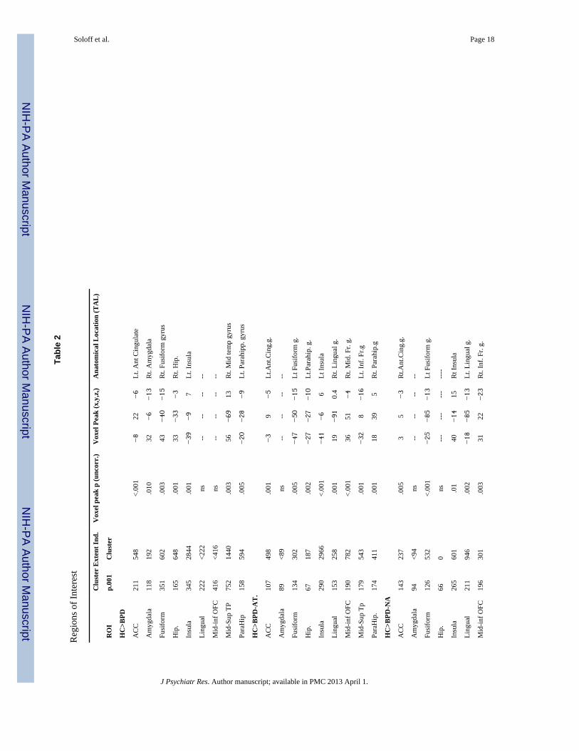
VBM contrasts were conducted using 9 ROIs which have demonstrated differences betweenBPD and healthy control subjects in previous structural studies and included: the middle-inferior orbitofrontal cortex, anterior cingulate cortex, middle-superior temporal cortex,insula, hippocampus, parahippocampus, fusiform gyrus, lingual gyrus and amygdala. Ahierarchical approach to group-wise analyses was employed to systematically assess graymatter differences between: a) Healthy control subjects compared to the total BPD sample,b) healthy control subjects against BPD sub-groups defined by attempter status, and, c)within BPD attempters, investigating the effects of lethality (High vs. Low Lethalityattempters). Cluster level correction (p<.001) was employed to optimize sensitivity to detectclusters with minimal extent (pthr<.05) (Ward, 2000).
All statistical analyses were conducted using a cluster-level correction approach that hasbeen formally characterized (Ward, 2000) and utilized in several imaging related studies(Ladouceur et al., In Press; Hagler et al., 2006; Bakshi et al., 2011; Diwadkar et al., 2011a;2011b; Diwadkar et al., In Press). The approach is based on “n” Monte Carlo simulations ofthe process of image generation, spatial correlations of voxels (i.e., clusters), voxel intensitythresholding (voxel-level values), probability thresholding, and minimum cluster sizethresholding where the probability of a true positive detection of a significant cluster isdetermined from the simulations themselves.
The basic assumptions behind cluster level approaches is that true differences in imagingdata will tend to occur over contiguous tissue or voxels, rather than individual voxels. Weemployed “n”=105 or 10,000 Monte Carlo simulations for data derived from each region ofinterest. For these simulations, we were interested in the frequency of occurrence of aminimum cluster size of contiguous voxels (voxel intensity p<.05) for 1 in 1000 instances,that is a cluster level correction of p<.001 (Ward, 2000; p 13). Thus the cluster extents inTable 2 reflect the minimum cluster size for the relevant region of interest that (based on10,000 Monte Carlo simulations) for α<.001 cluster level correction. Structural data fromsome subjects in this study were previously reported in contrasts between BPD and healthycontrol subjects (Soloff et al., 2008).
Soloff et al. Page 5
J Psychiatr Res. Author manuscript; available in PMC 2013 April 1.
NIH
-PA Author Manuscript
NIH
-PA Author Manuscript
NIH
-PA Author Manuscript

Results1. Structural analyses
Results of structural analyses are presented in Table 2. In the contrast between all BPDsubjects and healthy controls, BPD subjects had diminished grey matter concentrations in 7of 9 ROIs, with most robust reductions in the insula (cluster size = 2844), with peak clustersin the left hemisphere, and in the middle superior temporal cortex (cluster size = 1440), withpeak clusters in the right hemisphere (Fig. 1). Significant decreases in grey matterconcentrations were also noted in hippocampus, fusiform gyrus, parahippocampus, anteriorcingulate and amygdala, (in order of decreasing cluster sizes).
Compared to healthy controls, BPD attempters had diminished grey matter concentrations in8 of 9 ROIs (Fig. 2). As noted in the contrast with all BPD subjects (above), the most robustdecrease in grey matter concentration among BPD attempters was found in the insular cortex(cluster size = 2966), with peak clusters located in the left hemisphere. Insular cortex relaysinformation from the limbic system, both physical and emotional, to the prefrontalregulatory systems. Diminished grey matter concentrations were also significant in middle-inferior orbital frontal cortex, important in mediating behavioral inhibition, and middlesuperior temporal cortex, which processes facial recognition, including assessment of threat.In concert with the insula, these structures are critical for adaptive responding to socialstimuli. Amygdala was the only ROI which showed no significant difference betweengroups.
Compared to healthy controls, BPD non-attempters had diminished grey matterconcentrations in 6 of 9 ROIs, with some noteworthy differences from the contrast withsuicide attempters. Differences between groups were most robust in lingual gyrus (clustersize = 946), though decreased grey matter concentrations were also prominent in middle-superior temporal cortex (cluster size = 759), and insula (cluster size = 601). Lingual gyrusis part of a complex face recognition system, especially the assessment of angry faces andthreat. Hippocampus, parahippocampus and amygdala showed no significant differencesbetween groups.
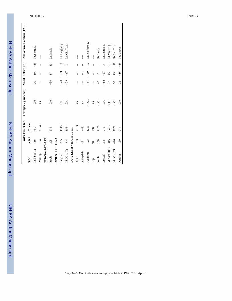
Within the BPD sample, attempters had decreased grey matter concentrations in insularcortex compared to non-attempters, with peak clusters in the left hemisphere. In the reversecontrast, non-attempters had diminished grey matter concentrations in middle-superiortemporal cortex, and lingual gyrus with peaks in the left hemisphere.
Among suicide attempters, those with High Lethality attempts had significant decreases in 6of 9 ROIs compared to Low Lethality attempters. The most robust findings (in order ofindividual cluster size) indicate diminished grey matter concentrations in High Lethalityattempters in middle-superior temporal gyrus (cluster size = 7752), middle-inferior orbitalfrontal gyrus (cluster size = 3403), and insular cortex (cluster size = 2168) (all with peakclusters in the right hemisphere), fusiform gyrus (cluster size = 1231), lingual gyrus (clustersize = 943) (with peaks in left hemisphere) and the parahippocampal gyrus (cluster size =274) (with a peak cluster in the right hemisphere) (Fig. 3). Fusiform, lingual andparahippocampal gyrii process face recognition, facial emotion, and familiar scenerecognition in social settings, relaying information forward to orbital frontal and superiortemporal cortex.
DiscussionThis is the first imaging study to specifically address structural brain changes associatedwith suicidal behavior in BPD. The relatively large sample size adds confidence to our
Soloff et al. Page 6
J Psychiatr Res. Author manuscript; available in PMC 2013 April 1.
NIH
-PA Author Manuscript
NIH
-PA Author Manuscript
NIH
-PA Author Manuscript

findings. We found significant differences in grey matter concentrations between BPDattempters and non-attempters, high and low lethality attempters, suggesting a possible rolefor specific neural circuits in suicidal behavior. Affected areas include orbital frontal,temporal, insular and paralimbic structures, broadly involved in emotion regulation,behavioral control, executive cognitive function and adaptive responding in social situations.Vulnerability to suicidal behavior in BPD, especially high lethality behavior, may be due toaffective interference at times of stress with the specific behavioral functions of thesestructures.
Orbital frontal cortex: response inhibition and impulsivityDiminished grey matter concentrations were found in high lethality compared to lowlethality BPD attempters in middle-inferior orbital frontal cortex. Regulation of behavioralimpulsivity and response inhibition are among the best studied functions of the orbitalfrontal cortex, and most relevant to suicidal behavior. Tasks involving inhibition of aprepotent response (e.g. the Go No-Go test) activate the orbital frontal cortex (Casey et al.,1997). Task performance on the Go No-Go test is impaired by lesions in the orbital frontalcortex in both animal and human studies (Fuster, 1989).
Impaired performance on the Go No-Go test, (i.e. responding when one should not),discriminates BPD from control subjects, and suicidal BPD subjects from BPD subjects withno suicidal behavior (Leyton et al., 1999).
An orbital frontal circuit connects the frontal monitoring systems with the limbic system andreceives information concerning the internal state of the individual (Tekin & Cumming,2002; Bonelli & Cummings, 2007). In concert with the anterior cingulate cortex (whichmediates conflicts between competing choices), the orbital frontal cortex facilitates selectionof external stimuli for attention and regulates response. The orbital frontal cortex is also acomponent part of a circuit that assesses emotion in facial expressions. Along with thefusiform gyrus and amygdala, the orbital frontal cortex is activated by angry faces (Blair etal., 1999). Positive covariation in activity occurs between Lt. amygdala and Rt. prefrontalcortex when perceiving negative faces, suggesting a neural mechanism for suppressinginappropriate responses to aversive stimuli in a social context (Iidaka et al., 2001). In BPDsubjects, the connectivity between (Rt) orbital frontal cortex and ventral amygdala isdecreased compared to healthy control subjects (New et al., 2008). Dysfunction in orbitalfrontal circuits results in behavioral disinhibition, aggression and emotional instability, allrisk factors for suicidal behavior (Tekin & Cumming, 2002).
Middle-superior temporal cortex: perception of facial emotionDiminished gray matter concentrations were found in high compared to low lethality BPDattempters in the middle-superior temporal cortex. In healthy control subjects, the superiortemporal cortex (gyrus and sulcus) is involved in the perception of emotion, and where thatemotion is being directed (Campbell et al, 1990). Superior temporal gyrus and sulcus arecomponent parts of a complex face processing system (which also includes fusiform andlingual gyrii, amygdala and orbital frontal cortex). The superior temporal gyrus is activatedby fearful faces (Radua et al., 2010; Iidaka et al., 2001). The superior temporal sulcus, inconcert with the insula, fusiform gyrus and amygdala, analyzes bodily movements toprovide information about the intentions of others (Frith & Frith, 1999; Allison &McCarthy, 2000). fMRI studies suggest that these structures mediate a rapid (“reflexive”)response to visual social inputs, especially negative visual stimuli (Koenigsberg, et al.,2009). Superior temporal sulcus is also involved in fear-based hypervigilance in attachmentrelationships (Buchheim et al., 2008).
Soloff et al. Page 7
J Psychiatr Res. Author manuscript; available in PMC 2013 April 1.
NIH
-PA Author Manuscript
NIH
-PA Author Manuscript
NIH
-PA Author Manuscript
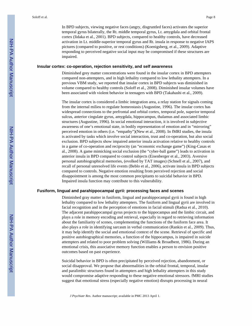
In BPD subjects, viewing negative faces (angry, disgruntled faces) activates the superiortemporal gyrus bilaterally, the Rt. middle temporal gyrus, Lt. amygdala and orbital frontalcortex (Iidaka et al., 2001). BPD subjects, compared to healthy controls, have decreasedactivation in Lt. middle-superior temporal gyrus and Rt. insula in response to negative IAPSpictures (compared to positive, or rest conditions) (Koenigsberg, et al., 2009). Adaptiveresponding to perceived negative social input may be compromised if these structures areimpaired.
Insular cortex: co-operation, rejection sensitivity, and self awarenessDiminished grey matter concentrations were found in the insular cortex in BPD attempterscompared non-attempters, and in high lethality compared to low lethality attempters. In aprevious VBM study, we reported that insular cortex in BPD subjects was diminished involume compared to healthy controls (Soloff et al., 2008). Diminished insular volumes havebeen associated with violent behavior in teenagers with BPD (Takahashi et al., 2009).
The insular cortex is considered a limbic integration area, a relay station for signals comingfrom the internal milieu to regulate homeostasis (Augustine, 1996). The insular cortex haswidespread connections to the prefrontal and orbital cortex, temporal pole, superior temporalsulcus, anterior cingulate gyrus, amygdala, hippocampus, thalamus and associated limbicstructures (Augustine, 1996). In social emotional interaction, it is involved in subjectiveawareness of one’s emotional state, in bodily representation of emotion and in “mirroring”perceived emotion in others (i.e. “empathy”)(New et al., 2008). In fMRI studies, the insulais activated by tasks which involve social interaction, trust and co-operation, but also socialexclusion. BPD subjects show impaired anterior insula activation relative to healthy controlsin a game of co-operation and reciprocity (an “economic exchange game”) (King-Casas etal., 2008). A game mimicking social exclusion (the “cyber-ball game”) leads to activation inanterior insula in BPD compared to control subjects (Eisenberger et al., 2003). Aversivepersonal autobiographical memories, (evolked by TAT images) (Schnell et al., 2007), andrecall of personal unresolved life events (Beblo et al., 2006), activate insula in BPD subjectscompared to controls. Negative emotion resulting from perceived rejection and socialdisappointment is among the most common precipitants to suicidal behavior in BPD.Impaired insula function may contribute to this vulnerability.
Fusiform, lingual and parahippocampal gyrii: processing faces and scenesDiminished gray matter in fusiform, lingual and parahippocampal gyrii is found in highlethality compared to low lethality attempters. The fusiform and lingual gyrii are involved infacial recognition and in the perception of emotions in facial stimuli (Radua et al., 2010).The adjacent parahippocampal gyrus projects to the hippocampus and the limbic circuit, andplays a role in memory encoding and retrieval, especially in regard to retrieving informationabout the familiarity of scenes, complementing the functions of the fusiform face area. Italso plays a role in identifying sarcasm in verbal communication (Rankin et al., 2009). Thus,it may help identify the social and emotional context of the scene. Retrieval of specific andpositive autobiographical memories, a function of the hippocampus, is impaired in suicideattempters and related to poor problem solving (Williams & Broadbent, 1986). During anemotional crisis, this associative memory function enables a person to envision positiveoutcomes based on past experience.
Suicidal behavior in BPD is often precipitated by perceived rejection, abandonment, orsocial disapproval. We propose that abnormalities in the orbital frontal, temporal, insularand paralimbic structures found in attempters and high lethality attempters in this studywould compromise adaptive responding to these negative emotional stressors. fMRI studiessuggest that emotional stress (especially negative emotion) disrupts processing in neural
Soloff et al. Page 8
J Psychiatr Res. Author manuscript; available in PMC 2013 April 1.
NIH
-PA Author Manuscript
NIH
-PA Author Manuscript
NIH
-PA Author Manuscript

circuits responsible for emotion regulation, impulse control, executive cognitive functionand episodic memory (Silbersweig et al., 2007; Minzenberg et al., 2007). Loss of cognitiveinhibitory control in adverse social situations would increase the likelihood of impulsive,aggressive, self-destructive behavior in BPD.
Could structural differences between attempters and non-attempters, high and low lethalityattempters be due to differences in BPD syndrome severity? These groups do notsignificantly differ on the DIB total section score suggesting that VBM differencesassociated with suicidality are independent of overall syndrome severity. Our researchfollows the stress-diathesis model of suicide advanced by Mann et. al. (1999 al. (2003). Thismodel postulates a neurobiologic vulnerability to suicidal behavior, increasing the likelihoodof suicidal behavior at times of stress. This biological vulnerability is expressed, in part,through behavioral traits of affective instability, impulsivity, aggression, and impairedexecutive cognitive functions. We propose that the structural anomalies identified in ourcontrasts of attempters and non-attempters, high and low lethality attempters, representstructural abnormalities in neural networks that mediate and regulate these functions.Whereas overall syndrome severity does not discriminate between groups, future analysesshould look at the effects of specific personality traits (e.g. impulsivity, aggression) onstructural changes in attempters and non-attempters, high and low lethality attempters.
LimitationsWe were unable to control for medical consequences of suicide attempts, which may resultin structural brain injury. This may be particularly relevant to differences between high andlow lethality attempters. It is noteworthy that we found no differences between high and lowlethality attempters in the hippocampus, which is sensitive to anoxic injury (Caine &Watson, 2000).
Structural brain differences between attempters and non-attempters, high and low lethalityattempters may reflect the neurobiology of endophenotypic traits which contribute adiathesis to suicidal behavior, but are not specific for BPD, i.e. impulsive-aggression andemotion dysregulation. These traits are risk factors for suicide and may be found in otherhigh risk disorders.
Structural brain abnormalities do not prove functional impairment. e.g. Functionalimpairment, demonstrated by fMRI, may be independent of structural abnormalities asdemonstrated by VBM related morphometry (Diwadkar et al.2011). In this study, we foundno volume loss in amygdala among BPD attempters compared to non-attempters or highversus low lethality BPD attempters, though fMRI studies have demonstrated abnormalhyperarousal of amygdala in BPD subjects in response to aversive pictures (Herpertz et al.2001) or faces (Donegan et al. 2003). Hyperarousal of amygdala in response to negativeemotional stimuli is believed to contribute to emotion dysregulation in BPD, a major riskfactor for suicidal behavior (Silbersweig et al. 2007). We did find decreased volume ofamygdala in the contrast of all BPD subjects compared to healthy controls, providingconvergence with some previous studies (Driessen et al 2001, Tebarz van Elst et al. 2003,Schmahl et al. 2003, Soloff et al., 2008), but not all, (New et al. 2007, Zetzsche et al 2006,Irle et al 2005, Brambilla 2004).
fMRI, PET, and neuropsychological studies in BPD subjects have each demonstratedfunctional impairment related to the fronto-limbic structures identified in our study aspotential mediators of suicidal behavior. However, there are no published fMRI studiestargeting fronto-limbic functions (e.g. response inhibition, conflict resolution, episodicmemory) in BPD subjects ascertained for suicidal behavior. These studies are currentlyunderway in our laboratory.
Soloff et al. Page 9
J Psychiatr Res. Author manuscript; available in PMC 2013 April 1.
NIH
-PA Author Manuscript
NIH
-PA Author Manuscript
NIH
-PA Author Manuscript

Finally, while biologic diathesis plays a critical role in increasing the likelihood of suicidalbehavior in BPD, psychosocial factors such as poor social and vocational function,prospectively predict suicidal behavior in our long term follow-up studies.
ReferencesAguilar EJ, Garcia-Marti G, Marti-Bonmati L, Lull JJ, Moratal D, Escarti MJ, et al. Left orbitofrontal
and superior temporal gyrus structural changes associated to suicidal behavior in patients withschizophrenia. Progress in Neuro-psychopharmacology & Biological Psychiatry. 2008; 32:1673–1676. [PubMed: 18657587]
Ahearn EP, Jamison KR, Steffens DC, Cassidy F, Provenzale JM, Lehman A, et al. MRI correlates ofsuicide attempt history in unipolar depression. Biological Psychiatry. 2001; 50:266–270. [PubMed:11522261]
Allison T, McCarthy G. Social perception from visual cues: Role of the STS region. Trends inCognitive Sciences. 2000; 4:267–277. [PubMed: 10859571]
Arango V, Underwood MD, Gubbi AV, Mann JJ. Localized alterations in pre-and post synapticserotonin binding sites in the ventrolateral prefrontal cortex of suicide victims. Brain Research.1995; 688:121–133. [PubMed: 8542298]
Arango V, Underwood MD, Mann JJ. Postmortem findings in suicide victims. Implications for in vivostudies. Annals of the New York Academy of Sciences. 1997; 836:269–287. [PubMed: 9616804]
Ashburner J, Friston KJ. Why voxel-based morphometry should be used. Neuroimage. 2001; 14(6):1238–1243. [PubMed: 11707080]
Augustine JR. Circuitry and functional aspects of the insular lobe in primates including humans. BrainResearch Brain Research Reviews. 1996; 22:229–244. [PubMed: 8957561]
Bakshi N, Pruitt P, Radwan J, Keshavan MS, Rajan U, Zajac-Benitez C, Diwadkar VA. Inefficientlyincreased anterior cingulate modulation of cortical systems during working memory in youngoffspring of schizophrenia patients. J Psychiatr Res. 2011; 45:1067–1076. [PubMed: 21306732]
Beblo T, Driessen M, Mertens M, Wingenfeld K, Piefke M, Rullkoetter N, et al. Functional MRIcorrelates of the recall of unresolved life events in borderline personality disorder. PsychologicalMedicine. 2006; 36:845–856. [PubMed: 16704749]
Bennett CM, Wolford GL, Miller MB. The principled control of false positives in neuroimaging. SocCogn Affect Neurosci. 2009; 4:417–422. [PubMed: 20042432]
Blair RJ, Morris JS, Frith CD, Perrett DJ, Dolan RJ. Dissociable neural responses to facial expressionsof sadness and anger. Brain. 1996; 122:883–893. [PubMed: 10355673]
Bonelli RM, Cummings JL. Frontal subcortical circuitry and behavior. Dialogues in ClinicalNeuroscience. 2007; 9:141–151. [PubMed: 17726913]
Buchheim A, Erk S, George C, Kachele H, Kircher T, Martius P, et al. Neural correlates of attachmenttrauma in borderline personality disorder: A functional magnetic resonance imaging study.Psychiatry Research: Neuroimaging. 2008; 163:223–235.
Caine D, Watson JDG. Neuropsychological and neuropathological sequelae of cerebral anoxia: Acritical review. Journal of the International Neuropsychological Society. 2000; 6:86–99. [PubMed:10761372]
Campbell R, Haywood CA, Cowey A, Regard M, Landis T. Sensitivity to eye gaze in prosopagnosicpatients and monkeys with superior temporal sulcus ablation. Neuropsychologia. 1990; 28:1123–1142. [PubMed: 2290489]
Casey BJ, Trainor RJ, Orendi JL, Schubert AB, Nystrom LE, Giedd JN, et al. A developmentalfunctional MRI study of prefrontal activation during performance of a go–no-go task. Journal ofCognitive Neuroscience. 1997; 9:835–847.
Diwadkar VA, Pruitt P, Goradia D, Murphy E, Bakshi N, Keshavan MS, Rajan U, Reid A, Zajac-Benitez C. Fronto-parietal hypo-activation during working memory independent of structuralabnormalities: conjoint fMRI and sMRI analyses in adolescent offspring of schizophrenia patients.NeuroImage. 2011a; 58:234–241. [PubMed: 21729757]
Soloff et al. Page 10
J Psychiatr Res. Author manuscript; available in PMC 2013 April 1.
NIH
-PA Author Manuscript
NIH
-PA Author Manuscript
NIH
-PA Author Manuscript

Diwadkar VA, Pruitt P, Zhang A, Radwan J, Keshavan MS, Murphy E, Rajan U, Zajac-Benitez C. Theneural correlates of performance in adolescents at risk for schizophrenia: Inefficiently increasedcortico-striatal responses measured with fMRI. J Psychiatr Res. 2011b
Diwadkar VA, Wadehra S, Pruitt P, Keshavan MS, Rajan U, Zajac-Benitez C, Eickhoff SB.Disordered cortico-limbic interactions during affective processing in children and adolescents atrisk for schizophrenia revealed by fMRI and Dynamic Causal Modeling. Archives of GeneralPsychiatry. In Press.
Eisenberger NI, Lieberman MD, Williams KD. Does rejection hurt? An fMRI study of socialexclusion. Science. 2003; 302:290–292. [PubMed: 14551436]
Fertuck EA, Lenzenweger MF, Clarkin JF, Hoermann S, Stanley B. Executive neurocognition,memory systems and borderline personality disorder. Clinical Psychology Review. 2006; 26:346–375. [PubMed: 15992977]
First, MB.; Spitzer, RL.; Gibbon, M.; Williams, JBW. Structured clinical interview for DSM-IV-TRAxis I Disorders-Patient Edition (SCID-I/P, 4/2005 revision). New York: Biometrics ResearchDepartment, New York State Psychiatric Institute; 2005.
Frith CD, Frith U. Interacting minds- a biological basis. Science. 1999; 286:1692–1695. [PubMed:10576727]
Fuster, J. The Prefrontal Cortex. 2. New York: Raven Press; 1989.Good CD, Johnsrude IS, Ashburner J, Henson RN, Friston KJ, Frackowiak RS. A voxel-based
morphometric study of ageing in 465 normal adult human brains. NeuroImage. 2001; 14:21–36.[PubMed: 11525331]
Gunderson JG, Kolb JE, Austin V. The Diagnostic Interview for Borderlines. American Journal ofPsychiatry. 1981; 138:896–903. [PubMed: 7258348]
Guy, W. ECDEU Assessment Manual of Psychopharmacology- Revised. (DHEW Publ. No. ADM.76–338). Rockville, MD: 1976.
Hagler DJ Jr, Saygin AP, Sereno MI. Smoothing and cluster thresholding for cortical surface-basedgroup analysis of fMRI data. NeuroImage. 2006; 33:1093–1103. [PubMed: 17011792]
Hazlett EA, New AS, Newmark R, Haznedar MM, Lo JN, Speiser LJ, et al. Reduced anterior andposterior cingulate gray matter in borderline personality disorder. Biological Psychiatry. 2005;58:614–623. [PubMed: 15993861]
Hwang J-P, Lee T-W, Tsai S-J, Chen T-J, Yang C-H, Ling J-F, et al. Cortical and sub-corticalabnormalities in late-onset depression with history of suicide attempts investigated with MRI andvoxel-based morphometry. Journal of Geriatric Psychiatry and Neurology. 2010; 23:171–184.[PubMed: 20430976]
Iidaka T, Omori M, Murata T, Kosaka H, Yonekura Y, Okada T, et al. Neural interaction of theamygdala with the prefrontal and temporal cortices in the processing of facial expressions asrevealed by fMRI. Journal of Cognitive Neuroscience. 2001; 13:1035–1047. [PubMed: 11784442]
Jollant F, Bellivier F, Leboyer M, Astruc B, Torres S, Verdier R, et al. Impaired decision making insuicide attempters. American Journal of Psychiatry. 2005; 162:304–310. [PubMed: 15677595]
Keilp JG, Sackheim HA, Brodsky BS, Oquendo MA, Malone KM, Mann JJ. Neuropsychologicaldysfunction in depressed suicide attempters. American Journal of Psychiatry. 2001; 158:735–741.[PubMed: 11329395]
King-Casas B, Sharp C, Loman-Breqam L, Lohrenz T, Fonagy P, Montague PR. The rupture andrepair of co-operation in borderline personality disorder. Science. 2008; 321:806–810. [PubMed:18687957]
Klein A, Andersson J, Ardekani BA, Ashburner J, Avants B, Chiang M-C, et al. Evaluation of 14nonlinear deformation algorithms applied to human brain MRI registration. NeuroImage. 2009;46:786–802. [PubMed: 19195496]
Koenigsberg HW, Siever LJ, Lee H, Pizzarello S, New AS, Goodman M, et al. Neural correlates ofemotion processing in borderline personality disorder. Psychiatry Research: Neuroimaging. 2009;172:192–199.
Ladouceur CD, Farchione TR, Diwadkar VA, Pruitt P, Radwan J, Axelson D, Birmaher B, PhillipsML. Differential patterns of abnormal fronto-limbic activity and functional connectivity in to
Soloff et al. Page 11
J Psychiatr Res. Author manuscript; available in PMC 2013 April 1.
NIH
-PA Author Manuscript
NIH
-PA Author Manuscript
NIH
-PA Author Manuscript

emotional faces in bipolar and bipolar NOS youth. Journal of the American Academy of Child andAdolescent Psychiatry. In Press.
Leyton M, Young SN, Paris J, Pihl RO, Benkelfat C. Suicide attempts and impulsivity. BiologicalPsychiatry. 1999; 45(suppl):8, 146.
Loranger, AW.; Janca, A.; Sartorius, N. Assessment and diagnosis of personality disorder: The ICD-10International Personality Disorder Examination (IPDE). Cambridge: Cambridge University Press;1997.
Lyoo IK, Han MH, Cho CY. A brain MRI study in subjects with borderline personality disorder.Journal of Affective Disorders. 1998; 50:235–243. [PubMed: 9858082]
Mann JJ. Neurobiology of suicidal behavior. Nature reviews Neuroscience. 2003; 4:819–828.Mann JJ, Stoff DM. A synthesis of current findings regarding neurobiological correlates and treatment
of suicidal behavior. Annals of the New York Academy of Sciences. 1997; 836:352–363.[PubMed: 9616809]
Minzenberg MJ, Fan J, New AS, Tang CY, Siever LJ. Fronto-limbic dysfunction in response to facialemotion in borderline personality disorder: An event-related fMRI study. Psychiatry Research:Neuroimaging. 2007; 155:231–243.
Monkul ES, Hatch JP, Nicoletti MA, Spence S, Brambilla P, Lacerda ALT, et al. Fronto-limbic brainstructures in suicidal and non-suicidal female patients with major depressive disorder. MolecularPsychiatry. 2007; 12:360–366. [PubMed: 17389903]
New AS, Goodman M, Triebwasser J, Siever LJ. Recent advances in the biological study ofpersonality disorders. The Psychiatric clinics of North America. 2008; 31:441–461. [PubMed:18638645]
New AS, Hazlett EA, Buchsbaum MS, Goodman M, Reynolds D, Mitropoulou V, et al. Bluntedprefrontal cortical 18-Flurodeoxyglucose positron emission tomography response to meta-chlorophenylpiperazine in impulsive aggression. Archives of General Psychiatry. 2002; 59:621–629. [PubMed: 12090815]
Oquendo, MA.; Halberstam, B.; Mann, JJ. Risk factors for suicidal behavior. In: First, MB., editor.Standardized evaluation in clinical practice. Vol. 22. Washington, DC: American PsychiatricPublishing; 2003. p. 103-129.
Oquendo MA, Placidi GPA, Malone KM, Campbell C, Keilp J, Brodsky B, et al. Positron emissiontomography of regional brain metabolic responses to serotonergic challenge and lethality ofsuicide attempts in major depression. Archives of General Psychiatry. 2003; 60:14–22. [PubMed:12511168]
Paris J. Estimating the prevalence of personality disorders in the community. Journal of PersonalityDisorders. 2010; 24(4):405–411. [PubMed: 20695802]
Paris J, Zweig-Frank H. A 27-year follow-up of patients with borderline personality disorder.Comprehensive Psychiatry. 2001; 42:782–487.
Radua J, Phillips ML, Russell T, Lawrence N, Marshall N, Kalidindi S, et al. Neural response tospecific components of fearful faces in healthy and schizophrenic adults. NeuroImage. 2010;49:939–946. [PubMed: 19699306]
Rankin KP, Salazaqr A, Gorno-Tempini M, Sollberger M, Wilson SM, Pavlic D, et al. Detectingsarcasm from paralinguistic cues: Anatomic and cognitive correlates in neurodegenerative disease.NeuroImage. 2009; 47:2005–2015. [PubMed: 19501175]
Ruocco AC. The neuropsychology of borderline personality disorder: A meta-analysis and review.Psychiatry Research. 2005; 137:191–202. [PubMed: 16297985]
Rusch N, Tebartz van Elst L, Ludaescher P, Wilke M, Huppertz HJ, Thiel T, et al. A voxel-basedmorphometric MRI study in female patients with borderline personality disorder. NeuroImage.2003; 20:385–392. [PubMed: 14527598]
Schmahl C, Bremner JD. Neuroimaging in borderline personality disorder. Journal of PsychiatricResearch. 2006; 40:419–427. [PubMed: 16239012]
Schnell K, Dietrich T, Schnitker R, Daumann J, Herpertz SC. Processing of autobiographical memoryretrieval cues in borderline personality disorder. Journal of Affective Disorders. 2007; 97:253–259. [PubMed: 16837057]
Soloff et al. Page 12
J Psychiatr Res. Author manuscript; available in PMC 2013 April 1.
NIH
-PA Author Manuscript
NIH
-PA Author Manuscript
NIH
-PA Author Manuscript

Siever LJ, Buchsbaum M, New AS, Spiegel-Cohen J, Wei T, Hazlett E, et al. d,l fenfluramine responsein impulsive personality disorder assessed with [18F] fluorodeoxyglucose positron emissiontomography. Neuropsychopharmacology. 1999; 20:413–423. [PubMed: 10192822]
Silbersweig D, Clarkin JF, Goldstein M, Kernberg OF, Tuescher O, Levy KN, et al. Failure of fronto-limbic inhibitory function in the context of negative emotion in borderline personality disorder.American Journal of Psychiatry. 2007; 164:1832–1841. [PubMed: 18056238]
Soloff PH, Nutche J, Goradia D, Diwadkar VA. Structural brain abnormalities in borderlinepersonality disorder: A voxel-based morphometry study. Psychiatry Research: Neuroimaging.2008; 164:223–236.
Soloff PH, Meltzer CC, Becker C, Greer P, Constantine D. Gender differences in a fenfluramine-activated FDG-PET study of borderline personality disorder. Psychiatry Research: Neuroimaging.2005; 138:183–195.
Soloff PH, Meltzer CC, Becker C, Greer PJ, Kelly TM, Constantine D. Impulsivity and prefrontalhypometabolism in borderline personality disorder. Psychiatry Research: Neuroimaging. 2003;123:153–163.
Soloff PH, Lynch KG, Kelly TM, Malone KM, Mann JJ. Characteristics of suicide attempts of patientswith major depressive episode and borderline personality disorder: A comparative study.American Journal of Psychiatry. 2000; 157:601–608. [PubMed: 10739420]
Soloff PH, Meltzer CC, Greer PJ, Constantine D, Kelly TM. Fenfluramine-activated FDG study ofborderline personality disorder. Biological Psychiatry. 2000b; 47:540–547. [PubMed: 10715360]
Spitzer, RL.; Williams, JBW.; Gibbon, M.; First, MB. Instruction Manual for the Structured ClinicalInterview for DSM III-R (SCID). New York: Biometrics Research, New York State PsychiatricInstitute; 1988.
Swartz M, Blazer D, George L, Winfield I. Estimating the prevalence of borderline personalitydisorder in the community. Journal of Personality Disorders. 1990; 4:257–272.
Takahashi T, Chanen AM, Wood SJ, Yucel M, Tanino R, Suzukui M, et al. Insular cortex volume andimpulsivity in teenagers with first-presentation borderline personality disoder. Progress in Neuro-psychopharmacology & Biological Psychiatry. 2009; 33:1395–1400. [PubMed: 19632284]
Tebartz van Elst L, Hesslinger B, Thiel T, Geiger E, Haegele K, Lemieux L, et al. Frontolimbic brainabnormalities in patients with borderline personality disorder. A volumetric MRI study. BiologicalPsychiatry. 2003; 54:163–171. [PubMed: 12873806]
Tekin S, Cumming JL. Frontal-subcortical neuronal circuits and clinical neuropsychiatry an update.Journal of Psychosomatic Research. 2002; 53:647–654. [PubMed: 12169339]
Van Heeringen C, Bijttebier S, Godfrin K. Suicidal brains: A review of functional and structural brainstudies in association with suicidal behavior. Neuroscience and Biobehavioral Reviews. 2011;35:688–698. [PubMed: 20826179]
Ward, BD. Simultaneous inference for fMRI data. Milwaukee, WI: Medical College of Wisconsin;2000.
Williams JMG, Broadbent K. Autobiographical memory in suicide attempters. Journal of AbnormalPsychology. 1986; 95:144–149. [PubMed: 3711438]
Zanarini M, Gunderson JG, Frankenburg FR, Chauncey DL. The Revised Diagnostic Interview forBorderlines: Discriminating BPD from other Axis II disorders. Journal of Personality Disorders.1989; 3:10–18.
Zetzsche T, Preuss UW, Frodl T, Schmitt G, Seifert D, Munchhausen E, et al. Hippocampal volumereduction and history of aggressive behavior in patients with borderline personality disorder.Psychiatry Research: Neuroimaging. 2007; 154:157–170.
Soloff et al. Page 13
J Psychiatr Res. Author manuscript; available in PMC 2013 April 1.
NIH
-PA Author Manuscript
NIH
-PA Author Manuscript
NIH
-PA Author Manuscript

Figure 1.The figure depicts reductions in gray matter volume in all BPD relative to controls assessedusing DARTEL. Significant clusters (cluster level correction: p<.001) are projected to thecortical surface on bilateral lateral views (a), medial views (b) and ventral views (c).Collectively these ortho-projections convey volumetric reductions in brain regions relevantto BPD that are consistent with previous studies in independent samples. The regions withreductions including the insula, the middle and superior temporal cortex (Mid-Sup T), thefusiform gyrus (FG), the anterior cingulate cortex (ACC), the hippocampus and theparahippocampus and the amygdala (AMY).
Soloff et al. Page 14
J Psychiatr Res. Author manuscript; available in PMC 2013 April 1.
NIH
-PA Author Manuscript
NIH
-PA Author Manuscript
NIH
-PA Author Manuscript

Fig. 2.The figure depicts reductions in gray matter volume in BPD suicide attempters relative tocontrols assessed using DARTEL. Significant clusters (cluster level correction: p<.001) areprojected to the cortical surface on bilateral lateral views (a), medial views (b) and ventralviews (c) These ortho-projections convey robust volumetric reductions in BPD attempters inthe insula, the middle and inferior orbito-frontal cortex (OFC), the fusiform gyrus (FG), thelingual gyrus, the anterior cingulate cortex (ACC), the mid.-sup. temporal cortex, thehippocampus and the parahippocampus.
Soloff et al. Page 15
J Psychiatr Res. Author manuscript; available in PMC 2013 April 1.
NIH
-PA Author Manuscript
NIH
-PA Author Manuscript
NIH
-PA Author Manuscript

Fig. 3.The figure depicts reductions in gray matter volume in high lethality suicide attemptersrelative to low lethality attempters assessed using DARTEL. Significant clusters (clusterlevel correction: p<.001) are projected to the cortical surface on bilateral lateral views (a),medial views (b) and ventral views (c). The naming schemes are consistent with Figure 1.High-lethality attempters show robust volumetric reductions in mid-sup. temporal cortex,mid-inf-OFC, insula, fusiform gyrus, lingual gyrus, and parahippocampus.
Soloff et al. Page 16
J Psychiatr Res. Author manuscript; available in PMC 2013 April 1.
NIH
-PA Author Manuscript
NIH
-PA Author Manuscript
NIH
-PA Author Manuscript

NIH
-PA Author Manuscript
NIH
-PA Author Manuscript
NIH
-PA Author Manuscript
Soloff et al. Page 17
Table 1
Demographic, diagnostic and clinical characteristics of samples
BPD (68) Control (52) t-test/ChiSq./p value
Female 52 24 X2=11.70, df = 1, p.=.001
Male 16 28
Age (s.d.) 28.3 (7.5) 25.9 (7.2) t = 1.80, df =118, p.ns
SES (s.d.) 26.8 (12.0) 37.3 (14.8) t = 4.06, df =88, p<.001
Race (%Cau.) 79.4% 71.2% X2 = 5.32, df = 5, p. ns.
HamD-24 (s.d.) 17.0 (8.1) 0.64 (1.1) t =14.18, df=113, p.<.001
GAS (s.d.) 59.2 (9.5) 89.9 (5.2) t = 19.90, df = 108, p.<.001
Physical abuse (%) 50 1 X2 = 32.97, df=1, p.=.001
Sexual Abuse (%) 32.4 0 X2 = 20.60, df =1, p,.001
MDD, current* (%) 63.2 n.a.
MDD, lifetime* (%) 26.5 n.a
PTSD, current (%) 14.7 n.a
PTSD, lifetime (%) 25.0 n.a
AUD, current (%) 23.5 n.a
AUD, lifetime (%) 38.2 n.a
*SCID current = diagnoses at time of scan. SCID lifetime excludes current diagnoses.
J Psychiatr Res. Author manuscript; available in PMC 2013 April 1.
NIH
-PA Author Manuscript
NIH
-PA Author Manuscript
NIH
-PA Author Manuscript
Soloff et al. Page 18
Tabl
e 2
Reg
ions
of I
nter
est
RO
I
Clu
ster
Ext
ent I
nd.
Vox
el p
eak
p (u
ncor
r.)
Vox
el P
eak
(x,y
,z,)
Ana
tom
ical
Loc
atio
n (T
AL
)
p.00
1C
lust
er
HC
>BPD
AC
C21
154
8<.
001
−8
22−6
Lt. A
nt C
ingu
late
Am
ygda
la11
819
2.0
1032
−6
−13
Rt.
Am
ygda
la
Fusi
form
351
602
.003
43−40
−15
Rt.
Fusi
form
gyr
us
Hip
.16
564
8.0
0133
−33
−3
Rt.
Hip
.
Insu
la34
528
44.0
01−39
−9
7Lt
. Ins
ula
Ling
ual
222
<222
ns--
----
--
Mid
-inf O
FC41
6<4
16ns
----
----
Mid
-Sup
TP
752
1440
.003
56−69
13R
t. M
id te
mp
gyru
s
Para
Hip
158
594
.005
−20
−28
−9
Lt. P
arah
ipp.
gyr
us
HC
>BPD
-AT
.
AC
C10
749
8.0
01−3
9−5
Lt.A
nt.C
ing.
g.
Am
ygda
la89
<89
ns--
----
--
Fusi
form
134
302
.005
−47
−50
−15
Lt F
usifo
rm g
.
Hip
.67
187
.002
−27
−27
−10
Lt.P
arah
ip. g
.
Insu
la29
029
66<.
001
−41
−6
6Lt
Insu
la
Ling
ual
153
258
.001
19−91
0.4
Rt.
Ling
ual g
.
Mid
-inf O
FC19
078
2<.
001
3651
−4
Rt.
Mid
. Fr.
g.
Mid
-Sup
Tp
179
543
.001
−32
8−16
Lt. I
nf. F
r.g
Para
Hip
.17
441
1.0
0118
395
Rt.
Para
hip.
g
HC
>BPD
-NA
AC
C14
323
7.0
053
5−3
Rt.A
nt.C
ing.
g.
Am
ygda
la94
<94
ns--
----
--
Fusi
form
126
532
<.00
1−25
−85
−13
Lt F
usifo
rm g
.
Hip
.66
0ns
---
---
---
----
Insu
la26
560
1.0
140
−14
15R
t Ins
ula
Ling
ual
211
946
.002
−18
−85
−13
Lt. L
ingu
al g
.
Mid
-inf O
FC19
630
1.0
0331
22−23
Rt.
Inf.
Fr. g
.
J Psychiatr Res. Author manuscript; available in PMC 2013 April 1.
NIH
-PA Author Manuscript
NIH
-PA Author Manuscript
NIH
-PA Author Manuscript
Soloff et al. Page 19
RO
I
Clu
ster
Ext
ent I
nd.
Vox
el p
eak
p (u
ncor
r.)
Vox
el P
eak
(x,y
,z,)
Ana
tom
ical
Loc
atio
n (T
AL
)
p.00
1C
lust
er
Mid
-Sup
Tp
518
759
.003
3019
−26
Rt.T
emp.
L.
Para
Hip
.16
4<1
64ns
----
----
--
BPD
-NA
>BPD
-AT
T
Insu
la20
537
3.0
08−30
1713
Lt. I
nsul
a
BPD
-AT
T>B
PD-N
A
Ling
ual
205
1246
.001
−20
−83
−10
Lt. L
ingu
al g
Mid
-Sup
Tp
544
1924
.001
−53
−47
2Lt
.Mid
.Tp.
g.
LO
W L
ET
H >
HIG
H L
ET
H
AC
C18
3<1
83ns
----
----
--
Am
ygda
la49
<49
ns--
----
----
Fusi
form
123
1231
<.00
1−47
−69
−12
Lt.F
usifo
rm g
.
Hip
.94
<94
ns--
----
----
Insu
la23
821
68<.
001
460
1R
t.Ins
ula
Ling
ual
275
943
.005
−12
−67
3Lt
.Lin
gual
g.
Mid
-inf O
FC31
534
03<.
001
3745
−7
Rt.M
id.F
r.g.
Mid
-Sup
TP
426
7752
<.00
128
15−30
Rt.S
up.T
p.g.
Para
Hip
.18
027
4.0
0922
−16
−26
Rt.
Unc
us
J Psychiatr Res. Author manuscript; available in PMC 2013 April 1.
Related Documents