Nig. J. Pure & Appl. Sci. Vol. 28 (2015): ISNN 0794-0378 Printed in Nigeria (C) 2015 Faculty of Physical Sciences and Faculty of Life Sciences, Univ. of Ilorin, Nigeria Nig. J. Pure & Appl. Sci. Vol. 28 (2015): 2658 – 2669 2658 Full Length Research Paper EFFECT OF Adansonia digitata AQUEOUS LEAF EXTRACT ON CARBON TETRACHLORIDE-INDUCED NEUROTOXICITY ON THE BRAIN OF ADULT MALE WISTAR RATS A.O Oyewopo*, A. Olawepo, F.C.Nwabia Department of Anatomy, Faculty of Basic Medical Sciences, University of Ilorin, P.M.B. 1515, Ilorin, Nigeria. *Corresponding Author: Oyewopo A.O. [email protected] Tel: 08033925431 ABSTRACT Adansonia digitata linn is an important nutritional and medicinal plant. This research was done to investigate the effect of Adansonia digitata aqueous leaf extract on Carbon Tetrachloride (CCl 4 ) induced injury in the brain of adult male Wistar rats. Twenty adult male Wistar rats were divided into four groups of five rats in each group: A, B, C and D. The rats in group A served as control while group B were given 2.5ml/kg body weight of CCl 4 . Group C were given 500mg/kg body weight of Adansonia digitata leaf extract while group D was given both CCl 4 and Adansonia digitata leaf extract consecutively. Histological observation was done using brain tissues and blood samples were collected for hormonal and biochemical analysis. There were considerable changes in the weight of the grouped rats. Histological sections of the cerebrum, cerebellum and pituitary gland showed increased cellular damage in rats treated with CCl 4 only than those in the CCl 4 and Baobab leaf extract group. Biochemical analysis showed significant increase in Malondialdehyde (MDA) level in CCl4 group as compared to the control group, while in the hormonal assay, the Follicle stimulating hormone (FSH) level showed significant decrease in CCl4 as compared to groups C and D. Luteinizing hormone (LH) values showed significant change in group D as compared to group A. In conclusion, these results suggest that Adansonia digitata leaf extract has an ameliorative effect on CCl 4 induced neurotoxicity on the brain of adult male Wistar rats. Keywords: Adansonia digitata, leaf extract, CCl4, Neurotoxicity.

Welcome message from author
This document is posted to help you gain knowledge. Please leave a comment to let me know what you think about it! Share it to your friends and learn new things together.
Transcript

Nig. J. Pure & Appl. Sci. Vol. 28 (2015): ISNN 0794-0378 Printed in Nigeria (C) 2015 Faculty of Physical Sciences and Faculty of Life Sciences, Univ. of Ilorin, Nigeria
Nig. J. Pure & Appl. Sci. Vol. 28 (2015): 2658 – 2669
2658
Full Length Research Paper EFFECT OF Adansonia digitata AQUEOUS LEAF EXTRACT ON CARBON
TETRACHLORIDE-INDUCED NEUROTOXICITY ON THE BRAIN OF ADULT MALE WISTAR RATS
A.O Oyewopo*, A. Olawepo, F.C.Nwabia
Department of Anatomy, Faculty of Basic Medical Sciences, University of Ilorin, P.M.B. 1515, Ilorin, Nigeria.
*Corresponding Author: Oyewopo A.O. [email protected]
Tel: 08033925431
ABSTRACT
Adansonia digitata linn is an important nutritional and medicinal plant. This research was done to investigate the effect of Adansonia digitata aqueous leaf extract on Carbon Tetrachloride (CCl4) induced injury in the brain of adult male Wistar rats. Twenty adult male Wistar rats were divided into four groups of five rats in each group: A, B, C and D. The rats in group A served as control while group B were given 2.5ml/kg body weight of CCl4. Group C were given 500mg/kg body weight of Adansonia digitata leaf extract while group D was given both CCl4 and Adansonia digitata leaf extract consecutively. Histological observation was done using brain tissues and blood samples were collected for hormonal and biochemical analysis. There were considerable changes in the weight of the grouped rats. Histological sections of the cerebrum, cerebellum and pituitary gland showed increased cellular damage in rats treated with CCl4 only than those in the CCl4 and Baobab leaf extract group. Biochemical analysis showed significant increase in Malondialdehyde (MDA) level in CCl4 group as compared to the control group, while in the hormonal assay, the Follicle stimulating hormone (FSH) level showed significant decrease in CCl4 as compared to groups C and D. Luteinizing hormone (LH) values showed significant change in group D as compared to group A. In conclusion, these results suggest that Adansonia digitata leaf extract has an ameliorative effect on CCl4 induced neurotoxicity on the brain of adult male Wistar rats. Keywords: Adansonia digitata, leaf extract, CCl4, Neurotoxicity.

2659
A.O Oyewopo, A. Olawepo, F.C. Nwabia Nig. J. Pure & Appl. Sci. Vol. 28 (2015)
INTRODUCTION
Baobab is a multipurpose tree species widely used for food, non-food products and medicine. Its leaves are used for multiple medicinal purposes in many parts of Africa and believed to show interesting medicinal properties including antioxidant, prebiotic-like activity, anti-inflammatory, analgesic, antipyretic activity, anti-diarrhoea and antidysentry activity. Leave extracts usually aqueous is known and evidently used for a variety of traditional medicinal purposes including fever, respiratory and skin afflictions for safety reasons.
In recent years, epidemiological evidences and several studies of ionic intake of ascorbic acid and some other micronutrients to health has been directed towards the evaluation of certain antioxidant properties Adansonia digitata commonly known as baobab tree plant highlights on the antioxidant capacity and ameliorative effect of Adansonia digitata leaf phytochemical extract (Sidibe and Williams, 2002). It has been traditionally known to be used in the treatment of fever, diarrhea, haemoptysis and smallpox in humans (Sidibe and Williams, 2002). Adansonia digitata L. locally called kuka, kuwa and oshe in Hausa, Kanuri and Yoruba language of Nigeria respectively, is a typical plant found in parts of northern Nigeria (Sodipo and Mohammed, 1990). The leaves have hypo-sensitive and antihistamine properties used to treat kidney and bladder diseases, asthma, guinea worm and general fatigue (Burkill, 1985; Prajapati, 2003). Carbon tetrachloride, also known by many other names is the organic compound with the formula CCl4. It is a colourless liquid with a "sweet" smell that can be detected at low levels (Manfred et al., 2006). Acute exposure to CCl4 may lead to rapidly producing narcotic effect on the brain with immediate fatalities either from respiratory depression due to direct CNS effects or from cardiac dysrhythmias (Hoemhe et al., 2010). In general, baobab leaves are nutritionally superior to the fruits of the tree due to the presence of more essential amino acids, minerals and vitamin A which making them an interesting food source having broader range of nutritive factors (Sena et al., 1998).
MATERIALS AND METHODS Experimental design
The protocol of experimentation was approved by the Departmental Ethical Committee responsible for the use of Laboratory animals, University of Ilorin, Nigeria. The study was approved to be in compliance with the international guiding principles for research involving animals as recommended by the Declaration of Helsinki and Guiding Principles in the Care and Use of Animals
Experimental animals (rats) Twenty (20) male Wistar rats weighing 110g – 140g were used. The rats were purchased
at Animal holdings in Ogbomosho and fed standard rat pellets (growers mash from feed Master Limited, Offa garage, Ilorin) 60 grams with 10 liters distilled water. The rats were kept in the animal house of the Department of Anatomy, Faculty of Basic Medical Sciences, University of Ilorin. They were housed in cages made with wire gauze under natural atmosphere, normal room

2660
A.O Oyewopo, A. Olawepo, F.C. Nwabia Nig. J. Pure & Appl. Sci. Vol. 28 (2015)
temperature, and maintained under a 12 hour light/ 12 hour dark cycle. The rats were fed twice in a day and their cages were cleaned daily. The rats were left to acclimatize for three weeks before the start of administration. Animal grouping
Twenty adult male Wistar rats were used and were divided into four groups of five rats in each group: A, B, C and D. The rats in each group were fed twice in a day with distilled water for drinking. The rats in group A were the control group and were fed with rat pellets and water only while groups B, C and D served as the treatment groups.
Plant Material
The plant material used in this study was collected from Oke ose and Amilengbe in Ilorin. It was identified and authenticated by Mr. Bolu Ajayi of the department of Botany, University of Ilorin, on the 12th December 2012.The data collected is presented in the following procedure: i. Names of medicinal plant (Botanical and common) ii. Family name. iii. Description. iv. Preparation and administration.
Preparation of Extract Young leaves of Adansonia digitata were washed, air dried and grounded into powdery
form. The powdery form was then ubjected to aqueous extraction by dissolving 4.0g in 40ml distilled water at room temperature and allowed to stand for 20 minutes The clear supernatant was carefully separated from the residue and dried by heating on a hot plate. The dried residue was weighed and the value was used to calculate the concentration.
Administration of Extract Administration of extract was between 8.00am and 12.00pm every day. The rats in group
A were the control group which were fed with rat pellets and distilled water only while groups B, C and D served as the experimental groups. The rats in group B were administered with 2.5 ml/kg of body weight of CCl4 only. Group C rats were administered 500mg/kg of body weight of Adansonia digitata aqueous leave extract only. The rats in group D were administered with 2.5ml/kg of body weight of CCl4 and 500mg/kg of body weight of Adansonia digitata aqueous leave extract consecutively. The administration lasted for 16 days only and animals were fasted overnight and then sacrificed on the 17th day.
Animal Sacrifice The animals in group B were sacrificed on the 3rd day after administration and groups A,
C and D were sacrificed on the 17th day from the day of general administration by anaesthetic chloroform. Blood samples were collected in heparinized bottles and in plane specimen bottles before the brains were removed and dissected for the removal cerebrum, cerebellum and pituitary gland.

2661
A.O Oyewopo, A. Olawepo, F.C. Nwabia Nig. J. Pure & Appl. Sci. Vol. 28 (2015)
Photomicrography The photographs of the tissue were taken through a light microscope (Olympus, X5Z-
107BN) with a digital camera (Fuji film, 8.2 megapixels, China) by mounting the digital camera directly on the eyepiece.
Statistical Analysis The results were analyzed statistically by application of student’s t-test, and presented as
mean ± SEM with determination of level of significance. This also involved the use of the Statistical Package Sciences (SPSS) 16.0 version and analysis of variance (ANOVA) to determine the significant differences between groups at p<0.05. The analysis and comparison were evaluated for significance at 5% (α=0.05)
RESULTS Morphological Observations
There were some changes observed in the course of this experiment. On the experimental animals, there were slight loss of hair on body in the CCl4 group and slight changes in skin tone. Table 3: Effect of Adansonia digitata leaf and CCl4 on the weight of the rats. Groups Initial mean
weight (g)
Final mean weight
(g)
Weight difference
(g)
Control
146.10±3.88
181.08±5.67
34.98
CCl4 only
113.46±3.56
134.16±3.25
20.70
Adansonia digitata extract only
153.82±8.08
175.24±8.15
21.42
CCl4 and Adansonia digitata
extract
128.48±6.24
162.00±3.49
33.52
Values expressed as Mean ± SEM; P < 0.05
2662
A.O Oyewopo, A. Olawepo, F.C. Nwabia Nig. J. Pure & Appl. Sci. Vol. 28 (2015)
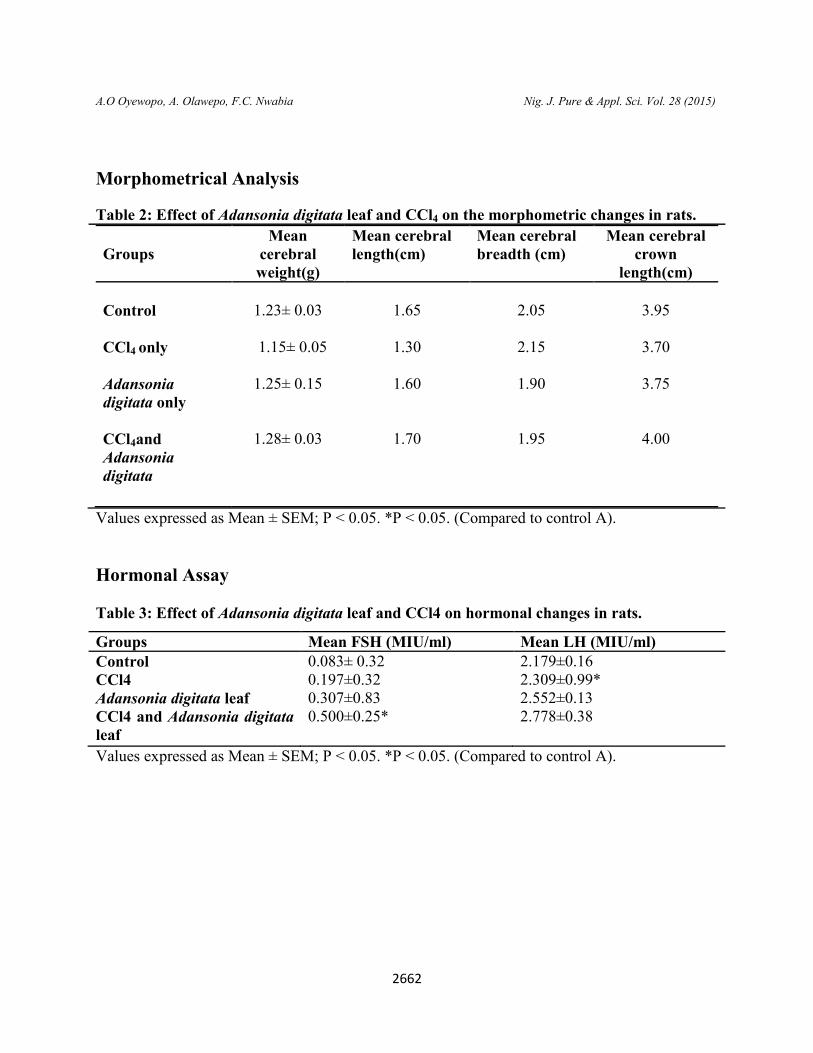
Morphometrical Analysis
Table 2: Effect of Adansonia digitata leaf and CCl4 on the morphometric changes in rats.
Groups
Mean cerebral weight(g)
Mean cerebral length(cm)
Mean cerebral breadth (cm)
Mean cerebral crown
length(cm)
Control
1.23± 0.03
1.65
2.05
3.95
CCl4 only
1.15± 0.05
1.30
2.15
3.70
Adansonia digitata only
1.25± 0.15
1.60
1.90
3.75
CCl4and Adansonia digitata
1.28± 0.03
1.70
1.95
4.00
Values expressed as Mean ± SEM; P < 0.05. *P < 0.05. (Compared to control A).
Hormonal Assay Table 3: Effect of Adansonia digitata leaf and CCl4 on hormonal changes in rats.
Groups Mean FSH (MIU/ml) Mean LH (MIU/ml) Control 0.083± 0.32 2.179±0.16 CCl4 0.197±0.32 2.309±0.99* Adansonia digitata leaf 0.307±0.83 2.552±0.13 CCl4 and Adansonia digitata leaf
0.500±0.25* 2.778±0.38
Values expressed as Mean ± SEM; P < 0.05. *P < 0.05. (Compared to control A).

A.O Oyewopo, A. Olawepo, F.C. Nwabia
Biochemical Analysis
Table 4: Effect of Adansonia digitata
Groups Control
MDA (µmol/L) 0.023±0.003
Values expressed as Mean ± SEM; P < 0.05. *P < 0.05; (Compared to control group A)
Graphic illustrations
Figure 1: showing comparison of body weight
changes in rats.
Figure 3: showing comparison of the
hormonal changes in the rats.
0
50
100
150
200
GR
OU
P A
GR
OU
P B
GR
OU
P C
GR
OU
P D
Me
anw
eig
ht(
g)
FINAL WEIGHT (g)
INITIAL WEIGHT (g)
DIFFERENCE (g)
0
1
2
3
Me
an F
SH a
nd
LH
(M
iu/m
l)
2663
Nig. J. Pure & Appl. Sci. Vol. 28 (2015)
Adansonia digitata leaf and CCl4 on the biochemical changes in the rats.
CCl4 only Adansonia digitata leaf
CCl4Adansonia digitata
0.023±0.003 0.150±0.000*
0.086±0.002
0.085±0.001
Values expressed as Mean ± SEM; P < 0.05. *P < 0.05; (Compared to control group A)
Figure 1: showing comparison of body weight Figure2: showing the comparison of
morphometric changes
. Figure 3: showing comparison of the
Figure 5: showing comparison in MDA level in each rat group.
FINAL WEIGHT (g)
INITIAL WEIGHT (g)
DIFFERENCE (g)
00.5
11.5
22.5
33.5
44.5
Control CCl4 only
Extract only
CCl4 and
Etract
mean cerebral weight(g)
mean cerebral length(cm)
mean cerebral breadth(cm)
mean cerebral crown length(cm)
FSH (Miu/ml)
LH (Miu/ml)0
0.05
0.1
0.15
MD
A v
alu
e (
µm
ol/
L)
Mean MDA value (µmol/L)
Mean MDA value (µmol/L)
Nig. J. Pure & Appl. Sci. Vol. 28 (2015)
leaf and CCl4 on the biochemical changes in the rats.
CCl4 and Adansonia digitata 0.085±0.001
Values expressed as Mean ± SEM; P < 0.05. *P < 0.05; (Compared to control group A)
Figure2: showing the comparison of
Figure 5: showing comparison in MDA level
mean cerebral weight(g)
mean cerebral length(cm)
mean cerebral breadth(cm)
mean cerebral crown length(cm)
Mean MDA value (µmol/L)
Mean MDA value (µmol/L)

A.O Oyewopo, A. Olawepo, F.C. Nwabia
HISTOLOGICAL OBSERVATIONS
Figure7: Photomicrograph showing the saggital section of the cerebrum of a rat in the control group stained with H&E(x400). P
Figure 8: Photomicrograph showing the saggital section of the cerebellum of a rat in the control group stained with H&E(x400). ML
Figure 9: Photomicrograph of the pituitary gland of a rat in the c(x400).A- Acidophil, B-Basophiles.
2664
Nig. J. Pure & Appl. Sci. Vol. 28 (2015)
HISTOLOGICAL OBSERVATIONS
Photomicrograph showing the saggital section of the cerebrum of a rat in the control group stained with H&E(x400). P-Pyramidal cells, N-Neuroglia cells.
Photomicrograph showing the saggital section of the cerebellum of a rat in the control group stained with H&E(x400). ML- Molecular layer, P- Purkinje cells, GL- Granular layer.
: Photomicrograph of the pituitary gland of a rat in the control group stained with H &E Basophiles.
Nig. J. Pure & Appl. Sci. Vol. 28 (2015)
Photomicrograph showing the saggital section of the cerebrum of a rat in the control
Photomicrograph showing the saggital section of the cerebellum of a rat in the control Granular layer.
ontrol group stained with H &E

A.O Oyewopo, A. Olawepo, F.C. Nwabia
Figure 10: Photomicrograph showing the saggital section of the cerebrum of a rat ingroup stained with H&E(x400).P
Figure 11: Photomicrograph showing the saggital section of the cerebellum of a rat in the CClgroup stained with H&E (x400). ML
Figure 12: Photomicrograph of the pituitary gland of a rat in the CClH&E(x400). A- Acidophil, B- Basophiles, V
2665
Nig. J. Pure & Appl. Sci. Vol. 28 (2015)
: Photomicrograph showing the saggital section of the cerebrum of a rat ingroup stained with H&E(x400).P-Pyramidal cells, N-Neuroglia cells, V-Vacuolation.
: Photomicrograph showing the saggital section of the cerebellum of a rat in the CClE (x400). ML- Molecular layer, P-Purkinje cells, GL- Granular layer.
: Photomicrograph of the pituitary gland of a rat in the CCl4 group stained with Basophiles, V- Vacuolation.
Nig. J. Pure & Appl. Sci. Vol. 28 (2015)
: Photomicrograph showing the saggital section of the cerebrum of a rat in the CCl4 Vacuolation.
: Photomicrograph showing the saggital section of the cerebellum of a rat in the CCl4 Granular layer.
group stained with
A.O Oyewopo, A. Olawepo, F.C. Nwabia
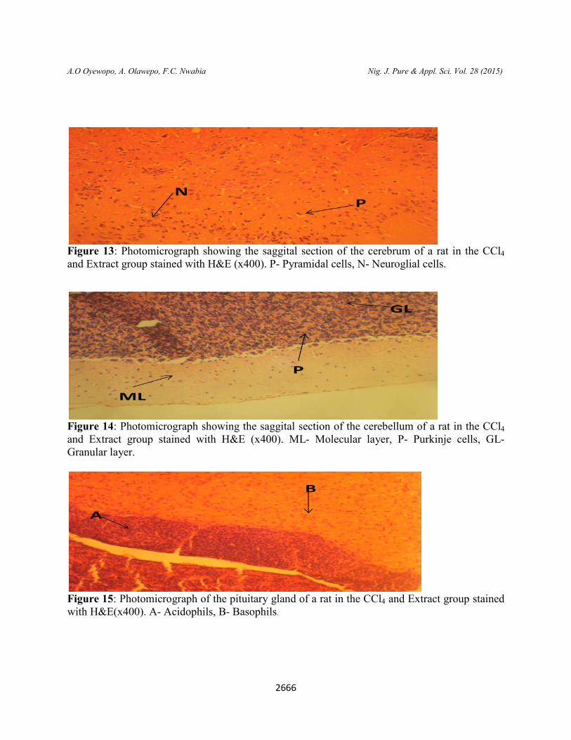
Figure 13: Photomicrograph showing the saggital section of the cerebrum of a rat in the CCland Extract group stained with H&E (x400). P
Figure 14: Photomicrograph showing the saggital section of the cerebellum of a rat in the CCland Extract group stained with H&E (x400). MLGranular layer.
Figure 15: Photomicrograph of the pituitary gland of a rat in with H&E(x400). A- Acidophils, B
2666
Nig. J. Pure & Appl. Sci. Vol. 28 (2015)
: Photomicrograph showing the saggital section of the cerebrum of a rat in the CCland Extract group stained with H&E (x400). P- Pyramidal cells, N- Neuroglial cells.
: Photomicrograph showing the saggital section of the cerebellum of a rat in the CCland Extract group stained with H&E (x400). ML- Molecular layer, P- Purkinje cells, GL
: Photomicrograph of the pituitary gland of a rat in the CCl4 and Extract group stained Acidophils, B- Basophils.
Nig. J. Pure & Appl. Sci. Vol. 28 (2015)
: Photomicrograph showing the saggital section of the cerebrum of a rat in the CCl4 Neuroglial cells.
: Photomicrograph showing the saggital section of the cerebellum of a rat in the CCl4 Purkinje cells, GL-
and Extract group stained

2667
A.O Oyewopo, A. Olawepo, F.C. Nwabia Nig. J. Pure & Appl. Sci. Vol. 28 (2015)
DISCUSSION Adansonia digitata aqueous leaf is known to have antioxidant capacity and ameliorative
effect in the phytochemical extract of its leaf. This study aimed to assess the effect of Adansonia digitata aqueous leaf extract on Carbon tetrachloride induced neurotoxicity on the brain of adult male Wistar rats observations revealed significant increase in the weight of rats in the group of CCl4 and Adansonia digitata leaf as compared to the control group and significant decrease in the CCl4 group B. In the effect of methanolic leaf extract of Adansonia digitatalinn result shows that the this extract has lowering effect on the level of serum albumin with no significant effect on serum electrolyte, glucose and protein in both alcohol and normal fed rats (Metawalli et al., 2003).
In the CCl4 group B administered with 2.5ml/kg of CCl4, histological observations showed less nucleated cells and less dense layers in the cerebrum, vacuolations in the molecular and granular layers of the cerebellum and pituitary gland consisted sparsely filled chromophils. The treated group D showed normal basic histology with prominent pyramidal and neuroglial cells of the cerebrum, less sparse purkinje cells of cerebellum as compared to the CCl4 group and a densely filled acidophil and basophiles in the pituitary gland. In the study of induced CCl4, Pomgranate juice has a potential protective effect as it can elevate antioxidant defense system, clean up free radicals, lessen oxidative damages and protect the brain tissue against CCl4 -induced toxicity (Mahmoud and Moneim, 2013).
Morphometric analysis showed significant decrease in the cerebral length and weight of group B with increase in breath as compared to the control group. Group D showed a significant increase in the weight, length and breathe as compared to the control group. Hormone analysis was done to know the level of neurotoxicity which was significant in the CCl4 group as compared to the control group. The gonardotroph of pars distalis in adenohypophysis of the pituitary gland contains the FSH and LH, analysis of the FSH showed significant increase in group C and D with decrease in FSH level in group B as compared to group A. It was also observed with significant increase in the LH level in group D as compared to the control group showing ameliorative effect of Adansonia digitata leaf. Studies investigating the biochemical analysis showed a significant elevation MDA level in the brain tissue of CCl4 treated group (Reckmage et al., 1989). Evidence of neurotoxicity by increased MDA level is one of the primary means of associated oxidative processes with an overall decrease in cellular function. The increase in MDA indicates neurotoxicity an index to identify free radicals-induced injuries (Rosenblat et al., 2006).
The level of oxidative stress marker using MDA showed significant increase in the level of MDA with increase in oxidative stress as compared to the control group.

2668
A.O Oyewopo, A. Olawepo, F.C. Nwabia Nig. J. Pure & Appl. Sci. Vol. 28 (2015)
CONCLUSION
This experiment showed that exposure of carbon tetraoxide caused neurotoxicity in the brain of Wistar rats and that aqueous leave extract of adansonia digitata has an ameliorative effect in the brain of male Wistar rats.
REFERENCES
Burkili, H.M. (1985). The useful plants of West Tropical Africa, 1985, Royal Botanic Gardens, Kew, Richmond
Duncan, R.C., Knapp, R.G., Miller, M.C. (1977). ‘Test of hypothesis in population. In:
Introductory Biostatistics of Health Sciences’. John Wiley and sons Inc, NYPP. 71-96. Hartley, D.P., Kolaja, K.L., Reinchord, J. and Peterson, D.R. (1999). 4 Hydroxynonenal and
malondialdehyde hepatic protein adducts in rats treated with carbon tetrachloride: immunochemical detection and lobular localization. Toxicol. Appl. Pharmacol., 161 (1): 23-33.
Hoehme, S., Brulport, M. and Bauer, A. (2010) "Prediction and validation of cell alignment
along microvessels as order principle to restore tissue architecture in liver regeneration", 107: 10371-10376.
Manfred, R., Wilhelm, L., Gerhard, P., Adolf, T., Eberhard, L.D., Ernst, L., Heinz, J., Peter, K.,
Heinz, S., Richard, C., Uwe, B., Karl-A, L., Theodore, R.T., Eckhard, L. and Klaus, K.B. (2006). “Chlorinated Hydrocarbons” Encyclopedia of Industrial ChemistryWiley.
Mahmoud, S.M. and Abdel Moneim, A.E. (2013). The protective effect of pomegranate (Punica
granatum) juice against carbon tetrachloride-induced oxidative stress in brain tissue of adult male albino rats. Life Sci. J., 10: 151-158
Metawali, A.G., Sunday, A.O. and Kokori, M. (2003). The effect of methanolic leaf extract of
Adansonia digitata Linn on serum electrolyte level in normal and alcohol fed rats. Pakistan Journal of Biological sciences, 7(8): 1404-1406
Muhammad, R.K., Gul, N.K. and Dawood, A. (2011). ‘Evaluation of antioxidant and fertility
effects of Digera muricata in male rats’. Nair V., O’Neil C.L., Wang P.G. (2008). ‘Malondialdehyde’ Encyclopedia of reagents for
organic synthesis. John Wiley & sons. Odell, W.D. and Parlow, A.F. (1981). Adansonia digitata aqueous leaf . J Clin Invest., 47:2551.

2669
A.O Oyewopo, A. Olawepo, F.C. Nwabia Nig. J. Pure & Appl. Sci. Vol. 28 (2015)
Prajapati, N.D., Purohit, S.S., Sharma, A.K. and Kumar, T. (2003). A Handbook of Medicinal
Plants. Agribios (India). 553pp Reckmage, R.O., Glende, E.A., Dolak, J.A. and Waller, R.L. (1989). Mechanism of carbon
tetrachloride toxicity. Pharmacol., 43: 139-154. Rosenblat, M., Hayek, T. and Aviram, M. (2006): Anti-oxidative effects of pomegranate juice
(pj) consumption by diabetic patients on serum and on macrophages. Atheroscler., 187(2): 363-371.
Sena, L.P., VanderJagt, D.J., Rivera, C., Tin, A.C., Muhamadu, I., Mahamadou, O., Millton, M.,
Pastuszyn, A. and Glew, R.H. (1998). Analysis of nutritional components of eight famine foods of the Republic of Niger. Plant Foods for Human Nutrition, 52:17-30.
Sibibe, M. and Williams, J.T. (2002). Baobab – Adansonia digitata. Fruits for the future.Int.
Centre Underutil. Crops, Southampton, Pg: 96. Sodipo, O.A and S.L. Mohammed (1990). Foam forming and haemolytic activities of the extract
of baobab tree. Nig. J Basic Applied Sci., pp: 41-52. Wennick, J.M., Delemarre-van de Waal, H.A., Schoemaker, R., Schoemaker, H. and
Schoemaker, J. (1990). Luteinizing hormone and Follicle stimulating hormone secretion patterns in girls throughout puberty measured using highly sensitive immunoradiometric assays. Clin Endocrinol., 33,333-344.
Related Documents