Nicotine-Like Effects of the Neonicotinoid Insecticides Acetamiprid and Imidacloprid on Cerebellar Neurons from Neonatal Rats Junko Kimura-Kuroda*, Yukari Komuta, Yoichiro Kuroda, Masaharu Hayashi, Hitoshi Kawano Department of Brain Development and Neural Regeneration, Tokyo Metropolitan Institute of Medical Science, Setagaya-city, Tokyo, Japan Abstract Background: Acetamiprid (ACE) and imidacloprid (IMI) belong to a new, widely used class of pesticide, the neonicotinoids. With similar chemical structures to nicotine, neonicotinoids also share agonist activity at nicotinic acetylcholine receptors (nAChRs). Although their toxicities against insects are well established, their precise effects on mammalian nAChRs remain to be elucidated. Because of the importance of nAChRs for mammalian brain function, especially brain development, detailed investigation of the neonicotinoids is needed to protect the health of human children. We aimed to determine the effects of neonicotinoids on the nAChRs of developing mammalian neurons and compare their effects with nicotine, a neurotoxin of brain development. Methodology/Principal Findings: Primary cultures of cerebellar neurons from neonatal rats allow for examinations of the developmental neurotoxicity of chemicals because the various stages of neurodevelopment—including proliferation, migration, differentiation, and morphological and functional maturation—can be observed in vitro. Using these cultures, an excitatory Ca 2+ - influx assay was employed as an indicator of neural physiological activity. Significant excitatory Ca 2+ influxes were evoked by ACE, IMI, and nicotine at concentrations greater than 1 mM in small neurons in cerebellar cultures that expressed the mRNA of the a3, a4, and a7 nAChR subunits. The firing patterns, proportion of excited neurons, and peak excitatory Ca 2+ influxes induced by ACE and IMI showed differences from those induced by nicotine. However, ACE and IMI had greater effects on mammalian neurons than those previously reported in binding assay studies. Furthermore, the effects of the neonicotinoids were significantly inhibited by the nAChR antagonists mecamylamine, a-bungarotoxin, and dihydro-b-erythroidine. Conclusions/Significance: This study is the first to show that ACE, IMI, and nicotine exert similar excitatory effects on mammalian nAChRs at concentrations greater than 1 mM. Therefore, the neonicotinoids may adversely affect human health, especially the developing brain. Citation: Kimura-Kuroda J, Komuta Y, Kuroda Y, Hayashi M, Kawano H (2012) Nicotine-Like Effects of the Neonicotinoid Insecticides Acetamiprid and Imidacloprid on Cerebellar Neurons from Neonatal Rats. PLoS ONE 7(2): e32432. doi:10.1371/journal.pone.0032432 Editor: Shu-ichi Okamoto, Sanford-Burnham Medical Research Institute, United States of America Received July 4, 2011; Accepted January 29, 2012; Published February 29, 2012 Copyright: ß 2012 Kimura-Kuroda et al. This is an open-access article distributed under the terms of the Creative Commons Attribution License, which permits unrestricted use, distribution, and reproduction in any medium, provided the original author and source are credited. Funding: This work was supported by Grants in Aid for Scientific Research from the Ministry of Education, Culture, Sports, Science and Technology of Japan (21510075). http://www.jsps.go.jp/english/e-grants/grants.html. The funders had no role in study design, data collection and analysis, decision to publish, or preparation of the manuscript. Competing Interests: The authors have declared that no competing interests exist. * E-mail: [email protected] Introduction The neonicotinoids acetamiprid (ACE) and imidacloprid (IMI) belong to a new class of insecticides that are used worldwide to protect crops from pest insects and domestic animals from fleas [1]. The neonicotinoids have been reported to act as agonists of nicotinic acetylcholine receptors (nAChRs), and their high toxicities to insects have been attributed to selective binding affinity to insect nAChRs [2,3]. Furthermore, X-ray crystallogra- phy has revealed that the binding sites of the neonicotinoids on nAChRs are electronegative, which contributes to their charac- teristic toxicities at insect nAChRs [4–6]. However, X-ray crystal analyses and binding assays against one type of nAChR have often yielded controversial results, and the structural conformations of receptors are often changed by physiological actions such as ligand binding or interactions with other proteins [7]. There have been a few studies of neonicotinoid-induced toxicity in the nervous systems of vertebrates, and these studies were conducted with only a few of the neonicotinoids, such as IMI, thiamethoxam, and clothianidin. IMI has been reported to act as an agonist or an antagonist of nAChRs at 10 mM in rat pheochromocytoma (PC12) cells [8] and to change the membrane properties of neurons at $10 mM in the mouse cochlear nucleus [9]. Exposure to IMI in utero causes decreased sensorimotor performance and increased expression of glial fibrillary acidic protein (GFAP) in the motor cortex and hippocampus of neonatal rats [10]. Furthermore, it has been reported that the neonicoti- noids thiamethoxam and clothianidin induce dopamine release in the rat striatum via nAChRs [11] and that thiamethoxam alters behavioral and biochemical processes related to the rat cholinergic systems [12]. Recently, IMI and clothianidin have been reported to agonize human a4b2 nAChR subtypes [13]. These findings suggest that the neonicotinoids affect mammalian nAChRs to a greater extent than previously believed based on binding-assay data and that further study of the neonicotinoids is needed to protect human health. The relevance of neonicotinoid exposure to PLoS ONE | www.plosone.org 1 February 2012 | Volume 7 | Issue 2 | e32432

Welcome message from author
This document is posted to help you gain knowledge. Please leave a comment to let me know what you think about it! Share it to your friends and learn new things together.
Transcript
Nicotine-Like Effects of the Neonicotinoid InsecticidesAcetamiprid and Imidacloprid on Cerebellar Neuronsfrom Neonatal RatsJunko Kimura-Kuroda*, Yukari Komuta, Yoichiro Kuroda, Masaharu Hayashi, Hitoshi Kawano
Department of Brain Development and Neural Regeneration, Tokyo Metropolitan Institute of Medical Science, Setagaya-city, Tokyo, Japan
Abstract
Background: Acetamiprid (ACE) and imidacloprid (IMI) belong to a new, widely used class of pesticide, the neonicotinoids. Withsimilar chemical structures to nicotine, neonicotinoids also share agonist activity at nicotinic acetylcholine receptors (nAChRs).Although their toxicities against insects are well established, their precise effects on mammalian nAChRs remain to be elucidated.Because of the importance of nAChRs for mammalian brain function, especially brain development, detailed investigation of theneonicotinoids is needed to protect the health of human children. We aimed to determine the effects of neonicotinoids on thenAChRs of developing mammalian neurons and compare their effects with nicotine, a neurotoxin of brain development.
Methodology/Principal Findings: Primary cultures of cerebellar neurons from neonatal rats allow for examinations of thedevelopmental neurotoxicity of chemicals because the various stages of neurodevelopment—including proliferation, migration,differentiation, and morphological and functional maturation—can be observed in vitro. Using these cultures, an excitatory Ca2+-influx assay was employed as an indicator of neural physiological activity. Significant excitatory Ca2+ influxes were evoked by ACE,IMI, and nicotine at concentrations greater than 1 mM in small neurons in cerebellar cultures that expressed the mRNA of the a3,a4, and a7 nAChR subunits. The firing patterns, proportion of excited neurons, and peak excitatory Ca2+ influxes induced by ACEand IMI showed differences from those induced by nicotine. However, ACE and IMI had greater effects on mammalian neuronsthan those previously reported in binding assay studies. Furthermore, the effects of the neonicotinoids were significantlyinhibited by the nAChR antagonists mecamylamine, a-bungarotoxin, and dihydro-b-erythroidine.
Conclusions/Significance: This study is the first to show that ACE, IMI, and nicotine exert similar excitatory effects onmammalian nAChRs at concentrations greater than 1 mM. Therefore, the neonicotinoids may adversely affect human health,especially the developing brain.
Citation: Kimura-Kuroda J, Komuta Y, Kuroda Y, Hayashi M, Kawano H (2012) Nicotine-Like Effects of the Neonicotinoid Insecticides Acetamiprid and Imidaclopridon Cerebellar Neurons from Neonatal Rats. PLoS ONE 7(2): e32432. doi:10.1371/journal.pone.0032432
Editor: Shu-ichi Okamoto, Sanford-Burnham Medical Research Institute, United States of America
Received July 4, 2011; Accepted January 29, 2012; Published February 29, 2012
Copyright: � 2012 Kimura-Kuroda et al. This is an open-access article distributed under the terms of the Creative Commons Attribution License, which permitsunrestricted use, distribution, and reproduction in any medium, provided the original author and source are credited.
Funding: This work was supported by Grants in Aid for Scientific Research from the Ministry of Education, Culture, Sports, Science and Technology of Japan(21510075). http://www.jsps.go.jp/english/e-grants/grants.html. The funders had no role in study design, data collection and analysis, decision to publish, orpreparation of the manuscript.
Competing Interests: The authors have declared that no competing interests exist.
* E-mail: [email protected]
Introduction
The neonicotinoids acetamiprid (ACE) and imidacloprid (IMI)
belong to a new class of insecticides that are used worldwide to
protect crops from pest insects and domestic animals from fleas
[1]. The neonicotinoids have been reported to act as agonists of
nicotinic acetylcholine receptors (nAChRs), and their high
toxicities to insects have been attributed to selective binding
affinity to insect nAChRs [2,3]. Furthermore, X-ray crystallogra-
phy has revealed that the binding sites of the neonicotinoids on
nAChRs are electronegative, which contributes to their charac-
teristic toxicities at insect nAChRs [4–6]. However, X-ray crystal
analyses and binding assays against one type of nAChR have often
yielded controversial results, and the structural conformations of
receptors are often changed by physiological actions such as ligand
binding or interactions with other proteins [7].
There have been a few studies of neonicotinoid-induced toxicity
in the nervous systems of vertebrates, and these studies were
conducted with only a few of the neonicotinoids, such as IMI,
thiamethoxam, and clothianidin. IMI has been reported to act as
an agonist or an antagonist of nAChRs at 10 mM in rat
pheochromocytoma (PC12) cells [8] and to change the membrane
properties of neurons at $10 mM in the mouse cochlear nucleus
[9]. Exposure to IMI in utero causes decreased sensorimotor
performance and increased expression of glial fibrillary acidic
protein (GFAP) in the motor cortex and hippocampus of neonatal
rats [10]. Furthermore, it has been reported that the neonicoti-
noids thiamethoxam and clothianidin induce dopamine release in
the rat striatum via nAChRs [11] and that thiamethoxam alters
behavioral and biochemical processes related to the rat cholinergic
systems [12]. Recently, IMI and clothianidin have been reported
to agonize human a4b2 nAChR subtypes [13]. These findings
suggest that the neonicotinoids affect mammalian nAChRs to a
greater extent than previously believed based on binding-assay
data and that further study of the neonicotinoids is needed to
protect human health. The relevance of neonicotinoid exposure to
PLoS ONE | www.plosone.org 1 February 2012 | Volume 7 | Issue 2 | e32432
human health is especially important in children because nAChRs
are important for normal brain development [14] and their
functions are disturbed by nicotine [15]. Therefore, the effects of
neonicotinoids on mammalian developing neurons should be
investigated and compared with those of nicotine, as a positive
control that is known to affect mammalian nAChRs.
In the present study, we chose the globally used neonicotinoids
ACE and IMI. Importantly, ACE has relatively higher affinities
for rodent nAChRs than other neonicotinoids [16], and little
published information is available regarding its effects on
mammalian nAChRs. We compared the effects of ACE and
IMI on mammalian nAChRs with those of nicotine, using an
excitatory Ca2+-influx assay as an index of neural physiological
activity. We conducted these assays in cultured cerebellar neurons
from neonatal rats, which natively express the a3, a4, and a7
nAChR subtypes in vitro [17]. Primary neonatal cerebellar neurons
have been used as a suitable model to evaluate developmental
neurotoxicity because they allow for the observation of the various
stages of neurodevelopment, including proliferation, migration,
differentiation, and morphological and functional maturation [18].
The overall aim of this study was to determine whether these two
neonicotinoids exert effects similar to those of nicotine on
cerebellar neurons from neonatal rats.
Results
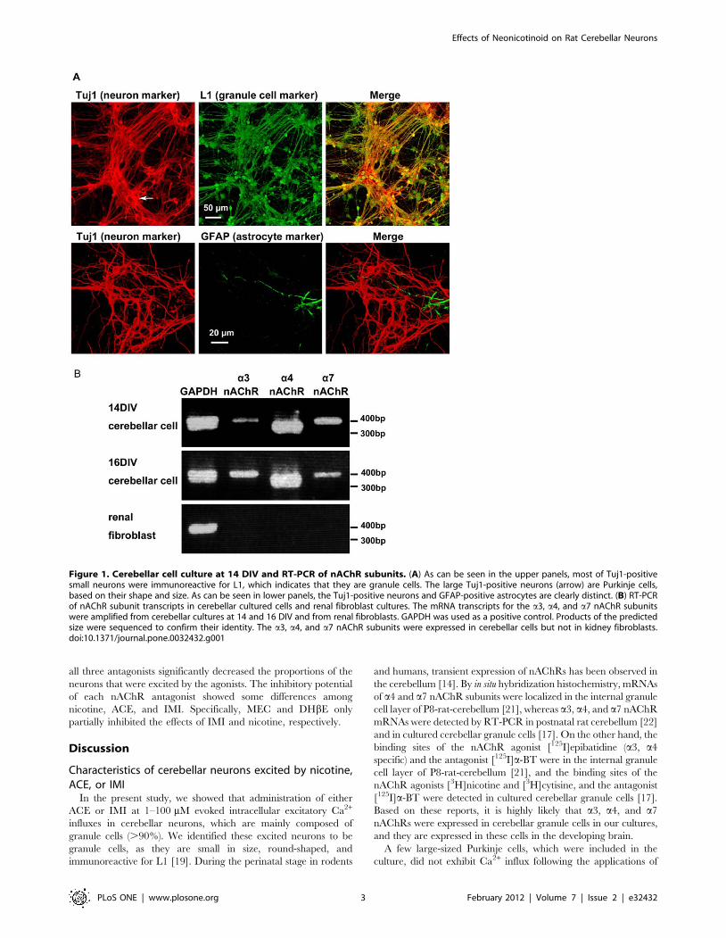
Cerebellar cultures and expression of nAChR mRNAIn cerebellar cultures from neonatal rats at postnatal day 1 (P1),
approximately 90% of the total cells were small neurons stained by
anti-Tuj1 (neuron specific b3-tubulin). Almost all of these small
neurons were identified to be cerebellar granule cells based on their
morphology and that they were stained by anti-neural cell adhesion
molecule L1 (Fig. 1A) [19]. The cerebellar cultures also contained a
few large, Tuj1-positive Purkinje neurons (about 1%, arrow in
Fig. 1A) and GFAP-positive astrocytes (about 5%, Fig. 1A). Because
most commercially available antibodies against each of the subunits
of nAChRs have been reported to be cross reactive to other subunits
or unknown factors [20], we examined the mRNA of nAChR
subunits with RT-PCR. As shown in Fig. 1B, mRNAs of the a3, a4,
and a7 nAChR subunits are expressed in cerebellar cells at 14 and
16 days in vitro (DIV), which was the time frame used for the Ca2+-
influx assay. The a4 nAChR subunit was expressed constantly at 14
and 16 DIV. The expressions of the a3 and a7 nAChR subunits
showed little difference between 14 and 16 DIV. In renal fibroblast
cultures, however, mRNAs of the a3, a4, and a7 nAChR subunits
were not expressed.
Ca2+ influx in cerebellar neuronsTo determine the effects of neonicotinoids on the nAChRs of
cerebellar neurons, we examined intracellular Ca2+ mobilization
using the calcium-sensitive fluorescent dye Fluo-4. The chemical
structures of nicotine, ACE, and IMI are shown in Figure 2. As
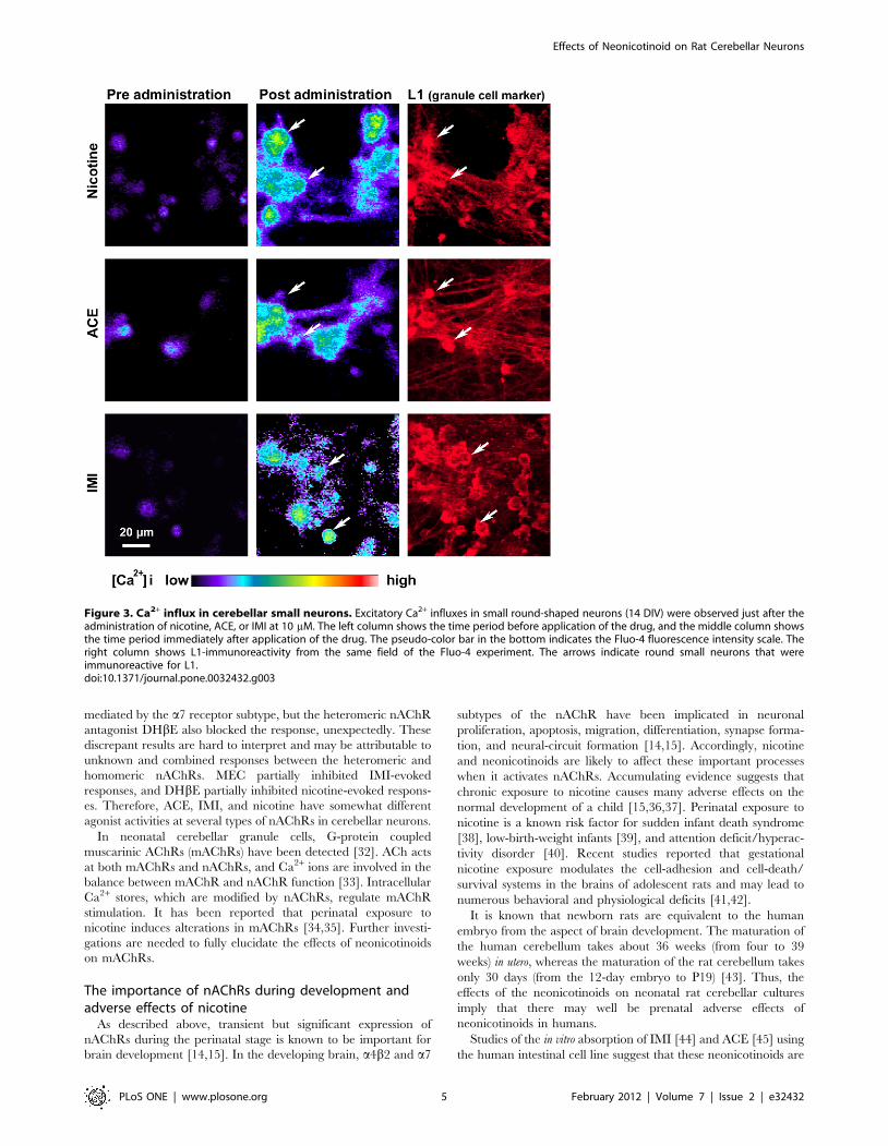
shown in the left column of Figure 3, Fluo-4 loading induced some
fluorescence in cerebellar small neurons at 14 DIV. Applications
of nicotine, ACE, and IMI at 10 mM robustly increased Fluo-4
fluorescence in round-shaped neurons (middle column of Fig. 3).
We confirmed that the excited small round cells were immuno-
reactive for L1 (right column of Fig. 3), which is known to be a
cerebellar granule cell marker [19].
A few large-sized Purkinje neurons did not exhibit significant
Ca2+ influx following applications of ACE, IMI, and nicotine.
Moreover, GFAP-positive astrocytes did not flux as much Ca2+ as
the small neurons.
In Figure 4A–C, administrations of nicotine, ACE, and IMI
induced a characteristic excitatory pattern of intracellular Ca2+
influx at 1–100 mM in small neurons. The main, line graphs
represent the mean values 6 the standard error of the mean
(S.E.M.) (n = 20–30) for the fluorescence intensity of Ca2+ influx.
The small, line graphs inset within each main graph show a
representative firing pattern from a single cell. There was a rapid
rise and fall in the firing patterns of these cells following applications
of nicotine at 1–100 mM (Fig. 4C) and ACE at 10–100 mM (Fig. 4A).
There was a rapid rise but gradual fall in the firing patterns of these
cells following applications of ACE at 1 mM (Fig. 4A) and IMI at 1–
100 mM (Fig. 4B). As shown in Figure 4A–C, administrations of
nicotine, ACE, and IMI attenuated the responses to KCl (100 mM)
washes 5–8 min after their applications compared with washes with
only KCl (Fig. 4D). As shown in Figure 4C, KCl induced robust
excitations after administration of nicotine at 100 mM. When
500 nM or lower concentrations of nicotine, ACE, and IMI were
applied to the cerebellar cells, we did not observe significant Ca2+
influx during at least 3 independent replications with each drug.
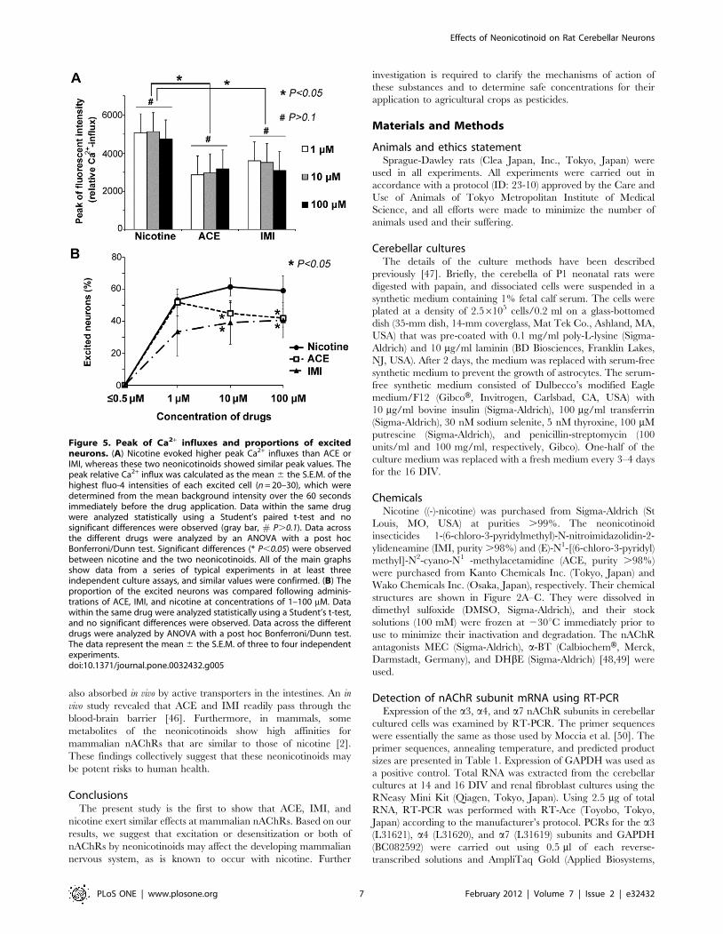
Peak value of Ca2+ influx and proportion of excitedneurons
Next, we examined the peak magnitudes of Ca2+ influx and the
dependence of the effects of the neonicotinoids on the administered
dose. The peak values were compared between each concentration
of the three drugs (Fig. 5A). Even at a concentration of 1 mM, ACE
and IMI caused distinctive excitations in numerous small neurons,
and the peak relative fluorescence intensities of Ca2+ influx were
similar to those following applications of 10 or 100 mM of the same
drug. Administration of nicotine evoked higher peaks of Ca2+ influx
than those of ACE and IMI, and these two neonicotinoids showed
similar peak values, as shown in Figure 5A.
Subsequently, we examined the proportion of cells among the
total number of small neurons (1–1.256103 cells) that were excited
by ACE, IMI, and nicotine. As shown in Figure 5B, the
proportions of the neurons excited by nicotine were higher than
those by excited by IMI. At 1 mM, ACE excited a similar
proportion of the neurons to nicotine, and ACE at 10 or 100 mM
excited similar proportions of the neurons to IMI.
Antagonist assayThe effects of neonicotinoids were also examined following
pretreatments with the specific nAChR antagonists mecamyl-
amine (MEC, nonselective nAChR antagonist), a-bungarotoxin
(a-BT, selective a7 nAChR antagonist), and dihydro-b-erythroi-
dine (DHbE, selective a4b2 and a3b4 nAChR antagonist). Pre-
incubation with MEC (100 mM), a-BT (1 mM), or DHbE (1 mM)
significantly inhibited the characteristic excitations and Ca2+
influxes in small neurons induced by nicotine, ACE, or IMI at
100 mM. Moreover, after removal of the antagonists by washes
with balanced salt solution (BSS), the same neurons were excited
by these agonists (Fig. 6A–C). The inhibitory effects of these
antagonists showed some differences among nicotine and the
neonicotinoids. As shown in Figure 6A, MEC strongly inhibited
nicotine- and ACE-evoked Ca2+ influx but only partially inhibited
IMI-evoked Ca2+ influx. The excitatory effects of all three agonists
were completely inhibited by a-BT (Fig. 6B). Furthermore, after
removal of a-BT, the nicotine- and ACE-evoked firing patterns
(Fig. 5B) were rather broad compared with baseline patterns
(Fig. 4A, C). DHbE partially inhibited nicotine-evoked firing
(Fig. 6C) but completely inhibited ACE- and IMI-evoked firing.
Subsequently, we examined the proportions of the neurons that
were excited by 100 mM of nicotine, ACE, or IMI in the presence
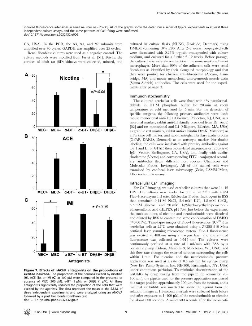
or absence of each nAChR antagonist. As shown in Figure 7A–C,
Effects of Neonicotinoid on Rat Cerebellar Neurons
PLoS ONE | www.plosone.org 2 February 2012 | Volume 7 | Issue 2 | e32432
all three antagonists significantly decreased the proportions of the
neurons that were excited by the agonists. The inhibitory potential
of each nAChR antagonist showed some differences among
nicotine, ACE, and IMI. Specifically, MEC and DHbE only
partially inhibited the effects of IMI and nicotine, respectively.
Discussion
Characteristics of cerebellar neurons excited by nicotine,ACE, or IMI
In the present study, we showed that administration of either
ACE or IMI at 1–100 mM evoked intracellular excitatory Ca2+
influxes in cerebellar neurons, which are mainly composed of
granule cells (.90%). We identified these excited neurons to be
granule cells, as they are small in size, round-shaped, and
immunoreactive for L1 [19]. During the perinatal stage in rodents
and humans, transient expression of nAChRs has been observed in
the cerebellum [14]. By in situ hybridization histochemistry, mRNAs
of a4 and a7 nAChR subunits were localized in the internal granule
cell layer of P8-rat-cerebellum [21], whereas a3, a4, and a7 nAChR
mRNAs were detected by RT-PCR in postnatal rat cerebellum [22]
and in cultured cerebellar granule cells [17]. On the other hand, the
binding sites of the nAChR agonist [125I]epibatidine (a3, a4
specific) and the antagonist [125I]a-BT were in the internal granule
cell layer of P8-rat-cerebellum [21], and the binding sites of the
nAChR agonists [3H]nicotine and [3H]cytisine, and the antagonist
[125I]a-BT were detected in cultured cerebellar granule cells [17].
Based on these reports, it is highly likely that a3, a4, and a7
nAChRs were expressed in cerebellar granule cells in our cultures,
and they are expressed in these cells in the developing brain.
A few large-sized Purkinje cells, which were included in the
culture, did not exhibit Ca2+ influx following the applications of
Figure 1. Cerebellar cell culture at 14 DIV and RT-PCR of nAChR subunits. (A) As can be seen in the upper panels, most of Tuj1-positivesmall neurons were immunoreactive for L1, which indicates that they are granule cells. The large Tuj1-positive neurons (arrow) are Purkinje cells,based on their shape and size. As can be seen in lower panels, the Tuj1-positive neurons and GFAP-positive astrocytes are clearly distinct. (B) RT-PCRof nAChR subunit transcripts in cerebellar cultured cells and renal fibroblast cultures. The mRNA transcripts for the a3, a4, and a7 nAChR subunitswere amplified from cerebellar cultures at 14 and 16 DIV and from renal fibroblasts. GAPDH was used as a positive control. Products of the predictedsize were sequenced to confirm their identity. The a3, a4, and a7 nAChR subunits were expressed in cerebellar cells but not in kidney fibroblasts.doi:10.1371/journal.pone.0032432.g001
Effects of Neonicotinoid on Rat Cerebellar Neurons
PLoS ONE | www.plosone.org 3 February 2012 | Volume 7 | Issue 2 | e32432

ACE, IMI, and nicotine. Previous reports have indicated that
developing Purkinje cells also express a7 nAChR mRNA at P8
stage in rat [21], and a4 nAChR mRNA at fetus stage in human
[23]. Excitatory or inhibitory postsynaptic currents in Purkinje
cells were observed by administration of nicotine in slice culture
from rats at P5–P10, however, they were not activated by nicotine
in cultures from older rats [24]. The authors of those studies
suggested that this lack of response was because of synaptic
maturation, and this may be also the case in our cultures at 14–16
DIV from P1 rats.
Furthermore, GFAP-positive astrocytes also did not show Ca2+
influx following applications of ACE, IMI, and nicotine. Sustained
exposure (5 min) to nicotine has been reported to induce light
uptake of intracellular Ca2+ in cortical astrocytes [25]. In our
study, we examined transient exposure to nicotine or neonicoti-
noids under continuous perfusion, which is likely the reason there
was no response in the astrocytes.
Similarities between the effects of neonicotinoids andnicotine on neuronal excitation
Our results indicate that at a concentration of 1 mM, ACE and
IMI robustly excited rat cerebellar neurons to a similar degree as
nicotine. In a previous report, using [3H]IMI or [3H]nicotine, the
binding affinities of ACE, IMI, and nicotine at insect nAChRs
have been reported to be 84, 565 and 0.002 times, respectively, as
large as their affinities at rodent a4b2 nAChRs [2]. The
discrepancy between these reported values and our results may
be attributable to differences between using simplified artificial
binding assays with one type of nAChR and examining cellular
excitatory actions mediated by several kinds of nAChRs on a
single neuron.
Previous studies have indicated that IMI and clothianidin
modify the amplitude of responses to acetylcholine (ACh) by
chicken or human nAChR a4b2 subtype receptors even at a low
concentration (3 mM) that did not activate these receptors when
administered alone [13,26] . It is possible that the binding of ACh
to nAChRs modifies the structure of the nAChRs, which may
allow neonicotinoids to affect mammalian nAChRs. The present
results indicate that ACE and IMI have agonist activity at
mammalian nAChRs at a concentration of 1 mM, which is lower
than the concentration one would predict from their binding
affinities.
The peak of Ca2+ influxes and the proportions of neurons that
were excited did not depend on the dose of nicotine, ACE, or IMI.
Rather, it exhibited an all or none response. Although the
nAChR-dependent increase of intracellular Ca2+ may be mainly
mediated by Ca2+ entry through nAChRs [27], the involvement of
other calcium channels is also feasible. The nAChR-mediated
Ca2+- influx activates voltage-dependent calcium channels
(VDCCs) and Ca2+-uptake via VDCC augments the primary
Ca2+ signals generated by nAChRs [27,28], which may underlie
the all or none response. However, the exact mechanism
underlying this response needs further investigations to be fully
understood.
Differences between the effects neonicotinoids andnicotine on neuronal excitation
Firing evoked by nicotine or ACE (10–100 mM) rapidly rose and
fell, whereas firing evoked by ACE (1 mM) or IMI rapidly rose but
gradually fell, probably because of differential in desensitization
potential to nAChRs. It is well known that nAChRs can undergo
desensitization, which is a reversible reduction in response, even
within a second of agonist applications at low concentrations of the
agonist. Although the role of desensitization in the effects of
nAChRs remains unclear, it has been proposed that desensitiza-
tion can modulate the cholinergic activity of nAChRs [29], and
chronic exposure to agonists can inhibit the normal actions of ACh
at nAChRs via desensitization [15]. The peaks of the Ca2+ influxes
induced by and the proportions of the neurons excited by ACE
and IMI were somewhat lower than those by nicotine.
Accordingly, there may be some differences among nicotine,
ACE, and IMI in their agonist effects at nAChRs.
Lack of a KCl-response after administrations of nicotine,ACE, or IMI
As shown in Figure 4A–C, applications of nicotine or the two
neonicotinoids significantly decreased the effects of KCl (100 mM)
on cerebellar neurons, even after they were removed by washing.
As mentioned above, nAChR-mediated Ca2+- influx by nicotine,
ACE, or IMI can activate VDCC. Because it has been reported
that KCl-evoked Ca2+ permeability is coupled to VDCCs [28,30],
the initial uptake of Ca2+ ions via VDCC may serve as a negative
feedback signal and elicit a transition of VDCC into a non-
conducting inactivated state [31]. These ideas suggest that
nAChR-mediated Ca2+-influx by nicotine, ACE, or IMI activates
VDCC, which is followed by inactivation of VDCC and an
attenuation of the KCl response. At 100 mM of nicotine, strong
desensitization of nAChRs may activate some VDCC and
subsequently induce relatively large responses by KCl. The precise
mechanisms mediating these phenomena are unknown at present.
Involvements of nAChR subtypesAs the three nAChR antagonists significantly inhibited the Ca2+
influxes in neurons induced by ACE and IMI, it is likely that ACE
and IMI have direct agonist activity at nAChRs in cerebellar
neurons. Complete blockade of the effects of all three drugs by the
homomeric nAChR antagonist a-BT suggests that the response is
Figure 2. Molecular structures of nicotine and the neonicoti-noids ACE and IMI. (A) nicotine, (B) acetamiprid (ACE), (C)imidacloprid (IMI).doi:10.1371/journal.pone.0032432.g002
Effects of Neonicotinoid on Rat Cerebellar Neurons
PLoS ONE | www.plosone.org 4 February 2012 | Volume 7 | Issue 2 | e32432
mediated by the a7 receptor subtype, but the heteromeric nAChR
antagonist DHbE also blocked the response, unexpectedly. These
discrepant results are hard to interpret and may be attributable to
unknown and combined responses between the heteromeric and
homomeric nAChRs. MEC partially inhibited IMI-evoked
responses, and DHbE partially inhibited nicotine-evoked respons-
es. Therefore, ACE, IMI, and nicotine have somewhat different
agonist activities at several types of nAChRs in cerebellar neurons.
In neonatal cerebellar granule cells, G-protein coupled
muscarinic AChRs (mAChRs) have been detected [32]. ACh acts
at both mAChRs and nAChRs, and Ca2+ ions are involved in the
balance between mAChR and nAChR function [33]. Intracellular
Ca2+ stores, which are modified by nAChRs, regulate mAChR
stimulation. It has been reported that perinatal exposure to
nicotine induces alterations in mAChRs [34,35]. Further investi-
gations are needed to fully elucidate the effects of neonicotinoids
on mAChRs.
The importance of nAChRs during development andadverse effects of nicotine
As described above, transient but significant expression of
nAChRs during the perinatal stage is known to be important for
brain development [14,15]. In the developing brain, a4b2 and a7
subtypes of the nAChR have been implicated in neuronal
proliferation, apoptosis, migration, differentiation, synapse forma-
tion, and neural-circuit formation [14,15]. Accordingly, nicotine
and neonicotinoids are likely to affect these important processes
when it activates nAChRs. Accumulating evidence suggests that
chronic exposure to nicotine causes many adverse effects on the
normal development of a child [15,36,37]. Perinatal exposure to
nicotine is a known risk factor for sudden infant death syndrome
[38], low-birth-weight infants [39], and attention deficit/hyperac-
tivity disorder [40]. Recent studies reported that gestational
nicotine exposure modulates the cell-adhesion and cell-death/
survival systems in the brains of adolescent rats and may lead to
numerous behavioral and physiological deficits [41,42].
It is known that newborn rats are equivalent to the human
embryo from the aspect of brain development. The maturation of
the human cerebellum takes about 36 weeks (from four to 39
weeks) in utero, whereas the maturation of the rat cerebellum takes
only 30 days (from the 12-day embryo to P19) [43]. Thus, the
effects of the neonicotinoids on neonatal rat cerebellar cultures
imply that there may well be prenatal adverse effects of
neonicotinoids in humans.
Studies of the in vitro absorption of IMI [44] and ACE [45] using
the human intestinal cell line suggest that these neonicotinoids are
Figure 3. Ca2+ influx in cerebellar small neurons. Excitatory Ca2+ influxes in small round-shaped neurons (14 DIV) were observed just after theadministration of nicotine, ACE, or IMI at 10 mM. The left column shows the time period before application of the drug, and the middle column showsthe time period immediately after application of the drug. The pseudo-color bar in the bottom indicates the Fluo-4 fluorescence intensity scale. Theright column shows L1-immunoreactivity from the same field of the Fluo-4 experiment. The arrows indicate round small neurons that wereimmunoreactive for L1.doi:10.1371/journal.pone.0032432.g003
Effects of Neonicotinoid on Rat Cerebellar Neurons
PLoS ONE | www.plosone.org 5 February 2012 | Volume 7 | Issue 2 | e32432
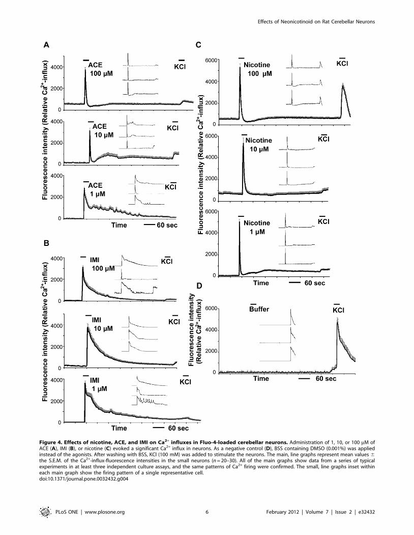
Figure 4. Effects of nicotine, ACE, and IMI on Ca2+ influxes in Fluo-4-loaded cerebellar neurons. Administration of 1, 10, or 100 mM ofACE (A), IMI (B), or nicotine (C) evoked a significant Ca2+ influx in neurons. As a negative control (D), BSS containing DMSO (0.001%) was appliedinstead of the agonists. After washing with BSS, KCl (100 mM) was added to stimulate the neurons. The main, line graphs represent mean values 6the S.E.M. of the Ca2+-influx-fluorescence intensities in the small neurons (n = 20–30). All of the main graphs show data from a series of typicalexperiments in at least three independent culture assays, and the same patterns of Ca2+ firing were confirmed. The small, line graphs inset withineach main graph show the firing pattern of a single representative cell.doi:10.1371/journal.pone.0032432.g004
Effects of Neonicotinoid on Rat Cerebellar Neurons
PLoS ONE | www.plosone.org 6 February 2012 | Volume 7 | Issue 2 | e32432
also absorbed in vivo by active transporters in the intestines. An in
vivo study revealed that ACE and IMI readily pass through the
blood-brain barrier [46]. Furthermore, in mammals, some
metabolites of the neonicotinoids show high affinities for
mammalian nAChRs that are similar to those of nicotine [2].
These findings collectively suggest that these neonicotinoids may
be potent risks to human health.
ConclusionsThe present study is the first to show that ACE, IMI, and
nicotine exert similar effects at mammalian nAChRs. Based on our
results, we suggest that excitation or desensitization or both of
nAChRs by neonicotinoids may affect the developing mammalian
nervous system, as is known to occur with nicotine. Further
investigation is required to clarify the mechanisms of action of
these substances and to determine safe concentrations for their
application to agricultural crops as pesticides.
Materials and Methods
Animals and ethics statementSprague-Dawley rats (Clea Japan, Inc., Tokyo, Japan) were
used in all experiments. All experiments were carried out in
accordance with a protocol (ID: 23-10) approved by the Care and
Use of Animals of Tokyo Metropolitan Institute of Medical
Science, and all efforts were made to minimize the number of
animals used and their suffering.
Cerebellar culturesThe details of the culture methods have been described
previously [47]. Briefly, the cerebella of P1 neonatal rats were
digested with papain, and dissociated cells were suspended in a
synthetic medium containing 1% fetal calf serum. The cells were
plated at a density of 2.56105 cells/0.2 ml on a glass-bottomed
dish (35-mm dish, 14-mm coverglass, Mat Tek Co., Ashland, MA,
USA) that was pre-coated with 0.1 mg/ml poly-L-lysine (Sigma-
Aldrich) and 10 mg/ml laminin (BD Biosciences, Franklin Lakes,
NJ, USA). After 2 days, the medium was replaced with serum-free
synthetic medium to prevent the growth of astrocytes. The serum-
free synthetic medium consisted of Dulbecco’s modified Eagle
medium/F12 (GibcoH, Invitrogen, Carlsbad, CA, USA) with
10 mg/ml bovine insulin (Sigma-Aldrich), 100 mg/ml transferrin
(Sigma-Aldrich), 30 nM sodium selenite, 5 nM thyroxine, 100 mM
putrescine (Sigma-Aldrich), and penicillin-streptomycin (100
units/ml and 100 mg/ml, respectively, Gibco). One-half of the
culture medium was replaced with a fresh medium every 3–4 days
for the 16 DIV.
ChemicalsNicotine ((-)-nicotine) was purchased from Sigma-Aldrich (St
Louis, MO, USA) at purities .99%. The neonicotinoid
insecticides 1-(6-chloro-3-pyridylmethyl)-N-nitroimidazolidin-2-
ylideneamine (IMI, purity .98%) and (E)-N1-[(6-chloro-3-pyridyl)
methyl]-N2-cyano-N1 -methylacetamidine (ACE, purity .98%)
were purchased from Kanto Chemicals Inc. (Tokyo, Japan) and
Wako Chemicals Inc. (Osaka, Japan), respectively. Their chemical
structures are shown in Figure 2A–C. They were dissolved in
dimethyl sulfoxide (DMSO, Sigma-Aldrich), and their stock
solutions (100 mM) were frozen at 230uC immediately prior to
use to minimize their inactivation and degradation. The nAChR
antagonists MEC (Sigma-Aldrich), a-BT (CalbiochemH, Merck,
Darmstadt, Germany), and DHbE (Sigma-Aldrich) [48,49] were
used.
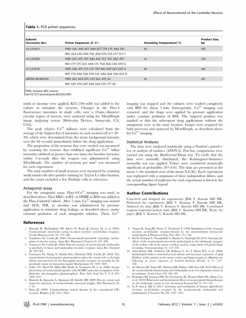
Detection of nAChR subunit mRNA using RT-PCRExpression of the a3, a4, and a7 nAChR subunits in cerebellar
cultured cells was examined by RT-PCR. The primer sequences
were essentially the same as those used by Moccia et al. [50]. The
primer sequences, annealing temperature, and predicted product
sizes are presented in Table 1. Expression of GAPDH was used as
a positive control. Total RNA was extracted from the cerebellar
cultures at 14 and 16 DIV and renal fibroblast cultures using the
RNeasy Mini Kit (Qiagen, Tokyo, Japan). Using 2.5 mg of total
RNA, RT-PCR was performed with RT-Ace (Toyobo, Tokyo,
Japan) according to the manufacturer’s protocol. PCRs for the a3
(L31621), a4 (L31620), and a7 (L31619) subunits and GAPDH
(BC082592) were carried out using 0.5 ml of each reverse-
transcribed solutions and AmpliTaq Gold (Applied Biosystems,
Figure 5. Peak of Ca2+ influxes and proportions of excitedneurons. (A) Nicotine evoked higher peak Ca2+ influxes than ACE orIMI, whereas these two neonicotinoids showed similar peak values. Thepeak relative Ca2+ influx was calculated as the mean 6 the S.E.M. of thehighest fluo-4 intensities of each excited cell (n = 20–30), which weredetermined from the mean background intensity over the 60 secondsimmediately before the drug application. Data within the same drugwere analyzed statistically using a Student’s paired t-test and nosignificant differences were observed (gray bar, # P.0.1). Data acrossthe different drugs were analyzed by an ANOVA with a post hocBonferroni/Dunn test. Significant differences (* P,0.05) were observedbetween nicotine and the two neonicotinoids. All of the main graphsshow data from a series of typical experiments in at least threeindependent culture assays, and similar values were confirmed. (B) Theproportion of the excited neurons was compared following adminis-trations of ACE, IMI, and nicotine at concentrations of 1–100 mM. Datawithin the same drug were analyzed statistically using a Student’s t-test,and no significant differences were observed. Data across the differentdrugs were analyzed by ANOVA with a post hoc Bonferroni/Dunn test.The data represent the mean 6 the S.E.M. of three to four independentexperiments.doi:10.1371/journal.pone.0032432.g005
Effects of Neonicotinoid on Rat Cerebellar Neurons
PLoS ONE | www.plosone.org 7 February 2012 | Volume 7 | Issue 2 | e32432
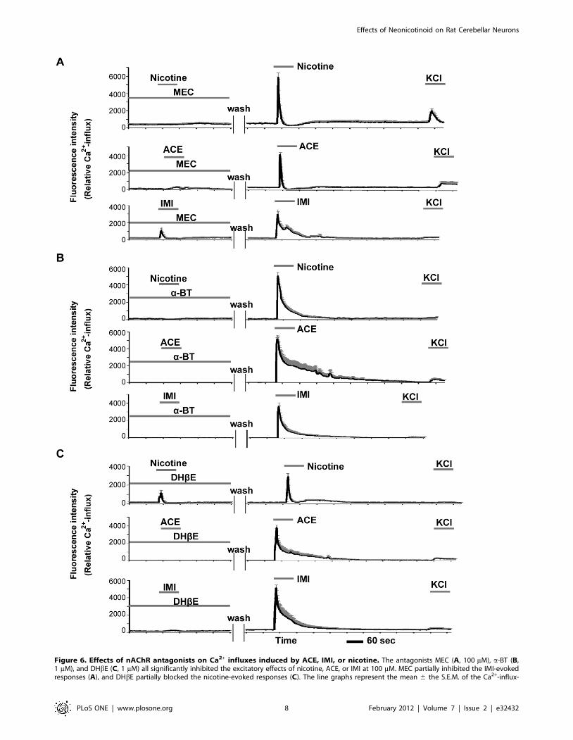
Figure 6. Effects of nAChR antagonists on Ca2+ influxes induced by ACE, IMI, or nicotine. The antagonists MEC (A, 100 mM), a-BT (B,1 mM), and DHbE (C, 1 mM) all significantly inhibited the excitatory effects of nicotine, ACE, or IMI at 100 mM. MEC partially inhibited the IMI-evokedresponses (A), and DHbE partially blocked the nicotine-evoked responses (C). The line graphs represent the mean 6 the S.E.M. of the Ca2+-influx-
Effects of Neonicotinoid on Rat Cerebellar Neurons
PLoS ONE | www.plosone.org 8 February 2012 | Volume 7 | Issue 2 | e32432
CA, USA). In the PCR, the a3, a4, and a7 subunits were
amplified over 40 cycles. GAPDH was amplified over 25 cycles.
Renal fibroblast cultures were used as a negative control. The
culture methods were modified from Fu et al. [51]. Briefly, the
cortices of adult rat (SD) kidneys were collected, minced, and
cultured in culture flasks (NUNC, Roskilde, Denmark) using
DMEM containing 10% FBS. After 2–3 weeks, propagated cells
were dissociated with 0.25% trypsin, resuspended with culture
medium, and cultured for a further 2–12 weeks. Before passage,
the culture flasks were shaken to detach the more weakly adherent
macrophages. More than 90% of the adherent cells were renal
fibroblasts as identified by their elongated morphology and that
they were positive for chicken anti–fibronectin (Abcam, Cam-
bridge, MA) and mouse monoclonal anti-a-smooth muscle actin
(Sigma-Aldrich) antibodies. The cells were used for the experi-
ments after passage 3.
ImmunohistochemistryThe cultured cerebellar cells were fixed with 4% paraformal-
dehyde in 0.1 M phosphate buffer for 20 min at room
temperature or cold methanol for 5 min. For the detection of
specific antigens, the following primary antibodies were used:
mouse monoclonal anti-Tuj1 (Covance, Princeton, NJ, USA) as a
neuronal marker, rabbit anti-L1 (kindly provided from Dr. Asou)
[52] and rat monoclonal anti-L1 (Millipore, Billerica, MA, USA)
as granule cell markers, rabbit anti-calbindin D28K (Millipore) as
a Purkinje cell marker, and rabbit anti-glial fibrillary acidic protein
(GFAP, DAKO, Denmark) as an astrocyte marker. For double
labeling, the cells were incubated with primary antibodies against
TuJ1 and L1 or GFAP, then biotinylated anti-mouse or rabbit (rat)
IgG (Vector, Burlingame, CA, USA), and finally with avidin-
rhodamine (Vector) and corresponding FITC -conjugated second-
ary antibodies (from different host species, Chemicon and
Molecular Probes, Invitrogen). All of the stained cells were
examined by confocal laser microscopy (Zeiss, LSM510Meta,
Oberkochen, Germany).
Intracellular Ca2+ imagingFor Ca2+ imaging, we used cerebellar cultures that were 14–16
DIV. The cultures were loaded for 30 min at 37uC with 4 mM
Fluo-4 acetoxymethyl ester (Molecular Probes, Invitrogen) in BSS
that contained 0.14 M NaCl, 5.4 mM KCl, 1.8 mM CaCl2,
5.5 mM glucose, and 20 mM 4-(2-hydroxyethyl)piperazine-1-
ethanesulfonic acid (HEPES, pH 7.4). Just before the experiment,
the stock solutions of nicotine and neonicotinoids were dissolved
and diluted by BSS to contain the same concentration of DMSO
(#0.001%). Time-lapse images of Fluo-4 fluorescence ([Ca2+]i) in
cerebellar cells at 25uC were obtained using a ZEISS 510 Meta
confocal laser scanning microscope system. Fluo-4 fluorescence
was excited at 488 nm using an argon laser and the emitted
fluorescence was collected at .515 nm. The cultures were
continuously perfused at a rate of 1 ml/min with BSS by a
peristaltic pump (Gilson, Minipuls 3, Middleton, WI, USA), and
this flow rate changes the external solution surrounding the cells
within 1 min. For nicotine and the neonicotinoids, pressure
application was used at a rate of 0.5 ml/min by syringe pump
(New Era Pump Systems, Inc. NE-300, Farmingdale, NY, USA)
under continuous perfusion. To minimize desensitization of the
nAChRs by drug leaking from the pipette tip (diameter 70–
100 mm), the pipette used for the pressure application was placed
at a target position approximately 100 mm from the neuron, and a
minimal air bubble was inserted to isolate the agonist from the
solution. Images at 1.5-second intervals were collected both before
and after exposure to 1–100 mM of the neonicotinoids or nicotine
for about 600 seconds. Around 500 seconds after the neonicoti-
induced fluorescence intensities in small neurons (n = 20–30). All of the graphs show the data from a series of typical experiments in at least threeindependent culture assays, and the same patterns of Ca2+ firing were confirmed.doi:10.1371/journal.pone.0032432.g006
Figure 7. Effects of nAChR antagonists on the proportions ofexcited neurons. The proportions of the neurons excited by nicotine(A), ACE (B), or IMI (C) at 100 mM were compared in the presence orabsence of MEC (100 mM), a-BT (1 mM), or DHbE (1 mM). All threeantagonists significantly reduced the proportion of the cells that wereexcited by the agonists. The data represent the mean 6 the S.E.M. ofthree independent experiments and were analyzed using an ANOVAfollowed by a post hoc Bonferroni/Dunn test.doi:10.1371/journal.pone.0032432.g007
Effects of Neonicotinoid on Rat Cerebellar Neurons
PLoS ONE | www.plosone.org 9 February 2012 | Volume 7 | Issue 2 | e32432
noids or nicotine were applied, KCl (100 mM) was added to the
culture to stimulate the neurons. Changes in the Fluo-4
fluorescence intensities in single cells, over a 10-mm diameter
circular region of interest, were analyzed using the MetaMorph
image analyzing system (Molecular Devices, Sunnyvale, CA,
USA).
The peak relative Ca2+ influxes were calculated from the
average of the highest fluo-4 intensities in each excited cell (n = 20–
30), which were determined from the mean background intensity
over the 60 seconds immediately before the drug application.
The proportion of the neurons that were excited was measured
by counting the neurons that exhibited significant Ca2+ influx
intensities (defined as greater than two times the baseline intensity)
within 3 seconds after the reagent was administered, using
MetaMorph. The number of neurons per mm2 was measured
for each experiment.
The total numbers of small neurons were measured by counting
small round cells after positive staining by Tuj1or L1-after fixation,
and the count excluded a few Purkinje cells or astrocytes.
Antagonist assayFor the antagonist assay, Fluo-4-Ca2+ imaging was used, as
described above. First MEC, a-BT, or DHbE in BSS was added to
the Fluo-4-labeled culture. After 5 min, Ca2+ imaging was started
and ACE, IMI, or nicotine was administered by pressure
application to minimize drug leakage, as described above, under
constant perfusion of each antagonist solution. Then, Ca2+
imaging was stopped and the cultures were washed completely
with BSS for about 5 min. Subsequently, Ca2+ imaging was
restarted, and the drugs were applied by pressure application
under constant perfusion of BSS. The targeted position was
marked so that the subsequent drug applications without the
antagonists were at the same location. Images were acquired for
both processes and analyzed by MetaMorph, as described above
for Ca2+ imaging.
Statistical AnalysesThe data were analyzed statistically using a Student’s paired t-
test or analysis of variance (ANOVA). Post hoc comparisons were
carried out using the Bonferroni/Dunn test. To verify that the
data were normally distributed, the Kolmogorov-Smirnov-
normality test was applied. Values were considered statistically
significant at probability (P),0.05. The data are presented as the
mean 6 the standard error of the mean (S.E.M.). Each experiment
was replicated with a minimum of three independent dishes, and
the actual number of replicates for each experiment is listed in the
corresponding figure legend.
Author Contributions
Conceived and designed the experiments: JKK Y. Kuroda MH HK.
Performed the experiments: JKK Y. Komuta Y. Kuroda MH HK.
Analyzed the data: JKK Y. Komuta Y. Kuroda MH HK. Contributed
reagents/materials/analysis tools: JKK Y. Komuta MH HK. Wrote the
paper: JKK Y. Komuta Y. Kuroda MH HK.
References
1. Matsuda K, Buckingham SD, Kleier D, Rauh JJ, Grauso M, et al. (2001)Neonicotinoids: insecticides acting on insect nicotinic acetylcholine receptors.
Trends Pharmacol Sci 22: 573–580.
2. Tomizawa M, Casida JE (2005) Neonicotinoid insecticide toxicology: mecha-nisms of selective action. Annu Rev Pharmacol Toxicol 45: 247–268.
3. Tomizawa M, Casida JE (2003) Selective toxicity of neonicotinoids attributable
to specificity of insect and mammalian nicotinic receptors. Annu Rev Entomol
48: 339–364.
4. Tomizawa M, Zhang N, Durkin KA, Olmstead MM, Casida JE (2003) The
neonicotinoid electronegative pharmacophore plays the crucial role in the high
affinity and selectivity for the Drosophila nicotinic receptor: an anomaly for thenicotinoid cation–pi interaction model. Biochemistry 42: 7819–7827.
5. Talley TT, Harel M, Hibbs RE, Radic Z, Tomizawa M, et al. (2008) Atomic
interactions of neonicotinoid agonists with AChBP: molecular recognition of thedistinctive electronegative pharmacophore. Proc Natl Acad Sci U S A 105:
7606–7611.
6. Matsuda K, Kanaoka S, Akamatsu M, Sattelle DB (2009) Diverse actions andtarget-site selectivity of neonicotinoids: structural insights. Mol Pharmacol 76:
1–10.
7. Shim JY (2009) Transmembrane helical domain of the cannabinoid CB1
receptor. Biophys J 96: 3251–3262.
8. Nagata K, Song JH, Shono T, Narahashi T (1998) Modulation of the neuronalnicotinic acetylcholine receptor-channel by the nitromethylene heterocycle
imidacloprid. J Pharmacol Exp Ther 285: 731–738.
9. Bal R, Erdogan S, Theophilidis G, Baydas G, Naziroglu M (2010) Assessing the
effects of the neonicotinoid insecticide imidacloprid in the cholinergic synapsesof the stellate cells of the mouse cochlear nucleus using whole-cell patch-clamp
recording. Neurotoxicology 31: 113–120.
10. Abou-Donia MB, Goldstein LB, Bullman S, Tu T, Khan WA, et al. (2008)
Imidacloprid induces neurobehavioral deficits and increases expression of glialfibrillary acidic protein in the motor cortex and hippocampus in offspring rats
following in utero exposure. J Toxicol Environ Health A 71: 119–
130.
11. de Oliveira IM, Nunes BV, Barbosa DR, Pallares AM, Faro LR (2010) Effects ofthe neonicotinoids thiametoxam and clothianidin on in vivo dopamine release in
rat striatum. Toxicol Lett 192: 294–297.
12. Rodrigues KJ, Santana MB, Do Nascimento JL, Picanco-Diniz DL, Maues LA,
et al. (2010) Behavioral and biochemical effects of neonicotinoid thiamethoxamon the cholinergic system in rats. Ecotoxicol Environ Saf 73: 101–107.
13. Li P, Ann J, Akk G (2011) Activation and modulation of human alpha4beta2
nicotinic acetylcholine receptors by the neonicotinoids clothianidin and
imidacloprid. J Neurosci Res 89: 1295–1301.
Table 1. PCR primer sequences.
Subunit(Accession No.) Primer Sequences (59-39) Annealing Temperature(6C)
Product Size,bp
a3 (L31621) FWD: GAC AAG ACC AAA GCT CTA CTC AAG TAC 65 435
REV: GCA CAG AGA TGC AGA GTG TCA CCT TCT C
a4 (L31620) FWD: GCC ATC TAT AAG AGC TCC TGC AGC ATC 55 359
REV: CTT CTC GCC AAA CTC TGA AGG CAG ATA G
a7 (L31619) FWD: GAC ATT CTC CTC TAT AAC AGT GCT GAT G 60 405
REV: CTG AAA TGA GTA CAC AAG GGA TGA GCA G
GAPDH (BC082592) FWD: ACC ACA GTC CAT GCC ATC AC 55 451
REV: GAT GTG GAT GAA GAA GTG TTT GC
FWD, forward; REV, reverse.doi:10.1371/journal.pone.0032432.t001
Effects of Neonicotinoid on Rat Cerebellar Neurons
PLoS ONE | www.plosone.org 10 February 2012 | Volume 7 | Issue 2 | e32432
14. Role LW, Berg DK (1996) Nicotinic receptors in the development and
modulation of CNS synapses. Neuron 16: 1077–1085.
15. Dwyer JB, McQuown SC, Leslie FM (2009) The dynamic effects of nicotine onthe developing brain. Pharmacol Ther 122: 125–139.
16. Tomizawa M, Casida JE (1999) Minor structural changes in nicotinoid
insecticides confer differential subtype selectivity for mammalian nicotinic
acetylcholine receptors. Br J Pharmacol 127: 115–122.
17. Didier M, Berman SA, Lindstrom J, Bursztajn S (1995) Characterization of
nicotinic acetylcholine receptors expressed in primary cultures of cerebellar
granule cells. Brain Res Mol Brain Res 30: 17–28.
18. Hogberg HT, Kinsner-Ovaskainen A, Coecke S, Hartung T, Bal-Price AK
(2010) mRNA expression is a relevant tool to identify developmental
neurotoxicants using an in vitro approach. Toxicol Sci 113: 95–115.
19. Nagata I, Ono K, Kawana A, Kimura-Kuroda J (2006) Aligned neurite bundlesof granule cells regulate orientation of Purkinje cell dendrites by perpendicular
contact guidance in two-dimensional and three-dimensional mouse cerebellar
cultures. J Comp Neurol 499: 274–289.
20. Moser N, Mechawar N, Jones I, Gochberg-Sarver A, Orr-Urtreger A, et al.(2007) Evaluating the suitability of nicotinic acetylcholine receptor antibodies for
standard immunodetection procedures. J Neurochem 102: 479–492.
21. Huang LZ, Abbott LC, Winzer-Serhan UH (2007) Effects of chronic neonatal
nicotine exposure on nicotinic acetylcholine receptor binding, cell death andmorphology in hippocampus and cerebellum. Neuroscience 146: 1854–1868.
22. Zhang X, Liu C, Miao H, Gong ZH, Nordberg A (1998) Postnatal changes of
nicotinic acetylcholine receptor alpha 2, alpha 3, alpha 4, alpha 7 and beta 2
subunits genes expression in rat brain. Int J Dev Neurosci 16: 507–518.
23. Agulhon C, Charnay Y, Vallet P, Abitbol M, Kobetz A, et al. (1998) Distribution
of mRNA for the alpha4 subunit of the nicotinic acetylcholine receptor in the
human fetal brain. Brain Res Mol Brain Res 58: 123–131.
24. Kawa K (2002) Acute synaptic modulation by nicotinic agonists in developing
cerebellar Purkinje cells of the rat. J Physiol 538: 87–102.
25. Oikawa H, Nakamichi N, Kambe Y, Ogura M, Yoneda Y (2005) An increase inintracellular free calcium ions by nicotinic acetylcholine receptors in a single
cultured rat cortical astrocyte. J Neurosci Res 79: 535–544.
26. Matsuda K, Shimomura M, Ihara M, Akamatsu M, Sattelle DB (2005)
Neonicotinoids show selective and diverse actions on their nicotinic receptortargets: electrophysiology, molecular biology, and receptor modeling studies.
Biosci Biotechnol Biochem 69: 1442–1452.
27. Dajas-Bailador F, Wonnacott S (2004) Nicotinic acetylcholine receptors and the
regulation of neuronal signalling. Trends Pharmacol Sci 25: 317–324.
28. Shen JX, Yakel JL (2009) Nicotinic acetylcholine receptor-mediated calcium
signaling in the nervous system. Acta Pharmacol Sin 30: 673–680.
29. Giniatullin R, Nistri A, Yakel JL (2005) Desensitization of nicotinic AChreceptors: shaping cholinergic signaling. Trends Neurosci 28: 371–378.
30. Pocock JM, Cousin MA, Parkin J, Nicholls DG (1995) Glutamate exocytosis
from cerebellar granule cells: the mechanism of a transition to an L-type Ca2+channel coupling. Neuroscience 67: 595–607.
31. Lacinova L (2005) Voltage-dependent calcium channels. Gen Physiol Biophys
24 Suppl 1: 1–78.
32. Masgrau R, Servitja JM, Sarri E, Young KW, Nahorski SR, et al. (2000)Intracellular Ca2+ stores regulate muscarinic receptor stimulation of phospho-
lipase C in cerebellar granule cells. J Neurochem 74: 818–826.
33. Marchi M, Lupinacci M, Bernero E, Bergaglia F, Raiteri M (1999) Nicotinic
receptors modulating ACh release in rat cortical synaptosomes: role of Ca2+ions in their function and desensitization. Neurochem Int 34: 319–328.
34. Mao C, Lv J, Li H, Chen Y, Wu J, et al. (2007) Development of fetal nicotine
and muscarinic receptors in utero. Braz J Med Biol Res 40: 735–741.35. Mao C, Yuan X, Cui Y, Li H, Lv J, et al. (2008) Prenatal exposure to nicotine
with associated in utero hypoxia decreased fetal brain muscarinic mRNA in the
rat. Brain Res 1189: 43–50.36. Slikker W, Jr., Xu ZA, Levin ED, Slotkin TA (2005) Mode of action: disruption
of brain cell replication, second messenger, and neurotransmitter systems duringdevelopment leading to cognitive dysfunction–developmental neurotoxicity of
nicotine. Crit Rev Toxicol 35: 703–711.
37. Winzer-Serhan UH (2008) Long-term consequences of maternal smoking anddevelopmental chronic nicotine exposure. Front Biosci 13: 636–649.
38. Eugenin J, Otarola M, Bravo E, Coddou C, Cerpa V, et al. (2008) Prenatal toearly postnatal nicotine exposure impairs central chemoreception and modifies
breathing pattern in mouse neonates: a probable link to sudden infant deathsyndrome. J Neurosci 28: 13907–13917.
39. Ward C, Lewis S, Coleman T (2007) Prevalence of maternal smoking and
environmental tobacco smoke exposure during pregnancy and impact on birthweight: retrospective study using Millennium Cohort. BMC Public Health 7: 81.
40. Neuman RJ, Lobos E, Reich W, Henderson CA, Sun LW, et al. (2007) Prenatalsmoking exposure and dopaminergic genotypes interact to cause a severe
ADHD subtype. Biol Psychiatry 61: 1320–1328.
41. Cao J, Dwyer JB, Mangold JE, Wang J, Wei J, et al. (2011) Modulation of celladhesion systems by prenatal nicotine exposure in limbic brain regions of
adolescent female rats. Int J Neuropsychopharmacol 14: 157–174.42. Wei J, Wang J, Dwyer JB, Mangold J, Cao J, et al. (2011) Gestational nicotine
treatment modulates cell death/survival-related pathways in the brains ofadolescent female rats. Int J Neuropsychopharmacol 14: 91–106.
43. Rice D, Barone S, Jr. (2000) Critical periods of vulnerability for the developing
nervous system: evidence from humans and animal models. Environ HealthPerspect 108 Suppl 3: 511–533.
44. Brunet JL, Maresca M, Fantini J, Belzunces LP (2004) Human intestinalabsorption of imidacloprid with Caco-2 cells as enterocyte model. Toxicol Appl
Pharmacol 194: 1–9.
45. Brunet JL, Maresca M, Fantini J, Belzunces LP (2008) Intestinal absorption ofthe acetamiprid neonicotinoid by Caco-2 cells: transepithelial transport, cellular
uptake and efflux. J Environ Sci Health B 43: 261–270.46. Ford KA, Casida JE (2006) Chloropyridinyl neonicotinoid insecticides: diverse
molecular substituents contribute to facile metabolism in mice. Chem ResToxicol 19: 944–951.
47. Kimura-Kuroda J, Nagata I, Negishi-Kato M, Kuroda Y (2002) Thyroid
hormone-dependent development of mouse cerebellar Purkinje cells in vitro.Brain Res Dev Brain Res 137: 55–65.
48. Sampaio LF, Hamassaki-Britto DE, Markus RP (2005) Influence of melatoninon the development of functional nicotinic acetylcholine receptors in cultured
chick retinal cells. Braz J Med Biol Res 38: 603–613.
49. Wu J, Liu Q, Yu K, Hu J, Kuo YP, et al. (2006) Roles of nicotinic acetylcholinereceptor beta subunits in function of human alpha4-containing nicotinic
receptors. J Physiol 576: 103–118.50. Moccia F, Frost C, Berra-Romani R, Tanzi F, Adams DJ (2004) Expression and
function of neuronal nicotinic ACh receptors in rat microvascular endothelialcells. Am J Physiol Heart Circ Physiol 286: H486–491.
51. Fu P, Liu F, Su S, Wang W, Huang XR, et al. (2006) Signaling mechanism of
renal fibrosis in unilateral ureteral obstructive kidney disease in ROCK1knockout mice. J Am Soc Nephrol 17: 3105–3114.
52. Takeda Y, Murakami Y, Asou H, Uyemura K (2001) The roles of cell adhesionmolecules on the formation of peripheral myelin. Keio J Med 50: 240–248.
Effects of Neonicotinoid on Rat Cerebellar Neurons
PLoS ONE | www.plosone.org 11 February 2012 | Volume 7 | Issue 2 | e32432
Related Documents