NICOLE BOURY-ESNAULT and KLAUS RUTZLER m HDITORS SMITHSONIAN CONTRIBUTIONS TO ZOOLOGY • NUMBER 596

Welcome message from author
This document is posted to help you gain knowledge. Please leave a comment to let me know what you think about it! Share it to your friends and learn new things together.
Transcript

NICOLE BOURY-ESNAULTand
KLAUS RUTZLER
m HDITORS
SMITHSONIAN CONTRIBUTIONS TO ZOOLOGY • NUMBER 5 9 6
SERIES PUBLICATIONS OF THE SMITHSONIAN INSTITUTION
Emphasis upon publication as a means of "diffusing knowledge" was expressed by the firstSecretary of the Smithsonian. In his formal plan for the institution, Joseph Henry outlined aprogram that included the following statement: "It is proposed to publish a series of reports,giving an account of the new discoveries in science, and of the changes made from year to yearin all branches of knowledge." This theme of basic research has been adhered to through theyears by thousands of titles issued in series publications under the Smithsonian imprint,commencing with Smithsonian Contributions to Knowledge in 1848 and continuing with thefollowing active series:
Smithsonian Contributions to Anthropology
Smithsonian Contributions to Botany
Smithsonian Contributions to the Earth Sciences
Smithsonian Contributions to the Marine Sciences
Smithsonian Contributions to Paleobiology
Smithsonian Contributions to Zoology
Smithsonian Folklife Studies
Smithsonian Studies in Air and Space
Smithsonian Studies in History and Technology
In these series, the Institution publishes small papers and full-scale monographs that reportthe research and collections of its various museums and bureaux or of professional colleaguesin the world of science and scholarship. The publications are distributed by mailing lists tolibraries, universities, and similar institutions throughout the world.
Papers or monographs submitted for series publication are received by the SmithsonianInstitution Press, subject to its own review for format and style, only through departments of thevarious Smithsonian museums or bureaux, where the manuscripts are given substantive review.Press requirements for manuscript and art preparation are outlined on the inside back cover.
I. Michael HeymanSecretarySmithsonian Institution
S M I T H S O N I A N C O N T R I B U T I O N S T O Z O O L O G Y • N U M B E R 5 9 6
Thesaurus of Sponge Morphology
Nicole Boury-Esnaultand Klaus Rutzler
EDITORS
SMITHSONIAN INSTITUTION PRESS
Washington, D.C.
1997

A B S T R A C T
Boury-Esnault, Nicole, and Klaus Rutzler, editors. Thesaurus of Sponge Morphology.Smithsonian Contributions to Zoology, number 596, 55 pages, 305 figures, 1997.—This is avade mecum of terms for describing all aspects of sponge morphology, such as habit, surfacestructure, consistency, and anatomy. Definitions of microscopic features include aquiferoussystem, cytology, reproductive structures, skeletal architecture, and calcareous and siliceousspicules. Terms were selected and reviewed by an international group of 10 experts in spongesystematics, during several workshops. Each entry is accompanied by a precise diagnosis and anillustration. Terms listed as "rejected" are those judged unsuitable, redundant, or preoccupied.The thesaurus will aid descriptions in future systematic papers, computerized data banks, andidentification keys. The literature cited presents a historical perspective on similar attempts tocreate a precise terminology of sponge morphology and helps to further clarify the selectedterms.
OFFICIAL PUBLICATION DATE is handstamped in a limited number of initial copies and isrecorded in the Institution's annual report, Annals of the Smithsonian Institution. SERIES COVERDESIGN: The coral Montastrea cavernosa (Linnaeus).
Library of Congress Cataloging-in-Publication DataThesaurus of sponge morphology / Nicole Boury-Esnault and Klaus Rutzler.p. cm.—(Smithsonian contributions to zoology ; no. 596)Includes bibliographical references (p. ).I. Sponges—Morphology—Terminology. I. Boury-Esnault, Nicole. II. Rutzler, Klaus. III. Series.QLl.S54no. 596[QL374]57l.3'I4'014-dc21 97-20213
© The paper used in this publication meets the minimum requirements of the AmericanNational Standard for Permanence of Paper for Printed Library Materials Z39.48—1984.

Contents
Page
Contributors ivIntroduction 1
Acknowledgments 1Recommended Procedures 2
Thesaurus 3Macroscopical Features: Habit, Surface Characteristics, and Consistency 4Anatomy and Aquiferous System 8Cytology 10Reproduction 16Architecture of the Skeleton and Spicules of Hexactinellida 18Calcareous Spicules and Calcareous Spicule Skeleton 26Architecture of the Skeleton of Demospongiae 28Calcareous Rigid Skeleton 36Spicules of Demospongiae 42Spicule Types of "Lithistids" 50
Literature Cited 54
in

Contributors
Radovan Borojevic, Universidade Federal de Rio de Janeiro, Departamento de Histologiae Embriologia, C? 68021, 21944-970 Ilha do Fundao, Rio de Janeiro, Brazil.
Nicole Boury-Esnault, Centre d'Oceanologie de Marseille, Station marine d'Endoume, ruede la Batterie des Lions, 13007 Marseille, France.
Ruth Desqueyroux-Faundez, Musee d'Histoire Naturelle, case postale 6434,1211 Geneve6, Switzerland.
Claude Levi, Museum National d'Histoire Naturelle, Laboratoire de Biologie desInvertebres marins et Malacologie, 57 rue Cuvier, 75231 Paris cedex 5, France.
Maurizio Pansini, Istituto di Zoologia della Universita, Via Balbi 5, 16126 Genova, Italy.
Klaus Rutzler, National Museum of Natural History, Smithsonian Institution, Washington,DC. 20560, USA.
Rob W.M. van Soest, Institute of Systematics and Population Biology (ZoologicalMuseum), University of Amsterdam, P.O. Box 94766, 1090 GT Amsterdam, TheNetherlands.
Shirley Stone, British Museum Natural History, Department of Zoology, SpongeDepartment, Cromwell Road, London SW 7, Great Britain.
Maria-Jesus Uriz, Centro d'Estudis Avancats de Blanes, CSIC, cami de Santa Barbara,17300 Blanes Girona, Spain.
Jean Vacelet, Centre d'Oceanologie de Marseille, Station marine d'Endoume, rue de laBatterie des Lions, 13007 Marseille, France.
IV

Thesaurus of Sponge Morphology
Nicole Boury-Esnaultand Klaus Rutzler
Editors
Introduction
Since the end of the nineteenth century, sponge systematistshave periodically pursued the important task of compiling a listof accepted terms that would allow species to be described in anobjective manner. In 1888, W.J. Sollas, S.O. Ridley, A. Dendy,and R. von Lendenfeld met in London to establish aterminology of spicule types, which is generally used to thisdate (Schulze and Lendenfeld, 1889). In the late 1960s R.Borojevic, W.G. Fry, W.C. Jones, C. Levi, R. Rasmont, M.Sara, and J. Vacelet met in Roscoff, France, to update andstandardize a terminology for sponges (Borojevic et al., 1968).Since then, many important events have taken place—such asthe increase in the availability of the electron microscope,appearance of automated data processing and its use in buildingdata banks, and application of cladistic theory to spongephylogeny—all of which necessitate another updating. Further-more, natural product chemists are showing more and moreinterest in marine invertebrates in general and sponges inparticular and thus require precise identifications for an everincreasing volume of specimens.
Nevertheless, sponge systematics is still in its infancy. Thatis to say, sponges are one of the rare phyla over whichagreement has not been reached, even at the level of order. Thissituation is partly due to a lack of specialists. Sponges arereportedly the phylum with the smallest ratio of number ofspecialists to number of species (Winston, 1988). In view ofthese problems, a group of European and American systema-tists met repeatedly over several years to establish a standard
Nicole Boury-Esnault, Centre d Oceanologie de Marseille, Stationmarine d'Endoume, rue de la Batterie des Lions, 13007 Marseille,France.Klaus Rutzler, National Museum of Natural History, SmithsonianInstitution, Washington, D.C. 20560.
terminology and precise definitions for descriptive morpho-logical purposes. This monograph is the outcome of thosemeetings. The principal aim here is to be as thorough aspossible in disposing of all ambiguities connected withlanguage in common use. For instance, a single term is oftenused for morphologically or functionally similar yet nothomologous characteristics. Just as the word "wing" is used forthe flight organ of birds, insects, and bats (although no onewould suggest it as a synapomorphy linking the three groups toa common ancestor), there are terms, such as "triactine" or"amphiblastula," that serve to designate analogous and more orless comparable but not homologous elements in differentclasses of sponges. To help researchers, particularly evolution-ary biologists and paleontologists, avoid misinterpretingcharacters based on such unsuitable terminology, we haveredefined existing designations more precisely and haveintroduced new terms where necessary.
To facilitate the use of this thesaurus, illustrations appear onthe page facing their definitions. Literature references used forpreparing figures and text are cited at the beginning of eachsection. Terms defined elsewhere in the text are set in smallcapitals.
ACKNOWLEDGM ENTS
We thank Patricia Condit for drawing or redrawing andinking the majority of illustrations. Molly Kelly Ryan designedthe layout of figures, advised on illustration techniques, andprepared the figures for "Macroscopical Features: Habit,Surface Characteristics, and Consistency." Kathleen P. Smithrendered the figures for "Spicule Types of Demospongiae"using an Apple Macintosh Power PC and Adobe Illustratorversion 5.5. Patricia R. Bergquist, John N.A. Hooper, ShirleyA. Pomponi, and Michele Sara reviewed various stages of theproject and made many valuable suggestions for its improve-ment.

SMITHSONIAN CONTRIBUTIONS TO ZOOLOGY
RECOMMENDED PROCEDURES
Below is an outline of the most basic procedures used inpreparing sponges for study. More details and additionaltechniques can be found in Rutzler (1978), Reiswig andBrowman (1987), De Vos et al. (1991), and Hooper (1991).References to the general sponge literature are found inBergquist (1978) and De Vos et al. (1991).
DESCRIPTION.—In describing or redescribing a species, onemust mention the following diagnostic elements: (1) generalshape, color, surface structure, and distribution of openings ofthe aquiferous system; (2) skeleton organization and elements(spicules and fibers), accompanied by camera lucida drawingsor photographs and by measurements of sizes (ranges, means)calculated from at least 10 measurements; (3) if possible, celland choanocyte-chamber types and sizes and reproductivecharacteristics; and (4) information on ecological and biologi-cal activity, including exact locality, habitat, depth, substrate,water clarity, light, temperature, associated species, andreproductive mode.
FIXATION AND PRESERVATION.—Numerous standard histo-
logical fixatives work well for sponges, but the simplest andmost reliable is 10% formalin in seawater. Buffer additions aregenerally not necessary; hexamine buffer should definitely notbe used because it promotes maceration of the sample. Afterfixation of a few hours to a few days (depending on the size andtype of material), the samples must be transferred to and storedin at least 70% alcohol.
SPICULE PREPARATION.—For siliceous sponges, boil a
fragment (~0.5 cm3) in a test tube with about 10 times thevolume of fuming nitric acid (HNO3) until the cellular materialis dissolved and the liquid is clear; fill the sample tube withdistilled water and let the spicules settle to the bottom for atleast 2 hr. (the process may be speeded up by gentlecentrifugation). Remove water by pipette and change (threetimes). After the third water change, rinse with 95% alcohol inthe same way, again three times. After the last change, suspendspicules in 1-2 ml alcohol and pour onto a microscope slide;
let the alcohol evaporate and then dry the slide at ~60°C(burning off the alcohol may cause unwanted clustering ofspicules). Some workers prefer to acid-boil small fragments orsections of sponge directly on a microscope slide (boil until justdry). Cover with a thin layer of mounting media (Caedax,Permount, Araldite), and cover slip. If the spicule size range isvery large, mount up to three size fractions separately to allowstudy of the smaller spicules by high magnification objectives,which have a very short working distance. If the spongespecimen under study is large, it may be important to make apreparation from the interior as well as from the surface area.
For calcareous sponges, dissolve sponge fragment in anaqueous solution of sodium hypochlorite (Clorox, Eau deJavelle) and continue processing according to the methodsgiven above.
In the case of very small specimens, one may prefer to makethe preparation directly on the microscope slide. Put a smallpiece of the sponge on the slide, and add two to three drops offuming nitric acid (for siliceous sponges); boil carefully byrepeatedly pulling the slide across the flame of a Bunsen oralcohol burner. Replace evaporated acid by adding a few moredrops. As soon as the remaining ash is clean, rinse with 90%alcohol, dry, and mount in resin.
PREPARATION OF SKELETON STRUCTURE.—Make thin hand
sections from a piece of the sponge, cutting perpendicular andparallel to the surface with a fresh, thin, razor blade; make sureto identify the direction of the sections, particularly the one thatincludes the surface layer. Dehydrate (for instance, 0.5 hr. inbutyl alcohol; saturated solution of phenol in xylene) andmount in the same kind of media as the spicules. If a stain isdesired for contrast, add basic fuchsin to one step of thedehydration alcohol. Very soft specimens may be embedded inparaffin for sectioning. Keratose sponge skeletons may bebetter studied after separating the spongin fibers or network byteasing away cellular material using fine forceps and dissectingneedles. The separated fibers can then be dehydrated andmounted using the aforementioned procedures.

Thesaurus

SMITHSONIAN CONTRIBUTIONS TO ZOOLOGY
Macroscopical Features: Habit, Surface Characteristics,and Consistency
LITERATURE CONSULTED: Brown, 1956.
agglutinating. Creating substrate or support by cementingtogether a mixture of foreign material, such as sand, shell,or rock.
amorphous. Without definite shape. To be rejected, vagueterm. See MASSIVE.
arborescent. Erect, branching habit, tree-like in appearance(Axinella polypoides) (Figure 1).
areolated. Surface covered by numerous circular ectosomalareas (Hamigera) (Figure 2).
boring. See EXCAVATING.branching. Spreading out in branches (see also ARBORESCENT
and REPENT).
burrowing. See EXCAVATING.caliculate. Cup shaped (Poterion neptuni) (Figure 3).clathrate. Resembling open latticework (Clathrina coriacea)
(Figure 4).clavate. Club shaped (Rhizaxinella pyrifera) (Figure 5).columnar. Shape of solid, erect cylinder (Petrosia capsa)
(Figure 6).compressible. Easily squeezed.conulose. Surface with numerous cone-shaped projections
raised up by underlying skeleton (Dysidea sp.) (Figure 7).corrugated. Surface with alternating parallel ridges and
grooves (Callyspongia plicifera) (Figure 8).crateriform. Massive shape, with a broad base and large
central depression (Xestospongia muta).cup shaped. See CALICULATE.dendritic. To be rejected for habit (term used as a descriptor of
skeleton, see "Architecture of the Skeleton of De-mospongiae").
digitate. Deeply divided, finger-like outgrowths from basalmass (Halichondria bowerbanki) (Figure 9).
elastic. Resilient.encrusting. Thin, sheet-like coating of the substrate (Hyme-
desmia spp.) (Figure 10).endolithic. Occupying cavities in hard substrata (see also
EXCAVATING).
endopsammic. Main part of body buried in sand (Oceanapiapeltata) (Figure 11).
erect. General term for having a vertical (away from substrate)growth strategy.
excavating. Living in galleries or cavities bored into lime-stone or other calcareous materials (species of Cliona,
Thoosa, Aka) (Figure 12).ficiform. Fig shaped {Petrosia ficiformis) (Figure 13).firm. Solid, requires considerable pressure to deform sponge
(species of Chondrosia).fistulose. Bearing fistules (Coelosphaera hechteli) (Figure
14). See also FISTULE in "Anatomy and AquiferousSystem."
flabellate. Fan shaped (Phakellia ventilabrum) (Figure 15).flagelliform. Shaped as a single, very long, erect branch
(Raspailia viminalis) (Figure 16).foliaceous. In the form of a leaf (Carteriospongia foliacens)
(Figure 17).foliose. See FOLIACEOUS.fragile. Easily broken.friable. Easily broken, delicate, brittle.glabrous. See SMOOTH.globular. Ball shaped, spherical (Tethya aurantium) (Figure
18).glutinous. Sticky (Reniera mucosa).gyriform. See SULCATE.hard. Firm consistency, unyielding to pressure (most lithis-
tids, species of Petrosia).hispid. Surface with long and scattered spicular projections
(Stelletta spp.) (Figure 19).honeycombed. Surface with polygonal pattern of ridges
(Verongula gigantea) (Figure 20).incompressible. Not easily squeezed.infundibuliform. Funnel shaped (Callyspongia plicifera)
(Figure 21).lamellate. Plate-like erect (Phyllospongia lamellosa).limp. Soft to the point of collapsing out of water.lipostomous. Condition of a surface in which oscula and ostia
are inconspicuous (in old descriptions).lobate. Having rounded projections.lobodigitate. To be rejected (tautology).massive. Large, compact structure without definable shape
(Spongia officinalis).muricate. See SPINY.ovate. Egg shaped, ellipsoid (Cinachyrella) (Figure 22).palmate. Hand shaped (Isodictya palmata) (Figure 23).palmodigitate. To be rejected (tautology).papillate. Surface bearing papillae (Polymastia spp.) (Figure
24). See also PAPILLA in "Anatomy and AquiferousSystem."
pedunculate. Supported by a short stalk containing choano-some (Aplysina pedunculata) (Figure 25).
perforating. See EXCAVATING.

NUMBER 596
y/yyyyy//zmy/
^22 v///y////y- 25

SMITHSONIAN CONTRIBUTIONS TO ZOOLOGY
pinnate. Feather shaped (Asbestopluma pennatula) (Figure26).
platy. Thickly lamellate, usually in horizontal orientation.polymorphic. Occurring in different shapes.punctate. Surface appearing dotted because of microscopic
pores.pyriform. See FICIFORM (Figure 13).repent. Growing along or just above the substrate, simple or
branching, attaching to the substrate at intervals (Hali-clona simulans) (Figure 27).
resilient. Resumes original shape after deformation.ribbed. Surface with series of ridges (Xestospongia muta)
(Figure 28); ridges not necessarily parallel, as in CORRU-GATED.
rooted. Anchored to or into substrate with root-like processes(Pheronema grayi).
rubbery. Resilient and tough.rugose. Having a rough and ridged surface {Topsentia spp.)
(Figure 29).sessile. Permanently attached to substrate.smooth. Surface without any projections (Chondrosia reni-
formis).soft Yielding to pressure, easily torn.spiny. Surface with scattered, stiff, and sharp spicular or fiber
projections (Pleraplysilla spinifera) (Figure 30).stipitate. Supported by a long stalk (Stylocordyla stipitata)
(Figure 31).stoloniferous. Condition in which distinct, massive parts of a
sponge are interconnected by repent, rope-like structures(Haliclona tubifera).
sulcate. Furrowed with longitudinal, or meandering (gyri-form), parallel channels (Myrmekioderma styx) (Figure32).
tough. Resistant to tearing (Ircinia).tuberculate. See VERRUCOSE.
tubular. Shape of hollow, erect cylinder (Aplysina fistularis)(Figure 33).
turbinate. Resembling an inverted cone (Cribochalina vas-culum) (Figure 34).
vallate. To be rejected (old-fashioned, heraldic term used todescribe massive forms).
velvety. Surface with dense, short spicular projections, feelingsoft and smooth to the touch.
verrucose. Warty (genus Tethya) (Figure 35).vesicular. Hollow, bladder-like (Inflatella belli).villose. Surface shaggy, with dense, long spicular bundles
(Rosella villosa) (Figure 36).

NUMBER 596
V 26 28 29
27
30 31
i .:;i
I32 /33 36

SMITHSONIAN CONTRIBUTIONS TO ZOOLOGY
Aanatomy and Aquiferous System
LITERATURE CONSULTED: Borojevic et al., 1968; Boury-Esnault,1972, 1974; Boury-Esnault et al., 1990; Rutzler, 1987; Sollas, 1888.
aphodal. Type of choanocyte chamber that connects directlywith the inhalant canals through prosopyles and with theexhalant canal through an apopyle extended by anaphodus. Only one chamber opens into one aphodus(Figure 37).
aphodus. Small exhalant canal leading from the apopyle to anexhalant canal (ap in Figure 37).
apopyle. Opening of a choanocyte chamber into an exhalantcanal (apo in Figure 37).
aquiferous papilla. See PAPILLA.aquiferous system. The entire water-conducting system be-
tween the ostia and the oscula, which comprises theinhalant system, choanocyte chambers, and the exhalantsystem (Figure 38).
asconoid. Aquiferous system in which the internal cavity ofthe sponge is entirely lined by choanocytes (Figure 39).
atrium ("spongocoel," "cloaca," or "gastral cavity").Preoscular cavity. This term is used specifically todesignate the central exhalant cavity (Figure 40).
choanochamber. New term for Hexactinellida (replacing"flagellated chamber") to designate the equivalent of thechoanocyte chambers of Demospongiae and Calcarea.
choanocyte chamber (flagellated chamber). Any cavitylined by choanocytes and located between inhalant andexhalant systems (Figures 37, 42).
choanosome. The internal region of a sponge, including thechoanocyte chambers.
chone (uniporal chone, cribriporal chone). See PORE SIEVE.cortex. A bark-like superficial region of a sponge distinct from
the choanosome (see "Architecture of the Skeleton ofDemospongiae," Figure 171).
diplodal. Type of choanocyte chamber that connects with theinhalant canals through a canaliculum called the prosodusand with the excurrent canal through an apopyle extendedby an aphodus (Figure 41).
ectosome. The superficial region of a sponge that has nochoanocyte chambers.
eurypylous. Type of choanocyte chamber that connectsdirectly with the inhalant canals through prosopyles andwith the excurrent canal through an apopyle. There is nospecial canal after the apopyle, and several chambers openinto the same exhalant canal (Figure 42).
exhalant canal. Any canal forming part of the exhalantsystem, lined by the apopinacoderm.
exhalant system. Part of the aquiferous system between theapopyle and oscule (Figure 37).
fistule. A tube-like protuberance projecting from the spongesurface. See also PAPILLA (Figure 43).
flagellated chamber. To be rejected. See CHOANOCYTECHAMBER and CHOANOCHAMBER.
inhalant canal. Any canal forming part of the inhalant systemand lined by the prosopinacoderm.
inhalant system. Part of the aquiferous system between ostiaand prosopyle.
intracellular pore. Term to be rejected. See PORE.leuconoid. Aquiferous system in which the choanocytes are
restricted to discrete choanocyte chambers, which aredispersed in the mesohyl (Figures 37, 42).
osculum (pi. oscula). Opening through which the water leavesa sponge (Figure 38).
ostium (pi. ostia) (= pore). Any opening in the exopinacod-erm through which water enters the sponge (Figures 38,45).
papilla. Nipple-like protuberance projecting from the spongesurface and bearing either ostia, oscula, or both (Figure43).
pore. See OSTIUM.pore groove. A furrow in the ectosome where the ostia are
located (Figure 44).pore sieve (fr. crible). A specialized area of the ectosome with
a cluster of ostia and an underlying inhalant cavity calledthe vestibule (Figure 45).
porocalyx. Cup-shaped depressions with pore-sieves, typicalof some genera of Tetillidae (Figure 46).
prosodus. A small inhalant canal leading to a prosopyle(Figures 37, 41).
prosopyle. Opening of an inhalant canal into a choanocytechamber (pro in Figure 37).
sieve plate. Perforated plate that extends over the broadterminal opening of some tubular-shaped Hexactinellida(Euplectella). See "Architecture and Spicules of Hexacti-nellida" (Figure 128).
syconoid. Aquiferous system with elongated choanocytechamber containing free distal cones or extending fromcortex to atrium (Figure 47).
sy I lei bid. Aquiferous system with elongate choanocyte cham-bers arranged radially around invagination of the atrialcavity (Leucilla uter) (Figure 48).
vestibule (= subdermal cavity). Subectosomal cavity. Aninhalant aquiferous cavity close to the surface (see alsoPORE SIEVE) (Figures 39, 45).

NUMBER 596
39
42
47

10 SMITHSONIAN CONTRIBUTIONS TO ZOOLOGY
Cytology
LITERATURE CONSULTED: Borojevic et al., 1968; Boury-Esnault,1972, 1973, 1974, 1977; Boury-Esnault et al., 1990; De Vos et al.,1990; Garrone, 1975, 1978; Mackie and Singla, 1983; Rutzler, 1987;Simpson, 1984; Sollas, 1888; Vacelet, 1975.
actinocyte. Elongated contractile cell often grouped in sphinc-ter-like structure around the osculum, below the sieve-plates, and around the large exhalant canals. Characterizedby numerous filaments of actin within their cytoplasm.New term for the contractile cell (ac in Figure 49; see alsoFigure 50). "Myocyte" to be rejected.
apopinacocyte. Endopinacocyte lining the exhalant canal (aiin Figure 51).
apopinacoderm. Surface lined by apopinacocytes.
apopylar cell. Cell surrounding the apopyle and locatedbetween the choanocytes and the apopinacocytes (ay inFigure 51).
archaeocyte. Amoeboid cell with large nucleolus and capableof phagocytosis. May differentiate into other types of cells(ar in Figure 49; see also Figure 52).
bacteriocyte. Cell containing prokaryotic microsymbionts(Figure 53).
basopinacocyte. Pinacocyte affixing the sponge to the sub-stratum by external secretion of a collagenous matrix (bs inFigure 49).
central cell. Single cell located at the apopyle of choanocytechambers (Figure 54).
choanoblast. Cell that buds anucleate outgrowth called COL-LAR BODY; in Hexactinellida (Figure 55).

NUMBER 596 11
ex ac
spo
so sp bs spo
52
12 SMITHSONIAN CONTRIBUTIONS TO ZOOLOGY
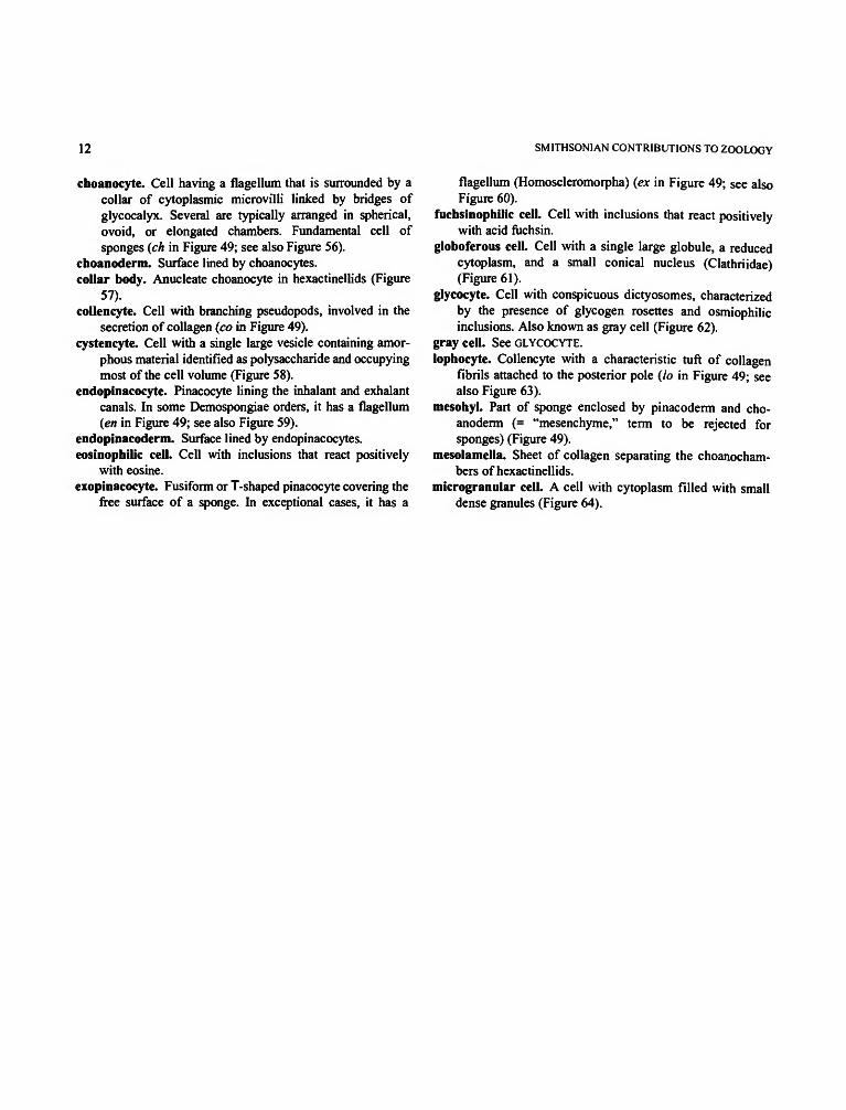
choanocyte. Cell having a flagellum that is surrounded by acollar of cytoplasmic microvilli linked by bridges ofglycocalyx. Several are typically arranged in spherical,ovoid, or elongated chambers. Fundamental cell ofsponges (ch in Figure 49; see also Figure 56).
choanoderm. Surface lined by choanocytes.collar body. Anucleate choanocyte in hexactinellids (Figure
57).collencyte. Cell with branching pseudopods, involved in the
secretion of collagen (co in Figure 49).cystencyte. Cell with a single large vesicle containing amor-
phous material identified as polysaccharide and occupyingmost of the cell volume (Figure 58).
endopinacocyte. Pinacocyte lining the inhalant and exhalantcanals. In some Demospongiae orders, it has a flagellum(en in Figure 49; see also Figure 59).
endopinacoderm. Surface lined by endopinacocytes.eosinophilic cell. Cell with inclusions that react positively
with eosine.exopinacocyte. Fusiform or T-shaped pinacocyte covering the
free surface of a sponge. In exceptional cases, it has a
flagellum (Homoscleromorpha) (ex in Figure 49; see alsoFigure 60).
fuchsinophilic cell. Cell with inclusions that react positivelywith acid fuchsin.
globoferous cell. Cell with a single large globule, a reducedcytoplasm, and a small conical nucleus (Clathriidae)(Figure 61).
glycocyte. Cell with conspicuous dictyosomes, characterizedby the presence of glycogen rosettes and osmiophilicinclusions. Also known as gray cell (Figure 62).
gray cell. See GLYCOCYTE.lophocyte. Collencyte with a characteristic tuft of collagen
fibrils attached to the posterior pole (lo in Figure 49; seealso Figure 63).
mesohyl. Part of sponge enclosed by pinacoderm and cho-anoderm (= "mesenchyme," term to be rejected forsponges) (Figure 49).
mesolamella. Sheet of collagen separating the choanocham-bers of hexactinellids.
microgranular cell. A cell with cytoplasm filled with smalldense granules (Figure 64).

NUMBER 596 13
58
61
64

14 SMITHSONIAN CONTRIBUTIONS TO ZOOLOGY
microsymbiont. Prokaryotic or more rarely eukaryotic mi-croorganism living inside a sponge. May be unicellular ormulticellular filamentous, with or without pigments, andmay occur intra- or intercellularly (Figure 65).
myocyte. See ACTINOCYTE.pinacocyte. Cell delimiting the sponge from the external
milieu and always only in a layer one-cell deep (Figures49,51,59).
pinacoderm. Surface lined by pinacocytes (ex and en inFigure 49).
pocket cell. Special kind of bacteriocyte that surrounds an areaof the intercellular matrix, including microsymbionts(Figure 66).
porocyte. Cell surrounding a pore (ostium) (Figure 67).prosopinacocyte. Endopinacocyte lining the inhalant canals.prosopinacoderm. Surface lined by prosopinacocytes.rhabdiferous cell. Large cell with numerous rod-like inclu-
sions parallel to the long axis of the cell containing acidmucopolysaccharides (Figure 68).
sclerocyte. Cell involved in spicule secretion. In Demospon-
ges, with intracellular secretion, sclerocytes are character-ized by numerous mitochondria and the presence ofspicule-axial filaments. In Calcarea, where secretion isextracellular, sclerocytes have septate junctions betweenthem (sc in Figure 49; see also Figure 69).
spherulous cell. Cell filled with large round spherules thatoccupy almost the entire cell body, compressing thecytoplasm into thin sheets (sp in Figure 49; see also Figure70).
spongocyte. Cell secreting spongin fibers (so (spo = spongin)in Figure 49; see also Figure 71).
thesocyte. Dormant archaeocyte in gemmules or gemmule-like structures in which the cytoplasm is full of reservespartly arranged in vitelline platelets (Figure 72).
trabecular tissue. Syncytia in hexactinellids occurring aroundthe choanochambers.
trophocyte. Cell providing reserves to archaeocytes duringgemmulogenesis and to the oocyte during ovogenesis(Figure 73).

NUMBER 596 15
70
73

16 SMITHSONIAN CONTRIBUTIONS TO ZOOLOGY
Reproduction
LITERATURE CONSULTED: Bergquist, 1978; Borojevic et al., 1968;Boury-Esnault and Vacelet, 1994; De Vos et al., 1991; Tuzet et al.,1970.
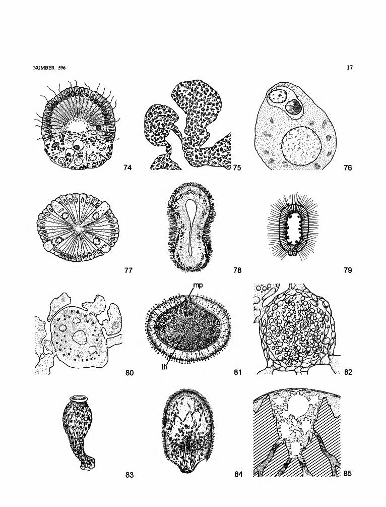
amphiblastula. Hollow, ovoid larva, with anterior flagellatedand posterior nonflagellated groups of cells; typical ofCalcaronea (Figure 74).
anterior pole. Pole directed forward when larva is swimming(Figures 74, 79, 84).
blastula. Term to be rejected. See COELOBLASTULA.bud. An asexual reproductive body composed of various types
of cells (e.g., Tethya spp., Axinella damicornis) (Figure75).
carrier cell. Modified choanocyte that transports a capturedspermatozoid to an oocyte (Figure 76).
cellules en croix. Nonflagellated cells arranged in tetraradialfashion in the amphiblastula of Calcaronea (Figure 77).
cinctoblastula. Hollow larva, entirely flagellated, with acentral ring of pigmented cells; typical of Homoscleromor-pha (Figure 78).
coeloblastula. Hollow larva composed of an envelope ofmorphologically similar equipotent cells, to which a fewlarger nonflagellated cells may be added at the posteriorpole (Figure 79). "Blastula" to be rejected.
follicle cell. Cell in a group surrounding an oocyte (Figure 80).fragmentation. Breakup of a sponge without concomitant
formation of special reproductive bodies.gem mule. A resistant asexual reproductive body, composed of
a mass of archaeocytes charged with reserves and enclosedin a noncellular protective envelope or coat (Figure 81).
micropyle. Opening in the gemmule coat through which cellsleave a hatching gemmule (mp in Figure 81).
nurse cells. Cells that are incorporated in developing oocytesto provide nutrient material for further growth (Figure 82).
olynthus. Earliest developmental stage with a functionalaquiferous system, with a single choanocyte chamberopening directly into the osculum; typical of somecalcareous sponges (Figure 83).
oviparity. Mode of reproduction in which embryo developsoutside the parent sponge.
parenchymella. Larva composed of an envelope of flagellatedcells surrounding an internal mass of cells (Figure 84).
posterior pole. Pole opposite to anterior pole when larva isswimming, often without flagella (Figures 74, 79, 84).
pseudogemmule. Accumulation of gemmular archaeocytes(crypt cells, tract cells) in basal cavities of a solidcalcareous skeleton (Figure 85).
reduction body. A multicellular mass resulting from thedisorganization of a sponge and presumably capable ofreorganizing into a new functional sponge.
NUMBER 596 17
76
79
83 84

18 SMITHSONIAN CONTRIBUTIONS TO ZOOLOGY
rhagon. Earliest functional stage with multiple choanocytechambers and aquiferous canals, typical for Demospongiae(Figure 86).
"smoking". Massive release of gametes.spermiocyst. Modified, encysted spermatozoid transported by
a carrier cell to the oocyte (Figure 76).statoblast. Particular gemmule that lacks a complex spongin
coat and is covered externally by megascleres; typical ofthe freshwater Potamolepidae.
stotnoblastula. Developmental stage of amphiblastulas thatprecedes the reversion stage and is characterized byinternal flagella and a large opening through which theeversion occurs (Figure 87).
thesocyte. Vitelline-rich archaeocyte in a sponge gemmule (thin Figure 81; see also Figure 72).
trichimella. Hexactinellid larva, distinguished by medianzone with multiflagellated mononucleate cells betweenanterior and posterior poles; with special larval stauractinskeleton (Figure 88).
trophocyte. Nurse cell involved in the initial stages ofgemmule and oocyte formation (see Figure 73).
viviparity. Mode of reproduction in which internal fertiliza-tion and incubation of the embryo occur inside the parentsponge.
Architecture of the Skeleton and Spicules of Hexactinellida
LITERATURE CONSULTED: Ijima, 1901, 1903, 1926; Schulze,
1887, 1895, 1900, 1904; Schulze and Lendenfeld, 1889; Topsent,1904, 1917, 1927, 1928. Spicule arrangement is illustrated in Figure89.

NUMBER 596 19
parenchymalia comitalia
parenchymalia principalia
parenchymalia intermedia
autodermalia
hypodermalia
prostalia pleuralia
88
dictyonalia
prostalia basalia
89

20 SMITHSONIAN CONTRIBUTIONS TO ZOOLOGY
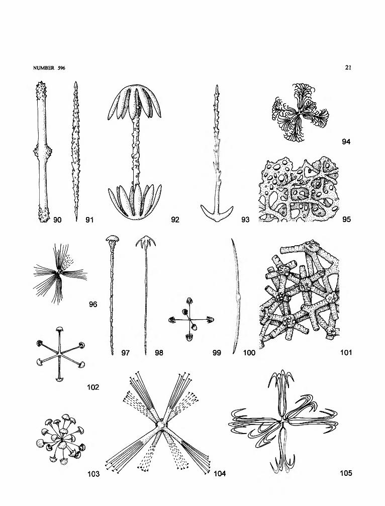
acanthophore. Spicule having two to six stout spined rays,typically surrounding the point of insertion of the root tuftin Hyalonema species (Figures 90, 136, 139).
amararhysis. Paleontological term for longitudinal, tunnel-like canal opening to the exhalant surface by slit-likeapertures.
ambucinate. Diactin with spines recurved toward the center(Figure 91).
amphidisc. Dumbbell-shaped microsclere with ends devel-oped into umbel-like expansions. Three size classes,termed macramphidisc, mesamphidisc, micramphidisc(Figure 92).
anchor. Basalia megasclere, one end with hooks (Figures 89,93).
apochete. Exhalant canal in fossil sponges.apopore. To be rejected; in fossil sponges, an opening
forming an exit from an apochete (= osculum).aporhysis. In fossil sponges, canal closed at the outer end (cf.
epirhysis).aspidoplumicome. Hexaster with numerous S-shaped termi-
nal rays whose external curved ends form severalsuccessive tiers (Figure 94).
autodermalia. Dermalia belonging wholly or at least primar-ily to the outer bounding skin, without actin protrudinginside the sponge body (Figure 89).
autogastralia. Gastralia with one actin protruding into theatrium (Figure 89).
barbule. See UNCINATE.
basalia. Spicules protruding from lower sponge surface;sometimes called root spicules, anchors, barbed basalia; insome species, forming a root tuft (Figure 89).
basidictyonal plate. Rigid basal skeletal framework (Figure95).
calycocome. Hexaster with stout main rays and with terminalrays directed outward (Figure 96).
canalaria. Spicules lining canals (Figure 89).clavule. Sceptrule with one terminal umbel (Figure 97) or with
terminal anchor teeth (Figure 98).codonhexactin. Discohexactin with long, dendate, umbel-like
structures (Figure 99).comitalia. Parenchymalia located close and parallel to princi-
palia (Figure 89).dermalia. Spicules of the external sponge surface. See
AUTODERMALIA, HYPODERMALIA (Figure 89).
diactin. Megascleres with two rays and frequently with centralswellings indicating the hexactin origin (Figures 90, 91,100, 117, 138, 140).
diarhysis. In fossil sponges, radial canal in the honeycomb-like skeleton of Aphrocallistidae.
dictyonal skeleton (= dicryonine). Three-dimensional net-work of fused, regular hexactins (Figure 89).
dictyonalia. Spicules fused into a rigid framework (Figures89, 101, 113).
discohexactin. Hexactin with rays terminating in dentate,umbel-like structures (Figure 102).
discohexaster. Hexaster with dentate, umbel-like terminalrays (Figures 103, 112, 132).
discoctaster. Octaster with terminal rays ending in umbel-likedisks (Figure 104).
drepanocome. Hexaster with sickle-like terminations (genusDictyaulus) (Figure 105).
NUMBER 596 21
90 91 92
94
93
102
97 98
103
9 9 ^ 1 0 0
104 105

22 SMITHSONIAN CONTRIBUTIONS TO ZOOLOGY
epirhysis. In fossil sponges, canal closed at inner end (cf.aporhysis).
fibule. Microsclere consisting of two bent rays derived fromgreatly reduced oxyhexaster (in Holascus fibulatus)(Figure 106).
floricome. Discohexaster with S-shaped terminal rays that endin a plate provided with teeth or claws (Figure 107).
gastralia. All spicules that line the main exhalant cavity. SeeAUTOGASTRALIA, HYPOGASTRALIA (Figure 89).
graphicome (= graphihexaster). Hexaster with long, thin,parallel branches forming terminal brushes (Figure 108).
hemioxyhexaster. Hexaster in which two opposite rays arelonger than the other four; the branched rays are reduced orabsent (genus Aphrocallistes) (Figure 109).
hexactin (hexact). Siliceous spicule with six unbranched raysperpendicular to one another (Figures 99, 102, 115, 118,123, 130).
hexadisc. Microhexactin with six actins ending in amphidisc-like umbels (Figure 111).
hexaster. Microsclere with six branched rays (Figures 94, 96,103, 105, 107-110, 112, 116, 120, 121, 129, 131, 132,134).
holactin. See MICROHOLACTIN.holoxyhexaster. See OXYHEXASTER.hypodermalia. Dermalia with one actin protruding inside the
body and the other ones tangential to the external surface(Figure 89).
hypogastralia. Gastralia with one actin protruding inward, theother ones oriented tangentially to the atrium (Figure 89).
intermedia. Parenchymalia situated among the principalia ordictyonalia (Figure 89).
lophocome (= lophodiscohexaster). Discohexaster withshort, stout main rays and numerous slender secondary
rays (Figure 112).lychnisc. Hexactin with a center resembling a perforated
octohedron; building block of fused (dictyonal) frameworkcalled LYCHNISCA (Figure 113; lower two lychnics shownin optical section).
lychnisca. Dictyonal hexactin framework formed by lychnises(Figure 113).
lyssacine. Spicular framework formed by the juxtapositionand interlacing of elongate rays of hexactins (Figure 114).
macruncinate. See UNCINATE.marginalia. Prostalia that project in a wreath-like arrangement
from the oscular margin (Figure 89).microhexactin. Hexactin microsclere.microholactin. Large category of microscleres lacking spines
and appendages (Monorhaphis) (Figure 115).micruncinate. See UNCINATE.octactin. Spicule with eight rays, generally six in one plane,
the other two perpendicular to them.octaster. Octactin microsclere with branched rays (Figure
104).onychaster (= onychohexaster, onychohexactin). Hexaster
with terminal rays ending in four actins that are perpen-dicular to the rays (Figure 116).
orthodiactin. Diactin in which the two rays belong to differentaxes of a triaxon and form an angle of 90° (Figure 117).
oxy-. Prefix indicating that rays are tapering to a point. Seeexamples for DIACTIN (Figures 100, 140), HEXACTIN
(HEXACT) (Figure 118), HEXASTER (Figures 109, 110,116), PENTACTIN (Figure 119), and STAURASTER (Figure120).
pappocome. Graphicome-like spicule with diverging terminalrays (Figure 121).

NUMBER 596 23
106 107
110 111
114 115 116 117
118 119 120

24 SMITHSONIAN CONTRIBUTIONS TO ZOOLOGY
parenchymalia. All spicules in the choanosome (Figure 89).pentactin (pentact). Five-rayed spicule (Figures 119, 122).pinule. Pentact or hexact spicule with four basal rays and one
perpendicular ray ornamented by obliquely set spinesdirected distally (Figures 122, 123).
pleuralia. Spicules protruding from lateral surface (Figure89).
plumicome. See ASPIDOPLUMICOME, STROBILOPLUMICOME.
posticum. Cf. apopore (= osculum); in fossil sponges.principalia. Spicules making up main skeletal framework
(Figure 89).prosochete. In fossil sponges, inhalant canal leading to
prosopore.prostalia. Spicules protruding from a surface (Figure 89).rhabdodiactin. Diactin in which the two rays are aligned in a
single axis (Figures 90, 91, 100, 138, 140).sarule. Sceptrule with one end swollen and ornamented with
distally directed spines (Figure 124).scepter. Uncinate marginalia, with spiny shaft acerated at one
end; terminating in four very short actins at the other(Pheronematidae, genus Semperella) (Figure 125).
sceptrule. A small, prostal monactin spicule exhibiting pecu-liar secondary developments at one end (order Hexacti-nosa) (Figures 97, 98, 125-127).
schizorhysis. In fossil sponges, continuous system of inter-conected canal spaces (Tretodictyidae).
scopule. Fork-like sceptrule with one end branched (lep-toscopule, pachyscopule) (Figures 126, 127).
sieve-plate. Perforated plate that covers the broad terminalopening of some tubular Hexactinellida (Euplectella spp.)(Figure 128).
sigmatocome. Hexaster with long, S-shaped terminal rays(Dictyaulus) (Figure 129).
sphaerohexactin (sphaerohexact). Hexactin with rays endingin spherical knobs (Figure 130).
sphaerohexaster. Hexaster with terminal rays ending inspherical knobs (Figure 131).
spirodiscohexaster. Discohexaster with a bunch of terminal,spirally twisted rays (genus Rhabdodictyum) (Figure 132).
stauractin. Reduced hexactin with four rays all arranged inone plane (Figure 133).
stauraster. Reduced hexaster with four rays all arranged inone plane (Figure 120).
strobiloplumicome. Plumicome with numerous slender ter-minal rays arranged in two concentric circles (Figure 134).
synapticule. Bridges composed of siliceous cement joiningspicules (Figure 135).
tauactin. Large, smooth triactin (genus Monorhaphis).tetractin. Spicule with four rays in one plane (Figure 136).tetradisc. Microsclere with four actins ending in amphidisc-
like umbels (Figure 137).tignule. Gigantic, isolated diactin (Hyalonema toxeres) (Fig-
ure 138).triactin. Spicule with three rays in one plane (Figure 139).triaxone. Spicule with three axes (Figures 99, 102, 118, 119,
122, 123, 130).trichaster. Graphicome (genus Trichasterina).uncinate. Diactin covered with short spines directed distally
(barbule); size classes are designated by macrouncinate,microuncinate (Figure 140).

NUMBER 596 25
123 124 125 126
128
127 129
o 130 132 133
134 135
136 137 138 139 140

26 SMITHSONIAN CONTRIBUTIONS TO ZOOLOGY
Calcareous Spicules and Calcareous Spicule Skeleton
LITERATURE CONSULTED: Borojevic, 1965, 1966a, 1966b; Boro-jevic et al., 1968, 1990; Hartman, 1980. Structural arrangements areillustrated in Figures 141 and 142.
actine. Ray of a spicule.anchor. Anchor-like spicule, protruding free from the basal
part of the sponge; derived from diactine, triactine, ortetractine spicules, with terminal rays or spines, curved inthe proximal direction along the main ray (Figure 143).
apical actine (ray). The fourth actine of a tetractine that isjoined to the basal triradiate system (Figure 144).
articulate skeleton. Choanoskeleton composed of severalrows of similar spicules (Figure 141).
atrial spicule/skeleton. Spicules surrounding the atrium (Fig-ures 141, 142). Terms "gastral spicule/skeleton" are to berejected.
basal actine (ray). See UNPAIRED ACTINE.basal triradiate system. The three rays of a tetractine that
correspond to those of a triactine (Figure 144).calcareous spicule. A spicule composed largely of calcite
having a radiate form, the number of rays being either two(diactine), three (triactine), or four (tetractine).
calcareous tripod. Regular triactine whose center is not in thesame plane as the extremities of the recurved rays (Figure145).
center. Point of union of the three rays of a triactine or of abasal triradiate system.
chiactine. Tetractine with the unpaired ray of the basal systemand the apical ray in the same alignment (Figure 146).
choanoskeleton. Skeleton of the part of the sponge thatcontains choanocytes (Figure 141).
cortical spicule/skeleton. Spicule or skeleton of the externallayer or cortex of the sponge ("dermal" spicule/skeleton,incorrect) (Figure 141).
diactine. A spicule composed of two actines (Figure 147).diapason (tuning-fork spicule). Sagittal triactine with paral-
lel paired rays (Figure 148).facial plane. The plane passing through tips of the three rays
of a triactine or of a basal triradiate system of a tetractine.free actine. A ray that projects through the external or the
atrial surface (Figures 141, 142).gastral. See ATRIAL.inarticulate skeleton. Choanoskeleton composed only of the
unpaired rays of the subatrial spicules and of one of therays of the cortical or subcortical spicules. Withoutspecific spicules of the choanoskeleton (Figure 142).
microanchor. A small tetractine anchor (described only fromthe atrial skeleton).
microdiactin. A small diactine spicule (Figure 149).needle-like biradiate. A diactine with a hole at one end
(Figure 149).optic axis. The crystallographic trigonal axis of the calcite
spicule.oral/oscular ray. See PAIRED ACTINE.
paired actine(s). One or both equivalent rays of a sagittalspicule. "Oscular" or "oral" rays to be rejected.
parasagittal spicule. Bilaterally symmetrical triactine ortetractine with unequal actines, having equal angles (120°)between the basal rays when projected into a planeperpendicular to the optic axis (Figure 150). "Secondarysagittal spicule" to be rejected.
primary sagittal spicule. See SAGITTAL SPICULE.pseudosagittal spicule. A subcortical triactine essentially
sagittal, but having unequally long and differently curvedrays on each side of the unpaired angle (Figure 151).
regular spicule. Triactine or tetractine spicule with basal raysof equal length, and with equal angles (120°) betweenthem, when projected into a plane perpendicular to theoptic axis (Figure 152).
sagittal spicule. Triactine or tetractine with two equal angles(paired angles) and one dissimilar angle (unpaired angle) atthe center, when projected into a plane perpendicular to theoptic axis (Figure 153). "Primary sagittal spicule" to berejected.
scale. Triangular or circular spicule derived from a triactine(Figure 154).
secondary sagittal spicule. See PARASAGITTAL SPICULE.spicule tract. See "Architecture of Demosponges."subatrial spicule. Sagittal spicule with paired rays adjacent to
the atrial skeleton (Figures 141, 142). "Subgastral spicule"to be rejected.
subcortical spicule. A pseudosagittal triactine, with unpairedactine and a shorter paired actine adjacent to the cortex, ora tetractine with basal triradiate system adjacent to thecortex (Figures 141, 142).
subdermal spicule. Term to be rejected. See SUBCORTICALSPICULE.
subgastral spicule. Term to be rejected. See SUBATRIALSPICULE.
tangential spicule. Spicule arranged parallel to the surface.tetractine. A spicule with four rays (in Figure 144, sagittal
tetractine at the top and regular tetractine at the bottom).triactine. A spicule with three rays (Figures 148, 150-153).trichox. Thin, hair-like straight monaxon spicules present in
general around the osculum or protruding from the cortex.unpaired actine. In sagittal spicules, the ray of a triactine or of
a triradiate basal system of a tetractine lying in the plane ofbilateral symmetry. "Basal ray" to be rejected.

NUMBER 596 27
142
cortical skeleton
subcortical skeleton
atrial skeleton
free actine
154

28 SMITHSONIAN CONTRIBUTIONS TO ZOOLOGY
Architecture of the Skeleton of Demospongiae
LITERATURE CONSULTED: Bergquist, 1978, 1980; Levi, 1973;Topsent, 1928; Wiedenmayer, 1977.
accessory spicule. A category of megasclere echinating theprimary skeleton. See ECHINATING SPICULE.
accretive. Regularly anisotropic reticulation with ascending(primary and radial) fibers or tracts and interconnectingfibers or tracts about equal in thickness (Figure 155).
alveolate. Skeleton arranged around choanosomal cavities(Figure 156).
anastomosing (= reticulate). Tracts, lines, or fibers areinterconnected (Figure 157).
anisotropic skeleton. Reticulate skeleton with primary andsecondary tracts, lines, or fibers (Figure 158).
ascending fiber/tract/line. See PRIMARY fiber, tract, or line(Figures 155, 158).
auxiliary spicule. See INTERSTITIAL SPICULE.
axial skeleton. A type of skeletal organization in which somecomponents are condensed to form a central region or axis(Figure 159).
axinellid skeleton. Structure with a dense core of parallelfibers or strands fanning out toward the periphery of a
sponge (Figure 159). See also PLUMOSE or PLUMORETICU-LATE.
bark. The dense area of compacted spongin surrounding thecentral zone in pithed fibers (Figure 160).
basal lamina. See BASAL SPONGIN PLATE.basal spongin plate. A spongin layer covering the substratum
(Figure 161).bouquet. Ectosomal brush of spicules perpendicular to the
surface of the sponge, with pointed ends outward (Figure162).
calcareous rigid skeleton. See "Calcareous Rigid Skeleton."chalinid skeleton. See ANISOTROPIC skeleton (Figure 163).choanosomal skeleton. Skeleton of the main body, supporting
the canal system and responsible for the form of thesponge.
choristid (= astrophorid). Predominantly radiate, occasion-ally confused arrangement of megascleres includingtriaenes with cladome outward, lacking spongin, andcommonly with a cortex (Figures 164, 174).
clathrate skeleton. Polyhedral arrangement of fibroreticulatesheets (Figure 165).
clathriid skeleton. Isodictyal or subisodictyal arrangement offibers cored and/or echinated by styles in the familyMicrocionidae (Figure 166).
collagen. See SPONGIN.

NUMBER 596 29
155 156 A157
159
161 163
164 165

30 SMITHSONIAN CONTRIBUTIONS TO ZOOLOGY
collagen fascicle. A dense band of intercellular collagenfibrils, which has a skeletal role in some sponges (e.g.,genus Chondrosia) (Figure 167).
compound (= fasciculate, trellised) fiber. Several fibersmerged or intricately interconnected (Figure 169).
confused skeleton. Irregularly positioned megascleres (Figure168).
connective tracts, lines, fibers. See SECONDARY tracts, lines,or fibers.
cored fiber. A fiber that incorporates indigenous spicules orforeign material (spicules and sediment); coring may belight and limited to a central axis or may fill in the wholefiber (Figure 170).
cortex. A superficial region of a sponge reinforced by a specialorganic or inorganic skeleton (Figure 171).
cortical skeleton. Ectosomal skeleton reinforced by a layer ofspecial skeleton elements.
crust. See CORTEX.cuticle. A spongin layer covering the surface or a part of the
surface of a sponge.dendritic skeleton. A skeleton consisting of single or ramify-
ing fibers or tracts that branch but rarely anastomose(tree-like branching) (Figure 172).
dendroreticulate fiber skeleton. Ambiguous, to be rejected.dermal skeleton. See ECTOSOMAL SKELETON.echinating spicule. Megasclere that protrudes from the spon-
gin plate, a fiber, or a spicule tract (Figure 173).ectochrote. Outer layer of the cortex (Figure 174).ectosomal crust See ECTOSOMAL SKELETON.ectosomal skeleton. Skeleton found in the superficial region
of a sponge, distinct from that of the choanosome (Figure175).
endosomal skeleton. See CHOANOSOMAL SKELETON.
extra-axial skeleton. Structurally distinct skeleton arisingfrom or surrounding an axial region (Figure 159).
fasciculate fiber. See COMPOUND FIBER.
fiber (or fibre). A column (strand, thread) of spongin forminga reticulate or dendritic skeleton, with or without indige-nous spicules or foreign material (e.g., Figures 160, 170).
fibrofascicle. See COMPOUND FIBER.
fibroreticulate. Forming a net composed of fibers.halichondrioid skeleton. Confused skeleton except at the
surface; choanosomal skeleton may be in vague tracts(Figure 168).
hispidation. Spicules projecting through the pinacoderm(Figures 166, 174).
homogeneous fiber. A fiber without central pith and withoutconspicuous layers (Figure 176).
hymedesmioid (= leptoelathriid) skeleton. Skeleton of en-crusting sponge where monactine megascleres are ar-ranged singly with heads fixed to a basal plate of sponginand points directed outward (Figure 177).
intercellular collagen fibrils. Fibrillar collagen, formerlycalled "spongin A," dispersed in the'intercellular matrix inall sponges. The fibrils are 20-30 ran in diameter, with atransverse banding of 64 ran intervals, and may be smoothor rugose.
interstitial (= auxiliary) spicule. Free choanosomal spiculenot included in tracts or fibers, common in subectosomalskeleton (Figure 166).
irciniid filament. Long, slender (0.5-15 urn thick) sponginelement, terminally knobbed; intertwined or free in thesponge body (Figure 178).

NUMBER 596 31
169
175
I 178

32 SMITHSONIAN CONTRIBUTIONS TO ZOOLOGY
isodictyal reticulation. Isotropic reticulation in which themeshes are triangular and have sides one spicule long(Figure 179).
isotropic reticulation. Reticulation without differentiationinto primary or secondary fibers, tracts, or lines (Figures156, 180).
laminated (= stratified) fiber. A fiber in which concentriclayers are visible (Figure 181).
leptoclathriid. See HYMEDESMIOID.lax (= loose, vague) skeleton. Skeleton lacking clear tracts or
fibers (Figure 182).line of spicules. Unispicular tract (Figure 183).lipo-. Prefix meaning without.lithistid skeleton. Main skeleton consisting of an interlocked
assemblage of desmas (Figure 184).longitudinal fiber/tract. See PRIMARY FIBER.main skeleton. See CHOANOSOMAL SKELETON.
microcionid. Structure of microcionid sponges in whichspicules project from an elevation of the basal plate ofspongin (Figure 185).
multispicular fiber (tract). Fiber or tract composed of six ormore spicules adjacent to one another.
node. Junction of spicule tips cemented by spongin (Figure186).
palisade. Perpendicular arrangement of ectosomal spicules,with points directed outward (Figure 187).
paratangential skeleton. Arrangement of ectosomal spiculesintermediate between the palisade and tangential type(Figure 188).
parchment. Tangential ectosomal skeleton in which themegascleres are arranged in a tight feltwork (Figure 189).
paucispicular fiber (tract). Fiber or tract with two to fivemegascleres adjacent to one another (Figure 190).

NUMBER 596 33
179
182
185
188
187
190

34 SMITHSONIAN CONTRIBUTIONS TO ZOOLOGY
pith. Central area of a fiber made up of more or less diffusewisps of collagen or of a coarsely granular collagenmaterial. See PITHED FIBER.
pithed fiber. A fiber in which pith is easily distinguished fromthe surrounding bark (see, e.g., Figures 160; 181, right; and200).
plumoreticulate skeleton. A type of plumose skeletal con-struction in which cross-connections occur (Figure 191).
plumose skeleton. A type of skeletal construction made ofprimary fibers or spicule tracts from which skeletalelements radiate obliquely (Figure 192).
plurispicular. See MULTISPICULAR.polyserial. See MULTISPICULAR.polyspicular. See MULTISPICULAR.primary (= main, ascending) fiber. An ascending fiber
ending at a right angle to the surface (Figure 193).primary skeleton. See CHOANOSOMAL SKELETON.primary spicule. The major structural megasclere in any
particular sponge.principal skeleton. See CHOANOSOMAL SKELETON.radiate skeleton. A type of skeleton in which the structural
components diverge from a central region toward thesponge surface (Figure 194).
renieroid skeleton. See RETICULATE SKELETON.reticulate skeleton. Three-dimensional network of fibers,
tracts, lines, or single spicules (see, e.g., Figures 155-158,
163, 175, 176, 180, 183, 195).secondary (= connecting) fiber. In a reticulate skeleton, a
fiber that links the primary fibers.skeleton. All structures supporting and protecting the sponge
body.spiculation. General term for spicule complement.spicule. A component of the mineral skeleton, typically
composed of silica or calcium carbonate (Figure 196).spiculoid (= spongin spicule). A spongin component of the
skeleton that has the form of a diactinal, triactinal, orpolyactinal spicule (Figure 197).
spongin. Skeletal substance in Demospongiae made up ofcollagen microfibrils of ~10 fim diameter (see, e.g.,Figures 160, 170, 176, 185, 200).
sublithistid skeleton. Skeleton consisting of a loose assem-blage of desmas (Figure 198).
tangential skeleton. Ectosomal skeleton arranged parallel tothe surface (Figure 199 shows upper right, view from thetop).
tertiary fiber. In a reticulate skeleton, a fiber interconnectingthe secondary fibers (Figure 200).
tract. A column of aligned megascleres (Figure 201 showstwo types of tract).
trellised fiber. See COMPOUND FIBER.unispicular fiber, tract. A single aligned row of megascleres
(Figure 202).


36 SMITHSONIAN CONTRIBUTIONS TO ZOOLOGY
Calcareous Rigid Skeleton
LITERATURE CONSULTED: Cuif, 1979; Cuif et al., 1979; Cuif andGautret, 1991; Jones, 1979; Senowbari-Daryan, 1991; Vacelet, 1979;Wendt, 1979; Wood, 1986, 1991.
astrorhiza. Traces of the excurrent canal system on or withina rigid skeleton that appear as radiating or star-shapedgrooves (Figures 203, 210).
calyx. See CORALLITE.
cemented (fused) spicule. Interlocked or adjacent spiculesfirmly linked by calcareous cement; the cement may berestricted to the junction area or may progressively encasethe entire spicule (Figure 204).
coenosteum. Term for cnidarians, used for the solid skeletonin stromatoporoids; to be rejected for sponges in favor ofrigid calcareous skeleton.
column. Radial skeletal element in stromatoporoids, with agreater degree of continuity and larger than PILLARS.
corallite (= calyx). In cnidarians, term for the externalskeleton of a polyp; to be rejected for sponges in favor ofPSEUDOCALYX.
crypt tissue. Masses of thesocyte-like cells lodged within thebasal crypts (chambers) of the calcareous skeleton (see ctin Figure 208).
crystal ultrastructure. Organization of the skeletal crystalsobserved after special preparation showing the relationshipbetween organic and mineral phases.
entrapped spicules. Calcareous or siliceous spicules that arenot part of the primary spicule framework but areprogressively enclosed within a solid skeleton duringgrowth (Figure 205).
epitheca. A thin, wrinkled, calcareous layer, different instructure from the normal skeleton, covering the dead basalpart of a rigid calcareous skeleton (Figure 203).
fascicular fibrous (= water-jet, penicillate, trabecular)microstructure. Crystal fibers fanning outward and radi-ating upward. In the penicillate type (e.g., Ceratoporella),crystal fibers are feebly divergent; in the water-jet type(e.g., Merlia), crystal fibers progressively fan outwardaround the growth axis; in the trabecular type (cnidarian,unknown in sponges), the crystal fibers are symmetricallyarranged around the growth axis and axial fibers are absent(Figure 206).

NUMBER 596 37
epitheca
astrorhiza
203
204 206

38 SMITHSONIAN CONTRIBUTIONS TO ZOOLOGY
filling (tissue) structure. Secondary calcareous skeleton thatpartitions or fills abandoned parts of the skeleton (Figure207).
foramen. Circular pore in laminae connecting adjoininginterlamellar spaces (fo in Figure 208, which also shows cc= choanocyte chamber; ct = crypt tissue; sk = rigidaspicular skeleton; spi = spicule; and ta = tabula).
fused. See CEMENTED.irregular (felt) microstructure. Structure in which tangled
crystal fibers have no preferred orientation (e.g., genus
Vaceletia). Shape and nature of the crystal fiber may bediverse (Figure 209).
lamina. Calcareous plate in stromatoporoids, perpendicular toradial skeletal components (Figure 210).
latilamina. A macroscopic, banded calcareous skeletal platein stromatoporoids (Figure 210).
linked spicules. Spicules that are interlocked by ZYGOSIS.mamelon. Rounded regular or irregular elevation of the
skeleton surface; may or may not show correlation withastrorhizae (Figure 210).

NUMBER 596 39
spicule
207
^209
lamina
pillar
208 210

40 SMITHSONIAN CONTRIBUTIONS TO ZOOLOGY
microgranular microstructure. Diverse structure in whichsmall, round elements are scattered (Figure 211).
microlamellar microstructure. Structure in which the crystalfibers are disposed in criss-cross layers, mostly parallel tothe surface of the skeleton (e.g., genus Acanthochaetetes)(Figure 212).
microstructure. The arrangement of crystals that form theskeletal components, as seen without special preparations.
orthogonal microstructure. Microstructure in which thecrystal fibers are in perpendicular and radial orientationrelative to a central axis (Figure 213).
penicillate. See FASCICULAR.pillar. Radial skeletal component, relatively short compared
with COLUMN (Figure 210).primary calcareous skeleton. Initially formed skeletal com-
ponents (Figure 207).primary spicule framework. Framework of spicules around
which a cement is deposited (Figure 207).pseudocalyx (pi. pseudocalices). One of a group of skeletal
tubes—closely packed together, mostly hexagonal inshape and containing the live material—that resemble thecalices or corallites of cnidarians (Figure 214).
rigid aspicular skeleton. Skeleton that originates from the
direct secretion of aspicular elements made up of calciumcarbonate; siliceous or calcareous spicules may be secon-darily entrapped in the skeleton during the growth process(sk in Figure 208).
rigid spicular skeleton. A skeleton in which the mainframework is first made up of fused or linked spicules thatmay later be invested by an aspicular cement (Figure 204).
sclerodermite. Aggregate of crystals forming a microstructu-ral unit of the calcareous skeleton (Figure 215).
secondary calcareous skeleton. Skeletal components formedafter the deposition of the primary calcareous skeleton(Figure 207).
spherulitic (= spheroidal) microstructure. Globular (centricor excentric) arrangement of crystal fibers radiating from acommon center (e.g., genus Astrosclerd) (Figure 215).
tabula. Plate or floor that partitions the basal part of a skeletalcavity (Figure 207; and ta in Figure 208).
tabulate osculum. Osculum in stromatoporoids that has beenrepeatedly partitioned by secondarily deposited tabulae.
trabecular. See FASCICULAR.water jet. See FASCICULAR.zygosis. Mode of interspicular junction without cement (see
SPICULE TYPES OF "LITHISTIDS"; Figure 301).

NUMBER 596 41
213
215

42 SMITHSONIAN CONTRIBUTIONS TO ZOOLOGY
Spicules of Demospongiae
LITERATURE CONSULTED: Bergquist, 1978; Lundbeck, 1902,1905, 1910; Ridley and Dendy, 1887; Sara, 1959; Schulze andLendenfeld, 1889; Sollas, 1888; Wiedenmayer, 1977, 1994.
acantho-. Prefix meaning spined, as in acanthostyle (Figure216) or acanthomicrorhabd (Figure 268).
acerate. Pointed like a needle; adjective referring to theextremities of megascleres. See OXEA.
actine. A centered ray containing an axis or axial canal. Seealso CLAD.
ala. One of the thin, wing-like or spatulate structures in eachrecurved portion of a chela. The anterior ala is the onefacing the shaft; others are lateral alae. See CHELA.
amphidisc. See BIROTULA.amphiaster. A microsclere with microspined rays radiating
from both ends of a shaft; the rays are shorter than the shaft(Figure 217).
amphitriaene. A double triaene with a short rhabd bearingtwo opposed cladomes (Figure 218).
anatriaene. A triaene in which the clads are sharply curvedbackward (Figure 219).
anchorate chela. An isochela with three or more free alae (ateach end) in the form of recurved processes shaped likeanchor claws (unguiferous, Figure 220) or anchor blades(spatuliferous, Figure 221); with two incipient lateral alaefused with the shaft over their entire length and a gentlycurved, not abruptly arched shaft. An anchorate chela withthree teeth is called a tridendate chela.
angulate. Sharply bent; referring to overall spicule shape, as inoxea or sigmas. See OXEA.
aniso-. Prefix meaning unequal; generally referring to the endsof a spicule; see ANISOCHELA.
anisochela. A chela with unequal ends (Figure 222).antho-. Prefix meaning thorny.anthosigma. A special form of spiraster (genus Anthosig-
mella; = Cliona); to be rejected.anthaster. An euaster with tuberculate, denticulate, or spined,
sometimes digitate, expansions at the ray tips (Chondrillagrandis) (Figure 223).
arcuate chela. An isochela with three free alae and the shaftcharacteristically curved outward, often bow-shaped (Fig-ure 224).
aspidaster. An elongate-compressed microsclere in which thenumerous rays are fused and end in minute spinoseprojections (Erylus spp.) (Figure 225).
asymmetric. Adjective referring to the extremities of amegasclere. See OXEA.
axial filament. The organic core around which a siliceousspicule is organized.
bipocillum. A modified anisochela with fused alae connectedby a short curved shaft (genus Iophon) (Figure 226).
birotula. A type of microsclere with a straight shaft andumbrella-shaped ends (Figure 227).
blunt. Adjective referring to the extremities of megascleres.See OXEA.
calthrop. Equiangular tetraxon with equal rays (Figure 228).candelabrum. A special form of lophocalthrop characterized
by branching, multispined rays (Figure 229).canonochela. Modified, ellipsoid isochela with wing-like
extensions on one (the "bottom") side (genus Cercido-chela) (Figure 230).
centrotriaene. A triaene with three clads and two small rhabds(genus Triptolemus) (Figure 231).
centrotylote. Spicule with a median tyle (globular swelling).See OXEA.
chela. A microsclere with a curved shaft and recurved ala ateach end (Figures 220-222, 224, 226, 235, 246, 261, 274,284).
chiaster. See STRONGYLASTER.ehiastosigma. Microsclere composed of two crossed sigmas
(genus Chiastosia) (Figure 232).clad. Any ray or axial branch containing an axis or axial canal
confluent with that of the protoclad or protorhabd in anytype of spicule; term chiefly used in triaene spicules(Figures 219, 240, 242, 252, 254, 256, 262, 265).
cladome. The clads of a triaene or triaene-derivative spicule(Figures 219, 231, 240, 242, 252, 256, 262).
cladotylote. Monaxon megasclere, one end with knobs, theother with hooks (Figure 233).
clavidisc. A ring-shaped microsclere (genus Merlia) (Figure234).
cleistochela. A chela with the ends (anterior alae) very close toeach other (e.g., Clathria echinata) (Figure 235).
coelodischela. See DISCHELA.comma. A curved microstyle (Figure 236).conical (= abruptly pointed). Adjective referring to the
extremities of megascleres. See OXEA.cri-. Prefix indicating spiral or annulate ornamentation, as in
cricalthrop (genus Sphinctrella) (Figure 237).croca. A J-shaped microsclere (Figure 238).curved. Referring to spicule shape (may be symmetrical or
asymmetrical). See OXEA.cyrtancistra. A sigma with semilanceolated extremities
(genus Pozziella) (Figure 239).desma. A typical interlocked megasclere of lithistids with
hypertrophic terminal secretion of silica (see "SpiculeTypes of 'Lithistids'").
deuteroclad. An actinal branch or a distal, branched portion ofa ray. See DiCHOTRiAENE.
diaene. Reduced triaene with two clads (Figure 240).diancistra. A microsclere with hooked, knife-shaped ends,
notched where they join the shaft and in the middle of theshaft (genus Hamacantha) (Figure 241).

NUMBER 596 43
217 220 222 224
218 219 221 223 225
226
227
234
235
228
o
230
229
231 232 233
236 238
237 239 240 241

44 SMITHSONIAN CONTRIBUTIONS TO ZOOLOGY
dichotriaene. An ortho or plagiotriaene in which the clads arebifurcate (Figure 242).
didiscorhabd. Microxea or microstrongyle ornamented bytwo discs along the shaft (genus Didiscus) (Figure 243).
diod. Any diactine spicule with a central double-bent kink(Figure 244).
diplaster. An astrose microsclere in which the rays or spinesradiate from two, slightly distant, points (Figure 245).
disc. A monocrepid spicule resembling a DISCOTRIAENE(genus Neopelta); see "Spicule Types of'Lithistids.'"
discaster. A collective term for DISCORHABD and SCEPTRUM;to be rejected.
dischela. A modified isochela whose disc-shaped alae are veryclose to each other and connected on their margins by twoextremely reduced shafts (genus Coelodischela) (Figure246).
discorhabd (= sceptrum). A rod-shaped microsclere withseveral smooth or serrated discs along the shaft (generaLatrunculia, Podospongia, Sceptrinus) (Figure 247).
discotriaene. A triaene with a disc-shaped cladome. Thesingle clads may be distinguished by the presence of shortaxial canals (genus Discodermia) (Figure 248).
dragma. See TRICHODRAGMA.equal. Adjective referring to the extremities of megascleres.
See OXEA.euaster. A collective term for astrose microscleres in which
the rays radiate from a central point (Figures 223,258,275,276, 279, 291).
exotyle. Styles or tylostyles with enlarged or omated distalend, projecting from the sponge surface (Figure 249).
fimbria. Narrow structure, similar to an ala, flanking the shaftof an anchorate chela (Figure 221).
flexuous (= sinuous, wavy). Referring to the spicule shape.See OXEA.
forceps (= labis). A U-shaped microsclere (Figure 250).fusiform. Shape of a monactin spicule, tapering regularly
toward a point. See OXEA.hastate. Spicule remaining isodiametric for most of its length,
with the point or points tapering abruptly. See OXEA.hilum. A more or less distinct, funnel-shaped depression in the
surface of a STERRASTER, situated on one side in ellipsoidalforms; also occurs in ASPIDASTERS in reduced form,resembling an umbilicus (see Figures 225, 278 in"Spicules of Demospongiae").
iso-. Prefix meaning equal (generally referring to the ends of aspicule).
isochela. A chela with equal ends (Figures 220, 221,224, 259,261).
labis. See FORCEPS.lopho-. Prefix for categories of spicules having one or more
rays affected by multiple, sometimes complex, branching(lophate spicules); see LOPHOCALTHROP.
lophocalthrop. Modified calthrop with one (monoloph), three(triloph), or four (tetraloph) rays (Figure 251).
mesotriaene. A triaene in which the rhabd is extended beyondthe cladome (Figure 252).
metaster. A microsclere with a twisted shaft no longer thanthe spines; intermediate between plesiaster and spi raster(Figure 253).
micro-. Prefix used for naming microscleres that are similar inshape to megascleres (e.g., microxea, microstyle).
microrhabd. A general term for a straight, monaxonicmicrosclere.
microsclere. Small spicule, often ornate in shape.monaene. Reduced triaene with only one clad (Figure 254).monaxon. Linear, nonradiate spicule, or a spicule type not
having more than two rays along one axis.mucronate. Adjective referring to the nipple-like point of a
megasclere. See OXEA.onychaete. A long, thin, finely spined, asymmetric micro-
sclere (Figure 255).orthotriaene. A triaene in which the clads are directed at right
angles to the rhabd (Figure 256).oxea. Monaxon (diactinal) spicule pointed at both ends
(Figure 257a-m). Different types are distinguished byshape and tip morphology. Shape may be fusiform (a),angulate (b), centrotylote (c), curved (d), or flexuous (e).Tips may be acerate (f), asymmetrical (g), blunt (h),conical (i), hastate (j), mucronate (k), stepped (1), orsymmetrical (m).

NUMBER 596 45
243 >nr 245
242 244
251 253
252 254
248 249
255
256
246 11 247 250
A
A
d V e m
257

46 SMITHSONIAN CONTRIBUTIONS TO ZOOLOGY
oxy-. Prefix referring to radiate spicule types with raysgradually tapering to a point.
oxyaster. An euaster with acerate free rays and a smallcentrum less than one-third the diameter of the wholespicule) (Figure 258).
oxyspheraster. See SPHEROXYASTER.
palmate chela. An iso- or anisochela in which the lateral alaecoalesce with the shaft over their entire length, and thesingle, median, anterior ala (one at each end) stands freeand widens distally (Figure 259).
phyllotriaene. A triaene with a leaf-shaped cladome (Figure260).
placochela. A special type of isochela with plate-like alae. Thealae and the shaft are internally ornamented by radialridges (genus Guitarra) (Figure 261).
plagiotriaene. A triaene in which the clads are directedforward and form with the rhabd an angle of about 45°, andmay be recurved distally (Figure 262).
plesiaster. A streptaster with a very short shaft (always shorterthan the spines) (Figure 263).
polytylote. Megasclere with several annular swelling along itsshaft (Figure 264).
protoclad. The proximal, unbranched portion of a branchedray. See DICHOTRJAENE).
protorhabd. The original simple shaft of ornamented monax-ons and of triaenes.
protriaene. A triaene in which the clads are directed orsharply curved forward, away from the rhabd (Figure 265).
pycnaster. A small euaster in which the rays are merely lowconules; to be rejected.
raphide. A very thin, hair-like microsclere, often in bundlescalled trichodragmas (Figure 266).
ray. See ACTINE.
rhabd. A collective term for a monaxonic megasclere, laterapplied to the one ray of a triaene that is distinct from theother three, mostly in length (= rhabdome).
rhabdome. The rhabd of a triaene and of a triaene-derivedspicule.
rhabdostyle. A style with a pronounced basal bend (Figure267).
sanidaster. A rhabd-derived, straight microsclere havingspines at intervals along the entire length. Spines along theshaft are perpendicular to the axis and may or may not bespirally arranged; those at the ends diverge obliquely (e.g.,genera Tribrachium, Disyringa) (Figure 268).
scale. Disc-shaped microsclere forming ectosomal ("dermal")armor (genus Lepidosphera) (Figure 269).
sceptrum. See DISCORHABD.selenaster. A special type of spiraster approaching the shape
of a sterraster (genus Placospongia) (Figure 270).sigma. A microsclere of C or S shape (Figure 271).sigmancistra. A slightly modified sigma found in Cladorhiza
(Figure 272).
sigmaspire. A contorted-sigmoid microspined microsclere(family Tetillidae) (Figure 273).
sinuous. See FLEXUOUS.spatuliferous-anchorate chela (= ancora spatulifera). An
anchorate chela (usually isochela) having alae that arecompletely free from the shaft and that are spatulate orshaped like anchor teeth (Figure 221).
sphaerancora. Modified isochela, consisting of two ellipsoidridged plates intersecting at right angles (genus Melonan-chora) (Figure 274).
spheraster. An euaster with short rays and a thick centrum;the diameter of the centrum (more than one-half the totaldiameter) exceeds the length of the rays (Figure 275).
spheroxyaster. Euaster with a discrete centrum that is morethan one-third the total diameter (Figure 276).
spinispira. See SPIRASTER.spiraster (= spinispira). Spiral, rod-shaped microsclere with
spines peripherally arranged (Figures 277).stepped (= "telescoped"). Adjective referring to the extremi-
ties of a megasclere. See OXEA.sterraster. A spherical or ellipsoidal microsclere in which the
numerous rays are fused and end in stellate terminations(e.g., genus Geodia; Figure 278).
sterrospheraster. Ball-shaped euaster with numerous conicalor flattened, often denticulate or tuberculate protuberances;to be rejected. See STERRASTER.
streptaster. Aster in which the rays proceed from an axis thatis usually spiral (Figure 263).
strongylaster (= chiaster). Aster with free, isodiametric, bluntrays (Figure 279).
stroogyle. An isodiametric, diactinal megasclere with roundedends (Figure 280).
strongyloxea. A fusiform oxea with one end blunt (Figure281).
style. Monaxon spicule with one end pointed, the other (heador base) blunt (Figure 282).
subtylostyle. Tylostyle with one end pointed, the other with aslight swelling or knob; the swelling, more or less distinct,may be displaced along the shaft (Figure 283).
symmetrica. Adjective referring to the extremities of megas-cleres. See OXEA.
telescoped. See STEPPED.tetrapocillum. A rare, modified isochela having two small
disks both at the extremities and in the middle of a straightshaft; the two in the middle are fused (genus Tetrapocil-lon) (Figure 284).
thaumatochela. A rare, minute anisochela with the smallerextremity jar- or slipper-shaped and partly enveloped byfour alae issuing from the opposite end and forming anumbrella-like structure (Mycale thaumatochela) (Figure285).
thraustoxa. Toxa-like, smooth to spinose microsclere, dou-ble-bent in the middle and often with central swelling(genus Rhabderemia) (Figure 286).

NUMBER 596 47
258
'259
260
261
263
262
266
264 265 267
268
269 271
272
273
274
275
276
V
277
**-̂ —-
279
284
285
280 I 281 V 282 I 283 286

48 SMITHSONIAN CONTRIBUTIONS TO ZOOLOGY
tornote. A straight, isodiametric, diactinal megasclere withconical or mucronate extremities (Figure 287).
toxa. Bow-shaped microsclere (Figure 288).triaene. General term for a tetractinal megasclere having one
unequal ray (termed rhabd) that is commonly much longerthan the other three (termed clads, forming the cladome)(Figures 218, 219, 240,242, 248, 252, 254, 256,260,262,265).
trichodragma (pi. trichodragmas or trichodragmata). Abundle of raphids (Figure 266).
trichotriaene. A triaene in which the clads are trifurcate.tridentate chela. A chela with three free alae. See also
ANCHORATE CHELA.
triod. Triact in which the rays are equal, straight, in one plane,and diverging at an angle of 120° (Figure 289).
tripod. A modified triod with rays forming a regular pyramid(Figure 290).
truncaster. Aster with crowded, blunt rays (genus Rhabdas-trella) (Figure 291).
tuberculate. Spicule ornamented with blunt knobs.tylaster. Aster with free, microtylote rays (Figure 292).tyle. Any rounded swelling or knob in a spicule (c in Figure
257; see also Figures 292-294).tylostyle. A style with a tyle (globular swelling) at the base
(Figure 293).tylote. Diactinal megasclere with a swelling on each end
(Figure 294).unequal. Adjective referring to the extremities of megas-
cleres. See also ANISO-.unguiferous-anchorate chela (= ancora unguifera). An
anchorate chela in which the recurved processes are in theform of claws or sickles and are normally connected to theshaft by expansions called falces (Figure 220).
verticillate. Spicule ornamented by whirls (e.g., genus Agelas)(Figure 295).
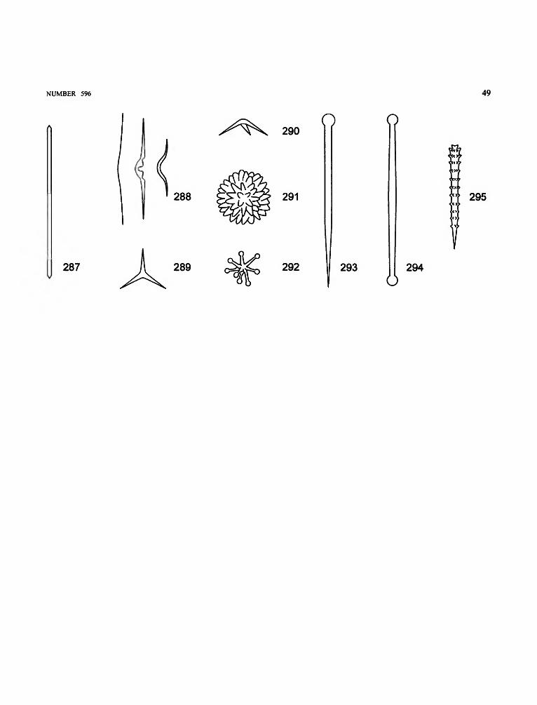
NUMBER 596 49
290O o
288 291 295
287 289 292 293 294o

50 SMITHSONIAN CONTRIBUTIONS TO ZOOLOGY
Spicule Types of "Lithistids"
LITERATURE CONSULTED: Levi, 1991; Reid, 1970; Rigby andSteam, 1983; Sollas, 1888.
acrepis. See CREPIS.amphitrider. Tetraclone resembling an amphitriaene.anaxial desma. Desma without central axis.anomoclone. To be rejected. See ASTROCLONE, SPHAERO-
CLONE.
astroclone. Desma made up of radiating arms (Figure 296).brachyome. Shortened arm of a trider.chiastoclone. Dendroclone with a very short central shaft.clone. Ray-like arm that is entirely anaxial.crepis. The fundamental, inceptional body of a desma before
it is modified by deposition of multiple layers of silica. Amonocrepid desma develops from a monaxon spicule thatis recognized by a straight, undivided axial thread (canal)(see top of Figure 297); a tetracrepid desma is based on atetraxon spicule with a four-rayed axial thread (a small
calthrops; see bottom of Figure 297, also "Spicule Typesof Demospongiae"); an acrepid desma is based on apolyaxon fundamental structure lacking axial threads.
dendroclone. Desma with branched ends or tetracladine,amphitrider-like; always positioned perpendicular toskeletal fibers (Figure 298).
desma (= desmome). A typical interlocked megasclere withhypertrophic terminal secretion of silica.
desmome. See DESMA.dicranoclone. Tuberculate monaxial desma, arch shaped,
tripodal, tetrapodal, or rarely polygonal; zygomes areterminal, like those of the tetraclone (Figure 299).
epirhabd. Part of the developed desma formed by thedeposition of concentric layers immediately around thecrepis.
heloclone. Desma with elongate and usually sinuous shaft,without branches, possibly with small digitations at theends; the zygomes are lateral notches. An axial canal isusually found along most of the length of the spicule(Figure 300).

NUMBER 596 51
296 297 298
299 300

52 SMITHSONIAN CONTRIBUTIONS TO ZOOLOGY
linked spicules. Spicules that are interlocked by zygosis,without cement (Figure 301).
megaclone. Arch-shaped desma with a short crepis. Thezygomes vary from oblique lateral facets to tongue- orhand-like terminal expansions (Figure 302).
megarhizoclone. Nontuberculate dicranoclone desma (Figure303).
mesotrider. Tetraclone with a mesotriaene (see "Spicules ofDemospongiae") crepis.
monocrepid. See CREPIS.
rhizoclone. Nontuberculate irregular monaxial desma, withspiny to root-like zygomes that are usually mainly lateral(Figure 304).
sphaeroclone. Anaxial desma in which several ray-like armsextend from one side of a globular centrum that is spiny onthe other side (Figure 305).
tetraclone. Tetraxial desma that does not show a triaenosesymmetry; the zygomes are typically terminal (Figure306).
tetracrepid. See CREPIS.trider. Variant of tetraclone, with triaenose symmetry and
three similar arms.zygome. Part of a desma that interlocks with another spicule.zygosis. Mode of junction between siliceous and calcareous
tetraradiates characterized by the interlocking of theirterminal or lateral expansions; cement, if present, issecondary (Figure 301).

NUMBER 596 53
302
303 304
305 306

Literature Cited
Bergquist, P.R.1978. Sponges. 268 pages. London: Hutchinson & Co.1980. A Revision of the Supraspecific Classification of the Orders
Dictyoceratida, Dendroceratida, and Verongida (Class De-mospongiae). New Zealand Journal of Zoology, 7:443-503.
Borojevic, R.1965. Eponges calcaires des cotes de France, I: Amphiute paulini Hanitsch;
les genres Amphiute Hanitsch et Paraheteropia n.g. Archives deZoologie Experimentale et Generate, 106:665-670.
1966a. Eponge calcaires des cotes de France, 11: Le genre Ascandra Haeckelemend. Archives de Zoologie Experimentale et Generate, 107:357-368.
1966b. Eponges calcaires des cotes de France, III: Discussion sur laTaxonomie des eponges calcaires: Aphroceras ensata (Bowerbank)et Ute gladiata sp.n. Archives de Zoologie Experimentale etGenerate, 107:703-724.
Borojevic, R., N. Boury-Esnault, and J. Vacelet1990. A Revision of the Supraspecific Classification of the Subclass
Calcinea (Porifera, Class Calcarea). Bulletin du Museum Nationald'Histoire Naturelle (Paris), series 4, 12:243-245.
Borojevic, R., W.G. Fry, W.C. Jones, C. Levi, R. Rasmont, M. Sara, and J.Vacelet
1968. Mise au point actuelle de la terminologie des eponges. Bulletin duMuseum National d'Histoire Naturelle (Paris), series 2, 39:1224-1235.
Boury-Esnault, N.1972. Une structure inhalante remarquable des spongiaires: Le crible;
Etude morphologique et cytologique. Archives de Zoologie Experi-mentale et Generate, 113:7-23.
1973. L'exopinacoderme des spongiaires. Bulletin du Museum Nationald 'Histoire Naturelle (Paris), series 3, 178:1193-1206.
1974. Structure et ultrastructure des papilles d'eponges du genre Polymas-tia Bowerbank. Archives de Zoologie Experimentale et Generate,115:141-165.
1977. A Cell Type in Sponge Involved in the Metabolism of Glycogen:The Gray Cells. Cell and Tissue Research, 175:523-539.
Boury-Esnault, N., L. De Vos, C. Donadey, and J. Vacelet1990. Ultrastructure of Choanosome and Sponge Classification. In K.
Rutzler, editor, New Perspectives in Sponge Biology, pages237-244. Washington, D.C.: Smithsonian Institution Press.
Boury-Esnault, N., and J. Vacelet1994. Preliminary Studies on the Organization and Development of a
Hexactinellid Sponge from a Mediterranean Cave, Oopsacas minuta.In R.W.M. van Soest, T.M.G. van Kempen, and J.C. Braekman,editors, Sponges in Time and Space: Biology, Chemistry, Paleontol-ogy, pages 407-415. Rotterdam: A.A. Balkema.
Braun, R.W.1956. Composition of Scientific Words. 882 pages. Washington, D.C.:
Smithsonian Institution Press. [Reprinted 1978.]Cuif, J.
1979. Caracteres morphologiques et microstructuraux de trois sclerospon-ges triasiques: Association avec des Chaetetida. In C. Levi and N.Boury-Esnault, editors, Biologie des Spongiaires. Colloques Inter-nationaux du Centre National de la Recherche Scientifique,291:475-481. Paris: Editions du CNRS.
Cuif, J.P, F. Debrenne, J.G. Lafuste, and J. Vacelet1979. Comparaison de la infrastructure du squelette carbonate non
spiculaire d'eponges actuelles et fossiles. In C. Levi and N.
Boury-Esnault, editors, Biologie des Spongiaires. Colloques Inter-
nationaux du Centre National de la Recherche Scientifique,291:459-465. Paris: Editions du CNRS.
Cuif, J.P., and P. Gautret1991. Taxonomie Value of Microstructural Features in Calcified Tissue
from Recent and Fossil Demospongiae and Calcarea. In J. Reitnerand H. Keupp, editors, Fossil and Recent Sponges, pages 159-169.Berlin: Springer-Verlag.
De Vos, L., N. Boury-Esnault, and J. Vacelet1990. The Apopylar Cell of Sponges. In K. Rutzler, editor. New
Perspectives in Sponge Biology, pages 153-158. Washington, D.C.:Smithsonian Institution Press.
De Vos, L., K. Rutzler, N. Boury-Esnault, C. Donadey, and J. Vacelet1991. Atlas of Sponge Morphology—Atlas de morphologie des Eponges.
117 pages. Washington, D.C.: Smithsonian Institution Press.Garrone, R.
1975. Collagen Resorption in Sponges, Involvement of Bacteria andMacrophages. In H. Peeters, editor, Protides of the BiologicalFluids, 22nd Colloquium, pages 59-63. Oxford: Pergamon Press.
1978. Phylogenesis of Connective Tissue: Morphological Aspects andBiosynthesis of Sponge Intercellular Matrix. In L. Robert, editor,Frontiers of Matrix Biology, 5: 250 pages. Basel: S. Karger.
Hartman, W.D.1980. Calcareous Sponges, Morphology of the Skeleton: Spicules. In N.
Ginsburg and P. Reid, editors, Living and Fossil Sponges (Notes fora Short Course). Sedimenta, 8R: 193-195. Miami: University ofMiami.
Hooper, J.N.A.1991. Revision of the Family Raspailiidae (Porifera: Demospongiae), with
Description of Australian Species. Invertebrate Taxonomy, 5:1179-1418.
Ijima, I.1901. Studies on the Hexactinelida, Contribution I (Euplectellidae).
Journal of the College of Science, 15:1-299.1903. Studies on the Hexactinellida, Contribution III: Placosoma a New
Euplectellid; Leucopsacidae and Caulophacidae. Journal of theCollege of Science, 17:1 -124.
1926. The Hexactinellida of the Siboga Expedition. In E.J. Brill, editor,Siboga Expeditie, volume 6: 229 pages. Leyden: E.J. Brill.
Jones, C.1979. The Microstructure of Sponge Biominerals. In C. Levi and N.
Boury-Esnault, editors, Biologie des Spongiaires. Colloques Inter-nationaux du Centre National de la Recherche Scientifique,291:425-447. Paris: Editions du CNRS.
Levi, C.1973. Systematique de la classe des Demospongiaria (Demosponges). In P.
P. Grasse, editor, Spongiaires. Traite de Zoologie, 3(l):577-632.Paris: Masson & Co.
1991. Lithistid Sponges from the Norfolk Rise: Recent and MesozoicGenera. In J. Reitner and H. Keupp, editors, Fossil and RecentSponges, pages 72-82. Berlin: Springer-Verlag.
Lundbeck, W.
1902. Porifera (Part I), Homorrhaphidae and Heterorrhaphidae. TheDanish IngolfiExpedition, volume 6(1): 104 pages. Copenhagen:Bianco Luno.
54
NUMBER 596 55
1905. Porifera (Part II), Desmacidonidae (pars). The Danish Ingolf-Expedition, volume 6(2): 219 pages. Copenhagen: Bianco Luno.
1910. Porifera (Part III), Desmacidonidae (pars). The Danish Ingolf-Expedition, volume 6(3): 124 pages. Copenhagen: Bianco Luno.
Mackie, CO. , and C.L. Singla1983. Studies on Hexactinellid Sponges, I: Histology of Rhabdocalyptus
dawsoni (Lambe, 1873). Philosophical Transactions of the RoyalSociety of London, 301:365-400.
Reid, R.E.1970. Tetraxons and Demosponge Phylogeny. In W.G. Fry, editor, The
Biology of the Porifera. Symposia of the Zoological Society ofLondon, 25:63-90. London: Academic Press.
Reiswig, H.M., and H.I. Browman1987. Use of Membrane Filters for Microscopic Preparation of Sponge
Spicules. Transactions of the American Microscopical Society,106:10-20.
Ridley, O.S., and A. Dendy1887. Report on the Monaxonida Collected by H.M.S. Challenger during
the Years 1873-1876. Report on the Scientific Results of the Voyageof H.M.S. Challenger, volume 20(50): 263 pages. London: HerMajesty's Stationery Office.
Rigby, J.K., and C.W. Steam1983. Sponges and Spongiomorphs, Notes for a Short Course. In T.W.
Broadhead, editor, Studies in Geology, 7: 220 pages. Knoxville,Tenn.: University of Tennessee.
Rutzler, K.1978. Sponges in Coral Reefs. In D.R. Stoddart and R.E. Johannes, editors,
Coral Reefs: Research Methods. Monographs on OceanographicMethodology, 5:299-313. Paris: UNESCO.
1987. Tetillidae (Spirophorida, Porifera): A Taxonomic Reevaluation. In J.Vacelet and N. Boury-Esnault, editors, Taxonomy of Porifera fromthe N.E. Atlantic and Mediterranean Sea, pages 187-203. London:Springer-Verlag. [NATO, ASI Series G. 13.]
Sara, M.1959. Specie nuove di Demospongie provenienti da acque superficiali del
golfo di Napoli. Annuario, Istituto e Museo di Zoologia deltaUniversita di Napoli, 11(7): 1-22.
Schulze, F.E.1887. Report on the Hexactinellida Collected by H.M.S. Challenger during
the Years 1873-1876. Report on the Scientific Results of the Voyageof H.M.S. Challenger, volume 21(53): 514 pages. London: HerMajesty's Stationery Office.
1895. Hexactinelliden des Indischen Oceanes, II: Die Hexasterophora. 92pages. Berlin: Konigliche Akademie der Wissenschaften.
1900. Hexactinelliden des Indischen Oceanes, III. 46 pages. Berlin:Konigliche Akademie der Wissenschaften.
1904. Hexactinellida: Wissenschaftliche Ergebnisse der DeutschenTiefsee-Expedition, "Valdivia" 1898-1899, 4: 266 pages. Jena:Gustav Fischer.
Schulze, F.E., and R. von Lendenfeld1889. Uber die Bezeichnung der Spongiennadeln. Abhandlungen der
Akademie der Wissenschaften Berlin (for 1889), PhysikalischeAbhandlungen, 2:1-35.
Senowbari-Daryan, B.1991. "Sphinctozoa": An Overview. In J. Reitner and H. Keupp, editors,
Fossil and Recent Sponges, pages 224-241. Berlin: Springer-
Verlag.
Simpson, T.L.
1984. The Cell Biology of Sponges. 662 pages. New York: SpringerVerlag.
Sollas, W.J.
1888. Report on the Tetractinellida Collected by H.M.S. Challenger duringthe Years 1873-1876. Report on the Scientific Results of the Voyageof H.M.S. Challenger, volume 25:458 pages. London: Her Majesty'sStationery Office.
Topsent, E.
1904. Spongiaires des Acores. Resultats des Campagnes Scientifiques duPrince Albert I de Monaco, 25: 280 pages. Monaco: Imprimerie deMonaco.
1917. Spongiaires. Deuxieme Expedition Antarctique Francaise (1908-1910) commandee par le Dr. Jean Charcot L. Joubin. 84 pages.Paris: Masson & Cie.
1927. Diagnoses d'eponges nouvelles recueillies par le Prince Albert lerde Monaco. Bulletin de I'lnstitut Oceanographique de Monaco,502:1-19.
1928. Spongiaires de l'Atlantique et de la Mediterranee. provenant descroisieres du prince Albert ler de Monaco. Resultats des CampagnesScientifiques du Prince Albert I de Monaco, 74: 376 pages. Monaco:Imprimerie de Monaco.
Tuzet, O., R. Garrone, and M. Pavans de Ceccatty1970. Observations ultrastructurales sur la spermatogenese chez la demo-
sponge Aplysilla rosea Schulze (Dendroceratide): une metaplasieexemplaire. Annales des Sciences Naturelles Zoologie et BiologieAnimale, 12:27-50.
Vacelet, J.
1975. Etude en microscopie electronique de 1 'association entre bacteries etspongiaires du genre Verongia (Dictyoceratida). Journal de Micro-scopie et de Biologie Cellulaire, 23:271-288.
1979. Description et affinites d'une Eponge Sphinctozoaire actuelle. In C.Levi and N. Boury-Esnault, editors, Biologie des Spongiaires.Colloques Internationaux du Centre National de la RechercheScientifique, 291:483-493. Paris: Editions du CNRS.
Wendt, J.1979. Development of Skeletal Formation, Microstructure, and Mineral-
ogy of Rigid Calcareous Sponges from the Late Palaeozoic toRecent. In C. Levi and N. Boury-Esnault, editors, Biologie desSpongiaires. Colloques Internationaux du Centre National de laRecherche Scientifique, 291:449-457. Paris: Editions du CNRS.
Wiedenmayer, F.1977. Shallow-Water Sponges of the Western Bahamas. Experientia,
supplement 28: 287 pages. Basel: Birkhauser.1994. Contributions to the Knowledge of Post-Palaeozoic Neritic and
Archibenthal Sponges (Porifera). Schweizerische PaldontologischeAbhandlungen, 116: 47 pages. Basel: Kommission der Schweizeris-chen Palaontologischen Abhandlungen.
Winston, J.1988. The Systematists' Perspective. In D. Fautin, editor, Biomedical
Importance of Marine Organisms, pages 1-6. San Francisco:California Academy of Sciences.
Wood, R.A.1986. The Biology and Taxonomy of Mesozoic Stromatoporoids. 473
pages. Doctoral dissertation, Department of Earth Sciences, OpenUniversity, Milton Keynes, England.
1991. Non-Spicular Biomineralization in Calcified Demosponges. In J.Reitner and H. Keupp, editors, Fossil and Recent Sponges, pages322-340. Berlin: Springer-Verlag.


REQUIREMENTS FOR SMITHSONIAN SERIES PUBLICATION
Manuscripts intended for series publication receive substantivereview (conducted by their originating Smithsonian museums oroffices) and are submitted to the Smithsonian Institution Press withForm SI-36, which must show the approval of the appropriateauthority designated by the sponsoring organizational unit. Requestsfor special treatment—use of color, foldouts, case-bound covers,etc.—require, on the same form, the added approval of thesponsoring authority.
Review of manuscripts and art by the Press for requirements ofseries format and style, completeness and clarity of copy, andarrangement of all material, as outlined below, will govern, within thejudgment of the Press, acceptance or rejection of manuscripts andart.
Copy must be prepared on typewriter or word processor,double-spaced, on one side of standard white bond paper (noterasable), with 11/4" margins, submitted as ribbon copy (not carbonor xerox), in loose sheets (not stapled or bound), and accompaniedby original art. Minimum acceptable length is 30 pages.
Front matter (preceding the text) should include: title page withonly title and author and no other information; abstract page withauthor, title, series, etc., following the established format; table ofcontents with indents reflecting the hierarchy of heads in the paper;also, foreword and/or preface, if appropriate.
First page of text should carry the title and author at the top of thepage; second page should have only the author's name andprofessional mailing address, to be used as an unnumbered footnoteon the first page of printed text.
Center heads of whatever level should be typed with initial caps ofmajor words, with extra space above and below the head, but noother preparation (such as all caps or underline, except for theunderline necessary for generic and specific epithets). Run-inparagraph heads should use period/dashes or colons as necessary.
Tabulations within text (lists of data, often in parallel columns) canbe typed on the text page where they occur, but they should notcontain rules or numbered table captions.
Formal tables (numbered, with captions, boxheads, stubs, rules)should be submitted as carefully typed, double-spaced copyseparate from the text; they will be typeset unless otherwiserequested. If camera-copy use is anticipated, do not draw rules onmanuscript copy.
Taxonomic keys in natural history papers should use thealigned-couplet form for zoology and may use the multi-level indentform for botany. If cross referencing is required between key and text,do not include page references within the key, but number thekeyed-out taxa, using the same numbers with their correspondingheads in the text.
Synonymy in zoology must use the short form (taxon, author,yearpage), with full reference at the end of the paper under"Literature Cited." For botany, the long form (taxon, author,abbreviated journal or book title, volume, page, year, with noreference in "Literature Cited") is optional.
Text-reference system (author, yearpage used within the text,with full citation in "Literature Cited" at the end of the text) must beused in place of bibliographic footnotes in all Contributions Seriesand is strongly recommended in the Studies Series: "(Jones,1910:122)" or "...Jones (1910:122)." If bibliographic footnotes are
required, use the short form (author, brief title, page) with the fullcitation in the bibliography.
Footnotes, when few in number, whether annotative or biblio-graphic, should be typed on separate sheets and inserted immedi-ately after the text pages on which the references occur. Extensivenotes must be gathered together and placed at the end of the text ina notes section.
Bibliography, depending upon use, is termed "Literature Cited,""References," or "Bibliography." Spell out titles of books, articles,journals, and monographic series. For book and article titles usesentence-style capitalization according to the rules of the languageemployed (exception: capitalize all major words in English). Forjournal and series titles, capitalize the initial word and all subsequentwords except articles, conjunctions, and prepositions. Transliteratelanguages that use a non-Roman alphabet according to the Libraryof Congress system. Underline (for italics) titles of journals andseries and titles of books that are not part of a series. Use theparentheses/colon system for volume (number):pagination:"10(2):5-9." For alignment and arrangement of elements, follow theformat of recent publications in the series for which the manuscript isintended. Guidelines for preparing bibliography may be secured fromSeries Section, SI Press.
Legends for illustrations must be submitted at the end of themanuscript, with as many legends typed, double-spaced, to a pageas convenient.
Illustrations must be submitted as original art (not copies)accompanying, but separate from, the manuscript. Guidelines forpreparing art may be secured from the Series Section, SI Press. Alltypes of illustrations (photographs, line drawings, maps, etc.) may beintermixed throughout the printed text. They should be termedFigures and should be numbered consecutively as they will appearin the monograph. If several illustrations are treated as componentsof a single composite figure, they should be designated by lowercaseitalic letters on the illustration; also, in the legend and in textreferences the italic letters (underlined in copy) should be used:"Figure 9b." Illustrations that are intended to follow the printed textmay be termed Plates, and any components should be similarlylettered and referenced: "Plate 9b." Keys to any symbols within anillustation should appear on the art rather than in the legend.
Some points of style: Do not use periods after such abbrevia-tions as "mm, ft, USNM, NNE." Spell out numbers "one" through"nine" in expository text, but use digits in all other cases if possible.Use of the metric system of measurement is preferable; where use ofthe English system is unavoidable, supply metric equivalents inparentheses. Use the decimal system for precise measurements andrelationships, common fractions for approximations. Use day/month/year sequence for dates: "9 April 1976." For months in tabular listingsor data sections, use three-letter abbreviations with no periods: "Jan,Mar, Jun," etc. Omit space between initials of a personal name: "J.B.Jones."
Arrange and paginate sequentially every sheet of manuscriptin the following order: (1) title page, (2) abstract, (3) contents, (4)foreword and/or preface, (5) text, (6) appendices, (7) notes section,(8) glossary, (9) bibliography, (10) legends, (11) tables. Index copymay be submitted at page proof stage, but plans for an index shouldbe indicated when the manuscript is submitted.

i
Related Documents