NEWSLETTER JANUARY - FEBRUARY N° 122 | BIMONTHLY | JANUARY - FEBRUARY 2019 ESTRO | EUROPEAN SOCIETY FOR RADIOTHERAPY & ONCOLOGY PHYSICS YOUNG ESTRO RESEARCH PROJECTS Report on the 2nd physics workshop ESTRO 38: What to expect from the Young Track? Predict a patient’s risk of late toxicity following radiotherapy

Welcome message from author
This document is posted to help you gain knowledge. Please leave a comment to let me know what you think about it! Share it to your friends and learn new things together.
Transcript
NEWSLETTER JANUARY - FEBRUARY
N° 122 | BIMONTHLY | JANUARY - FEBRUARY 2019
ESTRO | EUROPEAN SOCIETY FOR RADIOTHERAPY & ONCOLOGY
PHYSICS YOUNG ESTRORESEARCH PROJECTS
Report on the 2nd physics workshop
ESTRO 38: What to expect from the Young Track?
Predict a patient’s risk of late toxicity following
radiotherapy
Editorial
Brachytherapy
Read it before your patients
Physics
ESTRO School
Radiobiology
RTT
Young ESTRO
Conferences
Research projects
Make it happen
Calendar of eventsESTRO | EUROPEAN SOCIETY FOR RADIOTHERAPY & ONCOLOGY
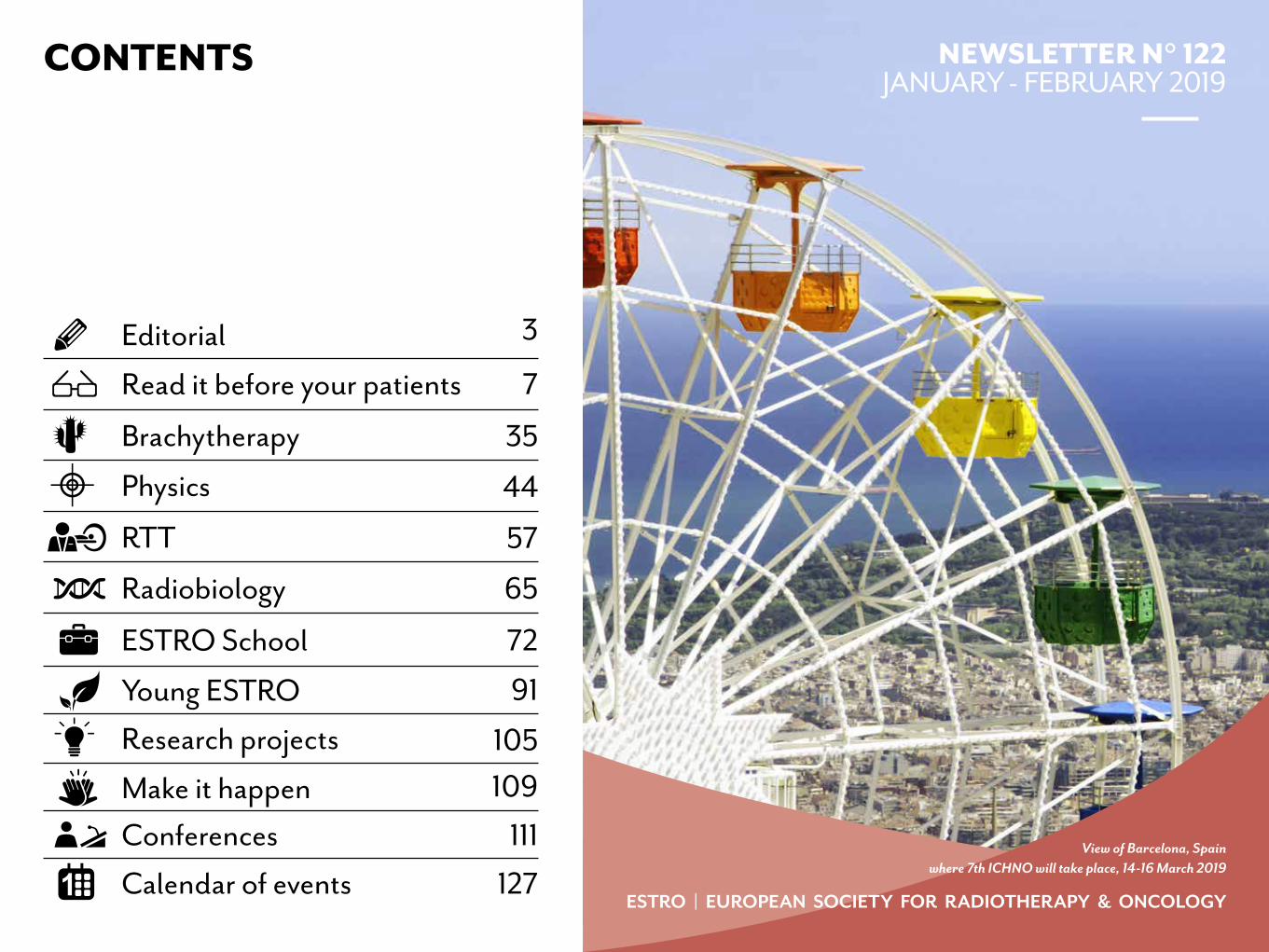
View of Barcelona, Spain where 7th ICHNO will take place, 14-16 March 2019
NEWSLETTER N° 122JANUARY - FEBRUARY 2019
7
3
35
44
57
91
72
65
105109
111
127
CONTENTS
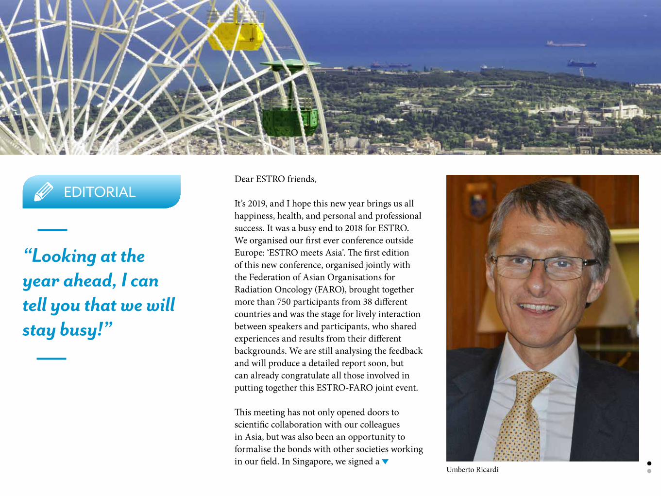
EDITORIALDear ESTRO friends,
It’s 2019, and I hope this new year brings us all happiness, health, and personal and professional success. It was a busy end to 2018 for ESTRO. We organised our first ever conference outside Europe: ‘ESTRO meets Asia’. The first edition of this new conference, organised jointly with the Federation of Asian Organisations for Radiation Oncology (FARO), brought together more than 750 participants from 38 different countries and was the stage for lively interaction between speakers and participants, who shared experiences and results from their different backgrounds. We are still analysing the feedback and will produce a detailed report soon, but can already congratulate all those involved in putting together this ESTRO-FARO joint event.
This meeting has not only opened doors to scientific collaboration with our colleagues in Asia, but was also been an opportunity to formalise the bonds with other societies working in our field. In Singapore, we signed a
“Looking at the year ahead, I can tell you that we will stay busy!”
Umberto Ricardi
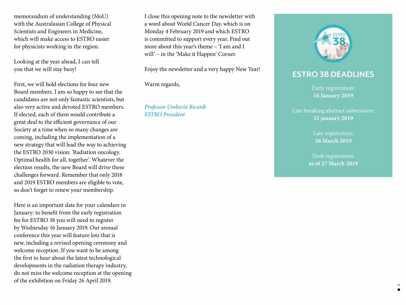
ESTRO 38 DEADLINES
Early registration: 16 January 2019
Late breaking abstract submission: 21 january 2019
Late registration: 26 March 2019
Desk registration: as of 27 March 2019
memorandum of understanding (MoU) with the Australasian College of Physical Scientists and Engineers in Medicine, which will make access to ESTRO easier for physicists working in the region.
Looking at the year ahead, I can tell you that we will stay busy!
First, we will hold elections for four new Board members. I am so happy to see that the candidates are not only fantastic scientists, but also very active and devoted ESTRO members. If elected, each of them would contribute a great deal to the efficient governance of our Society at a time when so many changes are coming, including the implementation of a new strategy that will lead the way to achieving the ESTRO 2030 vision: ‘Radiation oncology. Optimal health for all, together’. Whatever the election results, the new Board will drive these challenges forward. Remember that only 2018 and 2019 ESTRO members are eligible to vote, so don’t forget to renew your membership.
Here is an important date for your calendars in January: to benefit from the early registration fee for ESTRO 38 you will need to register by Wednesday 16 January 2019. Our annual conference this year will feature lots that is new, including a revised opening ceremony and welcome reception. If you want to be among the first to hear about the latest technological developments in the radiation therapy industry, do not miss the welcome reception at the opening of the exhibition on Friday 26 April 2019.
I close this opening note to the newsletter with a word about World Cancer Day, which is on Monday 4 February 2019 and which ESTRO is committed to support every year. Find out more about this year’s theme – ‘I am and I will’ – in the ‘Make it Happen’ Corner.
Enjoy the newsletter and a very happy New Year!
Warm regards,
Professor Umberto RicardiESTRO President
BOARD MEMBERS ELECTIONSBe ready to vote!
The new year sees an important moment in the life of our Society. The ESTRO Board elections will take place from 25 February to 17 March 2019. All ESTRO full members in 2019, who were also members in 2018, are eligible to vote for the four ESTRO Board directorship positions. This year, we will be electing two clinicians, one physicist and one radiobiologist. ESTRO Board members are elected for a three-year term, which is renewable once. Candidates elected now will serve office until 2022.
Two days before the election, all ESTRO members eligible to vote will receive a username and password with a link to the election platform. All candidates’ information is already
available on the ESTRO website – do take a moment to read the candidates’ statements and get to know more about them.
The official announcement of the new Board members and the beginning of their term will take place at the General Assembly during ESTRO 38 in Milan, Italy.
If you were a member in 2018, make sure you renew your membership for 2019 (by 20 February), so you are eligible to vote.

WWW.ESTRO.ORG #ESTRO38
Targeting optimal care,
together

INTRODUCTION BREAST CERVIX HEAD AND NECK BRAIN ORAL CANCER SURVIVAL SUPPORTIVE CARE OLDER PATIENTS CHILDHOODPROSTATE OESOPHAGUS LUNG
READ IT BEFORE YOUR PATIENTS
BREAST CERVIX HEAD AND NECK BRAIN ORAL CANCER SURVIVAL SUPPORTIVE CARE OLDER PATIENTS CHILDHOODPROSTATE OESOPHAGUS LUNGINTRODUCTION
READ IT BEFORE YOUR PATIENTS
Too important to miss...A digest of essential reading for all radiation oncologistsBY PHILIPPE LAMBIN, DIRK DE RUYSSCHER AND HANS KAANDERS
HANS KAANDERS
DIRK DE RUYSSCHER
PHILIPPE LAMBIN
INTRODUCTION CERVIX HEAD AND NECK BRAIN ORAL CANCER SURVIVAL SUPPORTIVE CARE OLDER PATIENTS CHILDHOODPROSTATE OESOPHAGUS LUNGBREAST
READ IT BEFORE YOUR PATIENTS
BREAST
BackgroundLocal cancer relapse risk after breast conservation surgery followed by radiotherapy has fallen sharply in many countries, and is influenced by patient age and clinicopathological factors. We hypothesise that partial-breast radiotherapy restricted to the vicinity of the original tumour in women at lower than average risk of local relapse will improve the balance of beneficial versus adverse effects compared with whole-breast radiotherapy.
Methods30 radiotherapy centres in the UK. Women aged 50 years or older who had undergone breast-conserving surgery for unifocal invasive ductal adenocarcinoma of grade 1-3, with a tumour size of 3 cm or less (pT1-2), none to three positive axillary nodes (pN0-1), and minimum microscopic margins of non-cancerous tissue of 2 mm or more, were recruited. Patients were randomly assigned (1:1:1) to receive 40 Gy whole-breast radiotherapy (control), 36 Gy whole-breast radiotherapy and 40 Gy to the partial breast (reduced-dose group), or 40 Gy to the partial breast only (partial-breast group) in 15 daily treatment fractions. Computer-generated random permuted blocks (mixed sizes of six and nine) were used to assign patients to groups, stratifying patients by radiotherapy treatment centre. Patients and clinicians were not masked to treatment allocation. Field-in-field intensity-modulated radiotherapy was delivered using
standard tangential beams that were simply reduced in length for the partial-breast group. The primary endpoint was ipsilateral local relapse (80% power to exclude a 2.5% increase [non-inferiority margin] at five years for each experimental group; non-inferiority was shown if the upper limit of the two-sided 95% CI for the local relapse hazard ratio [HR] was less than 2.03), analysed by intention to treat. Safety analyses were done in all patients for whom data were available (i.e. a modified intention-to-treat population). This study is registered in the ISRCTN registry, number ISRCTN12852634.
FindingsBetween 3 May 2007, and 5 October 2010, 2018 women were recruited. Two women withdrew consent for use of their data in the analysis. In total, 674 patients were analysed in the whole-breast radiotherapy (control) group, 673 in the reduced-dose group, and 669 in the partial-breast group. Median follow-up was 72.2 months (IQR 61.7-83.2), and five-year estimates of local relapse cumulative incidence were 1.1% (95% CI 0.5-2.3) of patients in the control group, 0.2% (0.02-1.2) in the reduced-dose group, and 0.5% (0.2-1.4) in the partial-breast group. Estimated five-year absolute differences in local relapse compared with the control group were -0.73% (-0.99 to 0.22) for the reduced-dose and -0.38% (-0.84 to 0.90) for the partial-breast groups. Non-inferiority can be claimed for both reduced-dose and partial-breast radiotherapy, and was confirmed by the
Partial-breast radiotherapy after breast conservation surgery for patients with early breast cancer (UK IMPORT LOW trial): five-year results from a multicentre, randomised, controlled, phase 3, non-inferiority trial
Coles CE, Griffin CL, Kirby AM, Titley J, Agrawal RK, Alhasso A, Bhattacharya IS, Brunt AM, Ciurlionis L, Chan C, Donovan EM, Emson MA, Harnett AN, Haviland JS, Hopwood P, Jefford ML, Kaggwa R, Sawyer EJ, Syndikus I, Tsang YM, Wheatley DA, Wilcox M, Yarnold JR, Bliss JM; IMPORT Trialists.
Lancet. 2017 Sep 9;390(10099):1048-1060. doi: 10.1016/S0140-6736(17)31145-5. Epub 2017 Aug 2.
INTRODUCTION CERVIX HEAD AND NECK BRAIN ORAL CANCER SURVIVAL SUPPORTIVE CARE OLDER PATIENTS CHILDHOODPROSTATE OESOPHAGUS LUNGBREAST
test against the critical HR being more than 2.03 (p=0.003 for the reduced-dose group and p=0.016 for the partial-breast group, compared with the whole-breast radiotherapy group). Photographic, patient, and clinical assessments recorded similar adverse effects after reduced-dose or partial-breast radiotherapy, including two patient domains achieving statistically significantly lower adverse effects (change in breast appearance [p=0.007 for partial-breast] and breast harder or firmer [p=0.002 for reduced-dose and p<0.0001 for partial-breast]) compared with whole-breast radiotherapy.
InterpretationWe showed non-inferiority of partial-breast and reduced-dose radiotherapy compared with the standard whole-breast radiotherapy in terms of local relapse in a cohort of patients with early breast cancer, and equivalent or fewer late normal-tissue adverse effects were seen. This simple radiotherapy technique is implementable in radiotherapy centres worldwide.
INTRODUCTION CERVIX HEAD AND NECK BRAIN ORAL CANCER SURVIVAL SUPPORTIVE CARE OLDER PATIENTS CHILDHOODPROSTATE OESOPHAGUS LUNGBREAST
READ IT BEFORE YOUR PATIENTS
BREAST
Background Post-mastectomy radiotherapy in patients with four or more positive axillary nodes reduces breast cancer mortality, but its role in patients with one to three involved nodes is controversial. We assessed the effects of post-mastectomy radiotherapy on quality of life (QOL) in women with intermediate-risk breast cancer.
MethodsSUPREMO is an open-label, international, parallel-group, randomised, controlled trial. Women aged 18 years or older with intermediate-risk breast cancer (defined as pT1-2N1; pT3N0; or pT2N0 if also grade III or with lymphovascular invasion) who had undergone mastectomy and, if node positive, axillary surgery, were randomly assigned (1:1) to receive chest wall radiotherapy (50 Gy in 25 fractions or a radiobiologically equivalent dose of 45 Gy in 20 fractions or 40 Gy in 15 fractions) or no radiotherapy. Randomisation was done with permuted blocks of varying block length, and stratified by centre, without masking of patients or investigators. The primary endpoint is ten-year overall survival. Here, we present two-year results of QOL (a pre-specified secondary endpoint). The QOL sub-study, open to all UK patients, consists of questionnaires (European Organisation for Research and Treatment of Cancer QLQ-C30 and QLQ-BR23, Body Image Scale, Hospital Anxiety and Depression Scale [HADS], and EQ-5D-3L) completed before randomisation, and at one, two, five and ten
Quality of life after post-mastectomy radiotherapy in patients with intermediate-risk breast cancer (SUPREMO): two-year follow-up results of a randomised controlled trial
Velikova G, Williams LJ, Willis S, Dixon JM, Loncaster J, Hatton M, Clarke J, Kunkler IH, Russell NS; MRC SUPREMO trial UK investigators.
Lancet Oncol. 2018 Oct 15. pii: S1470-2045(18)30515-1. doi: 10.1016/S1470-2045(18)30515-1. [Epub ahead of print]
years. The pre-specified primary outcomes within this QOL sub-study were global QOL, fatigue, physical function, chest wall symptoms, shoulder and arm symptoms, body image, and anxiety and depression. Data were analysed by intention to treat, using repeated mixed-effects methods. This trial is registered with the ISRCTN registry, number ISRCTN61145589.
Findings Between 4 August 2006 and 29 April 2013, 1,688 patients were enrolled internationally and randomly assigned to receive chest wall radiotherapy (n=853) or not (n=835). In total, 989 (79%) of 1,258 patients from 111 UK centres consented to participate in the QOL sub-study (487 in the radiotherapy group and 502 in the no radiotherapy group), of whom 947 (96%) returned the baseline questionnaires and were included in the analysis (radiotherapy, n=471; no radiotherapy, n=476). At up to two years, chest wall symptoms were worse in the radiotherapy group than in the no radiotherapy group (mean score 14.1 [SD 15.8] in the radiotherapy group vs 11.6 [14.6] in the no radiotherapy group; effect estimate 2.17, 95% CI 0.40-3.94; p=0.016); however, there was an improvement in both groups between years one and two (visit effect -1.34, 95% CI -2.36 to -0.31; p=0.010). No differences were seen between treatment groups in arm and shoulder symptoms, body image, fatigue, overall QOL, physical function, or anxiety or depression scores.
INTRODUCTION CERVIX HEAD AND NECK BRAIN ORAL CANCER SURVIVAL SUPPORTIVE CARE OLDER PATIENTS CHILDHOODPROSTATE OESOPHAGUS LUNGBREAST
InterpretationPost-mastectomy radiotherapy led to more local (chest wall) symptoms up to two years post-randomisation compared with no radiotherapy, but the difference between groups was small. These data will inform shared decision-making while we await survival (trial primary endpoint) results.
INTRODUCTION BREAST HEAD AND NECK BRAIN ORAL CANCER SURVIVAL SUPPORTIVE CARE OLDER PATIENTS CHILDHOODPROSTATE OESOPHAGUS LUNGCERVIX
READ IT BEFORE YOUR PATIENTS
CERVIX
BackgroundOn 14 August 2014, the US Food and Drug Administration approved the anti-angiogenesis drug bevacizumab for women with advanced cervical cancer on the basis of improved overall survival (OS) after the second interim analysis (in 2012) of 271 deaths in the Gynaecologic Oncology Group (GOG) 240 trial. In this study, we report the pre-specified final analysis of the primary objectives, OS and adverse events.
MethodsIn this randomised, controlled, open-label, phase 3 trial, we recruited patients with metastatic, persistent, or recurrent cervical carcinoma from 81 centres in the USA, Canada and Spain. Inclusion criteria included a GOG performance status score of 0 or 1; adequate renal, hepatic, and bone marrow function; adequately anticoagulated thromboembolism; a urine protein to creatinine ratio of less than 1; and measurable disease. Patients who had received chemotherapy for recurrence and those with non-healing wounds or active bleeding conditions were ineligible. We randomly allocated patients 1:1:1:1 (blocking used; block size of four) to intravenous chemotherapy of either cisplatin (50 mg/m2 on day 1 or 2) plus paclitaxel (135 mg/m2 or 175 mg/m2 on day 1) or topotecan (0.75 mg/m2 on days 1-3) plus paclitaxel (175 mg/m2 on day 1) with or without intravenous bevacizumab (15 mg/kg on day 1) in 21 day cycles until disease progression, unacceptable toxic effects, voluntary withdrawal
Bevacizumab for advanced cervical cancer: final overall survival and adverse event analysis of a randomised, controlled, open-label, phase 3 trial (Gynecologic Oncology Group 240)
Tewari KS, Sill MW, Penson RT, Huang H, Ramondetta LM, Landrum LM, Oaknin A, Reid TJ, Leitao MM, Michael HE, DiSaia PJ, Copeland LJ, Creasman WT, Stehman FB, Brady MF, Burger RA, Thigpen JT, Birrer MJ, Waggoner SE, Moore DH, Look KY, Koh WJ, Monk BJ.
Lancet. 2017 Oct 7;390(10103):1654-1663. doi: 10.1016/S0140-6736(17)31607-0. Epub 2017 Jul 27.
by the patient, or complete response. We stratified randomisation by GOG performance status (0 vs 1), previous radiosensitising platinum-based chemotherapy, and disease status (recurrent or persistent vs metastatic). We gave treatment open label. Primary outcomes were OS (analysed in the intention-to-treat population) and adverse events (analysed in all patients who received treatment and submitted adverse event information), assessed at the second interim and final analysis by the masked Data and Safety Monitoring Board. The cut-off for final analysis was 450 patients with 346 deaths. This trial is registered with ClinicalTrials.gov, number NCT00803062.
FindingsBetween 6 April 2009, and 3 January 2012, we enrolled 452 patients (225 [50%] in the two chemotherapy-alone groups and 227 [50%] in the two chemotherapy plus bevacizumab groups). By 7 March 2014, 348 deaths had occurred, meeting the pre-specified cut-off for final analysis. The chemotherapy plus bevacizumab groups continued to show significant improvement in OS compared with the chemotherapy-alone groups: 16.8 months in the chemotherapy plus bevacizumab groups versus 13.3 months in the chemotherapy-alone groups (hazard ratio 0.77 [95% CI 0.62-0.95]; p=0.007). Final OS among patients not receiving previous pelvic radiotherapy was 24.5 months versus 16.8 months (0.64 [0.37-1.10]; p=0.11). Post-progression OS was not significantly different between the
INTRODUCTION BREAST HEAD AND NECK BRAIN ORAL CANCER SURVIVAL SUPPORTIVE CARE OLDER PATIENTS CHILDHOODPROSTATE OESOPHAGUS LUNGCERVIX
chemotherapy plus bevacizumab groups (8.4 months) and chemotherapy-alone groups (7.1 months; 0.83 [0.66-1.05]; p=0.06). Fistula (any grade) occurred in 32 (15%) of 220 patients in the chemotherapy plus bevacizumab groups (all previously irradiated) versus three (1%) of 220 in the chemotherapy-alone groups (all previously irradiated). Grade 3 fistula developed in 13 (6%) versus one (<1%). No fistulas resulted in surgical emergencies, sepsis or death.
InterpretationThe benefit conferred by incorporation of bevacizumab is sustained with extended follow-up as evidenced by the overall survival curves remaining separated. After progression while receiving bevacizumab, we did not observe a negative rebound effect (i.e. shorter survival after bevacizumab is stopped than after chemotherapy alone is stopped). These findings represent proof-of-concept of the efficacy and tolerability of anti-angiogenesis therapy in advanced cervical cancer.
INTRODUCTION BREAST CERVIX HEAD AND NECK BRAIN ORAL CANCER SURVIVAL SUPPORTIVE CARE OLDER PATIENTS CHILDHOODOESOPHAGUS LUNGPROSTATE
READ IT BEFORE YOUR PATIENTS
PROSTATE
Background Based on previous findings, we hypothesised that radiotherapy to the prostate would improve overall survival in men with metastatic prostate cancer, and that the benefit would be greatest in patients with a low metastatic burden. We aimed to compare standard of care for metastatic prostate cancer, with and without radiotherapy.
Methods We did a randomised controlled phase 3 trial at 117 hospitals in Switzerland and the UK. Eligible patients had newly diagnosed metastatic prostate cancer. We randomly allocated patients open-label in a 1:1 ratio to standard of care (control group) or standard of care and radiotherapy (radiotherapy group). Randomisation was stratified by hospital, age at randomisation, nodal involvement, World Health Organization (WHO) performance status, planned androgen deprivation therapy, planned docetaxel use (from December 2015), and regular aspirin or non-steroidal anti-inflammatory drug use. Standard of care was lifelong androgen deprivation therapy, with up-front docetaxel permitted from December 2015. Men allocated radiotherapy received either a daily (55 Gy in 20 fractions over four weeks) or weekly (36 Gy in six fractions over six weeks) schedule that was nominated before randomisation. The primary outcome was overall survival, measured as the number of deaths; this analysis had 90% power with a one-sided α of 2.5% for a hazard ratio (HR) of 0.75. Secondary
Radiotherapy to the primary tumour for newly diagnosed, metastatic prostate cancer (STAMPEDE): a randomised controlled phase 3 trial
Parker CC, James ND, Brawley CD, Clarke NW, Hoyle AP, Ali A, Ritchie AWS, Attard G, Chowdhury S, Cross W, Dearnaley DP, Gillessen S, Gilson C, Jones RJ, Langley RE, Malik ZI, Mason MD, Matheson D, Millman R, Russell JM, Thalmann GN, Amos CL, Alonzi R, Bahl A, Birtle A, Din O, Douis H, Eswar C, Gale J, Gannon MR, Jonnada S, Khaksar S, Lester JF, O‘Sullivan JM, Parikh OA, Pedley ID, Pudney DM, Sheehan DJ, Srihari NN, Tran ATH, Parmar MKB, Sydes MR; Systemic Therapy for Advanced or Metastatic Prostate cancer: Evaluation of Drug Efficacy (STAMPEDE) investigators.
Lancet. 2018 Oct 18. pii: S0140-6736(18)32486-3. doi: 10.1016/S0140-6736(18)32486-3. [Epub ahead of print]
outcomes were failure-free survival, progression-free survival, metastatic progression-free survival, prostate cancer-specific survival, and symptomatic local event-free survival. Analyses used Cox proportional hazards and flexible parametric models, adjusted for stratification factors. The primary outcome analysis was by intention to treat. Two pre-specified subgroup analyses tested the effects of prostate radiotherapy by baseline metastatic burden and radiotherapy schedule. This trial is registered with ClinicalTrials.gov, number NCT00268476.
Findings Between 22 January 2013 and 2 September 2016, 2,061 men underwent randomisation, 1,029 were allocated the control and 1,032 radiotherapy. Allocated groups were balanced, with a median age of 68 years (IQR 63–73) and median amount of prostate-specific antigen of 97 ng/mL (33–315). In total, 367 (18%) patients received early docetaxel. In terms of schedules, 1,082 (52%) participants nominated the daily radiotherapy schedule before randomisation and 979 (48%) the weekly schedule. 819 (40%) men had a low metastatic burden, 1,120 (54%) had a high metastatic burden, and the metastatic burden was unknown for 122 (6%). Radiotherapy improved failure-free survival (HR 0.76, 95% CI 0.68–0.84; p<0.0001) but not overall survival (0.92, 0.80–1.06; p=0.266). Radiotherapy was well tolerated, with 48 (5%) adverse events (Radiation Therapy Oncology Group grade 3–4) reported during
INTRODUCTION BREAST CERVIX HEAD AND NECK BRAIN ORAL CANCER SURVIVAL SUPPORTIVE CARE OLDER PATIENTS CHILDHOODOESOPHAGUS LUNGPROSTATE
radiotherapy and 37 (4%) after radiotherapy. The proportion reporting at least one severe adverse event (Common Terminology Criteria for Adverse Events grade 3 or worse) was similar by treatment group in the safety population (398 [38%] with control and 380 [39%] with radiotherapy).
Interpretation Radiotherapy to the prostate did not improve overall survival for unselected patients with newly diagnosed metastatic prostate cancer.
INTRODUCTION BREAST CERVIX HEAD AND NECK BRAIN ORAL CANCER SURVIVAL SUPPORTIVE CARE OLDER PATIENTS CHILDHOODOESOPHAGUS LUNGPROSTATE
READ IT BEFORE YOUR PATIENTS
Background The NRG/RTOG 9413 study showed that whole pelvic radiotherapy (WPRT) plus neoadjuvant hormonal therapy (NHT) improved progression-free survival in patients with intermediate-risk or high-risk localised prostate cancer compared with prostate only radiotherapy (PORT) plus NHT, WPRT plus adjuvant hormonal therapy (AHT), and PORT plus AHT. We provide a long-term update after ten years of follow-up of the primary endpoint (progression-free survival) and report on the late toxicities of treatment.
Methods The trial was designed as a 2 × 2 factorial study with hormonal sequencing as one stratification factor and radiation field as the other factor and tested whether NHT improved progression-free survival versus AHT, and NHT plus WPRT versus NHT plus PORT. Eligible patients had histologically confirmed, clinically localised adenocarcinoma of the prostate, an estimated risk of lymph node involvement of more than 15% and a Karnofsky performance status of more than 70, with no age limitations. Patients were randomly assigned (1:1:1:1) by permuted block randomisation to receive either NHT two months before and during WPRT followed by a prostate boost to 70 Gy (NHT plus WPRT group), NHT two months before and during PORT to 70 Gy (NHT plus PORT group), WPRT followed by four months of AHT (WPRT plus AHT group), or PORT followed by four months
of AHT (PORT plus AHT group). Hormonal therapy was combined androgen suppression, consisting of goserelin acetate 3.6 mg once a month subcutaneously or leuprolide acetate 7.5 mg once a month intramuscularly, and flutamide 250 mg twice a day orally for four months. Randomisation was stratified by T stage, Gleason Score, and prostate-specific antigen concentration. NHT was given two months before radiotherapy and was continued until radiotherapy completion; AHT was given at the completion of radiotherapy for four months. The primary endpoint progression-free survival was analysed by intention to treat. This study is registered with ClinicalTrials.gov, number NCT00769548. The trial has been terminated to additional follow-up collection and this is the final analysis for this trial.
Findings Between 1 April 1995 and 1 June 1999, 1,322 patients were enrolled from 53 centres and randomly assigned to the four treatment groups. With a median follow-up of 8.8 years (IQR 5.07-13.84) for all patients and 14.8 years (7.18-17.4) for living patients (n=346), progression-free survival across all timepoints continued to differ significantly across the four treatment groups (p=0.002). The ten-year estimates of progression-free survival were 28.4% (95% CI 23.3-33.6) in the NHT plus WPRT group, 23.5% (18.7-28.3) in the NHT plus PORT group, 19.4% (14.9-24.0) in the WPRT plus AHT group, and 30.2%
Sequence of hormonal therapy and radiotherapy field size in unfavourable, localised prostate cancer (NRG/RTOG 9413): long-term results of a randomised, phase 3 trial
Roach M, Moughan J, Lawton CAF, Dicker AP, Zeitzer KL, Gore EM, Kwok Y, Seider MJ, Hu IC, Hartford AC, Horwitz EM, Yamoah K, Jones CU, Michalski JM, Lee WR, Pisansky TM, Rabinovitch R, Rotman M, Pryzant RM, Kim HE, Thomas CR Jr, Shipley WU, Sandler HM.
Lancet Oncol. 2018 Oct 10. pii: S1470-2045(18)30528-X. doi: 10.1016/S1470-2045(18)30528-X. [Epub ahead of print]
PROSTATE
INTRODUCTION BREAST CERVIX HEAD AND NECK BRAIN ORAL CANCER SURVIVAL SUPPORTIVE CARE OLDER PATIENTS CHILDHOODOESOPHAGUS LUNGPROSTATE
(25.0-35.4) in the PORT plus AHT group. Bladder toxicity was the most common grade 3 or worse late toxicity, affecting 18 (6%) of 316 patients in the NHT plus WPRT group, 17 (5%) of 313 in the NHT plus PORT group, 22 (7%) of 317 in the WPRT plus AHT group, and 14 (4%) of 315 in the PORT plus AHT group. Late grade 3 or worse gastrointestinal adverse events occurred in 22 (7%) of 316 patients in the NHT plus WPRT group, five (2%) of 313 in the NHT plus PORT group, ten (3%) of 317 in the WPRT plus AHT group, and seven (2%) of 315 in the PORT plus AHT group.
Interpretation In this cohort of patients with intermediate-risk and high-risk localised prostate cancer, NHT plus WPRT improved progression-free survival compared with NHT plus PORT and WPRT plus AHT at long-term follow-up albeit increased risk of grade 3 or worse intestinal toxicity. Interactions between radiotherapy and hormonal therapy suggests that WPRT should be avoided without NHT.
INTRODUCTION BREAST CERVIX BRAIN ORAL CANCER SURVIVAL SUPPORTIVE CARE OLDER PATIENTS CHILDHOODPROSTATE OESOPHAGUS LUNGHEAD AND NECK
READ IT BEFORE YOUR PATIENTS
HEAD AND NECK
Introduction Radiotherapy treatment plan quality can influence clinical trial outcomes and general quality assurance (QA) may not identify suboptimal organ-at-risk (OAR) sparing. We retrospectively performed patient-specific QA of 100 head-and-neck cancer (HNC) plans from the EORTC-1219-DAHANCA-29 study.
Materials and methods A 177-patient RapidPlan (Varian Medical Systems) model comprising institutional HNC plans was used to QA trial plans (Ptrial). RapidPlan plans (Prapidplan) were created using RapidPlan and Eclipse scripting to achieve a high degree of automation. Comparison between Prapidplan mean predicted/achieved OAR doses, and Ptrial mean OAR doses was made for parotid/submandibular glands (PGs/SMGs) and swallowing muscles (SM).
Results OAR predictions were made within two minutes per patient. Averaged PG/SMG/SM mean doses were 2.0/9.0/3.8 Gy lower in Prapidplan. Using predicted Prapidplan combined mean OAR dose as the benchmark, a total of 60/27/4 trial plans could be improved by 3/6/9 Gy respectively.
Discussion Individualised QA indicated that OAR sparing could frequently be improved in EORTC-1219 study plans, even though they met the trial’s generic plan criteria. Automated, patient-specific QA can be performed within a few minutes and should be considered to reduce the influence of planning variation on trial outcomes.
Analysis of EORTC-1219-DAHANCA-29 trial plans demonstrates the potential of knowledge-based planning to provide patient-specific treatment plan quality assurance
Tol JP, Dahele M, Gregoire V, Overgaard J, Slotman BJ, Verbakel WFAR.
Radiother Oncol. 2018 [Epub ahead of print]
INTRODUCTION BREAST CERVIX HEAD AND NECK ORAL CANCER SURVIVAL SUPPORTIVE CARE OLDER PATIENTS CHILDHOODPROSTATE OESOPHAGUS LUNGBRAIN
READ IT BEFORE YOUR PATIENTS
BRAIN
PurposeWhereas whole-brain radiotherapy (WBRT) has been the standard treatment of brain metastases (BMs), stereotactic radiosurgery (SRS) is increasingly preferred to avoid cognitive dysfunction; however, it has not been clearly determined whether treatment with SRS is as effective as that with WBRT or WBRT plus SRS. We thus assessed the non-inferiority of salvage SRS to WBRT in patients with BMs.
Patients and methodsPatients age 20 to 79 years old with performance status scores of 0 to 2 – and 3 if caused only by neurologic deficits – and with four or fewer surgically resected BMs with only one lesion > 3 cm in diameter were eligible. Patients were randomly assigned to WBRT or salvage SRS arms within 21 days of surgery. The primary end point was overall survival. A one-sided α of .05 was used.
ResultsBetween January 2006 and May 2014, 137 and 134 patients were enrolled in the WBRT and salvage SRS arms, respectively. Median overall survival was 15.6 months in both arms (hazard ratio, 1.05; 90% CI, 0.83 to 1.33; one-sided P for non-inferiority = .027). Median intracranial progression-free survival of patients in the WBRT arm (10.4 months) was longer than that of
patients in the salvage SRS arm (4.0 months). The proportions of patients whose Mini-Mental Status Examination and performance status scores that did not worsen at 12 months were similar in both arms; however, 16.4% of patients in the WBRT arm experienced grade 2 to 4 cognitive dysfunction after 91 days post-enrolment, whereas only 7.7% of those in the SRS arm did (P = .048).
ConclusionSalvage SRS is non-inferior to WBRT and can be established as a standard therapy for patients with four or fewer BMs.
Effects of surgery with salvage stereotactic radiosurgery versus surgery with whole-brain radiation therapy in patients with one to four brain metastases (JCOG0504): a phase III, non-inferiority, randomised controlled trial
Kayama T, Sato S, Sakurada K, Mizusawa J, Nishikawa R, Narita Y, Sumi M, Miyakita Y, Kumabe T, Sonoda Y, Arakawa Y, Miyamoto S, Beppu T, Sugiyama K, Nakamura H, Nagane M, Nakasu Y, Hashimoto N, Terasaki M, Matsumura A, Ishikawa E, Wakabayashi T, Iwadate Y, Ohue S, Kobayashi H, Kinoshita M, Asano K, Mukasa A, Tanaka K, Asai A, Nakamura H, Abe T, Muragaki Y, Iwasaki K, Aoki T, Watanabe T, Sasaki H, Izumoto S, Mizoguchi M, Matsuo T, Takeshima H, Hayashi M, Jokura H, Mizowaki T, Shimizu E, Shirato H, Tago M, Katayama H, Fukuda H, Shibui S; Japan Clinical Oncology Group.
J Clin Oncol. 2018 Jun 20:JCO2018786186. doi: 10.1200/JCO.2018.78.6186. [Epub ahead of print]
INTRODUCTION BREAST CERVIX HEAD AND NECK BRAIN ORAL CANCER SURVIVAL SUPPORTIVE CARE OLDER PATIENTS CHILDHOODPROSTATE LUNGOESOPHAGUS
READ IT BEFORE YOUR PATIENTS
PurposeThe efficacy of neoadjuvant chemoradiotherapy (NCRT) plus surgery for locally advanced oesophageal squamous cell carcinoma (ESCC) remains controversial. In this trial, we compared the survival and safety of NCRT plus surgery with surgery alone in patients with locally advanced ESCC.
Patients and methodsFrom June 2007 to December 2014, 451 patients with potentially resectable thoracic ESCC, clinically staged as T1-4N1M0/T4N0M0, were randomly allocated to NCRT plus surgery (group CRT; n = 224) and surgery alone (group S; n = 227). In group CRT, patients received vinorelbine 25 mg/m2 intravenously (IV) on days 1 and 8 and cisplatin 75 mg/m2 IV day 1, or 25 mg/m2 IV on days 1 to 4 every three weeks for two cycles, with a total concurrent radiation dose of 40.0 Gy administered in 20 fractions of 2.0 Gy on five days per week. In both groups, patients underwent McKeown or Ivor Lewis oesophagectomy. The primary end point was overall survival.
ResultsThe pathologic complete response rate was 43.2% in group CRT. Compared with group S, group CRT had a higher R0 resection rate (98.4% vs 91.2%; P = .002), a better median overall survival
(100.1 months v 66.5 months; hazard ratio, 0.71; 95% CI, 0.53 to 0.96; P = .025), and a prolonged disease-free survival (100.1 months vs 41.7 months; hazard ratio, 0.58; 95% CI, 0.43 to 0.78; P < .001). Leukopenia (48.9%) and neutropenia (45.7%) were the most common grade 3 or 4 adverse events during chemoradiotherapy. Incidences of postoperative complications were similar between groups, with the exception of arrhythmia (group CRT: 13% vs group S: 4.0%; P = .001). Peri-treatment mortality was 2.2% in group CRT versus 0.4% in group S (P = .212).
ConclusionThis trial shows that NCRT plus surgery improves survival over surgery alone among patients with locally advanced ESCC, with acceptable and manageable adverse events.
OESOPHAGUSNeoadjuvant chemoradiotherapy followed by surgery versus surgery alone for locally advanced squamous cell carcinoma of the oesophagus (NEOCRTEC5010): a phase III multicentre, randomised, open-label clinical trial
Yang H, Liu H, Chen Y, Zhu C, Fang W, Yu Z, Mao W, Xiang J, Han Y, Chen Z, Yang H, Wang J, Pang Q, Zheng X, Yang H, Li T, Lordick F, D‘Journo XB, Cerfolio RJ, Korst RJ, Novoa NM, Swanson SJ, Brunelli A, Ismail M, Fernando HC, Zhang X, Li Q, Wang G, Chen B, Mao T, Kong M, Guo X, Lin T, Liu M, Fu J; AME Thoracic Surgery Collaborative Group.
J Clin Oncol. 2018;36(27):2796-2803. doi: 10.1200/JCO.2018.79.1483. Epub 2018 Aug 8
INTRODUCTION BREAST CERVIX HEAD AND NECK BRAIN ORAL CANCER SURVIVAL SUPPORTIVE CARE OLDER PATIENTS CHILDHOODPROSTATE OESOPHAGUS LUNG
READ IT BEFORE YOUR PATIENTS
LUNG
BackgroundAn earlier analysis in this phase 3 trial showed that durvalumab significantly prolonged progression-free survival, as compared with placebo, among patients with stage III, unresectable non-small-cell lung cancer (NSCLC) who did not have disease progression after concurrent chemoradiotherapy. Here we report the results for the second primary end point of overall survival.
MethodsWe randomly assigned patients, in a 2:1 ratio, to receive durvalumab intravenously, at a dose of 10 mg per kilogramme of body weight, or matching placebo every two weeks for up to 12 months. Randomisation occurred 1 to 42 days after the patients had received chemoradiotherapy and was stratified according to age, sex and smoking history. The primary end points were progression-free survival (as assessed by blinded independent central review) and overall survival. Secondary end points included the time to death or distant metastasis, the time to second progression, and safety.
ResultsOf the 713 patients who underwent randomisation, 709 received the assigned intervention (473 patients received durvalumab and 236 received placebo). As of 22 March 2018,
Overall survival with durvalum-ab after chemoradiotherapy in stage III NSCLC
Antonia SJ, Villegas A, Daniel D, Vicente D, Murakami S, Hui R, Kurata T, Chiappori A, Lee KH, de Wit M, Cho BC, Bourhaba M, Quantin X, Tokito T, Mekhail T, Planchard D, Kim YC, Karapetis CS, Hiret S, Ostoros G, Kubota K, Gray JE, Paz-Ares L, de Castro Carpeño J, Faivre-Finn C, Reck M, Vansteenkiste J, Spigel DR, Wadsworth C, Melillo G, Taboada M, Dennis PA, Özgüroglu M; PACIFIC Investigators.
N Engl J Med. 2018 Sep 25. doi: 10.1056/NEJMoa1809697. [Epub ahead of print]
the median follow-up was 25.2 months. The 24-month overall survival rate was 66.3% (95% confidence interval [CI], 61.7 to 70.4) in the durvalumab group, as compared with 55.6% (95% CI, 48.9 to 61.8) in the placebo group (two-sided P=0.005). Durvalumab significantly prolonged overall survival, as compared with placebo (stratified hazard ratio for death, 0.68; 99.73% CI, 0.47 to 0.997; P=0.0025). Updated analyses regarding progression-free survival were similar to those previously reported, with a median duration of 17.2 months in the durvalumab group and 5.6 months in the placebo group (stratified hazard ratio for disease progression or death, 0.51; 95% CI, 0.41 to 0.63). The median time to death or distant metastasis was 28.3 months in the durvalumab group and 16.2 months in the placebo group (stratified hazard ratio, 0.53; 95% CI, 0.41 to 0.68). A total of 30.5% of the patients in the durvalumab group and 26.1% of those in the placebo group had grade 3 or 4 adverse events of any cause; 15.4% and 9.8% of the patients, respectively, discontinued the trial regimen because of adverse events.
ConclusionsDurvalumab therapy resulted in significantly longer overall survival than placebo. No new safety signals were identified.
INTRODUCTION BREAST CERVIX HEAD AND NECK BRAIN ORAL CANCER SURVIVAL SUPPORTIVE CARE OLDER PATIENTS CHILDHOODPROSTATE OESOPHAGUS LUNG
READ IT BEFORE YOUR PATIENTS
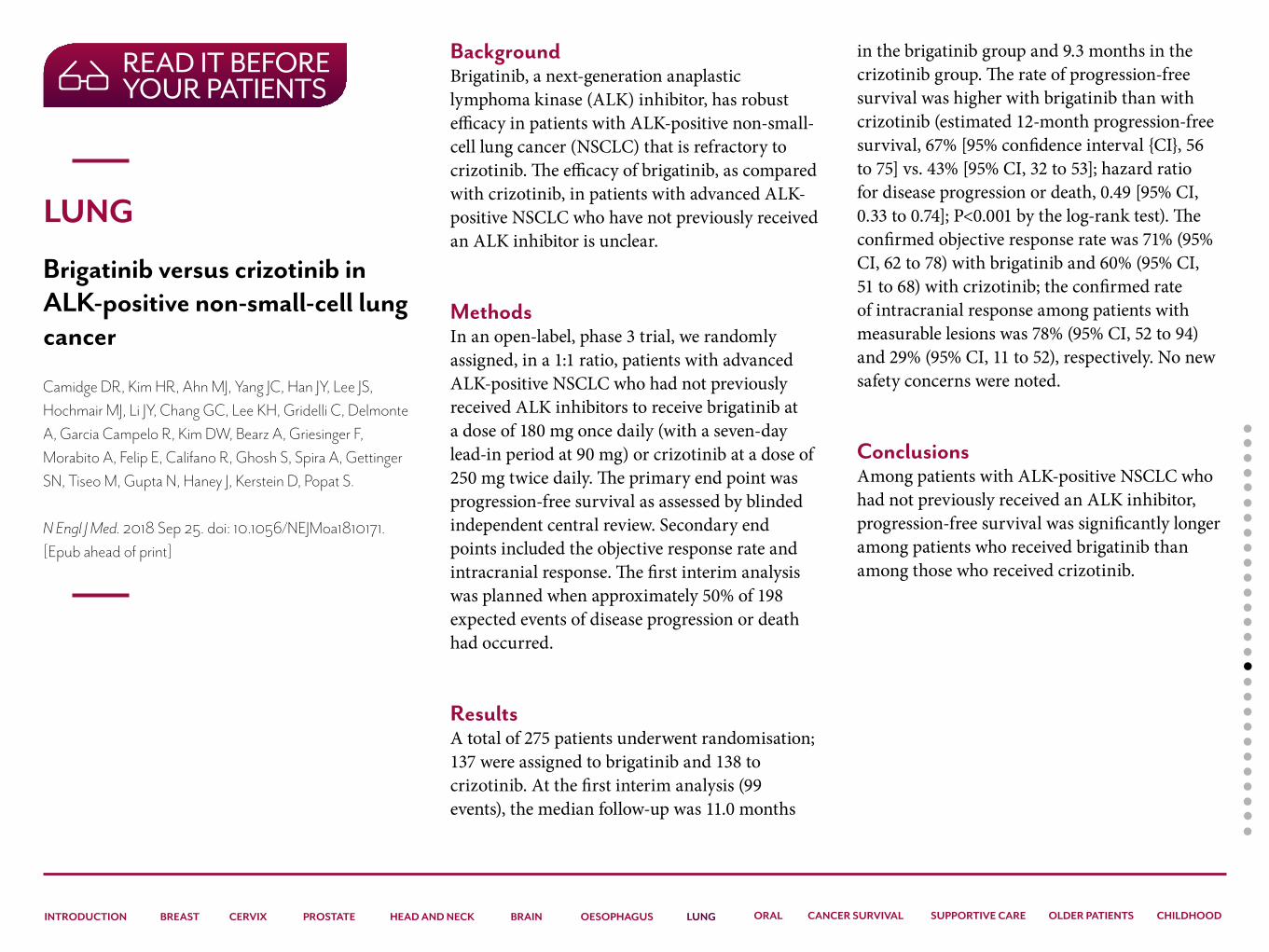
Background Brigatinib, a next-generation anaplastic lymphoma kinase (ALK) inhibitor, has robust efficacy in patients with ALK-positive non-small-cell lung cancer (NSCLC) that is refractory to crizotinib. The efficacy of brigatinib, as compared with crizotinib, in patients with advanced ALK-positive NSCLC who have not previously received an ALK inhibitor is unclear.
Methods In an open-label, phase 3 trial, we randomly assigned, in a 1:1 ratio, patients with advanced ALK-positive NSCLC who had not previously received ALK inhibitors to receive brigatinib at a dose of 180 mg once daily (with a seven-day lead-in period at 90 mg) or crizotinib at a dose of 250 mg twice daily. The primary end point was progression-free survival as assessed by blinded independent central review. Secondary end points included the objective response rate and intracranial response. The first interim analysis was planned when approximately 50% of 198 expected events of disease progression or death had occurred.
Results A total of 275 patients underwent randomisation; 137 were assigned to brigatinib and 138 to crizotinib. At the first interim analysis (99 events), the median follow-up was 11.0 months
in the brigatinib group and 9.3 months in the crizotinib group. The rate of progression-free survival was higher with brigatinib than with crizotinib (estimated 12-month progression-free survival, 67% [95% confidence interval {CI}, 56 to 75] vs. 43% [95% CI, 32 to 53]; hazard ratio for disease progression or death, 0.49 [95% CI, 0.33 to 0.74]; P<0.001 by the log-rank test). The confirmed objective response rate was 71% (95% CI, 62 to 78) with brigatinib and 60% (95% CI, 51 to 68) with crizotinib; the confirmed rate of intracranial response among patients with measurable lesions was 78% (95% CI, 52 to 94) and 29% (95% CI, 11 to 52), respectively. No new safety concerns were noted.
Conclusions Among patients with ALK-positive NSCLC who had not previously received an ALK inhibitor, progression-free survival was significantly longer among patients who received brigatinib than among those who received crizotinib.
Brigatinib versus crizotinib in ALK-positive non-small-cell lung cancer
Camidge DR, Kim HR, Ahn MJ, Yang JC, Han JY, Lee JS, Hochmair MJ, Li JY, Chang GC, Lee KH, Gridelli C, Delmonte A, Garcia Campelo R, Kim DW, Bearz A, Griesinger F, Morabito A, Felip E, Califano R, Ghosh S, Spira A, Gettinger SN, Tiseo M, Gupta N, Haney J, Kerstein D, Popat S.
N Engl J Med. 2018 Sep 25. doi: 10.1056/NEJMoa1810171. [Epub ahead of print]
LUNG
INTRODUCTION BREAST CERVIX HEAD AND NECK BRAIN ORAL CANCER SURVIVAL SUPPORTIVE CARE OLDER PATIENTS CHILDHOODPROSTATE OESOPHAGUS LUNG
READ IT BEFORE YOUR PATIENTS
LUNG
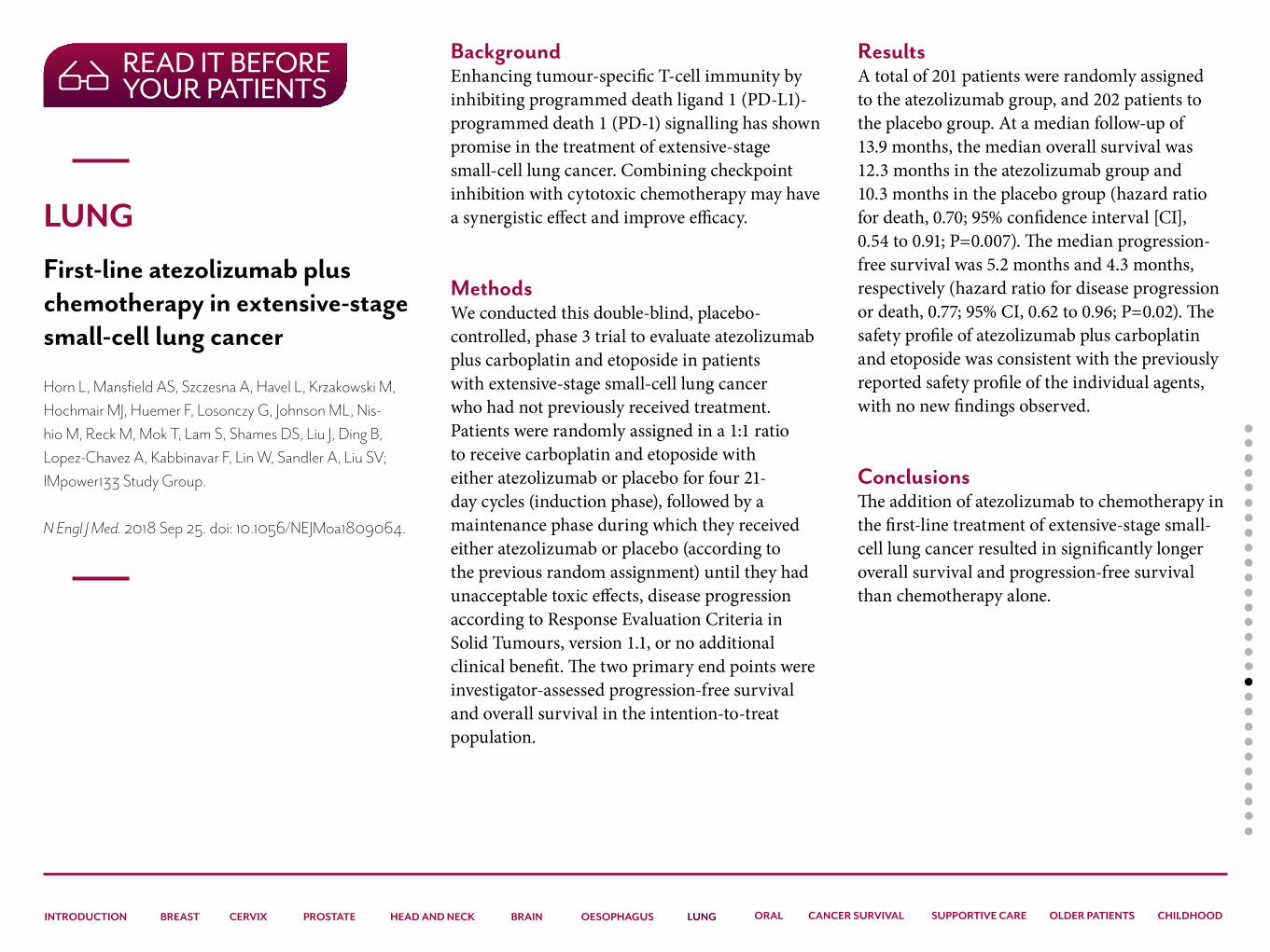
BackgroundEnhancing tumour-specific T-cell immunity by inhibiting programmed death ligand 1 (PD-L1)-programmed death 1 (PD-1) signalling has shown promise in the treatment of extensive-stage small-cell lung cancer. Combining checkpoint inhibition with cytotoxic chemotherapy may have a synergistic effect and improve efficacy.
MethodsWe conducted this double-blind, placebo-controlled, phase 3 trial to evaluate atezolizumab plus carboplatin and etoposide in patients with extensive-stage small-cell lung cancer who had not previously received treatment. Patients were randomly assigned in a 1:1 ratio to receive carboplatin and etoposide with either atezolizumab or placebo for four 21-day cycles (induction phase), followed by a maintenance phase during which they received either atezolizumab or placebo (according to the previous random assignment) until they had unacceptable toxic effects, disease progression according to Response Evaluation Criteria in Solid Tumours, version 1.1, or no additional clinical benefit. The two primary end points were investigator-assessed progression-free survival and overall survival in the intention-to-treat population.
ResultsA total of 201 patients were randomly assigned to the atezolizumab group, and 202 patients to the placebo group. At a median follow-up of 13.9 months, the median overall survival was 12.3 months in the atezolizumab group and 10.3 months in the placebo group (hazard ratio for death, 0.70; 95% confidence interval [CI], 0.54 to 0.91; P=0.007). The median progression-free survival was 5.2 months and 4.3 months, respectively (hazard ratio for disease progression or death, 0.77; 95% CI, 0.62 to 0.96; P=0.02). The safety profile of atezolizumab plus carboplatin and etoposide was consistent with the previously reported safety profile of the individual agents, with no new findings observed.
ConclusionsThe addition of atezolizumab to chemotherapy in the first-line treatment of extensive-stage small-cell lung cancer resulted in significantly longer overall survival and progression-free survival than chemotherapy alone.
First-line atezolizumab plus chemotherapy in extensive-stage small-cell lung cancer
Horn L, Mansfield AS, Szczesna A, Havel L, Krzakowski M, Hochmair MJ, Huemer F, Losonczy G, Johnson ML, Nis-hio M, Reck M, Mok T, Lam S, Shames DS, Liu J, Ding B, Lopez-Chavez A, Kabbinavar F, Lin W, Sandler A, Liu SV; IMpower133 Study Group.
N Engl J Med. 2018 Sep 25. doi: 10.1056/NEJMoa1809064.
INTRODUCTION BREAST CERVIX HEAD AND NECK BRAIN ORAL CANCER SURVIVAL SUPPORTIVE CARE OLDER PATIENTS CHILDHOODPROSTATE OESOPHAGUS LUNG
READ IT BEFORE YOUR PATIENTS
LUNG
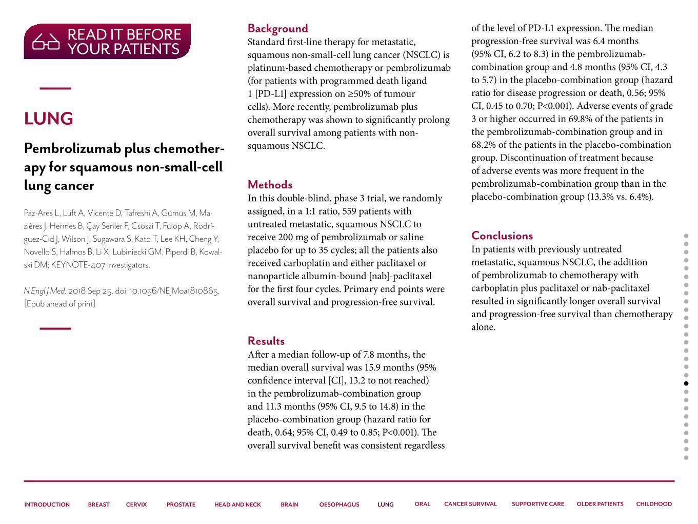
BackgroundStandard first-line therapy for metastatic, squamous non-small-cell lung cancer (NSCLC) is platinum-based chemotherapy or pembrolizumab (for patients with programmed death ligand 1 [PD-L1] expression on ≥50% of tumour cells). More recently, pembrolizumab plus chemotherapy was shown to significantly prolong overall survival among patients with non-squamous NSCLC.
MethodsIn this double-blind, phase 3 trial, we randomly assigned, in a 1:1 ratio, 559 patients with untreated metastatic, squamous NSCLC to receive 200 mg of pembrolizumab or saline placebo for up to 35 cycles; all the patients also received carboplatin and either paclitaxel or nanoparticle albumin-bound [nab]-paclitaxel for the first four cycles. Primary end points were overall survival and progression-free survival.
ResultsAfter a median follow-up of 7.8 months, the median overall survival was 15.9 months (95% confidence interval [CI], 13.2 to not reached) in the pembrolizumab-combination group and 11.3 months (95% CI, 9.5 to 14.8) in the placebo-combination group (hazard ratio for death, 0.64; 95% CI, 0.49 to 0.85; P<0.001). The overall survival benefit was consistent regardless
of the level of PD-L1 expression. The median progression-free survival was 6.4 months (95% CI, 6.2 to 8.3) in the pembrolizumab-combination group and 4.8 months (95% CI, 4.3 to 5.7) in the placebo-combination group (hazard ratio for disease progression or death, 0.56; 95% CI, 0.45 to 0.70; P<0.001). Adverse events of grade 3 or higher occurred in 69.8% of the patients in the pembrolizumab-combination group and in 68.2% of the patients in the placebo-combination group. Discontinuation of treatment because of adverse events was more frequent in the pembrolizumab-combination group than in the placebo-combination group (13.3% vs. 6.4%).
ConclusionsIn patients with previously untreated metastatic, squamous NSCLC, the addition of pembrolizumab to chemotherapy with carboplatin plus paclitaxel or nab-paclitaxel resulted in significantly longer overall survival and progression-free survival than chemotherapy alone.
Pembrolizumab plus chemother-apy for squamous non-small-cell lung cancer
Paz-Ares L, Luft A, Vicente D, Tafreshi A, Gümüs M, Ma-zières J, Hermes B, Çay Senler F, Csoszi T, Fülöp A, Rodrí-guez-Cid J, Wilson J, Sugawara S, Kato T, Lee KH, Cheng Y, Novello S, Halmos B, Li X, Lubiniecki GM, Piperdi B, Kowal-ski DM; KEYNOTE-407 Investigators.
N Engl J Med. 2018 Sep 25. doi: 10.1056/NEJMoa1810865. [Epub ahead of print]
INTRODUCTION BREAST CERVIX HEAD AND NECK BRAIN ORAL CANCER SURVIVAL SUPPORTIVE CARE OLDER PATIENTS CHILDHOODPROSTATE OESOPHAGUS LUNG
INTRODUCTION BREAST CERVIX HEAD AND NECK BRAIN CANCER SURVIVAL SUPPORTIVE CARE OLDER PATIENTS CHILDHOODPROSTATE OESOPHAGUS LUNG ORAL
READ IT BEFORE YOUR PATIENTS
ORAL
PurposeWe previously reported preventive and therapeutic effects of Smad7, a multifunctional protein, on radiation-induced mucositis in mice without promoting human oral cancer cell survival or migration in vitro. The current study aims to determine whether a Smad7-based biologic can treat existing oral mucositis during radiotherapy for oral cancer and whether this treatment compromises radiotherapy-induced cancer cell killing in neighbouring oral cancer.
Experimental designWe transplanted human oral cancer cells into the tongues of mice and applied craniofacial irradiation to simultaneously kill tumour cells and induce oral mucositis, thus modelling radiotherapy and mucositis in oral cancer patients. We topically applied a recombinant human Smad7 protein fused with the cell-penetrating Tat tag (Tat-Smad7) to the oral mucosa of tumour-bearing mice post-radiotherapy when oral mucositis began to develop.
ResultsTopically applied Tat-Smad7 penetrated cells in both the oral mucosa and oral cancer, attenuating TGFβ and NFκB signalling as well as inflammation at both sites. Tat-Smad7 treatment alleviated oral mucositis with reductions in DNA
damage and apoptosis in keratinocytes, but increased keratinocyte proliferation compared to vehicle-treated mucositis lesions. In contrast, adjacent oral cancer exposed to Tat-Smad7 did not show alterations in proliferation or direct DNA damage, but showed increased oxidative stress damage and apoptosis compared to tumours treated with vehicle.
ConclusionOur results suggest that short-course Tat-Smad7 application to oral mucositis promotes its healing but does not compromise the cytotoxic effect of radiotherapy on oral cancer and has context-specific effects on oral mucosa versus oral cancer.
Smad7 promotes healing of radiotherapy-induced oral mucositis without compromising oral cancer therapy in a xenograft mouse model
Luo J, Bian L, Blevins MA, Wang D, Liang C, Du D, Wu F, Holwerda B, Zhao R, Raben D, Zhou H, Young C, Wang XJ.
Clin Cancer Res. 2018 Sep 5. pii: clincanres.1081.2018. Doi: 10.1158/1078-0432.CCR-18-1081. [Epub ahead of print]
INTRODUCTION BREAST CERVIX HEAD AND NECK BRAIN ORAL SUPPORTIVE CARE OLDER PATIENTS CHILDHOODPROSTATE OESOPHAGUS LUNG CANCER SURVIVAL
READ IT BEFORE YOUR PATIENTS
CANCER SURVIVAL
BackgroundIn 2015, the second cycle of the CONCORD programme established global surveillance of cancer survival as a metric of the effectiveness of health systems and to inform global policy on cancer control. CONCORD-3 updates the worldwide surveillance of cancer survival to 2014.
MethodsCONCORD-3 includes individual records for 37.5 million patients diagnosed with cancer during the 14-year period from 2000-2014. Data were provided by 322 population-based cancer registries in 71 countries and territories, 47 of which provided data with 100% population coverage. The study includes 18 cancers or groups of cancers: oesophagus, stomach, colon, rectum, liver, pancreas, lung, breast (women), cervix, ovary, prostate, and melanoma of the skin in adults, and brain tumours, leukaemias, and lymphomas in both adults and children. Standardised quality control procedures were applied; errors were rectified by the registry concerned. We estimated five-year net survival. Estimates were age-standardised with the International Cancer Survival Standard weights.
FindingsFor most cancers, five-year net survival remains among the highest in the world in the USA and Canada, in Australia and New Zealand, and in Finland, Iceland, Norway and Sweden. For
many cancers, Denmark is closing the survival gap with the other Nordic countries. Survival trends are generally increasing, even for some of the more lethal cancers: in some countries, survival has increased by up to 5% for cancers of the liver, pancreas, and lung. For women diagnosed during 2010-14, five-year survival for breast cancer is now 89.5% in Australia and 90.2% in the USA, but international differences remain very wide, with levels as low as 66.1% in India. For gastrointestinal cancers, the highest levels of five-year survival are seen in southeast Asia: in South Korea for cancers of the stomach (68.9%), colon (71.8%), and rectum (71.1%); in Japan for oesophageal cancer (36.0%); and in Taiwan for liver cancer (27.9%). By contrast, in the same world region, survival is generally lower than elsewhere for melanoma of the skin (59.9% in South Korea, 52.1% in Taiwan and 49.6% in China), and for both lymphoid malignancies (52.5%, 50.5% and 38.3%) and myeloid malignancies (45.9%, 33.4% and 24.8%). For children diagnosed during 2010-14, five-year survival for acute lymphoblastic leukaemia ranged from 49.8% in Ecuador to 95.2% in Finland. Five-year survival from brain tumours in children is higher than for adults, but the global range is very wide (from 28.9% in Brazil to nearly 80% in Sweden and Denmark).
InterpretationThe CONCORD programme enables timely comparisons of the overall effectiveness of
Global surveillance of trends in cancer survival 2000-14 (CONCORD-3): analysis of individual records for 37,513,025 patients diagnosed with one of 18 cancers from 322 population-based registries in 71 countries.
Allemani C, Matsuda T, Di Carlo V, Harewood R, Matz M, Nikšic M, Bonaventure A, Valkov M, Johnson CJ, Estève J, Ogunbiyi OJ, Azevedo E Silva G, Chen WQ, Eser S, Engholm G, Stiller CA, Monnereau A, Woods RR, Visser O, Lim GH, Aitken J, Weir HK, Coleman MP; CONCORD Working Group.
Lancet. 2018 Mar 17;391(10125):1023-1075. doi: 10.1016/S0140-6736(17)33326-3. Epub 2018 Jan 31.
INTRODUCTION BREAST CERVIX HEAD AND NECK BRAIN ORAL SUPPORTIVE CARE OLDER PATIENTS CHILDHOODPROSTATE OESOPHAGUS LUNG CANCER SURVIVAL
health systems in providing care for 18 cancers that collectively represent 75% of all cancers diagnosed worldwide every year. It contributes to the evidence base for global policy on cancer control. Since 2017, the Organisation for Economic Co-operation and Development (OECD) has used findings from the CONCORD programme as the official benchmark of cancer survival, among their indicators of the quality of health care in 48 countries worldwide. Governments must recognise population-based cancer registries as key policy tools that can be used to evaluate both the impact of cancer prevention strategies and the effectiveness of health systems for all patients diagnosed with cancer.
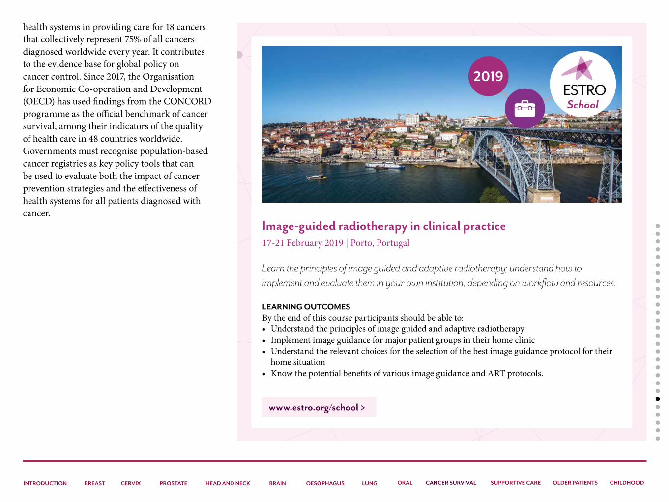
Image-guided radiotherapy in clinical practice17-21 February 2019 | Porto, Portugal
www.estro.org/school >
LEARNING OUTCOMESBy the end of this course participants should be able to:• Understand the principles of image guided and adaptive radiotherapy• Implement image guidance for major patient groups in their home clinic• Understand the relevant choices for the selection of the best image guidance protocol for their
home situation• Know the potential benefits of various image guidance and ART protocols.
Learn the principles of image guided and adaptive radiotherapy; understand how to implement and evaluate them in your own institution, depending on workflow and resources.
2019
INTRODUCTION BREAST CERVIX HEAD AND NECK BRAIN ORAL CANCER SURVIVAL OLDER PATIENTS CHILDHOODPROSTATE OESOPHAGUS LUNG SUPPORTIVE CARE
READ IT BEFORE YOUR PATIENTS
SUPPORTIVE CARE
ImportanceRadiation dermatitis is common and often treated with topical therapy. Patients are typically advised to avoid topical agents for several hours before daily radiotherapy out of concern that topical agents might increase the radiation dose to the skin. With modern radiotherapy’s improved skin-sparing properties, this recommendation may be irrelevant.
ObjectiveTo assess whether applying either metallic or non-metallic topical agents before radiation treatment alters the skin dose.
FindingsA 24-question online survey of patients and clinicians was conducted from 15 January 2015 to 15 March 2017, to determine current practices regarding topical therapy use. In preclinical studies, dosimetric effect of the topical agents was evaluated by delivering 200 monitor units and measuring the dose at the surface and at 2-cm depth in a tissue-equivalent phantom with or without two common topical agents: a petroleum-based ointment (Aquaphor, petrolatum 41%) and silver sulfadiazine cream, 1%. Skin doses associated with various photon and electron energies, topical agent thicknesses, and beam incidence were assessed. Whether topical agents altered the skin dose was also evaluated in 24 C57BL/6 mice by using phosphorylated histone
(γ-H2AX) immunofluorescent staining and terminal deoxynucleotidyl transferase dUTP nick end labelling (TUNEL) assay. Preclinical studies took place at the University of Pennsylvania, USA.
Main outcomes and measuresPatient and clinician survey responses; surface radiation dose readings in tissue-equivalent phantom; and γ-H2AX and TUNEL intensity measured in mice.
ResultsThe 133 patients surveyed received radiotherapy for cancer and had a median (range) age of 60 (18-86) years; 117 (87.9%) were women. In total, 108 clinicians completed the survey with 105 reporting that they were involved in managing patient skin care during radiotherapy. Of these, 111 (83.4%) of the patients and 96 (91.4%) of the 105 clinicians received or gave the advice to avoid applying topical agents before radiotherapy treatments. Dosimetric measurements showed no difference in the delivered dose at either the surface or a 2-cm depth with or without a 1- to 2-mm application of either topical agent when using en face 6- or 15-megavoltage (MV) photons. The same application of topicals did not alter the surface dose as a function of beam incident angle from 15° to 60°, except for a 6% increase at 60° with the silver sulfadiazine cream.
Assessing the validity of clinician advice that patients avoid use of topical agents before daily radiotherapy treatments
Baumann BC, Verginadis II, Zeng C, Bell B, Koduri S, Vachani C, MacArthur KM, Solberg TD, Koumenis C, Metz JM.
JAMA Oncol. 2018 Oct 18. doi: 10.1001/jamaoncol.2018.4292. [Epub ahead of print]
INTRODUCTION BREAST CERVIX HEAD AND NECK BRAIN ORAL CANCER SURVIVAL OLDER PATIENTS CHILDHOODPROSTATE OESOPHAGUS LUNG SUPPORTIVE CARE
Surface dose for 6- and 15-MV beams were significantly increased with a thicker (≥3-mm) topical application. For 6 MV, the surface dose was 1.05 Gy with a thick layer of petroleum-based ointment and 1.02 Gy for silver sulfadiazine cream vs 0.88 Gy without topical agents. For 15 MV, the doses were 0.70 Gy for a thick layer of petroleum-based ointment and 0.60 Gy for silver sulfadiazine cream vs 0.52 Gy for the controls. With 6- and 9-MeV electrons, there was a 2% to 5% increase in surface dose with the use of the topical agents. There were no dose differences at 2-cm depth. Irradiated skin in mice showed no differences in γ-H2AX-positive foci or in TUNEL staining with or without topical agents of varying thickness.
Conclusions and relevanceThin or moderately applied topical agents, even if applied just before radiotherapy, may have minimal influence on skin dose regardless of beam energy or beam incidence. The findings of this study suggest that applying very thick amounts of a topical agent before radiotherapy may increase the surface dose and should be avoided.
INTRODUCTION BREAST CERVIX HEAD AND NECK BRAIN ORAL CANCER SURVIVAL SUPPORTIVE CARE CHILDHOODPROSTATE OESOPHAGUS LUNG OLDER PATIENTS
READ IT BEFORE YOUR PATIENTS
OLDER PATIENTS
BackgroundIt is increasingly recognised that older adults with cancer represent a diverse cohort of patients and that other comorbidities may have an equal impact on survival and quality of life as any diagnosis of malignancy. Competing risk has consequently emerged as an important concept in the design and reporting of geriatric oncology trials.
MethodsWe performed a systematic review of phase II and III oncology trials for systemic therapy in older patients with solid organ malignancy from the year 2000 until 30 April 2017. Forty-one trials including 7,864 patients were identified for evaluation.
ResultsOnly 15 trials (36.6%) employed disease-related end points to account for death from other causes, and only one study used statistical analysis that addressed competing risk. Seventeen studies (41.5%) of trials included some assessment of comorbidity or frailty. Twenty-one trials (51.2%) included any assessment of quality of life.
ConclusionsThis review demonstrates clear areas for improvement for future studies and highlights the need for careful consideration of trial design, data collection, and appropriate statistical methodology for reporting of competing risks in geriatric oncology trials.
Competing risks in older patients with cancer: a systematic review of geriatric oncology trials
Burdett N, Vincent AD, O‘Callaghan M, Kichenadasse G.
J Natl Cancer Inst. 2018;110(8):825-830. doi: 10.1093/jnci/djy111.
INTRODUCTION BREAST CERVIX HEAD AND NECK BRAIN ORAL CANCER SURVIVAL SUPPORTIVE CARE OLDER PATIENTSPROSTATE OESOPHAGUS LUNG CHILDHOOD
READ IT BEFORE YOUR PATIENTS
CHILDHOOD
BackgroundChildhood cancer survivors are at risk of subsequent primary soft-tissue sarcomas (STS), but the risks of specific STS histological subtypes are unknown. We quantified the risk of STS histological subtypes after specific types of childhood cancer.
MethodsWe pooled data from 13 European cohorts, yielding a cohort of 69,460 five-year survivors of childhood cancer. Standardised incidence ratios (SIRs) and absolute excess risks (AERs) were calculated.
ResultsOverall, 301 STS developed compared with 19 expected (SIR = 15.7, 95% confidence interval [CI] = 14.0 to 17.6). The highest standardised incidence ratios were for malignant peripheral nerve sheath tumours (MPNST; SIR = 40.6, 95% CI = 29.6 to 54.3), leiomyosarcomas (SIR = 29.9, 95% CI = 23.7 to 37.2), and fibromatous neoplasms (SIR = 12.3, 95% CI = 9.3 to 16.0). SIRs for MPNST were highest following central nervous system tumours (SIR = 80.5, 95% CI = 48.4 to 125.7), Hodgkin lymphoma (SIR = 81.3, 95% CI = 35.1 to 160.1), and Wilms tumour (SIR = 76.0, 95% CI = 27.9 to 165.4). Standardised incidence ratios for leiomyosarcoma were highest following retinoblastoma (SIR = 342.9, 95% CI = 245.0 to 466.9) and Wilms tumour
(SIR = 74.2, 95% CI = 37.1 to 132.8). AERs for all STS subtypes were generally low at all years from diagnosis (AER < 1 per 10 000 person-years), except for leiomyosarcoma following retinoblastoma, for which the AER reached 52.7 (95% CI = 20.0 to 85.5) per 10,000 person-years among patients who had survived at least 45 years from diagnosis of retinoblastoma.
ConclusionsFor the first time, we provide risk estimates of specific STS subtypes following childhood cancers and give evidence that risks of MPNSTs, leiomyosarcomas, and fibromatous neoplasms are particularly increased. While the multiplicative excess risks relative to the general population are substantial, the absolute excess risk of developing any STS subtype is low, except for leiomyosarcoma after retinoblastoma. These results are likely to be informative for both survivors and health care providers.
Risk of soft-tissue sarcoma among 69,460 five-year survivors of childhood cancer in Europe.
Bright CJ, Hawkins MM, Winter DL, Alessi D, Allodji RS, Bagnasco F, Bárdi E, Bautz A, Byrne J, Feijen EAM, Fidler MM, Garwicz S, Grabow D, Gudmundsdottir T, Guha J, Haddy N, Jankovic M, Kaatsch P, Kaiser M, Kuehni CE, Linge H, Øfstaas H, Ronckers CM, Skinner R, Teepen JC, Terenziani M, Vu-Bezin G, Wesenberg F, Wiebe T, Sacerdote C, Jakab Z, Haupt R, Lähteenmäki P, Zaletel LZ, Kuonen R, Winther JF, de Vathaire F, Kremer LC, Hjorth L, Reulen RC; PanCareSurFup Consortium.
J Natl Cancer Inst. 2018;110(6):649-660. doi: 10.1093/jnci/djx235.
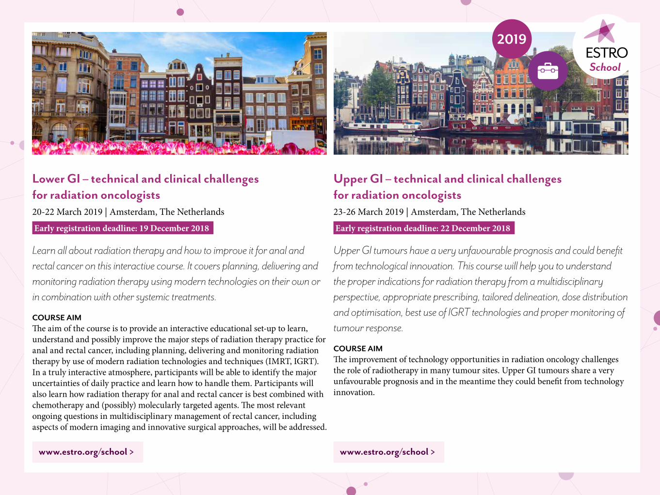
Lower GI – technical and clinical challenges for radiation oncologists 20-22 March 2019 | Amsterdam, The Netherlands
Upper GI – technical and clinical challenges for radiation oncologists 23-26 March 2019 | Amsterdam, The Netherlands
www.estro.org/school > www.estro.org/school >
COURSE AIMThe improvement of technology opportunities in radiation oncology challenges the role of radiotherapy in many tumour sites. Upper GI tumours share a very unfavourable prognosis and in the meantime they could benefit from technology innovation.
COURSE AIMThe aim of the course is to provide an interactive educational set-up to learn, understand and possibly improve the major steps of radiation therapy practice for anal and rectal cancer, including planning, delivering and monitoring radiation therapy by use of modern radiation technologies and techniques (IMRT, IGRT). In a truly interactive atmosphere, participants will be able to identify the major uncertainties of daily practice and learn how to handle them. Participants will also learn how radiation therapy for anal and rectal cancer is best combined with chemotherapy and (possibly) molecularly targeted agents. The most relevant ongoing questions in multidisciplinary management of rectal cancer, including aspects of modern imaging and innovative surgical approaches, will be addressed.
Learn all about radiation therapy and how to improve it for anal and rectal cancer on this interactive course. It covers planning, delivering and monitoring radiation therapy using modern technologies on their own or in combination with other systemic treatments.
Upper GI tumours have a very unfavourable prognosis and could benefit from technological innovation. This course will help you to understand the proper indications for radiation therapy from a multidisciplinary perspective, appropriate prescribing, tailored delineation, dose distribution and optimisation, best use of IGRT technologies and proper monitoring of tumour response.
Early registration deadline: 22 December 2018Early registration deadline: 19 December 2018
2019
INTRODUCTION IN-MEMORIAM EDITORS' PICK ASTRO60TH ANNUAL MEETING
BRACHYTHERAPY
IN-MEMORIAM EDITORS' PICK ASTRO60TH ANNUAL MEETINGINTRODUCTION
BRACHYTHERAPY
“Recently, a follow up recommendation paper was published in Radiotherapy and Oncology; it describes practical aspects of this form of brachytherapy” BRADLEY PIETERS
PETER HOSKIN
ÅSA CARLSSON TEDGREN
Welcome to the first edition of the Brachytherapy Corner in 2019. We hope you had an enjoyable holiday, and would like to wish you all a very happy new year.
In this edition, Peter Hoskin reports on the American Society for Radiation Oncology’s (ASTRO) 60th annual meeting in San Antonio, USA, highlighting a number of interesting papers in brachytherapy.
In 2015 and 2016 the Groupe Européen de Curiethérapie (GEC)-ESTRO published two recommendation papers on partial breast brachytherapy. Recently, a follow up recommendation paper was published in Radiotherapy and Oncology; led by Vratislav Strnad, it describes practical aspects of this form of brachytherapy, and you can read more about the paper here.
It was a pleasure meeting many of you in Brussels at the sixth GEC-ESTRO workshop. We will have some reports from the workshop in the next issue of the newsletter.
We hope you enjoy reading this edition of the Brachytherapy Corner and others to come in 2019.
Peter Hoskin, Bradley Pieters and Åsa Tedgren
INTRODUCTION IN-MEMORIAM EDITORS' PICK ASTRO60TH ANNUAL MEETING
PROSTATESU_27_2273 – A phase II randomised pilot study comparing high-dose-rate (HDR) brachytherapy and low-dose-rate (LDR) brachytherapy as monotherapy in localised prostate cancerPresented by Laura Hathout on behalf of a co-operative Canadian group, this study featured early quality of life data on 31 patients in a pilot phase II study comprising LDR brachytherapy 144Gy I125 with HDR single dose 19Gy. The main conclusion was that at three months after implant there was less urinary irritation with LDR. However, on repeated measures taken over 12 months, overall, HDR had a better urinary toxicity profile with time to international prostate symptom score (IPSS) normalisation 3.3 months after HDR and 6.5 months after LDR. There was no difference in reported incontinence, sexual function or bowel habits. This trial is now proceeding in Canada and may give valuable data in the future comparing these two modalities.
SU_30_2307 – Assessment of the prostate-specific antigen bounce in patients treated with
BRACHYTHERAPY
American Society for Radiation Oncology (ASTRO) 60th annual meeting
21-23 October 2018San Antonio, USA
PETER HOSKIN
125I-brachytherapy for prostate cancer and its correlation with testosteronePresented by Yasushi Nakai on behalf of a Japanese group based in Kashihara, Nara, this was an analysis of 252 patients receiving LDR I125 brachytherapy of whom 74 exhibited a PSA bounce. The only significant predictive factor was age, bounce being more common in younger patients and being seen to be mirrored by a rise in testosterone above the nadir level. This may shed some light on a clinical phenomenon that we are all aware of but have no good explanation for.
SU_27_2272 – Impact of prostate gland size =60 cc on physician and patient-reported toxicity after high-dose-rate prostate brachytherapyThis study was presented by Alexander Harris from Loyola University Medical Centre, Chicago, USA, and was a retrospective analysis of 119 patients receiving HDR brachytherapy monotherapy 27Gy in two fractions or as a boost of 13.5-15Gy. The median gland volume was 36ml and 13 men were identified who had a volume >60ml. Not surprisingly, given the
Brachytherapy was surprisingly well represented at the American Society for Radiation Oncology’s (ASTRO) 60th annual meeting, with many sessions including presentations on brachytherapy. While the main areas of interest were prostate and cervix studies of brachytherapy, many other sites, including oesophagus, rectum, breast, skin and ocular were covered. Below are summaries of some of the more interesting abstracts that were presented.
Full details can be found on the ASTRO website >
INTRODUCTION IN-MEMORIAM EDITORS' PICK ASTRO60TH ANNUAL MEETING
small sample size, no differences in physician-reported toxicity scores were seen and although there was a tendency for greater acute retention in the larger glands, this was not significant.
SU_30_2306 – Long term results of Cs-131 monotherapy as definitive therapy in a prospectively-followed group of low risk localised prostate cancer patientsThis study was presented by Brian Moran from Chicago, USA, and was a series of 269 low-risk
patients treated with LDR brachytherapy using Cs-131 seeds. This is the largest series of mature Cs131 implant patients having a median follow up of 66.9 months. The results mirror those expected with I125 brachytherapy in low-risk patients with biochemical-relapse-free survival rates of 97% at five years and 90% at ten years.
304 – A meta-analysis of randomised trials to compare the added benefit of a brachytherapy boost versus the addition of androgen
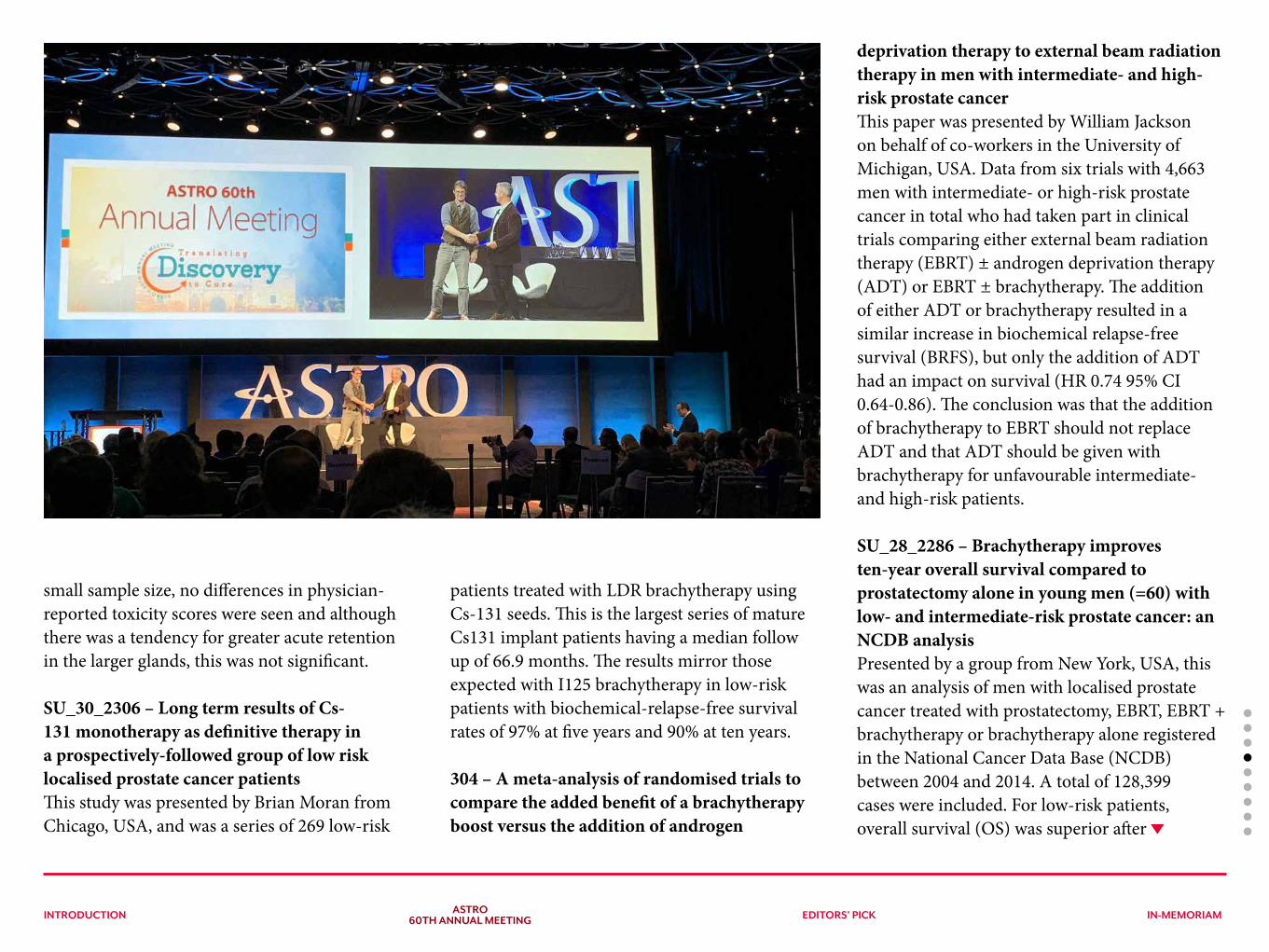
deprivation therapy to external beam radiation therapy in men with intermediate- and high-risk prostate cancerThis paper was presented by William Jackson on behalf of co-workers in the University of Michigan, USA. Data from six trials with 4,663 men with intermediate- or high-risk prostate cancer in total who had taken part in clinical trials comparing either external beam radiation therapy (EBRT) ± androgen deprivation therapy (ADT) or EBRT ± brachytherapy. The addition of either ADT or brachytherapy resulted in a similar increase in biochemical relapse-free survival (BRFS), but only the addition of ADT had an impact on survival (HR 0.74 95% CI 0.64-0.86). The conclusion was that the addition of brachytherapy to EBRT should not replace ADT and that ADT should be given with brachytherapy for unfavourable intermediate- and high-risk patients.
SU_28_2286 – Brachytherapy improves ten-year overall survival compared to prostatectomy alone in young men (=60) with low- and intermediate-risk prostate cancer: an NCDB analysisPresented by a group from New York, USA, this was an analysis of men with localised prostate cancer treated with prostatectomy, EBRT, EBRT + brachytherapy or brachytherapy alone registered in the National Cancer Data Base (NCDB) between 2004 and 2014. A total of 128,399 cases were included. For low-risk patients, overall survival (OS) was superior after
INTRODUCTION IN-MEMORIAM EDITORS' PICK ASTRO60TH ANNUAL MEETING
brachytherapy alone compared to prostatectomy (ten-year OS 93% vs 91.7%), for intermediate risk patients combined EBRT + brachytherapy had a better survival than prostatectomy (ten-year OS 91.4% vs 85.8%) and for high-risk patients brachytherapy had similar outcomes to prostatectomy (ten-year OS 84.3% vs 86.3%). There are of course considerable sources of bias in such analyses, but the overall picture is to strongly support the role of brachytherapy in the management of all risk groups.
CERVIXTU_13_3443 – Effect of radiation boost modality in overall survival of cervical cancer patientsPresented by Tithi Biswas from Cleveland, USA, this analysis of patients in the National Cancer Data Base with stage IB to IIIB cervical cancer treated between 2004 and 2014 further reinforces the importance of brachytherapy in the radical treatment of cervical cancer and demonstrated once again that EBRT cannot be used to replace brachytherapy. In total, 9,936 patients are included with median follow up of 33.8 months. Median survival across all stages was 38.2 months for EBRT boost compared to 112.9 months after brachytherapy boost. This difference was mirrored across all stages: 71.9 vs 133.6 for stage IB, 65.6 vs 127.9 for stage II and 29.1. vs 71.6 for stage III.
TU_17_3486 – Impact of tumour size, shape and patterns of response to chemoradiation in locally advanced cervical cancer patientsPresented by Antoine Schernberg on behalf of colleagues from the Institut Gustave Roussy (IGR), Paris, France, this paper explores the prognostic value of tumour size and shape on MR scan in patients with locally advanced prostate cancer. A total of 247 patients treated with chemoradiation for stage ≥IIB were analysed. Tumour width came out as the most
important parameter; those with a tumour width greater than height at diagnosis or at the time of brachytherapy had a worse prognosis and reduction in tumour. Width rather than height was also important in predicting local control and survival.
An MR-based radiomic signature for disease-free survival in locally advanced cervical cancerPresented by Kathy Han on behalf of her colleagues in Princess Margaret Hospital,
INTRODUCTION IN-MEMORIAM EDITORS' PICK ASTRO60TH ANNUAL MEETING
Toronto, Canada, this paper, while not exclusively on brachytherapy, explored the prognostic value of radiomic analysis of the MR signature from the primary tumour in locally advanced cervical cancer after radical chemoradiation. An initial sample of 80 patients was analysed followed by a validation cohort of a further 81 patients. Two radiomic features based on shape and wavelet were found to be prognostic for disease-free survival, independent of other clinical features. The power was increased when added to a model including stage and nodal status with a hazard ratio of 2.65
TU_15_3469 – Less than whole uterus irradiation for locally advanced cervical cancer maintains locoregional control and potentially decreases GI toxicityPresented by Margaret Kozak on behalf of colleagues at Stanford University, USA, this study of 48 patients reviewed the results of a policy of treating less than the whole uterus (LTWU) in the radiation volume. Gross tumour volume (GTV) was defined using PET and no attempt to include the entire uterus was made. The median proportion of uterus included in the treated volume was 63%; ten patients had >90% included. The two-year locoregional failure rate was 10.2% and the patients with LTWU volumes had lower bowel doses and volumes, although the clinical impact of this could not be defined. The possibility of reducing the CTV when treating cervical cancer is attractive and indeed, where there is a non-adaptive approach, almost certainly
there will be days when the uterine fundus is outside the treated volume. Unfortunately, this study does not have sufficiently robust data or adequate numbers to address the potential gains or losses in doing so.
SKINMO_24_2576 – Definitive high-dose-rate (HDR) brachytherapy for non-melanomatous skin cancers: an effective and efficient cureThere has been renewed interest in the role of brachytherapy for skin cancers and this presentation by Courtney Hentz with colleagues from Loyola University Medical Centre, USA, reported the results of treating 81 non-melanomatous skin cancers in 60 patients with HDR brachytherapy using predominantly the Freiberg flap technique. Doses of 32-40Gy at 3-5mm in 8-20 fractions were delivered. The one-year local control rate was 97.3%. Physician-rated cosmesis and late follow-up was good to excellent in 96% and patient-graded late cosmesis was 98%. Late grade 2 skin toxicity rates were 1.3% with no grade 3 or 4 events.
Peter HoskinMount Vernon Cancer CentreNorthwood Middlesex, UKManchester University,Manchester, UK
INTRODUCTION IN-MEMORIAM ASTRO60TH ANNUAL MEETING EDITORS' PICK
BRACHYTHERAPY
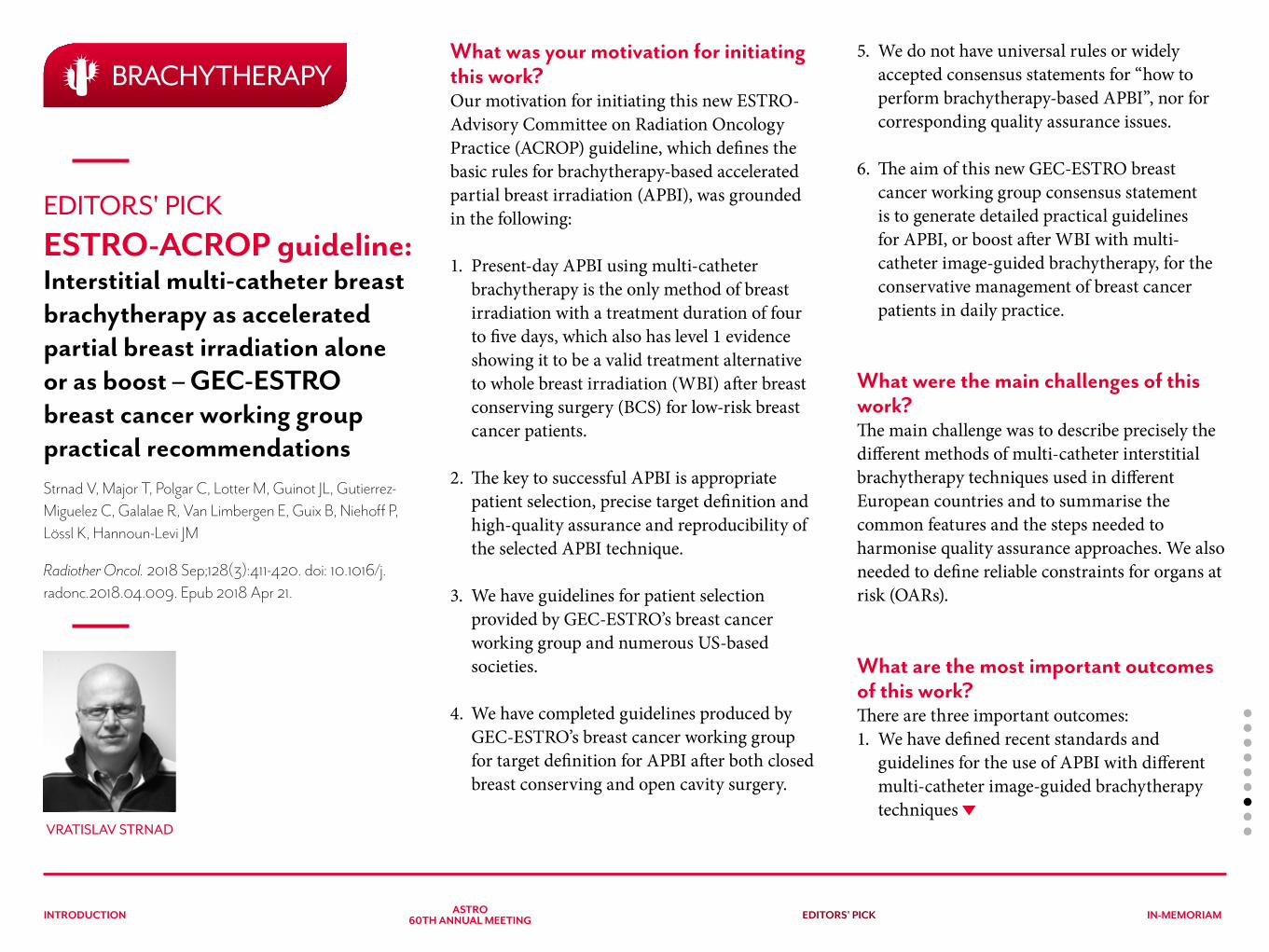
EDITORS' PICK ESTRO-ACROP guideline: Interstitial multi-catheter breast brachytherapy as accelerated partial breast irradiation alone or as boost – GEC-ESTRO breast cancer working group practical recommendations Strnad V, Major T, Polgar C, Lotter M, Guinot JL, Gutierrez-Miguelez C, Galalae R, Van Limbergen E, Guix B, Niehoff P, Lössl K, Hannoun-Levi JM
Radiother Oncol. 2018 Sep;128(3):411-420. doi: 10.1016/j.radonc.2018.04.009. Epub 2018 Apr 21.
What was your motivation for initiating this work?Our motivation for initiating this new ESTRO-Advisory Committee on Radiation Oncology Practice (ACROP) guideline, which defines the basic rules for brachytherapy-based accelerated partial breast irradiation (APBI), was grounded in the following:
1. Present-day APBI using multi-catheter brachytherapy is the only method of breast irradiation with a treatment duration of four to five days, which also has level 1 evidence showing it to be a valid treatment alternative to whole breast irradiation (WBI) after breast conserving surgery (BCS) for low-risk breast cancer patients.
2. The key to successful APBI is appropriate patient selection, precise target definition and high-quality assurance and reproducibility of the selected APBI technique.
3. We have guidelines for patient selection provided by GEC-ESTRO’s breast cancer working group and numerous US-based societies.
4. We have completed guidelines produced by GEC-ESTRO’s breast cancer working group for target definition for APBI after both closed breast conserving and open cavity surgery.
5. We do not have universal rules or widely accepted consensus statements for “how to perform brachytherapy-based APBI”, nor for corresponding quality assurance issues.
6. The aim of this new GEC-ESTRO breast cancer working group consensus statement is to generate detailed practical guidelines for APBI, or boost after WBI with multi-catheter image-guided brachytherapy, for the conservative management of breast cancer patients in daily practice.
What were the main challenges of this work?The main challenge was to describe precisely the different methods of multi-catheter interstitial brachytherapy techniques used in different European countries and to summarise the common features and the steps needed to harmonise quality assurance approaches. We also needed to define reliable constraints for organs at risk (OARs).
What are the most important outcomes of this work?There are three important outcomes:1. We have defined recent standards and
guidelines for the use of APBI with different multi-catheter image-guided brachytherapy techniques
VRATISLAV STRNAD
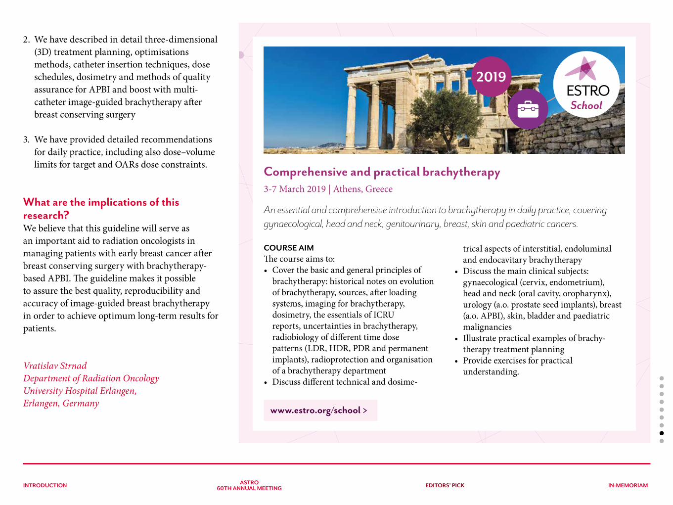
INTRODUCTION IN-MEMORIAM ASTRO60TH ANNUAL MEETING EDITORS' PICK
2. We have described in detail three-dimensional (3D) treatment planning, optimisations methods, catheter insertion techniques, dose schedules, dosimetry and methods of quality assurance for APBI and boost with multi-catheter image-guided brachytherapy after breast conserving surgery
3. We have provided detailed recommendations for daily practice, including also dose–volume limits for target and OARs dose constraints.
What are the implications of this research?We believe that this guideline will serve as an important aid to radiation oncologists in managing patients with early breast cancer after breast conserving surgery with brachytherapy-based APBI. The guideline makes it possible to assure the best quality, reproducibility and accuracy of image-guided breast brachytherapy in order to achieve optimum long-term results for patients.
Vratislav StrnadDepartment of Radiation OncologyUniversity Hospital Erlangen, Erlangen, Germany
Comprehensive and practical brachytherapy3-7 March 2019 | Athens, Greece
www.estro.org/school >
COURSE AIMThe course aims to:• Cover the basic and general principles of
brachytherapy: historical notes on evolution of brachytherapy, sources, after loading systems, imaging for brachytherapy, dosimetry, the essentials of ICRU reports, uncertainties in brachytherapy, radiobiology of different time dose patterns (LDR, HDR, PDR and permanent implants), radioprotection and organisation of a brachytherapy department
• Discuss different technical and dosime-
trical aspects of interstitial, endoluminal and endocavitary brachytherapy
• Discuss the main clinical subjects: gynaecological (cervix, endometrium), head and neck (oral cavity, oropharynx), urology (a.o. prostate seed implants), breast (a.o. APBI), skin, bladder and paediatric malignancies
• Illustrate practical examples of brachy-therapy treatment planning
• Provide exercises for practical understanding.
An essential and comprehensive introduction to brachytherapy in daily practice, covering gynaecological, head and neck, genitourinary, breast, skin and paediatric cancers.
2019
INTRODUCTION EDITORS' PICK ASTRO60TH ANNUAL MEETING IN-MEMORIAM
With great sadness we took notice of the death of Professor Janusz Skowronek.
Janusz was the head of the Brachytherapy Unit at The Greater Poland Cancer Centre in Poznań, Poland. From 2013 till 2017 he was member of the GEC-ESTRO Committee. We have learned Janusz as a person very devoted to his work and in principal his predilection for brachytherapy. He contributed to the development of not only brachytherapy in the Eastern part of Europe, but also worldwide. As an example is the release of the Journal of Contemporary Brachytherapy. Janusz was the driving force and Chief-editor of this journal. Nowadays JCB is a well-respected journal that has earned its place in the field of radiation oncology.
With his passing away we lose a friend who, for his love to brachytherapy, was an example for all of us. Our greatest sympathy is with his family, friends, and colleagues at the Greater Poland Cancer Centre.
Bradley Pieters, Christian Kirisits, Ina Jürgenliemk-Schulz, and Marisol De BrabandereOn behalf of the GEC-ESTRO Committee
BRACHYTHERAPY
In-memoriam Janusz Skowronek
INTRODUCTION 2ND ESTRO PHYSICS WORKSHOP
PHYSICS
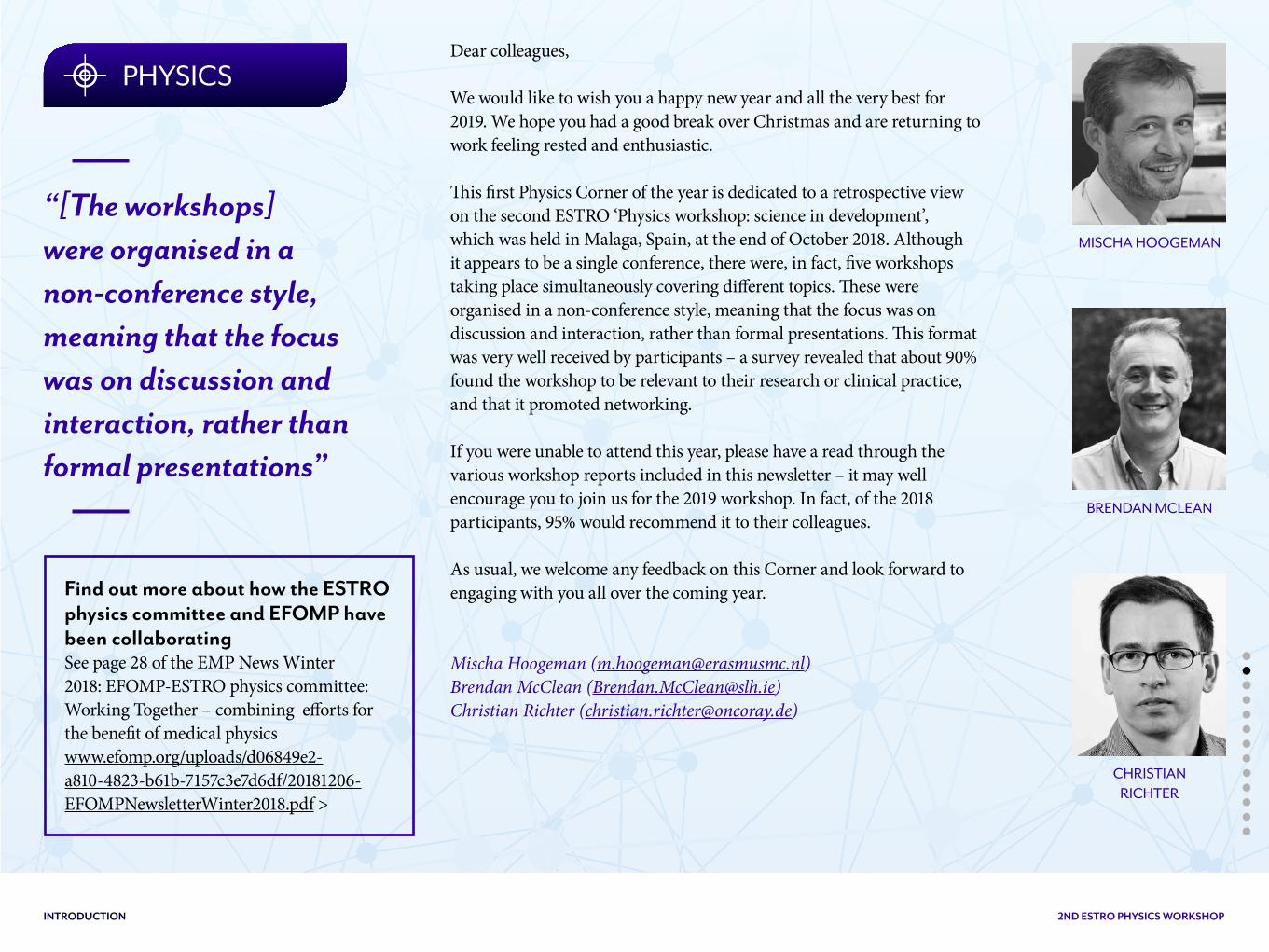
2ND ESTRO PHYSICS WORKSHOPINTRODUCTION
Dear colleagues,
We would like to wish you a happy new year and all the very best for 2019. We hope you had a good break over Christmas and are returning to work feeling rested and enthusiastic.
This first Physics Corner of the year is dedicated to a retrospective view on the second ESTRO ‘Physics workshop: science in development’, which was held in Malaga, Spain, at the end of October 2018. Although it appears to be a single conference, there were, in fact, five workshops taking place simultaneously covering different topics. These were organised in a non-conference style, meaning that the focus was on discussion and interaction, rather than formal presentations. This format was very well received by participants – a survey revealed that about 90% found the workshop to be relevant to their research or clinical practice, and that it promoted networking.
If you were unable to attend this year, please have a read through the various workshop reports included in this newsletter – it may well encourage you to join us for the 2019 workshop. In fact, of the 2018 participants, 95% would recommend it to their colleagues.
As usual, we welcome any feedback on this Corner and look forward to engaging with you all over the coming year.
Mischa Hoogeman ([email protected]) Brendan McClean ([email protected])Christian Richter ([email protected])
PHYSICS
“[The workshops] were organised in a non-conference style, meaning that the focus was on discussion and interaction, rather than formal presentations”
MISCHA HOOGEMAN
BRENDAN MCLEAN
CHRISTIANRICHTER
Find out more about how the ESTRO physics committee and EFOMP have been collaboratingSee page 28 of the EMP News Winter 2018: EFOMP-ESTRO physics committee: Working Together – combining efforts for the benefit of medical physicswww.efomp.org/uploads/d06849e2-a810-4823-b61b-7157c3e7d6df/20181206-EFOMPNewsletterWinter2018.pdf >
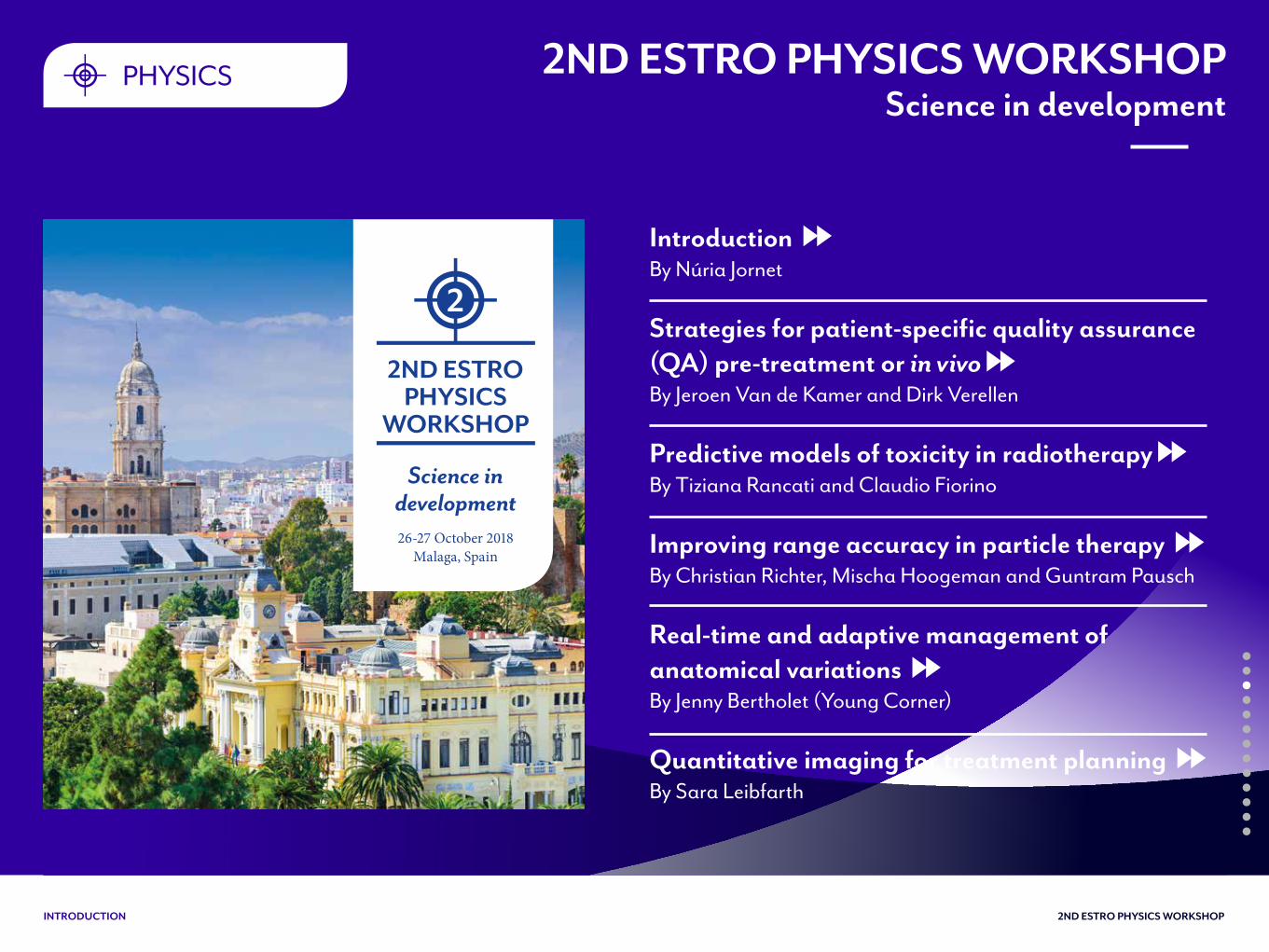
INTRODUCTION 2ND ESTRO PHYSICS WORKSHOP
Introduction By Núria Jornet
Strategies for patient-specific quality assurance (QA) pre-treatment or in vivo By Jeroen Van de Kamer and Dirk Verellen
Predictive models of toxicity in radiotherapy By Tiziana Rancati and Claudio Fiorino
Improving range accuracy in particle therapy By Christian Richter, Mischa Hoogeman and Guntram Pausch
Real-time and adaptive management of anatomical variations By Jenny Bertholet (Young Corner)
Quantitative imaging for treatment planning By Sara Leibfarth
2ND ESTRO PHYSICS WORKSHOPScience in development
PHYSICS
2ND ESTRO PHYSICS
WORKSHOP
Science in development26-27 October 2018
Malaga, Spain
2
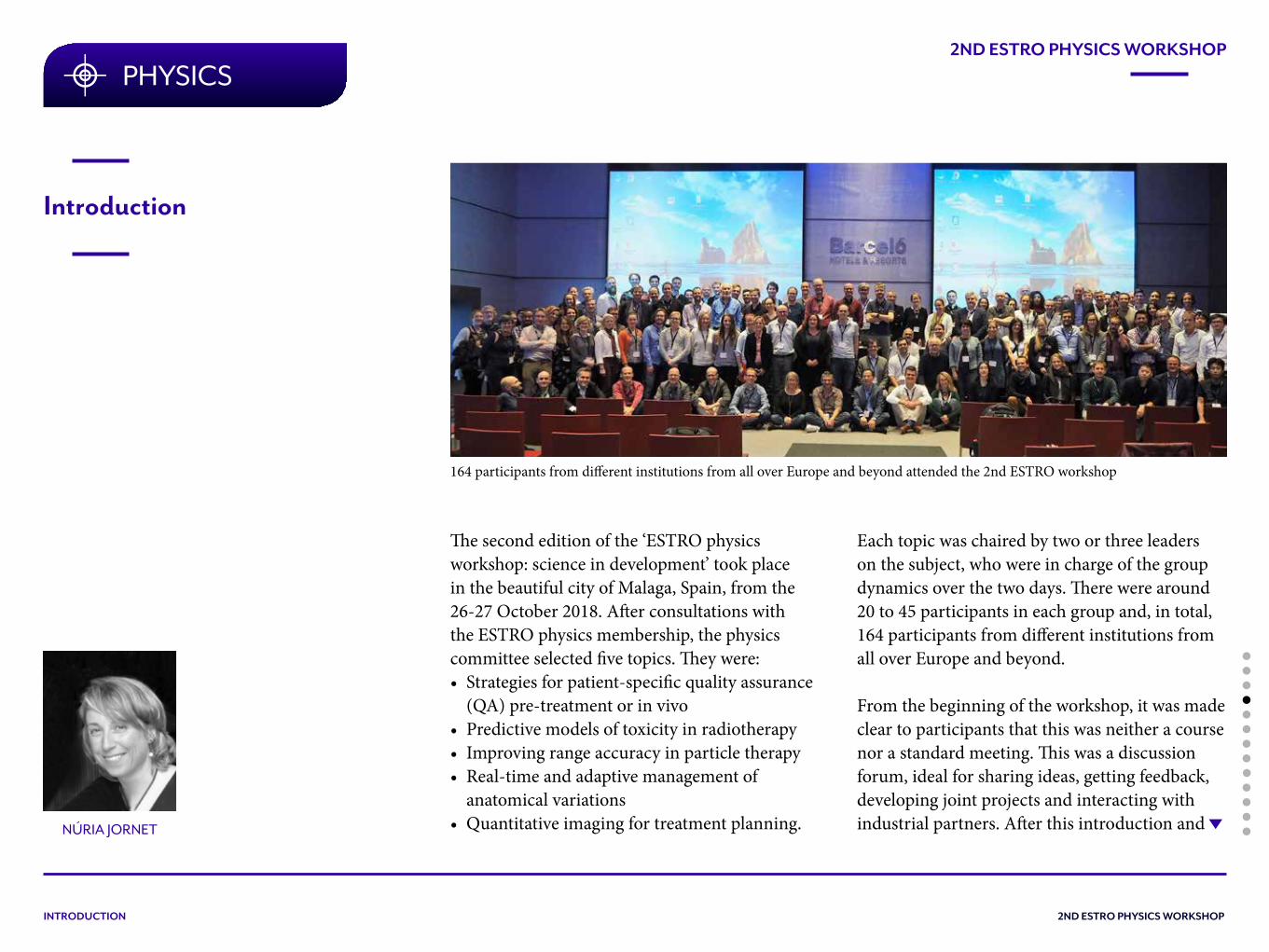
INTRODUCTION 2ND ESTRO PHYSICS WORKSHOP
The second edition of the ‘ESTRO physics workshop: science in development’ took place in the beautiful city of Malaga, Spain, from the 26-27 October 2018. After consultations with the ESTRO physics membership, the physics committee selected five topics. They were: • Strategies for patient-specific quality assurance
(QA) pre-treatment or in vivo • Predictive models of toxicity in radiotherapy • Improving range accuracy in particle therapy• Real-time and adaptive management of
anatomical variations• Quantitative imaging for treatment planning.
Each topic was chaired by two or three leaders on the subject, who were in charge of the group dynamics over the two days. There were around 20 to 45 participants in each group and, in total, 164 participants from different institutions from all over Europe and beyond.
From the beginning of the workshop, it was made clear to participants that this was neither a course nor a standard meeting. This was a discussion forum, ideal for sharing ideas, getting feedback, developing joint projects and interacting with industrial partners. After this introduction and
PHYSICS
Introduction
NÚRIA JORNET
2ND ESTRO PHYSICS WORKSHOP
164 participants from different institutions from all over Europe and beyond attended the 2nd ESTRO workshop
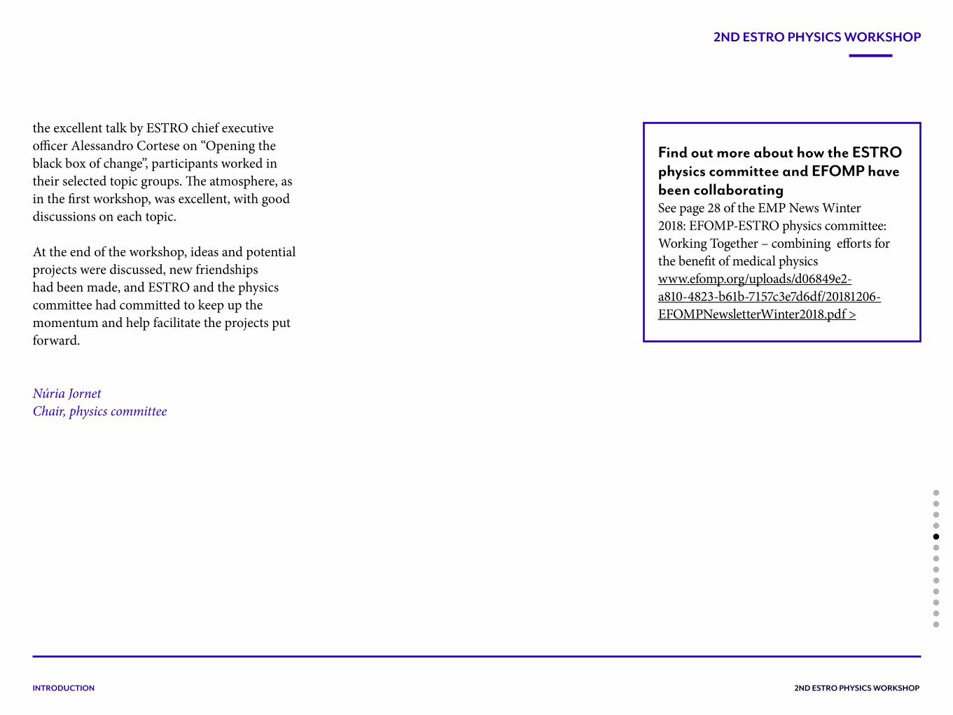
INTRODUCTION 2ND ESTRO PHYSICS WORKSHOP
2ND ESTRO PHYSICS WORKSHOP
the excellent talk by ESTRO chief executive officer Alessandro Cortese on “Opening the black box of change”, participants worked in their selected topic groups. The atmosphere, as in the first workshop, was excellent, with good discussions on each topic.
At the end of the workshop, ideas and potential projects were discussed, new friendships had been made, and ESTRO and the physics committee had committed to keep up the momentum and help facilitate the projects put forward.
Núria JornetChair, physics committee
Find out more about how the ESTRO physics committee and EFOMP have been collaboratingSee page 28 of the EMP News Winter 2018: EFOMP-ESTRO physics committee: Working Together – combining efforts for the benefit of medical physicswww.efomp.org/uploads/d06849e2-a810-4823-b61b-7157c3e7d6df/20181206-EFOMPNewsletterWinter2018.pdf >
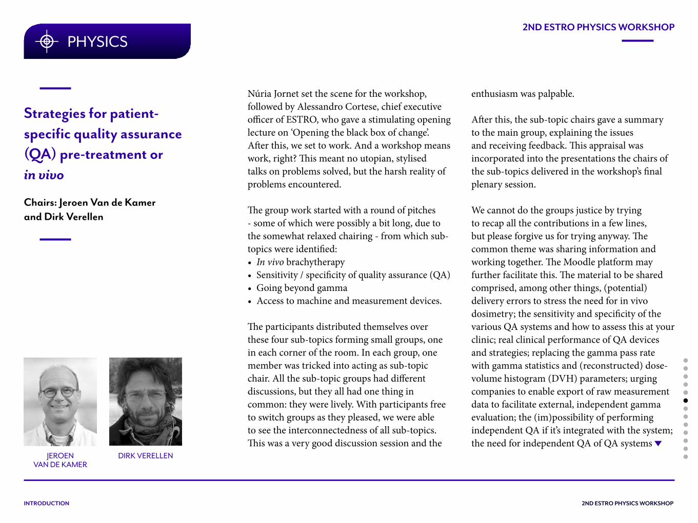
INTRODUCTION 2ND ESTRO PHYSICS WORKSHOP
Núria Jornet set the scene for the workshop, followed by Alessandro Cortese, chief executive officer of ESTRO, who gave a stimulating opening lecture on ‘Opening the black box of change’. After this, we set to work. And a workshop means work, right? This meant no utopian, stylised talks on problems solved, but the harsh reality of problems encountered.
The group work started with a round of pitches - some of which were possibly a bit long, due to the somewhat relaxed chairing - from which sub-topics were identified:• In vivo brachytherapy• Sensitivity / specificity of quality assurance (QA)• Going beyond gamma• Access to machine and measurement devices.
The participants distributed themselves over these four sub-topics forming small groups, one in each corner of the room. In each group, one member was tricked into acting as sub-topic chair. All the sub-topic groups had different discussions, but they all had one thing in common: they were lively. With participants free to switch groups as they pleased, we were able to see the interconnectedness of all sub-topics. This was a very good discussion session and the
enthusiasm was palpable.
After this, the sub-topic chairs gave a summary to the main group, explaining the issues and receiving feedback. This appraisal was incorporated into the presentations the chairs of the sub-topics delivered in the workshop’s final plenary session.
We cannot do the groups justice by trying to recap all the contributions in a few lines, but please forgive us for trying anyway. The common theme was sharing information and working together. The Moodle platform may further facilitate this. The material to be shared comprised, among other things, (potential) delivery errors to stress the need for in vivo dosimetry; the sensitivity and specificity of the various QA systems and how to assess this at your clinic; real clinical performance of QA devices and strategies; replacing the gamma pass rate with gamma statistics and (reconstructed) dose-volume histogram (DVH) parameters; urging companies to enable export of raw measurement data to facilitate external, independent gamma evaluation; the (im)possibility of performing independent QA if it’s integrated with the system; the need for independent QA of QA systems
PHYSICS
Strategies for patient-specific quality assurance (QA) pre-treatment or in vivo
Chairs: Jeroen Van de Kamer and Dirk Verellen
DIRK VERELLENJEROEN VAN DE KAMER
2ND ESTRO PHYSICS WORKSHOP
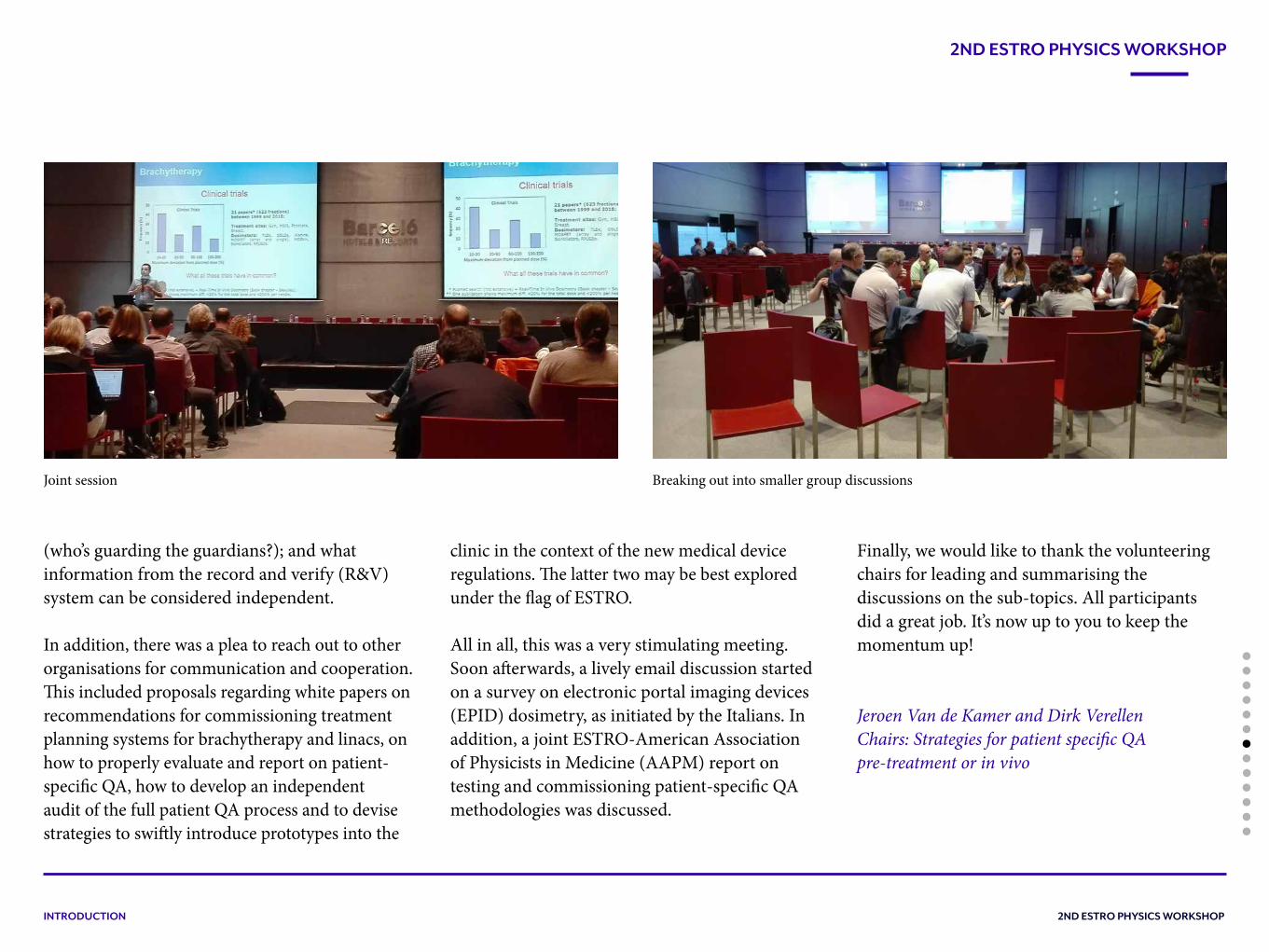
INTRODUCTION 2ND ESTRO PHYSICS WORKSHOP
(who’s guarding the guardians?); and what information from the record and verify (R&V) system can be considered independent.
In addition, there was a plea to reach out to other organisations for communication and cooperation. This included proposals regarding white papers on recommendations for commissioning treatment planning systems for brachytherapy and linacs, on how to properly evaluate and report on patient-specific QA, how to develop an independent audit of the full patient QA process and to devise strategies to swiftly introduce prototypes into the
clinic in the context of the new medical device regulations. The latter two may be best explored under the flag of ESTRO.
All in all, this was a very stimulating meeting. Soon afterwards, a lively email discussion started on a survey on electronic portal imaging devices (EPID) dosimetry, as initiated by the Italians. In addition, a joint ESTRO-American Association of Physicists in Medicine (AAPM) report on testing and commissioning patient-specific QA methodologies was discussed.
Finally, we would like to thank the volunteering chairs for leading and summarising the discussions on the sub-topics. All participants did a great job. It’s now up to you to keep the momentum up!
Jeroen Van de Kamer and Dirk VerellenChairs: Strategies for patient specific QA pre-treatment or in vivo
Joint session Breaking out into smaller group discussions
2ND ESTRO PHYSICS WORKSHOP
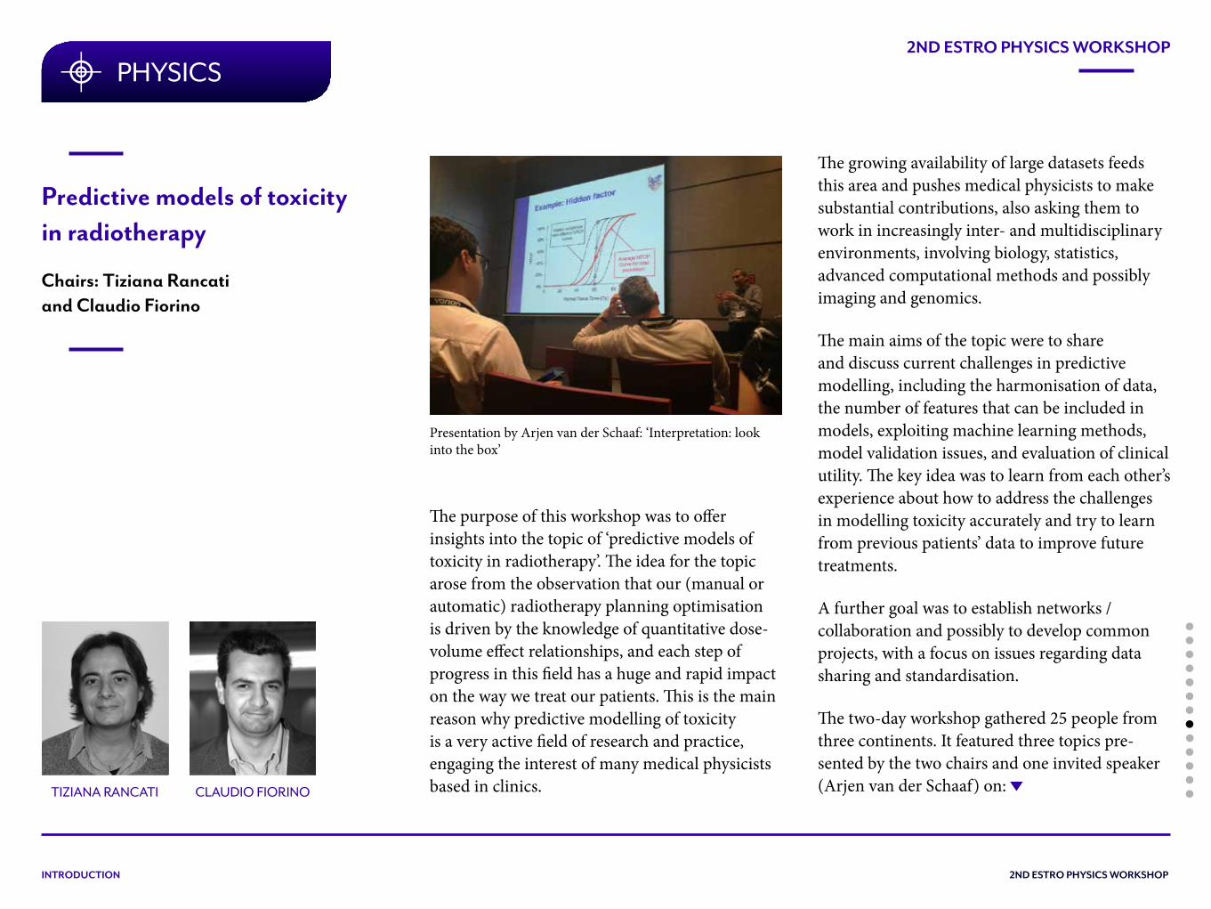
INTRODUCTION 2ND ESTRO PHYSICS WORKSHOP
The purpose of this workshop was to offer insights into the topic of ‘predictive models of toxicity in radiotherapy’. The idea for the topic arose from the observation that our (manual or automatic) radiotherapy planning optimisation is driven by the knowledge of quantitative dose-volume effect relationships, and each step of progress in this field has a huge and rapid impact on the way we treat our patients. This is the main reason why predictive modelling of toxicity is a very active field of research and practice, engaging the interest of many medical physicists based in clinics.
The growing availability of large datasets feeds this area and pushes medical physicists to make substantial contributions, also asking them to work in increasingly inter- and multidisciplinary environments, involving biology, statistics, advanced computational methods and possibly imaging and genomics.
The main aims of the topic were to share and discuss current challenges in predictive modelling, including the harmonisation of data, the number of features that can be included in models, exploiting machine learning methods, model validation issues, and evaluation of clinical utility. The key idea was to learn from each other’s experience about how to address the challenges in modelling toxicity accurately and try to learn from previous patients’ data to improve future treatments.
A further goal was to establish networks / collaboration and possibly to develop common projects, with a focus on issues regarding data sharing and standardisation.
The two-day workshop gathered 25 people from three continents. It featured three topics pre-sented by the two chairs and one invited speaker (Arjen van der Schaaf) on:
PHYSICS
Predictive models of toxicity in radiotherapy
Chairs: Tiziana Rancati and Claudio Fiorino
CLAUDIO FIORINOTIZIANA RANCATI
2ND ESTRO PHYSICS WORKSHOP
Presentation by Arjen van der Schaaf: ‘Interpretation: look into the box’
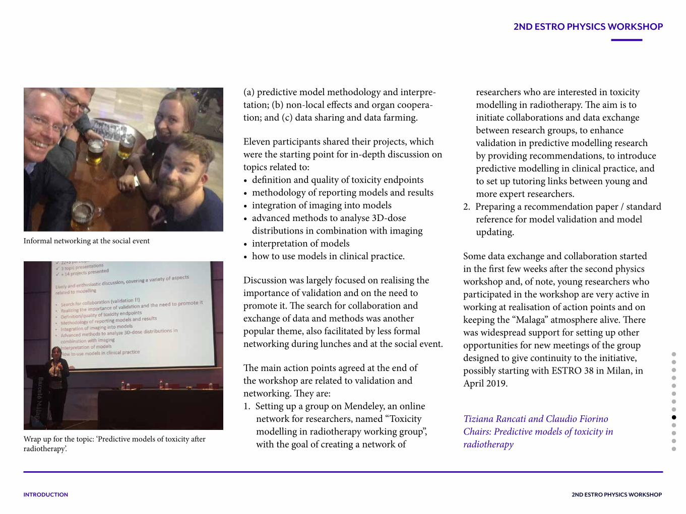
INTRODUCTION 2ND ESTRO PHYSICS WORKSHOP
(a) predictive model methodology and interpre-tation; (b) non-local effects and organ coopera-tion; and (c) data sharing and data farming.
Eleven participants shared their projects, which were the starting point for in-depth discussion on topics related to:• definition and quality of toxicity endpoints• methodology of reporting models and results • integration of imaging into models• advanced methods to analyse 3D-dose
distributions in combination with imaging• interpretation of models• how to use models in clinical practice.
Discussion was largely focused on realising the importance of validation and on the need to promote it. The search for collaboration and exchange of data and methods was another popular theme, also facilitated by less formal networking during lunches and at the social event.
The main action points agreed at the end of the workshop are related to validation and networking. They are:1. Setting up a group on Mendeley, an online
network for researchers, named “Toxicity modelling in radiotherapy working group”, with the goal of creating a network of
Informal networking at the social event
Wrap up for the topic: ‘Predictive models of toxicity after radiotherapy’.
researchers who are interested in toxicity modelling in radiotherapy. The aim is to initiate collaborations and data exchange between research groups, to enhance validation in predictive modelling research by providing recommendations, to introduce predictive modelling in clinical practice, and to set up tutoring links between young and more expert researchers.
2. Preparing a recommendation paper / standard reference for model validation and model updating.
Some data exchange and collaboration started in the first few weeks after the second physics workshop and, of note, young researchers who participated in the workshop are very active in working at realisation of action points and on keeping the “Malaga” atmosphere alive. There was widespread support for setting up other opportunities for new meetings of the group designed to give continuity to the initiative, possibly starting with ESTRO 38 in Milan, in April 2019.
Tiziana Rancati and Claudio FiorinoChairs: Predictive models of toxicity in radiotherapy
2ND ESTRO PHYSICS WORKSHOP
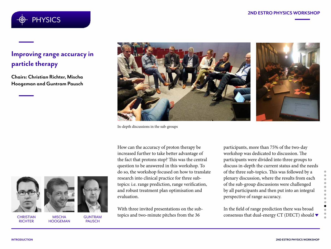
INTRODUCTION 2ND ESTRO PHYSICS WORKSHOP
How can the accuracy of proton therapy be increased further to take better advantage of the fact that protons stop? This was the central question to be answered in this workshop. To do so, the workshop focused on how to translate research into clinical practice for three sub-topics: i.e. range prediction, range verification, and robust treatment plan optimisation and evaluation.
With three invited presentations on the sub-topics and two-minute pitches from the 36
participants, more than 75% of the two-day workshop was dedicated to discussion. The participants were divided into three groups to discuss in-depth the current status and the needs of the three sub-topics. This was followed by a plenary discussion, where the results from each of the sub-group discussions were challenged by all participants and then put into an integral perspective of range accuracy.
In the field of range prediction there was broad consensus that dual-energy CT (DECT) should
PHYSICS
Improving range accuracy in particle therapy
Chairs: Christian Richter, Mischa Hoogeman and Guntram Pausch
MISCHA HOOGEMAN
GUNTRAM PAUSCH
CHRISTIAN RICHTER
2ND ESTRO PHYSICS WORKSHOP
In-depth discussions in the sub-groups
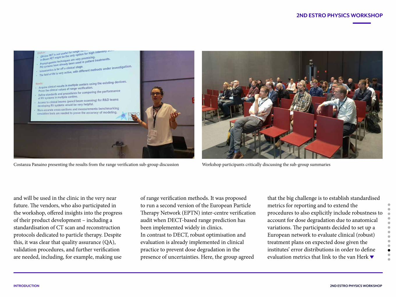
INTRODUCTION 2ND ESTRO PHYSICS WORKSHOP
and will be used in the clinic in the very near future. The vendors, who also participated in the workshop, offered insights into the progress of their product development – including a standardisation of CT scan and reconstruction protocols dedicated to particle therapy. Despite this, it was clear that quality assurance (QA), validation procedures, and further verification are needed, including, for example, making use
of range verification methods. It was proposed to run a second version of the European Particle Therapy Network (EPTN) inter-centre verification audit when DECT-based range prediction has been implemented widely in clinics. In contrast to DECT, robust optimisation and evaluation is already implemented in clinical practice to prevent dose degradation in the presence of uncertainties. Here, the group agreed
that the big challenge is to establish standardised metrics for reporting and to extend the procedures to also explicitly include robustness to account for dose degradation due to anatomical variations. The participants decided to set up a European network to evaluate clinical (robust) treatment plans on expected dose given the institutes’ error distributions in order to define evaluation metrics that link to the van Herk
Costanza Panaino presenting the results from the range verification sub-group discussion Workshop participants critically discussing the sub-group summaries
2ND ESTRO PHYSICS WORKSHOP
INTRODUCTION 2ND ESTRO PHYSICS WORKSHOP
treatment incentive. The results of improved range prediction (now) and range verification (future) should also be used to reduce the robustness needed, thereby improving the sparing of organs-at-risk.
Range verification was judged to be the approach that is the furthest from routine clinical application. Prompt-gamma based range verification seems to be promising and has reached the level of clinical applicability – however, only with a few “handmade” prototype systems and very few clinical applications. Participants agreed that the first priority should be to acquire more clinical data with the existing systems to prove the clinical value of range verification. For guidance on the development of novel detection systems in non-clinical research centres, the clinical needs and conditions for range verification should be summarised in a ‘request document’. This should contain both current and future treatments available in particle therapy facilities, as well as benchmark test scenarios for range verification systems.
The unique setting of this workshop, which brought together the communities of range prediction, range verification, and robust
optimisation and evaluation for the first time, enabled high-level and strategic thinking. One result was the proposal to create an integrated model, coupling range prediction and range verification results directly to robust optimisation. This model could then be optimised for value to patient and society to identify the most effective approach for further improvements. The active participation of key experts from industrial partners was very well received. Many participants felt that the workshop closed a gap between the clinically focused EPTN and research activities in non-clinical research institutions. In our view, by bringing together developers, industry and clinical users, the workshop helped to strengthen the translation bridge that is needed to turn promising innovations into clinical applications.
Christian Richter, Mischa Hoogeman and Guntram PauschChairs: Improving range accuracy in particle therapy
2ND ESTRO PHYSICS WORKSHOP
INTRODUCTION THE YESTRO PROJECT ON BURN-OUT IN RADIATION ONCOLOGY – PRO BONO
REPORT FROM THE SIXTH MR IN RADIATION THERAPY (RT) SYMPOSIUM
ESTRO TECHNOLOGY TRANSFER GRANT (TTG) REPORT: DUAL ENERGY PERFUSION CT DEVELOPMENT
LEARNING RADIATION ONCOLOGY IN EUROPE: RESULTS OF THE ESTRO MULTIDISCIPLINARY SURVEY
YOUNG ESTRO
Real-time and adaptive management of anatomical variations Chairs: Ben Heijmen and Marianne Aznar
For the report on this topic, please see Jenny Bertholet’s piece in the Young Corner on page 99 >
INTRODUCTION 2ND ESTRO PHYSICS WORKSHOP
The sub-group ‘Quantitative imaging for treatment planning’, was chaired by Uulke van der Heide (NKI Amsterdam, The Netherlands) and Daniela Thorwarth (Tübingen, Germany). There were 24 participants. Each had submitted a short abstract about his or her current research topic. There were also four invited speakers: Kathrine Røe Redalen (Trondheim, Norway), Oliver Gurney-Champion (London, UK), Marcel van Schie (NKI Amsterdam) and Sara Leibfarth (Tübingen).
The programme covered four major aspects in the field of quantitative imaging: biomarker discovery, standardisation, quantitative imaging for dose painting, and data processing. The discussion of each sub-topic started with a 15-minute introductory talk, which was given by one of the invited speakers, followed by five-minute pitch presentations from the other participants, who had been grouped into the respective sub-topics according to their submitted abstracts.
Plenty of time was allocated to discuss issues raised within the respective talks, and the relaxed, small-group atmosphere led to vivid and interesting in-depth discussions. At the end
of each of the sub-topics, the discussions were summarised with the guidance of the chairs and with active participation from other group members. The sub-topic-specific summaries were used as the basis for the presentation in the wrap-up plenary session at the end of the workshop. As a final outcome of the workshop, a manuscript based on the results elaborated by the sub-group is being prepared for submission to ESTRO’s online journal phiRO.
Sara LeibfarthSection for Biomedical PhysicsDepartment of Radiation Oncology University Hospital Tübingen Tübingen, Germany
PHYSICS
Quantitative imaging for treatment planning
Chairs: Uulke Van der Heide and Daniela Thorwarth
SARA LEIBFARTH
2ND ESTRO PHYSICS WORKSHOP

INTRODUCTION FROM LISBON TO LONDON: AN INTERNATIONAL CAREER PATH
ONWARDS AND UPWARDS: INSIGHTS ON WORKING TOWARDS A PHD IN RADIOTHERAPY SASRO
RTT
FROM LISBON TO LONDON: AN INTERNATIONAL CAREER PATH
ONWARDS AND UPWARDS: INSIGHTS ON WORKING TOWARDS A PHD IN RADIOTHERAPY SASROINTRODUCTION
RTTHappy new year from the RTT Corner.
We open this edition with an article from Dr Mirijam Mast, a former ESTRO RTT committee member and radiation therapist researcher based at Haaglanden Medical Centre, The Hague, Netherlands. It's a pleasure to read about Mirijam’s career and educational development, which includes a PhD on 'Avoiding the heart – About optimising whole breast irradiation’. She was awarded her PhD in 2015 following the publication of her research in peer-reviewed journals. Mirijam's career development illustrates the benefits to our profession of formalising research skills within a higher degree.
We are also pleased to feature a piece from Sophie Perryck from the University Hospital Zurich, Switzerland. Sophie writes about an annual multidisciplinary conference held in Zurich that welcomed more than 550 delegates to hear from national and international RTT speakers. RTTs play an important role within the multidisciplinary team, and it is great to hear of their involvement in national meetings like this.
Finally, we introduce Ana Rita Simões, who is part of the UK Radiotherapy Trials Quality Assurance (RTTQA) group, Mount Vernon, London. Rita's experience demonstrates the importance of RTT mobility throughout Europe. Here she reflects on her interesting and varied career, which has given her great job satisfaction. Again, Rita highlights some of the research opportunities that exist for RTTs within active research groups.
Thank you to all our contributors in this edition for sharing their experiences. We hope you enjoy the Corner. If you have any comments, or requests then please get in touch with us.
Best wishes for 2019.
Aileen Duffton ([email protected])Isabel Lobato ([email protected])Ilija Čurić ([email protected])
“Mirjam's career development illustrates the benefits to our profession of formalising research skills within a higher degree.”
AILEEN DUFFTON
ISABEL LOBATO
ILIJA ČURIĆ
INTRODUCTION FROM LISBON TO LONDON: AN INTERNATIONAL CAREER PATHSASROONWARDS AND UPWARDS: INSIGHTS ON WORKING
TOWARDS A PHD IN RADIOTHERAPY
Thank you to the editors of the RTT Corner for inviting me to offer my perspective on working towards a PhD in radiotherapy. I think we should start from the premise that all research in a radio- therapy department should be conducted as part of a multidisciplinary research team. Radiation therapists (RTTs) are, of course, a vital part of this team. However, all members of the team will have valuable insights to offer relating to their area of expertise. Undertaking research can help RTTs to improve the quality of radiation therapy treatment. It is important that RTTs are involved in the implementation of changes in clinical prac-tice, based on the latest research evidence. And even if this evidence cannot be implemented into clinical practice, being involved in the research process helps RTTs to develop a broader view on current developments in radiation therapy.
After receiving my MSc in epidemiology and working for seven years in research and development at the Haaglanden Medical Centre in The Netherlands, we were able to develop several research questions concerning the improvement of radiation treatment of (left-sided) breast cancer patients. It had become apparent that it was important to reduce the heart dose. We started several research projects in 2008. In 2010, it was clear that these research projects could be developed into a PhD.
These early years of my research were hard work, particularly as I was developing my thesis alongside my full-time role in research and
RTT
Onwards and upwards: insights on working towards a PhD in radiotherapy
MIRJAM MAST
development. Ultimately, the most important thing is that research questions address how to get the best radiation therapy treatment for the next generation of cancer patients. I will now describe in more detail the content of my thesis.
The following research aims were defined:1. To determine whether MRI could
enable a more precise delineation of the glandular breast cancer target volume and the lumpectomy cavity.
2. To identify which treatment technique would be best in reducing the dose to the critical structures, i.e. the heart and the left anterior descending (LAD). To answer this question, we carried out several treatment planning studies.
3. To determine whether breast cancer patients are more at risk of an increase in coronary artery calcium (CAC) scores (as
About optimising whole breast irradiation Mirjam Mast
AVOIDING THE HEART
AVOIDING THE HEART
Preclinical and clinical studies reveal that left-sided breast cancer radiotherapy is associated with an increased rate of major coronary events. Consequently, when irradiating women with left- sided breast cancer, specific measures should be taken to decrease the heart dose as much as possible and to avoid radiation-induced coronary artery disease. This thesis focuses on several strategies to optimise the radiation treatment for patients with left-sided breast cancer.
With respect to whole breast irradiation we concluded that:
- the routine use of MR images in addition to the CT scan, when delineating either the glandular breast tissue or the lumpectomy cavity, does not have added value.
- tangential IMRT technique combined with a breath-hold technique should be the treatment technique of choice for left-sided breast cancer.
- a breath-hold technique should and can be used in all left-sided breast cancer patients, regardless of age and breast size.
- breath-hold in left-sided whole breast radiotherapy results in a less pronoun ced increase of coronary calcium score and, hence, could result in less radiation-induced cardio vascular damage.
AVOID
ING
THE H
EART
Mirjam
Mast
Avoidingtheheart_cover_def.indd 1-2 11-05-15 12:23
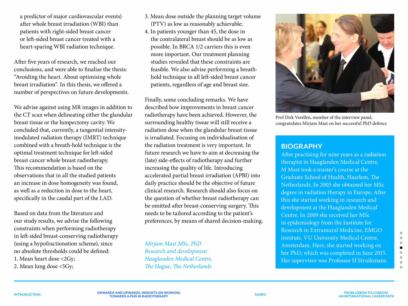
INTRODUCTION FROM LISBON TO LONDON: AN INTERNATIONAL CAREER PATHSASROONWARDS AND UPWARDS: INSIGHTS ON WORKING
TOWARDS A PHD IN RADIOTHERAPY
3. Mean dose outside the planning target volume (PTV) as low as reasonably achievable;
4. In patients younger than 45, the dose in the contralateral breast should be as low as possible. In BRCA 1/2 carriers this is even more important. Our treatment planning studies revealed that these constraints are feasible. We also advise performing a breath-hold technique in all left-sided breast cancer patients, regardless of age and breast size.
Finally, some concluding remarks. We have described how improvements in breast cancer radiotherapy have been achieved. However, the surrounding healthy tissue will still receive a radiation dose when the glandular breast tissue is irradiated. Focusing on individualisation of the radiation treatment is very important. In future research we have to aim at decreasing the (late) side-effects of radiotherapy and further increasing the quality of life. Introducing accelerated partial breast irradiation (APBI) into daily practice should be the objective of future clinical research. Research should also focus on the question of whether breast radiotherapy can be omitted after breast-conserving surgery. This needs to be tailored according to the patient’s preferences, by means of shared decision-making.
Mirjam Mast MSc, PhDResearch and developmentHaaglanden Medical Centre, The Hague, The Netherlands
Prof Dirk Verellen, member of the interview panel, congratulates Mirjam Mast on her successful PhD defence
a predictor of major cardiovascular events) after whole breast irradiation (WBI) than patients with right-sided breast cancer or left-sided breast cancer treated with a heart-sparing WBI radiation technique.
After five years of research, we reached our conclusions, and were able to finalise the thesis: “Avoiding the heart. About optimising whole breast irradiation”. In this thesis, we offered a number of perspectives on future developments.
We advise against using MR images in addition to the CT scan when delineating either the glandular breast tissue or the lumpectomy cavity. We concluded that, currently, a tangential intensity- modulated radiation therapy (IMRT) technique combined with a breath-hold technique is the optimal treatment technique for left-sided breast cancer whole breast radiotherapy. This recommendation is based on the observations that in all the studied patients an increase in dose homogeneity was found, as well as a reduction in dose to the heart, specifically in the caudal part of the LAD.
Based on data from the literature and our study results, we advise the following constraints when performing radiotherapy in left-sided breast-conserving radiotherapy (using a hypofractionation scheme), since no absolute thresholds could be defined:1. Mean heart dose <2Gy;2. Mean lung dose <5Gy;
BIOGRAPHYAfter practising for nine years as a radiation therapist in Haaglanden Medical Centre, M Mast took a master’s course at the Graduate School of Health, Haarlem, The Netherlands. In 2003 she obtained her MSc degree in radiation therapy in Europe. After this she started working in research and development at the Haaglanden Medical Centre. In 2009 she received her MSc in epidemiology from the Institute for Research in Extramural Medicine, EMGO institute, VU University Medical Centre, Amsterdam. Here, she started working on her PhD, which was completed in June 2015. Her supervisor was Professor H Struikmans.
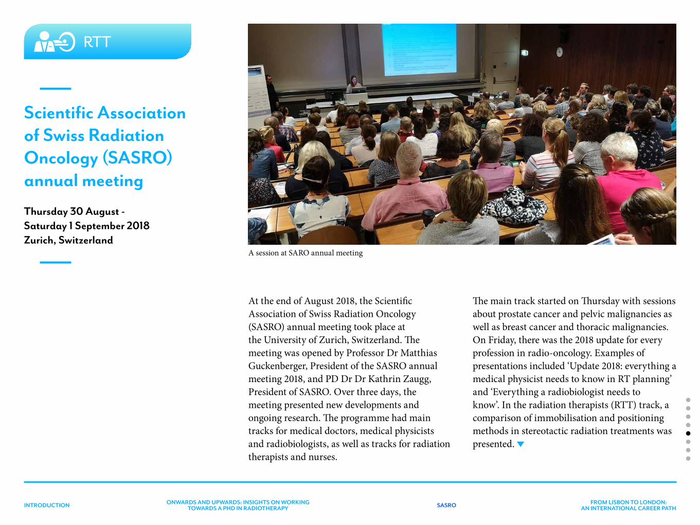
INTRODUCTION FROM LISBON TO LONDON: AN INTERNATIONAL CAREER PATH
ONWARDS AND UPWARDS: INSIGHTS ON WORKING TOWARDS A PHD IN RADIOTHERAPY SASRO
At the end of August 2018, the Scientific Association of Swiss Radiation Oncology (SASRO) annual meeting took place at the University of Zurich, Switzerland. The meeting was opened by Professor Dr Matthias Guckenberger, President of the SASRO annual meeting 2018, and PD Dr Dr Kathrin Zaugg, President of SASRO. Over three days, the meeting presented new developments and ongoing research. The programme had main tracks for medical doctors, medical physicists and radiobiologists, as well as tracks for radiation therapists and nurses.
The main track started on Thursday with sessions about prostate cancer and pelvic malignancies as well as breast cancer and thoracic malignancies. On Friday, there was the 2018 update for every profession in radio-oncology. Examples of presentations included ‘Update 2018: everything a medical physicist needs to know in RT planning’ and ‘Everything a radiobiologist needs to know’. In the radiation therapists (RTT) track, a comparison of immobilisation and positioning methods in stereotactic radiation treatments was presented.
RTT
Scientific Association of Swiss Radiation Oncology (SASRO) annual meeting
Thursday 30 August - Saturday 1 September 2018Zurich, Switzerland
A session at SARO annual meeting
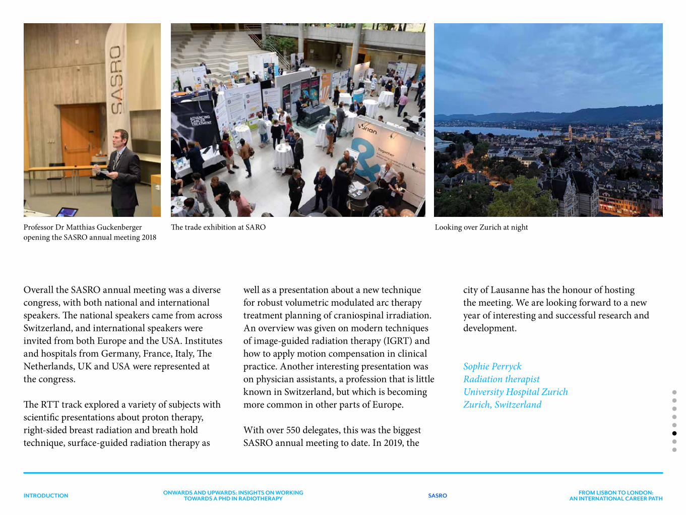
INTRODUCTION FROM LISBON TO LONDON: AN INTERNATIONAL CAREER PATH
ONWARDS AND UPWARDS: INSIGHTS ON WORKING TOWARDS A PHD IN RADIOTHERAPY SASRO
Overall the SASRO annual meeting was a diverse congress, with both national and international speakers. The national speakers came from across Switzerland, and international speakers were invited from both Europe and the USA. Institutes and hospitals from Germany, France, Italy, The Netherlands, UK and USA were represented at the congress.
The RTT track explored a variety of subjects with scientific presentations about proton therapy, right-sided breast radiation and breath hold technique, surface-guided radiation therapy as
well as a presentation about a new technique for robust volumetric modulated arc therapy treatment planning of craniospinal irradiation. An overview was given on modern techniques of image-guided radiation therapy (IGRT) and how to apply motion compensation in clinical practice. Another interesting presentation was on physician assistants, a profession that is little known in Switzerland, but which is becoming more common in other parts of Europe.
With over 550 delegates, this was the biggest SASRO annual meeting to date. In 2019, the
city of Lausanne has the honour of hosting the meeting. We are looking forward to a new year of interesting and successful research and development.
Sophie PerryckRadiation therapist University Hospital ZurichZurich, Switzerland
Professor Dr Matthias Guckenberger opening the SASRO annual meeting 2018
The trade exhibition at SARO Looking over Zurich at night
INTRODUCTION ONWARDS AND UPWARDS: INSIGHTS ON WORKING TOWARDS A PHD IN RADIOTHERAPY SASRO FROM LISBON TO LONDON:
AN INTERNATIONAL CAREER PATH
My name is Rita Simões and I am pleased to have been invited to tell you about my career, which has provided the opportunity to work internationally as a radiation therapist (RTT). I feel my experience across different countries and specialities has resulted in a somewhat unique example of an RTT’s career development. Let me explain…
I qualified as an RTT in 2008 at the Escola Superior de Tecnologia da Saude (ESTeSL) in Lisbon, Portugal, where I am from. I immediately started working at the Hospital de Santa Maria, a large university hospital in Lisbon. By the age of 22, I was developing my own skills and building strong connections with patients. I relished the professional responsibility and realised how important an RTT is for improving the experience of patients going through treatment for these potentially devastating diseases.
My strong work ethic was recognised by the department leads (Professor Isabel Monteiro Grillo and Dr Isabel Diegues) and I was given the opportunity to become a senior RTT in both pre-treatment and in the radiotherapy unit. I also recognised the value of participating in research and development to improve my skills as an RTT and enrolled on an MSc programme in ionising radiation applied to health sciences at ESTeSL. This led to the development of my first research project in image-guided radiation therapy (IGRT) for head and neck, and prostate cancers. As well as leading to my first research publications, this work was translated into tangible improvements
RTT
From Lisbon to London: an international career path
ANA RITA SIMÕES
in the radiotherapy service, which gave me a real sense of achievement.
Around that time, I decided I would like to diversify my experience as an RTT. The opportunity came when my husband was offered a job in London and we moved to the UK. Initially, I started at Mount Vernon Hospital as an RTT planning dosimetrist. Although I had never worked in dosimetry before, my university training and my MSc had a treatment-planning focus, so I felt able to take on the challenge of a totally new role. My previous clinical experience helped me to understand treatment delivery and what was required from a treatment plan to ensure that I provided the best quality for patient outcomes and experience. My physics colleagues and head of department (Dr Karen Venables) were key to my development. I learned a lot from my new colleagues and also enjoyed living in a new city with so much to experience and explore.In London I found myself constantly involved in development and research projects, as the culture of clinical trials participation in radiotherapy is strong. This intensified my interest in research and I looked for opportunities to become more involved. In 2015, I joined the UK Radiotherapy Trials Quality Assurance (RTTQA) Group, directed by Mrs Elizabeth Miles and Professor Peter Hoskin, and funded by the National Institute for Health Research (NIHR). In my current role, I contribute to the design of research protocols and trial radiotherapy procedures. My day-to-day work involves monitoring the
INTRODUCTION ONWARDS AND UPWARDS: INSIGHTS ON WORKING TOWARDS A PHD IN RADIOTHERAPY SASRO FROM LISBON TO LONDON:
AN INTERNATIONAL CAREER PATH
So far in my career, I have been able to undertake new projects and face the challenges they pose. I have been lucky to experience all areas of radiotherapy. My international experience has been key to broadening my perspectives and the RTTQA group experience has allowed me to be part of cutting-edge technique development, while learning scientific reasoning. For RTTs there are not many routes to lead research projects, so the whole process has not been as smooth as I would have wished. However, within my job I can learn from the best professionals and feel amazingly supported in undertaking all of the challenges. My career has been a unique experience. How else would I be able to roam the world and get paid for doing a job that I love?
Ana Rita Simões MScUK Radiotherapy Trials Quality Assurance (RTTQA) Group, Mount Vernon HospitalLondon, UK
safety, feasibility and rigour of radiotherapy delivered to patients recruited into clinical trials through the review of outlines, plans and patient progress throughout their treatment. This experience has helped me further develop my career and become more specialised, specifically in sarcoma, lung and head and neck cancers. I feel that I am contributing to the development and progression of radiotherapy techniques and to the improvement of care for patients with cancer. I am also able to work with many centres in the UK and some of the brightest minds in radiotherapy.
In parallel, I participate in an international teaching programme (FALCON) and I am developing my own research. I have published recent work and presented it nationally and internationally, including at ESTRO’s congress. I am now developing a research project in sarcoma, with which I hope to contribute to decreasing the toxicity of radiotherapy for patients with this disease. This will develop the academic side of my career through a PhD, which is a daunting endeavour. I feel that my research brings together my previous experience and will contribute to improving the lives of patients with cancer. In going through all the steps of preparing a research project – from the research question, to research design, project development, setting up a team, to actually involving patients and then seeking funding – I have learned even more and am privileged to be working with very helpful and expert professionals.
INTRODUCTION REPORTS FROM THE RADIOBIOLOGY TRACK AT ESTRO 37
RADIOBIOLOGY
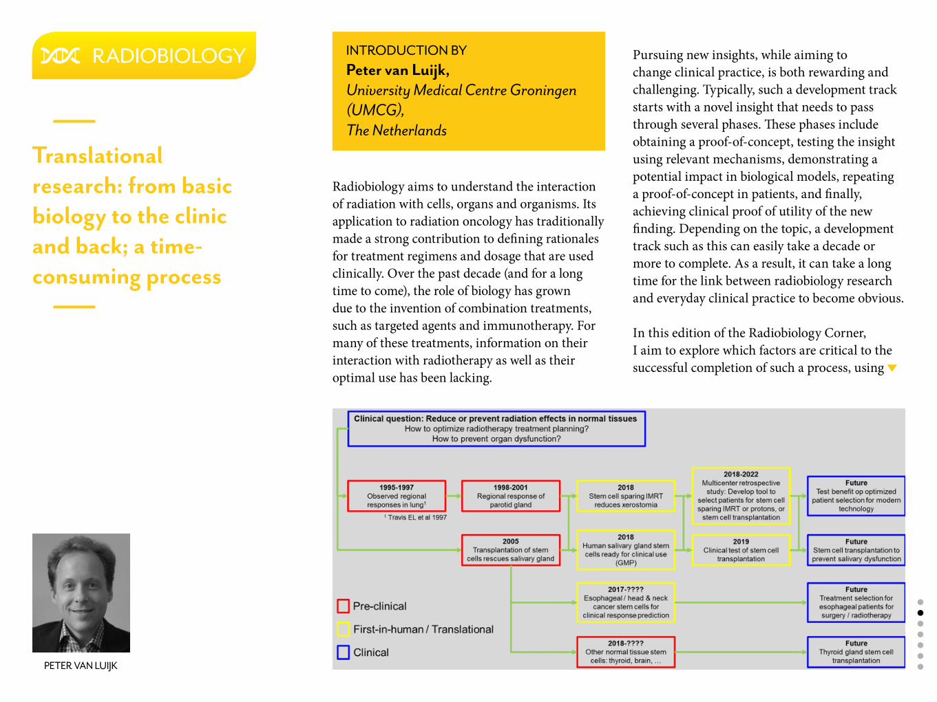
Radiobiology aims to understand the interaction of radiation with cells, organs and organisms. Its application to radiation oncology has traditionally made a strong contribution to defining rationales for treatment regimens and dosage that are used clinically. Over the past decade (and for a long time to come), the role of biology has grown due to the invention of combination treatments, such as targeted agents and immunotherapy. For many of these treatments, information on their interaction with radiotherapy as well as their optimal use has been lacking.
RADIOBIOLOGY
Translational research: from basic biology to the clinic and back; a time-consuming process
Pursuing new insights, while aiming to change clinical practice, is both rewarding and challenging. Typically, such a development track starts with a novel insight that needs to pass through several phases. These phases include obtaining a proof-of-concept, testing the insight using relevant mechanisms, demonstrating a potential impact in biological models, repeating a proof-of-concept in patients, and finally, achieving clinical proof of utility of the new finding. Depending on the topic, a development track such as this can easily take a decade or more to complete. As a result, it can take a long time for the link between radiobiology research and everyday clinical practice to become obvious.
In this edition of the Radiobiology Corner, I aim to explore which factors are critical to the successful completion of such a process, using
PETER VAN LUIJK
INTRODUCTION BYPeter van Luijk, University Medical Centre Groningen (UMCG), The Netherlands
our work at the University Medical Centre Groningen (UMCG), The Netherlands, as an example. The focus of research performed in Groningen has traditionally been on finding ways to prevent, predict and treat normal tissue damage associated with radiotherapy. As far back as the 1990s, the radiation and stress cell biology group started their quest for solutions to this problem. Two decades later this work is reaching maturity and is gradually being introduced into the clinic (see figure). To gain insight into what motivates researchers to start such a lengthy process, and to get a better understanding of the challenges and factors influencing the chances of successfully translating novel work into clinical practice, I have interviewed UMCG’s Professor Dr Rob Coppes, who has been driving pre-clinical research on normal tissue damage research for the past two decades, and Dr Roel Steenbakkers, who has been instrumental in adopting pre-clinical results for translation in clinical studies.
ROEL STEENBAKKERS
INTERVIEW WITH Dr Roel Steenbakkers, UMCG
What motivated you to choose to work in this area?When I finished secondary school, I was interested in the functioning of the human body. I decided to apply for medical school and was fortunate to be selected. If I hadn’t been selected I would have chosen chemistry or physics instead. Having started medical school, I soon felt a strong affinity to oncology. Given my interest in physics, my interest in radiation oncology followed naturally. Although I did not yet understand what radiation oncology was, I took the first possible opportunity to visit Philadelphia, USA, for an internship in this specialism. There, I had the opportunity to work on a simplified form of inverse planning to be used in the treatment of child eye tumours (retinoblastoma).
Can you elaborate on what sparked your interest in oncology?I am fascinated by cancer and motivated by its life-threatening character. Cancer patients are confronted with the possibility of dying. Being able to contribute to their cure, or if that’s no
longer possible, to try to improve their quality of life in this final phase of life, is what drew me to oncology.
Your first internship involved research. Is this something you have always been interested in? Or was this interest sparked by your internship in Philadelphia?No, I have always been interested in research. Before that internship I had already performed small research projects. Helping patients is important to me. But for me, this involves a strong interest in developing new ways of doing so.
When performing clinical research, one of the challenges is in managing the conflicts of interest between the researcher, the participating patient and future patients. How do you do this?The main challenge is to motivate the patient to participate. Especially when the patient is unlikely to benefit directly from the study, you have to make participation as easy and convenient as possible. Around the time that we ask patients – in this case head and neck patients – to make this extra effort, the patients are typically suffering from a large number of side-effects from their treatment. Therefore, I usually try to find a way in which the patient does have some potential benefit, or at least ensure that study assessments coincide with visits to UMCG related to their treatment.
How did your involvement in the xerostomia research start?When I started working in Groningen, I performed several imaging studies. This was a nice start because there was not much imaging research in head and neck patients being undertaken in Groningen at that time. When Fred Burlage left Groningen, I took over several of his projects.
Then I came to you with the idea of performing a stem-cell-sparing intensity modulated radiation therapy (IMRT) trial. This was a translational project with little clinical work done at that point.
How did you feel about the fact that this came from lab work, which typically sits further from the clinic?At that point, I had only undertaken clinical work. I liked the idea of doing translational studies because it really brought something new into the clinic. This made the study challenging in a very interesting way. Also, it was clear from the beginning that this study could lead directly to improved patient treatment. This made it different from work that I had seen elsewhere in which more abstract molecular hypotheses were being tested.
You are experienced in both full clinical projects and translational projects. How do these projects differ in terms of starting and completing them?The first major difference is in the time needed to get them started. For example, due to various formal and practical reasons the start of the first
salivary gland stem cell transplantation trial is taking much longer than expected. This is in sharp contrast to clinical research where the idea is usually very close to clinical practice. An example of this is the study where we tested the effectiveness of current standard radiotherapy methods to treat Ledderhose’s disease. As it was a straight-forward effectiveness trial using established techniques and methodology, the road to starting it was well established. As a result, the time between the conception of the clinical trial and the clinical application of its results, is much longer in translational studies than in pure clinical trials.
Second, the concepts tested in a translational study, such as the stem-cell-sparing IMRT study, are typically more different from clinical practice. Therefore, it takes more time and effort to secure the support of people in the department needed to execute the study. This is especially challenging because the potential benefit can’t be supported by the type of clinical evidence that people are used to. This clearly influenced our ability to arrange additional imaging for the stem-cell-sparing IMRT trial. Getting support for this type of research in other departments is probably the biggest challenge.
When did your two translational projects start and when do you expect them to result in changes in clinical practice?Given the initial results of the stem-cell-sparing IMRT trial, I expect this project will lead to changed clinical practice five years from now. Our current X-PREVENT project funded by the
Dutch Cancer Society will contribute to making this technique an international standard, rather than a local change.
The stem cell transplantation project is much more challenging in this respect. It differs fundamentally from other approaches and methods used in radiotherapy, which means that it has to go through all phases from safety and feasibility via testing of effectiveness to clinical implementations. Completing this track will take years. Transplantation is an invasive procedure, which will make patient recruitment for the first studies challenging. After completion of the first safety and feasibility tests, we still need to broaden the patient population group to demonstrate benefit and, finally, perform some validation.
That’s interesting to hear, especially because I remember that the general perception in the lab at the time these ideas came up was that we only needed to demonstrate in the lab that this works, subsequently do a trial to show effectiveness clinically and then simply start using it. You can see an interesting discrepancy between the two disciplines collaborating in this translational track.
Is there anything else you would like to add?Yes, I would like to stress that clinical research and lab-based research need each other in order to progress clinical radiation oncology.
Why did you choose to enter the fields of salivary gland and radiotherapy-related toxicity?While writing my PhD thesis, I was looking for a new position in academia. I came across a project in which a new hypothesis would be tested regarding the development of salivary gland damage after radiotherapy treatment. Acinar cells in salivary glands contain intracellular vesicles. The content of these vesicles is normally released into the saliva, which helps to digest food. However, their release inside salivary gland cells can damage these cells. In this new project I was going to test the hypothesis that damaging these vesicles and consequent cell loss contributes to the loss of saliva production after irradiation. To test this, we stimulated salivary glands before irradiation to force the vesicles out and observed a benefit. However, in a good experiment you do need additional controls to exclude other causes of this benefit. I did this by using agents to stimulate the gland to excrete saliva without stimulating excretion of the vesicles. To our surprise, this also led to protection, demonstrating that the benefit was in fact not related to the vesicles, but rather
the stimulation of saliva production. Ultimately this led to the insight that stimulating salivary glands stimulated proliferation of tissue-specific stem cells, which we found to be critical targets for radiation-induced salivary gland dysfunction.
Critical to the success of this process was the fact that I happened to share my room with stem cell biologists, who contributed a lot to the discussion. This was a totally new field for me and being close to them helped me to learn how to perform studies in this area.
This first position in academia does not sound very closely related to the clinic?No, not really. Though obviously the ultimate aim was to investigate a mechanism of salivary gland damage that might mean something clinically. Also, the agents we used to test our hypothesis were predominantly medications that were already used clinically, making it easier to use the results clinically. However, if you want to prove conclusively the role of a mechanism, you need to perform control experiments with many other agents that closely resemble the ones used in the clinic, but which differ on specific elements of the mechanism. Only this type of experiment will provide solid support for mechanistic conclusions.
Listening to this, it seems that the primary motivation in your work seems to be developing new insight on mechanisms. Is that right?Yes, I think that’s correct, especially at the start of my career.
This is a significant difference with Roel Steenbakkers, who likes to have a clearer view of clinical applicability. Another interesting aspect of your story is that it seems to develop from rejecting hypotheses and starting again from previous control experiments, which is in contrast to the clinician who primarily wants to see if something works and can subsequently be applied clinically.
This work is highly multidisciplinary and spans the lab and the clinic. How is it being received in the different disciplines?Very well. I have been invited to present this work at congresses of all the disciplines involved, including radiotherapy, oral maxillofacial surgery and stem cell biology. I was even invited to present at a stem cell meeting of the European Molecular Biology Organization (EMBO), which is one of the most prestigious places to present in the stem cell world. We have been cited in Nature and Cell publications as the discoverers of the salivary gland organoids. At this point the stem cell field eagerly seeks a proof-of-concept that stem cells can be used to restore organ function. Treatment of radiotherapy-related damage obviously is an ideal application for this. Though the patient is ill at the start of radiotherapy, the organ that needs to be rescued is typically healthy.
This is in contrast to other applications, such as cardiac stem cells for improving cardiac function after a myocardial infarction. In addition, the dynamics of the development of damage can be studied very accurately, because you know exactly when the damaging process started. This
ROB COPPES
INTERVIEW WITH Professor Dr Rob Coppes, UMCG
I agree completely. In addition, it makes me think: could this be a point where pre-clinical research in the lab fundamentally differs from clinical research that is combined with patient care? For a radiation oncologist it is hard, if not impossible, to change field and start performing care as a cardiologist in order to develop the skills in connecting fields.Yes, that is absolutely true. Therefore, it is very important that such multidisciplinarity is pursued at an earlier stage, during the basic education of physicians. For this reason, in Groningen, we now offer the molecular medicine programme in which students dedicate 20% of their time to biology and molecular mechanisms involved in many different diseases. We expect this will develop physicians who have a broader view of different fields, helping to facilitate the translation of knowledge between fields.
Similarly, I have attended radiotherapy meetings in which patients were discussed over a period of five years. This provided me with some insight in the clinical side of my ideas. This is, however, a time-consuming process. And today’s science moves very fast. Rapid proof-of-concepts are the quickest way to reach high-impact journals. Scientists aiming to develop a carrier in stem cell biology or cell biology in general, have to meet that speed in order not to lose out to the competition.
Could it be said that the current criteria by which fundamental science is judged and the speed required to survive in the competition are hampering innovation of clinical practice based on fundamental research?
Possibly biologists performing fundamental work do have translation in mind, but may not have the knowledge to actually achieve this. At the same time, you might ask physicians to learn more about fundamental sciences, but they also face their own constraints. However, meeting each other somewhere halfway is fundamental to innovating in radiotherapy.
The stem cell transplantation work started 20-25 years ago. What has kept you going all this time?In every phase of the project you discover new things. The excitement of developing understanding is important. In addition, this process has spanned all the stages of my career from PhD student, via post-doc, and eventually to becoming a professor. This has involved a lot of personal development, which has ensured that the process remains interesting. Another important aspect is the involvement of other people. Though I started alone, over time I have formed a reasonably large research group, consisting of associate investigators, postdoctoral researchers, PhD students and technicians. Their enthusiasm and the insights they have contributed to the research have kept me fascinated. Finally, broadening the topic, for example, by applying my newly obtained knowledge on culturing salivary gland stem cells to also developing stem cell therapy for the thyroid gland, or even to the development of tumour stem cell assays for the prediction of treatment outcome, ensures the work stays challenging and interesting.
provides excellent opportunities to develop optimised schedules for application of stem cell therapies.
In addition, stem cells (cultured as organoids) being the cell type that determines the long-term fate of an organ have found their way into fields like ageing and drug screening.
What about the radiotherapy field?In the radiotherapy field there is a lot of interest and it is often mentioned as a promising and important topic for moving the field forwards, but somehow the field does not actively engage on the topic. You can see this, for example, in the low number of abstracts submitted on stem cells for the annual ESTRO meeting. Maybe this is related to the complexity of the topic. In Groningen, we have been lucky in that the required expertise happened to be concentrated in the cell biology department and was adequately connected to the radiotherapy department. Creating conditions where these results can be achieved is much harder if you are located in a more isolated radiotherapy or radiobiology department. Another example of where it has worked is with Marc Vooijs at Maastricht University, who works with lung organoids. But he also worked in the Hubrecht Institute in Hans Clevers’ lab, which specialises in this type of research.
This shows clearly that looking outside your own speciality is critical to developing new insights. Science has become so complex that tools available in your own field are rarely sufficient to solve problems. Therefore, connecting to other fields has become mandatory.
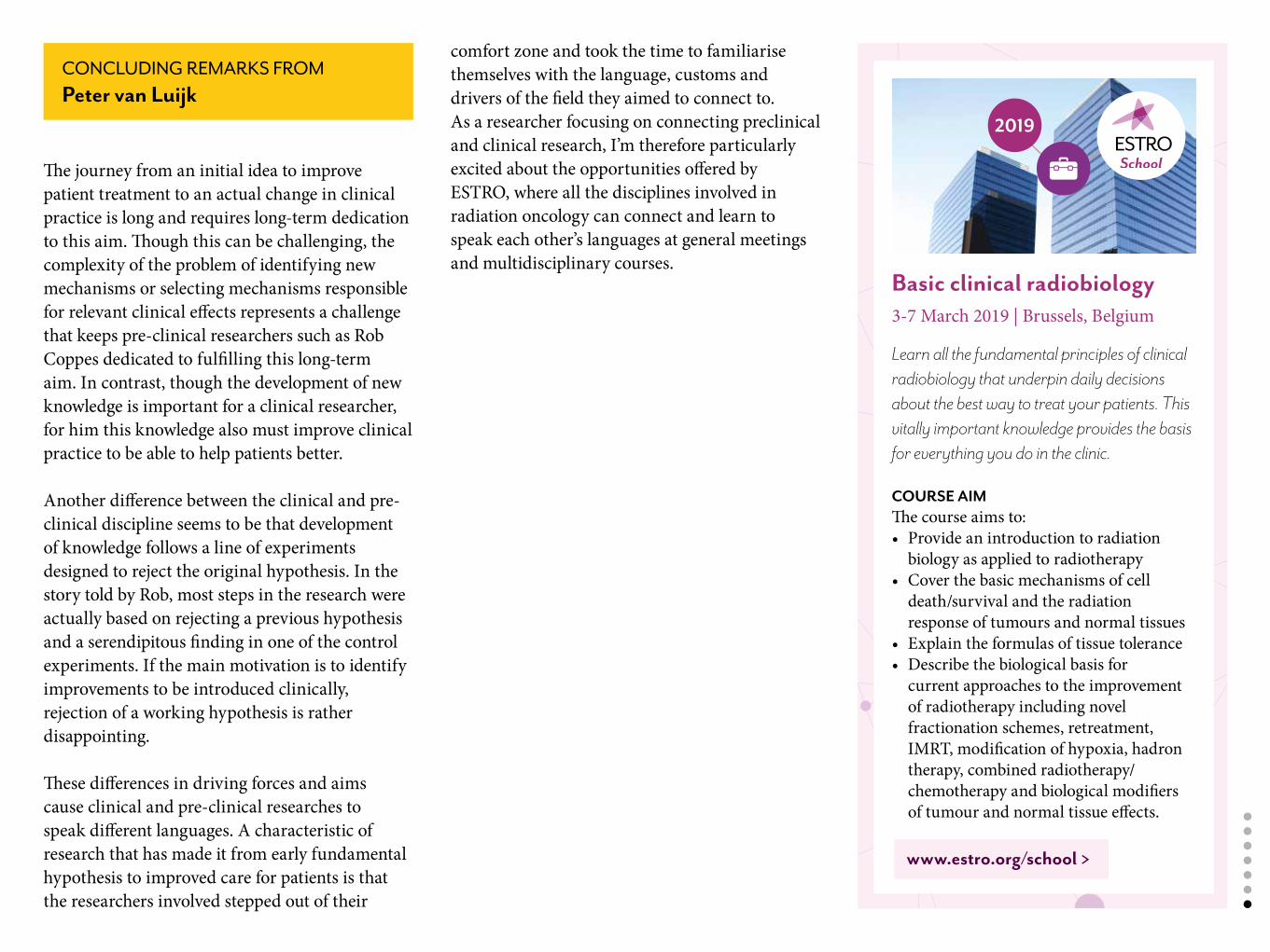
The journey from an initial idea to improve patient treatment to an actual change in clinical practice is long and requires long-term dedication to this aim. Though this can be challenging, the complexity of the problem of identifying new mechanisms or selecting mechanisms responsible for relevant clinical effects represents a challenge that keeps pre-clinical researchers such as Rob Coppes dedicated to fulfilling this long-term aim. In contrast, though the development of new knowledge is important for a clinical researcher, for him this knowledge also must improve clinical practice to be able to help patients better.
Another difference between the clinical and pre-clinical discipline seems to be that development of knowledge follows a line of experiments designed to reject the original hypothesis. In the story told by Rob, most steps in the research were actually based on rejecting a previous hypothesis and a serendipitous finding in one of the control experiments. If the main motivation is to identify improvements to be introduced clinically, rejection of a working hypothesis is rather disappointing.
These differences in driving forces and aims cause clinical and pre-clinical researches to speak different languages. A characteristic of research that has made it from early fundamental hypothesis to improved care for patients is that the researchers involved stepped out of their
comfort zone and took the time to familiarise themselves with the language, customs and drivers of the field they aimed to connect to.As a researcher focusing on connecting preclinical and clinical research, I’m therefore particularly excited about the opportunities offered by ESTRO, where all the disciplines involved in radiation oncology can connect and learn to speak each other’s languages at general meetings and multidisciplinary courses.
CONCLUDING REMARKS FROM Peter van Luijk
Basic clinical radiobiology3-7 March 2019 | Brussels, Belgium
www.estro.org/school >
COURSE AIMThe course aims to:• Provide an introduction to radiation
biology as applied to radiotherapy• Cover the basic mechanisms of cell
death/survival and the radiation response of tumours and normal tissues
• Explain the formulas of tissue tolerance• Describe the biological basis for
current approaches to the improvement of radiotherapy including novel fractionation schemes, retreatment, IMRT, modification of hypoxia, hadron therapy, combined radiotherapy/chemotherapy and biological modifiers of tumour and normal tissue effects.
Learn all the fundamental principles of clinical radiobiology that underpin daily decisions about the best way to treat your patients. This vitally important knowledge provides the basis for everything you do in the clinic.
2019
INTRODUCTION FALCON CONTOURING WORKSHOPSESTRO SCHOOL COURSE DIRECTORS AND TEACHERS WHO ARE LEAVING THE FACULTY COURSE REPORTS OBITUARY FOR CHRIS COTTRILL
ESTRO SCHOOL
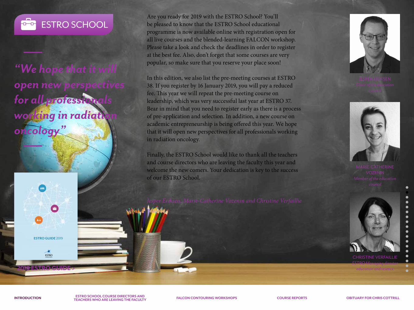
FALCON CONTOURING WORKSHOPSESTRO SCHOOL COURSE DIRECTORS AND TEACHERS WHO ARE LEAVING THE FACULTY COURSE REPORTS OBITUARY FOR CHRIS COTTRILLINTRODUCTION
ESTRO SCHOOL
CHRISTINE VERFAILLIEESTRO Managing director
education and science
Are you ready for 2019 with the ESTRO School? You’ll be pleased to know that the ESTRO School educational programme is now available online with registration open for all live courses and the blended-learning FALCON workshop. Please take a look and check the deadlines in order to register at the best fee. Also, don’t forget that some courses are very popular, so make sure that you reserve your place soon!
In this edition, we also list the pre-meeting courses at ESTRO 38. If you register by 16 January 2019, you will pay a reduced fee. This year we will repeat the pre-meeting course on leadership, which was very successful last year at ESTRO 37. Bear in mind that you need to register early as there is a process of pre-application and selection. In addition, a new course on academic entrepreneurship is being offered this year. We hope that it will open new perspectives for all professionals working in radiation oncology.
Finally, the ESTRO School would like to thank all the teachers and course directors who are leaving the faculty this year and welcome the new comers. Your dedication is key to the success of our ESTRO School.
Jesper Eriksen, Marie-Catherine Vozenin and Christine Verfaillie
MARIE-CATHERINE VOZENIN
Member of the education council
JESPER ERIKSENChair of the education
council
“We hope that it will open new perspectives for all professionals working in radiation oncology”
2019 ESTRO GUIDE >
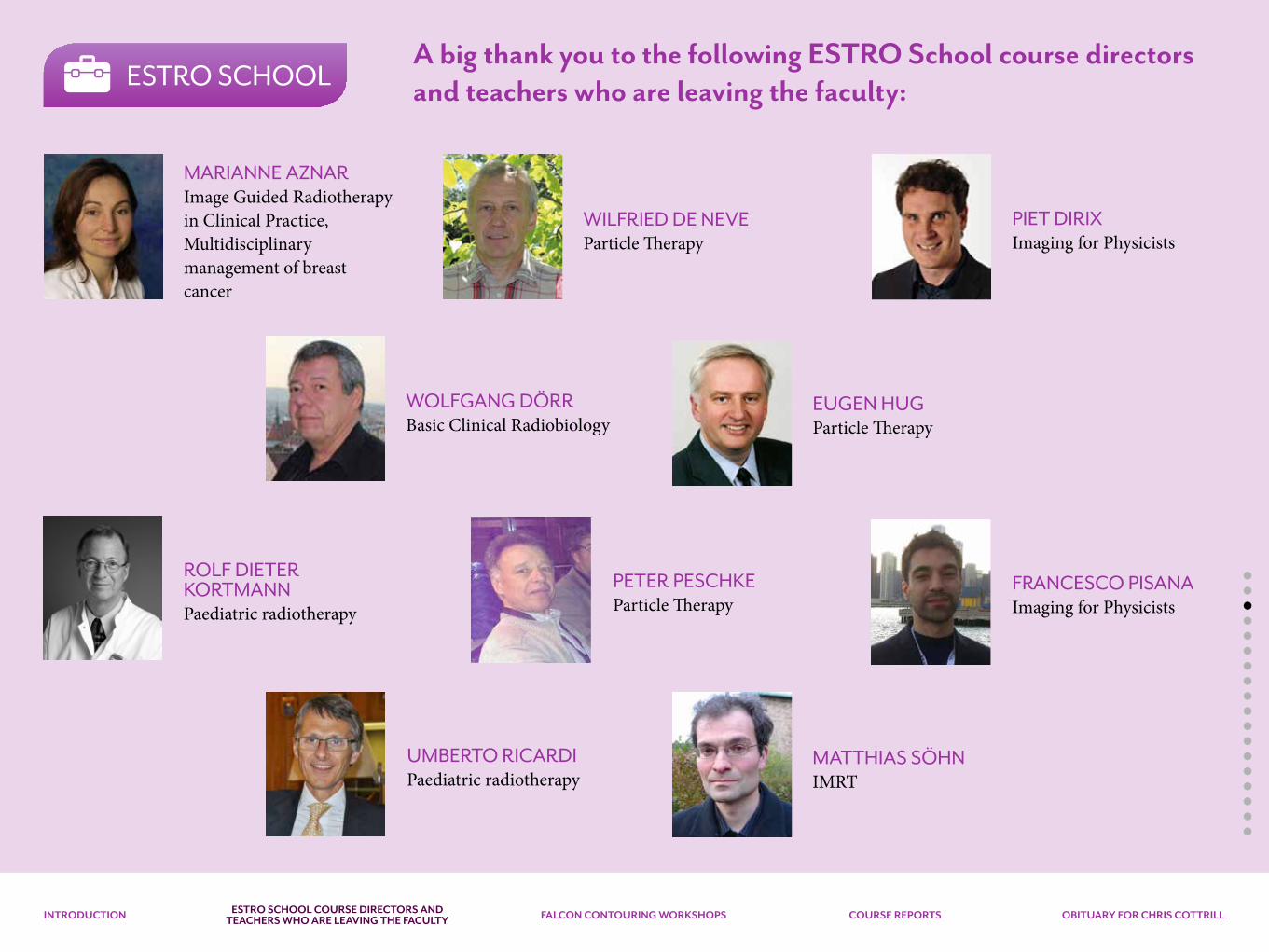
INTRODUCTION FALCON CONTOURING WORKSHOPS COURSE REPORTS OBITUARY FOR CHRIS COTTRILLESTRO SCHOOL COURSE DIRECTORS AND TEACHERS WHO ARE LEAVING THE FACULTY
ESTRO SCHOOL
EUGEN HUGParticle Therapy
WOLFGANG DÖRRBasic Clinical Radiobiology
MARIANNE AZNARImage Guided Radiotherapy in Clinical Practice, Multidisciplinary management of breast cancer
PETER PESCHKEParticle Therapy
PIET DIRIXImaging for Physicists
WILFRIED DE NEVEParticle Therapy
ROLF DIETER KORTMANN Paediatric radiotherapy
UMBERTO RICARDIPaediatric radiotherapy
FRANCESCO PISANAImaging for Physicists
MATTHIAS SÖHNIMRT
A big thank you to the following ESTRO School course directors and teachers who are leaving the faculty:
INTRODUCTION ESTRO SCHOOL COURSE DIRECTORS AND TEACHERS WHO ARE LEAVING THE FACULTY COURSE REPORTS OBITUARY FOR CHRIS COTTRILLFALCON CONTOURING WORKSHOPS
2019 ONLINE WORKSHOPS PROGRAMME
ESTRO SCHOOL FALCON CONTOURING WORKSHOPS
FALCONFellowship in Anatomic deLineation & CONtouring
Mark your calendar
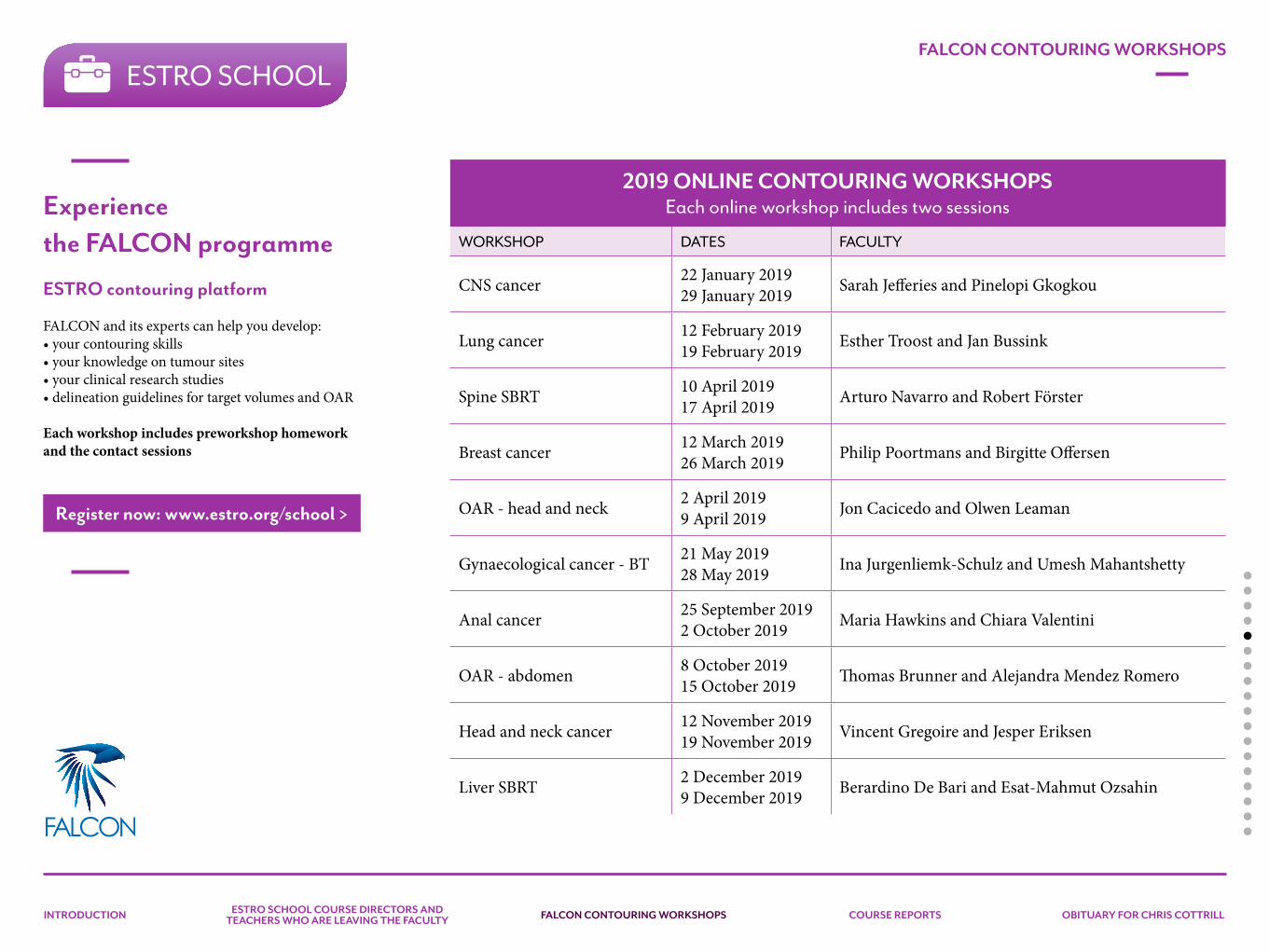
INTRODUCTION ESTRO SCHOOL COURSE DIRECTORS AND TEACHERS WHO ARE LEAVING THE FACULTY COURSE REPORTS OBITUARY FOR CHRIS COTTRILLFALCON CONTOURING WORKSHOPS
FALCONFellowship in Anatomic deLineation & CONtouring
ESTRO SCHOOLFALCON CONTOURING WORKSHOPS
Experiencethe FALCON programme
ESTRO contouring platform
FALCON and its experts can help you develop:• your contouring skills• your knowledge on tumour sites• your clinical research studies• delineation guidelines for target volumes and OAR
Each workshop includes preworkshop homework and the contact sessions
Register now: www.estro.org/school >
2019 ONLINE CONTOURING WORKSHOPSEach online workshop includes two sessions
WORKSHOP DATES FACULTY
CNS cancer 22 January 201929 January 2019 Sarah Jefferies and Pinelopi Gkogkou
Lung cancer 12 February 201919 February 2019 Esther Troost and Jan Bussink
Spine SBRT 10 April 201917 April 2019 Arturo Navarro and Robert Förster
Breast cancer 12 March 201926 March 2019 Philip Poortmans and Birgitte Offersen
OAR - head and neck 2 April 20199 April 2019 Jon Cacicedo and Olwen Leaman
Gynaecological cancer - BT 21 May 201928 May 2019 Ina Jurgenliemk-Schulz and Umesh Mahantshetty
Anal cancer 25 September 20192 October 2019 Maria Hawkins and Chiara Valentini
OAR - abdomen 8 October 201915 October 2019 Thomas Brunner and Alejandra Mendez Romero
Head and neck cancer 12 November 201919 November 2019 Vincent Gregoire and Jesper Eriksen
Liver SBRT 2 December 20199 December 2019 Berardino De Bari and Esat-Mahmut Ozsahin
INTRODUCTION FALCON CONTOURING WORKSHOPSESTRO SCHOOL COURSE DIRECTORS AND TEACHERS WHO ARE LEAVING THE FACULTY OBITUARY FOR CHRIS COTTRILLCOURSE REPORTS
Imaging for physicists
23-27 September 2018 | Vienna, Austria
Multidisciplinary management of non-melanoma skin cancer
4-6 October 2018 | Brussels, Belgium
Best practice in radiation oncology: a workshop to train radiation therapist trainers
22 - 26 October 2018 | Vienna, Austria
Comprehensive quality management in radiotherapy – risk management and patient safety
4-7 November 2018 | Athens, Greece
ESTRO SCHOOL COURSE REPORTS
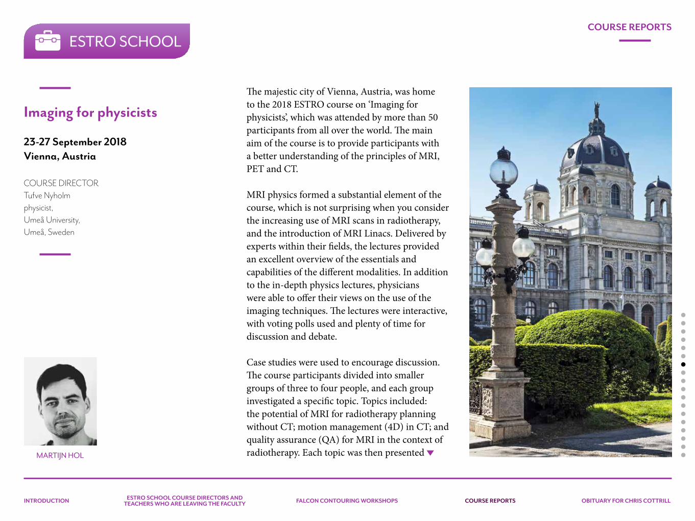
INTRODUCTION FALCON CONTOURING WORKSHOPSESTRO SCHOOL COURSE DIRECTORS AND TEACHERS WHO ARE LEAVING THE FACULTY OBITUARY FOR CHRIS COTTRILLCOURSE REPORTS
The majestic city of Vienna, Austria, was home to the 2018 ESTRO course on ‘Imaging for physicists’, which was attended by more than 50 participants from all over the world. The main aim of the course is to provide participants with a better understanding of the principles of MRI, PET and CT.
MRI physics formed a substantial element of the course, which is not surprising when you consider the increasing use of MRI scans in radiotherapy, and the introduction of MRI Linacs. Delivered by experts within their fields, the lectures provided an excellent overview of the essentials and capabilities of the different modalities. In addition to the in-depth physics lectures, physicians were able to offer their views on the use of the imaging techniques. The lectures were interactive, with voting polls used and plenty of time for discussion and debate.
Case studies were used to encourage discussion. The course participants divided into smaller groups of three to four people, and each group investigated a specific topic. Topics included: the potential of MRI for radiotherapy planning without CT; motion management (4D) in CT; and quality assurance (QA) for MRI in the context of radiotherapy. Each topic was then presented
ESTRO SCHOOL
MARTIJN HOL
Imaging for physicists
23-27 September 2018Vienna, Austria
COURSE DIRECTORTufve Nyholmphysicist, Umeå University, Umeå, Sweden
COURSE REPORTS
INTRODUCTION FALCON CONTOURING WORKSHOPSESTRO SCHOOL COURSE DIRECTORS AND TEACHERS WHO ARE LEAVING THE FACULTY OBITUARY FOR CHRIS COTTRILLCOURSE REPORTS
by the group to all participants and the lecturers, which resulted in lively and in-depth discussions.
The course was topped off with the guess-the-artefact quiz. Some wonderful prizes could be won in the quiz (an ESTRO cap and some chocolates) by applying our newly acquired knowledge on MRI to different artefacts that were presented.
In my opinion, this course gives an adequate foundation for understanding and applying complex imaging techniques. I certainly appreciated the open and friendly environment that was created by the lecturers. The interactive elements contributed to this, and enabled participants to discuss things with each other and the lecturers. The well organised course not only consisted of enjoyable and great presentations and interactive sessions, but also provided a successful social event: a nice dinner at the Melker Stiftskeller restaurant.
If you are thinking about taking a course on imaging techniques used in radiotherapy with a focus on MRI, this course will provide you with the foundational knowledge you need to work in this area.
Martijn HolMedical Physics Engineer,Leids Universitary Medical CentreLeiden, The Netherlands [email protected]
COURSE REPORTS
INTRODUCTION FALCON CONTOURING WORKSHOPSESTRO SCHOOL COURSE DIRECTORS AND TEACHERS WHO ARE LEAVING THE FACULTY OBITUARY FOR CHRIS COTTRILLCOURSE REPORTS
ESTRO SCHOOL
I am currently a registrar in Scotland preparing for the fellowship of the Royal College of Radiology (FRCR) exams in 2019. I wanted to add to my knowledge on skin cancer and gain any useful hints to help tackle this difficult exam. I was particularly interested in this course on ‘Multidisciplinary management of non-melanoma skin cancer’, as skin cancer is said to be a popular question in the exam.
The course was held in Brussels, Belgium, in October 2018 over three days. This was the first ESTRO course I’ve attended, and I found it to be quite comprehensive in covering the subdisciplines under skin cancer oncology. There were quite a few participants mostly at the consultant level from several different regions. The environment was relaxed, and there were many lively discussions and questions after most of the talks. I found the presentations involving case-based discussions most useful to me as they incorporated real-world practice with the theory.
Day one of the course covered dermatopathology, skin cancer diagnostics and surgery. Day two looked at radiotherapy, medical physics and
AN ONCOLOGY TRAINEE’S PERSPECTIVE
brachytherapy. The final day was a recap of what we had learned through case-based discussions and sessions dedicated to multidisciplinary approaches, as well as looking at skin cancer oncology from a geriatrician and a tissue viability nurses’ perspective.
We had a lovely dinner in the evening, which was a nice end to a busy day of radiotherapy and physics. I feel that this course has helped me understand the basics required for day-to-day radiotherapy planning as well as providing me with useful tips and hints for the FRCR exam.
I would recommend this course for anyone seeking to brush up their knowledge of non-melanoma skin oncology practice in Europe.
Timothy Mitchell Specialist registrar Beatson Oncology Centre Glasgow, [email protected]
COURSE REPORTS
Multidisciplinary management of non-melanoma skin cancer
4-6 October 2018Brussels, Belgium
COURSE DIRECTORAgata Rembielakradiation oncologist and clinical oncologist, The Christie Hospital, Manchester, UK
TIMOTHY MITCHELL
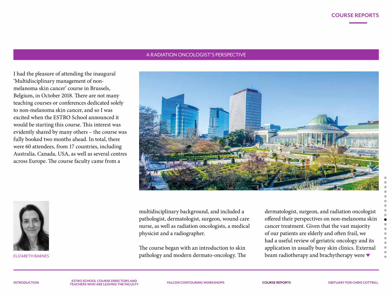
INTRODUCTION FALCON CONTOURING WORKSHOPSESTRO SCHOOL COURSE DIRECTORS AND TEACHERS WHO ARE LEAVING THE FACULTY OBITUARY FOR CHRIS COTTRILLCOURSE REPORTS
I had the pleasure of attending the inaugural ‘Multidisciplinary management of non-melanoma skin cancer’ course in Brussels, Belgium, in October 2018. There are not many teaching courses or conferences dedicated solely to non-melanoma skin cancer, and so I was excited when the ESTRO School announced it would be starting this course. This interest was evidently shared by many others – the course was fully booked two months ahead. In total, there were 60 attendees, from 17 countries, including Australia, Canada, USA, as well as several centres across Europe. The course faculty came from a
multidisciplinary background, and included a pathologist, dermatologist, surgeon, wound care nurse, as well as radiation oncologists, a medical physicist and a radiographer.
The course began with an introduction to skin pathology and modern dermato-oncology. The
A RADIATION ONCOLOGIST’S PERSPECTIVE
COURSE REPORTS
dermatologist, surgeon, and radiation oncologist offered their perspectives on non-melanoma skin cancer treatment. Given that the vast majority of our patients are elderly and often frail, we had a useful review of geriatric oncology and its application in usually busy skin clinics. External beam radiotherapy and brachytherapy were ELIZABETH BARNES
INTRODUCTION FALCON CONTOURING WORKSHOPSESTRO SCHOOL COURSE DIRECTORS AND TEACHERS WHO ARE LEAVING THE FACULTY OBITUARY FOR CHRIS COTTRILLCOURSE REPORTS
COURSE REPORTS
discussed in more detail the following day, along with talks on supportive care. The last day was dedicated to the role of the multidisciplinary team in patient management, current and future research in non-melanoma skin cancer, and the emerging role of biomarkers and targeted therapy.
Throughout the course, several clinical skin cases were presented for us to consider management options, and the course concluded by reviewing how the faculty had prescribed treatment for these patients. On the last day there was a very interesting interactive case discussion session, in which attendees had the opportunity to present their own challenging cases.
The course was held at the ESTRO offices in Brussels, an easy metro ride from the city centre. During the coffee breaks and lunches we could talk with fellow oncologists from around the world, and learn how non-melanoma skin cancer is treated at their centres. The social evening, held on the first night, was another enjoyable opportunity to mingle with colleagues. The weather in Brussels was lovely and warm, allowing us to explore this beautiful city on foot, and to sample the beer and chocolate.
This was a very useful and practical course on non-melanoma skin cancer, especially given the rising incidence of this disease, and the increase in patient volume and complexity we are going to be seeing in our practices over time. I would like to congratulate the course director Agata Rembielak for all her hard work over the past few years in designing, organising, and implementing this course. I would also like to thank the local organisers and teaching staff, for what will surely be the first of many successful ESTRO teaching courses on non-melanoma skin cancer.
Elizabeth (Toni) BarnesRadiation oncologistOdette Cancer Centre, Toronto, [email protected]
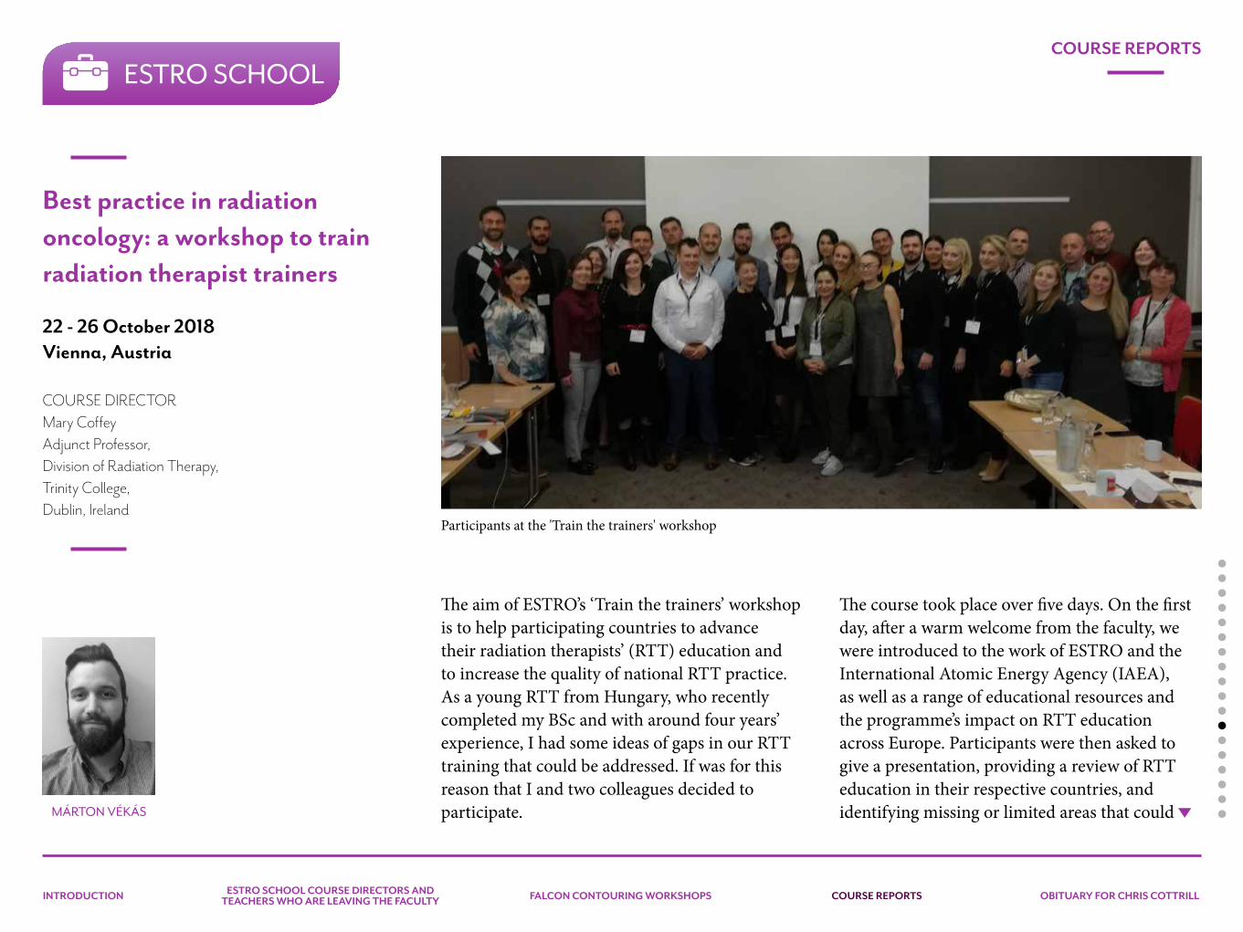
INTRODUCTION FALCON CONTOURING WORKSHOPSESTRO SCHOOL COURSE DIRECTORS AND TEACHERS WHO ARE LEAVING THE FACULTY OBITUARY FOR CHRIS COTTRILLCOURSE REPORTS
The aim of ESTRO’s ‘Train the trainers’ workshop is to help participating countries to advance their radiation therapists’ (RTT) education and to increase the quality of national RTT practice. As a young RTT from Hungary, who recently completed my BSc and with around four years’ experience, I had some ideas of gaps in our RTT training that could be addressed. If was for this reason that I and two colleagues decided to participate.
The course took place over five days. On the first day, after a warm welcome from the faculty, we were introduced to the work of ESTRO and the International Atomic Energy Agency (IAEA), as well as a range of educational resources and the programme’s impact on RTT education across Europe. Participants were then asked to give a presentation, providing a review of RTT education in their respective countries, and identifying missing or limited areas that could
COURSE REPORTS
ESTRO SCHOOL
MÁRTON VÉKÁS
Best practice in radiation oncology: a workshop to train radiation therapist trainers
22 - 26 October 2018Vienna, Austria
COURSE DIRECTORMary CoffeyAdjunct Professor,Division of Radiation Therapy,Trinity College,Dublin, Ireland
Participants at the 'Train the trainers' workshop
INTRODUCTION FALCON CONTOURING WORKSHOPSESTRO SCHOOL COURSE DIRECTORS AND TEACHERS WHO ARE LEAVING THE FACULTY OBITUARY FOR CHRIS COTTRILLCOURSE REPORTS
COURSE REPORTS
be improved. The faculty used these presentations to develop a topic for a new short course, the aim of which is not just to train RTTs, but also to liaise with the national educational institute to consider proposed changes to the curriculum.
From day two, we started to work on our short courses with the help of the faculty. Each day started with a presentation from the faculty, in which they gave us tips, talked about organisational methods and pitfalls to avoid, and shared their own experiences. After these presentations, we had workshops until the evening, in which we developed the content of our courses, exchanging ideas and thoughts with participants from other countries. At the end of each day, we reported our progress and received feedback from the faculty. By the end of the week everybody had successfully developed their short courses.
This outcome was exactly what I had expected from the course. I think I can speak for everyone in saying that we became more confident about organising a short course and potentially revising and developing new areas of national RTT education. The organisers were very professional, and really understood participants’ needs. They were also extremely kind and encouraged
us to ask for help whenever we needed it. For newcomers like us, this meant a lot. I would also like to thank ESTRO for offering this course. I am glad that I could represent my country. Overall, we learned a lot, made some good new friends, and also enjoyed some free time to explore a little of the beautiful city of Vienna.
Márton VékásRadiation therapistNational Institute of OncologyBudapest, [email protected]
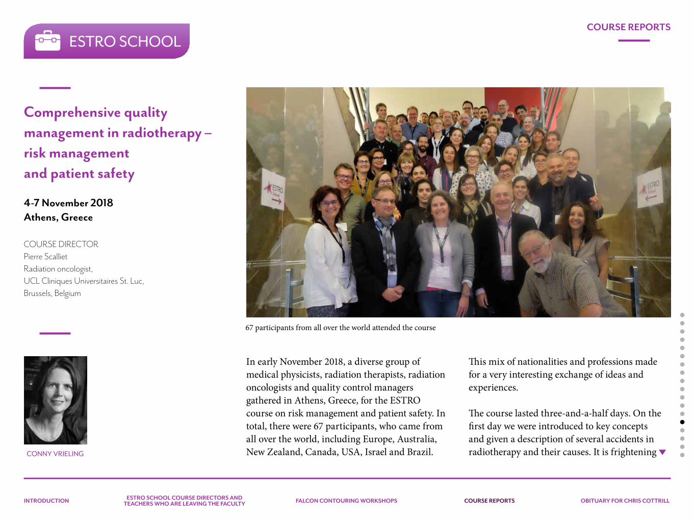
INTRODUCTION FALCON CONTOURING WORKSHOPSESTRO SCHOOL COURSE DIRECTORS AND TEACHERS WHO ARE LEAVING THE FACULTY OBITUARY FOR CHRIS COTTRILLCOURSE REPORTS
ESTRO SCHOOL
CONNY VRIELING
Comprehensive quality management in radiotherapy – risk management and patient safety
4-7 November 2018Athens, Greece
COURSE DIRECTORPierre ScallietRadiation oncologist,UCL Cliniques Universitaires St. Luc,Brussels, Belgium
COURSE REPORTS
In early November 2018, a diverse group of medical physicists, radiation therapists, radiation oncologists and quality control managers gathered in Athens, Greece, for the ESTRO course on risk management and patient safety. In total, there were 67 participants, who came from all over the world, including Europe, Australia, New Zealand, Canada, USA, Israel and Brazil.
This mix of nationalities and professions made for a very interesting exchange of ideas and experiences.
The course lasted three-and-a-half days. On the first day we were introduced to key concepts and given a description of several accidents in radiotherapy and their causes. It is frightening
67 participants from all over the world attended the course
INTRODUCTION FALCON CONTOURING WORKSHOPSESTRO SCHOOL COURSE DIRECTORS AND TEACHERS WHO ARE LEAVING THE FACULTY OBITUARY FOR CHRIS COTTRILLCOURSE REPORTS
COURSE REPORTS
to be confronted with the many things that can go wrong in delivering radiotherapy, but it is also very encouraging to know how many treatments are executed safely and according to plan. It is up to all of us to ensure this continues to be the case, and courses such as this play an important role in that.
At the end of the first day, we enjoyed a delicious dinner, with breath-taking view of the Acropolis and the Parthenon. It was incredible.
The subject of the second day was reactive risk management: how to report and analyse an incident. Root cause analysis using the PRISMA-RT method was discussed in depth. We also looked at incident reporting and learning tools created specifically for radiotherapy, ROSEIS and SAFRON. In the afternoon, working in several small groups, we conducted an analysis of an accident that had been presented the previous day, using the PRISMA method.
The third day was dedicated to prospective risk management: how to anticipate potential errors or incidents and how to avoid these when introducing a new technique or a new work procedure. This can be done using a process known as Healthcare Failure Mode and Effect
Analysis (HFMEA). After learning about this analytical method, we took part in another group exercise, analysing the potential pitfalls (and their causes) when introducing a new technique, and coming up with actions to eliminate these.
We discussed many other things over the course of the three days, such as different national legal requirements for reporting incidents or accidents, differences in work culture (in terms of hierarchy, inclination to blame), as well as very practical, but real issues, such as smartphone use in the department (and specifically at the treatment machine).
The group exercises provided ample time to get a clear idea of how to use different methods and analytical techniques and ensured that we completed the course not only with theoretical knowledge, but also with the practical tools and skills to implement incident reporting and avoiding systems in our own department. It also meant that we could exchange experiences with others, developing our awareness of cultural differences and the different barriers we face in improving incident reporting.
The last half-day of the course was dedicated to communication – with the patient and their
family, with the personnel directly involved in the incident, with the hospital management and finally with the media. This again was put into practice with an incident role-play. The conclusion of this exercise was that we need to simulate these situations in our own department so that we are ready whenever we need to be. Of course, we hope that by putting in place adequate systems, we will never be confronted with the need to use these communication skills.
In conclusion, I would like to say a big thank you to the course director, Pierre Scalliet, the other teachers, Nicolas Pourel, Brendan McClean, Petra Reijnders and Aude Vaandering, as well as the project manager, Mieke Akkers, for a well-organised course and excellent lectures.
Conny VrielingRadiation oncologistClinique des Grangettes HirslandenGeneva, [email protected]
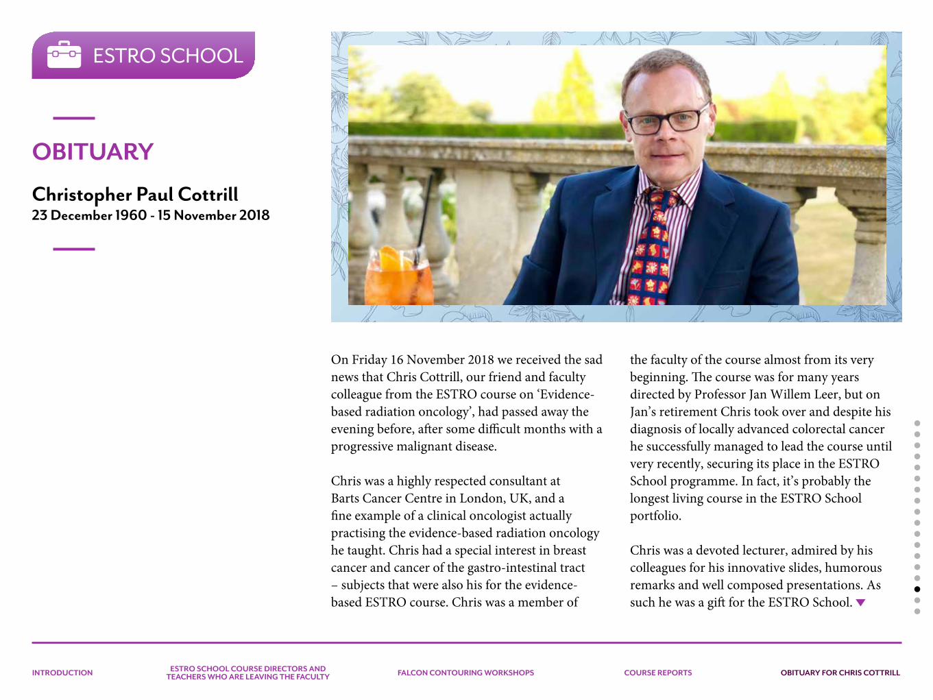
INTRODUCTION FALCON CONTOURING WORKSHOPSESTRO SCHOOL COURSE DIRECTORS AND TEACHERS WHO ARE LEAVING THE FACULTY COURSE REPORTS OBITUARY FOR CHRIS COTTRILL
On Friday 16 November 2018 we received the sad news that Chris Cottrill, our friend and faculty colleague from the ESTRO course on ‘Evidence-based radiation oncology’, had passed away the evening before, after some difficult months with a progressive malignant disease.
Chris was a highly respected consultant at Barts Cancer Centre in London, UK, and a fine example of a clinical oncologist actually practising the evidence-based radiation oncology he taught. Chris had a special interest in breast cancer and cancer of the gastro-intestinal tract – subjects that were also his for the evidence-based ESTRO course. Chris was a member of
the faculty of the course almost from its very beginning. The course was for many years directed by Professor Jan Willem Leer, but on Jan’s retirement Chris took over and despite his diagnosis of locally advanced colorectal cancer he successfully managed to lead the course until very recently, securing its place in the ESTRO School programme. In fact, it’s probably the longest living course in the ESTRO School portfolio.
Chris was a devoted lecturer, admired by his colleagues for his innovative slides, humorous remarks and well composed presentations. As such he was a gift for the ESTRO School.
ESTRO SCHOOL
OBITUARY
Christopher Paul Cottrill23 December 1960 - 15 November 2018
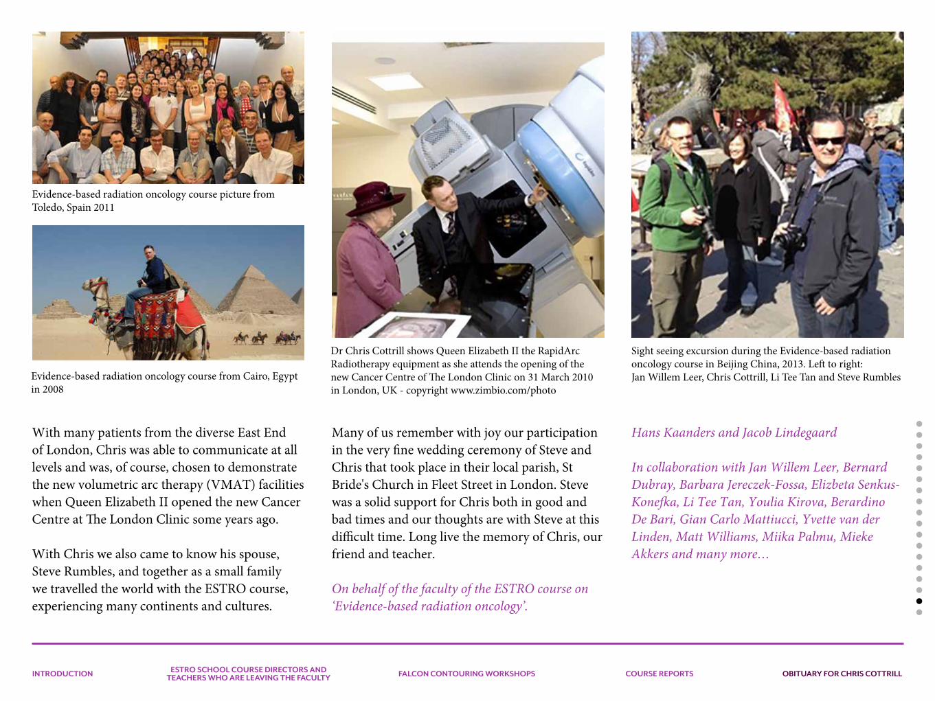
INTRODUCTION FALCON CONTOURING WORKSHOPSESTRO SCHOOL COURSE DIRECTORS AND TEACHERS WHO ARE LEAVING THE FACULTY COURSE REPORTS OBITUARY FOR CHRIS COTTRILL
With many patients from the diverse East End of London, Chris was able to communicate at all levels and was, of course, chosen to demonstrate the new volumetric arc therapy (VMAT) facilities when Queen Elizabeth II opened the new Cancer Centre at The London Clinic some years ago.
With Chris we also came to know his spouse, Steve Rumbles, and together as a small family we travelled the world with the ESTRO course, experiencing many continents and cultures.
Many of us remember with joy our participation in the very fine wedding ceremony of Steve and Chris that took place in their local parish, St Bride's Church in Fleet Street in London. Steve was a solid support for Chris both in good and bad times and our thoughts are with Steve at this difficult time. Long live the memory of Chris, our friend and teacher.
On behalf of the faculty of the ESTRO course on ‘Evidence-based radiation oncology’.
Hans Kaanders and Jacob Lindegaard
In collaboration with Jan Willem Leer, Bernard Dubray, Barbara Jereczek-Fossa, Elizbeta Senkus-Konefka, Li Tee Tan, Youlia Kirova, Berardino De Bari, Gian Carlo Mattiucci, Yvette van der Linden, Matt Williams, Miika Palmu, Mieke Akkers and many more…
Evidence-based radiation oncology course picture from Toledo, Spain 2011
Dr Chris Cottrill shows Queen Elizabeth II the RapidArc Radiotherapy equipment as she attends the opening of the new Cancer Centre of The London Clinic on 31 March 2010 in London, UK - copyright www.zimbio.com/photo
Evidence-based radiation oncology course from Cairo, Egypt in 2008
Sight seeing excursion during the Evidence-based radiation oncology course in Beijing China, 2013. Left to right: Jan Willem Leer, Chris Cottrill, Li Tee Tan and Steve Rumbles
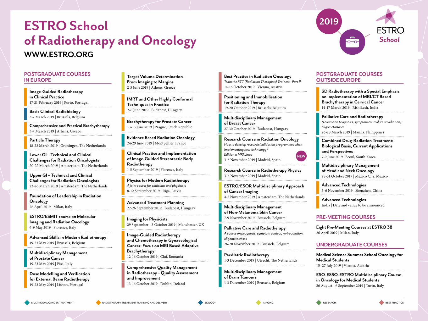
ESTRO School of Radiotherapy and OncologyWWW.ESTRO.ORG
MULTIMODAL CANCER TREATMENT RADIOTHERAPY TREATMENT PLANNING AND DELIVERY BIOLOGY IMAGING RESEARCH BEST PRACTICE
2019
POSTGRADUATE COURSES IN EUROPE
Image-Guided Radiotherapy in Clinical Practice17-21 February 2019 | Porto, Portugal
Basic Clinical Radiobiology3-7 March 2019 | Brussels, Belgium
Comprehensive and Practical Brachytherapy3-7 March 2019 | Athens, Greece
Particle Therapy18-22 March 2019 | Groningen, The Netherlands
Lower GI – Technical and Clinical Challenges for Radiation Oncologists 20-22 March 2019 | Amsterdam, The Netherlands
Upper GI – Technical and Clinical Challenges for Radiation Oncologists 23-26 March 2019 | Amsterdam, The Netherlands
Foundation of Leadership in Radiation Oncology 26 April 2019 | Milan, Italy
ESTRO/ESMIT course on Molecular Imaging and Radiation Oncology6-9 May 2019 | Florence, Italy
Advanced Skills in Modern Radiotherapy19-23 May 2019 | Brussels, Belgium
Multidisciplinary Management of Prostate Cancer19-23 May 2019 | Pisa, Italy
Dose Modelling and Verification for External Beam Radiotherapy19-23 May 2019 | Lisbon, Portugal
Target Volume Determination – From Imaging to Margins2-5 June 2019 | Athens, Greece
IMRT and Other Highly Conformal Techniques in Practice2-6 June 2019 | Budapest, Hungary
Brachytherapy for Prostate Cancer13-15 June 2019 | Prague, Czech Republic
Evidence Based Radiation Oncology24-29 June 2019 | Montpellier, France
Clinical Practice and Implementation of Image-Guided Stereotactic Body Radiotherapy1-5 September 2019 | Florence, Italy
Physics for Modern Radiotherapy A joint course for clinicians and physicists8-12 September 2019 | Riga, Latvia
Advanced Treatment Planning22-26 September 2019 | Budapest, Hungary
Imaging for Physicists29 September - 3 October 2019 | Manchester, UK
Image-Guided Radiotherapy and Chemotherapy in Gynaecological Cancer: Focus on MRI Based Adaptive Brachytherapy 12-16 October 2019 | Cluj, Romania
Comprehensive Quality Management in Radiotherapy – Quality Assessment and Improvement13-16 October 2019 | Dublin, Ireland
Best Practice in Radiation Oncology Train the RTT (Radiation Therapists) Trainers - Part II14-16 October 2019 | Vienna, Austria
Positioning and Immobilisation for Radiation Therapy19-20 October 2019 | Brussels, Belgium
Multidisciplinary Management of Breast Cancer27-30 October 2019 | Budapest, Hungary
Research Course in Radiation OncologyHow to develop research/validation programmes when implementing new technology? Edition 1: MRI Linac3-6 November 2019 | Madrid, Spain
Research Course in Radiotherapy Physics3-6 November 2019 | Madrid, Spain
ESTRO/ESOR Multidisciplinary Approach of Cancer Imaging4-5 November 2019 | Amsterdam, The Natherlands
Multidisciplinary Management of Non-Melanoma Skin Cancer7-9 November 2019 | Brussels, Belgium
Palliative Care and RadiotherapyA course on prognosis, symptom control, re-irradiation, oligometastases26-28 November 2019 | Brussels, Belgium
Paediatric Radiotherapy1-3 December 2019 | Utrecht, The Netherlands
Multidisciplinary Management of Brain Tumours1-3 December 2019 | Brussels, Belgium
POSTGRADUATE COURSES OUTSIDE EUROPE
3D Radiotherapy with a Special Emphasis on Implementation of MRI/CT Based Brachytherapy in Cervical Cancer14-17 March 2019 | Rishikesh, India
Palliative Care and RadiotherapyA course on prognosis, symptom control, re-irradiation, oligometastases26-28 March 2019 | Manila, Philippines
Combined Drug-Radiation Treatment:Biological Basis, Current Applications and Perspectives7-9 June 2019 | Seoul, South Korea
Multidisciplinary Management of Head and Neck Oncology28-31 October 2019 | Mexico City, Mexico
Advanced Technologies3-6 November 2019 | Shenzhen, China
Advanced TechnologiesIndia | Date and venue to be announced
PRE-MEETING COURSES
Eight Pre-Meeting Courses at ESTRO 3826 April 2019 | Milan, Italy
UNDERGRADUATE COURSES
Medical Science Summer School Oncology for Medical Students15 -27 July 2019 | Vienna, Austria
ESO-ESSO-ESTRO Multidisciplinary Course in Oncology for Medical Students26 August - 6 September 2019 | Turin, Italy
NEW
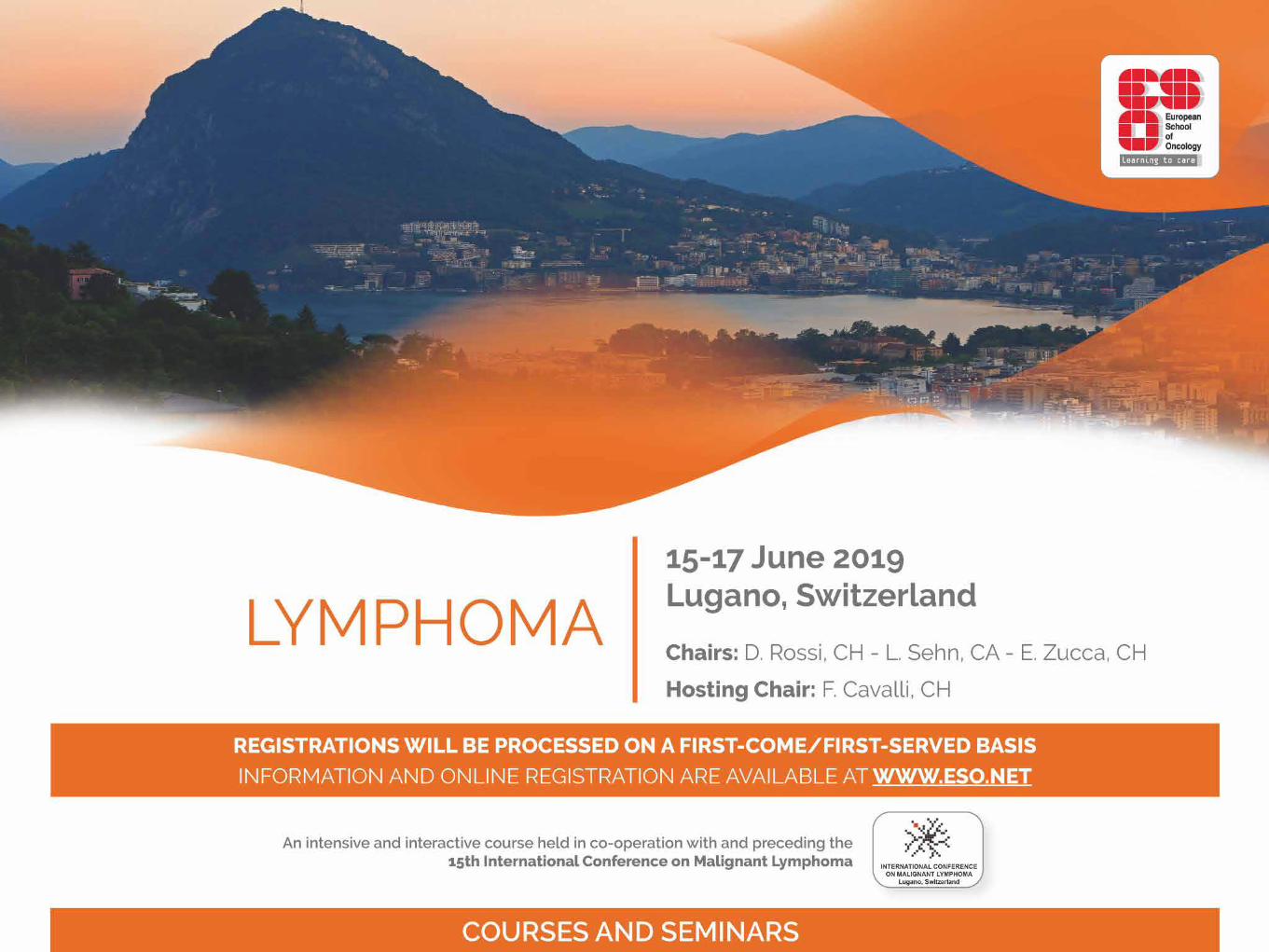
INTRODUCTION FALCON CONTOURING WORKSHOPSESTRO SCHOOL COURSE DIRECTORS AND TEACHERS WHO ARE LEAVING THE FACULTY COURSE REPORTS OBITUARY FOR CHRIS COTTRILL
INTRODUCTION PEER-REVIEWING: TIPS FROM THE ESTRO JOURNAL EDITORS
ESTRO 38: AN INTERVIEW WITH MARTIN-IMMANUEL BITTNER AND CYRUS CHARGARI
TECHNOLOGY TRANSFER GRANT (TTG) REPORT
REPORT FROM THE 2ND ESTRO PHYSICS WORKSHOP
YOUNG ESTRO
PEER-REVIEWING: TIPS FROM THE ESTRO JOURNAL EDITORS
ESTRO 38: AN INTERVIEW WITH MARTIN-IMMANUEL BITTNER AND CYRUS CHARGARI
TECHNOLOGY TRANSFER GRANT (TTG) REPORT
REPORT FROM THE 2ND ESTRO PHYSICS WORKSHOP
YOUNG ESTRO
PIERFRANCESCO FRANCO
KATHRINE RØE REDALEN
“Here, you will find some tips on how to produce a good review and a very useful checklist that you might want to go through next time you submit an article”
INTRODUCTION
Welcome to the Young Corner. It is an honour to be guest editor for this first edition of the year. I hope you enjoy reading it.
Our first article is about peer-reviewing, which is an important part of our work regardless of our specialty. However, we receive little or no training on the topic. Here, you will find some tips on how to produce a good review and a very useful checklist that
you might also want to go through next time you submit an article.
Next, you will find the programme for the Young Track at ESTRO 38, along with an interview with the chairs, Martin Immanuel Bittner and Cyrus Chargari. Read it to find out more about how the programme was developed and how it fits with the conference theme.
This Corner also features a report from the second physics workshop held in Malaga, Spain, at the end of October 2018. The workshop is a great opportunity for young researchers and oncology professionals to share ideas in a focused group and to get involved in ESTRO projects.
Finally, we have a technology transfer grant report from Candice Yu and colleagues, who visited the Chulalongkorn University Hospital in Bangkok, Thailand, to learn more about hybrid image-guided brachytherapy to treat gynaecological malignancies. The report outlines the workflow they are implementing in their home institution in the Philippines, drawing on what they learned in Bangkok.
Jenny Bertholet,Young ESTRO member
JENNY BERTHOLET
INTRODUCTION ESTRO 38: AN INTERVIEW WITH MARTIN-IMMANUEL BITTNER AND CYRUS CHARGARI
TECHNOLOGY TRANSFER GRANT (TTG) REPORT
REPORT FROM THE 2ND ESTRO PHYSICS WORKSHOP
PEER-REVIEWING: TIPS FROM THE ESTRO JOURNAL EDITORS
Peer-review is a well-established approach to validate research methods and research findings. Used in science for over 300 years [1], it is the only realistic method to validate research in practice. Peer-review is not only important for scientific publishing, but also for grant evaluation and academic promotions.
Why review?Reviewing is our professional responsibility. For each paper published in a journal, roughly four are rejected at different stages of the process. Three or four of these papers will have gone through the reviewing process with up to four reviewers. At least the same number of reviewers will have turned down the invitation to review. Therefore, for each published paper, 10-15 reviews are needed (without counting the reviews of the revisions).
But reviewing is not only a responsibility; it is an opportunity for development. In a study on second language learning, it was found that giving peer-review feedback was more effective at helping language learning than receiving peer-review feedback [2]. In this way, reviewing can be seen as an important part of our training in writing better papers.
YOUNG ESTRO
Peer-reviewing: tips from an ESTRO journal editorLudvig Muren, editor for Radiotherapy & Oncology and Acta Oncologica, editor-in-chief for Physics and Imaging in Radiation Oncology (phiRO)
How to review?Who would pass on the opportunity to help the research community and improve their writing skills? But... reviewing is not part of our formal training, so where should we start? Ethical guidelines for peer-reviewers are a good starting point [3]. In practice, here is a checklist of how to evaluate each part of a manuscript [4]:
The title: does it accurately reflect the purpose, design, results and conclusions of the study? The abstract: does it provide a clear aim, the key (numerical) results and a clear conclusion? Does it comply with the journal instructions?
The introduction: is it clear how the proposed study adds to the specific field? Is the literature review sufficient and are the main findings and limitations from previous studies clearly stated? Is the research question clearly stated and is it relevant? Is the previous research in the area linked together in a way that establishes the study hypothesis?
The materials and methods: is the patient cohort or material adequately described and appropriate for the purpose of the study; is it generalisable? Is the cohort / sample size sufficient to show a difference (proof of concept, feasibility study LUDVIG MUREN
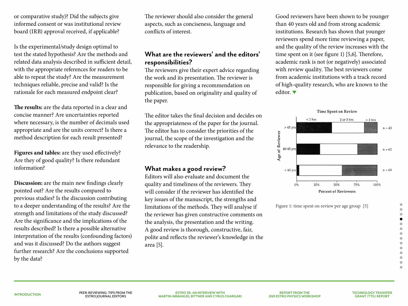
INTRODUCTION ESTRO 38: AN INTERVIEW WITH MARTIN-IMMANUEL BITTNER AND CYRUS CHARGARI
TECHNOLOGY TRANSFER GRANT (TTG) REPORT
REPORT FROM THE 2ND ESTRO PHYSICS WORKSHOP
PEER-REVIEWING: TIPS FROM THE ESTRO JOURNAL EDITORS
or comparative study)? Did the subjects give informed consent or was institutional review board (IRB) approval received, if applicable?
Is the experimental/study design optimal to test the stated hypothesis? Are the methods and related data analysis described in sufficient detail, with the appropriate references for readers to be able to repeat the study? Are the measurement techniques reliable, precise and valid? Is the rationale for each measured endpoint clear?
The results: are the data reported in a clear and concise manner? Are uncertainties reported where necessary, is the number of decimals used appropriate and are the units correct? Is there a method description for each result presented?
Figures and tables: are they used effectively? Are they of good quality? Is there redundant information?
Discussion: are the main new findings clearly pointed out? Are the results compared to previous studies? Is the discussion contributing to a deeper understanding of the results? Are the strength and limitations of the study discussed? Are the significance and the implications of the results described? Is there a possible alternative interpretation of the results (confounding factors) and was it discussed? Do the authors suggest further research? Are the conclusions supported by the data?
The reviewer should also consider the general aspects, such as conciseness, language and conflicts of interest.
What are the reviewers’ and the editors’ responsibilities?The reviewers give their expert advice regarding the work and its presentation. The reviewer is responsible for giving a recommendation on publication, based on originality and quality of the paper.
The editor takes the final decision and decides on the appropriateness of the paper for the journal. The editor has to consider the priorities of the journal, the scope of the investigation and the relevance to the readership.
What makes a good review?Editors will also evaluate and document the quality and timeliness of the reviewers. They will consider if the reviewer has identified the key issues of the manuscript, the strengths and limitations of the methods. They will analyse if the reviewer has given constructive comments on the analysis, the presentation and the writing. A good review is thorough, constructive, fair, polite and reflects the reviewer’s knowledge in the area [5].
Good reviewers have been shown to be younger than 40 years old and from strong academic institutions. Research has shown that younger reviewers spend more time reviewing a paper, and the quality of the review increases with the time spent on it (see figure 1) [5,6]. Therefore, academic rank is not (or negatively) associated with review quality. The best reviewers come from academic institutions with a track record of high-quality research, who are known to the editor.
Figure 1: time spent on review per age group [5]
INTRODUCTION ESTRO 38: AN INTERVIEW WITH MARTIN-IMMANUEL BITTNER AND CYRUS CHARGARI
TECHNOLOGY TRANSFER GRANT (TTG) REPORT
REPORT FROM THE 2ND ESTRO PHYSICS WORKSHOP
PEER-REVIEWING: TIPS FROM THE ESTRO JOURNAL EDITORS
How to improve reviewing skills?Several approaches have been tested unsuccessfully to improve the quality of peer-review, such as written feedback from the editors to the reviewers [7], structured training workshops [8,9], self-instruction with training packages [10] and mentoring by experienced expert reviewers [11]. Close collaboration with local colleagues, mentors or supervisors may be the best approach.
REFERENCES
[1] Spier R. The history of the peer-review process. Trends Biotechnol. 2002;20:357–358.
[2] Lundstrom K, Baker W. To give is better than to receive: The benefits of peer review to the reviewer’s own writing. J. Second Lang. Writ. 2009;18:30–43.
[3] www.publicationethics.org. COPE Ethical Guidelines for Peer Reviewers. 2013.
[4] Seals DR, Tanaka H. Manuscript peer review: a helpful checklist for students and novice referees. Adv. Physiol. Educ. 2000;23:52–58.
[5] Evans AT, Mcnutt RA, Fletcher SW, et al. The characteristics of peer reviewers who produce good-quality reviews. J. Gen. Intern. Med. 1993;8.
[6] Stossel T. Reviewer Status and Review Quality — Experience of the Journal of Clinical Investigation. N. Engl. J. Med. 1985;312.
[7] Callaham ML, Knopp RK, John Gallagher E. Effect of written feedback by editors on quality of reviews: Two randomized trials. J. Am. Med. Assoc. 2002;287:2781–2783.
[8] Callaham ML, Schriger DL. Effect of structured workshop training on subsequent performance of journal peer reviewers. Ann. Emerg. Med. 2002;40:323–328.
[9] Callaham ML, Wears RL, Waeckerle JF. Effect of attendance at a training session on peer reviewer quality and performance. Ann. Emerg. Med. 1998;32:318–322.
[10] Schroter S. Effects of training on quality of peer review: randomised controlled trial. Bmj. 2004;328:673–0.
[11] Houry D, Green S, Callaham M. Does mentoring new peer reviewers improve review quality? A randomized trial. BMC Med. Educ. 2012;12.
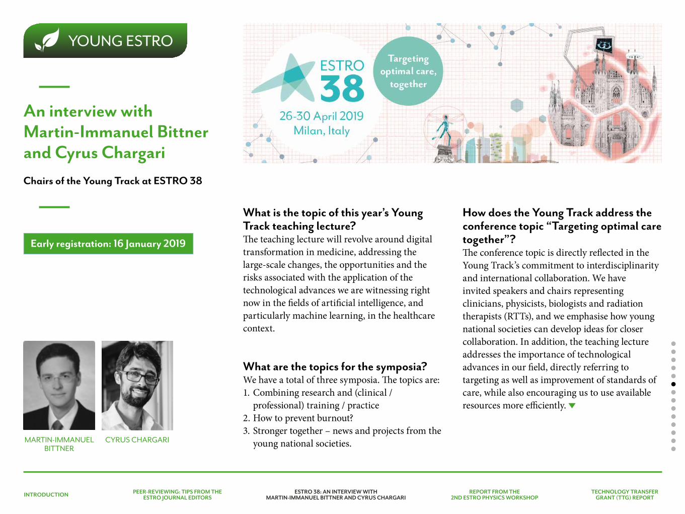
INTRODUCTION PEER-REVIEWING: TIPS FROM THE ESTRO JOURNAL EDITORS
TECHNOLOGY TRANSFER GRANT (TTG) REPORT
REPORT FROM THE 2ND ESTRO PHYSICS WORKSHOP
ESTRO 38: AN INTERVIEW WITH MARTIN-IMMANUEL BITTNER AND CYRUS CHARGARI
YOUNG ESTRO
What is the topic of this year’s Young Track teaching lecture?The teaching lecture will revolve around digital transformation in medicine, addressing the large-scale changes, the opportunities and the risks associated with the application of the technological advances we are witnessing right now in the fields of artificial intelligence, and particularly machine learning, in the healthcare context.
What are the topics for the symposia? We have a total of three symposia. The topics are: 1. Combining research and (clinical /
professional) training / practice2. How to prevent burnout?3. Stronger together – news and projects from the
young national societies.
How does the Young Track address the conference topic “Targeting optimal care together”?The conference topic is directly reflected in the Young Track’s commitment to interdisciplinarity and international collaboration. We have invited speakers and chairs representing clinicians, physicists, biologists and radiation therapists (RTTs), and we emphasise how young national societies can develop ideas for closer collaboration. In addition, the teaching lecture addresses the importance of technological advances in our field, directly referring to targeting as well as improvement of standards of care, while also encouraging us to use available resources more efficiently.
An interview with Martin-Immanuel Bittner and Cyrus ChargariChairs of the Young Track at ESTRO 38
MARTIN-IMMANUEL BITTNER
CYRUS CHARGARI
Early registration: 16 January 2019
INTRODUCTION PEER-REVIEWING: TIPS FROM THE ESTRO JOURNAL EDITORS
TECHNOLOGY TRANSFER GRANT (TTG) REPORT
REPORT FROM THE 2ND ESTRO PHYSICS WORKSHOP
ESTRO 38: AN INTERVIEW WITH MARTIN-IMMANUEL BITTNER AND CYRUS CHARGARI
How did you choose the topics for this year’s Young Track programme? Why are they important and timely?This year’s themes and sessions are directly aligned with the collaborative topic of the conference. With recent political developments undermining the spirit of collaboration, we thought it was very important to focus on how researchers and practitioners in radiation oncology can achieve more by working together. We firmly believe that only by building strong ties for improved research efforts in a European and global context, will we be able to truly advance cancer care, together.
A second focus of the ESTRO 38 Young Track is combining research and training / practice and the detection and prevention of burnout. We know that pressures in research and clinical practice are rising, with detrimental effects on the health and wellbeing of those who are supposed to help others. Therefore, we have dedicated one symposium to this important topic, while at the same time recognising how important it is for those in our field to be well versed in both research and practice.
What would you say are the learning objectives for the Young Track?We hope that everyone attending the ESTRO 38 Young Track – regardless of what stage they are at in their career – will be inspired by the efforts
of the young national societies to promote our discipline, and guide it into the future. We also hope that there will be helpful insights into how to combine research and training / practice, as well as how to prevent burnout, both topics that are highly relevant to all of us.
What was the biggest challenge in establishing the Young Track programme?There were so many suggestions and proposals within the ESTRO young committee that we found it difficult to decide which topics to prioritise this year. Therefore, we aimed to align our topics with the conference topic, and focus on areas and themes that we felt were particularly important to feature prominently, because of their importance for young clinicians, researchers or radiation therapy technologists, such as burnout.
How did you experience the process personally? Was it as you expected?Having been involved in the scientific programme committee for ESTRO 38, we found it remarkable to see how planning for ESTRO 38 started when ESTRO 37 was still underway. A lot of time and effort goes into planning a conference of the size and calibre of ESTRO’s annual meeting. It was – and still is – an enlightening experience to be involved in this process, and to experience how much thought and effort everyone in the
committee puts into choosing timely topics of broad interest and relevance to the radiation oncology community, deciding upon the best format for each session, and identifying potentially suitable chairs and speakers.
Which sessions from the other tracks are you particularly looking forward to?A difficult question – especially when having to choose between so many interesting sessions, often run in parallel. Given our own research background, however, we are especially interested in the sessions exploring “Artificial intelligence applications in radiation oncology” as well as “Radiation-drug combinations on the 2019 horizon”, both of which sound particularly interesting.
Among other highly promising sessions, one will be dedicated to modern brachytherapy, including “Real time navigation technologies in brachytherapy”, which will address the possibility of expanding indications in this therapeutic area.
The Young Track programme in a tweet?The Young Track highlights how we can work together across disciplines and borders, use technology to help us and act for both patients and carers.
#YoungTrack #ESTRO38
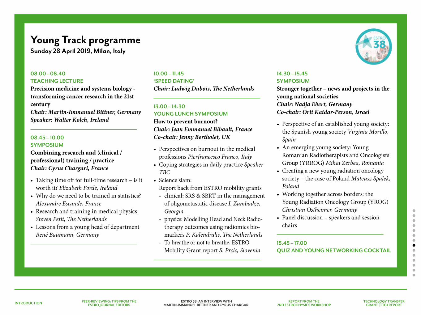
INTRODUCTION PEER-REVIEWING: TIPS FROM THE ESTRO JOURNAL EDITORS
TECHNOLOGY TRANSFER GRANT (TTG) REPORT
REPORT FROM THE 2ND ESTRO PHYSICS WORKSHOP
ESTRO 38: AN INTERVIEW WITH MARTIN-IMMANUEL BITTNER AND CYRUS CHARGARI
Young Track programmeSunday 28 April 2019, Milan, Italy
08.00 - 08.40 TEACHING LECTUREPrecision medicine and systems biology - transforming cancer research in the 21st centuryChair: Martin-Immanuel Bittner, GermanySpeaker: Walter Kolch, Ireland
08.45 – 10.00SYMPOSIUMCombining research and (clinical / professional) training / practiceChair: Cyrus Chargari, France
• Taking time off for full-time research – is it worth it? Elizabeth Forde, Ireland
• Why do we need to be trained in statistics? Alexandre Escande, France
• Research and training in medical physics Steven Petit, The Netherlands
• Lessons from a young head of department René Baumann, Germany
10.00 – 11.45‘SPEED DATING’Chair: Ludwig Dubois, The Netherlands
13.00 – 14.30 YOUNG LUNCH SYMPOSIUMHow to prevent burnout?Chair: Jean Emmanuel Bibault, FranceCo-chair: Jenny Bertholet, UK
• Perspectives on burnout in the medical professions Pierfrancesco Franco, Italy
• Coping strategies in daily practice Speaker TBC
• Science slam:Report back from ESTRO mobility grants- clinical: SRS & SBRT in the management
of oligometastatic disease I. Zumbadze, Georgia
- physics: Modelling Head and Neck Radio- therapy outcomes using radiomics bio-markers P. Kalendralis, The Netherlands
- To breathe or not to breathe, ESTRO Mobility Grant report S. Prcic, Slovenia
14.30 – 15.45SYMPOSIUMStronger together – news and projects in the young national societiesChair: Nadja Ebert, GermanyCo-chair: Orit Kaidar-Person, Israel
• Perspective of an established young society: the Spanish young society Virginia Morillo, Spain
• An emerging young society: Young Romanian Radiotherapists and Oncologists Group (YRROG) Mihai Zerbea, Romania
• Creating a new young radiation oncology society – the case of Poland Mateusz Spalek, Poland
• Working together across borders: the Young Radiation Oncology Group (YROG) Christian Ostheimer, Germany
• Panel discussion – speakers and session chairs
15.45 – 17.00QUIZ AND YOUNG NETWORKING COCKTAIL
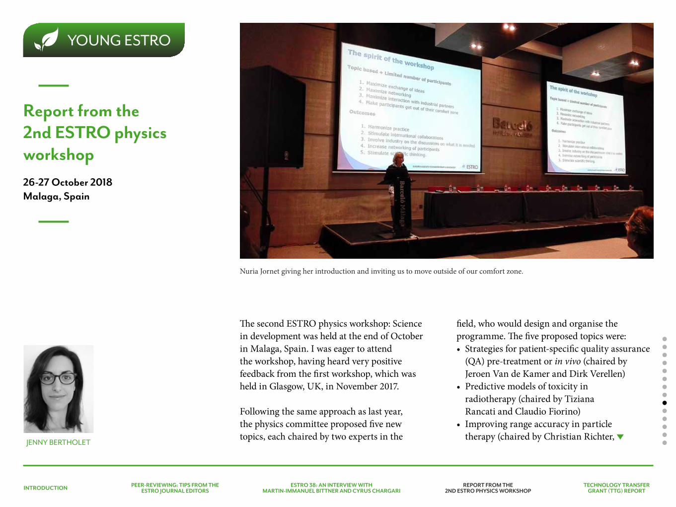
INTRODUCTION PEER-REVIEWING: TIPS FROM THE ESTRO JOURNAL EDITORS
ESTRO 38: AN INTERVIEW WITH MARTIN-IMMANUEL BITTNER AND CYRUS CHARGARI
TECHNOLOGY TRANSFER GRANT (TTG) REPORT
REPORT FROM THE 2ND ESTRO PHYSICS WORKSHOP
YOUNG ESTRO
The second ESTRO physics workshop: Science in development was held at the end of October in Malaga, Spain. I was eager to attend the workshop, having heard very positive feedback from the first workshop, which was held in Glasgow, UK, in November 2017.
Following the same approach as last year, the physics committee proposed five new topics, each chaired by two experts in the
JENNY BERTHOLET
field, who would design and organise the programme. The five proposed topics were:• Strategies for patient-specific quality assurance
(QA) pre-treatment or in vivo (chaired by Jeroen Van de Kamer and Dirk Verellen)
• Predictive models of toxicity in radiotherapy (chaired by Tiziana Rancati and Claudio Fiorino)
• Improving range accuracy in particle therapy (chaired by Christian Richter,
Report from the 2nd ESTRO physics workshop26-27 October 2018Malaga, Spain
Nuria Jornet giving her introduction and inviting us to move outside of our comfort zone.
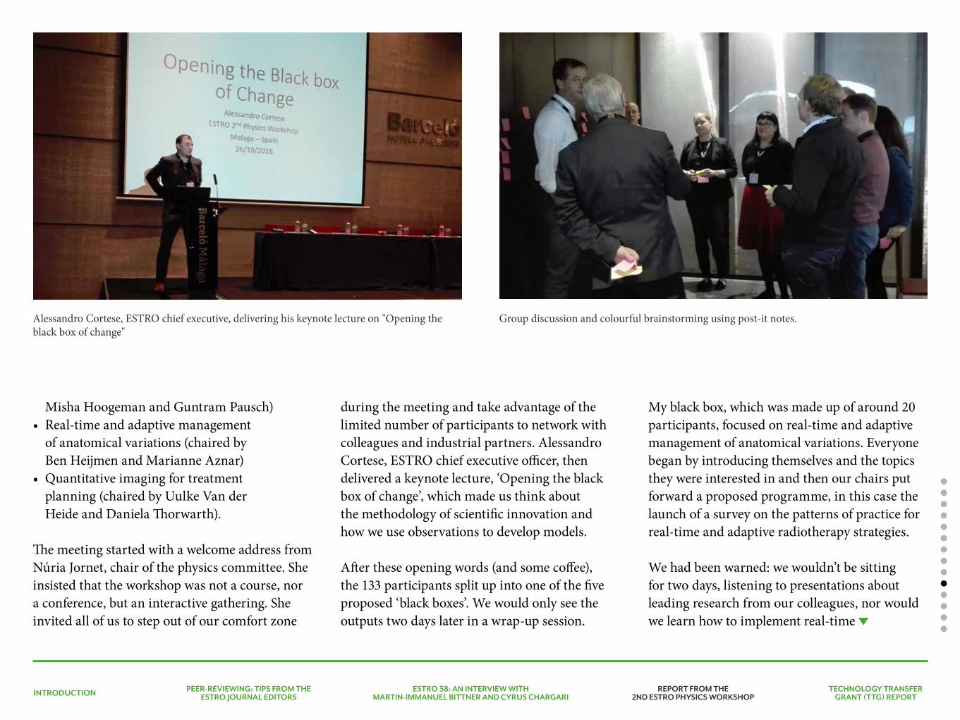
INTRODUCTION PEER-REVIEWING: TIPS FROM THE ESTRO JOURNAL EDITORS
ESTRO 38: AN INTERVIEW WITH MARTIN-IMMANUEL BITTNER AND CYRUS CHARGARI
TECHNOLOGY TRANSFER GRANT (TTG) REPORT
REPORT FROM THE 2ND ESTRO PHYSICS WORKSHOP
during the meeting and take advantage of the limited number of participants to network with colleagues and industrial partners. Alessandro Cortese, ESTRO chief executive officer, then delivered a keynote lecture, ‘Opening the black box of change’, which made us think about the methodology of scientific innovation and how we use observations to develop models.
After these opening words (and some coffee), the 133 participants split up into one of the five proposed ‘black boxes’. We would only see the outputs two days later in a wrap-up session.
My black box, which was made up of around 20 participants, focused on real-time and adaptive management of anatomical variations. Everyone began by introducing themselves and the topics they were interested in and then our chairs put forward a proposed programme, in this case the launch of a survey on the patterns of practice for real-time and adaptive radiotherapy strategies.
We had been warned: we wouldn’t be sitting for two days, listening to presentations about leading research from our colleagues, nor would we learn how to implement real-time
Alessandro Cortese, ESTRO chief executive, delivering his keynote lecture on "Opening the black box of change"
Group discussion and colourful brainstorming using post-it notes.
Misha Hoogeman and Guntram Pausch)• Real-time and adaptive management
of anatomical variations (chaired by Ben Heijmen and Marianne Aznar)
• Quantitative imaging for treatment planning (chaired by Uulke Van der Heide and Daniela Thorwarth).
The meeting started with a welcome address from Núria Jornet, chair of the physics committee. She insisted that the workshop was not a course, nor a conference, but an interactive gathering. She invited all of us to step out of our comfort zone
INTRODUCTION PEER-REVIEWING: TIPS FROM THE ESTRO JOURNAL EDITORS
ESTRO 38: AN INTERVIEW WITH MARTIN-IMMANUEL BITTNER AND CYRUS CHARGARI
TECHNOLOGY TRANSFER GRANT (TTG) REPORT
REPORT FROM THE 2ND ESTRO PHYSICS WORKSHOP
INTRODUCTION 2ND ESTRO PHYSICS WORKSHOP
PHYSICS
Read the other reports on the 2nd ESTRO physics workshop in the Physics Corner on page 46 >
and adaptive radiotherapy in our clinics. Instead, we would be working on and producing something new for our community. The group was enthusiastic about the proposal and started brainstorming right away, with practical questions, such as how to get the questionnaires to as many centres as possible.
Everybody agreed that the survey should result in more than just a set of statistics; it should help establish guidelines to standardise clinical practice and assist centres that have not yet implemented adaptive strategies to do so safely. The group divided naturally into a real-time and an adaptive branch, linked by a common structure, and we worked on questions for the survey for the rest of the day.
Course, conference or workshop, one thing is immutable in ESTRO events: great social events. This workshop was no exception, and after a productive day, all participants met at the Fabrica de cervezas (the brewery) to share beer, wine and tapas. There was no sit-down meal, which created an enjoyable and informal atmosphere, in which we could mingle easily and speak with everyone.
The second day continued with participants in the same group as the previous day and finished with a wrap-up session: the output of the black boxes. Very much like the real-time and adaptive group, the four other groups had also identified ways to standardise practice and share information and data within the
community. All groups presented concrete and thought-out strategies to reach their goals.
The workshop may have reached its end, but there is still plenty of work to be done until we can harvest the fruits of these two days. The participants will meet again, papers and guidelines will be written and sharing platforms will be set up. The enthusiasm of participants and the inclusiveness of the workshop was confirmed once again, when participants from the quantitative imaging group set up chairs in a circle to continue their discussions as soon as the group picture was taken and the workshop was officially closed.
As a young researcher, this workshop was, first, an opportunity to be more participative than in the big conferences. Second, it was a chance to get involved with ESTRO and think about how our own research and practice finds its place in the bigger picture of radiotherapy in Europe and the world. Third, the ideas and projects started during these two days will involve different skills to the ones we use on a day-to-day basis and will create opportunities for international collaborations.Convinced to join by the positive feedback from the first workshop, it is now my turn to urge radiotherapy professionals, especially young people, to join future editions of the ESTRO physics workshop.
Jenny BertholetThe Institute of Cancer Research and The Royal Marsden NHS Foundation TrustLondon, UK
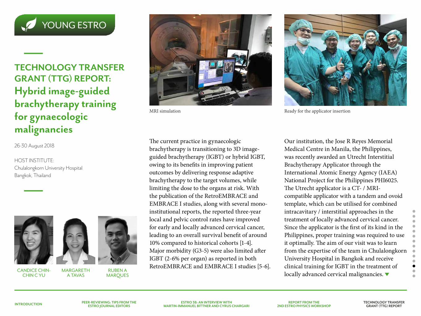
INTRODUCTION PEER-REVIEWING: TIPS FROM THE ESTRO JOURNAL EDITORS
ESTRO 38: AN INTERVIEW WITH MARTIN-IMMANUEL BITTNER AND CYRUS CHARGARI
REPORT FROM THE 2ND ESTRO PHYSICS WORKSHOP
TECHNOLOGY TRANSFER GRANT (TTG) REPORT
The current practice in gynaecologic brachytherapy is transitioning to 3D image-guided brachytherapy (IGBT) or hybrid IGBT, owing to its benefits in improving patient outcomes by delivering response adaptive brachytherapy to the target volumes, while limiting the dose to the organs at risk. With the publication of the RetroEMBRACE and EMBRACE I studies, along with several mono-institutional reports, the reported three-year local and pelvic control rates have improved for early and locally advanced cervical cancer, leading to an overall survival benefit of around 10% compared to historical cohorts [1-4]. Major morbidity (G3-5) were also limited after IGBT (2-6% per organ) as reported in both RetroEMBRACE and EMBRACE I studies [5-6].
Our institution, the Jose R Reyes Memorial Medical Centre in Manila, the Philippines, was recently awarded an Utrecht Interstitial Brachytherapy Applicator through the International Atomic Energy Agency (IAEA) National Project for the Philippines PHI6025. The Utrecht applicator is a CT- / MRI-compatible applicator with a tandem and ovoid template, which can be utilised for combined intracavitary / interstitial approaches in the treatment of locally advanced cervical cancer. Since the applicator is the first of its kind in the Philippines, proper training was required to use it optimally. The aim of our visit was to learn from the expertise of the team in Chulalongkorn University Hospital in Bangkok and receive clinical training for IGBT in the treatment of locally advanced cervical malignancies.
YOUNG ESTRO
TECHNOLOGY TRANSFER GRANT (TTG) REPORT: Hybrid image-guided brachytherapy training for gynaecologic malignancies26-30 August 2018
HOST INSTITUTE: Chulalongkorn University HospitalBangkok, Thailand
MRI simulation Ready for the applicator insertion
CANDICE CHIN-CHIN C YU
MARGARETH A TAVAS
RUBEN A MARQUES
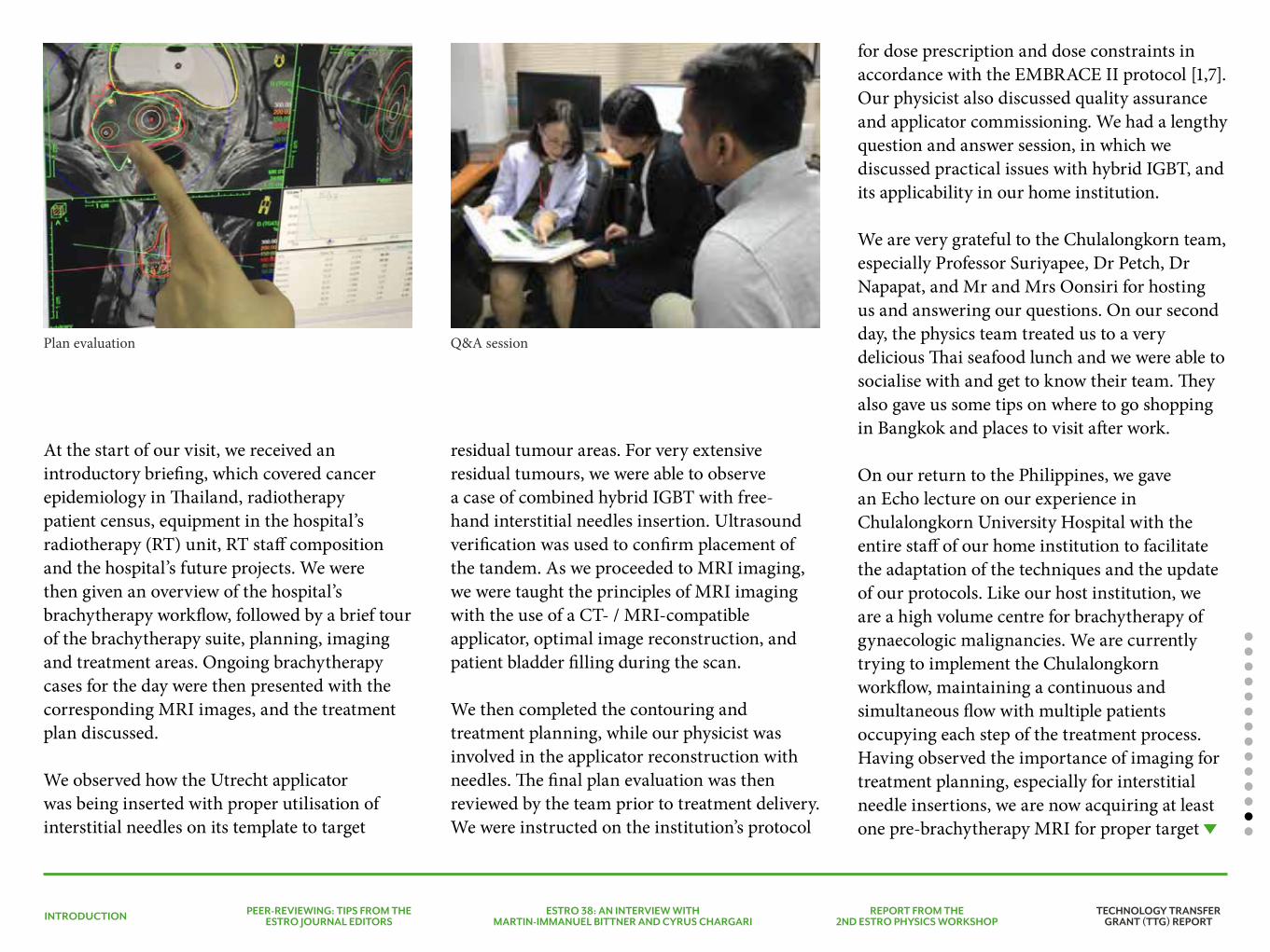
INTRODUCTION PEER-REVIEWING: TIPS FROM THE ESTRO JOURNAL EDITORS
ESTRO 38: AN INTERVIEW WITH MARTIN-IMMANUEL BITTNER AND CYRUS CHARGARI
REPORT FROM THE 2ND ESTRO PHYSICS WORKSHOP
TECHNOLOGY TRANSFER GRANT (TTG) REPORT
At the start of our visit, we received an introductory briefing, which covered cancer epidemiology in Thailand, radiotherapy patient census, equipment in the hospital’s radiotherapy (RT) unit, RT staff composition and the hospital’s future projects. We were then given an overview of the hospital’s brachytherapy workflow, followed by a brief tour of the brachytherapy suite, planning, imaging and treatment areas. Ongoing brachytherapy cases for the day were then presented with the corresponding MRI images, and the treatment plan discussed.
We observed how the Utrecht applicator was being inserted with proper utilisation of interstitial needles on its template to target
residual tumour areas. For very extensive residual tumours, we were able to observe a case of combined hybrid IGBT with free-hand interstitial needles insertion. Ultrasound verification was used to confirm placement of the tandem. As we proceeded to MRI imaging, we were taught the principles of MRI imaging with the use of a CT- / MRI-compatible applicator, optimal image reconstruction, and patient bladder filling during the scan.
We then completed the contouring and treatment planning, while our physicist was involved in the applicator reconstruction with needles. The final plan evaluation was then reviewed by the team prior to treatment delivery. We were instructed on the institution’s protocol
for dose prescription and dose constraints in accordance with the EMBRACE II protocol [1,7]. Our physicist also discussed quality assurance and applicator commissioning. We had a lengthy question and answer session, in which we discussed practical issues with hybrid IGBT, and its applicability in our home institution.
We are very grateful to the Chulalongkorn team, especially Professor Suriyapee, Dr Petch, Dr Napapat, and Mr and Mrs Oonsiri for hosting us and answering our questions. On our second day, the physics team treated us to a very delicious Thai seafood lunch and we were able to socialise with and get to know their team. They also gave us some tips on where to go shopping in Bangkok and places to visit after work.
On our return to the Philippines, we gave an Echo lecture on our experience in Chulalongkorn University Hospital with the entire staff of our home institution to facilitate the adaptation of the techniques and the update of our protocols. Like our host institution, we are a high volume centre for brachytherapy of gynaecologic malignancies. We are currently trying to implement the Chulalongkorn workflow, maintaining a continuous and simultaneous flow with multiple patients occupying each step of the treatment process. Having observed the importance of imaging for treatment planning, especially for interstitial needle insertions, we are now acquiring at least one pre-brachytherapy MRI for proper target
Plan evaluation Q&A session
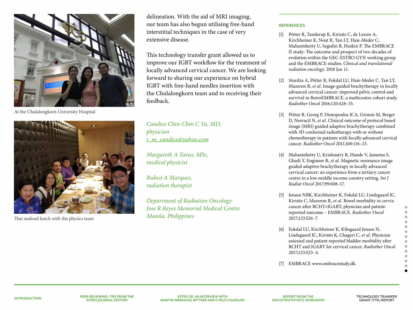
INTRODUCTION PEER-REVIEWING: TIPS FROM THE ESTRO JOURNAL EDITORS
ESTRO 38: AN INTERVIEW WITH MARTIN-IMMANUEL BITTNER AND CYRUS CHARGARI
REPORT FROM THE 2ND ESTRO PHYSICS WORKSHOP
TECHNOLOGY TRANSFER GRANT (TTG) REPORT
REFERENCES
[1] Pötter R, Tanderup K, Kirisits C, de Leeuw A, Kirchheiner K, Nout R, Tan LT, Haie-Meder C, Mahantshetty U, Segedin B, Hoskin P. The EMBRACE II study: The outcome and prospect of two decades of evolution within the GEC-ESTRO GYN working group and the EMBRACE studies. Clinical and translational radiation oncology. 2018 Jan 11.
[2] Sturdza A, Pötter R, Fokdal LU, Haie-Meder C, Tan LT, Mazeron R, et al. Image-guided brachytherapy in locally advanced cervical cancer: improved pelvic control and survival in RetroEMBRACE, a multicentre cohort study. Radiother Oncol 2016;120:428–33.
[3] Pötter R, Georg P, Dimopoulos JCA, Grimm M, Berger D, Nesvacil N, et al. Clinical outcome of protocol based image (MRI) guided adaptive brachytherapy combined with 3D conformal radiotherapy with or without chemotherapy in patients with locally advanced cervical cancer. Radiother Oncol 2011;100:116–23.
[4] Mahantshetty U, Krishnatry R, Hande V, Jamema S, Ghadi Y, Engineer R, et al. Magnetic resonance image guided adaptive brachytherapy in locally advanced cervical cancer: an experience from a tertiary cancer centre in a low-middle income country setting. Int J Radiat Oncol 2017;99:608–17.
[5] Jensen NBK, Kirchheiner K, Fokdal LU, Lindegaard JC, Kirisits C, Mazeron R, et al. Bowel morbidity in cervix cancer after RCHT+IGABT; physician and patient-reported outcome – EMBRACE. Radiother Oncol 2017;123:S26–7.
[6] Fokdal LU, Kirchheiner K, Kibsgaard Jensen N, Lindegaard JC, Kirisits K, Chagari C, et al. Physician assessed and patient reported bladder morbidity after RCHT and IGABT for cervical cancer. Radiother Oncol 2017;123:S23–4.
[7] EMBRACE www.embracestudy.dk.
delineation. With the aid of MRI imaging, our team has also begun utilising free-hand interstitial techniques in the case of very extensive disease.
This technology transfer grant allowed us to improve our IGBT workflow for the treatment of locally advanced cervical cancer. We are looking forward to sharing our experience on hybrid IGBT with free-hand needles insertion with the Chulalongkorn team and to receiving their feedback.
Candice Chin-Chin C Yu, MD, [email protected]
Margareth A Tavas, MSc, medical physicist
Ruben A Marques, radiation therapist
Department of Radiation OncologyJose R Reyes Memorial Medical CentreManila, Philippines
At the Chulalongkorn University Hospital
Thai seafood lunch with the physics team
RESEARCH PROJECTS
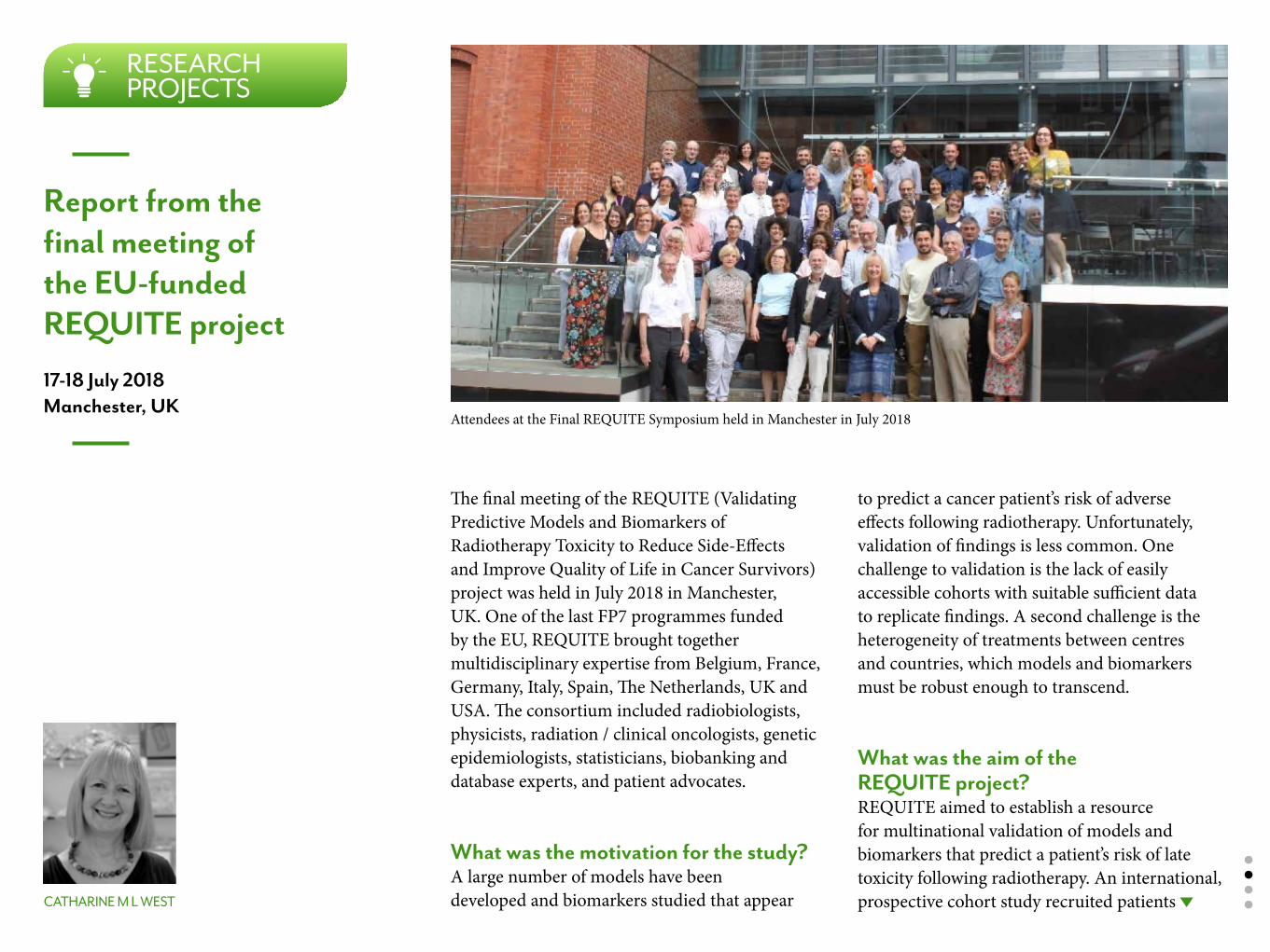
The final meeting of the REQUITE (Validating Predictive Models and Biomarkers of Radiotherapy Toxicity to Reduce Side-Effects and Improve Quality of Life in Cancer Survivors) project was held in July 2018 in Manchester, UK. One of the last FP7 programmes funded by the EU, REQUITE brought together multidisciplinary expertise from Belgium, France, Germany, Italy, Spain, The Netherlands, UK and USA. The consortium included radiobiologists, physicists, radiation / clinical oncologists, genetic epidemiologists, statisticians, biobanking and database experts, and patient advocates.
What was the motivation for the study?A large number of models have been developed and biomarkers studied that appear
to predict a cancer patient’s risk of adverse effects following radiotherapy. Unfortunately, validation of findings is less common. One challenge to validation is the lack of easily accessible cohorts with suitable sufficient data to replicate findings. A second challenge is the heterogeneity of treatments between centres and countries, which models and biomarkers must be robust enough to transcend.
What was the aim of the REQUITE project?REQUITE aimed to establish a resource for multinational validation of models and biomarkers that predict a patient’s risk of late toxicity following radiotherapy. An international, prospective cohort study recruited patients
RESEARCH PROJECTS
Report from the final meeting of the EU-funded REQUITE project17-18 July 2018Manchester, UK
CATHARINE M L WEST
Attendees at the Final REQUITE Symposium held in Manchester in July 2018
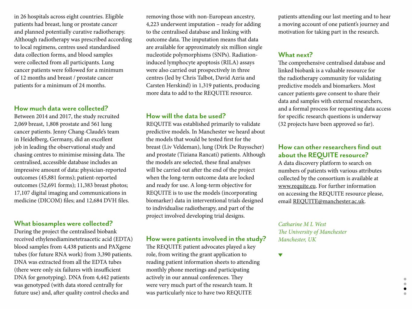
in 26 hospitals across eight countries. Eligible patients had breast, lung or prostate cancer and planned potentially curative radiotherapy. Although radiotherapy was prescribed according to local regimens, centres used standardised data collection forms, and blood samples were collected from all participants. Lung cancer patients were followed for a minimum of 12 months and breast / prostate cancer patients for a minimum of 24 months.
How much data were collected?Between 2014 and 2017, the study recruited 2,069 breast, 1,808 prostate and 561 lung cancer patients. Jenny Chang-Claude’s team in Heidelberg, Germany, did an excellent job in leading the observational study and chasing centres to minimise missing data. The centralised, accessible database includes an impressive amount of data: physician-reported outcomes (45,881 forms); patient-reported outcomes (52,691 forms); 11,383 breast photos; 17,107 digital imaging and communications in medicine (DICOM) files; and 12,684 DVH files.
What biosamples were collected?During the project the centralised biobank received ethylenediaminetetraacetic acid (EDTA) blood samples from 4,438 patients and PAXgene tubes (for future RNA work) from 3,390 patients. DNA was extracted from all the EDTA tubes (there were only six failures with insufficient DNA for genotyping). DNA from 4,442 patients was genotyped (with data stored centrally for future use) and, after quality control checks and
removing those with non-European ancestry, 4,223 underwent imputation – ready for adding to the centralised database and linking with outcome data. The imputation means that data are available for approximately six million single nucleotide polymorphisms (SNPs). Radiation-induced lymphocyte apoptosis (RILA) assays were also carried out prospectively in three centres (led by Chris Talbot, David Azria and Carsten Herskind) in 1,319 patients, producing more data to add to the REQUITE resource.
How will the data be used?REQUITE was established primarily to validate predictive models. In Manchester we heard about the models that would be tested first for the breast (Liv Veldeman), lung (Dirk De Ruysscher) and prostate (Tiziana Rancati) patients. Although the models are selected, these final analyses will be carried out after the end of the project when the long-term outcome data are locked and ready for use. A long-term objective for REQUITE is to use the models (incorporating biomarker) data in interventional trials designed to individualise radiotherapy, and part of the project involved developing trial designs.
How were patients involved in the study?The REQUITE patient advocates played a key role, from writing the grant application to reading patient information sheets to attending monthly phone meetings and participating actively in our annual conferences. They were very much part of the research team. It was particularly nice to have two REQUITE
patients attending our last meeting and to hear a moving account of one patient’s journey and motivation for taking part in the research.
What next?The comprehensive centralised database and linked biobank is a valuable resource for the radiotherapy community for validating predictive models and biomarkers. Most cancer patients gave consent to share their data and samples with external researchers, and a formal process for requesting data access for specific research questions is underway (32 projects have been approved so far).
How can other researchers find out about the REQUITE resource?A data discovery platform to search on numbers of patients with various attributes collected by the consortium is available at www.requite.eu. For further information on accessing the REQUITE resource please, email [email protected].
Catharine M L West The University of ManchesterManchester, UK
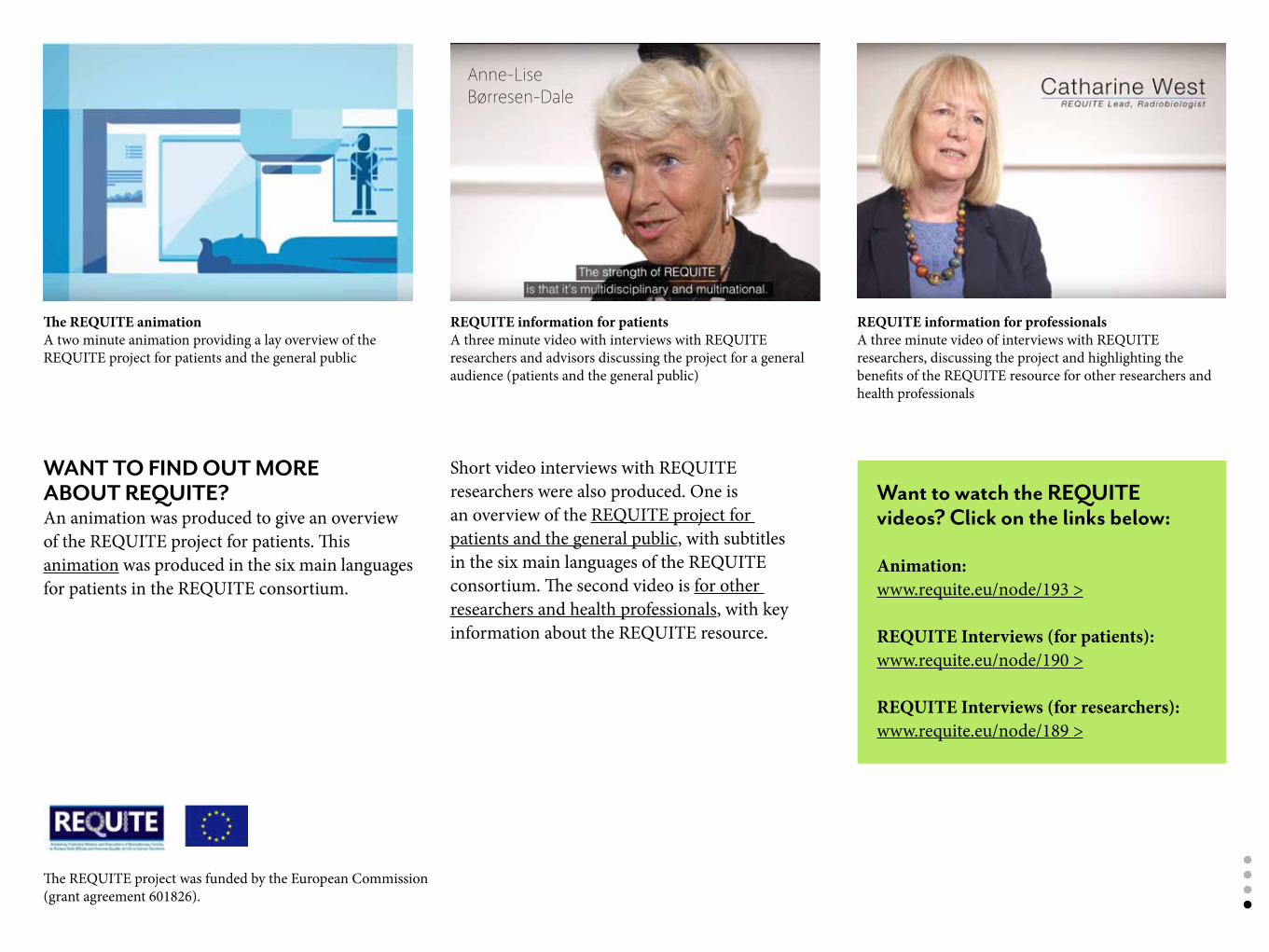
WANT TO FIND OUT MORE ABOUT REQUITE?An animation was produced to give an overview of the REQUITE project for patients. This animation was produced in the six main languages for patients in the REQUITE consortium.
Short video interviews with REQUITE researchers were also produced. One is an overview of the REQUITE project for patients and the general public, with subtitles in the six main languages of the REQUITE consortium. The second video is for other researchers and health professionals, with key information about the REQUITE resource.
The REQUITE animationA two minute animation providing a lay overview of the REQUITE project for patients and the general public
REQUITE information for patientsA three minute video with interviews with REQUITE researchers and advisors discussing the project for a general audience (patients and the general public)
REQUITE information for professionalsA three minute video of interviews with REQUITE researchers, discussing the project and highlighting the benefits of the REQUITE resource for other researchers and health professionals
The REQUITE project was funded by the European Commission (grant agreement 601826).
Want to watch the REQUITE videos? Click on the links below:
Animation:www.requite.eu/node/193 >
REQUITE Interviews (for patients):www.requite.eu/node/190 >
REQUITE Interviews (for researchers):www.requite.eu/node/189 >
Anne-Lise Børresen-Dale

MAKE IT HAPPEN
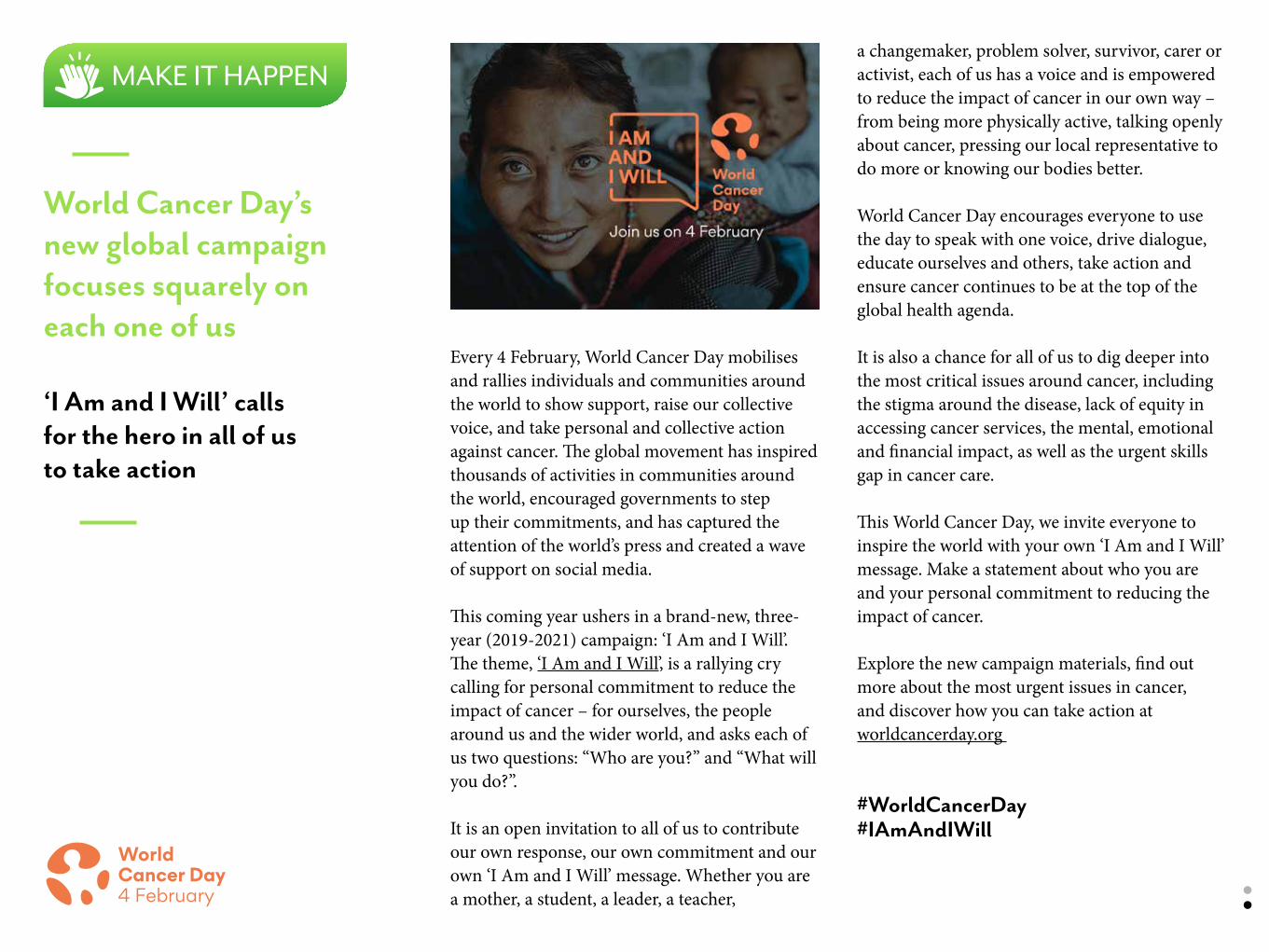
World Cancer Day’s new global campaign focuses squarely on each one of us
‘I Am and I Will’ calls for the hero in all of us to take action
MAKE IT HAPPEN
Every 4 February, World Cancer Day mobilises and rallies individuals and communities around the world to show support, raise our collective voice, and take personal and collective action against cancer. The global movement has inspired thousands of activities in communities around the world, encouraged governments to step up their commitments, and has captured the attention of the world’s press and created a wave of support on social media.
This coming year ushers in a brand-new, three-year (2019-2021) campaign: ‘I Am and I Will’. The theme, ‘I Am and I Will’, is a rallying cry calling for personal commitment to reduce the impact of cancer – for ourselves, the people around us and the wider world, and asks each of us two questions: “Who are you?” and “What will you do?”.
It is an open invitation to all of us to contribute our own response, our own commitment and our own ‘I Am and I Will’ message. Whether you are a mother, a student, a leader, a teacher,
a changemaker, problem solver, survivor, carer or activist, each of us has a voice and is empowered to reduce the impact of cancer in our own way – from being more physically active, talking openly about cancer, pressing our local representative to do more or knowing our bodies better.
World Cancer Day encourages everyone to use the day to speak with one voice, drive dialogue, educate ourselves and others, take action and ensure cancer continues to be at the top of the global health agenda.
It is also a chance for all of us to dig deeper into the most critical issues around cancer, including the stigma around the disease, lack of equity in accessing cancer services, the mental, emotional and financial impact, as well as the urgent skills gap in cancer care.
This World Cancer Day, we invite everyone to inspire the world with your own ‘I Am and I Will’ message. Make a statement about who you are and your personal commitment to reducing the impact of cancer.
Explore the new campaign materials, find out more about the most urgent issues in cancer, and discover how you can take action at worldcancerday.org
#WorldCancerDay#IAmAndIWill

PAST CONFERENCEFORTHCOMING CONFERENCESIN COLLABORATION WITH ESTROFORTHCOMING ESTRO EVENTS
CONFERENCES
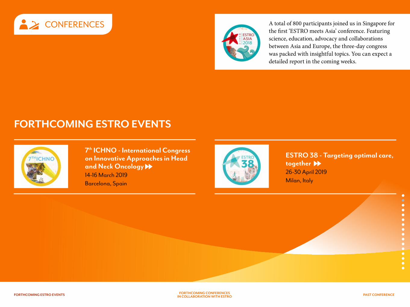
PAST CONFERENCEFORTHCOMING CONFERENCESIN COLLABORATION WITH ESTROFORTHCOMING ESTRO EVENTS
CONFERENCES
FORTHCOMING ESTRO EVENTS
7th ICHNO - International Congress on Innovative Approaches in Head and Neck Oncology 14-16 March 2019Barcelona, Spain
ESTRO 38 - Targeting optimal care, together 26-30 April 2019Milan, Italy
A total of 800 participants joined us in Singapore for the first ‘ESTRO meets Asia’ conference. Featuring science, education, advocacy and collaborations between Asia and Europe, the three-day congress was packed with insightful topics. You can expect a detailed report in the coming weeks.
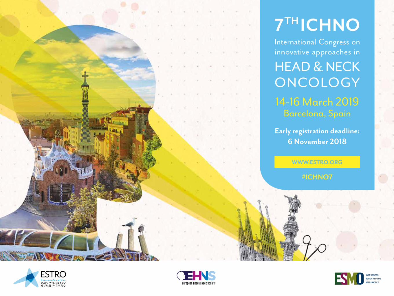
7TH ICHNOInternational Congress on innovative approaches in
HEAD & NECKONCOLOGY14-16 March 2019
Barcelona, Spain
Early registration deadline: 6 November 2018
#ICHNO7
WWW.ESTRO.ORG
PAST CONFERENCEFORTHCOMING CONFERENCESIN COLLABORATION WITH ESTROFORTHCOMING ESTRO EVENTS
Are there any hot topics in head and neck cancer that you noticed in the abstracts submitted? The hot topics in the submitted abstracts are reflected in ICHNO’s programme. Immunotherapy, of course, as a systemic treatment for advanced cases, but also as a concomitant, induction or adjuvant treatment with radiotherapy or surgery for locally advanced disease. Outside of immuno-oncology, many fields will be discussed, such as novel surgical techniques, human papillomavirus (HPV)-related oropharyngeal cancers and the results of early de-escalation trials, as well as molecular classification of diseases, image-guided treatment and radiomics.
Are there some presentations you are particularly looking forward to?Of course. On top of the symposia and proffered papers sessions, two great debates are being organised: one will debate the respective strengths of chemotherapy and immunotherapy, and the other challenge the clinical applications of radiomics. Also, six keynote speakers are invited to present state-of-the-art lectures on topics such as molecular classification of disease,
CONFERENCES
7th ICHNOInternational Conference on Innovative Approaches in Head and Neck Oncology
14-16 March 2019Barcelona, Spain
Interview with Dr Pierre Blanchard, chair of the ICHNO advisory committee for radiation oncology
PIERRE BLANCHARD
the immune landscape of head and neck cancers, integration of imaging in advanced radiotherapy, the status and potential evolution of the eighth version of the TNM staging system, and the future of robotic surgery. The diversity of the programme reflects the true multidisciplinarity that is at the heart of the management of head and neck cancers.
Will any clinical trials results be presented?Yes. The updated results of the De-ESCALaTE trial will be presented, along with an update on the results of the pembrolizumab phase III trial in first-line therapy in recurrent and metastatic patients. In addition, other prospective trials will be presented dealing with preoperative radiotherapy, laser microsurgery, combined chemotherapy, radiotherapy and nimorazole, among others.
WWW.ESTRO.ORG #ESTRO38
Targeting optimal care,
together
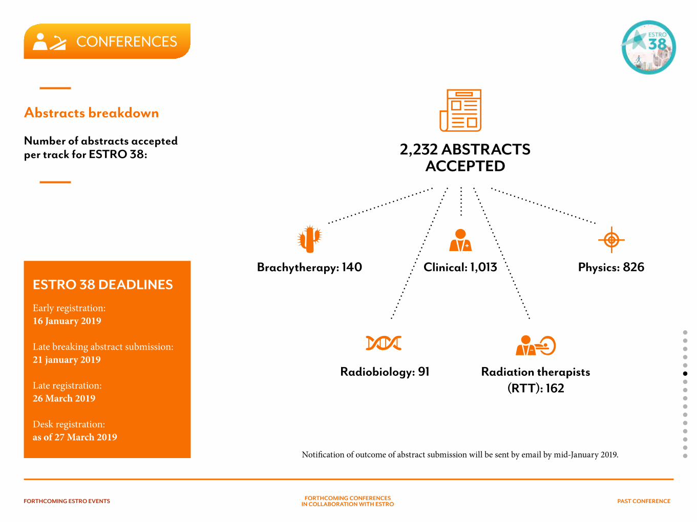
PAST CONFERENCEFORTHCOMING CONFERENCESIN COLLABORATION WITH ESTROFORTHCOMING ESTRO EVENTS
CONFERENCES
Abstracts breakdown
Number of abstracts accepted per track for ESTRO 38:
Notification of outcome of abstract submission will be sent by email by mid-January 2019.
2,232 ABSTRACTS ACCEPTED
Brachytherapy: 140
Radiobiology: 91 Radiation therapists (RTT): 162
Clinical: 1,013 Physics: 826ESTRO 38 DEADLINES
Early registration: 16 January 2019
Late breaking abstract submission: 21 january 2019
Late registration: 26 March 2019
Desk registration: as of 27 March 2019
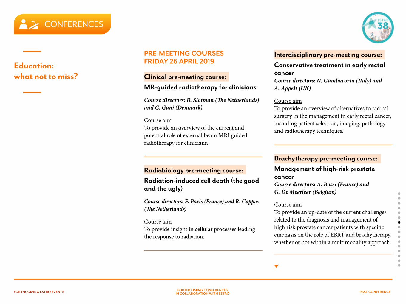
PAST CONFERENCEFORTHCOMING CONFERENCESIN COLLABORATION WITH ESTROFORTHCOMING ESTRO EVENTS
CONFERENCES
Education: what not to miss?
PRE-MEETING COURSESFRIDAY 26 APRIL 2019
Clinical pre-meeting course:MR-guided radiotherapy for clinicians
Course directors: B. Slotman (The Netherlands) and C. Gani (Denmark)
Course aimTo provide an overview of the current and potential role of external beam MRI guided radiotherapy for clinicians.
Radiobiology pre-meeting course:Radiation-induced cell death (the good and the ugly)
Course directors: F. Paris (France) and R. Coppes (The Netherlands)
Course aimTo provide insight in cellular processes leading the response to radiation.
Interdisciplinary pre-meeting course:Conservative treatment in early rectal cancerCourse directors: N. Gambacorta (Italy) and A. Appelt (UK)
Course aimTo provide an overview of alternatives to radical surgery in the management in early rectal cancer, including patient selection, imaging, pathology and radiotherapy techniques.
Brachytherapy pre-meeting course:Management of high-risk prostate cancerCourse directors: A. Bossi (France) and G. De Meerleer (Belgium)
Course aimTo provide an up-date of the current challenges related to the diagnosis and management of high risk prostate cancer patients with specific emphasis on the role of EBRT and brachytherapy, whether or not within a multimodality approach.
PAST CONFERENCEFORTHCOMING CONFERENCESIN COLLABORATION WITH ESTROFORTHCOMING ESTRO EVENTS
Physics pre-meeting course:Machine learning for physicists
Course directors: B. Heijmen (The Netherlands) and D. Verellen (Belgium)
Course aimTo provide basic knowledge on machine learning and its potential use in Radiation Oncology. The course aims at enabling medical physicists to understand the basics behind clinical applications from a user point of view and, provide information for interested developers to get started without prior knowledge. The course assumes that the participants have no knowledge on the subject.
RTT pre-meeting course:Basic course brachytherapy treatment
Course directors: R.I. Schokker (The Nether-lands) and B. Wisgrill (Austria)
Course aimRadiation therapists (RTTs), dosimetrists and RT nurses have several tasks in the brachytherapy treatment. To get to a more uniform level of knowledge, this course will provide with the
basic principles of brachytherapy. Next, to these basic principles, there will be different hospitals from various European countries presenting their workflow. It will be an interactive program, where participants can share their experiences.
Education pre-meeting course:Academic entrepreneurship, innovation, and technology transfer in radiation oncology
Course directors: P. Lambin (The Netherlands) and K. Tanderup (Denmark)
Course aim: Radiotherapy is a discipline involving a high degree of technology and using various discipline (imaging, biology, computer sciences, clinic, physics…). This course is meant as a workshop to stimulate collaboration between academia and industry and technology transfer which is essential for new technology to bridge the “second translational gap” and reach the patients.
Education pre-meeting course:Foundations of leadership in radiation oncology Joint ESTRO-CARO-RANZCR
Course directors: K. Benstead (UK), M. Giuliani (Canada), S. Turner (Australia)Course teachers: A. Cortese (Belgium), J. Eriksen (Denmark), B-A Millar (Canada), L. Morris (Australia)
Course duration:8 weeks with live pre-meeting workshop at ESTRO 38• Online programme to start on 20 March 2019• Live session on 26 April 2019 and 2 lunch
meetings
Course aim: This course, run for the first time in 2018, introduces foundation principles of effective leadership as they apply to senior trainees and junior practitioners in the radiation oncology professions. Feedback from the first fully-subscribed course has been very positive.
The course aims to equip participants with the knowledge, skills and attributes viewed as the building blocks for effective leadership.
PAST CONFERENCEFORTHCOMING CONFERENCESIN COLLABORATION WITH ESTROFORTHCOMING ESTRO EVENTS
The course is directed at professionals interested in developing expertise in leading teams for quality improvement, advocacy and in all situations where they might positively influence the future of our discipline within both local and international settings. The course links to a specific radiation oncology competency knowledge and skill-set developed through Delphi consensus process. (dx.doi.org/10.1016/j.radonc.2017.04.009).
CONTOURING WORKSHOPS
Eight contouring workshops have been planned(each session is repeated once):• OAR on head and neck cancer:
Friday 26 April 2019 from 08:00-10:00 (repeated Saturday 27 April from 14:30-16:30)
• Rectal cancer: Friday 26 April 2019 from 10:30-12:30 (repeated Sunday 28 April from 14:30-16:30)
• Lung SBRT: Friday 26 April 2019 from 13:30-15:30 (repeated Monday 29 April from 14:30-16:30)
• Image guided adaptive brachytherapy in vaginal cancer: Friday 26 April 2019 from 16:00-18:00 (repeated Tuesday 30 April from 09:15 – 11:15)
Target audienceThe delineation workshops are aimed at all radiation oncology professionals who want to improve their contouring skills.
Three types of cases are dedicated to radiation oncologists: a common case, a rare case and a more advanced case. The OAR case is especially targeted to radiation therapists (RTTs) and dosimetrists.
INTERACTIVE MULTIDISCIPLINARYTUMOUR BOARD SESSIONS
Soft tissue sarcomasSaturday 27 April 201914:30 - 15:45
Prostate cancerSunday 28 April 201914:30 - 15:45
Bladder cancerMonday 29 April 201914:30 - 15:45
PAST CONFERENCEFORTHCOMING CONFERENCESIN COLLABORATION WITH ESTROFORTHCOMING ESTRO EVENTS
USEFUL LINKS
More information on the pre-meeting courses:www.estro.org/congresses-meetings/articles/estro-38-pre-meetings--contouring
More information on the contouring workshops:www.estro.org/congresses-meetings/articles/estro-38-pre-meetings--contouring
More information on the scientific programme:www.estro.org/congresses-meetings/articles/estro38-programme
Surprise, surprise...! JOIN US NO LATER THAN FRIDAY 26 APRIL AT 18.00 HRS...
We can’t wait to welcome you to Milan for a thrilling ESTRO 38, which will start on Friday 26 April 2019 with an opening ceremony to remember. We recommend
you book your travel accordingly and get there for 18.00 hrs sharp!
Welcome
PAST CONFERENCEFORTHCOMING CONFERENCESIN COLLABORATION WITH ESTROFORTHCOMING ESTRO EVENTS
DOWNLOAD THE ESTRO APP!
Find out about all the next ESTRO conferences, including ESTRO 38: essential information, networking opportunities and the latest news at your fingertips!
Download the app from www.estro.org
PAST CONFERENCEFORTHCOMING ESTRO EVENTS FORTHCOMING CONFERENCESIN COLLABORATION WITH ESTRO
CONFERENCES
BGICC 2019: the 11th Breast-Gynaecological and Immunooncology International Cancer Conference 17-18 January 2019Cairo, Egypt
Breast - Gynecological & Immunooncology International Cancer ConferenceONE WORLD AGANIST CANCER
2019 RSS annual scientific meeting 21-23 March 2019San Diego, California, USA
FORTHCOMING CONFERENCESIn collaboration with ESTRO
PAST CONFERENCEFORTHCOMING ESTRO EVENTS FORTHCOMING CONFERENCESIN COLLABORATION WITH ESTRO
CONFERENCES
BGICC 2019: the 11th Breast-Gynaecological and Immunooncology International Cancer Conference
17-18 January 2019Cairo, Egypt
www.bgicc.netYou are warmly invited to the 11th Breast-Gynaecological and Immunooncology International Cancer Conference (BGICC 2019), the largest and most prestigious cancer conference in Africa and the Middle East. The conference will be held on 17-18 January 2019 in Cairo, Egypt.
The conference will provide state-of-the-art information on cancer research, offering breakthrough advances and insight in scientific and clinical research, patient management and practice through a range of scientific and educational symposia, special sessions, teaching lectures, workshops, debates and more.
We are building a strong strand of multidisciplinary clinical and translational research in the Middle East through the work
of our Society. BGICC is designed to strengthen this base in Egypt, the Middle East and Africa, educating young doctors and medical staff, and promoting the latest guidelines.
In the 11th BGICC, we aim to set a new standard and look to the future. Your participation in multidisciplinary sessions, workshops and consensus sessions focusing on points of debate will help us to fulfil this aim. BGICC brings together oncologists, radiologists, pathologists and surgical oncologists, gynaecologists, pharmacists, researchers and other medical professionals to participate in our multidisciplinary approach to advance progress against cancer, in close collaboration with high-calibre international scientific societies. Together, we believe that “Cancer is a disease, not a death sentence”.
Breast - Gynecological & Immunooncology International Cancer ConferenceONE WORLD AGANIST CANCER
FORTHCOMING CONFERENCES - In collaboration with ESTROBGICC 2019
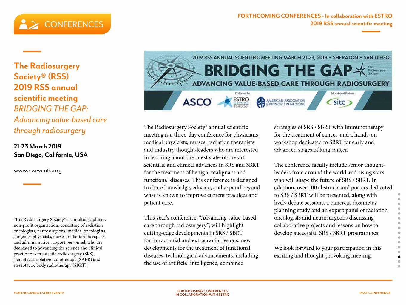
PAST CONFERENCEFORTHCOMING ESTRO EVENTS FORTHCOMING CONFERENCESIN COLLABORATION WITH ESTRO
CONFERENCES
The Radiosurgery Society® (RSS)2019 RSS annual scientific meetingBRIDGING THE GAP: Advancing value-based care through radiosurgery
21-23 March 2019San Diego, California, USA
www.rssevents.org
The Radiosurgery Society® annual scientific meeting is a three-day conference for physicians, medical physicists, nurses, radiation therapists and industry thought-leaders who are interested in learning about the latest state-of-the-art scientific and clinical advances in SRS and SBRT for the treatment of benign, malignant and functional diseases. This conference is designed to share knowledge, educate, and expand beyond what is known to improve current practices and patient care.
This year’s conference, “Advancing value-based care through radiosurgery”, will highlight cutting-edge developments in SRS / SBRT for intracranial and extracranial lesions, new developments for the treatment of functional diseases, technological advancements, including the use of artificial intelligence, combined
strategies of SRS / SBRT with immunotherapy for the treatment of cancer, and a hands-on workshop dedicated to SBRT for early and advanced stages of lung cancer.
The conference faculty include senior thought-leaders from around the world and rising stars who will shape the future of SRS / SBRT. In addition, over 100 abstracts and posters dedicated to SRS / SBRT will be presented, along with lively debate sessions, a pancreas dosimetry planning study and an expert panel of radiation oncologists and neurosurgeons discussing collaborative projects and lessons on how to develop successful SRS / SBRT programmes.
We look forward to your participation in this exciting and thought-provoking meeting.
FORTHCOMING CONFERENCES - In collaboration with ESTRO2019 RSS annual scientific meeting
"The Radiosurgery Society® is a multidisciplinary non-profit organisation, consisting of radiation oncologists, neurosurgeons, medical oncologists, surgeons, physicists, nurses, radiation therapists, and administrative support personnel, who are dedicated to advancing the science and clinical practice of stereotactic radiosurgery (SRS), stereotactic ablative radiotherapy (SABR) and stereotactic body radiotherapy (SBRT)."
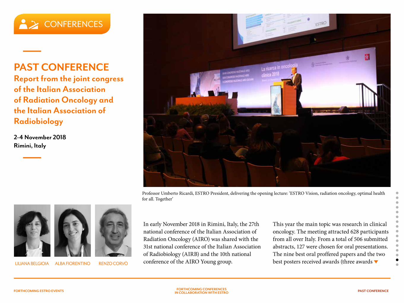
FORTHCOMING CONFERENCESIN COLLABORATION WITH ESTROFORTHCOMING ESTRO EVENTS PAST CONFERENCE
CONFERENCES
This year the main topic was research in clinical oncology. The meeting attracted 628 participants from all over Italy. From a total of 506 submitted abstracts, 127 were chosen for oral presentations. The nine best oral proffered papers and the two best posters received awards (three awards
In early November 2018 in Rimini, Italy, the 27th national conference of the Italian Association of Radiation Oncology (AIRO) was shared with the 31st national conference of the Italian Association of Radiobiology (AIRB) and the 10th national conference of the AIRO Young group.
PAST CONFERENCE Report from the joint congress of the Italian Association of Radiation Oncology and the Italian Association of Radiobiology
2-4 November 2018Rimini, Italy
LILIANA BELGIOIA RENZO CORVÒALBA FIORENTINO
Professor Umberto Ricardi, ESTRO President, delivering the opening lecture: ‘ESTRO Vision, radiation oncology, optimal health for all. Together’
FORTHCOMING CONFERENCESIN COLLABORATION WITH ESTROFORTHCOMING ESTRO EVENTS PAST CONFERENCE
from AIRO, two from Elekta, three from the Berlucchi Foundation, two from a patient making a grant to the AIRO Young group and one from AIRB).
On Friday morning two topics were discussed by the AIRO Young group: new ways to reduce toxicity and improve outcomes; and volume de-escalation, focusing on state-of-the-art and future perspectives. In the late afternoon, the conference was officially opened by Professor Stefano Magrini, AIRO President, Riccardo Santoni, AIRB President, and Alba Fiorentino, AIRO Young coordinator. Professor Umberto Ricardi, ESTRO’s President, delivered an interesting plenary lecture on the ESTRO Vision.
During the conference, multiple symposia addressed various topics, including organ-preserving radiotherapy, cardiovascular toxicity in survivorship, treatment of vulvar cancer, ablative radiotherapy in metastatic patients, and brachytherapy in prostate cancer.
Two particularly interesting sessions concerned the combination of radiotherapy and immunotherapy that underlies the molecular mechanism, dose, volume and timing of radiotherapy and the role of radiotherapy in elderly patients with lung, head and neck, and breast cancer.
The AIRB session developed the topic of how functional imaging could increase the safety of treatment and the identification of biomarkers as predictors of radiotherapy efficacy.
This year’s drug-lab session was dedicated to the management of the prescription and administration of anti-EGFR antibodies, anti-angiogenetic drugs, tyrosine kinase inhibitors (TKI) and immunotherapy. The lab contouring session focused on the definition of volume in head and neck tumours and upper abdominal stereotactic body radiation therapy (SBRT). These two sessions were very successful and sold out months before the conference.
The congress ended with a session providing updates on the activities of several AIRO disease-specific research groups.
Liliana BelgioiaAIRO grant recipient
Alba FiorentinoAIRO Young coordinator
Renzo CorvòChair of AIRO-AIRB scientific committee

CALENDAR OF EVENTS
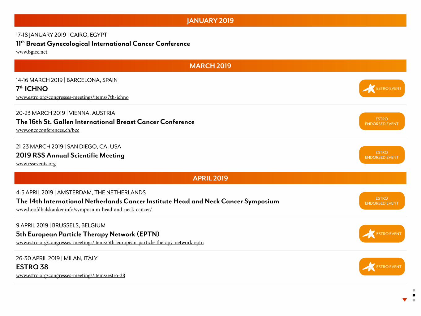
JANUARY 2019
17-18 JANUARY 2019 | CAIRO, EGYPT
11th Breast Gynecological International Cancer Conferencewww.bgicc.net
MARCH 2019
14-16 MARCH 2019 | BARCELONA, SPAIN
7th ICHNOwww.estro.org/congresses-meetings/items/7th-ichno
ESTRO EVENT
20-23 MARCH 2019 | VIENNA, AUSTRIA
The 16th St. Gallen International Breast Cancer Conferencewww.oncoconferences.ch/bcc
ESTROENDORSED EVENT
21-23 MARCH 2019 | SAN DIEGO, CA, USA
2019 RSS Annual Scientific Meeting www.rssevents.org
ESTROENDORSED EVENT
APRIL 2019
4-5 APRIL 2019 | AMSTERDAM, THE NETHERLANDS
The 14th International Netherlands Cancer Institute Head and Neck Cancer Symposiumwww.hoofdhalskanker.info/symposium-head-and-neck-cancer/
ESTROENDORSED EVENT
9 APRIL 2019 | BRUSSELS, BELGIUM
5th European Particle Therapy Network (EPTN) www.estro.org/congresses-meetings/items/5th-european-particle-therapy-network-eptn
ESTRO EVENT
26-30 APRIL 2019 | MILAN, ITALY
ESTRO 38www.estro.org/congresses-meetings/items/estro-38
ESTRO EVENT
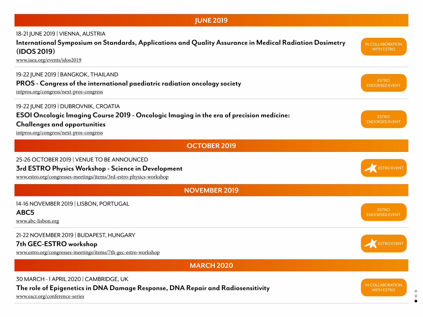
JUNE 2019
18-21 JUNE 2019 | VIENNA, AUSTRIA
International Symposium on Standards, Applications and Quality Assurance in Medical Radiation Dosimetry (IDOS 2019)www.iaea.org/events/idos2019
IN COLLABORATION WITH ESTRO
19-22 JUNE 2019 | BANGKOK, THAILAND
PROS - Congress of the international paediatric radiation oncology societyintpros.org/congress/next-pros-congress
ESTROENDORSED EVENT
19-22 JUNE 2019 | DUBROVNIK, CROATIA
ESOI Oncologic Imaging Course 2019 - Oncologic Imaging in the era of precision medicine: Challenges and opportunities intpros.org/congress/next-pros-congress
ESTROENDORSED EVENT
OCTOBER 2019
25-26 OCTOBER 2019 | VENUE TO BE ANNOUNCED
3rd ESTRO Physics Workshop - Science in Developmentwww.estro.org/congresses-meetings/items/3rd-estro-physics-workshop
ESTRO EVENT
NOVEMBER 2019
14-16 NOVEMBER 2019 | LISBON, PORTUGAL
ABC5 www.abc-lisbon.org
ESTROENDORSED EVENT
21-22 NOVEMBER 2019 | BUDAPEST, HUNGARY
7th GEC-ESTRO workshopwww.estro.org/congresses-meetings/items/7th-gec-estro-workshop
ESTRO EVENT
MARCH 2020
30 MARCH - 1 APRIL 2020 | CAMBRIDGE, UK
The role of Epigenetics in DNA Damage Response, DNA Repair and Radiosensitivity www.eacr.org/conference-series
IN COLLABORATION WITH ESTRO
ESTRO Bimonthly newsletterN° 122 | January - February 2019 European Society forRadiotherapy & Oncology
OFFICERSPresident: Umberto RicardiPresident-elect: Ben SlotmanPast-president: Yolande Lievens
EDITORIAL TEAMManaging editorCécile Hardon-Villard
Editorial coordinatorsEvelyn ChimfwembeMichela Mizzi
Sub-editorsEmma MasonNick Sarson
Graphic designersDaneel Bogaerts
Published every two months and distributedby the European Society for Radiotherapy& Oncology.
DEADLINES FOR SUBMISSION OF ARTICLES IN 2019March-April 2019 > 2 January 2019May-June 2019 > 2 March 2019July-August 2019 > 2 May 2019September-October 2019 > 2 July 2019November-December 2019 > 2 September 2019
For permission to reprint articles pleasecontact the editor.
If you want to submit articles forpublication, please contact:[email protected]
For advertising, please contact:[email protected]
ARCHIVELatest issues of the newsletter can be found on the ESTRO website under www.estro.org/about and older issues are accessible on DOVE, from the home page of www.estro.org.
Opinions expressed in the ESTRO newsletter do not necessary reflect those of the Society or of its officers.
CREDITS
Related Documents