6 | Cardiometry | Issue 8. May 2016 New theory of arrhythmia. Conceptual substantiation of arrhythmia mechanisms Vladimir I. Ermoshkin 1 1 Research-and-development center “RESONANS” Ltd. Russia, 107258, Moscow, 1 st Bukhvostova str. 12/11, building 20 * Corresponding author: phone: +7 (964) 506-95-50, e-mail: [email protected] Aims A new attempt is made to substantiate the concept of the mechanism of arrhythmia and sudden cardiac death. Methods The paper is based on a theoretical analysis of special literature, personal experience of participation in confer- ences and discussions with leading Russian cardiologists. Results We have succeeded in demonstrating the fact that researchers ignore the fact that cardiomyocytes can be ex- cited by mechanical pulses, when considering the arrhythmia mechanisms. We have conducted trials using the Cardiocode device. Under stress in a human, opened may be large and small arteriovenous anastomoses, via which blood under high pressure is ejected into veins. It leads to pressure surges in arteries and veins. The vena cava dilates, its tonus increases. In some cases, the pulse waves travel via anastomoses along the vena cava walls to the atria and the ventricles. An above-threshold concentration of tensions from mechanical pulses may excite cardiomyocytes from different points of the myocardium, disturbing the sinus rhythm. As a result, extrasystoles, tachycardia attacks, blocking of blood circulation in the peripheral segments of the venous arterial networks, edemata, thrombosis and metabolism disorders appear. Arrhythmia, tachycardia attacks and concomitant myo- cardial ischemia lead to progression of heart fibrosis. Such changes increase the probability of fibrillations and sudden cardiac death. Conclusions Unhealthy lifestyle, the presence of opening and not properly closing anastomoses may provoke a number of diseases. To avoid the cardiac arrhythmia attacks and prevent SCD, it is necessary to suppress travel of the me- chanical waves within the following circuitry: aorta – artery – anastomosis – vein – vena cava – atria – ventricles. The travel of the mechanical waves within the same vessel circuitry explains the fact that the fixed couplings under extrasystoles are observed, and the beat-to-beat RR intervals under tachycardia remain constant. Obviously, we may consider that the reentry mechanism is rather of biomechanical, but not of bioelectrical nature, as it is gen- erally accepted. Keywords Arrhythmia, Sudden cardiac death, Cardiomyocytes, Arteriovenous anastomosis, Reentry Imprint Vladimir I. Ermoshkin. New theory of arrhythmia. Conceptual substantiation of arrhythmia mechanisms. Cardiometry; Issue 8; May 2016; p.6–17; doi: 10.12710/cardiometry.2016.8.617; Available from: http://www.cardiometry.net/issues/no8-may-2016/new-theory-of-arrhythmia OPINION Submitted: 1 February 2016 Accepted: 11 March 2016 Published online: 23 May 2016

Welcome message from author
This document is posted to help you gain knowledge. Please leave a comment to let me know what you think about it! Share it to your friends and learn new things together.
Transcript
6 | Cardiometry | Issue 8. May 2016
New theory of arrhythmia. Conceptual substantiation of arrhythmia mechanisms Vladimir I. Ermoshkin1
1 Research-and-development center “RESONANS” Ltd. Russia, 107258, Moscow, 1st Bukhvostova str. 12/11, building 20* Corresponding author:
phone: +7 (964) 506-95-50, e-mail: [email protected]
AimsA new attempt is made to substantiate the concept of the mechanism of arrhythmia and sudden cardiac death.
MethodsThe paper is based on a theoretical analysis of special literature, personal experience of participation in confer-ences and discussions with leading Russian cardiologists.
ResultsWe have succeeded in demonstrating the fact that researchers ignore the fact that cardiomyocytes can be ex-cited by mechanical pulses, when considering the arrhythmia mechanisms. We have conducted trials using the Cardiocode device. Under stress in a human, opened may be large and small arteriovenous anastomoses, via which blood under high pressure is ejected into veins. It leads to pressure surges in arteries and veins. The vena cava dilates, its tonus increases. In some cases, the pulse waves travel via anastomoses along the vena cava walls to the atria and the ventricles. An above-threshold concentration of tensions from mechanical pulses may excite cardiomyocytes from different points of the myocardium, disturbing the sinus rhythm. As a result, extrasystoles, tachycardia attacks, blocking of blood circulation in the peripheral segments of the venous arterial networks, edemata, thrombosis and metabolism disorders appear. Arrhythmia, tachycardia attacks and concomitant myo-cardial ischemia lead to progression of heart fibrosis. Such changes increase the probability of fibrillations and sudden cardiac death.
ConclusionsUnhealthy lifestyle, the presence of opening and not properly closing anastomoses may provoke a number of diseases. To avoid the cardiac arrhythmia attacks and prevent SCD, it is necessary to suppress travel of the me-chanical waves within the following circuitry: aorta – artery – anastomosis – vein – vena cava – atria – ventricles. The travel of the mechanical waves within the same vessel circuitry explains the fact that the fixed couplings under extrasystoles are observed, and the beat-to-beat RR intervals under tachycardia remain constant. Obviously, we may consider that the reentry mechanism is rather of biomechanical, but not of bioelectrical nature, as it is gen-erally accepted.
KeywordsArrhythmia, Sudden cardiac death, Cardiomyocytes, Arteriovenous anastomosis, Reentry
ImprintVladimir I. Ermoshkin. New theory of arrhythmia. Conceptual substantiation of arrhythmia mechanisms. Cardiometry; Issue 8; May 2016; p.6–17; doi: 10.12710/cardiometry.2016.8.617; Available from:http://www.cardiometry.net/issues/no8-may-2016/new-theory-of-arrhythmia
OPINION Submitted: 1 February 2016 Accepted: 11 March 2016 Published online: 23 May 2016

Issue 8. May 2016 | Cardiometry | 7
IntroductionThis article attempts to look at the
global problem of high cardiac mor-tality of people from a fresh point of view. It is no secret that in the official cardiology, especially during the last 50 years, there have been different trends in approaches to the solution of this important problem. In addi-tion, at the turn of 20-21 centuries, in the opinion of some Western cardiol-ogists, arisen has a crisis of ideas on the development of cardiology and arrhythmology. Now, in the majority of complex cases, often doctors re-commend RFA and ICD, i.e. radiof-requency ablation and implantable cardioverter-defibrillators. Practical world medicine teaches people to think, that to completely solve some of the problems and significantly in-crease the life expectancy in the short term is impossible. According to WHO estimates, by 2030 from car-diovascular disease (CVD) will die per year approximately 23.6 million people (in 2012, 17.5 million), and the only major cause of death will remain heart diseases. But, apparent-ly, there is another way of cardiolo-gy development and related medical disciplines. This promising way will be discussed in this article.
Materials and methodsFinding information via Internet
about possible causes of arrhythmias, comorbidity for certain diseases, studying classical and contemporary primary relevant sources, partici-pation and presentation at medical conferences in 2012-2015, discus-sions of theoretical and clinical is-sues with health care experts: Rus-sian Academician Yevgeny Chazov, Russian Academician Leo Bokeria, Russian Academician Amiran Re-vishvili, Professor E. Shirokov, Doc-
tors A. Arutyunov, V. Frolov, V. Luk-yanchenko, M. Rudenko and others laid the groundwork for the materials and methods of this paper. In fair-ness, it should be noted that not all of the above experts currently fully support the basic idea of the theo-ry proposed by the author herein. During the discussions, I was asked a number of fundamental questions to which, I think, I managed to find eligible logical answers.
ResultsGenerally speaking, the current results
achieved in the theory and practice of cardiology and arrhythmology pro-voke objections not only from my side. Criticism has been expressed by many experts, when assessing the progress in these fields in medicine. The objective criticism is confirmed by the data on the Nobel Prize awards. Only two Nobel Prizes were granted to cardiologists for 115 years: in 1924 for the discovery of the mechanism of the ECG (W. Einthoven) and in 1998 for the discovery of the role of nitric oxide as a signaling molecule in the regulation of the cardiovas-cular system (L. Ignarro et al.). Just to compare: Genetics scientists were awarded with Nobel Prize 48 times!
But how to expedite and advance the process of definite solving of the mystery attributed to CVD? How to break the deadlock, when due to no apparent reasons, often suddenly, people die and premature mortality is growing? Perhaps something was missed? Maybe, we need to recon-sider some of the scientific achieve-ments and the reported “discoveries” in cardiology? Where is the solution to this problem?
In 2010-2016 I probably managed to find ways to solve this problem: my new theory of arrhythmia (NTA) was
born in those years. In the process of studying the primary sources of car-diology and searching for causes of hypertension and arrhythmia mech-anisms, I was able to show that in the theoretical cardiology there has been a critical error at least for the last 100 years. The error is that after the ECG invention, for some mysterious rea-sons, for many years researchers have suddenly forgotten that myocardial excitation can be provided by me-chanical pulses. But even in the late 19th century it was known that the muscle fibers and cardiomyocytes (CMC) could be excited by a vari-ety of physical effects: by pulses of mechanical, electrical, chemical and thermal nature...
To our much regret, almost every-body has forgotten the “mechanics” in the heart performance. But several groups of scientists still stand out, in particular, Professor A.G. Kamkin’s team, which upon special studies in 1985-2002 has made some far-reach-ing conclusions [1]: “The cardiomy-ocytes and the fibroblasts to an even greater extent efficiently convert me-chanical stimulation into electrical responses, and as this takes place, the performance of the first sort of the cells (cardiomyocytes) is modulated by the second one (fibroblasts).” Un-fortunately, those theoretical and ex-perimental conceptual findings were not used, in my opinion, by a reason as follows: nobody dared or has seen any possibility of “how to apply it”.
At present, electrocardiography has become a standard routine pro-cedure, where doctors are trying to find answers to many questions by interpreting standard-type ECG tapes. In my opinion, unreasonably believed is that nearly all rhythm ab-normalities are associated with dis-turbances in the performance of the
8 | Cardiometry | Issue 8. May 2016
cardiac conduction system (CCS), with deviations of its conductivity and automaticity [2-4]. It is stated that the causes of arrhythmia are re-ported to be by the dozens, and the arrhythmia factor list is known by all the doctors, and there is no need to discuss this rather trivial list herein. I would like to stress here, not ar-rhythmia causes are listed there, but following NTA, they are treated to be factors which change the tonus of the CVS and lower threshold of the heart rate generation mainly due to mechanical excitations. In addition, the official medicine believes that there is not one “mechanism” of heart arrhythmias, but they are rather sev-eral ones available. Summing all the arguments of researchers, we get “a set” of “mechanisms”: macro- and micro-“re-entry”, increased automa-ticity of P-cells, trigger activity, the presence of ischemic areas in ische-mic heart disease, atherosclerosis, the presence of irregular structures in the form of necrosis, the blockade of conductivity, the presence of ecto-pic foci, the presence of “autowaves of excitation in active media, i.e. in the myocardium.” For each type of arrhythmia there are a specific “the-ory” and a “mechanism” available. Every “Cardiac School” offers its own particular treatment. Natural-ly, under such a “strict and accurate” diagnostics of arrhythmias, drugs are randomly chosen from a rec-ommended list of approved medi-cations. The only rule has to be ap-plied: if a drug does not produce an effect, it must be changed, and that is the case throughout the life of an arrhythmia patient. But it should be noted that an abundance of theories and hypotheses, existing in any sci-ence, bears witness to the fact that it is actually dead end therein.
I pioneered to show that (except when some specific cases of patho-logy area considered) the nature and the mechanism of all known forms of arrhythmia up to ventricular fibril-lation (VF) is always the same (IT’S THE ONLY ONE!). It is a superpo-sition of bioelectrical and “forgotten for 100 years “above-threshold me-chanical pulses, in amounts of one up to three excitation pulses “simultane-ously” [5] on condition that the ho-meostasis changes within predeter-mined limits. It is known that CMC can be equally effectively excited both by bioelectrical and mechanical pulses [6]. It is believed that at the apex of the heart, or anywhere in the myocardium in general, life-threat-ening “ectopic foci” of excitation may be formed, but the mechanism of this phenomenon has never been under-stood by most patients and physi-cians [7].
I assumed that mechanical pulse waves, which reach the atria from the vena cava in a certain manner, can further travel on the sphere-shaped heart (the travel is facilitated espe-cially in the case of heart affected by fibrosis), with their further focusing, for example, at the point of “an an-tipode” relative to the mouths of the veins, i.e. at the points close to the apex of the heart. It should be borne in mind that, in general, mechanical pulses in an elastic medium can be reflected from the walls, from necrot-ic irregularities and cavities; they can propagate in the myocardium tissue regardless of the excitation phase of the tissue refractoriness, but electri-cal pulses are not capable of doing so. Electrical excitation can suddenly break off his path in collisions with zones of refractoriness, for example, when two waves traveling towards each other are colliding. Now it be-
comes clear why the sudden exci-tations of CMC (of mechanical-sen-sitive nature) can suddenly appear at any point in the heart, especially in the presence of cardiac fibrosis, or in the presence of coronary artery disease, because the fibroblasts “net-work” is a higher quality “conductor” of mechanical waves in comparison with the “soft” myocardium, consist-ing mainly of young CMC. I stress that early unexplained excitations (except for the cases of the presence of additional pathways) always pro-duce confusion for researchers, be-cause early excitations sometimes may result not from the current QRS, but, according to NTA, from an earli-er beat of heart.
If there are several excitation sour-ces simultaneously available (the total of the mechanical and electri-cal pulses), fibrillations either of the atria or of the entire myocardium ap-pear, depending on the degree of fi-brosis, cardiomyopathy and points of “simultaneous” excitation. Of partic-ularly danger is ventricular fibrilla-tion (VF), since when erratic, chaotic ventricular excitations are available, they impair the pumping function of the heart that leads in most cases to sudden cardiac death (SCD). During period of attacks of heart fibrillation (VF) at each fixed time the heart conditionally is divided into separate independent sections with different phases of excitation with arbitrarily changing boundaries between them. According to NTA, the average size of the independent zones of exci-tation is defined by two parameters: the averaged interval refractoriness of CMC and the averaged speed of propagation of mechanical waves within the myocardium. It is easy to guess that due to very small metric sizes of hearts the number of inde-
Issue 8. May 2016 | Cardiometry | 9
pendent parts of the heart excitations cannot be more than one: therefore in small-sized animals (puppy dogs, cats, rabbits, rats, guinea pigs) and small birds fibrillation cannot be generated at all [8], and they cannot be killed by electrical shocks, as it is the case with a human or an ele-phant! Before creation of NTA, sci-entists tried to explain this paradox by some physiological features (?!) of the myocardial structure of hearts in small animals. But it turns out, the reason is of another nature, and NTA gives the definite answer how to properly interpret this phenomenon.
Obviously, the most effective healthy heart function is provided when ex-citations are initiated only by the car-diac conduction system (CCS). CCS generates sinus rhythm under proba-bly existing sub-threshold mechanical stimuli of CMC. I have shown [5] that numerous variants of the superpo-sitions of excitations lead to a great variety of arrhythmias: extrasystoles and tachycardia (atrial or ventricu-lar), allorhythmia (mainly bigemia, trigeminia), atrial and ventricular fibrillation. In the presence of necro-tic areas in the myocardium detected are even “more sorts” of arrhythmia according electrocardiogram (ECG) and electrophysiology study (EPS) so that it makes much more difficult to classify the said rhythm abnormal-ities. In particular, the question of the mysterious stability of length of RR-intervals (on ECG) during an at-tack of paroxysmal ventricular tachy-cardia has found its logical explana-tion: because there is a stable time of travel of the pulse wave along the path “the aorta - the artery – the anas-tomosis AVA – the vein (veins) – the vena cava – the atria - the myocardi-um”, with the floating or slowly drift-ing focus point of maximum stress
(ectopic foci) in the apex of the heart. Well, of course, with this initiation of the wave of mechanical-electrical excitation of CMC, the wave travels not across the “fast” CCS, but accord-ing to the principle of propagation of mechanical waves “from cell to cell.” This is the reason the ventricular QRS waves are extended in time and may change their polarity!
An additional evidence in favor of the fact, that premature firing of ex-citations is of mechanical nature, is that many experts in cardiac murmur auscultations detect the presence of unexplained pulse waves near the 3rd heart sound before ES. “In case of physical examination, the fact of a premature heart contraction is usual-ly easy to establish. When examining the arterial pulse, some prematurely appearing pulse waves of small am-plitude are found which correspond to extrasystole.”
“There exist a mechanic systole and an electrical one, and the latter does not correspond to the mechanic sys-tole. The 3rd sound may be detected in 20% of healthy individuals, but more often it is found in ill patients. It is recorded at the beginning of di-astole, not earlier than 0.12 sec. after the 2nd sound. The pathological 3rd sound forms the triple rhythm. It ap-pears as a result of rapid relaxation of the ventricle muscle fibers which have lost their tonus, when blood is rapidly entering the ventricles. It is as if the heart were crying for help, or it is a gallop rhythm.”
Thus, the CMC mechanical ex-citation is initiated either by the mechanical wave, passing through AVA along the vessel walls, or by the mechanical impact of the blood flow acting on the heart apex during opening of the tricuspid valve, or by both factors acting together.
Let’s take a closer look at the ques-tion of how abnormal mechanical pulses, generated by the heart and the aorta, can pass through the ves-sels and reach the atria or the ven-tricles from the back side (as viewed from the atria). Previously such ideas have never been offered because it has been considered to be generally impossible.
2010 the author hereof has proposed a concept that in order to protect arter-ies from high blood pressure, not only small-scale precapillary anasto moses (including glomura), but also large arteriovenous anastomoses (AVA) should be opened periodically [9].
AVAs are unique natural shunts, connecting (joining) directly the ar-terial and venous basins under cer-tain conditions. It can be assumed that when a human individual is healthy, demonstrates an acceptable training condition, when the daily prevailing arterial blood pressure is close to normal, the large AVAs are closed. Otherwise, when the arterial blood pressure at the local level ex-ceeds a specified norm for a certain time, the AVAs may be opened. So far an exact algorithm of AVA regulating (by an organism) is unknown in de-tail, but hopefully it will be revealed and defined in the nearest future. Apparently, the AVA performance depends on many factors: duration of hypertension disease, the degree of degenerative disc disease, actual arterial pressure at a specified time, intensity of the physical exercise load, the state of “deterioration” and remodeling of the cardiovascular sys-tem, myocardial hypertrophy, the de-gree of atherosclerosis, the presence of plaques, the degree of coronary artery disease and myocardial fibro-sis, the stress conditions, the current stomach performance status, obesity,
10 | Cardiometry | Issue 8. May 2016
smoking & alcohol factors, the cur-rent patient’s posture and some other conditions.
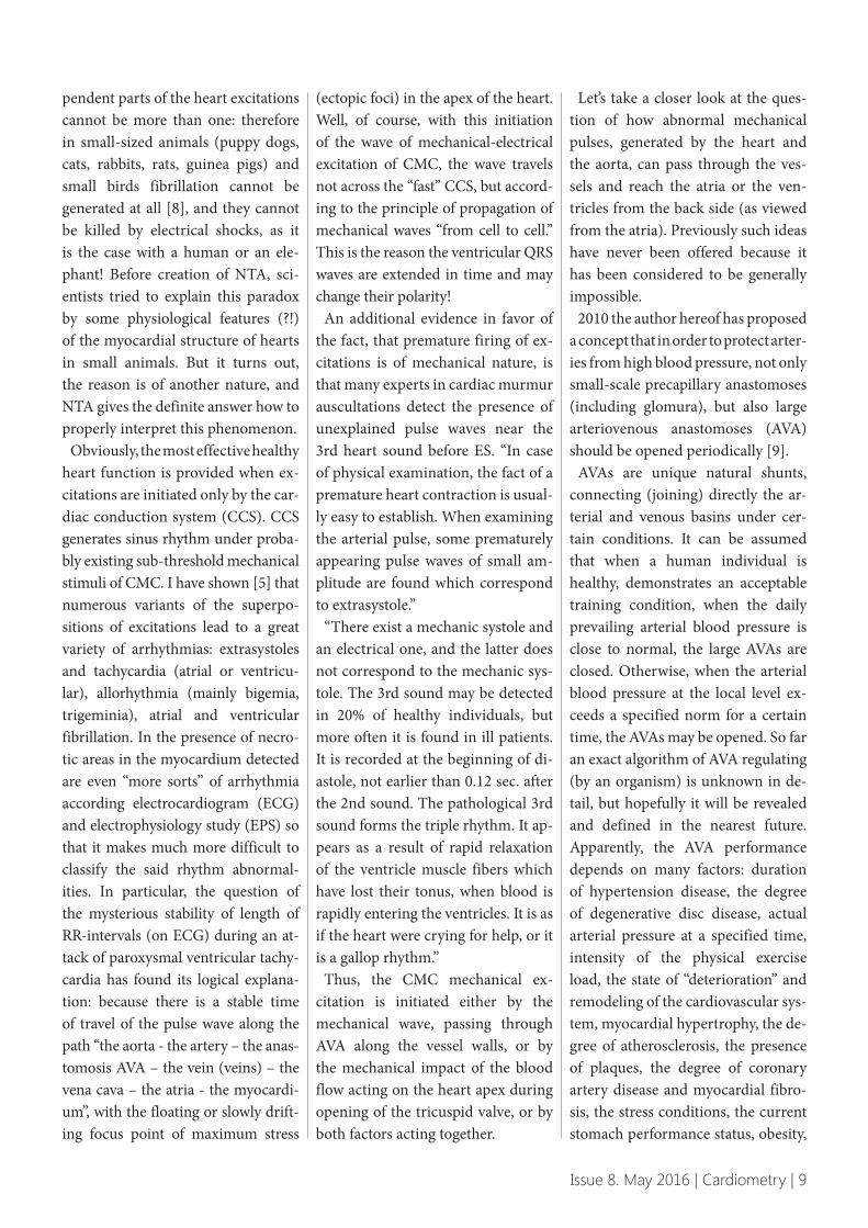
The first targeted searches for car-diometrical markers indicating ana-tomical and physiological rearrange-ment of the vascular system (opening / closing of the AVA) brought success. Using the original hemodynamic analyzer “Cardiocode” in 2015 Luk-yanchenko V.A. and Rudenko M.Y. with my participation have detected, that some specific complaints like episodes of dizziness in patients are related to a pressure amplitude fall in the aorta (for example, from 150/90 to 100/80 mm Hg or less). The said data have been derived by digital pro-cessing of actual ECG and Rheogram curves. Fig. 1 shows ECG & RHEO segments in the same patient demon-strating such pressure fluctuations both in the aorta and the entire sys-tem due to opening / closing of arte-riovenous anastomoses.
Later, the effect of “surges in arteri-al blood pressure” within short-time intervals (up to 3-10 heart beats) was named after their discoverers “the Er-moshkin-Lukyanchenko syndrome”. But the most important thing is not that periodically dizziness, syncope and “abrupt changes in blood pres-sure” occur. The most critical mat-ter is the fact that such cardiometric re-setting of the organism parameters provokes more dangerous changes of two types of cardiovascular diseases.
The first dangerous change in the CVS performance
With periodic opening of AVA, blood from the arteries at a high pressure, for example, at a pressure of 130-150/85 mm Hg, is entering the large and small veins. Then blood en-ters the vena cava, where the normal pressure should be considerably low-
er, reaching about 0-10mm Hg. Due to a high pressure drop, blood, being intensively discharged via AVA into the vena cava, is mixed with venous blood, fills the veins to their limit, deforms with time venous valves, rises for some minutes or hours the systemic venous pressure up to a level significantly higher than the normal one, for example, in some veins up to 40 mm Hg.
The walls of the overfilled major veins are stretched and change their mechanical properties; they become solid and elastic (similar to the wall of a child balloon inflated with a gas). These changes create a possibility for arterial pulse waves to travel via AVAs along the walls of the hollow veins to the heart. An evidence for the presence of large AVA is based on the following clinical observation. It is assumed by me that the pulse wave at some point of time, upon filling of the hollow veins, starts propagating in a closed loop of the blood ves-sels comprising the following items: the left ventricular myocardium, the aorta, the large artery, AVA, the portal veins, the vena cava and the atria. In its turn, the mechanical ac-tion on the myocardium can lead to extraordinary, premature excitations of the cardiomyocytes. It triggers atrial or ventricular extrasystoles, tachycardia and some other types of
arrhythmia. For example, tachycar-dia occurs when the mechanical ac-tion coincides exactly with the center point of the RR-interval on ECG, and in this case heart rate (HR) changes sharply by 2 times, for example, from 70 to 140 beats per minute. Power of pulse wave traveling via the overfilled veins depends on the specific struc-tural features of the venous bed in a patient or on rising head pressures in the veins: under the conditions, pa-tients report either no symptoms at all, or some patients report pulsations in the ears, the abdomen, the legs, the throat, the neck or even throughout the entire body.
Taken into account such a descrip-tion of “the syndrome of AVA open-ing”, some doubts are cast upon the validity of the concept interpreting the phenomenon of “bioelectrical reentry” in the myocardium, gen-erally accepted by the conventional medicine. In accordance with the conceptual NTA philosophy, “reen-try” of cardiac excitation is primarily of mechanical, but not of bioelectri-cal nature, as erroneously believed for more 50 years. The support in favor of the fresh interpretation of “reentry” are numerous reports by patients that they feel pulsation in their abdomen area, their solar plex-us area, and sometimes patients note cramps in the area around the navel
Figure 1. Sharp fluctuations in the ECG shape and Rheogram (below): cardio metric signs of remodeling system detected by CARDIOCODE technique
Issue 8. May 2016 | Cardiometry | 11
and even can observe visible beats in the blood vessels under the skin. As a rule, after pulsations, colics and ab-dominal cramping, i.e. probably after the opening of AVA, cardiac rhythm abnormalities occur. But in most cas-es, doctors come to their standard findings in making diagnoses like vegetative-vascular dystonia (VVD), panic attacks (PA), etc. In doing so, as a rule, they treat their patients based on their classical ECG interpretation, some blood tests, standard medica-tions, but all this without any favor-able therapy effect. Below offered are a number of descriptions of fearful syndromes reported by acutely af-fected patients.
If we think that opening / closing of AVA in many cases is accompanied by unpleasant sensations because of, for example, significant pressure fluctuations in the hepatic, the supe-rior mesenteric artery, as well as in the portal vein and in the vena cava. Most often, AVAs can be opened af-ter a heavy meal, or after drinking alcohol, coffee, or after eating fatty food, or when being squeezed, and these observations are recorded by a lot of patients and physicians; some other cases associated with the above phenomenon are attributed to the presence of liver or gallbladder dis-eases, even at their early stages. For example, famous Russian doctor Bot-kin S.P., from his young age, suffered of arrhythmia which appeared with abdominal cramps. In his research reports [10] he emphasized, that pain and other changes in the heart might occur even without abdominal pains. “We obtained evidence in favor of assumption that a lethal outcome is possible in a patient, even when such events are not accompanied by pains, when colics are not reported, but when, according to our strict analysis
of the complete disease picture, we have no slightest doubt that the cause of the death were concretions which produced irritation of the nerve appa-ratus, integrated in the ducts, so that it triggered a number of reflex chang-es in the heart or in the central ner-vous system.” From my standpoint, the narrative by Botkin S.P. should be completed by the description of the phenomenon of periodical opening of AVA. Without the AVA opening description the relationship between abdominal cramps and cardiac ar-rhythmias looks unconvincing.
“However, we should remember that the heart pains might be the only manifestation of pathology of the gallbladder ... Possible is also a pain-free form of the syndrome of cholecystitis coexisting with car-diovascular disease”, when the only manifestation of cholecystitis is a heart rhythm abnormality. Car-diac arrhythmias in combination with cholecystitis, according to the available reference source data, are found in 16,2-21,8% of the patient cohort [11].
The characteristic feature of the dual pathology implying the gall bladder disorders in combination with arrhythmias is that arrhythmia is appearing with developing biliary colic; it results in low efficiency of the conventional antiarrhythmic therapy and conversion of arrhythmia into an unfavorable form in case of acute inflammation of the biliary tract and biliary hypertension progression.
Of particular note is that arrhyth-mias are found not only in case of the above pain-free form disease, but often they are combined with pains in the heart and the sternum. Many clinicians focusing on biliary tract pathology notice the appearance of such symptoms like tachycardia, in-
creased pulse and venous pressure; occasionally revealed are symptoms of hypertension in the pulmonary circulation. In my opinion, this pa-thology picture should be completed by the description of the initiating role of the performance of AVA, for example, between the superior mes-enteric artery and the portal vein.
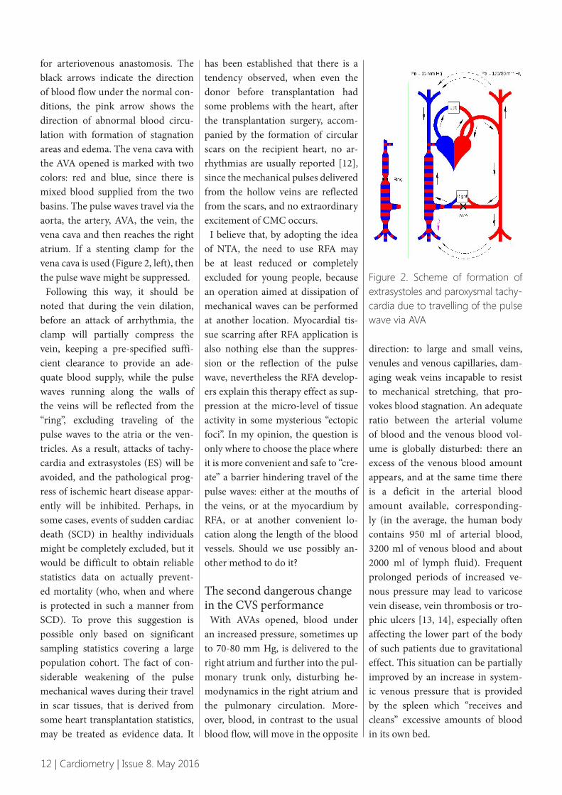
So, if my proposed theory is true, then it should become clear: in order to prevent traveling of a pathological pulse wave to the myocardium, it is required to use a method to dissi-pate, weaken, “extend” the timeline and “round-off ” sharp cutting edges of mechanical waves at any conve-nient location along the length of the vascular circuit. For example, in the superior mesenteric artery, the most likely artery capable of conducting pulse waves, an arterial stenting may be provided, or the preventive mea-sure can be taken by suppressing the same wave in another location within the AVA or in the vena cava, or even immediately at the heart by RFA, or any other technology to “kill” the normally functioning cardiomyo-cytes. With regard to the suppression of the wave in the vena cava, it seems possible to make it easy by the surgi-cal performance of “the circumferen-tial seam” in the vena cava or using latex litigation of the veins. I believe that there is a large field opened for inventors of medical techniques and devices which may be utilized against arrhythmias. Also, I have proposed vein stenting with use of the stent de-vice in the form of a ring, a clamp to be loosely placed around the circum-ference of the vena cava. Figure 2 dis-plays schematically systemic and pul-monary circulation in a human. Letter “L (Left)” stands for lungs, letters “LG (Right)” denote the liver and the gall-bladder, and AVA is an abbreviation
12 | Cardiometry | Issue 8. May 2016
for arteriovenous anastomosis. The black arrows indicate the direction of blood flow under the normal con-ditions, the pink arrow shows the direction of abnormal blood circu-lation with formation of stagnation areas and edema. The vena cava with the AVA opened is marked with two colors: red and blue, since there is mixed blood supplied from the two basins. The pulse waves travel via the aorta, the artery, AVA, the vein, the vena cava and then reaches the right atrium. If a stenting clamp for the vena cava is used (Figure 2, left), then the pulse wave might be suppressed.
Following this way, it should be noted that during the vein dilation, before an attack of arrhythmia, the clamp will partially compress the vein, keeping a pre-specified suffi-cient clearance to provide an ade-quate blood supply, while the pulse waves running along the walls of the veins will be reflected from the “ring”, excluding traveling of the pulse waves to the atria or the ven-tricles. As a result, attacks of tachy-cardia and extrasystoles (ES) will be avoided, and the pathological prog-ress of ischemic heart disease appar-ently will be inhibited. Perhaps, in some cases, events of sudden cardiac death (SCD) in healthy individuals might be completely excluded, but it would be difficult to obtain reliable statistics data on actually prevent-ed mortality (who, when and where is protected in such a manner from SCD). To prove this suggestion is possible only based on significant sampling statistics covering a large population cohort. The fact of con-siderable weakening of the pulse mechanical waves during their travel in scar tissues, that is derived from some heart transplantation statistics, may be treated as evidence data. It
has been established that there is a tendency observed, when even the donor before transplantation had some problems with the heart, after the transplantation surgery, accom-panied by the formation of circular scars on the recipient heart, no ar-rhythmias are usually reported [12], since the mechanical pulses delivered from the hollow veins are reflected from the scars, and no extraordinary excitement of CMC occurs.
I believe that, by adopting the idea of NTA, the need to use RFA may be at least reduced or completely excluded for young people, because an operation aimed at dissipation of mechanical waves can be performed at another location. Myocardial tis-sue scarring after RFA application is also nothing else than the suppres-sion or the reflection of the pulse wave, ne vertheless the RFA develop-ers explain this therapy effect as sup-pression at the micro-level of tissue activity in some mysterious “ectopic foci”. In my opinion, the question is only where to choose the place where it is more convenient and safe to “cre-ate” a barrier hindering travel of the pulse waves: either at the mouths of the veins, or at the myocardium by RFA, or at another convenient lo-cation along the length of the blood vessels. Should we use possibly an-other method to do it?
The second dangerous change in the CVS performance
With AVAs opened, blood under an increased pressure, sometimes up to 70-80 mm Hg, is delivered to the right atrium and further into the pul-monary trunk only, disturbing he-modynamics in the right atrium and the pulmonary circulation. More-over, blood, in contrast to the usual blood flow, will move in the opposite
direction: to large and small veins, venules and venous capillaries, dam-aging weak veins incapable to resist to mechanical stretching, that pro-vokes blood stagnation. An adequate ratio between the arterial volume of blood and the venous blood vol-ume is globally disturbed: there an excess of the venous blood amount appears, and at the same time there is a deficit in the arterial blood amount available, corresponding-ly (in the average, the human body contains 950 ml of arterial blood, 3200 ml of venous blood and about 2000 ml of lymph fluid). Frequent prolonged periods of increased ve-nous pressure may lead to varicose vein disease, vein thrombosis or tro-phic ulcers [13, 14], especially often affecting the lower part of the body of such patients due to gravitational effect. This situation can be partially improved by an increase in system-ic venous pressure that is provided by the spleen which “receives and cleans” excessive amounts of blood in its own bed.
Figure 2. Scheme of formation of extrasystoles and paroxysmal tachy-cardia due to travelling of the pulse wave via AVA
Issue 8. May 2016 | Cardiometry | 13
The conditions can be compared with an inflation of a football ball having a small hole and showing air leakage. Using a damaged ball in football is ineffective, and the same case is with a human individual when the organism attempts to provide the proper performance with AVA opened, under overloading condi-tions, showing no efficiency and de-stroying generally its own biological system.
Destruction of the veins due to the opening AVAs, where the small-sized veins are affected, is clearly demon-strated in hypertensive patients in their fundus photographs [15]: found are usually increased diameters of veins, vessel tortuosity, stasis, hya-linosis of the vessel walls and some other catastrophic changes. Similar pathology may occur in any organ, since, according to NTA, small or large AVAs can exist in almost all or-gans in the organism.
I suppose, when AVAs are opened in the cardiovascular system of the brain (or even in other internal or-gans), due to the overflow of large venous vessels and an increase in the system pressure, severe pains in the occipital region of the head may oc-cur, and possible are also headache attacks of migraine type.
The proposed theory agrees well with the following research data [16]. The authors write: “The most hot topics are studies on a variety of mechanisms, that lead to the deve-lopment of cerebrovascular diseases, as well studies on pre-stroke forms of cerebrovascular pathology, in partic-ular chronic ischemia of brain: dis-circulatory encephalopathy (DE). DE involves a slow progression of mul-tifocal or diffuse vascular da mage of the brain as well as a combined form thereof. This is a consequence of var-
ious diseases caused by a great vari-ety of factors, showing the common feature: lesion of the small arteries and arterioles (microangiopathy). The clinical picture of the various forms of DE depends not only on disorders in the performance of the arterial system in the brain, but also to a large extent on the failure in the performance of the venous circula-tion [17]. Step by step, angioneurol-ogy is moving towards accepting a concept of venous dysfunction, when considering the formation of vascu-lar brain pathology. Disorders of ve-nous circulation in DE follows from the fact that the arterial system and that of the veins form in the brain their functional integrity… Until the present time, studies of the venous component of the cerebral circula-tion lagged behind the research of ar-terial hemodynamics. Difficulties in diagnostics of venous circulation dis-orders of the brain in living subjects are attributed to the wrong concepts supported by many clinicians, stress-ing the rarity of the said diseases, and, consequently, of minor signifi-cance of their studies ... According to some research data, in 71.5% of the patients with arterial hypertension (AH) ultrasonic examination detects compression of the internal jugular, the brachiocephalic and the vertebral veins, and in some patients (with ve-nous hypotonia) reported are insuffi-ciency of valves at the mouths of the veins and structural anomalies of the veins. According to MR venography, signs of cerebral venous outflow in patients with severe malignant hy-pertension are found in 91% of ca-ses, and for patients with hyperten-sion of the 1st and 2nd degree the signs are reported in 55% of cases. (I would like to add: it is evident from the above description of DE that the
authors are ignoring a link between abnormalities of the arterial and the venous basins, namely, the large- and small-sized AVAs.)
So, again, as a result of periodical AVA opening procedures, the nor-mal blood circulation within any cir-culation segment or within the entire system is stopped, the tissue pressure necessarily increases, and the in-crease is performed exactly accord-ing to the laws of hydrodynamics: the amount of intercellular fluid increas-es, because circulatory blockage oc-curs; swelling takes place in the liver and the gallbladder, so that in some cases, due to poor circulation, an ab-normal swelling of the body appears. However, in such periods of time, a certain portion of the working cells are starving, since blood circulation via the artery and the arterial capil-lary network is almost interrupted, and a considerable portion of the oxygenated red blood is supplied not into the arterial capillary network upon passing AVA, but it is deli-vered into the veins immediately, af-ter the capillary circulation stoppage, with long periods of organ ischemia. Thus, a certain portion of the blood circulates not through the systemic loop, but via circuits shortened by AVA, or, in more exact terms, via semi-circuits. A partial restoration of the proper blood circulation, appar-ently, takes place only in a horizontal position at night during sleep or after special exercises, sauna or massage. However, due to untrained muscles and a sedentary lifestyle, some cellu-lar waste products and unprocessed lymph fluid, containing cellular de-bris, are accumulated in the intercel-lular fluid, in the joints, partly in the cells of some body parts located most distant from the heart and the aorta, among them human skin. Gradually,
14 | Cardiometry | Issue 8. May 2016
the cellular waste product accumula-tion in tissues reaches certain limits, since the lifestyle of some individuals has not changed for years and decades.
In the course of time, owing to the presence of opened (and not closed in time) AVAs, the right heart failure may appear, and later, or even at the same time, the left heart one may de-velop. Thus, it is logical to suppose that the cause of heart insufficiency, abnormal performance of the heart valves and the pulmonary heart is in the majority of cases not the heart it-self, but the cause explaining thereof is the presence of AVAs and an im-proper performance of the latter. In the above mentioned cases, the ve-nous pressure significantly increas-es, and blood circulation is blocked in small arteries and veins like a hy-draulic gate valve (lock). We see a stalemate when the heart has to adapt to an increased venous pressure that leads to progression of many diseases at once during a short period of life. It may be the cause of the change in acid-base balance pH in blood and tissues, and it may be responsible for the metabolic syndrome (MS) and comorbidity.
More than 50 years ago, prominent Russian physician Zalmanov A.S. (1875-1965) came to the same con-clusion: in his opinion, myocardium diseases are very often secondary to the above mentioned ones: “Tissue and cellular hypoxia may occur de-spite an excellent state of the cardio-vascular system. When we deal with a patient suffering of a cardiovascu-lar disorder, we should never exam-ine only the heart and electrocardio-grams; other sources which may be responsible for hypoxia like the state of lungs, blood, diaphragm mobil-ity, liver and spleen volume should be checked, too, in order to approx-
imately identify the circulating blood volume.” [18]
At first glance, following the NTA logic, it seems that some way out would not be found, and no drugs and operations are capable to change the system, where cellular waste products are stored in tissues due to opening of AVAs and due to in-creased venous pressure, because of some trivial “metabolic disorders”, up to vein thrombosis and venous ulcers. Probably, it is impossible to surgically shut off AVAs and let red blood flow via small arteries. First, before such a hypothetical surgery, it should be required to identify and detect what AVA (AVAs) is (are) opened in a pa-tient, and that is not an easy task, and, secondly, a surgical AVA closure is nothing more than a very serious intervention into living organism settings of the arterial pressure level. Apparently, a surgical AVA blocking can lead to the maintenance of high systemic blood pressure and increase the likelihood of strokes or infarc-tions. In general, the AVA research topics call for further investigation. On the other hand, if to clean blood with the use of plasmapheresis, when large or medium-sized veins are in-volved, the scope of such cleaning of blood will not include the majority of the CVS peripheral areas, so that the most “debris-laden” thick blood will remain almost unprocessed, as it may be the case with human skin, including all adjacent areas. If to ap-ply a simple manual massage, then it should be used more often and cover the entire body area, since it is not excluded that cellular waste products disturbed at one place will be simply moved to another place throughout the body.
Fortunately, it has been detected that in addition to widespread manu-
al and can massage, plasmapheresis, warm water therapy, sauna, steam sauna, capillary therapy and mox-ibustion technique there are some other possibilities available, which also offer removal of thick stagnant venous blood and lymph in the pe-ripheral areas of the blood vessel network! They are blood-letting me-thods as follows: cupping (Arabian Hijama), simple cupping technique (or blood sampling) from veins and hirudotherapy (leech therapy).
Let us consider the three techniques of blood-letting.
1. Certainly, some people in the modern world have never heard about cupping as the “barbaric” treatment, but this sort of treatment has been using by the Arab world for many thousands of years. Moreover, advanced medical research confirms high therapeutic efficacy of cupping [19-21].
“Hijama (cupping) was utilized by medieval inhabitants in the Arabi-an desert for treatment of a number of diseases. For the technique used by the Arabs, used were sharp tools like needles. An incision was made within a particular area of the body, usually on the neck surface or the head. Then, an experienced individ-ual applied glasses or cups to dispel some blood in a patient. The Arabs believe that this therapy method is a good remedy against headache. His-torical evidence suggests that Pro phet Muhammed suffered from chronic migraine. He always had to resort to cupping to relieve his pain. Cupping had to be performed by qualified in-dividuals only, so that Muhammed had several with them ... Today, cup-ping is considered to be not in compli-ance with hygienic requirements and scien tific principles, and therefore it is treated as a high-risk life-threaten-
Issue 8. May 2016 | Cardiometry | 15
ing method. “The Messenger of Al-lah said: “Healing is in three things: a gulp of honey, cupping, and branding with fire (cauterizing).”
Another source states: “Hijama (skin incisions and vacuum glasses or cups in some areas of the body as recommended) is an ingenious way of getting rid of most of diseases sim-ply by removal of “dirty” blood from the body. But what is the essence and the secret of Hijama (cupping)? Us-ing the said blood-letting technique, we are able to initiate some stand-by recovery mechanisms in our organ-ism, which are responsible for gene-ration of fresh blood to improve the general conditions of a human indi-vidual and cure many diseases. Cup-ping is applied for prevention and treatment of the following diseases: prostatitis, back pain, impotence, low or high blood pressure, immune sys-tem weakness, narrowing or block-age of veins, pain in the neck or the shoulders, hemorrhoids, headaches, infertility, tuberculosis, skin diseases, heart pain (CVD), diabetes, urinary incontinence, muscle pains, numb-ness in extremities, rheumatism, body paralysis, poor vision, eye pressure, tearfulness, poor hearing, sinusitis, seizures, menstruation irregularities and so on... Thus, we should assume that there are two positive factors in cupping: the removal of “dirty” blood plus an initiation of stand-by recov-ery functions by the organism. The main feature of cupping is that there is no need at all to introduce any chemical agents into the human body or to perform any surgery interven-tions. This method involves internal reserves of the human body only, and efficacy of cupping is supported by a thousand-years’ experience.
2. In Europe in the Middle Ages, doctors and healers treated people
from all diseases with the use of ve-nesection. The European procedure of blood-letting shows an essential difference from Hijama. So, cupping (Hijama) provides withdrawal of blood according to the under-the-skin technique, i.e. the intercellular fluid is taken using vacuum through special incisions on the human body, and the suction effect of this proce-dure is distributed in the body to a depth of 6-8 cm, while according to the “European” blood-letting tech-nique blood was drawn usually from large veins opened. But according to the NTA concept, stagnated cell waste products are found in concen-trated form not in large, but rather in small and smallest veins and tissue fluid as well. We note here, that mod-ern donation, when venous blood is taken, is also a kind of blood-letting, but under the proper sterile condi-tions. Typically, people who donate blood on a regular basis report some health improvement after the dona-tion procedure.
3. In addition to cupping and vene-section, there is the third method of blood-letting available: it is leeching, or hirudotherapy [22-25]. Hirudo-therapy applications were reported even thousand years BC. Medical leeches were used for treatment of Egyptian Pharaohs; leeching therapy is mentioned both in the Bible and the Koran. Later, great physicians Hippo-crates, Galen and Avicenna relieved with the help of leeches suffering of their patients. But the uncontrolled use of leeches in the Middle Ages and persecution of science by the Church discredited the idea of hirudothera-py, and by the beginning of the twen-tieth century, many doctors have discarded the leeching treatment. To-day, the interest in leeching treatment practice has been rekindled in the
world. Why? The answer is that be-cause such treatment is in many cases more effective as compared to that in accordance with the canons of classi-cal medicine. A noticeable advantage of hirudotherapy is that the leech secretes its saliva, containing more than hundred of various biologically active substances (natural medica-tions), into the bloodstream, which are capable of mild acting to reduce the viscosity of blood and normalize the performance of the human body. The said substances include hyaluro-nidase, hirudin, bdellins, eglins, a de-stabilase complex (natural liposome) and some other agents. Furthermore, leech saliva also has a moderate anti-bacterial effect.
There is no doubt that the main achievement of NTA is a substanti-ation of the integrated mechanism of arrhythmia in the human heart, the rationale for an introduction of the mechanical component of myo-cardial excitation into the existing mathematical models at macro- and micro-levels [26]. With regard to metabolic disorders and deposition of toxins under opened AVA, the best, at first glance, is to promote healthy lifestyle (HLS) for everybody, starting from young age. Various every day’s physical activities contribute to a cy-clic work of muscles, and the muscle performance facilitates pumping of venous blood from the most distant parts in the human body.
ConclusionThus, the new theory of arrhyth-
mia (NTA) should be accepted not only as a concept of an integrated physical mechanism of arrhythmia, demonstrating a fresh idea of the cause of sudden cardiac death, with due consideration of opening AVAs. This phenomenon, taken for a short
16 | Cardiometry | Issue 8. May 2016
period of time, results in a decrease in systolic arterial pressure. On the other hand, it might be supposed that possible ways of counteracting frequent opening of AVAs can be utilized as a panacea in development of most perfect methods addressing treatment and prevention of a great variety of diseases.
It should be stated that NTA mer-its notice not only because of justi-fying the integrated mechanism of arrhythmia and explaining the phe-nomenon of sudden cardiac death, but also due to offering possible means of how successfully to solve the problems associated with high CVD mortality and high SCD inci-dence rate, among them applications of vein scarring and installation of vein clamps in order to exclude or prevent arrhythmias and ischemic heart disease (IHD). The only minor requirement to be met is to conduct some decisive experiments for testing the theory and obtain the evidence for the presence of the mechanical waves propagating along the walls of the vena cava. Sketches of the theo-ry validation techniques are already available.
Thanks to NTA, there is a hope for an opportunity to prove the primary cause of venous dysfunction in the progression of vascular pathology of the of the human brain as an impact of an increased systemic venous pres-sure with AVA opening.
NTA also attempts to substantiate the mechanism of metabolic disor-ders in the peripheral areas of the blood circulation network, mainly in individuals with usually high blood pressure. With arteriovenous anas-tomoses (AVA) opened (and they are found in many organs), following the laws of hydrodynamics, the proper full-scale blood circulation may be
periodically blocked within some individual segments. This leads to slowing down of blood flow, ische-mia, stagnation and cooling of fluid (that results in abundant cold sweat), possibly to metabolic syndrome, co-morbidity, right- and left-sided heart failures, diabetes, edemata, accu-mulation of cellular waste products in interstitial fluid and in the tissue cells. It may provoke incidence and progression of many diseases, and, unfortunately, some of them cannot be prevented by modern medicine.
On the other hand, there are in the world for thousands of years effective practices to detoxify the organism. These practices include blood-let-ting, hyrudotherapy, cupping or Ar-abic Hijama, plus healthy lifestyle (HLS). Previously official medicine periodically prohibited cupping and hirudotherapy due to lack of “a strong theory”, but now it is just NTA that is capable to give a new chance to applications of these promising pro-cedures to be duly accepted and pro-moted. In addition, nothing prevents developing and further improving of the said theories of treatment with re-spect to their specific, but not princi-ple questions, for example, by ad ding knowledge where leeches should be placed and what number of leech-es should be used for the purpose of hirudotherapy, or what Hijama points should be involved, depending on the kind of disease, the degree and duration of vacuum conditions, the degree of allowable mechanical dama ge to the skin, the frequency of organism detoxication, the patient’s age and the condition of the body, based on a new biological expertise at cellular, nano- and micro-levels.
I hope the future of clinical cardiolo-gy implies the promotion of healthy lifestyle, a fresh combination of some
therapy procedures similar to hyrudo-therapy (and / or cupping (Hijama)), telemetry, telemedicine, general ac-ceptance and validation of NTA rec-ommendations for the purpose of further development of NTA and an advanced improvement of existing mathematical models of the cardio-vascular performance at micro-levels, because probably in the future we have to deal with some more gentle challenges of prevention and treat-ment of CVD and other malfunc-tions in a human body.
Let’s hope: after many years’ stagna-tion in cardiology, there comes a new time offering effective conceptual and technological advances in health care service and medicine in general.
And, of course, in the more distant future, when NTA will be introduced and generally recognized, genetic en-gineering and gerontology must con-tribute to significant reduction in the likelihood of sudden cardiac death in the young and middle age human in-dividuals.
List of abbreviationsRFA – radiofrequency ablation, ICD – implantable cardioverter- defibrillators, WHO – world health organization, CVD – cardiovascular disease, CMC – cardiomyocytes, CCS – cardiac conduction system, VF – ventricular fibrillation, SCD – sudden cardiac death, EPS – electrophysiology study, AVA – arteriovenous anastomose, CVS – cardiovascular system, HR – heart rate, ES – extrasystole, VVD – vegetative-vascular dystonia, PA – panic attacks, NTA – New theory of arrhythmia, DE – discirculatory encephalopathy, AH – arterial hypertension,
Issue 8. May 2016 | Cardiometry | 17
MR – Magnetic resonance, MS – metabolic syndrome, HLS – healthy lifestyle, IHD – ischemic heart disease.
Statement on ethical issuesResearch involving people and/or an-imals is in full compliance with cur-rent national and international ethical standards.
Conflict of interestNone declared.
Author contributionsThe author read the ICMJE criteria for authorship and approved the final manuscript.
References1. Kamkin AG, Kiseleva IS, Yarygin VN. Fibrillation, defibrillation. Priroda. 2002; 4:1040. [in Russian]2. Guidelines on rhythm disorders. Ed. by E.I. Chazov. Moscow: GEO-TAR-Media. 2010. [in Russian]3. Cardiology: national guidelines. Ed. by Y.N. Belenkov, R.G. Oganov. Moscow: GEOTAR-Media, 2008. 1232 pages. [in Russian]4. Bockeria LA, Bockeria OL, Glush-ko LA. Mechanisms of heart rhythm disorders. Annaly aritmologii. 2010; 7(3):69-79. [in Russian]5. Ermoshkin VI. Hypothesis of ar-rhythmia and pathological tachycar-dia. Health and science in the XXI century. 2012;14(1). [in Russian]6. Grigiriev MG, Babich LN. Model of excitation of pacemaker cardiomyo-cytes in cardiac conduction system. Young scientist. 2015;10:178-84. [in Russian]7. Bockeria LA, Revishvili AS, Ka-baev UT, Bazaev VA, Bockeria OL. Coronarogenic ventricular arrhyth-mias: anatomic substrate, electro-physiology, source identification and
evolution of surgical and interven-tional treatment. Annaly aritmologii. 2004;1(1):32-42. [in Russian]8. Chanturia AV, Vismont FI. Electric current damaging action. Pathophys-iological aspects: guidelines. MGMI, 2000. 31 pages. [in Russian]9. Ermoshkin VI. Hypothesis of cause-less hypertension. Health and science in the XXI century. 2012;14(1). [in Russian]10. Botkin SP. Biliary colic: course of lectures on internal diseases behavior. Moscow:Medgiz, 1950;2:467-505. [in Russian]11. Ozeki M, Takeda Y, Morita H, et al. Acute cholecystitis mimicking or ac-companying cardiovascular disease among Japanese patients hospitalized in a Cardiology Department. BMC Res Notes. 2015;8:805. doi: 10.1186/s13104-015-1790-8.12. J Thorac Cardiovasc Surg. 2010; 140(3):700-8.13. Cherkashin MA, Andriyashkin AV. Varicosity of lower extremities. Gene-ral issues of diagnostics and treat-ment. Russ Med J. 2008;14:968. [in Russian] 14. Handbook of Venous Disorders; Guidelines of the American Venous Forum, Third Edition. Ed. by Peter Gloviczki. London: Hodder Arnold, 2009.15. Mikshina EY. Modern methods for retinal vein thrombosis treat-ment. RMJ Clinical Ophthalmology. 2008;2:66 [in Russian] 16. Mischenko TS, Zdesenko IV. Thera-peutic capabilities of venous disorders correction in discirculatory encephalo-pathy. International neurological jour-nal. 2013;2(56). [in Russian] 17. Gusev EI, Skvortsova VI. Brain ischemia. Moscow: Meditsina, 2001. 328 pages. [in Russian]18. Zalmanov AS. Mysterious wisdom of human organism. Rostov-on-Don:
Feniks, 2006. ISBN 5-222-08172-9 [in Russian]19. Albedah A, Khalil M, Elolemy A, Elsubai I, Khalil A. Hijama (cup-ping): A review of the evidence. Fo-cus on Alternative and Complementa-ry Therapies. March 2011;16(1):12-6.20. Hanan SA, Eman SE. Cupping therapy (Al-Hijama): It’s impact on persistent non-specific lower back pain and client disability. Life Science Journal. 2013;10(4):631-42.21. Bilal M, Khan RA, Danial K. Hija-ma improves overall quality of life in chronic renal failure patients: A pilot study. Pakistan Journal of Pharma-ceutical Sciences. 1 September 2015; 28(5):1731-5.22. Kumar VD, Kumar CP, Kumar SA, Singh OP. A critical review on histori-cal aspects of Jalaukavacharan (hiru-dotherapy). International Journal of Research in Ayurveda and Pharmacy. 2012;3(1):47-9.23. Poprotskiy AV, Aivazov VN, Khin chagov BP, et al. Efficiency and safety of hirudotherapy in ischemic cerebrovascular disorders. Voprosy kurortologii, fizioterapii, i lechebnoi fizicheskoi kultury. July 2008;4:51-5. [in Russian]24. Jha K, Garg A, Narang R, Das S. Hirudotherapy in medicine and den-tistry. Journal of Clinical and Diagnostic Re-search. December 2015;9(12):ZE05-ZE07. 25. Singh AP. Medicinal leech therapy (Hirudotherapy): A brief overview. Complementary Therapies in Clinical Practice. November 2010;16(4):213-5. 26. Elkin YE, Moskalenko AV. Fun-damental mechanisms of cardiac ar-rhythmias. In: Clinical arrhythmol-ogy. Ed. by A.V. Ardashev. Мoscow: ID Medpraktika-М. 2009. 1200 p. ISBN 978-5-98803-198-7 [in Rus-sian]
Related Documents