antibiotics Article New Quinone Antibiotics against Methicillin-Resistant S. aureus Javier Campanini-Salinas 1,2, * , Juan Andrades-Lagos 1 , Nicolás Hinojosa 1 , Fabián Moreno 1 , Pedro Alarcón 3 , Gerardo González-Rocha 4,5 , Ian E. Burbulis 6,7 and David Vásquez-Velásquez 1, * Citation: Campanini-Salinas, J.; Andrades-Lagos, J.; Hinojosa, N.; Moreno, F.; Alarcón, P.; González-Rocha, G.; Burbulis, I.E.; Vásquez-Velásquez, D. New Quinone Antibiotics against Methicillin-Resistant S. aureus. Antibiotics 2021, 10, 614. https:// doi.org/10.3390/antibiotics10060614 Academic Editor: Marc Maresca Received: 14 March 2021 Accepted: 18 May 2021 Published: 21 May 2021 Publisher’s Note: MDPI stays neutral with regard to jurisdictional claims in published maps and institutional affil- iations. Copyright: © 2021 by the authors. Licensee MDPI, Basel, Switzerland. This article is an open access article distributed under the terms and conditions of the Creative Commons Attribution (CC BY) license (https:// creativecommons.org/licenses/by/ 4.0/). 1 Drug Development Laboratory, Faculty of Chemical and Pharmaceutical, Sciences, Universidad de Chile, Sergio Livingstone 1007, Santiago 8380492, Chile; [email protected] (J.A.-L.); [email protected] (N.H.); [email protected] (F.M.) 2 Facultad de Medicina y Ciencia, Universidad San Sebastián, Lago Panguipulli 1390, Puerto Montt 5501842, Chile 3 Agents of Bacterial Meningitis Laboratory, Instituto de Salud Pública de Chile, Santiago 7780050, Chile; [email protected] 4 Laboratorio de Investigación en Agentes Antibacterianos (LIAA), Departamento de Microbiología, Facultad de Ciencias Biológicas, Universidad de Concepción, Concepción 4070386, Chile; [email protected] 5 Millennium Initiative for Collaborative Research on Bacterial Resistance (MICROB-R), Av. Las Condes 12.438, Lo Barnechea, Región Metropolitana, Santiago 8320000, Chile 6 Centro de Investigación Biomédica, Facultad de Medicina y Ciencias, Universidad San Sebastián, Sede de la Patagonia, Lago Panguipulli 1390, Puerto Montt 5501842, Chile; [email protected] 7 Department of Biochemistry and Molecular Genetics, University of Virginia School of Medicine, 1340 Jefferson Park Ave., Charlottesville, VA 22908, USA * Correspondence: [email protected] (J.C.-S.);[email protected] (D.V.-V.) Abstract: There is an urgent need for the development of new antibiotics. Here, we describe the inhibitory activity of new quinone compounds against methicillin-resistant Staphylococcus aureus (ATCC ® 43300), methicillin-sensitive S. aureus (ATCC ® 29213), and two clinical isolates from Chile (ISP-213 and ISP-214). We observed 99.9% reduction in viability within 2 h of exposure without the cultures exhibiting any post-antibiotic effect, which was twice the kinetics to that observed with vancomycin. These clinical isolates did not acquire resistance to these quinone derivatives during the course of our study. We found that these compounds protected larvae of the greater wax moth, sp. Galleria mellonella, from infection by these MRSA clinical strains as effectively as vancomycin. These quinone derivatives are potential drug candidates worth further development. Keywords: methicillin-resistant S. aureus (MRSA); drug discovery; quinone-antibiotics 1. Introduction The World Health Organization (WHO) identified antimicrobial resistance as one of the main threats to global public health in 2019 [1]. Antimicrobial resistance has reached all con- tinents and currently negatively impacts most countries of global health systems [2]. One of the most problematic pathogens is methicillin-resistant Staphylococcus aureus (MRSA). This agent causes high morbidity in the United States [3], and the Pan American Health Organization (PAHO) warns that there is a 90% prevalence of MRSA in 21 countries of the Americas [4], which indicates that patients infected with MRSA are now 64% more likely to die due to treatment failure compared to an MSSA infection [4]. Not surprisingly, the WHO generated a list of priority pathogens to focus the development of new antibacterial drugs against in 2017; MRSA ranked as a high priority [4]. Unfortunately, the development of new antibacterial drugs has progressively declined since the 1980s [5]. An analysis of new antibiotics approvals in the recent decades indicates a sharp fall in the last 30 years [6]. In particular, there has been widespread reluctance in the commercial sectors to de- velop new antibacterial drugs for reasons that include funding and barriers to market Antibiotics 2021, 10, 614. https://doi.org/10.3390/antibiotics10060614 https://www.mdpi.com/journal/antibiotics

Welcome message from author
This document is posted to help you gain knowledge. Please leave a comment to let me know what you think about it! Share it to your friends and learn new things together.
Transcript
antibiotics
Article
New Quinone Antibiotics against Methicillin-ResistantS. aureus
Javier Campanini-Salinas 1,2,* , Juan Andrades-Lagos 1, Nicolás Hinojosa 1, Fabián Moreno 1, Pedro Alarcón 3,Gerardo González-Rocha 4,5 , Ian E. Burbulis 6,7 and David Vásquez-Velásquez 1,*
�����������������
Citation: Campanini-Salinas, J.;
Andrades-Lagos, J.; Hinojosa, N.;
Moreno, F.; Alarcón, P.;
González-Rocha, G.; Burbulis, I.E.;
Vásquez-Velásquez, D. New Quinone
Antibiotics against
Methicillin-Resistant S. aureus.
Antibiotics 2021, 10, 614. https://
doi.org/10.3390/antibiotics10060614
Academic Editor: Marc Maresca
Received: 14 March 2021
Accepted: 18 May 2021
Published: 21 May 2021
Publisher’s Note: MDPI stays neutral
with regard to jurisdictional claims in
published maps and institutional affil-
iations.
Copyright: © 2021 by the authors.
Licensee MDPI, Basel, Switzerland.
This article is an open access article
distributed under the terms and
conditions of the Creative Commons
Attribution (CC BY) license (https://
creativecommons.org/licenses/by/
4.0/).
1 Drug Development Laboratory, Faculty of Chemical and Pharmaceutical, Sciences, Universidad de Chile,Sergio Livingstone 1007, Santiago 8380492, Chile; [email protected] (J.A.-L.);[email protected] (N.H.); [email protected] (F.M.)
2 Facultad de Medicina y Ciencia, Universidad San Sebastián, Lago Panguipulli 1390,Puerto Montt 5501842, Chile
3 Agents of Bacterial Meningitis Laboratory, Instituto de Salud Pública de Chile, Santiago 7780050, Chile;[email protected]
4 Laboratorio de Investigación en Agentes Antibacterianos (LIAA), Departamento de Microbiología,Facultad de Ciencias Biológicas, Universidad de Concepción, Concepción 4070386, Chile; [email protected]
5 Millennium Initiative for Collaborative Research on Bacterial Resistance (MICROB-R), Av. Las Condes 12.438,Lo Barnechea, Región Metropolitana, Santiago 8320000, Chile
6 Centro de Investigación Biomédica, Facultad de Medicina y Ciencias, Universidad San Sebastián,Sede de la Patagonia, Lago Panguipulli 1390, Puerto Montt 5501842, Chile; [email protected]
7 Department of Biochemistry and Molecular Genetics, University of Virginia School of Medicine,1340 Jefferson Park Ave., Charlottesville, VA 22908, USA
* Correspondence: [email protected] (J.C.-S.); [email protected] (D.V.-V.)
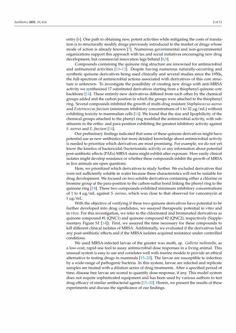
Abstract: There is an urgent need for the development of new antibiotics. Here, we describe theinhibitory activity of new quinone compounds against methicillin-resistant Staphylococcus aureus(ATCC® 43300), methicillin-sensitive S. aureus (ATCC® 29213), and two clinical isolates from Chile(ISP-213 and ISP-214). We observed 99.9% reduction in viability within 2 h of exposure without thecultures exhibiting any post-antibiotic effect, which was twice the kinetics to that observed withvancomycin. These clinical isolates did not acquire resistance to these quinone derivatives during thecourse of our study. We found that these compounds protected larvae of the greater wax moth, sp.Galleria mellonella, from infection by these MRSA clinical strains as effectively as vancomycin. Thesequinone derivatives are potential drug candidates worth further development.
Keywords: methicillin-resistant S. aureus (MRSA); drug discovery; quinone-antibiotics
1. Introduction
The World Health Organization (WHO) identified antimicrobial resistance as one of themain threats to global public health in 2019 [1]. Antimicrobial resistance has reached all con-tinents and currently negatively impacts most countries of global health systems [2]. Oneof the most problematic pathogens is methicillin-resistant Staphylococcus aureus (MRSA).This agent causes high morbidity in the United States [3], and the Pan American HealthOrganization (PAHO) warns that there is a 90% prevalence of MRSA in 21 countries of theAmericas [4], which indicates that patients infected with MRSA are now 64% more likelyto die due to treatment failure compared to an MSSA infection [4]. Not surprisingly, theWHO generated a list of priority pathogens to focus the development of new antibacterialdrugs against in 2017; MRSA ranked as a high priority [4]. Unfortunately, the developmentof new antibacterial drugs has progressively declined since the 1980s [5]. An analysis ofnew antibiotics approvals in the recent decades indicates a sharp fall in the last 30 years [6].
In particular, there has been widespread reluctance in the commercial sectors to de-velop new antibacterial drugs for reasons that include funding and barriers to market
Antibiotics 2021, 10, 614. https://doi.org/10.3390/antibiotics10060614 https://www.mdpi.com/journal/antibiotics
Antibiotics 2021, 10, 614 2 of 11
entry [6]. One path to obtaining new, potent activities while mitigating the costs of transla-tion is to structurally modify drugs previously introduced to the market or drugs whosemode of action is already known [7]. Numerous governmental and non-governmentalorganizations support this approach with tax and social initiatives encouraging new drugdevelopment, but commercial innovation lags behind [8,9].
Compounds containing the quinone ring structure are renowned for antimicrobialand antitumoral activities [10–13]. Despite having numerous naturally-occurring andsynthetic quinone derivatives being used clinically and several studies since the 1950s,the full-spectrum of antimicrobial actions associated with derivatives of this core struc-ture is unknown. To investigate the possibility of creating new drugs with anti-MRSAactivity we synthesized 17 substituted derivatives starting from a thiophenyl quinone corebackbone [14]. These entirely-new derivatives differed from each other by the chemicalgroups added and the carbon position in which the groups were attached to the thiophenylring. Several compounds inhibited the growth of multi-drug resistant Staphylococcus aureusand Enterococcus faecium (minimum inhibitory concentrations of 1 to 32 µg/mL) withoutexhibiting toxicity to mammalian cells [14]. We found that the size and lipophilicity of thechemical groups attached to the phenyl ring modified the antimicrobial activity, with sub-stituents in the ortho- and para-position exhibiting the greatest inhibitory activity againstS. aureus and E. faecium [14].
Our preliminary findings indicated that some of these quinone derivatives might havepotential use as new antibiotics but more detailed knowledge about antimicrobial activityis needed to prioritize which derivatives are most promising. For example, we do not yetknow the kinetics of bactericidal/bacteriostatic activity or any information about potentialpost-antibiotic effects (PAEs) MRSA stains might exhibit after exposure. How easily clinicalisolates might develop resistance or whether these compounds inhibit the growth of MRSAin live animals are open questions.
Here, we prioritized which derivatives to study further. We excluded derivatives thatwere not sufficiently soluble in water because these characteristics will not be suitable fordrug development. We focused on two soluble derivatives containing either a chlorine orbromine group at the para-position to the carbon-sulfur bond linking the phenyl ring to thequinone ring [14]. These two compounds exhibited minimum inhibitory concentrationsof 1 to 4 µg/mL against S. aureus, which was close to that observed for vancomycin at1 µg/mL.
With the objective of verifying if these two quinone derivatives have potential to befurther developed into drug candidates, we assayed therapeutic potential in vitro andin vivo. For this investigation, we refer to the chlorinated and brominated derivatives asquinone compound #1 (QNC1) and quinone compound #2 (QNC2), respectively (Supple-mentary Figure S1 [14]). First, we assayed the time necessary for these compounds tokill different clinical isolates of MRSA. Additionally, we evaluated if the derivatives hadany post-antibiotic effects and if the MRSA isolates acquired resistance under controlledconditions.
We used MRSA-infected larvae of the greater wax moth, sp. Galleria mellonella, asa low-cost, rapid-use tool to assay antimicrobial dose responses in a living animal. Thisunusual system is easy to use and correlates well with murine models to provide an ethicalalternative to testing drugs in mammals [15–20]. The larvae are susceptible to infectionby a wide-range of pathogenic bacteria. In this system, larvae are infected and replicatesamples are treated with a dilution series of drug treatments. After a specified period oftime, disease free larvae are scored to quantify dose-response, if any. This model systemdoes not require sophisticated equipment and has been used by various authors to testdrug efficacy of similar antibacterial agents [15–20]. Herein, we present the results of theseexperiments and discuss the significance of our findings.
Antibiotics 2021, 10, 614 3 of 11
2. Results2.1. Kill Kinetics
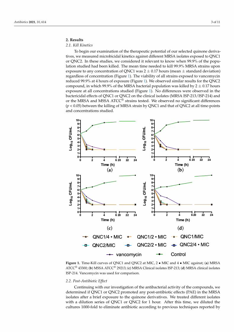
To begin our examination of the therapeutic potential of our selected quinone deriva-tives, we measured microbicidal kinetics against different MRSA isolates exposed to QNC1or QNC2. In these studies, we considered it relevant to know when 99.9% of the popu-lation studied had been killed. The mean time needed to kill 99.9% MRSA strains uponexposure to any concentration of QNC1 was 2 ± 0.17 hours (mean ± standard deviation)regardless of concentration (Figure 1). The viability of all strains exposed to vancomycinreduced 99.9% at 4 hours of exposure (Figure 1). We observed similar results for the QNC2compound, in which 99.9% of the MRSA bacterial population was killed by 2 ± 0.17 hoursexposure at all concentrations studied (Figure 1). No differences were observed in thebactericidal effects of QNC1 or QNC2 on the clinical isolates (MRSA ISP-213/ISP-214) andor the MRSA and MSSA ATCC® strains tested. We observed no significant differences(p < 0.05) between the killing of MRSA strain by QNC1 and that of QNC2 at all time-pointsand concentrations studied.
Antibiotics 2021, 10, x FOR PEER REVIEW 3 of 11
2. Results
2.1. Kill Kinetics
To begin our examination of the therapeutic potential of our selected quinone deriv-
atives, we measured microbicidal kinetics against different MRSA isolates exposed to
QNC1 or QNC2. In these studies, we considered it relevant to know when 99.9% of the
population studied had been killed. The mean time needed to kill 99.9% MRSA strains
upon exposure to any concentration of QNC1 was 2 ± 0.17 hours (mean ± standard devia-
tion) regardless of concentration (Figure 1). The viability of all strains exposed to vanco-
mycin reduced 99.9% at 4 hours of exposure (Figure 1). We observed similar results for
the QNC2 compound, in which 99.9% of the MRSA bacterial population was killed by 2 ±
0.17 hours exposure at all concentrations studied (Figure 1). No differences were observed
in the bactericidal effects of QNC1 or QNC2 on the clinical isolates (MRSA ISP-213/ISP-
214) and or the MRSA and MSSA ATCC® strains tested. We observed no significant dif-
ferences (p < 0.05) between the killing of MRSA strain by QNC1 and that of QNC2 at all
time-points and concentrations studied.
Figure 1. Time-Kill curves of QNC1 and QNC2 at MIC, 2 MIC and 4 MIC against; (a) MRSA
ATCC® 43300; (b) MSSA ATCC® 29213; (c) MRSA Clinical isolates ISP-213; (d) MRSA clinical iso-
lates ISP-214. Vancomycin was used for comparison.
2.2. Post-Antibiotic Effect
Continuing with our investigation of the antibacterial activity of the compounds, we
determined if QNC1 or QNC2 promoted any post-antibiotic effects (PAE) in the MRSA
isolates after a brief exposure to the quinone derivatives. We treated different isolates with
a dilution series of QNC1 or QNC2 for 1 hour. After this time, we diluted the cultures
Figure 1. Time-Kill curves of QNC1 and QNC2 at MIC, 2 • MIC and 4 • MIC against; (a) MRSAATCC® 43300; (b) MSSA ATCC® 29213; (c) MRSA Clinical isolates ISP-213; (d) MRSA clinical isolatesISP-214. Vancomycin was used for comparison.
2.2. Post-Antibiotic Effect
Continuing with our investigation of the antibacterial activity of the compounds, wedetermined if QNC1 or QNC2 promoted any post-antibiotic effects (PAE) in the MRSAisolates after a brief exposure to the quinone derivatives. We treated different isolateswith a dilution series of QNC1 or QNC2 for 1 hour. After this time, we diluted thecultures 1000-fold to eliminate antibiotic according to previous techniques reported by
Antibiotics 2021, 10, 614 4 of 11
Craig et al. [21]. We measured the growth curves of cultures exposed to the quinonederivatives and compared these curves to matched controlled cultures that were not treated.
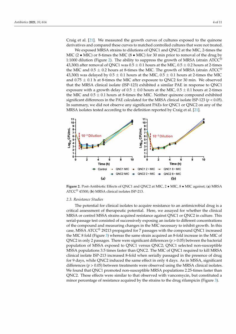
We exposed MRSA strains to dilutions of QNC1 and QNC2 at the MIC, 2-times theMIC (2 • MIC) or 8-times the MIC (8 • MIC) for 30 min prior to removal of the drug by1:1000 dilution (Figure 2). The ability to suppress the growth of MRSA (strain ATCC®
43,300) after removal of QNC1 was 0.5 ± 0.1 hours at the MIC, 0.5 ± 0.2 hours at 2-timesthe MIC and 0.5 ± 0.2 hours at 8-times the MIC. The growth of MRSA (strain ATCC®
43,300) was delayed by 0.5 ± 0.1 hours at the MIC, 0.5 ± 0.1 hours at 2-times the MICand 0.75 ± 0.1 h at 8-times the MIC after exposure to QNC2 for 30 min. We observedthat the MRSA clinical isolate (ISP-123) exhibited a similar PAE in response to QNC1exposure with a growth delay of 0.5 ± 0.0 hours at the MIC, 0.5 ± 0.1 hours at 2-timesthe MIC and 0.5 ± 0.1 hours at 8-times the MIC. Neither quinone compound exhibitedsignificant differences in the PAE calculated for the MRSA clinical isolate ISP-123 (p < 0.05).In summary, we did not observe any significant PAEs for QNC1 or QNC2 on any of theMRSA isolates tested according to the definition reported by Craig et al. [21].
Antibiotics 2021, 10, x FOR PEER REVIEW 4 of 11
1000-fold to eliminate antibiotic according to previous techniques reported by Craig et al.
[21]. We measured the growth curves of cultures exposed to the quinone derivatives and
compared these curves to matched controlled cultures that were not treated.
We exposed MRSA strains to dilutions of QNC1 and QNC2 at the MIC, 2-times the
MIC (2 MIC) or 8-times the MIC (8 MIC) for 30 min prior to removal of the drug by
1:1000 dilution (Figure 2). The ability to suppress the growth of MRSA (strain ATCC®
43,300) after removal of QNC1 was 0.5 ± 0.1 hours at the MIC, 0.5 ± 0.2 hours at 2-times
the MIC and 0.5 ± 0.2 hours at 8-times the MIC. The growth of MRSA (strain ATCC®
43,300) was delayed by 0.5 ± 0.1 hours at the MIC, 0.5 ± 0.1 hours at 2-times the MIC and
0.75 ± 0.1 h at 8-times the MIC after exposure to QNC2 for 30 min. We observed that the
MRSA clinical isolate (ISP-123) exhibited a similar PAE in response to QNC1 exposure
with a growth delay of 0.5 ± 0.0 hours at the MIC, 0.5 ± 0.1 hours at 2-times the MIC and
0.5 ± 0.1 hours at 8-times the MIC. Neither quinone compound exhibited significant dif-
ferences in the PAE calculated for the MRSA clinical isolate ISP-123 (p < 0.05). In summary,
we did not observe any significant PAEs for QNC1 or QNC2 on any of the MRSA isolates
tested according to the definition reported by Craig et al. [21].
Figure 2. Post-Antibiotic Effects of QNC1 and QNC2 at MIC, 2 MIC, 8 MIC against; (a) MRSA
ATCC® 43300; (b) MRSA clinical isolates ISP-213.
2.3. Resistance Studies
The potential for clinical isolates to acquire resistance to an antimicrobial drug is a
critical assessment of therapeutic potential. Here, we assayed for whether the clinical
MRSA or control MSSA strains acquired resistance against QNC1 or QNC2 in culture.
This serial-passage test consisted of successively exposing an isolate to different concen-
trations of the compound and measuring changes in the MIC necessary to inhibit growth.
In this case, MSSA ATCC® 29213 propagated for 7 passages with the compound QNC1
increased the MIC 8 fold (Figure 3) whereas the same strain acquired an 8-fold increase in
the MIC of QNC2 in only 2 passages. There were significant differences (p > 0.05) between
the bacterial population of MSSA exposed to QNC1 versus QNC2; QNC1 selected non-
susceptible MSSA populations 3.5 times faster than QNC2. The MIC of QNC1 required to
kill MRSA clinical isolate ISP-213 increased 8-fold when serially passaged in the presence
of drug for 9 days, while QNC2 induced the same effect in only 4 days. As in MSSA, sig-
nificant differences (p > 0.05) between treatments were observed using the MRSA clinical
isolates. We found that QNC1 promoted non-susceptible MRSA populations 2.25-times
faster than QNC2. These effects were similar to that observed with vancomycin, but con-
stituted a minor percentage of resistance acquired by the strains to the drug rifampicin
(Figure 3).
Figure 2. Post-Antibiotic Effects of QNC1 and QNC2 at MIC, 2 • MIC, 8 • MIC against; (a) MRSAATCC® 43300; (b) MRSA clinical isolates ISP-213.
2.3. Resistance Studies
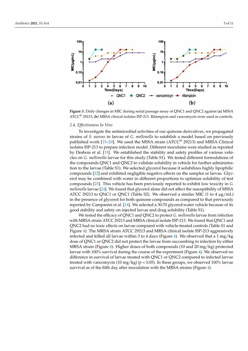
The potential for clinical isolates to acquire resistance to an antimicrobial drug is acritical assessment of therapeutic potential. Here, we assayed for whether the clinicalMRSA or control MSSA strains acquired resistance against QNC1 or QNC2 in culture. Thisserial-passage test consisted of successively exposing an isolate to different concentrationsof the compound and measuring changes in the MIC necessary to inhibit growth. In thiscase, MSSA ATCC® 29213 propagated for 7 passages with the compound QNC1 increasedthe MIC 8 fold (Figure 3) whereas the same strain acquired an 8-fold increase in the MIC ofQNC2 in only 2 passages. There were significant differences (p > 0.05) between the bacterialpopulation of MSSA exposed to QNC1 versus QNC2; QNC1 selected non-susceptibleMSSA populations 3.5 times faster than QNC2. The MIC of QNC1 required to kill MRSAclinical isolate ISP-213 increased 8-fold when serially passaged in the presence of drugfor 9 days, while QNC2 induced the same effect in only 4 days. As in MSSA, significantdifferences (p > 0.05) between treatments were observed using the MRSA clinical isolates.We found that QNC1 promoted non-susceptible MRSA populations 2.25-times faster thanQNC2. These effects were similar to that observed with vancomycin, but constituted aminor percentage of resistance acquired by the strains to the drug rifampicin (Figure 3).
Antibiotics 2021, 10, 614 5 of 11Antibiotics 2021, 10, x FOR PEER REVIEW 5 of 11
Figure 3. Daily changes in MIC during serial passage assay of QNC1 and QNC2 against (a) MSSA
ATCC® 29213; (b) MRSA clinical isolates ISP-213. Rifampicin and vancomycin were used as con-
trols.
2.4. Effectiveness In Vivo
To investigate the antimicrobial activities of our quinone derivatives, we propagated
strains of S. aureus in larvae of G. mellonella to establish a model based on previously pub-
lished work [15–20]. We used the MSSA strain (ATCC® 29213) and MRSA Clinical isolates
ISP-213 to prepare infection model. Different inoculums were studied as reported by
Desbois et al. [15]. We established the stability and safety profiles of various vehicles on
G. mellonella larvae for this study (Table S1). We tested different formulations of the com-
pounds QNC1 and QNC2 to validate solubility in vehicle for further administration to the
larvae (Table S1). We selected glycerol because it solubilizes highly lipophilic compounds
[22] and exhibited negligible negative effects on the samples or larvae. Glycerol may be
combined with water in different proportions to optimize solubility of test compounds
[23]. This vehicle has been previously reported to exhibit low toxicity in G. mellonella lar-
vae [24]. We found that glycerol alone did not affect the susceptibility of MSSA ATCC
29213 to QNC1 or QNC1 (Table S2). We observed a similar MIC (1 to 4 μg/mL) in the
presence of glycerol for both quinone compounds as compared to that previously re-
ported by Campanini et al. [14]. We selected a 30:70 glycerol:water vehicle because of its
good stability and safety on injected larvae and drug solubility (Table S1).
We tested the efficacy of QNC1 and QNC2 to protect G. mellonella larvae from infec-
tion with MRSA strain ATCC 29213 and MRSA clinical isolate ISP-213. We found that
QNC1 and QNC2 had no toxic effects on larvae compared with vehicle-treated controls
(Table S1 and Figure 4). The MRSA strain ATCC 29213 and MRSA clinical isolate ISP-213
aggressively infected and killed all larvae within 3 to 4 days (Figure 4). We observed that
a 1 mg/kg dose of QNC1 or QNC2 did not protect the larvae from succumbing to infection
by either MRSA strain (Figure 4). Higher doses of both compounds (10 and 20 mg/kg)
protected larvae with 100% survival during the course of the experiment (Figure 4). We
observed no difference in survival of larvae treated with QNC1 or QNC2 compared to
infected larvae treated with vancomycin (10 mg/kg) (p < 0.05). In these groups, we ob-
served 100% larvae survival as of the fifth day after inoculation with the MRSA strains
(Figure 4).
Figure 3. Daily changes in MIC during serial passage assay of QNC1 and QNC2 against (a) MSSAATCC® 29213; (b) MRSA clinical isolates ISP-213. Rifampicin and vancomycin were used as controls.
2.4. Effectiveness In Vivo
To investigate the antimicrobial activities of our quinone derivatives, we propagatedstrains of S. aureus in larvae of G. mellonella to establish a model based on previouslypublished work [15–20]. We used the MSSA strain (ATCC® 29213) and MRSA Clinicalisolates ISP-213 to prepare infection model. Different inoculums were studied as reportedby Desbois et al. [15]. We established the stability and safety profiles of various vehi-cles on G. mellonella larvae for this study (Table S1). We tested different formulations ofthe compounds QNC1 and QNC2 to validate solubility in vehicle for further administra-tion to the larvae (Table S1). We selected glycerol because it solubilizes highly lipophiliccompounds [22] and exhibited negligible negative effects on the samples or larvae. Glyc-erol may be combined with water in different proportions to optimize solubility of testcompounds [23]. This vehicle has been previously reported to exhibit low toxicity in G.mellonella larvae [24]. We found that glycerol alone did not affect the susceptibility of MSSAATCC 29213 to QNC1 or QNC1 (Table S2). We observed a similar MIC (1 to 4 µg/mL)in the presence of glycerol for both quinone compounds as compared to that previouslyreported by Campanini et al. [14]. We selected a 30:70 glycerol:water vehicle because of itsgood stability and safety on injected larvae and drug solubility (Table S1).
We tested the efficacy of QNC1 and QNC2 to protect G. mellonella larvae from infectionwith MRSA strain ATCC 29213 and MRSA clinical isolate ISP-213. We found that QNC1 andQNC2 had no toxic effects on larvae compared with vehicle-treated controls (Table S1 andFigure 4). The MRSA strain ATCC 29213 and MRSA clinical isolate ISP-213 aggressivelyinfected and killed all larvae within 3 to 4 days (Figure 4). We observed that a 1 mg/kgdose of QNC1 or QNC2 did not protect the larvae from succumbing to infection by eitherMRSA strain (Figure 4). Higher doses of both compounds (10 and 20 mg/kg) protectedlarvae with 100% survival during the course of the experiment (Figure 4). We observed nodifference in survival of larvae treated with QNC1 or QNC2 compared to infected larvaetreated with vancomycin (10 mg/kg) (p < 0.05). In these groups, we observed 100% larvaesurvival as of the fifth day after inoculation with the MRSA strains (Figure 4).
Antibiotics 2021, 10, 614 6 of 11Antibiotics 2021, 10, x FOR PEER REVIEW 6 of 11
Figure 4. Effects of drug treatment on G. mellonella larvae survival inoculated with MRSA ATCC 29213 (a,b) or MRSA ISP-
213 (c,d), then QNC1(a,c) or QNC2 (b,d) at 1, 10 and 20 mg/kg were administered at 0 Days (i.e., 1 hour after inoculation).
Vancomycin (1 mg/kg) was used as control.
Figure 4 shows the results using MRSA ISP-213 clinical strains for larvae inoculation.
We observed no significant differences (p < 0.05) in effect when comparing the dose of 10
mg/kg of QNC1 or QNC2 to control groups of larvae treated with vancomycin. We ob-
served 100% larvae survival in observed larvae groups receiving doses of 20 and 10 mg/kg
QNC1 or QNC2.
3. Discussion
We explored the therapeutic potential of new chlorinated and brominated quinone
derivatives to address the urgent need for antimicrobial drugs against multidrug-resistant
MRSA [25,26]. First, we studied the kinetics of bactericidal effects on MRSA and MSSA
strains exposed to different concentrations of QNC1 or QNC2. This level of bactericidal
activity was consistent with other quinone derivatives we previously synthesized [14] and
is similar to the Minimum Bactericidal Concentration/Minimum Inhibitory Concentration
(MBC/MIC) ratio used against clinical isolates [14]. In a previously reported study, we
showed that these compounds had a MBC/MIC ratio of less than 2 on MRSA isolated in
Chile [14]. As defined by Craig et al., we concluded these compounds exhibited bacteri-
cidal activity on the MRSA isolates tested.
It was relevant to compare bactericidal kinetics of these compounds to a commercial
antibiotic, such as vancomycin. Both quinone derivatives reduced 99.9% of the different
bacterial populations studied 2 times faster than vancomycin. This result represents a
comparative advantage over vancomycin since various authors have pointed out that one
of the main limitations of vancomycin in the management of infections complicated by
MRSA is its slow bactericidal capacity [27,28].
Microorganisms briefly exposed to inhibitory concentrations of antibiotics some-
times exhibit growth inhibition that persists even after the drug is removed. This post-
antibiotic effect phenomenon has implications for dosing regimens and the determination
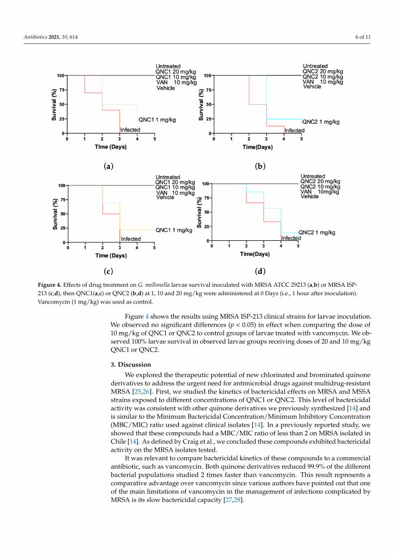
Figure 4. Effects of drug treatment on G. mellonella larvae survival inoculated with MRSA ATCC 29213 (a,b) or MRSA ISP-213 (c,d), then QNC1(a,c) or QNC2 (b,d) at 1, 10 and 20 mg/kg were administered at 0 Days (i.e., 1 hour after inoculation).Vancomycin (1 mg/kg) was used as control.
Figure 4 shows the results using MRSA ISP-213 clinical strains for larvae inoculation.We observed no significant differences (p < 0.05) in effect when comparing the dose of10 mg/kg of QNC1 or QNC2 to control groups of larvae treated with vancomycin. We ob-served 100% larvae survival in observed larvae groups receiving doses of 20 and 10 mg/kgQNC1 or QNC2.
3. Discussion
We explored the therapeutic potential of new chlorinated and brominated quinonederivatives to address the urgent need for antimicrobial drugs against multidrug-resistantMRSA [25,26]. First, we studied the kinetics of bactericidal effects on MRSA and MSSAstrains exposed to different concentrations of QNC1 or QNC2. This level of bactericidalactivity was consistent with other quinone derivatives we previously synthesized [14] andis similar to the Minimum Bactericidal Concentration/Minimum Inhibitory Concentration(MBC/MIC) ratio used against clinical isolates [14]. In a previously reported study, weshowed that these compounds had a MBC/MIC ratio of less than 2 on MRSA isolated inChile [14]. As defined by Craig et al., we concluded these compounds exhibited bactericidalactivity on the MRSA isolates tested.
It was relevant to compare bactericidal kinetics of these compounds to a commercialantibiotic, such as vancomycin. Both quinone derivatives reduced 99.9% of the differentbacterial populations studied 2 times faster than vancomycin. This result represents acomparative advantage over vancomycin since various authors have pointed out that oneof the main limitations of vancomycin in the management of infections complicated byMRSA is its slow bactericidal capacity [27,28].
Antibiotics 2021, 10, 614 7 of 11
Microorganisms briefly exposed to inhibitory concentrations of antibiotics sometimesexhibit growth inhibition that persists even after the drug is removed. This post-antibioticeffect phenomenon has implications for dosing regimens and the determination of clinicalefficacy. For this reason, it was important to identify any PAEs during the characterizationstage of these new antibiotics. The PAE has traditionally been measured by exposingbacterial isolates to different drug concentrations for a short period of time, then theantibiotic is removed, and the kinetics of recovery versus the same isolate without treatmentis determined. Our findings indicated that QNC1 and QNC2 promoted few, if any, PAEsupon MRSA clinical isolates. This observation is consistent with compounds that have atime-dependent antibacterial mode of action (T/MIC) and indicates that the antibacterialactivity will depend on the time in which its drug concentration exceeds the MIC in plasmaand not necessarily when the plasma concentration reached [29]. Antibiotics that act ina time-dependent manner (T/MIC) show a greater clinical response, with values greaterthan 40% of the MIC between two doses; an example is beta-lactam antibiotics that do notshow PAE in Gram-negative bacilli [30]. Our results may guide concentrations’ compoundadministered in a murine model of infection since this type of compound (T/MIC) must beadministered in repeated doses and at concentrations that do not exceed four times theMIC of the target microorganism being studied [21]. These dosage schemes would decreasethe possibility of selecting resistant bacterial populations, according to recommendationsby Craig W. [31].
With the advancement of antimicrobial resistance, it is necessary to study the newantibiotic potential to select for non-sensitive mutants’ populations [32]. In a best-case-scenario, newly-developed inhibitors should be difficult to inactivate through acquisitionof new mutations, and in general, microorganisms should remain sensitive to the drugover time. These studies are an important part of characterizing the inhibitory activity ofnewly developed antimicrobial chemicals. These standard methods of assessing resistancehave been in practice for decades. A common tactic is to test if a microorganism becomesless sensitive to a drug is to measure the MIC of the microorganism to the drug througha series of successive subcultures in the presence of the compound to be studied. Thisserial passage test is a good approximation of whether a certain compound can enrich fornon-susceptible strains.
In this work, we observed substantial differences between both derivatives. QNC1was select non-susceptible S. aureus populations with a rate similar to rifampin [33]. QNC2exhibited similar kinetics to vancomycin and a slower selection speed non-susceptiblepopulations compared to QNC1 [34]. Molecular studies are necessary to explain thesedifferences, which theoretically could have the same mechanism of action [14]. However,both compounds have different physicochemical properties due to substitutions on thethiophenyl ring [14]. These differences might influence the stability of the compounds or ac-cess to bacterial cells. We conclude that our evidence indicates that MRSA strains acquiredresistance to these derivatives no faster than to vancomycin under these conditions.
Finally, a model of infection in the larvae of G. mellonella was used to evaluate theeffectiveness of new anti-staphylococcal agents in vivo [15]. Galleria mellonella has charac-teristics that allow it to simulate a human physiological environment; it can live at 37 ◦Cfor days with controlled humidity [35]. This insect possesses an immune system thatworks through hemocyte-mediated cellular responses [36] and humoral responses usingantimicrobial peptides, melanin and other effector proteins [37]. These characteristics madeG. mellonela a robust model for us to approximate the effectiveness of our compoundsin vivo. We showed that a 10 mg/kg dose of QNC1 or QNC2 was as effective comparedto 10 mg/kg of vancomycin in preventing the death of G. mellonella due to staphylococcalinfection. No significant differences were found in the effectiveness observed betweenboth derivatives. A direct relationship was observed between the administered dose ofQNC1 or QNC2 and the survival percentage observed on the fifth-day post-inoculationof S. aureus. This collective evidence indicates these quinone compounds might representnew alternatives to today’s limited antibiotic options against MRSA.
Antibiotics 2021, 10, 614 8 of 11
4. Materials and Methods4.1. Bacterial Strains and Isolates
We tested the following strains: methicillin-resistant Staphylococcus aureus (MRSA)ATCC® 43300 and methicillin-susceptible Staphylococcus aureus (MSSA) ATCC® 29213. Wealso tested the MRSA clinical isolates, ISP-213 and ISP-214, which were provided by theChilean Public Health Institute. These MRSA strains were originally isolated from thebloodstream of septic patients and shown to be resistant to at least three additional classesof antimicrobial agents. These isolated strains were defined as multidrug-resistant. Isolateswere collected in 2014 and were obtained from Chilean hospitals. All strains tested weretransferred until they reached a logarithmic phase of growth.
4.2. Media and Antibacterial Compounds
Mueller-Hinton broth (Difco, Beckton Dickinson, Sparks, MD, USA) was used forall experiments, including killing curve, postantibiotic effect determination, and serialpassage assay. Mueller-Hinton agar (Difco, Beckton Dickinson, Sparks, MD, USA) wasused for performing colony counts and detecting antibiotic resistance. QNC1 and QNC2were provided by the Drug Development Laboratory, University of Chile. Obtainingand characterization procedure was described by Campanini-Salinas et al. in 2018 [14].Vancomycin was used as a comparator for all experiments. This was supplied by Abcam-Biochemicals®, Cambridge, UK. Glycerol 1 (Ensure®, Merck, Darmstadt, Germany) wasused for vehicle formulation at in vivo tests.
4.3. In Vitro Activity Characterization4.3.1. Kill Kinetics
QNC1 and QNC2 were studied at 1-, 2-, or 4-times the MIC. Time–kill curves wereperformed in an Erlenmeyer flask with a screw cap containing Mueller-Hinton broth, usingan inoculum of 5 × 10⁶–1 × 10⁷ CFU/mL in the presence of QNC1 or QNC2. Survivingbacteria were counted after 0, 1, 2, 4, 8, 12, and 24 h of incubation at 37 ◦C by subculturingserial dilutions of samples on Mueller Hinton agar plates. An aliquot was diluted 1000 timesto remove the tested compounds [21]. 100 uL was transferred to a Mueller-Hinton agarplate, and the inoculum was dispersed with a Drigalsky loop previously sterilized withheat and 75% ethanol. A bactericidal effect was defined as a ≥3 log10 CFU/mL decreasecompared with the initial inoculum after 24 h of incubation [21]. A flask inoculated withMH broth with no antibiotic served as sterility control. Vancomycin was used as control.Tests were carried out in triplicate.
4.3.2. Post-Antibiotic Effect Determination
PAE method was used as described by Mercier et al. [38]. QNC1, QNC2, and van-comycin were tested at different multiples of the MIC. Mueller-Hinton I broth was usedin the test tubes, and a 1/1000 dilution removed the antibiotics after the bacteria wereexposed to them for 1 h. The PAE was calculated by using Equation (1):
PAE = T − C (1)
where T is the time required for the count in the test culture to increase 1 log10 above thecount observed immediately after drug removal, and C is the time required for the countof the untreated control tube to increase by 1 log10.
4.3.3. Serial Passage Assay
The serial passage method was used as described by Silverman et al. [39]. Briefly,on the first day, Mueller-Hinton broth containing QNC1 or QNC2 at 0.25-, 0.5-, 1-, 2-, or4-times the MIC was inoculated with different MRSA strains from a single colony. Cultureswere incubated overnight at 37 ◦C with shaking. From the highest concentration thatsupported growth, cultures were diluted 1:10,000 into fresh media plus daptomycin at
Antibiotics 2021, 10, 614 9 of 11
two-fold dilutions. This process was continued for 21 days or until three successive culturesfailed to show any decrease in susceptibility. The transfers were carried out until there wasgrowth in all the cultures. Cultures that showed growth were subcultured in drug-freeHAM plates, and MIC was determined by the method of microdilution following theCLSI protocol after 24 h [40]. After the test, the 4× CIM concentration tubes with bacterialgrowth were again seeded in compound-free MHA plates, and subsequently, 3 passageswere made in compound-free MHA plates, determining the MIC in each passage.
4.4. Galleria mellonella Infection Model
G. mellonella larvae obtained from a commercial supplier (BioBichos, Ltd., Chillan,Chile) were used. All experiments were performed according to the protocols described byvarious authors [15,41,42]. Briefly, study groups consisted of 10 final stages of G. mellonellalarvae, with an approximate weight of 250 mg each. They were randomly selected. AHamilton® syringe (Merck® Ltd., Kenilworth, NJ, USA) was used to inject the larvae—inoculation volume used in each test was 10 uL. MRSA inoculum (CFU/mL) that decreasesthe survival of G. mellonella larvae was studied. Inoculum that managed to reduce survivalto less than 20% in 72 h was selected. Once the inoculum of bacteria was chosen and thesafety of the compounds to be tested was determined, the larvae were inoculated withthe chosen suspension. After 1 h, they were injected with the compound to be studied atdifferent concentrations. After each treatment, the larvae were incubated for 5 days at 37 ◦C,80% humidity, and their survival was determined daily. One group of larvae was injectedwith phosphate buffer solution, and another group was not subjected to treatment. Bothgroups were used as controls in each of the experiments. The results of any experimentwith more than two dead larvae in any control group were discarded.
4.5. Statistical Analysis
The data were analyzed with one-way ANOVA and t-tests, with the criterion forstatistical significance set at p < 0.05, using the GraphPad Prism 5.03 program (GraphPadSoftware, Inc., San Diego, CA, USA, www.graphpad.com, accessed on 5 May 2014). Infec-tion model data were plotted using the Kaplan–Meier method, and comparisons betweengroups were made using the log-rank test.
5. Conclusions
Herein, we demonstrated that quinone compounds, QNC1 and QNC2, are effectiveand safe in a model of S. aureus infection in G. mellonella. They had rapid bactericidalactivity, and the QNC2 derivative has a low rate of selection of non-susceptible populations.Based on the evidence shown, we believe that the compounds have therapeutic potentialand are candidates to advance towards studies in murine models.
6. Patents
Chilean Patent Application number 201503780, PCT/CL2016/050080; EPO 16880235.3;MX/a/2018/008192; US20190367505, CN109121411, EP3404026 titled: “Pyrimi-dine-Isoquinoline-Quinone Derived Compounds, their Salts, Isomers, Pharmaceutically Acceptable Tau-tomers; Pharmaceutical Composition; Preparation Procedure; and their Use in the Treat-ment of Bacterial and Multiresistant Bacterial Diseases.”
Supplementary Materials: The following are available online at http://www.mdpi.com/xxx/s1.Figure S1: Most potent quinonic compounds against multiresistant gram-positive isolates, Table S1:Attributes of the different vehicles to incorporate quinonic compounds, Table S2: Antibacterialactivity of quinonic compounds in different vehicles.
Antibiotics 2021, 10, 614 10 of 11
Author Contributions: Conceptualization, J.C.-S. and D.V.-V.; Formal analysis, G.G.-R., P.A. andD.V.-V.; Investigation, J.C.-S., J.A.-L., N.H., F.M. and D.V.-V.; Methodology, G.G.-R., D.V.-V. and J.C.-S.;Resources, G.G.-R., P.A.; Visualization, J.C.-S.; Writing—original draft, J.C.-S., J.A.-L. and D.V.-V.;Writing—review and editing, J.C.-S., J.A.-L., I.E.B. and D.V.-V. All authors have read and agreed tothe published version of the manuscript.
Funding: This research was funded by FONDECYT grant numbers 11110516 (D.V.-V.) and 79100006(D.V.-V.).
Data Availability Statement: Data is contained within the article or supplementary material.
Acknowledgments: The authors gratefully acknowledge support by the Agencia Nacional de In-vestigación y Desarrollo (ANID) de Chile through a FONDECYT Iniciación en Investigación GrantNo. 11110516 (D.V.-V.), Chile; FONDECYT Proyecto de Inserción Grant No. 79100006 (D.V.-V.),Chile; Programa de Estímulo a la Excelencia Institucional PEEI 2017, Universidad de Chile (D.V.-V.);and FONDECYT Regular Grant No. 1191737 (I.E.B.). The authors also gratefully acknowledgesupport from CONICYT de Chile in the form of a Beca Doctorados Nacional No. 21130643 (J.C.-S.)and 21130628 (J.A.-L.). The authors lastly acknowledge contributing support from the McDonnellfoundation though a Fellowship from the Salk Institute (I.E.B.) and the National Institute of Allergyand Infectious Disease of the United States of America National Institutes of Health under awardnumber NIH 7R21AI111072 (I.E.B.).
Conflicts of Interest: The authors declare no conflict of interest.
References1. White, A.; Hughes, J.M. Critical importance of a one health approach to antimicrobial resistance. EcoHealth 2019, 16, 404–409.
[CrossRef]2. O’neill, J. Antimicrobial Resistance: Tackling a Crisis for the Health and Wealth of Nations; Review Paper; Wellcome Trust: London,
UK, 2014.3. Boucher, H.W.; Talbot, G.H.; Bradley, J.S.; Edwards, J.E.; Gilbert, D.; Rice, L.B.; Scheld, M.; Spellberg, B.; Bartlett, J. Bad bugs,
no drugs: No ESKAPE! An update from the Infectious Diseases Society of America. Clin. Infect. Dis. 2009, 48, 1–12. [CrossRef][PubMed]
4. World Health Organization. Antimicrobial Resistance: Global Report on Surveillance; World Health Organization: Geneva, Switzer-land, 2014.
5. Luepke, K.H.; Suda, K.J.; Boucher, H.; Russo, R.L.; Bonney, M.W.; Hunt, T.D.; Mohr, J.F., III. Past, present, and future ofantibacterial economics: Increasing bacterial resistance, limited antibiotic pipeline, and societal implications. Pharmacotherapy2017, 37, 71–84. [CrossRef]
6. Silver, L.L. Challenges of antibacterial discovery. Clin. Microbiol. Rev. 2011, 24, 71–109. [CrossRef]7. Campanini-Salinas, J.; Andrades-Lagos, J.; Mella-Raipan, J.; Vasquez-Velasquez, D. Novel classes of antibacterial drugs in clinical
development, a hope in a post-antibiotic era. Clin. Microbiol. Rev. 2018, 18, 1188–1202. [CrossRef]8. Simpkin, V.L.; Renwick, M.J.; Kelly, R.; Mossialos, E. Incentivising innovation in antibiotic drug discovery and development:
Progress, challenges and next steps. J. Antibiot. 2017, 70, 1087–1096. [CrossRef] [PubMed]9. Shrivastava, S.R.; Shrivastava, P.S.; Ramasamy, J. World health organization releases global priority list of antibiotic-resistant
bacteria to guide research, discovery, and development of new antibiotics. J. Med. Soc. 2018, 32, 76. [CrossRef]10. Ishidate, M.; Kobayashi, K.; Sakurai, Y.; Sato, H.; Yoshida, T. Experimental Studies on Chemotherapy of Malignant Growth Em-
ploying Yoshida Sarcoma Animals XI. Effect of quinone derivatives, antibiotics, alkaloids, organoarsen, and other miscellaneouscompounds. Gan 1955, 46, 482–484.
11. Resegotti, L.; Infelise, V.E. The effect of a combination of phenanthroline quinone plus iodochlorhydroxyquinoline in theprevention of dysvitaminosis caused by antibiotics. Minerva Med. 1966, 57, 947–949. [PubMed]
12. Wanke, H.; Kersten, W.; Kersten, H. Polysomes in Bacillus subitilis: Influence of amino quinones and quinone antibiotics on thesynthesis and stability of mRNA. Hoppe-Seyler’s Z. Physiol. Chem. 1969, 350, 1162–1163.
13. Nagasawa, T.; Fukao, H.; Irie, H.; Yamada, H. Sakyomicins. A, B, C and D: New quinone-type antibiotics produced by a strain ofnocardia taxonomy, production, isolation and biological properties. J. Antibiot. 1984, 37, 693–699. [CrossRef] [PubMed]
14. Campanini-Salinas, J.; Andrades-Lagos, J.; González-Rocha, G.; Choquesillo-Lazarte, D.; Dragnic, S.B.; Faúndez, M.; Alarcón, P.;Silva, F.; Vidal, R.; Salas-Huenuleo, E.; et al. A new kind of quinonic-antibiotic useful against multidrug-resistant S. aureus and E.faecium infections. Molecules 2018, 23, 1776. [CrossRef] [PubMed]
15. Desbois, A.P.; Coote, P.J. Wax moth larva (Galleria mellonella): An in vivo model for assessing the efficacy of antistaphylococcalagents. J. Antimicrob. Chemother. 2011, 66, 1785–1790. [CrossRef] [PubMed]
16. Desbois, A.P.; Coote, P.J. Utility of greater wax moth larva (Galleria mellonella) for evaluating the toxicity and efficacy of newantimicrobial agents. Adv. Appl. Microbiol. 2012, 78, 25–53. [CrossRef]
Antibiotics 2021, 10, 614 11 of 11
17. Tsai, C.J.-Y.; Loh, J.M.S.; Proft, T. Galleria mellonella infection models for the study of bacterial diseases and for antimicrobial drugtesting. Virulence 2016, 7, 214–229. [CrossRef]
18. Cutuli, M.A.; Petronio, G.; Vergalito, F.; Magnifico, I.; Pietrangelo, L.; Venditti, N.; Di Marco, R. Galleria mellonella as a consolidatedin vivo model hosts: New developments in antibacterial strategies and novel drug testing. Virulence 2019, 10, 527–541. [CrossRef]
19. Hornsey, M.; Wareham, D.W. In vivo efficacy of glycopeptide-colistin combination therapies in a Galleria mellonella model ofAcinetobacter baumannii infection. Antimicrob. Agents Chemother. 2011, 55, 3534–3537. [CrossRef]
20. Piatek, M.; Sheehan, G.; Kavanagh, K. Utilising Galleria mellonella larvae for studying in vivo activity of conventional and novelantimicrobial agents. Pathog. Dis. 2020, 78, ftaa059. [CrossRef]
21. Craig, W.A. The postantibiotic effect. Clin. Microbiol. Newsl. 1991, 13, 121–124. [CrossRef]22. Srivastava, V.C. Glycerol as a Green Solvent in Organic Reactions. Mater. Res. Found. 2019, 54, 202–223. [CrossRef]23. Niazi, S.K. Handbook of Pharmaceutical Manufacturing Formulations: Volume Four, Semisolid Products; CRC Press: Boca Raton, FL,
USA, 2019; ISBN 1351593331.24. Allegra, E.; Titball, R.W.; Carter, J.; Champion, O.L. Galleria mellonella larvae allow the discrimination of toxic and non-toxic
chemicals. Chemosphere 2018, 198, 469–472. [CrossRef]25. Livermore, D.M. The need for new antibiotics. Clin. Microbiol. Infect. 2004, 10, 1–9. [CrossRef]26. Kmietowicz, Z. Few novel antibiotics in the pipeline, WHO warns. BMJ 2017, 358, j4339. [CrossRef]27. Sakoulas, G.; Moise-Broder, P.A.; Schentag, J.; Forrest, A.; Moellering, R.C.; Eliopoulos, G.M. Relationship of MIC and bactericidal
activity to efficacy of vancomycin for treatment of methicillin-resistant Staphylococcus aureus bacteremia. J. Clin. Microbiol. 2004,42, 2398–2402. [CrossRef]
28. Kollef, M.H. Limitations of vancomycin in the management of resistant staphylococcal infections. Clin. Infect. Dis. 2007, 45,S191–S195. [CrossRef] [PubMed]
29. Mandell, G.L.; Douglas, R.G., Jr.; Bennett, J.E. Principles and Practice of Infectious Diseases; John Wiley & Sons: Hoboken, NJ, USA,2014; Volume 2, p. 233. ISBN 0471034894.
30. Goodman, L.S. Goodman and Gilman’s the Pharmacological Basis of Therapeutics; McGraw-Hill: New York, NY, USA, 1996; Volume1549, pp. 1361–1373.
31. Craig, W. Pharmacodynamics of antimicrobial agents as a basis for determining dosage regimens. Eur. J. Clin. Microbiol. Infect.Dis. 1993, 12, S6–S8. [CrossRef] [PubMed]
32. Martínez, J.L.; Baquero, F.; Andersson, D.I. Beyond serial passages: New methods for predicting the emergence of resistance tonovel antibiotics. Curr. Opin. Pharmacol. 2011, 11, 439–445. [CrossRef] [PubMed]
33. Sakoulas, G.; Alder, J.; Thauvin-Eliopoulos, C.; Moellering, R.C.; Eliopoulos, G.M. Induction of daptomycin heterogeneoussusceptibility in Staphylococcus aureus by exposure to vancomycin. Antimicrob. Agents Chemother. 2006, 50, 1581–1585. [CrossRef][PubMed]
34. Clark, C.; Kosowska-Shick, K.; McGhee, P.; Dewasse, B.; Beachel, L.; Appelbaum, P.C. Resistance selection studies comparing theactivity of razupenem (PTZ601) to vancomycin and linezolid against eight methicillin-resistant and two methicillin-susceptibleStaphylococcus aureus strains. Antimicrob. Agents Chemother. 2009, 53, 3118–3121. [CrossRef]
35. Ramarao, N.; Nielsen-Leroux, C.; Lereclus, D. The insect Galleria mellonella as a powerful infection model to investigate bacterialpathogenesis. J. Vis. Exp. 2012, 70. [CrossRef]
36. Browne, N.; Heelan, M.; Kavanagh, K. An analysis of the structural and functional similarities of insect hemocytes and mammalianphagocytes. Virulence 2013, 4, 597–603. [CrossRef] [PubMed]
37. Harding, C.R.; Schroeder, G.N.; Collins, J.W.; Frankel, G. Use of Galleria mellonella as a model organism to study Legionellapneumophila infection. J. Vis. Exp. 2013, 81. [CrossRef]
38. Mercier, R.-C.; Houlihan, H.H.; Rybak, M.J. Pharmacodynamic evaluation of a new glycopeptide, LY333328, and in vitro activityagainst Staphylococcus aureus and Enterococcus faecium. Antimicrob. Agents Chemother. 1997, 41, 1307–1312. [CrossRef]
39. Silverman, J.A.; Oliver, N.; Andrew, T.; Li, T. Resistance studies with daptomycin. Antimicrob. Agents Chemother. 2001, 45,1799–1802. [CrossRef] [PubMed]
40. CLSI. Methods for Dilution Antimicrobial Susceptibility Tests for Bacteria that Grow Aerobi-Cally, Approved Standard, 8th ed.; Clinicaland Laboratory Standards Institute: Wayne, PA, USA, 2009.
41. Luther, M.K.; Arvanitis, M.; Mylonakis, E.; LaPlante, K.L. Activity of daptomycin or linezolid in combination with rifampin orgentamicin against biofilm-forming Enterococcus faecalis or E. faecium in an in vitro pharmacodynamic model using simulatedendocardial vegetations and an in vivo survival assay using Galleria mellonella larvae. Antimicrob. Agents Chemother. 2014, 58,4612–4620. [CrossRef]
42. Gibreel, T.M.; Upton, M. Synthetic epidermicin NI01 can protect Galleria mellonella larvae from infection with Staphylococcusaureus. J. Antimicrob. Chemother. 2013, 68, 2269–2273. [CrossRef] [PubMed]
Related Documents