Zeitschrift der Deutschen Gesellschaft für Zahnärztliche Implantologie e.V. SPECIAL PRINT Edition 5/11 and 5/12 • Volume 16 “New Materials in GBR” and “Safe Bone Regeneration through a new Collagen Membrane” Dr. med. dent. Bastian Wessing, Dr. med. dent. Martin Emmerich, M.Sc.

Welcome message from author
This document is posted to help you gain knowledge. Please leave a comment to let me know what you think about it! Share it to your friends and learn new things together.
Transcript

Zeitschrift der Deutschen Gesellschaft für Zahnärztliche Implantologie e.V.
SPECIAL PRINTEdition 5/11 and 5/12 • Volume 16
“New Materials in GBR” and “Safe Bone Regeneration through
a new Collagen Membrane”Dr. med. dent. Bastian Wessing, Dr. med. dent. Martin Emmerich, M.Sc.


n Augmentation using various techniques has beendeveloped since the paradigm shift from bone-drivenimplantation techniques to “backward planning” in or-der to avoid an unfavourable crown-implant length re-lationship in general, or to avoid for example pink ce-ramic in the aesthetically visible area. In planning the replacement of a lost or extracted toothwith an implant, the aesthetic and static final resultmust be taken into consideration. According to the cur-rent academic opinion, the bony and gingival tissuearound the implant site must be restored in manycases—at least in the visible area. Disregarding suchprerequisites can lead to serious failure. Membranes are frequently required in implantologyand periodontology for protection and for fixation ofaugmented sites, or in “Guided Bone Regeneration”(GBR). In the USA, the FDA recommends using suitablemembranes when building up bone in order to pre-vent the migration of bone replacement material intothe soft tissue. Resorbable membranes are superior tonon-resorbable membranes as they show higher tis-sue compatibility, have a lower dehiscence rate andare easier to handle.4,26 They show a higher biocom-patibility to the PDL fibroblasts as well as to osteo -blasts,22and are significantly more biocompatible thanPTFE membranes.24
For this reason, in Germany, resorbable membranes de-rived from collagen from an animal source are most frequently used. The use of such membranes has beenwell documented and has demonstrated a high proba-bility of success.9,13,14
The barrier function is particularly important in the aug-mentation of larger bony defects, especially in case ofearly exposures of the membrane (dehiscence). Becauseof bacterial contamination, non-resorbable membranesshould be removed at an early stage if there is exposureto the oral cavity. Dehiscence with a resorbable mem-brane does not necessarily lead to contamination of theaugmentation site, but results, however, in a reducedvolume of the augmentated bone due to a faster dis -integration of the membrane itself.26Non-cross-linked collagen membranes show a dehis-cence rate of 22–32%,18,20,25 with a stable barrier functionof approximately four to six weeks.5,19 By chemical cross-linking processes, the stability of a membrane againstdegradation can be increased, leading to a slower resorp-tion. There is, however, a significantly higher dehiscencerate of 39–64% of cross-linked membranes.10,20,25
The non-cross-linked collagen membrane (Remaix, Ma-tricel GmbH, Herzogenrath, Germany/creos™ xeno.pro-
tect, Nobel Biocare, Gothenburg, Sweden) that wasused in this study is a novel bio-resorbable barriermembrane derived from porcine collagen and elastin.It was clinically approved in Germany in 2009 and ismarketed as a CE certified class III medical device. It is ahighly purified natural membrane that is not chemi-cally cross-linked. The membrane development was based on Matricel’sexperience with the collagen membrane ACI-Maix inthe field of orthopaedics. ACI-Maix has been used clini-cally for the tissue engineering of cartilage according tothe matrix-induced autologous chondrocyte implanta-tion (MACI) technique in more than 10,000 patientssince 2002.3,6,16 It has been specifically optimised for theindicated use as a dental barrier membrane. The membrane has a homogenous structure on bothsides, which obviates a risk of confusing the sides duringsurgery. In vitro studies show a significantly higher col-lagen stability of the membrane in comparison to non-cross-linked reference membranes (unpublished Matri-cel data). Comparative studies with rats also show alower degradation rate of the membrane compared to other non-cross-linked membranes in vivo. Rothamel et al. determined a degradation time of 2–4 weeks forBio-Gide® (Geistlich Biomaterials); Jäger et al. observeda degradation time of 6–12 weeks for ACI-Remaix.17,23 Unpublished results from animal implantation studiesconducted by Matricel have shown a degradation timeof 12–16 weeks for the membrane. There is definitely a need for a degradable collagenmembrane without clinical side effects and risk of in -fection with a low dehiscence rate and high stabilityagainst degradation, in order to guarantee, also for ma-
IMPLANTOLOGIE JOURNAL 5/2011
New Materials in GBR Given the current general guidelines for dental implantation with regard to aesthetic aspectsand the required long-term prognoses, it is frequently necessary to use augmentative meas-ures to improve the implant site before or during implant placement.
Dr. med. dent. Bastian Wessing, Dr. med. dent. Martin Emmerich, M.Sc.
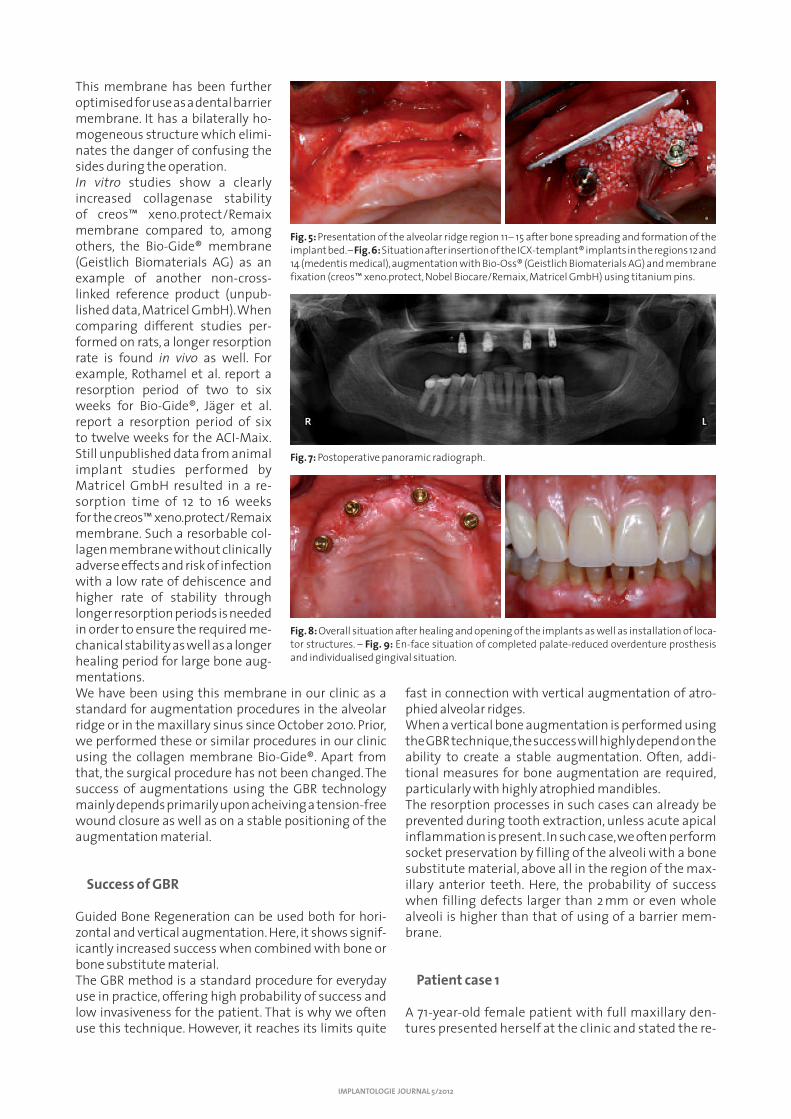
Fig. 1: Electronmicroscopic scan: cross section of the membrane show-ing both surfaces.
jor guided bone augmentation procedures, the neces-sary mechanical stability and a sufficiently protectedspace during bone regeneration. We have been using the Remaix/creos™ xeno.protectmembrane (Matricel/Nobel Biocare) in our clinic as ourclinical standard for augmentative operations of thejaw or maxillary sinus since October 2010. These or sim-ilar operations in our clinic were previously carried outusing the Bio-Gide® (Geistlich Biomaterials) collagenmembrane. Apart from this, the operative procedurehas not been changed. The success of augmentation
with the aid of the GBR technique depends largely onprimary, tension-free wound closure as well as on a stable positioning of the augmentated volume.11,12,21
“Guided Bone Regeneration” can be used for both hori-zontal and vertical augmentation. It is significantlymore successful if it is combined with bone or bonegraft material.7The GBR method is a minimally invasive method with ahigh success rate which is commonly used in everydaypractice.1 For this reason, we use this technique fre-quently. However, it has its limitations, particularly in the field of vertical augmentations of atrophied jaws. Especially in a severely atrophied lower mandible, othermeasures are thus required for bone construction. If avertical bone construction with GBR is carried out, itssuccess depends largely on whether a mechanically stable space for regenerating bone can be maintained(for example by using titanium meshes).2Precautions against the previously described bone re-sorption processes can already be taken directly aftertooth extraction if there is no acute apical inflammation.In such cases, especially in the region of front teeth in the upper jaw, we frequently perform a “socket preser-vation” by filling the alveolus with bone replacementmaterial. When filling defects larger than 2mm or evenentire alveoli, the success rate is higher if an additionalbarrier membrane is used.15
Case 1 (Figs. 2–11)
The 61-year-old patient consulted our clinic with thewish for an aesthetic improvement of the situation ofhis upper front teeth. He was diagnosed to have severeperiodontitis. The prognosis following periodontitis
IMPLANTOLOGIE JOURNAL 5/2011
Fig. 2: Orthopantomograph of the first consultation.
Fig. 3: Orthopantomograph with X-ray measuring template and 5mmsteel balls in regions 13,15.
Fig. 7: Positioning the creos™ xeno.protect/Remaix membrane (Nobel Biocare/Matricel), size 25 x 30mm. – Fig. 8: Filling the “membrane pocket”with BioOss® bone replacement material (Geistlich Biomaterials). – Fig. 9: Crest fixation of the membrane with titanium pins.
Fig.4: The jaw crest in the implant area seems to be wide enough prior to surgical opening of the operation site. – Fig. 5: The jaw crest in the im-plant area after the formation of a muco-periosteal flap. After a vertical alveolotomy to create a plateau, the jaw crest is just wide enough for theinsertion of implants with a diameter of 3.75mm. – Fig. 6: View of the thin buccal bone lamellae after insertion of the implant.

treatment and root canal treatment (tooth 25) statedthat only three teeth (teeth 23–25) were in a states tobe preserved permanently. The teeth which could notbe preserved were extracted during the preliminary pe-riodontitis treatment and a temporary prosthesis wasfitted for two months. As a consequence, the patient decided on a telescopic,removable model cast prosthesis via strategic addi-tional supports with implantation in regions 13 and 15.Two Templant ICX implants (Medentis Medical) with asize of 4.1mm x 12.5mm were planned. However, duringimplantation it became obvious that the implant re-gion in the jaw was not wide enough, even after the creation of a plateau through vertical reduction. Thustwo implants of the same type but with a width of3.75mm were placed. As the buccal bone lamella priorto implantation was only approx. 0.5mm thick, partic-ularly at the level of the final apical third, there was arisk of absorption of the buccal bone around the im-plants. For this reason, we decided on a prophylacticbuccal augmentation with Bio-Oss® (Geistlich Bioma-terials) as well as a Remaix/creos™ xeno.protect mem-brane (Matricel/Nobel Biocare) according to “GuidedBone Regeneration” (GBR) guidelines. Before bone aug-mentation , the buccal bone was perforated into thespongiosa at several locations using a rose-head bur, inorder to allow a blood and cell migration into the boneaugmentation material. The membrane was fixed withtitanium pins in order to form a stable pocket. After pe-riosteal incision, the gingiva was closed with sutures.There were no negative side-effects during woundhealing; everything proceeded as planned with nosigns of inflammation.
Case 2 (Figs. 12–21)
The 75-year-old female patient consulted our clinic withthe wish for a prosthesis with implants in the upperjaw, as her denture was becoming increasingly loose,especially during eating, due to a pronounced resorp-tion processes. Due to the health situation and the ad-vanced age of the patient and in spite of the expectedmajor GBR augmentation, we decided against taking abone block from the lower jaw or even from the iliaccrest. The implantation was to be performed in the re-gions of 12, 14, 22 and 24, as the patient also did notwant a sinus lift operation. In the orthopantomograph,
an adequate height of remaining bone for the inser-tion of implants with a length of 11.5mm was shown.The insertion of four 11.5mm/3.75mm ICX Templantimplants (Medentis Medical) was planned simultane-ously to a ridge augmentation by means of GBR, or asole augmentation without implant insertion if thewidth of the existing bone should be absolutely insuf-ficient. Surgical opening of the jaw revealed that bothsides of the jaw were too narrow for a single-stage pro-cedure with simultaneous augmentation in the plannedarea. For this reason, we decided on vertical and hori-zontal ridge augmentation according to the GBR tech-nique. As a large amount of bone needed to be regen-erated, we harvested autologous bone chips from themandibular ramus on the right side with the aid of theSafescraper (Imtegra). These were mixed with approx-imately the same amount of Bio-Oss® (Geistlich Bio-materials). After buccal perforation of the area to beaugmented with a Rose-head bur, a stable pocket forthe bone mixture was formed on both sides with a
IMPLANTOLOGIE JOURNAL 5/2011
Fig. 10: Tension-free closure of the mucous membrane. Fig. 11: Postoperative orthopantomograph.
Fig. 12: Preoperative orthopantomograph.
Fig. 13: Toothless upper jaw.

IMPLANTOLOGIE JOURNAL 5/2011
25 x 30mm Remaix/creos™ xeno.protect membrane(Matricel/Nobel Biocare) using titanium pins. Aftersubstantial, repeated periosteal incision, the woundedges were closed again and sutured tension-free. Thepatient was prescribed a temporary prosthesis for aten-day period of primary healing. When sutures wereremoved after ten days, the prosthesis was grounddown massively in the augmented region and linedwith soft material. The wound healing phase proceeded without any prob-lems, and implants will be inserted in the augmentedarea after a healing time of at least six months.
Discussion
Since we substituted the previously-used barriermembranes by the new membrane in our clinic, wehave not seen any increase in wound healing prob-lems, pain or failure.
One advantage of the new mem-brane is that during surgery, incontrast to other competitiveproducts, there can be no mix-upof the membrane surfaces. Evenwhen wetted, the membrane hasa higher stability than the colla-gen membranes we have usedpreviously, enabling safer cover-ing and adaptation during GBR. The clinical implementation of the Remaix/creos™ xeno.protectmembrane for augmentation in
the field of implantation shows promising first re-sults. However, evidence-based data from larger-scale clinical case studies need to be collected. Here,the significantly higher membrane stability againstdegradation that has been confirmed in laboratory experiments should also be substantiated clinically.n
Fig. 14: Presentation of the operation site in regions 11–15 after formation of a mucoperiosteal flap. Implantation is not possible because the jawcrest is too narrow. – Fig. 15: Removal of auto logous bone chips from the right mandibular ramus with the Safescraper (Imtegra). – Fig. 16: Buccalperforation of the jaw crest for the extraction of blood and cells from the bone.
Fig. 17: Positioning and fixation of the membrane with titanium pins. –Fig. 18: Extracted bone chips mixed with Bio-Oss® (Geistlich Biomaterials). –Fig. 19: “Membrane pocket” filled with bone mixture.
Fig. 20: Crest fixation of the membrane with titanium pins. – Fig. 21: Tension-free closure of themucoperiosteal flaps. Note the increase in width of the jaw crest!
CONTACT
Dr. med. dent. Bastian WessingPraxisklinik der Zahnheilkunde am Luisenhospital AachenBoxgraben 9952064 Aachen, GermanyPhone: +49 241 [email protected]
author information
n The US Food and Drug Administration recommendsthe use of suitable membranes for bone augmentationin order to prevent bone substitute material from mi-grating into the soft tissue. Resorbable membranes aresuperior to non-resorbable membranes due to their im-proved histocompatibility, lower rates of dehiscence,and easier handling. They have high cell compatibilitywith respect to PDL fibroblasts as well as osteoblasts,and are clearly more biocompatible than PTFE mem-branes. This translates to improved tissue integration,reduced foreign body reactions and, as a result, lowerrates of dehiscence. Moreover, quick transmembranousangiogenesis has been observed, which was associatedwith faster bone regeneration in animal studies. Finally,good form stability of the augmented area is required inorder to prevent collapse of the the space created dur-ing GBR. When using collagen membranes, this is gen-erally achieved by applying the patient’s own bone orbone substitute material under the membrane. Themembrane itself, however, should be of sufficient me-chanical stability to keep the augmentation materialsafely in place until remodelling has advanced to thepoint that a new bone precursor tissue of stable posi-tion is present.Therefore, membranes made of resorbable collagen ofanimal origin are used in most cases in Germany today.The use of such membranes is well documented andbacked up by results showing a high probability of suc-cess.Non-chemically cross-linked collagen membranes havea rate of dehiscence ranging between 22 and 32 per centwith a stable barrier function of approximately four to
six months. It is true that an additional chemical cross-linking results in a longer in situ function of the mem-brane before its resorption, but this is also associatedwith increased rates of dehiscence ranging from 39 to 64 per cent.
Bioresorbable barrier membrane
The novel collagen membrane presented here is a biore-sorbable barrier membrane made of porcine collagenand elastin. It was marketed as a CE-certified class-3medical device in Germany in 2009 under the name Remaix (Matricel GmbH, Herzogenrath Germany) andsince 2013 it is distributed as creos™ xeno.protect (NobelBiocare, Gothenburg, Sweden). The membrane is basedon a Matricel development for the orthopaedic field,where it has been used under the name of ACI-Maix formatrix-induced autologous chondrocyte implantation(MACI) in more than 10,000 patients already since 2002.
IMPLANTOLOGIE JOURNAL 5/2012
Safe Bone Regeneration through a new Collagen MembraneGuided bone regeneration (GBR) as a standard procedure in implant surgery is a well de-scribed and well investigated procedure for the restoration of an adequate implant bed andaesthetically pleasing results. The use of a barrier membrane to prevent quickly proliferat-ing epithelial and connective tissue cells from growing into the implant site is a basic pre-requisite for a sufficient regeneration of bony defects or alveolar ridge augmentation usingguided bone regeneration (GBR).
Dr. med. dent. Bastian Wessing, Dr. med. dent. Martin Emmerich, M.Sc.
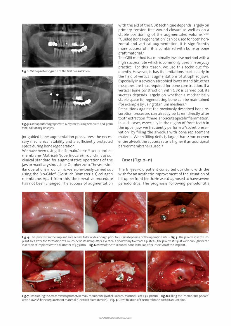
Patient case 1 – Fig. 1: Preoperative panoramic radiograph.
Fig.2: Presentation of the alveolar ridge region 11–15. – Fig. 3:Bone splitting region 11–15 using piezosurgery. – Fig. 4:Formation of the implant bedusing osteotomes.
This membrane has been furtheroptimised for use as a dental barriermembrane. It has a bilaterally ho-mogeneous structure which elimi-nates the danger of confusing thesides during the operation.In vitro studies show a clearly increased collagenase stability of creos™ xeno.protect/Remaixmembrane compared to, amongothers, the Bio-Gide® membrane(Geistlich Biomaterials AG) as anexample of another non-cross-linked reference product (unpub-lished data, Matricel GmbH). Whencomparing different studies per-formed on rats, a longer resorptionrate is found in vivo as well. For example, Rothamel et al. report a resorption period of two to sixweeks for Bio-Gide®, Jäger et al. report a resorption period of six to twelve weeks for the ACI-Maix. Still unpublished data from animal implant studies performed by Matricel GmbH resulted in a re-sorption time of 12 to 16 weeks for the creos™ xeno.protect/Remaixmembrane. Such a resorbable col-lagen membrane without clinicallyadverse effects and risk of infectionwith a low rate of dehiscence andhigher rate of stability throughlonger resorption periods is neededin order to ensure the required me-chanical stability as well as a longerhealing period for large bone aug-mentations.We have been using this membrane in our clinic as astandard for augmentation procedures in the alveolarridge or in the maxillary sinus since October 2010. Prior,we performed these or similar procedures in our clinicusing the collagen membrane Bio-Gide®. Apart fromthat, the surgical procedure has not been changed. Thesuccess of augmentations using the GBR technologymainly depends primarily upon acheiving a tension-freewound closure as well as on a stable positioning of theaugmentation material.
Success of GBR
Guided Bone Regeneration can be used both for hori-zontal and vertical augmentation. Here, it shows signif-icantly increased success when combined with bone orbone substitute material.The GBR method is a standard procedure for everydayuse in practice, offering high probability of success andlow invasiveness for the patient. That is why we oftenuse this technique. However, it reaches its limits quite
fast in connection with vertical augmentation of atro-phied alveolar ridges. When a vertical bone augmentation is performed usingthe GBR technique, the success will highly depend on theability to create a stable augmentation. Often, addi-tional measures for bone augmentation are required,particularly with highly atrophied mandibles.The resorption processes in such cases can already beprevented during tooth extraction, unless acute apicalinflammation is present. In such case, we often performsocket preservation by filling of the alveoli with a bonesubstitute material, above all in the region of the max-illary anterior teeth. Here, the probability of successwhen filling defects larger than 2mm or even wholealveoli is higher than that of using of a barrier mem-brane.
Patient case 1
A 71-year-old female patient with full maxillary den-tures presented herself at the clinic and stated the re-
IMPLANTOLOGIE JOURNAL 5/2012
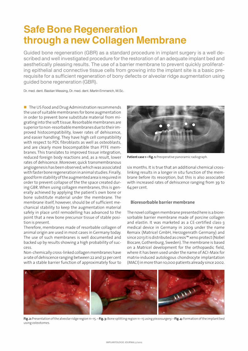
Fig. 5: Presentation of the alveolar ridge region 11– 15 after bone spreading and formation of theimplant bed. – Fig. 6:Situation after insertion of the ICX-templant® implants in the regions 12 and14 (medentis medical), augmentation with Bio-Oss® (Geistlich Biomaterials AG) and membranefixation (creos™ xeno.protect, Nobel Biocare/Remaix, Matricel GmbH) using titanium pins.
Fig. 7:Postoperative panoramic radiograph.
Fig.8: Overall situation after healing and opening of the implants as well as installation of loca-tor structures. – Fig. 9: En-face situation of completed palate-reduced overdenture prosthesisand individualised gingival situation.

quest of a palate-reduced pros-thetic treatment. Due to the fi-nancial situation of the patent, it was decided to perform theminimal version of a palate-re-duced denture in the maxilla. Im-plantation was to be made in re-gions 13, 15, 23, 25. The panoramicradio graph (Fig. 1) showed a suffi-cient residual bone height for the insertion of implants with alength of 11.5mm. It was plannedto insert four 11.5mm/4.1mm ICX-templant® implants(medentis medical). The operative presentation of thealveolar ridge (Fig. 2) showed the alveolar ridge to havea width of only 4mm on both sides at the plannedarea, thus insufficient even for thinner implants ofthis type. Therefore, the decision was made to per-form bone splitting with simultaneous implantationand augmentation (Fig. 3). The planned area wasopened in the centre up to approx. two thirds of theimplant length by using a piezo-electronic bone saw,and then expanded with a chisel. After performing a pilot bore, the implant bed was formed with the desired length and width by using osteotomes (Fig. 4). The four planned implants could be inserted at 30Ncm. The remaining bone gap as well as the buccal bone lamella were augmented with Bio-Oss®(Geistlich Biomaterials AG) and previously collectedblood from the bone bore. Each augmentation was covered with a creos™ xeno.protect/Remaix25x30mm membrane (Figs. 5–7). The region 13, 15 re-quired significant augmentation. Here, the mem-brane was fixed with three titanium pins. Region 23, 25 was augmented safely and closed without strain orthe necessity to use titanium pins.
After a covered healing time of six months, the os-seointegrated implants were opened and providedwith locator structures. The patient was provided witha palate-free overdenture prosthesis on four locators(Figs. 8 and 9).
Patient case 2
A 49-year-old male patient presented himself at theclinic and stated the request of aesthetic improvementof his maxillary anterior teeth (Fig. 10). Tooth 21 showeda dark region above the marginal gingival edge. X-rayshowed an oversized pin design with (after 15 years) in-sufficient metal-ceramic crown treatment. New treat-ment of the tooth with adherence to the biologicalwidth and simultaneous aesthetic improvement of thissituation was not possible (Figs. 11 and 12). The patientwas advised to have tooth 21 replaced by an implant.Given the fact that no apical infection was present, thepatient and the attending dentist decided to perform aninitial augmentation at extraction through socketpreservation. Furthermore, the patient stated the re-quest for a fixed temporary adhesive bridge. Tooth 21 was
IMPLANTOLOGIE JOURNAL 5/2012
Patient case 2 – Fig. 10: En-face situation of the patient at time of presentation at the practiceclinic. – Fig. 11: Situation after dropping out of pin structure shortly before surgery date.
Fig. 15: Probing of alveolus using a periodontal probe; the buccal lamella is still completely present. – Fig. 16: Formation of a buccal muco -periostal pocket. – Fig. 17: Filling of alveolus with Bio-Oss® (Geistlich Biomaterials AG).
Fig.12:Dropped-out massive pin structure with cemented crown, alio loco, no consideration of biological width. – Fig. 13:Atraumatic loosening ofperiodontal fibres by using a narrow periotome. – Fig. 14:“Removal” of root residues.

removed in a tissue-conserving manner by using narrowperiotomes, and the alveolus was filled with Bio-Oss®(Geistlich Biomaterials AG) (Figs. 13–17). The alveolus wasclosed with a Remaix membrane as well as an Ovate Pon-tic adhesive bridge (Figs. 18–21). After opening of the sur-gery site four months after surgery, there was a com-pletely ossified alveolus with conservation of the wholealveolar ridge width (Figs. 22 and 23). An implant sized4.3mm x 13mm (NobelReplace™ Straight Groovy, NobelBiocare) was inserted safely at 40Ncm (Fig. 24).
Discussion
Since the change to this novel membrane at our clinic,we have not recorded any increased wound healingdisturbance, pain, or failure in treatment. The operativehandling of the new membranes is beneficial, as themembrane—contrary to some products of competi-tors—prevents a mix-up of surfaces. The membranealso shows a higher strength after wetting than thepreviously used collagen membrane; therefore, cover-ing and shaping within the context of GBR can be per-formed more safely.The clinical use of the creos™ xeno.protect/Remaixmembrane for augmentation in the field of implantol-ogy shows promising first results. However, well-de-signed clinical studies should be performed to collectevidence-based data. Here, above all, the significantlyincreased implantation duration of this membrane as compared to products of competitors—confirmedwithin the scope of laboratory tests—should be veri-fied clinically. n
IMPLANTOLOGIE JOURNAL 5/2012
Fig. 18: Placement of creos™ xeno.protect/Remaix (Nobel Biocare/Matricel GmbH) membrane using a narrow raspatory, and subsequentfilling of buccal “pocket” with bone substitute material as resorptionprotection of the buccal lamella. – Fig. 19: Fixation suture above aug-mentation and membrane.
Fig. 20:Adhesive bridge fixed with Panavia F (Kuraray company) aftersocket preservation. – Fig. 21:En-face situation six weeks after surgery.Please note the regenerated soft tissue situation (as compared to themucosal colour, Fig. 10).
CONTACT
Dr. med. dent. Bastian WessingPraxisklinik der Zahnheilkunde am Luisenhospital AachenBoxgraben 9952064 Aachen, GermanyPhone: +49 241 [email protected]
author information
Fig.22:Occlusal view of the healed extraction alveolus after a healingperiod of four months. – Fig. 23:Operation site of the ossified alveolus.– Fig. 24:NobelReplace™ Straight Groovy 4.3mm x 13mm implant in-serted at 40Ncm.

1. Aghaloo TL, Moy PK. Which hard tissue augmentation techniques are themost successful in furnishing bony support for implant placement? Int JOral Maxillofac Implants 2007; 22 Suppl:49–70.
2. Bernstein S, Cooke J, Fotek P, Wang HL. Vertical bone augmentation:where are we now? Implant Dent. 2006; 15: 219–28.
3. Brittberg M. Cell carriers as the next generation of cell therapy for carti-lage repair: a review of the matrix-induced autologous chondrocyte im-plantation procedure. Am J Sports Med. 2010 Jun;38(6):1259–71.
4. Bunyaratavej P, Wang HL. Collagen membranes: a review. J Periodontol2001; 72: 215–29.
5. Buser D, Weber HP, Brägger U, Balsiger C. Tissue integration of one-stageimplants: three-year results of a prospective longitudinal study with hollowcylinder and hollow screw implants. Quintessence Int 1994; 25: 679–86.
6. Chen JM, Willers C, Xu J, Wang A, Zheng MH. Autologous tenocyte ther-apy using porcine-derived bioscaffolds for massive rotator cuff defect inrabbits. Tissue Eng. 2007 Jul;13(7):1479–91.
7. Esposito M, Grusovin MG, Kwan S, Worthington HV, Coulthard P. Interven-tions for replacing missing teeth: bone augmentation techniques for dentalimplant treatment. Cochrane Database Syst Rev. 2008; 16 (3):CD003607.
8. Ferreira CE, Novaes AB, Haraszthy VI, Bittencourt M, Martinelli CB,Luczyszyn SM. A clinical study of 406 sinus augmentations with 100%anorganic bovine bone. J Periodontol 2009 Dec; 80: 1920–7.
9. Fiorellini JP, Nevins ML. Localized ridge augmentation/preservation. Asystematic review. Ann Periodontol 2003; 8: 321–7.
10. Friedmann A, Strietzel FP, Maretzki B, Pitaru S, Bernimoulin JP. Observa-tions on a new collagen barrier membrane in 16 consecutively treated pa-tients. Clinical and histological findings. J Periodontol 2001; 72: 1616–23.
11. Fugazzotto PA. GBR using bovine bone matrix and resorbable and non-resorbable membranes. Part 1: histologic results. Int J PeriodonticsRestorative Dent 2003; 4: 361–569.
12. Fugazzotto PA. GBR using bovine bone matrix and resorbable and non-resorbable membranes. Part 2: Clinical results., Int J Periodontics Resto -rative Dent 2003; 6: 599–605.
13. Hämmerle CH, Lang NP. Single stage surgery combining transmucosalimplant placement with guided bone regeneration and bioresorbablematerials. Clin Oral Implants Res 2001; 12: 9–18.
14. Hämmerle CH, Jung RE, Yaman D, Lang NP. Ridge augmentation by apply-ing bioresorbable membranes and deproteinized bovine bone mineral: areport of twelve consecutive cases Clin Oral Implants Res. 2008; 19: 19–25.
15. Hämmerle CH, Chen ST, Wilson TG Jr. Consensus statements and recommended clinical procedures regarding the placement of im-plants in extraction sockets. Int J Oral Maxillofac Implants 2004; 19Suppl: 26–8.
16. Jäger M, Fischer J, Schultheis A, Lensing-Höhn S, Krauspe R. ExtensiveH(+) release by bone substitutes affects biocompatibility in vitro testing.J Biomed Mater Res A. 2006 Feb;76(2):310–22.
17. Jäger M, Degistirici O, Knipper A, Fischer J, Sager M, Krauspe R. Bone heal-ing and migration of cord blood-derived stem cells into a critical sizefemoral defect after xenotransplantation. J Bone Miner Res. 2007 Aug;22(8):1224–33.
18. Jung RE, Hälg GA, Thoma DS, Hämmerle CH. A randomized, controlledclinical trial to evaluate a new membrane for guided bone regenerationaround dental implants. Clin Oral Implants Res 2009; 20: 162–8.
19. McAllister BS. Scalloped implant designs enhance interproximal bonelevels. Int J Periodontics Restorative Dent 2007; 27: 9–15.
20. Moses O, Pitaru S, Artzi Z, Nemcovsky CE. Healing of dehiscence-type defects in implants placed together with different barrier membranes: acomparative clinical study. Clin Oral Implants Res 2005; 16: 210–9.
21. Nevins M, Mellonig JT. The advantages of localized ridge augmentationprior to implant placement: a staged event. Int J Periodontics RestorativeDent 1994; 14: 96–111.
22. Rothamel D, Schwarz F, Sculean A, Herten M, Scherbaum W, Becker J. Bio-compatibility of various collagen membranes in cultures of human PDL fibroblasts and human osteoblast-like cells. Clin Oral Implants Res 2004;15: 443–9.
23. Rothamel D, Schwarz F, Sager M, Herten M, Sculean A, Becker J. Biodegra-dation of differently cross-linked collagen membranes: an experimentalstudy in the rat. Clin Oral Implants Res 2005; 16: 369–78.
24. Siar CH, Toh CG, Romanos G, Ng KH. Subcutaneous reactions and degra-dation characteristics of collagenous and noncollagenous membranesin a macaque model. Clin Oral Implants Res 2011; 22: 113–20.
25. Tal H, Kozlovsky A, Artzi Z, Nemcovsky CE, Moses O. Cross-linked andnon-cross-linked collagen barrier membranes disintegrate followingsurgical exposure to the oral environment: a histological study in the cat.Clin Oral Implants Res 2008; 19: 760–6.
26. Zitzmann NU, Naef R, Schärer P. Resorbable versus nonresorbable mem-branes in combination with Bio-Oss for guided bone regeneration. Int JOral Maxillofac Implants 1997; 12: 844–52.
NOTES
IMPLANTOLOGIE JOURNAL 5/2011
References

REF 76422
Related Documents