NEW JERSEY HAZMAT EMERGENCY RESPONSE COURSE STUDENT GUIDE COURSE NUMBER: 06061 Emegency Department Operations Hazmat/WMD Hospital Provider PRESENTED THROUGH: NEW JERSEY STATE POLICE-HOMELAND SECURITY BRANCH SPECIAL OPERATIONS SECTION, TECHNCIAL RESPONSE BUREAU HAZARDOUS MATERIALS RESPONSE UNIT (HMRU) STUDENT GUIDE 5 th Edition 1004

Welcome message from author
This document is posted to help you gain knowledge. Please leave a comment to let me know what you think about it! Share it to your friends and learn new things together.
Transcript

NEW JERSEY HAZMAT EMERGENCY RESPONSE COURSE
STUDENT GUIDE COURSE NUMBER: 06061
Emegency Department Operations Hazmat/WMD Hospital Provider
PRESENTED THROUGH: NEW JERSEY STATE POLICE-HOMELAND SECURITY BRANCH SPECIAL OPERATIONS SECTION, TECHNCIAL RESPONSE BUREAU HAZARDOUS MATERIALS RESPONSE UNIT (HMRU) STUDENT GUIDE 5th Edition 1004

TABLE OF CONTENTS
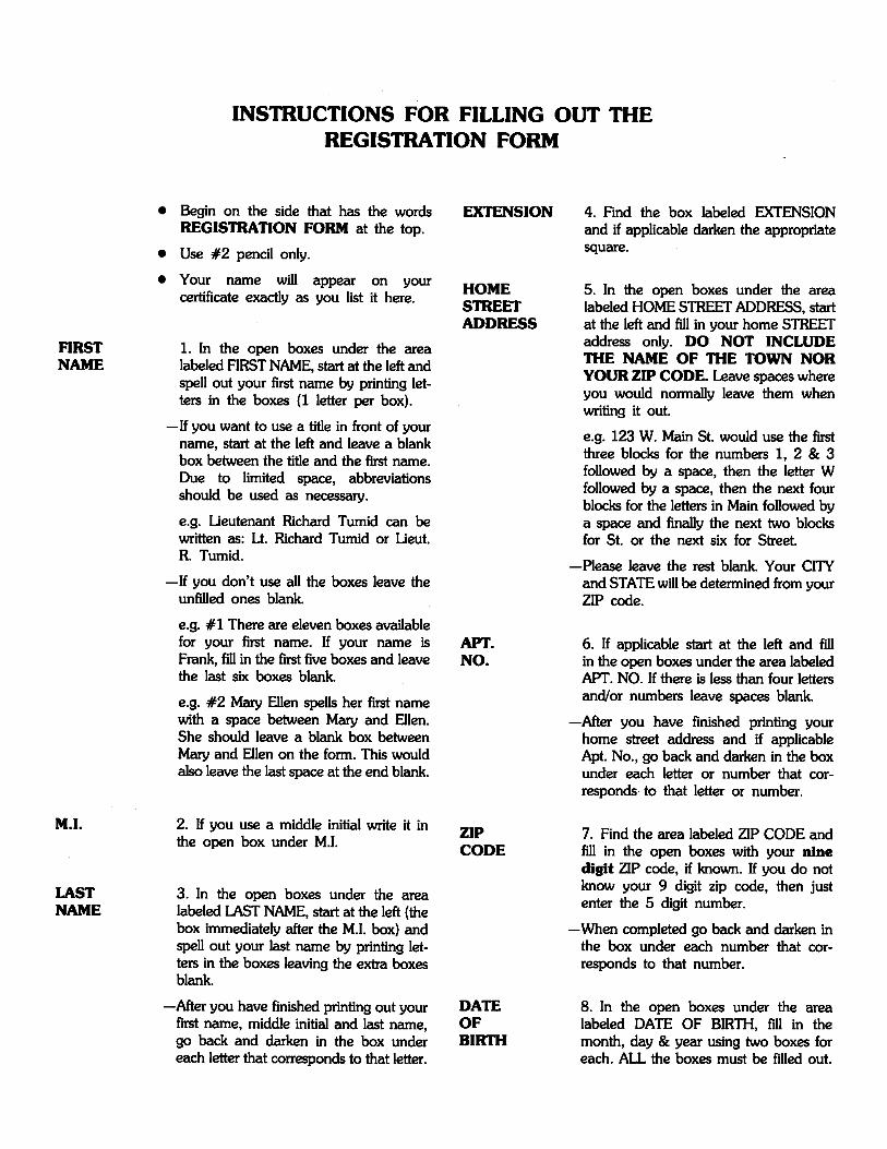
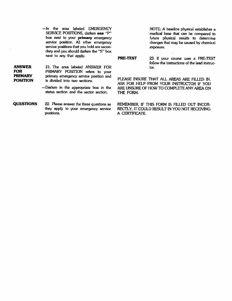
Instructions for Scantron Forms
Introduction ............................................................................................................................ 1
Planning for the Hazmat/WMD Incident ................................................................................ 3
Personal Protective Equipment............................................................................................... 7
Emergency Department Decontamination............................................................................. 21
Toxicology ............................................................................................................................... 41
Treatment Protocols ............................................................................................................... 57
Hazardous Materials Contamination....................................................................................... 79
Chemical Agent Contamination ..............................................................................................107
Biological Agent Contamination..............................................................................................163
Explosive Agents .....................................................................................................................223
Radiological Agents .................................................................................................................231
Appendices..............................................................................................................................245

FUNDING FOR THE DEVELOPMENT OF THIS MANUAL WAS PROVIDED BY THE
FEDERAL EMERGENCY MANAGEMENT AGENCY (FEMA) UNDER THE TERRORISM
CONSEQUENCE MANAGEMENT PREPAREDNESS ASSISTANCE GRANT.
The NJOEM Domestic Preparedness/Hazardous Materials Emergency Response Planning Unit
would like to acknowledge the following agencies/individuals for their assistance in the develop-
ment of this manual:
Dr. William Gluckman, DO, Emergency Department Physician,
University of Medicine & Dentistry of NJ–University Hospital
Federal Emergency Management Agency, Emergency Response to Terrorism Job Aid
Military Medical Operations Office, Armed Forces Radiobiology Research Institute
Naval School of Health Sciences, Management of Chemical Warfare Injuries
U.S. Military Field Manual, Treatment of Biological Warfare Agent Casualties
NJ Department of Health and Senior Services, Epidemiology, Environmental & Occupational Health
Mr. Eugene J. O’Neill, NREMT-B, University of Medicine & Dentistry of NJ–University Hospital
Although the information set forth in this program is presented in good faith and believed to be
correct, persons or agencies using this information must make their own determination as to its
suitability for their purposes. This document may be reproduced in part or entirely, provided its
use clearly indicates that it was prepared by the Domestic Preparedness/Hazardous Materials
Emergency Response Planning Unit, New Jersey Office of Emergency Management, Division of
State Police.


INTRODUCTION
The presence of hazardous materials or toxic chemicals at an incident location or other emer-
gency situation adds a new dimension of risk to those handling and treating casualties. The fun-
damental difference between a hazardous materials incident and other emergencies is the poten-
tial for acute risk from contamination to both patient and responder. In some cases, traditional
practices must be altered to avoid compounding a critical situation.
Hospital emergency departments must protect their personnel and other people within the hos-
pital, while providing the best care for the chemically contaminated patient. This guide is
intended to help hospital emergency departments plan for incidents that involve haz-
ardous materials and improve their ability to respond to these incidents appropriately.
To ensure appropriate and timely patient care, as well as optimal worker protection, emergency
personnel must have an understanding of decontamination procedures and personal protective
equipment that they do not generally receive in the course of their routine professional training.
They should also be aware of community resources that could be called upon to assist in emer-
gency response.
Current training curricula for emergency physicians, nurses, and emergency medical technicians
(EMTs) often do not adequately prepare these professionals to either manage the contaminated
individual or decontaminate patients exposed to toxic substances. High-quality, specific, and con-
cise guidance is needed to describe appropriate procedures to be followed by emergency med-
ical personnel to safely care for a patient, as well as to protect equipment, hospital personnel, and
others from risk of exposure.
This guide for emergency department personnel is designed to familiarize readers with the con-
cepts, terminology, and key considerations that affect the management of incidents of chemical
contamination. It has been developed not only to present uniform guidance for emergency care
of chemically contaminated patients, but also to provide basic information critical to advance
planning and implementation of emergency medical services’ (EMS) strategies. It is intended to
illustrate the characteristics of hazardous materials incidents that mandate modifications to tradi-
tional emergency response and the preparatory actions that should be taken to respond effec-
tively to hazardous materials incidents.
All hospital and community emergency response systems may not be prepared to respond to a
hazardous chemical incident to the same degree. This document may be used to assess capabil-
ities with respect to potential community hazards and to develop response plans using national
and community-specific resources. Worker safety and training are also key factors in effective
management of medical emergencies. This document is intended to provide source material for
developing local training and safety protocols.
1


PLANNING FOR
THE HAZMAT/
WMD INCIDENT
3


ADVANCE PLANNING FOR AHAZMAT/WEAPONS OF MASS DESTRUCTION
(WMD) INCIDENT
Advance pre-planning is the most essential phase of preparation for a HAZMAT/WMD incident.
I. External Pre-planning
External pre-replanning should involve coordination with local agencies such as fire, EMS, and
HazMat teams, along with local chemical or industrial sites. It will be important to establish an
integrated plan that outlines a common response philosophy AND the roles and responsibilities
that should be taken in the event of a HAZMAT/WMD case.
For example, it will be important to know the following types of information in the preparation
for a HAZMAT/WMD incident:
1. What are the sources of Hazardous Material accidents in the community?
2. Where is the nearest HazMat team located?
3. Which local EMS organization and fire departments are trained and equipped to
respond to a HazMat incident?
4. Do any of the local industrial sites have HazMat teams that can be called upon for
assistance in response to a HazMat incident?
5. How will victims from a HAZMAT/WMD incident be handled?
6. Will all contaminated/injured patients be decontaminated prior to transport to the
hospital? Does this include trauma patients? Are there any exceptions? Who will per-
form field decontamination?
7. To what extent will patients be decontaminated? (i.e. gross, partial or fully decontam-
inated)
8. Which local hospitals are trained and equipped to handle hazardous material accident
victims?
9. How will a hazardous material incident involving mass casualties be handled? How will
the patient load be dispersed in this type of incident?
5
6
II. Internal Pre-planning
A formal written HAZMAT/WMD Response Plan should be developed as part of the overall
Hospital Disaster Plan. It should be developed in conjunction with the local EMS organization, fire
department, HazMat team and local industry. It should be comprehensive in addressing all possi-
ble situations involving hazardous material accident patients:
• walk in patient(s)
• announced pre-hospital delivered patients
• unannounced pre-hospital delivered patients
• stable vs. unstable patient
• mass casualties incident
The formal plan should address the following types of issues:
A. Determine the responsibilities of the department ranging from emergency medicine, admin-
istration and security to clinical specialist such as toxicology, laboratory medicine and occu-
pational medicine.
B. Determine the responsibilities of emergency department personnel who will be involved in
the handling of a hazardous material accident patient.
C. Selection and set-up of an outside or external decontamination area.
D. Selection and set-up of an inside or internal decontamination area.
E. Selection of decontamination equipment and supplies.
F. Selection of proper personal protective clothing.
G. Establish pre-hospital notification procedure.
H. Development of a list of resource organizations that can be contacted in the event of a
HazMat incident.
REMEMBER: Coordinate your planning efforts with JCAHO and Office of Emergency
Management Standards.
Essential Public Health Services
• Monitor health status to identify community health problems
• Diagnose and investigate health problems and health hazards in the community
• Inform, educate, and empower people about health issues
• Mobilize community partnerships to identify and solve health problems
• Develop policies and plans that support individual and community health efforts
• Enforce laws and regulations that protect health and ensure safety
• Link people to needed personal health services and assure the provision of health care when
otherwise unavailable
• Assure a competent public health and personal health care workforce
• Evaluate effectiveness, accessibility, and quality of personal and population-based health
services
• Research for new insights and innovative solutions to health problems
From “Public Health in America.” Public Health Functions Steering Committee, 1994.
PERSONAL PROTECTIVE
EQUIPMENT (PPE)
7


Introduction
Dealing with a HAZMAT/WMD incident is risky business. The first responder initially deals with
unknown factors which can clearly be hazardous to his health. As such, he must handle the inci-
dent differently than he would normally and with much more caution.
Greater care should be given to personal protection of the emergency services personnel with
more detail given to approach and operational procedures. The availability of proper protective
equipment, or the lack of it, has a direct bearing on how and if an approach is made; what the
incident mitigation objectives can be; how work area assignments are made and defined; and how
the establishment of working limits (operating time, work zones, and personal protection) are
determined.
The most critical factor here is the life threat to E.D. personnel. Without knowledge of exactly
what personal protection equipment is necessary for the materials involved in the incident and
the protective limits of the equipment, the incident response team can get into immediate seri-
ous trouble. The first concern should be the proper protection of the first responder.
Federal Regulations Pertaining to the Use of Personal
Protective Equipment (PPE)
The term Personal Protective Equipment (PPE) is used in this document to refer to both personal
protective clothing and equipment. The purpose of PPE is to shield or isolate individuals from the
chemical, physical, and biological hazards that may be encountered at a hazardous materials inci-
dent.
OSHA standards mandate specific training requirements (8 hours of initial training or sufficient
experience to demonstrate competency) for employees engaged in emergency response to haz-
ardous substances incidents at the first responder operations level. Additionally, each employer
must develop a safety and health program and provide for emergency response. These standards
also are intended to provide additional protection for those who respond to hazardous materials
incidents, such as firefighters, police officers, and EMS personnel. OSHA’s March 6, 1989, 29 CFR
[1910.120] final rule as it applies to emergency medical personnel states that: “Training shall be
based on the duties and functions to be performed by each responder of an emergency response
organization (p. 9329).
9

Training Is Essential Before Any Individual Attempts To
Use PPE
No single combination of protective equipment and clothing is capable of protecting against all
hazards. Thus, PPE should be used in conjunction with other protective methods. The use of PPE
can itself create significant worker hazards, such as heat stress, physical and psychological stress,
and impaired vision, mobility, and communication. In general, the greater the level of PPE pro-
tection, the greater are the associated risks. For any given situation, equipment and clothing
should be selected that provide an adequate level of protection. Over-protection can be as haz-
ardous as under-protection and should be avoided. Personnel should not be expected to use PPE
without adequate training. The two basic objectives of any PPE program should be to protect the
wearer from safety and health hazard and to prevent injury to the wearer from incorrect use
and/or malfunction of the PPE. To accomplish these goals, a comprehensive PPE program should
include: hazard identification; medical monitoring; environmental surveillance; selection, use,
maintenance, and decontamination of PPE; and training.
Protective Equipment for ED Personnel
The first consideration of all E.D. personnel must be their own ability to survive the incident. That
thought sounds simple enough. You would think that if you’re out in the E.D., some distance from
the incident in fresh air, that you’re okay. This may not necessarily be true! The products involved
in hazardous materials incidents can be colorless, odorless, tasteless, and, you may not feel their
presence as they envelop you in a destructive cloud that may not be noticed until years later. The
question of E.D. personnel safety is dependent upon three factors:
1. what products are involved in the incident
2. what are their associated risks under the incident conditions
3. what level of protection should operating personnel have to deal with the incident.
The first and second factors can only be answered through discovery of what products are
involved. If, and as long as, the products remain unknown, then a worse case probability
should be assumed. Once the product or products are known and the risks have been evaluated
accordingly, then the level of personal protection can be set to match the needs of the opera-
tional objectives.
All personnel must therefore understand what constitutes personal protection or personal pro-
tective equipment. This includes all personnel who work in or near the incident site, regardless
of whether they be the nurses, physicians, emergency medical services personnel, x-ray, lab or
respiratory services.
10

The typical first responder will arrive on the scene in the least acceptable level of protection. If
it is a police officer it will be a blue uniform with a badge and a side arm for personal protection.
Emergency medical personnel will report to the scene with a medical kit, a stethoscope and
other implements sticking out of numerous pockets. The firefighter will roll onto the scene in full
structural fire fighting gear with hoses and tools ready at hand. Unless the product exposure
risk is known and determined to be no risk at all, or of little risk to personnel, these
people should be denied access to the incident site, and their proximity to the opera-
tional area should be clearly defined and closely monitored.
The psychological feeling of invulnerability is a significant factor when dealing with emergency
services personnel. The danger must always be in the mind of the incident commander as a con-
cern during operations.
Everyone is vulnerable unless they:
1. are properly protected before they enter the incident site
2. are aware of the risks present at the site
3. know what objectives can be realistically attained.
It should also be noted here that no one type of personal protection will satisfy every con-
dition encountered at hazardous materials incidents. The Mayor may find his sport jacket dis-
integrating; the police officer may find his badge and side arm turning green; the EMS personnel
may find themselves unable to dispense medical treatment because they cannot see or breathe,
and the firefighter may be running away from the scene at the best possible speed. Obviously,
selecting the appropriate level of personal protective equipment necessary for the incident and
properly wearing it, is the key to a safe and effective operation.
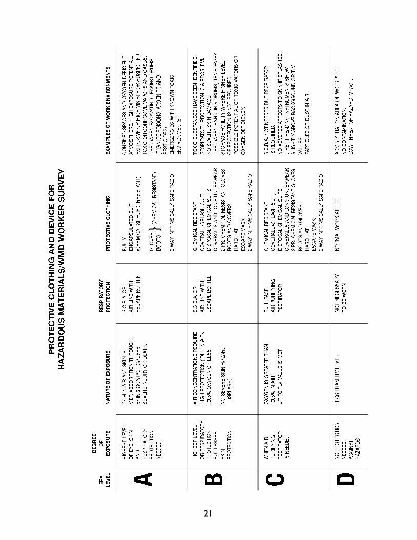
Clothing which is specifically designed for hazardous materials incidents, and for use with specif-
ic types of chemicals, falls into four categories: Level A, Level B, Level C, and Level D. The pre-
dominant physical, chemical, and toxic properties of a chemical, or chemicals, involved in a haz-
ardous materials incident will dictate the specific type of chemical protection required. The
guidelines for the use of these various levels of protection are as follows:
11

Level A: MAXIMUM PROTECTION
Should be worn when the highest level of respiratory, skin, and eye
protection is required.
Level A Conditions:
• Unknown gas concentrations.
• Known extremely toxic or corrosive gases.
• Possible or expected skin exposure to toxic or corrosive liquids, gases or
solids.
• IDLH Atmospheres
Level A Configuration:
• Fully-encapsulating chemical resistant suit completely encloses user and SCBA.
Level B: HIGH RESPIRATORY PROTECTION
Should be worn when the highest level of respiratory protection is needed
but a lesser level of skin protection is required. (SPLASH PROTECTION)
Level B Conditions:
• Known contaminant levels below IDLH concentrations.
• Atmosphere with less than 19.5% oxygen.
• Chemical concentrations which are above the TLV level.
Level B Configuration:
• Chemical resistant clothing including boots and gloves, that generally do not
fully enclose user and SCBA.
12

Level C: LIMITED RESPIRATORY PROTECTION
Should be worn when the criteria for using air-purifying/respirators has
been met.
Level C Conditions:
• Greater than 19.5% oxygen.
• Contaminant level below IDLH and above TLV.
• Skin contact hazards are minimal or do not exist.
Level C Configuration:
• Level B and Level C differ only in type of respiratory protection required. The
chemical protective clothing requirements are the same.
Level D: MINIMUM PROTECTION
Should be worn only as a work uniform and not on any site with a
respiratory or skin hazard.
Level D Conditions:
• No possibility of respiratory exposure.
• No possibility of skin exposure.
• No contaminant levels below TWA.
Level D Configuration:
• Standard Work Uniform, including structural firefighter protective equipment.
13
Note: OSHA Final Rule 29 CFR Part 1910(q)(3)(iv)
(iv) Employees engaged in emergency response and exposed to hazardous substances
presenting an inhalation hazard or potential inhalation hazard shall wear positive
pressure self-contained breathing apparatus while engaged in emergency response,
until such time that the individual in charge of the ICS determines through the use
of air monitoring that a decreased level of respiratory protection will not result in
hazardous exposures to employees.
NFPA CHEMICAL PROTECTIVE CLOTHING STANDARDS
The National Fire Protection Association has completed the development and publishing of three
(3) national standards regarding chemical protective clothing for use during hazardous chemical
emergencies.
NFPA 1991 Standard:
This standard is for specifying the design and performance criteria for a chemical protective gar-
ment that is intended to be used in a gaseous or vapor atmosphere of chemicals. This garment
must be totally encapsulating.
NFPA 1992 Standard:
This standard is for the design and manufacture of a garment that did not have to meet the rigid
permeation resistance requirements found in the 1991 standard. In the 1992 standard, Standard
on Liquid Splash-protective Suits for Hazardous Chemical Emergencies, the emphasis was basi-
cally on two things:
1. single to multi-piece garments
2. suitable chemical test that reflected resistance to liquids.
Its use is for liquid splash environments only.
NFPA 1993 Standard:
This standard deals with support functions and is described as hazardous chemical operations
involving controlled chemical uses or exposures in non-flammable atmospheres with minimum
threats to loss of life, personnel injury, or damage to property or to the environment. Functions
include, but are not limited to, decontamination, remedial cleanup, and training.
14

PROTECTION FROM CHEMICAL WARFARE AGENTS
Military-issued equipment to protect against these agents varies widely based on the level of
anticipated exposure. Civilian activities in the presence of these materials are regulated by HAZ-
WOPER, which is more stringent than military standards. As in any chemical emergency, use the
highest level of protection available until the chemical is identified. Modify that level of protection as
appropriate after determining what chemicals are present. For example, nerve and blister agents
require SCBA with Level A protection. Other toxic chemicals may require a lower level of pro-
tection.
The level of protection necessary for the hazardous materials responder at an incident should be
based on the following factors which must be critically assessed:
A. The type and measured concentration of the chemical substance in the ambient
atmosphere and its toxicity.
B. The potential for exposures to substances in the air; to splashes of liquids; and to
direct contact with materials for substances due to the work being done at the inci-
dent site.
When To Remove Personal Protective Equipment (PPE)
If It Has Been Contaminated
There is always the possibility that circumstances will cause PPE to become contaminated despite
all precautions. Personnel should continually check each other to detect any contamination. The
question of exactly when it is safe to remove contaminated PPE is dependent on several factors
which can become quite complicated. The scope of the incident and the probability that multiple
chemicals are involved must be considered. The dilemma goes beyond “when” to: where can
protective equipment be removed; why should it always be removed when you leave the inci-
dent area; what should be removed based on the conditions; and who should do the removal of
the equipment. Who, what, when, where, why and how are all critical questions which must be
answered when dealing with the removal of personal protective equipment.
The removal of personal protective equipment should never be done within the incident “hot
zone” or in any contaminated area until recognized professionals have determined, through the
use of appropriate equipment, that the hazard risk has been removed. The incident commander
is responsible for insuring that incident operations in the work area, the decontamination area,
and any other areas used during the incident are safe.
If personal protective clothing and equipment is removed within the incident site, even where it
has been declared safe, incident commanders must continue to monitor personnel, who should
be checking each other, to insure that symptoms of exposure are not becoming apparent. There
15
is always the chance that something was missed. A test may have been performed incorrectly or
a testing device may fail. The final responsibility again lies with the incident commander.
Regardless of the type of contaminated protective clothing, the removal of the protective enve-
lope should be a closely monitored and planned exercise. It should only be done when it has been
declared safe to do so, and only in an area which has been specifically designated and designed
for the purpose. Where the risks to health are unknown or found to be serious—great care must
be taken in removal supervision and personnel safety. Personnel are not safe until they have
removed their protective clothing an equipment, and are returned to a safe and clean environ-
ment.
Respiratory Protection
The use of respiratory protection at a hazardous materials incident is mandatory. The level
(degree) of respiratory protection must be in compliance with both OSHA regulations, NIOSH
guidance documents, standard operating procedures, and, most of all, be suited for the hazard
and the wearer. Air purifying respirators (APR’s) and self-contained breathing apparatus (SCBA)
are the only two forms of respiratory protection that is addressed.
Air Purifying Respirators (APR’s)
The use of APR’s is limited to the available approved cartridges or canisters. Both cartridges and
canisters have very limited use, if used at all, during a hazardous materials incident. This is due to
several very critical factors:
1. APR’s are negative system, thus allowing for infiltration of contaminated air into the
mask,
2. APR’s have very limited use times, which does not afford the wearer any substantial
protection,
3. APR’s require individual fit testing prior to actual use and wearing,
4. APR’s do not protect the wearer from unknown air contaminants,
5. APR’s ARE NOT APPROVED FOR USE BY EMERGENCY RESPONDERS AT A HAZ-
ARDOUS MATERIALS INCIDENT.
6. You cannot be sure that the contaminants at the emergency will not elevate nor con-
trol the oxygen content of the atmosphere.
As stated in 4 above, in order for APR’s to provide the safe and proper level of protection nec-
essary for the wearer to be protected, the wearer must know both the contaminant type and
concentration. This is not the case for the emergency worker or hazardous materials responder.
16

As such, this form of respiratory protection is reserved for use by those workers that are outside
both the hot and warm zone, and who have been properly fit tested as well as supplied with the
appropriate canister or cartridge, based upon verifiable air monitoring. ONLY UNDER THE
DIRECT SUPERVISION OF THE ON SCENE COORDINATOR OR OTHER HEALTH OR
SAFETY OFFICER CAN THESE DEVICES BE USED AT A HAZARDOUS MATERIALS INCI-
DENT.
Self-Contained Breathing Apparatus (SCBA)
The SCBA affords the wearer the best, and highest, level of respiratory protection (Level B and
Level A). It provides the wearer with his or her personal air supply, totally segregated from the
environmental air.
Although there exist various makes, models, styles, and manufacturers, the important thing to
remember is that there exists only three types:
1. Re-breathers,
2. Demand,
3. Pressure Demand.
RE-BREATHERS are basically air generators. The wearer is supplied with a closed, recirculating
system, whereby exhaled air is sent through a carbon dioxide scrubber, and returned, after a
small “injection” of oxygen. The wearer also carries a small canister of oxygen in the unit. THESE
UNITS ARE NOT TOTALLY POSITIVE PRESSURE AND ARE NOT APPROVED FOR HAZ-
ARDOUS MATERIAL RESPONDERS.
The DEMAND type, NOT APPROVED FOR USE AT HAZARDOUS MATERIAL INCIDENTS,
only provides positive pressure to the user upon exhalation. As such, the possibility exists that
the wearer may breathe contaminated air.
The PRESSURE DEMAND type is the ONLY TYPE APPROVED FOR USE AT HAZARDOUS
MATERIAL INCIDENTS, since positive pressure is always present in the system, thus preventing
the wearer from inhaling environmental air.
Lastly, the hazardous materials responder may be subject to or required to work using an air line
system. In this set-up, the worker is “tethered” to a fixed air supply with a pressurized airline.
The wearer may also be equipped with a dual mode operating SCBA or an escape pack.
However, UNDER ANY AIRLINE SET-UP THE WORKER MUST BE SUPPLIED WITH A MINI-
MUM OF A FIVE MINUTE ESCAPE PACK. Normally, however, this operational set-up is used
during extensive containment or remedial operations.
17

The Hazardous Materials Involved:
The form of hazardous material involved will have a direct bearing on the choice of personal pro-
tective equipment, or the decision to withdraw from the area. These are three broad categories
of hazardous material to consider: chemical materials, biological (etiologic) materials, and
radioactive materials.
These categories can be defined as follows:
1. Chemical Materials: Are materials which are hazardous because of their chemical
and physical properties.
2. Biological Materials: Are organisms which can have a dangerous effect on life or the
environment, and they can exist in normal ambient environments.
3. Radioactive Materials: These are materials which emit ionizing radiation.
Each of these categories and the risks associated with that particular category of hazardous mate-
rial will influence the choice of personal protective equipment. In addition, the type of material,
as referenced to these categories, can have far reaching effects on how personal protective
equipment is used (operationally), how and whether it can be decontaminated, and whether it
can be reused during the operation. An incident involving radioactive material, for example, can
lead to the disposal of all personal protective equipment utilized during the incident—and it can
never be used again . This can certainly be an expensive proposition for many communities.
The physical state of the hazardous material involved is also a factor of concern in choosing pro-
tective equipment. Materials, or elements, can be classified into three basic states of matter:
gases, liquids, and solids. Each of these states can affect your choice of equipment and how you
wear it. As an example, large solids are not as much of a problem as liquids, gases or fine dusts
(solid particles) and vapors, which can permeate or penetrate protective clothing as well as con-
taminate it.
The Means By Which Personal Protective Equipment
Performance May Become Compromised
Personal protective equipment used in the E.D. may become defective leaving incident person-
nel vulnerable to the life threatening effects of hazardous chemicals. Personal protective equip-
ment must be inspected on a regular basis to determine if its reliability meets the minimum pro-
tection requirements to sustain the protective envelope.
Personal protective equipment may be affected in the following ways:
18

Chemical resistance is the ability of the chemical material or materials which make up the pro-
tective clothing and equipment to prevent or reduce degradation and permeation of the fabric by
the attack chemical. In the case of structural fire fighting clothing this ability is extremely limited
as compared to the numerous chemical products which may affect its integrity.
Degradation is a chemical action involving the molecular breakdown of the material due to con-
tact with a chemical.
Permeation is a chemical action involving the movement of chemicals, on a molecular level,
through intact material. There usually is no indication that this process is occurring.
Penetration is the movement of material through a suit’s closures, such as zippers, buttonholes,
seams, flaps or other design features. This also includes loose stitching, and rips and tears in per-
sonal protective clothing.
19

20
21

EMERGENCY
DEPARTMENT
DECONTAMINATION
21


Introduction
DEFINITION
Decontamination is the process of removing potentially harmful contaminants from exposed indi-
viduals and equipment in order to reduce the spread of contamination in the work area and to
prevent inadvertent and unnecessary contact with contaminated materials.
Personnel should not handle a contaminated suit, tool, or person without proper protective
equipment. Failure to do so may lead to skin absorption or inhalation of the contaminant, result-
ing in injury, illness, or death.
Not every patient you deal with will be contaminated. In fact, the majorityof your patients will
not be contaminated and can be handled in the routine fashion. However, until proven other-
wise, you must assume that every patient is contaminated. Every attempt must be made to keep
contaminated patients separated from those who are uncontaminated. This is best achieved by
the use of at least twoEMS units at the site of the emergency. One unit should be available to be
dedicated to the treatment, care, and transportation of contaminated patients (if required) while
the other should treat only those who have not been contaminated and do not require special
handling procedures. A third may be needed for medical monitoring of personnel.
Units that have been set up and designated to handle contaminated patients need to be identi-
fied to EMS personnel and others on the site. One of the more popular means in use is to place
a red “X”over the Star-of-Life symbol on the four sides of the vehicle. Red duct tape, or any red
plastic or cloth tape can serve this purpose. This identification also becomes important upon
arrival at the medical facility. Special entry locations may have been set up to deal with the con-
taminated patient so traffic control officers can direct marked units to the appropriate locations.
THE CONCEPT OF SECONDARY CONTAMINATION
An essential question to ask is, “What is the risk of secondary contamination(to rescuing person-
nel, transport vehicles, hospital emergency departments) from this chemical?” It is traditionally
axiomatic in hazardous materials emergency management that chemicals should be considered
both highly toxic and highly contaminating to personnel, vehicles, and the environment. However,
a great many chemicals are very highly toxic only in the high concentrations found in the imme-
diate exposure area (hot zone) but pose little or no riskto persons outside the hot zone. Small
amounts of some chemicals may produce relatively little acute toxicity, but because they are sus-
pected of causing cancer or other chronic disease they are considered to create a risk of sec-
ondary contamination.
23

Tables 1 and 2 list selected examples of hazardous substances which carry a high vs. a low risk
for secondary contamination. The lists are meant to be illustrative, not exhaustive. Note that high-
ly toxic chemicals may be found in either list. The Haz Mat Team, Regional Poison Control Center
or County Health Department can assist you in determining the potential for secondary contam-
ination of other hazardous materials.
SUBSTANCES WITH SERIOUS POTENTIAL FOR SECONDARY
CONTAMINATION:
Unless the victim has been properly decontaminated, substances like those listed in Table 1 may
persist in significant amounts on the victim’s clothing, skin, hair, or personal belongings, and may
jeopardize health care workers or other attendants. Recommended protective gear should be
worn. Reducing the potential for chemical exposure from any form of mouth-to-mouth resusci-
tation, including use of pocket one-way valve mouth-to-mouth resuscitation devices should be
carefully considered when the victim has been exposed to one of the listed gases. If resuscitation
efforts are necessary, a bag valve mask with reservoir device connected to oxygen, should be
applied to the patient. Contact with even lightly contaminated skin or clothing should be mini-
mized prior to decontamination. Proper decontamination by adequately protected personnel must be
carried out before the victim is treated by prehospital or emergency department personnel.
Table 1: Substances with a High Risk for Secondary Contamination
Examples:
• Acids, alkali & corrosives (if concentrated)
• Asbestos (large amounts, crumbling)
• Cyanide salts & related compounds (e.g., nitriles) and hydrogen cyanide gas
• Hydrofluoric acid solutions
• Nitrogen-containing and other oxidizers which may produce methemoglobinemia
(aniline, aryl amines, aromatic nitro-compounds, chlorates, etc.)
• Pesticides (organophosphates)
• PCBs (polychlorinated biphenyls)
• Phenol and phenolic compounds
• Many other oily or adherent toxic dusts and liquids
• Radioactive material
24
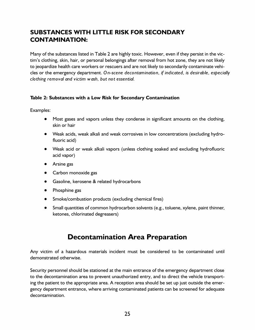
SUBSTANCES WITH LITTLE RISK FOR SECONDARY
CONTAMINATION:
Many of the substances listed in Table 2 are highly toxic. However, even if they persist in the vic-
tim’s clothing, skin, hair, or personal belongings after removal from hot zone, they are not likely
to jeopardize health care workers or rescuers and are not likely to secondarily contaminate vehi-
cles or the emergency department. On-scene decontamination, if indicated, is desirable, especially
clothing removal and victim wash, but not essential.
Table 2: Substances with a Low Risk for Secondary Contamination
Examples:
• Most gases and vapors unless they condense in significant amounts on the clothing,
skin or hair
• Weak acids, weak alkali and weak corrosives in low concentrations (excluding hydro-
fluoric acid)
• Weak acid or weak alkali vapors (unless clothing soaked and excluding hydrofluoric
acid vapor)
• Arsine gas
• Carbon monoxide gas
• Gasoline, kerosene & related hydrocarbons
• Phosphine gas
• Smoke/combustion products (excluding chemical fires)
• Small quantities of common hydrocarbon solvents (e.g., toluene, xylene, paint thinner,
ketones, chlorinated degreasers)
Decontamination Area Preparation
Any victim of a hazardous materials incident must be considered to be contaminated until
demonstrated otherwise.
Security personnel should be stationed at the main entrance of the emergency department close
to the decontamination area to prevent unauthorized entry, and to direct the vehicle transport-
ing the patient to the appropriate area. A reception area should be set up just outside the emer-
gency department entrance, where arriving contaminated patients can be screened for adequate
decontamination.
25

A decontamination area should be large enough to facilitate decontamination of more than one
patient and accommodate the many personnel involved in patient treatment and contamination
reduction. The ventilation system should either be separate from the rest of the hospital or
turned off in order to prevent spread of airborne contaminants throughout the facility. The best
place (weather permitting) to evaluate and initially treat contaminated patients is outside where
ambient ventilation will keep cross-exposure low. Some hospitals have radiation decontamination
facilities that can be used with minor changes. An outside or portable decontamination system is
a viable substitute and would aid in preventing contamination of the emergency department and
other patients. A practical alternative for facilities with limited resources is to have a warm show-
er nozzle, soap, a wading pool, and plastic garbage bags in a predesignated area outside the emer-
gency department back door. The patient may be able to remove his or her own contaminated
clothing, place it in a double bag, and do his or her own soap and water decontamination. A par-
tial tent or curtain can provide privacy for the patients. In most circumstances, ordinary hospital
gowns, plastic goggles, and plain latex gloves will adequately protect hospital staff in case they
have to assist the patient in removing soaked clothing, wash exposed skin and hair, or perform
eye irrigation. With large amounts of concentrated corrosives or very oily materials, such as pes-
ticides, disposable CPC and unmilled nitrile gloves will offer additional protection. If it is antici-
pated that your facility is likely to receive heavy contaminated patients who have not received
prior decontamination, then it may be appropriate to purchase appropriate protective gear and
to fit and train emergency department staff in its use. However, no person should wear and use
specialized PPE, especially respiratory protective gear, without prior training.
To prevent unnecessary contamination, all nonessential and nondisposable equipment should be
removed from the decontamination area. A “clean” member of the staff should stand on the clean
side of the decontamination area to hand in supplies and receive medical specimens.
DECONTAMINATION PROCEDURES
Hazardous materials incidents involve numerous on-site problems and operational concerns.
Common to all these responses is the threat of contamination. Decontamination must be con-
sidered an essential part of hazardous materials response operations. This module will detail the
purpose and steps taken in field decon operations.
Personnel may become contaminated in a number of ways including:
• contacting vapors, gases, mists or particles in the air
• being splashed by materials during rescue or containment operations
• walking through puddles of liquids or on contaminated soil
• treating contaminated patients
• using contaminated instruments or equipment
26

Decontamination is the process of making response personnel, victims and equipment free from
contamination by eliminating or reducing harmful substances to a safe level. Response team per-
sonnel must undergo decon prior to removing their protective equipment. Victims need to be
decontaminated before being turned over to EMS transport personnel. Equipment must be thor-
oughly cleaned so that its subsequent use will not lead to a spread of contamination.
Different chemical threats require varying levels of decon. In cases of extremely hazardous or
unknown substances, the following minimum decon procedures should be complied with:
1. Establishment of an entry/exit point: This point will be used by all personnel to enter
and exit the area of contamination. The use of one entrance will reduce the chance of
contamination leaving the area. An emergency exit should also be established. This
will allow for a secondary exit should conditions deteriorate and demand immediate
evacuation.
2. Primary Decontamination: This step may actually entail many intermediate steps. The
personnel should undergo water rinsing and soap or solution washes to remove as
much contaminant as possible. The number of washes will depend on the nature of
the contaminant.
3. Removal and isolation of protective clothing: Outer protective clothing should be
removed at this station. Outer gloves and overboots should be removed first. The
protective clothing can then be removed with special care taken to reduce the risk of
contaminating the worker. Inner gloves are the last piece of protective equipment to
be removed.
4. Removal of personal clothing: With extreme hazardous substances, the removal and
isolation of the worker’s personal clothing is necessary. All clothing should be isolated
for future cleaning or disposal.
5. Personnel shower: In order to ensure complete decontamination, all personnel should
shower. Liquid soaps work best. Special attention should be directed to the hair, fin-
gernails and areas such as the underarms and groin. With known exposure, all run-off
should be contained if possible.
6. Drying off and redressing: Disposable towels should be used for drying. Clean clothes
can then be worn. Many teams use disposable coveralls or hospital scrubs.
7. Medical evaluation: All personnel with potential exposure must undergo a medical
evaluation. Entry personnel should have received a pre-entry exam as a baseline. Vital
signs, indications of exposure and signs of heat stress should all be evaluated.
Personnel should be transported to a hospital for further evaluation if necessary.
27
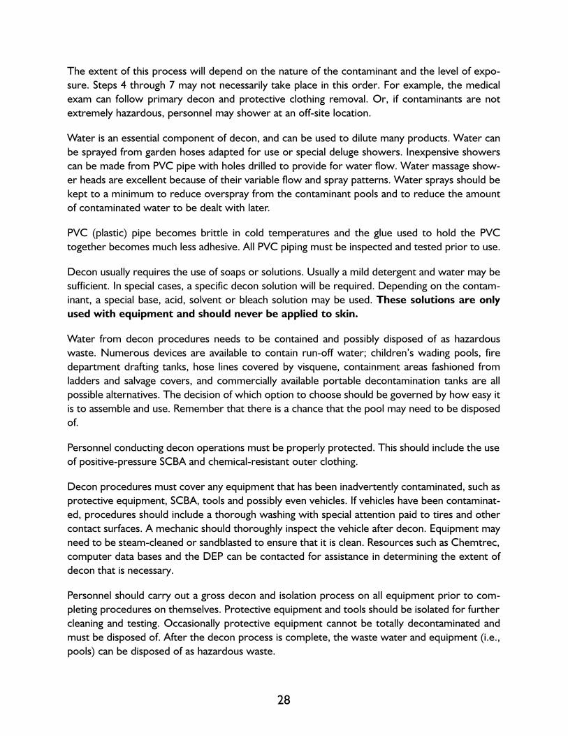
The extent of this process will depend on the nature of the contaminant and the level of expo-
sure. Steps 4 through 7 may not necessarily take place in this order. For example, the medical
exam can follow primary decon and protective clothing removal. Or, if contaminants are not
extremely hazardous, personnel may shower at an off-site location.
Water is an essential component of decon, and can be used to dilute many products. Water can
be sprayed from garden hoses adapted for use or special deluge showers. Inexpensive showers
can be made from PVC pipe with holes drilled to provide for water flow. Water massage show-
er heads are excellent because of their variable flow and spray patterns. Water sprays should be
kept to a minimum to reduce overspray from the contaminant pools and to reduce the amount
of contaminated water to be dealt with later.
PVC (plastic) pipe becomes brittle in cold temperatures and the glue used to hold the PVC
together becomes much less adhesive. All PVC piping must be inspected and tested prior to use.
Decon usually requires the use of soaps or solutions. Usually a mild detergent and water may be
sufficient. In special cases, a specific decon solution will be required. Depending on the contam-
inant, a special base, acid, solvent or bleach solution may be used. These solutions are only
used with equipment and should never be applied to skin.
Water from decon procedures needs to be contained and possibly disposed of as hazardous
waste. Numerous devices are available to contain run-off water; children’s wading pools, fire
department drafting tanks, hose lines covered by visquene, containment areas fashioned from
ladders and salvage covers, and commercially available portable decontamination tanks are all
possible alternatives. The decision of which option to choose should be governed by how easy it
is to assemble and use. Remember that there is a chance that the pool may need to be disposed
of.
Personnel conducting decon operations must be properly protected. This should include the use
of positive-pressure SCBA and chemical-resistant outer clothing.
Decon procedures must cover any equipment that has been inadvertently contaminated, such as
protective equipment, SCBA, tools and possibly even vehicles. If vehicles have been contaminat-
ed, procedures should include a thorough washing with special attention paid to tires and other
contact surfaces. A mechanic should thoroughly inspect the vehicle after decon. Equipment may
need to be steam-cleaned or sandblasted to ensure that it is clean. Resources such as Chemtrec,
computer data bases and the DEP can be contacted for assistance in determining the extent of
decon that is necessary.
Personnel should carry out a gross decon and isolation process on all equipment prior to com-
pleting procedures on themselves. Protective equipment and tools should be isolated for further
cleaning and testing. Occasionally protective equipment cannot be totally decontaminated and
must be disposed of. After the decon process is complete, the waste water and equipment (i.e.,
pools) can be disposed of as hazardous waste.
28

The personnel who are conducting the decon operations must also go through a cleaning
process. Personnel should work their way through the decon area, becoming cleaner as they
progress. The object is to be absolutely clean when leaving the contamination reduction corridor.
They should decon each other, with the last person finishing procedures on himself.
A trend in the hazardous waste industry is to move toward “dry decontamination.” While the
term may be misleading, this process does allow for a minimum of liquid waste by-products.
This concept requires the use of layered, disposable protective clothing. A water/solution may be
necessary for the areas of gross contamination such as overboots and gloves. Most clothing
should be removed and disposed of without extensive washing and rinsing. This allows for easi-
er cleanup and reduces the chance of secondary contamination from toxins trapped in reusable
protective clothing.
Unfortunately, there is no method to immediately determine how effective decon procedures
have been in removing contaminants. Discolorations, stains, corrosive effects and substances
adhering to objects can indicate that the contaminants have not been removed. However,
observable effects only point to surface contamination and not permeation (absorption) into the
clothing. Also, many contaminants are not easily detected.
Two methods of measuring the effectiveness of decon procedures are swipe and permeation
testing. Cloth or paper patches (swipes) are wiped over decontaminated surfaces and sent to a
laboratory for analysis. Swipe tests can be done on protective clothing, equipment and skin.
Permeation tests require that a piece of protective clothing be sent for analysis. However, both
swipe and permeation testing provides after-the-fact confirmation. Along with visual observa-
tions, the test results can help evaluate the effectiveness of the completed decon procedures.
CONTAMINATED VICTIMS
Special attention needs to be devoted to contaminated patients. These patients pose a risk of sec-
ondary exposure to the transport personnel and vehicle. Also at risk is the receiving hospital and
ED staff. Every effort must be made to decontaminate the patient prior to transport. Gross
decontamination can be accomplished by simply removing the patient’s clothing and
using a water rinse. A more complete decon can be accomplished with a soap and water wash.
The process of patient decon should start with the removal of all clothing, jewelry and shoes.
Then any visible contaminants should be removed from the patient. Dry particles can be gently
brushed away, while liquids should be blotted away with absorbent cloth. This will reduce the
chance of water reacting with the chemicals or increasing the absorption of a nonsoluble liquid.
Care must be taken not to scrape the skin during this process. Soft tissue damage (burns, bruis-
es, abrasions or lacerations) increases skin permeability and the absorption rate of the toxin.
29

Soaps used for patient decon should be mild and non-abrasive. Tincture of green soap is desir-
able because of its slightly alkaline nature that approximates the body’s pH level. Its alcohol base
also helps to remove hydrocarbons and solvents from the skin. If green soap is not available, any
mild liquid soap such as Dawn dishwashing detergent will work. Never use decon solutions on
skin. The patient should be washed with soft sponges to reduce the chance of skin abrasion.
Water spray should be mild to avoid aggravating any soft tissue damage. The temperature should
be warm—never hot. If cold water must be used, there is a risk of hypothermia. Try to contain
the run-off as hazardous waste, but do not delay treatment in life-threatening situations if con-
tainment is not available. In these cases, try instead to avoid allowing run-off to enter drains or
water sources.
Patient decon should begin at the head and then proceed to any areas where skin is damaged.
Care must be taken not to flush contaminants into wounds. Carefully wash and rinse the wound
area from the center out. After the wound area is clean, cover it with a water-occlusive dressing
or plastic wrap to preclude any further contamination. Once all wound areas are clean, proce-
dures can progress to other areas of the body. Ear/nose cavities should be irrigated, hair washed
and fingernails cleaned. Special attention should be focused on opposing surface areas, such as
the underarms and groin. Eyes should be flushed at the scene and irrigation continued during
transport, preferably with saline.
Privacy is an important consideration in field decontamination activities. In order to obtain coop-
eration from the patient, steps to assure patient privacy must be undertaken. Tarps, salvage cov-
ers, sheets, blankets, and other such items may be used to construct privacy screens on-site.
Remember, that both male and female decontamination areas may be needed.
30

Clothing must be provided to ambulatory patients following field decontamination activities.
Disposable clothing, such as Tyvek(tm) coveralls, may be used for this purpose. If such clothing is
not available, blankets, disposable sheets, etc., can be used. The Red Cross or Salvation Army
may be able to assist in this task.
Initial patient stabilization should be carried out simultaneously with decon. This will mandate that
the person providing patient care is trained in the use of and provided with proper protective
equipment. Under no circumstances should personnel be allowed to use protective equipment
without proper training. If proper training and equipment are unavailable, arrangements should
be made with the local fire department for a co-response to all possible chemical emergencies.
Under ideal circumstances, patients should be fully decontaminated prior to transport. In most
cases, this will eliminate the chance of secondary contamination of response personnel. However,
hazardous materials incidents are unlike many of our standard responses. Often, the incident will
continue to escalate and can endanger an entire community. In such cases, total commitment can-
not be focused on complete patient decontamination. Patient care is only one aspect of these
incidents, and manpower may be limited. As a result, patient decon may be less than optimal.
31
CONSIDERATIONS FOR
AMBULATORY DECONTAMINATION
• Remove any signs of contamination by scraping, sweeping or blotting the material away.
Remember, that you must be protected from cross contamination.
• Have patient remove clothing rapidly but cautiously. Direct patient not to have outer surface
of garments come in contact with their skin. Removal should be from top to bottom. (Patient’s
clothing could have absorbed most of the contaminant. Just think, normal clothing covers
about 85% of the human body).
• Remove all external items from having contact with body. These items may include hearing
aids, jewelry, watches, toupees, wigs and artificial limbs. Eye glasses, if needed by patient, must
be washed prior to being worn.
• If the patient wore glasses or contact lenses, flush the eyes with large amounts of water.
• Gently wash face and hair with soap and lukewarm water, followed by a thorough rinse. Try
not to have runoff contact any other part of the patient’s body.
• Begin to decon other body surfaces starting from the neck down. Try to blot the skin instead
of swabbing or wiping. Get into areas such as underneath the fingernails.
• Put patient into uncontaminated clothing.
CONSIDERATIONS FOR
NON-AMBULATORY DECONTAMINATION
• Remove any signs of gross contamination. Remember, you must be protected from cross con-
tamination.
• Cut away patient’s clothing and remove all personal property. All property should be bagged,
secured and clearly identified.
• Make sure your hands (the rescuer or health care provider) are decontaminated and thor-
oughly rinsed with water before removing contact lenses. Contact lenses should be removed
to decrease the risk of cross contamination.
• Eyeglasses from patient must be decontaminated. Eyeglasses in metal frames can be decon-
taminated in a bath of solution for 5 minutes followed by a thorough rinsing. Eyeglasses in a
composite or plastic frame should be secured in an impermeable bag for later decontamina-
tion.
32
• The victim’s skin excluding the face should be blotted with the solution of 0.5% hypochlorite.
Superficial wounds are flushed with a 0.5% hypochlorite solution and new dressings applied
as needed. Splints are not removed but saturated to the skin with 0.5% solution. If the splint
cannot be saturated it must be removed sufficiently so that everything under the splint can be
saturated with a 0.5% hypochlorite solution.
• The victim should then be showered or otherwise washed with copious amounts of water,
starting with the face and hands and then the rest of the body.
• Medical screening (triage) should now continue.
• Patient should receive new clothing (i.e., hospital scrubs) and continue to be observed for fur-
ther signs of exposure.
• Each individual, having been processed through decontamination, should be marked and iden-
tified as such. This can be accomplished with a triage tag or by marking victim’s forehead.
During processing, each individual should receive a certificate indicating:
—Description of decontamination actions taken;
—Time decontamination was completed;
—Time released from observation area; and
—Any medical treatment performed in conjunction with decontamination.
A copy should also go to decontamination record management.
TRANSPORT OF CONTAMINATED PATIENTS
Other situations may necessitate the transport of patients before they are completely clean.
Inclement weather can also be a major factor. Patient condition may require that only a gross
decontamination be undertaken before rapid transport. Perhaps a more realistic approach is to
attempt to get the patient as clean as possible (ACAP). In these cases, patient isolation principles
should be instituted. Depending on the contaminant and the level of contamination, protective
equipment may be necessary during transport. Keep ventilation to as high a level as weather con-
ditions permit. Remember that airflow in the patient compartment of ambulances is usually min-
imal at best.
In extreme cases, the ambulance may need to be protected by covering surfaces with plastic and
removing non-essential equipment prior to transport. Due to the slippery nature of wet plastic,
cover the floor with a sheet or blanket. An alternative to covering the ambulance surfaces is to
encapsulate the patient in blankets, sheets or plastic. Some response teams have excellent results
using zip-front body bags. These allow for the rapid containment of a patient yet still provide
quick access to the patient via the zipper. Obviously, the bag should only be zipped to the chest
level. With highly absorptive contaminants, toxicity can actually be increased by the use of plas-
tic or body bags. This can be reduced by placing a disposable blanket in the bag before the
patient. The blanket will keep the plastic from touching the patient’s skin. There are also com-
mercially available products on the market to contain contaminated run off from a patient during
transport.
33

34
It is important to note that many patients may come into the ED by private vehicle with no decon
prior to the arrival. Thus, it is essential that all hospitals be able to provide decontamination and
immediate treatment. Routine decon should be carried out if there is any question regarding con-
tamination status. EDs are required to have a means to decon and contain run-off in order to be
accredited by the Joint Commission on the Accreditation of Health Care Organizations. Some
hospitals have fully contained decon rooms, while many others are starting to use portable con-
tainment tanks or special decon tables.
The possibility of secondary contamination from patients, response team members and equip-
ment is a dangerous and real threat. Decontamination, correctly removing personal protective
equipment and using site response zones can minimize cross-contamination to personnel and
other areas. This module only provides general guidance on methods and techniques. The exact
decon procedure must be determined after evaluating the factors specific to the incident.
Haz Mat Incident Team Members
PERSONNEL RESPONSIBILITIES
Chemical Safety Provides technical consultation/information for handling the inci-
Officer (CSO): dent. Overall management of non-medical aspects at the scene. A
Haz Mat team member might be a possible backup.
Security: Sets up decon area. Restricts access to ER. Directs traffic. Restricts
access to news media.
Decontamination Team: Decontaminates patient. Makes sure all equipment is properly
decontaminated and disposed of. Provides for safe handling of all
waste. Cleans up decon area when procedures are completed.
Medical Team: Provides for treatment of patient.
Public Information Meets with members of the news media. Provides all press
Officer (PIO): releases.

35
Decontamination Area Set Up
The decontamination area is set up by both security and members of the decon team.
Security: Marks off restricted area with barrier cones and warning tape to
designated restricted area depending on hospital.
All personnel not associated with decontamination of the patients
are to be restricted from the area by security.
Security directs all ambulances, rescue units, other transportation
vehicles with CONTAMINATED PATIENTS to the decon area.
Decon Team: Assists in setting up the decon containment pool and shower setup.
Prepares decontamination supplies, wash solutions, attaches hose
to water supply.
Tests water quantity and quality before the patient arrives.
Determines if any additional supplies or materials are needed.
Decon team suits up and waits for patients.
Once patients are in the area, only properly protected decon members or medical staff, if in PPE,
are permitted in the area.
Shutting Down the DECON Area
At the conclusion of the decon process, it is important that the decon area itself be decontami-
nated to prevent the spread of any contaminated material.
1. Clean up is to be done by Decon Team in PPE. Your local hazmat team should handle the
clean up.
2. All solid waste that is contaminated is to be collected and placed in a “Contamination Bag.”
(Double lined plastic garbage bags will work.)
If it is determined to be a hazard, it will be disposed of by a hazardous waste company. For
WMD events, be careful to protect and secure evidence.
3. Waste water is to be held as follows:
a. if it is determined not to be hazardous, it can be disposed of in the sewer system.
b. if hazardous, the waste water must be sealed in drums and arrangements made for pick
up by a hazardous waste disposal company.
c. the Chemical Safety Officer will make these determinations and arrangements.
36
4. The entire decon area is to be straightened up and cleaned down.
5. All supplies and decon equipment is to be properly put away. Inventory is to be taken as to
what is to be needed.
6. Haz Mat supplies are to be relocated to storage area.
Common Sense Techniques
When performing decontamination, the goals, as well as the tasks needed to accomplish these
goals, should be kept simple. There are “common sense” techniques that could be used to help
protect the health and safety of all personnel involved and to prevent the spread of the hazardous
material. Some of the “common sense” techniques to be considered are the following:
1. Check your own hands and feet (both should be protected upon arrival at the inci-
dent) for any signs of contamination.
2. Observe each other. Do a complete visual check of other personnel for signs of con-
tamination. If a substance is noted, decontamination procedures must be employed.
3. If you are unsure that any piece of protective clothing or equipment has been com-
pletely decontaminated, carefully remove articles and leave them behind to be prop-
erly collected. YOUR SAFETY COMES FIRST. EQUIPMENT CAN BE REPLACED.
4. While decontaminating, avoid direct contact with the contaminated item.
Hazardous Materials/WMD Incident
Decontamination Equipment and Supplies
Many of these items are available in the hospital. It would be advisable to set up an area where
these supplies can be stored so that they are readily available when an incident occurs.
PERSONNEL PROTECTION EQUIPMENT
Face Shield
Chemical goggles
Surgical gloves
Chemical protection suit with hood
Chemical resistant boots
Duct tape
ID badges
37
DECONTAMINATION SUPPLIES
Sheets (Disposable)
Surgical Scrub brushes
Cotton tip applicators
Sterile water (for irrigation)
Wraps
Wash cloths (Disposable)
Spray container for soap
Soap
Scrub suits (Disposable)—For redress of ambulatory patient
DECON EQUIPMENT
The following supplies will be needed to set up the decontamination area:
Long handled scrub brushes (for decontamination of suits)
Warning tape
Warning signs
Cones
Containment pools
Decontamination table
Plastic floor covering
Hazardous Material labels for waste containers
Garden hose
Nozzle
Hazardous Materials Bags/Garbage bags
Markers
Scissors
Buckets
Waste containers
Recommended Decontamination Supplies
1. Patient Decontamination System that provides for the medical treatment and decontam-
ination of a patient. This system should include a means of collecting waste water.
2. Protective Floor Covering constructed of a non-skid chemically resistant material.
3. Waste Container with a dolly, lid and liner. All contaminated articles such as the patient’s
clothes, dressing and medical supplies should be placed in this container for proper disposal.
4. Sample Collection Kit that contains all the instructions and necessary supplies for collect-
ing samples. Should be done by qualified individuals.
5. Decontamination Kit that contains the necessary procedures, as well as fluids and materi-
als, for patient decontamination.
6. Antidotes for use in specific cases.
38
7. Contamination Control Measures used to control access to contaminated area thereby
minimizing the spread of the contamination.
• warning rope
• warning signs
• boundary cones
• step-off pad
Additional Supplies
• hose with splash reducing spray nozzle
• EMT scissors
• tincture of green soap
• waterproof drapes (i.e., Chucks)
• Irri-jet
• adhesive tape
• towels
• soft scrub brush
CHEMICAL WARFARE AGENT DECONTAMINATION
Few HAZMAT teams are equipped to provide the extensive level of decontamination called for
in these situations. Residual contamination on the CPC components can cause injury long after
the incident itself. Therefore, response teams need to consider how to safely dispose of the con-
taminated equipment.
In addition, commercial cleanup companies that typically clean up chemical spills may not be pre-
pared to deal with these chemical agents. Specialized government teams would be involved in the
cleanup and decontamination of any chemical warfare incident.
PREVENTING HOME CONTAMINATION
Contamination of worker’s homes with hazardous chemicals and substances transported from
the workplace is a world wide problem. So says the National Institute for Occupational Safety
and Health (NIOSH), which has released a report on this issue.
NIOSH found that workers can inadvertently carry hazardous materials home from work on:
U clothes U skin
U hair U tools
U in their vehicles
The incidents of home contamination have resulted in a wide range of diseases and, in some
cases, death among workers’ families.
Here are some tips to prevent contamination at work and at home:
U Change clothes before going home and leave soiled clothing at work to be laundered
by the employer
U Store street clothes in separate areas of the workplace to prevent their contamination
U Shower before leaving work
U Prohibit removal of toxic substances or contaminated items from the workplace
U Do not allow family members to visit the workplace
U Inform workers of the risk to family members and of preventive measures
U If contaminated clothing must be laundered at home, keep it separate from family
laundry
39

WATCH OUT FOR YOURSELF AND EVERYONE ELSE!
Emergency Department ignition sources:
A. Most of the electrical/equipment switches.
B. Radios.
C. Electrical clocks.
D. Cigarettes, pipes, cigars, lighters, matches, etc.
E. Flashlights (including penlights) that are not intrinsically safe.
F. Portable radios that are not intrinsically safe.
G. Pagers that are not intrinsically safe.
H. Telemetry equipment that is not intrinsically safe.
I. Use of striking tools causing sparks.
J. Static electricity sparks (nylon jackets act like batteries for static electricity).
K. Battery operated hearing aids, watches, etc.
L. Defibrillators.
40

TOXICOLOGY
41

INTRODUCTION
• The toxicity of a substance is its ability to cause harmful effects. These effects can strike a sin-
gle cell, a group of cells, an organ system, or the entire body. A toxic effect may be visible dam-
age, or a decrease in performance or function measurable only by a test. All chemicals can cause
harm. When only a very large amount of the chemical can cause damage, the chemical is consid-
ered to be relatively non-toxic. When a small amount can be harmful, the chemical is considered
toxic.
• The toxicity of a substance depends on three factors:
(1) its chemical structure,
(2) the extent to which the substance is absorbed by the body,
(3) and the body’s ability to detoxify the substance (change it into less toxic substances)
and eliminate it from the body.
• The toxicity of a substance is the potential of that substance to cause harm, and is only one
factoring determining whether a hazard exists. The hazard of a chemical is the practical likelihood
that the chemical will cause harm. A chemical is determined to be a hazard depending on the fol-
lowing factors:
(1) toxicity: how much of the substance is required to cause harm,
(2) route of exposure: how the substance enters your body,
(4) dose: how much enters your body,
(5) duration: the length of time you are exposed,
(6) reaction and interaction: other substances you are exposed to,
(7) sensitivity: how your body reacts to the substance compared to others.
• Some chemicals are hazardous because of the risk of fire or explosion. These are important
dangers, but are considered to be safety rather than toxic hazards. The factors of a toxic hazard
are more fully explained below.
• The longer you are exposed to a chemical, the more likely you are to be affected by it. The
dose is still important-at very low levels you may not experience any effects no matter how long
you are exposed. At higher concentrations you may not be affected following a short-term expo-
sure, but repeated exposure over time may cause harm. Chemical exposure which continues
over a long period of time is often particularly hazardous because some chemicals can accumu-
late in the body or because the damage does not have a chance to be repaired. The combination
of dose and duration is called the rate of exposure.
43
• The body has several systems, most importantly the liver, kidneys and lungs, that change
chemicals to a less toxic form (detoxify) and eliminate them. If your rate of exposure to a chem-
ical exceeds the rate at which you can eliminate it some of the chemical will accumulate in your
body. For example, if you work with a chemical for eight hours each day, you have the rest of the
day (16 hours) to eliminate it from your body before you are exposed again the next day. If your
body can’t eliminate all the chemical in 16 hours and you continue to be exposed, the amount in
the body will accumulate each day you are exposed. Illness that affects the organs for detoxifica-
tion and elimination, such as hepatitis (inflammation of the liver), can also decrease their ability to
eliminate chemicals from the body.
• Accumulation does not continue indefinitely. There is a point where the amount in the body
reaches a maximum and remains the same as long as your exposure remains the same. This point
will be different for each chemical. Some chemicals, such as ammonia and formaldehyde, leave
the body quickly and do not accumulate at all. Other chemicals are stored in the body for long
periods. For instance, lead is stored in the bone, calcium is stored in the liver and kidneys. There
are a few substances, such as asbestos fibers, that, once deposited, remain in the body forever.
• The effects of toxic substances may appear immediately or soon after exposure, or they may
take many years to appear. Acute exposure is a single exposure or a few exposures. Acute effects
are those which occur following acute exposures. Acute effects can occur immediately, or be
delayed and occur days or weeks after exposure.
PREVENTION & CONTROL
• Prevention and control measures include, but are not limited to, the following:
(1) Elimination/substitution and process modification;
(2) Engineering controls;
(3) Administrative controls; and
(4) Use of personal protective equipment.
• In certain circumstances, personal protection of the individual employee is necessary. Personal
protective devices should be regarded as being supplementary to substitution and engineering
control and should not be used in preference to the latter because they do nothing to eliminate
the hazard.
• Personal protective equipment must be appropriately selected, individually fitted and workers
trained in their correct use and maintenance. The equipment must be regularly checked and
maintained to ensure that the worker is being protected.
44

• Monitoring may be used for the evaluation of a hazard and for assessing the effectiveness of
control measures. The design and implementation of a monitoring program should be carried out
by, or in consultation with, a properly qualified person. Monitoring of the work environment
involves the measurement of atmospheric contaminants at selected locations in the workplace
(static, positional monitoring).
• Biological monitoring involves measurement of the concentration of a contaminant, its
metabolites or other indicators in the tissues or body fluids of the worker. In some cases, bio-
logical monitoring may be required to supplement static or personal monitoring.
f. In the control of health hazards due to a specific contaminant, where it has been demonstrat-
ed that the exposure of the employee to the contaminant is approaching the relevant exposure
standard, or where biological monitoring indicates that an unacceptable exposure is occurring,
immediate action must be taken to reduce the health hazard and intensive monitoring should
continue.
ROUTES OF ENTRY
• Injury can be caused by chemicals only if they reach sensitive parts of a person or other living
organism at a sufficiently high concentration and for a sufficient length of time. Thus, injury
depends upon the physicochemical properties of the potentially toxic substances, the exact
nature of the exposure circumstances, and the health and developmental state of the person or
organism at risk.
• Major routes of exposure are through the skin (topical), through the lungs (inhalation), or
through the gastrointestinal tract (ingestion). In general, for exposure to any given concentration
of a substance for a given time, inhalation is likely to cause more harm than ingestion which, in
turn, will be more harmful than topical exposure faster.
a. Inhalation Route
Inhalation is the most significant route of entry by which harmful substances enter the human
body at work. Toxic atmospheric contaminants may have local or systematic effects. Local effects
harm only the part of the body they come in contact with, for example, inhalation of silica dust
causing pneumoconiosis. Systemic effects, cause changes to the function of other organs, as in the
case of inhaled particles that are soluble in the fluid of the tissues that line the lung, for example,
lead and mercury fumes. Inhalation results in the introduction of toxic compounds into the res-
piratory system. Most of the compounds that are commonly inhaled are gases or vapors of
volatile liquids; however, solids and liquids can be inhaled as dusts or aerosols. Inhalation of toxic
agents generally results in a rapid and effective absorption of the compound.
45

When you inhale a toxic chemical, the dose you receive depends on four factors:
(1) The level (concentration) of chemical in the air;
(2) How hard (fast and deep) you are breathing, which depends on your degree of phys-
ical exertion;
(3) How much of the chemical that is inhaled stays in your lungs and is absorbed into your
bloodstream; and
(4) How long the exposure lasts.
b. Absorption Route
Some atmospheric contaminants may be absorbed through the skin without any noticeable
change to the skin, while others may cause serious damage to the skin itself. Ingestion is of rela-
tively minor significance in occupational exposure to toxic materials.
(1) Skin contact exposure does not typically result in as rapid systemic dosage as
Inhalation, although some chemicals are readily absorbed through the skin. Many
organic compounds are lipid (fat) soluble and can therefore be rapidly absorbed
through the skin. Some materials that come in contact with the eyes can also be
absorbed. Ingestion is a less common route of exposure for emergency response per-
sonnel at hazardous materials incidents. However, incidental hand-to-mouth contact,
smoking, and swallowing of saliva and mucus containing trapped airborne contami-
nants can cause exposure by this route. In addition, emergency medical personnel in
both hospital or prehospital settings will see chemical exposures in patients who have
ingested toxic substances as a result of accidental poisonings or suicide attempts.
(2) Many people do not realize that chemicals can penetrate healthy intact skin and so this
fact should be emphasized.
c. Ingestion
Airborne particles breathed through the mouth or cleared by the cilia of the lungs will be ingest-
ed. Otherwise, ingestion of potentially toxic substances in the work, domestic, or natural envi-
ronment is likely to be accidental and commonsense precautions should minimize this. The
nature of the absorption processes following ingestion is discussed elsewhere. The importance
of concentration and time of exposure has already been pointed out. It should be remembered
that exposure may be continuous or repeated at intervals over a period of time; the conse-
quences of different patterns of exposure to the same amount of a potentially toxic substance
may vary considerably in their seriousness.
46
d. Injection
The injection of hazardous materials into the body sounds, at first, like a bad joke. Who in his
right mind would inject themselves—especially when it is not required by a doctor? However, it
can occur by stepping on a sharp object, or impaling yourself on or being cut by a sharp object
while working at an incident site. It will happen before you even realize it has occurred and the
reality of possibly being internally contaminated sinks in. The best precaution for this eventuality
is to have protective clothing on, including steel shank and toed foot protection, and by strictly
instituting and observing safe work habits.
EXPOSURE LIMITS
These limits are established by health and safety authorities to control exposure to hazardous
substances. Exposure limits usually represent the maximum amount (concentration) of a chemi-
cal which can be present in the air without presenting a health hazard. However, exposure lim-
its may not always be completely protective, for the following reasons:
• Although exposure limits are usually based on the best available information, this
information, particularly for chronic (long-term) health effects, may be incomplete.
Often we learn about chronic health effects only after workers have been exposed to
a chemical for many years, and then as new information is learned, the exposure lim-
its are changed.
• Exposure limits are set to protect most workers. However, there may be a few work-
ers who will be affected by a chemical at levels below these limits (see “Sensitivity”).
Employees performing extremely heavy physical exertion breathe in more air and
more of a chemical, and so may absorb an excessive amount.
• Exposure limits do not take into account chemical interactions. When two or more
chemicals in the workplace have the same health effects, industrial hygienists use a
mathematical formula to adjust the exposure limits for those substances in that work-
place.
• When toxic chemicals are present in the workplace, your exposure can be estimated
by measuring the concentration of a given chemical in the air and the duration of
exposure. This measurement is called air or environmental monitoring or sampling
and is usually done by industrial hygienists, using various types of instruments. The air
is collected from your breathing zone (the air around your nose and mouth) so that
the concentrations measured will accurately reflect the concentration you are inhal-
ing. The exposure levels calculated from this monitoring can then be compared to the
Permissible Exposure Level for that chemical.
47
• Environmental monitoring is the most accurate way to determine your exposure to
most chemicals. However, for chemicals that are absorbed by routes other than
inhalation, such as through the skin and by ingestion, air monitoring may underesti-
mate the amount of chemical you absorb. For these and some other chemicals, the
levels of the chemical (or its breakdown products) in the body can sometimes be
measured in the blood, urine or exhaled air. Such testing is called biological monitor-
ing, and the results may give an estimate of the actual dose absorbed into the body.
For one substance, lead, biological monitoring is required by law when air monitoring
results are above a certain level. The American Conference of Governmental
Industrial Hygienists (ACGIH) has recommended the exposure limits for biological
monitoring for a small number of chemicals. These are called Biological Exposure
Indices (BEIs) and are published together with TLVs.
• If you smell a chemical, you are inhaling it. However, some chemicals can be smelled
at levels well below those that are harmful, so that detecting an odor does not mean
that you are inhaling harmful amounts. On the other hand, if you cannot smell a chem-
ical, it may still be present. Some chemicals cannot be smelled even at levels that are
harmful. The odor threshold is the lowest level of a chemical that can be smelled by
most people. If a chemical’s odor threshold is lower than the amount that is haz-
ardous, the chemical is said to have good warning properties.
• If you or your co-workers experience symptoms known to be caused by a chemical
during or its use, you may have been overexposed. Symptoms might include tears in
your eyes; a burning sensation of skin, nose, or throat; a cough; dizziness or a
headache.
OCCUPATIONAL EXPOSURE LIMITS
Occupational exposure limits exist to serve one main purpose: protect workers from excessive
exposure to toxic chemicals in the workplace. They were designed for healthy adults, usually for
an exposure duration of a day’s workshift (8 hours). They were not meant to be used for pro-
tection of the public, since the general public includes:
• Sensitive groups such as the very young and very old, people with respiratory diseases
and other illnesses, and people who are hypersensitive to some chemicals.
Occupational exposure limits were also not designed to compare toxicity of chemi-
cals, or to be the fine line between “safe” and “unsafe.”
• The current definition has no exposure duration associated with it. Workers should
not be in an IDLH environment for any length of time unless they are equipped and
protected to be in that environment. They may be found in the NIOSH Pocket Guide
to Chemical Hazards.
48

EXPOSURE LIMITS
The various occupational exposure limits found in the literature or in an MSDS are based prima-
rily on time-weighted average limits, ceiling values, or ceiling concentration limits to which the
worker can be exposed to without adverse effects. Examples of these are listed in (Table 8).
These values were established to provide worker protection in occupational settings. Because
the settings in which these values are appropriate are quite different than an uncontrolled spill
site, it is difficult to interpret how these values should be used by emergency medical personnel
dealing with a hazardous materials incident. At best, TLV, PEL, IDLH, and REL.
a. Lethal Concentration 50 (LC 50)
Is the concentration of a material in air that on the basis of respiratory exposure in laboratory
tests is expected to kill 50% of test animals when administered as a single exposure (usually 1
hour). A dose of 3,000-3,800 mg/kg tetrachloroethylene is lethal to 50% of rats that received the
compound orally; however, only 6.4 to 10 mg/kg of sodium cyanide is required to produce the
same effect. Therefore, compounds with low LD50 values are more acutely toxic than substances
with larger LD50 values. The LD50 values that appear in an MSDS or in the literature must be used
with caution by emergency medical personnel. These values are an index of only one type of
response and give no indication of the ability of the compound to cause nonlethal, adverse or
chronic effects. Furthermore, LD50 values typically come from experimental animal studies.
Because of the anatomical and physiological differences between animals and humans, it is diffi-
cult to compare the effects seen in experimental animal studies to the effects expected in humans
exposed to hazardous materials in the field. Therefore, emergency medical personnel should
remember that the LD50 and LC50 values are only useful for comparing the relative toxicity of
compounds and should only be used to determine if one chemical is more toxic than another.
b. Lethal Dose 50 (LD50)
Toxicity information is often expressed as the dose of the compound that causes an effect in a
percentage of the exposed subjects, which are mostly experimental animals. These dose-
response terms are often found in Material Safety Data Sheets (MSDS) and other sources of
health information. One dose-response term that is commonly used is the lethal dose of 50
(LD50), the dose which is lethal to 50% of an animal population from exposure by any route other
than inhalation when given all in one dose. Another similar term is the lethal concentration 50
(LC50), which is the concentration of a material in air that on the basis of respiratory exposure in
laboratory tests is expected to kill 50% of a group of test animals when administered as a single
exposure (usually 1 hour). Exhibit I lists a number of chemicals that may be encountered in deal-
ing with hazardous materials incidents, and the reported acute LD50 values of these compounds
when they are administered orally to rats.
49

From Exhibit I, it can be seen that a dose of 3,000-3,800 mg/kg tetrachloroethylene is lethal to
50% of rats that received the compound orally; however, only 6.4 to 10 mg/kg of sodium cyanide
is required to produce the same effect. Therefore, compounds with low LD50 values are more
acutely toxic than substances with larger LD50 values.
The LD50 values that appear in an MSDS or in the literature must be used with caution by emer-
gency medical personnel. These values are an index of only one type of response and give no indi-
cation of the ability of the compound to cause nonlethal, adverse or chronic effects. Furthermore,
LD50 values typically come from experimental animal studies. Because of the anatomical and phys-
iological differences between animals and humans, it is difficult to compare the effects seen in
experimental animal studies to the effects expected in humans exposed to hazardous materials in
the field. Therefore, emergency medical personnel should remember that the LD50 and LC50 val-
ues are only useful for comparing the relative toxicity of compounds and should only be used to
determine if one chemical is more toxic than another.
Exhibit I
Acute LD50 Values for Representative Chemicals When Administered Orally to Rats
Chemical Acute Oral LD50
(mg/kg)*
Sodium cyanide 6.4-10
Pentachlorophenol 50-230
Chlordane 83-560
Lindane 88-91
Toulene 2,600-7,000
Tetrachloroethylene 3,000-3,800
*Milligrams of the compound administered per kilogram body weight of the experimental animal.
Responses to toxic chemicals may differ among individuals because of the physiological variabili-
ty that is present in the human population. For example, an individual may be more likely to expe-
rience an adverse health effect after exposure to a toxic chemical because of a reduced ability to
metabolize that compound. The presence of preexisting medical conditions can also increase
one’s susceptibility to toxic chemicals. Respiratory distress in patients or workers with asthma
may be triggered by exposure to toxic chemicals at lower concentrations than might be expect-
ed to produce the same effect in individuals without respiratory disease. Factors such as age, per-
sonal habits (i.e., smoking, diet), previous exposure to toxic chemicals, and medications may also
increase one’s sensitivity to toxic chemicals. Therefore, exposure to concentrations of toxic com-
pounds that would not be expected to result in the development of a toxic response.
50
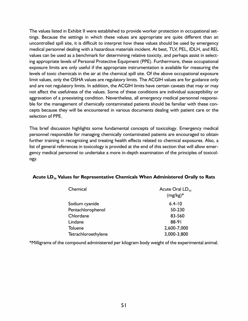
The values listed in Exhibit II were established to provide worker protection in occupational set-
tings. Because the settings in which these values are appropriate are quite different than an
uncontrolled spill site, it is difficult to interpret how these values should be used by emergency
medical personnel dealing with a hazardous materials incident. At best, TLV, PEL, IDLH, and REL
values can be used as a benchmark for determining relative toxicity, and perhaps assist in select-
ing appropriate levels of Personal Protective Equipment (PPE). Furthermore, these occupational
exposure limits are only useful if the appropriate instrumentation is available for measuring the
levels of toxic chemicals in the air at the chemical spill site. Of the above occupational exposure
limit values, only the OSHA values are regulatory limits. The ACGIH values are for guidance only
and are not regulatory limits. In addition, the ACGIH limits have certain caveats that may or may
not affect the usefulness of the values. Some of these conditions are individual susceptibility or
aggravation of a preexisting condition. Nevertheless, all emergency medical personnel responsi-
ble for the management of chemically contaminated patients should be familiar with these con-
cepts because they will be encountered in various documents dealing with patient care or the
selection of PPE.
This brief discussion highlights some fundamental concepts of toxicology. Emergency medical
personnel responsible for managing chemically contaminated patients are encouraged to obtain
further training in recognizing and treating health effects related to chemical exposures. Also, a
list of general references in toxicology is provided at the end of this section that will allow emer-
gency medical personnel to undertake a more in-depth examination of the principles of toxicol-
ogy.
Acute LD50 Values for Representative Chemicals When Administered Orally to Rats
Chemical Acute Oral LD50
(mg/kg)*
Sodium cyanide 6.4-10
Pentachlorophenol 50-230
Chlordane 83-560
Lindane 88-91
Toluene 2,600-7,000
Tetrachloroethylene 3,000-3,800
*Milligrams of the compound administered per kilogram body weight of the experimental animal.
51

How Much (Or How Little) is 1 Part Per Million
To give you an example of how small an amount we are talking about: 1 drop of liquid from an
eye dropper is 1 millionth of a tank of gas in an average compact car (13 gallon tank), or 1/8 inch
is 1 millionth of a mile.
A milligram is 1/1000 of a gram and there are 28.3 grams in an ounce.
c. Immediately Dangerous to Life and Health (IDLH)
The Immediately Dangerous to Life and Health (IDLH) guidelines are not occupational exposure
limits similar to the TLV or PEL. They were developed in the 1970s by NIOSH to guide respira-
tor selection. A recent revision in 1994 updated many of the IDLH concentrations, and changed
the IDLH definition. The current (1994) definition of the IDLH is a condition “that poses a threat
of exposure to airborne contaminants when that exposure is likely to cause death or immediate
or delayed permanent adverse health effects or prevent escape from such an enviroment.” For
concentrations above the IDLH, a self-contained breathing apparatus (SCBA) is required. Below
that level, air-purifying respirators may be used, if appropriate.
• Notice that unlike the previous definition of the IDLH, which incorporated a 30-
minute time period, the new definition does not have an exposure duration associat-
ed with it. If you are in an IDLH condition, you need to get out of there immediately!
IDLHs are based on analysis of human and animal studies and were developed for
fewer than 100 substances. The rationale for each standard is not clearly stated.
Unlike the TLVs, no formal updating mechanism exists for IDLH.
d. Ceiling
A concentration that should not be exceeded at any time. Note that both TWA and STEL permit
limited excursion if, in the end, the average is below the exposure limit. The ceiling value, how-
ever, may not be exceeded.
52
e. TLV-STEL
(1) Some substances in the TLV booklet have a short-term exposure limit (STEL). The
STEL is a 15-minute exposure limit that should not be exceeded even if the 8-hour
TLV remains within the limit. Such limits were assigned to substances exerting toxic
effects even over a short period of time. Where a STEL limit is not available (but is
believed to be justified), the TLV committee recommends using a limit three times as
high as the TLV for a 15-minute exposure.
(2) These values can be used as a benchmark for determining relative toxicity, and per-
haps assist in selecting appropriate levels of Personal Protective Equipment (PPE).
Furthermore, these occupational exposure limits are only useful if the appropriate
instrumentation is available for measuring the levels of toxic chemicals in the air at the
chemical spill site. Of the above occupational exposure limit values, only the OSHA
values are regulatory limits.
(3) Of the above occupational exposure limit values, only OSHA values are regulatory
limits. The ACGIH values are for guidance only and are not regulatory limits. In addi-
tion, the ACGIH limits have certain caveats that may or may not affect the usefulness
of the values. Some of these conditions are individual susceptibility or aggravation of
a preexisting condition. Nevertheless, all emergency medical personnel responsible
for the management of chemically contaminated patients should be familiar with these
concepts because they will be encountered in various documents dealing with patient
care or the selection of PPE.
f. TWA (Time-Weighted Average)
Unless otherwise mentioned, it is the concentration of contaminants over an 8-hour period. It is
determined by sampling the breathing zone of the worker for 8 hours.
g. Permissible Exposure Limits
PELs are values set by OSHA. These limits are a legal requirement for occupational exposures,
and exceeding them is a violation of the law, for which fines may be imposed. Most of the PELs
are based on older TLVs, and some have STELs as well. OSHA’s PELs are published yearly in the
Code of Federal Regulations (CFR). A recent PEL update was invalidated by a judge in the sum-
mer of 1993. Therefore, many of the currently valid PELs date back to 1968. Many states, how-
ever, have established their own occupational standards. These are as strict or stricter than
OSHA’s, were not affected by the recent ruling, and are enforced as before.
53

h. Threshold Limit values
1. Threshold Limit Values-Time Weighted Average (TLV-TWA) are exposure limits rec-
ommended by a committee of the American Conference of Governmental Industrial
Hygienists (ACGIH), and are published yearly in a little booklet. All the substances in
the TLV booklet have an 8-hour Time-Weighted Average (TWA) exposure which is
the level to which workers may be exposed for an 8-hour work shift without suffer-
ing an adverse effect. The rationale for setting the limits is explained in a separate pub-
lication, “Documentation of the Threshold Limit Values and Biological Exposure
Indices” (ACGIH, Cincinnati, Ohio). The TLVs are derived from human studies includ-
ing epidemiological research and exposure studies with volunteers, occupational acci-
dents, animal studies, and “similar structure analysis” (based on the assumption that
compounds similar in structure are similar in toxicity). The TLV committee meets
several times every year, and the TLVs are updated regularly.
2. TLV-TWA is meant to regulate exposure over an 8-hour period. Don’t extrapolate to
shorter periods of time. Don’t assume that if a certain limit applies for 8 hours, then
eight times that limit may be applied if the exposure lasts for only 1 hour. It simply
doesn’t work that way. Therefore, the 8-hour limits may not be very useful for spill
response, where exposure durations are usually much shorter than 8 hours.
i. Recommended Exposure Limits (RELs)
The Recommended Exposure Limits (RELs) were developed by the National Institute of
Occupational Safety and Health (NIOSH). Those standards are similar to TLVs in the way that
they were derived, but are often stricter. The RELs are published in the NIOSH Pocket Guide to
Chemical Hazards, which is updated every few years, and in other NIOSH publications.
54

Occupational Exposure Limits
Value Abbreviation Definition
Threshold Limit Value TLV Refers to airborne concentrations of substances
(3 Types) and represents conditions under which it is
(ACGIH)* believed that nearly all workers may be
repeatedly exposed day after day without
adverse effect.
1) Threshold Limit Value— TLV-TWA The time-weighted average concentration for
Time-Weighted Average a normal 8-hour workday and a 40-hour
(ACGIH)* workweek, to which nearly all wokers may
be repeatedly exposed, day after day,
without adverse effect.
2) Threshold Limit Value— TLV-STEL The concentration to which workers can be
Short-Term Exposure exposed continuously for a short period of time
Limit (ACGIH)* without suffering from: 1) irritation, 2) chronic
or irreversible tissue damage, or 3) narcosis of
sufficient degree to increase the likeihood of
accidental injury, impair self-rescue or
materially reduce work efficiency, and provided
that the daily TLV-TWA is not exceeded.
3) Threshold Limit Value— TLV-C The concentration that should not be exceeded
Ceiling (ACGIH)* during any part of the working exposure.
Permissible Exposure PEL Same as TLV-TWA.
Limit (OSHA)**
Immediately Dangerous IDLH A maximum concentration (in air) from which
To Life and Health one could escape within 30 minutes without any
(OSHA)** escape-impairing symptoms or any irreversible
health effects.
Recommended Exposure REL Highest allowable airborne concentration is not
Limit (NIOSH)*** expected to injure a worker; expressed as a
ceiling limit or time-weighted average for an
8- or 10-hour work day.
*American Conference of Governmental Industrial Hygienists
**Occupational Safety and Health Administration
***National Institute for Occupational Safety and Health
55

TREATMENT
PROTOCOLS
FOR THE
HAZARDOUS MATERIALS
(INDUSTRIAL CHEMICALS)
CONTAMINATED PATIENT
57


Patient Management
When a hospital receives a call that a patient exposed to hazardous materials is to be received, a
planned course of actionshould be implemented. Steps in a protocol must be practiced before a
hazardous materials emergency occurs. All staff members of an emergency department should
know their responsibilities and how to perform them. All required equipment should be imme-
diately available or readily accessed.
Individuals receiving a potential hazardous materials call should obtain as much information as
possible. A checklist should be developed and made available for all telephone or radio commu-
nication centers. Information that will aid in initiating appropriate actions includes:
• Type and nature of incident
• Caller’s telephone number
• Number of patients
• Signs/symptoms being experienced by the patients
• Nature of injuries
• Name of chemical(s) involved
• Extent of patient decontamination in the field
• Estimated time of arrival
After the above information is received, a predesignated resource center (e.g., New Jersey poi-
son control center, ATSDR) should be contacted for information regarding definitive care proce-
dures, which should include decontamination methods that need to be performed.
Communications should be kept open with on-site response personnel to obtain as much
advance information as possible.
If incident notification comes from other than usual emergency communication channels, the call
should be verified before a hazardous materials response plan is initiated. EMS personnel should
be notified of any special approach or entrance to the emergency department and also advised
not to bring the patient into the emergency department until the patient has been assessed and
accepted by the emergency department.
Often patients contaminated by hazardous materials may be brought into the emergency
department unannounced or not through regular EMS channels. This could be an ambulatory
patient or a patient transported by private vehicle. The ideal response to this is to call a fire
department which is properly trained and equipped or a hazmat team to come to the hospital
and set up a decontamination area outside the ambulance entrance. In any event, these patients
should be isolated from other patients and assessed and decontaminated as soon as possible.
59

Emergency Department Preparation
Every member of the emergency department should be familiar with the hospital’s hazardous
materials response plan and be required to participate in scheduled drills. Preparation for arrival
of a contaminated patient should include: notification of all services involved, preparation of a
Decontamination Area, and suiting up of the Decontamination Team.
Emergency Department Mobilization
The person receiving a call of incoming victims should notify the Nursing Supervisor who will in
turn notify appropriate personnel according to the hospital’s response plan. The hospital opera-
tor should be instructed to notify security and maintenance, and the nurse on duty should con-
tact the predesignated resource center.
Patient Arrival
The emergency physician-in-charge or an emergency department nurse should meet the ambu-
lance upon arrival and assess the condition of the patients as well as the degree of contamination.
Personnel should keep in mind that the actual contamination may be (or become) a life-threat-
ening condition. Triage procedures should also be initiated at this point, if necessary. During ini-
tial patient survey and stabilization, contamination reduction should simultaneously be per-
formed. This consists of cutting away or otherwise removing all suspected contaminated cloth-
ing, including jewelry and watches, and brushing or wiping off any contamination. Care should be
taken to protect any open wounds from contamination. Emergency department personnel
must make every effort to avoid contact with any potentially hazardous substance, and
avoid cross contamination.
Ideally, decontamination should be performed before patient transport; however, field deconta-
mination facilities are limited and emergency department personnel should consider that all haz-
ardous materials patients need decontamination. If a patient’s clothing was not removed at
the incident site, it should be removed outside the ambulance but before entry into the
emergency department. This will reduce further exposure to the patient and lessen the extent
of contamination introduced to the emergency department. Contaminated clothing should be
double bagged in plastic bags, sealed, and labeled. The decontamination team should bring the
prepared stretcher to the ambulance, transfer the patient, and take him or her directly to the
decontamination area along the predesignated route.
60

Priority should be given to the ABC (Airway, Breathing, and Circulation) and simultaneous con-
tamination reduction. Once life-threatening matters have been addressed, emergency depart-
ment personnel can then direct attention to thorough decontamination and secondary patient
assessment. Identification of hazardous materials involved can be simultaneously performed by
other personnel. It is important to remember that appropriate person protective clothing must
be worn until personnel are no longer in danger. Therefore, the sooner the patient becomes
decontaminated the sooner personnel may reduce protective measures.
Effective decontamination consists of making the patient As Clean As Possible (ACAP).
This means that the contamination has been reduced to a level that is no longer a
threat to the patient or the responder. The recorder notes on a diagram of the body the
areas found by the physician to be contaminated.
Considerations for Patient Treatment
Primary goals for emergency department personnel in handling a contaminated patient include
termination of exposure to the patient, patient stabilization, and patient treatment—while not
jeopardizing the safety of emergency department personnel. Termination of exposure can best
be accomplished by removing the patient from the area of exposure and by removing contami-
nants from the patient. Basically, a contaminated patient is like any other and may be treated as
such except that staff must protect themselves and others from dangers due to contamination.
Personnel must first address life-threatening issues and then decontamination and supportive
measures. Priority should be given to the ABC with simultaneous contamination reduction. Once
life-threatening matters have been addressed, emergency department personnel can then direct
attention to thorough decontamination, secondary patient assessment, and identification of mate-
rials involved. It is important to remember that appropriate personal protective clothing must be
worn until personnel are no longer in danger. Therefore, the sooner the patient becomes decon-
taminated the sooner personnel may reduce protective measures or downgrade the level of pro-
tection. Primary and secondary surveys should be completed as conditions allow. In treating
patients, personnel should consider the chemical-specific information received from the haz-
ardous materials response resources. In multiple patient situations, proper triage procedures
should be implemented. Presenting signs and symptoms should be treated as appropriate and
when conditions allow. The sooner a patient has been decontaminated the sooner he or she can
be treated like a “normal” patient. Orders of the designated poison control center and attending
61

physician should be administered. Invasive procedures, such as IVs or intubation, should be per-
formed only for life-threatening conditions, until decontamination is performed. These proce-
dures may create a direct route for introducing the hazardous material into the patient. The
patient should be frequently re-assessed because many hazardous materials have latent physio-
logical effects.
Note: The attending staff must remember that since exposure to some substances can result in seri-
ous delayed effects, sustained observation and monitoring are required.
Patient Management Under Mass Casualty Conditions
Involving Hazardous Chemicals
Basic medical procedures in a large-scale hazardous materials incident are not substantially dif-
ferent from life-saving measures in other mass casualty disasters. Primary attention to the ABC
continues to have first priority with decontamination performed at the same time. A chemical dis-
aster may overwhelm any one hospital, particularly if it occurs along with another disaster such
as an earthquake. Hospitals need to preplan what they will do if they are overwhelmed with haz-
mat patients.
There are, however, several important differences in disasters involving hazardous materials.
Such differences include the need for the effective decontamination of exposed patients and
response personnel, and the need for effective safety measures to protect response personnel.
Training in the appropriate procedures to be followed is essential for potential responders to a
hazardous materials incident involving mass casualties. Standard principles of triage apply in
chemical disasters, except in exposures to very toxic substances. The patient, injured or not,
must be decontaminated before being transported to the emergency department to protect EMS
and emergency department staff.
62

Exposure to Toxic Chemicals
Exposure to toxic chemicals is frequently the primary concern at Haz Mat incidents. Many scenes
contain a variety of chemicals in solid, liquid or gaseous form.
Contaminants can enter the body through the four pathways consisting of inhalation, injection,
ingestion and absorption. Inhalation and absorption are considered to be the most common
routes of entry and protective equipment is available to minimize the risk. Remember, that the
first responder with operational training will be limited in both operation and types of protective
equipment.
We must always be aware of the ingestion route and how contaminants enter the body in this
manner. The act of smoking, drinking, eating or rubbing ones face with the hands may introduce
contaminants to the body through ingestion. For this reason, one must always be aware of the
presence of contaminants at a scene and the need for safety, proper protection, and decontami-
nation.
Exposure and Dose
Exposure is the act of coming in contact with a contaminant.
Dose is the amount of contaminant taken into the body.
Warning Properties are the physical characteristics of a chemical identified by the senses.
Carcinogen—substance that causes cancer.
Mutagen—a substance that causes mutations. A mutation is a change in the genetic material in
a body cell. Mutations can lead to birth defects, miscarriages, or cancer.
Teratogen—a substance that causes birth defects by damaging a fetus.
A contaminant could cause damage at the point of contact (local) or at another point in the body
(systemic). Effects may be immediate (acute), in the case of a large concentrated dose, or more
commonly, delayed (chronic) and will not be detected for over a period of time. These are
known as acute and chronic doses.
63


LOCALIZED EFFECTS
Toxic Inhalation Injuries
Chemical properties that affect site & types of injuries
PHYSICAL PROPERTIES
• Concentration
• Duration
• Dose
• Solubility
PHYSICAL FORMS
• Gases and Vapors
• Mists
• Fumes
CHEMICAL PROPERTIES
• Reactivity
• pH
• Direct Acting Chemicals
• Indirect Acting Chemicals
Patients baseline condition contributes to injury severity
• Rate and pattern of respirations
• Hx of lung disease
• Pts Age
• Nutrition Status
• Thyroid Function
65

66
Upper Airway Injuries (laryngeal edema/spasm)
Most often caused by water soluble/highly reactive chemicals
• Acids (chlorine & hydrogen chloride)
• Alkalis (ammonia)
• Vesicants (formaldehyde)
Lower Airway Injuries (bronchiolar obstruction)
Most often caused by chemical not very water soluble, inhaled in high concentrations, with pro-
longed exposure to dust/fumes.
• Clinically presents as wheezing/respiratory distress
Bronchospasm & Asthma
• Allergic (sensitized to industrial chemicals)
• Non-allergic (irritants)
• Attacks can be immediate or delayed up to 6 hours
• Anaphylaxis
Pulmonary Edema
• Caused by damage to lung vs. cardiogenic edema
Pulmonary capillaries become more permeable
Alveolar lining cells
Surfactant disrupted
• May develop rapidly or delayed even days later

Chemical Injuries to the Eye
Effects of Acid and Alkali
• Most common cause of serious chemical eye injuries
• Alkali generally more severe than acids
• Acids
Coagulation necrosis
Coagulum serves as a barrier to deep penetration
At pH <2, acids penetrate easy and deep
• Alkali
Liquefaction necrosis
Creates no barrier, facilitating penetration
• Acids/Alkalis can cause vessel thrombosis
• Ischemic necrosis of the cornea can lead to eye loss
• Severity cannot be judged until 48+ hours
• Alkalis progress for weeks to months
Management of Acid and Alkali Eye Exposures
• Prompt and continuous irrigation with water
Acids for at least 15 mins
Alkali for at least 20 mins
• Remove by washing foreign bodies/solid particles
• Remove contact lens
• Be gentle in handling the eye
67

Solvents
Detergents and surfactants
Lacrimators
Delayed corneal injuries
Chemical Injuries to the Skin
Mechanisms of chemical skin injuries:
• Thermal
Hot
Cold
• Mechanical
• Ischemic
• Irritation
Contact dermatitis
Vesiculation
• Chemical burns and corrosion
Coagulation necrosis
Liquefaction necrosis
Corrosion
Determinants of severity:
• Nature and reactivity of chemical
• Concentration of the chemical
• Integrity of the skin
• Duration of the exposure
Remove chemical by prompt/adequate washing
Water should not be used on chemical such as water reactive metals.
68
Complications:
• Do not underestimate severity
• Severity difficult to judge for first 48 hrs.
• Aggressive fluid replacement (dehydration)
• Infectious complications
Hydrogen fluoride and hydrofluoric acid:
• Sequestration of calcium
• Seizures and cardiac dysrhythmias
• Need to replace calcium in tissues
Calcium gel/subcutaneously
Phosphorus:
• Spontaneously ignites at temps. >86 degrees F
• Vapors if burning are toxic if inhaled
• Treatment: extinguish burning
Submerge burning part in cool water
Keep moist and cool
Water-reactive metals (alkaline metals):
• React with water yielding heat and strong alkali
• Do NOT wash these chemicals with water
• Extinguish with class-D fire extinguisher or sand
• During transport, cover metal with cooking or mineral to prevent air contact and
burning
69

Phenol:
• Strong corrosive agents that are weak acids
• Water washing contributes to absorption and systemic intoxication
• Wipe skin with polyethylene glycol first
Systemic Effects
Systemic routes of exposure:
• Inhalation
Most common
Water and fat soluble chemicals
Reactive chemical may be poorly absorbed
• Percutaneous absorption
Fat soluble chemical are better absorbed
Other determinants
n Concentration
n Duration of exposure
n Size of exposed surface
n Skin moisture and temperature
n Skin integrity
Eye exposure
• Ingestion
• Injection
Onset of action:
• Immediate for chemicals which require no transformation to be toxic
• Delayed onset of action with chemicals that are transformed into a toxic form
• Some chemicals have both an immediate and delayed toxic effect
70

Asphyxiants:
• Injury to organs that require large amounts of oxygen
Central Nervous System
Kidneys
Liver
• Chemical induced
Inhibition of cellular respiration by enzyme poisons
n Intracellular hypoxia
Production of abnormal hemoglobin
n Carboxyhemoglobin
n Methemoglobin
Acute hemolysis
Interference of ventilation
n Heavier than air gases
Central Nervous System depressants:
• Many chemicals have anesthetic or narcotic effects
Usually fat soluble chemicals
Act on nerve cell membranes
• Clinical effects (mild/moderate/severe)
• Effects similar to common medical conditions
Management:
• Airway/Oxygen
• Cardiac monitoring
71

Cardiac sensitizers:
• Chemicals can sensitize the heart to the effects of catecholamines released by the
body potentially resulting in V-Fib and cardiac arrest.
• V-Fib and cardiac arrest can result from sensitization and catecholamine release
Management:
• Keep victims at rest
• Cardiac monitoring
• Consider reducing doses of catecholamines (when indicated)
Neurotoxic insecticides:
• Activity of organophosphates and carbamates
Inhibit cholinesterase
Excessive activity of parasympathetic nervous system
• Clinical effects
Bradycardia leading to heart block
Chest tightness and wheezing
Constricted pupils
Weakness, twitching, muscle tremors, cramps
SLUDGE
Seizures
Management:
• Airway/Oxygen
• Antidotes/atropine in large dosages
72

Electrolyte disorders:
• Sequestration agents (hydrogen fluoride, oxalic acid, phosphorus) bind calcium
Clinical effects of hypocalcemia
n Twitching, muscle spasms, seizures
n Cardiac dysrhythmias and cardiac arrest
Management
n Suspect problem from history of exposure
n Measurement of calcium in blood
n Replacement of calcium by IV
73

ACUTE (IMMEDIATE) VS
CHRONIC (DELAYED) EFFECTS
Important factors to consider when determining the toxicity of a material are the relationships
between concentration, exposure time and the threshold sensitivity of the person exposed.
Generally, a serious exposure refers to a large, single dose received over a short period of time
and an immediate response occurs (acute).
A serious exposure may result from a small, single dose over a short period of time and there is
no immediate effect. This small dose may exceed the threshold sensitivity of the individual caus-
ing a serious delayed effect (chronic). The classic example of this is cancer.
Doses from several small exposures over a period of time (chronic exposure), causing no
immediate effect may also result in a delayed effect. This cumulative effect may be serious or
minor (chronic).
Examples of Adverse Health Effects from
Exposure to Toxic Chemicals
Target Example of
Organ Causative Health Effect
Toxic End Point Systems Agent Acute Chronic
Carcinogenicity Multiple Benzene Dermatitis Aleukemia
Sites Tightness in Myeloblastic
Chest leukemia
Hepatotoxicity Liver Carbon Vomiting Liver Necrosis
Tetrachloride Vessication Fatty liver
Dizziness
Neurotoxicity Nervous Lead Nausea Wrist Drop
System Vomiting IQ Deficits
Abdominal Pain Encephalopathy
Nephrotoxicity Kidney Cadmium Vomiting Kidney Damage
Diarrhea Anemia
Chest Pain
74

Heat Stress
Heat stress can occur very rapidly—within as little as 15 minutes. It can pose a far greater dan-
ger to worker health than chemical exposure. In its early stages, heat stress can cause rashes,
cramps, discomfort and drowsiness, resulting in impaired functional ability. Continued heat stress
can lead to heat cramps, heat exhaustion, heat stroke, and even death.
Personnel exhibiting the following symptoms require immediate medical attention.
Treatment of Heat Stress
Initial treatment for heat stress includes removing the victim to a cool area, at the very least out
of the sun. Heavy clothing should be removed and fluids administered.
Heat stroke is a life threatening emergency and Advanced Life Support (ALS) treatment is
required as soon as possible. The same procedures used for heat stress should be followed until
ALS care is available. Rapid cooling is essential for a victim with heat stroke.
Frostbite
Frostbite usually occurs on the face or the extremities. Signs and symptoms include pain followed
by numbness and white chalky appearance. Use of metal tools or working on metal surfaces
which are cold exacerbate the problem.
Personnel experiencing frostbite should be given immediate medical attention.
Treatment of Frostbite
A person with frostbite or hypothermia should be taken to a warm area indoors.
The frostbitten area should be covered with a soft cloth and gradually warmed. It should not be
rubbed. Rubbing will further damage the frozen tissue. Frostbite is a serious injury which, if
improperly treated, may result in loss of the affected body part.
75


MEDICAL PROTOCOLS
NOTE: FOR ADVICE ON CLINICAL MANAGEMENT
CALL THE NEW JERSEY POISON CONTROL
CENTER (800) 962-1253
77

TREATMENT
PROTOCOLS
FOR THE
HAZMAT CONTAMINATED
PATIENT
79


81
HAZMAT
ACIDS
&
ACID
MISTS
ACIDS & ACID MISTS(NOT Including Hydrofluoric Acid)
FORMS:
Gas, liquid (variable concentrations), mixtures with water, and aerosolized dusts.
BACKGROUND:
Acids act as direct irritants and corrosive agents to moist mucous membranes, and to intact skin
to a lesser extent. Generally, these substances have very good warning properties: even fairly low
airborne concentrations of acids produce rapid onset of eye, nose and throat irritation. Higher
concentrations can produce cough, stridor, wheezing, chemical pneumonia or non-cardiogenic
pulmonary edema. Occasionally, pulmonary edema may be delayed for several hours, especially
with low-solubility gases such as nitrogen oxides. Ingestion of acids can result in severe injury to
the upper airway, esophagus and stomach.
POTENTIAL FOR SECONDARY CONTAMINATION:
Small amounts of acid mists can be trapped in clothing after an overwhelming exposure but are
not usually sufficient to create a hazard for health care personnel away from the scene. However,
clothing which has become soaked with concentrated acid may be corrosive to rescuers. Once
the victim has been stripped and flushed with water, there is no significant risk of secondary con-
tamination.
MANAGEMENT IN THE HOSPITAL:
1. Activate decontamination protocol if indicated, and if not previously decontaminated. Health
care personnel should don gloves, gowns, goggles, and other protective clothing until decon-
tamination is completed. Activation of decontamination protocol is probably not needed for
acid exposures unless the victim’s clothing has been soaked with acid liquid.
2. Evaluate ABC’s (airway, breathing, and circulation).
3. O2 by mask. Intubate if patient manifests severe respiratory distress from pulmonary edema
or upper airway swelling. Obtain arterial blood gases and chest x-ray if respiratory distress is
present. Severe upper airway edema may necessitate cricothyroidotomy or tracheostomy.
82
HAZMAT
ACIDS
&
ACID
MISTS
4. Irrigate the eyes copiously with saline or water for at least 15 to 20 minutes if eye irritation
is present.
5. Remove and double-bag clothing if not already done. Wash skin copiously with water.
6. If respiratory distress is present or anticipated, admit and observe 24 to 48 hours for possi-
ble delayed onset pulmonary edema. Bronchodilators may be helpful.
7. Examine eyes using slit-lamp and/or fluorescein strips, if corneal injury is suspected.
8. If a significant ingestion occurred, consider endoscopy to evaluate the esophagus and stom-
ach.
9. Advise patient that full recovery is generally the rule, but cases of chronic airway disease have
been reported following severe exposures. Advise and arrange for follow-up in case victim
begins to experience respiratory distress. After exposure to oxides of nitrogen, sudden
severe relapse may occur two to three weeks later.
83
HAZMAT
AMMONIA
AMMONIA (LIQUID AND GAS)
FORMS:
Gas (anhydrous) and liquid (aqueous solutions, variable concentrations).
NOTE: liquified compressed gas may produce cryogenic (freezing) hazard as it is released into the
atmosphere.
BACKGROUND:
Ammonia (NH3) is a direct irritant and alkaline corrosive agent to moist mucous membranes and,
to a lesser extent, to intact skin. Ammonia has very good warning properties. Even fairly low air-
borne concentrations produce rapid onset of eye, nose and throat irritation. Higher concentra-
tions can produce cough, stridor, wheezing, chemical pneumonitis or non-cardiogenic pulmonary
edema. The onset of pulmonary edema is usually rapid but may occasionally be delayed for 12-
24 hours. Ingestion of concentrated ammonia solutions (e.g., >5%) may cause serious corrosive
injury to the esophagus and stomach and poses an aspiration risk.
POTENTIAL FOR SECONDARY CONTAMINATION:
Small amounts of ammonia vapor can be trapped in clothing after an overwhelming exposure but
are not usually sufficient to create a hazard for health care personnel away from the scene.
However, clothing which has become soaked with concentrated liquid ammonia may be corro-
sive to rescuers. Once the victim has been stripped and flushed with water, there is no significant
risk of secondary contamination.
MANAGEMENT IN THE HOSPITAL:
1. Activate decontamination protocol for liquid exposure (and if not previously decontaminat-
ed). Health care personnel should don gloves, gowns, goggles or protective clothing until
decontamination is completed.
2. Evaluate and support ABC’s (airway, breathing, and circulation).
84
HAZMAT
AMMONIA
3. O2 by mask. Intubate if patient manifests severe respiratory distress from pulmonary edema
or upper airway swelling. Watch for signs of airway closure and laryngeal edema, such s
hoarseness, stridor or retractions. Obtain arterial blood gases and chest x-ray if respiratory
distress is present. Severe upper airway edema may necessitate cricothyroidotomy or tra-
cheostomy.
4. Irrigate the eyes copiously with saline or water for at least 15 to 20 minutes if eye irritation
is present.
5. Remove and double-bag clothing if not already done. Wash skin copiously with water.
6. Cardiac monitor; 12-lead EKG.
7. If severe respiratory distress is present, admit and observe for 24 hours for delayed-onset
pulmonary edema.
8. Examine eyes using slit-lamp and/or fluorescein strips, if corneal injury is suspected.
9. Advise that full recovery is generally the rule, but cases of chronic airway disease have been
reported following severe exposures.

85
HAZMAT
ARSINE
GAS
ARSINE GAS
FORMS:
Gas may be generated in metal ore processing and electronic component manufacturing.
BACKGROUND:
Arsine (AsH3) is an extremely toxic and nearly odorless gas (it has a slight odor of garlic). It is used
widely in the microelectronics industry and occasionally occurs as a by-product in metallurgy and
pesticide manufacturing. Arsine’s effects are quite distinct from other arsenic compounds; even
in very small quantities, inhaled arsine produces acute hemolysis (rupture of red blood cells),
which can result in cardiac decompensation due to anemia, or renal failure due to massive kid-
ney deposition of hemoglobin. Symptoms may be delayed for 2-24 hours, and include weakness,
abdominal and flank pain, brown urine, and jaundice. Massive acute exposure appears capable of
causing immediate death by an unknown mechanism.
POTENTIAL FOR SECONDARY CONTAMINATION:
Very small amounts of arsine can be trapped in a victim’s clothing after an overwhelming expo-
sure but are not usually sufficient to create a hazard for health care personnel away from the
scene.
MANAGEMENT IN THE HOSPITAL:
1. Evaluate and support ABC’s (airway, breathing, and circulation).
2. Provide O2 by mask.
3. Monitor cardiac rhythm; obtain 12-lead EKG.
4. Laboratory Tests: Perform urine dipstick for occult blood and hemoglobin. Send for CBC,
plasma free hemoglobin (PFHgb), urine hemoglobin, electrolytes, BUN and/or creatinine,
bilirubin, blood type and screen, and other laboratory tests as appropriate. Urinary arsenic
levels may be elevated for a few weeks after exposure.
86
HAZMAT
ARSINE
GAS
5. If there is evidence of acute hemolysis, alkalinize urine with sodium bicarbonate, 50-100 mEq
in or added to 1 (one) liter of 5% dextrose or 1/2 NS administered IV at a rate to maintain
urine output at 2-3 cc/kg/hr. Consider furosemide or mannitol. Follow electrolytes, BUN,
creatinine and fluid status closely because renal failure may result in acute fluid overload.
6. If PFHgb exceeds 1.5 gm/dl, there has been a significant rapid drop in hematocrit (e.g., from
40 to 30 without other explanation) or there are other indications of intravascular hemolysis
(severe abdominal pain, jaundice, shock), consider exchange transfusion after consultation
with a medical toxicologist. Prepare for dialysis in the event of renal failure. Shock may occur
and should be treated appropriately.
NOTE: BAL and other chelating agents are not effective for arsine exposure. Arsine
does not produce the classical symptoms of arsenic poisoning.
87
HAZMAT
CARBON
MONOXIDE
CARBON MONOXIDE
FORMS:
Gas.
BACKGROUND:
Carbon monoxide (CO) is a colorless, odorless gas. It is a common product of combustion of any
organic material and is a major toxic component in cases of smoke inhalation. Carbon monoxide
causes poisoning by interfering with the binding of oxygen to hemoglobin in the blood, myoglo-
bin in heart and muscle tissue, and by interfering with oxygen utilization in the cell. Symptoms of
progessively worse exposure include, in order, headache, dizziness, giddiness, tinnitus, nausea,
muscle weakness, chest pain, dyspnea, syncope, seizures, and coma. Cherry-red skin col-
oration is not commonly seen (except post-mortem) and should not be relied upon for diag-
nosis. The half-life of CO in the blood is from 5 to 9 hours when the victim is breathing room air,
compared to 60-90 minutes when breathing 100% oxygen.
POTENTIAL FOR SECONDARY CONTAMINATION:
Very small amounts of CO can be trapped in victim’s clothing after an overwhelming exposure,
but are not usually sufficient to create a hazard for health care personnel away from the scene.
MANAGEMENT IN THE HOSPITAL:
1. Evaluate and support ABC’s (airway, breathing, and circulation).
2. Provide 100% O2 by a tight-fitting mask, preferably with oxygen reservoir.
3. Monitor cardiac rhythm, and obtain 12-lead EKG. Watch for ischemic changes.
NOTE: Carbon Monoxide poisoning will cause a falsely elevated O2 saturation
(SPO2) and may give a false sense of security to the health care provider that
the patient is not hypoxic.
4. Laboratory tests—Send for carboxyhemoglobin level (COHb), arterial blood gases, Hct, elec-
trolytes, and other tests as appropriate.
5. Treat cerebral edema with fluid restriction, hyperventilation, and/or mannitol.
88
HAZMAT
CARBON
MONOXIDE
6. Admit to the hospital if any of the following are present:
a. Mental status changes are present or were present.
b. COHb >25%.
c. COHb >15% in a patient with coronary disease, or current symptoms suggestive of
coronary disease.
d. Any EKG change thought to be acute, particularly ST segment depression, regardless of
COHb level.
e. Metabolic acidosis or disordered thermoregulation.
f. Patient is pregnant and symptomatic or has COHb >10%.
7. A hyperbaric chamber may be helpful in pregnant patients, those with an altered level of con-
sciousness, or the patient that does not rapidly respond to 100% O2. Consultation with a
medical toxicologist is advised. Speed in instituting therapy is very important, and anticipation
of hyperbaric oxygen treatment should not delay intubation and the delivery of 100% O2. In
cases of severe exposure, delayed 100% oxygen treatment may increase the risk of perma-
nent brain damage or prolonged (many months) convalescence, although this is not definite-
ly established in the medical literature.
89
HAZMAT
CHLORINE
GAS
CHLORINE GAS
FORMS:
Gas (anhydrous) or liquid (aqueous chlorine usually in the form of hypochlorite, variable concen-
trations). The liquid hypochlorite solutions are very unstable and react with acids to release chlo-
rine gas. NOTE: liquified compressed gas may produce cryogenic (freezing) hazard as it
is released into the atmosphere.
BACKGROUND:
Chlorine is a highly irritating gas which rapidly forms hydrochloric acid after contact with moist
mucous membranes in the upper airway and in the lungs. Symptoms occur rapidly and provide
good warning properties for exposure. Low concentrations produce eye, noise and throat irrita-
tion. Higher concentrations produce cough, wheezing, choking, chemical pneumonitis, or pul-
monary edema. Ingestion of concentrated hypochlorite solutions can cause serious corrosive
esophageal or stomach injury.
POTENTIAL FOR SECONDARY CONTAMINATION:
Small amounts of chlorine gas can be trapped in clothing after an overwhelming exposure but are
not usually sufficient to create a hazard for health care personnel away from the scene. However,
clothing which has become soaked with concentrated hypochlorite solution may be corrosive to
rescuers. Once the victim has been stripped and flushed with water, there is no significant risk of
secondary contamination.

90
HAZMAT
CHLORINE
GAS
MANAGEMENT IN THE HOSPITAL:
1. Evaluate and support ABC’s (airway, breathing, and circulation).
2. Provide O2 by mask. Intubation may be required for severe respiratory distress.
3. Irrigate eyes copiously with water or saline if eye irritation is present.
4. Monitor cardiac rhythm if clinically indicated.
5. Obtain chest x-ray, arterial blood gases. Obtain other laboratory tests as appropriate.
6. Observe 6-12 hours for delayed-onset pulmonary edema for symptomatic patients.
7. Advise that full recovery is generally expected, but may take several months. Cases of chron-
ic airways disease have been reported following severe exposure.
91
HAZMAT
CYANIDE
CYANIDE
FORMS:
Gas (hydrogen cyanide), liquid (solutions of cyanide salts), and solid (cyanide salts). Hydrogen
cyanide gas may be formed when acid is added to a cyanide salt or a nitrile.
BACKGROUND:
Cyanide (CN) is an extremely toxic compound which is widely used in industry in a variety of
forms (gas, liquid, solid). CN gas (HCN) is a major toxic component in cases of smoke inhalation.
CN produces toxicity by interfering with cellular oxygen utilization. Symptoms and signs include
headache, dizziness, vomiting, tachypnea, tachycardia, and coma. There may be a distinctive odor
(“bitter almonds”) on the victim’s clothing or breath. Death can occur within minutes of expo-
sure. If exposure is by inhalation of CN gas, peak toxic effects are seen within minutes, but after
ingestion of a CN salt, effects may be delayed until the CN is absorbed from the stomach.
POTENTIAL FOR SECONDARY CONTAMINATION:
If the exposure was by inhalation of HCN gas, even though there may be small amounts of gas
trapped in clothing after an overwhelming exposure, this is not usually sufficient to create a haz-
ard for health care personnel away from the scene. The risk of secondary contamination to res-
cuers is greater if there are large amounts of liquid or solid material on the victim’s clothing or
skin.
MANAGEMENT IN THE HOSPITAL:
1. Activate decontamination protocol (unless decontaminated before arrival); medical person-
nel to don gloves, gown, and goggles until decontamination is completed.
2. Evaluate and support ABC’s (airway, breathing, and circulation).
3. Administer O2 by mask or endotracheal tube. Reducing the potential for chemical exposure
from any form by mouth-to-mouth resuscitation, including use of pocket one-way valve
mouth-to-mouth resuscitation, should be carefully considered.
4. If not decontaminated before arrival, remove and double-bag clothing; wash skin with soap
and water if dermal exposure is suspected. Of note, liquid chlorine bleach will decontami-
nate contaminated equipment and should be used when laundering contaminated clothing.
5. Monitor cardiac rhythm, and obtain 12-lead EKG.

92
HAZMAT
CYANIDE
6. Laboratory Tests: Serum thiocyanate, blood cyanide, CBC, electrolytes, arterial blood gases,
lactate, and other laboratory tests as appropriate. Appropriate treatment should not be
delayed.
7. Respiratory Exposure: If the patient arrives asymptomatic, probably no treatment will be
needed. If the patient is ill, begin (8) below.
Ingestion or Skin Contact: ER staff should be ready to initiate therapy immediately, regard-
less of the presence of symptoms on arrival. Be prepared to intubate quickly.
8. In the symptomatic patient with a significant exposure, administer treatment in the follow-
ing order (use Cyanide Antidote Kit):
a. Amyl nitrite—break pearls into gauze sponge and hold under patient’s nose or Ambu
intake valve for 15 to 30 seconds/minute, until sodium nitrite solution is ready.
b. Sodium nitrate (NaNO2) 3% IV solution:
Adults: 10 ml at 2.5 to 5 ml/minute, or 0.35 ml/kg.
Children: 0.2 ml/kg, not to exceed 10 ml.
c. Through the same IV line in (B) above, give sodium thiosulfate (NA2S2O2), 25%.
Adults: 12.5 gm (50 cc of 25% solution).
Children: 1.6 to 1.8 ml/kg of a 25% solution.
d. Repeat antidote at 50% of initial dose if symptoms persist after 20 minutes. If symptoms
worsen after treatment, consider nitrite toxicity causing methemoglobinemia greater
than 25%.
WARNING: Methemoglobinemia may be particularly dangerous in children. Also,
be extremely cautious in treating with nitrite if there has also been carbon monox-
ide exposure. The same dose of nitrites can cause excessive methemoglobinemia.
Normal therapeutic amounts of methemoglobinemia in the face of carbon monox-
ide poisoning can be a problem and should be closely watched.
9. If ingestion is suspected, perform gastric lavage and administer activated charcoal.
10. Admit and observe 24-48 hours. Watch for metabolic acidosis; treat with sodium bicarbon-
ate if needed. Watch for hypotension; treat with fluid and pressors if needed. Hyperbaric
oxygen may be helpful in displacing cyanide ion from cellular enzymes.
11. Hyperbaric oxygen may be indicated for victims of smoke inhalation who have had both
cyanide and carbon monoxide exposures and who do not respond to treatment.

93
HAZMAT
HYDROFLUORIC
ACID
HYDROFLUORIC ACID
FORMS:
Gas, liquid (variable concentrations), and fluoride salts in the presence of acids may generate toxic
quantities of hydrogen fluoride.
BACKGROUND:
Hydrofluoric acid (HF) produces toxicity quite distinct from other mineral acids. The “acid” moi-
ety (hydrogen ion) is relatively unimportant, producing little burning sensation on initial contact.
In contrast, the highly toxic fluoride ion has the ability to penetrate tissue and produce indolent
ulceration or bony destruction. Solutions of greater than 10-20% are particularly destructive.
Inhalation may cause eye, nose and throat irritation, cough, tracheobronchitis, and delayed onset
pulmonary edema. Ingestion may produce severe corrosive burns of the esophagus and stomach.
Systemic absorption of fluoride (i.e., from a burn or after ingestion) may result in severe hypocal-
cemia, hypomagnesemia, and hyperkalemia, resulting in tetany and cardiac arrest.
POTENTIAL FOR SECONDARY CONTAMINATION:
Until the soaked clothing has been removed and the affected body part has been flushed, there
is some hazard to treating health care personnel, depending on the concentration. Following
basic decontamination, there is usually no significant risk of secondary contamination.
MANAGEMENT IN THE HOSPITAL:
1. Activate decontamination protocol unless the victim was decontaminated before arrival.
Medical personnel should don gown, gloves, and goggles until decontamination is completed.
2. Evaluate and support ABC’s (airway, breathing, and circulation).
3. If not decontaminated before arrival, remove and double-bag clothing; wash skin with soap
and water. Continue to irrigate copiously. HF will continue to leach from exposed skin and
tissues for at least 15 minutes. DILUTION IS BETTER THAN NEUTRALIZATION IN
THE FIRST CRITICAL MINUTES!!

94
HAZMAT
HYDROFLUORIC
ACID
4. Evaluate the extent of skin exposure and following irrigation, administer additional treatment:
a. If the HF concentration was >20% or is unknown or exposure was prolonged:
(1) Infiltrate affected area with 10% calcium gluconate, using a 25-30 gauge needle and
multiple injections of 0.5 ml per square centimeter, taking care to prevent damaging
underlying structures. Pain should resolve with the injection.
DO NOT USE calcium chloride, which is extremely painful and may further
injure tissues.
a. Repeat after several hours if pain recurs.
b. Avoid local anesthetics, which may mask clinical findings but do give morphine IV
for pain. HF burns can be intensly painful and pain is out of proportion to physi-
cal findings.
c. Limit injection to 0.5 ml per phalanx.
(2) Remove blisters and debride underlying tissues, as these may contain HF.
(3) Remove nails if evidence of periungual or ungual tissue involvement. Use a regional
anesthesia proximal to the site of injury.
b. If the HF concentration was <20% and the duration of exposure was brief (less than a
few minutes), administer calcium gluconate gel (2.5%) or 30%-50% magnesium sulfate
solution by massage or soaks to affected area for at least 30 minutes. This treatment binds
HF as the insoluble CaF2 or MgF2 salts. If pain persist, go to step 4A. If more than an hour
or two has elapsed since the time of initial decontamination topical soaks are probably of
marginal benefit.
c. For extremity burns where topical agents are ineffective in relieving pain or as alternative
modality to fingernail removal, an intra arterial injection of calcium gluconate may be
effective. Consultation with a medical toxicologist should be obtained.
d. 2.5-5 cc of a 2.5% calcium gluconate solution may be administered via nebilizer for inhala-
tional exposure.
5. Ingestion—treat as severely corrosive agent. Consider endoscopy to evaluate extent of dam-
age. Consider lavage with calcium containing solution.
6. Additional steps for all patients:
a. Admit to burn unit or intensive care unit if the total extent of the burn is greater than 2%-
3% BSA, or if there is significant respiratory distress.
b. Observe for hypocalcemia, hyperkalemia or other systemic effects if HF concentration
was greater than 20% or if there was prolonged contact with a significant percent of BSA
(2% to 3% or more).

95
HAZMAT
HYDROFLUORIC
ACID
c. Obtain 12 lead EKG and provide continuous cardiac monitoring to look for QT prolon-
gation which may be early sign of hypocalcemia. Consider giving IV calcium 1 gram (10
ml) 10% calcium gluconate or calcium chloride prophylactically for higher concentration
exposures to greater than 5% BSA or for dilute exposures to larger surface areas.
7. Establish baseline and serial electrolytes, Ca, Mg. Follow blood gases in the event of respira-
tory exposure.

96
HYDROGEN
SULFIDE
SULFIDES
MERCAPTANS
HYDROGEN SULFIDE,SULFIDES & MERCAPTANS
FORMS:
Gas (hydrogen sulfide, methyl & short-chain alkyl mercaptans) and liquid (other mercaptans).
BACKGROUND:
Hydrogen sulfide (H2S) is a highly toxic gas with an odor of rotten eggs at low concentrations. At
higher concentrations olfactory fatigue rapidly occurs, making odor a poor warning symptom of
danger. Mercaptans are sulfur-containing, highly malodorous compounds. All of these compounds
are direct irritants, but their major toxicity is due to interference with cellular oxygen utilization.
Low-level exposures produce irritation of the eyes, nose and throat, cough, headache, nausea,
and dizziness. Higher exposures can cause syncope, seizures, coma, tracheobronchitis, and pul-
monary edema (which may occur up to 48-72 hours later). Death may occur within minutes of
acute massive exposure.
POTENTIAL FOR SECONDARY CONTAMINATION:
Small amounts of H2S can be trapped in clothing after an overwhelming exposure but are not usu-
ally sufficient to create a hazard for health care personnel away from the scene. However, cloth-
ing which has become soaked with concentrated liquid sulfide solutions or mercaptans may pose
a risk to rescuers. Once the victim has been stripped and flushed with water, there is no signifi-
cant risk of secondary contamination. Sulfides are highly water soluble.
MANAGEMENT IN THE HOSPITAL:
1. Activate decontamination protocol, unless decontaminated before arrival; medical person-
nel should don gown, gloves, and goggles until decontamination is completed. Odor will pro-
vide a warning about the need for decontamination. A WELL-VENTILATED AREA WILL BE
VERY HELPFUL.
2. Evaluate and support ABC’s (airway, breathing, and circulation).
3. Provide O2 by mask, if the victim has respiratory distress or altered mental status.
97
HYDROGEN
SULFIDE
SULFIDES
MERCAPTANS
4. If not decontaminated before arrival, remove and double-bag clothing; wash skin with soap
and water if clothing had been wet. Irrigate the eyes with saline or water if there is eye irri-
tation.
5. Monitor cardiac rhythm and obtain 12-lead EKG. Tachyarrhythmias may occur.
6. Laboratory tests: CBC, electrolytes, creatinine and/or BUN, blood gases, liver function stud-
ies, urinalysis, and other laboratory tests as appropriate.
7. If the patient is severely affected with coma or cardiovascular collapse, administer treatment
in the following order, using the Cyanide Antidote Kit:
a. Amyl nitrite: break pearls into gauze sponge and hold under patient’s nose or Ambu
intake valve for 15 to 30 seconds/minute, until sodium nitrite solution is ready.
b. Sodium nitrate (NaNO2) 3% IV solution:
Adults: 10 ml at 2.5 to 5 ml/minute or 0.35 ml/kg.
Children: 0.2 ml/kg, not to exceed 10 ml.
c. Sodium thiosulfate is not effective for H2S exposure.
d. Repeat antidote at 50% of initial dose if symptoms persist after 20 minutes. If symptoms
worsen after treatment, consider the possibility of nitrite toxicity causing methemoglo-
binemia greater than 25%.
e. Continue O2 for at least 2 hours afterward.
8. If symptoms are mild, including eye and throat irritation, headache, nausea, or dizziness,
supportive care will suffice.
9. In severe cases, observe for delayed onset pulmonary edema, liver toxicity, or
hematuria/proteinuria.
10. Hyperbaric oxygen may be helpful, although the medical literature on this point is still some-
what controversial.
98
NITROGEN
CONTAINING
COMPOUNDS
NITROGEN-CONTAINING COMPOUNDSAND OTHER CHEMICALS CAUSING
METHEMOGLOBINEMIA
FORMS:
Gas, liquid and solid. Substances tend to be brown or yellow in color, especially when impure.
BACKGROUND:
A wide variety of nitrogen-containing compounds, including anilines, aryl amines, and aromatic
nitrogen compounds, are potent oxidizing agents which can produce methemoglobinemia.
Methemoglobin is unable to transport oxygen. Patients with methemoglobinemia greater than
15% will appear grey or cyanotic, and their blood will appear chocolate brown. With higher lev-
els signs and symptoms of hypoxia are present, including headache, dizziness, nausea, dyspnea,
syncope, seizures, and coma. These methemoglobin-forming compounds may also produce
direct systemic effects such as skin or respiratory irritation, vasodilation, hypotension, headache,
nausea and CNS depression. Many of the liquid compounds are highly volatile and may be inhaled,
and many are well-absorbed through the skin.
POTENTIAL FOR SECONDARY CONTAMINATION:
Depending on the individual compound, these agents may pose a significant health hazard for res-
cuers and health care personnel. Many are well-absorbed through intact skin. Simple water wash-
ing may be insufficient to remove oil compounds.
MANAGEMENT IN THE HOSPITAL:
1. Activate decontamination protocol, unless decontaminated before arrival or until decontam-
ination is completed. Health care personnel should don neoprene gloves (do not use canvas,
cotton, rubber, or latex gloves)and protective clothing. If the victim’s clothing is wet, or dusty
material is present, respiratory protection is appropriate.
2. Evaluate and support ABC’s (airway, breathing, and circulation).
99
NITROGEN
CONTAINING
COMPOUNDS
3. If not decontaminated before arrival, remove and double-bag contaminated clothing, and
wash skin with soap and water.
4. O2 by mask.
5. Monitor cardiac monitor; obtain 12-lead EKG.
6. The following laboratory tests should be performed: Send methemoglobin (MetHb) level
STAT (MUST be done within 1 hour to be accurate and helpful). Note that waiting for this
result may jeopardize the patient. Chocolate brown blood suggests that significant
methemoglobinemia is present.
7. Additional laboratory tests: CBC, electrolytes, BUN and/or creatinine. Obtain other labora-
tory tests as appropriate.
8. Administer methylene blue if MetHb >40% or if MetHb is between 25% and 40% AND the
patient is symptomatic. Worrisome symptoms include severe headache, disorientation,
tachypnea, tachycardia, or other indications of cardiovascular decompensation.
a. Give methylene blue, 1% solution (10 mg/ml), 1 to 2 mg/kg IV over 5–10 minutes (equiv-
alent to 0.1 to 0.2 mL/kg, or total of about 5 to 20 mL). Observe for elevated BP, nausea,
disorientation.
b. Repeat in 30–60 minutes if cyanosis or severe symptoms persist.
c. The total dose of methylene blue should not exceed 7 mg/kg.
d. Continue Oxygen for at least 2 hours following methylene blue administration.
WARNING: Methylene blue is itself toxic, and may produce disorientation, elevat-
ed BP, nausea, diarrhea, and delayed hemolytic anemia.
9. Once the patient is stable, rule out other causes for methemoglobinemia (drug use, G-6-PD
deficiency, hemoglobinopathies).
NOTE: Methylene blue will cause urine to turn blue-green.
100
HAZMAT
PESTICIDES
CARBAMATES
PESTICIDES—CARBAMATES
FORMS:
Liquid (usually in solution with xylene or other organic solvent), solid (wettable power). May be
inhaled in an aerosol form or as a component of smoke.
BACKGROUND:
Carbamate pesticides are widely used in home gardening and commercial agriculture. Like
organophosphates, they inhibit the enzyme cholinesterase, resulting in buildup of excessive
acetylcholine. Unlike organophosphates, the inhibition of cholinesterase is transient and self-lim-
ited. Symptoms and signs include hypersalivation, sweating, bronchospasm, abdominal cramps,
diarrhea, muscle weakness, small pupils, twitching and seizures. Death is due to respiratory mus-
cle paralysis. Nonspecific symptoms such as upper airway irritation, dizziness, nausea and
headache after inhalation exposure may be due to the solvent vehicle (e.g., xylene) and not due
to cholinesterase inhibition. Potential toxicity of the solvent vehicle should always be considered.
POTENTIAL FOR SECONDARY CONTAMINATION:
Many carbamates are well-absorbed through intact skin, and thus may pose a serious hazard to
rescuers or health care personnel. Simple water washing may be insufficient to remove oily com-
pounds.
MANAGEMENT IN THE HOSPITAL:
1. Activate decontamination protocol, unless decontaminated before arrival. Hospital person-
nel should don gloves, gowns, and occasionally goggles, until decontamination has been com-
pleted.
2. Evaluate and support ABC’s (airway, breathing, and circulation). Intubation is occasionally
required.
3. Provide O2 by mask or endotracheal tube. Obtain and follow blood gases if respiratory dis-
tress is present.
4. If not decontaminated before arrival, remove clothing; wash skin with soap and water.
Shampoo hair and scalp, clean under nails, and in ears.
101
HAZMAT
PESTICIDES
CARBAMATES
5. Monitor cardiac rhythm; watch for either bradycardia or tachycardia.
6. Laboratory Tests: RBC and plasma cholinesterase. Levels should be sent.
7. Treat with atropine when appropriate.
(DO NOT TREAT IF ASYMPTOMATIC!)
a. In general, atropine is needed only if at least one or more of the following are present:
• Alteration in mental status, confusion, seizures.
• Nausea, vomiting, diarrhea, or abdominal cramps.
• Pupillary constriction.
• Salivation.
• Diaphoresis.
• Respiratory distress, wheezing, pulmonary edema.
• Significant arrhythmia (particularly bradycardia).
b. Atropine treatment:
(1) Adults: Give atropine sulfate 0.5 to 1.0 mg initially, followed by 2 to 4 mg IV; repeat
2 to 4 mg every 3-10 minutes as needed until signs of parasympathetic (muscarinic)
toxicity are controlled, the mouth is dry, and airway is clear. At this point, the pupils
will generally be dilated, although not invariably, and the skin will be warm and dry).
(2) Children: Atropine sulfate, .02–.05 mg/kg IV, as above, with a minimum dose of
.1 mg.
c. Pralidoxime (2-PAM) is not recommended for carbamate poisoning.
8. If ingestion is suspected, initiate gastric lavage and administer activated charcoal.
9. Other general treatment guidelines:
a. Watch for signs of atropine toxicity. Note that disorientation, uncooperative behavior,
hallucinations, blurred vision, tachycardia, fever, and convulsions may be due to atropine
itself.
b. Respiratory depression, due in part to respiratory muscle paralysis, is the usual cause of
death, and is not completely prevented by atropine.
c. Remove pulmonary secretions by suction if necessary.
d. If seizures are not responsive to atropine, treat with diazepam 5 to 10 mg by slow IV
push, Phenobarbital, or phenytoin, may be used.
102
10. Significant poisoning does not occur unless cholinesterase levels are depressed at least 30%
below the individual’s baseline levels, although this level may be within the laboratory range
for normal values. In severe poisoning, levels are depressed 90% or more. It may be neces-
sary to recheck cholinesterase levels in a few days to determine the individual’s normal base-
line cholinesterase levels. Note that some other conditions, including chronic cocaine use,
can depress cholinesterase levels. Cholinesterase levels are helpful in documenting exposure
to carbamate pesticides, although they may be less helpful for emergency management.
HAZMAT
PESTICIDES
CARBAMATES

103
PESTICIDES—ORGANOPHOSPHATES
FORMS:
Liquid (usually solution with xylene or other organic solvent), solid (wettable power). May be
inhaled in an aerosol form or as a component of smoke.
BACKGROUND:
Organophosphate pesticides are widely used in home gardening and commercial agriculture. A
variety of products are available, with widely varying potencies. They inhibit the enzyme
cholinesterase, resulting in buildup of excessive acetylcholine. Symptoms and signs include hyper-
salivation, sweating, bronchospasm, abdominal cramps, diarrhea, muscle weakness, small pupils,
twitching and seizures. Death is due to respiratory muscle paralysis. If the victim survives the
acute poisoning, they may develop delayed onset peripheral neuropathy. Nonspecific symptoms
such as upper respiratory irritation, dizziness, nausea and headache after inhalation exposure may
be due to the solvent vehicle (e.g., xylene) and not due to cholinesterase inhibition. Potential tox-
icity of the solvent vehicle should always be considered.
POTENTIAL FOR SECONDARY CONTAMINATION:
Many organophosphates are well-absorbed through intact skin, and thus may pose a serious haz-
ard to rescuers or health care personnel. Simple water washing may be insufficient to remove oily
compounds.
MANAGEMENT IN THE HOSPITAL:
1. Activate decontamination protocol, unless decontaminated before arrival. Medical person-
nel should don gown, gloves, and goggles. Respiratory protection will be needed if dust or
high vapor levels are present.
2. Evaluate and support ABC’s (airway, breathing, and circulation).
3. Administer O2 by mask or endotracheal tube.
4. If not decontaminated before arrival, remove and double-bag clothing; wash skin with soap
and water. Mucous membranes may require vigorous lavage. Wash ear canals and under fin-
gernails.
PESTICIDES
ORGANOPHOSPHATES

104
5. Monitor cardiac rhythm; watch for either bradycardia or tachycardia, or for ventricular
ectopy.
6. Laboratory Tests: RBC and plasma cholinesterase levels. Follow arterial blood gases if the
patient has respiratory distress or altered mental status.
7. Treat with atropine when appropriate.
(DO NOT TREAT IF ASYMPTOMATIC!)
a. In general, atropine is needed if one or more of the following are present:
• Altered mental status or seizures.
• Nausea, vomiting, diarrhea, or abdominal cramps.
• Pupillary constriction.
• Salivation.
• Diaphoresis.
• Respiratory distress, wheezing, pulmonary edema.
• Significant arrhythmia (particularly bradycardia).
• Other medical conditions may cause these symptoms and should be ruled out.
b. Atropine treatment:
(1) Adults: Atropine sulfate, 0.5 to 1.0 mg initially, followed by 2 to 4 mg IV; repeat 2
to 4 mg every 3-10 minutes as needed until signs of parasympathetic (muscarinic)
toxicity are controlled, the mouth is dry, and airway is clear. At this point, the pupils
will generally be dilated, although not invariably, and the skin will be warm and dry).
(2) Children: Atropine sulfate, .02–.05 mg/kg IV as above, minimum dose of .1 mg.
c. Pralidoxime (2-PAM, or Protopam) is best given early (but may be of value in the first
few days) and will reactive some cholinesterase activity. Treatment with pralidoxime is
most helpful for control of nicotinic symptoms, particularly generalized muscle weakness
or fasiculations which may contribute to respiratory paralysis. Dose is 1 gm for adults
and 25-50 mg/kg for children, given IV over 5-10 minutes. In severe cases this may be
repeated in 1 hour. Repeat treatment may be needed (can give 1-3 gm IV every 6 to 8
hours), especially for agents with prolonged effects like fenethion.
8. If symptoms have not appeared and ingestion is suspected, initiate gastric lavage and admin-
ister activated charcoal.
9. Other general treatment guidelines:
a. Watch for signs of atropine toxicity. Note that disorientation, uncooperative behavior,
hallucinations, blurred vision, tachycardia, fever, and convulsions may be due to atropine
itself.
PESTICIDES
ORGANOPHOSPHATES
105
b. Respiratory depression, due in part to respiratory muscle paralysis, is the usual cause of
death, and is not completely prevented by atropine.
c. Remove pulmonary secretions by suction if necessary.
d. If seizures are not responsive to atropine, treat with diazepam, 5 to 10 mg by slow IV
push. Phenobarbital may also be used.
10. Significant poisoning does not occur unless cholinesterase levels are depressed at least 30%
below the individual’s baseline levels, although this level may be within the laboratory range
for normal values. In severe poisoning, levels are depressed 90% or more. It may be nec-
essary to recheck cholinesterase levels in 3 to 6 weeks to determine the individual’s normal
baseline cholinesterase levels. Note that some other conditions, including chronic cocaine
use, can depress cholinesterase levels. Cholinesterase levels are helpful in documenting
exposure to organophosphate pesticides, although they may be less helpful for emergency
management
PESTICIDES
ORGANOPHOSPHATES

106
PHOSPINE
FORMS:
Gas. Extremely flammable, may ignite spontaneously in air or explode on contact with flame.
BACKGROUND:
Phosphine (PH3) is an extremely toxic gas with a nauseating odor, used in the electronics indus-
try, as an insect fumigant, and occasionally occurring as a by-product in manufacturing. Its toxi-
cology is not well understood, but it appears to affect the central nervous system, the heart,
lungs, and liver. Symptoms following low to moderate exposure include nausea, vomiting,
headache, cough, dizziness, diarrhea, myalgias, fever, and chills. Severe exposure may produce
syncope, stupor, coma, pulmonary edema and death. Unlike arsine, phosphine does not produce
hemolysis.
POTENTIAL FOR SECONDARY CONTAMINATION:
Very small amounts of phosphine can be trapped in a victim’s clothing after an overwhelming
exposure, but are not usually sufficient to create a hazard for health care personnel away from
the scene.
MANAGEMENT IN THE HOSPITAL:
1. Evaluate and support ABC’s (airway, breathing, and circulation).
2. Administer O2 by mask, if the patient has respiratory distress.
3. Monitor cardiac rhythm; obtain 12-lead EKG. Following severe exposures, rule out myocar-
dial infarction.
4. Laboratory Tests: Hct, electrolytes, BUN and/or creatinine, liver enzymes, Ca, Mg, and blood
gases. Other laboratory tests should be requested.
5. Treat pulmonary edema. Symptoms may not develop for 72 hours.
6. Liver damage may become evident 2-3 days later.
HAZMAT
PHOSPHINE
TREATMENT
PROTOCOLS
FOR THE
CHEMICAL AGENT
CONTAMINATED PATIENT
107

109
CHEMICAL
AGENTS
Known Chemical Warfare Agents
NERVE AGENTS
Tabun (GA)—cholinesterase inhibitor
Sarin (GB)—cholinesterase inhibitor
Soman (GD)—cholinesterase inhibitor
GP—cholinesterase inhibitor
Thickened Soman (GD or VR-55)—cholinesterase inhibitor (U.S.S.R.)
Thickened Soman (VX)—cholinesterase inhibitor (U.S.)
Yellow Rain—Unknown compound that causes bleeding and rapid death. May include
mycotoxins produced by the genus Fusarium fungi.
Black Rain—Unknown compound that causes instant death; used by U.S.S.R. in
Afghanistan.
Novichok—Recently developed choline sterase inhibitor (U.S.S.R.). May affect human
genes and thus damage could be genetically transmitted to offspring.
BLISTER AGENTS
Ethyldichlorarsine (ED)—blister agent
Lewisite (L)—irritates nasal passages, causes skin and membrane burns, poisonous
Mustard (H, HID, HS)—causes skin and membrane inflammation, blindness
Phosgene Oxime (CX)—destroys skin and membrane tissue
BLOOD AGENTS
A blood agent is absorbed into the body through the lungs where it is then picked up by the blood
and carried to the rest of the body.
Arsine Trihydride (SA)—causes gasping and choking, asphyxiation
Cyangen Chloride (CK)—causes convulsions, asphyxiation
Hydrogen Cyanide (AC)—causes convulsions, gasping, choking, asphyxiation
Hydrogen Cyanide (PB) Penetrates current issue U.S. military gas masks. Allegedly used
against U.S. forces by Iraq during Persian Gulf War. Causes convulsions, gasping, choking,
asphyxiation

110
CHEMICAL
AGENTS
CHOKING AGENTS
Chlorpicrin (PS)—causes severe coughing, lung edema, choking, asphyxiation
Chlorine (CL)—causes severe coughing, choking, skin and membrane burns, asphyxia-
tion
Phosgene (CG)—causes severe coughing, choking, asphyxiation
TEAR GASES
Tear gases cause eyes to smart and tear and irritate nerves in mucous membranes, including nose,
mouth, throat and airway.
Brombenzylcyanide (CA)—long acting
Chloracetophenone (CN)—short acting
Chloracetophenone in Chlorpicrin (CS)
Dibenz (CR)
NAUSEA GASES
Adamsite (DM)—arsenic compound, causes sneezing, nausea and depression
Diphenylchlorarsine (DA)—causes sneezing, nausea and depression
OTHER
Buzz (13Z)—Hallucinogenic LSD derivative (U.S.)
Blue X—Unknown composition. Incapacitating variously estimated for 1-2 and 8-12
hours (U.S.S.R.)

111
CHEMICAL
AGENTS
CHEMICAL WARFARE AGENTSNerve Agents
Lethal substances that disable enzymes responsible for the transmission of nerve impulses.
Means of Lethal Antidotes/Methods ofName/Symbol Exposure Dosage2 Rate of Action3 Effects Treatment
Tabun (GA) Skin Via Very rapidcontact inhalation: 4 steps to management ofand/or 400 LCt50 Incapacitating exposure to nerve agents:inhalation effects occur
Via skin within 1 to 10 • decontaminationexposure: minutes; lethal Effects seen in eyes • ventilation1,000 LD50 effects occur (contraction of pupils, • antidotes
within 10 to 15 pain, dim or blurred • supportive therapyminutes vision), nose (runny
nose), and airways Therapeutic drug options:Sarin (GB) Skin Via Very rapid (chest tightness)
contact inhalation: • Atropine andand/or 100 LCt50 Incapacitating Nausea and vomiting Pralidoximeinhalation effects occur also possible Chloride
Via skin within 1 to 10 (autoinjectorsexposure: minutes; lethal Twitching/convulsions packaged together in1,700 LD50 effects occur result when skeletal kits provided to
within 2 to 15 muscle reached military personnel)minutes
Soman (GD) Skin Via Very rapid Fluctuations in heart • Diazepamcontact inhalation: rate (anticonvulsantand/or 70 LCt50 Incapacitating drug)inhalation effects occur Loss of consciousness
Via skin within 1 to 10 and seizure activity can Pretreatment option:exposure: minutes; lethal occur within one50 LD50 effects occur minute of exposure in • Pyridostigmine (can
within 1 to 15 cases of exposure to increase the lethalminutes high concentration of dose threshold
VX Skin Via Rapid agent significantly ifcontact inhalation: ingested prior toand/or 50 LCt50 Incapacitating Eventual paralysis, exposure and ifinhalation effects occur death paired with
Via skin within 1 to 10 traditionalexposure: minutes; lethal therapeutic options)10 LD50 effects occur
within 4 to 42hours
Novichok5 Novichok 5 Very rapid Assumed to be similar Assumed to be similar toagents estimated to to the effects of other treatment methods for other
exceed nerve agents listed nerve agents listed aboveeffectiveness aboveof VX by 5to 8 times
Novichok 7estimated toexceedeffectivenessof soman by10 times
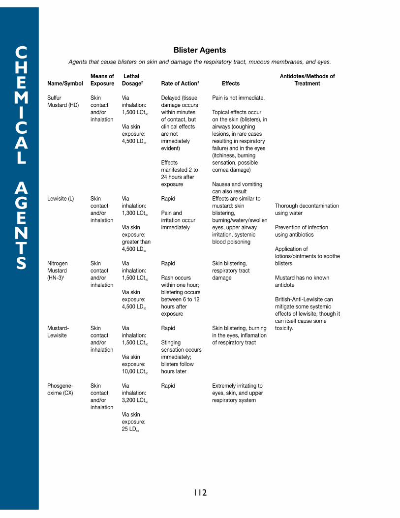
112
CHEMICAL
AGENTS
Blister AgentsAgents that cause blisters on skin and damage the respiratory tract, mucous membranes, and eyes.
Means of Lethal Antidotes/Methods ofName/Symbol Exposure Dosage2 Rate of Action3 Effects Treatment
Sulfur Skin Via Delayed (tissue Pain is not immediate.Mustard (HD) contact inhalation: damage occurs
and/or 1,500 LCt50 within minutes Topical effects occurinhalation of contact, but on the skin (blisters), in
Via skin clinical effects airways (coughingexposure: are not lesions, in rare cases4,500 LD50 immediately resulting in respiratory
evident) failure) and in the eyes(itchiness, burning
Effects sensation, possiblemanifested 2 to cornea damage)24 hours afterexposure Nausea and vomiting
can also resultLewisite (L) Skin Via Rapid Effects are similar to
contact inhalation: mustard: skin Thorough decontaminationand/or 1,300 LCt50 Pain and blistering, using waterinhalation irritation occur burning/watery/swollen
Via skin immediately eyes, upper airway Prevention of infectionexposure: irritation, systemic using antibioticsgreater than blood poisoning4,500 LD50 Application of
lotions/ointments to sootheNitrogen Skin Via Rapid Skin blistering, blistersMustard contact inhalation: respiratory tract(HN-3)4 and/or 1,500 LCt50 Rash occurs damage Mustard has no known
inhalation within one hour; antidoteVia skin blistering occursexposure: between 6 to 12 British-Anti-Lewisite can4,500 LD50 hours after mitigate some systemic
exposure effects of lewisite, though itcan itself cause some
Mustard- Skin Via Rapid Skin blistering, burning toxicity.Lewisite contact inhalation: in the eyes, inflamation
and/or 1,500 LCt50 Stinging of respiratory tractinhalation sensation occurs
Via skin immediately;exposure: blisters follow10,00 LCt50 hours later
Phosgene- Skin Via Rapid Extremely irritating tooxime (CX) contact inhalation: eyes, skin, and upper
and/or 3,200 LCt50 respiratory systeminhalation
Via skinexposure:25 LD50
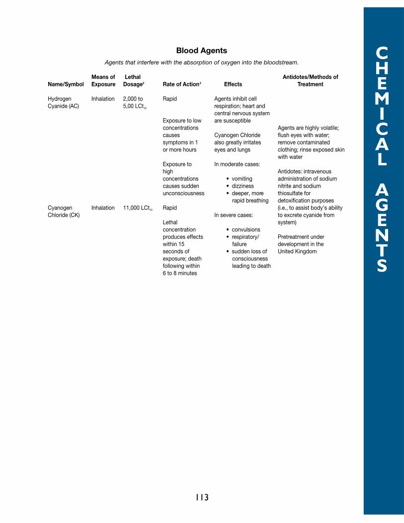
113
CHEMICAL
AGENTS
Blood AgentsAgents that interfere with the absorption of oxygen into the bloodstream.
Means of Lethal Antidotes/Methods ofName/Symbol Exposure Dosage2 Rate of Action3 Effects Treatment
Hydrogen Inhalation 2,000 to Rapid Agents inhibit cellCyanide (AC) 5,00 LCt50 respiration; heart and
central nervous systemExposure to low are susceptibleconcentrations Agents are highly volatile;causes Cyanogen Chloride flush eyes with water;symptoms in 1 also greatly irritates remove contaminatedor more hours eyes and lungs clothing; rinse exposed skin
with waterExposure to In moderate cases:high Antidotes: intravenousconcentrations • vomiting administration of sodiumcauses sudden • dizziness nitrite and sodiumunconsciousness • deeper, more thiosulfate for
rapid breathing detoxification purposesCyanogen Inhalation 11,000 LCt50 Rapid (i.e., to assist body’s abilityChloride (CK) In severe cases: to excrete cyanide from
Lethal system)concentration • convulsionsproduces effects • respiratory/ Pretreatment underwithin 15 failure development in theseconds of • sudden loss of United Kingdomexposure; death consciousnessfollowing within leading to death6 to 8 minutes
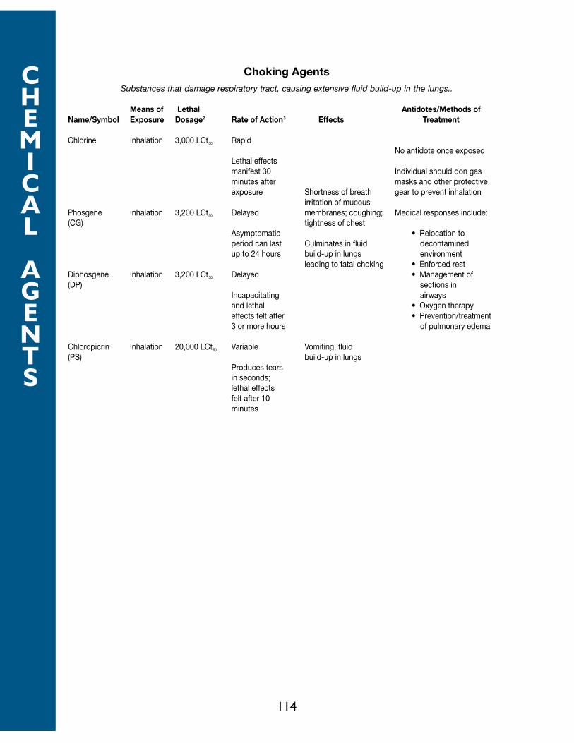
114
CHEMICAL
AGENTS
Choking AgentsSubstances that damage respiratory tract, causing extensive fluid build-up in the lungs..
Means of Lethal Antidotes/Methods ofName/Symbol Exposure Dosage2 Rate of Action3 Effects Treatment
Chlorine Inhalation 3,000 LCt50 RapidNo antidote once exposed
Lethal effectsmanifest 30 Individual should don gasminutes after masks and other protectiveexposure Shortness of breath gear to prevent inhalation
irritation of mucousPhosgene Inhalation 3,200 LCt50 Delayed membranes; coughing; Medical responses include:(CG) tightness of chest
Asymptomatic • Relocation toperiod can last Culminates in fluid decontaminedup to 24 hours build-up in lungs environment
leading to fatal choking • Enforced restDiphosgene Inhalation 3,200 LCt50 Delayed • Management of(DP) sections in
Incapacitating airwaysand lethal • Oxygen therapyeffects felt after • Prevention/treatment3 or more hours of pulmonary edema
Chloropicrin Inhalation 20,000 LCt50 Variable Vomiting, fluid(PS) build-up in lungs
Produces tearsin seconds;lethal effectsfelt after 10minutes

NERVE AGENTSGA GB GD GF VX
BACKGROUND:
Nerve agents are organophosphorous cholinesterase inhibitors. They inhibit the butyryl-
cholinesterase in the plasma, the acetylcholinesterase on the red cell, and the acetyl-
cholinesterase at cholinergic receptor sites in tissue.
After a nerve agent inhibits the tissue enzyme, the enzyme cannot hydrolyze acetylcholine, the
neurotransmitter at cholinergic receptor sites. Acetylcholine accumulates and continues to stim-
ulate the affected organ. The clinical effects from nerve agent exposure are caused by excess
acetylcholine.
The attachment of the agent to the enzyme is permanent (unless removed by therapy).
Erythrocyte enzyme activity returns at the rate of erythrocyte turnover, about 1% per day.
Tissue and plasma enzyme activities return with synthesis of new enzymes. The rate of return of
the tissue and plasma enzymes is not the same, nor is the rate the same for all tissue enzymes.
However, the agent can be removed from the enzyme and the enzyme “reactivated” by several
types of compounds, the most useful of which are the oximes. If the agent-enzyme complex has
not “aged,” oximes are useful therapeutically. Aging is a biochemical process by which the agent-
enzyme complex becomes refractory to oxime reactivation of the enzyme. For most nerve
agents the aging time is longer than the time within which acute casualties will be seen. However,
the aging time of the GD-enzyme complex is about two minutes, and the usefulness of oximes in
GD poisoning is greatly decreased after this period.
Organs with cholinergic receptor sites include the smooth muscles, the skeletal muscles, the cen-
tral nervous system, and most exocrine glands. In addition, cranial efferents and ganglionic affer-
ents are cholinergic nerves.
Muscarine will stimulate some of the cholinergic sites, and these are known as muscarinic sites.
Organs with these sites include the smooth muscles and glands. Nicotine will stimulate other
cholinergic sites, known as nicotinic sites, which are those in skeletal muscle and ganglia. The cen-
tral nervous system (CNS) contains both types of receptors, but the pharmacology in the CNS is
more complex and less well understood. Atropine and similar compounds block the effects of
excess acetylcholine more effectively at muscarinic sites than at nicotinic sites.
Some commonly used pesticides (for example, the organophosphate (OP) Malathion and the car-
bamate Sevin) and some common therapeutic drugs (the carbamates pyridostigmine [Mestinon]
and physostigmine [Antilirium]) also inhibit acetylcholinesterase and can be considered “nerve
agents.”
115
NERVE
AGENTS
GA
GB
GD
GF
VX

However, while the OP pesticides cause the same biological effects as nerve agents, there are
some important differences in the duration of biological activity and response to therapy.
CLINICAL FEATURES:
The initial effects of exposure to a nerve agent depend on the dose and on the route of expo-
sure. The initial effects from a sublethal amount of agent by vapor exposure are different from
the initial effects from a similar amount of liquid agent on the skin.
Toxicities: The large amounts of GA and GB required to produce effects after skin application
reflect the volatility of these agents. They evaporate rather than penetrate the skin. However, if
these agents are occluded and prevented from evaporating they penetrate the skin very well.
GB, the agent studied most thoroughly in man, will cause miosis, rhinorrhea, and a feeling of tight-
ness in the throat or chest at a Ct of 3 to 5 mg-min/m3.
Effects: Exposure to a small amount of nerve agent vapor causes effects in the eyes, nose, and
airways. These effects are from local contact of the vapor with the organ and do not indicate sys-
temic absorption of the agent. In this circumstance, the erythrocyte-ChE may be normal or
depressed. A small amount of liquid agent on the skin causes systemic effects initially in the gas-
trointestinal (GI) tract. Lethal amounts of vapor or liquid cause a rapid cascade of events culmi-
nating within a minute or two with loss of consciousness and convulsive activity followed by
apnea and muscular flaccidity within several more minutes.
Eye: Miosis is a characteristic sign of exposure to nerve agent vapor. It occurs as a result of direct
contact of vapor with the eye. Liquid agent on the skin will not cause miosis if the amount of liq-
uid is small; a moderate amount of liquid may or may not cause miosis; and a lethal or near-lethal
amount of agent usually causes miosis. A droplet of liquid in or near the eye will also cause mio-
sis. Miosis will begin within seconds or minutes after the onset of exposure to agent vapor, but it
may not be complete for many minutes if the vapor concentration is low. Miosis is bilateral in an
unprotected individual, but occasionally may be unilateral in a masked person with a leak in his
mask eyepiece.
Miosis is often accompanied by complaints of pain, dim vision, blurred vision, conjunctival injec-
tion, nausea, and occasionally vomiting. The pain may be sharp or dull in or around the eyeball,
but more often is a dull ache in the frontal part of the head. Dim vision is due in part to the small
pupil, and cholinergic mechanisms in the visual pathways also contribute. The complaint of
blurred vision is less easily explained, as objective testing usually indicates an improvement in
visual acuity because of the “pin-hole” effect. Conjunctival injection may be mild or severe, and
occasionally subconjunctival hemorrhage is present. Nausea (and sometimes vomiting) are part
of a generalized complaint of not feeling well. Miosis, pain, dim vision, and nausea can be relieved
by topical homatropine or atropine in the eye.
116
NERVE
AGENTS
GA
GB
GD
GF
VX

Nose: Rhinorrhea may be the first indication of nerve agent vapor exposure. Its severity is dose
dependent.
Airways: Nerve agent vapor causes bronchoconstriction and increased secretions of the glands
in the airways in a dose-related manner. The exposed person may feel a slight tightness in his
chest after a small amount of agent and may be in severe distress after a large amount of agent.
Cessation of respiration occurs within minutes after the onset of effects from exposure to a large
amount of nerve agent. This apnea is probably mediated through the CNS, although peripheral
factors (skeletal muscle weakness, e.g., the intercostal muscles, and bronchoconstriction) may
contribute.
Gastrointestinal tract: After they are absorbed, nerve agents cause an increase in the motility
of the GI tract and an increase in secretions by the glands in the wall of the GI tract. Nausea and
vomiting are early signs of liquid exposure on the skin. Diarrhea may occur with large amounts
of agent.
Glands: Nerve agent vapor causes increases in secretions from the glands it contacts, such as the
lacrimal, nasal, salivary, and bronchial glands. Localized sweating around site of liquid agent on the
skin is common, and generalized sweating after a large liquid or vapor exposure is common.
Increased secretions of the glands of the GI tract occur after systemic absorption of the agent by
either route.
Skeletal Muscle: The first effect of nerve agents on skeletal muscle is stimulation producing mus-
cular fasciculations and twitching. After a large amount of agent, fatigue and weakness of muscles
are rapidly followed by muscular flaccidity.
Fasciculations are sometimes seen early at the site of a droplet of liquid agent on the skin, and
generalized fasciculations are common after a large exposure. These may remain long after most
of the other acute signs decrease.
Central Nervous System: The acute of CNS signs of exposure to a large amount of nerve agent
are loss of consciousness, seizure activity, and apnea. These begin within a minute after exposure
to a large amount of agent vapor and may be preceded by an asymptomatic period of one to 30
minutes after contact of liquid with the skin.
After exposure to smaller amounts of nerve agents, CNS effects vary and are nonspecific. They
may include forgetfulness, an inability to concentrate fully, insomnia, bad dreams, irritability,
impaired judgement, and depression. They do not include frank confusion and misperceptions
(i.e., hallucinations). These may occur in the absence of physical signs or other symptoms of
exposure. After a severe exposure these symptoms occur upon recovery from the acute severe
effects. In either case they may persist for as long as four to six weeks.
117
NERVE
AGENTS
GA
GB
GD
GF
VX

Cardiovascular: The heart rate may be decreased because of stimulation by the vagus nerve, but
it is often increased because of other factors, such as fright, hypoxia, and the influence of adren-
ergic stimulation secondary to ganglionic stimulation. Thus, the heart rate may be high, low, or in
the normal range. Bradyarrhythmias, such as first-, second-, or third-degree heart block may
occur. The blood pressure may be elevated from adrenergic factors, but is generally normal until
the terminal decline.
DIAGNOSIS:
Physical findings depend on the amount and route of exposure. After exposure to small to mod-
erate amounts of vapor, there are usually miosis and conjunctival injection, rhinorrhea, and pul-
monary signs, although the latter may be absent even in the face of mild to moderate pulmonary
complaints. In addition to these signs, an exposure to a high Ct may precipitate copious secre-
tions from the nose and mouth, generalized muscular fasciculations, twitching or seizure activity,
loss of consciousness, and apnea. Cyanosis, hypotension, and bradycardia may be present just
before death.
Exposure to a small droplet of liquid on the skin may produce few physical findings. Sweating,
blanching, and occasionally fasciculations at the site may be present soon after exposure, but may
no longer be present at the onset of GI effects. After a large exposure, the signs are the same as
after vapor exposure.
Miosis is a useful sign of exposure to vapor, but does not occur after a liquid exposure unless the
amount of exposure is large or the exposure is in or close to the eye.
Time Course of Effects
Effects from nerve agent vapor begin within seconds to several minutes after exposure. Loss of
consciousness and onset of seizure activity have occurred within a minute of exposure to a high
CT. After exposure to a very low Ct, miosis and other effects may not begin for several mintues,
and miosis may not be complete for 15 to 30 minutes after removal from the vapor. There is no
latent period or delay in onset from vapor exposure. Effects may continue to progress for a peri-
od of time, but maximal effects usually occur within minutes after exposure stops.
A large amount of liquid on the skin causes effects within minutes. Commonly there is an asymp-
tomatic period of one to 30 minutes, and then the sudden onset of an overwhelming cascade of
events, including loss of consciousness, seizure activity, apnea, and muscular flaccidity. After small
amounts of liquid agent on the skin the onset of effects has been delayed for as long as 18 hours
after contact. These effects are initially gastrointestinal and are usually not life threatening.
Generally, the longer the interval the less severe are the effects.
118
NERVE
AGENTS
GA
GB
GD
GF
VX

Differential Diagnosis
The effects caused by a mild vapor exposure, namely rhinorrhea and a tightness in the chest, may
easily be confused with an upper respiratory malady or an allergy. Miosis, if present, will help to
distinguish these, but the eyes must be examined in very dim light to detect this. Similarly, GI
symptoms from another illness may be confused with those from nerve agent effects, and in this
instance there will be no useful physical signs. History of possible exposure will be helpful, and
laboratory evidence (decreased RBC-ChE activity), if available, will be useful to distinguish the
two.
The diagnosis is easier in the severely intoxicated patient. The combination of miosis, copious
secretions, and generalized muscular fasciculations in a gasping, cyanotic, and convulsing patient
is characteristic.
Laboratory Findings
The cholinesterase activity of the blood components is inhibited by nerve agents, and estimation
of this activity is useful in detecting exposure to these agents. The erythrocyte enzyme activity is
more sensitive to acute nerve agent exposure than is the plasma enzyme activity.
The amount of inhibition of this enzyme activity does not correlate well with the severity of local
effects from mild to moderate vapor exposure. The enzyme activity may be from 0% to 100%
of the individual’s normal activity in the face of miosis, rhinorrhea, and/or airway symptoms.
Normal or nearly normal erythrocyte acetylcholinesterase activity may be present with moder-
ate effects in these organs. At the other extreme, the enzyme may be inhibited 60% to 70%
when miosis or rhinorrhea is the only sign of exposure. Several systemic effects generally indicate
inhibition of the erythrocyte acetylcholinesterase by 70% to 80% or greater.
Other laboratory findings will relate to complications. For example, acidosis may occur after pro-
longed hypoxia.
MEDICAL MANAGEMENT:
Management of a casualty with nerve agent intoxication consists of decontamination, ventilation,
administration of the antidotes, and supportive therapy. The condition of the patient dictates the
need for each of these and the order in which they are done.
Decontamination is described elsewhere in this manual. Skin decontamination is not necessary
after exposure to vapor alone, but clothing should be removed because it may contain “trapped”
vapor.
119
NERVE
AGENTS
GA
GB
GD
GF
VX
The need for ventilation will be obvious, and the means of ventilation will depend on available
equipment. Airway resistance is high (50-70 cm of water) because of bronchoconstriction and
secretions, and initial ventilation is difficult. The resistance decreases after atropine administra-
tion, after which ventilation will be easier. The copious secretions, which may be thickened by
atropine, also impede ventilatory efforts and require frequent suctioning. In reported cases of
severe nerve agent exposure, ventilation has been required from 0.5 to 3 hours.
Three drugs are used to treat nerve agent exposure, and another is used as pretreatment for
potential nerve agent exposure. The three therapeutic drugs are atropine, pralidoxime chloride,
and diazepam. The use of the pretreatment drug, pyridostigmine bromide, is discussed later in
this chapter.
Atropine is a cholinergic blocking, or anticholinergic, compound. It is extremely effective in
blocking the effects of excess acetylcholine at peripheral muscarinic sites. Under experimental
conditions, very large amounts may block some cholinergic effects at nicotinic sites, but these
antinicotinic effects are not evident even at high clinical doses. When small amounts (2 mg) are
given to normal individuals without nerve agent intoxication, atropine causes mydriasis, a
decrease in secretions (including a decrease in sweating), mild sedation, a decrease in GI motili-
ty, and tachycardia. The amount in three MARK I kits may cause adverse effects on military per-
formance in a normal person. In people not exposed to nerve agents, amounts of 10 mg or high-
er may cause delirium. Potentially, the most hazardous effect of inadvertent use of atropine (2
mg, i.m.) in a young person not exposed to a cholinesterase inhibiting compound in a warm or
hot atmosphere is inhibition of sweating, which may lead to heat injury. In the military, atropine
is packaged in autoinjectors, each containing 2 mg.
Pralidoxime chloride (Protopam chloride; 2-PAMCl) is an oxime. Oximes attach to the nerve
agent that is inhibiting the cholinesterase and break the agent-enzyme bond to restore the nor-
mal activity of the enzyme. Clinically, this is noticable in those organs with nicotinic receptors.
Abnormal activity in skeletal muscles decreases, and normal strength returns. The effects of an
oxime are not apparent in organs with muscarinic receptors; oximes do not cause a decrease in
secretions, for example. They also are less useful after aging occurs, but with the exception of
GD (soman) intoxicated individuals, casualties will be treated before significant aging occurs.
Pralidoxime chloride (600 mg) is an autoinjector for self-use along with the atropine injector.
These atropine and pralidoxime chloride autoinjectors are packaged together in a MARK I kit.
Each military person is issued three MARK I kits. MARK I kits are now being carried by some EMS
agencies and kept in hospitals.
Diazepam is an anticonvulsant drug used to decrease convulsive activity and to reduce the brain
damage caused by prolonged seizure activity. Without the use of pyridostigmine pretreatment,
experimental animals died quickly after superlethal doses of nerve agents despite conventional
therapy. With pyridostigmine pretreatment (followed by conventional therapy) animals survived
superlethal doses of soman, but had prolonged periods of seizure activity before recovery. They
later had performance decrements and anatomic lesions in their brains. The administration of
diazepam with other standard therapy to soman-poisoned animals pretreated with pyridostig-
120
NERVE
AGENTS
GA
GB
GD
GF
VX
mine reduced the seizure activity and its sequelae. Current military doctrine is to administer
diazepam with other therapy (three MARK I’s) at the onset of severe effects from a nerve agent,
whether or not seizure activity is among those effects. Each military person carries one autoin-
jector containing 10 mg of diazepam for his buddy to administer to him (if he could self-adminis-
ter it, he would not need it). Diazepam should be administered with the three MARK I‘s
when the casualty’s condition warrants the use of three MARK I’s at the same time.
Medical personnel can administer more diazepam to a casualty if necessary. The medical corps-
man carries extra diazepam injectors and is authorized to administer two additional injectors at
10 minute intervals to a convulsing casualty.
The doctrine for self-aid for nerve agent intoxication states that if an individual has effects from
the agent he/she should self-administer one MARK I. If there is no improvement in 10 minutes,
he/she should seek out a buddy to assist in the evaluation of his/her condition before further
MARK I’s are given. If a buddy finds an individual severely intoxicated (e.g., gasping respirations,
twitching, etc.) so that the individual can not self-administer a MARK I, the buddy should admin-
ister three MARK I’s and diazepam immediately. The discussion below is advice for medical assis-
tance.
The appropriate number of MARK I kits to administer initially to a casualty from nerve agent
vapor depends on the severity of the effects. Systemic atropine will not reverse miosis (unless
administered in very large amounts), and miosis alone is not an indication for a MARK I. If the eye
or head pain and nausea associated with the miosis are severe, topical application of atropine (or
homatropine) in the eye will bring relief. Topical atropine should not be used without good rea-
son (severe pain), because it causes blurred vision for a day or longer. A casualty with miosis and
rhinorrhea should be given one MARK I only if the rhinorrhea is severe and troublesome (he can
not keep his mask on because of fluid). A casualty with mild to moderate dyspnea should be given
one or two MARK I’s, depending on the severity of his distress and the time between exposure
and therapy. Some of the respiratory distress from a mild exposure will spontaneously decrease
within 15 to 30 minutes after termination of exposure, so if the casualty is not severely uncom-
fortable only one MARK I should be used initially. Atropine is quite effective, and care should be
taken not to give too much in a casualty who does not need it.
A severe casualty from nerve agent vapor has miosis, copious secretions from the nose and
mouth, severe difficulty breathing or apnea, possibly some degree of cyanosis, muscular fascicu-
lations, and twitching or convulsive activity, and is unconscious. He should be given three MARK
I’s and diazepam immediately. Ventilation will be needed and should be done via an endotracheal
airway if possible. Suctioning of the excessive airway secretions will be necessary to enhance air
exchange and will make ventilatory efforts easier. Atropine, 2 mg, should be repeated at three-
to five-minute intervals and should be titrated to a reduction of secretions and to reduction of
ventilatory resistance. When the intravenous preparation is available, the preferred route of
atropine administration is via the intravenous route, but this route should be avoided until hypox-
ia is corrected, because intravenously administered atropine in hypoxic animals has produced
ventricular fibrillation. In a hypotensive patient or a patient with poor veins, atropine might be
121
NERVE
AGENTS
GA
GB
GD
GF
VX
given intratracheally, either via the endotracheal tube or directly into the trachea, for more rapid
absorption via the peribronchial vessels.
The medical care provider might err in giving too much atropine to a mild to moderate casualty.
More importantly, the care provider might err by giving too little atropine to a severe casualty. In
a severe casualty, atropine should be pushed at frequent intervals until secretions are dry (or
nearly dry) and until ventilation can be accomplished with ease. In reported cases this has
required 10 to 20 mg of atropine within the first several hours. A conscious, less-severely
exposed casualty should receive atropine until he is breathing comfortably, and he will be able to
communicate this. Dry secretions need not be an end point in mild to moderate casualties.
The casualty with skin exposure to liquid is more difficult to evaluate and manage than is a casu-
alty from vapor exposure. Agent on the surface of the skin can be decontaminated, but agent
absorbed into the skin cannot be removed. The initial effects from absorbed liquid agent can start
two to three hours after thorough decontamination of agent droplets on the skin. A casualty from
liquid exposure on the skin may continue to worsen because of continued absorption of the agent
from the skin depot.
The first effects of a liquid droplet on the skin are sweating with or without blanching and occa-
sionally with muscular fasciculations at the site. Gastrointestinal effects (nausea, vomiting, and
sometimes diarrhea) are the first systemic effects, and these may start from 0.5 to 18 hours after
contact with the agent. If these effects occur within the first several hours after exposure, they
may portend more severe effects, and initial therapy should be two MARK I’s. If effects begin
later, initial therapy should be one MARK I.
A large amount of liquid agent on the skin will cause effects 1 to 30 minutes after contact,
whether or not decontamination was done. Nevertheless, early decontamination may lessen the
magnitude of the effects. After a one- to thirty-minute latent or asymptomatic period, the casu-
alty will suddenly lose consciousness and begin seizure activity. The condition of the casualty and
management are the same as described for a severe casualty from vapor or exposure.
Further care of the severe casualty consists of atropine administration to minimize secretions and
of ventilation until spontaneous respiration resumes. Oxime administration should be repeated at
hourly intervals for two or three additional doses. The preferred method of administration of the
oxime is by intravenous drip of 1 gram over 20 to 30 minutes (more rapid administration will
cause hypertension), but three additional oxime autoinjectors (total dose of 1.8 grams) may be
given if the intravenous route cannot be used. The need for ventilation may continue for 0.5 to
3 hours. Unless prolonged hypoxia or other complications have occurred, the casualty will even-
tually begin having spontaneous muscular activity and make sporadic attempts to breathe.
Muscles will become stronger and breathing more regular, and the casualty will have intermittent
episodes of conscious behavior. Within an hour or two he will be breathing, moving, and con-
scious, although he will be weak and intermittently obtunded.
122
NERVE
AGENTS
GA
GB
GD
GF
VX
SUMMARY:
SIGNS AND SYMPTOMS:
Vapor: Small exposure—Miosis, rhinorrhea, mild difficulty breathing. Large exposure—Sudden loss
of consciousness, convulsions, apnea, flaccid paralysis, copious secretions, miosis.
Liquid on skin: Small to moderate exposure—Localized sweating; nausea, vomiting, feeling of
weakness. Large exposure—Sudden loss of consciousness, convulsions, apnea, flaccid paralysis,
copious secretions.
DIAGNOSIS:
Effects of Nerve Agent Vapor
• Small Amount:
Eyes: small pupils, red conjunctiva, dim/blurred vision, pain, nausea/vomiting
Nose: runny nose
Mouth: increased salivation
Airways: tightness in chest, shortness of breath, cough
• Large Amount:
Loss of consciousness
Convulsions
Flaccid paralysis
Breathing stops
Heart stops
Effects begin within seconds to a minute.
Effects of Nerve Agent Liquid on the Skin
• Very small drop: sweating, twitching at site
• Small drop: nausea, vomiting, diarrhea
• Drop: Loss of consciousness, convulsions, breathing stops, flaccid paralysis
Effects begin within 30 minutes (large amount) to 18 hours (small amount).
123
NERVE
AGENTS
GA
GB
GD
GF
VX

TREATMENT:
Administration of MARK I’s (atropine and pralidoxime chloride); diazepam in addition if casualty
is severe; ventilation and suction of airways for respiratory distress.
PROPHYLAXIS:
The U.S. military fielded pyridostigmine bromide as a pretreatment for nerve agent exposure.
Each individual received a blister pack containing 21 30-mg tablets. The dose regimen is one 30-
mg tablet every eight hours.
ISOLATION AND DECONTAMINATION:
Hypochlorite; large amounts of water. Protect yourself by wearing a mask (SCBA), gloves and a
protective suit until the casualty is decontaminated.
124
NERVE
AGENTS
GA
GB
GD
GF
VX

NERVE AGENT EFFECTS
Nerve Agents—How They Work
• Nerve Agents interfere with transmission of the message from nerve to organ.
• The nerve is normal; the transmission to the organ (muscle, gland) is faulty.
• The organ (muscle, gland) gets the wrong message, and does the wrong thing.
• This causes too much activity in muscles,
Vapor Exposure
Mild
Eyes Miosis
Dim vision
Headache
Nose Rhinorrhea
Mouth Salivation
Lungs Dyspnea (“tightness in the chest”)
Time of onset: Seconds to minutes after exposure
Self-aide: 1 MARK I
Buddy-aid: Stand by
Severe
All the above, plus
Severe breathing difficulty or cessation of respiration
Generalized muscular twitching, weakness or paralysis
Convulsions
Loss of consciousness
Loss of bladder, bowel control
Time of onset: Seconds to minutes after exposure
Self-aid: None. Victim will be unable to help self.
Buddy-aid: 3 MARK I’s and diazepam immediately
125
NERVE
AGENTS
EFFECTS
VAPOR

Liquid on skin
Mild/moderate
Muscle twitching at site of exposure
Sweating at site of exposure
Nausea, vomiting
Feeling of weakness
Time of onset: 10 minutes to 18 hours after exposure
Self-aid: 1-2 MARK I’s, depending on severity of symptoms
Buddy-aid: Stand by
Severe
All the above, plus
Severe breathing difficulty or cessation of breathing
Generalized muscular twitching, weakness, or paralysis
Convulsions
Loss of consciousness
Loss of bladder and bowel control
Time of onset: Minutes to an hour after exposure
Self-aid: None. Victim will be unable to help himself
Buddy-aid: 3 MARK I’s and diazepam immediately
126
NERVE
AGENTS
EFFECTS
VAPOR

BLISTER AGENTS(MUSTARD)
HD H
BACKGROUND:
Vesicant agents, specifically sulfur mustard (H; HD), have been major military threat agents since
their introduction in World War I. They constitute both a vapor and a liquid threat to all exposed
skin and mucous membranes. Mustard’s effects are delayed, appearing hours after exposure.
Organs most commonly affected are the skin (with erythema and vesicles), eyes (with mild con-
junctivitis to severe eye damage), and airways (with mild irritation of the upper respiratory tract
to severe bronchiolar damage leading to necrosis and hemorrhage of the airway mucosa and
musculature). Following exposure to large quantities of mustard, precursor cells of the bone mar-
row are damaged, leading to pancytopenia and increased susceptibility to infection. The gas-
trointestinal tract may be damaged, and there are sometimes central nervous system signs. There
is no specific antidote, and management is symptomatic therapy. Immediate decontamination is
the only way to reduce damage.
Mustard is an oily liquid with a color ranging from a light yellow to brown. Its odor is that of gar-
lic, onion, or mustard (hence its name), but because of accomodation of the sense of smell, odor
should not be relied on for detection. Under temperate conditions mustard evaporates slowly
and is primarily a liquid hazard, but its vapor hazard increases with increasing temperature. At
100°F or above, it is a definite vapor hazard. Mustard freezes at 57°F and, since a solid is difficult
to disperse, it is often mixed with substances with a lower freezing point, e.g., Lewisite (the mix-
ture is HL), or agent T, a closely related vesicant (the mixture is HT) so that the mixture will
remain liquid at lower temperatures.
After absorption into the body, mustard rapidly cyclizes (seconds to minutes) in extracellular
water. This cyclic compound is extremely reactive and quickly binds to intra- and extra-cellular
enzymes, proteins, and other substances. Mustard has many biological actions, but the exact
mechanism by which it produces tissue injury is not known. According to one prominent hypoth-
esis, biological damage from mustard results from DNA alkylation and crosslinking in rapidly
dividing cells, such as basal keratinocytes, mucosal epithelium, and bone marrow precursor cells.
This leads to cellular death and inflammatory reaction, and, in the skin, protease digestion of
anchoring filaments at the epidermal-dermal junction and the formation of blisters.
Mustard possesses mild cholinergic activity, which may be responsible for effects such as early
gastrointestinal symptoms and miosis.
127
BLISTER
AGENTS
MUSTARD

Mustard reacts with tissue within minutes of entering the body and is no longer an intact mole-
cule. Blood, tissue, and blister fluid do not contain mustard, and one cannot become exposed to
mustard by contact with body fluids or tissues.
CLINICAL FEATURES:
Topical effects of mustard occur in the eye, airways, and skin. Systemically absorbed mustard may
produce effects in the bone marrow, the gastrointestinal tract, and the central nervous system.
Direct injury to the GI tract may also occur following ingestion of the compound.
Skin: Erythema is the mildest and earliest form of skin injury after exposure to mustard. It resem-
bles sunburn, and is associated with pruritis or burning, stinging pain. Erythema begins to appear
in 2 to 24 hours after vapor exposure with time of onset dependent on Ct, ambient temperature
and humidity, and skin site exposed. The skin sites most sensitive are the warm, moist locations
with thinner skin, such as the perineum, external genitalia, axillae, antecubital fossae, and neck.
Within the erythematous areas, small vesicles can develop, which may later coalesce to form bul-
lae. The typical bulla, or blister, is large, dome-shaped, thin-walled, translucent, yellowish, and
surrounded by erythema. The blister fluid is clear, at first thin and straw-colored, but later yel-
lowish and tending to coagulate. The fluid does not contain mustard and is not a vesicant.
At extremely high doses, such as those from liquid exposure, lesions may develop a central zone
of coagulation necrosis with blister formation at the periphery. These lesions take longer to heal
and are more prone to secondary infection that the uncomplicated lesions seen at lower expo-
sure levels.
Pulmonary: The primary airway lesion from mustard is necrosis of the mucosa with later dam-
age to the musculature of the airways if the amount of agent is large. The damage begins in the
upper airways and descends to the lower airways in a dose-dependent manner. Usually, the ter-
minal airways and alveoli are affected only as a terminal event. Pulmonary edema is not usually
present unless the damage is very severe and then it usually is hemorrhagic.
The earliest effects from mustard—perhaps the only effects from a low Ct—involve the nose,
the sinuses, and the pharynx. There may be irritation or burning of the nares, epistaxis, sinus pain
or irritation, and irritation or soreness of the pharynx. As the Ct increases other effects occur:
laryngitis with voice changes and a nonproductive cough. Damage to the trachea and upper
bronchi leads to a cough productive of sputum. Lower airway involvement causes dyspnea and
an increasingly severe cough with increased quantities of sputum. Terminally, there may be necro-
sis of the smaller airways with hemorrhagic edema into surrounding alveoli. This hemorrhagic
pulmonary edema is rarely a feature.
128
BLISTER
AGENTS
MUSTARD

Necrosis of the airway mucosa with resulting inflammation can cause pseudomembrane forma-
tion, and pseudomembranes may occur from the most proximal parts of the airways to the most
distal portions. These membranes may cause local airway obstruction at the sites of formation,
and detachment may lead to obstruction of lower airways.
The cause of death in mustard poisoning is commonly respiratory failure. Mechanical obstruction
by pseudomembranes may be a cause, but more commonly deaths occurring from the third to
the sixth day after exposure result from secondary bacterial pneumonia caused by bacterial inva-
sion of denuded respiratory mucosa and necrotic debris. Agent-induced bone marrow suppres-
sion is a contributory factor in later, septic deaths from pneumonia.
Ocular: The eyes are the organs most sensitive to mustard vapor injury. The latent period is
shorter for eye injury than for skin injury and is also Ct dependent.
After low-dose vapor exposure, irritation, evidenced by reddening of the eyes, may be the only
effect. As the dose increases, the spectrum of injury includes progressively more severe con-
junctivitis, photophobia, blepharospasm, pain, and corneal damage.
Blisters do not normally form in the eyes. Instead, swelling and loosening of corneal epithelial cells
leads to corneal edema and clouding with leukocytes (which affects vision). Corneal vasculariza-
tion with secondary edema may last for weeks. Severe effects may be followed by scarring
between the iris and lens; this scarring may restrict pupillary movements and may predispose vic-
tims to glaucoma.
The most severe damage is caused by liquid mustard from airborne droplets or by self-contami-
nation. After extensive eye exposure, severe corneal damage with possible perforation of the
cornea and loss of the eye can occur. Eye loss also results from panophthalmitis if appropriate
therapy is not instituted.
Gastrointestinal tract: The mucosa of the gastrointestinal (GI) tract is very susceptible to mus-
tard damage, either from systemic absorption or ingestion of the agent.
Mustard exposure, even exposure to a small amount, will often cause nausea with or without
vomiting lasting 24 hours or less. The nausea and vomiting appear not to be a direct effect of the
agent on the gastrointestinal tract, but rather they are from a stress reaction, a nonspecific reac-
tion to the odor, or cholinergic stimulation by mustard. Diarrhea has been reported; constipation
is equally common. Diarrhea (rarely bloody) and vomiting beginning days after a high-dose expo-
sure imply a poor prognosis.
Central nervous system: The CNS effects of mustard remain poorly defined. Animal work
demonstrated that mustards (particularly the nitrogen mustards) are convulsants, and there are
several human case reports describing people who were exposed to very large amounts and who
had neurological effects within several hours after exposure just prior to death.
129
BLISTER
AGENTS
MUSTARD

Time Course of Effects
Mustard binds irreversibly to tissue within several minutes after contact. If decontamination is not
done immediately after exposure there is no way to prevent injury, although later decontamina-
tion might prevent a more severe lesion.
The clinical effects of mustard are delayed. Signs and symptoms may appear as early as two hours
after a high-dose exposure, whereas following a low-dose vapor exposure the latent or asymp-
tomatic period may extend to 24 hours. There are several reports of individuals exposed to very
large amounts who died within hours; this type of occurrence is extremely rare. The typical onset
time is between four and eight hours. The concentration (C) of the mustard vapor, the time (t)
of exposure, the ambient weather, and the body site exposed are factors in the onset time.
It must be emphasized that mustard causes tissue damage within several minutes after
contact without causing any concomitant clinical effects, e.g., burning or erythema.
Because of the lack of immediate effects, the contaminated person is often unaware of the expo-
sure and does not decontaminate. To prevent injury, decontamination must be done imme-
diately after contact. Later decontamination may prevent further damage, absorption, or
spread of the agent.
DIAGNOSIS:
Of the three vesicant agents, mustard is the only one that does not cause immediate pain. The
casualty is asymptomatic until the lesion becomes apparent hours later.
In contrast, Lewisite and phosgene oxime in either liquid or vapor form cause immediate pain or
irritation to the eye, skin, or respiratory tract. This is sufficient stimulus to decontaminate imme-
diately or to mask. Because of this, lesions from these agents may not be as severe as those from
mustard.
Isolated small blisters or a small group of blisters suggest possible exposure to mustard, to plants
such as poison ivy or poison oak, to drugs, or to other substances. The physical characteristics of
the lesion are not distinctive, therefore the history of exposure is invaluable.
Although the blisters of mustard and Lewisite are slightly different (there is less erythema around
the Lewisite blister) this information is of little value in individual cases.
Laboratory Findings
There are no available clinical laboratory tests for mustard exposure. Leukocytosis occurs during
the first day and the magnitude of increase in leukocytes during the subsequent days correlates
roughly with the amount of tissue injury, primarily to skin or pulmonary tissue. If systemic absorp-
130
BLISTER
AGENTS
MUSTARD

tion is large, leukocytes in the peripheral blood will decrease beginning on day three to day five;
this decrease indicates damage to precursor cells in the blood-forming organs. The fall may be
precipitate, e.g., a decrease of 5,000 to 10,000 cells/day. If the marrow damage is severe, ery-
throcytes and platelets may later decrease, but the victim usually recovers or dies before this is
apparent. A leukocyte count of 500 or fewer is a sign of an unfavorable prognosis.
Signs of a chemical pneumonitis may appear within the first 2 to 3 days after inhalational expo-
sure. Leukocytosis, fever, and sputum production suggest a bacterial process, but within this time
period sputum cultures are usually negative for pathogens. Organisms commonly invade the dam-
aged airway tissue at days three to five, and a change in the fever pattern, an increase in leuko-
cytosis, and a change in the character of the sputum in this time period suggest a bacterial
process. Sputum Gram stain and culture should be done for identification of the specific organ-
ism.
Damaged skin should be cultured routinely, particularly if there is an increase in the exudate or
an increase in the inflammatory reaction.
Although gastrointestinal bleeding is unusual, declining hematocrit values should prompt serial
analyses of stool for occult blood.
There is no clinical laboratory test for mustard in blood or tissue, nor is one expected as mustard
is biotransformed and bound to tissues within minutes after absorption. A method for analysis of
urine for thiodiglycol, a metabolite of mustard, is in the investigational stage.
MEDICAL MANAGEMENT:
The management of a patient exposed to mustard may be simple, as in the provision of sympto-
matic care for a sunburn-like erythema, or extremely complex as providing total management for
a severely ill patient with burns, immunosuppression, and multi-system involvement. The follow-
ing are suggested therapeutic measures for each organ system. Guidelines for general patient
care are not intended to take the place of sound clinical judgment, especially in the management
of complicated cases.
Skin: Erythema should be treated with calamine or other soothing lotion or cream (e.g., 0.25%
camphor and menthol, calamine) to reduce burning and itching. Small blisters (under 1-2 cm)
should be left intact, but because larger ones will eventually break (the blister fluid does not con-
tain mustard) they should be carefully unroofed. Denuded areas should be irrigated 3-4 times
daily with saline, another sterile solution, or soapy water and then liberally covered with a topi-
cal antibiotic such as silver sulfadiazine or mafenide acetate to a thickness of 1-2 mm. If an antibi-
otic cream is not available, sterile petrolatum will be useful. Modified Dakins solution (sodium
hypochlorite) was used in WWI and in Iranian casualties for irrigation and as an antiseptic.
131
BLISTER
AGENTS
MUSTARD

Multiple or large areas of vesication suggest the need for hospitalization and whirlpool bath irri-
gation.
Systemic analgesics should be used liberally, particularly before manipulation of the patient or irri-
gation of the burn areas. Systemic antipruritics such as trimeprazine should be tried if needed.
Monitoring of fluids and electrolytes is important in any sick patient, but it must be recognized
that fluid loss is not of the magnitude seen with thermal burns. Clinicians accustomed to
treating patients with thermal burns must resist the temptation to overhydrate a mustard casu-
alty with a similar amount of burned body surface.
Eyes: Conjunctival irritation from a low Ct will respond to any of a number of available oph-
thalmic solutions after the eyes are thoroughly irrigated. Regular application of homatropine (or
other anticholinergic drug) ophthalmic ointment will reduce or prevent future synechiae forma-
tion, and a topical antibiotic applied several times a day will reduce the incidence and severity of
infection. Vaseline or a similar substance should be applied to the edges of the lids regularly to
prevent them from sticking together. This prevents adhesions and later scarring during healing
and also permits drainage of any underlying infection. Topical analgesics may be useful initially if
blepharospasm is too severe to permit an adequate examination, but topical analgesics should
otherwise be avoided, and systemic analgesics should be given for eye pain. Topical steroids are
not of proven value, but their use during the first day or two might reduce inflammation. Further
use should be relegated to an ophthalmologist. Sunglasses may reduce discomfort from photo-
phobia.
The patient should be constantly reassured that complete healing and restoration of vision will be
the outcome.
Pulmonary: Upper airway symptoms (sore throat, non-productive cough, hoarseness) may
respond to steam inhalation and cough suppressants. Although a productive cough and dyspnea
accompanied by fever and leukocytosis occurring 12 to 24 hours after exposure may suggest a
bacterial process to the clinician, he must resist the urge to use antibiotics for this process, which
in fact is a sterile bronchitis or pneumonitis. Infection often occurs on about the third day and its
presence is signaled by an increased fever, an increase in the pulmonary infiltrate by x-ray, and an
increase in sputum production and a change in sputum character to purulent. Appropriate antibi-
otic therapy should await confirmation of the clinical impression by positive sputum studies
(Gram stain and culture).
Intubation should be performed early before laryngeal spasm or edema makes it difficult or
impossible. Intubation permits better ventilation and facilitates suction of the necrotic and inflam-
matory debris. Oxygen may be needed, and early use of PEEP or CPAP may be of benefit. If there
is a suggestion of pseudomembrane formation, bronchoscopy should be done to permit suction-
ing of the necrotic debris by direct vision.
132
BLISTER
AGENTS
MUSTARD

Bronchodilators may be of benefit for bronchospasm. If they fail, steriods may be tried. There is
little evidence that the routine use of steroids is beneficial. The need for continuous use of assist-
ed or controlled ventilation suggests a poor prognosis.
Death often occurs between the fifth and tenth day after exposure because of pulmonary insuf-
ficiency and infection complicated by a compromised immune response from agent-induced bone
marrow damage.
Gastrointestinal: Atropine (0.4-0.6 mg, i.m. or i.v.), another anticholinergic drug, or antiemetic
should control the early nausea and vomiting. Prolonged vomiting or voluminous diarrhea begin-
ning days after exposure suggests direct involvement of the gastrointestinal tract by severe sys-
temic poisoning, a poor prognostic sign.
Bone marrow: Sterilization of the gut by non-absorbable antibiotics should be considered to
reduce the possibility of sepsis from enteric organisms. Cellular replacement (bone marrow
transplants or transfusions) may be successful as intact mustard does not persist beyond the few
minutes following absorption and would not damage the new cells.
General: A patient severely ill from mustard poisoning requires the general supportive care pro-
vided for any severely ill patient as well as the specific care given to a burn patient. Liberal use of
systemic analgesics and antipruritics, as needed, maintenance of fluid and electrolyte balance, and
other supportive measures are necessary. Parenteral food supplements including vitamins may
also be helpful.
Other: Sulfur donors such as sodium thiosulfate decreased systemic effects and elevated the LD50
when given before exposure or within 20 minutes after exposure in experimental animals.
Activated charcoal given orally to casualties was of no value. Hemodialysis was not only ineffec-
tive, but was harmful in several casualties. The rapid biotransformation of the mustard molecule
suggests that none of these measures would be beneficial hours or days after exposure.
133
BLISTER
AGENTS
MUSTARD

SUMMARY:
SIGNS AND SYMPTOMS:
Asymptomatic latent period (hours). Erythema and blisters on the skin; irritation, conjunctivitis
and corneal opacity and damage in the eyes; mild upper respiratory signs to marked airway
damage; also gastrointestinal effects and bone marrow stem cell suppression.
DIAGNOSIS:
Redness of the skin, blisters. Irritation of eyes. Cough, shortness or breath.
TREATMENT:
Immediate Decontamination After Exposure is the only way to prevent damage. Symptomatic
management of lesions.
PROPHYLAXIS:
None
ISOLATION AND DECONTAMINATION:
Protect yourself by wearing a mask, gloves and a protective suit until the patient is decontami-
nated.
Remove patient from contamination and contamination from patient. Get the patient away from
the source, such as by moving him upwind or out of a contaminated building. If it is absolutely
certain that exposure was to vapor only, remove outer clothing. If there is a possibility of liquid
contamination, all clothing must be removed and the patient must be showered or washed with
soap and water, dilute hypochlorite or water.
134
BLISTER
AGENTS
MUSTARD

MUSTARD VAPOR EFFECTS
Mustard—How It Works
• Mustard quickly penetrates the skin, mucous membranes (eye, airways).
• It changes to another substance, and reacts with enzymes, proteins, DNA.
• It causes cell death.
• Mustard effects are like radiation (“radiomimetic”).
• Mustard causes damage within minutes.
Organ Severity Effects Onset of first effect
Eye Mild Tearing 4-12 hours
Itchy
Burning
Gritty feeling
Moderate Above, plus 3-6 hours
Reddening
Swelling of lids
Moderate pain
Severe Marked swelling of lids 1-2 hours
Possible cornea damage
Severe pain
Airways Mild Runny nose 12-24 hours
Sneezing
Nosebleed
Hoarseness
Hacking cough
Severe Above, plus 2-4 hours
Severe productivecough
Shortness of breathmild to severe
Skin Erythema (redness)
Blisters 2-24 hours
135
MUSTARD
AGENT
EFFECTS

BLISTER AGENTS(LEWISITE)
L
BACKGROUND:
Lewisite is a vesicant that damages the eyes, skin, and airways by direct contact. After absorp-
tion, it causes an increase in capillary permeability to produce hypovolemia, shock, and organ
damage. Exposure to Lewisite causes immediate pain or irritation, although lesions require hours
to become full-blown. Management of a Lewisite casualty is similar to management of a mustard
casualty, although a specific antidote, British-Anti-Lewisite (BAL; dimercaprol) will alleviate some
effects.
Lewisite is an oily, colorless liquid with the odor of geraniums. It is more volatile than mustard.
Although Lewisite contains trivalent arsenic and combines with thiol groups in many enzymes, its
exact mechanism of biological activity is unknown.
CLINICAL FEATURES:
Toxicities: Lewisite casues nasal irritation at a Ct of about 8 mg min/m3, and its odor is noted at
a Ct of about 20 mg min/m3. Lewisite causes vesication and death from inhalation at the same Cts
as mustard. Liquid Lewisite causes vesication at about 14µg, and the LD50 is about 2.8 grams on
the skin.
Organ Systems: Unlike mustard, Lewisite vapor or liquid causes immediate pain or irritation. A
person with a droplet of Lewisite on his skin will note the burning and will immediately take steps
to try to remove it. The vapor is so irritating that a person will seek to mask or to leave the con-
taminated area if possible. Because this warning causes the person exposed to take immediate
steps to decontaminate, the Lewisite lesion will probably not be as severe as the lesion from mus-
tard, as exposure to mustard is often undetected and decontamination is not done.
There are almost no data on humans exposed to Lewisite, and the following is based on animal
investigations.
Skin: Within about five minutes after contact liquid Lewisite will produce a grayish area of dead
epithelium. Erythema and blister formation follow more rapidly than in a similar lesion from mus-
tard, although the full lesion does not develop for 12 to 18 hours. The lesion has more tissue
necrosis and tissue sloughing than does a mustard lesion.
136
BLISTER
AGENTS
LEWISITE

Eye: Lewisite causes pain and blepharospasm on contact. Edema of the conjunctiva and lids fol-
lows, and the eyes may be swollen shut within an hour. Iritis and corneal damage may follow if
the dose is high. Liquid Lewisite causes severe eye damage within minutes of contact.
Respiratory: The extreme irritancy of Lewisite to the nasal area and upper airways causes the
person to mask or exit the area. Scanty data indicate that Lewisite causes the same airway signs
and symptoms as does mustard. The airway mucosa is the primary target and damage progress-
es down the airways in a dose-dependent manner. Pseudomembrane formation is prominent.
Pulmonary edema, which occurs rarely and usually only to a minimal degree after mustard expo-
sure, may complicate exposure to Lewisite.
Other: Available data suggest that Lewisite causes an increase in permeability of systemic capil-
laries with resulting intravascular fluid loss, hypovolemia, shock, and organ congestion. This may
lead to hepatic or renal necrosis with more prominent gastrointestinal effects (including vomiting
and diarrhea) than after mustard.
Physical Findings: The findings are similar to those caused by mustard. As noted, the tissue dam-
age at the site of the skin lesion may be more severe.
Time Course of Effects
Pain and irritation from either liquid or vapor Lewisite are immediate. Early tissue destruction is
more obvious than after mustard, but the lesion is not full-blown for 12 hours or longer.
DIAGNOSIS:
Although differences have been reported between the skin lesions from mustard and Lewisite
(less surrounding erythema and more tissue destruction characterize Lewisite blisters), these are
of little diagnostic assistance in a single patient. The history of immediate pain on contact is absent
after mustard exposure and present after Lewisite or phosgene oxime exposures.
Other substances cause erythema and blisters, and often the history of exposure is the most
helpful tool in diagnosis.
LABORATORY FINDINGS:
There is no specific diagnostic test for Lewisite. Leukocytosis, fever, and other signs of tissue
destruction will occur.
137
BLISTER
AGENTS
LEWISITE

MEDICAL MANAGEMENT:
Early decontamination is the only way of preventing or lessening Lewisite damage. Since this must
be accomplished within minutes after exposure, this is self-aid rather than medical management.
The guidelines for the management of a mustard casualty will be useful. Lewisite does not cause
damage to hematopoietic organs as mustard does. However, fluid loss from the capillaries neces-
sitates careful attention to fluid balance.
British-Anti-Lewisite (BAL; dimercaprol) was developed as an antidote for Lewisite and is used in
medicine as a chelating agent for heavy metals. There is evidence that BAL in oil, given intra-
muscularly, will reduce the systemic effects of Lewisite. However, BAL itself causes some toxici-
ty, and the user should read the package insert carefully. BAL skin ointment and BAL ophthalmic
ointment decrease the severity of skin and eye lesions when applied immediately after early
decontamination. However, neither is currently manufactured.
SUMMARY:
SIGNS AND SYMPTOMS:
Lewisite causes immediate pain or irritation of skin and mucous membranes. Erythema and blis-
ters on the skin and eye and airway damage similar to those seen after mustard exposure devel-
op later.
DIAGNOSIS:
Lewisite and phosgene oxime, in both their liquid and vapor forms, cause moderate to severe
pain on contact with skin or the mucous membranes and produce visible grayish tissue damage
within several minutes of contact. It also causes leakage of systemic capillaries, and hypovolemia
and hypotension may result.
TREATMENT:
Immediate decontamination; symptomatic management of lesions the same as for mustard
lesions; a specific antidote (BAL) will decrease systemic effects.
138
BLISTER
AGENTS
LEWISITE
PROPHYLAXIS:
None
ISOLATION AND DECONTAMINATION:
Protect yourself by wearing a mask, gloves and a protective suit until the patient is decontami-
nated.
Remove patient from contamination and contamination from patient. Get the patient away from
the source, such as by moving him upwind or out of a contaminated building. If it is absolutely
certain that exposure was to vapor only, remove outer clothing. If there is a possibility of liquid
contamination, all clothing must be removed and the patient must be showered or washed with
soap and water, dilute hypochlorite or water.
139
BLISTER
AGENTS
LEWISITE
BLISTER AGENTS(PHOSGENE OXIME)
CX
BACKGROUND:
Phosgene oxime is an urticant or nettle agent that causes a corrosive type of skin and tissue
lesion. It is not a true vesicant, since it does not cause blisters. The vapor is extremely irritating,
and both the vapor and liquid cause almost immediate tissue damage upon contact. There is very
scanty information on phosgene oxime.
CX is a solid at temperatures below 95°F, but the vapor pressure of the solid is high enough to
produce symptoms. Traces of many metals cause it to decompose. However, it corrodes most
metals.
The mechanism by which phosgene oxime causes biological effects is unknown.
CLINICAL FEATURES:
Toxicities: The estimated LCt50 by inhalation is 1500-2000 mg /min/m3. The LD50 for skin expo-
sure has been estimated as 25 mg/kg.
Skin: Phosgene oxime liquid or vapor causes pain on contact which is followed in turn by blanch-
ing with an erythematous ring in 30 seconds, a wheal in 30 minutes, and necrosis later. The
extreme pain may persist for days.
Eyes: Phosgene oxime is extremely painful to the eyes. The damage is probably similar to that
caused by Lewisite.
Pulmonary: Phosgene oxime is very irritating to the upper airways. This agent causes pulmonary
edema after inhalation and after skin application.
Other: Some animal data suggest that phosgene oxime may cause hemorrhagic inflammatory
changes in the gastrointestinal tract.
140
BLISTER
AGENTS
PHOSGENE
OXI
ME
Time Course of Effects
Phosgene oxime causes immediate pain and irritation to all exposed skin and mucous mem-
branes. The time course of damage to other tissue probably parallels that of damage to the skin.
DIAGNOSIS:
Other causes of urticaria and skin necrosis must be considered. Common urticants do not cause
the extreme pain that phosgene oxime does.
Laboratory Findings
There are no distinctive laboratory findings.
MEDICAL MANAGEMENT:
Management is supportive. The skin lesion should be managed in the same way that a necrotic
ulcerated lesion from another cause would be managed.
SUMMARY:
SIGNS AND SYMPTOMS:
Immediate burning and irritation followed by wheal-like skin lesions and eye and airway damage.
DIAGNOSIS:
Phosgene oxime, in both its liquid and vapor forms, cause moderate to severe pain on contact
with skin or the mucous membranes of the eyes, nose, mouth and airways. It also produces vis-
ible grayish tissue damage within several minutes of contact. Later, severe damage of the skin,
eyes and airways may appear.
141
BLISTER
AGENTS
PHOSGENE
OXI
ME

TREATMENT:
Immediate decontamination; symptomatic management of lesions.
PROPHYLAXIS:
None
ISOLATION AND DECONTAMINATION:
Protect yourself by wearing a mask, gloves and a protective suit until the patient is decontami-
nated.
Remove patient from contamination and contamination from patient. Get the patient away from
the source, such as by moving him upwind or out of a contaminated building. If it is absolutely
certain that exposure was to vapor only, remove outer clothing. If there is a possibility of liquid
contamination, all clothing must be removed and the patient must be showered or washed with
soap and water, dilute hypochlorite or water.
142
BLISTER
AGENTS
PHOSGENE
OXI
ME

BLOOD AGENTS(CYANIDE)
AC—HYDROCYANIC ACIDCK—CYANOGEN CHLORIDE
BACKGROUND:
Cyanide is a rapidly acting lethal agent that is limited in its military usefulness by its high LCt50 and
high volatility. Death occurs in 6 to 8 minutes after inhalation of a high Ct. Sodium nitrite and sodi-
um thiosulfate are effective antidotes.
Materials of interest as chemical agents are the cyanide hydrogen cyanide (hydrocyanic acid;
AC) and the simple cyanogen, cyanogen chloride (CK). Cyanogen bromide was used briefly in
World War I, but is of no present interest.
CLINICAL FEATURES:
Toxicities: Cyanide is the least toxic of the “lethal” chemical agents. The LCt50s of AC and CK
by inhalation have been estimated to be 2500-5000 mg•min/m3 for AC and about 11,000
mg•min/m3 for CK. LD50s for hydrogen cyanide have been estimated to be 1.1 mg/kg for intra-
venous administration and 100 mg/kg after skin exposure. The oral LD50s for sodium and potas-
sium cyanide are about 100 and 200 mg/kg respectively.
Cyanide is unique among military chemical agents because it is detoxified at a rate that is of prac-
tical importance, about 17 µg/kg. min. As a result the LCt50 is greater for a long exposure (e.g.,
60 min) than for a short exposure (e.g., 2 min).
Effects: The organs most susceptible to cyanide are the central nervous system (CNS) and the
heart. Most clinical effects are of CNS origin and are nonspecific.
About 15 seconds after inhalation of a high concentration of cyanide vapor concentration there
is a transient hyperpnea followed in 15-30 seconds by the onset of convulsions. Respiratory activ-
ity stops two to three minutes later, and cardiac activity ceases several minutes later still, or at
about six to eight minutes after exposure.
The onset and progression of signs and symptoms after ingestion of cyanide or after inhalation of
a lower concentration of vapor are slower. The first effects may not occur until several minutes
after exposure, and the time course of these effects depends on the amount absorbed and the
143
BLOOD
AGENTS
CYANIDE
rate of absorption. The initial transient hyperpnea may be followed by feelings of anxiety or
apprehension, agitation, vertigo, a feeling of weakness, nausea with or without vomiting, and
muscular trembling. Later, consciousness is lost, respiration decreases in rate and depth, and con-
vulsions, apnea, and cardiac dysrhythmias and standstill follow. Because this cascade of events is
prolonged, diagnosis and successful treatment are possible.
The effects of cyanogen chloride include those described for hydrogen cyanide. Cyanogen chlo-
ride is also similar to the riot control agents in causing irritation to the eyes, nose, and airways as
well as marked lacrimation, rhinorrhea, and bronchosecretions.
Physical Findings: Physical findings are few and non-specific. The two that are said to be char-
acteristic are in fact not always observed. The first is severe respiratory distress in an acyanotic
individual. When seen, “cherry-red” skin suggests either circulating carboxyhemoglobin from car-
bon monoxide poisoning or a high venous oxygen content from failure of extraction of oxygen by
tissues poisoned by cyanide or hydrogen sulfide. However, cyanide victims may have normal
appearing skin and may even be cyanotic, although cyanosis is not classically associated with
cyanide poisoning.
The second classic sign is the odor of bitter almonds. However, about 50% of the population is
genetically unable to detect the odor of cyanide.
The casualty may be diaphoretic with normal sized or large pupils. An initial hypertension and
compensatory bradycardia are followed by a declining blood pressure and tachycardia. Terminal
hypotension is accompanied by bradyarrhythmias before asystole.
Time Course of Effects
Effects begin in 15 seconds following inhalation of a lethal Ct; death ensues in six to eight min-
utes. The onset of effects following inhalation of lower Cts may be as early as minutes after the
beginning of the exposure. After exposure is terminated by evacuation to fresh air or by mask-
ing, there is little danger of delayed onset of effects.
DIAGNOSIS:
Inhalational exposure to either cyanide or a nerve agent may precipitate the sudden onset of loss
of consciousness followed by convulsions and apnea. The nerve agent victim has miosis (until
shortly before death), copious oral and nasal secretions, and muscular fasciculations. The cyanide
victim has normal sized or dilated pupils, few secretions, and muscular twitching but no fascicu-
lations. In addition, the nerve agent victim may be cyanotic, and the cyanide victim usually is not
cyanotic.
144
BLOOD
AGENTS
CYANIDE
Laboratory Findings
1. An elevated blood cyanide concentration: Mild effects may be apparent at concentrations of
0.5-1.0 µg/mL, and concentrations of 2.5 µg/mL and higher are associated with coma, convul-
sions and death.
2. Acidosis: Metabolic acidosis with a high concentration of lactic acid (lactic acidosis), or a meta-
bolic acidosis with an unexplained high anion gap (if the means to measure lactic acid are not avail-
able) may be present. Because oxygen cannot be utilized, anaerobic metabolism with the pro-
duction of lactic acid replaces aerobic metabolism. Lactic acidosis, however, may reflect other
disease states and is not specific for cyanide poisoning.
3. Oxygen content of venous blood greater than normal. This also is because of poisoning of the
intramitochondrial respiratory chain and the resulting failure of cells to extract oxygen from arte-
rial blood. This finding is also not specific for cyanide poisoning.
MEDICAL MANAGEMENT:
The primary goal in therapy is to remove the cyanide from the enzyme cytochrome a3 in the
cytochrome oxidase complex. A complicating factor is the rapidity with which cyanide, particu-
larly inhaled cyanide, causes death.
A secondary goal is to detoxify or bind the cyanide so that it can not reenter the cell to reinhib-
it the enzyme. A closely associated goal is supportive management.
Methemoglobin has a high affinity for cyanide, and cyanide will preferentially bind to methemo-
globin rather than to the cytochrome. Most methemoglobin formers have clinically significant side
effects. The nitrites, which were first used to antagonize the effects of cyanide over a century
ago, cause orthostatic hypotension, but this is relatively insignificant in a supine patient. Amyl
nitrite, historically the first nitrite used, is a volatile substance formulated in a perle that is crushed
or broken for the victim to inhale. In an apneic patient a means of ventilation is necessary.
Another methemoglobin former, sodium nitrite, is formulated for intravenous use. The standard
ampule contains 300 mg of the drug in 10 mL of diluent, and this is injected intravenously over a
two- to four-minute period.
Detoxification (metabolism) of cyanide is accomplished by the administration of a sulfur-contain-
ing compound that combines with cyanide to produce thiocyanate, a relatively non-toxic sub-
stance which is rapidly excreted via the kidneys. The hepatic enzyme rhodanese catalyzes the
one-way reaction of cyanide and a sulfane to thiocyanate. Sodium thiosulfate is packaged in a 50-
mL ampule containing 12.5 grams of the drug. Intravenous injection of all 12.5 grams follow suc-
cessful completion of the intravenous injection of sodium nitrite. Half of the original dosage of
each drug may be repeated if symptoms persist.
145
BLOOD
AGENTS
CYANIDE
A commercially available Cyanide Antidote Kit, containing amyl nitrite, sodium nitrite, and sodi-
um thiosulfate, is available to chemical depot medical facilities, military medical centers, and civil-
ian facilities.
Supportive care consists of providing oxygen and correcting the metabolic acidosis. Although in
theory oxygen should not help (because hemoglobin is saturated and the intracellular pathway for
oxygen utilization is blocked), in both experimental studies and in actual patient management nor-
mobaric oxygen has provided some benefit. There is no firm evidence to support the use of
hyperbaric oxygen. Correction of the acidosis has helped cyanide-poisoned patients in whom the
etiology was not recognized and to whom the antidote was not given.
Other countries use different compounds. Germany uses the dimethylaminophenol (DMAP), a
rapid methemoglobin former developed for intramuscular use. However, muscle necrosis at the
site of injection occurs, and only the intravenous route of administration is recommended.
Certain cobalt compounds directly chelate cyanide to reduce its toxicity. Because cobalt com-
pounds do not form the intermediate, methemoglobin, their antidotal activity may be faster than
that of the methemoglobin-formers. Great Britain and France use cobalt edetate (Kelocyanor),
but its clear superiority to the methemoglobin formers has not been demonstrated, and it occa-
sionally causes severe side effects, particularly if the patient has only a mild exposure. The other
cobalt compound sometimes used in Europe is hydroxycobalamin (vitamin B 12a), which complex-
es with cyanide on a molar basis; because of its high molecular weight with a large dose is
required.
SUMMARY:
SIGNS AND SYMPTOMS:
Effects of Cyanide
• Small amount: no effects
• Medium amount: dizziness, nausea, feeling of weakness
• Large amount:
Loss of consciousness
Convulsions
Breathing stops
Death
First effect: seconds
146
BLOOD
AGENTS
CYANIDE

Effects of Cyanogen Chloride
• Small amount: irritation; giddiness, nausea, feeling of weakness
• Large amount: unconsciousness, convulsions
DIAGNOSIS:
People convulsing or who have convulsed from cyanide usually have normal-sized to large
pupils, usually do not have excessive secretions and do not have muscular fasciculations
(ripples under the skin)—all of which are seen in nerve agent casualties. On the other hand,
those poisoned with cyanide often have skin that is redder than normal because of the reddish
blood going through their veins. The odor of bitter almonds may be present.
TREATMENT:
Antidote: Intravenous sodium nitrite and sodium thiosulfate. Supportive: Oxygen; correct aci-
dosis.
PROPHYLAXIS:
None
ISOLATION AND DECONTAMINATION:
Protect yourself by wearing a mask, gloves and a protective suit until the patient is decontami-
nated.
Remove patient from contamination and contamination from patient. Get the patient away from
the source, such as by moving him upwind or out of a contaminated building. If it is absolutely
certain that exposure was to vapor only, remove outer clothing. If there is a possibility of liquid
contamination, all clothing must be removed and the patient must be showered or washed with
soap and water, dilute hypochlorite or water.
ABC’s (Airways, Breathing, Circulation)
147
BLOOD
AGENTS
CYANIDE
CYANIDE VAPOR EFFECTS
Cyanide—How It Works
• Cyanide poisons cells (stops use of oxygen).
• The cell cannot use oxygen and it dies.
• Oxygen remains in the blood (blood stays red).
AC—Hydrocyanic Acid
CK—Cyanogen chloride
Moderate, from low concentration
Transient increase in rate and depth of breathing
Dizziness
Nausea, vomiting
Headache
These may progress to severe effects if exposure continues
The time of onset of these effects depends on the concentration, but is often within minutes after
start of exposure
Severe, from high concentration
Transient increase in rate and depth of breathing—15 seconds
Convulsions—30 seconds
Cessation of respiration—2-4 minutes
Cessation of heartbeat—4-8 minutes
In addition to the above, CK causes intense irritation of the eyes, nose, and airways.
148
CYANIDE
EFFECTS

CHOKING (PULMONARY) AGENTS(PHOSGENE)
CG
BACKGROUND:
Inhalation of selected organohalides, oxides of nitrogen (NOx), and other compounds can result
in varying degrees of pulmonary edema, usually after a symptom-free period that varies in dura-
tion with the amount inhaled. Chemically induced acute lung injury by these groups of agents
involves a permeability defect in the blood-air-barrier (the alveolar-capillary membrane); howev-
er, the precise mechanisms of toxicity remain an enigma.
Phosgene is transported as a liquid. It spontaneously converted to a colorless, low-lying (density
4 x air) gas. Because of its relatively low boiling point (7.5°C), phosgene was often mixed with
other substances. It has a characteristic odor of sweet, newly mown hay.
Mechanism of Toxicity
Phosgene is only slightly soluble in water and aqueous solutions. However, once dissolved it rap-
idly hydrolyzes to form carbon dioxide and hydrochloric acid. The early-onset ocular, nasal, and
central airway irritation from high concentrations of phosgene is caused by hydrochloric acid
released during phosgene hydrolysis; however, the carbonyl group (C=O) readily participates in
acylation reactions with amino (–NH2), hydroxyl (–OH), or sulfhydryl (–SH) groups and these
reactions account for the major pathophysiological effects of phosgene. These acylations occur at
alveolar-capillary membranes and lead to leakage of fluid from those capillaries into the intersti-
tial portions of the lung. This effect is from direct contact of phosgene with these membranes;
phosgene exposure by other routes, e.g., by intravenous administration, does not cause this dam-
age.
Phosgene-induced leakage of fluid from capillaries into the pulmonary interstitium is normally
opposed by lymphatic drainage from the parenchyma, but as the fluid leakage increases, normal
drainage mechanisms become progressively overwhelmed. After a 20 minute to 24-hour long
asymptomatic or latent period, fluid eventually reaches alveoli and peripheral airways, leading to
increasingly severe dyspnea and clinically evident pulmonary edema.
149
CHOKING
AGENTS
PHOSGENE
CLINICAL FEATURES:
Toxicities: The odor threshold for phosgene is about 1.5 mg/m3, and phosgene irritates the
mucous membranes at 4 mg/m3. The LCt50 is about 3200 mg•min/m3.
Effects: Phosgene produces pulmonary edema following a clinical latent period of variable length
that depends primarily on the intensity of exposure (i.e., the Ct) but also partly on the physical
activity of the exposed individual. After the latent period, the patient experiences worsening res-
piratory distress that at first is unaccompanied by objectively verifiable signs of pulmonary dam-
age but that may progress relentlessly to pulmonary edema and death.
During the time preceding the appearance of shortness of breath, individuals exposed to partic-
ularly high concentrations of organohalides may report symptoms associated with mucous mem-
brane irritation. Exposure to large quantities of phosgene may irritate moist mucous membranes,
presumably because of the generation of hydrochloric acid from the hydrolysis of phosgene.
Transient burning sensation in the eyes with lacrimation and chemical conjunctivitis may coexist
with mild, early-onset cough and a substernal ache with a sensation of pressure. Irritation of the
larynx by very large concentrations of the agent may lead to sudden laryngeal spasm and death.
A clinical latent period during which the patient is asymptomatic may follow low Ct exposure or
may follow the transient irritation associated with substantial phosgene exposure. This asympto-
matic period may persist up to 24 hours after organohalide inhalation. The duration of this latent
period is shorter following high Ct’s and is shortened by physical exertion following exposure.
The most prominent symptom following the clinical latent period is dyspnea, perceived as short-
ness of breath with or without chest tightness. These sensations reflect hypoxemia, increased
ventilatory drive, and decreased lung compliance, all of which result from the accumulation of
fluid in the pulmonary interstitium and peripheral airways. Fine crackles appear at the lung bases,
but these may not be clearly audible unless auscultation is conducted after a forced expiration.
Later, auscultation reveals coarse crackles and râles in all lung fields, and increasing quantities of
thin, watery secretions are noted. The buildup of fluid in the lungs has two clinically pertinent
effects: First, developing pulmonary edema interferes with oxygen delivery to alveolar capillaries
and may lead to hypoxemia, and if a sufficient percentage of hemoglobin is unoxygenated cyanosis
will become apparent. Secondly, the sequestration of plasma-derived fluid (up to one liter per
hour) in the lungs may lead to a hypovolemia and hypotension, interfering with oxygen delivery
to the brain, kidneys, and other crucial organs. Death results from respiratory failure, hypoxemia,
hypovolemia, or a combination of these factors. Hypoxia and hypotension may progress particu-
larly rapidly and suggest a poor prognosis. The development of symptoms and signs of pulmonary
edema within four hours of exposure is an especially accurate indicator of a poor prognosis; in
the absence of immediately available intensive medical support, such patients are at high risk of a
fatal outcome. Complications include infection of damaged lungs and delayed deaths following
such respiratory infections.
150
CHOKING
AGENTS
PHOSGENE
DIAGNOSIS:
Phosgene is distinguished by its odor, its generalized mucous-membrane irritation in high con-
centrations, dyspnea, and pulmonary edema of delayed onset.
Riot control agents produce a burning sensation predominantly in the eyes and upper airways.
This irritation is typically more intense than that caused by phosgene and is unaccompanied by
the distinctive odor of phosgene.
Nerve agents induce the production of watery secretions as well as respiratory distress.
However, their other characteristic effects distinguish nerve agent toxicity from organohalide
inhalational injury.
The respiratory toxicity associated with vesicants is usually delayed but predominantly affects the
central rather than the peripheral airways. Vesicant inhalation severe enough to cause dyspnea
typically causes signs of airway necrosis, often with pseudomembrane formation and partial or
complete upper airway obstruction. Finally, pulmonary parenchymal damage following vesicant
exposure usually manifests itself as hemorrhage rather than pulmonary edema.
Laboratory Findings
No commonly available laboratory tests exist for the specific identification or quantitation of
phosgene inhalation; however, an increase in the hematocrit may reflect the hemoconcentration
induced by transudation of fluid into the pulmonary parenchyma. Arterial blood gases may show
a low PaO2 or PaCO2, which are early nonspecific warnings of increased interstitial fluid in the
lung.
Peak expiratory flow rate may decrease early after a massive phosgene exposure. This nonspe-
cific test helps to assess the degree of airway damage and the effect of bronchodilator therapy.
Decreased lung compliance and carbon monoxide diffusing capacity are particularly sensitive indi-
cators of interstitial fluid volume in the lung, but are complex tests for hospital use only.
Early findings on chest x-ray are hyperinflation followed later by pulmonary edema without car-
diovascular changes of redistribution or cardiomegaly. V/Q scanning is very sensitive but nonspe-
cific and for hospital use only.
151
CHOKING
AGENTS
PHOSGENE

MEDICAL MANAGEMENT:
Terminate exposure as a vital first measure. This may be accomplished by physically removing
the casualty from the contaminated environment or by isolating him from surrounding contami-
nation by supplying a properly fitting mask. Decontamination of liquid agent on clothing or skin
terminates exposure from that source.
Execute the ABCs of resuscitation as required. Establishing a patent airway is especially crucial
in a patient exhibiting hoarseness or stridor; such individuals may face impending laryngeal spasm
and require intubation. Establishing a clear airway also aids in interpretation of auscultatory find-
ings. Steps to minimize the work of breathing must be taken. Because of the always present dan-
ger of hypotension induced by pulmonary edema or by positive airway pressure, accurate deter-
mination of the casualty’s circulatory status is vital not just initially but also at regularly repeated
intervals and whenever indicated by the clinical situation.
Enforce rest. Even minimal physical exertion may shorten the clinical latent period and increase
the severity of respiratory symptoms and signs in an organohalide casualty, and physical activity
in a symptomatic patient may precipitate acute clinical deterioration and even death. Strict limi-
tation of activity (i.e., forced bed rest) and litter evacuation are mandatory for patients suspect-
ed of having inhaled any of the edemagenic agents. This is true whether or not the patient has
respiratory symptoms and whether or not objective evidence of pulmonary edema is present.
Prepare to manage airway secretions and prevent/treat bronchospasm. Unless superin-
fection is present, secretions present in the airways of phosgene casualties are usually copious
and watery; they may serve as an index to the degree of pulmonary edema and do not require
specific therapy apart from suctioning and drainage. Antibiotics should be reserved for those
patients with an infectious process documented by sputum gram staining and culture.
Bronchospasm may occur in individuals with reactive airways, and these patients should receive
theophylline, or β-adrenergic bronchodilators. Steroid therapy is also indicated for bron-
chospasm as long as parenteral administration is chosen over topical therapy, which may result in
inadequate distribution to damaged airways. Methylprednisolone 700-1000 mg or its equivalent
may be given in divided doses (i.v.) during the first day and then tapered during the duration of
the clinical illness. The increased susceptibility to bacterial infection during steroid therapy man-
dates careful surveillance of the patient. No human studies have shown any benefit from steroids,
and steroids are thus not recommended in individuals without evidence of overt or latent reac-
tive airway disease.
Prevent/treat pulmonary edema. Positive airway pressure provides some control over the
clinical complications of pulmonary edema. Early use of a positive pressure mask may be benefi-
cial. Positive airway pressure may exacerbate hypotension by decreasing thoracic venous return,
necessitating intravenous fluid administration and perhaps judicious use of the pneumatic anti-
shock garment.
152
CHOKING
AGENTS
PHOSGENE
Prevent/treat hypoxia. Oxygen therapy is definitely indicated and may require supplemental
positive airway pressure administered via one of the several available devices for generating inter-
mittent or continuous positive pressure. Intubation with or without ventilatory assistance may be
required, and positive pressure may need to be applied during at least the end-expiratory phase
of the ventilator cycle.
Prevent/treat hypotension. Sequestration of plasma-derived fluid in the lungs may cause
hypotension, which may be exacerbated by positive airway pressure. Urgent intravenous admin-
istration of either crystalloid or colloid (which in this situation appear equally effective) may need
to be supplemented by the judicious application of the pneumatic anti-shock garment. The use of
vasopressors is a temporizing measure until fluids can be replaced.
SUMMARY:
SIGNS AND SYMPTOMS:
Eye and airway irritation, dyspnea, chest tightness, and delayed pulmonary edema.
DIAGNOSIS:
The casualty will appear short of breath and may be coughing up clear frothy fluid. On auscula-
tion there will be crackles and rales, initially at the bases and later throughout the lung fields.
Effects begin 2 to 24 hours after exposure.
TREATMENT:
Termination of exposure, ABCs of resuscitation, enforced rest and observation, oxygen with or
without positive airway pressure for signs of respiratory distress, other supportive therapy as
needed.
PROPHYLAXIS:
None
153
CHOKING
AGENTS
PHOSGENE
ISOLATION AND DECONTAMINATION:
Protect yourself by wearing a mask, gloves and a protective suit until the patient is decontami-
nated.
Remove patient from contamination and contamination from patient. Get the patient away from
the source, such as by moving him upwind or out of a contaminated building. If it is absolutely
certain that exposure was to vapor only, remove outer clothing. If there is a possibility of liquid
contamination, all clothing must be removed and the patient must be showered or washed with
soap and water, dilute hypochlorite or water.
ABC’s: oxygen with or without assisted ventilation. Suction secretions.
154
CHOKING
AGENTS
PHOSGENE

INCAPACITATING AGENTSCN = CHLORACETOPHENONE (MACE)
CS = ORTHO-CHLOROBENZYLMALONINITRILEOC = OLEORESIN CAPSICUM (PEPPER-SPRAY)
BACKGROUND:
Riot control agents, also called irritants, lacrimators, and tear gas, produce transient discomfort
and eye closure to render the recipient temporarily incapable of fighting or resisting. Law
enforcement agencies use them for riot control and military forces use them for training and in
combat. They have a high LCT50 and a low effective Ct50, and therefore have a high safety ratio.
Their major activity is to cause pain, burning, or discomfort on exposed mucous membranes and
skin; these effects occur within seconds of exposure, but seldom persist more than a few min-
utes after exposure has ended.
Today CN is in commercially available devices for self-protection (Mace®), but CS is the agent
otherwise used.
Unlike most agents, which are liquids under temperate conditions, riot control agents are solids
with low vapor pressures and are dispersed as fine particles or in solution. Dispersion devices
include small hand held spray cans, large spray tanks, grenades, and larger weapons.
The mechanism of biological activity is less well characterized for riot control agents than for
most other agents; fortunately a detailed knowledge of the mechanism of action is not a prereq-
uisite for appropriate medical management.
CS and CN are SN2 alkylating agents (mustard, in contrast, is an SN1 alkylator) and react readily
at nucleophilic sites. Prime targets include sulfhydryl-containing enzymes, such as lactic dehydro-
genase. In particular, CS reacts rapidly with the disulfhydryl form of lipoic acid, a coenzyme in the
pyruvate decarboxylase system. It has been suggested that tissue injury may be related to inacti-
vation of certain of these enzyme systems.
Pain can occur without tissue injury and may be bradykinin mediated. CS causes bradykinin
release in vivo and in vitro, and elimination of bradykininogen in vivo abolishes the systemic
response to CS.
155
INCAPACITATING
AGENTS
CN
CS
OC
The initial response to aerosolized CS is an increase in blood pressure and irregular respiration,
suggestive of the Sherrington pseudoaffective response. By-passing the pain receptors of the nose
and upper airway by endotracheal administration of CS leads to the same decrease in blood pres-
sure and in respiration seen after intravenous injection and suggests that the initial pressor effect
and irregular respiration are responses to a noxious stimulus rather than pharmacologic effects
of CS.
CLINICAL FEATURES:
The main effects of riot control agents are pain, burning, and irritation of exposed mucous mem-
branes and skin. These effects do not differ appreciably from one agent to another except in the
case of DM (ADAMSITE), which will be discussed in a separate section.
Eye: The eye is the most sensitive organ to riot control agents. Contact with agent produces a
sensation of conjunctival and corneal burning and leads to tearing, blepharospasm, and conjunc-
tival injection. The severe blepharospasm causes the lids to close tightly and produces transient
“blindness,” an effect that could inhibit the recipient’s ability to fight or resist. However, if the
recipient opens his eyes, his vision is near normal even if a significant concentration of the agent
persists.
Because these compounds are solids it is possible for a particle or clump to become embedded
in the cornea or conjunctiva to cause tissue damage. With the caveat noted below, there is no
evidence that this complication has ever occurred. However, a recipient seeking medical care for
eye pain after exposure should have his eyes thoroughly decontaminated and undergo a thorough
ophthalmic examination. It could be necessary to pick out the particles of agent from tissue.
Reviewers examined the evidence for permanent eye damage from riot control agents. In each
instance, the damage was from a weapon fired from close range (about 50% were self-inflicted).
The reviewers concluded that the blast force driving the agent deep into tissue (with or without
the wadding of the weapon) was major cause of the permanent injuries. This should not happen
under normal use.
Nose and mouth: Contact with the delicate mucous membranes of the nose produces a burn-
ing sensation, rhinorrhea, and sneezing; a similar burning sensation accompanied by increased
salivation occurs after contact with the mouth.
Airways: Inhalation causes burning and irritation of the airways with bronchorrhea, coughing, and
a perception of a “tight chest” or an inability to breathe. However, pulmonary function studies
done immediately after exposure have shown minimal alterations.
156
INCAPACITATING
AGENTS
CN
CS
OC

An inhaled irritating compound might be expected to exacerbate a chronic pulmonary disease
such as asthma, emphysema, or bronchitis, but this appears not to happen after CS or CN even
though these agents have been used widely in mixed populations. The medical care provider
should nevertheless anticipate airway problems in individuals with lung disease, particularly if they
are exposed to higher than the average field use concentrations.
There is no evidence that CS causes permanent lung damage after one or several exposures to
field concentrations. Following inhalation of lethal amounts animals died from severe airway dam-
age 12-24 hours post-exposure, but survivors from large exposures had minimal or no pulmonary
abnormalities. After multiple (50 or more) daily exposures to smaller amounts animals developed
laryngitis and tracheitis.
Skin: Contact with skin causes a tingling or burning sensation and may cause erythema, particu-
larly if the skin is raw or freshly abraded (e.g., shortly after shaving). The erythema begins sever-
al minutes after exposure and generally subsides 45-60 minutes after termination of exposure.
Under conditions of high temperature, high humidity, and high concentration of agent there may
be more severe dermatitis starting with erythema hours after exposure and followed by vesica-
tion. Generally these are second-degree burns not unlike, but more severe than, sunburn.
Firemen who entered contaminated buildings after summer riots several decades ago developed
these lesions. After stirring up the contaminating particles, they later developed erythema and
blisters on their exposed skin.
Hypersensitivity may develop. In one instance, an individual developed generalized vesication and
high fever after an uneventful exposure to CS more than 20 years after his only and equally
uneventful previous exposure.
Gastrointestinal tract: Gastrointestinal effects usually do not occur with most riot control
agents (DM is an exception), although there may be retching or vomiting if the agent concentra-
tion is high, if the exposure is prolonged, or if the individual is sensitive.
Cardiovascular: A transient increase in heart rate and blood pressure has occurred in people
immediately prior to an exposure to a riot control agent or immediately after onset of exposure.
The heart rate and blood pressure returned essentially to pre-test ranges while exposure con-
tinued and may have been caused by the anxiety or the initial pain rather than to a pharmaco-
logical effect of these agents. This “alarm reaction” may cause adverse effects in one with preex-
istent cardiovascular disease.
157
INCAPACITATING
AGENTS
CN
CS
OC

Oral ingestion: Children occasionally eat CS and several adults have swallowed CS pellets. Aside
from bouts of diarrhea and abdominal cramps (which might have been from the cathartics and
antacids used as therapy) their courses have been uneventful. In animals, the LD50 is about 200
mg/kg (which is about 14 grams/70-kg person), an amount unlikely to be ingested even deliber-
ately. A few animals fed lethal amounts (or greater) had gastric irritation or erosions, and sever-
al had signs of intestinal perforation. Recommended therapy after ingestion consists of cathartics,
antacids, and surgical observation.
Lethality: CN, occasionally in combination with DM, has caused deaths in people who refused
to exit a confined space. In each case the agent was used in excess. Death generally occurred
hours after initial exposure, and post-mortem findings were those of severe airway damage sim-
ilar to that seen in animals.
Metabolism: Animals given lethal amounts of CS by intravenous or intraperitoneal administration
developed increased blood thiocyanate concentrations hours later, indicating that the malononi-
trile portion of CS had been metabolized to cyanide. Cyanide was not a factor in causing death
(lung damage was). A significant increase in blood concentration of thiocyanate has not been
noted after aerosol administration of CS. Several popular data bases mention this cyanogenic
potential of CS and suggest that treatment of a CS casualty might require therapy for cyanide poi-
soning (this recommendation is apparently based on the i.v. or i.p. administration data). After
receiving lethal amounts of CS by inhalation animals died 12-24 hours later from severe airway
damage; cyanide was not implicated in their deaths.
DM
The effects of usual field concentrations of DM (Adamsite) are similar to those of the other riot
control agents, except that DM has little irritancy to the skin. However, at higher concentrations,
DM causes nausea, vomiting, and a feeling of generalized malaise. For this reason it is called a
vomiting agent.
Time Course of Effects
Except for those produced by DM, the biological effects from these agents begin seconds after
exposure and continue for 15 minutes or so after one exits the contamination to fresh, clean air.
The effects from DM begin 3-4 minutes after the onset of exposure and may last an hour or two.
(This is advantageous militarily as an individual, unaware of the agent, will continue to inhale it for
several minutes and absorb a larger dose. He may then vomit, requiring mask removal, which
leads to continued inhalation of agent.)
158
INCAPACITATING
AGENTS
CN
CS
OC

DIAGNOSIS:
Usually the circumstances of exposure make the diagnosis obvious. The history and the few phys-
ical signs (conjunctival injection with normal pupils, tearing, etc.) are usually adequate. The sud-
den onset of burning pain and irritation might lead one to consider Lewisite or phosgene oxime
exposure, but the signs and symptoms of riot control agents gradually recede, whereas those
from the vesicants worsen.
Laboratory Findings
There are no specific laboratory tests that will confirm the diagnosis. Complications, e.g., infec-
tion of a skin lesion, will produce the laboratory findings characteristic of the complication.
MEDICAL MANAGEMENT:
The effects of exposure to these agents under the usual field conditions usually are self limiting
and require no specific therapy. Most will disappear in 15-30 minutes, although erythema may
persist for an hour or longer.
The following section discusses potential complications occurring only under exceptional cir-
cumstances, such as exposure to a very large amount of agent (as in an enclosed space), expo-
sure in adverse weather, or experimental studies in humans or animals. They are not to be
expected with normal use of these agents.
Fewer than 1% of exposed people will have effects severe or prolonged enough to cause them
to seek medical care. Those who do probably will have eye, airway, or skin complaints. Because
there is no antidote for these agents, treatment consists of symptomatic management.
Eye: The eye should be carefully flushed with water or saline and impacted particles should be
sought. General care consists of a topical solution (many are available) to relieve the irritation and
topical antibiotics. An ophthalmalogist should be consulted for further evaluation and care.
Pulmonary: These agents may exacerbate chronic disease or unmask latent disease (although
there is little evidence of this). Bronchospasm with wheezing and mild distress hours continuing
after exposure may occur in a latent asthmatic and more severe effects and respiratory distress
may occur in one with chronic bronchitis or emphysema. Management includes oxygen adminis-
tration (with assisted ventilation, if necessary), bronchodilators if bronchospasm is present, and
specific antibiotics dictated by the results of sputum studies (Gram stains of smears followed by
culture). A specialist skilled in the treatment of inhalational injury should be consulted early.
Animal studies and very limited human data indicate that maximal effects occur 12 hours after
exposure.
159
INCAPACITATING
AGENTS
CN
CS
OC

Skin: The early erythema requires reassurance, but no specific therapy unless severe and pro-
longed more than an hour or two. The later-onset erythema, preciptated by a larger exposure in
a hot and humid atmosphere, is usually more severe and less likely to resolve quickly; it may
require the use of soothing compounds such as calamine, camphor, and mentholated creams.
Small vesicles should be left intact, but larger ones will ultimately break and should be drained.
Irrigation of denuded areas several times a day should be followed by the application of a topical
antibiotic. Large oozing areas have responded to compresses containing substances such as col-
loidal oatmeal, Burow’s solution, and other dermatologic preparations.
SUMMARY:
SIGNS AND SYMPTOMS:
Burning and pain on exposed mucous membranes and skin, eye pain and tearing, burning in the
nostrils, respiratory discomfort, and tingling of the exposed skin. OC will cause stomach, abdom-
inal and skin irritation along with constriction of the lungs bronchial tubes. OC—medical effects
usually develop 60 seconds following contact. Remains about 45 minutes. CS and CN—signs and
symptoms last about 15-30 minutes.
Effects start seconds after exposure.
DIAGNOSIS:
Burning in the eyes is usually accompanied by tearing, redness and closure of the eyes. If an indi-
vidual inhales these substances, there is discomfort in the airways and a feeling of difficulty breath-
ing or of a tight chest. They may also irritate or burn the skin, particularly if the temperature is
warm and the skin is moist.
TREATMENT:
Immediate management: Usually none is necessary; effects are self limiting.
PROPHYLAXIS:
None
160
INCAPACITATING
AGENTS
CN
CS
OC

ISOLATION AND DECONTAMINATION:
Eyes: Thoroughly flush with water, saline, or similar substance. Skin: Flush with copious amounts
of water, alkaline soap and water, or a mildly alkaline solution (sodium bicarbonate or sodium car-
bonate). Generally, decontamination is not needed if the wind is brisk. Hypochlorite exacerbates
the skin lesion and should not be used.
161
INCAPACITATING
AGENTS
CN
CS
OC


TREATMENT
PROTOCOLS
FOR THE
BIOLOGICAL AGENT
CONTAMINATED PATIENT
163


165
BIOLOGICAL
AGENTS
BIOLOGICAL AGENTS
• Biological agents are non-volatile and will be disseminated as either liquid or solid aerosols,
where the biological materials will be subjected to the environment. Many biological agents
are living organisms and adverse temperature and humidity will affect them. Sunlight, in par-
ticular ultraviolet rays, will kill many of them. In this environment, most will only last a few
hours or days. Because of this, use of biological agents is more likely at night or in enclosed
areas.
• By weight, biological agents are generally more toxic than chemical agents. For example,
Ricin, one of the toxins, is 2 to 3 times more toxic than VX and Botulinium, another toxin, is
5,000 to 10,000 times more toxic than VX.
• Are invisible to our senses. We cannot see, taste or smell them.
• Unfortunately, there are limited methods to detect them in the field. This means that we may
not know when we are under biological attack.
• As we look at biological agents, you will see some similarities with what we discussed earlier
with chemical agents, but you will also note some significant differences.
• From a responder’s point of view, the biggest difference is time. Unlike chemical agents, most
of which have an immediate effect, most biological agents all have a delayed effect ranging
from several hours to days, and in some cases weeks. Therefore, when you respond to a bio-
logical incident, there may be no casualties and nothing significant unless you or someone else
happens to witness the actual release or some type of suspected dissemination device has
been located.
• Are not dermally active: Unlike the nerve and blister agents, biological agents cannot pene-
trate healthy unbroken skin. (An exception is T-2 Mycotoxin, which causes skin damage). To
cause disease, most biological agents must be inhaled or ingested. Our skin provides a good
barrier to most agents, in contrast to some chemical agents which can cause toxic reactions
and symptoms if placed on the skin.
• Since biological agents are particulate in form, they are usually disseminated as an aerosol
(suspension of particles in air). In this Form the agent can most readily get into the lungs.
• Range of effects: Biological agents have a variety of effects, depending on the organism and
how it affects us, the dose we receive and the route of entry. This range can run from skin
irritation through death.

166
BIOLOGICAL
AGENTS
• Obtained from nature: Each of the biological agents has a natural host. In some instances, with
little training or equipment, a small amount of culture or material can be “grown” into larger
quantities which are then placed in a dissemination device.
• They can enter the body through inhalation or ingestion, through a break in the skin, or
through other body openings or orifices. In a deliberate use, inhalation through the lungs is
usually the targeted portal of entry.
• Some bacteria and viruses can cause epidemics, by being transmitted from one infected indi-
vidual to another. This is true of only a few of the agents such as pneumonic plague (bacte-
ria), smallpox and viral hemorrhagic fevers (viruses).
BACTERIAL AGENTS
Bacteria are unicellular organisms. They vary in shape and size from spherical cells—cocci—with
a diameter of 0.5-1.0 µ m (micrometer), to long rod-shaped organisms—bacill— which may be
from 1-5 µ m in size. Chains of bacilli may exceed 50 µ m. The shape of the bacterial cell is deter-
mined by the rigid cell wall. The interior of the cell contains the nuclear material (DNA), cyto-
plasm, and cell membrane, that are necessary for the life of the bacterium. Many bacteria also
have glycoproteins on their outer surfaces which aid in bacterial attachment to surface receptors
on cells and are of special importance in their ability to cause disease. Under special circum-
stances some types of bacteria can transform into spores. The spore of the bacterial cell is more
resistant to cold, heat, drying, chemicals and radiation than the bacterium itself. Spores are a
dormant form of the bacterium and, like the seeds of plants, they can germinate when conditions
are favorable.
Bacteria can cause diseases in human beings and animals by means of two mechanisms which dif-
fer in principle: in one case by invading the tissues, in the other by producing poisons (toxins). In
many cases pathogenic bacteria possess both properties. The diseases they produce often
respond to specific therapy with antibiotics. This manual will cover several of the bacteria or rick-
ettsia considered to be potential BW threat agents: Bacillus anthracis (Anthrax), Brucella spp.
(Brucellosis), Vibrio cholerae(Cholera), Burkholderia mallei(Glanders), Yersinia pestis (Plague),
Francisella tularensis (Tularemia), and Coxiella burnetii(Q Fever).

167
BIOLOGICAL
AGENTS
Biological Agent Quick Reference Guide
BIOLOGICAL AGENTS
AGENT INCUBATION LETHALITY PERSISTANCE DISSEMINATION
Bacteria
Anthrax 1-5 days 3-5 days fatal Very stable Aerosol
Cholera 12 hours-6 days Low with treatment Unstable Aerosol
High without Stable in Sabotage of
treatment saltwater water
Plague 1-3 days 1-6 days fatal Extremely stable Aerosol
Tularemia 1-10 days 2 weeks moderate Very stable Aerosol
Q fever 14-26 days Weeks? Stable Aerosol
Sabotage
Viruses
Smallpox 10-12 days High Very stable Aerosol
Venezuelan Equine 1-6 days Low Unstable Aerosol
Encephalitis Vectors
Ebola 4-6 days 7-16 days fatal Unstable Aerosol
Direct contact
Biological Toxins
Botulinum toxins Hours to days High without Stable Aerosol
treatment Sabotage
Staphylococcal 1-6 hours Low Stable Aerosol
enterotoxin B Sabotage
Ricin Hours to days 10-12 days fatal Stable Aerosol
Sabotage
Tricothecene 2-4 hours Moderate Extremely stable Aerosol
mycotoxins (T2) Sabotage

168
BIOLOGICAL
AGENTS
STANDARD
PRECAUTIONS
Isolation PrecautionStandard Precautions for all aspects of patient care X X X X X X X X X X X X X X X XContact Precautions X X XAirborne Precautions X XUse of N95 mask by all individuals entering the room XDroplet Precautions X XWash hands with antimicrobial soap X X X XPatient PlacementNo restrictions X X X X X XCohort ‘like’ patients when private room unavailable X X X X XPrivate Room X X X X X X X XNegative Pressure XDoor closed at all times X XPatient TransportNo restrictions X X X X X X X XLimit movement to essential medical purposes only X X X X X X X XPlace mask on patient to minimize dispersal of droplets X X X XCleaning,Disinfection of EquipmentRoutine terminal cleaning of room with hospital-approved disinfectantupon discharge X X X X X X X X X X XDisinfect surfaces with bleach/water sol.1:9 (10% sol.) X X X X XDedicated equipment disinfected prior to leaving room X X XLinen management as with all other patients X X X X X X X X X X X X X X X XRoutine medical waste handled per internal policy X X X X X X X X X X X X X X X XDischarge ManagementNo special discharge instruction necessary X X X X X X X X X X XHome care providers should be taught principles of Standard Precautions X X X X XPatient not discharged from hospital until determined to be no longer infectious X X XPatient generally not discharged until 72 hours of antibiotics completed XPost-morten CareFollow principles of Standard Precautions X X X X X X X X X X X X X X X XDroplet Precautions XAirborne Precautions XUse of N95 mask by all individuals entering the room XNegative Pressure XContact Precautions X XRoutine terminal cleaning of room with hospital-approved disinfectant upon autopsy X X X X X X X X X X X XDisinfect surfaces with bleach/water sol.1:9 (10% sol.) X X X X
STANDARD PRECAUTIONS prevent direct contact with all body fluids (including blood),secretions,excretions,non-intact
skin (including rashes) and mucous membranes.Standard Precautions routinely practiced by healthcare providers include:
Handwashing,gloves when contact with above, mask/eye protection/face shield while performing procedures.
BIO AGENTS
STANDARD PRECAUTIONS
HEALTHCARE PROVIDERS

169
BACTERIAL
AGENTS
ANTHRAX
ANTHRAX
BACKGROUND:
Bacillus anthracis, the causative agent of Anthrax, is a rod-shaped, gram-positive, sporulating
organism with the spores constituting the usual infective form. Anthrax is primarily a zoonotic dis-
ease of herbivores, with cattle, sheep and horses being the usual domesticated animal hosts, but
other animals may be infected. Human disease may be contracted by handling contaminated hair,
wool, hides, flesh, blood and excreta of infected animals and from manufactured products such
as bone meal, as well as by purposeful dissemination of spores. Infection is introduced through
scratches or abrasions of the skin, wounds, inhalation of spores, eating insufficiently cooked
infected meat, or by flies. All human populations are susceptible. Recovery from an attack of the
disease may be followed by immunity. The spores are very stable and may remain viable for many
years in soil and water. They will resist sunlight for varying periods.
POTENTIAL FOR SECONDARY CONTAMINATION:
Standard precautions for healthcare workers. After an invasive procedure or autopsy is per-
formed, the instruments and area used should be thoroughly disinfected with a sporicidal agent
(chlorine).
CLINICAL FEATURES:
Anthrax presents as three distinct clinical syndromes in man: cutaneous, inhalational, and gas-
trointestinal disease. The cutaneous form (also referred to as malignant pustule) occurs most fre-
quently on the hands and forearms of persons working with infected livestock. It begins with a
papule followed by formation of a blister-like fluid-filled vesicle. The vesicle typically dries and
forms a coal-black scab, hence the term anthrax (Greek for coal). Sometimes this local infection
will develop into a systemic infection which is often fatal. Endemic inhalational anthrax, known as
Woolsorters’ disease, is a rare infection contracted by inhalation of the spores. It occurs mainly
among workers handling infected hides, wool, and furs. The intestinal form, which is also very
rare in man, is contracted by the ingestion of insufficiently cooked meat from infected animals. In
man, the mortality of untreated cutaneous anthrax ranges up to 25 percent; in inhalational and
intestinal cases, the case fatality rate is almost 100 percent.
170
BACTERIAL
AGENTS
ANTHRAX
DIAGNOSIS:
After an incubation period of 1-6 days, presumably dependent upon the dose and strain of inhaled
organisms, the onset of inhalation anthrax is gradual and nonspecific. Fever, malaise, and fatigue
may be present, sometimes in association with a nonproductive cough and mild chest discomfort.
These initial symptoms are often followed by a short period of improvement (hours to 2-3 days),
followed by the abrupt development of severe respiratory distress with dyspnea, diaphoresis,
stridor, and cyanosis. Shock and death usually follow within 24-36 hours after the onset of respi-
ratory distress. Physical findings are typically non-specific. The chest X-ray may reveal a widened
mediastinum ± pleural effusions late in the disease in about 55% of the cases, but typically is
without infiltrates. Bacillus anthracis will be detectable by Gram stain of the blood and by blood
culture with routine media, but often not until late in the course of the illness. Only vegetative
encapsulated bacilli are present during infection. Spores are not found within the body unless it
is open to ambient air. Studies of inhalation anthrax in non-human primates (rhesus monkey)
showed that bacilli and toxin appear in the blood late in the course of illness.
MEDICAL MANAGEMENT IN HOSPITAL:
Almost all inhalational anthrax cases in which treatment was begun after patients were signifi-
cantly symptomatic have been fatal, regardless of treatment. Penicillin has been regarded as the
treatment of choice, with 2 million units given intravenously every 2 hours. Tetracyclines and
erythromycin have been recommended in penicillin allergic patients. The vast majority of natu-
rally-occurring anthrax strains are sensitive in vitro to penicillin. However, penicillin-resistant
strains exist naturally, and one has been recovered from a fatal human case. Moreover, it might
not be difficult for an adversary to induce resistance to penicillin, tetracyclines, erythromycin, and
many other antibiotics through laboratory manipulation of organisms. All naturally occurring
strains tested to date have been sensitive to erythromycin, chloramphenicol, gentamicin, and
ciprofloxacin. In the absence of information concerning antibiotic sensitivity, treatment should be
instituted at the earliest signs of disease with intravenous ciprofloxacin (400 mg q 8-12 hrs) or
intravenous doxycycline (200 mg initially, followed by 100 mg q 12 hrs). Supportive therapy for
shock, fluid volume deficit, and adequacy of airway may all be needed.
Standard Precautions should be practiced. After an invasive procedure or autopsy, the instru-
ments and area used should be thoroughly disinfected with a sporicidal agent. Iodine can be used,
but must be used at disinfectant strengths, as antiseptic-strength iodophors are not usually spo-
ricidal. Chlorine, in the form of sodium or calcium hypochlorite, can also be used, but with the
caution that the activity of hypochlorites is greatly reduced in the presence of organic material.
171
BACTERIAL
AGENTS
ANTHRAX
SUMMARY:
Signs and Symptoms: Incubation period is 1-6 days. Fever, malaise, fatigue, cough and mild
chest discomfort is followed by severe respiratory distress with dyspnea, diaphoresis, stridor, and
cyanosis. Shock and death occurs within 24-36 hours after onset of severe symptoms.
Diagnosis: Physical findings are non-specific. A widened mediastinum may be seen on CXR.
Detectable by Gram stain of the blood and by blood culture late in the course of illness.
Treatment: Although effectiveness may be limited after symptoms are present, high dose antibi-
otic treatment with penicillin, ciprofloxacin, or doxycycline should be undertaken and supportive
therapy maintained.

172
BACTERIAL
AGENTS
CHOLERA
CHOLERA
BACKGROUND:
Vibrio choleraeis a short, curved, motile, gram-negative, non-sporulating rod. There are two
serogroups, 01 and 0139, that have been associated with cholera in humans. The 01 serotype
exists as 2 biotypes, classical and EI Tor. The organisms are facultative anaerobes, growing best
at a pH of 7.0, but able to tolerate an alkaline environment. They do not invade the intestinal
mucosa, but rather “adhere” to it. Cholera is the prototype toxigenic diarrhea, which is secreto-
ry in nature. All strains elaborate the same enterotoxin, a protein molecule with a molecular
weight of 84,000 daltons. The entire clinical syndrome is caused by the action of the toxin on the
intestinal epithelial cell. Fluid loss in cholera originates in the small intestine with the colon being
relatively insensitive to the toxin. The large volume of fluid produced in the upper intestine over-
whelms the capacity of the lower intestine to absorb. Transmission is made through direct or indi-
rect fecal contamination of water or foods, and by heavily soiled hands or utensils. All populations
are susceptible, while natural resistance to infection is variable. Recovery from an attack is fol-
lowed by a temporary immunity which may furnish some protection for years. The organism is
easily killed by drying. It is not viable in pure water, but will survive up to 24 hours in sewage, and
as long as 6 weeks in certain types of relatively impure water containing organic matter. It can
withstand freezing for 3 to 4 days. It is readily killed by dry heat at 117° C, by steam and boiling,
by short exposure to ordinary disinfectants, and by chlorination of water.
Standard Precautions for healthcare workers. Personal contact rarely causes infection; however,
enteric precautions and careful hand-washing should by employed. Bactericidal solutions
(hypochlorite) would provide adequate decontamination.
CLINICAL FEATURES:
Cholera is an acute infectious disease, characterized by sudden onset with nausea, vomiting, pro-
fuse watery diarrhea with ‘rice water’ appearance, the rapid loss of body fluids, toxemia, and fre-
quent collapse. Mortality can range as high as 50 percent in untreated cases.

173
BACTERIAL
AGENTS
CHOLERA
DIAGNOSIS:
After an incubation period varying from 4 hours to 5 days (average 2-3 days), presumably
dependent upon the dose of ingested organisms, onset is usually rather sudden, although the clin-
ical manifestations range from an asymptomatic carrier state to severe illness. Initially the disease
presents with intestinal cramping and painless diarrhea. Vomiting, malaise and headache often
accompany the diarrhea, especially early in the illness. If fever is present, it is usually low grade.
Diarrhea may be mild or profuse and watery, with fluid losses exceeding 5 to 10 liters or more
per day. Electrolyte loss can explain almost all clinical signs and symptoms. Without treatment,
death may result from severe dehydration, hypovolemia and shock.
On microscopic examination of stool samples there are few or no red cells or white cells and
almost no protein. The absence of inflammatory cells and erythrocytes reflects the non-invasive
character of V. cholerae infection of the intestinal lumen. The organism can be identified in liquid
stool or enrichment broths by darkfield or phase contrast microscopy, and by identifying darting
motile vibrio. The organism must be transported using Cary-Blair medium and then streaked for
isolation onto TCBS (Thiosulfate Citrate Bile Salt Sucrose) medium. Bacteriologic identification is
not necessary to treat cholera, as it can be diagnosed clinically.
MEDICAL MANAGEMENT IN HOSPITAL:
Treatment of cholera depends primarily on replacement of fluid and electrolyte losses. This is
best accomplished using oral rehydration therapy with the World Health Organization solution
(3.5 g NaCl, 2.5 g NaHCO3, 1.5 g KCI and 20 g of glucose per liter). Intravenous fluid replace-
ment is occasionally needed in patients with persistent vomiting or high rates of stool loss
(>10ml/kg/hr). Antibiotics will shorten the duration of diarrhea and thereby reduce fluid losses.
Tetracycline (500 mg every 6 hours for 3 days) or doxycycline (300 mg once or 100 mg every 12
hours for 3 days) is generally adequate. However, due to widespread tetracycline resistance,
ciprofloxacin (500 mg every 12 hours for 3 days) or erythromycin (500 mg every 6 hours for 3
days) should be considered. For pediatric treatment, tetracycline (50 mg/kg/d divided into 4
doses2 3 days) can be used, as dental staining has only occurred after >6 courses of treatment
lasting 6 or more days. Alternates are erythromycin (40 mg/kg/d divided into 4 doses 2 3 days),
trimethoprim 8 mg and sulfamethoxazole 40 mg/kg day divided into 2 doses 2 3 days, and fura-
zolidone (5 mg/kg/d divided into 4 doses 2 3 days or 7 mg/kg 2 one dose).

174
BACTERIAL
AGENTS
CHOLERA
SUMMARY:
Signs and Symptoms: Incubation period 4 hours to 5 days; average 2-3 days. Asymptomatic to
severe with sudden onset. Vomiting, headache, intestinal cramping with little or no fever followed
rapidly by painless, voluminous diarrhea. Fluid losses may exceed 5 to 10 liters per day. Without
treatment, death may result from severe dehydration, hypovolemia and shock.
Diagnosis: Clinical diagnosis. ‘Rice water’ diarrhea and dehydration. Microscopic exam of stool
samples reveals few or no red or white cells. Can be identified by darkfield or phase contrast
microscopy, and by direct visualization of darting motile vibrio.
Treatment: Fluid and electrolyte replacement. Antibiotics (tetracycline, ciprofloxacin or eryth-
romycin) may shorten the duration of diarrhea and, more importantly, reduce shedding of the
organism.
Prophylaxis: A licensed, killed vaccine is available but provides only about 50 percent protection
that lasts for no more than 6 months. Vaccination schedule is at 0 and 4 weeks, with booster
doses every 6 months.

175
BACTERIAL
AGENTS
BRUCELLOSIS
BRUCELLOSIS
BACKGROUND:
The Brucellae are a group of gram-negative cocco-baccillary organisms, of which four species are
pathogenic in humans. Abattoir and laboratory worker infections suggest that Brucella spp. are
highly infectious via the aerosol route. It is estimated that inhalation of only 10 to 100 bacteria is
sufficient to cause disease in man. The relatively long and variable incubation period (5-60 days)
and the fact that many infections are asymptomatic under natural conditions has made it a less
desirable agent for weaponization, although large aerosol doses may shorten the incubation peri-
od and increase the clinical attack rate. Brucellosis infection has a low mortality rate (5% of
untreated cases) with most deaths caused by endocarditis or meningitis. It is an incapacitating and
disabling disease in its natural form.
CLINICAL FEATURES:
Brucellosis may present as a nonspecific febrile illness which resembles influenza. Fever,
headache, myalgia, arthralgia, back pain, sweats, chills, and generalized weakness and malaise are
common complaints. Cough and pleuritic chest pain may occur in up to twenty percent of cases,
but these are usually not associated with acute pneumonitis. Pulmonary symptoms may not cor-
relate with radiographic findings. The chest x-ray may be normal, or show lung abscesses, single
or miliary nodules, bronchopneumonia, enlarged hilar lymph nodes, and pleural effusions.
Gastrointestinal symptoms occur in up to 70 percent of adult cases, and less frequently in chil-
dren. These include anorexia, nausea, vomiting, diarrhea and constipation lleitis, colitis and gran-
ulomatous or a mononuclear infiltrative hepatitis may occur. Lumbar pain and tenderness can
occur in up to 60% of cases and is due to various osteoarticular infections of the axial skeletal
system. Paravertebral abscesses may occur and can be imaged by CT scan or MRI. CT scans often
show vertebral sclerosis. Vertebral and disc space destruction may occur in chronic cases. One
or, less frequently, both sacroiliac joints may be infected causing low back and buttock pain that
is intensified by stressing the sacroiliac joints on physical exam. Hepatomegaly and splenomegaly
can occur in up to 45-63 percent of cases. Peripheral joint involvement may vary from pain on
range of motion testing to joint immobility and effusion. Peripheral joint effusions usually show a
mononuclear cell predominance and organisms can be isolated in up to 50% of cases. The hip
joints are the most commonly involved peripheral joints, but ankle, knee, and sternoclavicular
joint infection may occur. Plain radiographs of involved sacroiliac joints usually show blurring of
articular margins and widening of the joint space. Technetium or Gallium-67 bone scans are 90%
sensitive for detecting sacroileitis and will also detect other sites of bone and joint involvement;
they are also useful for differentiating sacroiliac from hip joint involvement.

176
BACTERIAL
AGENTS
BRUCELLOSIS
Meningitis occurs in less than 5% of cases and may be an acute presenting illness of a chronic syn-
drome occurring late in the course of a persistent infection. The cerebrospinal fluid contains an
increased number of lymphocytes and a low to normal glucose. Culture of the CSF has sensitiv-
ity of 50%, and specific brucella antibodies can be detected in the fluid in a higher percentage of
cases. Encephalitis, peripheral neuropathy, radiculoneuropathy and meningovascular syndromes
have also been observed in rare cases. Behavioral disturbances in children and psychoses may
occur in the meningoencephalitic form of the disease. Epididymo-orchitis may occur in men as
the most frequent genitourinary form of brucellosis. Rashes occur in less than 5% of cases and
include macules, papules, ulcers, purpura, petechiae, and erythema nodosum.
DIAGNOSIS:
The leukocyte count is usually normal but may be low. Anemia and thrombocytopenia may occur.
Blood and bone marrow culture during the acute febrile phase of the illness will yield a positivi-
ty rate of 15-70% and 92% respectively. A biphasic culture method for blood (Castaneda bottle)
may increase the number of isolates. The serum agglutination test (SAT) will detect both IgM and
IgG antibodies. A titer of 1:160 or greater is indicative of active disease. The IgM titer can be
measured by adding a reduced agent such as 2-mercaptoethanol to the serum. This will destroy
the agglutinability of IgM allowing the IgM titer to be measured by subtracting the now lower titer
from the total serum agglutinin titer. A dot-ELISA using an autoclaved extract of B. abortus has
been found to be a sensitive and specific screening test for detection of Brucellaantibodies under
field conditions. ELISA tests for antibody detection require standardization using a specific anti-
gen before they will be widely available. Antigen detection on DNA extracted from blood
mononuclear cells has been accomplished using PCR analysis of a target sequence on the 31-kilo-
dalton B. abortus protein BCSP 31. This test has been proven to be rapid and specific and may
replace blood culture in the future, since the latter may require incubation for up to 6 weeks.
PCR for Brucella species is not available at this time except in research laboratories, but shows
promise for future use.
MEDICAL MANAGEMENT IN THE HOSPITAL:
Isolation is not required other than contact isolation for draining lesions. Person to person trans-
mission is possible via contact with such lesions. Biosafety level 3 practices should be used for sus-
pected brucella cultures in the laboratory because of the danger of inhalation infection. Antibiotic
therapy is recommended as the sole therapy unless there are surgical indications for the treat-
ment of localized diseases (e.g., valve replacement for endocarditis).

177
BACTERIAL
AGENTS
BRUCELLOSIS
The treatment recommended by the World Health Organization for acute brucellosis in adults is
doxycycline 200 mg/day p.o. plus rifampin 600-900 mg/day for a minimum of six weeks. The pre-
viously established regimen of intramuscular streptomycin along with an oral tetracycline may
give fewer relapses but is no longer the primary recommendation. Ofloxacin 400 mg/day and
rifampin 600 mg/day p.o. is also an effective combination. Combination therapy with rifampin, a
tetracycline, and an aminoglycoside is indicated for infections with complications such as menin-
goencephalitis or endocarditis. Doxycycline clearance is increased in the presence of rifampin and
plasma levels are lower than when streptomycin is used instead of rifampin.
SUMMARY:
Signs and Symptoms: Incubation period from 5-60 days; average of 1-2 months. Highly vari-
able. Acute and subacute brucellosis are non-specific. Irregular fever, headache, profound weak-
ness and fatigue, chills, sweating, arthralgias, myalgias. Depression and mental status changes.
Osteoarticular findings (i.e., sacroiliitis, vertebral osteomyleitis). Fatalities are uncommon.
Diagnosis: Blood cultures require a prolonged period of incubation in the acute phase. Bone
marrow cultures produce a higher yield. Confirmation requires phage-typing, oxidative metabo-
lism, or genotyping procedures. ELISA’s followed by Western blotting are used.
Treatment: Doxycycline and rifampin for a minimum of six weeks. Olfloxacin + rifampin is also
effective. Therapy with rifampin, a tetracycline, and an aminoglycoside is indicated for infections
with complications such as endocarditis or meningoencephalitis.
Prophylaxis: No approved human vaccine is available. Avoid consumption of unpasteurized milk
and cheese.
Isolation and Decontamination: Standard precautions for healthcare workers. Person-to-per-
son transmission via tissue transplantation and sexual contact have been reported but are insignif-
icant. Environmental decontamination can be accomplished with a 0.5% hypochlorite solution.

178
BACTERIAL
AGENTS
GLANDERS
GLANDERS
BACKGROUND:
The causative agent of Glanders is Burkholderia(formerly Pseudomonas) mallei, a gram-negative
bacillus primarily noted for producing disease in horses, mules, and donkeys. In the past man has
seldom been infected, despite frequent and often close contact with infected animals. This may
be due to exposure to low concentrations of organisms from infected sites in sick animals and the
fact that strains virulent for equids are often less virulent for man. There are four basic forms of
disease in horses and man. The acute forms are more common in mules and donkeys and death
typically follows in 3 to 4 weeks. The chronic form of the disease is more common in horses and
causes generalized lymphadenopathy, multiple skin nodules that ulcerate and drain, and indura-
tion, enlargement and nodularity of regional lymphatics on the extremities and in other areas. The
lymphatic thickening and induration has been called farcy. Human cases have occurred primarily
in veterinarians, horse and donkey caretakers, and abattoir workers. The organism spreads to
man by invading the nasal, oral, and conjunctival mucous membranes, by inhalation into the lungs,
and by invading abraded or lacerated skin. Aerosols from cultures have been observed to be high-
ly infectious to laboratory workers. Work with this organism in the laboratory requires biosafety
level 3 containment practices. Despite the rarity of contagion to man from infected horses and
donkeys, the attack rates caused by laboratory aerosols have been as high as 46% and cases have
been severe. Since aerosol spread is efficient, and there is no available vaccine or really depend-
able therapy, B. mallei has been viewed as a potential BW agent. The disease in Equidae in its nat-
ural form poses a minimal threat to military personnel.
CLINICAL FEATURES:
Glanders may occur in an acute localized form, as a septicemic rapidly fatal illness, or as an acute
pulmonary infection. Combinations of these syndromes commonly occur in human cases. A
chronic cutaneous form with lymphangitis and regional adenopathy is also frequent.
Aerosol infection produced by a BW weapon containing B. mallei could produce any of these syn-
dromes. The incubation period ranges from 10-14 days, depending on the inhaled dose and agent
virulence. The septicemic form begins suddenly with fever, rigors, sweats, myalgia, pleuritic chest
pain, photophobia, lacrimation, and diarrhea. Physical examination may reveal fever, tachycardia,
cervical adenopathy and mild splenomegaly. Blood cultures are usually negative until the patient
is moribund. Mild leukocytosis with a shift to the left or leukopenia may occur.

179
BACTERIAL
AGENTS
GLANDERS
The pulmonary form may follow inhalation or arise by hematogenous spread. Systemic symptoms
as described for the septicemic form occur. Chest radiographs may show miliary nodules (0.5-1.0
cm) and/or a bilateral bronchopneumonia, segmental, or lobar pneumonia and necrotizing nodu-
lar lesions.
Acute infections of the oral, nasal and/or conjunctival mucosa can cause mucopurulent, blood
streaked discharge from the nose, associated with septal and turbinate nodules and ulcerations.
If systemic invasion occurs from mucosal or cutaneous lesions then a papular and/or pustular rash
may occur that can be mistaken for smallpox (another possible BW agent).
The chronic form is unlikely to be present within 14 days after a BW aerosol attack. It is charac-
terized by cutaneous and intramuscular abscesses on the legs and arms. These lesions are asso-
ciated with enlargement and induration of the regional lymph channels and nodes. Rare cases
develop osteomyelitis, brain abscess, and meningitis. Recovery from chronic glanders may occur
or the disease may erupt into an acute septicemic illness. Nasal discharge and ulceration are pres-
ent in 50% of chronic cases.
DIAGNOSIS:
Gram stain of lesion exudates reveals small gram negative bacteria. These stain irregularly with
methylene blue. B. mallei grows slowly on ordinary nutrient agar, but growth is accelerated with
addition of 1-5% glucose and or 5% glycerol. Primary isolation requires 48 hous at 37.5 7C.
Growth is also rapid on most meat infusion nutrient media. Agglutination tests are not positive
for 7-10 days, and a high background titer in normal sera (1:320 to 1:640) makes interpretation
difficult. Complement fixation tests are more specific and are considered positive if the titer is
equal to, or exceeds 1:20. Cultures of autopsy nodules in septicemic cases will usually establish
the presence of B. mallei. Occurrence in the absence of animal contact and/or in a human epi-
demic form is presumptive evidence of a BW attack. Mortality will be high despite antibiotic use.
In the hamster 1 to 10 organisms administered by aerosol is lethal. “Resistant species” such as
albino mouse can be infected with higher inhalation doses.

180
BACTERIAL
AGENTS
GLANDERS
MEDICAL MANAGEMENT IN HOSPITAL:
Standard Precautions should be used to prevent person-to-person transmission in proven or sus-
pected cases. Sulfadiazine 100 mg/kg per day in divided doses for 3 weeks has been found to be
effective in experimental animals and in humans. Other antibiotics that have been effective in
experimental infection in hamsters include doxycycline, rifampin, trimethoprim-sulfamethoxa-
zole, and ciprofloxacin. The limited number of infections in humans has precluded therapeutic
evaluation of most of the antibiotic agents, therefore, most antibiotic sensitivities are based on
animal in vitrostudies. Various isolates have markedly different antibiotic sensitivities, so that each
isolate should be tested for its own individual resistance pattern.
SUMMARY:
Signs and Symptoms: Incubation period ranges from 10-14 days after inhalation. Inhalational
exposure produces fever, rigors, sweats, myalgia, headache, pleuritic chest pain, cervical
adenopathy, splenomegaly, and generalized papular/pustular eruptions. Almost always fatal with-
out treatment.
Diagnosis: Methylene blue stain of exudates may reveal scant small bacilli. CXR may show mil-
iary lesions, small multiple lung abscesses, or bronchopneumonia. B. mallei can be cultured from
infected secretions using meat nutrients.
Treatment: Few antibiotics have been evaluated in vivo. Sulfadiazine may be effective in some
cases. Ciprofloxacin, doxycycline, and rifampin have in vitro efficacy. Extrapolating from melioi-
dosis guidelines, a combination of TMP-SMX + ceftazidime 5 gentamicin might be considered.
Prophylaxis: No human or veterinary vaccine. Post-exposure prophylaxis may be tried with
TMP-SMX.
Isolation and Decontamination: Standard Precautions for healthcare workers. Person-to-per-
son airborne transmission is unlikely, although secondary cases may occur through improper han-
dling of infected secretions. Environmental decontamination using a 0.5% hypochlorite solution
is effective.

181
BACTERIAL
AGENTS
PLAGUE
PLAGUE
BACKGROUND:
Yersinia pestis, a rod-shaped, non-motile, non-sporulating, gram-negative, bipolar staining, facul-
tative anaerobic bacterium. It causes plague, normally a zoonotic disease of rodents (e.g., rats,
mice, ground squirrels). Fleas which live on the rodents can sometimes pass the bacteria to
human beings, who then suffer from the bubonic form of plague. The pneumonic form of the dis-
ease would be seen as the primary form after purposeful aerosol dissemination of the organisms.
The bubonic form would be seen after purposeful dissemination through the release of infected
fleas. All human populations are susceptible. Recovery from the disease may be followed by tem-
porary immunity. The organism will probably remain viable in water and moist meals and grains
for several weeks. At near freezing temperatures, it will remain alive from months to years but
is killed by 15 minutes exposure to 727 C. It also remains viable for some time in dry sputum, flea
feces, and buried bodies but is killed within several hours of exposure to sunlight.
CLINICAL FEATURES:
Plague normally appears in three forms in man; bubonic, primary septicemic, and pneumonic.
The buboes in the bubonic form are normally seen in the inguinal lymph nodes as the legs are the
most commonly “flea-bitten” part of the human body. Septicemia is common, as greater than 80
percent of blood cultures are positive for the organism in bubonic plague, although primary sep-
ticemia may occur without lymphadenopathy. The pneumonic form is an infection of the lungs
due either to inhalation of the organisms (primary pneumonic plague), or spread to the lungs
from septicemia (secondary pneumonic plague). In man, the mortality of untreated bubonic
plague is approximately 50 percent, whereas in pneumonic plague the mortality rate is 100 per-
cent.
182
BACTERIAL
AGENTS
PLAGUE
DIAGNOSIS:
After an incubation period varying from 2-3 days for primary pneumonic plague, onset is acute
and often fulminant. The presentation is one of malaise, high fever, chills, headache, myalgia,
cough with production of a bloody sputum, and toxemia. The chest x-ray reveals a patchy or con-
solidated bronchopneumonia. The pneumonia progresses rapidly, resulting in dyspnea, stridor,
and cyanosis. The terminal event is one of respiratory failure, circulatory collapse, and a bleeding
diathesis. In bubonic plague the incubation period ranges from 2 to 10 days with the onset also
being acute and often fulminant. The presentation is one of malaise, high fever, and one or more
tender lymph nodes. The liver and spleen are often tender and palpable. One quarter of patients
will have various types of skin lesions. Occasionally a pustule, vesicle, eschar or papule contain-
ing leukocytes and bacteria will be apparent in the bubo distribution and presumably represents
the site of the inoculating flea bite. Bubonic plague may progress spontaneously to the septicemic
form with organisms spreading to the central nervous system, lungs, and elsewhere. Black
necrotic and purpuric lesions caused by endotoxemia are also often present.
Laboratory findings include a leukocytosis, with a total WBC count up to 20,000 cells with
increased bands, and greater than 80 percent polymorphonuclear cells. One also often finds
increased fibrin split products in the blood indicative of a low-grade DIC, and the ALT, AST, and
bilirubin are also elevated.
A presumptive diagnosis can be made microscopically by identification of the gram-negative coc-
cobacillus with safety-pin bipolar staining in Gram or Wayson’s stained smears from a lymph node
needle aspirate, sputum, or cerebrospinal fluid sample. When available, immunofluorescent stain-
ing is very useful. A definitive diagnosis can be readily made by culturing the organism from blood,
sputum, and bubo aspirates. The organism grows slowly at normal incubation temperatures, and
may be misidentified by automated systems because of delayed biochemical reactions. It may be
cultured on blood agar, MacConkey agar or infusion broth. Most naturally occurring strains of Y
pestis produce an F1-antigen in vivo, which can be detected in serum samples by immunoassay. A
four-fold rise in antibody titer in patient serum is also diagnostic.
183
BACTERIAL
AGENTS
PLAGUE
MEDICAL MANAGEMENT IN HOSPITAL:
Use Standard Precautions for healthcare workers exposed to bubonic plague and Droplet
Precautions for healthcare workers exposed to pneumonic plague until the patient has been on
antibiotic therapy for at least 48 hours and there has been a favorable clinical response to treat-
ment. Streptomycin, tetracycline, chloramphenicol, and gentamicin are highly effective, especial-
ly if begun early (within 24 hours of onset of symptoms). Plague pneumonia is almost always fatal
if treatment is not initiated within 24 hours of the onset of symptoms. Streptomycin remains the
drug of choice and is given 30 mg/kg/day (IM) in two divided doses for ten days. Gentamicin is
acceptable if streptomycin is unavailable. While the patient is typically afebrile after 3 days, the
extra week of therapy prevents relapses. Intravenous doxycycline 200 mg initially, followed by
100 mg every 12 hours for 10-14 days is also effective. Results obtained from laboratory animal,
but not human, experience, indicate that quinolone antibiotics, such as ofloxacin and
ciprofloxacin, may also be effective. The addition of chloramphenicol (1 gm IV QID x 10-14 days)
is required for the treatment of plague meningitis.
Usual supportive therapy required includes IV crystalloids and hemodynamic monitoring.
Although low-grade DIC may occur, clinically significant hemorrhage is uncommon as is the need
to treat with heparin. Finally, buboes rarely require incision and drainage or any form of local
care, but instead recede with systemic antibiotic therapy. In fact, incision and drainage may pose
a risk to others in contact with the patient.
184
BACTERIAL
AGENTS
PLAGUE
SUMMARY:
Signs and Symptoms: Pneumonic plague incubates 2-3 days. High fever, chills, headache,
hemoptysis, and toxemia, progressing rapidly to dyspnea, stridor, and cyanosis. Death from res-
piratory failure, circulatory collapse, and a bleeding diathesis. Bubonic plague incubates 2-10 days.
Malaise, high fever, and tender lymph nodes (buboes); may progress spontaneously to the sep-
ticemic form, with spread to the CNS, lungs, etc.
Diagnosis: Presumptive diagnosis can be made by Gram or Wayson stain of lymph node aspi-
rates, sputum, or CSF. Plague bacilli may also be cultured on standard media.
Treatment: Early administration of antibiotics is very effective. Supportive therapy is required.
Prophylaxis: A licensed, killed vaccine is available. Primary series of an initial dose followed by a
second smaller dose 1-3 months later, and a third dose 5-6 months after the second dose. Give
3 booster doses at 6 month intervals following dose 3 of the primary series then every 1-2 years.
This vaccine is effective against bubonic plague, but probably not against aerosol exposure.
Isolation and Decontamination: Standard Precautions for healthcare workers exposed to
bubonic plague. Droplet Precautions for healthcare workers exposed to pneumonic plague.
Heat, disinfectants (2-5% hypochlorite) and exposure to sunlight renders bacteria harmless.

185
BACTERIAL
AGENTS
TULAREMIA
TULAREMIA
BACKGROUND:
Francisella tularensis, the causative agent of tularemia, is a small aerobic non-motile, gram-nega-
tive cocco-bacillus. Tularemia (also known as rabbit fever and deer fly fever) is a zoonotic disease
which humans typically acquire after contact of their skin or mucous membranes with tissues or
body fluids of infected animals, or from bites of infected deerflies, mosquitoes, or ticks. Less com-
monly, inhalation of contaminated dusts or ingestion of contaminated foods or water may pro-
duce clinical disease. Respiratory exposure by aerosol would cause typhoidal or pneumonic
tularemia. F. tularensis can remain viable for weeks in water, soil, carcasses, and hides, and for
years in frozen rabbit meat. It is resistant for months to temperatures of freezing and below. It is
rather easily killed by heat and disinfectants.
CLINICAL FEATURES:
After an incubation period varying from 1-21 days (average 3-5 days), presumably dependent
upon the dose of organisms, onset is usually acute. Tularemia may appear in several forms in man
depending upon the route of inoculation: ulceroglandular, glandular, typhoidal, oculoglandular,
pharyngeal, and pneumonic tularemia. In humans, as few as 10 to 50 organisms will cause disease
if inhaled or injected intradermally, whereas approximately 108 organisms are required with oral
challenge.
Ulceroglandular tularemia (75-85 percent of cases) is most often acquired through inoculation of
the skin or mucous membranes with blood or tissue fluids of infected animals. It is characterized
by fever, chills, headache, and malaise, an ulcerated skin lesion and painful regional lym-
phadenopathy. The skin lesion is usually located on the fingers or hand.
Glandular tularemia (5-10 percent of cases) results in fever and tender lymphadenopathy but no
skin ulcer.
Typhoidal tularemia accounts for 5-15 percent of naturally occurring cases and occurs mainly after
inhalation of infectious aerosols, but can occur after intradermal or gastrointestinal challenge. It
manifests as fever, prostration, and weight loss but without lymphadenopathy. Pneumonia may be
associated with any form but is most common in typhoidal tularemia. Diagnosis of primary
typhoidal tularemia is difficult, as signs and symptoms are non-specific and there frequently is no
suggestive exposure history. Respiratory symptoms, substernal discomfort, and a non-productive
cough may also be present. Radiologic evidence of pneumonia or mediastinal lymphadenopathy
is most common with typhoidal disease but may or may not be present in all other forms of
tularemia.
186
BACTERIAL
AGENTS
TULAREMIA
Oculoglandular tularemia (1-2 percent of cases) occurs after inoculation of the conjunctivae with
infectious material. Patients have unilateral, painful, purulent conjunctivitis with preauricular or
cervical lymphadenopathy. Chemosis, periorbital edema, and small nodular lesions or ulcerations
of the palpebral conjunctiva are noted in some patients.
Oropharyngeal tularemia refers to primary ulceroglandular disease confined to the throat. It pro-
duces an acute exudative or membranous pharyngotonsillitis with cervical lymphadenopathy.
Pneumonic tularemia is an illness characterized primarily by pneumonia. Pneumonia is common
in tularemia. It is seen in 30-80 percent of the typhoidal cases and in 10-15 percent of the ulcerog-
landular cases. The case fatality rate without treatment is approximately 5 percent for the
ulceroglandular form and 35 percent for the typhoidal form. All ages are susceptible, and recov-
ery is generally followed by permanent immunity.
DIAGNOSIS:
Identification of organisms by staining ulcer or sputum is generally not helpful. Routine culture is
difficult, due to unusual growth requirements and/or overgrowth of commensal bacteria.
Isolation represents a clear hazard to laboratory personnel and should only be attempted in BL-
3 laboratory. The diagnosis can be established retrospectively serologically. A fourfold rise in the
tularemia tube agglutination or microagglutination titer is diagnostic of infection. A single conva-
lescent titer of 1:160 or greater is diagnostic of past or current infection. Titers are usually neg-
ative the first week of infection, positive the second week in 50-70 percent of cases and reach a
maximum in 4-8 weeks.
MEDICAL MANAGEMENT IN HOSPITAL:
Standard Precautions are recommended for healthcare workers. Streptomycin (1 gm every 12
hours IM for 10-14 days) is the treatment of choice. Gentamicin 3-5 mg/kg/day divided TID par-
enterally for 10-14 days is also effective. Tetracycline and chloramphenicol treatment are effec-
tive as well, but are associated with significant relapse rates. Although laboratory related infec-
tions with this organism are very common, person-to-person spread is unusual and respiratory
isolation is not required.

187
SUMMARY:
Signs and Symptoms: Ulceroglandular tularemia presents with a local ulcer and regional lym-
phadenopathy, fever, chills, headache and malaise. Typhoidal tularemia presents with fever,
headache, malaise, substernal discomfort, prostration, weight loss and a non-productive cough.
Diagnosis: Clinical diagnosis. Physical findings are usually non-specific. Chest x-ray may reveal a
pneumonic process, mediasternal lymphadenopathy or pleural effusion. Routine culture is possi-
ble but difficult. The diagnosis can be established restrospectively by serology.
Treatment: Administration of antibiotics (streptomycin or gentamicin) with early treatment is
very effective.
Prophylaxis: A live, attenuated vaccine is available as an investigational new drug. It is adminis-
tered once by scarification. A two week course of tetracycline is effective as prophylaxis when
given after exposure.
Isolation and Decontamination: Standard Precautions for healthcare workers. Organisms are
relatively easy to render harmless by mild heat (55 degrees Celsius for 10 minutes) and standard
disinfectants.
BACTERIAL
AGENTS
TULAREMIA

188
Q FEVER
BACKGROUND:
The endemic form of Q fever is a zoonotic disease caused by a rickettsia, Coxiella burnetii. Its nat-
ural reservoirs are sheep, cattle and goats, and grows to especially high concentrations in pla-
cental tissues. Exposure to infected animals at parturition is an important risk factor for endem-
ic disease. The organisms are also excreted in animal milk, urine, and feces. Humans acquire the
disease by inhalation of aerosols contaminated with the organisms. Farmers and abattoir work-
ers are at greatest risk occupationally. A biological warfare attack with Q fever would cause a dis-
ease similar to that occurring naturally. Q fever is also a significant hazard in laboratory person-
nel who are working with the organism.
CLINICAL FEATURES:
Following the usual incubation period of 10-40 days, Q fever generally occurs as a self-limiting
febrile illness lasting 2 days to 2 weeks. The incubation period varies according to the numbers
of organisms inhaled, with longer periods between exposure and illness with lower numbers of
inhaled organisms (up to forty days in some cases). The disease generally presents as an acute
nondifferentiated febrile illness, with headaches, fatigue, and myalgias as prominent symptoms.
Pneumonia manifested by an abnormal chest X-ray occurs in half of all patients, but only half of
these, or 25 percent of patients, will have a cough (usually non-productive) or rales. Pleuritic
chest pain occurs in about one-fourth of patients with Q fever pneumonia. Chest radiograph
abnormalities, when present, are patchy infiltrates that may resemble viral or mycoplasma pneu-
monia. Rounded opacities and adenopathy have also been described.
Uncommon complications include chronic hepatitis, culture-negative endocarditis, aseptic
meningitis, encephalitis and osteomyelitis. Most patients who develop endocarditis have pre-
existing valvular heart disease.
BACTERIAL
AGENTS
Q
FEVER
189
DIAGNOSIS:
Routine Laboratory Findings: The white blood cell count is elevated in one third of patients.
Most patients with Q fever have a mild elevation of hepatic transaminase levels.
Differential Diagnosis: As Q fever usually presents as an undifferentiated febrile illness, or a pri-
mary atypical pneumonia, it may be difficult to distinguish from viral illnesses and must be differ-
entiated from pneumonia caused by Mycoplasma pneumoniae, Legionella pneumophila, Chlamydia
psittaci, and Chlamydia pneumoniae(TWAR). More rapidly progressive forms of Q fever pneu-
monia may look like bacterial pneumonias such as tularemia or plague. Significant numbers of sol-
diers (from the same geographic area) presenting over a one to two week period with a non-
specific febrile illness, with associated pneumonic symptoms in about half of cases, should trigger
the possibility of an attack with aerosolized Q fever in the minds of the treating physicians. The
diagnosis will often rest on the clinical and epidemiologic picture in the setting of a possible
biowarfare attack.
Specific Laboratory Diagnosis: Identification of organisms by examination of the sputum is not
helpful. Isolation of the organism is impractical, as the organism is difficult to culture and a signif-
icant hazard to laboratory workers. Serological tests for Q fever include identification of antibody
to C. burnetii by indirect fluorescent antibody (IFA), enzyme-linked immunosorbent assay
(ELISA), and complement fixation. Specific IgM antibodies may be detectable as early as the sec-
ond week after onset of illness. ELISA testing is available at USAMRIID. A single serum specimen
can be used to reliably diagnose acute Q fever with this test as early as 11/2-2 weeks into the ill-
ness. The most commonly available serologic test is the complement fixation test (CF) which is
relatively insensitive and may not be useful if sera have intrinsic anti-complement activity.
MEDICAL MANAGEMENT IN HOSPITAL:
Standard Precautions are recommended for healthcare workers. Most cases of acute Q fever will
eventually resolve without antibiotic treatment. Tetracycline 500 mg every 6 hr or doxycycline
100 mg every 12 hr for 5-7 days will shorten the duration of illness, and fever usually disappears
within one to two days after treatment is begun. Successful treatment of Q fever endocarditis is
much more difficult. Tetracycline or doxycycline given in combination with trimethoprim-sul-
famethoxazole (TMP-SMX) or rifampin for 12 months or longer has been successful in some
cases. However, valve replacement is often required to achieve a cure.
BACTERIAL
AGENTS
Q
FEVER

190
SUMMARY:
Signs and Symptoms: Fever, cough, and pleuritic chest pain may occur as early as ten days after
exposure. Patients are not generally critically ill, and the illness lasts from 2 days to 2 weeks.
Diagnosis: Q fever is not a clinically distinct illness and may resemble a viral illness or other types
of atypical pneumonia. The diagnosis is confirmed serologically.
Treatment: Q fever is generally a self-limited illness even without treatment. Tetracycline or
doxycycline are the treatments of choice and are given orally for 5 to 7 days. Q fever endocarditis
(rare) is much more difficult to treat.
Prophylaxis: Treatment with tetracycline during the incubation period may delay but not pre-
vent the onset of symptoms. An inactivated whole cell IND vaccine is effective in eliciting pro-
tection against exposure, but severe local reactions to this vaccine may be seen in those who
already possess immunity.
Isolation and Decontamination: Standard Precautions are recommended for healthcare
workers. Person-to-person transmission is rare. Patients exposed to Q fever by aerosol do not
present a risk for secondary contamination or a re-aerosolization of the organism.
Decontamination is accomplished with soap and water or after a 30 minute contact time with 5%
microchem plus (quaternary ammonium compound) or 70% ethyl alcohol.
BACTERIAL
AGENTS
Q
FEVER

191
VIRAL AGENTS
Viruses are the simplest type of microorganism and consist of a nucleocapsid protein coat con-
taining genetic material, either RNA or DNA. In some cases the virus particle is also surrounded
by an outer layer of lipids. Viruses are much smaller than bacteria and vary in size from 0.02 µm
to 0.2 µm (1 µm = 1/1000 mm). Viruses lack a system for their own metabolism and are there-
fore dependent on the synthetic machinery of their host cells: viruses are thus intracellular para-
sites. This also means that the virus, unlike the bacterium, cannot be cultivated in synthetic nutri-
tive solutions but requires living cells in order to multiply. The host cells can be from human
beings, animals, plants, or bacteria. Every virus needs its own special type of host cell because a
complicated interaction is required between the cell and virus if the virus is to be able to multi-
ply. Many virus-specific host cells can be cultivated in synthetic nutrient solutions and afterwards
can be infected with the virus in question. Another usual way of cultivating viruses is to let them
grown on chorioallantoic membranes (from fertilized eggs). The cultivation of viruses is costly,
demanding, and time-consuming. A virus normally brings about changes in the host cell such that
the cell dies. This handbook will cover a virus considered by some to be the most likely viral agent
that would be used in a BW attack, the alpha virus that causes Venezuelan equine encephalitis,
known as VEE. We also discuss smallpox and homorrhagic fever viruses which could potentially
be employed as BW agents.
VIRAL
AGENTS

192
SMALLPOX
BACKGROUND:
Variola virus causes smallpox. It is an Orthopox virus and occurs in at least two strains, variola
major and the milder disease, variola minor. Despite the global eradication of smallpox and con-
tinued availability of a vaccine, the potential weaponization of variola continues to pose a military
threat. This threat can be attributed to the aerosol infectivity of the virus, the relative ease of
large-scale production, and an increasingly Orthopoxvirus-naive populace. Although the fully-
developed cutaneous eruption of smallpox is unique, earlier stages of the rash could be mistaken
for varicella. Secondary spread of infection constitutes a nosocomial hazard from the time of
onset of a smallpox patient’s exanthem until scabs have separated. Quarantine with respiratory
isolation should be applied to secondary contacts for 17 days post-exposure. Vaccinia vaccination
and vaccinia immune globulin each possess some efficacy in post-exposure prophylaxis.
Control and Prevention (CDC) in Atlanta and the Institute for Viral Preparations in Moscow, the
extent of clandestine stockpiles in other parts of the world remains unknown. In January 1996,
WHO’s governing board recommended that all stocks of smallpox be destroyed by 30 June,
1999.
The United States stopped vaccinating its military population in 1989 and civilians in the early
1980s. These populations are now susceptible to variola major, although recruits immunized in
1989 may retain some degree of immunity. Variola may have been used by the British Army
against native Americans by giving them contaminated blankets from the beds of smallpox victims
during the eighteenth century. Japan considered the use of smallpox as a BW weapon in World
War II and it has been considered as a possible threat agent against US forces for many years.
VIRAL
AGENTS
SMALL
POX

193
CLINICAL FEATURES:
The incubation period of smallpox averaged 12 days, and contacts were quarantined for a mini-
mum of 16-17 days following exposure. Clinical manifestations began acutely with malise, fever,
rigors, vomiting, headache, and backache; 15% of patients developed delirium. Approximately
10% of light-skinned patients exhibited an erythematous rash during this phase. Two to three
days later, an enanthem appeared concomitantly with a discrete rash about the face, hands and
forearms.
Following eruptions on the lower extremities, the rash spread centrally to the trunk over the
next week. Lesions quickly progressed from macules to papules, and eventually to pustular vesi-
cles. Lesions were more abundant on the extremities and face, and this centrifugal distribution is
an important diagnostic feature. In distinct contrast to varicella, lesions on various segments of
the body remained generally synchronous in their stage of development. From 8 to 14 days after
onset, the pustules formed scabs which leave depressed depigmented scars upon healing.
Although variola concentrations in the throat, conjunctiva, and urine diminished with time, virus
could readily be recovered from scabs throughout convalescence. Therefore, patients should be
isolated and considered infectious until all scabs separate.
For the past century, two distinct types of smallpox were recognized. Variola minor was distin-
guished by milder systemic toxicity and more diminutive pox lesions, and caused 1% mortality in
unvaccinated victims. However, the prototypical disease variola major caused mortality of 3%
and 30% in the vaccinated and unvaccinated, respectively. Other clinical forms associated with
variola major, flat-type and hemorrhagic-type smallpox, were notable for severe mortality. A nat-
urally occurring relative of variola, monkeypox, occurs in Africa, and is clinically indistinguishable
from smallpox with the exception of notable enlargement of cervical and inguinal lymph nodes.
DIAGNOSIS:
Smallpox must be distinguished from other vesicular exanthems, such as chickenpox, erythema
multiforme with bullae, or allergic contact dermatitis. Particularly problematic to infection con-
trol measures would be the failure to recognize relatively mild cases of smallpox in persons with
partial immunity. An additional threat to effective quarantine is the fact that exposed persons may
shed virus from the oropharynx without ever manifesting disease. Therefore, quarantine and ini-
tiation of medical countermeasures should be promptly followed by an accurate diagnosis so as
to avert panic.
VIRAL
AGENTS
SMALL
POX

194
The usual method of diagnosis is demonstration of characteristic virions on electron microscopy
of vesicular scrapings. Under light microscopy, aggregations of variola virus particles, called
Guarnieri bodies, are found. Another rapid but relatively insensitive test for Guarnieri bodies in
vesicular scrapings is Gispen’s modified silver stain, in which cytoplasmic inclusions appear black.
None of the above laboratory tests are capable of discriminating variola from vaccinia, monkey-
pox or cowpox. This differentiation classically required isolation of the virus and characterization
of its growth on chorioallantoic membrane. The development of polymerase chain reaction diag-
nostic techniques promises a more accurate and less cumbersome method of discriminating
between variola and other Orthopoxviruses.
MEDICAL MANAGEMENT IN HOSPITAL:
Medical personnel must be prepared to recognize a vesicular exanthem in possible biowarfare
theaters as potentially variola, and to initiate appropriate countermeasures. Any confirmed case
of smallpox should be considered an international emergency with immediate report made to
public health authorities. Droplet and Airborne Precautions for a minimum of 16-17 days follow-
ing exposure for all persons in direct contact with the index case, especially the unvaccinated.
Patients should be considered infectious until all scabs separate. Immediate vaccination or revac-
cination should also be undertaken for all personnel exposed to either weaponized variola virus
or a clinical case of smallpox.
The potential for airborne spread to other than close contacts is controversial. In general, close
person-to-person proximity is required for transmission to reliably occur. Nevertheless, variola’s
potential in low relative humidity for airborne dissemination was alarming in two hospital out-
breaks. Smallpox patients were infectious from the time of onset of their eruptive exanthem,
most commonly from days 3-6 after onset of fever. Infectivity was markedly enhanced if the
patient manifested a cough. Indirect transmission via contaminated bedding or other fomites was
infrequent. Some close contacts harbored virus in their throats without developing disease, and
hence might have served as a means of secondary transmission.
Vaccination with a verified clinical “take” (vesicle with scar formation) within the past 3 years is
considered to render a person immune to smallpox. However, given the difficulties and uncer-
tainties under wartime conditions of verifying the adequacy of troops’ prior vaccination, routine
revaccination of all potentially exposed personnel would seem prudent if there existed a signifi-
cant prospect of smallpox exposure.
Antivirals for use against smallpox are under investigation. Cidofovir has been shown to have sig-
nificant in vitroand in vivoactivity in experimental animals.
VIRAL
AGENTS
SMALL
POX
195
SUMMARY:
Signs and Symptoms: Clinical manifestations begin acutely with malaise, fever, rigors, vomiting,
headache, and backache. 2-3 days later lesions appear which quickly progress from macules to
papules, and eventually to pustular vesicles. They are more abundant on the extremities and face,
and develop synchronously.
Diagnosis: Electron and light microscopy are not capable of discriminating variola from vaccinia,
monkeypox or cowpox. The new PCR diagnostic techniques may be more accurate in discrimi-
nating between variola and other Orthopoxviruses.
Treatment: At present there is no effective chemotherapy, and treatment of a clinical case
remains supportive.
Prophylaxis: Immediate vaccination or revaccination should be undertaken for all personnel
exposed. Vaccinia immune globulin (VIG) is of value in post-exposure prophylaxis of smallpox
when given within the first week following exposure.
Isolation and Decontamination: Droplet and Airborne Precautions for a minimum of 16-17
days following exposure for all contacts. Patients should be considered infectious until all scabs
separate.
VIRAL
AGENTS
SMALL
POX
196
VENEZUELAN EQUINE ENCEPHALITIS
BACKGROUND:
Venezuelan equine encephalitis (VEE) virus is an arthropod-borne alphavirus that is endemic in
northern South America, Trinidad, Central America, Mexico, and Florida. Eight serologically dis-
tinct viruses belonging to the VEE complex have been associated with human disease; the two
most important of these pathogens are designated subtype I, variants A/B, and C. These agents
also cause severe disease in horses, mules, burros and donkeys (Equidae). Natural infections are
acquired by the bites of a wide variety of mosquitoes. Equidae serve as amplifying hosts and
source of mosquito infection. In natural human epidemics, severe and often fatal encephalitis in
Equidae always precedes disease in humans. The virus is rather easily killed by heat and disinfec-
tants.
CLINICAL FEATURES:
VEE is characterized by inflammation of the meninges of the brain and of the brain itself, thus
accounting for the predominance of CNS symptoms in the small percentage of infections that
develop encephalitis. The disease is usually acute, prostrating and of short duration. The case
fatality rate is less than 1 percent, although is somewhat higher in the very young or aged. Nearly
100 percent of those infected suffer an overt illness. Recovery from an infection results in excel-
lent short-term and long-term immunity.
DIAGNOSIS:
After an incubation period varying from 2-6 days, onset is usually sudden. It is manifested by gen-
eralized malaise, spiking fever, rigors, severe headache, photophobia, and myalgias in the legs and
lumbosacral area. Nausea, vomiting, cough, sore throat, and diarrhea may follow. This acute
phase lasts 24-72 hours. A prolonged period of asthenia and lethargy may follow, with full health
and activity regained after 1-2 weeks. Approximately 4 percent of children during natural epi-
demics develop signs of central nervous system infection, with meningismus, convulsions, coma,
and paralysis. Adults rarely develop neurologic complications. In children manifesting severe
encephalitis, the fatality rate may reach 20 percent. Permanent neurologic sequelae are report-
ed in survivors. Experimental aerosol challenges in animals suggest that the incidence of CNS dis-
ease and associated morbidity and mortality would be high after a BW attack, as the VEE virus
would infect the olfactory nerve and spread directly to the CNS. A VEE infection during preg-
nancy may cause encephalitis in the fetus, placental damage, abortion, or severe congenital neu-
roanatomical anomalies.
VIRAL
AGENTS
V
E
E
197
The white blood cell count shows a striking leukopenia and lymphopenia. In cases with encephali-
tis, the cerebrospinal fluid may be under increased pressure and contain up to 1,000 white
cells/mm3 (predominantly mononuclear cells) and mildly elevated protein concentration. Viremia
during the acute phase of the illness (but not during encephalitis) is generally high enough to allow
detection by antigen-capture enzyme immunoassay. Virus isolation may be made from serum,
and in some cases throat swab specimens, by inoculation of cell cultures or suckling mice. A vari-
ety of serological tests are applicable, including the IgM ELISA indirect FA, hemagglutination inhi-
bition, complement-fixation, and neutralization. For persons without prior exposure to VEE com-
plex viruses, a presumptive diagnosis may be made by finding IgM antibody in a single serum sam-
ple taken 5 to 7 days after onset of illness.
MEDICAL MANAGEMENT IN HOSPITAL:
Standard Precautions are recommended for healthcare workers. Person-to-person transmission
may theoreticallyoccur by means of respiratory droplet infection. There is no specific therapy.
Patients with uncomplicated VEE infection may be treated with analgesics to relieve headache
and myalgia. Patients who develop encephalitis may require anticonvulsants and intensive sup-
portive care to maintain fluid and electrolyte balance, ensure adequate ventilation, and avoid
complicating secondary bacterial infections. Patients should be treated in a screened room or in
quarters treated with a residual insecticide for at least 5 days after onset, or until afebrile, as
human cases may be infectious for mosquitoes for at least 72 hours. The virus can be destroyed
by heat and disinfectants.
VIRAL
AGENTS
V
E
E

198
SUMMARY:
Signs and Symptoms: Sudden onset of illness with generalized malaise, spiking fevers, rigors,
severe headache, photophobia, and myalgias. Nausea, vomiting, cough, sore throat, and diarrhea
may follow. Full recovery takes 1-2 weeks.
Diagnosis: Clinical diagnosis. Physical findings are usually non-specific. The white blood cell
count often shows a striking leukopenia and lymphopenia. Virus isolation may be made from
serum, and in some cases throat swab specimens. Both neutralizing or IgG antibody in paired sera
or VEE specific IgM present in a single serum sample indicate recent infection.
Treatment: Supportive only.
Prophylaxis: A live, attenuated vaccine is available as an investigational new drug. A second, for-
malin-inactivated, killed vaccine is available for boosting antibody titers in those initially receiving
the live vaccine.
Isolation and Decontamination: Standard Precautions for healthcare workers. Human cases
are infectious for mosquitoes for at least 72 hours. The virus can be destroyed by heat (80
degrees centrigrade for 30 minutes) and standard disinfectants.
VIRAL
AGENTS
V
E
E

199
VIRAL HEMORRHAGIC FEVERS
BACKGROUND:
The viral hemorrhagic fevers are a diverse group of human illnesses that are due to RNA viruses
from several different viral families: the Filoviridae, which consists of Ebola and Marburg viruses;
the Arenaviridae, including Lassa fever, Argentine and Bolivian hemorrhagic fever viruses; the
Bunyaviridae, including various members from the Hantavirus genus, Congo-Crimean hemor-
rhagic fever virus from the Nairovirus genus, and Rift Valley fever from the Phlebovirusgenus; and
Flaviviridae, such as Yellow fever virus, Dengue hemorrhagic fever virus, and others. The viruses
may be spread in a variety of ways, and for some there is a possibility that humans could be infect-
ed through a respiratory portal of entry. Although evidence for weaponization does not exist for
many of these viruses, many are included in this handbook because of their potential for aerosol
dissemination or weaponization, or likelihood for confusion with similar agents which might be
weaponized.
CLINICAL FEATURES:
The clinical syndrome which these viruses may cause in humans is generally referred to as viral
hemorrhagic fever or VHF. Not all infected patients develop VHF; there is both divergence and
uncertainty about which host factors and virus strain differences might be responsible for clini-
cally manifesting hemorrhagic disease. For instance, an immunopathogenic mechanism has been
identified for dengue hemorrhagic fever, which is seen only in patients previously infected with
heterologous dengue serotype. The target organ in the VHF syndrome is the vascular bed; cor-
respondingly, the dominant clinical features are usually a consequence of microvascular damage
and changes in vascular permeability. Common presenting complaints are fever, myalgia, and
prostration; clinical examination may reveal only conjunctival injection, mild hypotension, flush-
ing, and petechial hemorrhages. Full-blown VHF typically evolves to shock and generalized
mucous membrane hemorrhage and often is accompanied by evidence of neurologic, hematopoi-
etic, or pulmonary involvement. Apart from epidemiologic and intelligence information, some
distinctive clinical features may suggest a specific etiologic agent: high AST elevation correlates
with severity of illness from Lassa fever, and jaundice is a poor prognostic sign in yellow fever.
Hepatic involvement is common among the VHFs, but a clinical picture dominated by jaundice
and other evidence of hepatic failure is only seen in some cases of Rift Valley fever, Congo-
Crimean HF, Marburg HF, Ebola HF, and yellow fever. Neurologic symptoms and thrombocy-
topenia are common in Argentine and Bolivian hemorrhagic fever. Kyanasur Forest disease and
Omsk hemorrhagic fever are notable for concomitant pulmonary involvement, and a biphasic ill-
ness with subsequent CNS manifestations. With regard to the Bunyaviruses, copious hemorrhage
and nosocomial transmission are typical for Congo-Crimean HF, and retinitis is commonly seen
in Rift Valley fever. Renal insufficiency is proportional to cardiovascular compromise, except in
VIRAL
HEMORRHAGIC
FEVERS
200
hemorrhagic fever with renal syndrome (HFRS) due to hantaviruses, where renal azotemia is an
integral part of the disease process. Mortality may be substantial, ranging from 5 to 20 percent
or higher in recognized cases. Ebola outbreaks in Africa have been notable for the extreme pros-
tration and toxicity of the victims, as well as frighteningly high case fatality rates ranging from 50
to 90 percent. This particularly virulent virus could conceivably be chosen by an adversary as a
biological warfare agent due to its probable aerosol infectivity and high mortality.
DIAGNOSIS:
A detailed travel history and a high index of suspicion are essential in making the diagnosis of VHF.
Patients with arenaviral or hantaviral infections often recall having seen rodents during the pre-
sumed incubation period, but, since the viruses are spread to man by aerosolized excreta or envi-
ronmental contamination, actual contact with the reservoir is not necessary. Large mosquito
populations are common during Rift Valley fever or flaviviral transmission, but a history of mos-
quito bite is sufficiently common to be of little assistance, whereas tick bites or nosocomial expo-
sure are of some significance in suspecting Congo-Crimean hemorrhagic fever. Large numbers of
military personnel presenting with VHF manifestations in the same geographic area over a short
time period should lead treating medical care providers to suspect either a natural outbreak if in
an endemic setting, or possibly a biowarfare attack, particularly if this type of disease does not
occur naturally in the local area where troops are deployed.
VHF should be suspected in any patient presenting with a severe febrile illness and evidence of
vascular involvement (subnormal blood pressure, postural hypotension, petechiae, easy bleeding,
flushing of face and chest, non-dependent edema) who has traveled to an area where the virus is
known to occur, or where intelligence information suggests a biological warfare threat. Signs and
symptoms suggesting additional organ system involvement are common (headache, photophobia,
pharyngitis, cough, nausea or vomiting, diarrhea, constipation, abdominal pain, hyperesthesia,
dizziness, confusion, tremor), but usually do not dominate the picture with the exceptions listed
above under “Clinical Features.”
For much of the world, the major differential diagnosis is malaria. It must be borne in mind that
parasitemia in patients partially immune to malaria does not prove that symptoms are due to
malaria. Typhoid fever, rickettsial, and leptospiral diseases are major confounding infections, with
nontyphoidal salmonellosis, shigellosis, relapsing fever, fulminant hepatitis, and meningococcemia
being some of the other important diagnoses to exclude. Any condition leading to disseminated
intravascular coagulation could present in a confusing fashion, as well as diseases such as acute
leukemia, lupus erythematosus, idiopathic or thrombotic thrombocytopenic purpura, and
hemolytic uremic syndrome.
VIRAL
HEMORRHAGIC
FEVERS
201
Because of recent recognition of their worldwide occurrence, additional consideration should be
given to infection with hantavirus. Classic HFRS (also referred to as Korean hemorrhagic fever or
epidemic hemorrhagic fever) has a severe course which progresses sequentially from fever
through hemorrhage, shock, renal failure, and polyuria. This clinical form of HFRS is widely dis-
tributed in China, the Korean peninsula, and the Far Eastern USSR. Severe disease also is found
in some Balkan states, including Bosnia/Serbia and Greece. However, the Scandinavian and most
European virus strains carried by bank voles usually produce a milder disease (referred to as
nephropathia epidemica) with prominent fever, myalgia, abdominal pain, and oliguria, but with-
out shock or severe hemorrhagic manifestations. Hantavirus Pulmonary Syndrome, recently rec-
ognized in the Americas and probably worldwide, lacks hemorrhagic manifestations, but never-
theless carries a very high mortality due to its rapidly progressive and severe pulmonary capillary
leak which presents as ARDS.
The clinical laboratory can be very helpful. Thrombocytopenia (exception: Lassa) and leukopenia
(exception: Lassa, Hantaan, and some severe CCHF cases) are the rule. Proteinuria and/or hema-
turia are common, and their presence is the rule for Argentine HF, Bolivian HF, and HFRS. A pos-
itive tourniquet test has been particularly useful in Dengue hemorrhagic fever, but should be
sought in other hemorrhagic fevers as well.
Definitive diagnosis in an individual case rests on specific virologic diagnosis. Most patients have
readily detectable viremia at presentation (exception: hantaviral infections). Rapid enzyme
immunoassays can detect viral antigens in acute sera from patients with Lassa, Argentine HF, Rift
Valley fever, Congo-Crimean HF, yellow fever and specific IgM antibodies in early convalescence.
Lassa- and Hantaan-specific IgM often are detectable during the acute illness. Diagnosis by virus
cultivation and identification will require 3 to 10 days or longer. With the exception of dengue,
specialized microbiologic containment is required for safe handling of these viruses. Appropriate
precautions should be observed in collection, handling, shipping, and processing of diagnostic
samples. Both the Centers for Disease Control and Prevention (CDC, Atlanta, Georgia) and the
U.S. Army Medical Research Institute of Infectious Diseases (USAMRIID), Frederick, Maryland)
have diagnostic laboratories functioning at the highest (BL-4 or P-4) containment level.
VIRAL
HEMORRHAGIC
FEVERS

202
MEDICAL MANAGEMENT IN HOSPITAL:
Contact Precautions required for healthcare workers. General principles of supportive care apply
to hemodynamic, hematologic, pulmonary, and neurologic manifestations of VHF, regardless of
the specific etiologic agent concerned. Patients generally are either moribund or recovering by
the second week of illness, but only intensive care will save the most severely ill patients. Health
care providers employing vigorous fluid resuscitation of patients with hemodynamic compromise
must be mindful of the propensity of some VHF cases (e.g., hantaviral) for pulmonary capillary
leak. Pressor agents are frequently required. Invasive hemodynamic monitoring should be used
where normal indications warrant, but extra caution should be exercised with regard to sharp
objects and their potential for nosocomial transmission of a viral agent (see below). Intramuscular
injections, aspirin and other anticoagulant drugs should be avoided. Restlessness, confusion, myal-
gia, and hyperesthesia should be managed by conservative measures and judicious use of seda-
tive, pain-relieving, and amnestic medications. Secondary infections may occur as with any patient
undergoing intensive care and invasive procedures, such as intravenous lines and indwelling
catheters.
The management of clinical bleeding should follow the same principles as for any patient with a
systemic coagulopathy, assisted by coagulation studies. DIC has been implicated specifically in Rift
Valley fever and Marburg/Ebola infections, but in most VHF the etiology of the coagulopathy is
multifactorial (e.g., hepatic damage, consumptive coagulopathy, and primary marrow injury to
megakaryocytes). Dengue HF is a notable case where antibody-mediated enhancement of
dengue virus infection of monocytes and cytotoxic T-cell responses to these presented viral anti-
gens precipitates vascular injury and permeability, complement activation, and a systemic coagu-
lopathy.
The investigational antiviral drug ribavirin is available via compassionate use protocols for thera-
py of Lassa fever hemorrhagic fever with renal syndrome (HFRS), Congo-Crimean hemorrhagic
fever, and Rift Valley fever. Separate Phase III efficacy trials have indicated that parenteral ribavirin
reduces morbidity in both HFRS and Lassa fever, in addition to lowering mortality in the latter
disease. In the human field trial with HFRS, treatment was effective if begun within the first 4 days
of fever, and was continued for 7 days total. For Lassa fever patients, a compassionate use pro-
tocol utilizing intravenous ribavirin as a treatment is sponsored by the CDC. Dosages used were
slightly different, and continued for 10 days total; treatment is most effective if begun within 7
days of onset. The only significant side effect of ribavirin is a modest anemia related to reversible
block in erythropoiesis and mild hemolysis. Although ribavirin has demonstrated teratogenicity in
animal studies, its use in a pregnant woman with grave illness from one of these VHFs must be
weighed against potential benefit. Safety in infants and children has not been established. A simi-
lar dose of ribavirin begun within 4 days of disease may be effective in HFRS patients. It is impor-
tant to note that ribavirin has poor in vitro and in vivo activity against either the filoviruses
(Marburg and Ebola) or the flaviviruses (Dengue, Yellow Fever, Omsk HF and Kyanasur Forest
Disease).
Argentine HF responds to therapy with 2 or more units of convalescent plasma containing ade-
quate amounts of neutralizing antibody and given within 8 days of onset.
VIRAL
HEMORRHAGIC
FEVERS

203
SUMMARY:
Signs and Symptoms: VHFs are febrile illnesses which can be complicated by easy bleeding,
petechiae, hypotension and even shock, flushing of the face and chest, and edema. Constitutional
symptoms such as malaise, myalgias, headache, vomiting, and diarrhea may occur in any of the
hemorrhagic fevers.
Diagnosis: Definitive diagnosis rests on specific virologic techniques. Significant numbers of mil-
itary personnel with a hemorrhagic fever syndrome should suggest the diagnosis of viral hemor-
rhagic fever.
Treatment: Intensive supportive care may be required. Antiviral therapy with ribavirin may be
useful in several of these infections. Convalescent plasma may be effective in Argentine hemor-
rhagic fever.
Prophylaxis: The only licensed VHF vaccine is yellow fever vaccine. Prophylactic ribavirin may
be effective for Lassa fever, Rift Valley fever, CCHF, and possibly HFRS.
Isolation and Decontamination: Contact Precautions for healthcare workers.
Decontamination is accomplished with hypochlorite or phenolic disinfectants. Isolation measures
and barrier nursing procedures are indicated.
Isolation and Containment: It should be noted that strict adherence to Contact Precautions
has halted secondary transmission in the vast majority of circumstances. With the exception of
dengue (virus present, but no secondary infection hazard) and hanta viruses (infectious virus not
present in blood or excreta at the time of diagnosis), VHF patients generally have significant quan-
tities of virus in blood and often other secretions. Special caution must be exercised in handling
sharps, needles, and other potential sources of parenteral exposure. Clinical laboratory person-
nel are also at risk for exposure, and should employ a biosafety cabinet (if available) and barrier
precautions when handling specimens.
Caution should be exercised in evaluating and treating the patient with a suspected VHF. Over-
reaction on the part of health care providers is inappropriate and detrimental to both patient and
staff, but it is prudent to provide as rigorous isolation measures as feasible. These should include:
isolation of the patient; stringent adherence to barrier nursing practices; mask, gown, glove, and
needle precautions; decontamination of the outside of double-bagged specimens proceeding
from the patient’s room; autoclaving or liberal application of hypochlorite or phenolic disinfec-
tants to excreta and other contaminated materials; and biosafety cabinet containment of labora-
tory specimens undergoing analysis.
VIRAL
HEMORRHAGIC
FEVERS

204
Experience has shown that Marburg, Ebola, Lassa, and Congo-Crimean HF viruses may be par-
ticularly prone to aerosol nosocomial spread. Well-documented secondary infections among con-
tacts and medical personnel who were not parenterally exposed have occurred. Sometimes this
occurred when the acute hemorrhagic disease (as seen in CCHF) mimicked a surgical emergency
such as a bleeding gastric ulcer, with subsequent exposure and secondary spread among emer-
gency and operating room personnel. Therefore, when a significant suspicion of one of these dis-
eases exists, additional management measures should include: an anteroom adjoining the
patient’s isolation room to facilitate putting on and removing protective barriers and storage of
supplies; use of a negative pressure room for patient care if available; minimal handling of the
body should the patient die, with sealing of the corpse in leak-proof material for prompt burial
or cremation.
No carrier state has ever been observed with any VHF, but excretion of virus in urine (e.g., han-
taviruses) or semen (e.g., Argentine hemorrhagic fever) may occur in convalescence.
VIRAL
HEMORRHAGIC
FEVERS

205
BIOLOGICAL TOXINS
Toxins are defined as any toxic substance of natural origin produced by an animal, plant, or
microbe. They are different from chemical agents such as VX, cyanide, or mustard in that they
are not man-made. They are non-volatile, are usually not dermally active (mycotoxins are an
exception), and tend to be more toxic per weight than many chemical agents. Their lack of
volatility also distinguishes them from many of the chemical threat agents, and is very important
in that they would not be either a persistent battlefield threat or be likely to produce secondary
or person to person exposures. Many of the toxins, such as low molecular weight toxins and
some peptides, are quite stable, as where the stability of the larger protein bacterial toxins is
more variable. The bacterial toxins, such as botulinum toxins or shiga toxin, tend to be the most
toxic in terms of dose required for lethality (Appendix C), whereas the mycotoxins tend to be
among the least toxic compounds, thousands of times less toxic than the botulinum toxins. Some
toxins are more toxic by the aerosol route than when delivered orally or parenterally (ricin, sax-
itoxin, and T2 mycotoxins are examples), whereas botulinum toxins have lower toxicity when
delivered by the aerosol route than when ingested. However, botulinum is so toxic inherently
that this characteristic does not limit its potential as a biological warfare agent. The utility of many
toxins as military weapons is potentially limited by their inherent low toxicity (too much toxin
would be required), or by the fact that some, such as saxitoxin, can only feasibly be produced in
minute quantities. The relationship between aerosol toxicity and the quantity of toxin required
to provide an effective open-air exposure is shown in Appendix D. The lower the lethal dose for
fifty percent of those exposed (LD50), in micrograms per kilogram, the less agent would be
required to cover a large battlefield sized area. The converse is also true, and means that for
some agents such as ricin, very large quantities (tons) would be needed for an effective open-air
attack.
Where toxins are concerned, incapacitation as well as lethality must be considered. Several tox-
ins cause significant illness at levels much lower than the level required for lethality, and are thus
militarily significant in their ability to incapacitate soldiers.
This manual will cover four toxins considered to be among the most likely toxins which could be
used: botulinum toxins, staphylococcal enterotoxin B (SEB), ricin, and T-2 mycotoxins.
BIOLOGICAL
TOXINS
206
BOTULINUM
BACKGROUND:
The botulinum toxins are a group of seven related neurotoxins produced by the bacillus
Clostridium botulinum. These toxins, types A through G, could be delivered by aerosol over con-
centrations of troops. When inhaled, these toxins produce a clinical picture very similar to food-
borne intoxication, although the time to onset of paralytic symptoms may actually be longer than
for foodborne cases, and may vary by type and dose of toxin. The clinical syndrome produced by
one or more of these toxins is known as “botulism.”
Botulinum toxins are proteins of approximately 150 kD molecular weight which can be produced
from the anaerobic bacterium Clostridium botulinum. As noted above, there are seven distinct but
related neurotoxins, A through G, produced by different strains of the clostridial bacillus. All
seven types act by similar mechanisms. The toxins produce similar effects when inhaled or ingest-
ed, although the time course may vary depending on the route of exposure and the dose
received. Although an aerosol attack is by far the most likely scenario for the use of botulinum
toxins, theoretically the agent could be used to sabotage food supplies; enemy special forces or
terrorists might use this method in certain scenarios to produce foodborne botulism in those so
targated.
MECHANISM OF TOXICITY:
The botulinum toxins as a group are among the most toxic compounds known to man. Appendix
C shows the comparative lethality of selected toxins and chemical agents in laboratory mice.
Botulinum toxin is the most toxic compound per weight of agent, requiring only 0.001 microgram
per kilogram of body weight to kill 50 percent of the animals studied. As a group, bacterial tox-
ins such as botulinum tend to be the most lethal of all toxins. Note that botulinum toxin type A
is 15,000 times more toxic than VX and 100,000 times more toxic than Sarin, two of the well
known organophosphate nerve agents.
Botulinum toxins act by binding to the presynaptic nerve terminal at the neuromuscular junction
and at cholinergic autonomic sites. These toxins then act to prevent the release of acetylcholine
presynaptically, and thus block neurotransmission. This interruption of neurotransmission causes
both bulbar palsies and the skeletal muscle weakness seen in clinical botulism.
Unlike the situation with nerve agent intoxication, where there is too much acetylcholine due to
inhibition of acetylcholinesterase, the problem in botulism is lack of the neurotransmitter in the
synapse. Thus, pharmacologic measures such as atropine are not indicated in botulism and would
likely exacerbate symptoms.
BIO
TOXINS
BOTULINUM

207
CLINICAL FEATURES:
The onset of symptoms of inhalation botulism may vary from 24 to 36 hours, to several days fol-
lowing exposure. Recent primate studies indicate that the signs and symptoms may in fact not
appear for several days when a low dose of the toxin is inhaled versus a shorter time period fol-
lowing ingestion of toxin or inhalation of higher doses. Bulbar palsies are prominent early, with
eye symptoms such as blurred vision due to mydriasis, diplopia, ptosis, and photophobia, in addi-
tion to other bulbar signs such as dysarthria, dysphonia, and dysphagia. Skeletal muscle paralysis
follows, with a symmetrical, descending, and progressive weakness which may culminate abrupt-
ly in respiratory failure. Progression from onset of symptoms to respiratory failure has occurred
in as little as 24 hours in cases of foodborne botulims.
Physical examination usually reveals an alert and oriented patient without fever. Postural hypoten-
sion may be present. Mucous membranes may be dry and crusted and the patient may complain
of dry mouth or even sore throat. There may be difficulty with speaking and with swallowing. gag
reflex may be absent. Pupils may be dilated and even fixed. Ptosis and extraocular muscle palsies
may also be observed. Variable degrees of skeletal muscle weakness may be observed depend-
ing on the degree of progression in an individual patient. Deep tendon reflexes may be present
or absent. With severe respiratory muscle paralysis, the patient may become cyanotic or exhibit
narcosis from CO2 retention.
DIAGNOSIS:
The occurrence of an epidemic of cases of a descending and progressive bulbar and skeletal paral-
ysis in afebrile patients points to the diagnosis of botulinum intoxication. Foodborne outbreaks
tend to occur in small clusters.
Individual cases might be confused clinically with other neuromuscular disorders such as Guillain-
Barre syndrome, myasthenia gravis, or tick paralysis. The edrophonium or Tensilon® test may be
transiently positive in botulism, so it may not distinguish botulinum intoxication from myasthenia.
The cerebrospinal fluid in botulism is normal and the paralysis is generally symmetrical, which dis-
tinguishes it from enteroviral myelitis. Mental status changes generally seen in viral encephalitis
should not occur with botulinum intoxication.
BIO
TOXINS
BOTULINUM
208
It may become necessary to distinguish nerve agent and/or atropine poisoning from botulinum
intoxication. Nerve agent poisoning produces copious respiratory secretions and miotic pupils,
whereas there is if anything a decrease in secretions in botulinum intoxication. Atropine overdose
is distinguished from botulism by its central nervous system excitation (hallucinations and deliri-
um) even though the mucous membranes are dry and mydriasis is present. The clinical differ-
ences between botulinum intoxication and nerve agent poisoning are depicted in Appendix E.
Laboratory testing is generally not helpful in the diagnosis of botulism. Survivors do not usually
develop an antibody response due to the very small amount of toxin necessary to produce clini-
cal symptoms. Detection of toxin in serum or gastric contents is possible, and mouse neutraliza-
tion (bioassay) remains the most sensitive test. Other assays include gel hydralization or ELISA.
Serum specimens should be drawn from suspected cases and held for testing at a facility cabable
of performing these tests.
MEDICAL MANAGEMENT IN HOSPITAL:
Respiratory failure secondary to paralysis of respiratory muscles is the most serious complication
and, generally, the cause of death. Reported cases of botulism prior to 1950 had a mortality of
60%. With tracheotomy or endotracheal intubation and ventilatory assistance, fatalities should be
less than five percent. Intensive and prolonged nursing care may be required for recovery which
may take several weeks or even months.
Antitoxin: In isolated cases of food-borne botulism, circulating toxin is present, perhaps due to
continued absorption through the gut wall. Botulinum antitoxin (equine origin) has been used in
those circumstances, and is thought to be helpful. Animal experiments show that after aerosol
exposure, botulinum antitoxin can be very effective if given before the onset of clinical signs.
Administration of antitoxin is reasonable if disease has not progressed to a stable state.
A trivalent equine antitoxin has been available from the Centers for Disease Control and
Prevention for cases of foodborne botulism. This product has all the disadvantages of a horse
serum product, including the risks of anaphylaxis and serum sickness. A “despeciated” equine
heptavalent antitoxin against types A, B, C, D, E, F, and G has been prepared by cleaving the Fc
fragments from horse IgG molecules, leaving F(ab)2 fragments. This product is under advanced
development, and is currently available under IND status. Its efficacy is inferred from its per-
formance in animal studies. Disadvantages include a reduced, but theoretical risk of serum sick-
ness.
BIO
TOXINS
BOTULINUM
209
Use of the antitoxin requires skin testing for horse serum sensitivity prior to administration. Skin
testing is performed by injecting 0.1 ml of a 1:10 dilution (in sterile physiological saline) of anti-
toxin intradermally in the patient’s forearm with a 26 or 27 gauge needle. Monitor the injection
site and observe the patient for allergic reaction for 20 minutes. The skin test is positive if any of
these allergic reactions occur: hyperemic areola at the site of the injection >0.5 cm; fever or
chills; hypotension with decrease of blood pressure >20 mm Hg for systolic and diastolic pres-
sures; skin rash; respiratory difficulty; nausea or vomiting; generalized itching. Do NOT adminis-
ter botulinum F(ab*)2 Antitoxin, Heptavalent (equine derived) if the skin test is positive. If no
allergic symptoms are observed, the antitoxin is administered intravenously in a normal saline
solution, 10 mls over 20 minutes.
With a positive skin test, desensitization is carried out by administering 0.01-0.1 ml of antitoxin
subcutaneously, doubling the previous dose every 20 minutes until 1.0-2.0 ml can be sustained
without any marked reaction.
SUMMARY:
Signs and Symptoms: Ptosis, generalized weakness, dizziness, dry mouth and throat, blurred
vision and diplopia, dysarthria, dysphonia, and dysphagia followed by symmetrical descending
flaccid paralysis and development of respiratory failure. Symptoms begin as early as 24-36 hours
but may take several days after inhalation of toxin.
Diagnosis: Clinical diagnosis. No routine laboratory findings. Biowarfare attack should be sus-
pected if multiple casualties simultaneously present with progressive descending bulbar, muscu-
lar, and respiratory weakness.
Treatment: Intubation and ventilatory assistance for respiratory failure. Tracheostomy may be
required for long term management. Administration of heptavalent botulinum antitoxin (IND
product) may prevent or decrease progression to respiratory failure and hasten recovery.
Prophylaxis: Pentavalent toxoid vaccine (types A, B, C, D, and E) is available as an IND product
for those at high risk of exposure.
Isolation and Decontamination: Standard Precautions for healthcare workers. Toxin is not
dermally active and secondary aerosols are not a hazard from patients. Hypochlorite (0.5% for
10-15 minutes) and/or soap and water.
BIO
TOXINS
BOTULINUM
210
STAPHYLOCOCCAL ENTEROTOXIN B
BACKGROUND:
Staphylococcus aureusproduces a number of exotoxins, one of which is Staphylococcal entero-
toxin B, or SEB. Such toxins are referred to as exotoxins since they are excreted from the organ-
ism; however, they normally exert their effects on the intestines and thereby are called entero-
toxins. SEB is one of the pyrogenic toxins that commonly causes food poisoning in humans after
the toxin is produced in improperly handled foodstuffs and subsequently ingested. SEB has a very
broad spectrum of biological activity. This toxin causes a markedly different clinical syndrome
when inhaled than it characteristically produces when ingested. Significant morbidity is produced
in individuals who are exposed to SEB by either portal of entry to the body.
Staphylococcal enterotoxins are extracellular products produced by coagulase-positive staphy-
locci. They are produced in culture media and also in foods when there is overgrowth of the
staph organisms. At least five antigenically distinct enterotoxins have been identified, SEB being
one of them. These toxins are heat stable. SEB causes symptoms when inhaled at very low doses
in humans; a dose of several logs lower than the lethal dose by the inhaled route would be suffi-
cient to incapacitate 50 percent of those persons so exposed. This toxin could also be used (the-
oretically) in a special forces or terrorist mode to sabotage food or small volume water supplies.
MECHANISM OF TOXICITY:
Staphylococcal enterotoxins produce a variety of toxic effects. Inhalation of SEB can induce
extensive pathophysiological changes to include widespread systemic damage and even septic
shock. Many of the effects of staphylococcal enterotoxins are mediated by interactions with the
host’s own immune system. The mechanisms of toxicity are complex, but are related to toxin
binding directly to the major histocompatibility complex that subsequently stimulates the prolif-
eration of large numbers of T cell lymphocytes. Because these exotoxins are extremely potent
activators of T cells, they are commonly referred to as bacterial superantigens. These superanti-
gens stimulate the production and secretion of various cytokines, such as tumor necrosis factor,
interferon, interleukin-1 and interleukin-2, from immune system cells. Released cytokines are
thought to mediate many of the toxic effects of SEB.
STAPHYLOCOCCAL
ENTEROTOXIN
B
211
CLINICAL FEATURES:
Relevant exposures to SEB are projected to cause primarily clinical illness and incapacitation.
However, higher exposure levels can presumably lead to septic shock and death. Intoxication
with SEB begins 3 to 12 hours after inhalation of the toxin. Victims may experience the sudden
onset of fever, headache, chills, myalgias, and a nonproductive cough. More severe cases may
develop dyspnea and retrosternal chest pain. Nausea, vomiting, and diarrhea will also occur in
many patients due to inadvertently swallowed toxin, and fluid losses can be marked. The fever
may last up to five days and range from 103 to 106 degrees F, with variable degrees of chills and
prostration. The cough may persist up to four weeks, and patients may not be able to return to
duty for two weeks.
Physical examination in patients with SEB intoxication is often unremarkable. Conjunctival injec-
tion may be present, and postural hypotension may develop due to fluid losses. Chest examina-
tion is unremarkable except in the unusual case where pulmonary edema develops. The chest X-
ray is also generally normal, but in severe cases increased interstitial markings, atelectasis, and
possibly overt pulmonary edema or an ARDS picture may develop.
DIAGNOSIS:
As is the case with botulinum toxins, intoxication due to SEB inhalation is a clinical and epidemi-
ologic diagnosis. Because the symptoms of SEB intoxication may be similar to several respirato-
ry pathogens such as influenza, adenovirus, and mycoplasma, the diagnosis may initially be
unclear. All of these might present with fever, nonproductive cough, myalgia, and headache. SEB
attack would cause cases to present in large numbers over a very short period of time, probably
within a single 24 hour period. Naturally occurring pneumonias or influenza would involve
patients presenting over a more prolonged interval of time. Naturally occurring staphylococcal
food poisoning cases would not present with pulmonary symptoms.
SEB intoxication tends to progress rapidly to a fairly stable clinical state, whereas pulmonary
anthrax, tularemia pneumonia, or pneumonic plague would all progress if left untreated.
Tularemia and plague, as well as Q fever, would be associated with infiltrates on chest radi-
ographs. Nerve agent intoxication would cause fasciculations and copious secretions, and mus-
tard would cause skin lesions in addition to pulmonary findings; SEB inhalation would not be char-
acterized by these findings. The dyspnea associated with botulinum intoxication is associated with
obvious signs of muscular paralysis, bulbar palsies, lack of fever, and a dry pulmonary tree due to
cholinergic blockade; respiratory difficulties occur late rather than early as with SEB inhalation.
STAPHYLOCOCCAL
ENTEROTOXIN
B
212
Laboratory findings are not very helpful in the diagnosis of SEB intoxication. A nonspecific neu-
trophilic leukocytosis and an elevated erythrocyte sedimentation rate may be seen, but these
abnormalities are present in many illnesses. Toxin is very difficult to detect in the serum by the
tie symptoms occur; however, a serum specimen should be drawn as early as possible after expo-
sure. Data from rabbit studies clearly show that SEB in the serum is transient; however, it accu-
mulates in the urine and can be detected for several hours post exposure. Therefore, urine sam-
ples should be obtained and tested for SEB. Because most patients will develop a significant anti-
body response to the toxin, acute and convalescent serum should be drawn which may be help-
ful retrospectively in the diagnosis.
MEDICAL MANAGEMENT IN HOSPITAL:
Currently, therapy is limited to supportive care. Close attention to oxygenation and hydration are
important, and in severe cases with pulmonary edema, ventilation with positive and expiratory
pressure and diuretics might be necessary. Acetaminophen for fever, and cough suppressants may
make the patient more comfortable. The value of steroids is unknown. Most patients would be
expected to do quite well after the initial acute phase of their illness, but most would generally
be unfit for duty for one to two weeks.
STAPHYLOCOCCAL
ENTEROTOXIN
B

213
SUMMARY:
Signs and Symptoms: From 3-12 hours after aerosol exposure, sudden onset of fever, chills,
headache, myalgia, and nonproductive cough. Some patients may develop shortness of breath
and retrosternal chest pain. Fever may last 2 to 5 days, and cough may persist for up to 4 weeks.
Patients may also present with nausea, vomiting, and diarrhea if they swallow toxin. Presumably,
higher exposure can lead to septic shock and death.
Diagnosis: Diagnosis is clinical. Patients present with a febrile respiratory syndrome without
CXR abnormalities. Large numbers of soldiers presenting with typical symptoms and signs of SEB
pulmonary exposure would suggest an intentional attack with this toxin.
Treatment: Treatment is limited to supportive care. Artificial ventilation might be needed for
very severe cases, and attention to fluid management is important.
Prophylaxis: Use of protective mask. There is currently no human vaccine available to prevent
SEB intoxication.
Isolation and Decontamination: Standard Precautions for healthcare workers. Hypochlorite
(0.5% for 10-15 minutes) and/or soap and water. Destroy any food that may have been contam-
inated.
STAPHYLOCOCCAL
ENTEROTOXIN
B

214
RICIN
BACKGROUND:
Ricin is a potent protein toxin derived from the beans of the castor plant (Ricinus communis).
Castor beans are ubiquitous worldwide, and the toxin is fairly easily produced. Ricin is therefore
a potentially widely available toxin. When inhaled as a small particle aerosol, this toxin may pro-
duce pathologic changes within 8 hours and severe respiratory symptoms followed by acute
hypoxic respiratory failure in 36-72 hours. When ingested, ricin causes severe gastrointestinal
symptoms followed by vascular collapse and death. This toxin may also cause disseminated
intravascular coagulation, microcirculatory failure and multiple organ failure if given intravenous-
ly in laboratory animals.
Ricin is actually made up of two hemagglutinins and two toxins. The toxins, RCL III and RCL IV,
are dimers of about 66,000 daltons molecular weight. The toxins are made up of two polypep-
tide chains, an A chain and a B chain, which are joined by a disulfide bond. Ricin can be produced
relatively easily and inexpensively in large quantities in a fairly low technology setting. It is of mar-
ginal toxicity in terms of its LED50 in comparison to toxins such as botulinum and SEB (incapaci-
tating dose), so an enemy would have to produce it in larger quantities to cover a significant area
on the battlefield. This might limit large-scale use of ricin by an adversary. Ricin can be prepared
in liquid or crystalline form, or it can be lyophilized to make it a dry powder. It could be dissem-
inated by an enemy as an aerosol, or it could be used as a sabotage, assassination, or terrorist
weapon.
MECHANISM OF TOXICITY:
Ricin is very toxic to cells. It acts by inhibiting protein synthesis. The B chain binds to cell surface
receptors and the toxin-receptor complex is taken into the cell; the A chain has endonuclease
activity and the extremely low concentrations will inhibit protein synthesis. In rodents, the
histopathology of aerosol exposure is characterized by necrotizing airway lesions causing tra-
cheitis, bronchitis, bronchiolitis, and interstitial pneumonia with perivascular and alveolar edema.
There is a latent period of 8 hours post-inhalation exposure before histologic lesions are
observed in animal models. In rodents, ricin is more toxic by the aerosol route than by other
routes of exposure.
There is little toxicity data in humans. The exact cause of morbidity and mortality would be
dependent upon the route of exposure. Aerosol exposure in man would be expected to cause
acute lung injury, pulmonary edema secondary to increased capillary permeability, and eventual
acute hypoxic respiratory failure.
BIO
TOXINS
RICIN
215
CLINICAL FEATURES:
The clinical picture in intoxicated victims would depend on the route of exposure. After aerosol
exposure, signs and symptoms would depend on the dose inhaled. Accidental sublethal aerosol
exposures which occurred in humans in the 1940’s were characterized by onset of the following
symptoms in four to eight hours: fever, chest tightness, cough, dyspnea, nausea, and arthralgias.
The onset of profuse sweating some hours later was commonly the sign of termination of most
of the symptoms. Although lethal human aerosol exposures have not been described, the severe
pathophysiologic changes seen in the animal respiratory tract, including necrosis and severe alve-
olar flooding, are probably sufficient to cause death if enough toxin is inhaled. Time to death in
experimental animals is dose dependent, occurring 36-72 hours post inhalation exposure.
Humans would be expected to develop severe lung inflammation with progressive cough, dysp-
nea, cyanosis and pulmonary edema.
By other routes of exposure, ricin is not a direct lung irritant; however, intravascular injection can
cause minimal pulmonary perivascular edema due to vascular endothelial injury. Ingestion causes
gastrointestinal hemorrhage with hepatic, splenic, and renal necrosis. Intramuscular administra-
tion causes severe local necrosis of muscle and regional lymph nodes with moderate visceral
organ involvement.
DIAGNOSIS:
An attack with aerosolized ricin would be, as with many biological warfare agents, primarily diag-
nosed by the clinical and epidemiological setting. Acute lung injury affecting a large number of
cases in a war zone (where a BW attack could occur) should raise suspicion of an attack with a
pulmonary irritant such as ricin, although other pulmonary pathogens could present with similar
signs and symptoms. Other biological threats, such as SEB, Q fever, tularemia, plague, and some
chemical warfare agents like phosgene, need to be included in a differential diagnosis. Ricin intox-
ication would be expected to progress despite treatment with antibiotics, as opposed to an infec-
tious process. There would be no mediastinitis as seen with inhalation anthrax. SEB would be dif-
ferent in that most patients would not progress to a life-threatening syndrome but would tend to
plateau clinically. Phosgene-induced acute lung injury would progress much faster than that
caused by ricin.
Additional supportive clinical or diagnostic features after aerosol exposure to ricin may include
the following: bilateral infiltrates on chest radiographs, arterial hypoxemia, neutrophilic leukocy-
tosis, and a bronchial aspirate rich in protein compared to plasma which is characteristic of high
permeability pulmonary edema. Specific ELISA testing on serum or immunohistochemical tech-
niques for direct tissue analysis may be used where available to confirm the diagnosis. Ricin is an
extremely immunogenic toxin, and acute as well as convalescent sera should be obtained from
survivors for measurement of antibody response.
BIO
TOXINS
RICIN
216
MEDICAL MANAGEMENT IN HOSPITAL:
Management of ricin-intoxicated patients again depends on the route of exposure. Patients with
pulmonary intoxication are managed by appropriate treatment for pulmonary edema and respi-
ratory support as indicated. Gastrointestinal intoxication is best managed by vigorous gastric
decontamination with superactivated charcoal, followed by use of cathartics such as magnesium
citrate. Volume replacement of GI fluid losses is important. In percutaneous exposures, treat-
ment would be primarily supportive.
SUMMARY:
Signs and Symptoms: Weakness, fever, cough and pulmonary edema occur 18-24 hours after
inhalation exposure, followed by severe respiratory distress and death from hypoxemia in 36-72
hours.
Diagnosis: Signs and symptoms noted above in large numbers of geographically clustered
patients could suggest an exposure to aerosolized ricin. The rapid time course to severe symp-
toms and death would be unusual for infectious agents. Laboratory findings are nonspecific but
similar to other pulmonary irritants which cause pulmonary edema. Specific serum ELISA is avail-
able. Acute and convalescent sera should be collected.
Treatment: Management is supportive and should include treatment for pulmonary edema.
Gastric decontamination measures should be used if ingested.
Prophylaxis: There is currently no vaccine or propohylactic antitoxin available for human use,
although immunization appears promising in animal models. Use of the protective mask is cur-
rently the best protection against inhalation.
Isolation and Decontamination: Standard Precautions for healthcare workers. Secondary
aerosols should generally not be a danger to healthcare providers. Weak hypochlorite solutions
(0.1% sodium hypochlorite) and/or soap and water can decontaminate skin surfaces.
BIO
TOXINS
RICIN

217
T-2 MYCOTOXINS
BACKGROUND:
The trichothecene mycotoxins are low molecular weight (250-500 daltons) nonvolatile com-
pounds produced by filamentous fungi (molds) of the genera Fusarium, Myrotecium, Trichoderma,
Stachybotrysand others. The structures of approximately 150 trichothecene derivatives have
been described in the literature. These substances are relatively insoluble in water but are high-
ly soluble in ethanol, methanol and propylene glycol. The trichothecenes are extremely stable to
heat and ultraviolet light inactivation. Heating to 1500° F for 30 minutes is required for inactiva-
tion, while brief exposure to NaOCl destroys toxic activity. The potential for use as a BW toxin
was demonstrated to the Russian military shortly after World War II when flour contaminated
with species of Fusariumwas unknowningly baked into bread that was ingested by civilians. Some
developed a protracted lethal illness called alimentary toxic aleukia (ATA) characterized by initial
symptoms of abdominal pain, diarrhea, vomiting, prostration, and within days fever, chills, myal-
gias and bone marrow depression with granulocytopenia and secondary sepsis. Survival beyond
this point allowed the development of painful pharyngeal/laryngeal ulceration and diffuse bleed-
ing into the skin (petechiae and ecchymoses), melena, bloody diarrhea, hematuria, hematemesis,
epistaxis and vaginal bleeding. Pancytopenia, and gastrointestinal ulceration and erosion were sec-
ondary to the ability of these toxins to profoundly arrest bone marrow and mucosal protein syn-
thesis and cell cycle progression through DNA replication.
CLINICAL FEATURES:
T-2 and other mycotoxins may enter the body through the skin and digestive or respiratory
epithelium. They are fast acting potent inhibitors of protein and nucleic acid synthesis. Their main
effects are on rapidly proliferating tissues such as the bone marrow, skin, mucosal epithelia, and
germ cells. In a successful BW attack with trichothecene toxin (T-2), the toxin(s) can adhere to
and penetrate the skin, be inhaled, or can be ingested. Clothing would be contaminated and serve
as a reservoir for further toxin exposure. Early symptoms beginning within minutes of exposure
include burning skin pain, redness, tenderness, blistering, and progression to skin necrosis with
leathery blackening and sloughing of large areas of skin in lethal cases. Nasal contact is manifest-
ed by nasal itching and pain, sneezing, epistaxis and rhinorrhea; pulmonary/tracheobronchial tox-
icity by dyspnea, wheezing, and cough; and mouth and throat exposure by pain and blood tinged
saliva and sputum. Anorexia, nausea, vomiting and water or bloody diarrhea with abdominal
BIO
TOXINS
T2
MYCOTOXINS

218
crampy pain occurs with gastrointestinal toxicity. Eye pain, tearing, redness, foreign body sensa-
tion and blurred vision may follow entry of toxin into the eyes. Skin symptoms occur in minutes
to hours and eye symptoms in minutes. Systemic toxicity is manifested by weakness, prostration,
dizziness, ataxia, and loss of coordination. Tachycardia, hypothermia, and hypotension follow in
fatal cases. Death may occur in minutes, hours or days. The most common symptoms are vom-
iting, diarrhea, skin involvement with burning pain, redness and pruritus, rash or blisters, bleed-
ing, and dyspnea.
DIAGNOSIS:
Rapid onset of symptoms in minutes to hours supports a diagnosis of a chemical or toxin attack.
Mustard agents must be considered but they have an odor, are visible, and can be rapidly detect-
ed by a field available chemical test. Symptoms from mustard toxicity are also delayed for sever-
al hours after which mustard can cause skin, eye and respiratory symptoms. Staphylococcal
enterotoxin B delivered by an aerosol attack can cause fever, cough, dyspnea and wheezing but
does not involve the skin and eyes. Nausea, vomiting, and diarrhea may follow swallowing of
inhaled toxin. Ricin inhalation can cause severe respiratory distress, cough, nausea and arthralgias.
Swallowed agent can cause vomiting, diarrhea, and gastrointestinal bleeding, but it spares the
skin, nose and eyes. Specific diagnosis of T-2 mycotoxins in the form of a rapid diagnostic test is
not presently available in the field. Removal of blood, tissue from fatal cases, and environmental
samples for testing using a gas liquid chromatography-mass spectrometry technique will confirm
the toxic exposure. This system can detect as little as 0.1-1.0 ppb of T-2. This degree of sensi-
tivity is capable of measuring T-2 levels in the plasma of toxin victims.
MEDICAL MANAGEMENT IN HOSPITAL:
Use of a chemical protective mask and clothing prior to and during a mycotoxin aerosol attack
will prevent illness. If a person is exposed during an attack the clothing should be removed and
decontaminated by exposure to 5% hypochlorite for 6-10 hours. The skin should be thoroughly
washed with soap and water if available. Superactivated charcoal can absorb swallowed T-2 and
should be administered to victims of an unprotected aerosol attack. The eyes should be irrigat-
ed with normal saline or water to remove toxin. No specific antidote or therapeutic regimen is
currently available. All therapy is supportive.
BIO
TOXINS
T2
MYCOTOXINS
219
SUMMARY:
Signs and symptoms: Exposure causes skin pain, pruritus, redness, vesicles, necrosis and
sloughing of epidermis. Effects on the airway include nose and throat pain, nasal discharge, itch-
ing and sneezing, cough, dyspnea, wheezing, chest pain and hemoptysis. Toxin also produces
effects after ingestion or eye contact. Severe poisoning results in prostration, weakness, ataxia,
collapse, shock, and death.
Diagnosis: Should be suspected if an aerosol attack occurs in the form of “yellow rain” with
droplets of yellow fluid contaminating clothes and the environment. Confirmation requires test-
ing of blood, tissue and environmental samples.
Treatment: There is no specific antidote. Superactivated charcoal should be given orally if the
toxin is swallowed.
Prophylaxis: The only defense is to wear a protective mask and clothing during an attack. No
specific immunotherapy or chemotherapy is available.
Isolation and Decontamination: Standard Precautions for healthcare workers. Outer clothing
should be removed and exposed skin should be decontaminated with soap and water. Eye expo-
sure should be treated with copious saline irrigation. Once decontamination is complete, isola-
tion is not required. Environmental decontamination requires the use of a hypochlorite solution
under alkaline conditions such as 1% sodium hypochlorite and 0.1M NAOH with 1 hour contact
time.
BIO
TOXINS
T2
MYCOTOXINS

220
BIO
AGENTS
SPECI
MEN
COLLECTION
SPECIMEN COLLECTION FOR
SUSPECT BIOLOGICAL WARFARE AGENTS
CONVALESCENT/TERMINAL/EARLY POST-EXPOSURE CLINICAL POSTMORTEM
ANTHRAX0 TO 24 HOURS. 24 TO 72 HOURS. 3 TO 10 DAYS.NASAL AND THROAT SWABS, AND SERUM (TT OR RT) FOR TOXIN ASSAYS. SERUM (TT OR RT) FOR TOXIN ASSAYSINDUCED RESPIRATORY SECRETIONS BLOOD (E, C, H) FOR PCR. BLOOD (BC OR C) FOR CULTURE.FOR CULTURE, FA, AND PCR. BLOOD (BC OR C) FOR CULTURES. PATHOLOGY SPECIMENS.
PLAGUE0 TO 24 HOURS. 24 TO 72 HOURS. >6 DAYS.NASAL SWABS; SPUTUM, AND BLOOD (BC AND C) FOR CULTURE AND SERUM (TT OR RT) FOR IgM, LATERINDUCED RESPIRATORY BLOODY SPUTUM (C) FOR FA. SERUM FOR IgG.SECRETIONS FOR CULTURE, FA, (TT OR RT) FOR F-1 ANTIGEN ASSAYS. PATHOLOGY SPECIMENS.AND PCR. BLOOD (E, C, OR H) FOR PCR.
TULAREMIA0 TO 24 HOURS. 24 TO 72 HOURS. >6 DAYS.NASAL SWABS, SPUTUM, AND BLOOD (BC OR C) FOR CULTURE. SERUM (TT OR RT) FOR IgM ANDINDUCED RESPIRATORY BLOOD (E, C, OR H) FOR PCR. LATER IgG, AGGLUTINATION TITERS.SECRETIONS FOR CULTURE, FA, SPUTUM FOR FA AND PCR. PATHOLOGY SPECIMENS.AND PCR.
MELIOIDOSIS/GLANDERS0 TO 24 HOURS. 24 TO 72 HOURS. >6 DAYS.NASAL SWABS, SPUTUM, AND BLOOD (BC OR C) FOR CULTURE. BLOOD (BC OR C) AND TISSUE FORINDUCED RESPIRATORY BLOOD (E, C, OR H) FOR PCR. CULTURE.SECRETIONS FOR CULTURE, SPUTUM AND DRAINAGE FROM SKIN SERUM (TT OR RT) FORAND PCR. LESIONS FOR PCR AND CULTURE. IMMUNOASSAYS.
PATHOLOGY SPECIMENS.
BRUCELLOSIS0 TO 24 HOURS. 24 TO 72 HOURS. >6 DAYS.NASAL SWABS, SPUTUM, AND BLOOD (BC OR C) FOR CULTURE. BLOOD (BC OR C) AND TISSUE FORINDUCED RESPIRATORY BLOOD (E, C, AND H) FOR PCR. CULTURE.SECRETIONS FOR CULTURE AND SERUM (TT OR RT) FORPCR. IMMUNOASSAYS.
PATHOLOGY SPECIMENS.
Q FEVER0 TO 24 HOURS. 2 TO 5 DAYS. >6 DAYS.NASAL SWABS, SPUTUM, AND BLOOD (BC OR C) FOR CULTURE IN BLOOD (BC OR C) FOR CULTURE ININDUCED RESPIRATORY EGGS OR MOUSE INOCULATION. EGGS OR MOUSE INOCULATION.SECRETIONS FOR CULTURE AND BLOOD (E, C, AND H) FOR PCR. PATHOLOGY SPECIMENS.PCR.
BOTULISM0 TO 24 HOURS. 24 TO 72 HOURS. >6 DAYS.NASAL SWABS AND INDUCED NASAL SWABS AND RESPIRATORY USUALLY NO IgM OR IgG.RESPIRATORY SECRETIONS FOR SECRETIONS FOR PCR PATHOLOGY SPECIMENS (LIVER ANDPCR (CONTAMINATING (CONTAMINATING BACTERIAL DNA) SPLEEN FOR TOXIN DETECTION).BACTERIAL DNA) AND TOXIN AND TOXIN ASSAYS.ASSAYS.SERUM (TT OR RT) FOR TOXINASSAYS.

221
BIO
AGENTS
SPECI
MEN
COLLECTION
RICIN INTOXICATION0 TO 24 HOURS. 36 TO 48 HOURS. >6 DAYS.NASAL SWABS AND INDUCED SERUM (TT OR RT) FOR TOXIN SERUM (TT OR RT) FOR IgM AND IgG INRESPIRATORY SECRETIONS FOR ASSAY. SURVIVORS.PCR (CONTAMINATING CASTOR TISSUE FOR IMMUNOHISTOLOGICALBEAN DNA) AND TOXIN ASSAYS. STAINING.SERUM (TT OR RT) FOR TOXIN PATHOLOGY SPECIMENS.ASSAYS.
STAPH ENTEROTOXICOSIS0 TO 3 HOURS. 2 TO 6 HOURS. >6 DAYS.NASAL SWABS AND INDUCED URINE FOR IMMUNOASSAYS. SERUM FOR IgM AND IgG.RESPIRATORY SECRETIONS FOR NASAL SWABS AND INDUCEDPCR (CONTAMINATING BACTERIAL RESPIRATORY SECRETIONS FORDNA) AND TOXIN ASSAYS. PCR (CONTAMINATING BACTERIALSERUM (TT OR RT) FOR TOXIN DNA) AND TOXIN ASSAYS.ASSAYS. SERUM (TT OR RT) FOR TOXIN
ASSAYS.
T-2 TOXICOSIS0 TO 24 HOURS POST EXPOSURE. 1 TO 5 DAYS. >6 DAYS POST EXPOSURE.NASAL AND THROAT SWABS AND SERUM (TT OR RT) AND TISSUE FOR URINE FOR DETECTION OF TOXININDUCED RESPIRATORY TOXIN DETECTION. METABOLITES.SECRETIONS FOR IMMUNOASSAYS,HPLC/MASS SPECTROMETRY
EQUINE ENCEPHALOMYELITIS(VEE, EEE, AND WEE VIRUSES) 24 TO 72 HOURS. >6 DAYS.0 TO 24 HOURS. SERUM (TT OR RT) AND THROAT SERUM (TT OR RT) FOR IgM.NASAL SWABS AND INDUCED FOR CULTURE. PATHOLOGY SPECIMENS PLUS BRAIN.RESPIRATORY SECRETIONS FOR SERUM (E, C, H, TT, OR RT) FOR RT-RT-PCR AND VIRAL CULTURE. PCR. THROAT SWABS UP TO 5 DAYS
FOR CULTURE THEN CSFSERUM (TT OR RT) FOR ANTIGENELISA.
POX(SMALL POX AND MONKEYPOX) 2 TO 5 DAYS. >6 DAYS.0 TO 24 HOURS. SERUM (TT OR RT) FOR VIRAL SERUM (TT OR RT) FOR VIRALNASAL SWABS AND INDUCED CULTURE. CULTURE.RESPIRATORY SECRETIONS FOR DRAINAGE FROM SKIN LESIONS/PCR AND VIRAL CULTURE. SCRAPINGS FOR MICROSCOPY, EM,
VIRAL CULTURE, AND PCR.PATHOLOGY SPECIMENS.
EBOLA0 TO 24 HOURS. 2 TO 5 DAYS. >6 DAYS.NASAL SWABS AND INDUCED SERUM (TT OR RT) FOR VIRAL SERUM (TT OR RT) FOR VIRALRESPIRATORY SECRETIONS FOR CULTURE. CULTURE.RT-PCR AND VIRAL CULTURE. PATHOLOGY SPECIMENS PLUS
ADRENAL GLAND.
LEGEND:
BC Blood Culture H HeparinC Citrated blood HPLC high-pressure liquid chromatographyCSF cerebrospinal fluid IgG immunoglobulin class GDNA deoxyribonucleic acid IgM immunoglobulin class ME EDTA PCR polymerase chain reactionEEE eastern equine encephalitis RT Red Top, if TT is not availableELISA enzyme-linked immunosorbent assay RT-PCR reverse transcriptase/polymerase chain reactionEM electron microscopy TT Tiger topF-1 fraction-1 VEE Venezuelan equine encephalitisFA fluorescent antibody WEE western equine encephalitis

TREATMENT
PROTOCOLS
FOR THE
EXPLOSIVE AGENT/DEVICE
INJURED PATIENT
223


225
EXPLOSIVE
AGENTS
1. Introduction
• Preparing and training to respond to a terrorist event presents many unique challenges to first
responders. When studying the consequences of terrorism, the effects of nuclear, biological, and
chemical (NBC) exposures must be analyzed individually to determine the impact on victims.
Unfortunately, in many terrorist attacks, these types of injuries do not occur in isolation. Rather,
multiple complicating factors occur simultaneously. For example, a terrorist may use a small con-
ventional explosive device laced with radioactive materials. In this instance, first responders will
be forced to deal with four situations simultaneously: bomb blast injuries, radioactive contamina-
tion, psychological injuries, and the threat of personal contamination. Numerous victims will need
to be evaluated, triaged, decontaminated, treated, and transported to local hospitals. Since the
incident area is now a crime scene, issues of evidence preservation become extremely important.
2. Objective
The purpose of this module is to present an overview of the challenges that face medical profes-
sionals responding to
• A mass casualty terrorist incident that involves an explosive device
• A mass casualty terrorist incident which may also contain a nuclear, biological, or
chemical weapon of mass destruction (WMD)
3. Mass Casualty Incident
• In an MCI, available resources are taxed by an unusually high number of patients. Triage deci-
sions must be made regarding treatment and disposition of these victims. The number of victims
and the availability of personnel and equipment govern these decisions. This definition of an MCI
will vary from community to community and from hospital to hospital, depending on the avail-
ability of resources.
226
EXPLOSIVE
AGENTS
4. Challenges
• A mass casualty event associated with a terrorist attack may pose special hazards to the
responder. It is imperative that first responders take appropriate steps to routinely protect them-
selves from the possibility of exposure to NBC hazards, and injury from secondary explosive
devices.
• Terrorist attacks using NBC weapons may produce large numbers of casualties. In past inci-
dents, many of the victims who sought medical care were suffering from psychosomatic ailments
produced by the stress of the incident. These psychogenic casualties can create major logistical
problems for the healthcare system. These victims should be transported to a Casualty
Collection Point (CCP), where they can be observed by medical personnel for worsening condi-
tions and “defused” by crisis intervention teams. Consider using schools, parks, gymnasiums, etc.,
as patient collection points. Crisis teams should have sufficient personnel to aid in the assessment
of these victims within emergency departments (EDs) as well.
5. Mechanics of an Explosion
• Bombs are composed of a variety of explosive materials. When these bombs are detonated,
the reaction produces an instantaneous chain of events in which the explosive material is rapidly
converted into a gas under extremely high pressure and temperature. This gaseous by-product
is transmitted to the surrounding medium as a blast wave (or shock wave) that travels outward
from the explosion.
• After the explosion occurs, a mass movement of air (blast wind) that was originally displaced
by the explosive products follows the explosion at speeds that can reach hurricane proportions.
This blast wind may be as damaging as the original explosion.
227
EXPLOSIVE
AGENTS
INJURIES
6. Injury Mechanisms
• This type of reaction occurs when high-energy explosives are used (such as plastic explosives,
TNT, diesel fuel, and fertilizer. High-energy explosives detonate faster than the speed of sound.
In low-energy explosives such as a pipe bomb, the pressure within the casing increases so rapid-
ly that it explodes, releasing high velocity shrapnel as its most deadly byproduct. Low-energy
explosives react slower than the speed of sound.
• If a solid structure such as a wall or building is present in the path of the explosion, the blast
wave will rebound off this structure and generate a reflective force that is magnified almost nine
times its original strength. As a result, victims caught between the blast and a building may suffer
injuries two to three times greater than expected for the amount of explosive detonated and the
distance from the explosion.
7. Mechanism of Injury
After the bomb explodes, the sudden change in pressure causes a variety of injuries that are divid-
ed into four main categories.
• Primary blast injuries.
• These injuries occur when the blast wave travels through the body and damages
organs and tissues that have air and fluid (blood) in contact with each other. This
is most readily seen in the lungs, ears, bowel, heart, and brain. As the blast wave
strikes these organs, the blood, which is a more dense and non-compressible tis-
sue, is either thrown (spalling) or pulled into the less dense air containing tissue,
resulting in injury. For example, when a blast wave strikes and begins to pass
through the chest, the pressure of the blast wave forces the blood of the pul-
monary vasculature into the less dense air cells of the lungs (alveoli). As the blast
wave passes through, additional blood from the pulmonary vasculature is then
“pulled” into the lung tissue. Both processes combine to cause hemorrhage.
• In addition, when the blast wave passes through an organ containing pockets of
air (that is, middle ear, lungs, and intestines), the pressure of the wave compress-
es the air within. Once the shock wave passes, the compressed air re-expands
with a greater intensity causing miniature explosions called “implosions.”
• Secondary blast injuries. These injuries occur from the rapid acceleration of small
debris such as flying glass and shrapnel produced from the explosion. These small frag-
ments may be accelerated to velocities capable of causing skin lacerations and body
cavity penetrations. The energy from the shrapnel (related to mass and velocity) is
transmitted directly and completely to the traumatized tissue, causing fractures to
bones and massive soft tissue damage.
228
EXPLOSIVE
AGENTS
INJURIES
• Tertiary blast injuries. These injuries occur when a victim is thrown in the air from
the force of the explosion (blast wind) and is pushed into a stationary object. If a 70-
kilogram (kg) person is accelerated into a solid vertical object at 18 mph, 50 percent
mortality can be expected.
• Miscellaneous blast effects. These include flash injuries from the thermal compo-
nent of the explosion, burns from secondary fires started from the blast, and crush
injuries resulting in kidney failure and sepsis. Inhalation of toxic fumes or exposure to
NBC contaminants is also possible. Neuropsychiatric conditions such as amnesia, tem-
porary blindness, or paresthesias are common.
8. Injury Patterns
• Most victims who survive a bomb blast will suffer from some degree of secondary and/or ter-
tiary bomb injuries. Primary blast injuries, beyond injuries to the ear (such as eardrum rupture,
nerve injury), are infrequently seen in survivors. Individuals who would suffer primary blast
injuries are usually so close to the explosion that they are typically killed by the secondary and
tertiary blast effects. They die from brain injuries, skull fractures, diffuse lung contusions, liver and
spleen lacerations, or traumatic amputations.
• There are, however, exceptions to this general rule. For example, after a recent bus bombing
in Israel, a number of survivors were found to have primary blast injuries to the lung and gut.
From a number of terrorists bombing studies, only about 15 percent of survivors require hospi-
tal admission. Most of these individuals suffered multiple injuries, but their admission was related
to one single cause, such as concussion, fracture, or burn. Most victims are treated and released
from the ED.
9. Explosive Agent Triage
• Bombing casualties that can walk and talk, who are alert and oriented, and have intact hear-
ing are triaged as minimal, but those who have experienced a decrease or loss of hearing may
have suffered trauma from the blast and are placed in the immediate category. These patients
should be observed closely for at least 6 to 12 hours after the incident because primary blast
injuries may not always be present when the victim is first evaluated.
• In a study of victims after a bus bombing in Israel, two victims had serious gut injuries that
were missed for 3 to 7 days after the explosion.

229
EXPLOSIVE
AGENTS
• The basic principles of trauma life support emphasize life-saving intervention (ABCs). Oxygen
should be used liberally for those complaining of shortness of breath. Respiratory assistance (that
is, bag-mask ventilation or intubation) should be provided with care, especially in those patients
suspected to have primary blast injury to the lungs (i.e., short of breath and hypoxic). In these
patients, the torn lung tissue and damaged blood vessels are in direct communication with each
other, increasing the likelihood of air entering the vasculature and causing an air embolism. These
patients will require high frequency/low pressure ventilation. In addition, the increased pressure
generated from mechanical ventilation may cause air to leak out of the damaged alveoli and col-
lect in the pleural space, resulting in a pneumothorax. If this were to occur, chest tube placement
would be required (preceded by needle decompression in the case of a tension pneumothorax).
• Air embolism appears clinically as dyspnea, tachycardia, hypoxia, tachypnea, chest pain, altered
mental status, anxiety, and syncope. Treatment of an air embolism initially requires the patient to
lie on their left side with legs elevated (Trendelenburg position). Hyperbarictherapy is the pre-
ferred treatment and must be instituted quickly. Injured extremities should be splinted.
Intravenous fluids should be used in a gentle manner to prevent further harm to the blast-injured
pulmonary tissue.
• Wound management takes on great importance since the amount of tissue damaged from an
explosion is typically severe. The bodily injuries from terrorist bombings are caused by high-
velocity, irregularly shaped shrapnel and debris that result in extensive tissue destruction and con-
tamination. For these reasons, adequate and extensive surgical debridement is essential and pri-
mary closure (sutures) should be delayed for at least 5 days.
10. Special Considerations
• In a terrorist bombing, the potential for secondary contamination with NBC agents should
always be considered. If contaminants are found or suspected, victims should be decontaminat-
ed with soap and water. At a minimum, their clothing should be removed, double-bagged (paper
bags for explosives, paper bags into plastic bags for chemicals and explosives), and their wounds
irrigated with sterile water and covered with a sterile dressing prior to hospital transport. This is
especially true in unstable, multiple trauma victims who are potentially contaminated with NBC
agents. Contaminated foreign bodies that remain in the wound require emergency surgical inter-
vention and removal.

230
EXPLOSIVE
AGENTS
• A bombing site should be secured and declared free of any additional explosive, chemical, or
radioactive material before unprotected emergency responders are allowed to enter the scene.
A second weapon has been frequently utilized by terrorists in Ireland and Israel, and most recent-
ly in the U.S. (Atlanta, Georgia in January 1997). The presence of radiological materials (alpha,
beta, and gamma) can be quickly determined by using survey meters (such as an alpha meter
and/or a Geiger Counter). This should be considered a routine practice at any bombing site. The
responding Hazardous Materials Response Team (HMRT) should also undertake a routine survey
for chemical contamination. If a biological weapon is suspected, routine protective gear worn by
first responders (fire, EMS, law enforcement) will be adequate if it includes gloves and respirato-
ry protection [such as high-efficiency air particulate (HEPA) filter-style mask or an air-purifying or
atmosphere-supplied respirator].
11. Crush Syndrome
• Produced by prolonged and continuous pressure on extremities
• Skeletal muscle death releases cellular toxins
• Results in renal failure, lethal cardiac arrhythmias, and sudden death
• Clinical presentation depends upon length of time extremity has been crushed

TREATMENT
PROTOCOLS
FOR THE
RADIOLOGICAL AGENT
INJURED PATIENT
231


233
RADIOLOGICAL
AGENTS
1. Definitions
a. Radiation: In its simplest definition, radiation can be defined as either electromagnetic or par-
ticulate emissions of energy from the disintegration of the nucleus of an atom. This energy, when
impacting on or passing through material, including us, can cause some form of reaction. This
radiation is also referred to as ionizing radiation.
b. Radioactive material: Again, this is simply any material which is giving off some form of radi-
ation.
2. Ionizing Radiation
a. When ionizing radiation is absorbed by our bodies, it can cause changes to our cells. Small
amounts can be tolerated; larger amounts can be harmful.
For our purposes, this radiation can be classified as:
(1) Alpha particles
(2) Beta particles
(3) Gamma Radiation
Again, for our purposes, we’re not so concerned with the mechanism of radiation as we are with
the hazard, the detection of it and protection from it.
Types of Ionizing Radiation
Alpha particles are massive, charged particles (4 times the mass of a neutron). Because of their
size, alpha particles cannot travel far and are fully stopped by the dead layers of the skin or by a
uniform. Alpha particles are a negligible external hazard, but when they are emitted from an
internalized radionuclide source, they can cause significant cellular damage in the region immedi-
ately adjacent to their physical location.
Beta particles are very light, charged particles that are found primarily in fallout radiation. These
particles can travel a short distance in tissue; if large quantities are involved, they can produce
damage to the basal stratum of the skin. The lesion produced, a “beta burn,” can appear similar
to a thermal burn.

234
RADIOLOGICAL
AGENTS
Gamma rays, emitted during a nuclear detonation and in fallout, are uncharged radiation similar
to x rays. They are highly energetic and pass through matter easily. Because of its high penetra-
bility, gamma radiation can result in whole-body exposure.
Neutrons, like gamma rays, are uncharged, are only emitted during the nuclear detonation, and
are not a fallout hazard. However, neutrons have significant mass and interact with the nuclei of
atoms, severely disrupting atomic structures. Compared to gamma rays, they can cause 20 times
more damage to tissue.
When radiation interacts with atoms, energy is deposited, resulting in ionization (electron exci-
tation). This ionization may damage certain critical molecules or structures in a cell. Two modes
of action in the cell are direct and indirect action. The radiation may directly hit a particularly sen-
sitive atom or molecule in the cell. The damage from this is irreparable; the cell either dies or is
caused to malfunction.
The radiation can also damage a cell indirectly by interacting with water molecules in the body.
The energy deposited in the water leads to the creation of unstable, toxic hyperoxide molecules;
these then damage sensitive molecules and afflict subcellular structures.
3. Units of Radiation
To quantify amounts of radiation, the term rem or millirem is used. It has a specific definition, but
we’re concerned with how many rather than a definition.
Note: rem = roentgen equivalent man
rem = rad 2 RBE
rad = radiation absorbed dose (deposition of 100 ergs of radia-
tion energy per gram of absorbed material)
RBE = relative biological effectiveness
This charge will give you an idea of doses we receive through some normal activities. The thresh-
old for any real consequences begins around 200 rem. The LD50 is around 450 rem.
Note: In 1975, the 15th General Conference on Weights and Measurements adopted
the International System of Units (SI System).
Other terms you may see or encounter are:
a. Gray (Gy): 1 rad = 1cGy or 1Gy = 100 rads
b. Sievert (Sv): 1 Sv = 100 rems
c. So again for our purposes, 1 Sv = 1 Gy

235
RADIOLOGICAL
AGENTS
4. Detection
Radiation cannot be detected by our senses, but each type can be detected and identified with
instrumentation.
Most HAZMAT teams are already equipped with radiation detectors. These types of detectors
will be covered in a later class.
Most of the radiation detection instruments will measure radiation in dose rates, or how much
radiation is being absorbed per unit of time, i.e., 50 mrem/hr.
Because the threat exists, checking for the presence of radiation as part of a HAZMAT response
is probably a good idea.
a. Symptoms of Radiation
In most instances, it takes considerable time before an individual begins to show symptoms of
radiation. Of course, there are always exceptions. If one would pick up a very active material,
he/she could receive radioactive burns on the skin which would show up in a matter of hours.
5. Health Hazards
Risk depends upon several factors:
a. The total of radiation received (dose)
b. The dose rate (how fast the dose is received)
c. The specific type of radiation
The dose rate can further be defined by the duration of exposure. Radiation effects are further
defined or categorized as acute, where you begin to show symptoms within 24 hours; chronic,
where one receives a lesser dose of radiation resulting in less noticeable symptoms; and delayed,
where symptoms such as a tumor cancer may not show up until years later.
6. Health Risks During an Incident
The three concerns at an incident involve whole body exposure, ingestion of radioactive materi-
al (inhalation, ingestion) or contamination by radioactive material. Incidents involving either an
explosion or fire will elevate the potential for the ingestion or contamination by the spreading of
the radioactive material in the form of small fragments (dust) or smoke.
236
RADIOLOGICAL
AGENTS
a. Terrorist Use of Radioactive Material
It is not inconceivable that a terrorist could obtain radioactive material from a medical facility or
other activity and place it in a facility, more to cause an incident and scare a lot of people rather
than actually create casualties. This exact scenario occurred in Russia in November 1995. A 30
pound package containing explosives and Cesium, a radioactive material, was placed in a Moscow
park by Chechan Separatists. In this instance, the device was located and rendered safe before it
detonated. If it had detonated, it would have created a significant cleanup problem; Cesium137 has
a half-life of about 30 years.
7. Protection
a. Time
b. Distance
c. Shielding
Looking at each of these, the amount of radiation you receive will depend on the type and
strength of the radiation and the amount of time you are exposed.
a. Time
An example is as follows: you are exposed to radioactive source and are receiving 100 mrems
per hour. If your are exposed for 15 minutes, you have received 25 mrems. Cutting down your
time reduces your exposure.
b. Distance
Distance is also critical. Referring back to our forms of radiation, Alpha particles only travel a lit-
tle over an inch in air. Beta particles will not travel over a few yards in air. However, gamma will
travel extensive distances and this is the radiation we are the most concerned with. The farther
you are from a source the better. With gamma, the intensity decreases by a factor of the square
of the distance.

237
RADIOLOGICAL
AGENTS
Note: There is a simple formula for computing the distance factor:
D=S/d2
Suppose you are standing 1 meter (2 steps) from a source, and are being exposed to
100 mrem per hour. By moving back to 2 meters (4 steps), you reduce your exposure
to 25 mrem per hour (D=S/d2 or D=100/4 or 25). Conversely, if you move to within 1@
meter (1 step), your exposure jumps to 400 mrem per hour. (D=S/d2 or D=100/.25 or
400 mrem per hour).
c. Shielding
Radiation can also be blocked or partially blocked by various materials: Alpha radiation is stopped
by a sheet of paper, Beta radiation is stopped by aluminum foil or clothing, and Gamma rays are
only reduced by dense materials such as lead or earth.
Note:
a. Alpha travels approximately 1-1.5 inches in air and cannot penetrate unbro-
ken skin or paper.
b. Beta travels approximately 10 feet in air and can penetrate a few millimeters
of tissue. Can be stopped by light layers of clothing, aluminum foil or an aver-
age book (approx. 1-1.5 inches thick).
c. Gamma travels indefinitely in air, and can penetrate the human body.
Intensity is reduced by heavy, dense materials such as steel, concrete, earth
or lead.
8. HEPA Filters
Here is an example of High Efficiency Particulate Absorbing P100 (HEPA) filter attached to a
full-face Air Purifying Respirator (APR). There are numerous manufacturers.
Because of the ease of protecting from alpha and beta radiation, our main concern from these is
inhalation or ingestion of actual radioactive material in the form of dust or contaminated food or
water. This type of mask filter provides effective protection against inhalation of radionuclides.
Gamma is more difficult to protect against and this is where time, distance and shielding are most
important.
238
RADIOLOGICAL
AGENTS
9. Decontamination
a. Wet—wetting down will tend to cause the radioactive material to adhere to clothing and skin,
rather than re-aerosolizing, thus preventing it from being ingested.
b. Strip—remove contaminated clothing.
c. Flush—remove any contamination from exposed skin and hair.
d. Cover—for protection
Radiological decontamination is performed in an identical manner to doctrinal chemical deconta-
mination. The main difference is in timing. Chemical decontamination is an emergency.
Radiological decontamination is not.
Decontamination of casualties is an enormous task. The process requires dedication of both large
numbers of personnel and large amounts of time. Even with appropriate planning and training,
the requirement demands a significant contribution of resources.
Removal of outer clothing and rapid washing of exposed skin and hair removes 95% of contam-
ination. The 0.5% hypochlorite solution used for chemicals will also remove radiological con-
taminants. Care must be taken to not irritate the skin. If the skin becomes erythematous, some
radionuclides can be absorbed directly through the skin. Surgical irrigation solutions should be
used in liberal amounts in wounds, the abdomen, and the chest. All such solutions should be
removed by suction instead of sponging and wiping. Only copious amounts of water, normal
saline, or eye solutions are recommended for the eye. Additional care of contaminated wounds
is discussed below.
Radiological particulate transfer is a potential problem that can be resolved by a second deliber-
ate decontamination. Decontamination at the medical treatment facility prevents spread of con-
tamination to areas of the body previously uncontaminated, contamination of personnel assisting
the patient, and contamination of the medical facility.
Wound Decontamination
All casualties entering a medical unit after experiencing a radiological attack are to be considered
contaminated unless there is certification of noncontamination.
The initial management of a casualty contaminated by radiological agents is to perform all imme-
diate life/limb-saving actions without regard to contamination. Removal of clothing and other
exterior garments during the course of resuscitation will remove nearly all contamination except
where the suit has been breached.

239
RADIOLOGICAL
AGENTS
Initial Decontamination
During initial decontamination in the receiving areas, bandages are removed and the wounds are
flushed; the bandages are replaced only if bleeding recurs.
General Considerations
Only high energetic gamma emitters present any immediate hazard in wound contamination. It
is impossible for a living patient to be so contaminated as to pose a threat to medical providers.
Local wound contamination is by particulate matter that should be removed if possible. Alpha and
beta emitters left in the wound will cause extensive local damage and may be absorbed into the
systemic circulation and redistributed as internal contaminants.
Aggressive surgery such as amputation or extensive exploration should not be undertaken to
“eliminate radioactive contamination.” The surgical damage will far exceed any potential decrease
in lifetime radiological exposure risk.
Partial-thickness burns should be thoroughly irrigated and cleaned with mild solutions to mini-
mize irritation of the burned skin. Blisters should be left closed; open blisters should be irrigated
and treated in accordance with appropriate burn protocols. In full-thickness burns, radioactive
contaminants will slough in the eschar. As there is no circulation in the burned tissue, contami-
nants will remain in the layers of dead tissue.
Excision of wounds is appropriate when surgically reasonable. Radioactive contaminants will be
in the wound surfaces and will be removed with the tissue.
Decontamination of Equipment
In most cases of contamination of equipment and buildings, a mixture of normal housecleaning
methods will remove the material. Vacuum cleaners that can handle wet material and have high-
efficiency filters are particularly useful. Some surfaces may require repeated scrubbing and vacu-
uming before they are free of contamination.

240
RAD
MEDICAL
ASSAY
TABLE
Table for Medical Assay of the Radiological Patient
Medic.Decon treat Tertiary
Test/location point unit Hospital care
Nasal swabs for +Inhalation ofcontaminants
External + + +Contamination
Urine and stool Base- 24-h sample +sample for lineinternal samplecontamination
CBC*/platelets Daily Daily 2 Daily 21 wk 1wk
Absolute Every Every 12 hlymphocyte 12 h 2 3 dcount
HLA† subtyping Draw Draw Drawsample sample sample
before beforelymphocyte lymphocytecount falls count falls
Cytomegalovirus + +
Hemoglobin + +agglutinin
Human syncytial +cell virusantibodies
Human + +immunovirus
Vesiculovirus +
Lymphocyte Draw Draw +cytogenetics sample sample
and beforesend lymphocyte
forward count falls
*CBC = complete blood count
†HLA = human leucocyte antigen

241
INTERNAL
CONTAMINANT
RADIONUCLIDE
Table of Internal Contaminant Radionuclide
Respiratory GI Skin
absorption, absorption, wound Primary
Element deposition deposition absorption toxicity Treatment
241Am 75% Minimal, Rapid in Skeletal Chelation
absorbed usually first few deposition with
10% insoluble days Marrow DTPA or
retained suppres- EDTA
sion
Hepatic
deposition
137- Completely Com- Com- Renal Ion134Ce absorbed pletely pletely excretion exchange
Follows absorbed absorbed Beta and resins
Potassium Follows Follows gamma Prussian
potassium potassium emissions blue
60Co High <5% Unknown Gamma Gastric
absorption absorption emitter lavage
Limited Penicilla-
retention mine in se-
vere cases
131I High High High Thyroid Iodine
absorption absorption absorption ablation therapy
Limited Limited Limited
retention retention retention
32P High High High Bone, Lavage,
absorption absorption absorption rapidly Aluminum
Limited Limited Limited replicating hydroxide
retention retention retention cells Phosphates
238-239Pu High Minimal, Limited Lung, Chelation
metal or absorption usually absorption bone, liver with DTPA
salt Limited insoluble May form or EDTA
retention nodules

242
INTERNAL
CONTAMINANT
RADIONUCLIDE
238-239Pu Limited Minimal, Limited Local Chelation
High- absorption usually absorption effects with DTPA
Fired High insoluble May form from or EDTA
oxides retention nodules retention Pulmonary
in lung lavage*
210Po Moderate Minimal Moderate Spleen, Lavage
absorption absorption kidney Dimer-
Moderate caprol
retention
226Ra Unknown 30% Unknown Skeletal MgSO4
absorption, deposition lavage
but 95% Marrow Ammo-
fecal suppres- nium
excretion sion chloride
Sarcoma Calcium
Alginates
90Sr Limited Moderate Unknown Bone- Strontium
retention absorption follows Calcium
calcium Ammo-
nium
chloride
Tritium HT— HT— HTO— Panmyelo- Dilution
(T or 3H) minimal minimal complete cytopenia with
Hydro- HTO– HTO— controlled
gen-3 complete complete water
Tritiated intake,
water = Diuresis
HTO
238-235U High High High Renal, Chelation
fluorides absorption absorption absorption urinary with
UO3, High Skin excretion DPTA* or
sulfates, retention irritant EDTA
carbonates NaHCO3
238-235U Moderate Moderate Unknown Nephro- Chelation
Some absorption absorption toxic with
oxides, High Urinary DTPA* or
nitrates retention excretion EDTA
NaHCO3

243
INTERNAL
CONTAMINANT
RADIONUCLIDE
238-235U Minimal Minimal Unknown Nephro- Chelation
High absorption absorption toxic with
oxides, Retention High Urinary DTPA* or
hydrides, based on excretion excretion EDTA
carbides particle size NaHCO3
salvage
ash
228U Retention Minimal Forms Nephro- Particulate
Depleted based on absorption pseudo- toxic removal
uranium particle size High cysts with Deposits when
metal excretion urinary in bone, possible
excretion kidney,
Limited brain
absorption
*Treatment is not approved by the Food and Drug Administration. Clinical investigations have not begun in the
United States.


APPENDICES
245


247
MSDS
REVIEW
APPENDIXMSDS Review
Material Safety Data Sheets (MSDS) are critical documents. Getting them or getting any infor-
mation about a hazardous material that might be involved in an incident can sometimes be a
problem.
Here are some possible solutions:
1. Get MSDS from the responding fire company with whom the MSDS should have been
filed according to SARA Title III regulations or from the company whose product is
involved.
2. Ask questions of any company personnel on site who may have manufacturing expert-
ise.
3. Encourage local companies to place near their entrance a lock box with their MSDS
inside.
4. Bring with you in the rig whatever MSDS books or hazardous materials guides you
may have.
Using an MSDS
Materials Safety Data Sheets vary in format. Since there is no standard format, manufacturers can
present their document information in their own way. The information you need to know, how-
ever, is always present—but you may have to hunt for it. Tip: Get MSDS from local companies
and review their approach to communicating this kind of information.
In looking at an MSDS, search for:
1. the product name,
2. precautionary statements,
3. first aid information regarding routes of exposure, exposure limits, effects of exposure
(target organs),
Look for this information under headings such as:
“BLS/First Aid Procedures”
“Primary Routes of Entry”
“Emergency and First Aid Procedures”
“Health Hazard Information”
4. personal protective clothing recommended.
5. Other chemical and physical characteristics such as fire, explosion and reactivity haz-
ards.

248
MSDS
REVIEW
6. The name of the manufacturer or MSDS preparer, address and emergency telephone
number.
7. Safe handling procedures for spills or leaks.
8. An indication if the material is listed with National Toxicology Program, IARC or
OSHA.
At times certain information on chemical contents will be withheld from the MSDS as “trade
secrets.” A treating physician can obtain this information on an emergency need to know basis
from the manufacturer immediately. The physician will be required to follow up with a written
request and sign a non-disclosure agreement.
Enclosed are sections from Materials Safety Data Sheets for your review. For each material,
please discuss:
1. What should be your basic response to an incident involving this material?
2. What EMS protective clothing would be needed?
3. What equipment would be needed?
4. What disposable equipment could we use?*
5. What equipment could we commit that could be decontaminated later?
*Tip: Practice working with both surgical gloves and latex gloves on. Test what duties you can and
cannot perform when wearing this protective combination.
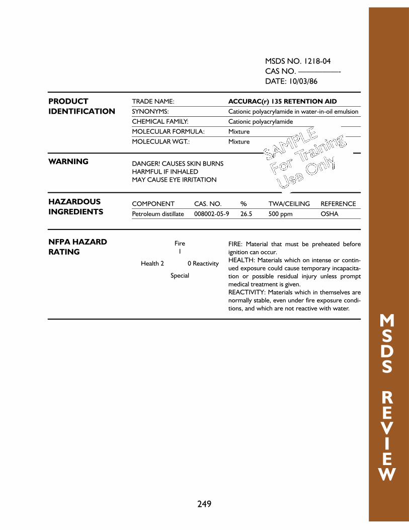
249
MSDS
REVIEW
MSDS NO. 1218-04
CAS NO. —————-
DATE: 10/03/86
PRODUCT
IDENTIFICATION
WARNING
HAZARDOUS
INGREDIENTS
NFPA HAZARD
RATING
TRADE NAME: ACCURAC(r) 135 RETENTION AID
SYNONYMS: Cationic polyacrylamide in water-in-oil emulsion
CHEMICAL FAMILY: Cationic polyacrylamide
MOLECULAR FORMULA: Mixture
MOLECULAR WGT.: Mixture
DANGER! CAUSES SKIN BURNS
HARMFUL IF INHALED
MAY CAUSE EYE IRRITATION
COMPONENT CAS. NO. % TWA/CEILING REFERENCE
Petroleum distillate 008002-05-9 26.5 500 ppm OSHA
FIRE: Material that must be preheated before
ignition can occur.
HEALTH: Materials which on intense or contin-
ued exposure could cause temporary incapacita-
tion or possible residual injury unless prompt
medical treatment is given.
REACTIVITY: Materials which in themselves are
normally stable, even under fire exposure condi-
tions, and which are not reactive with water.
Fire
1
Health 2 0 Reactivity
Special
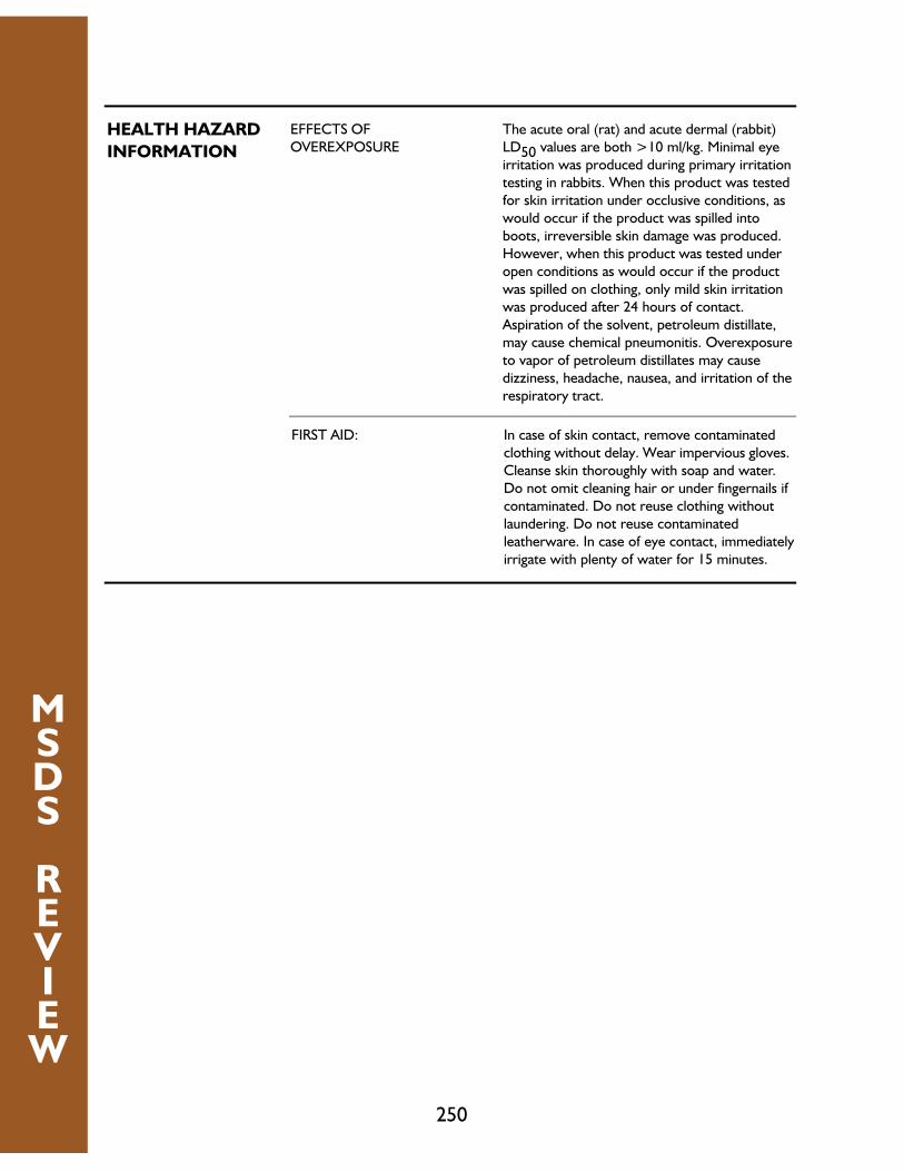
250
MSDS
REVIEW
HEALTH HAZARD
INFORMATION
EFFECTS OF
OVEREXPOSURE
The acute oral (rat) and acute dermal (rabbit)
LD50 values are both >10 ml/kg. Minimal eye
irritation was produced during primary irritation
testing in rabbits. When this product was tested
for skin irritation under occlusive conditions, as
would occur if the product was spilled into
boots, irreversible skin damage was produced.
However, when this product was tested under
open conditions as would occur if the product
was spilled on clothing, only mild skin irritation
was produced after 24 hours of contact.
Aspiration of the solvent, petroleum distillate,
may cause chemical pneumonitis. Overexposure
to vapor of petroleum distillates may cause
dizziness, headache, nausea, and irritation of the
respiratory tract.
FIRST AID: In case of skin contact, remove contaminated
clothing without delay. Wear impervious gloves.
Cleanse skin thoroughly with soap and water.
Do not omit cleaning hair or under fingernails if
contaminated. Do not reuse clothing without
laundering. Do not reuse contaminated
leatherware. In case of eye contact, immediately
irrigate with plenty of water for 15 minutes.

251
MSDS
REVIEW
PRODUCT NAME: Glutaraldehyde (25% by weight)
III. INGREDIENTS
MATERIAL % TLV (Units) HAZARD
Glutaraldehyde 25 See Section V See Section VCAS # 111-30-8
Water ~75 None established See Section VCAS # 7732-18-5
Methanol <0.05 See Section V See Section VCAS # 67-56-1
IV. FIRE AND EXPLOSION HAZARD DATA
FLASH POINT None, Tag Closed Cup ASTM D 56(test method(s)): None, Cleveland Open Cup ASTM D 92
FLAMMABLE LIMITS IN AIR, LOWER: Not determined (aqueous system)% by volume: UPPER: Not determined (aqueous system)
EXTINGUISHING MEDIA: Non-Flammable (Aqueous System): After the water evaporates, the remainingmaterial will burn. Use alcohol-type or all-purpose-type foam applied bymanufacturer’s recommended technique for large fires. Use CO2 or drychemical media for small fires.
SPECIAL FIRE FIGHTINGPROCEDURES: Use self-contained breathing apparatus and protective clothing.
UNUSUAL FIRE ANDEXPLOSION HAZARDS: None
252
MSDS
REVIEW
PRODUCT NAME: Glutaraldehyde (25% by weight)
V. HEALTH HAZARD DATA
TLV AND SOURCE: Glutaraldehyde—0.2 ppmv, ceiling OSHA & ACGIH 1988-89
Methanol—200 ppm, skin OSHA & ACGIH 1988-89
EFFECTS OF SINGLE OVEREXPOSURE:SWALLOWING: Moderately toxic. May cause moderate to marked irritation or chemical
burns of the mouth, throat, esophagus, and stomach. There will be discomfort
or pain in the chest and abdomen, nausea, vomiting, diarrhea, dizziness,
faintness, drowsiness, weakness, circulatory shock, collapse and coma.
SKIN ABSORPTION: Toxicology studies indicate that prolonged or widespread contact could result
in the absorption of potentially harmful amounts of material.
INHALATION: Vapor is irritating and will cause stinging sensations in the nose and
throat, coughing, chest discomfort and tightness, difficulty with breathing,
and headache.
SKIN CONTACT: Brief contact may result in mild to moderate local redness and possibly
swelling. Prolonged contact may result in severe inflammation.
EYE CONTACT: Liquid will cause severe conjunctivitis, seen as discharge with marked
swelling and excess redness of the conjunctiva. Severe corneal injury
may occur. Vapor will cause stinging sensations with excess lachrymation,
but not injury.
EFFECTS OF REPEATED OVEREXPOSURE: None known from currently available information.
MEDICAL CONDITIONS AGGRAVATED BY OVEREXPOSURE:Because of its irritating properties, this material may aggravate an
existing dermatitis.
SIGNIFICANT LABORATORY DATA WITH POSSIBLE RELEVANCE TOHUMAN HEALTH HAZARD EVALUATION: Laboratory studies have shown that glutaraldehyde is not teratogenic,
and several studies have shown the material not to be a mutagen.
OTHER EFFECTS OF OVEREXPOSURE:May cause skin sensitization in a small proportion of individuals, and
present as an allergic contact dermatitis.
EMERGENCY AND FIRST AID PROCEDURES:SWALLOWING: Give at least two glasses of water. Do not induce vomiting.
Seek medical assistance with urgency.
SKIN: Wash contaminated skin with soap and water. If contact has been widespread
and prolonged, or if irritation persists, seek medical advice. Contaminated
clothing should be washed before reuse.
INHALATION: Remove to fresh air. If breathing is difficult, administer oxygen.
If symptoms persist, call a physician.
EYES: Immediately flush eyes thoroughly with water and continue flushing for at
least 15 minutes. See an ophthalmologist urgently.

253
MSDS
REVIEW
NOTES TO PHYSICAN: Aspiration may cause lung damage. Probable mucosal damage may contraindicatethe use of gastric lavage; however, if gastric lavage is considered necessary,it should be undertaken with caution. Most of the adverse effects of glutar-aldehyde are due to its intensely irritating properties. Because of thisvomiting should not be induced in cases of poisoning by swallowing. Thereis no specific antidote. Treatment of overexposure should be directed at thecontrol of symptoms and the clinical condition of the patient.
IX. SPECIAL PRECAUTIONS
PRECAUTIONS TO BE TAKEN IN HANDLING AND STORAGE:DANGER: CORROSIVE
CAUSES IRREVERSIBLE EYE DAMAGE.CAUSES SKIN IRRITATION.HARMFUL IF INHALED.HARMFUL IF SWALLOWED.HARMFUL IF ABSORBED THROUGH SKIN.MAY CAUSE SKIN SENSITIZATION.
Do not get in eyes, on skin, on clothing.Avoid breathing vapor.Do not swallow.Wear goggles, protective clothing, and rubber gloves.Wash thoroughly with soap and water after handling.Remove contaminated clothing and wash before reuse.
FOR INDUSTRY USE ONLY
OTHER PRECAUTIONS: Laboratory studies, using an odor test panel, indicated glutaraldehydevapors in air may be ‘irritating’ to humans at about 0.3 ppm in air; the TLVhas been established as 0.2 ppm ceiling. Thus, if vapors are concentratedenough to be irritating, the TLV is probably being exceeded.

254
MSDS
REVIEW
SECTION I PRODUCT IDENTIFICATION & EMERGENCY INFORMATION
PRODUCT NAME
ECA 10454
CHEMICAL FAMILY
Lube oil additive containing a zinc salt of dialkyl dithio-
phosphoric acid, borated polyisobutenyl succinic anhydride
nitrogen functionalized dispersant, magnesium alkylaryl
detergent, solvent extracted mineral oil, and other comp-
onents judged not to affect the potential health or
environmental impact of the product.
EMERGENCY TELEPHONE NUMBERS:
CHEMTREC 800-424-9300
SECTION II HAZARDOUS COMPONENTS OF MIXTURES
THE PRECISE COMPOSITION OF THIS MIXTURE IS PROPRIETARY INFORMATION. A MORE COMPLETE DISCLOSURE WILL BEPROVIDED TO A PHYSICIAN OR NURSE IN THE EVENT OF A MEDICAL EMERGENCY. THE FOLLOWING COMPONENTS AREDEFINED HAZARDOUS IN ACCORDANCE WITH 29 CFR 1910, 1200:
OSHA HAZARD COMPONENT
Eye irritant Zinc salt of dialkyl dithiophosphoric acid
For additional information see Section X.
SECTION III HEALTH INFORMATION AND PROTECTION
FIRST AID & NATURE OF HAZARD
EYE CONTACT:
Flush eyes with large amounts of water until irritation subsides. If
irritation persists, get medical attention.
Irritating, and may injure eye tissue if not removed promptly.
SKIN CONTACT:
Flush with large amounts of water; use soap if available.
Remove grossly contaminated clothing, including shoes, and launder before reuse.
Low order of toxicity.
Frequent or prolonged contact may irritate.
INHALATION:
Using proper respiratory protection, immediately remove the affected
victim from exposure. Administer artificial respiration if breathing
is stopped. Keep at rest. Call for prompt medical attention.
Negligible hazard at ambient (–18 to 38 Deg. C) or recommended blending temperature.
Warning if heated above 60 Deg. C (140 Deg. F) especially in the presence
of water, hydrogen sulfide may be released; this can cause respiratory
collapse, coma and death without necessarily any warning odor being sensed.
Avoid breathing vapors or mists.
INGESTION:
DO NOT induce vomiting. If individual is conscious, give milk or water
to dilute stomach contents. Keep warm and quiet. Get prompt medical
attention. DO NOT attempt to give anything by mouth to an unconscious person.
Minimal toxicity.

255
MSDS
REVIEW
Toxicity information is often expressed as the dose of the compound that causes an effect in a
percentage of the exposed subjects, which are mostly experimental animals. These dose-
response terms are often found in Material Safety Data Sheets (MSDS) and other sources of
health information. One dose-response term that is commonly used is the lethal dose 50 (LD50),
the dose which is lethal to 50% of an animal population from exposure by any route other than
inhalation when given all in one dose. Another similar term is the lethal concentration 50 (LC50),
which is the concentration of a material in air that on the basis of respiratory exposure in labo-
ratory tests is expected to kill 50% of a group of test animals when administered as a single expo-
sure (usually 1 hour).
The LD50 values that appear in an MSDS or in the literature must be used with caution by emer-
gency medical personnel. These values are an index of only one type of response and give no indi-
cation of the ability of the compound to cause nonlethal, adverse or chronic effects. Furthermore,
LD50 values typically come from experimental animal studies. Because of the anatomical and phys-
iological differences between animals and humans, it is difficult to compare the effects seen in
experimental animal studies to the effects expected in humans exposed to hazardous materials in
the field. Therefore, emergency medical personnel should remember that the LD50 and LC50 val-
ues are only useful for comparing the relative toxicity of compounds and should only be used to
determine if one chemical is more toxic than another.
Responses to toxic chemicals may differ among individuals because of the physiological variabili-
ty that is present in the human population. For example, an individual may be more likely to expe-
rience an adverse health effect after exposure to a toxic chemical because of a reduced ability to
metabolize that compound. The presence of preexisting medical conditions can also increase
one’s susceptibility to toxic chemicals. Respiratory distress in patients or workers with asthma
may be triggered by exposure to toxic chemicals at lower concentrations than might be expect-
ed to produce the same effect in individuals without respiratory disease. Factors such as age, per-
sonal habits (i.e., smoking, diet), previous exposure to toxic chemicals, and medications may also
increase one’s sensitivity to toxic chemicals. Therefore, exposure to concentrations of toxic com-
pounds that would not be expected to result in the development of a toxic response in most indi-
viduals may cause an effect in susceptible individuals. Not all chemicals, however, have a thresh-
old level. Some chemicals that produce cancer (carcinogens) may produce a response (tumors)
at any dose level. Any exposure to these compounds may be associated with some risk of devel-
oping cancer. Thus, literature values for levels which are not likely to produce an effect do not
guarantee that an effect will not occur.
Exposure Limits
The various occupational exposure limits found in the literature or in an MSDS are based prima-
rily on time-weighted average limits, ceiling values, or ceiling concentration limits to which the
worker can be exposed to without adverse effects.

256
MSDS
REVIEW
Because the settings in which these values are appropriate are quite different than an uncon-
trolled spill site, it is difficult to interpret how these values should be used by emergency medical
personnel dealing with a hazardous materials incident. At best, TLV, PEL, IDLH, and REL values
can be used as a benchmark for determining relative toxicity, and perhaps assist in selecting
appropriate levels of Personal Protective Equipment (PPE). Furthermore, these occupational
exposure limits are only useful if the appropriate instrumentation is available for measuring the
levels of toxic chemicals in the air at the chemical spill site. Of the above occupational exposure
limit values, only the OSHA values are regulatory limits. The ACGIH values are for guidance only
and are not regulatory limits. In addition, the ACGIH limits have certain caveats that may or may
not affect the usefulness of the values. Some of these conditions are individual susceptibility or
aggravation of a preexisting condition. Nevertheless, all emergency medical personnel responsi-
ble for the management of chemically contaminated patients should be familiar with these con-
cepts because they will be encountered in various documents dealing with patient care or the
selection of PPE.
This brief discussion highlights some fundamental concepts of toxicology. Emergency medical
personnel responsible for managing chemically contaminated patients are encouraged to obtain
further training in recognizing and treating health effects related to chemical exposures. Also, a
list of general references in toxicology is provided at the end of this section that will allow emer-
gency medical personnel to undertake a more in-depth examination of the principles of toxicol-
ogy.

257
WEBSITES
APPENDIX
SOME USEFUL WEBSITES
1. FEDERAL RESOURCES:
A. EPA/FEMA Websites:
EPA Homepage: http://www.epa.gov/
EPA News and Events, Laws and regulations, Offices, Publications and other resources available
to access information about EPA.
Brownfields: http://www.epa.gov/brownfields/
This site provides information on all facets relating to Brownfields development. Information is
provided on Brownfields Pilots, liability & cleanup, partnership & outreach, laws & regulations,
publications, money matters and other resources.
Chemical Accident Prevention and Risk Management Planning (RMP):
http:/www.epa.gov/swercepp/acc-pre.html
Contains information on the Clean Air Act, Section 112(r) legislation, the Risk Management
Program Rule, Fact Sheets, Basic Awareness brochures, training modules, Federal Register
notices, press releases, technical guidance documents, model risk management program plans by
industrial sector, downloadable computer software, downloadable RMP publications, and many
other resource links.
Chemical Emergency Preparedness and Prevention Office (CEPPO):
http://www.epa.gov/swercepp/acc-pre.html
This site provides helpful information on Chemical Accident Prevention and Risk Management
Planning. There are links to Fact Sheets, Laws & Regulations, Publications, Federal Register
Notices, Press Releases, Technical Guidance Documents, and General Guidance for Risk
Management Programs.
Chemical Fact Sheets: gopher://ecosys.drdr.Virginia.edu:70/11/library/gen/toxics
Summaries of information on over 300 chemicals including identifying characteristics, health haz-
ards, ecological effects, and methods to reduce exposure to the chemical. Maintained by the
University of Virginia.
Current Hazardous Waste Sites (CERCLIS):
http://www.epa.gov/superfund/oerr/impm/products/cursites/csitetoc.htm
A listing of sites on the CERCLIS list.

258
WEBSITES
Glossary of Terms of the Environment: http://earth1.epa.gov:80/OCEPAterms/
An alphabetical listing of terms associated with the environment.
Headquarters Resources Center Internet Newsbrief Resources:
http://www.epa.gov/natlibra/hqire/inb.htm
A weekly service from EPA Headquarters Resources Center that provides a sampling of new and
or useful internet resources for EPA staff or other Environmental Professionals.
Index of EPA Clearinghouses: http://www.epa.gov/epahome/clearing.htm
Includes a link to Air Risk Information Support Centers, Asbestos Management, Clean Air
Technology, EPA Learning Institute, Indoor Air Quality, National Response Center, etc.
Integrated Risk Information System (IRIS): http://www.epa.gov/ngispgm3/iris/index.html
IRIS Homepage, database of human health effects that may result from exposure to various sub-
stances found in the environment.
Numbers (EPA): http://www.epa.gov/epanumbers.html
Lists the various programs and updated telephone #’s for EPA. The site is supposed to update
when changes occur.
Office of Solid Waste and Emergency Response: http://www.epa.gov/swerrims/index.htm
Information on RCRA, Superfund Sites, Solid Waste, Underground Storage Tanks, Chemical
Emergency Preparedness and Prevention, Oil Spill Program, etc.
Regional EPA Offices: http://www.epa.gov/swercepp/pubs/regions.html
A listing of all EPA Regional Offices.
Regional FEMA Offices: http://www.epa.gov/swercepp/pubs/fema.html
A listing of all the FEMA Regional Offices.
Whats Hot in EPA: http://www.epa.gov/epahome/hot.html
What is hot on the EPA server.
Whats New in EPA: http://www.epa.gov/docs/WhatsNew.html
Lists daily minutes, updates, documents, standards, etc. as they are released.
B. Federal General Environmental:
Centers for Disease Control and Prevention (CDC): http://www.cdc.gov
This site provides links to health information, travelers’ health, subscriptions, publications and
products, data & statistics, training & employment opportunities, and funding.
259
WEBSITES
Federal Emergency Management Agency (FEMA): http://www.fema.gov:80/fema/
An index of links alphabetically listed to all FEMA related sites.
National Institute for Occupational Safety & Health (NIOSH):
http://www.cdc.gov/niosh/homepage.html
Information on NIOSH services, publications, documents, training, research, patterns and gener-
al information. There are also links to databases and health hazard evaluations.
Occupational Safety and Health Administration (OSHA):
http://www.OSHA.gov/index.html
Information regarding OSHA news releases, regulations, compliance, programs, statistics, train-
ing and a myriad of many other topics can be found here.
USGS Guide to Federal Environmental Laws and Regulations:
http://water.usgs.gov/public/eap/env guide/
Contains information on: Air Quality, Water Quality, Solid and Hazardous Substances, Lists of
Statutes by Sections, etc.
C. Federal Hazmat:
DOT’s Office of Hazardous Materials Safety: http://hazmat.dot.gov
Contains information on: Rules and Regulations, Exemptions and Approvals, Hazmat
Enforcement, Spills, International Standards, COHMED, Emergency Response Guidebook, etc.
The National Clearinghouse for Worker Safety and Health Training for Hazardous
Materials: http://www.niehs.nih.gov/wetp/clear.htm
Contains information on Safety and Health Resources.
D. Federal Other:
Federal Bureau of Investigation www.FBI.gov
Code of Federal Regulations (CFR): http://www.access.gpo.gov/nara/cfr/cfr-table-search.html
A listing of all available CFR tables available for internet access, search engine for CFR databases,
browse capability for all CFR titles and Federal Registry access.
Federal Registry: http://www.access.gpo.gov/su docs/
Access to Federal Registry books, Private Act Issuances, Public laws, United States Government
Manual and US Congress Information.

260
WEBSITES
Keeping America Informed—U.S. Government Printing Office: http://www.gpo.gov/
This site provides access to the Code of Federal Regulations, the Federal Register, Public Laws,
etc. This is probably todays best and fastest way to access the Federal Register and the CFRs.
National Archives and Records Administration, Code of Federal Regulations:
http://www.access.gpo.gov/nara/cfr/cfr-table-search.html#page1
Official Electronic Copy. Contains information on: Search CFR databases by keywords, Search
CFR titles and/or volumes, Search the Federal Register for related documents, etc.
2. STATE RESOURCES:
A. New Jersey Department of Environmental Protection (NJDEP)
Websites:
NJDEP Home Page: http://www.state.nj.us/dep/
Air Quality Permitting Program (AQPP): http://www.state.nj.us/dep/aqpp/
This site provides links to help sources on Minor Facilities & Preconstruction Permits, Major
Facilities & Operating Permits, Stack Testing, CEMs, Modeling & Risk Assessment, Engineering,
and RADIUS and electronic applications.
Bureau of Discharge Prevention:
http://www.state.nj.us/dep/enforcement/relprev/dpcc/fsdpcc.html
Compliance and Enforcement: http://www.state.nj.us/dep/enforcement/index.html
Division of Solid and Hazardous Waste: http://www.state.nj.us/dep/dshw/
Division of Water Quality: http://www.state.nj.us/dep/dwq/
Office of the Commissioner: http://www.state.nj.us/dep/commissioner/index.html
Radiation Protection Programs: http://www.state.nj.us/dep/rpp/
Radon Section: http://www.state.nj.us/dep/rpp/ber/radon/index.htm
This site provides useful information on radon testing and mitigation for home buyers and sellers,
and testing for radon in your home. It also provides information on average radon levels and tier
assessment in New Jersey. There is information on the Radon Certification Program as well as a
list of Certified Radon Testing Businesses and Certified Radon Mitigation Businesses in New
Jersey.
261
WEBSITES
Site Remediation Program: http://www.state.nj.us/dep/srp
Contains information on: ISRA, Brownfields, Superfund, UST, Known Contaminated Sites,
Regulations and Guidance, and Financial Assistance.
B. State & Local Resources:
State of New Jersey Homepage: http//www.state.nj.us
Access to state agencies, Governor’s office, and Legislature.
LOIS, Electronic Law Library: http://www.pita.com
Regulations for Other States.
New Jersey Online (NJO): http://www.njo.com/
Lists communities, forums, news, sports, entertainment, businesses, living, classifieds/ads, user
guides, etc.
New Jersey State Police: http://www.state.nj.us/lps/njsp/
Academy, Special School and EMS training, recruiting, current events and road and weather con-
ditions can be found here.
State, County and Local Governments on the net:
http://www.piperinfo.com/piper/state/states.html
A site that gives you links to all state and local government sponsored websites. Also included are
some Federal Resources, National Organizations and other miscellaneous links.
3. OTHER RESOURCES:
A. Miscellaneous Resources
Clay Net: http://www.clay.net/ep1.html
Good links to federal/state websites run by EPA, OSHA, DOD, NJDEP, PADEP, etc. It also con-
tains industry sites and links to professional organizations and references.
The Weather Channel: http://www.weather.com/twc/homepage.twc
B. General Environmental:
Agency for Toxic Substances and Disease Registry (ATSDR):
http://atsdr1.atsdr.cdc.gov:8080/atsdrhome.html

262
WEBSITES
All information about the ATSDR program can be found here: Announcements, Address and
Phone numbers, Health Assessments and Consultations, Education and Communication, HazMat
databases, health studies and many other topics are addressed at this website.
C. Organizations/Commissions:
Academy of Certified Hazardous Materials Managers (ACHMM): http://www.achmm.org
Access to state, federal and other hazardous materials management, safety and environmental
links as well as job postings, resumes, and currect technical articles.
American Congress of Governmental Industrial Hygienists (ACGIH) Publications:
http://www.acgih.org/catalog/catfind.asp
Provides a catalog list of ACGIH publications, meeting and event information, and membership.
Chemical Manufacturers Association (CMA): http://www.cmahq.com
Provides links to information on Responsible Care®, publications, workshops/seminars,
CHEMTREC, CHEMSTAR, ChemEcology, Health Research.
Joint Commission for Accreditation of Healthcare Organizations http://www.jcaho.org
N.J. Water Environment Association: http://www.njwea.org
Union/Middlesex County Hazardous Materials Advisory Council (HMAC):
http://www.hmac-inc.org
HMAC is a non-profit organization who’s mission is to promote the responsible handling of haz-
ardous materials. Committee projects support HMAC’s goals: to contribute to a reduction in
hazardous materials incidents; promote education to responders, industry, government and the
public regarding hazardous materials and their proper handling; promote open communications
among all types of residents in Union/Middlesex Counties; and to enhance preparedness,
response and recovery capabilities in the event of incidents in Union/Middlesex Counties.
Information on the committees and their projects can be found here along with available HMAC
publications, training and seminar information and other resource links.
D. Chemcial Databases:
NJ DHSS: http//www.state.nj.us/health/eoh/odisweb/
Access to information from NJRTK (imel. FactSheets) and Occupational Health programs.

263
WEBSITES
Chemfinder: http://chemfinder.camsoft.com/
Allows you to locate common types of chemical information by entering a chemical name, molec-
ular weight or CAS registry number.
Chemical Abstracts Service: http://www.cas.org
Database includes approximately 14 million document records and more than 18 million sub-
stance records respectively. Includes databases of chemical reactions, commercially available
chemicals and listed regulated chemicals.
Material Safety Data Sheet (MSDS) Search: http://www.msdssearch.com/
A forms-searchable database of MSDS entries providing FREE access to over 750,000 MSDSs.
RTK-Net, the Right to Know Network: http://rtk.net/www/rtknet/homepage.html
A network providing free access to numerous databases, text files and conferences on the envi-
ronment, housing and sustainable development.
Toxic Release Inventory (TRI): http://www.epa.gov/opptintr/tri/
Contains information on the TRI Program. TRI data, chemicals, envirofacts, national and interna-
tional programs and TRI contacts are some of the topics found on this page.
E. Bioterrorism Information:
Federal Bureau of Investigation: www.fbi.gov
Medical Management of Chemical and Biological Casualties: www.nbc-med.org
Anthrax Advisory: www.emergency.com
National Workshop on Domestic Preparedness: www.wmdnationalworkshop.com
Counter Terrorism Program Link: www.oep.dhhs.gov
Chemical and Biological Information Analysis Center: www.cbiac.apgea.armymil
Federal Emergency Management Agency www.fema.gov
Soldier Biological and Chemical Command: www.sbccom.gov
National Emergency Management Association: www.nemaweb.org
CDC Bioterrorism Preparedness & Response Network: www.bt.cdc.gov
US Army Medical Research Institute for Infectious Disease: www.dad.gov

264
IMPORTANT
TELEPHONE
NUMBERS
APPENDIX
IMPORTANT TELEPHONE NUMBERS
EMERGENCY RESPONSE
NJ DEP HOTLINE . . . (EMERGENCY ACTION LINE) ....................................1-877-WARNDEP
NEW JERSEY STATE POLICE NORMAL WORK HOURS ...............609-882-2000 x. 6453
AFTER WORK HOURS
(Weeknights, Weekends) ............609-882-2000 x. 6311
CHEMTREC ..............................................................................................................800-424-9300
ASSOCIATION OF AMERICAN RAILROADS
BUREAU OF EXPLOSIVES .......................................................................................202-639-2222
EPA REGION II EMERGENCY RESPONSE ..............................................................908-321-6657
NATIONAL RESPONSE CENTER (U.S.C.G.) ..........................................................800-424-8802
NEW JERSEY POISON CONTROL CENTER..........................................................800-962-1253
NATIONAL POISON CONTROL HOTLINE ..........................................................800-764-7661
STATE AGENCIES
NEW JERSEY STATE POLICE:
OFFICE OF EMERGENCY MANAGEMENT ..............................................609-882-2000 x. 6453
DOMESTIC PREPAREDNESS/HAZ MAT EMERGENCY
RESPONSE PLANNING UNIT ........................................................609-882-2000 x. 6463
HAZ MAT TRANSPORTATION ..................................................................609-882-2000 x. 2586
DEPARTMENT OF ENVIRONMENTAL PROTECTION:
COMMUNITY RIGHT TO KNOW ..........................................................................609-292-6714
TOXIC CATASTROPHE PREVENTION ACT..........................................................609-633-7289
COUNTY ENVIRONMENTAL HEALTH ACT.........................................................609-292-6028
DISCHARGE PREVENTION CONTAINMENT &
COUNTERMEASURE ...................................................................................609-633-0610
RESPONSIBLE PARTY SITE REMEDIATION ...........................................................609-633-2168
BUREAU OF HAZARDOUS WASTE ENFORCEMENT ..........................................609-984-5855
DEPARTMENT OF HEALTH:
WORKER RIGHT TO KNOW ..................................................................................609-984-2202
PUBLIC EMPLOYEES OSH SERVICE .......................................................................609-984-1863

265
IMPORTANT
TELEPHONE
NUMBERS
DEPARTMENT OF LABOR:
WORKER RIGHT TO KNOW ..................................................................................609-292-7036
DEPARTMENT OF PERSONNEL—HUMAN RESOURCE
DEVELOPMENT INSTITUTE (HRDI) ..........................................................609-292-7115
FEDERAL AGENCIESFEDERAL BUREAU OF INVESTIGATION...............................................................973-792-3000
OCCUPATIONAL SAFETY AND HEALTH ADMINISTRATION
OSHA202-219-7162
OSHA REGION II OFFICE........................................................................................212-337-2378
NEW JERSEY AREA OSHA OFFICES:
HASBROUCK HEIGHTS...............................................................................201-288-1700
DOVER..........................................................................................................201-263-1003
AVENEL.........................................................................................................908-750-3270
CAMDEN ......................................................................................................609-757-5181
NIOSH HOTLINE .....................................................................................................800-356-4674
NIOSH HEADQUARTERS........................................................................................404-639-3771
NIOSH REGION II OFFICE ......................................................................................212-264-4600
ENVIRONMENTAL PROTECTION AGENCY
EPA HOTLINE ..........................................................................................................202-382-3000
EPA SUPERFUND HOTLINE ...................................................................................800-424-9346
REGION II SARA TITLE III ASSISTANCE .................................................................908-906-6900
DEPARTMENT OF TRANSPORTATION
U.S. COAST GUARD THIRD DISTRICT..................................................................212-668-7152
ATLANTIC STRIKE TEAM...............................................................................................................
DAYTIME 609-724-0008
NIGHTTIME (ANSWERING MACHINE) .....................................................609-562-6730
DOT HOTLINE (CFR TITLE 49)..............................................................................202-366-4488
DOT/FEMA HAZ MAT TRANSPORTATION HOTLINE.........................................800-752-6367
DOT/REGIONAL EMERGENCY TRANSPORTATION............................................617-223-8480
OTHER FEDERAL AGENCIES
DEPARTMENT OF ENERGY....................................................................................800-428-2525
TOXIC SUBSTANCES CONTROL ACT HOTLINE................................................202-554-1404
U.S. ARMY CORPS OF ENGINEERS........................................................................202-272-0001
U.S. AGENCY FOR TOXIC SUBSTANCES AND
DISEASE REGISTRY HOTLINE ....................................................................404-639-0615
CENTERS FOR DISEASE CONTROL ......................................................................404-639-3291
266
IMPORTANT
TELEPHONE
NUMBERS
OTHER ASSOCIATIONS
AMERICAN CONFERENCE OF GOVERNMENTAL
INDUSTRIAL HYGIENISTS (ACGIH) ...........................................................513-661-7881
AMERICAN INDUSTRIAL HYGIENE ASSOCIATION.............................................216-873-2442
AMERICAN INSTITUTE OF CHEMICAL ENGINEERS...........................................201-763-2877
AMERICAN SOCIETY OF SAFETY ENGINEERS.....................................................312-692-4121
AMERICAN TRUCKING ASSOCIATION................................................................800-ATA-LINE
CHEMICAL INDUSTRY COUNCIL OF NJ..............................................................609-392-4214
CHEMICAL MANUFACTURERS ASSOCIATION (CMA) ........................................202-887-1100
CMA CHEMICAL REFERRAL CENTER
(NON-EMERGENCY CHEMICAL INFORMATION) ................................800-CMA-8200
NATIONAL SAFETY COUNCIL (NSC) ...................................................................312-527-4800
NATIONAL FIRE PROTECTION ASSOCIATION...................................................617-770-3000
TEXAS TECH UNIVERSITY PESTICIDE HOTLINE................................................800-858-7378

GLOSSARY
-A-
29 CFR 1910.120—Hazardous Waste Operations and Emergency Response (OSHA)
A-310—(Public Law 1984, Ch. 210) Inter-Agency Notification
ABSORBANT MATERIAL—Loose or bagged material like commercial bagged clay, kitty litter,
Zorbal, or “pigs” used to soak up liquid hazardous materials.
ACTIVE IMMUNIZATION—The administration of a vaccine to stimulate the host immune sys-
tem to develop immunity (protection) against a specific pathogen or toxin.
ACGIH—American Conference of Governmental Industrial Hygienists. Recommends upper
limits (TLVs) for exposure to workplace chemicals.
AIRBORNE PRECAUTIONS—Standard Precautions plus: Placing the patient in a private room
that has negative air pressure, at least six air changes/hour, and appropriate filtration of air before
it is discharged from the room. Use of respiratory protection when entering the room. Limiting
movement and transport of the patient. Using a mask on the patient if he needs to be moved.
AIR REACTIVE MATERIALS—Materials that will react with atmospheric moisture and rapid-
ly decompose.
ANSI—American National Standards Institute
APR—Air Purifying Respirator
ASPHYXIANT—A substance that can cause unconsciousness or death by lowering the concen
tration of oxygen in the air by out competing oxygen metabolically in the body.
ASYMPTOMATIC—Exposed persons who are not exhibiting signs/symptoms of exposure.
-B-
BACTERIAL AGENT—A live pathogenic organism that can cause disease, illness, or death.
BIOLOGICAL CONTAMINATION—The presence of an infectious agent on a body surface or
on an environmental surface.
267
GLOSSARY
268
GLOSSARY
BIOLOGICAL WARFARE AGENT—A biological warfare agent is a pathogen (microorganism
capable of causing disease) or toxin derived from a living organism that is deliberately used to
produce disease or death in humans, animals, or plants.
BLEVE—Boiling, Liquid Expanding Vapor Explosion
B-NICE—Pertaining to biological, nuclear, incendiary, chemical, or explosives.
BUNG—1) The cap or plug used to seal the small opening in the top of a drum or barrel. 2) The
small opening in the top of a drum or barrel.
-C-
CARCINOGEN—A substance that causes cancer.
CAS—Chemical Abstract Service
CASUALTY COLLECTION POINT (CCP)—Predefined location at which patients are col-
lected, triaged, and provided with initial medical care.
CEHA—County Environmental Health Act
CFR—Code of Federal Regulations
CGI—Combustible Gas Indicator
CHEMOPROPHYLAXIS—The administration of an antibiotic to prevent an infection, or to
prevent an incubating infection from progressing to disease, or to eliminate a carrier state to pre-
vent transmission and disease in others.
CHEMTREC—Chemical Transportation Emergency Center
COCARCINOGEN— (or promoter)—Not a carcinogen by itself, but promotes the effects of a
carcinogen.
COLD (SUPPORT) ZONE—Clean area outside the inner perimeter where command and sup-
port functions take place. Special protective clothing is not required in this area.
COMBUSTIBLE SUBSTANCE—A solid, liquid, or gas that will burn.
COMMON NAME—Each of the agents has a complex chemical name based on its composition
and formula. They also have a common name that you need to recognize.
269
GLOSSARY
CONFINED SPACE—A space which, by design, has limited openings for entry and exit, unfa-
vorable natural ventilation which could contain or produce dangerous air contaminants, could
contain a hazardous atmosphere and which is not intended for continuous employee occupancy.
A confined space includes (but is not limited to) a tank, vessel, pit, ventilation duct work, vat, boil-
er, sewer, or underground utility vault. (NJAC 12:100-9.2).
CONTACT PRECAUTIONS—Standard Precautions plus: Placing the patient in a private room
or with someone with the same infection, if possible. Using gloves when entering the room.
Changing gloves after contact with infective material. Using gown when entering the room if con-
tact with patient is anticipated or if the patient has diarrhea, or colostomy, or wound drainage not
covered by a dressing. Limiting the movement or transport of the patient from the room.
Ensuring that patient care items, bedside equipment, and frequently touched surfaces receive
daily cleaning. Dedicating use of noncritical patient-care equipment to a single patient, or cohort
of patients with the same pathogen. If not feasible, adequate disinfection between patients is nec-
essary.
CRYOGENIC—Pertaining to materials at extreme low temperatures (below –90 degrees C or
–130 degrees F).
-D-
DECOMPOSITION—The basic breakdown of a substance into different substances. Energy
will be released by this reaction; in the case of highly reactive materials, the release may be sud-
den i.e. explosive.
DECONTAMINATION—The process of removing hazardous substances to prevent adverse
health, safety, or environmental effects. Takes place at three levels based on exposure.
DEGRADATION—(applied to protective clothing) Chemical decomposition brought about by
exposure to heat, sunlight, solvents, or oxidation.
DEP—Department of Environmental Protection
DMAT—Disaster Medical Assistance Team
DMORT—Disaster Mortuary Response Team
DOE—Department Of Energy
DOH—Department Of Health
DOL—Department Of Labor
270
GLOSSARY
DOT—Department Of Transportation
DOWNWIND—The area directly in the path of the wind from the incident site.
DROPLET PRECAUTIONS—Standard Precautions plus: Placing the patient in a private room
or with someone with the same infection. If not feasible, maintaining at least 3 feet between
patients. Using a mask when working within 3 feet of the patient. Limiting movement and trans-
port of the patient. Using a mask on the patient if he needs to be moved.
-E-
EFFLUENT—Waste material (such as smoke, liquid industrial refuse, or sewage) discharge into
the environment. It generally refers to water pollution.
EGRESS—Designated exit area.
EIS—Emergency Information System
EMS—Emergency Medical Service
ENDEMIC—A disease that is present in a human population, or in an animal population that is
transmittable to humans, but has a very low morbidity rate.
ENZOOTIC—A disease that is present in an animal population at all times, but has a low mor-
bidity rate.
EOC—Emergency Operations Center
EOD—Explosive Ordinance Disposal
EPA—United States Environmental Protection Agency
EPIDEMIC—A disease that is only present for a limited time in a human population or animal
population that is transmittable to humans, and has a very high morbidity rate.
EPIZOOTIC—A disease that is only present in an animal population for limited periods, but has
a high morbidity rate.
ERG—USDOT Emergency Response Guidebook
ERP—Emergency Response Plan

271
GLOSSARY
ETIOLOGIC—Cause of the disease/illness.
EXPLOSIVE LIMITS—The range of concentration of a gas or vapor (measured in percent by
volume in air) that can explode upon ignition in a confined space. The highest and lowest con-
centration are called, respectively, the Upper Explosive Limit (UEL) and the Lower Explosive
Limit (LEL). At concentrations lower than the LEL, there is not enough product in the air to
explode; the mixture is “too lean.” At concentrations above the UEL, there is not enough oxy-
gen to sustain an explosion; the mixture is “too rich.”
EXPLOSIVE RANGE—The number (as a percentage) that results from subtracting the LEL of
a substance from its UEL.
-F-
FEMA—Federal Emergency Management Agency
FLAMMABLE SUBSTANCE—A solid, liquid, vapor, or gas that will ignite easily and burn rap-
idly.
FLASH POINT (FP)—The lowest temperature at which the vapor given off by a liquid within
a test vessel forms an ignitable mixture with air. This is only a flash, not a sustained fire.
FR—Federal Register
FREEZING POINT—The freezing point or melting point of a substance is the temperature at
which its crystal are at equilibrium with its liquid state. The terms melting point and freezing point
are used interchangeably, depending on whether that temperature is approached by heating or
cooling the substance.
FUMES—The particulate, smoke-like emanation from the surface of heated metals. Also, the
vapor from concentrated acids, evaporating solvents, or as a result of combustion or other
decomposition reaction.
-G-
GROSS DECONTAMINATION—Initial decontamination to remove large amounts of decon-
taminants.

272
GLOSSARY
-H-
HAZARDOUS MATERIAL—Any substance that, when released from its container, is a poten-
tial or actual threat to the safety of life or property when it touches or impinges upon them.
HAZARDOUS WASTE—Any substance that may pose an unreasonable risk to health, safety,
or property when transported in commerce for the purpose of treatment, storage, or disposal
as waste.
HAZMAT—Hazardous Materials
HMT—Hazmat Technician
HMRT—Hazardous Materials Response Team
HOT (EXCLUSION) ZONE—Area immediately around the incident where serious threat of
harm exists. It should extend far enough to prevent adverse effects from B-NICE agents to per-
sonnel outside the zone. Entry into the hot zone requires appropriately trained personnel and
use of proper personal protective equipment.
HSFS—Hazardous Substance Fact Sheet (NJDOH publication)
HVAC—Heating, Ventilating, and Air Conditioning
-I-
IC—Incident Commander
ICS—Incident Command System
IDLH—Immediately Dangerous to Life and Health
IGNITION TEMPERATURE (Ign. Temp.)—The minimum temperature required to initiate sus-
tained self-combustion of a material or compound.
INNER PERIMETER—Secured inner area of operations.
INOCULUM—The amount of microorganisms introduced into a host.

273
GLOSSARY
-L-
LEL—Lower Explosive Limit
LEPC—Local Emergency Planning Committee
LINCS—Local Information Network Communication System
-M-
MASS DECONTAMINATION—Decontamination process used on large number of contami-
nated victims.
MILITARY DESIGNATION/SYMBOL—Each of the agents have been given a symbol. This is
not a chemical symbol or formula, but rather a shorthand way of designating the agent. You need
to learn these symbols.
MISCIBILITY—The ability of a liquid or gas to dissolve completely and evenly in another liquid
or gas at any concentration.
MMRS—Metropolitan Medical Response System
MSDS—Material Safety Data Sheet
MUTAGEN—A substance that causes mutations. A mutation is a change in the genetic material
in a body cell. Mutations can lead to birth defects, miscarriages, or cancer.
-N-
N.O.S.—Not Otherwise Specified
NFPA—National Fire Protection Association
NIOSH—National Institute for Occupational Safety and Health
NJAC—New Jersey Administrative Code
NJRTK—New Jersey Right To Know law (also called Worker and Community Right To Know)
NJSA—New Jersey Statutes Annotated

274
GLOSSARY
NMRT—National Medical Response Team
-O-
OEM—Office of Emergency Management
OSHA—Occupational Safety and Health Administration
OSIC—On Scene Incident Commander
OUTER PERIMETER—Outermost area from hazard that is secure.
OVERPACK—An enclosure used by a consignor to provide protection or convenience in han-
dling a package or to consolidate two or more packages. It does not include a freight container.
-P-
PASSIVE IMMUNIZATION—The administration of pre-formed antibodies to confer immuni-
ty to a specific pathogen or toxin.
PATIENT STAGING AREA (PSA)—Area where patients may receive continued medical
treatment.
PEL—Permissible Exposure Limit
PENETRATION—1) Refers to chemicals physically passing through protective clothing by way
of a tear, cut, or improperly sealed closure. 2) Introducing contaminants into the body by way of
exposed cuts or injection by sharp materials (broken glass, metal shards, etc.).
PEOSHA—Public Employee Occupational Safety and Health Act
PERMEATION—Refers to chemicals passing through protective clothing by absorption. All pro-
tective clothing is permeable to some extent.
PERSISTENT AGENT—An agent that upon release retains its casualty-producing effects for an
extended period of time, usually anywhere from 30 minutes to several days. A persistent agent
usually has a low evaporation rate and its vapor is heavier than air. Therefore, its vapor cloud
tends to hug the ground. It is considered to be a long-term hazard. Although inhalation hazards
are still a concern, take extreme caution to avoid skin contact as well.
PLUME—A vapor cloud formation which has shape and buoyancy.
275
GLOSSARY
POC—Point Of Contact
POINT SOURCE—Letter, package, or dispersal area of agent.
POISON—Any substance that is harmful to living tissue when applied in relatively small doses.
(See toxin).
PPE—Personal Protective Equipment
PROTECT IN PLACE—Method of protecting public by limiting exposure.
PULMONARY EDEMA—The condition of having fluid in the lungs. The condition may be fatal.
-R-
RALLY POINT—A predetermined location to which all persons evacuate in an emergency. In
industry, facilities are evacuated and a rally point is usually predetermined. It is at this rally point
that resources can regroup and a revised plan can be established.
RATE OF ACTION/ONSET TIME—The rate of action or onset time is the period of time that
elapses before a victim begins to show or feel the symptoms of the particular agent. With some
agents, this time will be just a few seconds; in other cases it could be minutes to hours.
Knowing onset time is important because it tells you how much time you have to react.
REACTIVE SUBSTANCE—A solid, liquid, or gas that can cause an explosion under certain
conditions or on contact with other specific substances.
ROUTE OF ENTRY—The route of entry is how the agent gets into your body. Most of the
agents will enter through the respiratory tract, that is, through inhalation. Some of the agents can
also attack through skin and eye.
RTK—Right To Know; May refer to State or Federal law
-S-
SAFE REFUGE AREA (SRA)—An area within the contamination reduction zone for assembling
individuals who are witnesses to the incident. This assemblage will provide for the separation of
contaminated persons from non-contaminated persons.
SAMPLE—Material collected from a source other than an animal or man for laboratory analysis
(such as water sample or soil sample).
276
GLOSSARY
SCBA—Self-Contained Breathing Apparatus
SHIPPING PAPER—A shipping order, bill of lading, manifest or other document containing the
information required by 172.202, 172.203 and 172.204.
SLUDGEM—Acronym for salivation, lacrimation, urination, defecation, gastric distress, emesis,
and miosis.
SOLUBILITY—The ability or tendency of one substance to dissolve evenly in another.
SOLVENT—A substance capable of dissolving another substance (the solute) to form a uni-
formly dispersed mixture (the solution). Water, referred to as the “universal solvent,” is a strong-
ly polar solvent.
SOP—Standard Operating Procedure
SPECIMEN—Material collected from a man or animal for laboratory analysis (such as tissue or
blood specimen).
SPONTANEOUSLY COMBUSTIBLE—The ignition of a substance from the rapid oxidation
of its own constituents.
STANDARD PRECAUTIONS—Handwashing after patient contact. Using gloves when touch-
ing blood, body fluids, secretions, excretions, and contaminated items. Using mask, eye protec-
tion, and gown during procedures likely to generate splashes or sprays of blood, body fluids,
secretions, or excretions. Handling contaminated patient-care equipment and linens in a manner
that prevents the transfer of microorganisms to people or equipment. Practicing care when han-
dling sharps and using a mouthpiece or other ventilation device as an alternative to mouth-to-
mouth resuscitation, when practical. Placing the patient in a private room if they contaminate the
environment, when feasible.
STEL—Short Term Exposure Limit
STLC—Short Term Lethal Concentration
SYMPTOMATIC—Exhibiting signs/symptoms of exposure.
SYMPTOMS—Each of the agents will cause the victim to exhibit symptoms. In many cases these
symptoms can be recognized and provide an indicator of the type of agent.

277
GLOSSARY
-T-
TERATOGEN—A substance that causes birth defects by damaging a fetus.
TIME, DISTANCE AND SHIELDING (TDS)—Three types of protective measures com-
monly associated with hazardous materials training.
TLV—Threshold Limit Value—recommended air concentration in which most persons can
work for an 8-hour work day without ill effects. Set by the ACGIH.
TLV-C—Threshold Limit Value—Ceiling—Exposure level to employees that shall not be
exceeded during any part of the work day.
TLV-STEL—See STEL
TOXICITY—Toxicity is the term used to indicate how much of a substance (in this case, one of
the agents), it takes to cause a specified effect such as incapacitation or death. The amount of
agent it takes to cause an effect is also referred to as a dose.
Respiratory lethality or toxicity can be expressed in parts per million (ppm). PPM is an expres-
sion of concentration (C) or how many parts of a given substance are mixed in a million parts of
air. If an individual is exposed to this concentration for a period of time (t), usually expressed in
one (1) minute, the he or she will receive a dosage. The exact ppm concentration required to
cause lethality is a variable, depending on breathing rate, overall health, etc. Taking into consid-
eration these variables, the term LCt50 is the expression used to indicate a given ppm concentra-
tion expected to be Lethal to 50 percent of those exposed for 1 minute. ICT50 would express the
same, except I is the Incapacitating dosage.
Skin lethality is expressed as LD50 and will normally be expressed in grams or milligrams per indi-
vidual.
TOXIN—Anything harmful, destructive, or poisonous to the body (adj. Toxic). (See Poison.)
TOXIN AGENTS—Poisonous by-products of living organisms used to cause disease, illness or
death in susceptible individuals.
TWA—Time Weighted Average—The calculated average concentration for an 8-hour work day,
10-hour work day or 40-hour work week to which workers may be exposed over their working
career without ill effects. Set by the ACGIH.
TRACEM—The acronym used to identify the six types of harm one may encounter at a terror-
ist incident: thermal, radioactive, asphyxiation, chemical, etiological, and mechanical. Note: Some
sources use the acronym TEAM CPR, which stands for thermal, etiological, asphyxiation,
mechanical, chemical, psychological, and radioactive.

278
GLOSSARY
-U-
UNIFIED COMMAND—In ICS, Unified Command is a unified team effort which allows all
agencies with responsibility for the incident to establish a common set of incident objectives and
strategies. This is accomplished without losing or abdicating agency authority, responsibility or
accountability.
UNSTABLE MATERIALS—Those which, in the pure state, will vigorously polymerize, decom-
pose, condense, or become self-reactive, and undergo other violent chemical changes.
UPWIND—The direction from which the wind is coming.
-V-
VAPOR—An air dispersion of molecules of a substance that is liquid or solid in its normal state
(room temperature).
VEE—Venezuelan Equine Encephalitis
VIRAL AGENTS—A group of viruses that have been selected as BW agents because of their
ability to produce disease, illness, and death in susceptible individuals.
VOC—Volatile Organic Oompound
VOLATILITY—The tendency of a solid or liquid to pass into the gaseous state at a given tem-
perature.
VOLATILITY/PERSISTENCY—Volatility is important because it gives you an indication of how
rapidly an agent will evaporate. The more volatile an agent is, the more rapidly it will evaporate.
Evaporation will cause the agent to become a true gas or vapor and reduce the liquid hazard.
Temperature, wind speed and humidity at the incident site influence how rapidly an agent will
evaporate.
This evaporation process is also referred to as persistency, or the amount of time an agent will
remain a threat in the incident site. A non-persistent agent will not remain at the incident site as
long as a persistent agent. Obviously, if an agent is released inside an enclosed space, weather will
not play a role and the persistency will normally increase.
Most of the agents we will discuss will be disseminated as gases or vapors and are heavier than
air.
A more definitive definition of Vapor Density pressure and Volatility is in the reference section.

279
GLOSSARY
-W-
WARM ZONE—In HazMat incidents, this zone is the contamination reduction zone where ini-
tial decontamination activities occur. This zone requires the use of proper personal protective
equipment once contaminated people or equipment enter the zone.
WATER REACTIVE MATERIALS—Materials which will violently decompose and/or burn vig-
orously when they come in contact with water.
WATER SOLUBILITY—The degree to which a material, or its vapors, are soluble in water.
Materials that are completely soluble in water are said to be miscible.
WEAPON OF MASS DESTRUCTION (WMD)—1) Any explosive, incendiary, poison gas,
bomb, grenade, or rocket having a propellant charge of more than four ounces, missile having an
explosive or incendiary charge of more than one-quarter ounce, or mine or device similar to the
above. 2) Poison gas. 3) Any weapon involving a disease organism. 4) Any weapon designed to
release radiation at a level dangerous to human life.

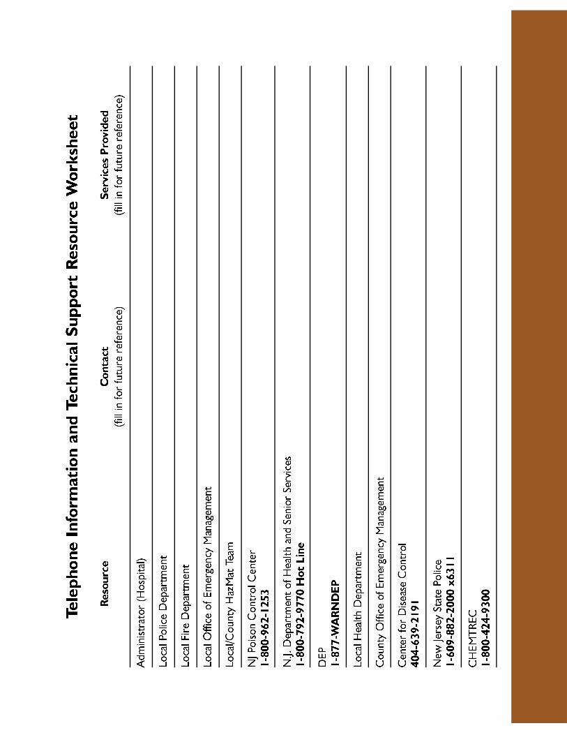

Related Documents