New insights into the symbiosis between Zanclea (Cnidaria, Hydrozoa) and scleractinians SIMONE MONTANO,ROBERTO ARRIGONI,DANIELA PICA,DAVIDE MAGGIONI &STEFANIA PUCE Submitted: 30 April 2014 Accepted: 2 August 2014 doi:10.1111/zsc.12081 Montano, S., Arrigoni, R., Pica, D., Maggioni, D. & Puce, S. (2014). New insights into the symbiosis between Zanclea (Cnidaria, Hydrozoa) and scleractinians. —Zoologica Scripta, 00, 000–000. Hydroids in the genus Zanclea are a recently discovered component of the fauna associated with reef-building corals. The phylogenetic relationships among these species are not well known. The present work is based on field surveys in the Republic of Maldives, and for the first time, morphological and molecular analyses are integrated to distinguish a new hydroid species and provide new information on the ecology of this symbiosis. This new hydroid, Zanclea gallii sp. n., was associated with the scleractinian Acropora muricata; it was living sympatrically with its congener Zanclea sango, which was observed for the first time at this locality on the new scleractinian host Pavona varians. The relationships between these two hydroids and other available scleractinian-associated Zanclea were investigated using two molecular markers, nuclear 28S rDNA and mitochondrial 16S rRNA. Zanclea gallii sp. n. and Z. sango were recovered as distinct lineages within a monophyletic group of scleractin- ian-associated Zanclea based on both molecular and morphological data. All Zanclea species that were observed living in association with scleractinians belong to the ‘polymorpha group’ and share the morphological characteristic ‘polymorphic colony’. The genus Leptoseris is the 16th host coral identified for Zanclea. Compared with the frequency of the Z. gallii sp. n. association with A. muricata and Z. sango with the scleractinian P. varians, the latter is twice as common; however, the former exhibited higher Zanclea polyps concentrations over the colony surface. Overall, the Zanclea survey indicates that these diminutive hydroids are more commonly associated with coral than previously known. Corresponding author: Simone Montano, Department of Biotechnologies and Biosciences, Univer- sity of Milan – Bicocca, Piazza della Scienza 2, 20126, Milan, Italy and MaRHE Center (Marine Research and High Education Center), Magoodhoo Island, Faafu Atoll, Republic of Maldives. E-mail: [email protected] Simone Montano, and Roberto Arrigoni, Department of Biotechnologies and Biosciences, University of Milan – Bicocca, Piazza della Scienza 2, 20126, Milan, Italy and MaRHE Centre (Marine Research and High Education Center), Magoodhoo Island, Faafu Atoll, Republic of Maldives. E- mail: [email protected], [email protected] Daniela Pica, Department of Life and Environmental Sciences, Polytechnic University of Marche, Via Brecce Bianche, 60131, Ancona, Italy. E-mail: [email protected] Davide Maggioni, Department of Biotechnologies and Biosciences, University of Milan – Bicocca, Piazza della Scienza 2, 20126, Milan, Italy and MaRHE Centre (Marine Research and High Education Center), Magoodhoo Island, Faafu Atoll, Republic of Maldives. E-mail: d.maggioni10@ campus.unimib.it Stefania Puce, Department of Life and Environmental Sciences, Polytechnic University of Marche, Via Brecce Bianche, 60131, Ancona, Italy. E-mail: [email protected] Introduction Most hydroids (Cnidaria, Hydrozoa) are considered substrate generalists, which live indiscriminately on many different types of biotic and abiotic substrates. Other hy- droids are symbiotic with metazoan organisms, such as sponges, cnidarians, molluscs, annelids, bryozoans, crusta- ª 2014 Royal Swedish Academy of sciences 1 Zoologica Scripta

Welcome message from author
This document is posted to help you gain knowledge. Please leave a comment to let me know what you think about it! Share it to your friends and learn new things together.
Transcript

New insights into the symbiosis between Zanclea (Cnidaria,Hydrozoa) and scleractiniansSIMONE MONTANO, ROBERTO ARRIGONI, DANIELA PICA, DAVIDE MAGGIONI & STEFANIA PUCE
Submitted: 30 April 2014Accepted: 2 August 2014doi:10.1111/zsc.12081
Montano, S., Arrigoni, R., Pica, D., Maggioni, D. & Puce, S. (2014). New insights into thesymbiosis between Zanclea (Cnidaria, Hydrozoa) and scleractinians. —Zoologica Scripta, 00,000–000.Hydroids in the genus Zanclea are a recently discovered component of the fauna associatedwith reef-building corals. The phylogenetic relationships among these species are not wellknown. The present work is based on field surveys in the Republic of Maldives, and for thefirst time, morphological and molecular analyses are integrated to distinguish a new hydroidspecies and provide new information on the ecology of this symbiosis. This new hydroid,Zanclea gallii sp. n., was associated with the scleractinian Acropora muricata; it was livingsympatrically with its congener Zanclea sango, which was observed for the first time at thislocality on the new scleractinian host Pavona varians. The relationships between these twohydroids and other available scleractinian-associated Zanclea were investigated using twomolecular markers, nuclear 28S rDNA and mitochondrial 16S rRNA. Zanclea gallii sp. n.and Z. sango were recovered as distinct lineages within a monophyletic group of scleractin-ian-associated Zanclea based on both molecular and morphological data. All Zanclea speciesthat were observed living in association with scleractinians belong to the ‘polymorpha group’and share the morphological characteristic ‘polymorphic colony’. The genus Leptoseris is the16th host coral identified for Zanclea. Compared with the frequency of the Z. gallii sp. n.association with A. muricata and Z. sango with the scleractinian P. varians, the latter is twiceas common; however, the former exhibited higher Zanclea polyps concentrations over thecolony surface. Overall, the Zanclea survey indicates that these diminutive hydroids are morecommonly associated with coral than previously known.Corresponding author: Simone Montano, Department of Biotechnologies and Biosciences, Univer-sity of Milan – Bicocca, Piazza della Scienza 2, 20126, Milan, Italyand MaRHE Center (Marine Research and High Education Center), Magoodhoo Island, FaafuAtoll, Republic of Maldives. E-mail: [email protected] Montano, and Roberto Arrigoni, Department of Biotechnologies and Biosciences, Universityof Milan – Bicocca, Piazza della Scienza 2, 20126, Milan, Italy and MaRHE Centre (MarineResearch and High Education Center), Magoodhoo Island, Faafu Atoll, Republic of Maldives. E-mail: [email protected], [email protected] Pica, Department of Life and Environmental Sciences, Polytechnic University of Marche,Via Brecce Bianche, 60131, Ancona, Italy. E-mail: [email protected] Maggioni, Department of Biotechnologies and Biosciences, University of Milan – Bicocca,Piazza della Scienza 2, 20126, Milan, Italy and MaRHE Centre (Marine Research and HighEducation Center), Magoodhoo Island, Faafu Atoll, Republic of Maldives. E-mail: [email protected] Puce, Department of Life and Environmental Sciences, Polytechnic University of Marche,Via Brecce Bianche, 60131, Ancona, Italy. E-mail: [email protected]
IntroductionMost hydroids (Cnidaria, Hydrozoa) are consideredsubstrate generalists, which live indiscriminately on many
different types of biotic and abiotic substrates. Other hy-droids are symbiotic with metazoan organisms, such assponges, cnidarians, molluscs, annelids, bryozoans, crusta-
ª 2014 Royal Swedish Academy of sciences 1
Zoologica Scripta

ceans, echinoderms, tunicates and vertebrates (fish) (Gili &Hughes 1995; Boero & Bouillon 2005; Puce et al. 2008a).These associations range from simple epibiosis to strictsymbiosis, in which the hydroid settles on the living epithe-lium or inside the tissue of the host (Puce et al. 2007). Hy-droids in the genus Zanclea Gegenbaur, 1857 are perceivedas highly specialised symbionts that mainly live in associa-tion with bryozoans (Puce et al. 2008a). In addition, certainZanclea species are symbiotic with reef-building scleractin-ians. This association was originally reported in Mozam-
bique (Millard & Bouillon 1974; Millard 1975) and PapuaNew Guinea (Boero et al. 2000), but the reports lack infor-mation on the hydroids or host. Ten years later, Pantos &Bythell (2010) provided the first complete description of aZanclea–scleractinian association in Australia, including thenew species Zanclea margaritae Pantos & Bythell, 2010 onAcropora muricata Linnaeus, 1758. After this paper was pub-lished, the number of related records rapidly increased,including a study on the recently described Zanclea sangoHirose & Hirose, 2011, from Japan, Taiwan, Indonesia,
A
B
C
Fig. 1 Map of the study area including the10 sampling sites. —A. Republic ofMaldives. —B. Faafu Atoll. —C.Magoodhoo Island.
2 ª 2014 Royal Swedish Academy of sciences
Zanclea–scleractinian symbiosis � S. Montano et al.

Republic of Maldives and Red Sea (Hirose & Hirose 2011;Fontana et al. 2012; Gravier-Bonnet & Bourmaud 2012;Montano et al. 2013a, 2014).To date, the molecular phylogeny of the Zanclea species
has been only partially investigated (Collins et al. 2005;Nawrocki et al. 2010; Schuchert 2010; Fontana et al. 2012).Studies based on both mitochondrial and nuclear DNAshow that scleractinian-associated Zanclea species clustertogether in a monophyletic lineage (Fontana et al. 2012);nevertheless, the genus Zanclea generally does not appearto be monophyletic (Nawrocki et al. 2010; Fontana et al.2012). However, only partial information is available onthe phylogenetic relationships among Zanclea species asso-ciated with corals. Moreover, combined morphological andmolecular phylogenetic analyses have not been performed,because Zanclea gilii Boero, Bouillon & Gravili, 2000,Z. margaritae, and Z. sango have only been describedthrough studying morphological characters (Boero et al.2000; Pantos & Bythell 2010; Hirose & Hirose 2011);however, the only available molecular analysis (Fontanaet al. 2012) lacks morphological information.The aims of this study are to describe the new species
Zanclea gallii sp. n. as associated with A. muricata andreport the geographic range of the species Z. sango. Fur-thermore, for the first time, we provide an integrative studyon the Zanclea species, wherein molecular, morphologicaland ecological data are combined.
Material and methodsSample collection and morphological studyUnderwater surveys were conducted in May, August andSeptember 2012 as well as December 2013 around theinhabited Magoodhoo Island, Faafu Atoll, Republic of Mal-dives (3°040N; 72°570E) (Fig. 1). Up to 10 colonies ofA. muricata and Pavona varians Verrill, 1864 hosting Zancleawere selected in situ. For each coral species, small fragmentswere collected and preserved in 4% formalin for morpho-logical analyses and in 95% ethanol for molecular analyses.Additional portions of the colony were placed in an outdoortank at the Marine Research and High Education CenterMarine Laboratory in Magoodhoo. There, they were cul-tured in small bowls through feeding Artemia nauplii to theZanclea polyps to observe the medusa release. Thereafter,the medusae were maintained in small bowls at ambienttemperature and fed Artemia nauplii. The water wasreplaced two hours after feeding every day. The rearedmedusae were observed on a daily basis, and certain medu-sae were fixed in 4% formalin. Morphological observations,pictures as well as measurements of the polyps, medusae andnematocysts, were mainly performed using living specimensunder low- and high-power microscopes with a camera.Histological examinations were conducted using thin
sections of resin-embedded samples. Small portions of coralswere slowly decalcified over 6–8 h in 4% hydrochloric acidand rinsed in distilled water. They were then dehydrated ina graded ethanol series, embedded in a cold-curing resin(Technovit 8100) and mounted on plastic supports. The sec-tions (6–10 lm) obtained using a microtome were stainedwith toluidine blue and then analysed using a high-powermicroscope with a camera.For scanning electron microscopy (SEM) analyses, cer-
tain portions of coral colonies with hydroids were fixed in4% formalin rinsed and gradually dehydrated in ascendingethanol concentrations. The samples were then dried in acritical point dryer, sputter-coated with gold–palladium ina Balzer Union evaporator and examined using a PhilipsXL20 scanning electron microscope.The polyp and medusa specimens were deposited at the
Museo Civico di Storia Naturale ‘Giacomo Doria’ ofGenoa (Italy) (MSNG).
Molecular analysesDNA extraction, amplification and sequencing. The totalgenomic DNA of eight ethanol-fixed Zanclea from fourA. muricata colonies and four P. varians colonies wasextracted following a protocol modified from Zietara et al.(2009). Two different molecular markers were amplified: (i)an approximately 300-bp portion of 28S from nuclearrDNA and (ii) an approximately 400-bp portion of themitochondrial 16S rRNA gene. These regions of DNAhave been successfully used to infer phylogenetic relation-ships among hydroids in previous molecular studies (Col-lins et al. 2005; Nawrocki et al. 2010; Schuchert 2010;Fontana et al. 2012). We also amplified and sequenced anapproximately 700-bp portion of the nuclear rDNA ITSregion (complete ITS1, 5.8S and ITS2 regions), but weexcluded it from our analyses because this molecular locusdid not vary (data not shown). All PCR amplifications wereperformed using hydroid-specific primers, and the proto-cols were published by Fontana et al. (2012). The PCRproducts were purified and directly sequenced using anautomated 3730xl DNA Analyzer (Applied Biosystem, Fos-ter City, CA, USA). The sequences obtained in this studywere deposited with the EMBL, and the accession numbersare in Table S1.
Phylogenetic analyses. The chromatograms were viewed,edited and assembled using CodonCode Aligner 3.7.0(CodonCode Corporation, Dedham, MA, USA). Multiplealignments were generated using the E-INS-i option inMAFFT 7.110 (Katoh et al. 2002; Katoh & Standley 2013)with the default parameters. The newly obtained 28Ssequences of Zanclea were aligned with 23 homologoussequences, which were selected from the following recent
ª 2014 Royal Swedish Academy of sciences 3
S. Montano et al. � Zanclea–scleractinian symbiosis

molecular studies (Collins et al. 2005; Nawrocki et al. 2010;Fontana et al. 2012). The sequences obtained from Gen-Bank belong to the clade Zancleida and include tensequences of the genus Zanclea (Zanclea prolifera Uchida &Sugiura, 1976, Zanclea costata Gegenbaur, 1857 and Zancleasp.), whereas the other sequences belong to the hydroidfamilies Porpitidae, Solanderiidae, Milleporidae, Asynco-rynidae, Moerisiidae, Hydrocorynidae, Cladocorynidae andPennariidae. Coryne producta (currently Stauridiosarsia pro-ducta Wright, 1858) represents the clade Corynidae (Naw-rocki et al. 2010) and was selected as an outgroup to inferphylogenetic relationships between our Zanclea sampleswithin the clade Zancleida (Cartwright et al. 2008; Naw-rocki et al. 2010). For 16S phylogeny reconstruction, ourZanclea sequences were aligned with the Zanclea sequencesthat were publically available on GenBank, and Coryne cliff-ordi (currently Stauridiosarsia cliffordi Brinckmann-Voss,1989) was used as the outgroup because it diverges fromthe clade Zancleida (Nawrocki et al. 2010). Phylogeneticinference analyses were performed separately for bothmolecular loci using three methods: maximum parsimony(MP), Bayesian inference (BI) and maximum-likelihood(ML). Maximum parsimony analyses were performed usingPAUP 4.0b10 (Swofford 2003) with heuristic, searchesstepwise addition and tree-bisection-reconnection (TBR)branch swapping. The node consistency was assessed using500 bootstrap replicates with randomly added taxa. Thesoftware MrModeltest2.3 (Nylander 2004) was used withPAUP 4.0b10 to select nucleotide substitution models forthe BI and ML phylogenetic analyses. The best modelsestimated using the Akaike information criterion (AIC)were GTR+I+Γ for the nuclear locus and HKY+Γ for themitochondrial gene. Bayesian inference analyses were per-formed using MrBayes 3.1.2 (Ronquist & Huelsenbeck2003). Two independent runs for four Markov chains wereconducted for 1 million generations for nuclear 28S locus(0.5 million generations for mitochondrial 16S), and thetrees were sampled every 10 generations for both markers.Based on checking the parameter estimates and conver-gence using Tracer 1.5 (Drummond & Rambaut 2007), thefirst 25001 trees for 28S (12 501 for 16S) were discarded asburn-in. Maximum-likelihood (ML) trees were calculatedwith PhyML 3.0 (Guindon & Gascuel 2003) using the evo-lutionary model selected by MrModeltest2.3, and therobustness of the phylogeny was tested using 500 bootstrapreplications.
Ecological studyQuantitative information on Zanclea was collected for thehost species A. muricata and P. varians. The frequency ofthe symbiosis was established for the belt transects(N = 40) parallel to the coastline at 10 shallow sites (0–5 m
deep), which were randomly selected. In each site, fourrandomly positioned 25 9 1 m belt transects spaced 10–20 m apart were analysed (see Montano et al. 2012,2013b). For each belt transect, we recorded the number ofcoral colonies that were symbiotic and non-symbiotic withZanclea to determine the prevalence of Zanclea-scleractiniansymbiosis. Thereafter, for each belt transect, the line inter-cept method was used to assess the mean coral cover forthe site. All corals were identified in situ at the genus level(according to Veron 2000), and we collected underwaterphotographs of coral colonies, both symbiotic with Zancleaor not. The population density of hydroid gastro-gonozo-oids was surveyed at the surface of the encrusting coralP. varians and 2 cm distal to the coral branch tips of theA. muricata branching using an Olympus (Tokyo, Japan)SZ61 stereomicroscope and KL 300 fibre-optic lightsource.The Zanclea–scleractinian symbiosis prevalence was sta-
tistically compared between scleractinian genera usingMann–Whitney U-test because the data did not meet thenormality assumption (Zar 1999). Spearman’s rank correla-tion was used to examine whether Zanclea–scleractiniansymbiosis prevalence was related to the host and overallcoral density. Statistical analyses were performed usingSPSS computer software (IBM, NY, USA). All data arepresented as the arithmetic means � standard deviation(SD) unless stated otherwise.
ResultsWe performed a qualitative survey along the Magoodhooreef, which revealed the presence of Zanclea polyps on cor-als of eleven scleractinian genera, namely Acropora, Favia,Favites, Goniastrea, Leptastrea, Leptoseris, Montipora, Pavona,Porites, Psammocora and Symphyllia, which belong to sixfamilies (Acroporidae, Agariciidae, Faviidae, Mussidae, Por-itidae and Siderastreidae); Leptoseris is a novel genus.Our detailed study on A. muricata and P. varians, which
are the common coral species in the investigated area,shows that they are in strict symbiosis with a new Zancleaspecies and with Z. sango, respectively; these two hydroidsare described in the ‘Systematics’ paragraph. The phyloge-netic and ecological results are discussed in separate para-graphs.
SystematicsFamily ZANCLEIDAEGenus Zanclea Gegenbaur, 1857Zanclea sango Hirose & Hirose, 2011 (Fig. S1).
Material examined. Fertile colony associated with P. vari-ans, Republic of Maldives, Faafu Atoll, Magoodhoo Island(3°040N; 72°570E), approximately 2 m depth, 10 August
4 ª 2014 Royal Swedish Academy of sciences
Zanclea–scleractinian symbiosis � S. Montano et al.

2012, Montano & Maggioni (snorkelling), (MSNG 57819)� fertile colony associated with P. varians, Republic ofMaldives, Faafu Atoll, Magoodhoo Island (3°040N;72°570E), approximately 2 m depth, 12 August 2012,Montano & Maggioni (snorkelling).
Description of the hydroid. Polymorphic colonies comprisegastro-gonozooids and dactylozooids and are associated withthe scleractinian P. varians (Fig. S1A, B). Hydrorhizas, sur-rounded by a thin perisarc, grow inside the coral. The peri-sarc stops at the base of the hydranths and forms a small cup(Fig. S1C). Polyps arise from the coral surface and aremainly scattered on the corallite edges. Cylindrical gastro-gonozooids (up to 0.9 mm high) exhibit a whorl of 4–6 oralcapitate tentacles (the capitula diameter is approximately40 lm), and 12–21 aboral capitate tentacles scattered alongthe hydranth body (the capitula diameter is approximately25–35 lm) (Fig. S1B–D). The hypostome is white. Extensi-ble dactylozooids (up to 2 mm high) lack tentacles and hypo-stome. The fully extended dactylozooids exhibit a thinhydranth body and globular apex (Fig. S1E) rich in glandularcells. The medusa buds typically arise in groups of 2–4 fromthe basal portion of the hydranths or short blastostyles.
Cnidome. The apotrichous macrobasic euryteles (Fig. S1F,G) with a shaft coiled along the main axis of the capsule(undischarged capsule 18–21 9 7–9 lm, discharged capsuleapproximately 19 9 7 lm and shaft 145–155 lm typicallycurved at its distal end) were observed in the gastro-gonozo-oid hypostome (Fig. S1D) at the base of the gastro-gonozo-oid hydranth, in the apex (Fig. S1E, see arrow) and base ofdactylozooids. They are abundant in blastostyles. Two sizesof stenoteles (undischarged capsule 11–15 9 11–14 lm anddischarged capsule approximately 9 9 8 lm; undischargedcapsule 7–9 9 6–7 lm and discharged capsule approxi-mately 7 9 4 lm) were observed in tentacle capitations ofgastro-gonozooids, in dactylozooids and in blastostyles.
Description of the medusa. The newly released medusa(Fig. S1H) has a spherical bell (700–750 lm diameter), andits cylindrical manubrium spans approximately one-third ofthe subumbrellar cavity (approximately 250 lm). Four per-radial nematocyst pouches were observed along the exum-brella up to a half of its height. A marginal bulb wasobserved at the base of each pouch. Two non-tentacledbulbs are small, while two bulbs are large, triangular andbear tentacles with 30–40 oval cnidophores (Fig. S1H).Each cnidophore contain 2–3 nematocysts (Fig. S1I).
Cnidome. The apotrichous macrobasic euryteles with ashaft coiled along the main axis of the capsule (undis-charged capsule approximately 19 9 7 lm) were observed
in nematocyst pouches. Large stenoteles (undischarged cap-sule 11–12 9 10–11 lm) were observed in nematocystpouches. Small stenoteles (undischarged capsule 8–10 9 8 lm) were observed in the manubrium and in ten-tacular bulbs. Bean-shaped euryteles (undischarged capsuleapproximately 8 9 5 lm and discharged capsule approxi-mately 7 9 4 lm) were observed in cnidophores.
Remarks. The polyp and medusa morphologies are con-sistent with the original description of Z. sango from Japan.However, the Maldivian specimens are symbiotic withP. varians, while the Japanese Z. sango is hosted by Pavonadivaricata (Lamarck, 1816) and Pavona venosa (Ehrenberg,1834). The specimens observed herein exhibit a small peri-sarc cup at the hydranth base, and the medusa buds arisenot only at the hydranth base but also from short blasto-styles. These two traits have not been reported for theJapanese Z. sango.Certain colonies of the unidentified Zanclea species were
previously described for the Indian Ocean. Three colonieswere recorded in the Seychelles Islands; however, exceptfor a specimen growing on a compound ascidia covering adead coral, the specimens were not observed in associationwith corals (Millard & Bouillon 1973). In addition, fourcolonies were collected from Inhaca Island, south Mozam-bique (Millard & Bouillon 1974; Millard 1975). One wasreported as commensal with a coral and is characterised bya perisarc-covered hydrorhiza as well as by nematocysts(13.8 9 6.0 lm) in the lower part of the hydranth body.While the presence of the perisarc is consistent with theZ. sango description, the nematocysts appear different, andthe colony is not polymorphic. Therefore, it is likely thatthe species from Mozambique is not Z. sango. Morerecently, Gravier-Bonnet & Bourmaud (2012) reported anunidentified Zanclea associated with hard coral from BaaAtoll, Republic of Maldives. Unfortunately, information onthe hydroid and its host is scarce and insufficient for acomparison with our specimens. Ristedt & Schuhmacher(1985: page 177) reported unpublished observations ofZanclea polyps ‘arising from a network of stolons sub-merged in living Acropora tissue’ from Red Sea. However,the information available on this Zanclea is limited to theapparent absence of a perisarc covering the hydrorhiza,which differs from Z. sango.
Reproductive period. Medusa buds were seen in August2012 and December 2013.
Relationship with the host. The hydrorhiza of the observedspecimens is protected by a perisarc and grows underneaththe scleractinian tissues. Pavona varians produces an apparentreaction, such as a raised collar surrounding the base of the
ª 2014 Royal Swedish Academy of sciences 5
S. Montano et al. � Zanclea–scleractinian symbiosis

hydranths. The hydroid polyps are typically distributedproximal to the coral polyps, and zooxanthellae have beenobserved in the hydranth coelenteron (Fig. S1J).
Zanclea gallii sp. n. Montano, Maggioni & Puce 2014(Figs 2, 3, S2).
Holotype. Fertile colony associated with A. muricata,Republic of Maldives, Faafu Atoll, Magoodhoo Island(3°040N; 72°570E), approximately 2 m depth, 3 August2012, Maggioni (snorkelling), (MSNG 57816).
Paratype. Fertile colony associated with A. muricata,Republic of Maldives, Faafu Atoll, Magoodhoo Island(3°040N; 72°570E), approximately 2 m depth, 7 August2012, Maggioni (snorkelling), (MSNG 57817).
Two newly released medusae collected from parent col-ony, Republic of Maldives, Faafu Atoll, Magoodhoo Island(3°040N; 72°570E), 9 August 2012, Maggioni, (MSNG57818).
Etymology. This species is named in honour of Dr. PaoloGalli (University of Milano Bicocca, Italy).
Diagnosis. This species can be distinguished from otherZanclea species by the polymorphic colony, the absence ofperisarc, the polyp cnidome including only stenoteles andthe symbiosis with a scleractinian.
Description of the hydroid. The polymorphic colonies livein association with the scleractinian A. muricata (Figs 2A,3A, B). Gastro-gonozooids that arise from the coral surface
A
DE
FG
H
I
J
K
L
B
C
Fig. 2 Zanclea gallii sp. n. – Drawings —A.Polymorphic colony associated withAcropora muricata (scale: 2 mm). —B.Gastro-gonozooid (scale: 400 lm). —C.Gastro-gonozooid bearing medusa buds(scale: 400 lm). —D. Medusa budlaterally arising from a short blastostyle(scale: 300 lm). —E. Medusa bud apicallyarising from a short blastostyle (scale:200 lm). —F. Undischarged anddischarged large stenotele from gastro-gonozooids (scale: 20 lm). —G.Undischarged and discharged smallstenotele from gastro-gonozooids (scale:15 lm). —H. Medusa (scale: 300 lm). —I. Cnidophores (scale: 55 lm). —J.Undischarged and discharged largestenotele from a medusa (12 lm). —K.Undischarged and discharged smallstenotele from a medusa (scale: 10 lm).—L. Undischarged and dischargedmacrobasic eurytele from cnidophores(scale: 8 lm).
6 ª 2014 Royal Swedish Academy of sciences
Zanclea–scleractinian symbiosis � S. Montano et al.

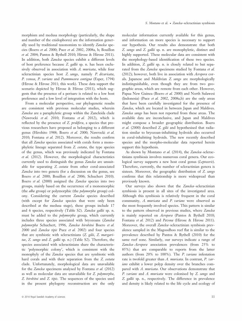
have been observed between the corallites or frequently onthe corallites; they have been observed highly proximale tothe Acropora polyps in certain instances (Fig. 3B). Hydro-rhizas lack a perisarc and grow inside the coral (Fig. S2A,B) at the interface between coral tissue and skeleton. Hostfrequently produces a distinct collar-like tissue elevation atthe base of hydroid polyps (Figs 3C, S2C, E). Gastro-gon-ozooids cylindrical (up to 1.1 mm high), with white hypo-stome surrounded by 4–6 oral capitate tentacles (diameterof capitula 50–55 lm). The hydranth body is surroundedby 14–26 aboral capitate tentacles (diameter of capitula 40–45 lm) (Figs 2B, C, 3D). Contractile dactylozooids arerarely present and have exclusively been observed in situ(Figs 2A, 3A). Dactylozooids include a thin hydranth bodyand a globular apex without tentacles.Medusa buds arise in groups of 2–4 from the basal
portion of the gastro-gonozooids (Figs 2C, 3D) or arise
laterally (Figs 2D, 3E) or apically (Figs 2E, 3F, S2D) fromshort blastostyles.
Cnidome. Both large stenoteles (Figs 2F, 3G) (undis-charged capsule 11–15 9 10–14 lm, and discharged cap-sule approximately 10–12 9 10–11 lm) and smallstenoteles (Figs 2G, 3H) (undischarged capsule 6–9 9 5–7 lm, discharged capsule 6–7 9 4–5 lm) are in the gastro-gonozooid tentacle capitations, but they are rare in thehydranth body and frequent in hydrorhiza as well as blasto-styles.
Description of the medusa. The newly released medusa isspherical (0.8–1.1 mm in diameter) with the cylindricalmanubrium length from approximately one-fourth to one-third of the subumbrellar cavity (about 150–350 lm)(Figs 2H, 3I, J). Four perradial nematocyst pouches extend
A B C
D E F G H
I
J K L M
N O P Q
Fig. 3 Zanclea gallii sp. n. —A.Photograph in situ of the polymorphiccolony associated with Acropora muricata(arrowheads) (scale: 2 mm). —B–P.Microphotographs. —B. Living gastro-gonozooid arising from an A. muricatacorallite (scale: 300 lm). —C. Livinggastro-gonozooids showing the basalcollar produced by A. muricata (scale:200 lm). —D. Gastro-gonozooid bearingmedusa bud (scale: 125 lm). —E. Livingmedusa bud laterally arising from a shortblastosyle (scale: 90 lm). —F. Livingmedusa bud apically arising from a shortblastostyle (scale: 150 lm). —G.Undischarged large stenotele from gastro-gonozooids (scale: 9 lm). —H.Undischarged small stenotele from gastro-gonozooids (scale: 15 lm). —I. Livingmedusa (scale: 300 lm). —J. Medusa(scale: 250 lm). —K. Cnidophores (scale:40 lm). —L. Large stenoteles in amedusa nematocyst pouche (scale: 30 lm).—M. Small stenoteles in the manubrium(scale: 25 lm). —N. Undischargedmacrobasic eurytele from cnidophores(scale: 5 lm). —O. Discharged macrobasiceurytele from cnidophore (scale: 6 lm).—P. Macrobasic euryteles inside a largetentacular bulb (scale: 40 lm). —Q.Photograph in situ of a colony associatedwith a partially bleached A. muricata(1.5 mm).
ª 2014 Royal Swedish Academy of sciences 7
S. Montano et al. � Zanclea–scleractinian symbiosis
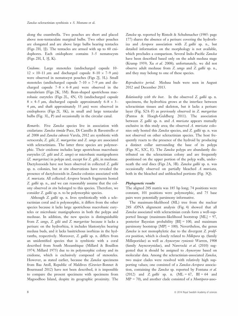
along the exumbrella. Two pouches are short and placedabove non-tentaculate marginal bulbs. Two other pouchesare elongated and are above large bulbs bearing tentacles(Figs 2H, 3J). The tentacles are armed with up to 60 cni-dophores. Each cnidophore contains 3–5 nematocysts(Figs 2H, I, 3J, K).
Cnidome. Large stenoteles (undischarged capsule 10–12 9 10–11 lm and discharged capsule 8–10 9 7–9 lm)were observed in nematocyst pouches (Figs 2J, 3L). Smallstenoteles (undischarged capsule 7–10 9 7–9 lm and dis-charged capsule 7–8 9 6–8 lm) were observed in themanubrium (Figs 2K, 3M). Bean-shaped apotrichous mac-robasic euryteles (Figs 2L, 4N, O) (undischarged capsule8 9 4–5 lm, discharged capsule approximately 6–8 9 3–4 lm, and shaft approximately 35 lm) were observed incnidophores (Figs 2I, 3K), in small and large tentacularbulbs (Fig. 3L, P) and occasionally in the circular canal.
Remarks. Five Zanclea species live in association withcnidarians: Zanclea timida Puce, Di Camillo & Bavestrello etal. 2008 and Zanclea cubensis Varela, 2012 are symbiotic withoctocorals; Z. gilii, Z. margaritae and Z. sango are associatedwith scleractinians. The latter three species are polymor-phic. Their cnidome includes large apotrichous macrobasiceuryteles (Z. gilii and Z. sango) or microbasic mastigophores(Z. margaritae) in polyps and, except for Z. gilii, in medusae.Dactylozooids have not been observed in collected Z. galliisp. n. colonies, but in situ observations have revealed thepresence of dactylozooids in Zanclea colonies associated withA. muricata. All collected Acropora branch fragments hostedZ. gallii sp. n., and we can reasonably assume that the col-ony observed in situ belonged to this species. Therefore, weconsider Z. gallii sp. n. to be polymorphic species.Although Z. gallii sp. n. lives symbiotically with a scle-
ractinian coral and is polymorphic, it differs from the otherspecies because it lacks large apotrichous macrobasic eury-teles or microbasic mastigophores in both the polyps andmedusae. In addition, the new species is distinguishablefrom Z. sango, Z. gilii and Z. margaritae because it lacks aperisarc on the hydrorhiza, it includes blastostyles bearingmedusa buds, and it lacks basitrichous isorhizas in the hyd-ranths, respectively. Moreover, Z. gallii sp. n. differs froman unidentified species that is symbiotic with a coraldescribed from South Mozambique (Millard & Bouillon1974; Millard 1975) due to its polymorphic colony and itscnidome, which is exclusively composed of stenoteles.However, as stated earlier, because the Zanclea specimensfrom Baa Atoll, Republic of Maldives (Gravier-Bonnet &Bourmaud 2012) have not been described, it is impossibleto compare the present specimens with specimens fromMagoodhoo Island, despite its geographic proximity. The
Zanclea sp. reported by Ristedt & Schuhmacher (1985: page177) shares the absence of a perisarc covering the hydrorh-iza and Acropora association with Z. gallii sp. n., butdetailed information on the morphology is not available,which precludes a comparison. Several Indo-Pacific Zancleahave been described based only on the adult medusa stage(Kramp 1959; Xu et al. 2008); unfortunately, we did notobserve adult medusae from Z. sango and Z. gallii sp. n.,and they may belong to one of these species.
Reproductive period. Medusa buds were seen in August2012 and December 2013.
Relationship with the host. In the observed Z. gallii sp. n.specimens, the hydrorhiza grows at the interface betweenscleractinian tissues and skeleton, but it lacks a perisarclayer (Fig. S2A–F) as previously observed in Z. margaritae(Pantos & Hoegh-Guldberg 2011). The associationbetween Z. gallii sp. n. and A. muricata appears mutuallyexclusive in this study area; the observed A. muricata colo-nies only hosted this Zanclea species, and Z. gallii sp. n. wasnot observed on other scleractinian species. The host fre-quently reacts to the presence of the hydroid by producinga distinct collar surrounding the base of its polyps(Figs 3C, S2C, E). The Zanclea polyps are abundantly dis-tributed on the scleractinian colony and are frequentlypositioned on the upper portion of the polyp walls, under-neath the oral discs (Figs 2A, 3B). Zanclea gallii sp. n. wasoccasionally observed on partially bleached A. muricata,both in the bleached and unbleached portions (Fig. 3Q).
Phylogenetic resultsThe aligned 28S matrix was 185 bp long; 74 positions wereconstant, 101 positions were polymorphic, and 75 basepairs were potentially parsimony informative.The maximum-likelihood (ML) tree from the nuclear
28S rDNA alignment analysis (Fig. 4) showed that allZanclea associated with scleractinian corals form a well-sup-ported lineage (maximum-likelihood bootstrap [ML] = 97,posterior Bayesian probability [BI] = 100, and maximumparsimony bootstrap [MP] = 100). Nevertheless, the genusZanclea is not monophyletic due to the divergent Z. prolif-era position, which is closely related to Millepora sp. (familyMilleporidae) as well as Asyncoryne ryniensis Warren, 1908(family Asyncorynidae), and Nawrocki et al. (2010) sug-gested that it should be assigned to Asyncoryne based onmolecular data. Among the scleractinian-associated Zanclea,two major clades were resolved with relatively high sup-porting values; one consisted of a Zanclea-Acropora associa-tion, containing the Zanclea sp. reported by Fontana et al.(2012) and Z. gallii sp. n. (ML = 67, BI = 64 andMP = 70), and another clade consisted of a Montipora-asso-
8 ª 2014 Royal Swedish Academy of sciences
Zanclea–scleractinian symbiosis � S. Montano et al.

ciated Zanclea sp., which was also reported by Fontanaet al. (2012) as well as Z. sango associated with P. varians(ML = 69, BI = 73 and MP = 56).The alignment data set for the 16S mitochondrial gene
consisted of 326 sites, comprising 211 invariable positionsand 97 polymorphic sites, of which 61 were parsimonyinformative with 131 mutation events. The resulting MLtree (Fig. 5) has similar overall topology as the nuclear 28Stopology, but the boundaries between closely related spe-cies were better resolved. Again, all Zanclea associated withscleractinians clustered in a monophyletic group with highsupport values (ML = 73, BI = 96 and MP = 97). Withinthis group, four major lineages that correspond to four dif-ferent Zanclea species were associated with scleractiniansand resolved with relatively high support. The most basallineage is only composed of the Zanclea sp. sequence asso-ciated with Montipora from Fontana et al. (2012). All of theZ. sango associated with P. varians form a well-supportedmonophyletic group (ML = 88, BI = 77 and MP = 94).Among the Zanclea associated with Acropora, two majormonophyletic clades were recovered; one consists of Z. gal-
lii sp. n. from Maldives (ML = 92, BI = 81 and MP = 60),and the second consists of Zanclea sp. from Taiwan andAustralia (ML = 89, BI = - and MP = 64). Thus, these twoAcropora-associated Zanclea species are closely related, butphylogenetically separate (Fig. 5).
Ecological resultsFrom the ecological survey, records of Zanclea–scleractinianassociations were observed in 38 belt transects (95%) at 10sites (100%) distributed over the reef flat of the island. Inthe investigated area, the mean overall prevalence per sitewas 10.64 � 8.7% (mean � SD). The highest Zanclea–scle-ractinian symbiosis prevalence (27.1%) was observed in theshallow site T4. For the two scleractinian hosts A. muricataand P. varians, the Zanclea–scleractinian symbiosis preva-lence ranged from 25.5 � 27.1% (A. muricata, n = 569) to57.1 � 15% (P. varians, n = 146) and a statistically signifi-cant difference has been reported between these(Mann–Whitney U-test P < 0.01). The highest Zanclea–scleractinian symbiosis prevalence was observed forA. muricata (85.7 � 8.1%) at the site T4. We did not
Fig. 4 Maximum-likelihood (ML) treeobtained from a portion of nuclear 28SrDNA. Values at branches represent MLbootstrap values, posterior Bayesianprobability, and maximum parsimonybootstrap values, respectively. Sequencesnewly obtained in this study are in bold.
ª 2014 Royal Swedish Academy of sciences 9
S. Montano et al. � Zanclea–scleractinian symbiosis

detect a significant correlation between the overall Zanclea–scleractinian symbiosis prevalence and the total live coralcoverage (Spearman’s rho P > 0.05); further, we did notdetect a correlation between the Zanclea–scleractinian sym-biosis prevalence and host coral coverage (Spearman’s rhoP > 0.05). In addition, the two scleractinians are character-ised by a different hydroid density; A. muricata exhibitedthe density 32.6 � 11.8 per cm2 (with a maximum of 53polyps per cm2), while the density for P. varians was10.5 � 5.6 per cm2.
DiscussionMany different organisms live in close association withreef-building corals and contribute to the structure andfunction of reef ecosystems (Boucher et al. 1982; Bruno
et al. 2003; Hay et al. 2004; Hoeksema et al. 2012; Hoek-sema & van der Meij 2013; Van der Meij & Hoeksema2013), and hydroids are a new component in this array ofrelationships (Pantos & Bythell 2010).In the present investigation, we provide new information
on the symbiosis between hydroids and scleractinians, andwe describe the new hydroid species Z. gallii sp. n., a newhost and geographical record for Z. sango through integrat-ing morphological, molecular and ecological data for thefirst time.The geographically co-occurring Z. gallii sp. n. and
Z. sango differ morphologically by their cnidome in boththe polyp and medusa stages as well as by a perisarc layerthat covers the hydrorhiza in Z. sango, but not in Z. galliisp. n. These characteristics together with the colony poly-
Fig. 5 Maximum-likelihood (ML) treeobtained from a portion of mitochondrial16S rRNA gene. Values at branchesrepresent ML bootstrap values, posteriorBayesian probability, and maximumparsimony bootstrap values, respectively.Sequences newly obtained in this studyare in bold.
10 ª 2014 Royal Swedish Academy of sciences
Zanclea–scleractinian symbiosis � S. Montano et al.
morphism and medusa morphology (particularly, the shapeand number of the cnidophores) are the information gener-ally used by traditional taxonomists to identify Zanclea spe-cies (Boero et al. 2000; Puce et al. 2002, 2008a, b; Bouillonet al. 2004; Pantos & Bythell 2010; Hirose & Hirose 2011).In addition, both Zanclea species exhibit a different levelsof host preference because Z. gallii sp. n. has been exclu-sively observed in association with A. muricata, while fourscleractinian species host Z. sango, namely P. divaricata,P. venosa, P. varians and Psammocora contigua (Esper, 1794)(Hirose & Hirose 2011; this work). These data support thescenario depicted by Hirose & Hirose (2011), which sug-gests that the presence of a perisarc is related to a low hostpreference and a low level of integration with the hosts.From a molecular perspective, our phylogenetic results
are consistent with previous molecular studies, whereinZanclea are a paraphyletic group within the Zancleida clade(Nawrocki et al. 2010; Fontana et al. 2012), which isreflected by the presence of Z. prolifera, a species that pre-vious researchers have proposed as belonging to a differentgenus (Hirohito 1988; Boero et al. 2000; Nawrocki et al.2010; Fontana et al. 2012). Moreover, the results confirmthat all Zanclea species associated with corals form a mono-phyletic lineage separated from Z. costata, the type speciesof the genus, which was previously indicated by Fontanaet al. (2012). However, the morphological characteristicscurrently used to distinguish the genus Zanclea are unsuit-able for separating Z. costata from other coral-associatedZanclea into two genera (for a discussion on the genus, seeBoero et al. 2000; Bouillon et al. 2006; Schuchert 2010).Boero et al. (2000) assigned the Zanclea species into twogroups, mainly based on the occurrence of a monomorphic(the alba group) or polymorphic (the polymorpha group) col-ony. Considering the current Zanclea species described(with except for Zanclea species that were only beendescribed at the medusa stage), these groups include 17and 6 species, respectively (Table S2). Zanclea gallii sp. n.must be added to the polymorpha group, which currentlyincludes three species associated with bryozoans (Zancleapolymorpha Schuchert, 1996, Zanclea hirohitoi Boero et al.2000 and Zanclea tipis Puce et al. 2002) and four speciesthat are symbiotic with scleractinians (Z. gilii, Z. margari-tae, Z. sango and Z. gallii sp. n.) (Table S2). Therefore, thespecies associated with scleractinians share the characteris-tic ‘polymorphic colony’, which is consistent with themonophyly of the Zanclea species that are symbiotic withhard corals and with their separation from the Z. costataclade. Unfortunately, morphological data are unavailablefor the Zanclea specimens analysed by Fontana et al. (2012)as well as molecular data are unavailable for Z. polymorpha,Z. hirohitoi and Z. tipis. The sequences of the species usedin the present phylogeny reconstruction are the only
molecular information currently available for this genus,and information on more species is necessary to supportour hypothesis. Our results also demonstrate that bothZ. sango and Z. gallii sp. n. are monophyletic, distinct andhighly supported. These molecular data are consistent withthe morphology-based identification of these two species.In addition, Z. gallii sp. n. is closely related to but sepa-rated from the Zanclea specimens studied by Fontana et al.(2012); however, both live in association with Acropora cor-als. Japanese and Maldivian Z. sango are morphologicallyindistinguishable, even though they are from two geo-graphic areas, which are remote from each other. However,Papua New Guinea (Boero et al. 2000) and North Sulawesi(Indonesia) (Puce et al. 2002, 2008a,b) are the only areasthat have been carefully investigated for the presence ofZanclea, which are located in between Japan and Maldives.Zanclea sango has been not reported from these areas. Theavailable data are inconclusive, and Japan and Maldivesmight compose a broader geographic distribution. Boeroet al. (2000) described Z. gilii and hypothesised that radia-tion similar to bryozoan-inhabiting hydroids also occurredin coral-inhabiting hydroids. The new recently describedspecies and the morpho-molecular data reported hereinsupport this hypothesis.As shown by Montano et al. (2014), the Zanclea–sclerac-
tinians symbiosis involves numerous coral genera. Our eco-logical survey supports a new host coral genus (Leptoseris).Therefore, currently, the number of scleractinian genera issixteen. Moreover, the geographic distribution of Z. sangoconfirms that this relationship is more widespread thanpreviously known.Our surveys also shown that the Zanclea–scleractinian
symbiosis is present in all sites of the investigated area.Although this symbiosis is relatively frequent in the coralcommunity, A. muricata and P. varians were observed asthe most frequently involved species. This pattern is similarto the pattern observed in previous studies, where Zancleais mainly reported on Acropora (Pantos & Bythell 2010;Fontana et al. 2012) and Pavona (Hirose & Hirose 2011).Moreover, the overall Zanclea–scleractinian symbiosis prev-alence sampled in the Magoodhoo reef flat is similar to theprevalence described by Pantos & Bythell (2010) for thesame reef zone. Similarly, our surveys indicate a range ofZanclea–Acropora association prevalences (from 25% to85%) that are comparable to reports from the latterauthors (from 20% to 100%). The P. varians infestationrate is twofold greater than A. muricata. In contrast, P. var-ians exhibit a lower polyp density over the branches com-pared with A. muricata. Our observations demonstrate thatP. varians and A. muricata were colonised by Z. sango andZ. gallii sp. n., respectively. The difference in prevalenceand density is likely related to the life cycle and ecology of
ª 2014 Royal Swedish Academy of sciences 11
S. Montano et al. � Zanclea–scleractinian symbiosis

the two hydroids. The length of the period during whichmedusae are released and the medusae and planulae sur-vival are factors that may influence their dispersal (Gili &Hughes 1995). Few data are available on the biologicalcycle of most Zanclea species. Boero & Fresi (1986) studiedhydroids on the rocky cliff of Portofino Promontory (Italy)and reported presence for Z. costata throughout the year,but reproduction was limited to August. In the same area,Puce et al. (2009) observed Z. costata and Z. sessilis during ashort period of the year, but they were not fertile. Thesymbiotic Zanclea planula must find a suitable host; thistask is more difficult when the hosts are rare and the asso-ciation is strict (e.g. Davenport 1955; Donaldson 1974).Moreover, this is also demonstrated in the strict associationbetween Z. gallii sp. n. and A. muricata, while Z. sango hasa wide range of hosts. The Zanclea–scleractinian symbiosisis not ubiquitous in the coral community, which suggeststhat certain morphological, chemical or ecological featuresof the involved species led this association to evolve. Acro-pora muricata is the preferred host of at least two Zancleaspecies, Z. margaritae and Z. gallii sp. n., which co-evolvedwith their host in two separate geographic areas and lostthe stolon perisarc, enabling a closer interaction with thehost tissues (Pantos & Hoegh-Guldberg 2011). The natureof the relationship involving Zanclea and scleractinians andthe features that drive the planula to settle on specific coralhave not been elucidated. Pantos & Bythell (2010) hypoth-esised mutualism related to increased protection of the hostthrough the additional hydrozoan nematocysts and thehydroid activity that removes detritus or pathogenic proto-zoans from its surface. On the other hand, the hydroidmay benefit from food provided by the increased waterflow and protective mucus on the coral surface. Alterna-tively, they hypothesised a parasitic relationship based onthe observed bleaching and white syndrome in aquariumwhere A. muricata hosting Zanclea were reared. The Z. san-go and Z. gallii sp. n. polyps are typically distributed proxi-mal to the scleractinian polyps, and few zooxanthellae havebeen observed in the Z. sango polyp coelenteron. Thesedata suggest a trophic interaction, which has not beendirectly observed. Moreover, Z. gallii sp. n. has beenobserved living on certain bleached A. muricata colonies.Similar observations may support the Pantos & Bythell(2010) hypothesis, which describe a parasitic symbiosis;however, supposing a relation between bleaching and thepresence of hydroids, the sequence of cause and effectremains unknown.To date, symbiotic associations have been mainly studied
by considering pairwise relationships. However, one host istypically inhabited by several other organisms (e.g. epi-bionts, commensals and parasites) that may interact witheach other and with the two partners (Stella et al. 2011;
Bos 2012; Hoeksema et al. 2012). Thus, it is necessary tounderstand how these co-occurring organisms influence thesymbiotic association considered and how their combinedeffects influence the two partners (Stella et al. 2011). Thisinformation could be used to predict the ability of singleorganisms to persist in a rapidly changing environment.Since 2010, Zanclea–scleractinian symbiosis has been morefrequently reported, and the data suggest that this associa-tion is more frequent and locally abundant than previouslyknown. Therefore, future studies must clarify the condi-tions involved in this symbiosis.
AcknowledgementsThe authors are indebted with Davide Seveso for assistancein the field. We thank the anonymous reviewers for theircomments, which greatly improved this manuscript.
ReferencesBoero, F. & Bouillon, J. (2005). Cnidaria and Ctenophora. In K.Rhode (Ed.) Marine Parasitology (pp. 177–182). Collingwood:CSIRO Publishing
Boero, F. & Fresi, E. (1986). Zonation and evolution of a rockybottom hydroid community. Pubblicazioni della Stazione Zoologicadi Napoli I: Marine Ecology, 7, 123–150.
Boero, F., Bouillon, J. & Gravili, C. (2000). A survey of Zanclea,Halocoryne and Zanclella (Cnidaria, Hydrozoa, Anthomedusae,Zancleidae) with description of new species. Italian Journal ofZoology, 67, 93–124.
Bos, A. R. (2012). Fishes (Gobiidae and Labridae) associated withthe mushroom coral Heliofungia actiniformis (Scleractinia: Fungii-dae) in the Philippines. Coral Reefs, 31, 133.
Boucher, D. H., James, S. & Keeler, K. H. (1982). The ecology ofmutualism. Annual Review of Ecology and Systematics, 13, 315–347.
Bouillon, J., Medel, M. D., Pag�es, F., Gili, J. M., Boero, F. & Gra-vili, C. (2004). Fauna of the Mediterranean hydrozoa. ScientiaMarina, 68, 1–448.
Bouillon, J., Gravili, C., Pag�es, F., Gili, J. M. & Boero, F. (2006).An Introduction to Hydrozoa. Paris: Publications Scientifiques duMus�eum.
Bruno, J. F., Petes, L. E., Harvell, C. D. & Hettinger, A. (2003).Nutrient enrichment can increase the severity of coral diseases.Ecology Letters, 6, 1056–1061.
Cartwright, P., Evans, N. M., Dunn, C. W., Marques, A. C., Migl-ietta, M. P., Schuchert, P. & Collins, A. G. (2008). Phylogenet-ics of Hydroidolina (Hydrozoa: Cnidaria). Journal of the MarineBiological Association of the UK, 88, 1663–1672.
Collins, A. G., Winkelmann, S., Hadrys, H. & Schierwater, B.(2005). Phylogeny of Capitata and Corynidae (Cnidaria,Hydrozoa) in light of mitochondrial 16S rDNA data. ZoologicaScripta, 34, 91–99.
Davenport, D. (1955). Specificity and behavior in symbioses. Quar-terly Review of Biology, 30, 29–46.
Donaldson, S. (1974). Larval settlement of a symbiotic hydroid:specificity and nematocyst responses in planulae of Proboscidactylaflavicirrata. The Biological Bulletin, 147, 573–585.
12 ª 2014 Royal Swedish Academy of sciences
Zanclea–scleractinian symbiosis � S. Montano et al.

Drummond, A. J. & Rambaut, A. (2007). BEAST: Bayesian evolu-tionary analysis by sampling trees. BMC Evolutionary Biology, 7,214.
Fontana, S., Keshavmurthy, S., Hsieh, H. J., Denis, V., Kuo, C. Y.,Hsu, C. M., Leung, J. K. L., Tsai, W. S., Wallace, C. C. & Chen,C. A. (2012). Molecular evidence shows low species diversity ofcoral-associated hydroids in Acropora corals. PLoS One, 7, e50130.
Gegenbaur, C. (1857). Versuch eines Systemes der Medusen, mitBeschreibung neuer oder wenig bekannter Formen; zugleich einBeitrag zur Kenntniss der Fauna des Mittelmeeres. Zeitschrift furWisseaschaftliche Zoologie, 8, 202–273.
Gili, J. M. & Hughes, R. G. (1995). The ecology of marine ben-thic hydroids. Oceanography and Marine Biology, 33, 351–426.
Gravier-Bonnet, N. & Bourmaud, C. A. (2012). Hydroids (Cnida-ria, Hydrozoa) of Baa Atoll (Indian Ocean, Maldives Archipel-ago). Atoll Research Bulletin, 590, 82–123.
Guindon, S. & Gascuel, O. (2003). A simple, fast, and accuratealgorithm to estimate large phylogenies by maximum likelihood.Systematic Biology, 52, 696–704.
Hay, M. E., Parker, J. D., Burkepile, D. E., Caudill, C. C., Wilson, A.E., Hallinan, Z. P. & Chequer, A. D. (2004). Mutualisms and aqua-tic community structure: the enemy of my enemy is my friend.Annual Review of Ecology, Evolution, and Systematics, 35, 175–197.
Hirohito, E. S. (1988). The Hydroids of Sagami Bay – Athecata.Tokyo: Biological Laboratory Imperial Household.
Hirose, M. & Hirose, E. (2011). A new species of Zanclea (Cnida-ria: Hydrozoa) associated with scleractinian corals from Oki-nawa, Japan. Journal of the Marine Biological Association of theUnited Kingdom, 92, 877–884.
Hoeksema, B. W. & van der Meij, S. E. T. (2013). Gall crab city:an aggregation of endosymbiotic crabs inhabiting a colossal col-ony of Pavona clavus. Coral Reefs, 32, 59.
Hoeksema, B. W., van der Meij, S. E. T. & Fransen, C. H. J. M.(2012). The mushroom coral as a habitat. Journal of the MarineBiological Association of the United Kingdom, 92, 647–663.
Katoh, K. & Standley, D. M. (2013). MAFFT multiple sequencealignment software version 7: improvements in performance andusability. Molecular Biology and Evolution, 30, 772–780.
Katoh, K., Misawa, K., Kuma, K. & Miyata, T. (2002). MA-FFT: a novel method for rapid multiple sequence alignmentbased on fast Fourier transform. Nucleic Acids Research, 30,3059–3066.
Kramp, P. L. (1959). Some new and little known Indo-Pacificmedusae. Videnskabelige Meddelelser Dansk Naturhistorisk Forening,121, 223–259.
Millard, N. A. H. (1975). Monograph on the hydroida of SouthernAfrica. Annals of the South African Museum, 68, 1–513.
Millard, N. A. H. & Bouillon, J. (1973). Hydroids from the Sey-chelles (Coelenterata). Annales–Mus�ee Royal de l’Afrique Centrale.S�erie in 8: Sciences zoologiques, 206, 1–106.
Millard, N. A. H. & Bouillon, J. (1974). A collection of hydroidsfrom Moc�ambique, East Africa. Annals of the South AfricanMuseum, 65, 1–40.
Montano, S., Strona, G., Seveso, D. & Galli, P. (2012). Firstreport of coral diseases in the Republic of Maldives. Diseases ofAquatic Organisms, 101, 159–165.
Montano, S., Maggioni, D., Galli, P., Seveso, D. & Puce, S.(2013a). Zanclea-coral association: new records from Maldives.Coral Reefs, 32, 701.
Montano, S., Strona, G., Seveso, D. & Galli, P. (2013b). Preva-lence, host range, and spatial distribution of black band diseasein the Maldivian Archipelago. Diseases of Aquatic Organisms, 105,65–74.
Montano, S., Galli, P., Maggioni, D., Seveso, D. & Puce, S.(2014). First record of coral-associated Zanclea (Hydrozoa, Zan-cleidae) from the Red Sea. Marine Biodiversity, DOI 10.1007/s12526-014-0207-6.
Nawrocki, A. M., Schuchert, P. & Cartwright, P. (2010). Phyloge-netics and evolution of Capitata (Cnidaria: Hydrozoa), and thesystematics of Corynidae. Zoologica Scripta, 39, 290–304.
Nylander, J. A. A. (2004). MrModeltest v2. Software Distributed bythe Author. Uppsala: Evolutionary Biology Centre, Uppsala Uni-versity.
Pantos, O. & Bythell, J. C. (2010). A novel reef coral symbiosis.Coral Reefs, 29, 761–770.
Pantos, O. & Hoegh-Guldberg, O. (2011). Shared skeletal supportin a coral-hydroid symbiosis. PLoS One, 6, e20946.
Puce, S., Cerrano, C., Boyer, M., Ferretti, C. & Bavestrello, G.(2002). Zanclea (Cnidaria: Hydrozoa) species from Bunaken Mar-ine Park (Sulawesi Sea, Indonesia). Journal of the Marine Biologi-cal Association of the UK, 82, 943–954.
Puce, S., Bavestrello, G., Di Camillo, C. G. & Boero, F. (2007).Symbiotic relationships between hydroids and bryozoans. Symbi-osis, 44, 137–143.
Puce, S., Cerrano, C., Di Camillo, C. G. & Bavestrello, G.(2008a). Hydroidomedusae (Cnidaria: Hydrozoa) symbiotic radi-ation. Journal of the Marine Biological Association of the UK, 88,1715–1721.
Puce, S., Di Camillo, C. G. & Bavestrello, G. (2008b). Hydroidssymbiotic with octocorals from Sulawesi Sea, Indonesia. Journalof the Marine Biological Association of the United Kingdom, 88,1643–1654.
Puce, S., Bavestrello, G., Di Camillo, C. G. & Boero, F. (2009).Long-term changes in hydroid (Cnidaria, Hydrozoa) assem-blages: effect of Mediterranean warming? Marine Ecology, 30,313–326.
Ristedt, H. & Schuhmacher, H. (1985). The bryozoan Rhynchozoonlarrey a successful competitor in coral reef communities of theRed Sea. Marine Ecology, 6, 167–179.
Ronquist, F. & Huelsenbeck, J. P. (2003). MrBayes 3: Bayesianphylogenetic inference under mixed models. Bioinformatics, 19,1572–1574.
Schuchert, P. (1996). The marine fauna of New Zealand: athecatehydroids and their medusae (Cnidaria: Hydrozoa). New ZealandOceanographic Institute Memoir, 106, 1–159.
Schuchert, P. (2010). The European athecate hydroids and theirmedusae (Hydrozoa, Cnidaria): capitata part 2. Revue Suisse deZoologie, 117, 337–555.
Stella, J. S., Pratchett, M. S., Hutchings, P. A. & Jones, G. P.(2011). Coral-associated invertebrates: diversity, ecologicalimportance and vulnerability to disturbance. Oceanography andMarine Biology: An Annual Review, 49, 43–104.
Swofford, D. L. (2003). PAUP* Phylogenetic Analysis Using Parsi-mony (*and Other Methods), Version 4.b.10. Sunderland, MA: Si-nauer Associates.
Van der Meij, S. E. T. & Hoeksema, B. W. (2013). Distributionof gall crabs inhabiting mushroom corals on Semporna reefs,Malaysia. Marine Biodiversity, 43, 53–59.
ª 2014 Royal Swedish Academy of sciences 13
S. Montano et al. � Zanclea–scleractinian symbiosis

Varela, C. (2012). Registros nuevos de hidrozoos (Cnidaria: Hy-droidomedusae) para Cuba, con la descripci�on de una especienueva. Solendon, 10, 1–7.
Veron, J. E. N. (2000). Corals of the World. Townsville: AustralianInstitute of Marine Science.
Xu, Z.-z., Huang, J.-q. & Guo, D.-h. (2008). Six new species ofAnthomedusae (Hydrozoa, Hydroidomedusae) from the BeibuGulf, China. In J.-y. Hu & S.-y. Yang (Eds) Symposium onOceanography of the Beibu Gulf China (pp. 209–221). Beijing:Ocean Press.
Zar, J. H. (1999). Biostatistical Analysis. London: Prentice-Hall.Zietara, M. S., Arndt, A., Geerts, A., Hellemans, B. & Volckaert,F. A. M. (2009). The nuclear rDNA region of Gyrodactylus arcu-
atus and G. branchicus (Monogenea: Gyrodactylidae). Journal ofParasitology, 86, 1368–1373.
Supporting InformationAdditional Supporting Information may be found in theonline version of this article:Fig. S1. Zanclea sango.Fig. S2. Zanclea gallii sp. n.Table S1. List of analyzed specimens.Table S2. List of the species belonging to the alba and
polymorpha groups.
14 ª 2014 Royal Swedish Academy of sciences
Zanclea–scleractinian symbiosis � S. Montano et al.
Related Documents