New Insights into the Evolution of Wolbachia Infections in Filarial Nematodes Inferred from a Large Range of Screened Species Emanuele Ferri 1. , Odile Bain 2. , Michela Barbuto 1 , Coralie Martin 2 , Nathan Lo 3 , Shigehiko Uni 4 , Frederic Landmann 5 , Sara G. Baccei 1 , Ricardo Guerrero 6 , Sueli de Souza Lima 7 , Claudio Bandi 8 , Samuel Wanji 9 , Moustapha Diagne 10 , Maurizio Casiraghi 1 * 1 Dipartimento di Biotecnologie e Bioscienze, Universita ` degli Studi di Milano Bicocca, Milano, Italy, 2 Parasitologie Compare ´e UMR 7205 CNRS & UMR 7245 CNRS, Muse ´um National d’Histoire Naturelle, Paris, France, 3 School of Biological Sciences, University of Sydney, Sidney, Australia, 4 Department of Medical Zoology, Osaka City University Medical School, Osaka, Japan, 5 Department of Molecular, Cell and Developmental Biology, University of California Santa Cruz, Santa Cruz, California, United States of America, 6 Instituto de Zoologia Tropical, Universidad Central de Venezuela, Caracas, Venezuela, 7 Departamento de Zoologia, Universidade Federal de Juiz de Fora, Minas Geraes, Brasil, 8 Dipartimento di Patologia Animale, Igiene e Sanita ` Pubblica Veterinaria, Universita ` degli Studi di Milano, Milano, Italy, 9 Research Foundation in Tropical Diseases and Environment, Buea, Cameroun, 10 De ´partement de Biologie Animale, Universite ´ Cheikh Anta Diop de Dakar, Dakar, Se ´ne ´gal Abstract Background: Wolbachia are intriguing symbiotic endobacteria with a peculiar host range that includes arthropods and a single nematode family, the Onchocercidae encompassing agents of filariases. This raises the question of the origin of infection in filariae. Wolbachia infect the female germline and the hypodermis. Some evidences lead to the theory that Wolbachia act as mutualist and coevolved with filariae from one infection event: their removal sterilizes female filariae; all the specimens of a positive species are infected; Wolbachia are vertically inherited; a few species lost the symbiont. However, most data on Wolbachia and filaria relationships derive from studies on few species of Onchocercinae and Dirofilariinae, from mammals. Methodology/Principal Findings: We investigated the Wolbachia distribution testing 35 filarial species, including 28 species and 7 genera and/or subgenera newly screened, using PCR, immunohistochemical staining, whole mount fluorescent analysis, and cocladogenesis analysis. (i) Among the newly screened Onchocercinae from mammals eight species harbour Wolbachia but for some of them, bacteria are absent in the hypodermis, or in variable density. (ii) Wolbachia are not detected in the pathological model Monanema martini and in 8, upon 9, species of Cercopithifilaria. (iii) Supergroup F Wolbachia is identified in two newly screened Mansonella species and in Cercopithifilaria japonica. (iv) Type F Wolbachia infect the intestinal cells and somatic female genital tract. (v) Among Oswaldofilariinae, Waltonellinae and Splendidofilariinae, from saurian, anuran and bird respectively, Wolbachia are not detected. Conclusions/Significance: The absence of Wolbachia in 63% of onchocercids, notably in the ancestral Oswaldofilariinae estimated 140 mya old, the diverse tissues or specimens distribution, and a recent lateral transfer in supergroup F Wolbachia, modify the current view on the role and evolution of the endosymbiont and their hosts. Further genomic analyses on some of the newly sampled species are welcomed to decipher the open questions. Citation: Ferri E, Bain O, Barbuto M, Martin C, Lo N, et al. (2011) New Insights into the Evolution of Wolbachia Infections in Filarial Nematodes Inferred from a Large Range of Screened Species. PLoS ONE 6(6): e20843. doi:10.1371/journal.pone.0020843 Editor: Laurent Re ´nia, Agency for Science, Technology and Research - Singapore Immunology Network, Singapore Received February 22, 2011; Accepted May 10, 2011; Published June 22, 2011 Copyright: ß 2011 Ferri et al. This is an open-access article distributed under the terms of the Creative Commons Attribution License, which permits unrestricted use, distribution, and reproduction in any medium, provided the original author and source are credited. Funding: This work was supported by European Community Grant INCO-CT-2006-032321, a French national grant Action Transversale du Muse ´um ‘‘Taxonomie mole ´culaire: DNA Barcode et gestion durable des collections’’ and by Italian national grant MIUR-PRIN. The funders had no role in study design, data collection and analysis, decision to publish, or preparation of the manuscript. Competing Interests: The authors have declared that no competing interests exist. * E-mail: [email protected] . These authors contributed equally to this work. Introduction The alpha proteobacteria Wolbachia (Rickettsiales) are present in two distinct zoological groups: the arthropods, where they are widespread [1], and the nematodes, where they are restricted to a single but notable family of parasites, the Onchocercidae [2,3]. They encompass the agents of human onchocerciasis and lymphatic filariases [4]. The zoological host range of Wolbachia raised a fundamental question on the origin of infection in the filarial nematodes [5,6]. Investigations performed during the past fifteen years on Wolbachia in filarial and arthropod hosts has led to establish a rather clear and complex picture of the taxonomic status of the bacterium, its distribution and phylogeny [7]. Several distinct bacterial lineages have been called supergroups [8], and, at this date, they are all attributed to the only valid recognized species Wolbachia pipientis. The taxonomy of this species is quite uncertain, PLoS ONE | www.plosone.org 1 June 2011 | Volume 6 | Issue 6 | e20843

Welcome message from author
This document is posted to help you gain knowledge. Please leave a comment to let me know what you think about it! Share it to your friends and learn new things together.
Transcript
New Insights into the Evolution of Wolbachia Infectionsin Filarial Nematodes Inferred from a Large Range ofScreened SpeciesEmanuele Ferri1., Odile Bain2., Michela Barbuto1, Coralie Martin2, Nathan Lo3, Shigehiko Uni4, Frederic
Landmann5, Sara G. Baccei1, Ricardo Guerrero6, Sueli de Souza Lima7, Claudio Bandi8, Samuel Wanji9,
Moustapha Diagne10, Maurizio Casiraghi1*
1 Dipartimento di Biotecnologie e Bioscienze, Universita degli Studi di Milano Bicocca, Milano, Italy, 2 Parasitologie Comparee UMR 7205 CNRS & UMR 7245 CNRS,
Museum National d’Histoire Naturelle, Paris, France, 3 School of Biological Sciences, University of Sydney, Sidney, Australia, 4 Department of Medical Zoology, Osaka City
University Medical School, Osaka, Japan, 5 Department of Molecular, Cell and Developmental Biology, University of California Santa Cruz, Santa Cruz, California, United
States of America, 6 Instituto de Zoologia Tropical, Universidad Central de Venezuela, Caracas, Venezuela, 7 Departamento de Zoologia, Universidade Federal de Juiz de
Fora, Minas Geraes, Brasil, 8 Dipartimento di Patologia Animale, Igiene e Sanita Pubblica Veterinaria, Universita degli Studi di Milano, Milano, Italy, 9 Research Foundation
in Tropical Diseases and Environment, Buea, Cameroun, 10 Departement de Biologie Animale, Universite Cheikh Anta Diop de Dakar, Dakar, Senegal
Abstract
Background: Wolbachia are intriguing symbiotic endobacteria with a peculiar host range that includes arthropods and asingle nematode family, the Onchocercidae encompassing agents of filariases. This raises the question of the origin ofinfection in filariae. Wolbachia infect the female germline and the hypodermis. Some evidences lead to the theory thatWolbachia act as mutualist and coevolved with filariae from one infection event: their removal sterilizes female filariae; allthe specimens of a positive species are infected; Wolbachia are vertically inherited; a few species lost the symbiont.However, most data on Wolbachia and filaria relationships derive from studies on few species of Onchocercinae andDirofilariinae, from mammals.
Methodology/Principal Findings: We investigated the Wolbachia distribution testing 35 filarial species, including 28 speciesand 7 genera and/or subgenera newly screened, using PCR, immunohistochemical staining, whole mount fluorescentanalysis, and cocladogenesis analysis. (i) Among the newly screened Onchocercinae from mammals eight species harbourWolbachia but for some of them, bacteria are absent in the hypodermis, or in variable density. (ii) Wolbachia are notdetected in the pathological model Monanema martini and in 8, upon 9, species of Cercopithifilaria. (iii) Supergroup FWolbachia is identified in two newly screened Mansonella species and in Cercopithifilaria japonica. (iv) Type F Wolbachiainfect the intestinal cells and somatic female genital tract. (v) Among Oswaldofilariinae, Waltonellinae andSplendidofilariinae, from saurian, anuran and bird respectively, Wolbachia are not detected.
Conclusions/Significance: The absence of Wolbachia in 63% of onchocercids, notably in the ancestral Oswaldofilariinaeestimated 140 mya old, the diverse tissues or specimens distribution, and a recent lateral transfer in supergroup FWolbachia, modify the current view on the role and evolution of the endosymbiont and their hosts. Further genomicanalyses on some of the newly sampled species are welcomed to decipher the open questions.
Citation: Ferri E, Bain O, Barbuto M, Martin C, Lo N, et al. (2011) New Insights into the Evolution of Wolbachia Infections in Filarial Nematodes Inferred from aLarge Range of Screened Species. PLoS ONE 6(6): e20843. doi:10.1371/journal.pone.0020843
Editor: Laurent Renia, Agency for Science, Technology and Research - Singapore Immunology Network, Singapore
Received February 22, 2011; Accepted May 10, 2011; Published June 22, 2011
Copyright: � 2011 Ferri et al. This is an open-access article distributed under the terms of the Creative Commons Attribution License, which permits unrestricteduse, distribution, and reproduction in any medium, provided the original author and source are credited.
Funding: This work was supported by European Community Grant INCO-CT-2006-032321, a French national grant Action Transversale du Museum ‘‘Taxonomiemoleculaire: DNA Barcode et gestion durable des collections’’ and by Italian national grant MIUR-PRIN. The funders had no role in study design, data collectionand analysis, decision to publish, or preparation of the manuscript.
Competing Interests: The authors have declared that no competing interests exist.
* E-mail: [email protected]
. These authors contributed equally to this work.
Introduction
The alpha proteobacteria Wolbachia (Rickettsiales) are present in
two distinct zoological groups: the arthropods, where they are
widespread [1], and the nematodes, where they are restricted to a
single but notable family of parasites, the Onchocercidae [2,3].
They encompass the agents of human onchocerciasis and
lymphatic filariases [4]. The zoological host range of Wolbachia
raised a fundamental question on the origin of infection in the
filarial nematodes [5,6]. Investigations performed during the past
fifteen years on Wolbachia in filarial and arthropod hosts has led to
establish a rather clear and complex picture of the taxonomic
status of the bacterium, its distribution and phylogeny [7]. Several
distinct bacterial lineages have been called supergroups [8], and, at
this date, they are all attributed to the only valid recognized species
Wolbachia pipientis. The taxonomy of this species is quite uncertain,
PLoS ONE | www.plosone.org 1 June 2011 | Volume 6 | Issue 6 | e20843
and in the scientific literature the genus name Wolbachia has been
widely used as a specific name. This is taxomically incorrect, but
common in microbiology (where species concept is usually
complicated) and in the present work we will follow this trend
until new data will be made available for a proper taxonomic
restructuring [9,10]. The supergroups are in majority distinct in
arthropods and filariae: A, B, E, H, I, K are found in the
arthropods; C, D and J in the nematodes [6,8,11]. However, the
supergroup F is a relevant and very well supported exception,
encompassing arthropod and filarial hosts (i.e. some insects such as
termites and the human filariae of the genus Mansonella, [12–15]).
Moreover, a newly discovered Wolbachia harboured by a plant
parasitic nematode might represent a further supergroup [16],
while the supergroup G [17] has been decommissioned due to the
high probability of being characterised on the basis of an event of
recombination [18].
Whereas the bacteria are mainly parasites in arthropods, usually
acting as manipulators of reproduction [19–21], they are
mutualistic in filariae [4,22]. These mechanisms may be diverse,
considering that Wolbachia is not only present in the germline but
also in a somatic tissue, the hypodermis (lateral chords) of both
females and males [23–25]. The biological studies and the
Wolbachia genome projects [26] allowed us to suppose that the
bacteria may be essential in the biosynthesis of some molecules
necessary for filarial host fertility and viability, such as heme,
riboflavin or nucleotide synthesis. Biosynthetic pathways are
currently analyzed to determine the components of the symbiotic
relationships [27–32]. To date, the mutualistic partnership is
targeted in treatments against filariases using antibiotics [33].
The spiruroid ancestors of filariae that have been screened so far
are devoid of endobacteria [5,34]. The presence/absence of
Wolbachia mapped on a filarial nematodes phylogenetic tree
suggests that the bacteria may have possibly been acquired as a
single event in the lineage leading to the onchocercid nematodes,
followed by host-parasite co-evolution, assuming that Wolbachia in
filariae was strictly vertically transmitted to the offspring through
the infected female germline [3,35]. Analysis of supergroup F is
changing this view, due to the presence of Wolbachia from both
filariae and some insects.
Another potential discrepancy with respect to the suggestion of
coevolution is provided by observations of the absence of
Wolbachia in two filarial species within the onchocercid lineage:
the human parasite Loa loa and the rodent parasite Acanthochei-
lonema viteae [3,36]. It has been suggested that for these host
species, the endobacteria had been present but subsequently were
lost during further evolution [5]. As a corollary, the loss of
Wolbachia led to a further hypothesis that the bacterial genes
essential to the host fitness might have been successfully
transferred and expressed into the host genome. Although still
subject of discussion [21], some support for this hypothesis
derives from studies on lateral gene transfer, as shown with
several insect and filarial hosts [37]. Remnants of Wolbachia-like
gene sequences have been identified in the filarial host genomes
of the endobacteria-free L. loa and A. viteae, with some of the
transferred genes being transcribed [38]. The elimination of the
bacteria might be an adaptive advantage because their antigens
are inflammation inducers and contribute to filarial pathologies
and immunological responses [39]. However, a recent study
suggests that the bacteria might act as a decoy target for
polynuclear neutrophils, preventing harmful effect of eosinophils
on filariae [40]. Furthermore, a strain of Wolbachia that over-
replicates in Aedes aegypti inhibits the development of Brugia malayi
larvae and switches on a few important immune system genes
[41–43]. Thus the limitation of the filarial infection may either be
due to immune activation of the invertebrate host or/and the
bacteria may outcompetes filariae for some metabolites.
In our previous study [5], it appeared that the number of
endobacteria-free filarial species had been underestimated and
that several species without Wolbachia detected were parasitic in
lizards and frogs. Thus it was suggested that these filariae from
reptiles and anurans diversified before the first bacterial invasion
on the onchocercid lineage which had been tentatively dated to
110 mya [3,6]. Indeed the origin of the Oswaldofilariinae,
parasitic in crocodiles and squamates, was hypothetically dated
from the late Jurassic, at the beginning of Gondwanian dislocation,
140 mya [44,45]. However representatives of this subfamily had
not yet been screened.
Until the present study, Wolbachia screening had been done in
only about 10% of the 93 genera currently recognized in the
Onchocercidae [46,47]. This is not surprising since the recovery of
filariae from connective tissues, their main localization, is not easy.
Our investigation was resolutely rooted in biodiversity, expecting
that the exploration of a broader range of filarial species would
contribute to decipher the history of the Wolbachia-filaria
symbiosis. The recovery of materials from wild animals from
several biomes was undertaken. The first oswaldofilarine, the first
splendidofilarine (a parasite of birds), and several onchocercid
genera from mammals have now been screened through PCR, as
well as classic immunohistochemical staining and whole mount
fluorescent analysis [21].
This study confirms that Wolbachia are not detected until now in
the filarioid species parasitic in amphibians and reptiles. Several
other features have emerged from this study: i) lateral hypodermal
localization of Wolbachia is not obligatory in bacteria-positive
filarial species; ii) new somatic tissue localizations of the bacteria
are observed; iii) the number of Wolbachia-free filarial species is
greater than expected among filariae of mammals; (iv) lastly, one
secondary event of Wolbachia infection, also well supported by a
formal cocladogenesis, likely took place in filariae in supergroup F.
Results
The screening for Wolbachia was performed on 35 species
(Table 1; specimens detailed in Figure S1 and Tables S1, S2), of
which 28 are here examined for the first time and one recently by
us [48]. These were the first representatives of Oswaldofilariinae,
Piratuba scaffi from a lizard, and of Splendidofilariinae, Aproctella sp.
1 from passeriforms; two more species, Ochoterenella sp. 1 and O.
royi, in Waltonellinae, a subfamily restricted to anurans; five genera
of Onchocercinae parasitic in mammals, a species of Monanema,
Mo. martini, parasitic in African murids and used during a decade
as a model for onchocerciasis because of its skin-dwelling
microfilariae [49]; several species of Cercopithifilaria, six from
ruminants, one from a bear (all from Japan), and one species from
an African porcupine; Loxodontofilaria, with the recently described
Lo. caprini, recovered from a Japanese caprine bovid [50]; in the
genus Mansonella, two of the six subgenera, Tetrapetalonema and
Cutifilaria, with a species each, M. (T.) atelensis amazonae from a
monkey, and M. (Cu.) perforata from a cervid; and the recently
studied species of Litomosa from a South African bat (reported in
[48].
The histoimmunostainings are presented according to the
following genera: Litomosoides and Litomosa (Figure 1), Onchocerca
and Loxodontofilaria (Figure 2), Cercopithifilaria japonica and Mansonella
(Figure 3), other species of Cercopithifilaria (Figure 4). Whole mount
fluorescent analysis is presented on Figure 5.
The results (Table 1 and 2; Figures 1–7) can be summarised as
following:
Wolbachia Distribution in Filarial Nematodes
PLoS ONE | www.plosone.org 2 June 2011 | Volume 6 | Issue 6 | e20843
1. The presence or absence of Wolbachia wereconfirmed in species previously screened (Tables 2and S2). Species previously studied and as expected confirmed
positive are: Onchocerca volvulus (female worm from a human
nodule, Cameroon), Dirofilaria repens (from an Italian patient),
Litomosoides sigmodontis (2 females recovered from a wild Sigmodon
hispidus, Venezuela) and a less commonly studied species,
Dipetalonema gracile (a female recovered from Cebus olivaceus,
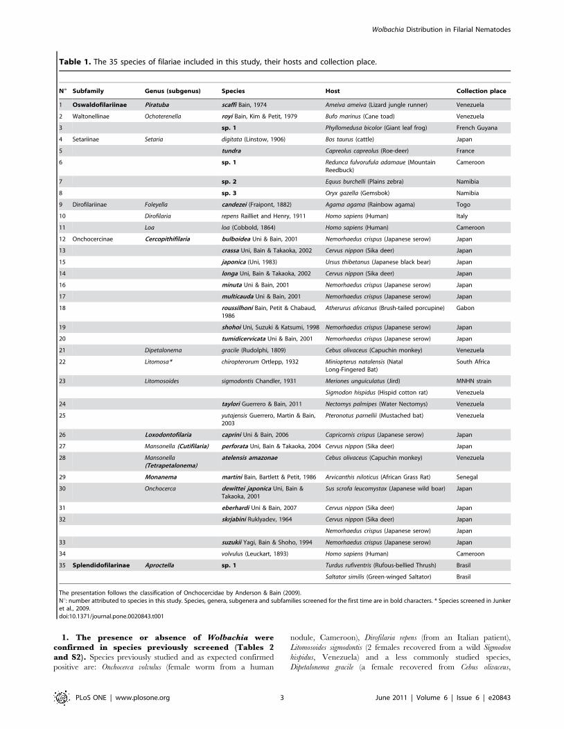
Table 1. The 35 species of filariae included in this study, their hosts and collection place.
N6 Subfamily Genus (subgenus) Species Host Collection place
1 Oswaldofilariinae Piratuba scaffi Bain, 1974 Ameiva ameiva (Lizard jungle runner) Venezuela
2 Waltonellinae Ochoterenella royi Bain, Kim & Petit, 1979 Bufo marinus (Cane toad) Venezuela
3 sp. 1 Phyllomedusa bicolor (Giant leaf frog) French Guyana
4 Setariinae Setaria digitata (Linstow, 1906) Bos taurus (cattle) Japan
5 tundra Capreolus capreolus (Roe-deer) France
6 sp. 1 Redunca fulvorufula adamaue (MountainReedbuck)
Cameroon
7 sp. 2 Equus burchelli (Plains zebra) Namibia
8 sp. 3 Oryx gazella (Gemsbok) Namibia
9 Dirofilariinae Foleyella candezei (Fraipont, 1882) Agama agama (Rainbow agama) Togo
10 Dirofilaria repens Railliet and Henry, 1911 Homo sapiens (Human) Italy
11 Loa loa (Cobbold, 1864) Homo sapiens (Human) Cameroon
12 Onchocercinae Cercopithifilaria bulboidea Uni & Bain, 2001 Nemorhaedus crispus (Japanese serow) Japan
13 crassa Uni, Bain & Takaoka, 2002 Cervus nippon (Sika deer) Japan
15 japonica (Uni, 1983) Ursus thibetanus (Japanese black bear) Japan
14 longa Uni, Bain & Takaoka, 2002 Cervus nippon (Sika deer) Japan
16 minuta Uni & Bain, 2001 Nemorhaedus crispus (Japanese serow) Japan
17 multicauda Uni & Bain, 2001 Nemorhaedus crispus (Japanese serow) Japan
18 roussilhoni Bain, Petit & Chabaud,1986
Atherurus africanus (Brush-tailed porcupine) Gabon
19 shohoi Uni, Suzuki & Katsumi, 1998 Nemorhaedus crispus (Japanese serow) Japan
20 tumidicervicata Uni & Bain, 2001 Nemorhaedus crispus (Japanese serow) Japan
21 Dipetalonema gracile (Rudolphi, 1809) Cebus olivaceus (Capuchin monkey) Venezuela
22 Litomosa* chiropterorum Ortlepp, 1932 Miniopterus natalensis (NatalLong-Fingered Bat)
South Africa
23 Litomosoides sigmodontis Chandler, 1931 Meriones unguiculatus (Jird) MNHN strain
Sigmodon hispidus (Hispid cotton rat) Venezuela
24 taylori Guerrero & Bain, 2011 Nectomys palmipes (Water Nectomys) Venezuela
25 yutajensis Guerrero, Martin & Bain,2003
Pteronotus parnellii (Mustached bat) Venezuela
26 Loxodontofilaria caprini Uni & Bain, 2006 Capricornis crispus (Japanese serow) Japan
27 Mansonella (Cutifilaria) perforata Uni, Bain & Takaoka, 2004 Cervus nippon (Sika deer) Japan
28 Mansonella(Tetrapetalonema)
atelensis amazonae Cebus olivaceus (Capuchin monkey) Venezuela
29 Monanema martini Bain, Bartlett & Petit, 1986 Arvicanthis niloticus (African Grass Rat) Senegal
30 Onchocerca dewittei japonica Uni, Bain &Takaoka, 2001
Sus scrofa leucomystax (Japanese wild boar) Japan
31 eberhardi Uni & Bain, 2007 Cervus nippon (Sika deer) Japan
32 skrjabini Ruklyadev, 1964 Cervus nippon (Sika deer) Japan
Nemorhaedus crispus (Japanese serow) Japan
33 suzukii Yagi, Bain & Shoho, 1994 Nemorhaedus crispus (Japanese serow) Japan
34 volvulus (Leuckart, 1893) Homo sapiens (Human) Cameroon
35 Splendidofilarinae Aproctella sp. 1 Turdus rufiventris (Rufous-bellied Thrush) Brasil
Saltator similis (Green-winged Saltator) Brasil
The presentation follows the classification of Onchocercidae by Anderson & Bain (2009).Nu: number attributed to species in this study. Species, genera, subgenera and subfamilies screened for the first time are in bold characters. * Species screened in Junkeret al., 2009.doi:10.1371/journal.pone.0020843.t001
Wolbachia Distribution in Filarial Nematodes
PLoS ONE | www.plosone.org 3 June 2011 | Volume 6 | Issue 6 | e20843

Wolbachia Distribution in Filarial Nematodes
PLoS ONE | www.plosone.org 4 June 2011 | Volume 6 | Issue 6 | e20843
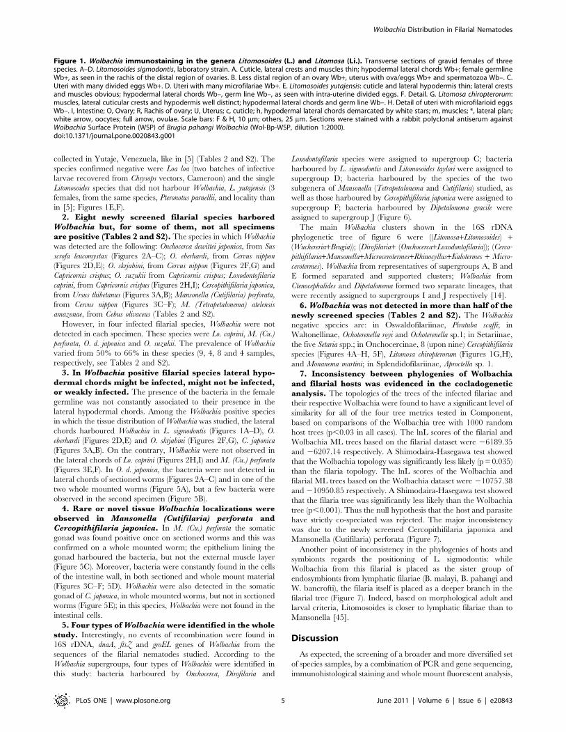
collected in Yutaje, Venezuela, like in [5] (Tables 2 and S2). The
species confirmed negative were Loa loa (two batches of infective
larvae recovered from Chrysops vectors, Cameroon) and the single
Litomosoides species that did not harbour Wolbachia, L. yutajensis (3
females, from the same species, Pteronotus parnellii, and locality than
in [5]; Figures 1E,F).
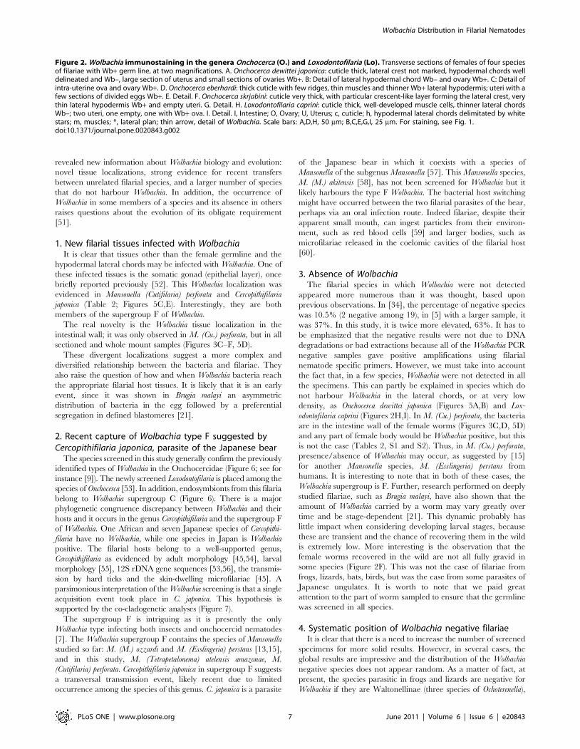
2. Eight newly screened filarial species harboredWolbachia but, for some of them, not all specimensare positive (Tables 2 and S2). The species in which Wolbachia
was detected are the following: Onchocerca dewittei japonica, from Sus
scrofa leucomystax (Figures 2A–C); O. eberhardi, from Cervus nippon
(Figures 2D,E); O. skrjabini, from Cervus nippon (Figures 2F,G) and
Capricornis crispus; O. suzukii from Capricornis crispus; Loxodontofilaria
caprini, from Capricornis crispus (Figures 2H,I); Cercopithifilaria japonica,
from Ursus thibetanus (Figures 3A,B); Mansonella (Cutifilaria) perforata,
from Cervus nippon (Figures 3C–F); M. (Tetrapetalonema) atelensis
amazonae, from Cebus olivaceus (Tables 2 and S2).
However, in four infected filarial species, Wolbachia were not
detected in each specimen. These species were Lo. caprini, M. (Cu.)
perforata, O. d. japonica and O. suzukii. The prevalence of Wolbachia
varied from 50% to 66% in these species (9, 4, 8 and 4 samples,
respectively, see Tables 2 and S2).
3. In Wolbachia positive filarial species lateral hypo-dermal chords might be infected, might not be infected,or weakly infected. The presence of the bacteria in the female
germline was not constantly associated to their presence in the
lateral hypodermal chords. Among the Wolbachia positive species
in which the tissue distribution of Wolbachia was studied, the lateral
chords harboured Wolbachia in L. sigmodontis (Figures 1A–D), O.
eberhardi (Figures 2D,E) and O. skrjabini (Figures 2F,G), C. japonica
(Figures 3A,B). On the contrary, Wolbachia were not observed in
the lateral chords of Lo. caprini (Figures 2H,I) and M. (Cu.) perforata
(Figures 3E,F). In O. d. japonica, the bacteria were not detected in
lateral chords of sectioned worms (Figures 2A–C) and in one of the
two whole mounted worms (Figure 5A), but a few bacteria were
observed in the second specimen (Figure 5B).
4. Rare or novel tissue Wolbachia localizations wereobserved in Mansonella (Cutifilaria) perforata andCercopithifilaria japonica. In M. (Cu.) perforata the somatic
gonad was found positive once on sectioned worms and this was
confirmed on a whole mounted worm; the epithelium lining the
gonad harboured the bacteria, but not the external muscle layer
(Figure 5C). Moreover, bacteria were constantly found in the cells
of the intestine wall, in both sectioned and whole mount material
(Figures 3C–F; 5D). Wolbachia were also detected in the somatic
gonad of C. japonica, in whole mounted worms, but not in sectioned
worms (Figure 5E); in this species, Wolbachia were not found in the
intestinal cells.
5. Four types of Wolbachia were identified in the wholestudy. Interestingly, no events of recombination were found in
16S rDNA, dnaA, ftsZ and groEL genes of Wolbachia from the
sequences of the filarial nematodes studied. According to the
Wolbachia supergroups, four types of Wolbachia were identified in
this study: bacteria harboured by Onchocerca, Dirofilaria and
Loxodontofilaria species were assigned to supergroup C; bacteria
harboured by L. sigmodontis and Litomosoides taylori were assigned to
supergroup D; bacteria harboured by the species of the two
subgenera of Mansonella (Tetrapetalonema and Cutifilaria) studied, as
well as those harboured by Cercopithifilaria japonica were assigned to
supergroup F; bacteria harboured by Dipetalonema gracile were
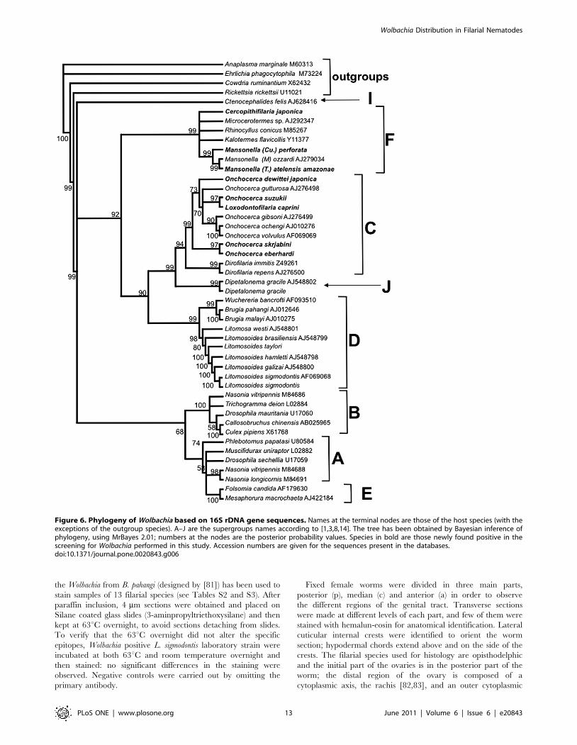
assigned to supergroup J (Figure 6).
The main Wolbachia clusters shown in the 16S rDNA
phylogenetic tree of figure 6 were ((Litomosa+Litomosoides) +(Wuchereria+Brugia)); (Dirofilaria+ (Onchocerca+Loxodontofilaria)); (Cerco-
pithifilaria+Mansonella+Microcerotermes+Rhinocyllus+Kalotermes + Micro-
cerotermes). Wolbachia from representatives of supergroups A, B and
E formed separated and supported clusters; Wolbachia from
Ctenocephalides and Dipetalonema formed two separate lineages, that
were recently assigned to supergroups I and J respectively [14].
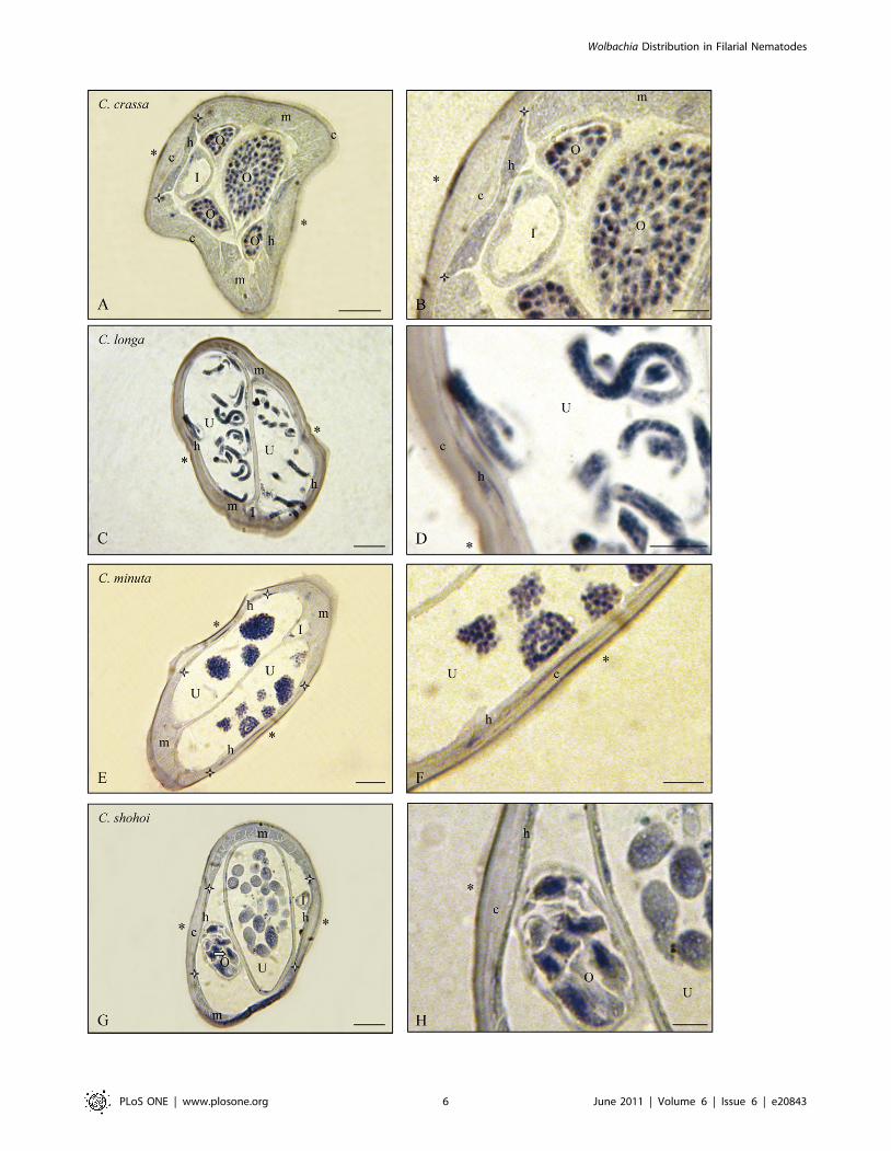
6. Wolbachia was not detected in more than half of thenewly screened species (Tables 2 and S2). The Wolbachia
negative species are: in Oswaldofilariinae, Piratuba scaffi; in
Waltonellinae, Ochoterenella royi and Ochoterenella sp.1; in Setariinae,
the five Setaria spp.; in Onchocercinae, 8 (upon nine) Cercopithifilaria
species (Figures 4A–H, 5F), Litomosa chiropterorum (Figures 1G,H),
and Monanema martini; in Splendidofilariinae, Aproctella sp. 1.
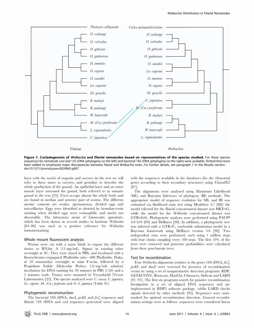
7. Inconsistency between phylogenies of Wolbachiaand filarial hosts was evidenced in the cocladogeneticanalysis. The topologies of the trees of the infected filariae and
their respective Wolbachia were found to have a significant level of
similarity for all of the four tree metrics tested in Component,
based on comparisons of the Wolbachia tree with 1000 random
host trees (p,0.03 in all cases). The lnL scores of the filarial and
Wolbachia ML trees based on the filarial dataset were 26189.35
and 26207.14 respectively. A Shimodaira-Hasegawa test showed
that the Wolbachia topology was significantly less likely (p = 0.035)
than the filaria topology. The lnL scores of the Wolbachia and
filarial ML trees based on the Wolbachia dataset were 210757.38
and 210950.85 respectively. A Shimodaira-Hasegawa test showed
that the filaria tree was significantly less likely than the Wolbachia
tree (p,0.001). Thus the null hypothesis that the host and parasite
have strictly co-speciated was rejected. The major inconsistency
was due to the newly screened Cercopithifilaria japonica and
Mansonella (Cutifilaria) perforata (Figure 7).
Another point of inconsistency in the phylogenies of hosts and
symbionts regards the positioning of L. sigmodontis: while
Wolbachia from this filarial is placed as the sister group of
endosymbionts from lymphatic filariae (B. malayi, B. pahangi and
W. bancrofti), the filaria itself is placed as a deeper branch in the
filarial tree (Figure 7). Indeed, based on morphological adult and
larval criteria, Litomosoides is closer to lymphatic filariae than to
Mansonella [45].
Discussion
As expected, the screening of a broader and more diversified set
of species samples, by a combination of PCR and gene sequencing,
immunohistological staining and whole mount fluorescent analysis,
Figure 1. Wolbachia immunostaining in the genera Litomosoides (L.) and Litomosa (Li.). Transverse sections of gravid females of threespecies. A–D. Litomosoides sigmodontis, laboratory strain. A. Cuticle, lateral crests and muscles thin; hypodermal lateral chords Wb+; female germlineWb+, as seen in the rachis of the distal region of ovaries. B. Less distal region of an ovary Wb+, uterus with ova/eggs Wb+ and spermatozoa Wb–. C.Uteri with many divided eggs Wb+. D. Uteri with many microfilariae Wb+. E. Litomosoides yutajensis: cuticle and lateral hypodermis thin; lateral crestsand muscles obvious; hypodermal lateral chords Wb–, germ line Wb–, as seen with intra-uterine divided eggs. F. Detail. G. Litomosa chiropterorum:muscles, lateral cuticular crests and hypodermis well distinct; hypodermal lateral chords and germ line Wb–. H. Detail of uteri with microfilarioid eggsWb–. I, Intestine; O, Ovary; R, Rachis of ovary; U, Uterus; c, cuticle; h, hypodermal lateral chords demarcated by white stars; m, muscles; *, lateral plan;white arrow, oocytes; full arrow, ovulae. Scale bars: F & H, 10 mm; others, 25 mm. Sections were stained with a rabbit polyclonal antiserum againstWolbachia Surface Protein (WSP) of Brugia pahangi Wolbachia (Wol-Bp-WSP, dilution 1:2000).doi:10.1371/journal.pone.0020843.g001
Wolbachia Distribution in Filarial Nematodes
PLoS ONE | www.plosone.org 5 June 2011 | Volume 6 | Issue 6 | e20843
Wolbachia Distribution in Filarial Nematodes
PLoS ONE | www.plosone.org 6 June 2011 | Volume 6 | Issue 6 | e20843
revealed new information about Wolbachia biology and evolution:
novel tissue localizations, strong evidence for recent transfers
between unrelated filarial species, and a larger number of species
that do not harbour Wolbachia. In addition, the occurrence of
Wolbachia in some members of a species and its absence in others
raises questions about the evolution of its obligate requirement
[51].
1. New filarial tissues infected with WolbachiaIt is clear that tissues other than the female germline and the
hypodermal lateral chords may be infected with Wolbachia. One of
these infected tissues is the somatic gonad (epithelial layer), once
briefly reported previously [52]. This Wolbachia localization was
evidenced in Mansonella (Cutifilaria) perforata and Cercopithifilaria
japonica (Table 2; Figures 5C,E). Interestingly, they are both
members of the supergroup F of Wolbachia.
The real novelty is the Wolbachia tissue localization in the
intestinal wall; it was only observed in M. (Cu.) perforata, but in all
sectioned and whole mount samples (Figures 3C–F, 5D).
These divergent localizations suggest a more complex and
diversified relationship between the bacteria and filariae. They
also raise the question of how and when Wolbachia bacteria reach
the appropriate filarial host tissues. It is likely that it is an early
event, since it was shown in Brugia malayi an asymmetric
distribution of bacteria in the egg followed by a preferential
segregation in defined blastomeres [21].
2. Recent capture of Wolbachia type F suggested byCercopithifilaria japonica, parasite of the Japanese bear
The species screened in this study generally confirm the previously
identified types of Wolbachia in the Onchocercidae (Figure 6; see for
instance [9]). The newly screened Loxodontofilaria is placed among the
species of Onchocerca [53]. In addition, endosymbionts from this filaria
belong to Wolbachia supergroup C (Figure 6). There is a major
phylogenetic congruence discrepancy between Wolbachia and their
hosts and it occurs in the genus Cercopithifilaria and the supergroup F
of Wolbachia. One African and seven Japanese species of Cercopithi-
filaria have no Wolbachia, while one species in Japan is Wolbachia
positive. The filarial hosts belong to a well-supported genus,
Cercopithifilaria as evidenced by adult morphology [45,54], larval
morphology [55], 12S rDNA gene sequences [53,56], the transmis-
sion by hard ticks and the skin-dwelling microfilariae [45]. A
parsimonious interpretation of the Wolbachia screening is that a single
acquisition event took place in C. japonica. This hypothesis is
supported by the co-cladogenetic analyses (Figure 7).
The supergroup F is intriguing as it is presently the only
Wolbachia type infecting both insects and onchocercid nematodes
[7]. The Wolbachia supergroup F contains the species of Mansonella
studied so far: M. (M.) ozzardi and M. (Esslingeria) perstans [13,15],
and in this study, M. (Tetrapetalonema) atelensis amazonae, M.
(Cutifilaria) perforata. Cercopithifilaria japonica in supergroup F suggests
a transversal transmission event, likely recent due to limited
occurrence among the species of this genus. C. japonica is a parasite
of the Japanese bear in which it coexists with a species of
Mansonella of the subgenus Mansonella [57]. This Mansonella species,
M. (M.) akitensis [58], has not been screened for Wolbachia but it
likely harbours the type F Wolbachia. The bacterial host switching
might have occurred between the two filarial parasites of the bear,
perhaps via an oral infection route. Indeed filariae, despite their
apparent small mouth, can ingest particles from their environ-
ment, such as red blood cells [59] and larger bodies, such as
microfilariae released in the coelomic cavities of the filarial host
[60].
3. Absence of WolbachiaThe filarial species in which Wolbachia were not detected
appeared more numerous than it was thought, based upon
previous observations. In [34], the percentage of negative species
was 10.5% (2 negative among 19), in [5] with a larger sample, it
was 37%. In this study, it is twice more elevated, 63%. It has to
be emphasized that the negative results were not due to DNA
degradations or bad extractions because all of the Wolbachia PCR
negative samples gave positive amplifications using filarial
nematode specific primers. However, we must take into account
the fact that, in a few species, Wolbachia were not detected in all
the specimens. This can partly be explained in species which do
not harbour Wolbachia in the lateral chords, or at very low
density, as Onchocerca dewittei japonica (Figures 5A,B) and Lox-
odontofilaria caprini (Figures 2H,I). In M. (Cu.) perforata, the bacteria
are in the intestine wall of the female worms (Figures 3C,D, 5D)
and any part of female body would be Wolbachia positive, but this
is not the case (Tables 2, S1 and S2). Thus, in M. (Cu.) perforata,
presence/absence of Wolbachia may occur, as suggested by [15]
for another Mansonella species, M. (Esslingeria) perstans from
humans. It is interesting to note that in both of these cases, the
Wolbachia supergroup is F. Further, research performed on deeply
studied filariae, such as Brugia malayi, have also shown that the
amount of Wolbachia carried by a worm may vary greatly over
time and be stage-dependent [21]. This dynamic probably has
little impact when considering developing larval stages, because
these are transient and the chance of recovering them in the wild
is extremely low. More interesting is the observation that the
female worms recovered in the wild are not all fully gravid in
some species (Figure 2F). This was not the case of filariae from
frogs, lizards, bats, birds, but was the case from some parasites of
Japanese ungulates. It is worth to note that we paid great
attention to the part of worm sampled to ensure that the germline
was screened in all species.
4. Systematic position of Wolbachia negative filariaeIt is clear that there is a need to increase the number of screened
specimens for more solid results. However, in several cases, the
global results are impressive and the distribution of the Wolbachia
negative species does not appear random. As a matter of fact, at
present, the species parasitic in frogs and lizards are negative for
Wolbachia if they are Waltonellinae (three species of Ochoterenella),
Figure 2. Wolbachia immunostaining in the genera Onchocerca (O.) and Loxodontofilaria (Lo). Transverse sections of females of four speciesof filariae with Wb+ germ line, at two magnifications. A. Onchocerca dewittei japonica: cuticle thick, lateral crest not marked, hypodermal chords welldelineated and Wb–, large section of uterus and small sections of ovaries Wb+. B: Detail of lateral hypodermal chord Wb– and ovary Wb+. C: Detail ofintra-uterine ova and ovary Wb+. D. Onchocerca eberhardi: thick cuticle with few ridges, thin muscles and thinner Wb+ lateral hypodermis; uteri with afew sections of divided eggs Wb+. E. Detail. F. Onchocerca skrjabini: cuticle very thick, with particular crescent-like layer forming the lateral crest, verythin lateral hypodermis Wb+ and empty uteri. G. Detail. H. Loxodontofilaria caprini: cuticle thick, well-developed muscle cells, thinner lateral chordsWb–; two uteri, one empty, one with Wb+ ova. I. Detail. I, Intestine; O, Ovary; U, Uterus; c, cuticle; h, hypodermal lateral chords delimitated by whitestars; m, muscles; *, lateral plan; thin arrow, detail of Wolbachia. Scale bars: A,D,H, 50 mm; B,C,E,G,I, 25 mm. For staining, see Fig. 1.doi:10.1371/journal.pone.0020843.g002
Wolbachia Distribution in Filarial Nematodes
PLoS ONE | www.plosone.org 7 June 2011 | Volume 6 | Issue 6 | e20843

Wolbachia Distribution in Filarial Nematodes
PLoS ONE | www.plosone.org 8 June 2011 | Volume 6 | Issue 6 | e20843
Oswaldofilariinae (one species of Piratuba), or Dirofilariinae (two
species of Foleyella). It was also shown here that the first screened
species parasitic in birds, an Aproctella in the Splendidofilariinae,
was Wolbachia negative (7 females screened with PCR, from two
passeriform species, totalising 7 specimens hosts). Wolbachia were
not detected also in several species of Onchocercinae from
mammals. The first is Litomosa chiropterorum, from an African bat (8
specimens screened from 8 Miniopterus schreibersi), an unexpected
observation since the single Litomosa species screened previously,
Li. westi from a North American rodent Geomyoidea, was
Wolbachia positive [5].
The second species is Monanema martini (8 females screened from
8 murid specimens). The absence of the bacteria is an important
feature, considering that this filaria was used, in the past, as a
model of onchocerciasis, a true limit due to the important role
played by the bacteria in the pathology of the disease [39,61–63].
The absence of Wolbachia could explain partly the weak ocular
pathology induced in the murid host [49]. Severe or mild ocular
onchocerciases have been related to a strain-dependent variation
of density of Wolbachia per filarial genome [64].
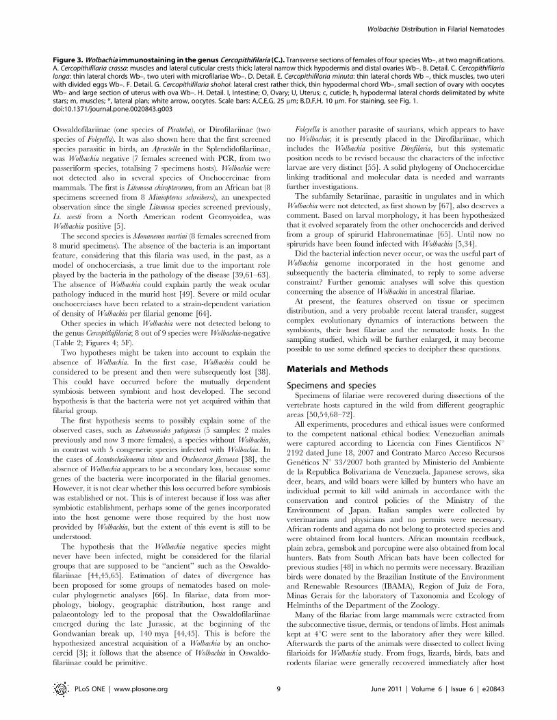
Other species in which Wolbachia were not detected belong to
the genus Cercopithifilaria; 8 out of 9 species were Wolbachia-negative
(Table 2; Figures 4; 5F).
Two hypotheses might be taken into account to explain the
absence of Wolbachia. In the first case, Wolbachia could be
considered to be present and then were subsequently lost [38].
This could have occurred before the mutually dependent
symbiosis between symbiont and host developed. The second
hypothesis is that the bacteria were not yet acquired within that
filarial group.
The first hypothesis seems to possibly explain some of the
observed cases, such as Litomosoides yutajensis (5 samples: 2 males
previously and now 3 more females), a species without Wolbachia,
in contrast with 5 congeneric species infected with Wolbachia. In
the cases of Acantocheilonema viteae and Onchocerca flexuosa [38], the
absence of Wolbachia appears to be a secondary loss, because some
genes of the bacteria were incorporated in the filarial genomes.
However, it is not clear whether this loss occurred before symbiosis
was established or not. This is of interest because if loss was after
symbiotic establishment, perhaps some of the genes incorporated
into the host genome were those required by the host now
provided by Wolbachia, but the extent of this event is still to be
understood.
The hypothesis that the Wolbachia negative species might
never have been infected, might be considered for the filarial
groups that are supposed to be ‘‘ancient’’ such as the Oswaldo-
filariinae [44,45,65]. Estimation of dates of divergence has
been proposed for some groups of nematodes based on mole-
cular phylogenetic analyses [66]. In filariae, data from mor-
phology, biology, geographic distribution, host range and
palaeontology led to the proposal that the Oswaldofilariinae
emerged during the late Jurassic, at the beginning of the
Gondwanian break up, 140 mya [44,45]. This is before the
hypothesized ancestral acquisition of a Wolbachia by an oncho-
cercid [3]; it follows that the absence of Wolbachia in Oswaldo-
filariinae could be primitive.
Foleyella is another parasite of saurians, which appears to have
no Wolbachia; it is presently placed in the Dirofilariinae, which
includes the Wolbachia positive Dirofilaria, but this systematic
position needs to be revised because the characters of the infective
larvae are very distinct [55]. A solid phylogeny of Onchocercidae
linking traditional and molecular data is needed and warrants
further investigations.
The subfamily Setariinae, parasitic in ungulates and in which
Wolbachia were not detected, as first shown by [67], also deserves a
comment. Based on larval morphology, it has been hypothesized
that it evolved separately from the other onchocercids and derived
from a group of spirurid Habronematinae [65]. Until now no
spirurids have been found infected with Wolbachia [5,34].
Did the bacterial infection never occur, or was the useful part of
Wolbachia genome incorporated in the host genome and
subsequently the bacteria eliminated, to reply to some adverse
constraint? Further genomic analyses will solve this question
concerning the absence of Wolbachia in ancestral filariae.
At present, the features observed on tissue or specimen
distribution, and a very probable recent lateral transfer, suggest
complex evolutionary dynamics of interactions between the
symbionts, their host filariae and the nematode hosts. In the
sampling studied, which will be further enlarged, it may become
possible to use some defined species to decipher these questions.
Materials and Methods
Specimens and speciesSpecimens of filariae were recovered during dissections of the
vertebrate hosts captured in the wild from different geographic
areas [50,54,68–72].
All experiments, procedures and ethical issues were conformed
to the competent national ethical bodies: Venezuelian animals
were captured according to Licencia con Fines Cientificos Nu2192 dated June 18, 2007 and Contrato Marco Acceso Recursos
Geneticos Nu 33/2007 both granted by Ministerio del Ambiente
de la Republica Bolivariana de Venezuela. Japanese serows, sika
deer, bears, and wild boars were killed by hunters who have an
individual permit to kill wild animals in accordance with the
conservation and control policies of the Ministry of the
Environment of Japan. Italian samples were collected by
veterinarians and physicians and no permits were necessary.
African rodents and agama do not belong to protected species and
were obtained from local hunters. African mountain reedbuck,
plain zebra, gemsbok and porcupine were also obtained from local
hunters. Bats from South African bats have been collected for
previous studies [48] in which no permits were necessary. Brazilian
birds were donated by the Brazilian Institute of the Environment
and Renewable Resources (IBAMA), Region of Juiz de Fora,
Minas Gerais for the laboratory of Taxonomia and Ecology of
Helminths of the Department of the Zoology.
Many of the filariae from large mammals were extracted from
the subconnective tissue, dermis, or tendons of limbs. Host animals
kept at 4uC were sent to the laboratory after they were killed.
Afterwards the parts of the animals were dissected to collect living
filarioids for Wolbachia study. From frogs, lizards, birds, bats and
rodents filariae were generally recovered immediately after host
Figure 3. Wolbachia immunostaining in the genus Cercopithifilaria (C.). Transverse sections of females of four species Wb–, at two magnifications.A. Cercopithifilaria crassa: muscles and lateral cuticular crests thick; lateral narrow thick hypodermis and distal ovaries Wb–. B. Detail. C. Cercopithifilarialonga: thin lateral chords Wb–, two uteri with microfilariae Wb–. D. Detail. E. Cercopithifilaria minuta: thin lateral chords Wb –, thick muscles, two uteriwith divided eggs Wb–. F. Detail. G. Cercopithifilaria shohoi: lateral crest rather thick, thin hypodermal chord Wb–, small section of ovary with oocytesWb– and large section of uterus with ova Wb–. H. Detail. I, Intestine; O, Ovary; U, Uterus; c, cuticle; h, hypodermal lateral chords delimitated by whitestars; m, muscles; *, lateral plan; white arrow, oocytes. Scale bars: A,C,E,G, 25 mm; B,D,F,H, 10 mm. For staining, see Fig. 1.doi:10.1371/journal.pone.0020843.g003
Wolbachia Distribution in Filarial Nematodes
PLoS ONE | www.plosone.org 9 June 2011 | Volume 6 | Issue 6 | e20843
death. Samples used for positive and negative PCR controls were
laboratory strains.
Species identification of the specimens was done with
morphological studies performed by several of us (OB, SU, RG,
SL, MD). Some species are not yet named, but all are under study,
and morphological analysis and sequencing of coxI gene in this
study and in [53] showed that they represent distinct molecular
entities. Co-infection of a host specimen by several congeneric or
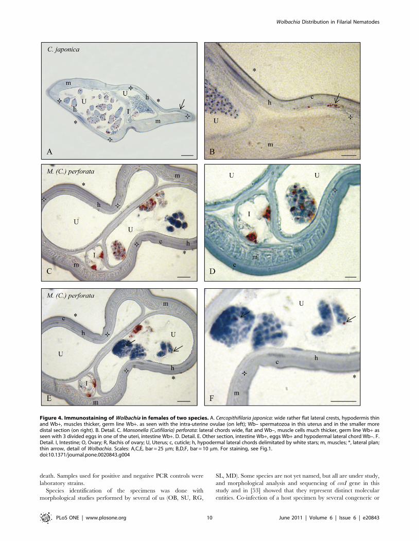
Figure 4. Immunostaining of Wolbachia in females of two species. A. Cercopithifilaria japonica: wide rather flat lateral crests, hypodermis thinand Wb+, muscles thicker, germ line Wb+. as seen with the intra-uterine ovulae (on left); Wb– spermatozoa in this uterus and in the smaller moredistal section (on right). B. Detail. C. Mansonella (Cutifilaria) perforata: lateral chords wide, flat and Wb–, muscle cells much thicker, germ line Wb+ asseen with 3 divided eggs in one of the uteri, intestine Wb+. D. Detail. E. Other section, intestine Wb+, eggs Wb+ and hypodermal lateral chord Wb–. F.Detail. I, Intestine; O, Ovary; R, Rachis of ovary; U, Uterus; c, cuticle; h, hypodermal lateral chords delimitated by white stars; m, muscles; *, lateral plan;thin arrow, detail of Wolbachia. Scales: A,C,E, bar = 25 mm; B,D,F, bar = 10 mm. For staining, see Fig.1.doi:10.1371/journal.pone.0020843.g004
Wolbachia Distribution in Filarial Nematodes
PLoS ONE | www.plosone.org 10 June 2011 | Volume 6 | Issue 6 | e20843
non-congeneric filarial species was rather frequent in large
mammals, but in a few cases, the same filarial species was
recovered from two host species. The supraspecific levels of
taxonomy followed the systematic works of [46,73,74] and more
recent studies for some taxa: [65,71,75] for the subgeneric
divisions of Mansonella Faust, 1929; [54,56] for the genus
Cercopithifilaria Eberhard, 1980 (created as a subgenus); and [54]
for the genus Loxodontofilaria Berghe & Gillain, 1939.
Abbreviations used in the text are: A. for Aproctella; C. for
Cercopithifilaria; Cu. for Cutifilaria; D. for Dirofilaria; Di. for
Dipetalonema; F. for Foleyella; L. for Litomosoides; Li. for Litomosa; Lo.
for Loxondontofilaria; M. for Mansonella; Mo. for Monanema; O. for
Onchocerca; Oc. for Ochoterenella; P. for Piratuba; S. for Setaria; T. for
Tetrapetalonema. The material screened in this study is detailed in
Figure S1, Tables S1 and S2.
Filarial worms were fixed and kept in absolute alcohol at 4uCfor PCR analyses, in 4% PFA (paraformaldehyde) for overnight at
4uC for immunohistochemical staining and whole mount fluores-
cent analysis. In many cases, filarial specimens were cut into
anterior (a), median (m) and posterior (p) parts, which were fixed
for the different analysis approaches. Since Wolbachia are
transmitted by female filariae, almost all of the studies were based
on female worms; male specimens were rarely examined, and we
analysed this sex alone in a single case, Di. gracile (Table S3).
Molecular screening for Wolbachia on filarial nematodesPCR screening for Wolbachia was conducted according to [5,12],
using general Wolbachia primers for 16S rDNA (99f and 994r [76]),
originally designed to work on Wolbachia from the supergroups A
and B, and primers for 16S rDNA (16SWolbF and 16SWolbR3),
originally designed to work on Wolbachia from the supergroups A–
D [12], but whose target sites are also conserved in Wolbachia from
supergroups E and F [8].
PCRs were performed in a 20 ml final volume under the
following conditions: 1x buffer (containing 1.5 mM MgCl2,
EppendorfTM), 0.2 mM of each dNTP, 1 mM of each primer,
and 0.5 U of Taq DNA Polymerase (EppendorfTM). The thermal
profile used was: 94uC 45 sec, 52uC 45 sec, and 72uC 90 sec for
40 cycles.
When the PCRs were negative under the above PCR
conditions, a nested-PCR approach was implemented in order
to improve the sensitivity of the PCR screening [5,77]. The first
PCR was performed using the general eubacterial primer 27F [78]
combined with 16SWolbR3; PCR volumes and conditions were as
above. Five ml were visualised on a 1.5% w/v agarose gel and one
ml of the first PCR was diluted 1/10 and 1/100 in water, and then
both used as templates in a second PCR, performed using primers
W-EF and W-ER [70]. W-ER and W-EF recognize sites that are
conserved in supergroups E–F and that are internal to the primers
used in the first PCR. PCR conditions for this amplification were
as described in [79].
Of samples remaining negative after the two PCRs approaches
described above, PCRs with primers 16SWolbF and 16SWolbR3
were performed varying the following parameters: MgCl2concentrations at 2.5, 4 and 6 mM and annealing temperatures
of 52uC +/25uC.
DNA preparations from filarial species harbouring Wolbachia (D.
immitis and Brugia pahangi) and from a Wolbachia-infected strain of
mosquitoes (Culex pipiens) [5] were included in the screening as
positive controls. DNA preparations from a filarial species not
harbouring Wolbachia (A. viteae) [5] were included as negative
controls.
Of the samples positive for PCR screening homologous to
Wolbachia 16S rDNA, dnaA, ftsZ and groEL were also amplified
using the primers described in [5,80] under the following
conditions: 1x Eppendorf buffer including 1.5 mM MgCl2,
0.2 mM of each dNTP, 1 mM each of forward and reverse
primers, and 0.5 units MasterTaq (Eppendorf). The thermal
profiles we used were: (1) dnaA, 94uC 45 sec, 52uC 45 sec, and
72uC 90 sec, for 40 cycles; (2) groEL, 94uC 45 sec, 60uC 45 sec,
72uC 80 sec, for 5 cycles, and 94uC 45 sec, 55uC 45 sec, and 72uC80 sec, for 34 cycles; (3) ftsZ, 94uC 30 sec, 60uC 45 sec, 72uC90 sec, for 5 cycles, and 94uC 30 sec, 57uC 45 sec, and 72uC90 sec, for 34 cycles.
Amplifications were performed in 20–50 ml volumes.
In all cases, in order to ascertain the DNA conditions before
Wolbachia screenings and to confirm morphological identification,
coxI amplification was performed as described in [12].
Sequencing conditionscoxI sequencing was performed as described in [12]. dnaA,
ftsZ and groEL sequencing were performed as described in
[5,80]. From the Wolbachia PCR positive samples, almost the full
length of the 16S rDNA gene of Wolbachia was sequenced using
primers 27F and 16SWolbR3. The amplifications obtained
(about 1400 bp) were gel-purified (using the QIAquickH PCR
Purification Kit, Qiagen) and directly sequenced using ABI
technology. The sequences obtained have been deposited in
the EMBL Data Library. The dnaA, groEL and ftsZ sequences
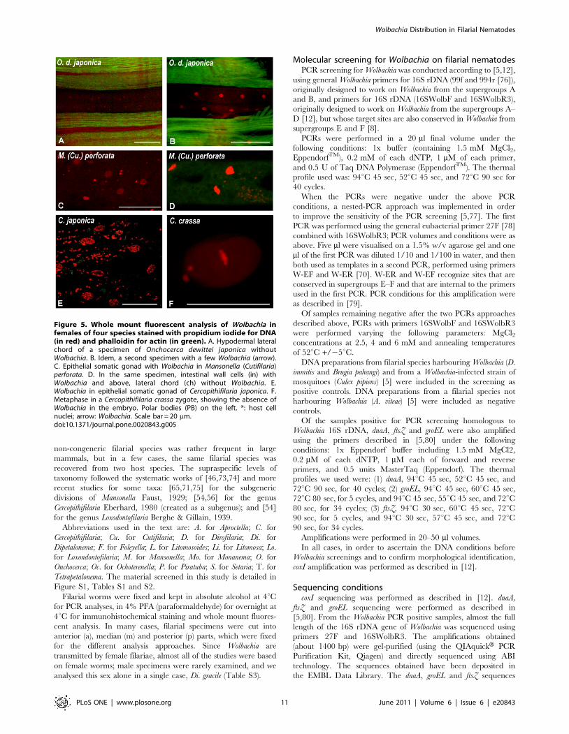
Figure 5. Whole mount fluorescent analysis of Wolbachia infemales of four species stained with propidium iodide for DNA(in red) and phalloidin for actin (in green). A. Hypodermal lateralchord of a specimen of Onchocerca dewittei japonica withoutWolbachia. B. Idem, a second specimen with a few Wolbachia (arrow).C. Epithelial somatic gonad with Wolbachia in Mansonella (Cutifilaria)perforata. D. In the same specimen, intestinal wall cells (in) withWolbachia and above, lateral chord (ch) without Wolbachia. E.Wolbachia in epithelial somatic gonad of Cercopithifilaria japonica. F.Metaphase in a Cercopithifilaria crassa zygote, showing the absence ofWolbachia in the embryo. Polar bodies (PB) on the left. *: host cellnuclei; arrow: Wolbachia. Scale bar = 20 mm.doi:10.1371/journal.pone.0020843.g005
Wolbachia Distribution in Filarial Nematodes
PLoS ONE | www.plosone.org 11 June 2011 | Volume 6 | Issue 6 | e20843
were not obtained from all the taxa included in this study
mainly caused by the scarcity of certain specimens, and
amplification/sequencing problems for some of the species
examined. Where DNA of the host was amplified, the PCR
product was purified as above and directly sequenced using ABI
technology.
A list of the sequences including accession numbers is available
in Table S2.
Immunohistochemical staining of worm sectionsImmunohistochemical staining was performed according to
[24,48]. A rabbit polyclonal antiserum raised against the WSP of
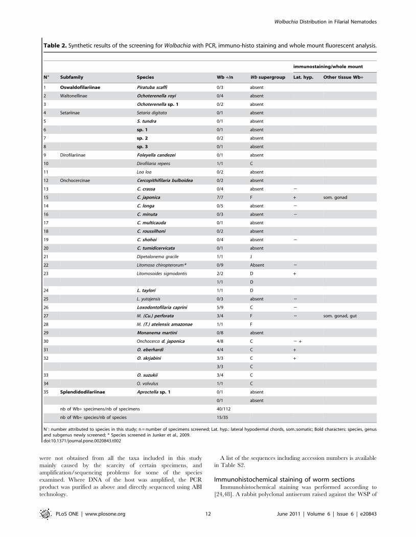
Table 2. Synthetic results of the screening for Wolbachia with PCR, immuno-histo staining and whole mount fluorescent analysis.
immunostaining/whole mount
N6 Subfamily Species Wb +/n Wb supergroup Lat. hyp. Other tissue Wb+
1 Oswaldofilariinae Piratuba scaffi 0/3 absent
2 Waltonellinae Ochoterenella royi 0/4 absent
3 Ochoterenella sp. 1 0/2 absent
4 Setariinae Setaria digitata 0/1 absent
5 S. tundra 0/1 absent
6 sp. 1 0/1 absent
7 sp. 2 0/2 absent
8 sp. 3 0/1 absent
9 Dirofilariinae Foleyella candezei 0/1 absent
10 Dirofilaria repens 1/1 C
11 Loa loa 0/2 absent
12 Onchocercinae Cercopithifilaria bulboidea 0/2 absent
13 C. crassa 0/4 absent 2
15 C. japonica 7/7 F + som. gonad
14 C. longa 0/5 absent 2
16 C. minuta 0/3 absent 2
17 C. multicauda 0/1 absent
18 C. roussilhoni 0/2 absent
19 C. shohoi 0/4 absent 2
20 C. tumidicervicata 0/1 absent
21 Dipetalonema gracile 1/1 J
22 Litomosa chiropterorum* 0/9 Absent 2
23 Litomosoides sigmodontis 2/2 D +
1/1 D
24 L. taylori 1/1 D
25 L. yutajensis 0/3 absent 2
26 Loxodontofilaria caprini 5/9 C 2
27 M. (Cu.) perforata 3/4 F 2 som. gonad, gut
28 M. (T.) atelensis amazonae 1/1 F
29 Monanema martini 0/8 absent
30 Onchocerca d. japonica 4/8 C 2 +
31 O. eberhardi 4/4 C +
32 O. skrjabini 3/3 C +
3/3 C
33 O. suzukii 3/4 C
34 O. volvulus 1/1 C
35 Splendidodilariinae Aproctella sp. 1 0/1 absent
0/1 absent
nb of Wb+ specimens/nb of specimens 40/112
nb of Wb+ species/nb of species 15/35
Nu: number attributed to species in this study; n = number of specimens screened; Lat. hyp.: lateral hypodermal chords, som.:somatic; Bold characters: species, genusand subgenus newly screened; * Species screened in Junker et al., 2009.doi:10.1371/journal.pone.0020843.t002
Wolbachia Distribution in Filarial Nematodes
PLoS ONE | www.plosone.org 12 June 2011 | Volume 6 | Issue 6 | e20843
the Wolbachia from B. pahangi (designed by [81]) has been used to
stain samples of 13 filarial species (see Tables S2 and S3). After
paraffin inclusion, 4 mm sections were obtained and placed on
Silane coated glass slides (3-aminpropyltriethoxysilane) and then
kept at 63uC overnight, to avoid sections detaching from slides.
To verify that the 63uC overnight did not alter the specific
epitopes, Wolbachia positive L. sigmodontis laboratory strain were
incubated at both 63uC and room temperature overnight and
then stained: no significant differences in the staining were
observed. Negative controls were carried out by omitting the
primary antibody.
Fixed female worms were divided in three main parts,
posterior (p), median (c) and anterior (a) in order to observe
the different regions of the genital tract. Transverse sections
were made at different levels of each part, and few of them were
stained with hemalun-eosin for anatomical identification. Lateral
cuticular internal crests were identified to orient the worm
section; hypodermal chords extend above and on the side of the
crests. The filarial species used for histology are opisthodelphic
and the initial part of the ovaries is in the posterior part of the
worm; the distal region of the ovary is composed of a
cytoplasmic axis, the rachis [82,83], and an outer cytoplasmic
Figure 6. Phylogeny of Wolbachia based on 16S rDNA gene sequences. Names at the terminal nodes are those of the host species (with theexceptions of the outgroup species). A–J are the supergroups names according to [1,3,8,14]. The tree has been obtained by Bayesian inference ofphylogeny, using MrBayes 2.01; numbers at the nodes are the posterior probability values. Species in bold are those newly found positive in thescreening for Wolbachia performed in this study. Accession numbers are given for the sequences present in the databases.doi:10.1371/journal.pone.0020843.g006
Wolbachia Distribution in Filarial Nematodes
PLoS ONE | www.plosone.org 13 June 2011 | Volume 6 | Issue 6 | e20843
layer with the nuclei of oogonia and oocytes (in the text we will
refer to these states as oocytes, and germline to describe the
whole production of the gonad). An epithelial layer and an outer
muscle layer surround the gonad, both referred to as somatic
gonad in the text [25]. Uteri occupy almost the whole body and
are found in median and anterior part of worms. The different
uterine contents are ovulae, spermatozoa, divided eggs and
microfilariae. Eggs were identified as aborted by hemalun-eosin
staining when divided eggs were eosinophilic and nuclei not
discernible. The laboratory strain of Litomosoides sigmodontis,
which has been shown in several studies to harbour Wolbachia
[84–86] was used as a positive reference for Wolbachia
immunostaining.
Whole mount fluorescent analysisWorms were cut with a razor blade to expose the different
tissues to RNAse A (15 mg/mL, Sigma) in rotating tubes
overnight at 4C. They were rinsed in PBS, and incubated with a
fluorochrome-conjugated Phalloidin (atto2488 Phalloidin, Fluka,
at 10 nanomolar) overnight to stain F-actin, followed by a
Propidium Iodide (Molecular Probes, 1.0 mg/mL solution)
incubation for DNA staining for 20 minutes in PBS (1:50) and a
5 minutes wash. Tissues were mounted in Vectashield (Vector
Laboratories) [25]. The species analyzed were C. crassa, C. japonica,
Lo. caprini, M. (Cu.) perforata and O. d. japonica (Table S1).
Phylogenetic reconstructionThe bacterial 16S rDNA, dnaA, groEL and ftsZ sequences and
filarial 12S rDNA and coxI sequences generated were aligned
with the sequences available in the databases (for the ribosomal
genes according to their secondary structures) using ClustalX2
[87].
The alignments were analysed using Maximum Likelihood
(ML) and Bayesian Inference of phylogeny (BI) methods. The
appropriate model of sequence evolution for ML and BI was
estimated via likelihood ratio test using Modeltest 3.7 [88]: the
model selected for the filarial concatenated dataset was HKY+G,
while the model for the Wolbachia concatenated dataset was
GTR+I+G. Phylogenetic analyses were performed using PAUP*
4.0 b10 [89] and MrBayes [90]. In addition, a phylogenetic tree
was inferred with a GTR+C4 nucleotide substitution model in a
Bayesian framework using MrBayes version 3.0 [90]. Two
independent runs were performed, each using 1 million steps
with four chains sampling every 100 steps. The first 10% of the
trees were removed and posterior probabilities were calculated
from these post-burnin trees.
Test for recombinationFour Wolbachia alignments (relative to the genes 16S rDNA, ftsZ,
groEL and dnaA) were screened for presence of recombination
events by using a set of nonparametric detection programs: RDP,
GENECONV, Bootscan, MaxChi, Chimaera, SisScan and LARD
[91–97]. The first six programs search for putative recombination
breakpoints in a set of aligned DNA sequences and are
implemented in RDP3 software package, whilst LARD checks
signals detected by other methods [92]. Sequences were auto-
masked for optimal recombination detection. General recombi-
nation settings were as follows: sequences were considered linear
Figure 7. Cocladogenesis of Wolbachia and filarial nematodes based on representatives of the species studied. For these speciessequences for nematode coxI and 12S rDNA (phylogeny on the left) and bacterial 16S rDNA (phylogeny on the right) were available. Dotted lines havebeen added to emphasize major discrepancies between filarial and Wolbachia trees. For further details, see paragraph 7 in the Results section.doi:10.1371/journal.pone.0020843.g007
Wolbachia Distribution in Filarial Nematodes
PLoS ONE | www.plosone.org 14 June 2011 | Volume 6 | Issue 6 | e20843
and the highest acceptable P-Value was set to 0.01 and
overlapping signals were disentangled.
Method specific options were as follow: MaxChi and Chimaera
were run with a variable window size; Bootscan and SisScan were
forced for exploratory screening. Successive phases of refining
analysis and manual tests were performed where needed.
Cocladogenesis analysisTo test for congruence, phylogenies for the hosts (based on a
concatenated dataset of coxI and 12S rDNA) and for the bacterium
(based on a concatenated dataset of 16S rDNA, ftsZ, dnaA and
groEL) were compared with two methods. The first tested whether
there was a greater than random correspondence between
reconstructed nodes for host and symbiont. This was performed
in Component (R. Page, University of Glasgow, UK) using the
‘‘compare tree with’’ function, with 1,000 randomized trees and
the four available tree-comparison metrics (partition; triplets;
quartets, nearest neighbour interchange). The second test
examined the null hypothesis that the endosymbionts have
undergone cocladogenesis with their hosts. ML trees for each
dataset were first estimated using the successive approximation
method [89]. Then, the scores for each of these ML trees based on
the host dataset were compared using the [98] test. This was
repeated using the parasite dataset. A single uninfected filarial (T.
callipaeda) and a non-filarial Wolbachia (from C. quinquefasciatus) were
used as outgroups.
Supporting Information
Figure S1 Position of the genera screened in the present study
indicated on a schematic representation of a key of the
onchocercid subfamilies, based on morphological characters
(following [46]). Total number of genera per subfamily listed.
*Genus screened for the first time. ** Two subgenera in Mansonella.
(DOC)
Table S1 Details of material studied with PCR, immunostaining
assays (IHS) and whole mount fluorescent analysis (fluo). The
scheme follows the classification as in Tables 1 and 2. Species,
genus and subgenus, subfamily are in bold characters when newly
screened; specimens ids are in bold characters when female
worms; m = male; f = female; a = anterior part; c = central part;
p = posterior part; *150 infective larvae; + = Wolbachia positive
specimens.
(DOC)
Table S2 Results of Wolbachia screening based on PCR,
immunostaining assays and whole mount fluorescent analysis in
35 filarial nematodes. Taxa are presented in alphabetical order.
(DOC)
Table S3 Wolbachia distribution in the tissues of 13 onchocercid
species. +: stained; 2: not stained; NA: not available because the
body structure is not present. *staining shown on Figures 1–4.
(DOC)
Acknowledgments
The authors would like to thank Andrea Galimberti and the personnel at
ZooPlantLab of University of Milan-Bicocca for their help.
We are also grateful to Prof. H. Takaoka and Dr. M. Fukuda, Oita
University, Japan for their contribution in providing animal hosts and Mr.
T. Kenko, the Central Laboratory, Osaka City University Medical School,
Osaka, Japan for histologic preparation of the parasites.
Author Contributions
Conceived and designed the experiments: MC OB. Performed the
experiments: EF OB MB CM SU FL SGB RG SdSL SW MD MC.
Analyzed the data: EF OB MB CM NL SU FL RG SdSL CB MC.
Contributed reagents/materials/analysis tools: EF OB MB CM NL SU FL
SGB RG CB SW MD. Wrote the paper: EF OB FL CB MC.
References
1. Werren JH, Zhang W, Guo LR (1995) Evolution and phylogeny of W. pipientis:
reproductive parasites of arthropods. Proc R Soc Lond B 261: 55–71.
2. Sironi M, Bandi C, Sacchi L, Di Sacco B, Damiani G, et al. (1995) Molecularevidence for a close relative of the arthropod endosymbiont W. pipientis in a
filarial worm. Mol Biochem Parasitol 74: 223–227.
3. Bandi C, Anderson TJC, Genchi C, Blaxter ML (1998) Phylogeny of W. pipientis
in filarial nematodes. Proc R Soc Lond B 265: 2407–2413.
4. Taylor M J, Bandi C, Hoerauf A (2005) W. pipientis bacterial endosymbionts of
filarial nematodes. Adv Parasitol 60: 245–284.
5. Casiraghi M, Bain O, Guerrero R, Martin C, Pocacqua V, et al. (2004)
Mapping the presence of W. pipientis pipientis on the phylogeny of filarial
nematodes: evidence for symbiont loss during evolution. Int J Parasitol 34:191–203.
6. Fenn K, Conlon C, Jones M, Quail MA, Holroyd NE, et al. (2006) Phylogeneticrelationships of the W. pipientis of nematodes and arthropods. PLoS Pathog 2(10):
e94.
7. Hoerauf A, Rao RU (2008) Wolbachia: a bug’s life in another bug, Issues in
Infectious diseases. Es H. Zeichhardt, B. Mahyvol. 5 : 150.
8. Lo N, Casiraghi M, Salati E, Mazzocchi C, Bandi C (2002) How many W.
pipientis supergroups exist? Mol Biol Evol 19: 341–346.
9. Lo N, Paraskevopoulos C, Bourtzis K, O’Neill SL, Werren JH, et al. (2007)Taxonomic status of the intracellular bacterium Wolbachia pipientis. Int J Syst Evol
Microbiol Mar 57: 654–657.
10. Wieatanaratanabutr I, Kittayapong P, Caubet Y, Bouchon D (2009) Molecular
phylogeny of Wolbachia strains in arthropod hosts based on groEL-homologuousgene sequence. Zoo Sci 28(2): 171–177.
11. Fischer P, Schmetz C, Bandi C, Bonow I, Mand S, et al. (2002) Tunga penetrans:
molecular identification of Wolbachia endobacteria and their recognition byantibodies against proteins of endobacteria from filarial parasites. Exp Parasitol
102: 201–211.
12. Casiraghi M, Favia G, Cancrini G, Bartoloni A, Bandi C (2001) Molecular
identification of W. pipientis from the filarial nematode Mansonella ozzardi.Parasitol Res 87: 417–420.
13. Casiraghi M, Bordenstein SR, Baldo L, Lo N, Beninati T, et al. (2005)Phylogeny of W. pipientis pipientis based on gltA, groEL and ftsZ gene sequences:
clustering of arthropod and nematode symbionts in the F supergroup, andevidence for further diversity in the W. pipientis tree. Microbiology 151:
4015–4022.
14. Ros VL, Fleming VM, Feil EJ, Breeuwer JA (2009) How diverse is the genusWolbachia? Multiple-gene sequencing reveals a putatively new Wolbachia
supergroup recovered from spider mites (Acari: Tetranychidae). Appl EnvironMicrobiol 75: 1036–1043.
15. Keiser PB, Coulibaly Y, Kubofcik J, Diallo AA, Klion AD, et al. (2008)
Molecular identification of Wolbachia from the filarial nematode Mansonella
perstans. Mol Biochem Parasitol 160: 123–128.
16. Haegeman A, Vanholme B, Jacob J, Vandekerckhove TT, Claeys M, et al.
(2009) An endosymbiotic bacterium in a plant-parasitic nematode: Member of anew Wolbachia supergroup. Int J Parasitol 39: 1045–1054.
17. Rowley SM, Raven RJ, McGraw EA (2004) W. pipientis in Australian spiders.
Curr Microbiol 49: 208–214.
18. Baldo L, Werren JH (2007) Revisiting W. pipientis supergroup typingbased on WSP: spurious lineages and discordance with MLST. Cur Microbiol
55: 81–87.
19. Werren JH (1997) Biology of W. pipientis. Ann Rev Entomol 42: 587–609.
20. Bandi C, Dunn AM, Hurst GD, Rigaud, T (2001) Inherited microor-ganisms, sex-specific virulence and reproductive parasitism. Trends Parasitol
17: 88–94.
21. Fenn K, Blaxter M (2006) Wolbachia genomes: revealing the biology of parasitismand mutualism. Trends Parasit 22: 60–65.
22. Bordenstein SR, Paraskevopoulos C, Dunning Hotopp JC, Sapountzis P, Lo N,
et al. (2009) Parasitism and mutualism in Wolbachia: what the phylogenomic treescan and cannot say. Mol Biol Evol 26: 231–241.
23. Brattig NW, Buttner DW, Hoerauf A (2001) Neutrophil accumulation around
Onchocerca worms and chemotaxis of neutrophils are dependent on Wolbachia
endobacteria. Microbes & Infection 3: 439–436.
24. Kramer LH, Passeri B, Corona S, Simoncini L, Casiraghi M (2003)
Immunohistochemical/immunogold detection and distribution of the endosym-biont W. pipientis of Dirofilaria immitis and Brugia pahangi using a polyclonal
antiserum raised against WSP (W. pipientis surface protein). Parasitol Res 89:381–386.
Wolbachia Distribution in Filarial Nematodes
PLoS ONE | www.plosone.org 15 June 2011 | Volume 6 | Issue 6 | e20843
25. Landmann F, Foster JM, Slatko B, Sullivan W (2010) Asymmetric Wolbachia
segregation during early Brugia malayi embryogenesis determines its distribution
in adult host tissues. PLoS Negl Trop Dis 4(7): e758.
26. Foster J, Ganatra M, Kamal I, Ware J, Makarova K, et al. (2005) The Wolbachia
genome of Brugia malayi: endosymbiont evolution within a human pathogenic
nematode. PLoS Biol 3: e121.
27. Heider U, Blaxter M, Hoerauf A, Pfarr K (2006) Differential display of genes
expressed in the filarial nematode Litomosoides sigmodontis reveals a putative
phosphate permease up-regulated after depletion of Wolbachia endobacteria.
J Med Microbiol 296: 287–299.
28. Raverdy S, Foster JM, Roopenian E, Carlow CK (2008) The Wolbachia
endosymbiont of Brugia malayi has an active pyruvate phosphate dikinase. Mol
Biochem Parasitol 160: 163–166.
29. Foster J M, Raverdy S, Ganatra M B, Colussi P A, Taron C H, et al. (2009) The
W. pipientis endosymbiont of Brugia malayi has an active phosphoglycerate mutase:
a candidate target for anti-filarial therapies. Parasitol Res 104: 1047–1052.
30. Holmal AG, Davis PJ, Foster JM, Carlow CK, Kumar S (2009) Computational
prediction of essential genes in an unculturable endosymbiotic bacterium,
Wolbachia of Brugia malayi. BMC Microbiol 28: 243.
31. Johnston KL, Wu B, Guimaraes A, Ford L, Slatko BE, et al. (2010) Lipoprotein
biosynthesis as a target for anti-Wolbachia treatment of filarial nematodes. Parasit
Vectors 3: 99.
32. Strubing U, Lucius R, Hoerauf A, Pfarr KM (2010) Mitochondrial genes for
heme-dependent respiratory chain complexes are up-regulated after depletion of
Wolbachia from filarial nematodes. Int J Parasitol 40: 1193–1202.
33. Hoerauf A (2008) Filariasis: new drugs and new opportunities for lymphatic
filariasis and onchocerciasis. Curr Opin Infect Dis 21: 673–81.
34. Bordenstein SR, Fitch DH, Werren JH (2003) Absence of Wolbachia in
nonfilariid nematodes. J Nematol 35: 266–270.
35. Casiraghi M, McCall JW, Simoncini L, Kramer LH, Sacchi L, et al. (2002)
Tetracycline treatment and sex-ratio distortion: a role for W. pipientis in the
moulting of filarial nematodes? Int J Parasitol 32: 1457–1468.
36. Buttner D W, Wanji S, Bazzocchi C, Bain O, Fischer P (2003) Obligatory
symbiotic W. pipientis endobacteria are absent from Loa loa. Filaria J 2: 10.
37. Dunning Hotopp JC, Clark ME, Oliveira DC, Foster JM, et al. (2007)
Widespread lateral gene transfer from intracellular bacteria to multicellular
eukaryotes. Science 317: 1753–1756.
38. McNulty SN, Foster JM, Mitreva M, Dunning Hotopp JC, Martin J, et al. (2010)
Endosymbiont DNA in endobacteria-free filarial nematodes indicates ancient
horizontal genetic transfer. PLoS One 5(6): e11029.
39. Turner JD, Langley RS, Johnston KL, Gentil K, Ford L, et al. (2009) Wolbachia
lipoprotein stimulates innate and adaptive immunity through toll-like receptors 2
and 6 to induce disease manifestations of filariasis. J Biol Chem 284:
22364–22378.
40. Hansen RD, Trees AJ, Bah GS, Hetzel U, Martin C, et al. (2010) A worm’s best
friend: recruitment of neutrophils by Wolbachia confounds eosinophil degranu-
lation against the filarial nematode Onchocerca ochengi. Proc Biol Sci Epub ahead
of print.
41. Kambris Z, Cook PE, Phuc HK, Sinkins SP (2009) Immune Activation by life-
shortening Wolbachia and reduced filarial competence in mosquitoes. Science
326: 134–136.
42. Kambris Z, Blagborough AM, Pinto SB, Blagrove MSC, Godfray HCJ, et al.
(2010) Wolbachia stimulates immune gene expression and inhibits plasmodium
development in Anopheles gambiae. PLoS Pathog 6: e1001143.
43. Hughes GL, Ren X, Ramirez JL, Sakamoto JM, Bailey JA, et al. (2011)
Wolbachia infections in Anopheles gambiae cells: transcriptomic characterization of a
novel host-symbiont interaction. PLoS Pathog 7: e1001296.
44. Chabaud AG, Bain O (1994) The evolutionary expansion of the Spirurida.
Int J Parasitol 24: 1179–1201.
45. Bain O, Casiraghi M, Martin C, Uni S (2008) The Nematoda Filarioidea:
critical analysis linking molecular and traditional approaches. Parasite 15:
342–348.
46. Anderson RC, Bain O (2009) Spirurida. Diplotriaenoidea, Aproctoidea and
Filarioidea. In:Keys to the Nematode Parasites of Vertebrates Anderson RC,
Chabaud AG, Willmott S, eds. Archival Volume.CAB International, Wall-
ingford (U K), pp 391–448.
47. Gibbons LM (2010) Keys to the Nematode Parasites of Vertebrates.
Supplementary volume. CAB International, Wallingford (U K), 416 p.
48. Junker K, Barbuto M, Casiraghi M, Martin C, Boomker J, et al. (2009) Litomosa
chiropterorum Ortlepp, 1932 (Nematode: Filarioidea) from a South African
miniopterid: redescription, Wolbachia screening and phylogenetic relationships
with Litomosoides. Parasite 16: 43–50.
49. Vuong PN, Wanji S, Sakka L, Klager S, Bain O (1991) The murid filarial
Monanema martini: a model for onchocerciasis. Part I Description of lesions. Ann
Parasitol Hum Comp 66: 109–120.
50. Uni S, Bain O, Agatsuma T, Katsumi A, Baba M, et al. (2006) New filaria
nematode from Japanese serows (Naemorhedus crispus: Bovidae) close to parasites
from elephants. Parasite 13: 193–200.
51. Keiser PB, Coulibaly Y, Kubofcik J, Diallo AA, Klion AD, et al. (2008)
Molecular identification of Wolbachia from the filarial nematode Mansonella
perstans. Mol Biochem Parasitol 160: 123–128.
52. Taylor MJ, Bilo K, Cross HF, Archer JP, Underwood AP (1999) 16S rDNA
phylogeny and ultrastructural characterization of Wolbachia intracellular bacteria
of the filarial nematodes Brugia malayi, B. pahangi, and Wuchereria bancrofti. ExpParasitol 91: 356–61.
53. Ferri E, Barbuto M, Bain O, Galimberti A, Uni S, et al. (2009) Integratedtaxonomy: traditional approach and DNA barcoding for the identification of
filarioid worms and related parasites (Nematoda). Frontiers in Zool 6: 1–12.
54. Uni S, Bain O, Takaoka H, Fujita H, Suzuki Y (2002) Diversification of
Cercopithifilaria species (Nematoda Filarioidea) in Japanese wild ruminants with
description of two new species. Parasite 9: 293–304.
55. Bain O, Chabaud A (1986) Atlas des larves infectantes de Filaires. Trop Med
Parasitol 37: 237–340.
56. Bain O, Baker M, Chabaud A G (1982) Nouvelles donnees sur la lignee
Dipetalonema (Filarioidea, Nematoda). Ann Parasitol Hum Comp 57: 593–620.
57. Agatsuma T, Iwagami M, Uni S, Takaoka H, Katsumi A, et al. (2005) Molecular
phylogenetic relationships among seven Japanese species of Cercopithifilaria.Parasitol Int 54: 195–199.
58. Uni S (1983) Filarial parasites of the black bear of Japan. Ann Parasit HumComp 58: 71–84.
59. Attout T, Babayan S, Hoerauf A, Taylor DW, Kozek WJ, et al. (2004) Blood-feeding in the young adult filarial worms Litomosoides sigmdontis. Parasitology 130:
421–428.
60. Manzanell R (1986) Oswaldofilaria kanbaya n. sp., a new filarioid worm
(Nematoda, Filarioidea). Ann Parasit Hum Comp 61: 245–254.
61. Saint Andre AV, Blackwell NM, Hall LR, Hoerauf A, Brattig NW et al (2002)
The role of endosymbiotic W. pipientis bacteria in the pathogenesis of river
blindness. Science 295: 1892–1895.
62. Brattig NW, Bazzocchi C, Kirschning CJ, Reiling N, Buttner DW et al (2004)
The major surface protein of W. pipientis endosymbionts in filarial nematodeselicits immune responses through TLR2 and TLR4. J Immunol 173: 437–445.
63. Gillette-Ferguson I, Daehnel K, Hise AG, Sun Y, Carlson E, et al. (2007) Toll-like receptor 2 regulates CXC chemokine production and neutrophil
recruitment to the corner in Onchocerca volvulus/Wolbachia-induced keratitis.Infect Immun 75: 5908–5915.
64. Higazi T B, Filiano A, Katholi CR, Dadzie Y, Remme JH et al (2005) Wolbachia
endosymbiont levels in severe and mild strains of Onchocerca volvulus. Mol
Biochem Parasitol 141: 109–112.
65. Bain O (2002) Evolutionary relationships among filarial nematodes. In: World
Class Parasites, Kluwer Acad Publ, vol 5, The Filaria(Eds.Klei & Rajan). pp
21–29.
66. Blaxter M (2009) Nematodes: the worm and their relatives. PLoS Biology 9:
e1001050.
67. Chirgwin SR, Porthouse KH, Nowling JM, Klei TR (2002) The filarial
endosymbiont W. pipientis sp. is absent from Setaria equina. J Parasitol 88:1248–1250.
68. Bain O, Bartlett C, Petit G (1986) Une filaire de Murides africains dans la paroidu colon, Monanema martini n. sp. Ann Parasit Hum Comp 61: 465–472.
69. Bain O, Petit G, Chabaud AG (1986) Une nouvelle filaire, Cercopithifilaria
roussilhoni n. sp., parasite de l’Atherure au Gabon, transmise par tiques:
hypothese sur l’evolution du genre. Ann Parasit Hum Comp 61: 81–93.
70. Guerrero R, Martin C, Bain O (2003) Litomosoides yutajensis n. sp., first record of
this filarial genus in a mormoopiid bat. Parasite 10: 219–225.
71. Uni S, Bain O, Takaoka H (2004) Affinities between Cutifilaria (Nematoda:
Filarioidea), parasite of deer, and Mansonella as seen in a new onchocercid, M.
(C.) perforata n. sp., from Japan. Parasite 11: 131–140.
72. Uni S, Bain O, Agatsuma K, Harada M, Torii H, et al. (2007) Onchocerca eberhardi
n. sp. (Nematoda: Filarioidea) from the sika deer in Japan; relationships betweenspecies parasitic in cervids and bovids in the Holarctic region. Parasite 14:
199–211.
73. Guerrero R, Bain O, Martin C, Barbuto M (2011) A new species of Litomosoides
(Nematoda: Onchocercidae) parasite of Nectomys palmipes (Rodentia: Cricetidae:Sigmodontinae) from Venezuela: description, molecular evidence, Wolbachia
pipientis screening. Folia Parasitologica 58, in press.
74. Chabaud AG, Bain O (1976) La lignee Dipetalonema. Nouvel essai declassification. Ann Parasit Hum Comp 51: 365–397.
75. Eberhard ML, Orihel TC (1984) The genus Mansonella (Syn. Tetrapetalonema). Anew classification. Ann Parasitol Hum Comp 59: 483–496.
76. O’Neill SL, Gooding RH, Aksoy S (1993) Phylogenetically distant symbioticmicroorganisms reside in Glossina midgut and ovary tissues. Med Vet Entomol 7:
377–383.
77. Simoes PM, Mialdea G, Reiss D, Sagot MF, Charlat S (2011) Wolbachia
detection: an assessment of standard PCR protocols. Mol Ecol Res. In Press.
78. Lane DJ (1991) 16S/23S rRNA sequencing. In: Nucleic acid techniques in
bacterial systematics: Stackebrandt E, Goodfellow M, eds. John Wiley & SonsNew York, pp 115–175.
79. Werren JH, Windsor DM (2000) W. pipientis infection frequencies in insects:evidence of a global equilibrium? Proc R Soc Lond, B 267: 1277–1285.
80. Casiraghi M, Werren JH, Bazzocchi C, Biserni A, Bandi C (2003) dnaA gene
sequences from W. pipientis support subdivision into supergroups and provide noevidence for recombination in the lineages infecting nematodes. Parassitologia
45: 13–18.
81. Bazzocchi C, Jamnongluk W, O’Neill S, Anderson TJC, Genchi C, et al. (2000)
wsp gene sequences from the Wolbachia of filarial nematodes. Curr Microbiol41: 96–100.
82. Chitwood BG, Chitwood MB (1950) An introduction to nematology. Section 1.Anatomy. Monumental Printing Company: Baltimore, USA, pp Revised 213.
Wolbachia Distribution in Filarial Nematodes
PLoS ONE | www.plosone.org 16 June 2011 | Volume 6 | Issue 6 | e20843
83. Justine J-L (2002) Male and female gametes and fertilization. In: The biology of
nematodes. D L Lee (ed) Taylor & Francis, London & New York. pp 73–119.84. Hoerauf A, Nissen-Paehle K, Schmetz C, Henkle-Dursen K, Blaxter ML et al
(1999) Tetracycline therapy targets intracellular bacteria in the filarial nematode
Litomosoides sigmodontis and results in filarial infertility. J Clin Invest 103: 11–17.85. Volkman L, Fisher K, Taylor M, Hoerauf A (2003) Antibiotic therapy in murine
filariasis (Litomosoides sigmodontis): comparative effects of doxycycline andrifampicin on W. pipientis and filarial viability. Trop Med Intern Health 8:
392–401.
86. Attout T, Martin C, Babayan SA, Kozek WJ, Bazzochi C, et al. (2008) Pleuralcellular reaction to the filarial infection Litomosoides sigmodontis is determined by
the moulting process, the worm alteration, and the host strain. Parasitol Intern57: 201–211.
87. Larkin MA, Blackshields G, Brown NP, Chenna R, McGettigan PA, et al. (2007)Clustal W and Clustal X version 2.0. Bioinformatics 23: 2947–2948.
88. Posada D, Crandall KA (1998) Modeltest: testing the model of DNA
substitution. Bioinformatics 14: 817–818.89. Swofford DL (1998) PAUP, phylogenetic analysis using parsimony (*and other
methods): version 4. Sinauer Associates: Sunderland, Massachusets, USA.90. Ronquist F, Huelsenbeck JP (2003) MRBAYES 3: Bayesian phylogenetic
inference under mixed models. Bioinformatics 19: 1572–1574.
91. Martin DP (2009) Recombination detection and analysis using RDP3. Methods
Mol Biol 537: 185–205.
92. Padidam M, Sawyer S, Fauquet CM (1999) Possible emergence of new
geminiviruses by frequent recombination. Virology 265: 218–225.
93. Martin DP, Posada D, Crandall KA Williamson C (2005) A modified bootscan
algorithm for automated identification of recombinant sequences and recom-
bination breakpoints. AIDS Res Hum Retroviruses 2: 98–102.
94. Maynard Smith J (1992) Analyzing the mosaic structure of genes. J Mol Evol 34:
126–129.
95. Posada D, Crandall KA (2001) Evaluation of methods for detecting
recombination from DNA sequences: Computer simulations. Proc Natl Acad
Sci 98: 13757–13762.
96. Gibbs MJ, Armstrong JS, Gibbs, AJ (2000) Sister-Scanning: a Monte Carlo
procedure for assessing signals in recombinant sequences. Bioinformatics 16:
573–582.
97. Holmes EC, Worobey M, Rambaut A (1999) Phylogenetic evidence for
recombination in dengue virus. Mol Biol Evol 16: 405–409.
98. Shimodaira H, Hasegawa M (1999) Multiple comparisons of log-likelihoods with
applications to phylogenetic inferences. Mol Biol Evol 16: 1114–1116.
Wolbachia Distribution in Filarial Nematodes
PLoS ONE | www.plosone.org 17 June 2011 | Volume 6 | Issue 6 | e20843
Related Documents