87 © 2021 The Anthropological Society of Nippon Introduction Since the first Asian hominoid fossil was discovered from the Siwalik sediments of India/Pakistan in the 19th century (Lydekker, 1879), numerous hominoid fossils have been discovered and described as various taxa by early workers (e.g. Pilgrim, 1915, 1927; Gregory et al., 1935; Pilbeam et al., 1990; Pilbeam, 1997; Ward, 1997; Kelley, 2002, 2005). Their classification has long been disputed among research- ers because most of these specimens are isolated teeth and/or fragmentary jaw materials showing large variation in size and morphology. It is now widely recognized that they con- sist of two genera: the large-sized Indopithecus (formerly known as Gigantopithecus), including the single species, I. giganteus; and the medium-sized Sivapithecus, including three species, S. indicus, S. sivalensis, and S. parvada (Kelley, 2002, 2005). Some researchers also propose anoth- er, smaller species, S. hysudricus (or S. simonsi) (Kay, 1982; Pickford, 2010; Bhandari et al., 2018). To date, the localities of Sivapithecus fossils are limited to South Asia, from north- ern Pakistan through northwest India to western Nepal (Munthe et al., 1983; Kelley, 2002). Although some Late Miocene hominoids discovered in southern China and west Asia had once been assigned to Sivapithecus (Andrews and Tekkaya, 1980; Wu and Oxnard, 1983), they are now recog- nized as different genera, Lufengpithecus and Ankara- pithecus, respectively (Alpagut et al., 1996; Begun and Güleç, 1998; Kelley, 2002; Kappelman et al., 2003). Re- gardless of distribution, Sivapithecus has been considered as the ancestral form of extant Pongo (orangutan), mainly based on the strong similarity in facial morphology. Howev- er, several postcranial bones of Sivapithecus, especially hu- merus and carpals, are not similar to those of extant Pongo, suggesting differences in their positional behavior (Pilbeam et al., 1990; Pilbeam, 1997; Richmond and Whalen, 2001; Madar et al., 2002; Begun, 2015). Recently a new fossil hominoid, Khoratpithecus, includ- ing the two species K. chiangmuanensis and K. piriyai, was discovered at the early Late Miocene sites in northern and northeastern Thailand (Chaimanee et al., 2003, 2004, 2006). In particular, the almost complete mandible of Khorat- pithecus piriyai, discovered from the Late Miocene site of Anthropological Science Vol. 129(1), 87–98, 2021 New hominoid mandible from the early Late Miocene Irrawaddy Formation in Tebingan area, central Myanmar Masanaru T akai 1 *, Khin Nyo 2 , Reiko T. Kono 3 , Thaung Htike 4 , Nao Kusuhashi 5 , Zin Maung Maung Thein 6 1 Primate Research Institute, Kyoto University, 41 Kanrin, Inuyama, Aichi 484-8506, Japan 2 Zaykabar Museum, No. 1, Mingaradon Garden City, Highway No. 3, Mingaradon Township, Yangon, Myanmar 3 Keio University, 4-1-1 Hiyoshi, Kouhoku-Ku, Yokohama, Kanagawa 223-8521, Japan 4 University of Yangon, Hlaing Campus, Block (12), Hlaing Township, Yangon, Myanmar 5 Ehime University, 2-5 Bunkyo-cho, Matsuyama, Ehime 790-8577, Japan 6 University of Mandalay, Mandalay, Myanmar Received 14 August 2020; accepted 13 December 2020 Abstract A new medium-sized hominoid mandibular fossil was discovered at an early Late Miocene site, Tebingan area, south of Magway city, central Myanmar. The specimen is a left adult mandibular corpus preserving strongly worn M 2 and M 3 , fragmentary roots of P 4 and M 1 , alveoli of canine and P 3 , and the lower half of the mandibular symphysis. In Southeast Asia, two Late Miocene medium-sized hominoids have been discovered so far: Lufengpithecus from the Yunnan Province, southern China, and Khoratpithecus from northern Thailand and central Myanmar. In particular, the mandibular specimen of Khoratpithecus was discovered from the neighboring village of Tebingan. However, the new mandible shows apparent differences from both genera in the shape of the outline of the mandibular symphyseal section. The new Tebingan mandible has a well-developed superior transverse torus, a deep intertoral sulcus (= genioglossal fossa), and a thin, shelf-like inferior transverse torus. In contrast, Lufengpithecus and Khoratpithecus each have very shallow intertoral sulcus and a thick, rounded inferior transverse torus. The characteristic morphology of the mandibular symphysis in the Tebingan specimen suggests a different taxon from Khoratpithecus. In central Myanmar at least two kinds of medium-sized hominoids were likely present in the early Late Miocene. Key words: hominoid, Ponginae, mandibular symphysis, Southeast Asia * Correspondence to: Masanaru Takai, Primate Research Institute, Kyoto University, Inuyama, Aichi 484-8506, Japan. E-mail: [email protected] Published online 27 February 2021 in J-STAGE (www.jstage.jst.go.jp) DOI: 10.1537/ase.2012131

Welcome message from author
This document is posted to help you gain knowledge. Please leave a comment to let me know what you think about it! Share it to your friends and learn new things together.
Transcript

87© 2021 The Anthropological Society of Nippon
IntroductionSince the first Asian hominoid fossil was discovered from
the Siwalik sediments of India/Pakistan in the 19th century (Lydekker, 1879), numerous hominoid fossils have been discovered and described as various taxa by early workers (e.g. Pilgrim, 1915, 1927; Gregory et al., 1935; Pilbeam et al., 1990; Pilbeam, 1997; Ward, 1997; Kelley, 2002, 2005). Their classification has long been disputed among research-ers because most of these specimens are isolated teeth and/or fragmentary jaw materials showing large variation in size and morphology. It is now widely recognized that they con-sist of two genera: the large-sized Indopithecus (formerly known as Gigantopithecus), including the single species, I. giganteus; and the medium-sized Sivapithecus, including three species, S. indicus, S. sivalensis, and S. parvada (Kelley, 2002, 2005). Some researchers also propose anoth-er, smaller species, S. hysudricus (or S. simonsi) (Kay, 1982;
Pickford, 2010; Bhandari et al., 2018). To date, the localities of Sivapithecus fossils are limited to South Asia, from north-ern Pakistan through northwest India to western Nepal (Munthe et al., 1983; Kelley, 2002). Although some Late Miocene hominoids discovered in southern China and west Asia had once been assigned to Sivapithecus (Andrews and Tekkaya, 1980; Wu and Oxnard, 1983), they are now recog-nized as different genera, Lufengpithecus and Ankara-pithecus, respectively (Alpagut et al., 1996; Begun and Güleç, 1998; Kelley, 2002; Kappelman et al., 2003). Re-gardless of distribution, Sivapithecus has been considered as the ancestral form of extant Pongo (orangutan), mainly based on the strong similarity in facial morphology. Howev-er, several postcranial bones of Sivapithecus, especially hu-merus and carpals, are not similar to those of extant Pongo, suggesting differences in their positional behavior (Pilbeam et al., 1990; Pilbeam, 1997; Richmond and Whalen, 2001; Madar et al., 2002; Begun, 2015).
Recently a new fossil hominoid, Khoratpithecus, includ-ing the two species K. chiangmuanensis and K. piriyai, was discovered at the early Late Miocene sites in northern and northeastern Thailand (Chaimanee et al., 2003, 2004, 2006). In particular, the almost complete mandible of Khorat-pithecus piriyai, discovered from the Late Miocene site of
Anthropological Science Vol. 129(1), 87–98, 2021
New hominoid mandible from the early Late Miocene Irrawaddy Formation in Tebingan area, central Myanmar
Masanaru Takai1*, Khin Nyo2, Reiko T. Kono3, Thaung Htike4, Nao Kusuhashi5, Zin Maung Maung Thein6
1Primate Research Institute, Kyoto University, 41 Kanrin, Inuyama, Aichi 484-8506, Japan2Zaykabar Museum, No. 1, Mingaradon Garden City, Highway No. 3, Mingaradon Township, Yangon, Myanmar
3Keio University, 4-1-1 Hiyoshi, Kouhoku-Ku, Yokohama, Kanagawa 223-8521, Japan4University of Yangon, Hlaing Campus, Block (12), Hlaing Township, Yangon, Myanmar
5Ehime University, 2-5 Bunkyo-cho, Matsuyama, Ehime 790-8577, Japan6University of Mandalay, Mandalay, Myanmar
Received 14 August 2020; accepted 13 December 2020
Abstract A new medium-sized hominoid mandibular fossil was discovered at an early Late Miocene site, Tebingan area, south of Magway city, central Myanmar. The specimen is a left adult mandibular corpus preserving strongly worn M2 and M3, fragmentary roots of P4 and M1, alveoli of canine and P3, and the lower half of the mandibular symphysis. In Southeast Asia, two Late Miocene medium-sized hominoids have been discovered so far: Lufengpithecus from the Yunnan Province, southern China, and Khoratpithecus from northern Thailand and central Myanmar. In particular, the mandibular specimen of Khoratpithecus was discovered from the neighboring village of Tebingan. However, the new mandible shows apparent differences from both genera in the shape of the outline of the mandibular symphyseal section. The new Tebingan mandible has a well-developed superior transverse torus, a deep intertoral sulcus (= genioglossal fossa), and a thin, shelf-like inferior transverse torus. In contrast, Lufengpithecus and Khoratpithecus each have very shallow intertoral sulcus and a thick, rounded inferior transverse torus. The characteristic morphology of the mandibular symphysis in the Tebingan specimen suggests a different taxon from Khoratpithecus. In central Myanmar at least two kinds of medium-sized hominoids were likely present in the early Late Miocene.
Key words: hominoid, Ponginae, mandibular symphysis, Southeast Asia
* Correspondence to: Masanaru Takai, Primate Research Institute, Kyoto University, Inuyama, Aichi 484-8506, Japan. E-mail: [email protected] online 27 February 2021 in J-STAGE (www.jstage.jst.go.jp) DOI: 10.1537/ase.2012131

M. TAKAI ET AL.88 Anthropological Science
Nakhon Ratchasima Province (Khorat), shows strong simi-larity to that of extant Pongo, suggesting a close relationship between Khoratpithecus and Pongo (Chaimanee et al., 2004, 2006). In addition, the discovery of Khoratpithecus ayeyar-wadyensis from the lower Upper Miocene near Magway, central Myanmar, expanded the distribution of Khorat-pithecus westward to Myanmar (Jaeger et al., 2011).
The new hominoid mandible we introduce here was dis-covered from the early Late Miocene sediments in the Tebin-gan area, south of Magway, that neighbors the Yinseik area where an isolated molar of Khoratpithecus was collected (Figure 1, Jaeger et al., 2011). The precise stratigraphic cor-relation between strata of the two areas has yet to be well established. The lithological analysis suggests that the fos-sil-yielding horizon in the Yinseik area may be slightly low-er than in the Tebingan area (see below for details).
Materials and MethodsThe present mandibular specimen was scanned with com-
puted tomography (CT) using a microfocal X-ray CT system (TXS320-ACTIS; TESCO, Tokyo, Japan). The original scans were taken at 204 kV and 0.2 mA with a 0.5-mm-thick copper plate prefilter to lessen beam-hardening effects. The scanned images were reconstructed into a 512 × 512 matrix of 0.2-mm pixel size with 0.2-mm slice interval and thick-ness. The mandible and its dentition were segmented from the CT scan image stack, and then their polygon surfaces were generated and visualized using Avizo Lite version 9.0.1 software (FEI, Thermo Fisher Scientific, Waltham, MA, USA).
The surface configurations of casts of Late Miocene hom-
inoids were scanned using a one-shot three-dimensional (3D) measurement macroscope (VR-3050; Keyence, Osaka, Japan) and an all-direction 3D scanner (VL-300; Keyence).
Institutional abbreviationsMZKB-K, Zaykabar Museum, Yangon, Myanmar.NMMP-KU-IR, National Museum of Myanmar, Paleon-
tology–Kyoto University–Irrawaddy specimen, Myanmar.
Geological Setting and Tebingan Mammal FaunaThe present mandibular specimen was collected by a vil-
lager during farm work in December 2016 at the SMG1 site, near Sanmagyi village, Tebingan area, about 50 km south-east of Magway city (Figure 1; the Tebingan area described in this paper includes several villages: Tebingan, Inbingan, Alebo, and Sanmagyi). The authors confirmed the SMG1 site in the presence of the discoverer in March 2018. The strata of the Tebingan area generally dip slightly northeast and almost horizontally around the SMG1 site (Figure 1; Supplementary Figure 1). The horizons of the SMG1 site correspond to the lowermost part of the Irrawaddy Forma-tion (Figure 2).
The Irrawaddy Formation consists of Late Miocene to Early Pleistocene non-marine sediments widely distributed along the Ayeyarwaddy (= Irrawaddy) and Chindwin rivers in central Myanmar. It has been traditionally subdivided into lower and upper parts based on paleontological and litholog-ical criteria (Stamp, 1922; Pilgrim, 1937, 1939; Colbert, 1938, 1943; Bender, 1983; Nishioka et al., 2015; Zin Maung Maung Thein et al., 2017). The lower part of the formation is correlated to the Nagri and Dhok Pathan formations in the
Figure 1. Index map of the study area indicating Tebingan, Inbingan, Alebo, and Sanmagyi villages. The present hominoid mandible (MZKB-K-001) was discovered at the SMG1 site, which is indicated by a filled star. The columnar section (Figure 2) was measured along the Thabutkyaw stream between the two points indicated by the arrows. Khoratpithecus sp. specimen (MFI 89, an isolated left upper molar) was dis-covered at the YS1 site, Yinseik village.

EARLY LATE MIOCENE HOMINOID MANDIBLE FROM MYANMAR 89
Middle Siwaliks, and possibly to the uppermost part of the Chinji Formation in the Lower Siwaliks. Accordingly, the Lower Irrawaddy fauna are dated between the latest Middle Miocene and the Early Pliocene (Colbert, 1938; Chavasseau et al., 2006, 2013; Zin Maung Maung Thein et al., 2011; Takai et al., 2015, 2016; Naing Maw Than et al., 2017). The upper part of the Irrawaddy Formation is correlated with the Upper Siwaliks, which is dated from the Late Pliocene to the Early Pleistocene (Colbert, 1938, 1943; Bender 1983; Zin Maung Maung Thein et al., 2008).
In the Tebingan area, the fluvial Irrawaddy Formation overlies the shallow marine Obogon Formation (mainly tidal deposits) of the Pegu Group. Although the former formation is generally considered to unconformably overlie the latter (Zin Maung Maung Thein et al., 2008, 2011, 2017; Chit Sein et al., 2009), the two formations are conformable in this area (Figure 2; Supplementary Figure 2). The lithological suc-cession in the area shows that tidal deposits grade upward into fluvial deposits, with the transition zone characterized by interbedded tidal and fluvial deposits. Most of the Tebin-
gan vertebrate fossils are likely collected from the lower-most part of the Irrawaddy Formation (Figure 2), and some of them are likely from upper parts of the Obogon–Irrawaddy transition zone. Unfortunately, no volcaniclastic sediments providing reliable numerical depositional age have been discovered in the area. For this reason, the age of Tebingan vertebrate fossils is estimated from the combination of sev-eral mammal genera with well-established chronological distributions in the Siwalik deposits in northern Pakistan (Table 1, Barry et al., 2002). Namely, the presences of Hipparion cf. theobaldi (Equidae, Perissodactyla), Anisodon sp. (Chalicotheriidae, Perissodactyla), Bramatherium mega-cephalum (Giraffidae, Artiodactyla), and Hippopotamodon sivalense and Tetraconodon spp. (Suidae, Artiodactyla) suggest 10–8 Ma for the Tebingan sediments. In addition, the absence of Hexaprotodon (Hippopotamidae, Artiodacty-la) and Sivachoerus (Suidae, Artiodactyla) supports this age because both genera are supposed to have appeared in the late Late Miocene (Barry et al., 2002; Zin Maung Maung Thein et al., 2011; Nishioka et al., 2015; Thaung Htike and Takai, 2016).
Jaeger et al. (2011) reported two Khoratpithecus speci-mens, a right M2 (MFI 89, described as Khoratpithecus sp.) and a left mandible (MFI-K 171, described as Khorat-pithecus ayeyarwadyensis), discovered from the Magway area (Jaeger et al., 2011). The former was collected by the Myanmar–French Paleontological Team at the YS1 site in Yinseik, about 30 km northwest of SMG1 (Figure 1), but the exact locality of the latter, K. ayeyarwadyensis, is uncertain (Jaeger et al., 2011; Chavasseau et al., 2013). The YS1 site of MFI 89 lithologically corresponds to the Obogon–Irrawaddy transition zone, suggesting it is possibly stratigraphically slightly lower than the SMG1 site of MZKB-K-001. The Tebingan and Yinseik mammal fauna (Table 1, Table 2) show strong similarity to each other, except for the lack of Bramatherium (Giraffidae) in Yinseik, which is one of the most abundant fossils in the Tebingan area.
Systematic PaleontologyOrder Primates Linnaeus, 1758Family Hominidae Gray, 1825
Subfamily Ponginae Elliot, 1913Ponginae gen. et sp. indet.
Specimen: Adult left mandibular corpus preserving M2–3, fragmentary M1, root of P4, and alveoli of C–P3, MZKB- K-001 (Figure 3, Figure 4).Locality: SMG1 (N 19°51.356’, E 95°.542’), Sanmagyi village, Alebo Township, (referred to as the Tebingan area in this paper), Magway Region, central Myanmar (Figure 1).Age: early Late Miocene (about 10–8 Ma).
Description of the materialAn almost complete left mandibular corpus, preserving
M2–3, fragmentary roots of P4–M1, and alveoli of C–P3. The posterior part of the corpus is broken about 1 cm posterior to M3, lacking the mandibular ramus and angle. Incisal alveoli are likely missing around the mandibular symphysis, and in particular, the I1 alveolus is completely missing. The hori-
Figure 2. The columnar section measured along the Thabutkyaw stream (Figure 1), indicating the approximate horizon of the fossil site, SMG1. The sediments of the fossil site correspond to the lowermost part of the Irrawaddy Formation, which conformably overlies the Obo-gon Formation in this area. Most of the vertebrate fossils from this area are likely collected from the Obogon–Irrawaddy transition zone or the lowermost part of the Irrawaddy Formation.
Vol. 129, 2021
M. TAKAI ET AL.90 Anthropological Science
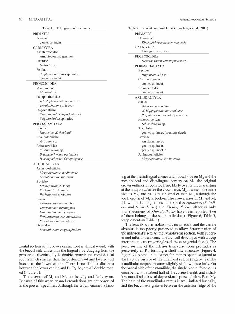
zontal section of the lower canine root is almost ovoid, with the buccal side wider than the lingual side. Judging from the preserved alveolus, P3 is double rooted: the mesiobuccal root is much smaller than the posterior root and located just buccal to the lower canine. There is no distinct diastema between the lower canine and P3. P4–M3 are all double-root-ed (Figure 5).
The crowns of M2 and M3 are heavily and flatly worn. Because of this wear, enamel crenulations are not observed in the present specimen. Although the crown enamel is lack-
ing at the mesiolingual corner and buccal side on M2 and the mesiobuccal and distolingual corners on M3, the original crown outlines of both teeth are likely oval without waisting at the midpoint. As for the crown area, M2 is almost the same size as M3, and M1 is much smaller than M2, although the tooth crown of M1 is broken. The crown sizes of M2 and M3 fall within the range of medium-sized Sivapithecus (S. indi-cus and S. sivalensis) and Khoratpithecus, although only four specimens of Khoratpithecus have been reported (two of them belong to the same individual) (Figure 6, Table 3, Supplementary Table 1).
The heavily worn molars indicate an adult, and the canine alveolus is too poorly preserved to allow determination of the individual’s sex. At the symphyseal section, both superi-or and inferior transverse tori are well developed with a deep intertoral sulcus (= genioglossal fossa or genial fossa). The posterior end of the inferior transverse torus protrudes as posteriorly as P4, forming a shelf-like structure (Figure 4, Figure 7). A small but distinct foramen is open just lateral to the fracture surface of the intertoral sulcus (Figure 4e). The mandibular corpus becomes slightly shallow posteriorly. On the buccal side of the mandible, the single mental foramen is open below P3, at about half of the corpus height, and a shal-low mandibular buccal depression is present below P4 to M1. The base of the mandibular ramus is well inflated buccally, and the buccinator groove between the anterior ridge of the
Table 1. Tebingan mammal fauna.
PRIMATESPonginae
gen. et sp. indet.CARNIVORA
AmphicyonidaeAmphicyoninae gen. nov.
UrsiidaeIndarctos sp.
FelidaeAmphimachairodus sp. indet.gen. et sp. indet.
PROBOSCIDEAMammutidae
Mammut sp.Gomphotheriidae
Tetralophodon cf. xiaohensisTetralophodon sp. indet.
StegodontidaeStegolophodon stegodontoidesStegolophodon sp. indet.
PERISSODACTYLAEquidae
Hipparion cf. theobaldiChalicotheriidae
Anisodon sp.Rhinocerotidae
cf. Rhinoceros sp.Brachypotherium perimenseBrachypotherium fatehjangense
ARTIODACTYLAAnthracotheriidae
Merycopotamus medioximusMicrobunodon milaensis
BovidaeSelenoportax sp. indet.Pachyportax latidensPachyportax giganteus
SuidaeTetraconodon irramediusTetraconodon irramagnusHippopotamodon sivalensePropotamochoerus hysudricusPropotamochoerus cf. wui
GiraffidaeBramatherium megacephalum
Table 2. Yinseik mammal fauna (from Jaeger et al., 2011).
PRIMATESHominidae
Khoratpithecus ayeyarwadyensisCARNIVORA
Fam. gen. et sp. indet.PROBOSCIDEA
Stegolophodon/Tetralophodon sp.PERISSODACTYLA
EquidaeHipparion (s.l.) sp.
Chalicotheriidaegen. et sp. indet.
Rhinocerotidaegen. et sp. indet.
ARTIODACTYLASuidae
Tetraconodon minorcf. Hippopotamodon sivalensePropotamochoerus cf. hysudricus
PalaeochoeridaeSchizochoerus sp.
Tragulidaegen. et sp. Indet. (medium-sized)
BovidaeAntilopini indet.gen. et sp. indet.gen. et sp. indet. 2
AnthracotheriidaeMerycopotamus medioximus

EARLY LATE MIOCENE HOMINOID MANDIBLE FROM MYANMAR 91
ramus and M3 is rather wide. At the lingual side of the man-dible, a distinct mylohyoid line extends straight from about
1 cm below M3 to the inferior transverse torus of the man-dibular symphysis (Figure 4c, Figure 5b, Figure 8). In the
Figure 3. Photographs of MZKB-K-001 in lingual (a), buccal (b), occlusal (c), ventral (d), anterior (e), posterior (f), and sagittal (g) views. Scale bar, 1 cm.
Figure 4. Reconstructed 3D image of MZKB-K-001 using X-ray CT data in occlusal (a), ventral (b), lingual (c), buccal (d), posterior (e), and anterior (f) views. The white arrow indicates the presence of the midline ridge (b).
Figure 5. Reconstructed 3D images of the mandibular corpus and lower dentition of MZKB-K-001 using X-ray CT data. Mandibular corpus displaying roots shadows for P4–M3 in buccal (a), mesiolingual (b, perpendicular to the sagittal section of the mandibular symphysis) and lingual (c) views. Dental row with virtually extracted roots in buc-cal (d), ventral (e), and lingual (f) views.
Vol. 129, 2021
M. TAKAI ET AL.92 Anthropological Science
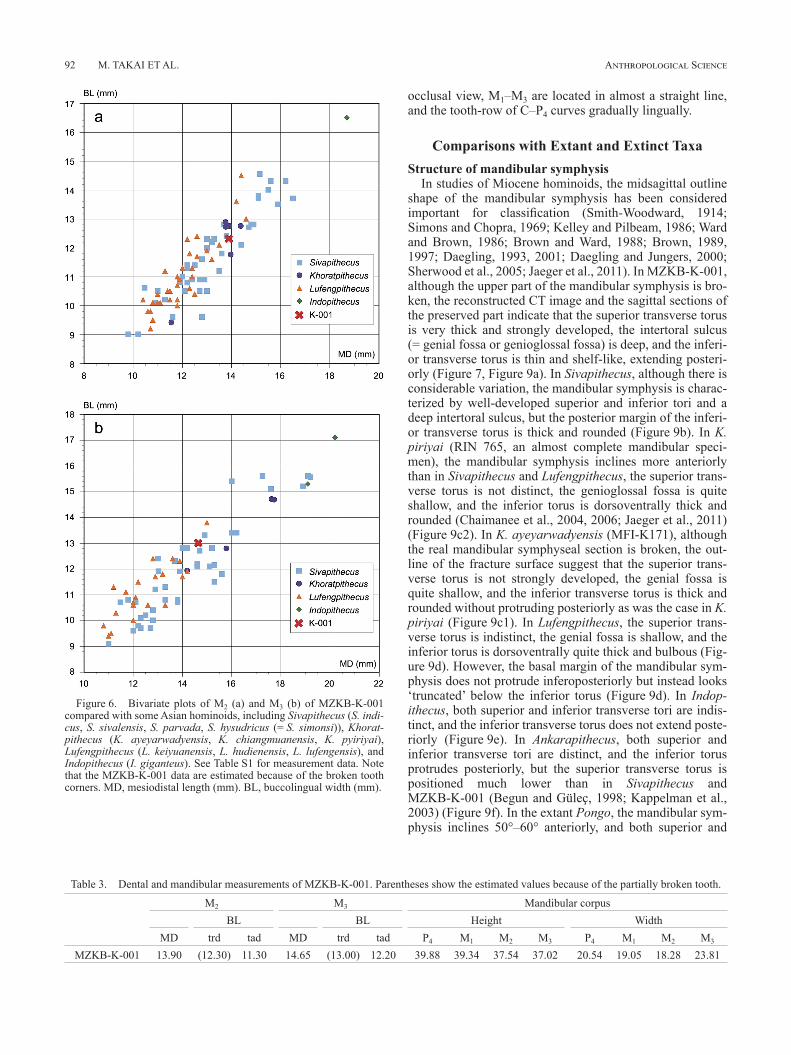
occlusal view, M1–M3 are located in almost a straight line, and the tooth-row of C–P4 curves gradually lingually.
Comparisons with Extant and Extinct TaxaStructure of mandibular symphysis
In studies of Miocene hominoids, the midsagittal outline shape of the mandibular symphysis has been considered important for classification (Smith-Woodward, 1914; Simons and Chopra, 1969; Kelley and Pilbeam, 1986; Ward and Brown, 1986; Brown and Ward, 1988; Brown, 1989, 1997; Daegling, 1993, 2001; Daegling and Jungers, 2000; Sherwood et al., 2005; Jaeger et al., 2011). In MZKB-K-001, although the upper part of the mandibular symphysis is bro-ken, the reconstructed CT image and the sagittal sections of the preserved part indicate that the superior transverse torus is very thick and strongly developed, the intertoral sulcus (= genial fossa or genioglossal fossa) is deep, and the inferi-or transverse torus is thin and shelf-like, extending posteri-orly (Figure 7, Figure 9a). In Sivapithecus, although there is considerable variation, the mandibular symphysis is charac-terized by well-developed superior and inferior tori and a deep intertoral sulcus, but the posterior margin of the inferi-or transverse torus is thick and rounded (Figure 9b). In K. piriyai (RIN 765, an almost complete mandibular speci-men), the mandibular symphysis inclines more anteriorly than in Sivapithecus and Lufengpithecus, the superior trans-verse torus is not distinct, the genioglossal fossa is quite shallow, and the inferior torus is dorsoventrally thick and rounded (Chaimanee et al., 2004, 2006; Jaeger et al., 2011) (Figure 9c2). In K. ayeyarwadyensis (MFI-K171), although the real mandibular symphyseal section is broken, the out-line of the fracture surface suggest that the superior trans-verse torus is not strongly developed, the genial fossa is quite shallow, and the inferior transverse torus is thick and rounded without protruding posteriorly as was the case in K. piriyai (Figure 9c1). In Lufengpithecus, the superior trans-verse torus is indistinct, the genial fossa is shallow, and the inferior torus is dorsoventrally quite thick and bulbous (Fig-ure 9d). However, the basal margin of the mandibular sym-physis does not protrude inferoposteriorly but instead looks ‘truncated’ below the inferior torus (Figure 9d). In Indop-ithecus, both superior and inferior transverse tori are indis-tinct, and the inferior transverse torus does not extend poste-riorly (Figure 9e). In Ankarapithecus, both superior and inferior transverse tori are distinct, and the inferior torus protrudes posteriorly, but the superior transverse torus is positioned much lower than in Sivapithecus and MZKB-K-001 (Begun and Güleç, 1998; Kappelman et al., 2003) (Figure 9f). In the extant Pongo, the mandibular sym-physis inclines 50°–60° anteriorly, and both superior and
Table 3. Dental and mandibular measurements of MZKB-K-001. Parentheses show the estimated values because of the partially broken tooth.
M2 M3 Mandibular corpusBL BL Height Width
MD trd tad MD trd tad P4 M1 M2 M3 P4 M1 M2 M3
MZKB-K-001 13.90 (12.30) 11.30 14.65 (13.00) 12.20 39.88 39.34 37.54 37.02 20.54 19.05 18.28 23.81
Figure 6. Bivariate plots of M2 (a) and M3 (b) of MZKB-K-001 compared with some Asian hominoids, including Sivapithecus (S. indi-cus, S. sivalensis, S. parvada, S. hysudricus (= S. simonsi)), Khorat-pithecus (K. ayeyarwadyensis, K. chiangmuanensis, K. pyiriyai), Lufengpithecus (L. keiyuanensis, L. hudienensis, L. lufengensis), and Indopithecus (I. giganteus). See Table S1 for measurement data. Note that the MZKB-K-001 data are estimated because of the broken tooth corners. MD, mesiodistal length (mm). BL, buccolingual width (mm).

EARLY LATE MIOCENE HOMINOID MANDIBLE FROM MYANMAR 93
inferior transverse torus are well developed with moderately deep genial fossa. The inferior transverse torus extends pos-teriorly to P4–M1, although with considerable shape varia-tions between individuals (Figure 9g). Sherwood et al. (2005) reported low sexual dimorphism in the symphyseal outline shape of extant Pongo.
Thus, concerning its mandibular symphyseal outline shape, MZKB-K-001 differs from the pattern seen in the two extinct Southeast Asian hominoids, Khoratpithecus and
Figure 7. Sagittal sections (a1–a6) of the mandibular symphyseal part of MZKB-K-001. The most mesial (a1) and most lateral (a6) sections are indicated by vertical lines at the bottom. Although the superior part of the mandibular symphysis is lost in MZKB-K-001, a strongly developed superior transverse torus is continuously observed from (a6) through (a2), and a thin, flat, posteriorly extending inferior transverse torus is observ-able even at the (a2) section.
Figure 8. Horizontal sections (a1–a3) of the mandibular corpus of MZKB-K-001. The positions of (a1, lowest section), (a2), and (a3) are indicated by lines in the bottom. The midline ridge protrudes posterior-ly at the mediolingual corner of the mandibular corpus (indicated by the arrow in a1).
Figure 9. Comparisons of the outline of the symphyseal section of MZKB-K-001 (a) with several Late Miocene hominoids in Asia: S. sivalensis (b1, GSP9564; b2, GSP13875); K. ayeyarwadyensis (c1, MFI-K171) and K. piriyai (c2, RIN 765); L. lufengensis (d1, PA895; d2, PA548); I. giganteus (e, CYP359/68); Ankarapithecus meteai (f, AS-95-500); and average shape of Pongo (g1, male; g2, female). The images in (b–d) were redrawn from Jaeger et al. (2011), (e) from Xu and Lu (2008), (f) from Kappelman et al. (2003), and (g) from Sher-wood et al. (2005). Symphyses not to scale.
Vol. 129, 2021

M. TAKAI ET AL.94 Anthropological Science
Lufengpithecus. However, it shows some similarity to Sivapithecus and extant Pongo in having well-developed superior and inferior transverse tori and a deep genioglossal fossa. The variation pattern seen in extinct hominoids sug-gests that the development of the superior transverse torus and the deepening of the genioglossal fossa may correlate to each other (Daegling, 1993).
Distinct impressions or fossae for the anterior digastric muscle are usually observed at the base of mandibular sym-physis in humans and African great apes. These are not seen in the Asian great apes (Pongo) because their anterior digas-tric muscle is absent (Smith-Woodward, 1914; Brown and Ward, 1988; Brown, 1989). In fossil taxa the digastric fossae are not observed in the two mandibles of Khoratpithecus (Chaimanee et al., 2004, 2006; Jaeger et al., 2011) but are reported in Lufengpithecus (Schwartz, 1997; Xu and Lu, 2008), Ankarapithecus (Begun and Güleç, 1998; Kappelman et al., 2003), and in many specimens of Sivapithecus (Ward and Brown, 1986; Brown and Ward, 1988; Brown, 1989). In MZKB-K-001 no distinct impressions are observed at the basal lingual surface of the inferior transverse torus (Fig-ure 3d, Figure 4e), suggesting the lack of an anterior digas-tric muscle in this individual, as in extant Pongo. However, there is only one sample of the Tebingan hominoid at pres-ent.
Dentition, dental arcade, and mandibular corpusAmong the Middle to Late Miocene Asian hominoids,
including Sivapithecus, Khoratpithecus, Lufengpithecus, Indopithecus, and Ankarapithecus, the lower canine and premolars are usually positioned more laterally than M1 and M2 in the cheek tooth row, and the major axis of M3 often rotates distolaterally (Figure 10a, e–g). Therefore, the straight or buccally concave cheek tooth row is retained in most specimens of Sivapithecus (e.g. GSP4622, GSP6160, GSP15000, GSP16077, AMNH 19412), Khoratpithecus
(RIN765 and MFI-K171) (Chaimanee et al., 2004, 2006; Jaeger et al., 2011), Lufengpithecus (PA580, PA948) (Xu and Lu, 2008), Indopithecus (CYP359/68) (Simons and Chopra, 1969), and Ankarapithecus (AS95-500) (Kappel-man et al., 2003) (Figure 11). It is notable that some of them are considered female specimens based on the size and shape of canine and/or P3: namely, Sivapithecus (GSP6160), Lufengpithecus (PA580), and Indopithecus (CYP359/68). In contrast, MZKB-K-001 preserves a buccally convex cheek tooth row because the alveoli of canine and premolars are situated more medially than molars and M3 does not rotate distolaterally (Figure 3c). Interestingly, one female Giganto-pithecus mandibular specimen (Mandible I, PA77) shows a parabolic dental arcade with relatively small lower incisors as seen in MZKB-K-001 (Zhang and Harrison, 2017, figure 3D).
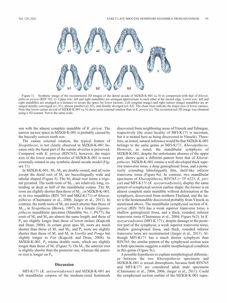
In Figure 11, several patterns of the synthetic dental ar-cade of MZKB-K-001 are presented using the mirror image. The left and right corpora are arranged more distally di-verged in a3 and b3 than in a1 and b1. In the a1–a3 models the corpora are arranged adjacent to each other, because the inferior mesiolingual corner of the symphyseal section pro-trudes distally, suggesting the presence of the midline ridge at the mandibular symphysis (indicated by the arrows in Figure 4b and Figure 8a1). The space between the lower canines might be too small for lower four incisors in these models, however, and therefore in the b1–b3 models, the corpora are arranged more widely separated to ensure space for lower incisors. In these models the assumption is that a relatively large midline ridge would be present at the sym-physeal part of the mandible (Figure 11).
Judging from the preserved space of the incisor alveoli, the lower incisors of MZKB-K-001 would have been rela-tively small, although the incisal alveolar region is missing. Even in the widely separated models (Figure 11b), the pre-sumed space for lower incisors is very narrow, in compari-
Figure 10. Comparisons of the reconstructed 3D images of the left cheek tooth row of several Asian Late Miocene hominoids, including Sivapithecus spp. (a–c), the Tebingan hominoid (d), Khoratpithecus piriyai (e), Lufengpithecus lufengensis (f), and Indopithecus giganteus (g). (a) GSP6160 (P3–M3, female, mirror image); (b) AMNH19412 (P3–M3, male); (c) GSP15000 (C1–M3, male); (d) MZKB-K-001 (M2–3 and roots of P4–M1); (e) RIN765 (P3–M3, male); (f) PA580 (P3–M3, female); and (g) CYP 359/68 (P3–M3, female, mirror image). Scale bar, 2 cm. The recon-structed 3D image was obtained from the casts of fossil specimens using a one-shot 3D scanner.
EARLY LATE MIOCENE HOMINOID MANDIBLE FROM MYANMAR 95
son with the almost complete mandible of K. piriyai. The narrow incisor space in MZKB-K-001 is probably caused by the buccally convex tooth row.
The canine external rotation, the typical feature of Sivapithecus, is not clearly observed in MZKB-K-001 be-cause only the basal part of the canine alveolus is preserved. Compared with K. piriyai (RIN765), however, the major axis of the lower canine alveolus of MZKB-K-001 is more externally rotated in any synthetic dental arcade model (Fig-ure 11).
In MZKB-K-001, M1–M3 are double rooted, and all roots except the distal root of M3 are buccolingually wide and tabular shaped (Figure 5). The M3 distal root forms a trigo-nal pyramid. The tooth roots of M2–3 are relatively long, ex-tending as deep as half of the mandibular corpus. The M1 roots are slightly shorter than those of M2–3 in MZKB-K-001, as in two mandibles (RIN 765 and MKI-K171) of Khorat-pithecus (Chaimanee et al., 2006; Jaeger et al., 2011). In contrast, the tooth roots of M1 are much shorter than those of M2–3 in Sivapithecus (Brown, 1997). In a female Giganto-pithecus mandibular specimen (Mandible No. 1, PA77), the roots of M1 and M2 are almost the same length, and those of P4 are slightly longer than those of lower molars (Kupczik and Dean, 2008). In extant great apes M3 roots are much shorter than those of M1 and M2, and P4 roots are slightly shorter than those of M1 and M2 in Gorilla and Pongo but slightly longer in Pan (Kupczik and Dean, 2008). In MZKB-K-001, P4 retains double roots, which are slightly longer than those of M1 (Figure 5). On M1, the anterior root is slightly shorter than the posterior one, whereas the anteri-or root is longer on P4.
DiscussionMFI-K171 (K. ayeyarwadyensis) and MZKB-K-001 are
left mandibular corpora of the medium-sized hominoids
discovered from neighboring areas of Yinseik and Tebingan, respectively (the exact locality of MFI-K171 is uncertain, but it is treated here as being discovered in Yinseik). There-fore, an initial, natural inference would be that MZKB-K-001 belongs to the same genus as MFI-K171, Khoratpithecus. However, as noted, the mandibular symphysis of MZKB-K-001, despite the unfortunate absence of the upper part, shows quite a different pattern from that of Khorat-pithecus. MZKB-K-001 retains a well-developed thick supe-rior transverse torus, a deep genioglossal fossa, and a poste-riorly extending labiolingually thin, shelf-like inferior transverse torus (Figure 9a). In contrast, two mandibular specimens of Khoratpithecus, RIN765 (= TF 6223, K. piri-yai) and MFI-K171 (K. ayeyarwadyensis), display the same pattern of symphyseal section outline shape: the former is an almost complete male mandible without deformation at the symphysis, discovered from northern Thailand, and the lat-ter is the hemimandible discovered probably from Yinseik as mentioned above. The mandibular symphyseal section of K. piriyai (RIN 765) has a weak superior transverse torus, a shallow genioglossal fossa, and a thick, rounded, inferior transverse torus (Chaimanee et al., 2004; Figure 9c2). In K. ayeyarwadyensis (MFI-K 171), despite damage to the poste-rior part of the symphysis, a weak superior transverse torus, shallow genioglossal fossa, and thick, rounded inferior transverse torus are reconstructed (Jaeger et al., 2011). Al-though MFI-K171 has a much shorter symphysis than RIN765, the similar pattern of the symphyseal section seen in both specimens suggests a stable morphological condition in this genus (Figure 9c).
A possible hypothesis to explain morphological differenc-es between the two Khoratpithecus specimens and MZKB-K-001 is sexual dimorphism, because both RIN765 and MFI-K171 are considered adult male specimens (Chaimanee et al., 2004, 2006; Jaeger et al., 2011). Could the symphyseal section outline of the MZKB-K-001 repre-
Figure 11. Synthetic image of the reconstructed 3D images of the dental arcade of MZKB-K-001 (a, b) in comparison with that of Khorat-pithecus piriyai (RIN 765, c). Upper row: left and right mandibles are arranged approximate to each other at the mesial edge. Lower row: left and right mandibles are arranged at a distance to secure the space for lower incisors. Left (original image) and right (mirror image) mandibles are ar-ranged distally converged (a1, b1), almost parallel (a2, b2), and distally diverged (a3, b3). The chain lines indicate the major axis of lower canines. Note that lower canine alveoli of MZKB-K-001 (a, b) show more external rotation than in K. piriyai (c). The reconstructed 3D image was obtained using a 3D scanner. Not to the same scale.
Vol. 129, 2021
M. TAKAI ET AL.96 Anthropological Science
sent a female feature of Khoratpithecus? Plenty of mandibu-lar specimens of Sivapithecus discovered to date do not support this hypothesis: the combination of the well-devel-oped superior and inferior transverse tori and deep intertoral sulcus is observed in some male specimens (e.g. GSP9564, GSP15000). Therefore, sexual dimorphism of Khorat-pithecus is implausible as an explanation for the morpholog-ical difference between MZKB-K-001 and RIN765. The Tebingan hominoid MZKB-K-001 is likely to be a different taxon from Khoratpithecus, although we still have just one mandibular specimen and far from a reliable identification for it.
If the Tebingan hominoid belongs to a different genus from Khoratpithecus, another question is now on the table regarding the distribution of early Late Miocene hominoids in central Myanmar. As noted, Yinseik and Tebingan are neighboring areas, and both horizons yielding mammal fos-sils correspond to the lowermost part of the Irrawaddy For-mation, suggesting a similar age. Were there two kinds of medium-sized hominoids distributed in the same area in central Myanmar during the early Late Miocene? Answering this question requires additional specimens preserving the complete or almost complete mandibular symphysis for identification of the taxonomic position of the Tebingan hominoid.
AcknowledgmentsWe are grateful to Mr. U Khin Shwe, the director of Zayk-
abar Museum, Yangon, for allowing us to study the present hominoid specimen. We also thank the staff of the Depart-ment of Archaeology and National Museum for their help in our paleontological research in Myanmar, and all Japanese research members who participated in the fieldwork in My-anmar. We thank Dr. Makoto Manabe and the National Mu-seum of Nature and Science, Tokyo, for allowing us to use their research facilities. We thank Dr. Jay Kelley of Arizona State University for his precious advice and for graciously providing access to the casts and original specimens of Sivapithecus at their respective institutions. We also thank Dr. Pratueng Jintasakul, Director of the Northeastern Re-search Institutes of Petrified Wood and Mineral Resources, Nakhon Ratchasima Rajabhat University, for providing us with the cast of K. piriyai (RIN 765). This study was partly supported by JSPS Grant-in-Aid for Scientific Research (#26304019 to MT).
ReferencesAlpagut B., Andrews P., Fortelius M., Kappelman J., Temizsoy I.,
Çekibi H., and Lindsay W. (1996) A new specimen of Ankarapithecus meteai from the Sinap Formation of central Anatolia. Nature, 382: 349–351.
Andrews P. and Tekkaya I. (1980) A revision of the Turkish Mio-cene hominoid Sivapithecus meteai. Paleontology, 23: 85–95.
Barry J.C., Morgan M.E., Flynn L.J., Pilbeam D.R., Behrensmeyer A.K., Raza S.M., Raza M., Khan I.A., Badgley C., Hicks J., and Kelley J. (2002) Faunal and environmental change in the Late Miocene Siwaliks of northern Pakistan. Paleobiology, 28(3): 1–71.
Begun D.R. (2015) Fossil Record of Miocene Hominoids. In: Henke W., Tattersall I., (eds), Handbook of Paleoanthropolo-
gy, Vol. III. Springer, Berlin, pp. 1261–1332.Begun D.R. and Güleç¸ E. (1998) Restoration of the type and palate
of Ankarapithecus meteai: taxonomic and phylogenetic impli-cations. American Journal of Physical Anthropology, 105: 279–314.
Bender F. (1983) Geology of Burma. Gebrüder Bortraeger, Berlin.Bhandari A., Kay R.F., Williams B.A., Tiwari B.N., Bajpai S., and
Hieronymus T. (2018) First record of the Miocene hominoid Sivapithecus from Kutch, Gujarat state, western India. PLoS One, 13(11): e0206314.
Brown B. (1989) The mandibles of Sivapithecus. PhD thesis, Kent State University, OH.
Brown B. (1997) Miocene hominoid mandible: functional and phy-logenetic perspectives. In: Begun D.R., Ward C.V., and Rose M.D., (eds.), Function, Phylogeny, and Fossils: Miocene Hominoid Evolution and Adaptations. Plenum Press, New York, pp. 153–171.
Brown B. and Ward S.C. (1988) Basicranial and facial topography in Pongo and Sivapithecus. In: Schwartz J.H. (ed.), Orang-utan Biology. Oxford University Press, New York, pp. 247–260.
Chaimanee Y., Jolly D., Benammi M., Tafforeau P., Duzer D., Moussa I., and Jaeger J.-J. (2003) A Middle Miocene homi-noid from Thailand and orangutan origins. Nature, 422: 61–65.
Chaimanee Y., Suteethorn V., Jintasakul P., Vidthayanon C., Marandat B., and Jaeger J.-J. (2004) A new orang-utan rela-tive from the Late Miocene of Thailand. Nature, 427: 439–441.
Chaimanee Y., Yamee C., Tian P., Khaowiset K., Marandat B., Taf-foreau P., Nemoz C., and Jaeger J.-J. (2006) Khoratpithecus piriyai, a late Miocene hominoid of Thailand. American Jour-nal of Physical Anthropology, 131(3): 311–323.
Chavasseau O., Chaimanee Y., Soe-Thura-Tun, Aung-Naing-Soe, Barry J.C., Marandat B., Sudre J., Marivaux L., Ducrocq S., and Jaeger J.-J. (2006) Chaungtha, a new Middle Miocene mammal locality from the Irrawaddy Formation, Myanmar. Journal of Asian Earth Sciences, 28: 354–362.
Chavasseau O., Aung Aung Kyaw, Chaimanee Y., Coster P., Emonet E.-G., Aung Naing Soe, Rugumbrung M., Soe Thura Tun, and Jaeger J.-J. (2013) Advances in the biochronology and biostratigraphy of the continental Neogene of Myanmar. In: Wang X., Flynn L.J., and Fortelius M. (eds.), Fossil Mam-mals of Asia: Neogene Biostratigraphy and Chronology. Co-lumbia University Press, New York, pp. 461–474.
Chit Sein, van der Made J., and Rössner G. (2009) New material of Propotamochoerus (Suidae, Mammalia) from the Irrawaddy Formation, Myanmar. Neues Jahrbuch für Geologie und Paläontologie-Abhandlungen, 251: 17–31.
Colbert E.H. (1938) Fossil mammals from Burma in the American Museum of Natural History. Bulletin of the American Muse-um of Natural History, 74: 255–436.
Colbert E.H. (1943) Pleistocene vertebrates collected in Burma by the American Southeast Asiatic Expedition. Transactions of the American Philosophical Society, New Series, 32: 395–429.
Daegling D.J. (1993) Shape variation in the mandibular symphysis of apes: an application of a median axis method. American Journal of Physical Anthropology, 91: 505–516.
Daegling D.J. (2001) Biomechanical scaling of the hominoid man-dibular symphysis. Journal of Morphology, 250: 12–23.
Daegling D.J. and Jungers W.L. (2000) Elliptical Fourier analysis of symphyseal shape in great ape mandibles. Journal of Hu-man Evolution, 39: 107–122.
Gregory W., Hellman M., and Lewis E. (1935) Fossil anthropoids of the Yale–Cambridge India Expedition of 1935. Washington, DC, Carnegie Institute.
Jaeger J.-J., Soe A.N., Chavasseau O., Coster P., Emonet E.-G., Guy F., Lebrun R., Maung A., Khyaw A.A., Shwe H., Tun S.T., Oo K.L., Rugbumrung M., Bocherens H., Benammi M.,
EARLY LATE MIOCENE HOMINOID MANDIBLE FROM MYANMAR 97
Chaivanich K., Tafforeau P., and Chaimanee Y. (2011) First hominoid from the Late Miocene of the Irrawaddy Formation (Myanmar). PLoS One, 011; 6: e17065.
Kappelman J., Richmond B.G., Seiffert E.R., Maga A.M., and Ryan T.M. (2003) Hominoidea (Primates). In: Fortelius M., Kappelman J., Sen S., and Bernor R.L. (eds.), Geology and Paleontology of the Miocene Sinap Formation, Turkey. Columbia University Press, New York, pp. 90–124.
Kay R.F. (1982) Sivapithecus simonsi, a new species of Miocene hominoid, with comments on the phylogenetic status of the Ramapithecinae. International Journal of Primatology, 3: 113–174.
Kelley J. (2002) The hominoid radiation in Asia. In: Hartwig W. (ed.), The Primate Fossil Record. Cambridge University Press, Cambridge, pp. 369–384.
Kelley J. (2005) Twenty-five years contemplating Sivapithecus taxonomy. In: Smith R.J., Kelley J., and Lieberman D.E. (eds.), Interpreting the Past: Essays on Human, Primate, and Mammal Evolution in Honor of David Pilbeam. Brill Aca-demic, Boston, MA, pp. 123–143.
Kelley J. and Pilbeam D.R. (1986) The Dryopithecines: taxonomy, comparative anatomy, and phylogeny of Miocene large homi-noids. In: Swindler D. and Irwin J. (eds.), Comparative Pri-mate Biology, Vol. I: Systematics, Evolution, and Anatomy. Alan R. Liss, New York, pp. 361–411.
Kupczik K. and Dean M.C. (2008) Comparative observations on the tooth root morphology of Gigantopithecus blacki. Journal of Human Evolution, 54: 196–204.
Lu Q. (1998) Dental sexual dimorphism and chewing efficiency of Lufengpithecus. In: Wang T. (ed.), Collected Works for “The 30 Anniversary of Yuanmou Man Discovery and the Inter-national Conference on Palaeoanthropological Studies.” Yunnan Science & Technology Press, Kunming, pp. 100–107 (in Chinese with English abstract)
Lydekker R. (1879) Further notices of Siwalik Mammalia. Records of the Geological Survey of India, 7: 33–52.
Madar S., Rose M.D., Kelley J., MacLatchy L., and Pilbeam D. (2002) New Sivapithecus postcranial specimens from the Siwaliks of Pakistan. Journal of Human Evolution, 42: 705–752.
Munthe J., Dongol B., Hutchison J.H., Kean W.F., Munthe K., and West R.M. (1983) New fossil discoveries from the Miocene of Nepal include a hominoid. Nature, 303: 331–333.
Naing Maw Than, Khi Khin, and Myint Thein (2017) Cretaceous geology of Myanmar and Cenozoic geology in the Central Myanmar Basin. In: Barber A.J., Khin Zaw, and Crow M.J. (eds.), Myanmar: Geology: Resources and Tectonics. Geolog-ical Society, London, Memoirs, 48: 143–167.
Nishioka Y., Takai M., Nishimura T., Thaung-Htike, Zin Maung Maung Thein, Egi N., Tsubamoto T., and Maung Maung (2015) Plio-Pleistocene rodents (Mammalia) from the Irrawaddy sediments of central Myanmar and palaeogeo-graphical significance. Journal of Systematic Palaeontology, 13: 287–314.
Pickford M. (2010) Additions to the Dehm collection of Siwalik hominoids, Pakistan: descriptions and interpretations. Zittelia-na, A 50: 111–125.
Pilbeam D. (1997) Research on Miocene hominoids and hominid origins: the last three decades. In: Begun D.R., Ward C.V., and Rose M.D. (eds.), Function, Phylogeny and Fossils: Miocene Hominoid Evolution and Adaptations. Plenum Press, New York, pp. 13–28.
Pilbeam D., Rose M., Barry J.C., and Shah S. (1990) New Sivapithecus humeri from Pakistan and the relationship of Sivapithecus and Pongo. Nature, 348: 237–239.
Pilgrim G.E. (1915) New Siwalik primates and their bearing on the question of the evolution of man and the Anthropoidea. Re-cords of the Geological Survey of India, 45: 1–74.
Pilgrim G.E. (1927) A Sivapithecus palate and other primate fossils from India. Memoirs of the Geological Survey of India, New
Series, 14: 1–26.Pilgrim G.E. (1937) Siwalik antelopes and oxen in the American
Museum of Natural History. Bulletin of American Museum of Natural History, 72: 729–874.
Pilgrim G.E. (1939) The fossil Bovidae of India. Palaeontologia Indica, 26: 1–356.
Qi G. and Dong W. (2006) Lufengpithecus hudienensis site. Sci-ence Press, Beijing.
Richmond B.G. and Whalen M. (2001) Forelimb function, bone curvature, and phylogeny of Sivapithecus. In: de Bonis L., Koufos G., and Andrews P. (eds.), Phylogeny of the Neogene Hominoid Primates of Eurasia. Cambridge University Press, Cambridge, pp. 326–348.
Schwartz J.H. (1997) Lufengpithecus and hominoid phylogeny: problems in delineating and evaluating phylogenetically rele-vant characters. In: Begun D., Ward C., and Rose M. (eds.), Function, Phylogeny, and Fossils: Miocene Hominoid Evolu-tion and Adaptations. Plenum Press, New York, pp. 363–388.
Sherwood R., Hlusko L.J., Duren D.L., Emch V.C., and Walker A. (2005) Mandibular symphysis of large-bodied hominoids. Human Biology, 77(6): 735–759.
Simons E.L. and Chopra S.R.K. (1969) Gigantopithecus (Pongi-dae, Hominoidea) a new species from North India. Postilla, Yale Peabody Museum, 138: 1–18.
Smith-Woodward A. (1914) On the lower jaw of an anthropoid ape (Dryopithecus) from the upper Miocene of Lerida (Spain). Quarterly Journal of the Geological Society, 70: 316–320.
Stamp L.D. (1922) An outline of the tertiary geology of Burma. Geological Magazine, 59: 481–501.
Takai M., Thaung-Htike, Zin Maung Maung Thein, Soe A.N., Maung Maung, Tsubamoto T., Egi N., Nishimura T.D., and Nishioka Y. (2015) First discovery of colobine fossils from the Late Miocene/Early Pliocene of central Myanmar. Journal of Human Evolution, 84: 1–15.
Takai M., Nishioka Y., Thaung Htike, Maung Maung, Kyaw Khaing, Zin Maung Maung Thein, Tsubamoto T., and Egi N. (2016) Late Pliocene Semnopithecus fossil from central My-anmar: rethinking of the evolutionary history of cercopithecid monkeys in Southeast Asia. Historical Biology, 28: 171–187.
Thaung Htike and Takai M. (2016) Reevaluation of the phylogeny and taxonomy of the Asian fossil hippopotamuses. Universi-ties Research Journal, 8(1): 171–177.
Tsung Aung (1980) Geological Map of Central Myanmar Basin. Myanmar Oil and Gas Enterprises.
Ward S. (1997) The taxonomy and phylogenetic relationships of Sivapithecus revisited. In: Begun D., Ward C., and Rose M. (eds.), Function, Phylogeny, and Fossils: Miocene Hominoid Evolution and Adaptations. Plenum Press, New York, pp. 269–290.
Ward S. and Brown B. (1986) The facial skeleton of Sivapithecus indicus. In: Swindler D.R. and Erwin J. (eds.), Comparative Primate Biology, Vol. I: Systematics, Evolution, and Anatomy. Alan R. Liss, New York, pp. 413–452.
Woo R. (1957) Dryopithecus teeth from Keiyuan, Yunnan Province. Vertebrata PalAsiatica, 1: 25–31, plate 1.
Woo R. (1958) New materials of Dryopithecus from Keiyuan, Yunnan. Vertebrata PalAsiatica, 2: 38–42, plate 1.
Wu R. and Oxnard C.E. (1983) Ramapithecines from China: evi-dence from tooth dimensions. Nature, 306: 258–260.
Xu Q. and Lu Q. (2008) Lufengpithecus lufengensis: an early mem-ber of Hominidae. Science Press, Beijing.
Zhang Y. and Harrison T. (2017) Gigantopithecus blacki: a giant ape from the Pleistocene of Asia revisited. American Journal of Physical Anthropology, 162: 153–177.
Zin Maung Maung Thein, Takai M., Tsubamoto T., Thaung Htike, Egi N., and Maung Maung (2008) A new species of Dicerorhinus (Rhinocerotidae) from the Plio-Pleistocene of Myanmar. Palaeontology, 51: 1419–1433.
Zin Maung Maung Thein, Takai M., Uno H., Wynn J.G., Egi N., Tsubamoto T., Thaung-Htike, Aung Naing Soe, Maung
Vol. 129, 2021

M. TAKAI ET AL.98 Anthropological Science
Maung, Nishimura T., and Yoneda M. (2011) Stable isotope analysis of the tooth enamel of Chaingzauk mammalian fauna (late Neogene, Myanmar) and its implication to paleoenviron-ment and paleogeography. Palaeogeography, Palaeoclimatolo-gy, Palaeoecology, 300: 11–22.
Zin Maung Maung Thein, Thaung Htike, Aung Naing Soe, Chit
Sein, Maung Maung, and Takai M. (2017) A review of the in-vestigation of primate fossils in Myanmar. In: Barber A.J., Khin Zaw, and Crow M.J. (eds.), Myanmar: Geology: Re-sources and Tectonics. Geological Society, London, Memoirs, 48: 185–206.
Related Documents