Journal of Clinical Medicine Review New and Innovative Treatments for Neovascular Age-Related Macular Degeneration (nAMD) Prem Patel 1 and Veeral Sheth 2, * Citation: Patel, P.; Sheth, V. New and Innovative Treatments for Neovascular Age-Related Macular Degeneration (nAMD). J. Clin. Med. 2021, 10, 2436. https://doi.org/ 10.3390/jcm10112436 Academic Editor: Laurent Kodjikian Received: 20 April 2021 Accepted: 28 May 2021 Published: 30 May 2021 Publisher’s Note: MDPI stays neutral with regard to jurisdictional claims in published maps and institutional affil- iations. Copyright: © 2021 by the authors. Licensee MDPI, Basel, Switzerland. This article is an open access article distributed under the terms and conditions of the Creative Commons Attribution (CC BY) license (https:// creativecommons.org/licenses/by/ 4.0/). 1 Department of Ophthalmology, University of Texas Southwestern Medical Center, Dallas, TX 75390, USA; [email protected] 2 University Retina and Macula Associates, Oak Forest, IL 60452, USA * Correspondence: [email protected] Abstract: Age-related macular degeneration (AMD) is one of the most common causes of vision loss. Advanced forms of AMD are seen in primarily two types—neovascular AMD (nAMD) with the presence of choroid neovascularization and non-neovascular AMD (nnAMD) with geographic atrophy. Neovascular AMD is characterized by choroidal neovascularization (CNV), which leads to a cascade of complications, including exudation, leakage, and ultimately fibrosis with photoreceptor loss. Inhibition of VEGF represents the current standard of care. However, there is a tremendous gap between the outcomes in randomized clinical trials and real-world settings. New agents for nAMD might offer the potential to improve treatment outcomes and reduce treatment of frequent intravitreal injections. We summarize all the newer molecules, their pivotal clinical trial results, and their unique mechanisms of action; these include longer-acting agents, combination strategies, sustained release, and genetic therapies. Keywords: emerging treatment; neovascular age-related macular degeneration (nAMD); Vascular Endothelial Growth Factor (VEGF) 1. Introduction Age-related macular degeneration (AMD) is a leading cause of degenerative vision loss in elder individuals [1–3]. Due to an aging population, the global prevalence of AMD is projected to rise from 170 to 288 million by the year 2040 [4]. AMD may be classified as early, intermediate, and advanced types based on severity [5,6]. In early AMD, multiple small- and medium-sized drusen lipids deposit under the retina, or there are mild pigmentation abnormalities of the retinal pigment epithelium (RPE) in at least one eye. Intermediate AMD is characterized by at least one large druse, retinal pigment abnormalities, or geographic atrophy of the RPE that does not involve the center of the fovea. Lastly, advanced AMD is vision threatening and is seen in primarily two types— neovascular AMD (nAMD) and non-neovascular AMD (nnAMD) with geographic atrophy. “Wet” or neovascular AMD (nAMD) is defined by choroidal neovascularization (CNV) that causes bleeding, fluid accumulation, and fibrosis of the macula [7]. While CNV only affects 10–15% of patients diagnosed with AMD, it accounts for 90% of severe vision loss caused by AMD [8,9]. Macular photocoagulation has been historically used to limit damage from choroidal lesions [10]. However, the past 15 years have experienced a paradigm shift in the treatment of nAMD. Intravitreal antivascular epithelial growth factor (VEGF) agents—bevacizumab, ranibizumab, and aflibercept—now represent the standard of care. Additionally, a fourth intravitreal drug, brolucizumab, was approved by the FDA in the last quarter of 2019. A large body of evidence from randomized clinical trials has helped to guide clinicians to use these agents with great success. However, despite their proven efficacy, anti-VEGF agents still face issues. Firstly, there is a high treatment burden due to their short duration of action. Patients may require monthly injections over many years of treatment. Furthermore, long-term administration of J. Clin. Med. 2021, 10, 2436. https://doi.org/10.3390/jcm10112436 https://www.mdpi.com/journal/jcm

Welcome message from author
This document is posted to help you gain knowledge. Please leave a comment to let me know what you think about it! Share it to your friends and learn new things together.
Transcript
Journal of
Clinical Medicine
Review
New and Innovative Treatments for Neovascular Age-RelatedMacular Degeneration (nAMD)
Prem Patel 1 and Veeral Sheth 2,*
�����������������
Citation: Patel, P.; Sheth, V. New and
Innovative Treatments for
Neovascular Age-Related Macular
Degeneration (nAMD). J. Clin. Med.
2021, 10, 2436. https://doi.org/
10.3390/jcm10112436
Academic Editor: Laurent Kodjikian
Received: 20 April 2021
Accepted: 28 May 2021
Published: 30 May 2021
Publisher’s Note: MDPI stays neutral
with regard to jurisdictional claims in
published maps and institutional affil-
iations.
Copyright: © 2021 by the authors.
Licensee MDPI, Basel, Switzerland.
This article is an open access article
distributed under the terms and
conditions of the Creative Commons
Attribution (CC BY) license (https://
creativecommons.org/licenses/by/
4.0/).
1 Department of Ophthalmology, University of Texas Southwestern Medical Center, Dallas, TX 75390, USA;[email protected]
2 University Retina and Macula Associates, Oak Forest, IL 60452, USA* Correspondence: [email protected]
Abstract: Age-related macular degeneration (AMD) is one of the most common causes of visionloss. Advanced forms of AMD are seen in primarily two types—neovascular AMD (nAMD) withthe presence of choroid neovascularization and non-neovascular AMD (nnAMD) with geographicatrophy. Neovascular AMD is characterized by choroidal neovascularization (CNV), which leads toa cascade of complications, including exudation, leakage, and ultimately fibrosis with photoreceptorloss. Inhibition of VEGF represents the current standard of care. However, there is a tremendous gapbetween the outcomes in randomized clinical trials and real-world settings. New agents for nAMDmight offer the potential to improve treatment outcomes and reduce treatment of frequent intravitrealinjections. We summarize all the newer molecules, their pivotal clinical trial results, and their uniquemechanisms of action; these include longer-acting agents, combination strategies, sustained release,and genetic therapies.
Keywords: emerging treatment; neovascular age-related macular degeneration (nAMD); VascularEndothelial Growth Factor (VEGF)
1. Introduction
Age-related macular degeneration (AMD) is a leading cause of degenerative visionloss in elder individuals [1–3]. Due to an aging population, the global prevalence ofAMD is projected to rise from 170 to 288 million by the year 2040 [4]. AMD may beclassified as early, intermediate, and advanced types based on severity [5,6]. In earlyAMD, multiple small- and medium-sized drusen lipids deposit under the retina, or thereare mild pigmentation abnormalities of the retinal pigment epithelium (RPE) in at leastone eye. Intermediate AMD is characterized by at least one large druse, retinal pigmentabnormalities, or geographic atrophy of the RPE that does not involve the center of thefovea. Lastly, advanced AMD is vision threatening and is seen in primarily two types—neovascular AMD (nAMD) and non-neovascular AMD (nnAMD) with geographic atrophy.
“Wet” or neovascular AMD (nAMD) is defined by choroidal neovascularization (CNV)that causes bleeding, fluid accumulation, and fibrosis of the macula [7]. While CNV onlyaffects 10–15% of patients diagnosed with AMD, it accounts for 90% of severe visionloss caused by AMD [8,9]. Macular photocoagulation has been historically used to limitdamage from choroidal lesions [10]. However, the past 15 years have experienced aparadigm shift in the treatment of nAMD. Intravitreal antivascular epithelial growth factor(VEGF) agents—bevacizumab, ranibizumab, and aflibercept—now represent the standardof care. Additionally, a fourth intravitreal drug, brolucizumab, was approved by the FDAin the last quarter of 2019. A large body of evidence from randomized clinical trials hashelped to guide clinicians to use these agents with great success.
However, despite their proven efficacy, anti-VEGF agents still face issues. Firstly, thereis a high treatment burden due to their short duration of action. Patients may requiremonthly injections over many years of treatment. Furthermore, long-term administration of
J. Clin. Med. 2021, 10, 2436. https://doi.org/10.3390/jcm10112436 https://www.mdpi.com/journal/jcm

J. Clin. Med. 2021, 10, 2436 2 of 12
intravitreal anti-VEGF injections is not ideal. Studies reflect worse visual acuity outcomesin the real world than those achieved in clinical trials [11,12]. This may be explained bychallenges with compliance to regular injections, resulting in a large share of real-worldpatients being undertreated. Additionally, anatomic features, such as the developmentof fibrosis, may affect this outcome. In the CATT study, 25% of patients on aggressiveanti-VEGF therapy developed some degree of fibrosis at 2 years [13]. Furthermore, therewas an increased risk of developing retinal scarring and geographic atrophy in nAMDpatients 2 to 5 years after initiating treatment [14]. Complications such as vitreous andsubconjunctival hemorrhage, fluid accumulation under the fovea, increased intraocularpressure, endophthalmitis, and ocular inflammation have also been described [15–18].
There is clearly an unmet need for more durable and longer-acting treatment againstnAMD. Several promising agents are in development, which improve upon current anti-VEGF therapy, exploit novel pathways, use innovative delivery systems, or offer combina-tion therapy. This review aims to summarize these emerging therapies, their mechanismsof action, and their pivotal clinical trial results (Table 1).
Table 1. Currently available and experimental treatments for neovascular age-related macular degeneration (nAMD).
Drug Mechanism of Action Company Relevant Studies Phase of Study
FaricimabAngiopoetin-2 and
VEGF-Aantibody
Genentech TENAYA, LUCERNE,AVONELLE-X 3
Port Delivery System(PDS) with
Ranibizumab
Surgically implantedreservoir with anti-VEGF Genentech/Roche ARCHWAY 3
Abicipar Pegol Anti-VEGF DARPin Allergan CEDAR, SEQUOIA 3
Brolucizumab Single-chain anti-VEGFantibody fragment Novartis HAWK, HARRIER 3
KSI-301 Antibody biopolymerconjugate Kodiak Sciences DAZZLE 1b
Conbercept Recombinant VEGFreceptor antibody
Chengdu KanghongBiotech Company PANDA-1, PANDA-2 3
OPT-302 VEGF-C and VEGF-Dblockade Molecular Partners ShORe, COAST 3
GB-102 Depot formulation ofsunitinib malate Graybug Vision ADAGIO, ALTISSIMO 1/2a, 2b
RGX-314 Gene therapy REGENXBIO ATMOSPHERE,AAVIATE 2b/3
ADVM-022 Gene therapy AdvernumBiotechnologies OPTIC 1
2. Methods of Literature Search
The literature search was conducted by searching PubMed and Google Scholar, alongwith sources cited from companies’ websites. The latter allowed us to locate findings thatwere presented at recent conferences. Only articles in the English language were included.The search was conducted up to the end of April 2021.
3. Pathophysiological Aspects of Current and Future Therapy
The pathophysiology of AMD is multifactorial and complex. In addition to strongage dependence, there are a variety of metabolic, functional, genetic, and environmentalfactors at play [19–26]. At least four key processes contribute to disease: lipofuscinogenesis,drusogenesis, neovascularization, and local inflammation [27].
With aging, several metabolites accumulate within the retina, leading to elevatedlevels of the age-related pigment, lipofuscin [27]. Lipofuscin is the product of incompletemetabolism of external segments of photoreceptors by phagolysosomes. Elevated con-centrations of this pigment have been associated with cell damage and oxidative stress.
J. Clin. Med. 2021, 10, 2436 3 of 12
These toxic effects impair the RPE, which is responsible for the maintenance of photore-ceptor cells and is involved in the recycling of visual pigments and daily phagocytosis ofconstantly shed photoreceptor outer segments. Additionally, A2E (breakdown productof lipofuscin) has been found to activate the complement system, further contributing topathogenesis [21].
Another component of AMD pathogenesis is the development of lipid deposits calleddrusen, which may be “soft” or “hard” depending on size and shape. Drusen are composedof similar protein components to the plaques found in Alzheimer’s disease [20]. Soft drusenappear as large, pale-colored, dome-shaped elevations that can resemble localized serousRPE detachments. In contrast, large drusen are usually a sign of diffuse thickening ofBruch’s membrane with basal linear deposit. Studies suggest that local inflammation andactivation of the complement cascade actively contribute to drusogenesis, photoreceptordegeneration, and Bruch’s membrane disruption.
Lastly, choroidal neovascularization (CNV) characterizes pathology for which nAMDgets its “wet” name. CNV leads to uncontrolled growth of leaky blood vessels under themacula in a variety of exudative eye conditions, such as AMD and diabetic retinopathy.This process may be mediated, in part, by local inflammation and immune reactions [21].Neutrophils, macrophages, mast cells, and activated microglia can release an array ofproangiogenic factors, including VEGF [28]. The VEGF family of proteins regulate vascularpermeability in the retina and are the target of current therapy.
Among these proteins, VEGF-A is the principal driver of CNV, binding to the extracel-lular ligand-binding domains of two tyrosine kinase receptors (VEGFR-1 and VEGFR-2).This cascade leads to the activation of genes for angiogenesis and vascular permeability.However, aside from VEGF-A, there has been recent therapeutic interest in targeting VEGF-C and VEGF-D. These isoforms have been shown to be increased in response to inhibitionof VEGF-A [29–32]. Selective blockade of VEGF-A may trigger compensatory upregulationof other members of the VEGF family. Therefore, avenues to suppress this mechanism ofresistance are under active exploration.
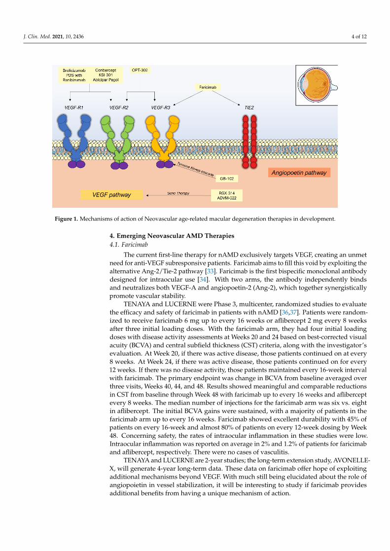
Additionally, angiogenesis receives a contribution from alternative pathways, such asthe Ang-Tie 2 axis. In this pathway, angiopoietin-1 and angiopoietin-2 are key cytokinesthat interact with transmembrane receptor tyrosine kinase (Tie-2) [33]. In healthy states,Tie-2 is bound by angiopoietin-1, which is a protective factor, promoting vascular stability,pericyte recruitment, and the inhibition of vascular permeability factors [34]. However,in angiogenic states, the competitive inhibitor angiopoietin-2 is upregulated, displacingAng-1, and causing endothelial destabilization, inflammation, and breakdown of the blood–retina barrier [35]. Combination therapies that target these non-VEGF angiogenic factorsmay provide additional benefit over current standard of care. However, this is yet to bedetermined. The agents discussed in this review are organized according to the mechanismof action in Figure 1.
J. Clin. Med. 2021, 10, 2436 4 of 12J. Clin. Med. 2021, 10, x FOR PEER REVIEW 4 of 12
Figure 1. Mechanisms of action of Neovascular age-related macular degeneration therapies in development.
4. Emerging Neovascular AMD Therapies 4.1. Faricimab
The current first-line therapy for nAMD exclusively targets VEGF, creating an unmet need for anti-VEGF subresponsive patients. Faricimab aims to fill this void by exploiting the alternative Ang-2/Tie-2 pathway [33]. Faricimab is the first bispecific monoclonal an-tibody designed for intraocular use [34]. With two arms, the antibody independently binds and neutralizes both VEGF-A and angiopoetin-2 (Ang-2), which together synergis-tically promote vascular stability.
TENAYA and LUCERNE were Phase 3, multicenter, randomized studies to evaluate the efficacy and safety of faricimab in patients with nAMD [36,37]. Patients were random-ized to receive faricimab 6 mg up to every 16 weeks or aflibercept 2 mg every 8 weeks after three initial loading doses. With the faricimab arm, they had four initial loading doses with disease activity assessments at Weeks 20 and 24 based on best-corrected visual acuity (BCVA) and central subfield thickness (CST) criteria, along with the investigator’s evaluation. At Week 20, if there was active disease, those patients continued on at every 8 weeks. At Week 24, if there was active disease, those patients continued on for every 12 weeks. If there was no disease activity, those patients maintained every 16-week interval with faricimab. The primary endpoint was change in BCVA from baseline averaged over three visits, Weeks 40, 44, and 48. Results showed meaningful and comparable reductions in CST from baseline through Week 48 with faricimab up to every 16 weeks and afliber-cept every 8 weeks. The median number of injections for the faricimab arm was six vs. eight in aflibercept. The initial BCVA gains were sustained, with a majority of patients in the faricimab arm up to every 16 weeks. Faricimab showed excellent durability with 45% of patients on every 16-week and almost 80% of patients on every 12-week dosing by Week 48. Concerning safety, the rates of intraocular inflammation in these studies were low. Intraocular inflammation was reported on average in 2% and 1.2% of patients for faricimab and aflibercept, respectively. There were no cases of vasculitis.
TENAYA and LUCERNE are 2-year studies; the long-term extension study, AVONELLE-X, will generate 4-year long-term data. These data on faricimab offer hope of exploiting additional mechanisms beyond VEGF. With much still being elucidated about the role of angiopoietin in vessel stabilization, it will be interesting to study if faricimab provides additional benefits from having a unique mechanism of action.
4.2. Port Delivery System (PDS)
Figure 1. Mechanisms of action of Neovascular age-related macular degeneration therapies in development.
4. Emerging Neovascular AMD Therapies4.1. Faricimab
The current first-line therapy for nAMD exclusively targets VEGF, creating an unmetneed for anti-VEGF subresponsive patients. Faricimab aims to fill this void by exploiting thealternative Ang-2/Tie-2 pathway [33]. Faricimab is the first bispecific monoclonal antibodydesigned for intraocular use [34]. With two arms, the antibody independently bindsand neutralizes both VEGF-A and angiopoetin-2 (Ang-2), which together synergisticallypromote vascular stability.
TENAYA and LUCERNE were Phase 3, multicenter, randomized studies to evaluatethe efficacy and safety of faricimab in patients with nAMD [36,37]. Patients were random-ized to receive faricimab 6 mg up to every 16 weeks or aflibercept 2 mg every 8 weeksafter three initial loading doses. With the faricimab arm, they had four initial loadingdoses with disease activity assessments at Weeks 20 and 24 based on best-corrected visualacuity (BCVA) and central subfield thickness (CST) criteria, along with the investigator’sevaluation. At Week 20, if there was active disease, those patients continued on at every8 weeks. At Week 24, if there was active disease, those patients continued on for every12 weeks. If there was no disease activity, those patients maintained every 16-week intervalwith faricimab. The primary endpoint was change in BCVA from baseline averaged overthree visits, Weeks 40, 44, and 48. Results showed meaningful and comparable reductionsin CST from baseline through Week 48 with faricimab up to every 16 weeks and afliberceptevery 8 weeks. The median number of injections for the faricimab arm was six vs. eightin aflibercept. The initial BCVA gains were sustained, with a majority of patients in thefaricimab arm up to every 16 weeks. Faricimab showed excellent durability with 45% ofpatients on every 16-week and almost 80% of patients on every 12-week dosing by Week48. Concerning safety, the rates of intraocular inflammation in these studies were low.Intraocular inflammation was reported on average in 2% and 1.2% of patients for faricimaband aflibercept, respectively. There were no cases of vasculitis.
TENAYA and LUCERNE are 2-year studies; the long-term extension study, AVONELLE-X, will generate 4-year long-term data. These data on faricimab offer hope of exploitingadditional mechanisms beyond VEGF. With much still being elucidated about the role ofangiopoietin in vessel stabilization, it will be interesting to study if faricimab providesadditional benefits from having a unique mechanism of action.
J. Clin. Med. 2021, 10, 2436 5 of 12
4.2. Port Delivery System (PDS)
The success of traditional anti-VEGF therapies raised the question of whether alonger-acting ranibizumab could be delivered via an implantable reservoir. Thus, the PortDelivery System (PDS) was developed, allowing for the continuous release of ranibizumabinto the vitreous via passive diffusion [38]. PDS is intended to reduce the frequency ofintravitreal injections and potentially allow patients with nAMD to go several monthsbefore needing a refill of the implant. The device is a self-sealing eye implant that requiressurgical implantation and can be refilled in the office via injection through the conjunctiva.Currently, the PDS holds 20 µL of a customized formulation of ranibizumab (100 mg/mL).This dosage was found to be the most effective dose from the Phase 2 LADDER trial in wetAMD, looking at visual and anatomic success [39].
Results of the Phase 3 ARCHWAY trial showed that PDS at every 24 weeks wasnoninferior and equivalent to monthly ranibizumab at its primary endpoint at Week 40 [40].As expected, there was a transient postsurgical drop in vision in the PDS arm that recoveredby Week 40. By Week 72, patients in the PDS arm had two refill exchanges at Weeks 24 and48, with vision and anatomic outcomes comparable with monthly ranibizumab. There wasequivalent vision and controlled retinal thickness, and PDS patients required five timesfewer treatments over a mean duration of 78 weeks.
The idea of a surgically implanted VEGF depot is intriguing but does carry potentialrisks. VEGF has been found to be a key neurotrophic factor involved in the maintenanceof retinal vasculature [41]. Potent, long-term inhibition may be disruptive to the health ofneurovascular cells. There is evidence of increased risk of geographic atrophy in patientstreated monthly as opposed to patients treated pro re nata [42,43]. With any of the extendeddurability anti-VEGF treatments in development, including PDS, little is known about theadverse events associated with the prolonged antagonism of VEGF. Additional considera-tions include risks from the surgical procedure itself and the possibility of endophthalmitisor vitreous hemorrhage. These risks are still being evaluated by the FDA for considerationfor use. Nonetheless, the promising results from ARCHWAY are an excellent step towardsincreased longevity of nAMD therapy.
4.3. Abicipar Pegol
New to the world of protein therapeutics, designed ankyrin repeat protein (DARPin)molecules are small, single-domain proteins that can selectively bind to a target proteinwith high affinity and specificity [44,45]. These molecules are highly stable, providingadvantages over currently available antibodies or antibody fragments. At present, abiciparpegol is a DARPin developed for use against nAMD [46]. Abicipar binds all isoforms ofVEGF-A with excellent tissue penetration. Furthermore, it has a longer intraocular half-lifecompared with ranibizumab (>13 days vs. 7.2 days) [47].
Following encouraging results from the Phase 2 REACH study [48], two identicalglobal Phase 3 studies were conducted (CEDAR and SEQUOIA) [49]. Participants withnAMD were divided into three arms: three monthly abicipar 2 mg injections followedby an injection every 8 weeks, two monthly abicipar 2 mg injections followed by aninjection after 8 weeks and every 12 weeks thereafter, and monthly ranibizumab injections.Results showed mean change in BCVA during Year 2 was similar when compared to Year1 across all treatment arms. Precisely 93% of patients in the 8-week abicipar group, 90%of patients in the 12-week abicipar group, and 94% of patients in the 4-week ranibizumabgroup achieved stable vision. Only four intravitreal injections of abicipar were requiredto maintain the outcomes, as compared to monthly intravitreal ranibizumab injections.Overall, abicipar demonstrated non-inferiority compared with ranibizumab, meeting itsprimary endpoint.
However, roughly 15% of abicipar-treated eyes experienced intraocular inflammation(IOI). In efforts to reduce this adverse effect, the manufacturing process has since beenmodified. The MAPLE study, a 28-week safety evaluation, was performed to determine therates of adverse events in 128 patients after the manufacturing process was changed. The
J. Clin. Med. 2021, 10, 2436 6 of 12
data showed a reduced intraocular inflammation rate of 8.9%, and only 1.6% of these caseswere deemed moderately severe or severe [50]. The improvement in the rate of adverseeffects is because reformulation is a step in the right direction; however, additional researchis required to validate the efficacy and extended duration of abicipar.
4.4. Brolucizumab
At a size of ~26 kDa, the humanized single-chain antibody fragment brolucizumabmay provide enhanced tissue penetration, clearance, and drug delivery characteristicscompared to more traditional anti-VEGF agents [51]. By comparison, ranibizumab andaflibercept have molecular weights of 48 and 115 kDa, respectively [52]. The molar dose ofbrolucizumab is 11.2 to 13.3 times higher than that of aflibercept, permitting greater drugconcentrations and therefore longer duration.
The safety and efficacy of brolucizumab were compared to aflibercept in two Phase3 trials, HAWK and HARRIER [53]. The primary endpoint in both studies was nonin-feriority to aflibercept in mean change in BCVA from baseline to Week 48. In HAWK,patients were randomized to intravitreal brolucizumab 3 mg, brolucizumab 6 mg, oraflibercept 2 mg. HARRIER randomized patients to brolucizumab 6 mg or aflibercept2 mg. Brolucizumab was noninferior to aflibercept in the primary outcomes in both studies.In the superiority analysis of HAWK at Week 16, the incidence of disease activity wassignificantly lower with brolucizumab 6 mg compared with aflibercept (24.0% vs. 34.5%).Intraretinal fluid/SRF was present in fewer brolucizumab-treated eyes versus aflibercept-treated eyes at Week 16 in both trials. Rates of ocular and nonocular AEs were similar withbrolucizumab and aflibercept.
Despite the efficacy of brolucizumab for nAMD, and its superior pharmacokinetics,many retina specialists are concerned about the risk of occlusive vasculitis and blindnesswith the drug. The rate of uveitis was 2.2% with brolucizumab 6 mg and 0.3% withaflibercept in HAWK, and <1% with both drugs in HARRIER. The incidence of iritis was2.2% with brolucizumab 6 mg and 0% with aflibercept in HAWK, and <1% with both drugsin HARRIER. While the FDA has recently given approval towards brolucizumab use, it isunclear whether these adverse events will outweigh the potential benefits.
4.5. KSI-301
Mechanically, KS-301 resembles the classic anti-VEGF agents; however, it is based on a950 kDa antibody biopolymer conjugate (ABC) platform that is engineered specifically forincreased durability [54]. Preclinical pharmacokinetic studies have demonstrated KSI-301’sextended ocular half-life of 10–12 days. In a Phase 1b study, patients received three loadingdoses at Weeks 0, 4, and 8. There was a durability assessment from Weeks 12 to 72 withan extension study from weeks 76 to 148. The efficacy of KSI-301 was determined bychange from baseline to Week 52 in mean BCVA and optical coherence tomography (OCT)thickness. There was an observed mean 5.7-letter improvement to 69.7 ETDRS eye chartletters (~20/40 Snellen) at Year 1 [55]. Additionally, thickness had decreased by 105 microns.Patients received three loading doses, followed by an average of two individualized dosesthereafter, resulting in a total of five mean injections in Year 1.
There was an excellent safety profile for KSI-301. Most adverse effects were assessedas mild and consistent with the profile of intravitreal (IVT) anti-VEGF injections. To date,43 serious AEs (SAEs) have been reported in 24 subjects; however, none were drug related.Additionally, three ocular SAEs in the study eye not drug related were all resolved. Onlytwo AEs of IOI (2/710, 0.28%) were noted, both traced to 1+ vitreous cell with completeresolution.
4.6. Conbercept
Conbercept is a 141 kDa engineered fusion protein that, like aflibercept, acts as re-ceptor decoy against VEGF [56]. However, conbercept has higher binding affinity andcontains an additional fourth binding domain of VEGFR2. This design is hypothesized
J. Clin. Med. 2021, 10, 2436 7 of 12
to provide increased stability of the receptor-ligand complex and extend the half-life ofconbercept [56–59]. Two global Phase 3 trials for nAMD were initiated: PANDA-1 andPANDA-2. Each trial is evaluating 1140 patients randomized to conbercept, 0.5 or 1 mg, oraflibercept 2 mg, with primary efficacy analysis at 36 weeks. In PANDA-1, patients receivedthree loading doses through Week 8, then continued with dosing every 8 weeks throughWeek 92. In PANDA-2, dosing was pro re nata after Week 40, with the 0.5 mg conberceptand aflibercept groups on the same regimen as PANDA-1 up until that point, after whichthe conbercept 1 mg arm shifted to 12-week dosing after 8 weeks and moved onto PRN atWeek 40. The PANDA trials recently reached a milestone by completing 36-week primaryendpoint visits of enrolled patients in December 2020.
4.7. OPT-302
With traditional VEGF blockade, current therapies target VEGF-A, which is consideredto be the most pathologic isoform. However, in this process, other VEGF isoforms areupregulated [29–32]. OPT-302 is a novel “trap” molecule that binds and neutralizes theactivity of VEGF-C/-D, blocking their activation of receptors VEGFR2 and VEGFR-3 [60].There is hope that combining OPT-302 with currently available anti-VEGF-A may addressmechanisms of resistance associated with existing therapies. Two concurrent global Phase3 trials known as Study of OPT-302 in combination with Ranibizumab (ShORe) and Combi-nation OPT-302 with Aflibercept Study (COAST) have begun [61]. These trials build uponthe successful Phase 2b nAMD clinical trial while additionally evaluating the administra-tion of OPT-302 in combination with ranibizumab and aflibercept over a longer treatmentperiod and in a greater number of patients. ShORe and COAST will enroll approximately990 treatment-naive patients each and assess the efficacy and safety of intravitreal 2.0 mgOPT-302 in combination with 0.5 mg ranibizumab or 2.0 mg aflibercept, compared toranibizumab or aflibercept monotherapy, respectively. The primary endpoint of both stud-ies is the mean change in BCVA from baseline to Week 52 for OPT-302 combination therapycompared to anti-VEGF-A monotherapy.
4.8. GB-102
Like PDS, GB-102 is another sustained-release anti-VEGF delivery system [62]. How-ever, GB-102 is formulated as an intravitreal formulation of sunitinib malate-containing,biodegradable microparticles. The controlled microparticle release is intended for biannualinjection to maintain comparable visual acuity and central subfield thickness outcomes.The ADAGIO Phase 1/2a study consisted of patients with nAMD who received fourescalating dose cohorts of eight patients, each receiving a single dose of either 0.25, 0.5,1, or 2 mg of GB-102 [63]. Precisely 88% of the patients at 3 months and 68% of the pa-tients at 6 months were maintained on a single dose of GB-102. Positive outcomes wereobserved for up to 8 months. CST was significantly reduced at all months compared withpretreatment. However, one concern that emerged was the nonaggregation of the drugonce in the vitreous cavity, resulting in particle dispersion. Nine of thirty-two subjectsexperienced related symptoms, including eye pain, photophobia, and blurriness [64]. Anew manufacturing process was developed to eliminate the microparticle dispersion andincomplete aggregation, which was used for future trials.
Phase 2b ALTISSIMO was initiated to further evaluate GB-102 for CNV lesions inpreviously treated nAMD patients [65]. The study consists of three cohorts: 1 mg of GB-102,2 mg of GB-102, or 2 mg of aflibercept at baseline. The GB-102 cohorts will then receivetheir same initial dose every 6 months, whereas the latter control group will continueto receive aflibercept 2 mg every 2 months. The primary outcome is the proportion oftreated subjects remaining rescue free through Month 10. While the efficacy of GB-102 isstill being determined, it provides an immediate bridge towards longer-lasting therapiesfurther down the pipeline. GB-102 has by far the longest time needed between treatments,and the development of GB-103, which aims for once-a-year dosing, has already begun
J. Clin. Med. 2021, 10, 2436 8 of 12
4.9. RGX-314
Gene therapy has shown promise for the treatment of inherited retinal diseases, andrecently there has been a push for gene therapy solutions for nAMD. RGX-314 is a vectordesigned to bind and neutralize VEGF in a manner similar to ranibizumab [66]. RGX-314utilizes adeno-associated virus serotype 8 (AAV8) as its vector, with research suggestingthat AAV vectors provide long-term transgene expression [67]. The gene therapy vectoris preferentially taken up by retinal cells, leading to high levels of production of themonoclonal antibody fragment. The company is advancing two separate routes of ocularadministration of RGX-314: a one-time subretinal administration during vitrectomy; and in-office suprachoroidal delivery. The hope is that the long-standing and stable production ofthe anti-VEGF therapeutic protein could reduce the need for frequent intravitreal injections.
ATMOSPHERE is the first of two planned pivotal trials for the evaluation of subretinaldelivery of RGX-314 in patients who have received prior treatment for nAMD [68,69].Patients underwent vitrectomy and were delivered subretinal RGX-314 across five dosecohorts (3 × 10⁹ genome copies (GC)/eye, 1 × 10¹⁰ GC/eye, 6 × 10¹⁰ GC/eye, 1.6 × 10¹¹
GC/eye, 2.5 × 10¹¹ GC/eye). RGX-314 continued to be generally well tolerated acrossall cohorts, with 20 serious adverse events (SAEs) reported in 13 patients, including1 possibly drug-related SAE of a significant decrease in vision in Cohort 5. The mostcommon nonserious adverse events in the eye were generally assessed as mild (87%).These included postoperative conjunctival hemorrhage (69% of patients), postoperativeinflammation (36% of patients), eye irritation (17% of patients), eye pain (17% of patients),and postoperative visual acuity reduction (17% of patients). In 67% of patients across allcohorts, and in 83% of patients in Cohorts 3 through 5, retinal pigmentary changes wereobserved on imaging, the majority of which were in the peripheral inferior retina. Retinalhemorrhage was observed in 26% of patients and is an anticipated event in patients withsevere wet AMD. There have been no reports of clinically determined immune responses,drug-related ocular inflammation, or postsurgical inflammation beyond what is expectedfollowing routine vitrectomy. In the two higher dose cohorts (four and five), patients at1.5 years after treatment demonstrated stable visual acuity with a mean BCVA change of+1 letters and −1 letters from baseline, respectively, as well as decreased CRT, with a meanchange of −46 and −93 µm, respectively. In Cohort 4, 4 out of 12 (33%) patients havereceived no anti-VEGF injections after 6 months following RGX-314 administration anddemonstrated a mean BCVA change from baseline of +2 letters at 1.5 years. Eight out ofeleven (73%) patients have received no anti-VEGF injections after 6 months following RGX-314 administration and demonstrated a mean BCVA change from baseline of −2 letters at1.5 years. These data show a meaningful reduction in anti-VEGF treatment burden in bothCohorts 4 and 5. With the positive results of ATMOSPHERE, and the pending results ofsuprachoroidal RGX-314 delivery in AAVIATE, there is much promise for gene therapy innAMD treatment.
4.10. ADVM-022
ADVM-022 is another gene therapy that aims to provide sustained anti-VEGF expres-sion from the retina. In the OPTIC trial, the primary objective was to assess the safety andtolerability of a single IVT injection of ADVM-022. Secondary objectives were to evaluateBCVA and anatomy using spectral-domain OCT (SD-OCT), and to assess the need forrescue therapy [69]. All patients received aflibercept injection 1 to 2 weeks prior to dosingof ADVM-022. There was a 24-week safety and efficacy assessment. Again, the same wasdone at Week 52 with a follow-up at 104 weeks. Patients received oral steroid prophylaxisin Cohorts 1 and 2 and steroid eyedrop prophylaxis in Cohorts 3 and 4.
Overall, ADVM-022 continues to be well tolerated with a favorable safety profileat both high and low doses. It showed robust and sustained efficacy in both high andlow doses. There was excellent durability out to 92 weeks from a single IVT injectionwith 0 supplemental injections in Cohort 1. There was robust aqueous anti-VEGF proteinexpression observed at 18 months in Cohort 1. This study showed a substantial reduction
J. Clin. Med. 2021, 10, 2436 9 of 12
in annualized injection frequency following ADVM-022. Most patients did not require anysupplemental injection in OPTIC. Patients completing 2 years in OPTIC are being enrolledinto an extension trial to be followed for up to 5 years. Two global Phase 3 (PIVOTAL-aand PIVOTAL-b) trials are targeted to initiate in the fourth quarter of 2021.
Gene therapy differs from other extended durability therapies as no hardware isimplanted in the eye. This may circumvent potential complications such as conjunctivalerosion. Moreover, there is tremendous value in the potential role of gene therapy inpreventing chronic exudative eye conditions such as nAMD, as many researchers believethat early intervention is valuable in limiting the progression of nAMD. While one or twointravitreal injections are generally tolerable, ongoing treatment with no definite cessationfor patients who are asymptomatic can often be untenable for them. So, there is muchexcitement about the possibility of a one-time treatment with sustained intraocular VEGFsuppression that could slow the course of nAMD. However, a potential disadvantage togene therapy is the inability to turn it off. The consequences of long-term VEGF blockadeare still being elucidated.
5. Conclusions
While anti-VEGF agents have revolutionized our treatment of nAMD, the field con-tinues to evolve in the hope of providing better options for our patients. As discussed,numerous novel molecular targets may allow us to improve upon the clinical outcomesachieved by the VEGF blockade. Beyond VEGF, there are several trials underway in-vestigating alternative factors in retinal and choroidal angiogenesis, such as PDGF, FGF,and EGF. Furthermore, research towards longer-acting pharmaceuticals might yield goodresults with fewer treatments, helping to improve compliance, possibly allowing us to treatmore patients.
Author Contributions: Conceptualization, P.P. and V.S.; methodology, P.P.; formal analysis, P.P.; datacuration, P.P. and V.S.; writing—original draft preparation, P.P.; writing—review and editing, V.S.;visualization, P.P.; supervision, V.S.; project administration, P.P.; funding acquisition, V.S. All authorshave read and agreed to the published version of the manuscript.
Funding: This research received no external funding.
Institutional Review Board Statement: Not applicable.
Informed Consent Statement: Not applicable.
Data Availability Statement: No new data were created or analyzed in this study. Data sharing isnot applicable to this article.
Conflicts of Interest: The authors declare no conflict of interest.
References1. Klein, R. Prevalence of age-related macular degeneration in the US population. Arch. Ophthal. 2011, 129, 75. [CrossRef] [PubMed]2. Klein, B.E.K. Forecasting age-related macular degeneration through 2050. JAMA 2009, 301, 2152–2153. [CrossRef]3. Bourne, R.R.A.; Jonas, J.B.; Bron, A.M.; Cicinelli, M.V.; Das, A.; Flaxman, S.R.; Friedman, D.S.; Keeffe, J.E.; Kempen, J.H.; Leasher,
J.; et al. Prevalence and causes of vision loss in high-income countries and in Eastern and Central Europe in 2015: Magnitude,temporal trends and projections. Br. J. Ophthalmol. 2018, 102, 575–585. [CrossRef] [PubMed]
4. Wong, W.L.; Su, X.; Li, X.; Cheung, C.M.; Klein, R.; Cheng, C.Y.; Wong, T.Y. Global prevalence of age-related macular degenerationand disease burden projection for 2020 and 2040: A systematic review and meta-analysis. Lancet Glob. Health 2014, 2, e106–e116.[CrossRef]
5. Ferris, F.L.; Wilkinson, C.P.; Bird, A.; Chakravarthy, U.; Chew, E.; Csaky, K.; Sadda, S.R. Clinical Classification of Age-relatedMacular Degeneration. Ophthalmology 2013, 120, 844–851. [CrossRef]
6. Kolar, P. Classification and clinical features of AMD. In Age-Related Macular Degeneration-Etiology, Diagnosis and Management—AGlance at the Future; InTech: London, UK, 2013; pp. 105–132.
7. Tadayoni, R. Choroidal Neovascularization Induces Retinal Edema and its Treatment Addresses this Problem. J. Ophthalmic. Vis.Res. 2014, 9, 405–406. [CrossRef]
J. Clin. Med. 2021, 10, 2436 10 of 12
8. Guyer, D.R.; Fine, S.L.; Maguire, M.G.; Hawkins, B.S.; Owens, S.L.; Murphy, R.P. Subfoveal choroidal neovascular membranes inage-related macular degeneration. Visual prognosis in eyes with relatively good initial visual acuity. Arch. Ophthalmol. 1986, 104,702–705. [CrossRef]
9. Wong, T.Y.; Chakravarthy, U.; Klein, R.; Mitchell, P.; Zlateva, G.; Buggage, R.; Fahrbach, K.; Probst, C.; Sledge, I. The naturalhistory and prognosis of neovascular age-related macular degeneration: A systematic review of the literature and meta-analysis.Ophthalmology 2008, 115, 116–126. [CrossRef]
10. Virgili, G.; Bini, A. Laser photocoagulation for neovascular age-related macular degeneration. Cochrane Database Syst. Rev. 2007,18, CD004763. [CrossRef]
11. Ciulla, T.A.; Huang, F.; Westby, K.; Williams, D.F.; Zaveri, S.; Patel, S.C. Real-world Outcomes of Anti-Vascular EndothelialGrowth Factor Therapy in Neovascular Age-Related Macular Degeneration in the United States. Ophthalmol. Retina 2018, 2,645–653. [CrossRef]
12. Mehta, H.; Tufail, A.; Daien, V.; Lee, A.Y.; Nguyen, V.; Ozturk, M.; Barthelmes, D.; Gillies, M.C. Real-world outcomes in patientswith neovascular age-related macular degeneration treated with intravitreal vascular endothelial growth factor inhibitors. Prog.Retin. Eye Res. 2018, 65, 127–146. [CrossRef]
13. Daniel, E.; Toth, C.A.; Grunwald, J.E.; Jaffe, G.J.; Martin, D.F.; Fine, S.L.; Huang, J.; Ying, G.S.; Hagstrom, S.A.; Winter, K.; et al.Risk of scar in the comparison of age-related macular degeneration treatments trials. Ophthalmology 2014, 121, 656–666. [CrossRef]
14. Grunwald, J.E.; Pistilli, M.; Daniel, E.; Ying, G.S.; Pan, W.; Jaffe, G.J.; Toth, C.A.; Hagstrom, S.A.; Maguire, M.G.; Martin, D.F.Incidence and Growth of Geographic Atrophy during 5 Years of Comparison of Age-Related Macular Degeneration TreatmentsTrials. Ophthalmology 2017, 124, 97–104. [CrossRef]
15. Cox, J.T.; Eliott, D.; Sobrin, L. Inflammatory Complications of Intravitreal Anti-VEGF Injections. J. Clin. Med. 2021, 10, 981.[CrossRef]
16. Daien, V.; Nguyen, V.; Essex, R.W.; Morlet, N.; Barthelmes, D.; Gillies, M.C.; Gillies, M.; Hunt, A.; Essex, R.; Dayajeewa, C.; et al.Incidence and Outcomes of Infectious and Noninfectious Endophthalmitis after Intravitreal Injections for Age-Related MacularDegeneration. Ophthalmology 2018, 125, 66–74. [CrossRef]
17. Knickelbein, J.E.; Chew, E.Y.; Sen, H.N. Intraocular Inflammation Following Intravitreal Injection of Anti-VEGF Medications forNeovascular Age-Related Macular Degeneration. Ophthalmic Epidemiol. 2016, 23, 69–70. [CrossRef]
18. De Vries, V.A.; Bassil, F.L.; Ramdas, W.D. The effects of intravitreal injections on intraocular pressure and retinal nerve fiber layer:A systematic review and meta-analysis. Sci. Rep. 2020, 10, 13248. [CrossRef]
19. Ambati, J.; Fowler, B.J. Mechanisms of age-related macular degeneration. Neuron 2012, 75, 26–39. [CrossRef] [PubMed]20. Hageman, G.S.; Luthert, P.J.; Victor Chong, N.H.; Johnson, L.V.; Anderson, D.H.; Mullins, R.F. An integrated hypothesis that
considers drusen as biomarkers of immune-mediated processes at the RPE-Bruch’s membrane interface in aging and age-relatedmacular degeneration. Prog. Retin Eye Res. 2001, 20, 705–732. [CrossRef]
21. Handa, J.T.; Rickman, C.B.; Dick, A.D.; Gorin, M.B.; Miller, J.W.; Toth, C.A.; Ueffing, M.; Zarbin, M.; Farrer, L.A. A systems biologyapproach towards understanding and treating non-neovascular age-related macular degeneration. Nat. Commun. 2019, 10, 3347.[CrossRef] [PubMed]
22. Zarbin, M.A. Current concepts in the pathogenesis of age-related macular degeneration. Arch Ophthalmol. 2004, 122, 598–614.[CrossRef] [PubMed]
23. Thurman, J.M.; Renner, B.; Kunchithapautham, K.; Ferreira, V.P.; Pangburn, M.K.; Ablonczy, Z.; Tomlinson, S.; Holers, V.M.;Rohrer, B. Oxidative Stress Renders Retinal Pigment Epithelial Cells Susceptible to Complement-mediated Injury. J. Biol. Chem.2009, 284, 16939–16947. [CrossRef] [PubMed]
24. Wu, Z.; Lauer, T.W.; Sick, A.; Hackett, S.F.; Campochiaro, P.A. Oxidative Stress Modulates Complement Factor H Expression inRetinal Pigmented Epithelial Cells by Acetylation of FOXO. J. Biol. Chem. 2007, 282, 22414–22425. [CrossRef]
25. Gold, B.; The AMD Genetics Clinical Study Group; E Merriam, J.; Zernant, J.; Hancox, L.S.; Taiber, A.J.; Gehrs, K.; Cramer, K.;Neel, J.; Bergeron, J.; et al. Variation in factor B (BF) and complement component 2 (C2) genes is associated with age-relatedmacular degeneration. Nat. Genetics 2006, 38, 458–462. [CrossRef] [PubMed]
26. Zarbin, M.A.; Rosenfeld, P.J. Pathway-based therapies for age-related macular degeneration: An integrated survey of emergingtreatment alternatives. Retina 2010, 30, 1350–1367. [CrossRef] [PubMed]
27. Nowak, J.Z. Age-related macular degeneration (AMD): Pathogenesis and therapy. Pharmacol. Rep. 2006, 58, 353–363.28. Anderson, D.H.; Mullins, R.F.; Hageman, G.S.; Johnson, L.V. A role for local inflammation in the formation of drusen in the aging
eye. Am. J. Ophthalmol. 2002, 134, 411–431. [CrossRef]29. Li, D.; Xie, K.; Ding, G.; Li, J.; Chen, K.; Li, H.; Qian, J.; Jiang, C.; Fang, J. Tumor resistance to anti-VEGF therapy through
up-regulation of VEGF-C expression. Cancer Lett. 2014, 346, 45–52. [CrossRef]30. Lieu, C.H.; Tran, H.; Jiang, Z.Q.; Mao, M.; Overman, M.J.; Lin, E.; Eng, C.; Morris, J.; Ellis, L.; Heymach, J.V.; et al. The association
of alternate VEGF ligands with resistance to anti-VEGF therapy in metastatic colorectal cancer. PLoS ONE 2013, 8, e77117.[CrossRef] [PubMed]
31. Grau, S.; Thorsteinsdottir, J.; von Baumgarten, L.; Winkler, F.; Tonn, J.C.; Schichor, C. Bevacizumab can induce reactivity toVEGF-C and -D in human brain and tumour derived endothelial cells. J. Neurooncol. 2011, 104, 103–112. [CrossRef]
J. Clin. Med. 2021, 10, 2436 11 of 12
32. Cabral, T.; Lima, L.H.; Mello, L.G.M.; Polido, J.; Correa, É.P.; Oshima, A.; Duong, J.; Serracarbassa, P.; Regatieri, C.V.; Mahajan,V.B. Belfort, R., Jr. Bevacizumab Injection in Patients with Neovascular Age-Related Macular Degeneration Increases AngiogenicBiomarkers. Ophthalmol. Retina 2018, 2, 31–37. [CrossRef] [PubMed]
33. Khan, M.; Aziz, A.A.; Shafi, N.A.; Abbas, T.; Khanani, A.M. Targeting Angiopoietin in Retinal Vascular Diseases: A LiteratureReview and Summary of Clinical Trials Involving Faricimab. Cells 2020, 9, 1869. [CrossRef] [PubMed]
34. Korhonen, E.A.; Lampinen, A.; Giri, H.; Anisimov, A.; Kim, M.; Allen, B.; Fang, S.; D’Amico, G.; Sipila, T.J.; Lohela, M.; et al.Tie1 controls angiopoietin function in vascular remodeling and inflammation. J. Clin. Investig. 2016, 126, 3495–3510. [CrossRef][PubMed]
35. Maisonpierre, P.C.; Suri, C.; Jones, P.F.; Bartunkova, S.; Wiegand, S.J.; Radziejewski, C.; Compton, D.; McClain, J.; Aldrich, T.H.;Papadopoulos, N.; et al. Angiopoietin-2, a natural antagonist for Tie2 that disrupts in vivo angiogenesis. Science 1997, 277, 55–60.[CrossRef]
36. Genentech Press Release. Available online: https://www.roche.com/media/releases/med-cor-2021-01-25.htm (accessed on 8April 2021).
37. Genentech Press Release. Available online: https://www.roche.com/media/releases/med-cor-2021-02-12.htm (accessed on 8April 2021).
38. Chen, E.R.; Kaiser, P.K. Therapeutic Potential of the Ranibizumab Port Delivery System in the Treatment of AMD: Evidence toDate. Clin. Ophthalmol. 2020, 14, 1349–1355. [CrossRef]
39. Campochiaro, P.A.; Marcus, D.M.; Awh, C.C.; Regillo, C.; Adamis, A.P.; Bantseev, V.; Chiang, Y.; Ehrlich, J.S.; Erickson, S.; Hanley,W.D.; et al. The port delivery system with ranibizumab for neovascular age-related macular degeneration: Results from therandomized phase 2 LADDER clinical trial. Ophthalmology 2019, 126, 1141–1154. [CrossRef]
40. EyeWire News. Available online: https://eyewire.news/articles/phase-3-data-show-port-delivery-system-with-ranibizumab-enabled-over-98-of-patients-to-go-6-months-between-treatments-for-wet-amd/ (accessed on 9 April 2021).
41. Usui, Y.; Westenskow, P.; Kurihara, T.; Aguilar, E.; Sakimoto, S.; Paris, L.P.; Wittgrove, C.; Feitelberg, D.; Friedlander, M.; Moreno,S.K.; et al. Neurovascular crosstalk between interneurons and capillaries is required for vision. J. Clin. Investig. 2015, 125,2335–2346. [CrossRef] [PubMed]
42. Sadda, S.R.; Tuomi, L.L.; Ding, B.; Fung, A.E.; Hopkins, J.J. Macular atrophy in the HARBOR study for neovascular age-relatedmacular degeneration. Ophthalmology 2018, 125, 878–886. [CrossRef]
43. Chakravarthy, U.; Harding, S.P.; Rogers, C.A.; Downes, S.M.; Lotery, A.J.; Culliford, L.A.; Reeves, B.C.; IVAN Study Investigators.Alternative treatments to inhibit VEGF in age-related choroidal neovascularisation: 2-year findings of the IVAN randomisedcontrolled trial. Lancet 2013, 382, 1258–1267. [CrossRef]
44. Binz, H.; Stumpp, M.T.; Forrer, P.; Amstutz, P.; Plückthun, A. Designing Repeat Proteins: Well-expressed, Soluble and StableProteins from Combinatorial Libraries of Consensus Ankyrin Repeat Proteins. J. Mol. Biol. 2003, 332, 489–503. [CrossRef]
45. Stumpp, M.T.; Binz, H.K.; Amstutz, P. DARPins: A new generation of protein therapeutics. Drug Discov. Today 2008, 13, 695–701.[CrossRef] [PubMed]
46. Krohne, T.U.; Liu, Z.; Holz, F.G.; Meyer, C.H. Intraocular pharmacokinetics of ranibizumab following a single intravitreal injectionin humans. Am. J. Ophthalmol. 2012, 154, 682–686. [CrossRef] [PubMed]
47. Rodrigues, G.A.; Mason, M.; Christie, L.-A.; Hansen, C.; Hernandez, L.M.; Burke, J.; Luhrs, K.A.; Hohman, T.C. Functional char-acterization of abicipar-pegol, an Anti-VEGF DARPin therapeutic that potently inhibits angiogenesis and vascular permeability.Investig. Ophthalmol. Vis. Sci. 2018, 59, 5836–5846. [CrossRef] [PubMed]
48. Callanan, D.; Kunimoto, D.; Maturi, R.K.; Patel, S.S.; Staurenghi, G.; Wolf, S.; Cheetham, J.K.; Hohman, T.C.; Kim, K.; López, F.J.;et al. Double-Masked, Randomized, Phase 2 Evaluation of Abicipar Pegol (an Anti-VEGF DARPin Therapeutic) in NeovascularAge-Related Macular Degeneration. J. Ocul. Pharmacol. Ther. 2018, 34, 700–709. [CrossRef] [PubMed]
49. Kunimoto, D.; Yoon, Y.H.; Wykoff, C.C.; Chang, A.; Khurana, R.N.; Maturi, R.K.; Agostini, H.; Souied, E.; Chow, D.R.; Lotery, A.J.;et al. Efficacy and Safety of Abicipar in Neovascular Age-Related Macular Degeneration: 52-Week Results of Phase 3 RandomizedControlled Study. Ophthalmology 2020, 127, 1331–1344. [CrossRef]
50. Molecular Partners Press Release. Available online: https://www.molecularpartners.com/allergan-and-molecular-partners-announce-topline-safety-results-from-maple-study-of-abicipar-pegol/ (accessed on 9 April 2021).
51. Munoz-Ramon, P.V.; Hernandez Martinez, P.; Munoz-Negrete, F.J. New therapeutic targets in the treatment of age-related maculardegeneration. Arch. Soc. Esp. Oftalmol. 2020, 95, 75–83.
52. Nguyen, Q.D.; Das, A.; Do, D.V.; Dugel, P.U.; Gomes, A.; Holz, F.G.; Koh, A.; Pan, C.K.; Sepah, Y.J.; Patel, N.; et al. Brolucizumab:Evolution through preclinical and clinical studies and the implications for the management of neovascular age-related maculardegeneration. Ophthalmology 2020, 127, 963–976. [CrossRef]
53. Dugel, P.U.; Koh, A.; Ogura, Y.; Jaffe, G.J.; Schmidt-Erfurth, U.; Brown, D.M.; Gomes, A.V.; Warburton, J.; Weichselberger, A.;Holz, F.G. HAWK and HARRIER: Phase 3, Multicenter, Randomized, Double-Masked Trials of Brolucizumab for NeovascularAge-Related Macular Degeneration. Ophthalmology 2020, 127, 72–84. [CrossRef]
54. Patel, S.S.; Naor, J.; Qudrat, A.; Do, D.V.; Buetelspacher, D.; Perlroth, D.V. Phase 1 first-in-human study of KSI-301: A novelanti-VEGF antibody biopolymer conjugate with extended durability. Investig. Ophthalmol. Vis. Sci. 2019, 60, 3670.
55. EyeWire News. Available online: https://eyewire.news/articles/kodiak-sciences-announces-1-year-data-from-ongoing-phase-1b-study-of-ksi-301-in-patients-with-retinal-vascular-diseases/ (accessed on 9 April 2021).
J. Clin. Med. 2021, 10, 2436 12 of 12
56. Li, X.; Xu, G.; Wang, Y.; Xu, X.; Liu, X.; Tang, S.; Zhang, F.; Zhang, J.; Tang, L.; Wu, Q.; et al. Safety and efficacy of conberceptin neovascular age-related macular degeneration: Results from a 12-month randomized phase 2 study: AURORA study.Ophthalmology 2014, 121, 1740–1747. [CrossRef]
57. Li, H.; Lei, N.; Zhang, M.; Li, Y.; Xiao, H.; Hao, X. Pharmacokinetics of a long-lasting anti-VEGF fusion protein in rabbit. Exp EyeRes. 2012, 97, 154–159. [CrossRef]
58. Zhang, M.; Yu, D.; Yang, C.; Xia, Q.; Li, W.; Liu, B.; Li, H. The pharmacology study of a new recombinant human VEGF receptor-fcfusion protein on experimental choroidal neovascularization. Pharm. Res. 2009, 26, 204–210. [CrossRef]
59. Sun, X.; Lu, X. Profile of conbercept in the treatment of neovascular age-related macular degeneration. Drug Des. Dev. Ther. 2015,9, 2311–2320. [CrossRef]
60. Dugel, P.U.; Boyer, D.S.; Antoszyk, A.N.; Steinle, N.C.; Varenhorst, M.P.; Pearlman, J.A.; Gillies, M.C.; Finger, R.P.; Baldwin, M.E.;Leitch, I.M. Phase 1 Study of OPT-302 Inhibition of Vascular Endothelial Growth Factors C and D for Neovascular Age-RelatedMacular Degeneration. Ophthalmol. Retina 2020, 4, 250–263. [CrossRef] [PubMed]
61. Biotech Dispatch. Available online: https://biotechdispatch.com.au/news/opthea-confirms-plans-and-protocols-for-late-stage-opt-302-studi (accessed on 7 April 2021).
62. Samanta, A.; Aziz, A.A.; Jhingan, M.; Singh, S.R.; Khanani, A.M.; Chhablani, J. Emerging Therapies in Neovascular Age-RelatedMacular Degeneration in 2020. Asia Pac. J. Ophthalmol. (Phila.) 2020, 9, 250–259. [CrossRef] [PubMed]
63. BusinessWire. Available online: https://www.businesswire.com/news/home/20190121005424/en/Graybug-Vision-Presents-Top-Line-Results-of-Phase-12a-ADAGIO-Study-at-Hawaiian-Eye-Retina-2019 (accessed on 1 April 2021).
64. Kaiser, P.K.; Boyer, D. Most exciting retinal drugs: 2019. In Retina; Waikoloa, HI, USA, 2019.65. EyeWire News. Available online: https://eyewire.news/articles/graybug-vision-completes-treatment-phase-of-altissimo-trial-
in-wet-amd-with-12-month-topline-data/ (accessed on 1 April 2021).66. REGENXBIO’s Gene Therapy for Wet Amd Performing Encouragingly in Human Study. Available online: https://www.
fightingblindness.org/research/regenxbio-s-gene-therapy-for-wet-amd-performing-encouragingly-in-human-study-15 (ac-cessed on 22 January 2021).
67. Nam, H.-J.; Lane, M.D.; Padron, E.; Gurda, B.; McKenna, R.; Kohlbrenner, E.; Aslanidi, G.; Byrne, B.; Muzyczka, N.; Zolotukhin, S.;et al. Structure of adeno-associated virus serotype 8, a gene therapy vector. J. Virol. 2007, 81, 12260–12271. [CrossRef] [PubMed]
68. PRNewswire. Available online: https://www.prnewswire.com/news-releases/regenxbio-announces-additional-positive-interim-phase-iiia-and-long-term-follow-up-data-of-rgx-314-for-the-treatment-of-wet-amd-301228344.html (accessed on 1April 2021).
69. EyeWire News. Available online: https://eyewire.news/articles/adverum-reports-new-interim-data-from-optic-phase-1-trial-of-advm-022-intravitreal-gene-therapy-for-wet-amd/ (accessed on 9 April 2021).
https://biotechdispatch.com.au/news/opthea-confirms-plans-and-protocols-for-late-stage-opt-302-studi
Related Documents