Journal of Immunology Research New Advances in Drug Hypersensitivity Research and Treatment Lead Guest Editor: Yi-Giien Tsai Guest Editors: Wen-Hung Chung, Riichiro Abe, and Wichittra Tassaneeyakul

Welcome message from author
This document is posted to help you gain knowledge. Please leave a comment to let me know what you think about it! Share it to your friends and learn new things together.
Transcript

Journal of Immunology Research
New Advances in Drug Hypersensitivity Research and Treatment
Lead Guest Editor: Yi-Giien TsaiGuest Editors: Wen-Hung Chung, Riichiro Abe, and Wichittra Tassaneeyakul
New Advances in Drug HypersensitivityResearch and Treatment

Journal of Immunology Research
New Advances in Drug HypersensitivityResearch and Treatment
Lead Guest Editor: Yi-Giien TsaiGuest Editors: Wen-Hung Chung, Riichiro Abe,and Wichittra Tassaneeyakul

Copyright © 2018 Hindawi. All rights reserved.
This is a special issue published in “Journal of Immunology Research.” All articles are open access articles distributed under the CreativeCommons Attribution License, which permits unrestricted use, distribution, and reproduction in any medium, provided the originalwork is properly cited.

Editorial Board
B. D. Akanmori, CongoJagadeesh Bayry, FranceKurt Blaser, SwitzerlandEduardo F. Borba, BrazilFederico Bussolino, ItalyNitya G. Chakraborty, USACinzia Ciccacci, ItalyRobert B. Clark, USAMario Clerici, ItalyNathalie Cools, BelgiumM. Victoria Delpino, ArgentinaNejat K. Egilmez, USAEyad Elkord, UKSteven E. Finkelstein, USAMaria Cristina Gagliardi, ItalyLuca Gattinoni, USAAlvaro González, SpainTheresa Hautz, AustriaMartin Holland, UKDouglas C. Hooper, USA
Eung-Jun Im, USAHidetoshi Inoko, JapanJuraj Ivanyi, UKRavirajsinh N. Jadeja, USAPeirong Jiao, ChinaTaro Kawai, JapanAlexandre Keller, BrazilHiroshi Kiyono, JapanBogdan Kolarz, PolandHerbert K. Lyerly, USAMahboobeh Mahdavinia, USAGiulia Marchetti, ItalyEiji Matsuura, JapanChikao Morimoto, JapanHiroshi Nakajima, JapanPaola Nistico, ItalyEnrique Ortega, MexicoPatrice Petit, FranceIsabella Quinti, ItalyEirini Rigopoulou, Greece
Ilaria Roato, ItalyLuigina Romani, ItalyAurelia Rughetti, ItalyFrancesca Santilli, ItalyTakami Sato, USASenthamil R. Selvan, USANaohiro Seo, JapanTrina J. Stewart, AustraliaBenoit Stijlemans, BelgiumJacek Tabarkiewicz, PolandMizue Terai, USABan-Hock Toh, AustraliaJoseph F. Urban, USAPaulina Wlasiuk, PolandBaohui Xu, USAXiao-Feng Yang, USAMaria Zervou, GreeceQiang Zhang, USA
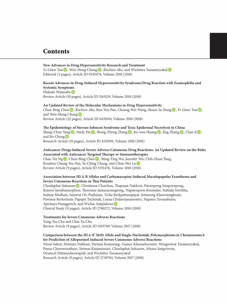
Contents
New Advances in Drug Hypersensitivity Research and TreatmentYi-Giien Tsai , Wen-Hung Chung , Riichiro Abe, and Wichittra TassaneeyakulEditorial (2 pages), Article ID 9345078, Volume 2018 (2018)
Recent Advances in Drug-Induced Hypersensitivity Syndrome/Drug Reaction with Eosinophilia andSystemic SymptomsHideaki WatanabeReview Article (10 pages), Article ID 5163129, Volume 2018 (2018)
An Updated Review of the Molecular Mechanisms in Drug HypersensitivityChun-Bing Chen , Riichiro Abe, Ren-You Pan, Chuang-Wei Wang, Shuen-Iu Hung , Yi-Giien Tsai ,and Wen-Hung ChungReview Article (22 pages), Article ID 6431694, Volume 2018 (2018)
The Epidemiology of Stevens-Johnson Syndrome and Toxic Epidermal Necrolysis in ChinaShang-Chen Yang , Sindy Hu , Sheng-Zheng Zhang , Jin-wen Huang , Jing Zhang , Chao Ji ,and Bo ChengResearch Article (10 pages), Article ID 4320195, Volume 2018 (2018)
Anticancer Drugs Induced Severe Adverse Cutaneous Drug Reactions: An Updated Review on the RisksAssociated with Anticancer TargetedTherapy or ImmunotherapiesChau Yee Ng , Chun-Bing Chen , Ming-Ying Wu, Jennifer Wu, Chih-Hsun Yang,Rosaline Chung-Yee Hui, Ya-Ching Chang, and Chun-Wei LuReview Article (9 pages), Article ID 5376476, Volume 2018 (2018)
Association between HLA-B Alleles and Carbamazepine-Induced Maculopapular Exanthema andSevere Cutaneous Reactions inThai PatientsChonlaphat Sukasem , Chonlawat Chaichan, Thapanat Nakkrut, Patompong Satapornpong,Kanoot Jaruthamsophon, Thawinee Jantararoungtong, Napatrupron Koomdee, Suthida Sririttha,Sadeep Medhasi, Sarawut Oo-Puthinan, Ticha Rerkpattanapipat, Jettanong Klaewsongkram,Pawinee Rerknimitr, Papapit Tuchinda, Leena Chularojanamontri, Napatra Tovanabutra,Apichaya Puangpetch, and Wichai AekplakornClinical Study (11 pages), Article ID 2780272, Volume 2018 (2018)
Treatments for Severe Cutaneous Adverse ReactionsYung-Tsu Cho and Chia-Yu ChuReview Article (9 pages), Article ID 1503709, Volume 2017 (2018)
Comparison between theHLA-B*58:01 Allele and Single-Nucleotide Polymorphisms in Chromosome 6for Prediction of Allopurinol-Induced Severe Cutaneous Adverse ReactionsNiwat Saksit, Nontaya Nakkam, Parinya Konyoung, Usanee Khunarkornsiri, Wongwiwat Tassaneeyakul,Pansu Chumworathayi, Sirimas Kanjanawart, Chonlaphat Sukasem, Alisara Sangviroon,Oranuch Pattanacheewapull, and Wichittra TassaneeyakulResearch Article (9 pages), Article ID 2738784, Volume 2017 (2018)

HLA Association with Drug-Induced Adverse ReactionsWen-Lang Fan, Meng-Shin Shiao, Rosaline Chung-Yee Hui, Shih-Chi Su, Chuang-Wei Wang,Ya-Ching Chang, and Wen-Hung ChungReview Article (10 pages), Article ID 3186328, Volume 2017 (2018)
Immunohistopathological Findings of Severe Cutaneous Adverse Drug ReactionsMari OrimeReview Article (5 pages), Article ID 6928363, Volume 2017 (2018)

EditorialNew Advances in Drug Hypersensitivity Research and Treatment
Yi-Giien Tsai ,1,2,3 Wen-Hung Chung ,4,5,6,7,8 Riichiro Abe,9
and Wichittra Tassaneeyakul 10
1Division of Pediatric Allergy and Immunology, Changhua Christian Hospital, Changhua City, Taiwan2School of Medicine, Kaohsiung Medical University, Kaohsiung, Taiwan3School of Medicine, Chung Shan Medical University, Taichung, Taiwan4Department of Dermatology, Drug Hypersensitivity Clinical and Research Center, Chang Gung Memorial Hospital,Taipei and Linkou, Taiwan5Chang Gung Immunology Consortium, Chang Gung Memorial Hospital, Chang Gung University, Taoyuan, Taiwan6Whole-Genome Research Core Laboratory of Human Diseases, Chang Gung Memorial Hospital, Keelung, Taiwan7College of Medicine, Chang Gung University, Taoyuan, Taiwan8Department of Dermatology, Xiamen Chang Gung Hospital, Xiamen, China9Division of Dermatology, Niigata University Graduate School of Medical and Dental Sciences, Niigata, Japan10Department of Pharmacology, Faculty of Medicine, Khon Kaen University, Khon Kaen, Thailand
Correspondence should be addressed to Yi-Giien Tsai; [email protected]
Received 15 March 2018; Accepted 15 March 2018; Published 21 June 2018
Copyright © 2018 Yi-Giien Tsai et al. This is an open access article distributed under the Creative Commons Attribution License,which permits unrestricted use, distribution, and reproduction in any medium, provided the original work is properly cited.
Drug hypersensitivity remains an important clinical issuewhich is common and can be fatal with long-term com-plications. Severe cutaneous adverse reaction (SCAR) isT-cell-mediated delayed-type hypersensitivity, includingStevens-Johnson syndrome/toxic epidermal necrolysis (SJS/TEN), drug reactions with eosinophilia and systemic symp-toms (DRESS)/drug-induced hypersensitivity syndrome(DIHS), and acute generalized exanthematous pustulosis(AGEP). These spectra of drug hypersensitivity are challeng-ing in clinical practice and associated with the high rate ofmorbidity and mortality. This special issue focuses on newadvances in drug hypersensitivity research and treatment.We have invited some papers to address such issues.
The first paper of this special issue provides a generaloverview on recent advances in the epidemiologic, geneticfactors, immune mechanisms, diagnostic tools, and thera-peutic approaches of drug hypersensitivity [1]. Specificimmune molecules involved in SCAR, such as IL-15 in SJS/TEN or the characteristic immunohistopathological featuresof SJS/TEN, DRESS, and AGEP, were also reviewed in thisspecial issue. A better illustration of the histopathological
features could improve the accuracy of diagnosis and leadto give essential insight into the pathomechanism of drughypersensitivity reactions or SCAR. This review shows anupdated knowledge of drug hypersensitivity that can helpclinical practice or research in this field.
To broaden our understanding of the situation of SCARin different countries, the special issue includes epidemio-logic studies of SCAR from different Asian countries,including Japan, China, and Thailand. More and morereports show anticancer drugs, especially new targeting orimmune therapeutic drugs which may also cause SCAR; thisspecial issue also includes a literature review of SJS/TENrelated to anticancer drugs, including chemotherapy, tar-geted therapy, and immunotherapy. The rapid developmentof variable targeting or immunologic anticancer drugsmay potentially contribute to a new threat of SCAR inthe future. This article also increases clinician awareness ofthe differential diagnosis between immune-related hypersen-sitivity reactions or direct skin toxicity related to anticancerdrugs that can further improve the managements of SCARin cancer patients.
HindawiJournal of Immunology ResearchVolume 2018, Article ID 9345078, 2 pageshttps://doi.org/10.1155/2018/9345078

Recent advancement in pharmacogenomics revealsgenetic links to SCAR. There are 3 papers in this special issuedemonstrating the association between single-nucleotidepolymorphisms and HLA-B alleles with adverse drug reac-tions, including anticonvulsant or antihyperuricemic agent-induced hypersensitivity reactions or drug-induced liverinjury. Furthermore, different techniques used to screenHLA alleles or predict drug hypersensitivity reactions innew drug users were also reviewed in one paper.
There is still no consensus-specific treatment for SCAR.Due to the rarity of SCAR, there were only few well-designed and implemented large-scale randomized controltrials of treatment for patients with SCAR. Systemic cortico-steroid is still controversial for the management of SJS/TEN.There are more evidences showing beneficial therapeuticeffects of cyclosporine and biologic anti-TNF alpha blockadeon patients with SJS/TEN. In this special issue, one papergives a concise review on the management of each SCARbased on current clinical evidences.
Authors’ Contributions
Yi-Giien Tsai and Wen-Hung Chung contributed equally tothis work.
Yi-Giien TsaiWen-Hung Chung
Riichiro AbeWichittra Tassaneeyakul
References
[1] C. B. Chen, R. Abe, R. Y. Pan et al., “An updated review of themolecular mechanisms in drug hypersensitivity,” Journal ofImmunology Research, Article ID 6431694, 22 pages, 2018.
2 Journal of Immunology Research

Review ArticleRecent Advances in Drug-Induced Hypersensitivity Syndrome/Drug Reaction with Eosinophilia and Systemic Symptoms
Hideaki Watanabe
Department of Dermatology, Showa University School of Medicine, Tokyo, Japan
Correspondence should be addressed to Hideaki Watanabe; [email protected]
Received 2 September 2017; Revised 2 December 2017; Accepted 8 February 2018; Published 18 March 2018
Academic Editor: Wichittra Tassaneeyakul
Copyright © 2018 Hideaki Watanabe. This is an open access article distributed under the Creative Commons Attribution License,which permits unrestricted use, distribution, and reproduction in any medium, provided the original work is properly cited.
Drug-induced hypersensitivity syndrome (DIHS), also termed as drug reaction with eosinophilia and systemic symptoms (DRESS),is a multiorgan systemic reaction characterized by a close relationship with the reactivation of herpes virus. Published data hasdemonstrated that among patients with DIHS/DRESS, 75–95% have leukocytosis, 18.2–90% show atypical lymphocytes, 52–95%have eosinophilia, and 75–100% have hepatic abnormalities. Histologically, eosinophils were observed less frequently than weexpected (20%). The mainstay of DIHS/DRESS treatment is a moderate dose of systemic corticosteroids, followed by gradualdose reduction. In this review, we will emphasize that elevations in the levels of several cytokines/chemokines, including tumornecrosis factor- (TNF-) α and the thymus and activation-regulated chemokine (TARC/CCL17), during the early stage of disease,are good markers allowing the early recognition of HHV-6 reactivation. TNF-α and TARC levels also reflect therapeuticresponses and may be useful markers of the DIHS disease process. Recently, the pathogenic mechanism of T-cell activationtriggered by human leukocyte antigen- (HLA-) restricted presentation of a drug or metabolites was elucidated. Additionally, werecently reported that dapsone would fit within the unique subpocket of the antigen-recognition site of HLA-B∗13:01. Furtherstudies will render it possible to choose better strategies for DIHS prevention and therapy.
1. Introduction
Drug-induced hypersensitivity syndrome (DIHS), alsotermed drug reaction with eosinophilia and systemic symp-toms (DRESS), is a multiorgan systemic reaction character-ized by rashes, fever, lymphadenopathy, leukocytosis witheosinophilia and atypical lymphocytes, and liver dysfunction[1–4]. DIHS/DRESS is closely associated with the reactivationof herpes viruses, especially human herpesvirus 6 (HHV-6)and cytomegalovirus (CMV), in patients on long-term drugtherapy [1–4]. DIHS/DRESS tends to exhibit a relativelylater onset (≥2–8 weeks after commencing administrationof the causative drug) than other types of drug eruptions.DIHS/DRESS is usually associated with only a limited num-ber of drugs, including carbamazepine, phenytoin, phenobar-bital, lamotrigine, dapsone, mexiletine, salazosulfapyridine,allopurinol, and minocycline [1–4]. Published works andour investigations indicated that oxidative metabolites of tri-chloroethylene, which may include trichloroacetylated pro-tein adducts, can also induce a hypersensitivity syndrome
quite similar to DIHS/DRESS [5]. The estimated risk at thefirst or second prescription of an aromatic antiepileptic drugis 2.3–4.5 in 10,000 [6]. This review explains the catachresticfeatures of DIHS/DRESS, the markers allowing early recogni-tion of HHV-6 reactivation, and the recent advances in thegenetics of DIHS/DRESS.
2. Criteria for DIHS/DRESS
DRESS, first defined in 1996 by Bocquet et al. [2], presentswith a constellation of symptoms and signs, the main featuresbeing a cutaneous eruption after exposure to the culprit drug,associated with fever and organ involvement (Table 1(a)).Hematologic (lymphadenopathy, eosinophilia, and atypicallymphocytosis) and hepatic (elevation of serum transami-nases) manifestations are frequently reported [2]. Subse-quently, inclusion criteria for HSS/DRESS were defined inRegiSCAR, a research group investigating severe cutaneousadverse reactions (SCAR), and a scoring system for classify-ing DRESS cases was established (Table 1(b)) [7]. In 2006, a
HindawiJournal of Immunology ResearchVolume 2018, Article ID 5163129, 10 pageshttps://doi.org/10.1155/2018/5163129

Japanese consensus group established a set of criteria for thediagnosis of DIHS (Table 1(c)) [3]. The diagnosis of the typ-ical syndrome requires all seven criteria. Importantly, a seriesof >60 patients diagnosed by clinical findings consistentlyshowed detection of HHV-6 reactivation in the vast majorityof patients who satisfied the other six criteria and showedclinical manifestations consistent with those reported by
Bocquet et al. [2], but not in those with other types of drugeruption such as papillomacular rash, Stevens–Johnson syn-drome (SJS), and toxic epidermal necrolysis (TEN). In con-trast, HHV-6 reactivation is rarely detected in patients witha tendency toward milder disease. Thus, it appears thatpatients fulfilling the criteria of DIHS may represent thosewith a more severe form of DRESS [3].
3. Clinical Findings
DIHS/DRESS commonly commences with a fever, followedsoon by a maculopapular rash that is usually pruritic, and avariable degree of lymphadenopathy [1–4]. The rash oftengeneralizes to become severe exfoliative dermatitis or ery-throderma [1, 2]. Symptom onset is highly variable; usually,patients develop two or three symptoms followed by the step-wise development of other symptoms [1, 2]. In many severecases, the symptoms continue to deteriorate, and/or severalflare-ups occur, in the weeks after the offending drug isstopped [1–4].
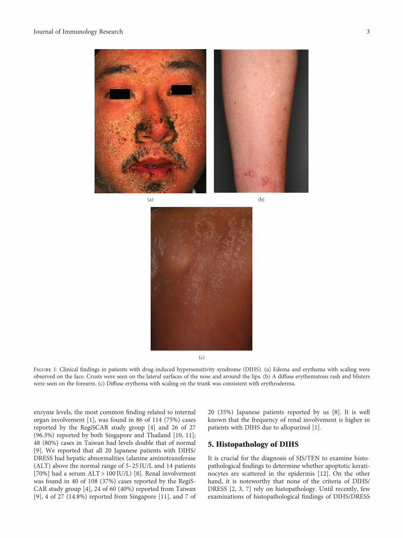
The skin manifestations of DIHS are maculopapularrash, erythema multiforme, exfoliative dermatitis, acute gen-eralized exanthematous pustular dermatosis-like eruption,and erythroderma [1–4]. We recently reviewed 20 patientswith DIHS/DRESS, including 7 with maculopapular rashtype, 5 with EM type, and 8 with erythroderma [8]. Initially,the upper trunk, face, and upper extremities are affected,followed by the involvement of lower extremities. Periorbital,facial edema with erythema and numerous scales and crustsaround the nose and lips are characteristic features ofDIHS/DRESS at the early stage (Figure 1(a)) [1, 5]. In somecases, bullous lesions are found on the forearm, which arealso characteristic features of DIHS/DRESS (Figure 1(b))[5]. The rash often generalizes into severe exfoliative derma-titis or erythroderma (Figure 1(c)) [1, 2, 5]. There is usuallyno mucocutaneous involvement, which helps distinguishDIHS/DRESS from other forms of severe drug eruptions,such as SJS and TEN [1].
4. Laboratory Data
Leukocytosis with atypical lymphocytes and eosinophilia ofvarying degree is a prominent feature of the syndrome [1].Leukocytosis was observed in 99 of 104 (95%) patientsreported by the RegiSCAR study group [4] and 15 of 20(75%) Japanese patients reported by us [8]. The presence ofatypical lymphocytes was demonstrated in 68 of 102 (67%)cases reported by the RegiSCAR study group [4], 38 of 60(63%) reported from Taiwan [9], 18.5% patients reportedfrom Thailand [10], 4 of 22 (18.2%) reported from Singapore[11], and 18 of 20 (90%) Japanese cases reported by us [8].Eosinophilia was observed in 108 of 114 (95%) cases reportedby the RegiSCAR study group [4], 31 of 60 (52%) reportedfrom Taiwan [9], 70.4% patients reported from Thailand[10], 22 out of 27 (81.5%) reported from Singapore [11],and 13 of 20 Japanese patients (65%) reported by us [8].Eosinophilia may often be delayed for 1 to 2 weeks andmay occur even after the elevations in liver enzyme levelsreturn to baseline [1]. In DIHS/DRESS, elevation of liver
Table 1
(a) Diagnostic criteria for drug reaction with eosinophilia andsystemic symptoms (DRESS) [2].
Diagnosis of DRESS is confirmed by the presence of all of thefollowing criteria:
(1) Cutaneous drug eruption
(2) Adenopathies≥ 2 cm in diameter or hepatitis (livertransaminases≥ 2 times upper limit of normal) or interstitialnephritis or interstitial pneumonitis or carditis
(3) Hematologic abnormalities: eosinophilia≥ 1.5× 10⁹ L−1 oratypical lymphocytes
(b) Criteria for potential cases of drug reaction with DRESS byRegiSCAR [7].
(1) Hospitalization
(2) Reaction suspected to be drug-related
(3) Acute skin rash∗
(4) Fever above 38°C∗
(5) Enlarged lymph nodes in at least two sites∗
(6) Involvement of at least one internal organ∗
(7) Blood count abnormalities
(i) Lymphocytes above or below the laboratory limits∗
(ii) Eosinophils above the laboratory limits∗
(iii) Platelets below the laboratory limits∗
∗Three or more criteria required. RegiSCAR: research group investigatingsevere cutaneous adverse reactions (SCAR) [7].
(c) Diagnostic criteria for drug-induced hypersensitivity syndrome(DIHS) established by a Japanese consensus group [3].
(1) Maculopapular rash developing 3 weeks after starting with alimited number of drugs
(2) Prolonged clinical symptoms 2 weeks after discontinuation ofthe causative drug
(3) Fever (≥38°C)(4) Liver abnormalities (alanine aminotransferase≥ 100U·L−1)a
(5) Leukocyte abnormalities (at least one present)
(a) Leukocytosis (≥11× 109 L−1)(b) Atypical lymphocytosis (≥5%)(c) Eosinophilia (≥1.5× 10⁹ L−1)
(6) Lymphadenopathy
(7) Human herpesvirus 6 reactivation
The diagnosis is confirmed by the presence of the seven criteria above(typical DIHS) or of the first five (1–5) criteria (atypical DIHS). aThis canbe replaced by other organ involvement, such as renal involvement.
2 Journal of Immunology Research
enzyme levels, the most common finding related to internalorgan involvement [1], was found in 86 of 114 (75%) casesreported by the RegiSCAR study group [4] and 26 of 27(96.3%) reported by both Singapore and Thailand [10, 11];48 (80%) cases in Taiwan had levels double that of normal[9]. We reported that all 20 Japanese patients with DIHS/DRESS had hepatic abnormalities (alanine aminotransferase(ALT) above the normal range of 5–25 IU/L and 14 patients[70%] had a serum ALT> 100 IU/L) [8]. Renal involvementwas found in 40 of 108 (37%) cases reported by the RegiS-CAR study group [4], 24 of 60 (40%) reported from Taiwan[9], 4 of 27 (14.8%) reported from Singapore [11], and 7 of
20 (35%) Japanese patients reported by us [8]. It is wellknown that the frequency of renal involvement is higher inpatients with DIHS due to allopurinol [1].
5. Histopathology of DIHS
It is crucial for the diagnosis of SJS/TEN to examine histo-pathological findings to determine whether apoptotic kerati-nocytes are scattered in the epidermis [12]. On the otherhand, it is noteworthy that none of the criteria of DIHS/DRESS [2, 3, 7] rely on histopathology. Until recently, fewexaminations of histopathological findings of DIHS/DRESS
(a) (b)
(c)
Figure 1: Clinical findings in patients with drug-induced hypersensitivity syndrome (DIHS). (a) Edema and erythema with scaling wereobserved on the face. Crusts were seen on the lateral surfaces of the nose and around the lips. (b) A diffuse erythematous rash and blisterswere seen on the forearm. (c) Diffuse erythema with scaling on the trunk was consistent with erythroderma.
3Journal of Immunology Research

were reported. Ortonne et al. [13] conducted a retrospectivestudy on 50 skin biopsies from 36 patients with DIHS/DRESSand demonstrated that patients with DIHS/DRESS fre-quently show foci of interface dermatitis, involving cutane-ous adnexa. Eosinophils were seen in only 20% andneutrophils in 42% of cases. Eczematous, interface dermati-tis, and acute generalized exanthematous pustulosis-likeand erythema multiforme-like patterns were observed in skinbiopsy samples from patients with DIHS/DRESS. The associ-ation of two or three of these patterns in a single biopsy wassignificantly more frequent in DRESS than in a series ofnondrug-induced dermatoses and appeared to be moremarked in DRESS with severe cutaneous lesions than inDRESS with less severe lesions. Interestingly, higher propor-tions of CD8+ and granzyme B+ lymphocytes were observedin DRESS with severe cutaneous eruptions. Furthermore,FoxP3+ regulatory T cells were found within the skin infil-trates in the acute phase of DRESS; however, these cells werenot numerous [13]. In addition, they found apoptotic kerati-nocytes in 60% of DRESS syndrome cases [13]. This observa-tion was consistent with the report by Walsh et al. [14],which showed that the presence of apoptotic keratinocytescorrelated with a more aggressive phenotype with liver injuryand an erythema multiforme-like cutaneous condition. Chiet al. [15] also found that skin biopsies of DIHS/DRESS dis-played various inflammatory aspects and showed that inter-face dermatitis with apoptotic keratinocytes was morefrequent in DIHS/DRESS than in maculopapular rash.
6. Treatment
The mortality rate of DIHS has recently been estimated tobe 2–14% [7, 9]. The mainstay of treatment is systemic cor-ticosteroids [1]. Wei et al. reviewed 91 cases with DRESS inTaiwan [9]. Patients treated with systemic corticosteroidslived longer than those not treated with corticosteroids(average 36.3 versus 12.7 days). In the survival group,approximately three-quarters of the patients received sys-temic corticosteroids, but their resolution time was 8 dayslonger than those without. A study from Singapore demon-strated that 25 of 27 (92.6%) patients with DIHS/DRESSreceived systemic corticosteroids, with no deaths resultingfrom DIHS/DRESS during the follow-up period in their caseseries [11].
Systemic corticosteroids, recommended for most cases ofDIHS/DRESS, should be initiated at a dose of 40–60mgprednisone equivalent daily, followed by a gradual dosereduction of prednisone given over 10 weeks to prevent rapidreconstitution of valid immune responses against variouspathogens; however, the mild form can resolve spontane-ously over a period of weeks [1, 17]. The development ofautoimmune diseases, such as lupus erythematosus and auto-immune thyroiditis, along with the generation of autoanti-bodies, was preferentially observed in the noncorticosteroidtreatment group in the late phase (>6 months) of DIHS/DRESS [16, 17]. Severe liver damage and noncorticosteroidtherapy during the acute stage were associated with thesubsequent generation of autoantibodies against plakinfamily proteins [16]. Therefore, corticosteroids, especially if
administered in the acute stage, may improve the long-termoutcome [17]. Recently, Leman et al. [18] described the suc-cessful treatment of a case of DIHS/DRESS with a tumornecrosis factor- (TNF-) α inhibitor containing lithium car-bonate. However, this is the only report of DIHS/DRESStreatment with a TNF-α inhibitor, and further clinical studiesare required.
7. Biomarkers of Disease Severity and HHV-6Reactivation in DIHS/DRESS
A major clinical focus during the diagnosis of DIHS and theselection of the most appropriate treatment is whether thereactivation of members of the Betaherpesvirinae subfamily,including HHV-6, develops subsequently to the drug hyper-sensitivity reaction [1–4]. HHV-6 DNA is detected in serumabout 3–5 weeks after disease onset, followed by dramaticrises in anti-HHV-6 IgG titers [1, 17]. Shiohara et al. per-formed a sequential analysis of viral loads and found thatthe cascade of reactivation events initiated by HHV-6 orEBV extended, after some delay, to HHV-7 also and eventu-ally to CMV [1]. In our previous study, when both HHV-6and CMV became reactivated in the same DIHS patients,HHV-6 DNA was detected 21–35 days after disease onsetand followed 10–21 days later by CMV DNA; the CMVIgG antibody titer also increased 10–21 days after elevationof the HHV-6 antibody titer [8]. In the cited study, 80% ofDIHS patients exhibited HHV-6 reactivation [8]. The magni-tudes of 2HHV-6 reactivation as evidenced by the increasesin HHV-6 DNA levels correlated well with the severities ofthe inflammatory responses [1]. However, no useful predic-tive marker of HHV-6 reactivation has yet been widelyaccepted. Moreover, useful biomarkers of the DIHS diseaseprocess have not yet been reported.
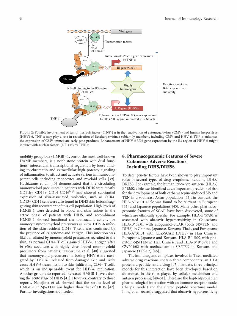
7.1. Tumor Necrosis Factor-α. We recently conducted com-parative assessments of, and detailed examinations on,patients with DIHS and measured their serum protein levels[8]. We found that the serum levels of TNF-α before treat-ment were significantly higher in the HHV-6 reactivationgroup than in the non-HHV-6 reactivation group. In that, aTNF-α level of 12 pg/mL allowed the detection of HHV-6reactivation [8]. Increased levels of proinflammatory cyto-kines including TNF-α and IL-6 have been reported inpatients with HHV-6 infections (severe cases of exanthemasubitum) and CMV infections [19, 20]. However, the exactmechanisms of the reactivation of these viruses have not beenfully elucidated. On the basis of both molecular and biologi-cal analyses, HHV-6, which is very similar to CMV, is theprototypic member of the Betaherpesvirinae [21, 22].Numerous in vitro and in vivo studies have sought to eluci-date the mechanisms of CMV reactivation and have reportedthat cytokine production, particularly of TNF-α, was impli-cated in reactivation [23–25]. TNF-α induces the expressionof CMV immediate early (IE) gene products, potentially ini-tiating viral replication from the latent state [26]. Expressionof CMV IE genes is controlled by IE promoter/enhancerregions, which contain binding sites for NF-κB, ATF (CREB),and Sp1. The NF-κB and ATF (CREB) sites are critical in
4 Journal of Immunology Research

terms of the regulation of IE gene expression [26, 27]. In con-trast, the R3 region of HHV-6 contains multiple putativebinding sites for cellular transcription factors, includingPEA3, NF-κB, and AP-2. Via interactions with NF-κB, thisregion strongly enhances the promoter activity of the U95gene, a potential homolog of the murine CMV IE2 gene[21]. These observations and our finding that the serumlevels of TNF-α were significantly higher in the HHV-6 reac-tivation group than in the non-HHV-6 reactivation group ofDIHS patients suggest that TNF-α may play a crucial role inHHV-6 reactivation (Figure 2). Moreover, an increase in thelevel of TNF-α before the commencement of treatment maybe an especially good biomarker allowing early recognitionof HHV-6 reactivation in patients with DIHS. Consistentwith this finding, it was reported that the TNF-α level washigher in hematopoietic stem cell transplantation recipientsexhibiting HHV-6 reactivation than in those who did notexhibit reactivation. Kamijima et al. recently investigated 28patients with trichloroethylene hypersensitivity syndromeand recorded the times of reaction onset after exposure totrichloroethylene/other drugs, the clinical manifestations,blood data, and the duration of virus reactivation [28]. Itwas found that an elevated TNF-α level on admission corre-lated significantly with an increase in HHV-6 DNA duringthe clinical course. This supports our suggestion that anincreased level of TNF-α prior to the commencement oftreatment may be an excellent biomarker allowing early rec-ognition of HHV-6 reactivation in patients with DIHS [8].Moreover, in our earlier study, the TNF-α levels decreasedsignificantly in parallel with the responses to treatment onlyin the DIHS group. To date, no widely accepted biomarkersof the DIHS disease process are available. Yoshikawa et al.reported elevated levels of TNF-α and IL-6 levels in four ofsix DIHS patients at the time of disease onset [29], indicatingthat the serum level of this protein reflected DIHS develop-ment. However, this report included only a small numberof DIHS/DRESS cases (n = 6), making it difficult to discussor compare these results with ours.
7.2. Interferon-Induced Protein 10. C-X-C motif chemokine10 (CXCL10), also known as interferon- (IFN-) γ-inducedprotein 10 kDa (IP-10), plays an important role in therecruitment of antiviral-specific cytotoxic T lymphocytesinto the target tissue [30]. Serum and/or tissue expressionof IP-10 is increased in organ-specific autoimmune diseasesand in interface dermatitis [30]. Contrary to other reports[8, 29], Chen et al. [31] demonstrated that many proinflam-matory cytokines and chemokines, including interleukin-(IL-) 1β, IL-2, IL-6, IFN-γ, and TNF-α, were significantlylower in DIHS/DRESS patients with HHV-6 reactivationwhen compared to those without HHV-6 reactivation. Inaddition, these mediators were significantly lower beforeand during HHV-6 reactivation, compared to cytokinelevels after HHV-6 reactivation in the same patients [31].These findings suggest the importance of the timing of sam-ple collection and that the influence of systemic corticoste-roids in patient treatment should be considered carefully.Future investigations using larger numbers of samples willbe needed.
7.3. Thymus and Activation-Regulated Chemokine and OtherTh2-Type Cytokines/Chemokines. Ogawa et al. recentlyreported that the serum thymus and activation-regulatedchemokine (TARC) levels were markedly higher in patientswith DIHS/DRESS than in patients with other forms of drugeruption including SJS/TEN and maculopapular erythema[32]. It was found that the serum TARC levels of patientsin the acute stage of DIHS correlated with disease activityand that the serum TARC levels in patients exhibitingHHV-6 reactivation were significantly higher than those inpatients not exhibiting HHV-6 reactivation [33]. Interest-ingly, the serum TARC levels correlated with the RegiSCARgroup diagnostic score for DRESS [33]. Such findings led usto suggest a pathogenic link between serum TARC levelsand HHV-6 reactivation. Although the precise mechanismremains largely unknown, one possible explanation is thatimmunosuppression triggers HHV-6 reactivation via regula-tory T cell activation induced by elevated TARC levels.Another possibility is that elevated TARC levels directlyactivate HHV-6 via the chemokine receptor homologues ofHHV-6 [33].
Yawalkar et al. [34] examined skin sections from patientswith characteristic, acute, drug-induced, maculopapularexanthem to determine the potential role of IL-5 and distinctchemokines in the recruitment and activation of eosinophilsinto the skin. They demonstrated that drug-induced maculo-papular exanthems express significantly increased amountsof IL-5 and eotaxin [34]. However, whether these Th2 cyto-kines/chemokines are involved in the reactivation of HHV-6 in DIHS/DRESS has not yet to be determined.
7.4. Plasmacytoid Dendritic Cells. Plasmacytoid dendriticcells (pDCs) play a defensive role against viruses [35]. Previ-ously, we demonstrated that pDCs accumulate in the skin ofpatients with DIHS/DRESS and that the number of pDCs incirculation decreases significantly around the time of viralreactivation. Upon viral infection, stimulated pDCs areprompted to differentiate into DCs by autocrine IFN-α andTNF-α and to prime naive CD4+ T cells to produce IFN-γand IL-10 [36]. In addition, pDCs preferentially secrete theproinflammatory chemokine macrophage inflammatoryprotein- (MIP-) 1α, which recruits mostly Th1-type effectorcells and causes the production of other proinflammatorycytokines [37]. Therefore, decreased levels of proinflamma-tory cytokines/chemokines may result from decreased levelsof pDCs and depress the antiviral capacity in patients withDRESS. After reactivation, HHV-6 may further modulatethe release of these cytokines from peripheral blood mono-nuclear cells, including IFN-γ, TNF-α, and IL-1β, as reflectedby their increased levels in the blood [31].
7.5. High-Mobility Group Box-1.Damage-associated molecu-lar pattern molecules (DAMPs) released from damaged cellsare signals for initiating immune responses in various organsthrough their activation after interacting with pattern recog-nition receptors and/or Toll-like receptors, thereby promot-ing rapid recruitment of bone marrow-derived leukocytesto the target tissues for inflammation and regeneration undervarious aseptic inflammatory conditions [38, 39]. High-
5Journal of Immunology Research
mobility group box (HMGB)-1, one of the most well-knownDAMP members, is a nonhistone protein with dual func-tions: intercellular transcriptional regulation by loose bind-ing to chromatin and extracellular high potency signalingof inflammation to attract and activate various immunocom-petent cells including monocytes and myeloid cells [39].Hashizume et al. [40] demonstrated that the circulatingmonomyeloid precursors in patients with DIHS were mostlyCD11b+ CD13+ CD14 CD16high and showed substantialexpression of skin-associated molecules, such as CCR4.CD13+ CD14 cells were also found in DIHS skin lesions, sug-gesting skin recruitment of this cell population. High levels ofHMGB-1 were detected in blood and skin lesions in theactive phase of patients with DIHS, and recombinantHMGB-1 showed functional chemoattractant activity formonocytes/monomyeloid precursors in vitro. HHV-6 infec-tion of the skin-resident CD4+ T cells was confirmed bythe presence of its genome and antigen. This infection waslikely mediated by monomyeloid precursors recruited to theskin, as normal CD4+ T cells gained HHV-6 antigen afterin vitro coculture with highly virus-loaded monomyeloidprecursors from patients. Hashizume et al. [40] suggestedthat monomyeloid precursors harboring HHV-6 are navi-gated by HMGB-1 released from damaged skin and likelycause HHV-6 transmission to skin-infiltrating CD4+ T cells,which is an indispensable event for HHV-6 replication.Another group also reported increased HMGB-1 levels dur-ing the acute stage of DIHS [41]. However, contrary to thosereports, Nakajima et al. showed that the serum level ofHMGB-1 in SJS/TEN was higher than that of DIHS [42].Further investigations are needed.
8. Pharmacogenomic Features of SevereCutaneous Adverse ReactionsIncluding DIHS/DRESS
To date, genetic factors have been shown to play importantroles in several types of drug eruptions, including DIHS/DRESS. For example, the human leucocyte antigen- (HLA-)B∗15:02 allele was identified as an important predictor of riskfor the development of both carbamazepine-induced SJS andTEN in a southeast Asian population [43]; in contrast, theHLA-A∗31:01 allele was found to be relevant in European[44] and Japanese populations [45]. Many other pharmaco-genomic features of SCAR have been discovered, some ofwhich are ethnically specific. For example, HLA-B∗57:01 isassociated with abacavir hypersensitivity in Caucasians;HLA-B∗58:01 with allopurinol-SCAR (both SJS/TEN andDIHS) in Chinese, Japanese, Koreans, Thais, and Europeans;HLA-A∗31:01 with CBZ-SCAR (DIHS) in Han Chinese,Europeans, Japanese and Koreans; HLA-B∗15:02 with phe-nytoin-SJS/TEN in Han Chinese; and HLA-B∗B∗59:01 andCW∗01:02 with methazolamide-SJS/TEN in Koreans andJapanese (Table 2) [46].
The immunogenic complexes involved in T cell-mediatedadverse drug reactions contain three components: an HLAprotein, a peptide, and a drug [47]. To date, three principalmodels for this interaction have been developed, based ondifferences in the roles played by cellular metabolism andantigen processing [48–51]. These are the hapten/prohaptenpharmacological interaction with an immune receptor model(the p.i. model) and the altered peptide repertoire model.Illing et al. recently suggested that abacavir hypersensitivity
MGI
Viral gene
IE gene (CMV)
Induction of CMV IE gene expressionby TNF-�훼
Transcription factorsCREB
NF-�휅B binding to the R3 regionof HHV6
Reactivation of theBetaherpesvirinaesubfamily
Enhancement of HHV6 U95 gene expressionby HHV6 R3 region interacted with NF-�휅BTNF-�훼?
TNF-�훼
Highlyhomologous
U95 gene (HHV6)
NF-�휅B
NF-�휅B
NF-�휅B HMGI
HMG HMG HMG HMGPRDIV PRDI-III PRDI-II NRDI
HMGI
HMGI
HMGI
ATF-2
ATF-2
ATF-2cJunNF-�휅BIRF (IRF3/7)
cJun
cJun
HMGIIRF3
IRF3
MGI
Figure 2: Possible involvement of tumor necrosis factor- (TNF-) α in the reactivation of cytomegalovirus (CMV) and human herpesvirus(HHV)-6. TNF-α may play a role in reactivation of Betaherpesvirinae subfamily members, including CMV and HHV-6. TNF-α enhancesthe expression of CMV immediate early gene products. Enhancement of HHV-6 U95 gene expression by the R3 region of HHV-6 mightinteract with nuclear factor- (NF-) κB by TNF-α.
6 Journal of Immunology Research
syndrome could be explained by reference to the altered pep-tide repertoire model [47, 48].
Recently, an HLA class I allele, HLA-B∗13:01, has beenidentified as a marker of susceptibility to DIHS attributableto dapsone (dapsone hypersensitivity syndrome) [52–54].It was initially unclear how dapsone interacted with HLA-B∗13:01.
9. Computational Analyses of theDapsone/HLA-B∗13:01 Interactions
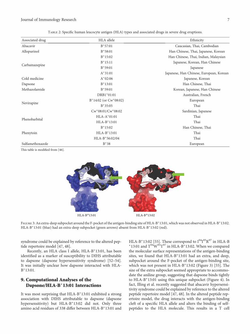
It was most surprising that HLA-B∗13:01 exhibited a strongassociation with DIHS attributable to dapsone (dapsonehypersensitivity) but HLA-B∗13:02 did not. Only threeamino acid residues of 338 differ between HLA-B∗13:01 and
HLA-B∗13:02 [55]. These correspond to I94I95R97 in HLA-B∗13:01 and T94W95T97 in HLA-B∗13:02. When we comparedthe molecular surface representations of the antigen-bindingsites, we found that HLA-B∗13:01 had an extra, and deep,subpocket around the F-pocket of the antigen-binding site,which was not present in HLA-B∗13:02 (Figure 3) [55]. Thesize of the extra subpocket seemed appropriate to accommo-date the aniline group, suggesting that dapsone binds tightlyto HLA-B∗13:01 using this unique subpocket (Figure 4). Infact, Illing et al. recently suggested that abacavir hypersensi-tivity syndrome could be explained by reference to the alteredpeptide repertoire model [47, 48]. In the altered peptide rep-ertoire model, the drug interacts with the antigen-bindingcleft of a specific HLA allele and alters the binding of self-peptides to the HLA molecule. This results in a T cell
HLA-B⁎13:01 HLA-B⁎13:02
Figure 3: An extra-deep subpocket around the F-pocket of the antigen-binding site ofHLA-B∗13:01, whichwas not observed inHLA-B∗13:02.HLA-B∗13:01 (blue) had an extra-deep subpocket (green arrows) absent from HLA-B∗13:02 (red).
Table 2: Specific human leucocyte antigen (HLA) types and associated drugs in severe drug eruptions.
Associated drug HLA allele Ethnicity
Abacavir B∗57:01 Caucasian, Thai, Cambodian
Allopurinol B∗58:01 Han Chinese, Thai, Japanese, Korean
Carbamazepine
B∗15:02 Han Chinese, Thai, Indian, Malaysian
B∗15:11 Japanese, Korean, Han Chinese
B∗59:01 Japanese
A∗31:01 Japanese, Han Chinese, European, Korean
Cold medicine A∗02:06 Japanese, Korean
Dapsone B∗13:01 Han Chinese, Thai
Methazolamide B∗59:01 Korean, Japanese, Han Chinese
Nevirapine
DRB1∗01:01 Australian, French
B∗14:02 (or Cw∗08:02) European
B∗35:05 Thai
Cw∗08:01/Cw∗08:02 Sardinian, Japanese
PhenobarbitalHLA-A∗01:01 Thai
HLA-B∗13:01 Thai
Phenytoin
B∗15:02 Han Chinese, Thai
HLA-B∗13:01 Thai
HLA-B∗56:02/04 Thai
Sulfamethoxazole B∗38 European
This table is modified from [46].
7Journal of Immunology Research

response. X-ray crystallography revealed that abacavir wasspecifically bound in the vicinity of the F-pocket of theantigen-binding cleft of the HLA-B∗57:01 allele. This regionwas identified as a marker of susceptibility to abacavirhypersensitivity syndrome. From these findings, an “alteredpeptide repertoire” model involving the binding of dapsoneto HLA-B∗13:01 may also be appropriate analogous to theabacavir allergy model.
10. Conclusion
During the course of DIHS, HHV-6 reactivation triggerssymptom recurrence and may be fatal by causing serious dys-functions including liver failure. Therefore, it is essential toidentify factors predictive of virus reactivation. In this review,we have emphasized that several cytokines/chemokinesincluding levels of TNF-α and TARC are good biomarkersof virus reactivation; however, further investigations arerequired. Moreover, the association between causative drugsand genetic factors, including HLA polymorphisms, rendersit possible to choose appropriate treatments and improvepatient outcomes.
Conflicts of Interest
The author declares that he has no conflicts of interest.
References
[1] T. Shiohara, M. Inaoka, and Y. Kano, “Drug-induced hyper-sensitivity syndrome (DIHS): a reaction induced by a complexinterplay among herpesviruses and antiviral and antidrugimmune responses,” Allergology International, vol. 55, no. 1,pp. 1–8, 2006.
[2] H. Bocquet, M. Bagot, and J. C. Roujeau, “Drug-induced pseu-dolymphoma and drug hypersensitivity syndrome (drug rashwith eosinophilia and systemic symptoms: DRESS),” Seminarsin Cutaneous Medicine and Surgery, vol. 15, no. 4, pp. 250–257, 1996.
[3] T. Shiohara, M. Iijima, Z. Ikezawa, and K. Hashimoto, “Thediagnosis of a DRESS syndrome has been sufficiently estab-lished on the basis of typical clinical features and viral reac-tivations,” British Journal of Dermatology, vol. 156, no. 5,pp. 1083-1084, 2007.
[4] S. H. Kardaun, P. Sekula, L. Valeyrie-Allanore et al., “Drugreaction with eosinophilia and systemic symptoms (DRESS):an original multisystem adverse drug reaction. Results fromthe prospective RegiSCAR study,” British Journal of Dermatol-ogy, vol. 169, no. 5, pp. 1071–1080, 2013.
[5] H.Watanabe, “Hypersensitivity syndrome due to trichloroeth-ylene exposure: a severe generalized skin reaction resemblingdrug-induced hypersensitivity syndrome,” The Journal of Der-matology, vol. 38, no. 3, pp. 229–235, 2011.
[6] P. Tennis and R. S. Stern, “Risk of serious cutaneous disordersafter initiation of use of phenytoin, carbamazepine, or sodiumvalproate: a record linkage study,” Neurology, vol. 49, no. 2,pp. 542–546, 1997.
[7] S. H. Kardaun, A. Sidoroff, L. Valeyrie-Allanore et al., “Var-iability in the clinical pattern of cutaneous side-effects ofdrugs with systemic symptoms: does a DRESS syndrome reallyexist?,” British Journal of Dermatology, vol. 156, no. 3, pp. 609–611, 2007.
[8] H. Uno, K. Kabashima, M. Tohyama et al., “TNF-α as a usefulpredictor of human herpesvirus-6 reactivation and indicator ofthe disease process in drug-induced hypersensitivity syndrome(DIHS)/drug reaction with eosinophilia and systemic symp-toms (DRESS),” Journal of Dermatological Science, vol. 74,no. 2, pp. 177–179, 2014.
[9] C. H. Wei, R. Chung-Yee Hui, C. J. Chang et al., “Identifyingprognostic factors for drug rash with eosinophilia and sys-temic symptoms (DRESS),” European Journal of Dermatology,vol. 21, no. 6, pp. 930–937, 2011.
[10] P. Wongkitisophon, K. Chanprapaph, P. Rattanakaemakorn,and V. Vachiramon, “Six-year retrospective review of drugreaction with eosinophilia and systemic symptoms,” Acta Der-mato-Venereologica, vol. 92, no. 2, pp. 200–205, 2012.
[11] C. C. Ang, Y. S. Wang, E. L. Yoosuff, and Y. K. Tay, “Retro-spective analysis of drug-induced hypersensitivity syndrome:a study of 27 patients,” Journal of the American Academy ofDermatology, vol. 63, no. 2, pp. 219–227, 2010.
[12] H. Assier, S. Bastuji-Garin, J. Revuz, and J. C. Roujeau, “Ery-thema multiforme with mucous membrane involvement andStevens-Johnson syndrome are clinically different disorderswith distinct causes,” Archives of Dermatology, vol. 131,no. 5, pp. 539–543, 1995.
[13] N. Ortonne, L. Valeyrie-Allanore, S. Bastuji-Garin et al., “His-topathology of drug rash with eosinophilia and systemicsymptoms syndrome: a morphological and phenotypicalstudy,” British Journal of Dermatology, vol. 173, no. 1,pp. 50–58, 2015.
HLA-B⁎1301 HLA-B⁎1302
Figure 4: Dapsone binds more tightly to HLA-B∗13:01 than to 13:02. Binding models before molecular dynamic simulations for dapsone–HLA-B∗13:01 (blue) and dapsone–HLA-B∗13:02 (red), based on observations of the stick representation of their HLA three-dimensionalhomology models. Dapsone (green) inserts are deeper in HLA-B∗13:01 (blue) than in HLA-B∗13:02 (red).
8 Journal of Immunology Research

[14] S. Walsh, S. Diaz-Cano, E. Higgins et al., “Drug reaction witheosinophilia and systemic symptoms: is cutaneous phenotypea prognostic marker for outcome? A review of clinicopatholog-ical features of 27 cases,” British Journal of Dermatology,vol. 168, no. 2, pp. 391–401, 2013.
[15] M. H. Chi, R. C.-Y. Hui, C. H. Yang et al., “Histopathologicalanalysis and clinical correlation of drug reaction with eosino-philia and systemic symptoms (DRESS),” British Journal ofDermatology, vol. 170, no. 4, pp. 866–873, 2014.
[16] A. Takehara, Y. Aoyama, M. Kurosawa et al., “Longitudinalanalysis of antibody profiles against plakins in severe drugeruptions: emphasis on correlation with tissue damage indrug-induced hypersensitivity syndrome and drug reactionwith eosinophilia and systemic symptoms,” British Journal ofDermatology, vol. 175, no. 5, pp. 944–952, 2016.
[17] T. Shiohara, Y. Kano, K. Hirahara, and Y. Aoyama, “Predictionand management of drug reaction with eosinophilia and sys-temic symptoms (DRESS),” Expert Opinion on Drug Metabo-lism & Toxicology, vol. 13, no. 7, pp. 701–704, 2017.
[18] R. E. Leman, L. Chen, X. Shi, S. P. Rolimpandoei, X. Ling,and Y. Su, “Drug reaction with eosinophilia and systemicsymptoms (DRESS) successfully treated with tumor necrosisfactor-α inhibitor,” JAAD Case Reports, vol. 3, no. 4,pp. 332–335, 2017.
[19] T. Ichiyama, Y. Ito, M. Kubota, T. Yamazaki, K. Nakamura,and S. Furukawa, “Serum and cerebrospinal fluid levels ofcytokines in acute encephalopathy associated with humanherpesvirus-6 infection,” Brain Development, vol. 31, no. 10,pp. 731–738, 2009.
[20] C. Y. W. Tong, A. Bakran, H. Williams, L. E. Cuevas, J. S. M.Peiris, and C. A. Hart, “Association of tumour necrosis factoralpha and interleukin 6 levels with cytomegalovirus DNAdetection and disease after renal transplantation,” Journal ofMedical Virology, vol. 64, no. 1, pp. 29–34, 2001.
[21] M. Takemoto, T. Shimamoto, Y. Isegawa, and K. Yamanishi,“The R3 region, one of three major repetitive regions of humanherpesvirus 6, is a strong enhancer of immediate-early geneU95,” Journal of Virology, vol. 75, no. 21, pp. 10149–10160,2001.
[22] A. Fujita, M. Ihira, R. Suzuki et al., “Elevated serum cytokinelevels are associated with human herpesvirus 6 reactivationin hematopoietic stem cell transplantation recipients,” TheJournal of Infection, vol. 57, no. 3, pp. 241–248, 2008.
[23] W. D. Döcke, S. Prösch, E. Fietze et al., “Cytomegalovirus reac-tivation and tumour necrosis factor,” Lancet, vol. 343,no. 8892, pp. 268-269, 1994.
[24] A. Humar, P. St Louis, T. Mazzulli et al., “Elevated serumcytokines are associated with cytomegalovirus infection anddisease in bone marrow transplant recipients,” The Journal ofInfectious Diseases, vol. 179, no. 2, pp. 484–488, 1999.
[25] E. Fietze, S. Prösch, P. Reinke et al., “Cytomegalovirusinfection in transplant recipients. The role of tumor necrosisfactor,” Transplantation, vol. 58, no. 6, pp. 675–680, 1994.
[26] M. Hummel and M. M. Abecassis, “A model for reactivationof CMV from latency,” Journal of Clinical Virology, vol. 25,Supplement 2, pp. S123–S136, 2002.
[27] C. O. Simon, C. K. Seckert, D. Dreis, M. J. Reddehase, andN. K. A. Grzimek, “Role for tumor necrosis factor alpha inmurine cytomegalovirus transcriptional reactivation inlatently infected lungs,” Journal of Virology, vol. 79, no. 1,pp. 326–340, 2005.
[28] M. Kamijima, H. Wang, O. Yamanoshita et al., “Occupationaltrichloroethylene hypersensitivity syndrome: human herpesvi-rus 6 reactivation and rash phenotypes,” Journal of Dermato-logical Science, vol. 72, no. 3, pp. 218–224, 2013.
[29] T. Yoshikawa, A. Fujita, A. Yagami et al., “Human herpesvirus6 reactivation and inflammatory cytokine production inpatients with drug-induced hypersensitivity syndrome,” Jour-nal of Clinical Virology, vol. 37, Supplement 1, pp. S92–S96,2006.
[30] S. Kasraie, M. Niebuhr, V. Kopfnagel, O. Dittrich-Breiholz,M. Kracht, and T. Werfel, “Macrophages from patients withatopic dermatitis show a reduced CXCL10 expression inresponse to staphylococcal α-toxin,” Allergy, vol. 67, no. 1,pp. 41–49, 2012.
[31] Y. C. Chen, H. H. Chiang, Y. T. Cho et al., “Human herpesvirus reactivations and dynamic cytokine profiles in patientswith cutaneous adverse drug reactions –a prospective compar-ative study,” Allergy, vol. 70, no. 5, pp. 568–575, 2015.
[32] K. Ogawa, H. Morito, A. Hasegawa et al., “Identification ofthymus and activation-regulated chemokine (TARC/CCL17)as a potential marker for early indication of disease and predic-tion of disease activity in drug-induced hypersensitivitysyndrome (DIHS)/drug rash with eosinophilia and systemicsymptoms (DRESS),” Journal of Dermatological Science,vol. 69, no. 1, pp. 38–43, 2013.
[33] K. Ogawa, H. Morito, A. Hasegawa et al., “Elevated serum thy-mus and activation-regulated chemokine (TARC/CCL17)relates to reactivation of human herpesvirus 6 in drug reactionwith eosinophilia and systemic symptoms (DRESS)/drug-induced hypersensitivity syndrome (DIHS),” British Journalof Dermatology, vol. 171, no. 2, pp. 425–427, 2014.
[34] N. Yawalkar, M. Shrikhande, Y. Hari, H. Nievergelt, L. R.Braathen, and W. J. Pichler, “Evidence for a role for IL-5 andeotaxin in activating and recruiting eosinophils in drug-induced cutaneous eruptions,” The Journal of Allergy and Clin-ical Immunology, vol. 106, no. 6, pp. 1171–1176, 2000.
[35] K. Sugita, M. Tohyama, H. Watanabe et al., “Fluctuation ofblood and skin plasmacytoid dendritic cells in drug-inducedhypersensitivity syndrome,” The Journal of Allergy and Clini-cal Immunology, vol. 126, no. 2, pp. 408–410, 2010.
[36] G. Penna, M. Vulcano, A. Roncari, F. Facchetti, S. Sozzani, andL. Adorini, “Cutting edge: differential chemokine productionby myeloid and plasmacytoid dendritic cells,” The Journal ofImmunology, vol. 169, no. 12, pp. 6673–6676, 2002.
[37] Y. J. Liu, “IPC: professional type 1 interferon producing cellsand plasmacytoid dendritic cell precursors,” Annual Reviewof Immunology, vol. 23, no. 1, pp. 275–306, 2005.
[38] M. E. Bianchi and A. A. Manfredi, “High-mobility group box 1(HMGB1) protein at the crossroads between innate and adap-tive immunity,” Immunological Reviews, vol. 220, no. 1,pp. 35–46, 2007.
[39] G. P. Sims, D. C. Rowe, S. T. Rietdijk, R. Herbst, and A. J.Coyle, “HMGB1 and RAGE in inflammation and cancer,”Annual Review of Immunology, vol. 28, no. 1, pp. 367–388,2010.
[40] H. Hashizume, T. Fujiyama, J. Kanebayashi, Y. Kito, M. Hata,and H. Yagi, “Skin recruitment of monomyeloid precursorsinvolves human herpesvirus-6 reactivation in drug allergy,”Allergy, vol. 68, no. 5, pp. 681–689, 2013.
[41] H. Fujita, S. Matsukura, T. Watanabe et al., “The serum level ofHMGB1 (high mobility group box 1 protein) is preferentiallyhigh in drug-induced hypersensitivity syndrome/drug reaction
9Journal of Immunology Research

with eosinophilia and systemic symptoms,” British Journal ofDermatology, vol. 171, no. 6, pp. 1585–1588, 2014.
[42] S. Nakajima, H. Watanabe, M. Tohyama et al., “High-mobilitygroup box 1 protein (HMGB1) as a novel diagnostic tool fortoxic epidermal necrolysis and Stevens-Johnson syndrome,”Archives of Dermatology, vol. 147, no. 9, pp. 1110–1112, 2011.
[43] W. H. Chung, S. I. Hung, H. S. Hong et al., “Medical genetics: amarker for Stevens-Johnson syndrome,” Nature, vol. 428,no. 6982, p. 486, 2004.
[44] M.McCormack, A. Alfirevic, S. Bourgeois et al., “HLA-A∗3101and carbamazepine-induced hypersensitivity reactions inEuropeans,” The New England Journal of Medicine, vol. 364,no. 12, pp. 1134–1143, 2011.
[45] T. Ozeki, T. Mushiroda, A. Yowang et al., “Genome-wideassociation study identifiesHLA-A∗3101 allele as a genetic riskfactor for carbamazepine-induced cutaneous adverse drugreactions in Japanese population,”HumanMolecular Genetics,vol. 20, no. 5, pp. 1034–1041, 2011.
[46] W. H. Chung, C. W. Wang, and R. L. Dao, “Severe cutaneousadverse drug reactions,” The Journal of Dermatology, vol. 43,no. 7, pp. 758–766, 2016.
[47] P. T. Illing, N. A. Mifsud, and A. W. Purcell, “Allotype specificinteractions of drugs and HLA molecules in hypersensitivityreactions,” Current Opinion in Immunology, vol. 42, pp. 31–40, 2016.
[48] P. T. Illing, J. P. Vivian, N. L. Dudek et al., “Immune self-reactivity triggered by drug-modified HLA-peptide reper-toire,” Nature, vol. 486, no. 7404, pp. 554–558, 2012.
[49] C. C. Bell, L. Faulkner, K. Martinsson et al., “T-cells fromHLA-B∗57:01+ human subjects are activated with abacavirthrough two independent pathways and induce cell death bymultiple mechanisms,” Chemical Research in Toxicology.,vol. 26, no. 5, pp. 759–766, 2013.
[50] W. J. Pichler, A. Beeler, M. Keller et al., “Pharmacologicalinteraction of drugs with immune receptors: the p-i concept,”Allergology International, vol. 55, no. 1, pp. 17–25, 2006.
[51] J. Adam, W. J. Pichler, and D. Yerly, “Delayed drug hypersen-sitivity: models of T-cell stimulation,” British Journal of Clini-cal Pharmacology, vol. 71, no. 5, pp. 701–707, 2011.
[52] F. R. Zhang, H. Liu, A. Irwanto et al., “HLA-B∗13:01 and thedapsone hypersensitivity syndrome,” The New England Jour-nal of Medicine, vol. 369, no. 17, pp. 1620–1628, 2013.
[53] H.Wang, L. Yan,G.Zhang et al., “Association betweenHLA-B∗
1301 and dapsone-induced hypersensitivity reactions amongleprosy patients in China,” The Journal of Investigative of Der-matology, vol. 133, no. 11, pp. 2642–2644, 2013.
[54] T. Tempark, P. Satapornpong, P. Rerknimitr et al., “Dapsone-induced severe cutaneous adverse drug reactions are stronglylinked with HLA-B∗13: 01 allele in the Thai population,” Phar-macogenetics and Genomics, vol. 27, no. 12, pp. 429–437, 2017.
[55] H. Watanabe, Y. Watanabe, Y. Tashiro et al., “A dockingmodel of dapsone bound to HLA-B∗13:01 explains the riskof dapsone hypersensitivity syndrome,” Journal of Dermato-logical Science, vol. 88, no. 3, pp. 320–329, 2017.
10 Journal of Immunology Research
Review ArticleAn Updated Review of the Molecular Mechanisms inDrug Hypersensitivity
Chun-Bing Chen ,1,2,3,4,5,6 Riichiro Abe,7 Ren-You Pan,1,2 Chuang-Wei Wang,1,2
Shuen-Iu Hung ,8 Yi-Giien Tsai ,9,10,11 and Wen-Hung Chung 1,2,3,4,6,12
1Department of Dermatology, Drug Hypersensitivity Clinical and Research Center, Chang Gung Memorial Hospital, Taipei, Linkou,Keelung, Taiwan2Chang Gung Immunology Consortium, Chang Gung Memorial Hospital and Chang Gung University, Taoyuan, Taiwan3Whole-Genome Research Core Laboratory of Human Diseases, Chang Gung Memorial Hospital, Keelung, Taiwan4College of Medicine, Chang Gung University, Taoyuan, Taiwan5Graduate Institute of Clinical Medical Sciences, Chang Gung University, Taoyuan, Taiwan6Immune-Oncology Center of Excellence, Chang Gung Memorial Hospital, Linkou, Taiwan7Division of Dermatology, Niigata University Graduate School of Medical and Dental Sciences, Niigata, Japan8Department and Institute of Pharmacology, School of Medicine, Infection and Immunity Research Center,National Yang-Ming University, Taipei, Taiwan9Division of Pediatric Allergy and Immunology, Changhua Christian Hospital, Changhua City, Taiwan10School of Medicine, Kaohsiung Medical University, Kaohsiung, Taiwan11School of Medicine, Chung Shan Medical University, Taichung, Taiwan12Department of Dermatology, Xiamen Chang Gung Hospital, Xiamen, China
Correspondence should be addressed to Yi-Giien Tsai; [email protected] and Wen-Hung Chung; [email protected]
Received 1 September 2017; Accepted 9 November 2017; Published 13 February 2018
Academic Editor: Kurt Blaser
Copyright © 2018 Chun-Bing Chen et al. This is an open access article distributed under the Creative Commons AttributionLicense, which permits unrestricted use, distribution, and reproduction in anymedium, provided the original work is properly cited.
Drug hypersensitivity may manifest ranging from milder skin reactions (e.g., maculopapular exanthema and urticaria) to severesystemic reactions, such as anaphylaxis, drug reactions with eosinophilia and systemic symptoms (DRESS)/drug-inducedhypersensitivity syndrome (DIHS), or Stevens–Johnson syndrome (SJS)/toxic epidermal necrolysis (TEN). Currentpharmacogenomic studies have made important strides in the prevention of some drug hypersensitivity through the identificationof relevant genetic variants, particularly for genes encoding drug-metabolizing enzymes and human leukocyte antigens (HLAs).The associations identified by these studies are usually drug, phenotype, and ethnic specific. The drug presentation models thatexplain how small drug antigens might interact with HLA and T cell receptor (TCR) molecules in drug hypersensitivity include thehapten theory, the p-i concept, the altered peptide repertoire model, and the altered TCR repertoire model. The broad spectrum ofclinical manifestations of drug hypersensitivity involving different drugs, as well as the various pathomechanisms involved, makesthe diagnosis and management of it more challenging. This review highlights recent advances in our understanding of thepredisposing factors, immune mechanisms, pathogenesis, diagnostic tools, and therapeutic approaches for drug hypersensitivity.
1. Introduction
Drug hypersensitivity reactions are an important publichealth problem due to their potential to cause life-threatening anaphylaxis and rare severe cutaneous adversereactions (SCAR). Drug hypersensitivity can be induced
by immunologically mediated reactions (referred as drugallergies) as well as nonallergic direct mast cell-mediateddrug reactions. Immunologic reactions have been dividedinto four categories according to the classical Gell andCoombs system: type I reactions, which are immediate inonset and mediated by IgE and mast cells and/or basophils;
HindawiJournal of Immunology ResearchVolume 2018, Article ID 6431694, 22 pageshttps://doi.org/10.1155/2018/6431694
type II reactions, which are delayed in onset and caused byantibody- (usually IgG) mediated cell destruction; type IIIreactions, which are delayed in onset and caused by IgG drugimmune complex deposition and complement activation;and type IV reactions, which are delayed in onset and are Tcell mediated [1]. According to the World Allergy Organiza-tion (WAO), drug hypersensitivity reactions can also be cat-egorized into immediate reactions and delayed reactionsbased upon the timing of the appearance of symptoms [2].
Immediate-type reactions usually occur within minutesor hours of drug exposure. The clinical manifestations rangefrom pruritus, urticaria, angioedema, and bronchospasm toanaphylaxis. Type I reactions require the presence of drug-specific IgE or the portion of the drug that forms a haptencomplex. Drug-specific IgE is produced upon the first expo-sure to the drug antigen, and then, it binds to basophils ormast cells with the high-affinity Fc receptor. Upon the nextexposure to the same drug, two or more IgE molecules onthe basophil or mast cell surface may then bind to onemultivalent antigen molecule, initiating a series of cellularactivation events. This activation causes the extracellularrelease of granules with preformed inflammatory mediators,including histamine, leukotrienes, prostaglandins, heparin,and other cytokines [3]. IgE-mediated immunologic drugallergy represents a smaller fraction of drug hypersensitivitycompared with nonimmunologic drug hypersensitivity [4].According to the WAO classification system, immunologicanaphylaxis can be caused by an IgE-mediated or non-IgE-mediated mechanism, whereas nonimmunologic anaphy-laxis involves direct mast cell activation [2]. Regardless ofthe underlying mechanism, however, the clinical symptomsof both types of anaphylaxis are similar and often indistin-guishable. The mechanism of immediate-type reactions isexplained more fully later in this article. In this review, theterminology used to categorize “immediate” or “delayed”drug hypersensitivity is in accordance with the WAOclassification system. At the same time, the immediate-typereactions discussed herein are composed of both IgE-mediated reactions as defined by the Gell and Coombssystem, as well as non-IgE-mediated and nonimmunologicanaphylactic reactions.
Delayed-type reactions consist primarily of type IVreactions, which are T cell-mediated delayed-type drughypersensitivity reactions. These reactions usually take sev-eral days or even weeks to manifest following drug exposure.These manifestations range from mild maculopapularexanthema (MPE), contact dermatitis, chronic allergicrhinitis, chronic asthma, nephritis, hepatitis, and fixed drugeruptions (FDEs) to life-threatening SCAR. SCAR includesdrug reactions with eosinophilia and systemic symptoms(DRESS)/drug-induced hypersensitivity syndrome (DIHS),Stevens–Johnson syndrome (SJS) and toxic epidermal necro-lysis (TEN), and acute generalized exanthematous pustulosis(AGEP) [5]. The MPE phenotype consists of self-limiteddiffuse erythematous macules and papules without systemicinvolvement [6]. DRESS syndrome, meanwhile, is character-ized by cutaneous involvement with typical skin eruptions(e.g., exfoliative dermatitis and generalized maculopapularexanthema), fever, atypical lymphocytosis, eosinophilia,
lymphadenopathy, and systemic involvement (e.g., liverinvolvement and kidney involvement). This hypersensitivitysyndrome was first named after many different terms hadalready been used to describe the syndrome, with thoseterms, such as “anticonvulsant hypersensitivity syndrome,”“allopurinol hypersensitivity syndrome,” and “sulfone syn-drome,” primarily depending on the culprit drug involved[7, 8]. The term “DRESS” was initially proposed by Bocquetet al. in 1996 in order to provide a more concise descriptionof the syndrome and decrease the ambiguity resultingfrom the various terms previously used to refer to it [9].That said, it should be noted that DRESS is also termed“DIHS” by Japanese experts, with the criteria of DRESSas defined by the RegiSCAR group and the criteria ofDIHS as defined by Japanese experts being similar, exceptthat HHV-6 reactivation is included in the diagnostic cri-teria for DIHS [10]. This nosology is somewhat confusing;however, there is a consensus that DRESS and DIHS arelikely within the same disease spectrum. Specifically,patients with typical DIHS may represent a severe formof DRESS syndrome [11]. SJS and TEN (SJS/TEN) arecharacterized as a rapidly progressing blistering exanthemaof purpuric macules and target-like lesions accompaniedby mucosal involvement and skin detachment. SJS isdefined as involving less than 10% body surface area skindetachment, SJS-TEN overlap as involving 10–29%, andTEN as involving more than 30% [12]. AGEP, meanwhile,typically presents as a sudden eruption of small nonfollicularpustules on a background of erythema with systemic involve-ment along with fever and neutrophilia [13].
Most forms of drug hypersensitivity involve T cell-mediated immune responses against specific drug/peptideantigens, leading to various clinical phenotypes. T cellreceptor (TCR), CD4+, and CD8+ T cells are involved inthe different delayed-type drug hypersensitivity reactions[14]. The molecular mechanisms and checkpoints for drughypersensitivity include T cell activation and immuneresponses, cytotoxic proteins and cytokine/chemokine secre-tion, specific TCR clonotypes, impaired drug metabolism orclearance (e.g., the strong association of cytochrome P450family 2 subfamily C member 9∗3 (CYP2C9∗3) withphenytoin-induced SCAR), and the cell death mechanisms(e.g., miR-18a-5p-induced apoptosis and annexin A1 andformyl peptide receptor 1-induced necroptosis in keratino-cytes). In addition, genetic polymorphisms and specificHLA loci also play an important role (e.g., HLA-B∗15:02for carbamazepine- (CBZ-) induced SJS/TEN, HLA-B∗58:01for allopurinol-induced SCAR, and HLA-B∗57:01 forabacavir-induced hypersensitivity reactions). Moreover,environmental factors, autoimmune disorders, and patientswith a prior medical history of viral infection havealso been reported to be implicated in susceptibility to drughypersensitivity.
2. Clinical Perspectives and Variabilities inSevere Drug Hypersensitivity
2.1. Immediate-Type Hypersensitivity. Immediate-typehypersensitivity reactions may range from urticaria and
2 Journal of Immunology Research

angioedema to severe fatal reactions, such as bronchospasmand anaphylaxis. Anaphylaxis is a life-threatening systemichypersensitivity reaction mainly mediated by mast cells andbasophil activation via IgE-mediated, non-IgE-mediated, ornonimmunologic mechanisms. Drugs are the most commonanaphylaxis triggers in adults, while foods are the most com-mon triggers in children and teenagers [15]. The incidence ofdrug-induced anaphylaxis has been reported to range from0.04 to 3.1%, with a mortality rate of around 0.65% [2].NSAIDs are the main culprits, followed by beta-lactam anti-biotics [16, 17]. Perioperative anaphylaxis also remains anissue due to the administration of various combinations ofneuromuscular blocking agents (NMBAs), induction agents(e.g., propofol, etomidate, midazolam, and ketamine), andantibiotics [18, 19]. Nonsteroidal anti-inflammatory drugs(NSAIDs) (with the exception of pyrazolones) are believedto rarely be among the causes of IgE-mediated anaphy-laxis, but such anaphylaxis is more commonly related to anaberrant arachidonic acid metabolism [20–22]. The non-IgE-mediated immunologic mechanisms can be mediatedby IgG antibodies, as well as by complement or contactsystem activation, but non-IgE-mediated anaphylaxis isclinically indistinguishable from IgE-mediated anaphylaxis[23, 24]. The causes of non-IgE-mediated immunologicanaphylaxis include biologics, lipid incipients, and dextran[2]. In contrast, nonimmunologic anaphylaxis, previouslyregarded as a form of pseudoallergic drug reaction, involvesthe direct stimulation of mast cell degranulation. Thesereactions are limited to certain groups of drugs, includingNSAIDs, such as aspirin, as well as opiates, vancomycin,quinolones, and NMBAs [24, 25]. For radiocontrast media-induced anaphylaxis, the mechanisms are not entirely clearand several mechanisms may be involved, includingIgE-mediated or direct stimulating histamine release orthe activation of the complement cascades [24, 26, 27].
Due to the complexity of NSAID-induced drug hyper-sensitivity, a panel of experts from the European Academyof Allergy and Clinical Immunology (EAACI) has proposeda classification and practical approach to cases of drughypersensitivity caused by NSAIDs [28]. The most frequentlyoccurring type of these cases is cross-reactive hypersensitiv-ity, for which the mechanism is not immunological but,rather, is primarily linked to cycloxygenase-1 inhibition. Thisimmunological type of NSAID-induced hypersensitivityincludes NSAID-exacerbated respiratory disease (NERD),NSAID-exacerbated cutaneous disease (NECD), and NSAID-induced urticaria/angioedema (NIUA) [28]. NSAIDs can alsoinduce immunological (noncross-reactive) hypersensitivityreactions, including IgE-mediated single-NSAID-inducedurticaria/angioedema or anaphylaxis (SNIUAA), and Tcell-mediated single-NSAID-induced delayed hypersensi-tivity reactions (SNIDHR). Both cross-reactive reactionsand SNIUAA are immediate-type reactions [28].
2.2. Delayed-Type Hypersensitivity
2.2.1. Drug Reactions with Eosinophilia and SystemicSymptoms (DRESS)/Drug-Induced Hypersensitivity Syndrome(DIHS). There have been no large epidemiologic studies of
DRESS/DIHS, a shortcoming which could be due to the factthat the term “hypersensitivity syndrome” was instead usedbefore [5]. It could also be explained by the difficulty of diag-nosing DRESS/DIHS, which presents with a complex naturalcourse, a wide diversity of manifestations, and various labora-tory abnormalities, and also because there is no specific codefor this condition [29]. The incidence of anticonvulsant-related DRESS/DIHS is about one per 1000 to one per10,000 new users [30]. DRESS/DIHS can occur in pediatricpatients, but is more common in adults [31]. Antiepilepticagents and allopurinol are the most commonly reportedoffending medications [32]. The symptoms often begin 2 to6 weeks after drug incubation [9]. Damage to multiple sys-temic organs may occur during the course of DRESS/DIHSsyndrome. The liver is most commonly involved amongthe organs, with liver involvement having been found in51–84% of patients [33, 34]. Renal involvement also occursfrequently, having been reported in 10–57% of patients[33, 34]. Lung involvement is the third most common typeof systemic involvement and may present in various formsranging from nonspecific symptoms to interstitial pneumo-nitis, pleuritis, and acute respiratory distress syndrome[35, 36]. Cardiac involvement, meanwhile, has been reportedin 4–27% of patients with DRESS/DIHS [37]. This compli-cation is likely associated with the fatal outcomes of thecondition, especially when acute necrotizing eosinophilicmyocarditis occurs [38]. Several other systemic organs canalso be involved in DRESS/DIHS, including the gastrointes-tinal tract, pancreas, central nervous system, and thyroid,while multiple organ failure associated with disseminatedintravascular coagulation or hemophagocytic syndromemay also occur [31, 39]. The overall mortality rate ofDRESS/DIHS is around 10% [32]. The likelihood of mortal-ity in cases of DRESS/DIHS is primarily determined by thedegree of systemic involvement [35]. Tachycardia, leukocy-tosis, tachypnea, coagulopathy, gastrointestinal bleeding,and systemic inflammatory response syndrome (SIRS) havealso been found to be associated with poor outcomes inDRESS/DIHS patients [33].
2.2.2. Stevens-Johnson Syndrome (SJS)/Toxic EpidermalNecrolysis (TEN). Large epidemiologic investigations ofSCAR, especially SJS/TEN, have been performed in Europebeginning 30 years [40, 41]. The reported incidence rates ofSJS/TEN for various countries and ethnicities have included0.93–1.89 cases (Germany), 1.2 cases (France (TEN)), 1.4cases (Italy), 5.76 cases (United Kingdom), 8.0 cases (HanChinese), and 12.7 cases (United States) per million peopleper year [5, 40–45]. The large variation among these ratesof incidence might be due to differences in the studies report-ing them, including differences in the populations studied,generational differences, differing diagnostic criteria, anddiffering methodologies (such as the use of registration data-bases or electronic nationwide healthcare databases). SJS/TEN can occur in different age groups, but the incidencesof SJS, SJS-TEN, and TEN appear to be lower in US childrenthan in adults [46]. Racial disparities in SJS/TEN incidencewere first reported by a large population-based study,which found that SJS/TEN is more strongly associated
3Journal of Immunology Research

with people of nonwhite ethnicities, particularly Asiansand blacks [42]. Pharmacogenetic studies, meanwhile, havepointed out that the strength of genetic associations isrelated to the prevalence with which susceptibility allelesare carried in different ethnic populations, such as HLA-B∗15:02 and HLA-B∗58:01 in Asians [47, 48]. Althoughthe above classical examples partially explain the phenom-enon of specific drug hypersensitivity in specific ethnicitieswith specific genetic factors, not all cases of drug hyper-sensitivity can be fully elucidated using this approach.
Cases of SJS/TEN are primarily induced by medications,butMycoplasma pneumonia infection, viral infection, and col-lagen vascular diseases have also been found to account for asmall portion of such cases [49–52]. The European ongoingcase-control surveillance of the SCAR (EuroSCAR) groupused a case-control study to identify the drugs carrying a highrisk of such reactions and found that they included sulfon-amides, aromatic convulsants, allopurinol, oxicam nonsteroi-dal anti-inflammatory drugs, and nevirapine [53]. Newlydeveloped drugs, such as anticancer target therapies, also havethe potential to induce SJS/TEN [54]. SJS/TEN induced bymonoclonal antibodies targeting the coinhibitory immunecheckpoint with antiprogrammed death-1 (PD-1) (nivolu-mab) and anticytotoxic T-lymphocyte-associated protein 4(CTLA-4) (ipilimumab) has likewise been reported [55, 56].Proton pump inhibitors, meanwhile, have been known toinduce type I hypersensitivity reactions, but they carry somerisk of inducing life-threatening type IV hypersensitivity reac-tions as well [57]. That risk, however, is mostly confined to thefirst 8 weeks drug exposure, after which the onset of SCAR ismuch less likely [53]. Meanwhile, the ALDEN (ALgorithmfor Drug causality in Epidermal Necrolysis) has been used toprovide structured assistance for the assessment of culpritdrugs in SJS/TEN patients [58].
The mortality rates of the various forms of SJS/TEN arehigh, at approximately 10% for SJS, 30% for overlappingSJS/TEN, and 50% for TEN, for an overall rate of about25% [34, 59]. Indeed, the mortality rate for cases of TENhas remained high, with reported rates of 15.8%–49.0%, evenwith the overall improvements to health care in recentdecades [42, 44, 60]. A disease severity scoring system calledSCORTEN (SCORe of Toxic Epidermal Necrolysis) built onseven independent variables (age> 40 years; presence ofmalignancy; body surface area involved> 10%; serum ureanitrogen level> 28mg/dL; glucose level> 252mg/dL; bicar-bonate [HCO3] level< 20mEq/L; and heart rate> 120 beatsper minute) can be used to help predict mortality in individ-ual cases of SJS/TEN [61, 62]. Modified versions of thisscoring system may be needed for specific populations, likepediatric patients [63].
2.2.3. Acute Generalized Exanthematous Pustulosis (AGEP).The annual incidence of AGEP is estimated to be one tofive per million [64]. The EuroSCAR group conducted alarge case cohort study of 97 validated cases of AGEP[13]. The mean age of the patients was 56 years (range:4–91 years) [13]. The list of drugs reported to have beeninvolved is extensive, but certain medications such as amino-penicillins, pristinamycin, quinolones, terbinafine, diltiazem,
antimalarials, and Chinese herbs are known to be associatedwith higher risks of AGEP [13, 65]. The mortality rate ofAGEP has been reported to be about 4%, a relatively low ratecompared to those of SJS/TEN and DRESS/DIHS [13].
3. Genetic Factors in Drug Hypersensitivity
3.1. Genetic Factors in Immediate-Type Drug Hypersensitivity.Genetic predisposing factors have been reported in casesof immediate-type drug hypersensitivity resulting from theuse of beta-lactams, aspirin, and other NSAIDs. Interestingly,HLA class II genes (HLA-DRA and the HLA-DRA|HLA-DRB5 interregion) have been linked to immediate reactionsto beta-lactams (Table 1) [66]. The genetic variants of proin-flammatory cytokines (IL4, IL13, IL10, IL18, TNF, andIFNGR1), the cytokine receptor (IL4R), the genes involvedin the IgE/FceRI pathway (the galectin-3 gene (LGALS3)),and nucleotide-binding oligomerization domain (NOD) genepolymorphisms are also strongly associated with beta-lactam-induced immediate reactions (Table 2) [67–73].
The involvements of HLA-DRA, ILR4, NOD2, andLGALS3 have also been further validated by a replicationstudy [72]. HLA-DRB1∗13:02 and HLA-DRB1∗06:09 areassociated, meanwhile, with aspirin-induced urticaria/angio-edema [74]. In addition, HLA-B44 and HLA-Cw5 have alsobeen reported to be associated with chronic idiopathic urti-caria associated with aspirin- and/or NSAID-induced hyper-sensitivity [75]. Several genetic predisposing factors havebeen reported to be associated with immediate-type aspirinhypersensitivity, with those factors involving cytokines(TGFB1, TNF, and IL18) and the production and release ofmediators (LTC4S, TBXA2R, PTGER4, FCER1A, MS4A2,FCER1G, and HNMT) [76, 77]. Immediate-type hypersensi-tivity to NSAIDs has also been reported to be associated withgenes belonging to the arachidonic acid pathway (ALOX5,ALOX5AP, ALOX15, TBXAS1, PTGDR, and CYSLTR1)[72, 78]. However, the association of common genetic varia-tions in histamine receptor genes was not found in patientswith hypersensitivity to NSAIDs [79].
3.2. Genetic Factors in Delayed-Type Drug Hypersensitivity.Recently, the number of pharmacogenetic studies of HLA-associated drug hypersensitivity and related drug-inducedsyndromes, such as fixed drug reaction, delayed rash, lupuserythematosus, drug-induced liver disease, DRESS/DIHS,SJS, and TEN, has been increasing. These associations areusually drug and ethnic specific (Table 1), which implies thatspecific HLA molecules may have higher binding affinitiesfor specific drug antigens and present the drug antigens tospecific TCRs, causing a series of T cell activations andadverse immune responses.
3.2.1. Aromatic Anticonvulsants. Aromatic anticonvulsants,such as carbamazepine (CBZ), phenytoin (PHT), oxcarbaze-pine (OXC), and lamotrigine (LTG), are known to carryhigher risks of inducing SCAR. A strong genetic associationbetweenHLA-B∗15:02 and CBZ-induced SJS/TEN was foundin 2004 in Han Chinese (corrected P value = 3.1× 10⁻²⁷,odds ratio (OR)=2504, and 95% confidence interval
4 Journal of Immunology Research
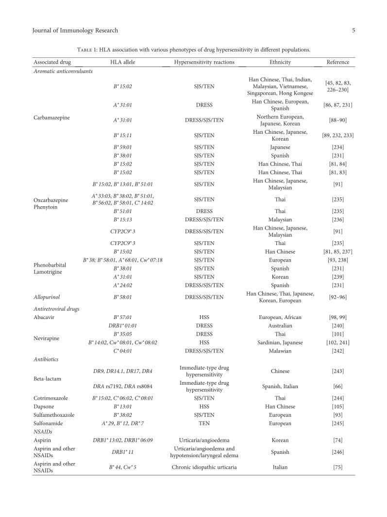
Table 1: HLA association with various phenotypes of drug hypersensitivity in different populations.
Associated drug HLA allele Hypersensitivity reactions Ethnicity Reference
Aromatic anticonvulsants
Carbamazepine
B∗15:02 SJS/TENHan Chinese, Thai, Indian,Malaysian, Vietnamese,
Singaporean, Hong Kongese
[45, 82, 83,226–230]
A∗31:01 DRESSHan Chinese, European,
Spanish[86, 87, 231]
A∗31:01 DRESS/SJS/TENNorthern European,Japanese, Korean
[88–90]
B∗15:11 SJS/TENHan Chinese, Japanese,
Korean[89, 232, 233]
B∗59:01 SJS/TEN Japanese [234]
B∗38:01 SJS/TEN Spanish [231]
OxcarbazepinePhenytoin
B∗15:02 SJS/TEN Han Chinese, Thai [81, 84]
B∗15:02 SJS/TEN Han Chinese, Thai [81, 83]
B∗15:02, B∗13:01, B∗51:01 SJS/TENHan Chinese, Japanese,
Malaysian[91]
A∗33:03, B∗38:02, B∗51:01,B∗56:02, B∗58:01, C∗14:02
SJS/TEN Thai [235]
B∗51:01 DRESS Thai [235]
B∗15:13 DRESS/SJS/TEN Malaysian [236]
CYP2C9∗3 DRESS/SJS/TENHan Chinese, Japanese,
Malaysian[91]
CYP2C9∗3 SJS/TEN Thai [235]
PhenobarbitalLamotrigine
B∗15:02 SJS/TEN Han Chinese [81, 85, 237]
B∗38; B∗58:01, A∗68:01, Cw∗07:18 SJS/TEN European [93, 238]
B∗38:01 SJS/TEN Spanish [231]
A∗31:01 SJS/TEN Korean [239]
A∗24:02 DRESS/SJS/TEN Spanish [231]
Allopurinol B∗58:01 DRESS/SJS/TENHan Chinese, Thai, Japanese,
Korean, European[92–96]
Antiretroviral drugs
Abacavir B∗57:01 HSS European, African [98, 99]
Nevirapine
DRB1∗01:01 DRESS Australian [240]
B∗35:05 DRESS Thai [101]
B∗14:02, Cw∗08:01, Cw∗08:02 HSS Sardinian, Japanese [102, 241]
C∗04:01 DRESS/SJS/TEN Malawian [242]
Antibiotics
Beta-lactamDR9, DR14.1, DR17, DR4
Immediate-type drughypersensitivity
Chinese [243]
DRA rs7192, DRA rs8084Immediate-type drug
hypersensitivitySpanish, Italian [66]
Cotrimoxazole B∗15:02, C∗06:02, C∗08:01 SJS/TEN Thai [244]
Dapsone B∗13:01 HSS Han Chinese [105]
Sulfamethoxazole B∗38:02 SJS/TEN European [93]
Sulfonamide A∗29, B∗12, DR∗7 TEN European [245]
NSAIDs
Aspirin DRB1∗13:02, DRB1∗06:09 Urticaria/angioedema Korean [74]
Aspirin and otherNSAIDs
DRB1∗11Urticaria/angioedema and
hypotension/laryngeal edemaSpanish [246]
Aspirin and otherNSAIDs
B∗44, Cw∗5 Chronic idiopathic urticaria Italian [75]
5Journal of Immunology Research
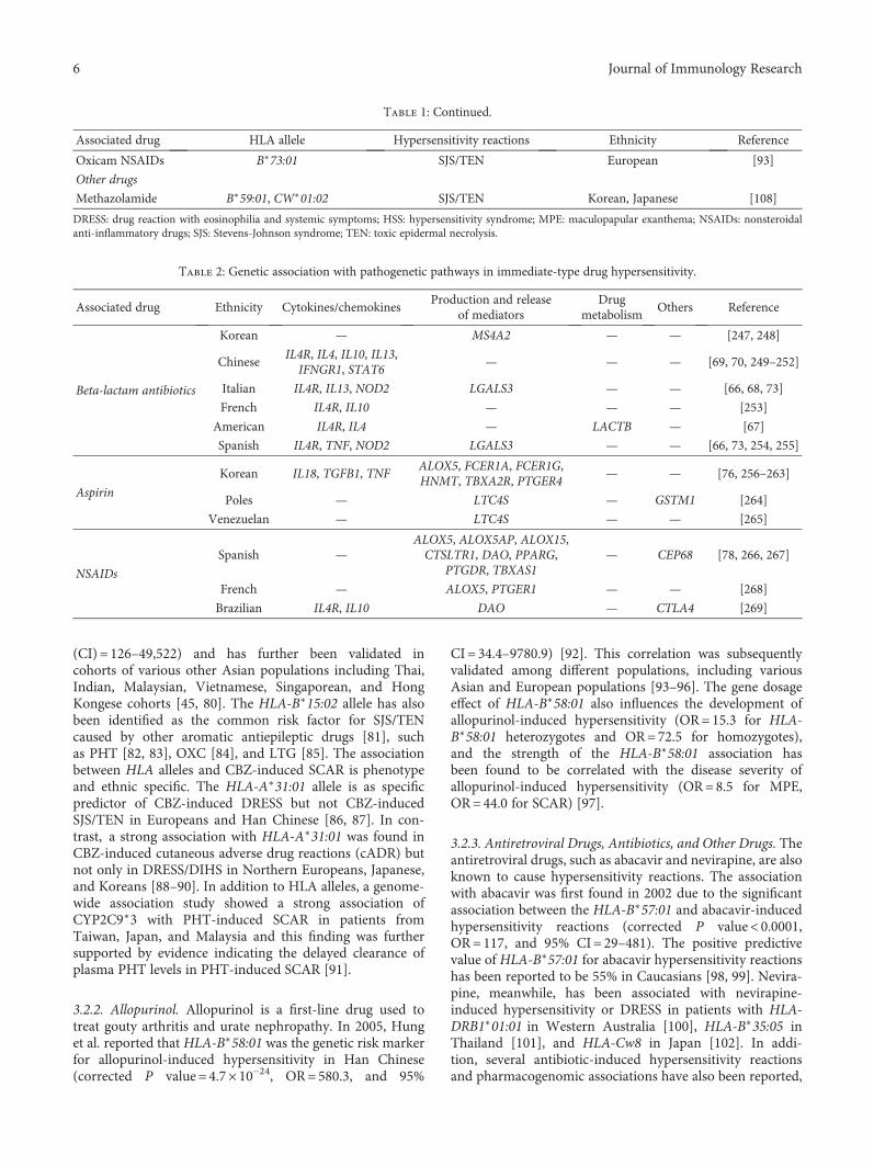
(CI) = 126–49,522) and has further been validated incohorts of various other Asian populations including Thai,Indian, Malaysian, Vietnamese, Singaporean, and HongKongese cohorts [45, 80]. The HLA-B∗15:02 allele has alsobeen identified as the common risk factor for SJS/TENcaused by other aromatic antiepileptic drugs [81], suchas PHT [82, 83], OXC [84], and LTG [85]. The associationbetween HLA alleles and CBZ-induced SCAR is phenotypeand ethnic specific. The HLA-A∗31:01 allele is as specificpredictor of CBZ-induced DRESS but not CBZ-inducedSJS/TEN in Europeans and Han Chinese [86, 87]. In con-trast, a strong association with HLA-A∗31:01 was found inCBZ-induced cutaneous adverse drug reactions (cADR) butnot only in DRESS/DIHS in Northern Europeans, Japanese,and Koreans [88–90]. In addition to HLA alleles, a genome-wide association study showed a strong association ofCYP2C9∗3 with PHT-induced SCAR in patients fromTaiwan, Japan, and Malaysia and this finding was furthersupported by evidence indicating the delayed clearance ofplasma PHT levels in PHT-induced SCAR [91].
3.2.2. Allopurinol. Allopurinol is a first-line drug used totreat gouty arthritis and urate nephropathy. In 2005, Hunget al. reported that HLA-B∗58:01 was the genetic risk markerfor allopurinol-induced hypersensitivity in Han Chinese(corrected P value = 4.7× 10⁻²⁴, OR=580.3, and 95%
CI=34.4–9780.9) [92]. This correlation was subsequentlyvalidated among different populations, including variousAsian and European populations [93–96]. The gene dosageeffect of HLA-B∗58:01 also influences the development ofallopurinol-induced hypersensitivity (OR=15.3 for HLA-B∗58:01 heterozygotes and OR=72.5 for homozygotes),and the strength of the HLA-B∗58:01 association hasbeen found to be correlated with the disease severity ofallopurinol-induced hypersensitivity (OR=8.5 for MPE,OR=44.0 for SCAR) [97].
3.2.3. Antiretroviral Drugs, Antibiotics, and Other Drugs. Theantiretroviral drugs, such as abacavir and nevirapine, are alsoknown to cause hypersensitivity reactions. The associationwith abacavir was first found in 2002 due to the significantassociation between the HLA-B∗57:01 and abacavir-inducedhypersensitivity reactions (corrected P value< 0.0001,OR=117, and 95% CI=29–481). The positive predictivevalue of HLA-B∗57:01 for abacavir hypersensitivity reactionshas been reported to be 55% in Caucasians [98, 99]. Nevira-pine, meanwhile, has been associated with nevirapine-induced hypersensitivity or DRESS in patients with HLA-DRB1∗01:01 in Western Australia [100], HLA-B∗35:05 inThailand [101], and HLA-Cw8 in Japan [102]. In addi-tion, several antibiotic-induced hypersensitivity reactionsand pharmacogenomic associations have also been reported,
Table 1: Continued.
Associated drug HLA allele Hypersensitivity reactions Ethnicity Reference
Oxicam NSAIDs B∗73:01 SJS/TEN European [93]
Other drugs
Methazolamide B∗59:01, CW∗01:02 SJS/TEN Korean, Japanese [108]
DRESS: drug reaction with eosinophilia and systemic symptoms; HSS: hypersensitivity syndrome; MPE: maculopapular exanthema; NSAIDs: nonsteroidalanti-inflammatory drugs; SJS: Stevens-Johnson syndrome; TEN: toxic epidermal necrolysis.
Table 2: Genetic association with pathogenetic pathways in immediate-type drug hypersensitivity.
Associated drug Ethnicity Cytokines/chemokinesProduction and release
of mediatorsDrug
metabolismOthers Reference
Beta-lactam antibiotics
Korean — MS4A2 — — [247, 248]
ChineseIL4R, IL4, IL10, IL13,
IFNGR1, STAT6— — — [69, 70, 249–252]
Italian IL4R, IL13, NOD2 LGALS3 — — [66, 68, 73]
French IL4R, IL10 — — — [253]
American IL4R, IL4 — LACTB — [67]
Spanish IL4R, TNF, NOD2 LGALS3 — — [66, 73, 254, 255]
Aspirin
Korean IL18, TGFB1, TNFALOX5, FCER1A, FCER1G,HNMT, TBXA2R, PTGER4
— — [76, 256–263]
Poles — LTC4S — GSTM1 [264]
Venezuelan — LTC4S — — [265]
NSAIDs
Spanish —ALOX5, ALOX5AP, ALOX15,CTSLTR1, DAO, PPARG,
PTGDR, TBXAS1— CEP68 [78, 266, 267]
French — ALOX5, PTGER1 — — [268]
Brazilian IL4R, IL10 DAO — CTLA4 [269]
6 Journal of Immunology Research

such as sulfonamide-induced allergic reactions [103],penicillin-induced SCAR [104], HLA-B∗13:01 and dapsone-induced hypersensitivity syndrome in Chinese [105],HLA-B∗57:01 and flucloxacillin-induced liver injury [106],and HLA-A∗02:01 and HLA-DQB1∗06:02 and amoxicillin-clavulanate hepatitis [107]. Other pharmacogenomic associ-ations include HLA-B∗59:01 and methazolamide-inducedSJS/TEN in Koreans and Japanese [108], HLA-B∗73:01 andoxicam-induced SJS/TEN in Europeans [93], and ABCB11,C-24T, UGT2B7∗2, and IL-4 C-590-A and diclofenac-induced liver disease in Europeans [109, 110].
4. Cellular Immunology and ImmuneMechanisms in Drug Hypersensitivity
4.1. Antigen Presentation and Processing. Drugs are consid-ered to be foreign antigens and bind to the HLA/peptide/TCR complex to trigger immune and hypersensitivity reac-tions. There are four hypotheses regarding drug presentationmechanisms that have been proposed to explain how smalldrug antigens might interact with HLA and TCR in drughypersensitivity: (1) the hapten theory, (2) the pharmacolog-ical interaction with immune receptors (p-i) concept, (3) thealtered peptide repertoire model, and (4) the altered TCRrepertoire model [111–115].
First, the hapten theory states that the culprit drugs ortheir reactive metabolites are too small to be immunogenicon their own, whereas they covalently bind to the endoge-nous peptides to form an antigenic hapten-carrier complex.The hapten-carrier complex is presented to the HLA mole-cule and then recognized by TCR, resulting in the inductionof drug-specific cellular or humoral immune responses. Thehapten theory has been shown to be valid in cases ofpenicillin-induced cADR [111, 116]. Second, the pharmaco-logical interaction with immune receptor (p-i) concept pos-tulates that drugs may directly, reversibly, and noncovalentlybind to the HLA and/or TCR protein and bypass the classicantigen-processing pathway in antigen-presenting cells. Weiet al. previously found that CBZ/aromatic antiepileptic drugscan directly interact with HLA-B∗15:02 protein. No intracel-lular antigen processing or drug metabolism was involved inthe HLA-B∗15:02 presentation of CBZ [112]. Oxypurinol,the reactive metabolite of allopurinol, provides anotherexample of the p-i concept in that it can directly and imme-diately activate drug-specific T cells via the preferential useof HLA-B∗58:01 without intracellular processing [113].Third, the altered peptide repertoire model states that theculprit drugs occupy the position in the peptide-bindinggroove of the HLA protein, changing the binding cleft andthe peptide specificity of HLA binding. Abacavir-inducedhypersensitivity has been found to belong to this model, asthe crystal structure of HLA-B∗57:01 has been found to formcomplexes with abacavir and peptides [114, 115]. Thesestudies showed that abacavir binds to the F-pocket ofHLA-B∗57:01 and alters the shape and chemistry of theantigen-binding cleft, thereby altering the repertoire ofendogenous peptides and resulting in polyclonal T cellactivation and autoimmune-like systemic reaction manifes-tations. Finally, the altered TCR repertoire model suggests
that some drugs, such as sulfamethoxazole, directly interactwith TCR, but not with the peptides or HLA molecules.The drug antigens bind to specific TCRs and alter theconformation of those TCRs, giving them the potential tobind to HLA-self peptide complexes to elicit immune reac-tions [117]. In this model, TCR is regarded as an initial druginteraction molecule, suggesting that TCR is as crucial asHLA molecules and contributes to the occurrence of drughypersensitivity. Furthermore, viruses have also been pro-posed to participate in HLA/drug/TCR interactions, in thatthey may provide exogenous peptides for drug presentationand play important roles in cADR [116].
4.2. Cellular Immunology and Immune Molecules Involved inDrug Hypersensitivity
4.2.1. Immediate-Type Drug Hypersensitivity. Immediate-type drug hypersensitivity can be mediated by IgE-mediatedor non-IgE-mediated mechanisms [118]. IgE-mediatedmechanisms are mediated by drug-specific IgE via animmune response to a hapten/carrier complex. In the pri-mary drug sensitization, drug-specific IgE is formed whenplasma cells are transformed from activated B cells andinteract with T cells. In an allergic reaction, drug allergensbind to mast cells or basophils with high-affinity Fc receptors,to which drug-specific IgE is bound, causing degranulation ofthe mast cells or basophils that results in the release ofvarious mediators, such as histamine, leukotrienes, prosta-glandins, and cytokines [3]. Degranulation has recentlybeen proposed to occur in two main forms that are relatedto reaction severity and progression: piecemeal degranula-tion and anaphylactic degranulation [2, 119]. Piecemealdegranulation is mediated through the upregulation ofCD203c on basophils via the formation of small vesicles fromthe histamine-containing granules quickly shuttling to theplasma membrane to cause more severe and rapid reactions[120]. Anaphylactic degranulation results in the fusion ofthe main histamine-containing granules with the plasmamembrane, releasing the entire contents of granules to theextracellular space and exposing CD63 on the surface ofbasophils [120].
The non-IgE-mediated immunologic mechanisms aremediated by IgG antibodies or by complement activation[23, 24]. IgG-mediated anaphylaxis has been establishedin mouse models, wherein the use of drugs with specificIgG bound to FcγRIII stimulates the release of platelet-activating factor (PAF) by basophils, macrophages, orneutrophils [24]. Although the IgG-mediated anaphylaxismechanism has not been fully demonstrated in humans,some studies have shown that PAF is an essential mediatorin such anaphylaxis [121]. In addition, a novel gain-of-function splice variant of FcγR FcγRIIA has been identifiedwith the presence of IgG anti-IgA antibodies in patientswith common variable immunodeficiency who developedanaphylaxis after intravenous immunoglobulin infusion[122]. Moreover, biological agents with IgA and infliximabhave been shown to induce anaphylaxis in the absence ofspecific IgE but with high levels of specific IgG [123–125].These observations also provide some additional evidence
7Journal of Immunology Research

for IgG-mediated anaphylaxis. Furthermore, complementactivation can be induced through the absence of agent-specific IgE or IgG antibody immunocomplexes [24]. Thiscondition can be observed in patients undergoing hemodi-alysis with a new dialysis membrane, protamine neutraliza-tion of heparin, and polyethylene glycol infusion [23, 126].Drugs solubilized in therapeutic liposomes and lipid-basedexcipients (such as Cremophor EL used as the diluent forolder preparations of propofol and paclitaxel) can formlarge micelles with serum lipids and cholesterol to stimulatethe complement system [23, 126]. This activation of com-plement mechanisms further causes the release of C3a,C5a, and C5b-9, which trigger, in turn, the activation ofmast cells, basophils, and other cells via their specific recep-tors, resulting in degranulation and mediator release [24].
The nonimmunologic-type hypersensitivity reactiondirectly activates mast cell degranulation without involvingthe activation of the immune system. There are several spe-cific agents that induce different mechanisms beyond thedirect immunoglobulin-mediated activation or complementactivation. Oversulfated chondroitin sulfate-contaminatedheparin was found to have caused various cases of anaphy-laxis around 2007-2008 via the direct activation of the kininsystem with increased production of bradykinin, C3a, andC5a [127]. The triggering of factor XII-driven contactsystem activation-mediated bradykinin formation alsoplays a key role in anaphylaxis [24]. NSAIDs, includingaspirin, can result in anaphylactic reactions via the inhibi-tion of cyclooxygenase with a decrease in the productionof prostaglandins and the increased generation of cysteinylleukotrienes [23]. Vancomycin can directly activate mastcells and/or basophils, leading to the release of histamine[128]. This mechanism was suggested to be mediated viathe calcium-dependent activation of phospholipase-C andphospholipase-A2 pathways [128]. Opiates (e.g., meperidine,codeine, and morphine) also cause histamine release viadirect mast cell degranulation [129]. Recently, it was pro-posed that nonimmunologic hypersensitivity reactions mayalso be mediated through the MAS-related G protein-coupled receptor-X2 (MRGPRX2) in cases involving specificdrugs, such as icatibant, neuromuscular blocking drugs, andquinolone antibiotics [25]. The interaction of certain drugswith this mast cell receptor can stimulate degranulation andthe release of TNF-α and prostaglandin D2 (PGD2), amongother molecules, leading to nonimmunologic anaphylacticreactions [25]. The mouse counterpart of MRGPRX2 thatparticipates in peptidergic drug-induced pseudoallergicreactions has been newly identified and could potentiallybe applied in preclinical screening models [25, 130].
4.2.2. Delayed-Type Drug Hypersensitivity. The main conceptused to explain the pathomechanisms of delayed-type drughypersensitivity consists of the view that specific T lym-phocytes or natural killer (NK) cells are activated uponantigen recognition or Fas/FasL interaction and that variouscytotoxic proteins, including perforin/granzyme B, andgranulysin, are then released to attack keratinocytes orother cells, inducing skin rash or epidermal necrosis. Inaddition, several other cytokines/chemokines, including
TNF-α, IFN-γ, GM-CSF, TARC/CCL17, IL-6, IL-8/CXCL8,IL-15, and IL-36, are also known to participate in theimmune reactions of drug hypersensitivity. These cyto-kines/chemokines have been found to be highly expressedin the skin lesions, blister fluids, blister cells, peripheral bloodmononuclear cells (PBMC), or plasma of patients. Theseimmune mediators are responsible for the trafficking,proliferation, regulation, or activation of T lymphocytesand other leukocytes, thereby affecting the clinical presenta-tions of drug hypersensitivity in various ways (Table 3).
(1) Fas-FasL Interaction. Fas ligand (FasL) belongs to thetumor necrosis factor (TNF) family. The binding of Fasand FasL plays an important role in regulating the immunesystem and is involved in the apoptosis of epidermal cellsin patients with drug hypersensitivity. Briefly, upon Fas–FasL interaction, the Fas-associated death domain protein(FADD) is recruited and binds to the Fas–FasL complex.The FADD then recruits procaspase 8, bringing multiplecopies of procaspase 8 together, which in turn autoactivateto become caspase 8, triggering the caspase cascade andresulting in intracellular DNA degradation [131]. Viard etal. proposed that a suicidal interaction between Fas andFasL, which are both expressed by keratinocytes, leads tothe extensive necrosis of epidermal cells in individuals withSJS/TEN [132].
(2) Perforin/Granzyme B. A controversial hypothesis suggeststhat perforin and granzyme B play more important roles inthe keratinocyte death in SJS/TEN than does the Fas–FasLinteraction [133]. Granzymes are serine proteases that arereleased by cytoplasmic granules and can induce pro-grammed cell death in the target cells. Upon activation,drug-specific cytotoxic T lymphocytes (CTL) and NK cellsproduce perforin, which can bind to and punch a channelthrough the cell membrane, promoting the entry of gran-zyme B into the target cells to activate the caspase cascadeand the succeeding apoptosis [134]. Delayed reactions todrugs have shown that increasing levels of perforin andgranzyme B are related to the disease severity of drughypersensitivity [131].
(3) Granulysin. Granulysin is a cytolytic protein mainlyreleased by CTL and NK cells. It functions to create holesin the cell membranes and thereby destroy target cells. In2008, Chung et al. reported that 15 kDa secretory granulysinserves as a key mediator for the disseminated keratinocyteapoptosis seen in SJS/TEN [135]. In that study, the increasedlevel of granulysin in blister fluids from the skin lesions ofSJS/TEN patients was much higher than the levels of othercytotoxic proteins, such as perforin, granzyme B, and FasL,and depleting the granulysin reduced the cytotoxicity [135].Further studies demonstrated that granulysin is stronglyexpressed in patients with drug-induced FDE, DRESS/DIHS,and SJS/TEN but not MPE [136–138].
(4) TNF-α, IFN-γ, TARC, IL-15, and Other Cytokines/Chemokines in SJS/TEN, DRESS/DIHS, and AGEP. TNF-αis a major proinflammatory cytokine and is produced by
8 Journal of Immunology Research

macrophages, T lymphocytes, NK cells, neutrophils, mastcells, and eosinophils. It regulates immune responses throughthe induction of cell apoptosis, activation, differentiation,and inflammation [139]. TNF-α was highly expressed andsuggested to be responsible for the extensive necrosis of skinlesions of SCAR patients [140, 141]. IFN-γ is critical for bothinnate and adaptive immunity against viral and bacterialinfection, and it is predominantly produced by CD4+ Thelper cells, CD8+ CTL, and NK cells. IFN-γ was found tobe increased in the skin tissue, blister cells, and plasma ofpatients with erythema multiforme, SJS, TEN, and DRESS/DIHS [131, 142, 143]. The immune mechanism of AGEPis not yet well understood. However, high levels of IL-8/CXCL8 production and the recruitment of neutrophilshave been observed in the skin lesions of AGEP patients[144–146]. Mutations in the IL36RN gene encoding theIL-36 receptor antagonist (IL-36Ra) have also been identifiedin AGEP patients [147, 148]. DRESS/DIHS is characterizedby leukocytosis with atypical lymphocytosis or eosinophilia[149]. Serum thymus and activation-regulated chemokine(TARC) was identified as a potential biomarker for early
indication of the disease and a predictor of disease activityin DRESS/DIHS [150, 151]. Compared to patients withMPE and SJS/TEN, the TARC levels in patients withDRESS/DIHS are significantly higher during the acutephase and are correlated with skin eruptions [151].Interleukin-15 (IL-15) is a cytokine that can induce theproliferation of NK cells and other leukocytes, and it hasbeen found to be associated with the disease severity andmortality of SJS/TEN [138]. IL-15 has also been shown toenhance the cytotoxicity of cultured NK cells and blistercells from TEN patients [138]. In addition, other cytokinesand chemokine receptors, including IL-2, IL-4, IL-5, IL-6,IL-8, IL-10, IL-12, IL-13, IL-18, CCR3, CXCR3, CXCR4,and CCR10, have been found to be upregulated in the skinlesions, blister fluids, PBMC, or plasma of drug hypersensi-tivity patients and to participate in the immune regulationof drug hypersensitivity [131, 138, 142, 143, 152–154].
(5) Syndrome-Specific Effector Cells. SJS/TEN is characterizedby profound necrosis localized to the epidermis. CytotoxicCD8 T cells, natural killer cells, and natural killer T cells
Table 3: Delayed-type drug hypersensitivity-related cytokines and chemokines.
Phenotype Cytokines/chemokines Skin or blister Plasma PBMC References
DRESS/DIHS
TNF-α + [160]
IFN-γ + + + [270–272]
IL-2 + [270]
IL-4 + [270]
IL-5 + [270]
IL-6 + [160]
IL-13 + [270]
IL-15 + [138]
TARC/CCL17 + [273]
SJS/TEN
TNF-α + + + [131, 138, 141–143, 274, 275]
IFN-γ + + [131, 142, 143, 274]
IL-2 + + [131, 143]
IL-5 + [143]
IL-6 + + + [143, 153, 154, 138]
IL-8/CXCL8 + [138]
IL-10 + + + [142, 153]
IL-12 NS [142]
IL-13 + [143]
IL-15 NS + [142, 138]
IL-18 + [142]
CCR3 + [143]
CXCR3 + [143]
CXCR4 NS [143]
CCR10 + [152]
AGEP
IL-8/CXCL8 + [145, 146]
IL-36 + [147, 148]
GM-CSF + [145]
AGEP: acute generalized exanthematous pustulosis; CCR: C–C chemokine receptor; CXCR: CX chemokine receptor; DIHS: drug-induced hypersensitivitysyndrome; DRESS: drug reactions with eosinophilia and systemic symptoms; IFN-γ: interferon-γ; IL: interleukin; NS: not significant; SJS/TEN: Stevens–Johnson syndrome and toxic epidermal necrolysis; TNF-α: tumor necrosis factor-α.
9Journal of Immunology Research
producing the cytotoxic molecules, especially granulysin,which causes extensive keratinocyte death, are enriched inblister fluid samples from the skin lesions of patients withSJS/TEN. Granulysin serum levels are correlated with theseverity of acute disease and mortality [135, 155]. These cyto-toxic cells mediate the disease pathogenesis. It is shown thatthe function of regulatory T cells (Tregs) in SJS/TEN isinadequate, although present in normal frequency [156].Immunological changes of DRESS/DIHS are characterizedby the increase of atypical lymphocytes or eosinophils [149,157]. Eosinophilia can be observed in 60–95% of DRESS/DIHS patients at the early stage of the illness [32, 157]. Mostof DRESS patients had increased numbers of CD4+ T cells inthe acute stage, which was associated with the severity ofclinical symptoms, such as the extent of skin rash andreactivations of virus [158]. In addition, Tregs play importantroles in DRESS/DIHS pathogenesis. Dramatic expansions offunctional Tregs are found in the acute stage of DRESS/DIHS[156]. It is hypothesized that CD4+FoxP3+ T cells that arehome to skin serve to limit the severity of acute disease byregulating the cytotoxic effector T cell responses. However,Treg responses eventually exhaust and this might contributeto ongoing viral replication and intermittent recurrence ofclinical symptoms [156, 159]. In patients with AGEP, it isshown that the increased neutrophilic inflammatory pro-cesses are regulated by T lymphocytes, which is importantin the pathogenesis. The recruitment of neutrophils wasobserved in the skin lesions of the patients with the late phaseof disease development [144, 145].
5. Environmental Factors and ViralInfections in Drug Hypersensitivity
In addition to drug antigens, hypersensitivity reactions maybe induced by other pathogens, such as Mycoplasma pneu-monia, or viral infections. Virus-drug interactions associatedwith viral reactivation may also exist. For example, it is wellknown that human herpesvirus-6 (HHV-6) plays an impor-tant role in DRESS/DIHS. HHV-6 reactivation in patientswith DRESS/DIHS may increase T cell activity after theinitiation of the drug eruption and induce the synthesis ofproinflammatory cytokines, including TNF-α and IL-6,which may in turn modulate the T cell-mediated responses[160]. Shiohara et al. reviewed the associations between viralinfections and drug rashes, as well as the mechanisms bywhich viral infections induce drug rashes. The sequentialreactivations of several herpes viruses (HHV-6, HHV-7,Epstein-Barr virus (EBV), and cytomegalovirus (CMV))were found to be coincident with the clinical symptoms ofdrug hypersensitivity reactions [161]. Chung et al. reportedthat a new variant of coxsackievirus A6 (CVA6) acting asthe causative agent may induce widespread mucocutaneousblistering reactions mimicking the features of erythema mul-tiforme major or SCAR [52]. In addition, the virus may alsoprovide exogenous peptides for drug presentation and partic-ipate in HLA/drug/TCR interactions. White et al. recentlyproposed that some patients may acquire primary infectionvia HHVs or other pathogens that in turn induce drughypersensitivity [116]. The presence of HHV peptides in
patients with high-risk HLA alleles may trigger the activationof cytotoxic T cells, thereby resulting in the development ofSCAR. The pathogenic factors underlying the unusualpresentations of drug hypersensitivity related to viralinfections need to be further investigated.
6. Diagnostic Tools for Drug Hypersensitivity
6.1. Diagnostic Tools for Immediate-Type DrugHypersensitivity. The most commonly used laboratory testfor confirming a diagnosis of anaphylaxis consists ofdetermining the patient’s total serum tryptase level [162].Serial measurements of tryptase levels can be taken duringan anaphylactic episode, although measurements of the base-line level are considered to be most useful. In fact, while serialmeasurements of tryptase levels taken during an anaphylacticepisode can serve as useful markers for evaluating these reac-tions, this approach is not used so widely in clinical practicedue to the limitations involved in measuring tryptase duringthe acute phase of an episode. Elevated levels of histamine,the first mediator released by mast cells, in plasma or urineare also consistent with anaphylaxis [2]. However, plasmahistamine levels are only transiently elevated, making themof little utility if the patient is evaluated more than 1 hourafter onset of the episode [163]. At the same time, normallevels of tryptase or histamine do not preclude a diagnosisof drug hypersensitivity [15]. Other newly identifiedbiomarkers, such as PAF and carboxypeptidase A3, bringhope for enhancing diagnostic accuracy, although their useremains experimental [15, 164].
For IgE-mediated hypersensitivity reactions, serum drug-specific IgE (sIgE) quantification and the basophil activationtest (BAT) are frequently used to assess the culprit drug. Thetests used to conduct sIgE immunoassays consist of radioal-lergosorbent testing (RAST), enzyme-linked immunosorbentassays (ELISAs), and fluoroenzyme immunoassays (FEIAs)[165]. While RAST or ELISAs are usually conducted usingin-house techniques, FEIAs can be performed using com-mercial products, such as the ImmunoCAP-FEIA system[166–168]. Only a few products are available, meanwhile,for some drugs, particularly beta-lactam antibiotics [167,169]. The sensitivity of the various immunoassays used hasbeen found to average 62.9%, while the average specificity,PPV, and NPV are 89.2%, 83.3%, and 77.8%, respectively[168]. The average NPV is also relatively low in order toexclude allergic reactions and determine whether to performa provocation test [170]. In comparison, the BAT test pro-vides a higher average specificity (94.6%) and PPV (93.4%)than immunoassays [168]. The test uses flow cytometry afterdrug stimulation to determine the levels of basophil activa-tion or degranulation markers; the upregulation of CD63and CD203c is also usually measured [171]. Of note, theresults of the BAT for aspirin/NSAID-induced hypersensitiv-ity remain inconclusive due to the fact that they encompassboth IgE-mediated allergic reactions and nonimmunologicalintolerances, limiting the use of the BAT in assessingnon-IgE-mediated reactions [172]. Mediator release assays,meanwhile, measure the mediator released (histamine orleukotriene 4) in a supernatant upon cell activation after
10 Journal of Immunology Research

drug stimulation, but these assays have exhibited sensitivityand specificity levels too low for them to be recommendedfor the purposes of diagnosis [169, 173].
6.2. Diagnostic Tools for Delayed-Type Drug Hypersensitivity.The discovery of biomarkers for drug hypersensitivity is cru-cial for clinical purposes, including the early diagnosis andbetter prediction of this disease in order to prevent complica-tions. We previously found granulysin to be a key cytotoxicmolecule responsible for disseminated keratinocyte necrosisthrough the action of cytotoxic lymphocytes or NK-cell-mediated cytotoxicity with no direct cellular contact [135].A significant correlation between the granulysin levels inblister fluids and clinical severity was also found [135]. Inaddition, the serum granulysin levels in patients with SJS/TEN have also been found to be significantly elevated beforethe development of skin detachment or mucosal lesions butthen to drop rapidly within 5 days of disease onset [136].As a potential marker for the early phase of SJS/TEN, a sim-ple rapid immunochromatographic test for elevated serumgranulysin was developed for immediate clinical use. Addi-tionally, prolonged elevation of serum granulysin has alsobeen found in DIHS patients, indicating that such elevationcould possibly be used for the purposes of early diagnosisand predicting disease prognosis [174]. Furthermore, thelevels of IL-15 were correlated with the disease progressionand mortality of SJS/TEN at early stage [138]. Serum IL-15levels can be further utilized as a marker for early diagnosisand prognosis monitoring [138]. For DRESS/DIHS, serumTARC levels in patients with DRESS/DIHS have beenreported to be significantly higher than those in patients withSJS/TEN and MPE during the acute phase and to be corre-lated with skin eruptions [151]. TARC was thus identifiedas a potential biomarker for the early indication and diseaseactivity of DRESS/DIHS and also for determining the prog-nosis of systemic severity of inflammation in drug eruptionsother than SJS/TEN [150, 151]. For AGEP, meanwhile, nospecific markers for diagnosing or predicting the disease havebeen identified at present [175].
Drug rechallenge is considered the gold standard for con-firming a potential offending drug; however, its use is notpractical due to the possible life-threatening consequences.As such, there is still no standard method for the confir-mation of drug causality. Nonetheless, since HLA genotyp-ing has been useful in screening for populations at risk forSCAR, HLA genotyping might be helpful for identifyingculprit drugs via specific HLA alleles in at-risk populations[48, 176]. Several in vitro tests can be used to assist in theconfirmation of drug causality, but the exact sensitivity andspecificity of such tests are not well known [177, 178]. Thereare several tests currently available: the lymphocyte transfor-mation test (LTT), ELISpot (Enzyme-linked immunospotassay) intracellular cytokine staining, and the enzyme-linked immunosorbent assay (ELISA) for the secretion ofcytotoxic mediators including inflammatory cytokines, che-mokine–chemokine receptors, IFN-γ, Fas–Fas ligand, per-forin, granzyme B, and granulysin [179]. The LTT is areproducible test for measuring the enhanced proliferativeresponse of PBMC after the sensitization of T cells to a drug
[180]. However, the sensitivity of the test has reportedly var-ied among various studies involving various drugs and clini-cal phenotypes and different timings for use of the test [181,182]. The relevance of using the LTT in testing for SJS/TENwas relatively lower those using than DRESS/DIHS andAGEP [182]. Several modifications can help to increase thesensitivity of the LTT or ELISPOT, including stimulationwith anti-CD-3/CD28 antibody-coated microbeads withIL-2, depletion of Treg/CD25hi cells, or the combinedaddition of anti-CTLA4 and anti-programmed cell deathligand 1 (PD-L1) antibodies to PBMC cultures [183–185].IFN-γ-ELISpot showed a similar sensitivity (67%) and spec-ificity in DRESS, but a higher sensitivity (71%) in SJS/TEN[179]. The data for an ELISA-based test used to detect gran-ulysin showed better sensitivity (86%) in SJS/TEN, but theevidence was limited due to the small number of cases inthe study [186]. Further larger studies will thus be neededto confirm both the sensitivity and specificity.
In vivo patch tests provide a low-risk method for repro-ducing delayed hypersensitivity with moderate reexposureof patients to suspected offending drugs [187]. The value ofpatch testing depends on the phenotypes and drugs involved.The sensitivity of such testing is generally <70%, buthigher sensitivities have been reported for AGEP and forsome selected populations such as abacavir-hypersensitiv-ity, carbamazepine-induced SJS/DRESS, and fixed drugeruption patients [178, 187, 188]. The skin tests involvinga prick or intradermal testing are considered to be crucialtools for evaluating drug hypersensitivity reactions, includingIgE-mediated or delayed-type hypersensitivity, in both theEuropean and American guidelines [22, 189–191]. However,these skin tests are usually not suggested for SCAR patientsdue to the risk of relapse, although late-reading intradermaltests are of value for AGEP patients and negative patch testsare of value for SCAR patients [187, 192].
7. Therapeutic Approaches inDrug Hypersensitivity
7.1. Therapeutic Approaches in Immediate-Type DrugHypersensitivity. Anaphylaxis is a medical emergency andepinephrine is the treatment of choice for anaphylaxis to pre-vent its progression to a life-threatening condition [15, 193].Epinephrine should be administered as soon as possiblewithout delay to avoid mortality [194]. The intramuscularinjection of epinephrine into the middle of the outer thighis recommended to treat anaphylaxis in most settings andin patients of all ages [195]. Glucagon is indicated for patientsreceiving beta-blockers with refractory symptoms [196]. Theuse of corticosteroids was previously believed to decrease therisk of biphasic and protracted reactions; however, a system-atic review of the literature failed to retrieve any randomizedcontrolled trials to confirm their effectiveness [197]. Anemergency department-based study also failed to find adecrease in the rates of return visits or biphasic reactionsamong patients treated with glucocorticoids [198]. Theseadjunctive therapies, including corticosteroids, antihista-mines, and bronchodilators, could help to relieve symptoms,
11Journal of Immunology Research

but should not be substituted for epinephrine or delay the useof epinephrine [199, 200].
7.2. Therapeutic Approaches in Delayed-Type DrugHypersensitivity. For the treatment of severe delayed-typedrug hypersensitivity, such as SJS/TEN, there are no optimaltreatment guidelines. Thus far, in fact, only a few randomizedtrials that could be regarded as references to guide treatmenthave been conducted. The efficacy of systemic immunosup-pressants or immunomodulatory treatments (e.g., corticoste-roids, cyclosporine, intravenous immunoglobulins (IVIg),and plasmapheresis) still remains controversial. Systemiccorticosteroids could be the most common treatment option,but the prior use of corticosteroids was found to prolong dis-ease progression with no definite benefit in terms of survival[60, 201–203]. IVIg is one of the most commonly utilizedtherapies for SJS/TEN and is frequently the adjunctive ther-apy used for severe cases or pediatric patients [204]. In ameta-analysis, however, IVIg, even high doses of IVIg, failedto achieve statistically significant results supporting theconclusion that it is clinically beneficial [204, 205]. IVIghas been found to yield better outcomes in pediatricpatients, but children with TEN usually have lower ratesof mortality and better prognoses than adult patients[204, 206]. Cyclosporine, has been found to decrease themortality rate and the progression of detachment in adultsin an open-label phase II trial [207]. However, one recentcohort study revealed a statistically insignificant survivalbenefit for cyclosporine therapy compared to supportivecare [208]. In contrast, the first meta-analysis of 7 studiesregarding the effect on mortality of cyclosporine in thetreatment of SJS/TEN showed a beneficial effect [209]. Atrend identified in the same study also indicated thatcyclosporine demonstrated better survival than IVIg [209].There have also been an increasing number of case reportsregarding the benefit of treatment with anti-TNF-α biologicagents for patients with TEN [210–215]. One recent systemicreview showed that glucocorticosteroids and cyclosporine arethe most promising therapies in terms of survival benefit, butno such benefits were observed for IVIg, plasmapheresis,thalidomide, cyclophosphamide, hemoperfusion, tumornecrosis factor inhibitors, or granulocyte colony-stimulatingfactor [216]. Meanwhile, IL-15 was demonstrated to be amajor cytokine orchestrating SJS/TEN, indicating thatfurther novel therapeutics including IL-15 blockers, themammalian target of rapamycin (mTOR) inhibitors, andJanus kinase/signal transducers and activators of transcrip-tion (JAK/STAT) inhibitors hold promise for impactingvarious therapeutic targets [138, 217]. That said, furtherprospective, randomized controlled studies are needed toprovide more definitive conclusions regarding treatment inpatients with SJS/TEN.
Systemic corticosteroids have been considered the treat-ment of choice for patients with DRESS/DIHS, but theymay be associated with an increased risk of complicationssuch as opportunistic infections [218]. CMV and HHV-6viral loads were also reported to be increased in patientsreceiving systemic corticosteroids, while EBV loads werehigher in patients not receiving systemic corticosteroids
[219]. Antiviral medications such as ganciclovir can be givenin addition to steroids and/or IVIg in cases of severe diseasewith confirmation of viral reactivation [220]. Several previ-ous studies have reported the effectiveness of treatment withIVIg [221]. However, the premature discontinuation of aprospective study regarding the role of IVIg treatmentoccurred due to severe adverse effects [222]. Plasmapheresisand other immunosuppressive drugs, such as cyclophospha-mide, cyclosporine, interferons, muromonab-CD3, myco-phenolate mofetil, and rituximab, may also be potentialtherapies [221]. Among the above treatments, the use ofcyclosporine was successful in 2 recent cases with rapidresponse, and so, its use could be considered for patientswith concerns about using longer courses of systemic cor-ticosteroids [223]. Supportive treatment with topicalsteroid-based treatments for AGEP is suggested due tothe mostly benign and self-limiting course of the condition[224, 225]. Meanwhile, the administration of systemic ste-roids for a short period can be considered for severe andrefractory cases [175].
Conflicts of Interest
The authors declare that there is no conflict of interestregarding the publication of this paper.
Authors’ Contributions
Yi-Giien Tsai and Wen-Hung Chung contributed equally tothis work.
Acknowledgments
The authors would like to thank the National ScienceCouncil, Taiwan (MOST103-2321-B-182-001, MOST101-2628-B-182-001-MY3, MOST104-2314-B-182A-148-MY3,and MOST104-2325-B-182A-006), and Chang GungMemorial Hospital (CLRPG2E0051~3, CORPG3F0041~2,OMRPG3E0041, and CMRPG1F0111~2) for kindly support-ing this work.
References
[1] W. J. Pichler, “Delayed drug hypersensitivity reactions,”Annals of Internal Medicine, vol. 139, no. 8, pp. 683–693,2003.
[2] M. I. Montañez, C. Mayorga, G. Bogas et al., “Epidemiology,mechanisms, and diagnosis of drug-induced anaphylaxis,”Frontiers in Immunology, vol. 8, p. 614, 2017.
[3] B. Schnyder and W. J. Pichler, “Mechanisms of drug-inducedallergy,”Mayo Clinic Proceedings, vol. 84, no. 3, pp. 268–272,2009.
[4] S. G. Johansson, T. Bieber, R. Dahl et al., “Revised nomen-clature for allergy for global use: Report of the Nomencla-ture Review Committee of the World Allergy Organization,October 2003,” The Journal of Allergy and Clinical Immunol-ogy, vol. 113, no. 5, pp. 832–836, 2004.
[5] M. Mockenhaupt, “Epidemiology of cutaneous adversedrug reactions,” Chemical Immunology and Allergy, vol. 97,pp. 1–17, 2012.
12 Journal of Immunology Research

[6] G. A. Wong and N. H. Shear, “Adverse drug interactionsand reactions in dermatology: current issues of clinical rel-evance,” Dermatologic Clinics, vol. 23, no. 2, pp. 335–342,2005.
[7] C. C. Vittorio and J. J. Muglia, “Anticonvulsant hypersensi-tivity syndrome,” Archives of Internal Medicine, vol. 155,no. 21, pp. 2285–2290, 1995.
[8] F. Arellano and J. A. Sacristan, “Allopurinol hypersensitivitysyndrome: a review,” Annals of Pharmacotherapy, vol. 27,no. 3, pp. 337–343, 1993.
[9] H. Bocquet, M. Bagot, and J. C. Roujeau, “Drug-inducedpseudolymphoma and drug hypersensitivity syndrome (drugrash with eosinophilia and systemic symptoms: DRESS),”Seminars in Cutaneous Medicine and Surgery, vol. 15, no. 4,pp. 250–257, 1996.
[10] T. Shiohara, M. Iijima, Z. Ikezawa, and K. Hashimoto,“The diagnosis of a DRESS syndrome has been sufficientlyestablished on the basis of typical clinical features and viralreactivations,” British Journal of Dermatology, vol. 156,no. 5, pp. 1083-1084, 2007.
[11] Y. Ushigome, Y. Kano, K. Hirahara, and T. Shiohara, “Humanherpesvirus 6 reactivation in drug-induced hypersensitivitysyndrome andDRESS validation score,” The American Journalof Medicine, vol. 125, no. 7, pp. e9–e10, 2012.
[12] S. Bastuji-Garin, B. Rzany, R. S. Stern, N. H. Shear, L. Naldi,and J. C. Roujeau, “Clinical classification of cases of toxicepidermal necrolysis, Stevens-Johnson syndrome, anderythema multiforme,” Archives of Dermatology, vol. 129,no. 1, pp. 92–96, 1993.
[13] A. Sidoroff, A. Dunant, C. Viboud et al., “Risk factors foracute generalized exanthematous pustulosis (AGEP)-resultsof a multinational case-control study (EuroSCAR),” BritishJournal of Dermatology, vol. 157, no. 5, pp. 989–996, 2007.
[14] M. Lerch and W. J. Pichler, “The immunological and clinicalspectrum of delayed drug-induced exanthems,” CurrentOpinion Allergy and Clinical Immunology, vol. 4, no. 5,pp. 411–419, 2004.
[15] F. E. Simons, L. R. Ardusso, M. B. Bilò et al., “World AllergyOrganization anaphylaxis guidelines: summary,” The Journalof Allergy and Clinical Immunology, vol. 127, no. 3, pp. 587–593.e22, 2011.
[16] E. J. Jares, C. E. Baena-Cagnani, M. Sánchez-Borges et al.,“Drug-induced anaphylaxis in Latin American countries,”The Journal of Allergy and Clinical Immunology: in Practice,vol. 3, no. 5, pp. 780–788, 2015.
[17] Y. S. Lee and W. Z. Sun, “Epidemiology of anaphylaxis: aretrospective cohort study in Taiwan,” Asian Journal ofAnesthesiology, vol. 55, no. 1, pp. 9–12, 2017.
[18] L. H. Garvey, “Old, new and hidden causes of perioperativehypersensitivity,” Current Pharmaceutical Design, vol. 22,no. 45, pp. 6814–6824, 2016.
[19] M. Iammatteo, T. Keskin, and E. Jerschow, “Evaluation ofperiprocedural hypersensitivity reactions,” Annals of AllergyAsthma & Immunology, vol. 119, no. 4, pp. 349.e2–355.e2,2017.
[20] N. Blanca-Lopez, D. Perez-Alzate, I. Andreu et al., “Immedi-ate hypersensitivity reactions to ibuprofen and other arylpro-pionic acid derivatives,” Allergy, vol. 71, no. 7, pp. 1048–1056,2016.
[21] M. G. Canto, I. Andreu, J. Fernandez, and M. Blanca,“Selective immediate hypersensitivity reactions to NSAIDs,”
Current Opinion in Allergy and Clinical Immunology, vol. 9,no. 4, pp. 293–297, 2009.
[22] K. Brockow, L. H. Garvey, W. Aberer et al., “Skin testconcentrations for systemically administered drugs - anENDA/EAACI Drug Allergy Interest Group position paper,”Allergy, vol. 68, no. 6, pp. 702–712, 2013.
[23] R. Munoz-Cano, C. Picado, A. Valero, and J. Bartra, “Mech-anisms of anaphylaxis beyond IgE,” Journal of InvestigationalAllergology and Clinical Immunology, vol. 26, no. 2, pp. 73–82, 2016.
[24] F. D. Finkelman, M. V. Khodoun, and R. Strait, “HumanIgE-independent systemic anaphylaxis,” The Journal ofAllergy and Clinical Immunology, vol. 137, no. 6, pp. 1674–1680, 2016.
[25] H. Subramanian, K. Gupta, and H. Ali, “Roles of Mas-relatedG protein-coupled receptor X2 on mast cell-mediated hostdefense, pseudoallergic drug reactions, and chronic inflam-matory diseases,” The Journal of Allergy and ClinicalImmunology, vol. 138, no. 3, pp. 700–710, 2016.
[26] D. Laroche, F. Namour, C. Lefrancois et al., “Anaphylactoidand anaphylactic reactions to iodinated contrast material,”Allergy, vol. 54, Supplement 58, pp. 13–16, 1999.
[27] K. Farnam, C. Chang, S. Teuber, and M. E. Gershwin,“Nonallergic drug hypersensitivity reactions,” InternationalArchives of Allergy and Immunology, vol. 159, no. 4,pp. 327–345, 2012.
[28] M. L. Kowalski, R. Asero, S. Bavbek et al., “Classification andpractical approach to the diagnosis and management ofhypersensitivity to nonsteroidal anti-inflammatory drugs,”Allergy, vol. 68, no. 10, pp. 1219–1232, 2013.
[29] S. H. Kardaun, A. Sidoroff, L. Valeyrie-Allanore et al.,“Variability in the clinical pattern of cutaneous side-effectsof drugs with systemic symptoms: does a DRESS syndromereally exist?,” British Journal of Dermatology, vol. 156, no. 3,pp. 609–611, 2007.
[30] J. R. Sullivan and N. H. Shear, “The drug hypersensitivity syn-drome: what is the pathogenesis?,” Archives of Dermatology,vol. 137, no. 3, pp. 357–364, 2001.
[31] P. Cacoub, P. Musette, V. Descamps et al., “The DRESSsyndrome: a literature review,” The American Journal ofMedicine, vol. 124, no. 7, pp. 588–597, 2011.
[32] S. H. Kardaun, P. Sekula, L. Valeyrie-Allanore et al., “Drugreaction with eosinophilia and systemic symptoms(DRESS): an original multisystem adverse drug reaction.Results from the prospective RegiSCAR study,” BritishJournal of Dermatology, vol. 169, no. 5, pp. 1071–1080,2013.
[33] C. H. Wei, R. Chung-Yee Hui, C. J. Chang et al., “Identifyingprognostic factors for drug rash with eosinophilia andsystemic symptoms (DRESS),” European Journal ofDermatology, vol. 21, no. 6, pp. 930–937, 2011.
[34] J. C. Roujeau and R. S. Stern, “Severe adverse cutaneousreactions to drugs,” The New England Journal of Medicine,vol. 331, no. 19, pp. 1272–1285, 1994.
[35] Y. Kano, T. Ishida, K. Hirahara, and T. Shiohara, “Visceralinvolvements and long-term sequelae in drug-inducedhypersensitivity syndrome,” Medical Clinics of NorthAmerica, vol. 94, no. 4, pp. 743–759, 2010.
[36] O. Matsuno, “Drug-induced interstitial lung disease: mecha-nisms and best diagnostic approaches,” Respiratory Research,vol. 13, no. 1, p. 39, 2012.
13Journal of Immunology Research
[37] T. Thongsri, L. Chularojanamontri, and W. J. Pichler, “Car-diac involvement in DRESS syndrome,” Asian Pacific JournalAllergy and Immunology, vol. 35, no. 1, pp. 3–10, 2017.
[38] G. P. Bourgeois, J. A. Cafardi, V. Groysman, and L. C.Hughey, “A review of DRESS-associated myocarditis,” Jour-nal of the American Academy of Dermatology, vol. 66, no. 6,pp. e229–e236, 2012.
[39] M. Ben m'rad, S. Leclerc-Mercier, P. Blanche et al.,“Drug-induced hypersensitivity syndrome: clinical andbiologic disease patterns in 24 patients,” Medicine, vol. 88,pp. 131–140, 2009.
[40] J. C. Roujeau, J. C. Guillaume, J. P. Fabre, D. Penso, M. L.Flechet, and J. P. Girre, “Toxic epidermal necrolysis (Lyellsyndrome). Incidence and drug etiology in France, 1981-1985,” Archives of Dermatology, vol. 126, no. 1, pp. 37–42,1990.
[41] E. Schopf, A. Stuhmer, B. Rzany, N. Victor, R. Zentgraf,and J. F. Kapp, “Toxic epidermal necrolysis and Stevens-Johnson syndrome. An epidemiologic study from WestGermany,” Archives of Dermatology, vol. 127, no. 6,pp. 839–842, 1991.
[42] D. Y. Hsu, J. Brieva, N. B. Silverberg, and J. I. Silverberg,“Morbidity and mortality of Stevens-Johnson syndrome andtoxic epidermal necrolysis in United States adults,” Journalof Investigative Dermatology, vol. 136, no. 7, pp. 1387–1397,2016.
[43] N. Frey, J. Jossi, M. Bodmer et al., “The epidemiology ofStevens-Johnson syndrome and toxic epidermal necrolysisin the UK,” Journal of Investigative Dermatology, vol. 137,no. 6, pp. 1240–1247, 2017.
[44] J. Diphoorn, S. Cazzaniga, C. Gamba et al., “Incidence,causative factors and mortality rates of Stevens-Johnsonsyndrome (SJS) and toxic epidermal necrolysis (TEN) innorthern Italy: data from the REACT registry,” Pharmacoepi-demiology and Drug Safety, vol. 25, no. 2, pp. 196–203, 2016.
[45] W. H. Chung, S. I. Hung, H. S. Hong et al., “Medical genetics:a marker for Stevens-Johnson syndrome,” Nature, vol. 428,no. 6982, p. 486, 2004.
[46] D. Y. Hsu, J. Brieva, N. B. Silverberg, A. S. Paller, and J. I.Silverberg, “Pediatric Stevens-Johnson syndrome and toxicepidermal necrolysis in the United States,” Journal of theAmerican Academy of Dermatology, vol. 76, no. 5,pp. 811.e4–817.e4, 2017.
[47] P. B. Ferrell Jr. and H. L. McLeod, “Carbamazepine, HLA-B∗
1502 and risk of Stevens-Johnson syndrome and toxicepidermal necrolysis: US FDA recommendations,” Pharma-cogenomics, vol. 9, no. 10, pp. 1543–1546, 2008.
[48] E. J. Phillips, W. H. Chung, M. Mockenhaupt, J. C. Roujeau,and S. A. Mallal, “Drug hypersensitivity: pharmacogeneticsand clinical syndromes,” The Journal of Allergy and ClinicalImmunology, vol. 127, no. 3, pp. S60–S66, 2011.
[49] A. Auquier-Dunant, M. Mockenhaupt, L. Naldi et al.,“Correlations between clinical patterns and causes oferythema multiforme majus, Stevens-Johnson syndrome,and toxic epidermal necrolysis - results of an internationalprospective study,” Archives of Dermatology, vol. 138,no. 8, pp. 1019–1024, 2002.
[50] G. Yildirim Cetin, H. Sayar, F. Ozkan, S. Kurtulus, F. Kesici,and M. Sayarlioglu, “A case of toxic epidermal necrolysis-like skin lesions with systemic lupus erythematosus andreview of the literature,” Lupus, vol. 22, no. 8, pp. 839–846,2013.
[51] D. Olson, L. K. Watkins, A. Demirjian et al., “Outbreakof mycoplasma pneumoniae-associated Stevens-Johnsonsyndrome,” Pediatrics, vol. 136, no. 2, pp. e386–e394, 2015.
[52] W. H. Chung, S. R. Shih, C. F. Chang et al., “Clinicopatho-logic analysis of coxsackievirus a6 new variant inducedwidespread mucocutaneous bullous reactions mimickingsevere cutaneous adverse reactions,” The Journal of InfectiousDiseases, vol. 208, no. 12, pp. 1968–1978, 2013.
[53] M. Mockenhaupt, C. Viboud, A. Dunant et al., “Stevens-Johnson syndrome and toxic epidermal necrolysis: assess-ment of medication risks with emphasis on recently marketeddrugs. The EuroSCAR-study,” Journal of InvestigativeDermatology, vol. 128, no. 1, pp. 35–44, 2008.
[54] A. C. Rosen, Y. Balagula, D. W. Raisch et al., “Life-threaten-ing dermatologic adverse events in oncology,” Anti-CancerDrugs, vol. 25, no. 2, pp. 225–234, 2014.
[55] E. Dika, G. M. Ravaioli, P. A. Fanti et al., “Cutaneousadverse effects during ipilimumab treatment for metastaticmelanoma: a prospective study,” European Journal ofDermatology, vol. 27, no. 3, pp. 266–270, 2017.
[56] K. L. Vivar, M. Deschaine, J. Messina et al., “Epidermalprogrammed cell death-ligand 1 expression in TENassociated with nivolumab therapy,” Journal of CutaneousPathology, 2016.
[57] C. Y. Lin, C. W. Wang, C. R. Hui et al., “Delayed-typehypersensitivity reactions induced by proton pump inhibi-tors: a clinical and in vitro T-cell reactivity study,” Allergy,2017.
[58] B. Sassolas, C. Haddad, M. Mockenhaupt et al.,“ALDEN, an algorithm for assessment of drug causalityin Stevens-Johnson syndrome and toxic epidermal necro-lysis: comparison with case-control analysis,” ClinicalPharmacology & Therapeutics, vol. 88, no. 1, pp. 60–68,2010.
[59] J. C. Roujeau, J. P. Kelly, L. Naldi et al., “Medication use andthe risk of Stevens-Johnson syndrome or toxic epidermalnecrolysis,” The New England Journal of Medicine, vol. 333,no. 24, pp. 1600–1608, 1995.
[60] P. Sekula, A. Dunant, M. Mockenhaupt et al., “Comprehen-sive survival analysis of a cohort of patients with Stevens-Johnson syndrome and toxic epidermal necrolysis,” Journalof Investigative Dermatology, vol. 133, no. 5, pp. 1197–1204,2013.
[61] S. Bastuji-Garin, N. Fouchard, M. Bertocchi, J. C. Roujeau,J. Revuz, and P. Wolkenstein, “SCORTEN: a severity-of-illness score for toxic epidermal necrolysis,” Journal ofInvestigative Dermatology, vol. 115, no. 2, pp. 149–153,2000.
[62] S. Guegan, S. Bastuji-Garin, E. Poszepczynska-Guigne,J. C. Roujeau, and J. Revuz, “Performance of the SCOR-TEN during the first five days of hospitalization to pre-dict the prognosis of epidermal necrolysis,” Journal ofInvestigative Dermatology, vol. 126, no. 2, pp. 272–276,2006.
[63] A. Beck, K. P. Quirke, R. L. Gamelli, and M. J. Mosier,“Pediatric toxic epidermal necrolysis: using SCORTEN andpredictive models to predict morbidity when a focus onmortality is not enough,” Journal of Burn Care & Research,vol. 36, no. 1, pp. 167–177, 2015.
[64] A. Sidoroff, S. Halevy, J. N. Bavinck, L. Vaillant, and J. C.Roujeau, “Acute generalized exanthematous pustulosis
14 Journal of Immunology Research

(AGEP) - a clinical reaction pattern,” Journal of CutaneousPathology, vol. 28, no. 3, pp. 113–119, 2001.
[65] E. H. Saissi, F. Beau-Salinas, A. P. Jonville-Bera, G. Lorette,E. Autret-Leca, and Centres Régionaux de Pharmacovigi-lance, “Drugs associated with acute generalized exanthematicpustulosis,” Annales de Dermatologie et de Venereologie,vol. 130, no. 6-7, pp. 612–618, 2003.
[66] J. L. Gueant, A. Romano, J. A. Cornejo-Garcia et al.,“HLA-DRA variants predict penicillin allergy in genome-wide fine-mapping genotyping,” The Journal of Allergy andClinical Immunology, vol. 135, no. 1, pp. 253–259.e10, 2015.
[67] A. J. Apter, H. Schelleman, A. Walker, K. Addya, andT. Rebbeck, “Clinical and genetic risk factors of self-reportedpenicillin allergy,” The Journal of Allergy and ClinicalImmunology, vol. 122, no. 1, pp. 152–158, 2008.
[68] R. M. Gueant-Rodriguez, A. Romano, M. Beri-Dexheimer,M. Viola, F. Gaeta, and J. L. Gueant, “Gene–gene interactionsof IL13 and IL4RA variants in immediate allergic reactions tobetalactam antibiotics,” Pharmacogenetics and Genomics,vol. 16, no. 10, pp. 713–719, 2006.
[69] H. L. Qiao, J. Yang, and Y. W. Zhang, “Relationships betweenspecific serum IgE, cytokines and polymorphisms in the IL-4,IL-4Rα in patients with penicillins allergy,” Allergy, vol. 60,no. 8, pp. 1053–1059, 2005.
[70] J. Yang, H. L. Qiao, and Z. M. Dong, “Polymorphisms ofIL-13 and IL-4-IL-13-SNPs in patients with penicillinallergies,” European Journal of Clinical Pharmacology,vol. 61, no. 11, pp. 803–809, 2005.
[71] L. Ming, Q. Wen, H. L. Qiao, and Z. M. Dong, “Interleukin-18 and IL18-607A/C and -137G/C gene polymorphisms inpatients with penicillin allergy,” Journal of InternationalMedical Research, vol. 39, no. 2, pp. 388–398, 2011.
[72] A. Oussalah, C. Mayorga, M. Blanca et al., “Genetic variantsassociated with drugs-induced immediate hypersensitivityreactions: a PRISMA-compliant systematic review,” Allergy,vol. 71, no. 4, pp. 443–462, 2016.
[73] A. C. Bursztejn, A. Romano, R. M. Gueant-Rodriguez et al.,“Allergy to betalactams and nucleotide-binding oligomeriza-tion domain (NOD) gene polymorphisms,” Allergy, vol. 68,no. 8, pp. 1076–1080, 2013.
[74] S. H. Kim, Y. M. Ye, S. K. Lee, and H. S. Park, “Genetic mech-anism of aspirin-induced urticaria/angioedema,” CurrentOpinion in Allergy and Clinical Immunology, vol. 6, no. 4,pp. 266–270, 2006.
[75] M. L. Pacor, G. Di Lorenzo, P. Mansueto et al., “Relationshipbetween human leucocyte antigen class I and class II andchronic idiopathic urticaria associated with aspirin and/orNSAIDs hypersensitivity,” Mediators of Inflammation,vol. 2006, Article ID 62489, 5 pages, 2006.
[76] N. Palikhe, S. H. Kim, E. M. Yang et al., “Analysis ofhigh-affinity IgE receptor (FcεR1) polymorphisms inpatients with aspirin-intolerant chronic urticaria,” Allergyand Asthma Proceedings, vol. 29, no. 3, pp. 250–257,2008.
[77] S. H. Kim, E. M. Yang, H. J. Park, Y. M. Ye, H. Y. Lee,and H. S. Park, “Differential contribution of the CysLTR1gene in patients with aspirin hypersensitivity,” Journalof Clinical Immunology, vol. 27, no. 6, pp. 613–619,2007.
[78] J. A. Cornejo-Garcia, L. R. Jagemann, N. Blanca-Lopez et al.,“Genetic variants of the arachidonic acid pathway in non-
steroidal anti-inflammatory drug-induced acute urticaria,”Clinical & Experimental Allergy, vol. 42, no. 12, pp. 1772–1781, 2012.
[79] P. Ayuso, M. Blanca, J. A. Cornejo-Garcia et al., “Variabilityin histamine receptor genes HRH1, HRH2 and HRH4 inpatients with hypersensitivity to NSAIDs,” Pharmacoge-nomics, vol. 14, no. 15, pp. 1871–1878, 2013.
[80] R. Y. Pan, R. L. Dao, S. I. Hung, and W. H. Chung, “Pharma-cogenomic advances in the prediction and prevention ofcutaneous idiosyncratic drug reactions,” Clinical Pharmacol-ogy & Therapeutics, vol. 102, no. 1, pp. 86–97, 2017.
[81] S. I. Hung, W. H. Chung, Z. S. Liu et al., “Common risk allelein aromatic antiepileptic-drug induced Stevens-Johnsonsyndrome and toxic epidermal necrolysis in Han Chinese,”Pharmacogenomics, vol. 11, no. 3, pp. 349–356, 2010.
[82] C. B. Man, P. Kwan, L. Baum et al., “Association betweenHLA-B∗1502 allele and antiepileptic drug-induced cutaneousreactions in Han Chinese,” Epilepsia, vol. 48, no. 5, pp. 1015–1018, 2007.
[83] C. Locharernkul, J. Loplumlert, C. Limotai et al., “Carbamaz-epine and phenytoin induced Stevens-Johnson syndrome isassociated with HLA-B∗1502 allele in Thai population,”Epilepsia, vol. 49, no. 12, pp. 2087–2091, 2008.
[84] C. B. Chen, Y. H. Hsiao, T. Wu et al., “Risk and associationof HLA with oxcarbazepine-induced cutaneous adversereactions in Asians,” Neurology, vol. 88, no. 1, pp. 78–86,2017.
[85] Y. W. Shi, F. L. Min, X. R. Liu et al., “Hla-B alleles andlamotrigine-induced cutaneous adverse drug reactions inthe Han Chinese population,” Basic & Clinical Pharmacology& Toxicology, vol. 109, no. 1, pp. 42–46, 2011.
[86] S. I. Hung, W. H. Chung, S. H. Jee et al., “Genetic susceptibil-ity to carbamazepine-induced cutaneous adverse drugreactions,” Pharmacogenetics and Genomics, vol. 16, no. 4,pp. 297–306, 2006.
[87] E. Genin, D. P. Chen, S. I. Hung et al., “HLA-A∗31:01 and dif-ferent types of carbamazepine-induced severe cutaneousadverse reactions: an international study and meta-analysis,”The Pharmacogenomics Journal, vol. 14, no. 3, pp. 281–288,2014.
[88] T. Ozeki, T. Mushiroda, A. Yowang et al., “Genome-wideassociation study identifies HLA-A∗3101 allele as a geneticrisk factor for carbamazepine-induced cutaneous adversedrug reactions in Japanese population,” Human MolecularGenetics, vol. 20, no. 5, pp. 1034–1041, 2011.
[89] S. H. Kim, K. W. Lee, W. J. Song et al., “Carbamazepine-induced severe cutaneous adverse reactions and HLA geno-types in Koreans,” Epilepsy Research, vol. 97, no. 1-2,pp. 190–197, 2011.
[90] M. McCormack, A. Alfirevic, S. Bourgeois et al., “HLA-A∗
3101 and carbamazepine-induced hypersensitivity reactionsin Europeans,” The New England Journal of Medicine,vol. 364, no. 12, pp. 1134–1143, 2011.
[91] W. H. Chung, W. C. Chang, Y. S. Lee et al., “Genetic variantsassociated with phenytoin-related severe cutaneous adversereactions,” JAMA, vol. 312, no. 5, pp. 525–534, 2014.
[92] S. I. Hung, W. H. Chung, L. B. Liou et al., “HLA-B∗5801 alleleas a genetic marker for severe cutaneous adverse reactionscaused by allopurinol,” Proceedings of the National Academyof Sciences of the United States of America, vol. 102, no. 11,pp. 4134–4139, 2005.
15Journal of Immunology Research
[93] C. Lonjou, N. Borot, P. Sekula et al., “A European study ofHLA-B in Stevens-Johnson syndrome and toxic epidermalnecrolysis related to five high-risk drugs,” Pharmacogeneticsand Genomics, vol. 18, no. 2, pp. 99–107, 2008.
[94] N. Kaniwa, Y. Saito, M. Aihara et al., “HLA-B locus inJapanese patients with anti-epileptics and allopurinol-related Stevens-Johnson syndrome and toxic epidermalnecrolysis,” Pharmacogenomics, vol. 9, no. 11, pp. 1617–1622, 2008.
[95] W. Tassaneeyakul, T. Jantararoungtong, P. Chen et al.,“Strong association between HLA-B∗5801 and allopurinol-induced Stevens-Johnson syndrome and toxic epidermalnecrolysis in a Thai population,” Pharmacogenetics andGenomics, vol. 19, no. 9, pp. 704–709, 2009.
[96] H. R. Kang, Y. K. Jee, Y. S. Kim et al., “Positive and negativeassociations of HLA class I alleles with allopurinol-inducedSCARs in Koreans,” Pharmacogenetics and Genomics,vol. 21, no. 5, pp. 303–307, 2011.
[97] C. Y. Ng, Y. T. Yeh, C.W.Wang et al., “Impact of theHLA-B∗
58:01 allele and renal impairment on allopurinol-inducedcutaneous adverse reactions,” Journal of InvestigativeDermatology, vol. 136, no. 7, pp. 1373–1381, 2016.
[98] S. Hetherington, A. R. Hughes, M. Mosteller et al., “Geneticvariations in HLA-B region and hypersensitivity reactionsto abacavir,” The Lancet, vol. 359, no. 9312, pp. 1121-1122,2002.
[99] S. Mallal, D. Nolan, C. Witt et al., “Association betweenpresence of HLA-B∗5701, HLA-DR7, and HLA-DQ3 andhypersensitivity to HIV-1 reverse-transcriptase inhibitorabacavir,” The Lancet, vol. 359, no. 9308, pp. 727–732, 2002.
[100] F. A. Shepherd, J. Rodrigues Pereira, T. Ciuleanu et al.,“Erlotinib in previously treated non-small-cell lung cancer,”The New England Journal of Medicine, vol. 353, no. 2,pp. 123–132, 2005.
[101] S. Chantarangsu, T. Mushiroda, S. Mahasirimongkol et al.,“HLA-B∗3505 allele is a strong predictor for nevirapine-induced skin adverse drug reactions in HIV-infected Thaipatients,” Pharmacogenetics and Genomics, vol. 19, no. 2,pp. 139–146, 2009.
[102] H. Gatanaga, H. Yazaki, J. Tanuma et al., “HLA-Cw8 primar-ily associated with hypersensitivity to nevirapine,” AIDS,vol. 21, no. 2, pp. 264-265, 2007.
[103] B. Schnyder andW. J. Pichler, “Allergy to sulfonamides,” TheJournal of Allergy and Clinical Immunology, vol. 131, no. 1,pp. 256-257.e5, 2013.
[104] Y. F. Lin, C. H. Yang, H. Sindy et al., “Severe cutaneousadverse reactions related to systemic antibiotics,” ClinicalInfectious Diseases, vol. 58, no. 10, pp. 1377–1385, 2014.
[105] F. R. Zhang, H. Liu, A. Irwanto et al., “HLA-B∗13:01 andthe dapsone hypersensitivity syndrome,” The New EnglandJournal of Medicine, vol. 369, no. 17, pp. 1620–1628,2013.
[106] M. M. Monshi, L. Faulkner, A. Gibson et al., “Humanleukocyte antigen (HLA)-B∗57:01-restricted activation ofdrug-specific T cells provides the immunological basis forflucloxacillin-induced liver injury,” Hepatology, vol. 57,no. 2, pp. 727–739, 2013.
[107] M. I. Lucena, M. Molokhia, Y. Shen et al., “Susceptibility toamoxicillin-clavulanate-induced liver injury is influenced bymultiple HLA class I and II alleles,” Gastroenterology,vol. 141, no. 1, pp. 338–347, 2011.
[108] S. H. Kim, M. Kim, K. W. Lee et al., “HLA-B∗5901 is stronglyassociated with methazolamide-induced Stevens–Johnsonsyndrome/toxic epidermal necrolysis,” Pharmacogenomics,vol. 11, no. 6, pp. 879–884, 2010.
[109] A. K. Daly, G. P. Aithal, J. B. Leathart, R. A. Swainsbury, T. S.Dang, and C. P. Day, “Genetic susceptibility to diclofenac-induced hepatotoxicity: contribution of UGT2B7, CYP2C8,and ABCC2 genotypes,” Gastroenterology, vol. 132, no. 1,pp. 272–281, 2007.
[110] A. K. Daly and C. P. Day, “Genetic association studies indrug-induced liver injury,” Drug Metabolism Reviews,vol. 44, no. 1, pp. 116–126, 2012.
[111] E. Padovan, T. Bauer, M. M. Tongio, H. Kalbacher, andH. U. Weltzien, “Penicilloyl peptides are recognized as Tcell antigenic determinants in penicillin allergy,” EuropeanJournal of Immunology, vol. 27, no. 6, pp. 1303–1307,1997.
[112] C. Y. Wei, W. H. Chung, H. W. Huang, Y. T. Chen, and S. I.Hung, “Direct interaction between HLA-B and carbamaze-pine activates T cells in patients with Stevens-Johnson syn-drome,” The Journal of Allergy and Clinical Immunology,vol. 129, no. 6, pp. 1562–1569.e5, 2012.
[113] J. Yun, M. J. Marcaida, K. K. Eriksson et al., “Oxypurinoldirectly and immediately activates the drug-specific T cellsvia the preferential use of HLA-B∗58:01,” The Journal ofImmunology, vol. 192, no. 7, pp. 2984–2993, 2014.
[114] P. T. Illing, J. P. Vivian, N. L. Dudek et al., “Immune self-reactivity triggered by drug-modified HLA-peptide reper-toire,” Nature, vol. 486, no. 7404, pp. 554–558, 2012.
[115] D. A. Ostrov, B. J. Grant, Y. A. Pompeu et al., “Drughypersensitivity caused by alteration of the MHC-presentedself-peptide repertoire,” Proceedings of the National Academyof Sciences of the United States of America, vol. 109, no. 25,pp. 9959–9964, 2012.
[116] K. D. White, W. H. Chung, S. I. Hung, S. Mallal, and E. J.Phillips, “Evolving models of the immunopathogenesis of Tcell-mediated drug allergy: the role of host, pathogens, anddrug response,” The Journal of Allergy and Clinical Immunol-ogy, vol. 136, no. 2, pp. 219–234, 2015.
[117] S. Watkins and W. J. Pichler, “Sulfamethoxazole induces aswitch mechanism in T cell receptors containingTCRVβ20-1, altering pHLA recognition,” PLoS One, vol. 8,no. 10, article e76211, 2013.
[118] K. W. Williams and H. P. Sharma, “Anaphylaxis andurticaria,” Immunology and Allergy Clinics of North America,vol. 35, no. 1, pp. 199–219, 2015.
[119] D. MacGlashan Jr, “Expression of CD203c and CD63 inhuman basophils: relationship to differential regulation ofpiecemeal and anaphylactic degranulation processes,”Clinical & Experimental Allergy, vol. 40, no. 9, pp. 1365–1377, 2010.
[120] D. W. MacGlashan Jr, “Basophil activation testing,” TheJournal of Allergy and Clinical Immunology, vol. 132, no. 4,pp. 777–787, 2013.
[121] P. Vadas, M. Gold, B. Perelman et al., “Platelet-activatingfactor, PAF acetylhydrolase, and severe anaphylaxis,” TheNew England Journal of Medicine, vol. 358, no. 1, pp. 28–35, 2008.
[122] J. van der Heijden, J. Geissler, E. van Mirre et al., “A novelsplice variant of FcγRIIa: a risk factor for anaphylaxisin patients with hypogammaglobulinemia,” The Journal
16 Journal of Immunology Research

of Allergy and Clinical Immunology, vol. 131, no. 5,pp. 1408–1416.e5, 2013.
[123] R. R. Vassallo, “Review: IgA anaphylactic transfusion reac-tions. Part I. Laboratory diagnosis, incidence, and supply ofIgA-deficient products,” Immunohematology, vol. 20, no. 4,pp. 226–233, 2004.
[124] C. Steenholdt, M. Svenson, K. Bendtzen, O. O. Thomsen,J. Brynskov, and M. A. Ainsworth, “Acute and delayedhypersensitivity reactions to infliximab and adalimumab ina patient with Crohn’s disease,” Journal of Crohn’s andColitis, vol. 6, no. 1, pp. 108–111, 2012.
[125] F. Baert, M. Noman, S. Vermeire et al., “Influence ofimmunogenicity on the long-term efficacy of infliximab inCrohn’s disease,” The New England Journal of Medicine,vol. 348, no. 7, pp. 601–608, 2003.
[126] J. Szebeni, “Complement activation-related pseudoallergy:a stress reaction in blood triggered by nanomedicinesand biologicals,” Molecular Immunology, vol. 61, no. 2,pp. 163–173, 2014.
[127] T. K. Kishimoto, K. Viswanathan, T. Ganguly et al.,“Contaminated heparin associated with adverse clinicalevents and activation of the contact system,” The NewEngland Journal of Medicine, vol. 358, no. 23, pp. 2457–2467, 2008.
[128] M. Veien, F. Szlam, J. T. Holden, K. Yamaguchi, D. D.Denson, and J. H. Levy, “Mechanisms of nonimmunologicalhistamine and tryptase release from human cutaneousmast cells,” Anesthesiology, vol. 92, no. 4, pp. 1074–1081,2000.
[129] J. A. Blunk, M. Schmelz, S. Zeck, P. Skov, R. Likar, andW. Koppert, “Opioid-induced mast cell activation and vascu-lar responses is not mediated by μ-opioid receptors: an in vivomicrodialysis study in human skin,” Anesthesia & Analgesia,vol. 98, no. 2, pp. 364–370, 2004.
[130] B. D. McNeil, P. Pundir, S. Meeker et al., “Identification of amast-cell-specific receptor crucial for pseudo-allergic drugreactions,” Nature, vol. 519, no. 7542, pp. 237–241, 2015.
[131] S. J. Posadas, A. Padial, M. J. Torres et al., “Delayed reactionsto drugs show levels of perforin, granzyme B, and Fas-L to berelated to disease severity,” The Journal of Allergy and ClinicalImmunology, vol. 109, no. 1, pp. 155–161, 2002.
[132] I. Viard, P. Wehrli, R. Bullani et al., “Inhibition of toxicepidermal necrolysis by blockade of CD95 with humanintravenous immunoglobulin,” Science, vol. 282, no. 5388,pp. 490–493, 1998.
[133] A. Nassif, A. Bensussan, G. Dorothee et al., “Drug specificcytotoxic T-cells in the skin lesions of a patient with toxicepidermal necrolysis,” Journal of Investigative Dermatology,vol. 118, no. 4, pp. 728–733, 2002.
[134] I. Voskoboinik, J. C. Whisstock, and J. A. Trapani, “Perforinand granzymes: function, dysfunction and human pathol-ogy,” Nature Reviews Immunology, vol. 15, no. 6, pp. 388–400, 2015.
[135] W. H. Chung, S. I. Hung, J. Y. Yang et al., “Granulysin is a keymediator for disseminated keratinocyte death in Stevens-Johnson syndrome and toxic epidermal necrolysis,” NatureMedicine, vol. 14, no. 12, pp. 1343–1350, 2008.
[136] R. Abe, N. Yoshioka, J. Murata, Y. Fujita, and H. Shimizu,“Granulysin as a marker for early diagnosis of the Stevens-Johnson syndrome,” Annals of Internal Medicine, vol. 151,no. 7, pp. 514-515, 2009.
[137] M. Weinborn, A. Barbaud, F. Truchetet, P. Beurey,L. Germain, and B. Cribier, “Histopathological study of sixtypes of adverse cutaneous drug reactions using granulysinexpression,” International Journal of Dermatology, vol. 55,no. 11, pp. 1225–1233, 2016.
[138] S. SC, M. Mockenhaupt, P. Wolkenstein et al., “Interleukin-15 is associated with severity and mortality in Stevens-Johnson syndrome/toxic epidermal necrolysis,” Journal ofInvestigative Dermatology, vol. 137, pp. 1065–1073, 2017.
[139] Z. G. Liu, “Molecular mechanism of TNF signaling andbeyond,” Cell Research, vol. 15, no. 1, pp. 24–27, 2005.
[140] P. Paquet, A. Nikkels, J. E. Arrese, A. Vanderkelen, andG. E. Pierard, “Macrophages and tumor necrosis factor a intoxic epidermal necrolysis,” Archives of Dermatology,vol. 130, no. 5, pp. 605–608, 1994.
[141] C. Paul, P. Wolkenstein, H. Adle et al., “Apoptosis as a mech-anism of keratinocyte death in toxic epidermal necrolysis,”British Journal of Dermatology, vol. 134, no. 4, pp. 710–714,1996.
[142] A. Nassif, H. Moslehi, S. Le Gouvello et al., “Evaluation of thepotential role of cytokines in toxic epidermal necrolysis,”Journal of Investigative Dermatology, vol. 123, no. 5,pp. 850–855, 2004.
[143] M. Caproni, D. Torchia, E. Schincaglia et al., “Expression ofcytokines and chemokine receptors in the cutaneous lesionsof erythema multiforme and Stevens-Johnson syndrome/toxic epidermal necrolysis,” British Journal of Dermatology,vol. 155, no. 4, pp. 722–728, 2006.
[144] S. Halevy, “Acute generalized exanthematous pustulosis,”Current Opinion in Allergy and Clinical Immunology, vol. 9,no. 4, pp. 322–328, 2009.
[145] P. Schaerli, M. Britschgi, M. Keller et al., “Characterization ofhuman T cells that regulate neutrophilic skin inflammation,”The Journal of Immunology, vol. 173, no. 3, pp. 2151–2158,2004.
[146] M. Britschgi and W. J. Pichler, “Acute generalizedexanthematous pustulosis, a clue to neutrophil-mediatedinflammatory processes orchestrated by T cells,” CurrentOpinion in Allergy and Clinical Immunology, vol. 2, no. 4,pp. 325–331, 2002.
[147] A. A. Navarini, L. Valeyrie-Allanore, N. Setta-Kaffetzi et al.,“Rare variations in IL36RN in severe adverse drug reactionsmanifesting as acute generalized exanthematous pustulosis,”Journal of Investigative Dermatology, vol. 133, no. 7,pp. 1904–1907, 2013.
[148] H. S. Song, S. J. Kim, T. I. Park, Y. H. Jang, and E. S. Lee,“Immunohistochemical comparison of IL-36 and the IL-23/Th17 axis of generalized pustular psoriasis and acutegeneralized exanthematous pustulosis,” Annals of Dermatol-ogy, vol. 28, no. 4, pp. 451–456, 2016.
[149] S. A. Walsh and D. Creamer, “Drug reaction with eosino-philia and systemic symptoms (DRESS): a clinical updateand review of current thinking,” Clinical and ExperimentalDermatology, vol. 36, no. 1, pp. 6–11, 2011.
[150] T. Komatsu-Fujii, Y. Chinuki, H. Niihara et al., “The thymusand activation-regulated chemokine (TARC) level in serumat an early stage of a drug eruption is a prognostic biomarkerof severity of systemic inflammation,” Allergology Interna-tional, 2017.
[151] K. Ogawa, H. Morito, A. Hasegawa et al., “Identification ofthymus and activation-regulated chemokine (TARC/CCL17)
17Journal of Immunology Research

as a potential marker for early indication of disease andprediction of disease activity in drug-induced hypersensitivitysyndrome (DIHS)/drug rash with eosinophilia and systemicsymptoms (DRESS),” Journal of Dermatological Science,vol. 69, no. 1, pp. 38–43, 2013.
[152] B. Tapia, A. Padial, E. Sanchez-Sabate et al., “Involvement ofCCL27-CCR10 interactions in drug-induced cutaneous reac-tions,” The Journal of Allergy and Clinical Immunology,vol. 114, no. 2, pp. 335–340, 2004.
[153] O. Correia, L. Delgado, I. L. Barbosa, F. Campilho, andJ. Fleming-Torrinha, “Increased interleukin 10, tumornecrosis factor α, and interleukin 6 levels in blister fluid oftoxic epidermal necrolysis,” Journal of the AmericanAcademy of Dermatology, vol. 47, no. 1, pp. 58–62, 2002.
[154] P. Paquet, F. Paquet, W. Al Saleh, P. Reper, A. Vanderkelen,and G. E. Pierard, “Immunoregulatory effector cells indrug-induced toxic epidermal necrolysis,” The AmericanJournal of Dermatopathology, vol. 22, no. 5, pp. 413–417,2000.
[155] W. H. Chung, W. C. Chang, S. L. Stocker et al., “Insights intothe poor prognosis of allopurinol-induced severe cutaneousadverse reactions: the impact of renal insufficiency, highplasma levels of oxypurinol and granulysin,” Annals of theRheumatic Diseases, vol. 74, no. 12, pp. 2157–2164, 2015.
[156] R. Takahashi, Y. Kano, Y. Yamazaki, M. Kimishima,Y. Mizukawa, and T. Shiohara, “Defective regulatory Tcells in patients with severe drug eruptions: timing of thedysfunction is associated with the pathological phenotypeand outcome,” The Journal of Immunology, vol. 182, no. 12,pp. 8071–8079, 2009.
[157] T. Shiohara, M. Inaoka, and Y. Kano, “Drug-inducedhypersensitivity syndrome (DIHS): a reaction induced bya complex interplay among herpesviruses and antiviraland antidrug immune responses,” Allergology International,vol. 55, no. 1, pp. 1–8, 2006.
[158] T. Shiohara, M. Kurata, Y. Mizukawa, and Y. Kano, “Rec-ognition of immune reconstitution syndrome necessary forbetter management of patients with severe drug eruptionsand those under immunosuppressive therapy,” AllergologyInternational, vol. 59, no. 4, pp. 333–343, 2010.
[159] T. Shiohara, Y. Ushigome, Y. Kano, and R. Takahashi, “Cru-cial role of viral reactivation in the development of severedrug eruptions: a comprehensive review,” Clinical Reviewsin Allergy & Immunology, vol. 49, no. 2, pp. 192–202, 2015.
[160] T. Yoshikawa, A. Fujita, A. Yagami et al., “Human herpesvi-rus 6 reactivation and inflammatory cytokine production inpatients with drug-induced hypersensitivity syndrome,” Jour-nal of Clinical Virology, vol. 37, Supplement 1, pp. S92–S96,2006.
[161] T. Shiohara and Y. Kano, “A complex interaction betweendrug allergy and viral infection,” Clinical Reviews in Allergy& Immunology, vol. 33, no. 1-2, pp. 124–133, 2007.
[162] F. E. Simons, L. R. Ardusso, M. B. Bilo et al., “2012 update:World Allergy Organization guidelines for the assessmentand management of anaphylaxis,” Current Opinion in Allergyand Clinical Immunology, vol. 12, no. 4, pp. 389–399, 2012.
[163] R. Y. Lin, L. B. Schwartz, A. Curry et al., “Histamine andtryptase levels in patients with acute allergic reactions: anemergency department-based study,” The Journal of Allergyand Clinical Immunology, vol. 106, no. 1, pp. 65–71, 2000.
[164] F. E. Simons, A. J. Frew, I. J. Ansotegui et al., “Risk assess-ment in anaphylaxis: current and future approaches,” The
Journal of Allergy and Clinical Immunology, vol. 120,no. 1, Supplement, pp. S2–S24, 2007.
[165] E. Gomez, M. J. Torres, C. Mayorga, and M. Blanca,“Immunologic evaluation of drug allergy,” Allergy, Asthma& Immunology Research, vol. 4, no. 5, pp. 251–263, 2012.
[166] M. Blanca, C. Mayorga, M. J. Torres et al., “Clinical evalua-tion of Pharmacia CAP system™ RAST FEIA amoxicilloyland benzylpenicilloyl in patients with penicillin allergy,”Allergy, vol. 56, no. 9, pp. 862–870, 2001.
[167] I. Dona, M. J. Torres, M. I. Montanez, and T. D. Fernandez,“In vitro diagnostic testing for antibiotic allergy,” Allergy,Asthma & Immunology Research, vol. 9, no. 4, pp. 288–298,2017.
[168] C. Mayorga, I. Dona, E. Perez-Inestrosa, T. D. Fernandez, andM. J. Torres, “The value of in vitro tests to diminish drugchallenges,” International Journal of Molecular Sciences,vol. 18, no. 6, 2017.
[169] C. Mayorga, G. Celik, P. Rouzaire et al., “In vitro tests for drughypersensitivity reactions: an ENDA/EAACI Drug AllergyInterest Group position paper,” Allergy, vol. 71, no. 8,pp. 1103–1134, 2016.
[170] H. J. Hoffmann, A. F. Santos, C. Mayorga et al., “The clinicalutility of basophil activation testing in diagnosis andmonitoring of allergic disease,” Allergy, vol. 70, no. 11,pp. 1393–1405, 2015.
[171] J. Leysen, V. Sabato, M. M. Verweij et al., “The basophilactivation test in the diagnosis of immediate drug hypersensi-tivity,” Expert Reviews of Clinical Immunology, vol. 7, no. 3,pp. 349–355, 2011.
[172] E. C. McGowan and S. Saini, “Update on the performanceand application of basophil activation tests,” Current Allergyand Asthma Reports, vol. 13, no. 1, pp. 101–109, 2013.
[173] A. L. De Week, M. L. Sanz, P. M. Gamboa et al.,“Diagnosis of immediate-type β-lactam allergy in vitro byflow-cytometric basophil activation test and sulfidoleuko-triene production: a multicenter study,” Journal of Investi-gational Allergology & Clinical Immunology, vol. 19, no. 2,pp. 91–109, 2009.
[174] N. Saito, R. Abe, N. Yoshioka, J. Murata, Y. Fujita, andH. Shimizu, “Prolonged elevation of serum granulysin indrug-induced hypersensitivity syndrome,” British Journal ofDermatology, vol. 167, no. 2, pp. 452-453, 2012.
[175] L. Feldmeyer, K. Heidemeyer, and N. Yawalkar, “Acutegeneralized exanthematous pustulosis: pathogenesis, geneticbackground, clinical variants and therapy,” InternationalJournal of Molecular Sciences, vol. 17, no. 8, 2016.
[176] S. SC, S. I. Hung, W. L. Fan, R. L. Dao, and W. H. Chung,“Severe cutaneous adverse reactions: the pharmacogenomicsfrom research to clinical implementation,” InternationalJournal of Molecular Sciences, vol. 17, p. 1890, 2016.
[177] A. A. Elzagallaai and M. J. Rieder, “In vitro testing fordiagnosis of idiosyncratic adverse drug reactions: implica-tions for pathophysiology,” British Journal of ClinicalPharmacology, vol. 80, no. 4, pp. 889–900, 2015.
[178] C. M. Rive, J. Bourke, and E. J. Phillips, “Testing for drughypersensitivity syndromes,” The Clinical BiochemistReviews, vol. 34, no. 1, pp. 15–38, 2013.
[179] G. Porebski, “In vitro assays in severe cutaneous adversedrug reactions: are they still research tools or diagnostictests already?,” International Journal of Molecular Sciences,vol. 18, no. 8, p. 1737, 2017.
18 Journal of Immunology Research

[180] W. J. Pichler and J. Tilch, “The lymphocyte transformationtest in the diagnosis of drug hypersensitivity,” Allergy,vol. 59, no. 8, pp. 809–820, 2004.
[181] A. T. Nagao-Dias, F. M. Teixeira, and H. L. Coelho, “Diag-nosing immune-mediated reactions to drugs,” Allergologiaet Immunopathologia, vol. 37, no. 2, pp. 98–104, 2009.
[182] Y. Kano, K. Hirahara, Y. Mitsuyama, R. Takahashi, andT. Shiohara, “Utility of the lymphocyte transformation testin the diagnosis of drug sensitivity: dependence on its timingand the type of drug eruption,” Allergy, vol. 62, no. 12,pp. 1439–1444, 2007.
[183] Y. Srinoulprasert and W. J. Pichler, “Enhancement of drug-specific lymphocyte proliferation using CD25hi-depletedCD3+ effector cells,” International Archives of Allergy andImmunology, vol. 163, no. 3, pp. 198–205, 2014.
[184] K. Kato, A. Kawase, H. Azukizawa et al., “Novelinterferon-γ enzyme-linked immunospot assay usingactivated cells for identifying hypersensitivity-inducingdrug culprits,” Journal of Dermatological Science, vol. 86,no. 3, pp. 222–229, 2017.
[185] L. Valeyrie-Allanore, M. Mockenhaupt, P. Sekula et al.,“Mechanisms that limit proliferative potential of drug-specific LTT in drug-induced severe cutaneous adversereaction patients,” Clinical and Translational Allergy, vol. 4,Supplement 3, pp. O1–P149, 2014.
[186] W. H. Chung, R. Y. Pan, M. T. Chu et al., “Oxypurinol-specific T cells possess preferential TCR clonotypes andexpress granulysin in allopurinol-induced severe cutaneousadverse reactions,” Journal of Investigative Dermatology,vol. 135, no. 9, pp. 2237–2248, 2015.
[187] A. Barbaud, E. Collet, B. Milpied et al., “A multicentrestudy to determine the value and safety of drug patch testsfor the three main classes of severe cutaneous adverse drugreactions,” British Journal of Dermatology, vol. 168, no. 3,pp. 555–562, 2013.
[188] Y. T. Lin, Y. C. Chang, R. C. Hui et al., “A patch testing andcross-sensitivity study of carbamazepine-induced severecutaneous adverse drug reactions,” Journal of the EuropeanAcademy of Dermatology and Venereology, vol. 27, no. 3,pp. 356–364, 2013.
[189] M. J. Torres, A. Romano, G. Celik et al., “Approach to thediagnosis of drug hypersensitivity reactions: similarities anddifferences between Europe and North America,” Clinicaland Translational Allergy, vol. 7, no. 1, p. 7, 2017.
[190] Joint Task Force on Practice Parameters, American Academyof Allergy, Asthma and Immunology et al., “Drug allergy: anupdated practice parameter,” Annals of Allergy, Asthma &Immunology, vol. 105, no. 4, pp. 259–273.e78, 2010.
[191] K. Brockow, A. Romano, M. Blanca, J. Ring, W. Pichler, andP. Demoly, “General considerations for skin test proceduresin the diagnosis of drug hypersensitivity,” Allergy, vol. 57,no. 1, pp. 45–51, 2002.
[192] T. A. Duong, L. Valeyrie-Allanore, P. Wolkenstein, andO. Chosidow, “Severe cutaneous adverse reactions to drugs,”The Lancet, vol. 390, no. 10106, pp. 1996–2011, 2017.
[193] M. Alrasbi and A. Sheikh, “Comparison of internationalguidelines for the emergency medical management ofanaphylaxis,” Allergy, vol. 62, no. 8, pp. 838–841, 2007.
[194] A. Sheikh, Y. A. Shehata, S. G. Brown, and F. E. Simons,“Adrenaline for the treatment of anaphylaxis: Cochrane sys-tematic review,” Allergy, vol. 64, no. 2, pp. 204–212, 2009.
[195] T. Kawano, F. X. Scheuermeyer, R. Stenstrom, B. H.Rowe, E. Grafstein, and B. Grunau, “Epinephrine use inolder patients with anaphylaxis: clinical outcomes andcardiovascular complications,” Resuscitation, vol. 112,pp. 53–58, 2017.
[196] M. Thomas and I. Crawford, “Best evidence topic report.Glucagon infusion in refractory anaphylactic shock inpatients on beta-blockers,” Emergency Medicine Journal,vol. 22, no. 4, pp. 272-273, 2005.
[197] K. J. Choo, F. E. Simons, and A. Sheikh, “Glucocorticoidsfor the treatment of anaphylaxis,” Cochrane Database ofSystematic Reviews, no. 4, article CD007596, 2012.
[198] K. A. Michelson, M. C. Monuteaux, and M. I. Neuman,“Glucocorticoids and hospital length of stay for childrenwith anaphylaxis: a retrospective study,” The Journal ofPediatrics, vol. 167, no. 3, pp. 719–724.e3, 2015.
[199] A. Sheikh, V. Ten Broek, S. G. Brown, and F. E. Simons, “H1-antihistamines for the treatment of anaphylaxis: Cochranesystematic review,” Allergy, vol. 62, no. 8, pp. 830–837, 2007.
[200] P. Lieberman, R. A. Nicklas, C. Randolph et al., “Anaphy-laxis-a practice parameter update 2015,” Annals of Allergy,Asthma & Immunology, vol. 115, no. 5, pp. 341–384, 2015.
[201] R. P. Dodiuk-Gad, W. H. Chung, C. H. Yang, L. CW, R. C.Hui, and N. H. Shear, “The 8th International Congress onCutaneous Adverse Drug Reactions, Taiwan, 2013: focus onsevere cutaneous adverse reactions,” Drug Safety, vol. 37,no. 6, pp. 459–464, 2014.
[202] H. Y. Lee, A. Dunant, P. Sekula et al., “The role of priorcorticosteroid use on the clinical course of Stevens-Johnsonsyndrome and toxic epidermal necrolysis: a case-controlanalysis of patients selected from the multinationalEuroSCAR and RegiSCAR studies,” British Journal ofDermatology, vol. 167, no. 3, pp. 555–562, 2012.
[203] E. H. Law andM. Leung, “Corticosteroids in Stevens-Johnsonsyndrome/toxic epidermal necrolysis: current evidence andimplications for future research,” Annals of Pharmacother-apy, vol. 49, no. 3, pp. 335–342, 2015.
[204] Y. C. Huang, Y. C. Li, and T. J. Chen, “The efficacy of intra-venous immunoglobulin for the treatment of toxic epidermalnecrolysis: a systematic review and meta-analysis,” BritishJournal of Dermatology, vol. 167, no. 2, pp. 424–432, 2012.
[205] Y. C. Huang, Y. N. Chien, Y. T. Chen, Y. C. Li, and T. J. Chen,“Intravenous immunoglobulin for the treatment of toxic epi-dermal necrolysis: a systematic review and meta-analysis,”Giornale Italiano di Dermatologia e Venereologia, vol. 151,no. 5, pp. 515–524, 2016.
[206] P. Tristani-Firouzi, M. J. Petersen, J. R. Saffle, S. E. Morris,and J. J. Zone, “Treatment of toxic epidermal necrolysis withintravenous immunoglobulin in children,” Journal of theAmerican Academy of Dermatology, vol. 47, no. 4, pp. 548–552, 2002.
[207] L. Valeyrie-Allanore, P. Wolkenstein, L. Brochard et al.,“Open trial of ciclosporin treatment for Stevens-Johnson syn-drome and toxic epidermal necrolysis,” British Journal ofDermatology, vol. 163, no. 4, pp. 847–853, 2010.
[208] H. Y. Lee, S. Fook-Chong, H. Y. Koh, T. Thirumoorthy, andS. M. Pang, “Cyclosporine treatment for Stevens-Johnsonsyndrome/toxic epidermal necrolysis: retrospective analysisof a cohort treated in a specialized referral center,” Journalof the American Academy of Dermatology, vol. 76, no. 1,pp. 106–113, 2017.
19Journal of Immunology Research
[209] Y.-T. H. Chen, C. Y.-N. Che-Yuan, W.-R. Lee, andY.-C. Huang, “Efficacy of cyclosporine for the treatment ofStevens-Johnson syndrome and toxic epidermal necrolysis:systemic review and meta-analysis,” Dermatologica Sinica,vol. 35, no. 3, pp. 131–137, 2017.
[210] A. Wojtkiewicz, M. Wysocki, J. Fortuna, M. Chrupek,M. Matczuk, and A. Koltan, “Beneficial and rapid effect ofinfliximab on the course of toxic epidermal necrolysis,” ActaDermato-Venereologica, vol. 88, no. 4, pp. 420-421, 2008.
[211] A. Paradisi, D. Abeni, F. Bergamo, F. Ricci, D. Didona, andB. Didona, “Etanercept therapy for toxic epidermal necroly-sis,” Journal of the American Academy of Dermatology,vol. 71, no. 2, pp. 278–283, 2014.
[212] M. Fischer, E. Fiedler, W. C. Marsch, and J. Wohlrab,“Antitumour necrosis factor-α antibodies (infliximab) inthe treatment of a patient with toxic epidermal necrolysis,”British Journal of Dermatology, vol. 146, no. 4, pp. 707–709, 2002.
[213] R. E. Hunger, T. Hunziker, U. Buettiker, L. R. Braathen,and N. Yawalkar, “Rapid resolution of toxic epidermalnecrolysis with anti-TNF-α treatment,” The Journal ofAllergy and Clinical Immunology, vol. 116, no. 4,pp. 923-924, 2005.
[214] B. Kreft, J. Wohlrab, I. Bramsiepe, R. Eismann, M. Winkler,and W. C. Marsch, “Etoricoxib-induced toxic epidermalnecrolysis: successful treatment with infliximab,” The Journalof Dermatology, vol. 37, no. 10, pp. 904–906, 2010.
[215] L. C. Zarate-Correa, D. C. Carrillo-Gomez, A. F. Ramirez-Escobar, and C. Serrano-Reyes, “Toxic epidermal necrolysissuccessfully treated with infliximab,” Journal of Investiga-tional Allergology & Clinical Immunology, vol. 23, no. 1,pp. 61–63, 2013.
[216] S. Zimmermann, P. Sekula, M. Venhoff et al., “Systemicimmunomodulating therapies for Stevens-Johnson syn-drome and toxic epidermal necrolysis: a systematic reviewand meta-analysis,” JAMA Dermatology, vol. 153, no. 6,pp. 514–522, 2017.
[217] R. S. Stern and S. J. Divito, “Stevens-Johnson syndrome andtoxic epidermal necrolysis: associations, outcomes, andpathobiology-thirty years of progress but still much to bedone,” Journal of Investigative Dermatology, vol. 137, no. 5,pp. 1004–1008, 2017.
[218] Y. T. Cho, C. W. Yang, and C. Y. Chu, “Drug reaction witheosinophilia and systemic symptoms (DRESS): an interplayamong drugs, viruses, and immune system,” InternationalJournal of Molecular Sciences, vol. 18, no. 6, 2017.
[219] T. Ishida, Y. Kano, Y. Mizukawa, and T. Shiohara, “Thedynamics of herpesvirus reactivations during and after severedrug eruptions: their relation to the clinical phenotype andtherapeutic outcome,” Allergy, vol. 69, no. 6, pp. 798–805,2014.
[220] V. Descamps, B. Ben Said, B. Sassolas et al., “Management ofdrug reaction with eosinophilia and systemic symptoms(DRESS),” Annales de Dermatologie et de Vénéréologie,vol. 137, no. 11, pp. 703–708, 2010.
[221] Z. Husain, B. Y. Reddy, and R. A. Schwartz, “DRESSsyndrome: part II. Management and therapeutics,” Journalof the American Academy of Dermatology, vol. 68, no. 5,pp. 709.e1–709.e9, 2013.
[222] P. Joly, B. Janela, F. Tetart et al., “Poor benefit/risk balance ofintravenous immunoglobulins in DRESS,” Archives ofDermatology, vol. 148, no. 4, pp. 543-544, 2012.
[223] M. G. Kirchhof, A. Wong, and J. P. Dutz, “Cyclosporinetreatment of drug-induced hypersensitivity syndrome,”JAMA Dermatology, vol. 152, no. 11, pp. 1254–1257, 2016.
[224] C. Hotz, L. Valeyrie-Allanore, C. Haddad et al., “Systemicinvolvement of acute generalized exanthematous pustulosis:a retrospective study on 58 patients,” British Journal ofDermatology, vol. 169, no. 6, pp. 1223–1232, 2013.
[225] S. Ingen-Housz-Oro, C. Hotz, L. Valeyrie-Allanore et al.,“Acute generalized exanthematous pustulosis: a retrospectiveaudit of practice between 1994 and 2011 at a single centre,”British Journal of Dermatology, vol. 172, no. 5, pp. 1455–1457, 2015.
[226] T. Y. Mehta, L. M. Prajapati, B. Mittal et al., “Association ofHLA-B∗1502 allele and carbamazepine-induced Stevens-Johnson syndrome among Indians,” Indian Journal ofDermatology, Venereology and Leprology, vol. 75, no. 6,pp. 579–582, 2009.
[227] C. C. Chang, C. L. Too, S. Murad, and S. H. Hussein,“Association of HLA-B∗1502 allele with carbamazepine-induced toxic epidermal necrolysis and Stevens-Johnsonsyndrome in the multi-ethnic Malaysian population,”International Journal of Dermatology, vol. 50, no. 2,pp. 221–224, 2011.
[228] K. W. Chong, D. W. Chan, Y. B. Cheung et al., “Associationof carbamazepine-induced severe cutaneous drug reactionsand HLA-B∗1502 allele status, and dose and treatment dura-tion in paediatric neurology patients in Singapore,” Archivesof Disease in Childhood, vol. 99, no. 6, pp. 581–584, 2014.
[229] D. V. Nguyen, H. C. Chu, D. V. Nguyen et al., “HLA-B∗1502and carbamazepine-induced severe cutaneous adverse drugreactions in Vietnamese,” Asia Pacific Allergy, vol. 5, no. 2,pp. 68–77, 2015.
[230] P. K. Kwan, M. H. Ng, and S. V. Lo, “Associationbetween HLA-B∗15:02 allele and antiepileptic drug-induced severe cutaneous reactions in Hong Kong Chinese:a population-based study,” Hong Kong Medical Journal,no. 20, Supplement 7, pp. S16–S18, 2014.
[231] E. Ramírez, T. Bellón, H. Y. Tong et al., “Significant HLAclass I type associations with aromatic antiepileptic drug(AED)-induced SJS/TEN are different from those found forthe same AED-induced DRESS in the Spanish population,”Pharmacological Research, vol. 115, pp. 168–178, 2017.
[232] N. Kaniwa, Y. Saito, M. Aihara et al., “HLA-B∗1511 is a riskfactor for carbamazepine-induced Stevens-Johnson syn-drome and toxic epidermal necrolysis in Japanese patients,”Epilepsia, vol. 51, no. 12, pp. 2461–2465, 2010.
[233] D. Sun, Y. CH, Z. S. Liu et al., “Association of HLA-B∗1502and ∗1511 allele with antiepileptic drug-induced Stevens-Johnson syndrome in central China,” Journal of HuazhongUniversity of Science and Technology Medical Sciences,vol. 34, pp. 146–150, 2014.
[234] H. Ikeda, Y. Takahashi, E. Yamazaki et al., “HLA class Imarkers in Japanese patients with carbamazepine-inducedcutaneous adverse reactions,” Epilepsia, vol. 51, no. 2,pp. 297–300, 2010.
[235] W. Tassaneeyakul, N. Prabmeechai, C. Sukasem et al., “Asso-ciations between HLA class I and cytochrome P450 2C9genetic polymorphisms and phenytoin-related severecutaneous adverse reactions in a Thai population,” Pharma-cogenetics and Genomics, vol. 26, no. 5, pp. 225–234, 2016.
[236] C. C. Chang, C. C. Ng, C. L. Too et al., “Association of HLA-B∗15:13 and HLA-B∗15:02 with phenytoin-induced severe
20 Journal of Immunology Research

cutaneous adverse reactions in a Malay population,” ThePharmacogenomics Journal, vol. 17, no. 2, p. 170, 2016.
[237] T. Zeng, Y. S. Long, F. L. Min, W. P. Liao, and Y. W. Shi,“Association of HLA-B∗1502 allele with lamotrigine-induced Stevens-Johnson syndrome and toxic epidermalnecrolysis in Han Chinese subjects: a meta-analysis,” Interna-tional Journal of Dermatology, vol. 54, no. 4, pp. 488–493,2015.
[238] G. R. Kazeem, C. Cox, J. Aponte et al., “High-resolution HLAgenotyping and severe cutaneous adverse reactions inlamotrigine-treated patients,” Pharmacogenetics and Geno-mics, vol. 19, no. 9, pp. 661–665, 2009.
[239] B. K. Kim, J. W. Jung, T. B. Kim et al., “HLA-A∗31:01 andlamotrigine-induced severe cutaneous adverse drug reactionsin a Korean population,” Annals of Allergy Asthma &Immunology, vol. 119, no. 5, pp. 629-630, 2017.
[240] A. M. Martin, D. Nolan, I. James et al., “Predisposition tonevirapine hypersensitivity associated with HLA-DRB1∗
0101 and abrogated by low CD4 T-cell counts,” AIDS,vol. 19, no. 1, pp. 97–99, 2005.
[241] R. Littera, C. Carcassi, A. Masala et al., “HLA-dependenthypersensitivity to nevirapine in Sardinian HIV patients,”AIDS, vol. 20, no. 12, pp. 1621–1626, 2006.
[242] D. F. Carr, M. Chaponda, A. L. Jorgensen et al., “Associa-tion of human leukocyte antigen alleles and nevirapinehypersensitivity in a Malawian HIV-infected population,”Clinical Infectious Diseases, vol. 56, no. 9, pp. 1330–1339,2013.
[243] J. Yang, H. L. Qiao, Y.W. Zhang, L. J. Jia, X. Tian, and N. Gao,“HLA-DRB genotype and specific IgE responses in patientswith allergies to penicillins,” Chinese Medical Journal,vol. 119, no. 6, pp. 458–466, 2006.
[244] T. Kongpan, S. Mahasirimongkol, P. Konyoung et al., “Can-didate HLA genes for prediction of co-trimoxazole-inducedsevere cutaneous reactions,” Pharmacogenetic and Genomics,vol. 25, no. 8, pp. 402–411, 2015.
[245] J. Revuz, D. Penso, J. C. Roujeau et al., “Toxic epidermalnecrolysis. Clinical findings and prognosis factors in 87patients,” Archives of Dermatology, vol. 123, no. 9,pp. 1160–1165, 1987.
[246] J. Quiralte, F. Sanchez-Garcia, M. J. Torres et al., “Associationof HLA-DR11 with the anaphylactoid reaction caused bynonsteroidal anti-inflammatory drugs,” The Journal ofAllergy and Clinical Immunology, vol. 103, no. 4, pp. 685–689, 1999.
[247] H. L. Qiao, J. Yang, and Y. W. Zhang, “Specific serum IgElevels and FcεRIβ genetic polymorphism in patients withpenicillins allergy,” Allergy, vol. 59, no. 12, pp. 1326–1332,2004.
[248] Y. H. Nam, J. E. Kim, S. H. Kim et al., “Identifying geneticsusceptibility to sensitization to cephalosporins in health careworkers,” Journal of Korean Medical Science, vol. 27, no. 11,pp. 1292–1299, 2012.
[249] H. L. Qiao, Q. Wen, N. Gao, X. Tian, and L. J. Jia,“Association of IL-10 level and IL-10 promoter SNPs withspecific antibodies in penicillin-allergic patients,” EuropeanJournal of Clinical Pharmacology, vol. 63, no. 3, pp. 263–269, 2007.
[250] N. Gao, H.-L. Qiao, L.-J. Jia, X. Tian, and Y.-W. Zhang,“Relationships between specific serum IgE, IgG, IFN-γlevel and IFN-γ, IFNR1 polymorphisms in patients with
penicillin allergy,” European Journal of Clinical Pharmacol-ogy, vol. 64, no. 10, pp. 971–977, 2008.
[251] C.-Z. Huang, J. Yang, H.-L. Qiao, and L.-J. Jia, “Polymor-phisms and haplotype analysis of IL-4Rα Q576R andI75V in patients with penicillin allergy,” European Journalof Clinical Pharmacology, vol. 65, no. 9, pp. 895–902,2009.
[252] C. Z. Huang, D. Zou, J. Yang, and H. L. Qiao, “Polymor-phisms of STAT6 and specific serum IgE levels in patientswith penicillin allergy,” International Journal of ClinicalPharmacology and Therapeutics, vol. 50, no. 07, pp. 461–467, 2012.
[253] L. Guglielmi, C. Fontaine, C. Gougat et al., “IL-10 promoterand IL4-R α gene SNPs are associated with immediateβ-lactam allergy in atopic women,” Allergy, vol. 61,no. 8, pp. 921–927, 2006.
[254] R. M. Gueant-Rodriguez, J. L. Gueant, M. Viola, D. Tramoy,F. Gaeta, and A. Romano, “Association of tumor necrosisfactor-α -308G>A polymorphism with IgE-mediated allergyto betalactams in an Italian population,” The Pharmacoge-nomics Journal, vol. 8, no. 2, pp. 162–168, 2008.
[255] J. A. Cornejo-Garcia, R. M. Gueant-Rodriguez, M. J. Torreset al., “Biological and genetic determinants of atopy arepredictors of immediate-type allergy to betalactams, inSpain,” Allergy, vol. 67, no. 9, pp. 1181–1185, 2012.
[256] S. H. Kim, J. H. Choi, J. W. Holloway et al., “Leukotriene-related gene polymorphisms in patients with aspirin-intolerant urticaria and aspirin-intolerant asthma: differingcontributions of ALOX5 polymorphism in Korean popula-tion,” Journal of Korean Medical Science, vol. 20, no. 6,pp. 926–931, 2005.
[257] J.-S. Bae, S.-H. Kim, Y.-M. Ye et al., “Significant association ofFcεRIα promoter polymorphisms with aspirin-intolerantchronic urticaria,” The Journal of Allergy and Clinical Immu-nology, vol. 119, no. 2, pp. 449–456.
[258] H. J. Park, Y. M. Ye, G. Y. Hur, S. H. Kim, and H. S. Park,“Association between a TGFβ1 promoter polymorphismand the phenotype of aspirin-intolerant chronic urticaria ina Korean population,” Journal of Clinical Pharmacy andTherapeutics, vol. 33, no. 6, pp. 691–697, 2008.
[259] J. H. Choi, S. H. Kim, B. Y. Cho et al., “Association of TNF-αpromoter polymorphisms with aspirin-induced urticaria,”Journal of Clinical Pharmacy and Therapeutics, vol. 34,no. 2, pp. 231–238, 2009.
[260] S. H. Kim, Y. M. Kang, S. H. Kim et al., “Histamine N-meth-yltransferase 939A>G polymorphism affects mRNA stabilityin patients with acetylsalicylic acid-intolerant chronic urti-caria,” Allergy, vol. 64, pp. 213–221, 2009.
[261] S. H. Kim, J. K. Son, E. M. Yang, J. E. Kim, and H. S. Park, “Afunctional promoter polymorphism of the human IL18 geneis associated with aspirin-induced urticaria,” British Journalof Dermatology, vol. 165, no. 5, pp. 976–984, 2011.
[262] N. S. Palikhe, S. H. Kim, H. Y. Lee, J. H. Kim, Y. M. Ye, andH. S. Park, “Association of thromboxane A2 receptor(TBXA2R) gene polymorphism in patients with aspirin-intolerant acute urticaria,” Clinical & Experimental Allergy,vol. 41, no. 2, pp. 179–185, 2011.
[263] N. S. Palikhe, H. J. Sin, S. H. Kim et al., “Genetic variability ofprostaglandin E2 receptor subtype EP4 gene in aspirin-intolerant chronic urticaria,” Journal of Human Genetics,vol. 57, no. 8, pp. 494–499, 2012.
21Journal of Immunology Research

[264] L. Mastalerz, M. Setkowicz, M. Sanak, H. Rybarczyk, andA. Szczeklik, “Familial aggregation of aspirin-induced urti-caria and leukotriene C4 synthase allelic variant,” BritishJournal of Dermatology, vol. 154, no. 2, pp. 256–260, 2006.
[265] M. Sanchez-Borges, N. Acevedo, C. Vergara et al., “TheA–444C polymorphism in the leukotriene C4 synthasegene is associated with aspirin-induced urticaria,” Journalof Investigational Allergology and Clinical Immunology,vol. 19, no. 5, pp. 375–382, 2009.
[266] J. A. Agundez, P. Ayuso, J. A. Cornejo-Garcia et al., “Thediamine oxidase gene is associated with hypersensitivityresponse to non-steroidal anti-inflammatory drugs,” PLoSOne, vol. 7, no. 11, article e47571, 2012.
[267] C. Vidal, L. Porras-Hurtado, R. Cruz et al., “Association ofthromboxane A1 synthase (TBXAS1) gene polymorphismwith acute urticaria induced by nonsteroidal anti-inflammatory drugs,” The Journal of Allergy and ClinicalImmunology, vol. 132, no. 4, pp. 989–991, 2013.
[268] C. Plaza-Seron Mdel, P. Ayuso, N. Perez-Sanchez et al.,“Copy number variation in ALOX5 and PTGER1 is associ-ated with NSAIDs-induced urticaria and/or angioedema,”Pharmacogenetics and Genomics, vol. 26, no. 6, pp. 280–287, 2016.
[269] L. M. Ferreira Vasconcelos, R. O. Rodrigues, A. A. Albuquer-que et al., “Polymorphism of IL10, IL4, CTLA4, and DAOgenes in cross-reactive nonsteroidal anti-inflammatory drughypersensitivity,” The Journal of Clinical Pharmacology,2017.
[270] A. Beeler, O. Engler, B. O. Gerber, and W. J. Pichler, “Long-lasting reactivity and high frequency of drug-specific T cellsafter severe systemic drug hypersensitivity reactions,” Journalof Allergy and Clinical Immunology, vol. 117, no. 2, pp. 455–462, 2006.
[271] D. Nishio, K. Izu, K. Kabashima, and Y. Tokura, “T cell pop-ulations propagating in the peripheral blood of patients withdrug eruptions,” Journal of Dermatological Science, vol. 48,no. 1, pp. 25–33, 2007.
[272] M. Hertl, H. Bohlen, F. Jugert, C. Boecker, R. Knaup, andH. F. Merk, “Predominance of epidermal CD8+ T lympho-cytes in bullous cutaneous reactions caused by β-lactam anti-biotics,” Journal of Investigative Dermatology, vol. 101, no. 6,pp. 794–799, 1993.
[273] K. Ogawa, H. Morito, A. Hasegawa et al., “Elevatedserum thymus and activation-regulated chemokine (TARC/CCL17) relates to reactivation of human herpesvirus 6 indrug reaction with eosinophilia and systemic symptoms(DRESS)/drug-induced hypersensitivity syndrome (DIHS),”British Journal of Dermatology, vol. 171, no. 2, pp. 425–427,2014.
[274] I. Viard-Leveugle, O. Gaide, D. Jankovic et al., “TNF-α andIFN-γ are potential inducers of Fas-mediated keratinocyteapoptosis through activation of inducible nitric oxidesynthase in toxic epidermal necrolysis,” Journal ofInvestigative Dermatology, vol. 133, no. 2, pp. 489–498, 2013.
[275] P. Paquet and G. E. Pierard, “Erythema multiforme and toxicepidermal necrolysis: a comparative study,” The AmericanJournal of Dermatopathology, vol. 19, no. 2, pp. 127–132,1997.
22 Journal of Immunology Research
Research ArticleThe Epidemiology of Stevens-Johnson Syndrome and ToxicEpidermal Necrolysis in China
Shang-Chen Yang ,1 Sindy Hu ,1,2 Sheng-Zheng Zhang ,1 Jin-wen Huang ,3
Jing Zhang ,3 Chao Ji ,3 and Bo Cheng 3
1Department of Dermatology, Xiamen Chang Gung Hospital, Xiamen, Fujian, China2Department of Dermatology, Chang Gung Memorial Hospital, Chang Gung University, Taoyuan, Taiwan3Department of Dermatology, The First Affiliated Hospital of Fujian Medical University, Fuzhou, Fujian, China
Correspondence should be addressed to Chao Ji; [email protected] and Bo Cheng; [email protected]
Received 31 August 2017; Revised 8 November 2017; Accepted 28 November 2017; Published 11 February 2018
Academic Editor: Yi-Giien Tsai
Copyright © 2018 Shang-Chen Yang et al. This is an open access article distributed under the Creative Commons AttributionLicense, which permits unrestricted use, distribution, and reproduction in any medium, provided the original work isproperly cited.
Stevens-Johnson syndrome and toxic epidermal necrolysis (SJS/TEN) are life-threatening disease. However, there are onlyfew epidemiologic studies of SJS/TEN from China. To analyze the clinical characteristics, causality, and outcome oftreatment for SJS/TEN in China, we reviewed case reports of patients with SJS/TEN from the China National KnowledgeInfrastructure (CNKI) and Wanfang database from 2006 to 2016 and patients with SJS/TEN who were admitted to theFirst Affiliated Hospital of Fujian Medical University during the same period. There were 166 patients enrolled, including70 SJS, 2 SJS/TEN overlap, and 94 TEN. The most common offending drugs were antibiotics (29.5%) andanticonvulsants (24.1%). Carbamazepine, allopurinol, and penicillins were the most common single offending drugs (17.5%,9.6%, and 7.2%). Chinese patent medicines accounted for 5.4%. There were 76 (45.8%) patients receiving systemic steroidand intravenous immunoglobulin (IVIG) in combination therapy, especially for TEN (80.3%), and others were treated withsystemic steroids alone. Mortality rate of combination treatment comparing with steroid alone in TEN patients had nostatistical significance. In conclusion, carbamazepine and allopurinol were the leading causative drugs for SJS/TEN inChina. Combination of IVIG and steroids is a common treatment for TEN, but its efficacy in improving mortality needsfurther investigation.
1. Introduction
Stevens-Johnson syndrome/toxic epidermal necrolysis (SJS/TEN) is a well-known severe cutaneous adverse reaction(SCAR) belonged to type IV hypersensitivity, mediated byimmunological effect [1]. This hypersensitivity reaction isrecognized as a dysregulation of cellular immunity [2],caused by a release of various cytotoxic signals includinggranulysin [3], perforin/granzyme B, and Fas/Fas ligand [4]which were activated by cytotoxic T lymphocytes and naturalkiller cells. SJS/TEN refers to a spectrum with widespreadepidermal detachment and mucocutaneous involvement[5]. Different total body surface areas (TBSA) of detachedor detachable skin lesions as <10%, 10–30%, and >30% are
representing Stevens-Johnson syndrome (SJS), SJS/TENoverlap (SJS-TEN), and toxic epidermal necrolysis (TEN)[6]. SCORTEN disease severity scoring system is widely usedin assessing the mortality of SJS/TEN [7]. The mortality ratesof SJS, SJS-TEN, and TEN were 5–10%, 30%, and 50%,respectively [2, 5]. Recently, IL-15 has been found to be use-ful in predicting severity and monitoring prognosis [2]. Aglobal population-based study had previously reported thatthe incidence of SJS and TEN is estimated 1.0 to 6.0 permillion and 0.4 to 1.2 per million, respectively [8]. However,Frey et al. [9] estimated that Asian patients were at a 2-fold risk of SJS/TEN when compared with Caucasianpatients in their recent study. There are few Englishliteratures related to SJS/TEN studies from China so far.
HindawiJournal of Immunology ResearchVolume 2018, Article ID 4320195, 10 pageshttps://doi.org/10.1155/2018/4320195

In this study, we analyzed case reports of SJS/TEN fromChinese literatures and cases from a tertiary referral med-ical center from the past 10 years. The clinical characteris-tics, common drug causality, and outcome of treatmentswere analyzed.
2. Methods
We reviewed cases of SJS/TEN from the China NationalKnowledge Infrastructure (CNKI) and Wanfang Data[10–37] from January 2006 to December 2016. CKNI andWanfang Data were well-known large comprehensive net-work full-text databases in China, established, respectively,since 1999 and 2000. Data from online database weresearched by the key word of Stevens-Johnson syndromeand toxic epidermal necrolysis. All cases from databases werepublished in Chinese journals. We only enrolled cases whichhad detailed description of skin lesions, photographs, orhistopathologic findings.
In addition, we also analyzed admission databasefrom the First Affiliated Hospital of Fujian MedicalUniversity (FJMU) during 2006 to 2016. This hospitalis the major tertiary referral medical center in FujianProvince and had total 4006 dermatology inpatientsduring this period. Data from admission database weresearched by the diagnosis of Stevens-Johnson syn-dromes and toxic epidermal necrolysis. One patientfrom the FJMU has been published as a case report inChinese literature.
All cases of SJS/TEN enrolled for this analysis from theCNKI, Wanfang Data, and FJMU fulfilled with RegiSCAR(European Registry of Severe Cutaneous Adverse Reactions)criteria of probable to definite cases. They were carefullyassessed by at least two dermatologists and further validatedby the Taiwan-SCAR consortium [38–40]. All cases met thecriteria of SJS/TEN from databases, and the hospital hadbeen double checked by sex, age, and causality to excludeoverlapping. The drug causalities of enrolled cases wereassessed by the ALDEN algorithm, only with probable ordefinite (ALDEN score ≥ 4), and were included as drug-induced SJS/TEN.
All cases in this study were Han Chinese. We analyzedthe detailed information collected from reviewed litera-tures or medical records, including patient demographics(sex and age), offending drugs, underlying medical dis-eases, treatments, and outcomes. We also further com-pared the causality of SJS/TEN in China and SoutheastAsia [41].
Statistical analyses were performed using SPSS forWindows version 21.0 (IBM, Armonk, NY). Fisher’s exacttests were used for analysis. Odds ratio (OR) and 95% confi-dence interval (CI) were also calculated. P < 0 05 (two-tailed)was considered to be statistically significant.
3. Results
There were total 230 SJS/TEN cases collected from reportedChinese literatures and admission database from the FirstAffiliated Hospital of FJMU between 2006 and 2016. Totally,
166 met the criteria of probable to definite cases of SJS/TEN, including 94 cases from literatures and 72 casesfrom the hospital (incidence rate of hospital populationwas 1.8%). Among them, there were 70 (42.2%) as SJS, 2(1.2%) as SJS-TEN, and 94 (56.6%) as TEN. Typical casesof SJS and TEN from Chinese literature were shown inFigures 1 and 2.
3.1. Demographic Data, Treatment, and Prognosis of Patientswith SJS/TEN. The demographic and characteristics are sum-marized in Table 1. The age of the onset of SJS/TEN rangedfrom 1 to 94 years. Mean age of both SJS/TEN is over 40years, with SJS or SJS-TEN in 43.4 years, and TEN in 43.6years. There were 46 (63.9%) males and 26 (36.1%) femalesdiagnosed with SJS or SJS-TEN and 54 (57.4%) male and 40(42.6%) females diagnosed with TEN. There were 4 patientsthat were found to have HIV positive. All the enrolledpatients received systemic corticosteroid, mostly methyl-prednisolone (67.8± 38.4mg/d). Among these 166 cases, 76(45.8%) patients had additional intravenous immune globu-lin (IVIG) (0.5± 0.3 g/kg/d), 11 patients received steroidpulse therapy (methylprednisolone 300–500mg/d), 1 patienthad cyclophosphamide, and 1 patient had plasmapheresis.
(a) (b)
Figure 1: Typical cases of SJS from Chinese literature [20]. (a)Detachment of the eyelids, erosions and crusts of lips, andbrownish macules on face and neck with scattered skindetachment. (b) Brownish macules with blisters and detachmenton the trunk.
(a) (b)
Figure 2: Typical cases of TEN from Chinese literature [12]. (a)Widespread reddish to purplish macules and bullae on the trunkand upper limbs, with erosions on swollen face. (b) Macules andlarge skin detachment on the lateral trunk and upper limbs.
2 Journal of Immunology Research
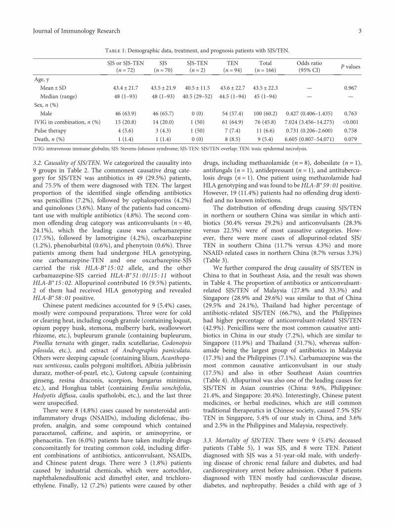
3.2. Causality of SJS/TEN. We categorized the causality into9 groups in Table 2. The commonest causative drug cate-gory for SJS/TEN was antibiotics in 49 (29.5%) patients,and 75.5% of them were diagnosed with TEN. The largestproportion of the identified single offending antibioticswas penicillins (7.2%), followed by cephalosporins (4.2%)and quinolones (3.6%). Many of the patients had concomi-tant use with multiple antibiotics (4.8%). The second com-mon offending drug category was anticonvulsants (n = 40,24.1%), which the leading cause was carbamazepine(17.5%), followed by lamotrigine (4.2%), oxcarbazepine(1.2%), phenobarbital (0.6%), and phenytoin (0.6%). Threepatients among them had undergone HLA genotyping,one carbamazepine-TEN and one oxcarbazepine-SJScarried the risk HLA-B∗15 : 02 allele, and the othercarbamazepine-SJS carried HLA-B∗51 : 01/15 : 11 withoutHLA-B∗15 : 02. Allopurinol contributed 16 (9.5%) patients,2 of them had received HLA genotyping and revealedHLA-B∗58 : 01 positive.
Chinese patent medicines accounted for 9 (5.4%) cases,mostly were compound preparations. Three were for coldor clearing heat, including cough granule (containing loquat,opium poppy husk, stemona, mulberry bark, swallowwortrhizome, etc.), bupleurum granule (containing bupleurum,Pinellia ternata with ginger, radix scutellariae, Codonopsispilosula, etc.), and extract of Andrographis paniculata.Others were sleeping capsule (containing lilium, Acanthopa-nax senticosus, caulis polygoni multiflori, Albizia julibrissindurazz, mother-of-pearl, etc.), Gutong capsule (containingginseng, resina draconis, scorpion, bungarus minimus,etc.), and Honghua tablet (containing Emilia sonchifolia,Hedyotis diffusa, caulis spatholobi, etc.), and the last threewere unspecified.
There were 8 (4.8%) cases caused by nonsteroidal anti-inflammatory drugs (NSAIDs), including diclofenac, ibu-profen, analgin, and some compound which containedparacetamol, caffeine, and aspirin, or aminopyrine, orphenacetin. Ten (6.0%) patients have taken multiple drugsconcomitantly for treating common cold, including differ-ent combinations of antibiotics, anticonvulsant, NSAIDs,and Chinese patent drugs. There were 3 (1.8%) patientscaused by industrial chemicals, which were acetochlor,naphthalenedisulfonic acid dimethyl ester, and trichloro-ethylene. Finally, 12 (7.2%) patients were caused by other
drugs, including methazolamide (n = 8), dobesilate (n = 1),antifungals (n = 1), antidepressant (n = 1), and antitubercu-losis drugs (n = 1). One patient using methazolamide hadHLA genotyping and was found to beHLA-B∗59 : 01 positive.However, 19 (11.4%) patients had no offending drug identi-fied and no known infections.
The distribution of offending drugs causing SJS/TENin northern or southern China was similar in which anti-biotics (30.4% versus 29.2%) and anticonvulsants (28.3%versus 22.5%) were of most causative categories. How-ever, there were more cases of allopurinol-related SJS/TEN in southern China (11.7% versus 4.3%) and moreNSAID-related cases in northern China (8.7% versus 3.3%)(Table 3).
We further compared the drug causality of SJS/TEN inChina to that in Southeast Asia, and the result was shownin Table 4. The proportion of antibiotics or anticonvulsant-related SJS/TEN of Malaysia (27.8% and 33.3%) andSingapore (28.9% and 29.6%) was similar to that of China(29.5% and 24.1%), Thailand had higher percentage ofantibiotic-related SJS/TEN (66.7%), and the Philippineshad higher percentage of anticonvulsant-related SJS/TEN(42.9%). Penicillins were the most common causative anti-biotics in China in our study (7.2%), which are similar toSingapore (11.9%) and Thailand (31.7%), whereas sulfon-amide being the largest group of antibiotics in Malaysia(17.3%) and the Philippines (7.1%). Carbamazepine was themost common causative anticonvulsant in our study(17.5%) and also in other Southeast Asian countries(Table 4). Allopurinol was also one of the leading causes forSJS/TEN in Asian countries (China: 9.6%, Philippines:21.4%, and Singapore: 20.4%). Interestingly, Chinese patentmedicines, or herbal medicines, which are still commontraditional therapeutics in Chinese society, caused 7.5% SJS/TEN in Singapore, 5.4% of our study in China, and 3.6%and 2.5% in the Philippines and Malaysia, respectively.
3.3. Mortality of SJS/TEN. There were 9 (5.4%) deceasedpatients (Table 5), 1 was SJS, and 8 were TEN. Patientdiagnosed with SJS was a 51-year-old male, with underly-ing disease of chronic renal failure and diabetes, and hadcardiorespiratory arrest before admission. Other 8 patientsdiagnosed with TEN mostly had cardiovascular disease,diabetes, and nephropathy. Besides a child with age of 3
Table 1: Demographic data, treatment, and prognosis patients with SJS/TEN.
SJS or SJS-TEN(n = 72)
SJS(n = 70)
SJS-TEN(n = 2)
TEN(n = 94)
Total(n = 166)
Odds ratio(95% CI)
P values
Age, y
Mean± SD 43.4± 21.7 43.5± 21.9 40.5± 11.5 43.6± 22.7 43.5± 22.3 — 0.967
Median (range) 48 (1–93) 48 (1–93) 40.5 (29–52) 44.5 (1–94) 45 (1–94) — —
Sex, n (%)
Male 46 (63.9) 46 (65.7) 0 (0) 54 (57.4) 100 (60.2) 0.427 (0.406–1.435) 0.763
IVIG in combination, n (%) 15 (20.8) 14 (20.0) 1 (50) 61 (64.9) 76 (45.8) 7.024 (3.456–14.275) <0.001Pulse therapy 4 (5.6) 3 (4.3) 1 (50) 7 (7.4) 11 (6.6) 0.731 (0.206–2.600) 0.758
Death, n (%) 1 (1.4) 1 (1.4) 0 (0) 8 (8.5) 9 (5.4) 6.605 (0.807–54.071) 0.079
IVIG: intravenous immune globulin; SJS: Stevens-Johnson syndrome; SJS-TEN: SJS/TEN overlap; TEN: toxic epidermal necrolysis.
3Journal of Immunology Research
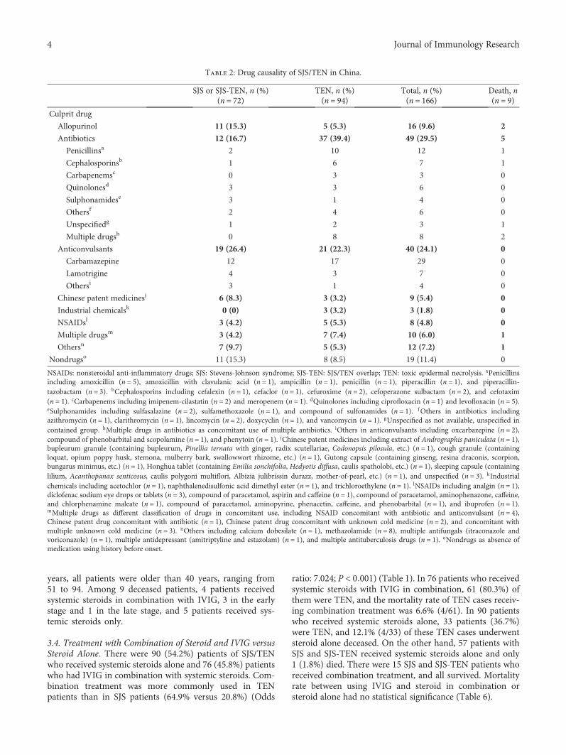
years, all patients were older than 40 years, ranging from51 to 94. Among 9 deceased patients, 4 patients receivedsystemic steroids in combination with IVIG, 3 in the earlystage and 1 in the late stage, and 5 patients received sys-temic steroids only.
3.4. Treatment with Combination of Steroid and IVIG versusSteroid Alone. There were 90 (54.2%) patients of SJS/TENwho received systemic steroids alone and 76 (45.8%) patientswho had IVIG in combination with systemic steroids. Com-bination treatment was more commonly used in TENpatients than in SJS patients (64.9% versus 20.8%) (Odds
ratio: 7.024; P < 0 001) (Table 1). In 76 patients who receivedsystemic steroids with IVIG in combination, 61 (80.3%) ofthem were TEN, and the mortality rate of TEN cases receiv-ing combination treatment was 6.6% (4/61). In 90 patientswho received systemic steroids alone, 33 patients (36.7%)were TEN, and 12.1% (4/33) of these TEN cases underwentsteroid alone deceased. On the other hand, 57 patients withSJS and SJS-TEN received systemic steroids alone and only1 (1.8%) died. There were 15 SJS and SJS-TEN patients whoreceived combination treatment, and all survived. Mortalityrate between using IVIG and steroid in combination orsteroid alone had no statistical significance (Table 6).
Table 2: Drug causality of SJS/TEN in China.
SJS or SJS-TEN, n (%)(n = 72)
TEN, n (%)(n = 94)
Total, n (%)(n = 166)
Death, n(n = 9)
Culprit drug
Allopurinol 11 (15.3) 5 (5.3) 16 (9.6) 2
Antibiotics 12 (16.7) 37 (39.4) 49 (29.5) 5
Penicillinsa 2 10 12 1
Cephalosporinsb 1 6 7 1
Carbapenemsc 0 3 3 0
Quinolonesd 3 3 6 0
Sulphonamidese 3 1 4 0
Othersf 2 4 6 0
Unspecifiedg 1 2 3 1
Multiple drugsh 0 8 8 2
Anticonvulsants 19 (26.4) 21 (22.3) 40 (24.1) 0
Carbamazepine 12 17 29 0
Lamotrigine 4 3 7 0
Othersi 3 1 4 0
Chinese patent medicinesj 6 (8.3) 3 (3.2) 9 (5.4) 0
Industrial chemicalsk 0 (0) 3 (3.2) 3 (1.8) 0
NSAIDsl 3 (4.2) 5 (5.3) 8 (4.8) 0
Multiple drugsm 3 (4.2) 7 (7.4) 10 (6.0) 1
Othersn 7 (9.7) 5 (5.3) 12 (7.2) 1
Nondrugso 11 (15.3) 8 (8.5) 19 (11.4) 0
NSAIDs: nonsteroidal anti-inflammatory drugs; SJS: Stevens-Johnson syndrome; SJS-TEN: SJS/TEN overlap; TEN: toxic epidermal necrolysis. aPenicillinsincluding amoxicillin (n = 5), amoxicillin with clavulanic acid (n = 1), ampicillin (n = 1), penicillin (n = 1), piperacillin (n = 1), and piperacillin-tazobactam (n = 3). bCephalosporins including cefalexin (n = 1), cefaclor (n = 1), cefuroxime (n = 2), cefoperazone sulbactam (n = 2), and cefotaxim(n = 1). cCarbapenems including imipenem-cilastatin (n = 2) and meropenem (n = 1). dQuinolones including ciprofloxacin (n = 1) and levofloxacin (n = 5).eSulphonamides including sulfasalazine (n = 2), sulfamethoxazole (n = 1), and compound of sulfonamides (n = 1). fOthers in antibiotics includingazithromycin (n = 1), clarithromycin (n = 1), lincomycin (n = 2), doxycyclin (n = 1), and vancomycin (n = 1). gUnspecified as not available, unspecified incontained group. hMultiple drugs in antibiotics as concomitant use of multiple antibiotics. iOthers in anticonvulsants including oxcarbazepine (n = 2),compound of phenobarbital and scopolamine (n = 1), and phenytoin (n = 1). jChinese patent medicines including extract of Andrographis paniculata (n = 1),bupleurum granule (containing bupleurum, Pinellia ternata with ginger, radix scutellariae, Codonopsis pilosula, etc.) (n = 1), cough granule (containingloquat, opium poppy husk, stemona, mulberry bark, swallowwort rhizome, etc.) (n = 1), Gutong capsule (containing ginseng, resina draconis, scorpion,bungarus minimus, etc.) (n = 1), Honghua tablet (containing Emilia sonchifolia, Hedyotis diffusa, caulis spatholobi, etc.) (n = 1), sleeping capsule (containinglilium, Acanthopanax senticosus, caulis polygoni multiflori, Albizia julibrissin durazz, mother-of-pearl, etc.) (n = 1), and unspecified (n = 3). kIndustrialchemicals including acetochlor (n = 1), naphthalenedisulfonic acid dimethyl ester (n = 1), and trichloroethylene (n = 1). lNSAIDs including analgin (n = 1),diclofenac sodium eye drops or tablets (n = 3), compound of paracetamol, aspirin and caffeine (n = 1), compound of paracetamol, aminophenazone, caffeine,and chlorphenamine maleate (n = 1), compound of paracetamol, aminopyrine, phenacetin, caffeine, and phenobarbital (n = 1), and ibuprofen (n = 1).mMultiple drugs as different classification of drugs in concomitant use, including NSAID concomitant with antibiotic and anticonvulsant (n = 4),Chinese patent drug concomitant with antibiotic (n = 1), Chinese patent drug concomitant with unknown cold medicine (n = 2), and concomitant withmultiple unknown cold medicine (n = 3). nOthers including calcium dobesilate (n = 1), methazolamide (n = 8), multiple antifungals (itraconazole andvoriconazole) (n = 1), multiple antidepressant (amitriptyline and estazolam) (n = 1), and multiple antituberculosis drugs (n = 1). oNondrugs as absence ofmedication using history before onset.
4 Journal of Immunology Research

4. Discussion
In this study, we enrolled a total 166 Han Chinese patientsdiagnosed with SJS, SJS-TEN overlap, and TEN from a ter-tiary medical center and Chinese literatures during 2006 to2016. We evaluated underlying condition, causation, treat-ment, and clinical outcome. Mean age of SJS/TEN was 43.5years, with little difference between SJS or SJS-TEN overlapand TEN. There was a male predominance in SJS or SJS-TEN overlap (male-to-female ratio 1.77 : 1) and TEN (male-to-female ratio 1.35 : 1). This observation was opposite towhat Mohammed et al. found in Egypt and different froman earlier study which showed equally affected by male andfemale [42, 43].
There were 88.6% of SJS/TEN patients had drug relation-ship, and the major contribution was antibiotics, followed byanticonvulsants and allopurinol. The difference between theantibiotics and anticonvulsants was small. This result wassimilar to the comparison of Malaysia and Singapore in areview of Southeast Asia [41], only different in sequence ofantibiotics and anticonvulsants, whereas Huang et al. foundanticonvulsants as the most common drug which causedSJS/TEN in China, followed by allopurinol, antipyretics/analgesics, and cephalosporins [44]. Similarly, Li and Mareported anticonvulsants and antibiotics to be the most com-mon single drug in SJS and traditional Chinese medicines inTEN [45]. It is known that allopurinol, aromatic anticonvul-sants, sulfonamide antibiotics, oxicam NSAIDs, and nevira-pine have higher risk to induced SCARs [46]. Nevertheless,there were only some sulphonamides and none oxicam typeof NSAIDs induced SJS/TEN in this study. This may due toprescribing habits of antibiotics in China and Taiwan,causing more penicillins and cephalosporins than the others[47–50]. Similarly, oxicam type of NSAIDs is less commonly
seen in Chinese literatures of case series [48, 49]. Allopurinolwas found to be a less common causality to induce SJS/TENin this study, especially in northern China. From previousreports, HLA-B∗58 : 01 was found positive in 93.3–100% ofpatients with allopurinol-induced SCARs whether in north-ern or southern China [51–54]. Moreover, the prevalence ofcarrying the risk HLA-B∗58 : 01 allele was 0.0515–0.085 inChina [55]. The discrepancy of the percentages between thisstudy and literature needs further investigation. Chinese pat-ent medicines were unique causative drugs to induce SJS/TEN in the Asian region [43, 56–59]. In our study, 5.4% ofthe SJS/TEN cases were related to Chinese patent medicine.Previously, Singapore was also reported to have more herbalmedicine-induced SJS/TEN cases [41]. However, there arepossibilities of adulteration with Western medicine in thecomponent of Chinese patent medicine [60–62], whichmakes it hard to identify the exact causality and may causebias. Patients also tend to received multiple drugs, includingcompound preparations of Western medicine or even anti-pyretic and analgetic in Chinese patent medicine [45]. Bothof these would increase the possibility of adverse drug reac-tion and enhance difficulty of identifying offending drug.
In our study, 19% of patients did not have definite or pos-sible relationship with drug according to ALDEN scoringsystem. The cause may be infection or idiopathic, and unfor-tunately there were no validation via further examinations.The annual incidence of SJS/TEN in the HIV-positive popu-lation is approximately 1000-fold higher than in the generalpopulation [63], and 4 patients with suspected causativedrugs were HIV positive in our study. Infections are possiblecausations besides drugs. Reactivations of human herpesvi-rus 6 (HHV6) and cytomegalovirus were found in SJS/TEN[64, 65]. A case has been reported of a teenage boy diagnosedwith SJS and primary Epstein-Barr virus infection withoutany attributing medication [66]. In addition, Mycoplasmapneumoniae infection may also be an additional cause ofSJS. Watkins et al. and Olson et al. have reported Myco-plasma pneumoniae infection outbreak associated with SJSin children [67, 68]. Although there were some reports withmalignancy-related SJS [69, 70], none of our non-drug-induced-SJS/TEN patients were found to have malignancy.
Withdrawal of offending drugs or treatment of causativeinfection, timely supportive treatment, immunomodulation,and management of complications and consequences arethe most common suggested treatments [71]. In this study,all of the patients received systemic corticosteroid. Despitesystemic corticosteroids remain a controversial treatmentfor SJS/TEN, it is the most commonly used medication acrossAsia [72–75].
Massive keratinocyte apoptosis induced by the intercellu-lar death receptor Fas and Fas ligand is now considered to bethe pathogenesis of SJS/TEN [76], yet IVIG inhibits keratino-cyte apoptosis by inhibiting the FAS receptor [77]. IVIG wasprescribed as an additional management in 45.8% of ourpatients, whether at the early or late stage of SJS/TEN, espe-cially with much higher percentage in TEN (80.3%) com-pared to SJS or SJS-TEN overlap (19.7%). Apparently IVIGis a common option of treating SJS/TEN in China, especiallyin TEN for their extensive skin lesion involvement, and is
Table 3: Comparison of the common drug causality betweennorthern and southern China.
Northern China,n (%)(n = 46)
Southern China,n (%)
(n = 120)
Total,n (%)
(n = 166)Antibiotics 14 (30.4) 35 (29.2) 49 (29.5)
Penicillins 3 (6.5) 9 (7.5) 12 (7.2)
Cephalosporins 1 (2.2) 6 (5.0) 7 (4.2)
Quinolones 2 (4.3) 4 (3.3) 6 (3.6)
Others 8 (17.4) 16 (13.3) 24 (14.5)
Anticonvulsants 13 (28.3) 27 (22.5) 40 (24.1)
Carbamazepine 9 (19.6) 20 (16.7) 29 (17.5)
Lamotrigine 2 (4.3) 5 (4.2) 7 (4.2)
Others 2 (4.3) 2 (1.7) 4 (2.4)
Nondrug 1 (2.2) 18 (15.0) 19 (11.4)
Allopurinol 2 (4.3) 14 (11.7) 16 (9.6)
Multiple drugs 6 (13.0) 4 (3.3) 10 (6.0)
Herbal medication 2 (4.3) 7 (5.8) 9 (5.4)
NSAIDs 4 (8.7) 4 (3.3) 8 (4.8)
Others 4 (8.7) 11 (9.2) 15 (9.0)
NSAIDs: nonsteroidal anti-inflammatory drugs.
5Journal of Immunology Research

usually in combination of systemic steroids instead of usingalone. A score-based comparison study of clinical outcomesfound that corticosteroid therapy combined with IVIG maylead to lower mortality when compared to corticosteroidalone [78]. However, several studies have shown limited suc-cess of IVIG in the clinical settings [79–82]. In our study,mortality rate in patients with TEN who received systemicsteroids with IVIG comparing to those who received systemic
steroids alone was 6.6% and 12.1%. However, this differenceof mortality rate was not statistically significant. Applicationof intravenous immunoglobulins or systemic corticosteroidsalso did not improve the outcome of SJS and TEN in a studyin Singapore [83]. Similarly, Lee et al. [84] demonstrated thatthe use of IVIG does not yield survival benefits in SJS/TENoverlap and TEN, even when corrected for IVIG dosages.Until now, the usage of IVIG in the treatment of SJS/TEN
Table 4: The comparison of the common drug causality from cases in China with other populations in Southeast Asia∗.
Culprit drug China (n = 166) Malaysia (n = 162) Singapore (n = 159) Thailand (n = 60) Philippines (n = 28)Antibiotics 49 (29.5) 45 (27.8) 46 (28.9) 40 (66.7) 5 (17.9)
Penicillins 12 (7.2) 14 (8.6) 19 (11.9) 19 (31.7) 1 (3.6)
Sulfonamide 4 (2.4) 28 (17.3) 11 (6.9) 9 (15.0) 2 (7.1)
Others 33 (19.9) 3 (1.9) 16 (10.1) 12 (20.0) 2 (7.1)
Anticonvulsants 40 (24.1) 54 (33.3) 47 (29.6) 9 (15.0) 12 (42.9)
Carbamazepine 29 (17.5) 34 (21.0) 29 (18.2) 4 (6.7) 4 (14.3)
Lamotrigine 7 (4.2) 7 (4.3) 2 (1.3) 0 (0) 0 (0)
Phenytoin 1 (0.6) 13 (8.0) 14 (8.8) 4 (6.7) 5 (17.9)
Allopurinol 16 (9.6) 33 (20.4) 23 (14.5) 1 (1.7) 6 (21.4)
NSAIDs 8 (4.8) 10 (6.2) 14 (8.8) 4 (6.7) 3 (10.7)
Herbal medications 9 (5.4) 4 (2.5) 12 (7.5) 0 (0) 1 (3.6)∗We compared the common drug causality from cases in China with other populations in Southeast Asia according to the previous literature report (41).
Table 5: Information of the deceased patients with SJS/TEN in this study (n=XXX).
Phenotype Sex Age, y Underlying disease SCORTEN Culprit drugs Treatment
SJS M 51 Chronic renal failure, diabetes 4 Allopurinol Systemic steroids
TEN M 70 Nil 6 Antibiotics Systemic steroids
TEN F 58 Aneurysm, subarachnoid hemorrhage NA Antibiotics Systemic steroids
TEN F 67Rheumatic heart disease, mitral
insufficiencyNA
Antibiotics and compoundwith aminopyrine, phenacetin,
caffeine, phenobarbital
Systemic steroids withIVIG use in the late
stage
TEN F 71Coronary heart disease, hypertension,
diabetes, diabetic nephropathy4 Calcium dobesilate
Systemic steroids withIVIG use in the early
stage
TEN M 62 Hypertension, diabetes NA AntibioticsSystemic steroids withIVIG use in the early
stage
TEN M 94Coronary heart disease, cardiac
insufficiency, hypertension, diabetes,interstitial lung disease
NA AntibioticsSystemic steroids withIVIG use in the early
stage
TEN M 62Hypertension, diabetes, chronic renal
failure, hyperuricemiaNA Allopurinol Systemic steroids
TEN M 3 Nil 2 Antibiotics Systemic steroids
IVIG use in the early stage ≤ 7 days of onset; IVIG use in the late stage ≥ 7 days of onset. NA: not available.
Table 6: A comparison of mortality rate between combination treatment of steroid with IVIG versus steroid alone.
Mortality Steroids with IVIG (n = 76) Steroids alone (n = 90) Odds ratio (95% CI) P values
TEN, n (%) 4/61 (6.6) 4/33 (12.1) 0.509 (0.119–2.183) 0.445
SJS and SJS/TEN, n (%) 0/15 (0.0) 1/57 (1.8) — 1.000
Total cases, n (%) 4/76 (5.3) 5/90 (5.6) 0.944 (0.244–3.650) 1.000
6 Journal of Immunology Research

is still controversial. Recent studies have shown that immu-nosuppressive treatment with tumor necrosis factor-alpha(TNF-α) inhibitors may be helpful [85] and cyclosporin Ais also safe and may contribute to rapid reepithelializationin patients with SJS/TEN [86–88]. The efficacy of usingcyclosporin in treating SJS/TEN has recently validated withthe decreased mortality rate both in adults and children[89–92].
There are several limitations in this study. First, weenrolled case reports only with careful checkup to preventoverlapping cases. However, ruling out the articles with caseseries also led to underestimation of SJS/TEN patients. Sec-ond, the mortality rate in our study is lower than interna-tional literatures which ranged from 10% to 70% [93, 94].The possibility of lower mortality in this study may be dueto underreported deceased cases of SJS/TEN from theChinese literatures. In addition, the underlying severity ofSJS/TEN in our study is unknown due to the lack of completedata of SCORTEN factors; hence, the efficacy of treatmentneeds to be further elucidated.
5. Conclusion
SJS/TEN is life-threatening drug adverse reaction, withhigher prevalence rate in Asian than in Western populationsin literature review. The most common offending drugs inour study are antibiotics, anticonvulsants, and allopurinol.IVIG in combination with systemic steroids is a commonoption especially for TEN in China. There was no significantdifference in the mortality rate of TEN patients with or with-out IVIG adjuvant treatment.
Conflicts of Interest
The authors declare that there is no conflict of interestregarding the publication of this paper.
Acknowledgments
The authors thank Chun-Bing Chen (Department ofDermatology, Drug Hypersensitivity Clinical and ResearchCenter, Chang Gung Memorial Hospital, Taipei, Linkou,and Keelung, Chang Gung University, Taoyuan, Taiwan)for assisting this article.
References
[1] J. C. Roujeau, “Immune mechanisms in drug allergy,” Allergol-ogy International, vol. 55, no. 1, pp. 27–33, 2006.
[2] S. C. Su, M. Mockenhaupt, P. Wolkenstein et al., “Interleukin-15 is associated with severity and mortality in Stevens-Johnsonsyndrome/toxic epidermal necrolysis,” Journal of InvestigativeDermatology, vol. 137, no. 5, pp. 1065–1073, 2017.
[3] W. H. Chung, S. I. Hung, J. Y. Yang et al., “Granulysin is a keymediator for disseminated keratinocyte death in Stevens-Johnson syndrome and toxic epidermal necrolysis,” NatureMedicine, vol. 14, no. 12, pp. 1343–1350, 2008.
[4] S. J. Posadas, A. Padial, M. J. Torres et al., “Delayed reactionsto drugs show levels of perforin, granzyme B, and Fas-L to be
related to disease severity,” The Journal of Allergy and ClinicalImmunology, vol. 109, no. 1, pp. 155–161, 2002.
[5] W. H. Chung, C. W. Wang, and R. L. Dao, “Severe cutaneousadverse drug reactions,” The Journal of Dermatology, vol. 43,no. 7, pp. 758–766, 2016.
[6] J. C. Roujeau and R. S. Stern, “Severe adverse cutaneousreactions to drugs,” The New England Journal of Medicine,vol. 331, no. 19, pp. 1272–1285, 1994.
[7] S. Bastuji-Garin, N. Fouchard, M. Bertocchi, J. C. Roujeau,J. Revuz, and P. Wolkenstein, “SCORTEN: a severity-of-illness score for toxic epidermal necrolysis,” Journal of Investi-gative Dermatology, vol. 115, no. 2, pp. 149–153, 2000.
[8] H. L. Chan, R. S. Stern, K. A. Arndt et al., “The incidence oferythema multiforme, Stevens-Johnson syndrome, and toxicepidermal necrolysis. A population-based study with particu-lar reference to reactions caused by drugs among outpatients,”Archives of Dermatology, vol. 126, no. 1, pp. 43–47, 1990.
[9] N. Frey, J. Jossi, M. Bodmer et al., “The epidemiology ofStevens-Johnson syndrome and toxic epidermal necrolysis inthe UK,” Journal of Investigative Dermatology, vol. 137, no. 6,pp. 1240–1247, 2017.
[10] S. Y. Liu, T. T. Liu, Y. J. Yu, and R. Q. Wang, “A case of toxicepidermal necrolysis,” China Journal of Leprosy and SkinDiseases, vol. 25, no. 6, p. 456, 2009.
[11] P. Xia, W. Y. Wu, J. Ren, X. M. Hou, and Z. Z. Zheng, “Toxicepidermal necrolysis: a case report,” Journal of Clinical Derma-tology, vol. 38, no. 12, pp. 778-779, 2009.
[12] Y. X. Li, Q. Fu, X. C. Zhang, and L. X. Du, “Toxic epidermalnecrolysis: a case report,” The Chinese Journal of Dermatove-nereology, vol. 24, no. 4, p. 388, 2010.
[13] J. B. Du, Y. Q. Chu, S. Guo, X. L. Qi, and C. Zhao, “Toxicepidermal necrolysis associated with imipenem/cilastatinsodium,” Adverse Drug Reactions Journal, vol. 12, no. 6,pp. 431–433, 2010.
[14] X. F. Fang, X. X. Zhang, and J.W. Li, “A case of toxic epidermalnecrolysis caused by exposure to acetochlor,” China Journal ofLeprosy and Skin Diseases, vol. 27, no. 4, pp. 273-274, 2011.
[15] J. J. Liu, S. C. Lv, H. M. Yang, B. Li, J. L. Liu, and T. B. Xia, “Acase of toxic epidermal necrolysis induced by carbamazepinesubcutaeous embedded,” Journal of Practical Dermatology,vol. 5, no. 6, pp. 372-373, 2012.
[16] F. Yang and F. Liu, “Fatal of severe aneurysm complicated withtoxic epidermal necrolysis,” West China Medical Journal,vol. 27, no. 8, pp. 1166-1167, 2012.
[17] M. Q. Sun, L. Tong, and Y. Liu, “A case report of toxic epider-mal necrolysis,” The Chinese Journal of Dermatovenereology,vol. 27, no. 12, pp. 1308-1309, 2013.
[18] K. Yu, Y. Y. Wang, L. Chang et al., “A case of Steven-Johnsonsyndrome caused by diclofenac sodium eye drops,” ChineseJournal of Dermatovenereology, vol. 28, no. 6, pp. 611-612,2014.
[19] H. Xu, X. F. Chen, W. G.Wang, andW. H. Ge, “A fatal of toxicepidermal necrolysis caused by compound of analgesictablets,” Chinese Journal of Difficult and Complicated Cases,vol. 13, no. 11, pp. 1195-1196, 2014.
[20] X. H. Tang, H. Zhou, M. H. Zhong, G. L. Cao, and Q. Gao,“Blindness caused by Stevens-Johnson syndrome in children:a case report,” Journal of Diagnosis and Therapy onDermato-venereology, vol. 22, no. 3, pp. 247–249, 2015.
[21] R. X. Ren, L. Jia, and H. Y. Zheng, “Toxic epidermal necrolysisin HLA-B∗1502 gene carrier induced by carbamazepine: a case
7Journal of Immunology Research

report,” Journal of Clinical Dermatology, vol. 44, no. 10,pp. 638-639, 2015.
[22] Y. Qiang, L. H. Chen, Y. H. Zhang, Y. Shen, X. Q. Zhao, andM. Pan, “Toxic epidermal necrolysis in a patient with adult-onset still disease and literature review,” Chinese Journal ofDermatovenereology, vol. 29, no. 9, pp. 943–945, 2015.
[23] J. J. Liu, Y. Jiang, J. Lv, and G. F. Yan, “A case of calciumdobesilate-induced toxic epidermal necrolysis,” Chinese Jour-nal of Dermatovenereology, vol. 29, no. 6, pp. 615-616, 2015.
[24] H. Bai, W. W. Tong, X. Q. Wei, and J. Guo, “Treatmentanalysis of a fatal with severe acute pancreatitis and toxicepidermal necrolysis,” Chinese Journal of Hospital Pharmacy,vol. 35, no. 8, pp. 763-764, 2015.
[25] H. Zhang and Y. Zhong, “One case of drug-induced bullosaepidermolysis caused by amoxicillin-sulbactam,” Chinese Jour-nal of New Drugs and Clinical Remedies, vol. 34, no. 9, pp. 689–691, 2015.
[26] C. Y. Wei and T. Xu, “A fatal of toxic epidermal necrolysisinduced by allopurinol,” Chinese Journal of Pharmacoepide-miology, vol. 24, no. 3, pp. 189-190, 2015.
[27] T. Zhang, R. F. Shi, and J. Zheng, “A case of toxic epidermalnecrolysis complicated with HIV infection,” Chinese Journalof Dermatology, vol. 41, no. 5, p. 345, 2008.
[28] J. P. Chu and Y. Wang, “Carbamazepine-induced toxic epider-mal necrolysis: a case report,” Shaanxi Medical Journal, vol. 40,no. 5, p. 640, 2011.
[29] R. J. Jin, X. Y. Zhan, J. X. Jiang, and L. P. Sun, “A case ofmethazolamide-induced Stevens-Johnson syndrome,” ChineseJournal of Dermatovenereology, vol. 26, no. 4, pp. 363-364,2012.
[30] X. L. He, Z. S. Liu, G. F. Wu et al., “Carbamazepine-inducedStevens-Johnson syndrome with negative HLA-B∗1502: casereport and literature review,” Journal of Clinical Pediatrics,vol. 30, no. 11, pp. 1006–1010, 2012.
[31] H. Wei, B. Wang, and F. Liu, “A case report: toxic epidermalnecrolysis caused by allopurinol,” Chinese Medical Record,vol. 14, no. 8, pp. 36–38, 2013.
[32] C.Wu, H. Z. Jin, K. Fang, and L. Li, “A case of Stevens-Johnsonsyndrome presented as chickenpox-like lesions,” Journal ofClinical Dermatology, vol. 43, no. 3, pp. 159–161, 2014.
[33] W. H. Liu, Y. N. Zhang, and Q. Zhang, “Methazolamide-induced Stevens-Johnson syndrome before and after glaucomasurgery,” Chinese Journal of Ocular Trauma and OccupationalEye Disease, vol. 36, no. 11, pp. 850–852, 2014.
[34] J. Li, J. J. Tao, J. Lu, and Y. D. Zhao, “A case of SJS/TEN overlapinduced by oral methazolamide,” Chinese Journal of Ophthal-mology, vol. 51, no. 12, pp. 934-935, 2015.
[35] J. M. Jiang and Z. W. Zhang, “A case of carbamazepine-induced Stevens-Johnson syndrome,” Evaluation and Analysisof Drug-Use in Hospitals of China, vol. 15, no. 9, article 1280,2015.
[36] Y. L. Li and J. Liu, “Allopurinol-induced Stevens-Johnsonsyndrome in a HLA-B∗5801 allele gene carrier,” Evaluationand Analysis of Drug-Use in Hospitals of China, vol. 15,no. 11, pp. 1567-1568, 2015.
[37] Y. H. Xu, Y. Su, J. Zhao, Y. J. Du, and Q. Sun, “Methazolamide-induced toxic epidermal necrolysis in a patient with HLA∗
B5901 allele,” Chinese Journal of Dermatology, vol. 48, no. 2,pp. 131–133, 2015.
[38] W. H. Chung, W. C. Chang, Y. S. Lee et al., “Genetic variantsassociated with phenytoin-related severe cutaneous adverse
reactions,” The Journal of the American Medical Association,vol. 312, no. 5, pp. 525–534, 2014.
[39] C. Y. Ng, Y. T. Yeh, C. W. Wang et al., “Impact of the HLA-B(∗)58:01 allele and renal impairment on allopurinol-inducedcutaneous adverse reactions,” The Journal of InvestigativeDermatology, vol. 136, no. 7, pp. 1373–1381, 2016.
[40] C. B. Chen, Y. H. Hsiao, T. Wu et al., “Risk and association ofHLA with oxcarbazepine-induced cutaneous adverse reactionsin Asians,” Neurology, vol. 88, no. 1, pp. 78–86, 2017.
[41] H. Y. Lee, W. Martanto, and T. Thirumoorthy, “Epidemiologyof Stevens-Johnson syndrome and toxic epidermal necrolysisin Southeast Asia,” Dermatologica Sinica, vol. 31, no. 4,pp. 217–220, 2013.
[42] A. E. Mohammed, E. E. Reham, and A. E. Wafaa, “A clinicoe-tiological analysis of patients with Stevens–Johnson syndromeand toxic epidermal necrolysis treated at Sohag UniversityHospital, Egypt,” Journal of the Egyptian Women’s Dermato-logic Society, vol. 14, no. 1, pp. 37–44, 2017.
[43] S. K. Tan and Y. K. Tay, “Profile and pattern of Stevens–Johnson syndrome and toxic epidermal necrolysis in a generalhospital in Singapore: treatment outcomes,” Acta Dermato-Venereologica, vol. 92, no. 1, pp. 62–66, 2012.
[44] H. Y. Huang, X. Q. Luo, L. S. Chan, Z. H. Cao, X. F. Sun, andJ. H. Xu, “Cutaneous adverse drug reactions in a hospital-based Chinese population,” Clinical and Experimental Derma-tology, vol. 36, no. 2, pp. 135–141, 2011.
[45] L. F. Li and C. Ma, “Epidemiological study of severe cutaneousadverse drug reactions in a city district of China,” Clinical andExperimental Dermatology, vol. 31, no. 5, pp. 642–647, 2006.
[46] S. C. Su, S. I. Huang, W. L. Fan, R. L. Dao, and W. H. Chung,“Severe cutaneous adverse reactions: the pharmacogenomicsfrom research to clinical implementation,” International Jour-nal of Molecular Sciences, vol. 17, no. 11, article E1890, 2016.
[47] J. Sun, J. Liu, Q. L. Gong et al., “Stevens–Johnson syndromeand toxic epidermal necrolysis: a multi-aspect comparative 7-year study from the People’s Republic of China,” Drug DesignDevelopment and Therapy, vol. 8, pp. 2539–2547, 2014.
[48] C. Y. Gong, B. X. Wang, and M.W. Yu, “Clinical analysis of 58cases of severe drug eruption,” Chinese Journal of Dermatove-nereology, vol. 29, no. 1, pp. 39–41, 2015.
[49] S. Wang, J. H. Zhao, Q. M. Sun, J. J. Qiao, and H. Fang, “Clin-ical analysis of 55 cases of severe drug eruption,” Chinese Jour-nal of Dermatovenereology, vol. 30, no. 12, pp. 1241–1243,2016.
[50] Y. F. Lin, C. H. Yang, S. Hu et al., “Severe cutaneous adversereactions related to systemic antibiotics,” Clinical InfectiousDiseases, vol. 58, no. 10, pp. 1377–1385, 2014.
[51] Z. H. Cao, Z. Y. Wei, Q. Y. Zhu et al., “HLA-B∗58:01 allele isassociated with augmented risk for both mild and severecutaneous adverse reactions induced by allopurinol in HanChinese,” Pharmacogenomics, vol. 13, no. 10, pp. 1193–1201,2012.
[52] Z. Y. Deng, J. Yang, andW. L. Yang, “Detection of theHLA-B∗
5801 allele in Han Chinese with allopurinol-induced severedrug eruption,” Journal of Diagnosis and Therapy onDermato-venereology, vol. 20, no. 6, pp. 379–382, 2013.
[53] L. Cheng, Y. Xiong, C. Z. Qin et al., “HLA-B∗58:01 is stronglyassociated with allopurinol-induced severe cutaneous adversereactions in Han Chinese patients: a multicentre retrospectivecase-control clinical study,” British Journal of Dermatology,vol. 173, no. 2, pp. 555–558, 2015.
8 Journal of Immunology Research
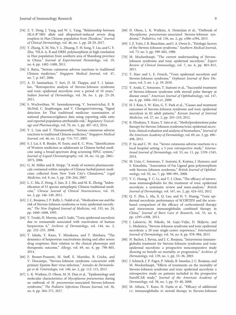
[54] Z. Y. Deng, J. Yang, and W. L. Yang, “Relationship betweenHLA-B∗5801 allele and allopurinol-induced severe drugeruption in Han Chinese population from Zhoukou,” Journalof Clinical Dermatology, vol. 46, no. 1, pp. 28-29, 2017.
[55] Y. Zhang, X. M. Nie, Y. L. Zhuang, Y. H. Song, Y. Liu, and C. F.Zhu, “HLA-A, B and DRB1 polymorphism at high-resolutionin Han population from southern area of Shandong provincein China,” Journal of Experimental Hematology, vol. 19,no. 6, pp. 1482–1488, 2011.
[56] S. Batra, “Serious cutaneous adverse reactions to traditionalChinese medicines,” Singapore Medical Journal, vol. 47,no. 7, p. 647, 2006.
[57] A. D. Sanmarkan, T. Sori, D. M. Thappa, and T. J. Jaisan-kar, “Retrospective analysis of Stevens-Johnson syndromeand toxic epidermal necrolysis over a period of 10 years,”Indian Journal of Dermatology, vol. 56, no. 1, pp. 25–29,2011.
[58] S. Wechwithan, W. Suwankesawong, V. Sornsrivichai, E. B.McNeil, C. Jiraphongsa, and V. Chongsuvivatwong, “Signaldetection for Thai traditional medicine: examination ofnational pharmacovigilance data using reporting odds ratioand reported population attributable risk,” Regulatory Toxicol-ogy and Pharmacology, vol. 70, no. 1, pp. 407–412, 2014.
[59] Y. L. Lim and T. Thirumoorthy, “Serious cutaneous adversereactions to traditional Chinese medicines,” Singapore MedicalJournal, vol. 46, no. 12, pp. 714–717, 2005.
[60] S. J. Lai, S. R. Binder, H. Essin, and K. C. Wen, “Identificationof Western medicines as adulterants in Chinese herbal medi-cine using a broad-spectrum drug screening HPLC system,”Journal of Liquid Chromatography, vol. 18, no. 14, pp. 2861–2875, 2006.
[61] G. M. Miller and R. Stripp, “A study of western pharmaceuti-cals contained within samples of Chinese herbal/patent medi-cines collected from New York City’s Chinatown,” LegalMedicine, vol. 9, no. 5, pp. 258–264, 2007.
[62] L. C. Ma, Z. Hong, Z. Jiao, X. J. Shi, and M. K. Zhong, “Inden-tification of 53 species antiepileptic Chinese traditional medi-cine,” Chinese Journal of Clinical Neurosciences, vol. 19,no. 2, pp. 146–149, 2011.
[63] J. C. Roujeau, J. P. Kelly, L. Naldi et al., “Medication use and therisk of Stevens-Johnson syndrome or toxic epidermal necroly-sis,” The New England Journal of Medicine, vol. 333, no. 24,pp. 1600–1608, 1995.
[64] Y. Teraki, H. Murota, and S. Izaki, “Toxic epidermal necrolysisdue to zonisamide associated with reactivation of humanherpesvirus 6,” Archives of Dermatology, vol. 144, no. 2,pp. 232–235, 2008.
[65] T. Ishida, Y. Kano, Y. Mizukawa, and T. Shiohara, “Thedynamics of herpesvirus reactivations during and after severedrug eruptions: their relation to the clinical phenotype andtherapeutic outcome,” Allergy, vol. 69, no. 6, pp. 798–805,2014.
[66] F. Brunet-Possenti, M. Steff, E. Marinho, B. Crickx, andV. Descamps, “Stevens-Johnson syndrome concurrent withprimary Epstein-Barr virus infection,” Annales de Dermatolo-gie et de Vénéréologie, vol. 140, no. 2, pp. 112–115, 2013.
[67] L. K. Watkins, D. Olson, M. H. Diaz et al., “Epidemiology andmolecular characteristics of Mycoplasma pneumoniae duringan outbreak of M. pneumoniae-associated Stevens-Johnsonsyndrome,” The Pediatric Infectious Disease Journal, vol. 36,no. 6, pp. 564–571, 2017.
[68] D. Olson, L. K. Watkins, A. Demirjian et al., “Outbreak ofMycoplasma pneumoniae-associated Stevens-Johnson syn-drome,” Pediatrics, vol. 136, no. 2, pp. e386–e394, 2015.
[69] J. Z. Yetiv, J. R. Bianchine, and J. A. Owen Jr., “Etiologic factorsof the Stevens-Johnson syndrome,” Southern Medical Journal,vol. 73, no. 5, pp. 599–602, 1980.
[70] M. Mockenhaupt, “The current understanding of Stevens-Johnson syndrome and toxic epidermal necrolysis,” ExpertReview of Clinical Immunology, vol. 7, no. 6, pp. 803–815,2011.
[71] T. Harr and L. E. French, “Toxic epidermal necrolysis andStevens-Johnson syndrome,” Orphanet Journal of Rare Dis-eases, vol. 5, no. 1, p. 39, 2010.
[72] Y. Araki, C. Sotozono, T. Inatomi et al., “Successful treatmentof Stevens-Johnson syndrome with steroid pulse therapy atdisease onset,” American Journal of Ophthalmology, vol. 147,no. 6, pp. 1004–1011.e1, 2009.
[73] H. I. Kim, S. W. Kim, G. Y. Park et al., “Causes and treatmentoutcomes of Stevens-Johnson syndrome and toxic epidermalnecrolysis in 82 adult patients,” Korean Journal of InternalMedicine, vol. 27, no. 2, pp. 203–210, 2012.
[74] K. Hirahara, Y. Kano, Y. Sato et al., “Methylprednisolone pulsetherapy for Stevens-Johnson syndrome/toxic epidermal necro-lysis: clinical evaluation and analysis of biomarkers,” Journal ofthe American Academy of Dermatology, vol. 69, no. 3, pp. 496–498, 2013.
[75] P. Su and C. W. Aw, “Severe cutaneous adverse reactions in alocal hospital setting: a 5-year retrospective study,” Interna-tional Journal of Dermatology, vol. 53, no. 11, pp. 1339–1345,2014.
[76] M. Ueta, C. Sotozono, T. Inatomi, K. Kojima, J. Hamuro, andS. Kinoshita, “Association of Fas Ligand gene polymorphismwith Stevens-Johnson syndrome,” British Journal of Ophthal-mology, vol. 92, no. 7, pp. 989–991, 2008.
[77] Y. C. Huang, Y. C. Li, and T. J. Chen, “The efficacy of intrave-nous immunoglobulin for the treatment of toxic epidermalnecrolysis: a systematic review and meta-analysis,” BritishJournal of Dermatology, vol. 167, no. 2, pp. 424–432, 2012.
[78] Q. Y. Zhu, L. Ma, X. Q. Luo, and H. Y. Huang, “Toxic epi-dermal necrolysis: performance of SCORTEN and the score-based comparison of the efficacy of corticosteroid therapyand intravenous immunoglobulin combined therapy inChina,” Journal of Burn Care & Research, vol. 33, no. 6,pp. e295–e308, 2012.
[79] J. Lalosevic, M. Nikolic, M. Gajic-Veljic, D. Skiljevic, andL. Medenica, “Stevens-Johnson syndrome and toxic epidermalnecrolysis: a 20-year single-center experience,” InternationalJournal of Dermatology, vol. 54, no. 8, pp. 978–984, 2015.
[80] N. Bachot, J. Revuz, and J. C. Roujeau, “Intravenous immuno-globulin treatment for Stevens-Johnson syndrome and toxicepidermal necrolysis: a prospective noncomparative studyshowing no benefit on mortality or progression,” Archives ofDermatology, vol. 139, no. 1, pp. 33–36, 2003.
[81] J. Schneck, J. P. Fagot, P. Sekula, B. Sassolas, J. C. Roujeau, andM. Mockenhaupt, “Effects of treatments on the mortality ofStevens-Johnson syndrome and toxic epidermal necrolysis: aretrospective study on patients included in the prospectiveEuroSCAR study,” Journal of the American Academy ofDermatology, vol. 58, no. 1, pp. 33–40, 2008.
[82] M. Aihara, Y. Kano, H. Fujita et al., “Efficacy of additionali.v. immunoglobulin to steroid therapy in Stevens-Johnson
9Journal of Immunology Research
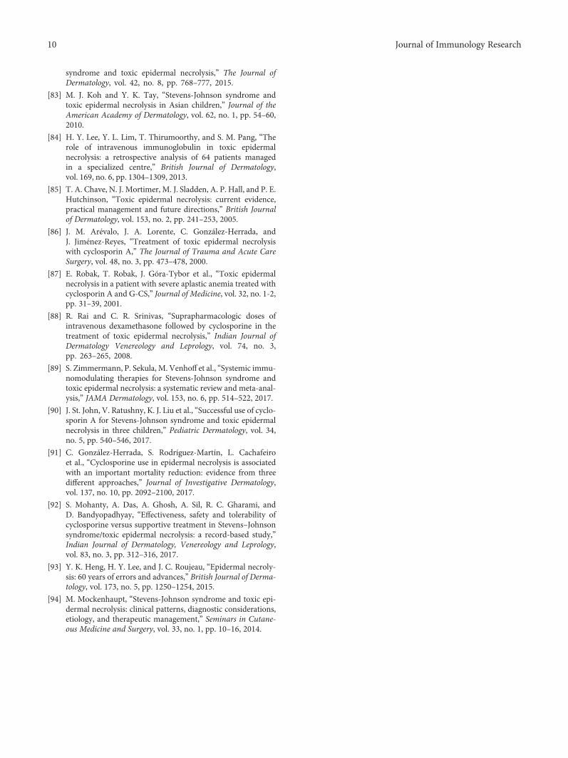
syndrome and toxic epidermal necrolysis,” The Journal ofDermatology, vol. 42, no. 8, pp. 768–777, 2015.
[83] M. J. Koh and Y. K. Tay, “Stevens-Johnson syndrome andtoxic epidermal necrolysis in Asian children,” Journal of theAmerican Academy of Dermatology, vol. 62, no. 1, pp. 54–60,2010.
[84] H. Y. Lee, Y. L. Lim, T. Thirumoorthy, and S. M. Pang, “Therole of intravenous immunoglobulin in toxic epidermalnecrolysis: a retrospective analysis of 64 patients managedin a specialized centre,” British Journal of Dermatology,vol. 169, no. 6, pp. 1304–1309, 2013.
[85] T. A. Chave, N. J. Mortimer, M. J. Sladden, A. P. Hall, and P. E.Hutchinson, “Toxic epidermal necrolysis: current evidence,practical management and future directions,” British Journalof Dermatology, vol. 153, no. 2, pp. 241–253, 2005.
[86] J. M. Arévalo, J. A. Lorente, C. González-Herrada, andJ. Jiménez-Reyes, “Treatment of toxic epidermal necrolysiswith cyclosporin A,” The Journal of Trauma and Acute CareSurgery, vol. 48, no. 3, pp. 473–478, 2000.
[87] E. Robak, T. Robak, J. Góra-Tybor et al., “Toxic epidermalnecrolysis in a patient with severe aplastic anemia treated withcyclosporin A and G-CS,” Journal of Medicine, vol. 32, no. 1-2,pp. 31–39, 2001.
[88] R. Rai and C. R. Srinivas, “Suprapharmacologic doses ofintravenous dexamethasone followed by cyclosporine in thetreatment of toxic epidermal necrolysis,” Indian Journal ofDermatology Venereology and Leprology, vol. 74, no. 3,pp. 263–265, 2008.
[89] S. Zimmermann, P. Sekula, M. Venhoff et al., “Systemic immu-nomodulating therapies for Stevens-Johnson syndrome andtoxic epidermal necrolysis: a systematic review and meta-anal-ysis,” JAMA Dermatology, vol. 153, no. 6, pp. 514–522, 2017.
[90] J. St. John, V. Ratushny, K. J. Liu et al., “Successful use of cyclo-sporin A for Stevens-Johnson syndrome and toxic epidermalnecrolysis in three children,” Pediatric Dermatology, vol. 34,no. 5, pp. 540–546, 2017.
[91] C. González-Herrada, S. Rodríguez-Martín, L. Cachafeiroet al., “Cyclosporine use in epidermal necrolysis is associatedwith an important mortality reduction: evidence from threedifferent approaches,” Journal of Investigative Dermatology,vol. 137, no. 10, pp. 2092–2100, 2017.
[92] S. Mohanty, A. Das, A. Ghosh, A. Sil, R. C. Gharami, andD. Bandyopadhyay, “Effectiveness, safety and tolerability ofcyclosporine versus supportive treatment in Stevens–Johnsonsyndrome/toxic epidermal necrolysis: a record-based study,”Indian Journal of Dermatology, Venereology and Leprology,vol. 83, no. 3, pp. 312–316, 2017.
[93] Y. K. Heng, H. Y. Lee, and J. C. Roujeau, “Epidermal necroly-sis: 60 years of errors and advances,” British Journal of Derma-tology, vol. 173, no. 5, pp. 1250–1254, 2015.
[94] M. Mockenhaupt, “Stevens-Johnson syndrome and toxic epi-dermal necrolysis: clinical patterns, diagnostic considerations,etiology, and therapeutic management,” Seminars in Cutane-ous Medicine and Surgery, vol. 33, no. 1, pp. 10–16, 2014.
10 Journal of Immunology Research

Review ArticleAnticancer Drugs Induced Severe Adverse Cutaneous DrugReactions: An Updated Review on the Risks Associated withAnticancer Targeted Therapy or Immunotherapies
Chau Yee Ng ,1,2,3,4 Chun-Bing Chen ,1,2,3,4 Ming-Ying Wu,1,2,3 Jennifer Wu,1,2,3
Chih-Hsun Yang,1,2,3,4 Rosaline Chung-Yee Hui,1,2,3,4 Ya-Ching Chang,1,2,3,4
and Chun-Wei Lu 1,2,3,4
1Department of Dermatology, College of Medicine, Chang Gung Memorial Hospital, Keelung, Linkou, Taipei, Taiwan2Drug Hypersensitivity Clinical and Research Center, Chang Gung Memorial Hospital, Taipei, Taiwan3School of Medicine, College of Medicine, Chang Gung University, Taoyuan, Taiwan4Graduate Institute of Clinical Medical Sciences, Chang Gung University, Taoyuan, Taiwan
Correspondence should be addressed to Chun-Wei Lu; [email protected]
Received 31 August 2017; Revised 7 November 2017; Accepted 8 November 2017; Published 17 January 2018
Academic Editor: Riichiro Abe
Copyright © 2018 Chau Yee Ng et al. This is an open access article distributed under the Creative Commons Attribution License,which permits unrestricted use, distribution, and reproduction in any medium, provided the original work is properly cited.
Cutaneous adverse drug reactions are commonly seen in patients with anticancer drug treatment. Anticancer drugs, includingchemotherapy, target therapy, and recent immunotherapy causing skin reactions ranging from mild skin rash to life-threateningsevere cutaneous adverse reactions (SCARs), such as Stevens-Johnson syndrome (SJS) and toxic epidermal necrosis (TEN) withincrease morbidity and mortality while they are receiving cancer treatments, have been proposed to be a result of direct skintoxicity or drug hypersensitivity reactions (these are proposed mechanism, not definite). Differentiating SCARs from other morecommonly seen reactions with a better outcome help prevent discontinuation of therapy and inappropriate use of systemicimmunosuppressants for presumable allergic reactions, of which will affect the clinical outcome. In this article, we have reviewedpublished articles from 1950 to August 2017 for SJS/TEN associated with anticancer drugs, including chemotherapy, targetedtherapy, and immunotherapy. We aimed to provide an overview of SJS/TEN associated with anticancer drugs to increaseclinician recognition and accelerate future studies on the pathomechanism and managements.
1. Introduction
The advancement in cancer detection and development ofanticancer drug therapy has led to increased incidence ofcutaneous adverse reactions following anticancer drug ther-apy. Conventional chemotherapy and targeted or immuno-therapy that are thought to be well tolerated and may causevarious cutaneous adverse reactions ranging from nonlife-threatening skin toxicities such as paronychia, acneiformeruption, and alopecia to life-threatening severe cutaneousadverse reactions (SCARs) such as Stevens-Johnson syn-drome (SJS) and toxic epidermal necrolysis (TEN). Thesedrug eruptions are thought to be immunologically mediatedreactions that are termed type B adverse reaction [1].
However, the pathomechanism of SCARs reactions in anti-cancer drugs including chemotherapy, targeted therapy,and immunotherapy is poorly understood and the literatureswere still limited.
SJS/TEN are a spectrum of fatal mucocutaneous adversereactions characterized by rapidly progressing purpuricatypical target-like rashes with blisters, cutaneous sloughing,and mucosal involvement. SJS and TEN are differentiatedby the degree of skin detachment: SJS involves less than10% body surface area skin detachment, TEN more than30%, while SJS/TEN overlap involves body surface areaof 10–30% [1, 2]. Despite their rare occurrence, the overallmortality was generally high in accordance with the bodysurface involve, ranging from 10% for SJS to approximately
HindawiJournal of Immunology ResearchVolume 2018, Article ID 5376476, 9 pageshttps://doi.org/10.1155/2018/5376476
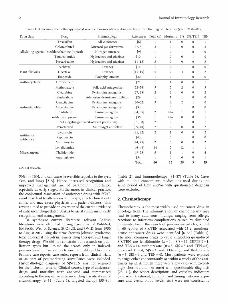
50% for TEN, and can cause irreversible sequelae to the eyes,skin, and lungs [2–5]. Hence, increased recognition andimproved management are of paramount importance,especially at early stages. Furthermore, in clinical practice,the conjectural association of anticancer drugs with SCARevent may lead to alterations in therapy, affects clinical out-come, and may cause physician and patient distress. Thisreview aimed to provide an overview of the current evidenceof anticancer drug-related SCARs to assist clinicians in earlyrecognition and management.
To synthesize current literature, relevant Englishliteratures were identified through searches of PubMed,EMBASE, Web of Science, SCOPUS, and OVID from 1950to August 2017 using the terms Stevens-Johnson syndrome,toxic epidermal necrolysis, cancer drug therapy, and targettherapy drugs. We did not constrain our research on pub-lication types but limited the search only in indexed,peer-reviewed journals so as to ensure quality publications.Primary case reports, case series, reports from clinical trials,or as part of postmarketing surveillance were included.Histopathologic diagnosis of SJS/TEN was not requiredfor the inclusion criteria. Clinical course, type of anticancerdrugs, and mortality were analyzed and summarizedaccording to the respective anticancer drug classifications ofchemotherapy [6–54] (Table 1), targeted therapy [55–80]
(Table 2), and immunotherapy [81–87] (Table 3). Caseswith multiple concomitant medications used during thesame period of time and/or with questionable diagnosiswere excluded.
2. Chemotherapy
Chemotherapy is the most widely used anticancer drug inoncology field. The administration of chemotherapy maylead to many cutaneous findings, ranging from allergicreactions to infectious complications caused by disruptedimmunity. From the search of peer-review articles, a totalof 60 reports of SJS/TEN associated with 23 chemothera-peutic anticancer drugs were identified [6–54] (Table 1).The most common drugs to cause chemotherapy-inducedSJS/TEN are lenalidomide (n = 14; SJS = 12, SJS/TEN=1,and TEN=1), methotrexate (n = 5; SJS = 2 and TEN=3),docetaxel (n = 4; SJS = 3 and TEN=1), and thalidomide(n = 5; SJS = 1 and TEN=4). Most patients were exposedto drugs either concomitantly or within 8 weeks of the anti-cancer agent. Although there were a few cases with exceed-ingly short duration of onset with questionable diagnosis[28, 31], the report descriptions and causality indicators(course of treatment, duration and timing between expo-sure and event, blood levels, etc.) were not consistently
Table 1: Anticancer chemotherapy-related severe cutaneous adverse drug reactions from the English literature (year: 1950–2017).
Drug class Drug Pharmacology References Total (n) Mortality SJS SJS/TEN TEN
Alkylating agents
Treosulfan Alkysufonates [6] 1 1 0 0 1
Chlorambucil Mustard gas derivatives [7, 8] 2 0 0 0 2
Mechlorethamine (topical) Nitrogen mustard [9] 1 0 1 0 0
Temozolomide Hydrazines and triazines [10] 1 0 0 1 0
Procarbazine Hydrazines and triazines [11–13] 3 0 0 0 3
Plant alkaloids
Paclitaxel Taxanes [14] 1 0 1 0 0
Docetaxel Taxanes [15–19] 5 2 3 0 2
Etoposide Podophyllotoxins [20] 1 0 1 0 0
Anthracyclines Doxorubicin [21] 1 1 0 0 1
Antimetabolites
Methotrexate Folic acid antagonists [22–26] 5 2 2 0 3
Cytarabine Pyrimidine antagonist [27, 28] 2 2 0 0 2
Fludarabine Adenosine deaminase inhibitor [29] 1 1 1 0 0
Gemcitabine Pyrimidine antagonist [30–32] 3 0 2 1 0
Capecitabine Pyrimidine antagonist [33] 1 0 1 0 0
Cladribine Purine antagonist [34, 35] 2 NA 1 0 1
6-Mercaptopurine Purine antagonist [36] 1 NA 0 0 1
TS-1 (tegafur-gimeracil-oteracil potassium) [37, 38] 2 0 1 0 1
Pemetrexed Multitarget antifolate [39, 40] 2 0 0 0 2
Antitumorantibiotics
Bleomycin [41, 42] 2 1 0 0 2
Peplomycin [43] 1 0 1 0 0
Mithramycin [44, 45] 2 0 0 0 2
Miscellaneous
Lenalidomide [46–48] 14 2 12 1 1
Thalidomide [49–53] 5 1 1 0 4
Asparaginase [54] 1 0 0 0 1
Total 60 13 28 3 29
NA: not available.
2 Journal of Immunology Research

reported in these articles. Some articles enclose picturesthat are not very suggestive of SJS/TEN but of an alterna-tive diagnosis, including erythema multiforme, GVHD,and toxic erythema of chemotherapy. For instance,methotrexate-induced epidermal necrosis is a distinct entitythat closely mimics SJS/TEN but exhibits distinct clinico-pathological features from SJS/TEN [88]. Many of thereported articles did not obtain skin biopsy for pathologyexamination and hence, it is difficult to draw to a definitivediagnosis of SJS/TEN. Another clinical mimic of SJS/TENassociated with chemotherapy is toxic erythema of chemo-therapy (TEC), characterized by painful erythematous erup-tions with edema and/or blisters which involves the acralpart, intertriginous areas, pressure points, and less often ears,knees, and elbows [89, 90]. TEC is a toxic phenomenon withminimal inflammatory infiltrates despite the dramatic clini-cal appearance, hence studies have hypothesized that the ery-thema is secondary to keratinocyte damage with release ofcytokines leading to vasodilation [90, 91]. Most cases involvethe use of either antimetabolites or alkylating agents thatinterferes RNA or DNA synthesis, including methotrexate,cytarabine, 5-fluorouracil, and mercaptopurine. By contrast,SJS/TEN is an immune-driven type 4 allergic reaction, wherecytotoxic T lymphocytes and natural killer cells are activated.Clinical recognition and differentiation of SJS/TEN from
toxic erythema are of importance because it helps preventthe inappropriate use of systemic immunosuppressants forpresumed allergic reactions, precludes subsequent dosing,and affects the patient’s clinical outcome.
3. Targeted Anticancer Therapy
From the literature review, a roster of 42 reports of SJS(n = 23), SJS/TEN (n = 4), or TEN (n = 15), associated with12 targeted anticancer drugs, were identified, includingEGFR inhibitors (afatinib, cetuximab, erlotinib, gefitinib,panitumumab, and vandetanib), MKI (imatinib, regorafenib,and sorafenib), recombinant IL-2 (aldesleukin), proteasome(bortezomib), anti-CD20 (rituximab), anti-CD30 (brentuxi-mab vedotin), and BRAF inhibitor (vemurafenib) (Table 2).The most common drugs to cause SJS/TEN reported areimatinib (n = 11), EGFR inhibitors (n = 10), and vemurafe-nib (n = 7). The response of cancer control is hard to analyzebecause it was not fully mentioned in the reports. All caseswere treated with immunosuppressant, including steroid,IVIG, and there was one TEN case with promising outcomeafter etanercept (anti-TNF α) treatment. In these reports,nine patients underwent drug rechallenge test with recur-rences, confirming the notoriety of exposed targeted anti-cancer drugs [67–72, 74, 80].
Table 2: Anticancer targeted therapy-related severe cutaneous adverse drug reactions from the English literature (year: 1950–2017).
Drug class Drug Pharmacology References Total (n) Mortality SJS SJS/TEN TEN
EGFR inhibitor
Afatinib Monoclonal antibody to EGFR [55, 56] 2 0 2 0 0
Cetuximab Monoclonal antibody to EGFR [57–59] 4 1 1 1 2
Erlotinib TKI specific to EGFR [60] 1 0 1 0 0
Gefitinib TKI specific to EGFR [61, 62] 2 1 0 0 2
Panitumumab Monoclonal antibody to EGFR [122] 1 0 1 0 0
Vandetanib Less specific multikinase inhibitors [63] 2 0 0 1 1
KIT and BCR-ABLinhibitors
Imatinib KIT, BCR-ABL, PDGFR [64–72] 11 1 11 0 0
Antiangiogenic agents SorafenibNonselective antiangiogenesis
multikinase agents[73–76] 3 0 2 0 1
Proteasome Bortezomib [77] 2 1 1 0 1
CD30 Brentuximab vedotin CD30 [78] 2 0 1 0 1
CD20 Rituximab Monoclonal antibody to CD20 [79] 5 2 2 2 1
BRAF inhibitors Vemurafenib A/B/C-Raf and B-Raf (V600E) [80] 7 1 1 0 6
Total 42 7 23 4 15
Table 3: Anticancer immune therapy-related adverse drug reactions from the English literature (year: 1950–2017).
Drug class Drug Pharmacology References Total Mortality SJS SJS/TEN TEN
Immunomodulators
Aldesleukin Recombinant interleukin-2 [81, 82] 2 1 0 0 2
Ipilimumab CTLA-4 inhibitors [83] 1 0 1 0 0
Nivolumab PD-1 inhibitors [84, 85] 2 1 0 0 2
Pembrolizumab PD-1 inhibitors [86, 114, 116] 4 0 4 0 0
DenileukinRecombinant interleukin-2 and
diphtheria toxin[87] 1 0 0 1
Total 9 3 5 0 5
3Journal of Immunology Research

3.1. EGFR Inhibitors. EGFR inhibitors are approved as thedrug for the treatment of non-small cell lung, colorectal,breast, pancreatic, head, and neck cancers with EGFRmutations [92]. The incidence of EGFR inhibitor-inducedcutaneous adverse drug reactions (cADRs) is high (36%–80%) [93], of which most were papulopustular eruptions,xerosis, paronychia, mucositis, and photosensitivity [94].In this article, we have identified 13 cases of SJS/TENinduced by EGFR inhibitors. Though rare, SJS/TEN shouldbe distinguished from EGFR inhibitor-related mucositis,particularly when the patient present with constitutionalsymptoms and widespread atypical target spots withblisters that extend beyond mucosa to the skin. Cross-reactivity between EGFR inhibitors was reported. It ishypothesized that the pathomechanism of SJS/TEN associ-ated with EGFR inhibitors could be caused by to the irre-versible inhibition of EGFR, of which hinders epidermaldifferentiation and reepithelialization and causing extensiveerosions [95].
3.2. KIT and BCR-ABL Inhibitors. Imatinib, a tyrosine kinaseinhibitor, is the standard treatment in chronic myeloid leu-kemia and gastrointestinal stromal tumors (GIST) [96, 97].In this article, imatinib accounts one of the most commoncausative targeted anticancer drug to induce SJS, with a ros-ter of 12 cases. This must be differentiated from other morecommonly seen cutaneous adverse effects of imatinib,maculopapular rashes, and facial edema [98], of which hasa better prognosis and dose-dependent pharmacologic effectrather than hypersensitivity reaction [99]. For maculopapu-lar rash/facial edema associated with imatinib, temporarydiscontinuation or dose reduction may be applied if thepatient’s cancer is susceptible to the drug. By contrast, rein-troducing the culprit drug with a dose reduction is usuallynot suggested [100, 101].
3.3. Multikinase Inhibitors.Multikinase inhibitors (sunitinib,sorafenib, pazopanib, and vandetanib) are small moleculeinhibitors of the tyrosine kinase of the VEGF, and also differ-ential binding capacities to other tyrosine kinases, includingPDGFR, EGFR, KIT, RET, FLT-3, CSF-1R, and RAF [102].They were approved for treatment of patients with renal cellcancer, gastrointestinal stromal tumors, and hepatocellularcancer. These drugs can cause hand-foot skin reaction, hairchange, maculopapular eruptions, stomatitis, genital ero-sions, and bleeding [103, 104], especially in patients usingsorafenib. These more common cutaneous toxicities arethought to be caused by direct VEGF inhibition, which resultin vessel regression, and impact on vascular repair capacities[74]. Other research has also shown that Fas/FasL interactionmediates keratinocyte death in sunitinib-induced HFSR [75].Recently, one recent study identified SLC22A20 (OAT6) asan uptake carrier of sorafenib and subsequently sorafenibenters the keratinocyte through OAT6 and then inhibitsmitogen-activated protein kinase MAP3K7 (TAK1) leadingto cytotoxicity and keratinocyte injury [76]. Interestingly,erythema multiforme, a spectrum of delayed type hypersen-sitivity, induced by sorafenib was around 19–25% in Japanesepopulation, which is much higher than the Caucassian
population [105]. This could imply a possible genetic rolein the pathogenesis of adverse drug reactions. The differ-ent incidence of cutaneous adverse reactions among differ-ent ethnicities need to be further investigated.
3.4. BRAF Inhibitors. Vemurafenib is a selective inhibitor ofBRAF-kinase approved for the treatment of metastaticmelanoma with BRAF mutation. Skin toxicity, such as pho-tosensitivity and maculopapular eruptions, and secondaryskin malignancy (keratoacanthoma and squamous cell carci-noma) were estimated to affect more than 90% of patients[106, 107]. One vemurafenib-TEN underwent a lymphocytetransformation test (LTT) assay to confirm the causality ofvemurafenib and also show positive cross-reactivity fordabrafenib [108]. On the contrary, another case reporteda successful switch from vemurafenib-induced cutaneousadverse reactions to dabrafenib [109]. Furthermore,cross-reactivity was also found between vemurafenib andsulfonamide antibiotics—sulfamethoxazole—based on LTTreports. These data suggested that there might be clinicalcross-reactivity between BRAF inhibitors and sulfonamides.Predisposing factors to sulfonamide-related adverse cutane-ous drug reactions could be implied in the pathomechanismstudies of vemurafenib-associated SJS/TEN [108].
3.5. mTOR Inhibitors. Mammalian target of rapamycin(mTOR) inhibitors, such as sirolimus, everolimus, and tem-sirolimus, are emerging drugs, increasingly applied inoncology and in the prevention of rejection in patientsreceiving solid organ transplantation [110]. The mostcommon cutaneous side effects are oral ulcers, acne-likeeruptions, and morbilliform drug eruptions [111]. Oralulcer is a very frequent (72%) adverse reaction and is oftenrecurrent and chronic following everolimus treatment in25% of patients. The adverse event was found to be dosedependent [112].
Severe drug eruptions of life-threatening lingual angio-edema after initiation of everolimus in heart transplantrecipients have also been reported in a case series. In thesepatients, lingual edema occurs predominantly within the firstweeks after initiation of everolimus therapy and disappearswithout recurrences in majority patients after adequatesymptomatic treatment [113].
There were otherwise no SCAR (SJS/TEN, DRESS)event being reported in the literature.
4. Immunotherapy
Immunotherapy is the latest breakthrough in anticancerdrug development with immunomodulatory therapeuticantibodies, targeting inhibitory receptors expressed by Tcell as CTLA-4 and PD-1. They are used to treat advancestage cancer with metastasis or unresectable tumor suchas melanoma and lung cancer. In this section, older immu-notherapy such as interleukin-2 was also included inTable 3. These therapeutic options are most widely usedin advanced and late cancer stages. From literature reviews,we have identified one ipilimumab-SJS, two nivolumab-TEN, and four pembrolizumab-SJS. All of the patients were
4 Journal of Immunology Research
advanced melanoma patients, and the onset of epidermalnecrolysis varies from 2.5 weeks to 3 months. In one caseof pembrolizumab-associated SJS, concomitant phenytoinfor epilepsy was used; hence, the exact culprit drug is hardto define. Two cases of pembrolizumab-SJS were beingreported by Saw et al. [114]. Interestingly, there was a strikingdemarcation of epidermal detachment along the radiother-apy field aside from typical mucocutaneous findings of SJS.Such findings, although rarely, have also been reported inprevious traditional culprit drugs and targeted therapy. Atotal of 3 cases were found with interleukin-2 immunetherapy with 2 fatalities [81, 82, 87]. One of the authorssuggested that IL-2 may increase patient’s susceptibility toallergy of other medication [87]. An increased expression ofPD-L1 in the epidermis by immunohistochemistry (IHC)was found, and they hypothesized that the use of anti-PD-1therapy could provoke the expression of PD-L1 of keratino-cytes and permit the activated CD8+ cytotoxic T cells totarget keratinocytes leading to keratinocyte apoptosis [86].PD-1 knockout mouse often exhibits symptoms related toadverse cutaneous reactions. It has been reported in a mousemodel that PD-L1 expressed on keratinocytes presentingself-antigens regulates autoreactive CD8+ T cell activity andprevents the development of cutaneous autoimmune disease[115]. Goldinger et al. had demonstrated that the geneexpression analysis of TEN-like lesional skin from anti-PD-1-treated patients revealed an upregulation of majorinflammatory chemokines, such as CXCL9, CXCL10, andCXCL11, of cytotoxic mediators such as PRF1 and GZMBand proapoptotic FASLG and upregulation of PD-L1[116]. These gene expression profiles resembling SJS/TENsuggest that PD-1/PD-L1 interaction is required to preserveepidermal integrity during inflammatory skin reactions.Interestingly, there was a case with preceding nivolumabtreatment followed by vemurafenib who developed TEN[117]. The authors suggest that nivolumab predisposepatients to drug hypersensitivity reactions through activationof CD8+ cells [84, 85].
In spite of being uncommon, SJS/TEN are severe life-threatening cutaneous diseases that should be concernedin patients treated with anticancer drugs. The typical pre-sentation and diagnosis often require proper drug exposuredocumentation, photography, and skin biopsies. Currently,there are many different classifications and models withdetail and validated diagnostic criteria to assist clinicaldiagnosis and can help predict patients’ mortality [118,119]. Standard reporting method is important for subse-quent investigation and analysis of these rare events. Inaddition, diagnosis of culprit drug is often challenging, thedrug notoriety scoring systems including ALDEN score,Naronjo score and in vitro test with lymphocyte transfor-mation test (LTT) are useful tests for the diagnosis of drughypersensitivity and cross-reactivity and helped to betterunderstand these reactions [120, 121]. Current evidenceon the pathomechanism of this complication was limited.Further research is warranted to elucidate the pathophysiol-ogy as well as help clinician coping with this notoriousadverse event, advancing towards personalized medicine inoncology treatment.
Conflicts of Interest
The authors declared no conflicts of interests.
Authors’ Contributions
Chau Yee Ng and Chun-Bing Chen contributed equally tothis work.
References
[1] J. C. Roujeau, “Immune mechanisms in drug allergy,”Allergology International, vol. 55, no. 1, pp. 27–33, 2006.
[2] L. E. French, J. T. Trent, and F. A. Kerdel, “Use of intravenousimmunoglobulin in toxic epidermal necrolysis and Stevens–Johnson syndrome: our current understanding,” Interna-tional Immunopharmacology, vol. 6, no. 4, pp. 543–549, 2006.
[3] M. Mockenhaupt and E. Schopf, “Epidemiology of drug-induced severe skin reactions,” Seminars in CutaneousMedicine and Surgery, vol. 15, no. 4, pp. 236–243, 1996.
[4] J. Revuz, D. Penso, J. C. Roujeau et al., “Toxic epidermalnecrolysis. Clinical findings and prognosis factors in87 patients,” Archives of Dermatology, vol. 123, no. 9,pp. 1160–1165, 1987.
[5] J. C. Roujeau, J. C. Guillaume, J. P. Fabre, D. Penso, M. L.Flechet, and J. P. Girre, “Toxic epidermal necrolysis (Lyellsyndrome). Incidence and drug etiology in France, 1981-1985,” Archives of Dermatology, vol. 126, no. 1, pp. 37–42, 1990.
[6] M. E. Scheulen, R. A. Hilger, C. Oberhoff et al., “Clinicalphase I dose escalation and pharmacokinetic study of high-dose chemotherapy with treosulfan and autologous periph-eral blood stem cell transplantation in patients with advancedmalignancies,” Clinical Cancer Research, vol. 6, no. 11,pp. 4209–4216, 2000.
[7] I. Aydogdu, C. Ozcan, M. Harputluoglu, Y. Karincaoglu,O. Turhan, and A. Ozcanu, “Severe adverse skin reaction tochlorambucil in a patient with chronic lymphocytic leuke-mia,” Anti-Cancer Drugs, vol. 8, no. 5, pp. 468-469, 1997.
[8] F. Pietrantonio, L. Moriconi, F. Torino, A. Romano, andA. Gargovich, “Unusual reaction to chlorambucil: a casereport,” Cancer Letters, vol. 54, no. 3, pp. 109–111, 1990.
[9] J. M. Newman, J. M. Rindler, W. F. Bergfeld, and J. K. Brydon,“Stevens-Johnson syndrome associated with topical nitrogenmustard therapy,” Journal of the American Academy ofDermatology, vol. 36, no. 1, pp. 112–114, 1997.
[10] N. Sarma, “Stevens-Johnson syndrome and toxic epidermalnecrolysis overlap due to oral temozolomide and cranialradiotherapy,” American Journal of Clinical Dermatology,vol. 10, no. 4, pp. 264–267, 2009.
[11] R. Jones, M. Kirkup, S. Guglani, and K. Hopkins, “Toxicepidermal necrolysis after PCV combination chemotherapyfor relapsed B-cell lymphoma,” Clinical Oncology, vol. 18,no. 1, p. 90, 2006.
[12] U. Garbarini and G. P. Valassina, “Report of a case of Lyell’ssyndrome during a course of treatment with Natulan,”Minerva Medica, vol. 64, no. 53, pp. 2775–2778, 1973.
[13] J. Guerrin and R. Michiels, “Syndrome de Lyell avee agranu-locyte et thrombopenie au cours d’une chimiotherapieantimitotique,” Rev Med Dijon, vol. 4, pp. 523–525, 1969.
5Journal of Immunology Research

[14] A. Hiraki, K. Aoe, T. Murakami, T. Maeda, R. Eda, andH. Takeyama, “Stevens-Johnson syndrome induced by pacli-taxel in a patient with squamous cell carcinoma of the lung: acase report,” Anticancer Research, vol. 24, no. 2C, pp. 1135–1137, 2004.
[15] J. G. Kattan, F. S. Farhat, G. Y. Chahine et al., “Weeklydocetaxel, zoledronic acid and estramustine in hormone-refractory prostate cancer (HRPC),” Investigational NewDrugs, vol. 26, no. 1, pp. 75–79, 2008.
[16] C. Moisidis and V. Möbus, “Erythema multiforme majorfollowing docetaxel,” Archives of Gynecology and Obstetrics,vol. 271, no. 3, pp. 268–270, 2005.
[17] Y. Sawada, K. Sugita, R. Kabashima, M. Nakamura,and Y. Tokura, “Docetaxel-induced Stevens–Johnson syn-drome with regenerating epidermis composed of atypicalkeratinocytes,” Journal of the European Academy of Der-matology and Venereology, vol. 23, no. 11, pp. 1333–1335, 2009.
[18] S. P. Dourakis, V. A. Sevastianos, A. Alexopoulou,M. Deutsch, and N. Stavrianeas, “Treatment side effects. Case2. Toxic, epidermal, necrolysis-like reaction associated withdocetaxel chemotherapy,” Journal of Clinical Oncology,vol. 20, no. 13, pp. 3030–3032, 2002.
[19] C. H. Ohlmann, S. Kohlmorgen, D. Sahi, U. Engelmann, andA. Heidenreich, “Lethal course after chemotherapy withdocetaxel. Acute liver failure with accompanying erythemamultiforme major,” Der Urologe, vol. 46, no. 10, pp. 1425–1427, 2007.
[20] C. H. Jameson and D. L. Solanki, “Stevens-Johnson syn-drome associated with etoposide therapy,” Cancer TreatmentReports, vol. 67, no. 11, pp. 1050-1051, 1983.
[21] L. A. Solberg Jr., M. R. Wick, and J. E. Bruckman,“Doxorubicin-enhanced skin reaction after whole-bodyelectron-beam irradiation for leukemia cutis,” Mayo ClinicProceedings, vol. 55, no. 11, pp. 711–715, 1980.
[22] R. J. Cuthbert, J. I. Craig, and C. A. Ludlam, “Stevens-Johnsonsyndrome associated with methotrexate treatment for non-Hodgkin’s lymphoma,” The Ulster Medical Journal, vol. 62,no. 1, pp. 95–97, 1993.
[23] P. J. Moe and M. Seip, “High dose methotrexate in acutelymphocytic leukemia in childhood,” Acta Paediatrica Scan-dinavica, vol. 67, no. 3, pp. 265–268, 1978.
[24] C. H. Yang, L. J. Yang, T. H. Jaing, and H. L. Chan, “Toxicepidermal necrolysis following combination of methotrexateand trimethoprim–sulfamethoxazole,” International Journalof Dermatology, vol. 39, no. 8, pp. 621–623, 2000.
[25] H. Cakesen and A. F. Oner, “Toxic epidermal necrolysis in agirl with leukemia receiving methotrexate,” Indian Pediatrics,vol. 38, no. 4, p. 426, 2001.
[26] N. Stone, S. Sheerin, and S. Burge, “Toxic epidermal necroly-sis and graft vs. host disease: a clinical spectrum but a diag-nostic dilemma,” Clinical and Experimental Dermatology,vol. 24, no. 4, pp. 260–262, 1999.
[27] A. Ozkan, H. Apak, T. Celkan, L. Yuksel, and I. Yildiz, “Toxicepidermal necrolysis after the use of high-dose cytosinearabinoside,” Pediatric Dermatology, vol. 18, no. 1, pp. 38–40, 2001.
[28] M. S. Figueiredo, M. Yamamoto, and J. Kerbauy, “Toxicepidermal necrolysis after the use of intermediate dose ofcytosine arabinoside,” Revista da Associação Médica Brasi-leira, vol. 44, no. 1, pp. 53–55, 1998.
[29] M. A. Angelopoulou, C. Poziopoulos, V. A. Boussiotis,F. Kontopidou, and G. A. Pangalis, “Fludarabine monopho-sphate in refractory B-chronic lymphocytic leukemia: main-tenance may be significant to sustain response,” Leukemia& Lymphoma, vol. 21, no. 3-4, pp. 321–324, 1996.
[30] M. S. Talamonti, P. J. Catalano, D. J. Vaughn et al., “Easterncooperative oncology group phase I trial of protractedvenous infusion fluorouracil plus weekly gemcitabine withconcurrent radiation therapy in patients with locallyadvanced pancreas cancer: a regimen with unexpectedearly toxicity,” Journal of Clinical Oncology, vol. 18,no. 19, pp. 3384–3389, 2000.
[31] K. R. Sommers, K. M. Kong, D. T. Bui, J. P. Fruehauf, andR. F. Holcombe, “Stevens–Johnson syndrome/toxic epider-mal necrolysis in a patient receiving concurrent radiationand gemcitabine,” Anti-Cancer Drugs, vol. 14, no. 8, pp. 659–662, 2003.
[32] W. Mermershtain, A. D. Cohen, I. Lazarev, M. Grunwald,and S. Ariad, “Toxic epidermal necrolysis associated withgemcitabine therapy in a patient with metastatic transitionalcell carcinoma of the bladder,” Journal of Chemotherapy,vol. 15, no. 5, pp. 510-511, 2003.
[33] M. A. N. Sendur and S. Kilickap, “Stevens–Johnson syn-drome after treatment with capecitabine,” Clinical Oncology,vol. 20, no. 2, pp. 202-203, 2008.
[34] M. S. Tallman, D. Hakimian, C. Zanzig et al., “Cladribine inthe treatment of relapsed or refractory chronic lymphocyticleukemia,” Journal of Clinical Oncology, vol. 13, no. 4,pp. 983–988, 1995.
[35] P. Meunier, S. Castaigne, J. N. Bastie, O. Chosidow,and S. Aractingi, “Cutaneous reactions after treatmentwith 2-chlorodeoxyadenosine,” Acta Dermato-Venereologica,vol. 76, no. 5, pp. 385-386, 1996.
[36] P. L. Amerio and A. Tulli, “Lyell’s syndrome caused by 6-mercaptopurine,”Giornale Italiano di Dermatolotia. MinervaDermatologica, vol. 44, no. 10, pp. 514–516, 1969.
[37] H. Okamoto, K. Yane, T. Yamanaka, T. Fukuda, andH. Hosoi, “The usefulness of TS-1 for the treatment of headand neck cancer,” Gan to Kagaku Ryoho, vol. 30, no. 8,pp. 1119–1124, 2003.
[38] C. S. Tan, R. Lim, T. C. Lim, C. W. Aw, S. W. Yeo, and S. C.Lee, “Toxic epidermal necrolysis associated with TS-1 in apatient with gastric cancer,” Japanese Journal of ClinicalOncology, vol. 41, no. 5, pp. 666–668, 2011.
[39] J. Bosch-Barrera, M. Gaztañaga, J. Ceballos et al., “Toxicepidermal necrolysis related to pemetrexed and carboplatinwith vitamin B12 and folic acid supplementation foradvanced non-small cell lung cancer,” Onkologie, vol. 32,no. 10, pp. 580–584, 2009.
[40] C. Tummino, F. Barlesi, C. Tchouhadjian et al., “Severe cuta-neous toxicity after Pemetrexed as second line treatment for arefractory non small cell lung cancer,” Revue des MaladiesRespiratoires, vol. 24, no. 5, pp. 635–638, 2007.
[41] G. Giaccone, M. Risio, G. Bonardi, and A. Calciati, “Stevens-Johnson syndrome and fatal pulmonary toxicity to combina-tion chemotherapy containing bleomycin: a case report,”Tumori, vol. 72, no. 3, pp. 331–333, 1986.
[42] A. Brodsky, I. Aparici, C. Argeri, and D. Goldenberg,“Stevens-Johnson syndrome, respiratory distress and acuterenal failure due to synergic bleomycin-cisplatin toxicity,”Journal of Clinical Pharmacology, vol. 29, no. 9, pp. 821–823,1989.
6 Journal of Immunology Research

[43] Y. Umebayashi, H. Enomoto, and M. Ogasawara, “Drugeruption due to peplomycin: an unusual form of Stevens-Johnson syndrome with pustules,” The Journal of Dermatol-ogy, vol. 31, no. 10, pp. 802–805, 2004.
[44] F. E. Eyster, C. B. Wilson, and H. I. Maibach, “Mithramycinas a possible cause of toxic epidermal necrolysis (Lyell’s syn-drome),” California Medicine, vol. 114, no. 2, pp. 42-43, 1971.
[45] D. Purpora, M. J. Ahern, and N. Silverman, “Toxic epidermalnecrolysis after mithramycin,” The New England Journal ofMedicine, vol. 299, no. 25, pp. 1412-1413, 1978.
[46] C. P. Castaneda, N. A. Brandenburg, R. Bwire, G. H. Burton,and J. B. Zeldis, “Erythema multiforme/Stevens-Johnson syn-drome/toxic epidermal necrolysis in lenalidomide-treatedpatients,” Journal of Clinical Oncology, vol. 27, no. 1,pp. 156-157, 2009.
[47] R. Wasch, T. Jakob, K. Technau, J. Finke, and M. Engelhardt,“Stevens–Johnson/toxic epidermal necrolysis overlap syn-drome following lenalidomide treatment for multiplemyeloma relapse after allogeneic transplantation,” Annals ofHematology, vol. 91, no. 2, pp. 287–289, 2012.
[48] P. K. Boruah, S. Bolesta, and S. M. Shetty, “Possiblelenalidomide-induced Stevens-Johnson syndrome duringtreatment for multiple myeloma,” Pharmacotherapy, vol. 31,no. 9, p. 925, 2011.
[49] T. E. Clark, N. Edom, J. Larson, and L. J. Lindsey, “Thalomid®(Thalidomide) capsules: a review of the first 18 monthsof spontaneous postmarketing adverse event surveillance,including off-label prescribing,” Drug Safety, vol. 24, no. 2,pp. 87–117, 2001.
[50] S. V. Rajkumar, M. A. Gertz, and T. E. Witzig, “Life-threaten-ing toxic epidermal necrolysis with thalidomide therapy formyeloma,” The New England Journal of Medicine, vol. 343,no. 13, pp. 972-973, 2000.
[51] S. B. Horowitz and A. L. Stirling, “Thalidomide-inducedtoxic epidermal necrolysis,” Pharmacotherapy, vol. 19,no. 10, pp. 1177–1180, 1999.
[52] W. K. Eo, S. H. Kim, S. H. Cheon et al., “Toxic epidermalnecrolysis following thalidomide and dexamethasone treat-ment for multiple myeloma: a case report,” Annals of Hema-tology, vol. 89, no. 4, pp. 421-422, 2010.
[53] M. Colagrande, M. Di Ianni, G. Coletti et al., “Toxicepidermal necrolysis in a patient with primary myelofibrosisreceiving thalidomide therapy,” International Journal ofHematology, vol. 89, no. 1, pp. 76–79, 2009.
[54] A. R. Rodriguez, “L-asparaginase and toxic epidermal necro-lysis,” Journal of the Medical Association of Georgia, vol. 69,no. 5, pp. 355–357, 1980.
[55] Y. Honda, Y. Hattori, S. Katsura et al., “Stevens-Johnsonsyndrome-like erosive dermatitis possibly related to afatinib,”European Journal of Dermatology, vol. 26, no. 4, pp. 413-414, 2016.
[56] J. Doesch, D. Debus, C. Meyer et al., “Afatinib-associatedStevens-Johnson syndrome in an EGFR-mutated lung cancerpatient,” Lung Cancer, vol. 95, pp. 35–38, 2016.
[57] M. Urosevic-Maiwald, T. Harr, L. E. French, and R. Dummer,“Stevens–Johnson syndrome and toxic epidermal necrolysisoverlap in a patient receiving cetuximab and radiotherapyfor head and neck cancer,” International Journal of Dermatol-ogy, vol. 51, no. 7, pp. 864–867, 2012.
[58] W. L. Lin, W. C. Lin, J. Y. Yang et al., “Fatal toxic epidermalnecrolysis associated with cetuximab in a patient with colon
cancer,” Journal of Clinical Oncology, vol. 26, no. 16,pp. 2779-2780, 2008.
[59] S. S. Lee and P. Y. Chu, “Toxic epidermal necrolysiscaused by cetuximab plus minocycline in head and neckcancer,” American Journal of Otolaryngology, vol. 31, no. 4,pp. 288–290, 2010.
[60] E. Liquete, S. Ali, R. Kammo et al., “Acute generalizedexanthematous pustulosis induced by Erlotinib (Tarceva)with superimposed Staphylococcus aureus skin infection ina pancreatic cancer patient: a case report,” Case Reports inOncology, vol. 5, no. 2, pp. 253–259, 2012.
[61] D. M. Jackman, L. A. Cioffredi, L. Jacobs et al., “A phase I trialof high dose gefitinib for patients with leptomeningeal metas-tases from non-small cell lung cancer,” Oncotarget, vol. 6,no. 6, pp. 4527–4536, 2015.
[62] J. J. Huang, S. X. Ma, X. Hou et al., “Toxic epidermal necro-lysis related to AP (pemetrexed plus cisplatin) and gefitinibcombination therapy in a patient with metastatic non-smallcell lung cancer,” Chinese Journal of Cancer, vol. 34, no. 2,pp. 94–98, 2015.
[63] J. Yoon, C. W. Oh, and C. Y. Kim, “Stevens-Johnson syn-drome induced by vandetanib,” Annals of Dermatology,vol. 23, Supplement 3, pp. S343–S345, 2011.
[64] D. Vidal, L. Puig, A. Sureda, and A. Alomar, “Sti571-inducedStevens–Johnson syndrome,” British Journal of Haematology,vol. 119, no. 1, pp. 274-275, 2002.
[65] G. Severino, C. Chillotti, R. De Lisa, M. Del Zompo, andR. Ardau, “Adverse reactions during imatinib and lansopra-zole treatment in gastrointestinal stromal tumors,” TheAnnals of Pharmacotherapy, vol. 39, no. 1, pp. 162–164,2005.
[66] K. Pavithran and M. Thomas, “Imatinib induced Stevens-Johnson syndrome: lack of recurrence following re-challengewith a lower dose,” Indian Journal of Dermatology, Venereol-ogy and Leprology, vol. 71, no. 4, pp. 288-289, 2005.
[67] S. A. Rule, S. G. O'Brien, and L. C. Crossman, “Managingcutaneous reactions to imatinib therapy,” Blood, vol. 100,no. 9, pp. 3434-3435, 2002.
[68] L. T. Hsiao, H. M. Chung, J. T. Lin et al., “Stevens–Johnsonsyndrome after treatment with STI571: a case report,”British Journal of Haematology, vol. 117, no. 3, pp. 620–622, 2002.
[69] H.-J. Hsieh, A. L. F. Chan, and S.-J. Lin, “Stevens-Johnsonsyndrome induced by combination of imatinib and allopuri-nol,” Chemotherapy, vol. 55, no. 4, pp. 197–199, 2009.
[70] M. Mahapatra, P. Mishra, and R. Kumar, “Imatinib-inducedStevens–Johnson syndrome: recurrence after re-challengewith a lower dose,” Annals of Hematology, vol. 86, no. 7,pp. 537-538, 2007.
[71] P. Jha, D. Himanshu, N. Jain, and A. K. Singh, “Imatinib-induced Stevens-Johnsons syndrome,” BMJ Case Reports,vol. 2013, 2013.
[72] M. Schaich, K. Schakel, T. Illmer, G. Ehninger, andM. Bornhauser, “Severe epidermal necrolysis after treatmentwith imatinib and consecutive allogeneic hematopoietic stemcell transplantation,” Annals of Hematology, vol. 82, no. 5,pp. 303-304, 2003.
[73] M. K. Choi, H. Y. Woo, J. Heo et al., “Toxic epidermal necro-lysis associated with sorafenib and tosufloxacin in a patientwith hepatocellular carcinoma,” Annals of Dermatology,vol. 23, Supplement 3, pp. S404–S407, 2011.
7Journal of Immunology Research

[74] B. Blanchet, B. Billemont, S. Barete et al., “Toxicity of sorafe-nib: clinical and molecular aspects,” Expert Opinion on DrugSafety, vol. 9, no. 2, pp. 275–287, 2010.
[75] C. N. Yeh, W. H. Chung, S. C. Su et al., “Fas/Fas ligandmediates keratinocyte death in sunitinib-induced hand-footskin reaction,” The Journal of Investigative Dermatology,vol. 134, no. 11, pp. 2768–2775, 2014.
[76] E. I. Zimmerman, A. A. Gibson, S. Hu et al., “Multiki-nase inhibitors induce cutaneous toxicity through OAT6-mediated uptake and MAP3K7-driven cell death,” CancerResearch, vol. 76, no. 1, pp. 117–126, 2016.
[77] B. Fang, Y. Song, J. Ma, and R. C. Zhao, “Severe epidermalnecrolysis after bortezomib treatment for multiple mye-loma,” Acta Haematologica, vol. 118, no. 2, pp. 65–67,2007.
[78] B. M. Tijink, J. Buter, R. de Bree et al., “A phase I dose esca-lation study with anti-CD44v6 bivatuzumab mertansine inpatients with incurable squamous cell carcinoma of the headand neck or esophagus,” Clinical Cancer Research, vol. 12,no. 20, pp. 6064–6072, 2006.
[79] S. Lowndes, A. Darby, G. Mead, and A. Lister, “Stevens-Johnson syndrome after treatment with rituximab,” Annalsof Oncology, vol. 13, no. 12, pp. 1948–1950, 2002.
[80] D. R. Minor, R. Rodvien, and M. Kashani-Sabet, “Successfuldesensitization in a case of Stevens–Johnson syndromedue to vemurafenib,” Melanoma Research, vol. 22, no. 5,pp. 410-411, 2012.
[81] A. A. Segura Huerta, P. Tordera, A. C. Cercos, A. L. Yuste,P. Lopez-Tendero, and G. Reynes, “Toxic epidermal necroly-sis associated with interleukin-2,” The Annals of Pharmaco-therapy, vol. 36, no. 7-8, pp. 1171–1174, 2002.
[82] J. S. Wiener, J. A. Tucker Jr., and P. J. Walther, “Interleukin-2-induced dermatotoxicity resembling toxic epidermalnecrolysis,” Southern Medical Journal, vol. 85, no. 6,pp. 656–659, 1992.
[83] E. Dika, G. M. Ravaioli, and P. A. Fanti, “Cutaneous adverseeffects during ipilimumab treatment for metastatic mela-noma: a prospective study,” European Journal of Dermatol-ogy, vol. 27, no. 3, pp. 266–270, 2017.
[84] K. L. Vivar, M. Deschaine, J. Messina et al., “Epidermal pro-grammed cell death-ligand 1 expression in TEN associatedwith nivolumab therapy,” Journal of Cutaneous Pathology,vol. 44, no. 4, pp. 381–384, 2017.
[85] N. Nayar, K. Briscoe, and P. Fernandez Penas, “Toxic epider-mal necrolysis–like reaction with severe satellite cell necrosisassociated with nivolumab in a patient with ipilimumabrefractory metastatic melanoma,” Journal of Immunotherapy,vol. 39, no. 3, pp. 149–152, 2016.
[86] E. Liniker, A. M. Menzies, B. Y. Kong et al., “Activity andsafety of radiotherapy with anti-PD-1 drug therapy inpatients with metastatic melanoma,” OncoImmunology,vol. 5, no. 9, article e1214788, 2016.
[87] K. Polder, C. Wang, M. Duvic et al., “Toxic epidermal necro-lysis associated with denileukin diftitox (DAB389IL-2)administration in a patient with follicular large cell lym-phoma,” Leukemia & Lymphoma, vol. 46, no. 12, pp. 1807–1811, 2005.
[88] T. J. Chen, W. H. Chung, and C. B. Chen, “Methotrexate-induced epidermal necrosis: a case series of 24 patients,” Jour-nal of the American Academy of Dermatology, vol. 77, no. 2,pp. 247–255.e2, 2017.
[89] L. E. Levine, M. M. Medenica, A. L. Lorincz, K. Soltani,B. Raab, and A. Ma, “Distinctive acral erythema occurringduring therapy for severe myelogenous leukemia,” Archivesof Dermatology, vol. 121, no. 1, pp. 102–104, 1985.
[90] J. L. Bolognia, D. L. Cooper, and E. J. Glusac, “Toxic erythemaof chemotherapy: a useful clinical term,” Journal of the Amer-ican Academy of Dermatology, vol. 59, no. 3, pp. 524–529,2008.
[91] T. D. Horn, “Antineoplastic chemotherapy, sweat, and theskin,” Archives of Dermatology, vol. 133, no. 7, pp. 905-906,1997.
[92] P. Seshacharyulu, M. P. Ponnusamy, D. Haridas, M. Jain,A. K. Ganti, and S. K. Batra, “Targeting the EGFR signalingpathway in cancer therapy,” Expert Opinion on TherapeuticTargets, vol. 16, no. 1, pp. 15–31, 2012.
[93] M. E. Lacouture, M. J. Anadkat, R. J. Bensadoun et al., “Clin-ical practice guidelines for the prevention and treatment ofEGFR inhibitor-associated dermatologic toxicities,” SupportCare Cancer, vol. 19, no. 8, pp. 1079–1095, 2011.
[94] J. B. Macdonald, B. Macdonald, L. E. Golitz, P. LoRusso, andA. Sekulic, “Cutaneous adverse effects of targeted therapies:part I: inhibitors of the cellular membrane,” Journal of theAmerican Academy of Dermatology, vol. 72, no. 2, pp. 203–218, 2015.
[95] A. M. Wnorowski, A. de Souza, A. Chachoua, and D. E.Cohen, “The management of EGFR inhibitor adverse events:a case series and treatment paradigm,” International Journalof Dermatology, vol. 51, no. 2, pp. 223–232, 2012.
[96] B. J. Druker, F. Guilhot, S. G. O'Brien et al., “Five-yearfollow-up of patients receiving imatinib for chronic myeloidleukemia,” New England Journal of Medicine, vol. 355,no. 23, pp. 2408–2417, 2006.
[97] P. G. Casali, A. Le Cesne, A. P. Velasco et al., “Imatinibfailure-free survival (IFS) in patients with localized gastroin-testinal stromal tumors (GIST) treated with adjuvant ima-tinib (IM): the EORTC/AGITG/FSG/GEIS/ISG randomizedcontrolled phase III trial,” Journal of Clinical Oncology,vol. 31, 2013.
[98] L. Valeyrie, S. Bastuji-Garin, J. Revuz et al., “Adversecutaneous reactions to imatinib (STI571) in Philadelphiachromosome-positive leukemias: a prospective study of 54patients,” Journal of the American Academy of Dermatology,vol. 48, no. 2, pp. 201–206, 2003.
[99] M. Brouard and J. H. Saurat, “Cutaneous reactions toSTI571,” The New England Journal of Medicine, vol. 345,no. 8, pp. 618-619, 2001.
[100] K. D. White, W. H. Chung, S. I. Hung, S. Mallal, and E. J.Phillips, “Evolving models of the immunopathogenesis of Tcell–mediated drug allergy: the role of host, pathogens, anddrug response,” The Journal of Allergy and Clinical Immunol-ogy, vol. 136, no. 2, pp. 219–234, 2015.
[101] T. A. Duong, L. Valeyrie-Allanore, P. Wolkenstein, andO. Chosidow, “Severe cutaneous adverse reactions to drugs,”Lancet, vol. 390, no. 10106, pp. 1996–2011, 2017.
[102] K. J. Gotink and H.M.W. Verheul, “Anti-angiogenic tyrosinekinase inhibitors: what is their mechanism of action?,” Angio-genesis, vol. 13, no. 1, pp. 1–14, 2010.
[103] R. S. Ishak, S. A. Aad, A. Kyei, and F. S. Farhat, “Cutaneousmanifestations of anti-angiogenic therapy in oncology:review with focus on VEGF inhibitors,” Critical Reviews inOncology/Hematology, vol. 90, no. 2, pp. 152–164, 2014.
8 Journal of Immunology Research
[104] B. Billemont, S. Barete, and O. Rixe, “Scrotal cutaneous sideeffects of sunitinib,” The New England Journal of Medicine,vol. 359, no. 9, pp. 975-976, 2008.
[105] M. Ikeda, T. Fujita, Y. Amoh, S. Mii, K. Matsumoto, andM. Iwamura, “Stevens-Johnson syndrome induced by sorafe-nib for metastatic renal cell carcinoma,” Urologia Internatio-nalis, vol. 91, no. 4, pp. 482-483, 2013.
[106] M. E. Lacouture, M. Duvic, A. Hauschild et al., “Analysis ofdermatologic events in vemurafenib-treated patients withmelanoma,” The Oncologist, vol. 18, no. 3, pp. 314–322, 2013.
[107] M. Arenbergerova, A. Fialova, P. Arenberger et al., “Severediclofenac photoallergy in a patient treated with vemurafe-nib,” Journal of the European Academy of Dermatology andVenereology, vol. 30, no. 4, pp. 713–715, 2016.
[108] T. Bellon, V. Lerma, O. Gonzalez-Valle, C. González Herrada,and F. J. de Abajo, “Vemurafenib-induced toxic epidermalnecrolysis: possible cross-reactivity with other sulfonamidecompounds,” The British Journal of Dermatology, vol. 174,no. 3, pp. 621–624, 2016.
[109] G. Jeudy, S. Dalac-Rat, B. Bonniaud et al., “Successful switchto dabrafenib after vemurafenib-induced toxic epidermalnecrolysis,” British Journal of Dermatology, vol. 172, no. 5,pp. 1454-1455, 2015.
[110] M. E. Peterson, “Management of adverse events in patientswith hormone receptor-positive breast cancer treated witheverolimus: observations from a phase III clinical trial,” Sup-portive Care in Cancer, vol. 21, no. 8, pp. 2341–2349, 2013.
[111] C. Ferté, A. Paci, M. Zizi et al., “Natural history, managementand pharmacokinetics of everolimus-induced-oral ulcers:insights into compliance issues,” European Journal of Cancer,vol. 47, no. 15, pp. 2249–2255, 2011.
[112] F. Martins, M. A. de Oliveira, Q.Wang et al., “A review of oraltoxicity associated with mTOR inhibitor therapy in cancerpatients,” Oral Oncology, vol. 49, no. 4, pp. 293–298, 2013.
[113] U. Fuchs, A. Zittermann, H. K. Berthold et al., “Immunosup-pressive therapy with everolimus can be associated withpotentially life-threatening lingual angioedema,” Transplan-tation, vol. 79, no. 8, pp. 981–983, 2005.
[114] S. Saw, H. Y. Lee, and Q. S. Ng, “Pembrolizumab-inducedStevens–Johnson syndrome in non-melanoma patients,”European Journal of Cancer, vol. 81, pp. 237–239, 2017.
[115] N. Okiyama and S. I. Katz, “Programmed cell death 1 (PD-1)regulates the effector function of CD8 T cells via PD-L1expressed on target keratinocytes,” Journal of Autoimmunity,vol. 53, pp. 1–9, 2014.
[116] S. M. Goldinger, P. Stieger, B. Meier et al., “Cytotoxic cutane-ous adverse drug reactions during anti-PD-1 therapy,” Clini-cal Cancer Research, vol. 22, no. 16, pp. 4023–4029, 2016.
[117] D. B. Johnson, E. K. Wallender, D. N. Cohen et al., “Severecutaneous and neurologic toxicity in melanoma patientsduring vemurafenib administration following anti-PD-1therapy,” Cancer Immunology Research, vol. 1, no. 6,pp. 373–377, 2013.
[118] (RegiSCAR), “Patients were diagnosed with SJS, SJS/TENoverlap or TEN according to the classification proposed bythe European registry of severe cutaneous adverse reactions,”9 SJS.
[119] S. Bastuji-Garin, N. Fouchard, M. Bertocchi, J. C. Roujeau,J. Revuz, and P. Wolkenstein, “SCORTEN: a severity-of-illness score for toxic epidermal necrolysis,” The Journal ofInvestigative Dermatology, vol. 115, no. 2, pp. 149–153, 2000.
[120] B. Sassolas, C. Haddad, M. Mockenhaupt et al., “ALDEN,an algorithm for assessment of drug causality in Stevens–Johnson syndrome and toxic epidermal necrolysis: com-parison with case–control analysis,” Clinical Pharmacology& Therapeutics, vol. 88, no. 1, pp. 60–68, 2010.
[121] W. J. Pichler and J. Tilch, “The lymphocyte transformationtest in the diagnosis of drug hypersensitivity,” Allergy,vol. 59, no. 8, pp. 809–820, 2004.
[122] F. Pantano, M. Silletta, A. Iovieno et al., “Stevens–Johnsonsyndrome associated with reduced tear production compli-cating the use of cetuximab and panitumumab,” Interna-tional Journal of Colorectal Disease, vol. 24, no. 10,pp. 1247-1248, 2009.
9Journal of Immunology Research

Clinical StudyAssociation between HLA-B Alleles and Carbamazepine-InducedMaculopapular Exanthema and Severe Cutaneous Reactions inThai Patients
Chonlaphat Sukasem ,1,2,3,4 Chonlawat Chaichan,1,2 Thapanat Nakkrut,5,6
Patompong Satapornpong,1,2 Kanoot Jaruthamsophon,7 Thawinee Jantararoungtong,1,2
Napatrupron Koomdee,1,2 Suthida Sririttha,1,2,8 Sadeep Medhasi,9 Sarawut Oo-Puthinan,5
Ticha Rerkpattanapipat,4,10 Jettanong Klaewsongkram,4,11 Pawinee Rerknimitr,4,12
Papapit Tuchinda,4,13 Leena Chularojanamontri,4,13 Napatra Tovanabutra,4,14
Apichaya Puangpetch,1,2 and Wichai Aekplakorn 15
1Division of Pharmacogenomics and Personalized Medicine, Department of Pathology, Faculty of Medicine Ramathibodi Hospital,Mahidol University, Bangkok, Thailand2Laboratory for Pharmacogenomics, Somdech Phra Debaratana Medical Center (SDMC), Ramathibodi Hospital, Bangkok, Thailand3Ramathibodi Multidisciplinary Epilepsy Center, Faculty of Medicine, Ramathibodi Hospital, Mahidol University, Bangkok,Thailand4The Thai Severe Cutaneous Adverse Drug Reaction (THAI-SCAR) Research Group, Bangkok, Thailand5Department of Pharmacy Practice, Faculty of Pharmaceutical Sciences, Naresuan University, Phitsanulok, Thailand6Department of Pharmacy, Prasat Neurological Institute, Bangkok, Thailand7Department of Pathology, Faculty of Medicine, Prince of Songkla University, Songkla, Thailand8Department of Clinical Pharmacy Practice, Faculty of Pharmacy, Mahidol University, Bangkok, Thailand9School of Medicine, Mae Fah Luang University, Chiang Rai, Thailand10Division of Allergy Immunology and Rheumatology, Department of Medicine, Faculty of Medicine, Ramathibodi Hospital, MahidolUniversity, Bangkok, Thailand
11Division of Allergy and Clinical Immunology, Skin and Allergy Research Unit, Department of Medicine, Faculty of Medicine,Chulalongkorn University, Bangkok, Thailand
12Division of Dermatology, Skin and Allergy Research Unit, Department of Medicine, Faculty of Medicine, Chulalongkorn University,Bangkok, Thailand
13Department of Dermatology, Faculty of Medicine Siriraj Hospital, Mahidol University, Bangkok, Thailand14Dermatological Division, Department of Internal Medicine, Chiang Mai University, Chiang Mai, Thailand15Department of Community Medicine, Ramathibodi Hospital, Mahidol University, Bangkok, Thailand
Correspondence should be addressed to Chonlaphat Sukasem; [email protected]
Received 17 August 2017; Revised 25 October 2017; Accepted 26 November 2017; Published 10 January 2018
Academic Editor: Riichiro Abe
Copyright © 2018 Chonlaphat Sukasem et al. This is an open access article distributed under the Creative Commons AttributionLicense, which permits unrestricted use, distribution, and reproduction in anymedium, provided the original work is properly cited.
The HLA-B∗ 15:02 allele has been reported to have a strong association with carbamazepine-induced Stevens-Johnson syndrome/toxic epidermal necrolysis (SJS/TEN) in Thai patients. The HLA-B alleles associated with carbamazepine-induced maculopapularexanthema (MPE) and the drug reaction with eosinophilia and systemic symptoms (DRESS) among the Thai population havenever been reported. The aim of the present study was to carry out an analysis of the involvement of HLA-B alleles incarbamazepine-induced cutaneous adverse drug reactions (cADRs) in the Thai population. A case-control study was performed bygenotyping the HLA-B alleles of Thai carbamazepine-induced hypersensitivity reaction patients (17 MPE, 16 SJS/TEN, and 5DRESS) and 271 carbamazepine-tolerant controls. We also recruited 470 healthy Thai candidate subjects who had not
HindawiJournal of Immunology ResearchVolume 2018, Article ID 2780272, 11 pageshttps://doi.org/10.1155/2018/2780272
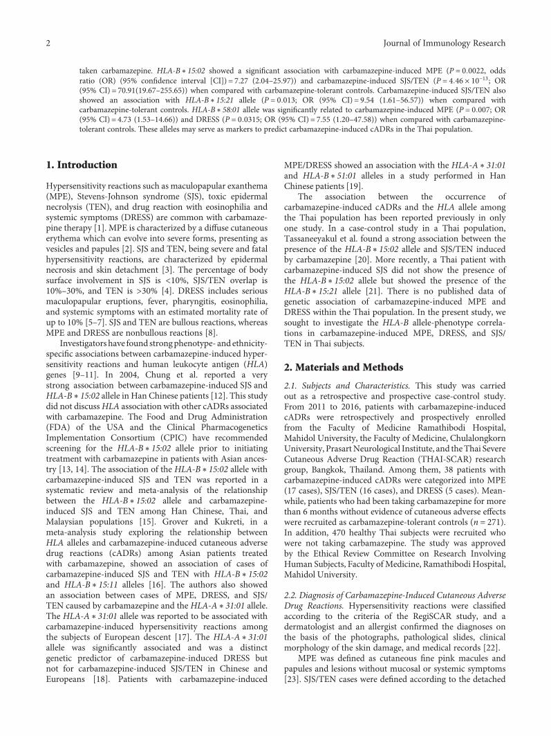
taken carbamazepine. HLA-B∗ 15:02 showed a significant association with carbamazepine-induced MPE (P = 0 0022, oddsratio (OR) (95% confidence interval [CI]) = 7.27 (2.04–25.97)) and carbamazepine-induced SJS/TEN (P = 4 46 × 10−13; OR(95% CI) = 70.91(19.67–255.65)) when compared with carbamazepine-tolerant controls. Carbamazepine-induced SJS/TEN alsoshowed an association with HLA-B∗ 15:21 allele (P = 0 013; OR (95% CI) = 9.54 (1.61–56.57)) when compared withcarbamazepine-tolerant controls. HLA-B∗ 58:01 allele was significantly related to carbamazepine-induced MPE (P = 0 007; OR(95% CI) = 4.73 (1.53–14.66)) and DRESS (P = 0 0315; OR (95% CI) = 7.55 (1.20–47.58)) when compared with carbamazepine-tolerant controls. These alleles may serve as markers to predict carbamazepine-induced cADRs in the Thai population.
1. Introduction
Hypersensitivity reactions such as maculopapular exanthema(MPE), Stevens-Johnson syndrome (SJS), toxic epidermalnecrolysis (TEN), and drug reaction with eosinophilia andsystemic symptoms (DRESS) are common with carbamaze-pine therapy [1]. MPE is characterized by a diffuse cutaneouserythema which can evolve into severe forms, presenting asvesicles and papules [2]. SJS and TEN, being severe and fatalhypersensitivity reactions, are characterized by epidermalnecrosis and skin detachment [3]. The percentage of bodysurface involvement in SJS is <10%, SJS/TEN overlap is10%–30%, and TEN is >30% [4]. DRESS includes seriousmaculopapular eruptions, fever, pharyngitis, eosinophilia,and systemic symptoms with an estimated mortality rate ofup to 10% [5–7]. SJS and TEN are bullous reactions, whereasMPE and DRESS are nonbullous reactions [8].
Investigators have found strongphenotype- and ethnicity-specific associations between carbamazepine-induced hyper-sensitivity reactions and human leukocyte antigen (HLA)genes [9–11]. In 2004, Chung et al. reported a verystrong association between carbamazepine-induced SJS andHLA-B∗ 15:02 allele in Han Chinese patients [12]. This studydid not discussHLA association with other cADRs associatedwith carbamazepine. The Food and Drug Administration(FDA) of the USA and the Clinical PharmacogeneticsImplementation Consortium (CPIC) have recommendedscreening for the HLA-B∗ 15:02 allele prior to initiatingtreatment with carbamazepine in patients with Asian ances-try [13, 14]. The association of the HLA-B∗ 15:02 allele withcarbamazepine-induced SJS and TEN was reported in asystematic review and meta-analysis of the relationshipbetween the HLA-B∗ 15:02 allele and carbamazepine-induced SJS and TEN among Han Chinese, Thai, andMalaysian populations [15]. Grover and Kukreti, in ameta-analysis study exploring the relationship betweenHLA alleles and carbamazepine-induced cutaneous adversedrug reactions (cADRs) among Asian patients treatedwith carbamazepine, showed an association of cases ofcarbamazepine-induced SJS and TEN with HLA-B∗ 15:02and HLA-B∗ 15:11 alleles [16]. The authors also showedan association between cases of MPE, DRESS, and SJS/TEN caused by carbamazepine and the HLA-A∗ 31:01 allele.The HLA-A∗ 31:01 allele was reported to be associated withcarbamazepine-induced hypersensitivity reactions amongthe subjects of European descent [17]. The HLA-A∗ 31:01allele was significantly associated and was a distinctgenetic predictor of carbamazepine-induced DRESS butnot for carbamazepine-induced SJS/TEN in Chinese andEuropeans [18]. Patients with carbamazepine-induced
MPE/DRESS showed an association with the HLA-A∗ 31:01and HLA-B∗ 51:01 alleles in a study performed in HanChinese patients [19].
The association between the occurrence ofcarbamazepine-induced cADRs and the HLA allele amongthe Thai population has been reported previously in onlyone study. In a case-control study in a Thai population,Tassaneeyakul et al. found a strong association between thepresence of the HLA-B∗ 15:02 allele and SJS/TEN inducedby carbamazepine [20]. More recently, a Thai patient withcarbamazepine-induced SJS did not show the presence ofthe HLA-B∗ 15:02 allele but showed the presence of theHLA-B∗ 15:21 allele [21]. There is no published data ofgenetic association of carbamazepine-induced MPE andDRESS within the Thai population. In the present study, wesought to investigate the HLA-B allele-phenotype correla-tions in carbamazepine-induced MPE, DRESS, and SJS/TEN in Thai subjects.
2. Materials and Methods
2.1. Subjects and Characteristics. This study was carriedout as a retrospective and prospective case-control study.From 2011 to 2016, patients with carbamazepine-inducedcADRs were retrospectively and prospectively enrolledfrom the Faculty of Medicine Ramathibodi Hospital,Mahidol University, the Faculty of Medicine, ChulalongkornUniversity, PrasartNeurological Institute, and theThai SevereCutaneous Adverse Drug Reaction (THAI-SCAR) researchgroup, Bangkok, Thailand. Among them, 38 patients withcarbamazepine-induced cADRs were categorized into MPE(17 cases), SJS/TEN (16 cases), and DRESS (5 cases). Mean-while, patients who had been taking carbamazepine for morethan 6 months without evidence of cutaneous adverse effectswere recruited as carbamazepine-tolerant controls (n = 271).In addition, 470 healthy Thai subjects were recruited whowere not taking carbamazepine. The study was approvedby the Ethical Review Committee on Research InvolvingHuman Subjects, Faculty of Medicine, Ramathibodi Hospital,Mahidol University.
2.2. Diagnosis of Carbamazepine-Induced Cutaneous AdverseDrug Reactions. Hypersensitivity reactions were classifiedaccording to the criteria of the RegiSCAR study, and adermatologist and an allergist confirmed the diagnoses onthe basis of the photographs, pathological slides, clinicalmorphology of the skin damage, and medical records [22].
MPE was defined as cutaneous fine pink macules andpapules and lesions without mucosal or systemic symptoms[23]. SJS/TEN cases were defined according to the detached
2 Journal of Immunology Research

body surface area as SJS (3–10%) and SJS/TEN overlap (10–30%) with or without associated systemic symptoms but notfulfilling the criteria of DRESS [22]. DRESS was defined asfollows: presence of fever, maculopapular rash with internalorgan involvement, and hematologic abnormalities [24].
2.3. DNA Isolation and HLA-B Typing. DNA extraction(MagNA Pure Compact nucleic acid purification kit, RocheDiagnostics Ltd., USA) was performed based on magneticbead technology. DNA was aliquoted and stored at −20°Cbefore HLA typing. HLA-B alleles were analyzed by thepolymerase chain reaction-sequence-specific oligonucleo-tide probe (PCR-SSOP) assay and Luminex™ MultiplexTechnology with well-established protocols [22]. In brief,PCR products were hybridized against a panel of oligonu-cleotide probes coated on polystyrene microspheres thathave sequences complementary to stretches of polymor-phic sequence within the target HLA-B alleles. Theamplicon-probe complex was visualized using a colorimet-ric reaction and fluorescence detection technology. Dataanalysis for the HLA-B assays was performed with HLAfusionTM2.0 software.
2.4. Statistical Analysis. Statistical analysis was performedwith SPSS version 18.0 (SPSS Inc., Chicago, IL, USA).Allele case-control comparisons were analyzed by Fisher’sexact test. A two-sided P value < 0.05 was considered tobe statistically significant.
3. Results
3.1. Subjects. Table 1 summarizes the clinical manifestationsand demographic variables of the 38 cases and 271carbamazepine-tolerant controls. Most cases received carba-mazepine to treat epilepsy (29 cases), except for 9 patientswho received carbamazepine to treat trigeminal neuralgia(5 cases), neuropathic pain (2 cases), bipolar disorder(1 case), and paroxysmal kinesigenic and nonkinesigenicdyskinesia (1 case). The mean treatment dose of carbamaze-pine in the carbamazepine-induced cADR patients was 325±75mg/day (mean± standard deviation). There was nosignificant differences between the case and tolerant group intreatment dose of carbamazepine. The mean duration for theonset of cADR was 16± 7 days (mean± standard deviation).
3.2. Association of HLA-B Alleles with Carbamazepine-Induced cADRs. Of the 38 patients who had carbamazepine-induced cADRs, 17 (44.74%) were found to carry the HLA-B∗ 15:02 allele. The HLA-B∗ 15:02 allele was observed in4.06% (11/271) of carbamazepine-tolerant controls and15.11% (71/470) of the general Thai population (Table 2).Our analysis of all subjects with cADRs and clinical controlsubjects showed a significant allelic association with HLA-B∗ 15:02 (P = 7 35 × 10−12), generating an odds ratio (OR) of19.13 (95% confidence interval [CI], 7.94–46.09). A compar-ison of all 38 carbamazepine-induced cADR subjects with 470general Thai subjects produced an OR of 4.55 (95% CI, 2.29–9.05, P = 3 44 × 10−6). Two patients with carbamazepine-induced cADRs carried HLA-B∗ 15:21, while the otherHLA-B serotypes 75 were not detected in this study.
3.3. Association between HLA-B Alleles and Various Types ofCarbamazepine-Induced cADRs. We analyzed the HLA-Bassociation between 17 patients with carbamazepine-induced MPE and 271 carbamazepine-tolerant controls. Wefound two HLA-B alleles, HLA-B∗ 15:02 and HLA-B∗ 58:01,as significant in the carbamazepine-induced MPE (Table 3).The HLA-B∗ 15:02 allele was observed in 23.53% (4/17) ofpatients with carbamazepine-induced MPE, but only in4.06% (11/271) of the carbamazepine-tolerant controls, giv-ing a significant association with carbamazepine-inducedMPE (P = 0 002; OR (95% CI) = 7.27 (2.04–25.97)). TheHLA-B∗ 58:01 allele appeared in 29.41% (5/17) of patientswith carbamazepine-induced MPE, which was more fre-quent than in carbamazepine-tolerant controls (8.12%,22/271; P = 0 007; OR (95% CI) = 4.73 (1.53–14.66)). Inthe included general population, the carrier rates ofHLA-B∗ 15:02 and HLA-B∗ 5801 were 12.34% (58/470)and 12.13% (57/470), respectively. Comparing the differenceof the HLA-B∗ 58:01 allele frequencies between the 17patients with carbamazepine-induced MPE and 470 generalsubjects, HLA-B∗ 58:01 showed the significant associationwith carbamazepine-induced MPE (P = 0 045; OR (95%CI) = 3.02 (1.03–8.88)). As for the carbamazepine-inducedSJS/TEN, the HLA-B∗ 15:02 and HLA-B∗ 15:21 alleleswere most significantly detected (Table 4). 75% (12/16)of carbamazepine-induced SJS/TEN patients carriedHLA-B∗ 15:02, which was more frequent than incarbamazepine-tolerant controls (4.1%, 11/271; P = 4 46× 10−13; OR (95% CI)= 70.91 (19.67–255.65)). TheHLA-B∗ 15:02 allele was present in 15.11% (71/470) of thegeneral population and when we compared the difference ofHLA-B∗ 15:02 frequency between carbamazepine-inducedSJS/TEN patients and the general population, HLA-B∗ 15:02showed a significant association with carbamazepine-induced SJS/TEN (P = 6 9 × 10⁻⁸; OR (95% CI) = 18.26(5.79–57.61)). HLA-B∗ 15:21 was significantly associatedwith carbamazepine-induced SJS/TEN appearing in 12.5%(2/16) of cases as compared to 1.48% (4/271) and 0.43%(2/470) in carbamazepine-tolerant controls and generalThai subjects, respectively.
As shown in Table 5, the HLA-B∗ 58:01 allele wasdetected as significant in the carbamazepine-induced DRESSgroup when compared with the carbamazepine-tolerantcontrol group (P = 0 032; OR (95% CI) = 7.55 (1.20–47.58)).The HLA-B∗ 58:01 allele was present in 40.00% (2/5) of theDRESS patients, but in only 8.12% (22/271) of thecarbamazepine-tolerant controls and 12.13% (57/470) ofthe general population.
4. Discussion
HLA-B alleles are reported to be associated with hypersensi-tivity reactions during the clinical usage of carbamazepine[25]. Pharmacogenetic screening of HLA-B alleles beforeinitiating carbamazepine therapy can prevent the risk ofsevere and life-threatening cutaneous adverse drug reac-tions. This study recruited patients with carbamazepine-induced hypersensitivity reactions, such as MPE, DRESS,and SJS/TEN and carbamazepine-tolerant patients from
3Journal of Immunology Research

Thailand. We found the association between HLA-B alleles(B∗ 15:02 and B∗ 58:01) and carbamazepine-inducedMPE. Further, the HLA-B∗ 15:02 and HLA-B∗ 15:21 alleleswere strongly associated with carbamazepine-induced SJS/TEN, and carbamazepine-induced DRESS had significantassociation with HLA-B∗ 58:01 allele.
The evidence of association of different types ofcarbamazepine-induced cADRs was shown by Hung et al. inHan Chinese patients [3]. In their study, the HLA-A∗ 31:01allele was associated with MPE (Pc = 2 2 × 10⁻³; OR(95% CI) = 17.5 (4.6–66.5)) and HLA-B∗ 15:02 was thesusceptible allele for SJS/TEN (Pc = 1 6 × 10−41; OR(95% CI) = 1357 (193.4–8838.3)). Few studies have beenconducted in the Thai population regarding the involve-ment of HLA alleles in carbamazepine-induced cADRs.The HLA-A∗ 31:01 allele has been mainly associatedwith carbamazepine-induced DRESS and MPE in HanChinese population, Japanese, and European populations[17, 19, 26]. Our study did not perform HLA-A typing,and we might have missed the potential associationbetween the HLA-A∗ 31:01 allele and carbamazepine-induced hypersensitivity reactions.
In 2008, Locharernkul et al. first identified that theHLA-B∗ 15:02 allele was strongly associated withcarbamazepine-induced SJS (P = 0 0005) in the Thai popu-lation [27]. A consistent association of the cases ofcarbamazepine-induced SJS/TEN were reported amongthe carriers of the HLA-B∗15:02 allele in this Thai popula-tion [20, 28]. Our findings justify the strongest association
of the HLA-B∗ 15:02 allele in the prediction ofcarbamazepine-induced SJS/TEN. In our study, weobserved that the HLA-B∗ 15:02 allele was not specificfor carbamazepine-induced SJS/TEN only, but it was alsosignificantly associated with carbamazepine-induced MPE.However, a previous study by Hung et al. reported thephenotype-specific HLA association of carbamazepine-induced cADRs [3]. This discrepancy might be due tothe different study populations. We observed the first evi-dence of a significant association of the HLA-B∗ 15:21allele with carbamazepine-induced SJS/TEN in Thai sub-jects. HLA-B∗15:21 allele belongs to the HLA-B75 family,which consists of the HLA-B∗ 15:02 allele as well [29].Jaruthamsophon et al. reported that HLA-B∗ 15:21 wasassociated with carbamazepine-induced SJS in differentpopulations and that a patient without the HLA-B∗ 15:02allele may be at a risk of carbamazepine-induced SJS dueto the presence of the HLA-B∗ 15:21 allele, anotherHLA-B75 serotype marker [21]. We can conclude thatthe presence of alternative forms of HLA alleles belongingto the same subfamilies of serotypes might contribute tothe susceptibility to cADRs. These observations imply thatmembers of the HLA-B75 serotype encode proteins shar-ing a similar conformation for carbamazepine bindingand presentation and trigger the immune response of SJScaused by carbamazepine [19].
In our study, we also found the association of theHLA-B∗ 58:01 allele with carbamazepine-induced MPEand DRESS. In contrast to our finding, Cheung et al. noted
Table 1: Clinical characteristic of patients with carbamazepine-induced cutaneous adverse drug reactions and carbamazepine-tolerantcontrols.
Demographic data Cases (n = 38) Tolerant controls (n = 271) P value
Gender (n/%) 0.145
Male 24/63.15 137/50.6
Female 14/33.84 134/49.4
Age (mean/range) 44/24–64 32/10–54 0.010
Indication (n/%)
Epilepsy 29/75.31 108/39.85 2.26× 10⁻⁵
Neuropathic pain 2/5.26 23/8.5 0.752
Trigeminal neuralgia 5/13.2 62/22.88 0.173
Bipolar disorder 1/2.6 10/3.7 1.000
Paroxysmal kinesigenic and nonkinesigenic dyskinesia 1/2.6 7/2.6 1.000
Autism — 35/12.9 0.012
Schizophrenia — 18/6.6 0.143
Others — 8/3.0 0.602
Dose of carbamazepine; mg/day (mean± SD) 325± 75 418± 19 0.397
Onset of cADRs; days (mean± SD) 16± 7 — —
cADRs (n/%)
MPE 17/45 — —
SJS/TEN 16/42 — —
DRESS 5/13 — —
cADRs: cutaneous adverse drug reactions; SJS/TEN: Stevens-Johnson syndrome/toxic epidermal necrolysis; DRESS: drug reaction with eosinophilia andsystemic symptoms; MPE: maculopapular exanthema.
4 Journal of Immunology Research

Table2:Association
ofHLA
-Balleleswithcarbam
azepine-indu
cedcA
DRs.
HLA
-Balleles
Carbamazepine-indu
ced
cADRs(n
=38)
Con
trols(n
=271)
Thaip
opulation(n
=470)
Carbamazepine-indu
cedcA
DRscases
versus
tolerant
controls
Carbamazepine-indu
cedcA
DRs
casesversus
Thaip
opulation
OR(95%
CI)
Pvalue
OR(95%
CI)
Pvalue
B∗07:05
3(7.89%
)12
(4.43%
)24
(5.11%
)1.85
(0.50–6.88)
0.357
1.59
(0.46–5.55)
0.4649
B∗13:01
1(2.63%
)37
(13.65%)
54(11.49%)
0.17
(0.02–1.28)
0.063
0.21
(0.03–1.55)
0.106
B∗13:02
1(2.63%
)6(2.21%
)20
(4.26%
)1.19
(0.14–10.19)
1.000
0.61
(0.08–4.66)
1.000
B∗15:01
1(2.63%
)10
(3.69%
)5(1.06%
)0.71
(0.09–5.67)
1.000
2.51
(0.29–22.08)
0.374
B∗15:02
17(44.74%)
11(4.06%
)71
(15.11%)
19.13(7.94–46.09)
7.35
×10
−12
∗4.55
(2.29–9.05)
3.44
×10
−6 ∗
B∗15:21
2(5.26%
)4(1.48%
)2(0.43%
)3.71
(0.66–20.97)
0.161
13.00(1.78–95.01)
0.030∗
B∗18:01
4(10.53%)
29(10.70%)
36(7.66%
)0.98
(0.33–2.97)
0.974
1.42
(0.48–3.22)
0.529
B∗18:15
2(5.26%
)0(0.00%
)0(0.00%
)15.06(1.33–170.25)
0.041∗
26.11(2.31–294.90)
0.016∗
B∗27:04
2(5.26%
)12
(4.43%
)19
(4.04%
)1.20
(0.26–5.58)
0.685
1.32
(0.30–5.89)
0.665
B∗27:06
1(2.63%
)8(2.95%
)12
(2.55%
)0.89
(0.11–7.31)
1.000
1.03
(0.13–8.15)
1.000
B∗40:01
5(13.16%)
41(15.13%)
58(12.34%)
0.85
(0.31–2.31)
0.749
1.08
(0.40–2.87)
0.883
B∗44:03
3(7.89%
)20
(7.38%
)42
(8.94%
)1.08
(0.30–3.81)
0.910
0.47
(0.14–1.59)
0.223
B∗46:01
8(21.05%)
64(23.62%)
122(25.96%)
0.86
(0.38–1.98)
0.718
0.77
(0.34–1.72)
0.524
B∗51:01
5(13.16%)
21(7.75%
)40
(8.51%
)1.80
(0.64–5.11)
0.267
1.63
(0.60–4.41)
0.337
B∗56:04
1(2.63%
)1(0.37%
)12
(2.55%
)7.30
(0.45–119.17)
0.231
1.03
(0.13–8.15)
1.000
B∗57:01
1(2.63%
)9(3.32%
)11
(2.34%
)0.79
(0.10–6.39)
1.000
1.13
(0.14–8.98)
0.611
B∗58:01
8(21.05%)
22(8.12%
)57
(12.13%)
3.02
(1.24–7.38)
0.015∗
1.93
(0.85–4.42)
0.119
cADRs:cutaneou
sadversedrug
reaction
s;OR:odd
sratio;95%
CI:confi
denceinterval95%.∗Pvaluelessthan
0.05.
5Journal of Immunology Research

Table3:Association
ofHLA
-Balleleswithcarbam
azepine-indu
cedMPE.
HLA
-Balleles
Carbamazepine-indu
ced
MPE(n
=17)
Con
trols(n
=271)
Thaip
opulation(n
=470)
Carbamazepine-indu
cedMPE
casesversus
tolerant
controls
Carbamazepine-indu
cedMPE
casesversus
Thaip
opulation
OR(95%
CI)
Pvalue
OR(95%
CI)
Pvalue
B∗07:05
1(5.88%
)12
(4.43%
)24
(5.11%
)1.44
(0.18–11.81)
0.533
1.234(0.16–9.78)
0.576
B∗13:02
1(5.88%
)6(2.21%
)20
(4.26%
)2.94
(0.33–26.05)
0.334
1.50
(0.19–11.93)
0.512
B∗15:02
4(23.52%)
11(4.06%
)71
(15.11%)
7.27
(2.04–25.97)
0.002∗
2.30
(0.36–4.67)
0.721
B∗18:01
1(5.88%
)29
(10.70%)
36(7.66%
)0.19
(0.03–1.40)
0.098
0.80
(0.10–6.26)
1.000
B∗18:15
1(5.88%
)0(0.00%
)0(0.00%
)18.07(1.08–303.14)
0.108
31.33(1.87–525.19)
0.065
B∗27:04
1(5.88%
)12
(4.43%
)19
(4.04%
)1.44
(0.18–11.81)
0.533
1.58
(0.20–12.61)
0.495
B∗40:01
3(17.65%)
41(15.13%)
58(12.34%)
1.30
(0.35–4.74)
0.720
1.64
(0.45–5.93)
0.438
B∗44:03
2(11.77%)
20(7.38%
)42
(8.94%
)1.72
(0.37–8.11)
0.369
1.46
(0.32–6.62)
0.648
B∗46
:01
3(17.65%)
64(23.62%)
122(25.96%)
0.69
(0.19–2.49)
0.574
0.66
(0.18–2.35)
0.772
B∗51:01
3(17.65%)
21(7.75%
)40
(8.51%
)2.55
(0.69–9.60)
0.166
2.30
(0.65–8.35)
0.204
B∗57:01
1(5.88%
)9(3.32%
)11
(2.34%
)1.94
(0.23–16.34)
0.442
2.78
(0.34–22.96)
0.334
B∗58:01
5(29.41%)
22(8.12%
)57
(12.13%)
4.74
(1.53–14.66)
0.007∗
3.03
(1.03–8.88)
0.045∗
MPE:m
aculop
apular
exanthem
a;OR:odd
sratio;95%
CI:confi
denceinterval95%.∗Pvaluelessthan
0.05.
6 Journal of Immunology Research

Table4:Association
ofHLA
-Balleleswithcarbam
azepine-indu
cedSJS/TEN.
HLA
-Balleles
Carbamazepine-indu
ced
SJS/TEN
(n=16)
Con
trols(n
=271)
Thaip
opulation(n
=470)
Carbamazepine-indu
cedSJS/TEN
casesversus
tolerant
controls
Carbamazepine-indu
cedSJS/TEN
casesversus
Thaip
opulation
OR(95%
CI)
Pvalue
OR(95%
CI)
Pvalue
B∗07:05
2(12.50%)
12(4.43%
)24
(5.11%
)3.08
(0.63–12.13)
0.165
2.65
(0.57–12.35)
0.213
B∗13:01
1(6.25%
)37
(13.65%)
54(11.49%)
0.40
(0.05–3.07)
0.709
0.48
(0.06–3.70)
0.707
B∗15:01
1(6.25%
)10
(3.69%
)5(1.06%
)1.63
(0.20–13.55)
0.494
5.81
(0.64–52.67)
0.193
B∗15:02
12(75.00%)
11(4.06%
)71
(15.11%)
70.91(19.67–255.65)
4.46
×10
−13
∗18.26(5.79–57.61)
6.9×10
−8
∗
B∗15:21
2(12.50%)
4(1.48%
)2(0.43%
)9.54
(1.61–56.57)
0.013∗
19.14(2.51–146.09)
0.004∗
B∗18:01
2(12.50%)
29(10.70%)
36(7.66%
)1.19
(0.26–5.51)
0.822
1.72
(0.38–7.88)
0.483
B∗18:15
1(6.25%
)0(0.00%
)0(0.00%
)16.94(1.01–183.39)
0.114
29.38(1.76–490.97)
0.069
B∗44:03
1(6.25%
)20
(7.38%
)42
(8.94%
)0.78
(0.10–6.22)
1.000
0.37
(0.05–2.89)
0.484
B∗46:01
4(25.00%)
64(23.62%)
122(25.96%)
1.08
(0.34–3.46)
0.899
0.96
(0.30–3.03)
0.947
B∗56:04
1(6.25%
)1(0.37%
)12
(2.55%
)16.88(1.01–282.35)
0.115
2.39
(0.29–19.48)
0.374
B∗58:01
1(6.25%
)22
(8.12%
)57
(12.13%)
0.71
(0.09–5.59)
1.000
0.45
(0.06–3.48)
0.707
SJS/TEN:Stevens-Joh
nson
synd
rome/toxicepidermalnecrolysis;O
R:odd
sratio;95%
CI:confi
denceinterval95%.∗Pvaluelessthan
0.05.
7Journal of Immunology Research

Table5:Association
ofHLA
-Balleleswithcarbam
azepine-indu
cedDRESS.
HLA
-Balleles
Carbamazepine-indu
ced
DRESS
(n=5)
Con
trols(n
=271)
Thaip
opulation(n
=470)
Carbamazepine-indu
cedDRESS
casesversus
tolerant
controls
Carbamazepine-indu
cedDRESS
casesversus
Thaip
opulation
OR(95%
CI)
Pvalue
OR(95%
CI)
Pvalue
B∗15:02
1(20.00%)
11(4.06%
)71
(15.11%)
5.91
(0.61–57.36)
0.126
1.41
(0.16–12.75)
0.562
B∗18:01
1(20.00%)
29(10.70%)
36(7.66%
)2.09
(0.23–19.30)
0.440
3.01
(0.33–27.68)
0.325
B∗27:04
1(20.00%)
12(4.43%
)19
(4.04%
)5.40
(0.56–52.04)
0.216
5.93
(0.63–55.68)
0.194
B∗27:06
1(20.00%)
8(2.95%
)12
(2.55%
)8.22
(0.82–82.09)
0.154
9.54
(0.99–91.90)
0.130
B∗40:01
2(40.00%)
41(15.13%)
58(12.34%)
3.74
(0.61–23.08)
0.174
4.74
(0.78–28.94)
0.122
B∗51:01
1(20.00%)
21(7.75%
)40
(8.51%
)2.98
(0.32–27.85)
0.339
2.69
(0.29–24.63)
0.382
B∗58:01
2(40.00%)
22(8.12%
)57
(12.13%)
7.55
(1.20–47.58)
0.032∗
4.83
(0.79–29.53)
0.088
DRESS:d
rugreaction
witheosino
philiaandsystem
icsymptom
s;OR:odd
sratio;95%
CI:confi
denceinterval95%.∗Pvaluelessthan
0.05.
8 Journal of Immunology Research

in Han Chinese that the presence of the HLA-B∗ 58:01 alleleappears to be protective against the development ofcarbamazepine-induced SJS/TEN [30]. A meta-analysisinvestigating the association of HLA-B alleles andcarbamazepine-induced SJS/TEN also found that the HLA-B∗ 58:01 allele was a protective marker among Asian popula-tions [31]. From these observations, we can conclude thatgenetic susceptibility to carbamazepine-induced cADRs isphenotype-specific. The HLA-B∗ 58:01 allele is mainly asso-ciated with allopurinol-induced MPE, DRESS, and SJS/TENin the Thai population [22, 32]. There are structural dissim-ilarities between carbamazepine and allopurinol; therefore,the details of the mechanism, including how exactly theHLA-B∗ 58:01 allele interacts with each drug and exhibitsthe immune response, should be explored in future studies.Genetic screening of theHLA-B∗ 15:02 allele in isolation willfail to prevent carbamazepine-induced MPE/DRESS. Theassociation of the HLA-B∗ 58:01 allele with carbamazepine-induced MPE and DRESS indicates the role of multipleHLA-B alleles, and the genetic testing of these alleles willimprove the prevention of carbamazepine-induced cADRs.The P value for the association of the HLA-B∗ 58:01 allelewith carbamazepine-induced DRESS is just below the margin
of significance (P = 0 032). This finding must be consideredpreliminary and further studies are required to confirm thisassociation of the HLA-B∗ 58:01 allele with carbamazepine-induced DRESS.
The pathogenesis of these carbamazepine-induced hyper-sensitivity reactions needs further research, due to the role ofgenetic and host factors in carbamazepine-induced cADRs.The role of carbamazepine-specific T cells and its T cell recep-tors (TCRs) in the pathogenesis of carbamazepine-inducedcADRs must be documented to evaluate the mechanismof carbamazepine-induced cADRs [33]. As illustrated inFigure 1, the “pharmacological interaction with immunereceptors (p–i)” concept is a useful model to explain how car-bamazepine triggers an immune-mediated hypersensitivityreactions [10].
Our study has provided substantial evidence of thedevelopment of MPE, SJS/TEN, and DRESS amongcarbamazepine-treated patients with HLA risk alleles.Screening of the risk alleles before carbamazepine use in theThai population will significantly reduce cADRs with theexclusion of high-risk patients. We did not carry out an anal-ysis of the involvement of HLA-A and HLA-C alleles incarbamazepine-induced hypersensitivity reactions, so this
APC
Golgi
Endoplasmic reticulum
Fas FasL
Carbamazepine
Self-protein
Proteasome
TAP
Self-peptide
P-I Model
Granulysin
Granzyme B
Perforin
Self-peptide
TCR
CD8+T cell
HLA class I(e.g., HLA-B⁎ 15:02)
Figure 1: The “pharmacological interaction with immune receptors (p–i)” model of immune activation during carbamazepine-inducedhypersensitivity reactions.
9Journal of Immunology Research

might limit the scope of the application of our findings inclinical settings. Therefore, further studies should includeassociation analysis of HLA-A and HLA-C variants withcADRs in Thai population. The adjusted significance levelafter Bonferroni’s correction is 0.003 with 17 HLA-B allelestested. Only the HLA-B∗ 15:02 allele remained significantwith P < 0 003 after Bonferroni adjustment. Because, thesmallest P value in Tables 2–5 is >0.003, no other alleles aredeemed significant after Bonferroni adjustment.
5. Conclusions
We found a strong association between the HLA-B∗ 15:02allele and carbamazepine-induced SJS/TEN and MPE inThai patients. We also reported an association of theHLA-B∗ 15:21 allele with carbamazepine-induced SJS/TEN providing a new perspective of the pharmacogeneticlinkage. In addition, the HLA-B∗ 58:01 allele was alsofound to be a significant predictor of carbamazepine-induced MPE and DRESS in Thai patients. These findingsmay need to be confirmed before clinical interpretation andusage with the inclusion of larger sample sizes in furtherstudies. Testing multiple related HLA alleles will aid in morereliable evaluation of the risks for developing SJS/TEN andMPE in patients prior to taking carbamazepine.
Conflicts of Interest
The authors declare that they have no conflicts of interest.
Acknowledgments
This study was supported by grants from the Faculty ofMedicine Ramathibodi Hospital, Mahidol University, andTHAI-SCAR project: WCU-002-HR-57, ChulalongkornUniversity. The authors thank the study participants andstaff of the Pharmacogenomics and Personalized MedicineLaboratory, Ramathibodi Hospital.
References
[1] P. T. Illing, A. W. Purcell, and J. McCluskey, “The role of HLAgenes in pharmacogenomics: unravelling HLA associatedadverse drug reactions,” Immunogenetics, vol. 69, no. 8-9,pp. 617–630, 2017.
[2] T. D. Fernandez, G. Canto, and M. Blanca, “Molecular mech-anisms of maculopapular exanthema,” Current Opinion inInfectious Diseases, vol. 22, no. 3, pp. 272–278, 2009.
[3] S. I. Hung, W. H. Chung, S. H. Jee et al., “Genetic susceptibilityto carbamazepine-induced cutaneous adverse drug reactions,”Pharmacogenetics and Genomics, vol. 16, no. 4, pp. 297–306,2006.
[4] T. Z. Vern-Gross and A. Kowal-Vern, “Erythema multiforme,Stevens Johnson syndrome, and toxic epidermal necrolysis syn-drome in patients undergoing radiation therapy: a literaturereview,” American Journal of Clinical Oncology, vol. 37, no. 5,pp. 506–513, 2014.
[5] J. Thankachen and V. Agarwal, “Challenges in diagnosis,management, and treatment of allopurinol-induced DRESS
syndrome: case report and literature review,” AmericanJournal of Therapeutics, vol. 22, no. 3, pp. e77–e83, 2015.
[6] G. E. Besli, S. Yildirim, K. Yilmaz, and E. Yuksel, “DRESSsyndrome or hematologic malignancy?: a case report of a4-year-old boy,” Pediatric Emergency Care, vol. 33, no. 7,pp. 494–496, 2017.
[7] H. M. Corneli, “DRESS syndrome: drug reaction with eosino-philia and systemic symptoms,” Pediatric Emergency Care,vol. 33, no. 7, pp. 499–502, 2017.
[8] M. Shirzadi, A. Reimers, G. Helde, W. Sjursen, andE. Brodtkorb, “No association between non-bullous skinreactions from lamotrigine and heterozygosity of UGT1A4genetic variants ∗2(P24T) or ∗3(L48V) in Norwegianpatients,” Seizure, vol. 45, pp. 169–171, 2017.
[9] S. C. Su, S. I. Hung, W. L. Fan, R. L. Dao, and W. H.Chung, “Severe cutaneous adverse reactions: the pharmaco-genomics from research to clinical implementation,” Inter-national Journal of Molecular Sciences, vol. 17, no. 11,2016.
[10] W. H. Chung, C. W. Wang, and R. L. Dao, “Severe cutaneousadverse drug reactions,” The Journal of Dermatology, vol. 43,no. 7, pp. 758–766, 2016.
[11] L. Dean, “Carbamazepine Therapy and HLA Genotypes,”in Medical Genetics Summaries, V. Pratt, H. McLeod, L.Dean, A. Malheiro and W. Rubinstein, Eds., pp. 87–97,National Center for Biotechnology Information (US),Bethesda, MD, USA, 2012.
[12] W. H. Chung, S. I. Hung, H. S. Hong et al., “Medical genetics: amarker for Stevens–Johnson syndrome,” Nature, vol. 428,no. 6982, p. 486, 2004.
[13] S. G. Leckband, J. R. Kelsoe, H. M. Dunnenberger et al., “Clin-ical pharmacogenetics implementation consortium guidelinesfor HLA-B genotype and carbamazepine dosing,” ClinicalPharmacology and Therapeutics, vol. 94, no. 3, pp. 324–328,2013.
[14] P. B. Ferrell Jr. and H. L. McLeod, “Carbamazepine, HLA-B∗1502 and risk of Stevens–Johnson syndrome and toxic epider-mal necrolysis: US FDA recommendations,” Pharmacoge-nomics, vol. 9, no. 10, pp. 1543–1546, 2008.
[15] W. Tangamornsuksan, N. Chaiyakunapruk, R. Somkrua,M. Lohitnavy, and W. Tassaneeyakul, “Relationship betweenthe HLA-B∗ 1502 allele and carbamazepine-induced Stevens-Johnson syndrome and toxic epidermal necrolysis: a system-atic review and meta-analysis,” JAMA Dermatology, vol. 149,no. 9, pp. 1025–1032, 2013.
[16] S. Grover and R. Kukreti, “HLA alleles and hypersensitivity tocarbamazepine: an updated systematic review with meta-analysis,” Pharmacogenetics and Genomics, vol. 24, no. 2,pp. 94–112, 2014.
[17] M. McCormack, A. Alfirevic, S. Bourgeois et al., “HLA-A∗3101 and carbamazepine-induced hypersensitivity reactionsin Europeans,” The New England Journal of Medicine,vol. 364, no. 12, pp. 1134–1143, 2011.
[18] E. Genin, D. P. Chen, S. I. Hung et al., “HLA-A∗ 31:01 anddifferent types of carbamazepine-induced severe cutaneousadverse reactions: an international study and meta-analysis,”The Pharmacogenomics Journal, vol. 14, no. 3, pp. 281–288,2014.
[19] Y. H. Hsiao, R. C. Y. Hui, T. Wu et al., “Genotype–pheno-type association between HLA and carbamazepine-inducedhypersensitivity reactions: strength and clinical correlations,”
10 Journal of Immunology Research

Journal of Dermatological Science, vol. 73, no. 2, pp. 101–109, 2014.
[20] W. Tassaneeyakul, S. Tiamkao, T. Jantararoungtong et al.,“Association between HLA-B∗ 1502 and carbamazepine-induced severe cutaneous adverse drug reactions in a Thaipopulation,” Epilepsia, vol. 51, no. 5, pp. 926–930, 2010.
[21] K. Jaruthamsophon, V. Tipmanee, A. Sangiemchoey,C. Sukasem, and P. Limprasert, “HLA-B∗ 15:21 andcarbamazepine-induced Stevens-Johnson syndrome: pooled-data and in silico analysis,” Scientific Reports, vol. 7, article45553, 2017.
[22] C. Sukasem, T. Jantararoungtong, P. Kuntawong et al.,“HLA-B∗58:01 for allopurinol-induced cutaneous adversedrug reactions: implication for clinical interpretation inThailand,” Frontiers in Pharmacology, vol. 7, p. 186, 2016.
[23] Y. W. Shi, F. L. Min, D. Zhou et al., “HLA-A∗ 24:02 as a com-mon risk factor for antiepileptic drug–induced cutaneousadverse reactions,” Neurology, vol. 88, no. 23, pp. 2183–2191,2017.
[24] Y. C. Chen, Y. T. Cho, C. Y. Chang, and C. Y. Chu, “Drug reac-tion with eosinophilia and systemic symptoms: a drug-inducedhypersensitivity syndrome with variable clinical features,”Dermatologica Sinica, vol. 31, no. 4, pp. 196–204, 2013.
[25] N. Kaniwa and Y. Saito, “Pharmacogenomics of severe cutane-ous adverse reactions and drug-induced liver injury,” Journalof Human Genetics, vol. 58, no. 6, pp. 317–326, 2013.
[26] T. Ozeki, T. Mushiroda, A. Yowang et al., “Genome-wide asso-ciation study identifies HLA-A∗ 3101 allele as a genetic riskfactor for carbamazepine-induced cutaneous adverse drugreactions in Japanese population,”HumanMolecular Genetics,vol. 20, no. 5, pp. 1034–1041, 2011.
[27] C. Locharernkul, J. Loplumlert, C. Limotai et al., “Carbamaze-pine and phenytoin induced Stevens-Johnson syndrome isassociated with HLA-B∗ 1502 allele in Thai population,”Epilepsia, vol. 49, no. 12, pp. 2087–2091, 2008.
[28] K. Kulkantrakorn, W. Tassaneeyakul, S. Tiamkao et al.,“HLA-B∗ 1502 strongly predicts carbamazepine-inducedStevens-Johnson syndrome and toxic epidermal necrolysisin Thai patients with neuropathic pain,” Pain Practice,vol. 12, no. 3, pp. 202–208, 2012.
[29] A. Puangpetch, N. Koomdee, M. Chamnanphol et al., “HLA-Ballele and haplotype diversity among Thai patients identifiedby PCR-SSOP: evidence for high risk of drug-induced hyper-sensitivity,” Frontiers in Genetics, vol. 5, p. 478, 2015.
[30] Y. K. Cheung, S. H. Cheng, E. J. M. Chan, S. V. Lo, M. H. L. Ng,and P. Kwan, “HLA-B alleles associated with severe cutaneousreactions to antiepileptic drugs in Han Chinese,” Epilepsia,vol. 54, no. 7, pp. 1307–1314, 2013.
[31] Q. Wang, S. Sun, M. Xie, K. Zhao, X. Li, and Z. Zhao, “Associ-ation between the HLA-B alleles and carbamazepine-inducedSJS/TEN: a meta-analysis,” Epilepsy Research, vol. 135,pp. 19–28, 2017.
[32] N. Saksit, W. Tassaneeyakul, N. Nakkam et al., “Risk factors ofallopurinol-induced severe cutaneous adverse reactions in aThai population,” Pharmacogenetics and Genomics, vol. 27,no. 7, pp. 255–263, 2017.
[33] M. Pirmohamed, D. A. Ostrov, and B. K. Park, “New geneticfindings lead the way to a better understanding of fundamentalmechanisms of drug hypersensitivity,” The Journal of Allergyand Clinical Immunology, vol. 136, no. 2, pp. 236–244, 2015.
11Journal of Immunology Research

Review ArticleTreatments for Severe Cutaneous Adverse Reactions
Yung-Tsu Cho and Chia-Yu Chu
Department of Dermatology, National Taiwan University Hospital and National Taiwan University College of Medicine,Taipei, Taiwan
Correspondence should be addressed to Chia-Yu Chu; [email protected]
Received 29 August 2017; Accepted 16 November 2017; Published 27 December 2017
Academic Editor: Riichiro Abe
Copyright © 2017 Yung-Tsu Cho and Chia-Yu Chu. This is an open access article distributed under the Creative CommonsAttribution License, which permits unrestricted use, distribution, and reproduction in any medium, provided the original workis properly cited.
Severe cutaneous adverse reaction (SCAR) is life-threatening. It consists of Stevens-Johnson syndrome/toxic epidermal necrolysis(SJS/TEN), drug reaction with eosinophilia and systemic symptoms (DRESS), acute generalized exanthematous pustulosis (AGEP),and generalized bullous fixed drug eruptions (GBFDE). In the past years, emerging studies have provided better understandingsregarding the pathogenesis of these diseases. These diseases have unique presentations and distinct pathomechanisms.Therefore, theoretically, the options of treatments might be different among various SCARs. However, due to the rarity of thesediseases, sufficient evidence is still lacking to support the best choice of treatment for patients with SCAR. Herein, we willprovide a concise review with an emphasis on the characteristics and treatments of each SCAR. It may serve as a guidance basedon the current best of knowledge and may shed light on the directions for further investigations.
1. Introduction
Drug hypersensitivity may result in several different kinds ofreactions. In most of the cases, drug hypersensitivity presentsas generalized maculopapular exanthema, which is mild andalmost self-limited after withdrawing the causative agents.However, in a small fraction of the cases, drug hypersensitiv-ity would show up as a severe drug reaction. These severereactions are life-threatening and termed as severe cutaneousadverse reactions (SCARs).
SCARs consist of some different disease entities, includ-ing Stevens-Johnson syndrome/toxic epidermal necrolysis(SJS/TEN), drug reaction with eosinophilia and systemicsymptoms (DRESS), acute generalized exanthematous pus-tulosis (AGEP), and generalized bullous fixed drug erup-tions (GBFDE) [1]. All of them harbor considerable ratesof morbidities and mortalities. However, each SCAR has itsown characteristic cutaneous presentations, causative drugs,clinical courses, pathomechanisms, and possible treatmentmodalities. Therefore, being familiar with SCARs and pro-viding prompt treatments are important to manage these dis-eases and to reduce the adverse impacts. For this purpose,in this review, we will summarize concise descriptions
regarding the characteristics of each SCAR with an emphasison the options of treatment for each SCAR.
2. Stevens-Johnson Syndrome (SJS) and ToxicEpidermal Necrolysis (TEN)
2.1. Basic Characteristics. SJS and TEN are among the mostimportant and well-known SCARs. The incidence of SJS/TEN has been reported to be 1.5–1.8/per million personsper year [2]. They are usually caused by a limited numberof drugs, including anticonvulsants, sulfa-containing drugs,antibiotics, nonsteroidal anti-inflammatory drugs, and uricacid-lowering agents [3]. Patients with SJS/TEN usuallydevelop mucosal erosions or ulcers with variable extents ofskin detachment after ingesting causative agents for a periodof 1–3 weeks [4]. The mucosal lesions may include the oralcavity, lips, conjunctivae, and genital areas. Skin lesionsare usually widespread with a predilection on the trunkand consist of atypical flat target lesions, which maybecome confluent or result in the formation of blisters[5]. Systemic symptoms may develop, which include fever,general malaise, flu-like symptoms, and possible internalorgan involvement [6].
HindawiJournal of Immunology ResearchVolume 2017, Article ID 1503709, 9 pageshttps://doi.org/10.1155/2017/1503709
SJS and TEN are thought be a spectrum of the samedisease. They are classified, by the definition, using theextent of blistering or detachment in relation to the bodysurface area (BSA) [5]. In SJS, skin detachment is limitedto less than 10% of BSA, while in TEN, it is more than30%. For those skin detachments between 10 and 30% ofBSA, they are classified as SJS/TEN overlap. The mortalityrate of SJS/TEN is quite high but varies depending on theseverity of the disease. It is usually between 1 and 5% inSJS but may be up to 25–30% in TEN [7]. The severity-of-illness score for TEN (SCORTEN) has been widely usedto predict mortality of patients with SJS/TEN [8]. TheSCORTEN consists of seven variables: (1) age> 40 years,(2) skin detachment> 10% of BSA, (3) heart rate> 120 perminute, (4) presence of malignancy, (5) blood urea nitrogenlevel> 28mg/dl, (6) blood glucose level> 252mg/dl, and (7)blood bicarbonate level< 20mEq/l. Each item gets one pointif it presents. A higher score of the SCORTEN correlates witha higher mortality rate [8].
Histopathological examination is important to confirmthe diagnosis of SJS/TEN. It is characterized by numerousapoptotic keratinocytes or forming confluent epidermalnecrosis, basal layer vacuolarization, and scarce superficialdermal and perivascular lymphohistiocytic infiltrations [9].Several mediators have been shown to account for the devel-opment of apoptosis of keratinocytes and to be involved inthe pathogenesis of SJS/TEN. These include tumor necrosisfactor- (TNF-) α [10, 11], Fas/Fas ligand [12–14], perforin/granzyme B [15–17], and granulysin [18]. Among them,granulysin exhibits potent toxic effects on keratinocytes andis thought to be the most important mediator in SJS/TENby far. Granulysin is produced by intraepidermal naturalkiller (NK) cells and cytotoxic CD8+ T-cells in the early phaseof SJS/TEN [18]. A rapid test for granulysin has been shownto be an aid for making the diagnosis [19].
In addition to the high mortality rate, several short-termand long-term sequelae have also been reported [20, 21].Cutaneous and ocular problems were the most commonsequelae with an incidence of 44% [22]. The common cuta-neous problems include chronic eczema, pigmentarychanges, and nail changes. The common ophthalmic compli-cations include dry eye syndrome, chronic conjunctivitis, tri-chiasis, corneal erosions, and symblepharon [20–22].
2.2. Treatment
2.2.1. General Management. Correct identification andprompt withdrawal of the culprit drug are the most impor-tant steps in treating patients with SJS/TEN [23]. A usefulalgorithm has been designed to assess drug causality in SJS/TEN (algorithm of drug causality for epidermal necrolysis(ALDEN)) [24], which could be very helpful to determinethe culprit drug.
Supportive care is basically the most important and fun-damental treatment for patients with SJS/TEN (Table 1) [25].Supportive care should include assessment and managementof skin wounds, fluid and nutrition status, electrolyte balance,renal and airway function, and adequate pain control [26].For skin wound care, an antishear handling should be applied
to minimize further skin damages. Some experts suggest thatthe detached skin should be left in situ to act as a biologicaldressing to protect the underlying dermis, while others arguethat the detached skin must be debrided to remove all thepotentially infected materials and then covered by biosyn-thetic dressings [25]. Both approaches are widely used withno good evidence to differentiate which is better. A guidelineproposed by the UK experts suggests that debridement maybe considered when failure of conservative treatment, pres-ence of wound infection, or delayed healing occurs [27]. Ade-quate covering of the denuded skin can improve skin barrierfunction, reduce transepidermal water and protein loss, limitmicrobial colonization, improve pain control, and promotereepithelialization. Currently, no evidence supports whichdressing is superior.
Keeping the fluid balance is an important measurementto prevent end-organ hypoperfusion [27]. It could be moni-tored daily by a urine output or when necessary by intra-arterial hemodynamic monitoring [27]. A urine output of0.5–1.0ml/kg/hr should be maintained [28]. Adequate nutri-tion support is mandatory because of a hypermetabolic statusand large amounts of protein loss in SJS/TEN. It has beensuggested to provide up to 20–25 kcal/kg/day in the earlyphase and up to 25–30 kcal/kg/day in the recovery phase ofSJS/TEN by oral intake or nasogastric feeding [27]. Analge-sia is necessary and should be adjusted according to thedegree of pain. In mild cases, acetaminophen may be ade-quate, while in severe cases, opiate-based analgesics may beconsidered [27].
2.2.2. Specific Treatments. There is still a lack of well-designed randomized controlled trial to assess treatmentefficacy in SJS/TEN because of rarity of the disease. How-ever, recently, new evidences support that compared tosupportive care, some treatments may provide more bene-fits to the patients. In the following section, we will discussthese commonly used treatments.
(1) Corticosteroids. Corticosteroid is by far the most com-monly used treatment in SJS/TEN other than supportive care[29]. In the past years, many studies showed noninferiority ofsystemic corticosteroids compared to the supportive care intreating patients with SJS [30, 31]. Kakourou et al. even foundthat corticosteroids were significantly associated withdecreased fever length and duration of skin lesions [32].For patients with TEN, there are controversies regardingthe usage of corticosteroids. Despite that more studiesshowed survival benefits on patients with TEN receiving sys-temic corticosteroids, some studies reported a lack of efficacyor even increased mortality [33, 34]. A high dose of systemiccorticosteroids has been shown to be effective in patients withTEN and is recommended by Japanese experts [35]. Arakiet al. has reported successfully the use of corticosteroid pulsetherapy with a dose of methylprednisolone 500mg/day for3 days in 5 patients with TEN [36]. All of them survived.Hirahara et al. have reported similar results in 8 patients withTEN using a dose of methylprednisolone 1000mg/day for 3days [37]. A recent published meta-analysis, which collectedstudies from 1990 to 2012, showed a trend toward survival
2 Journal of Immunology Research
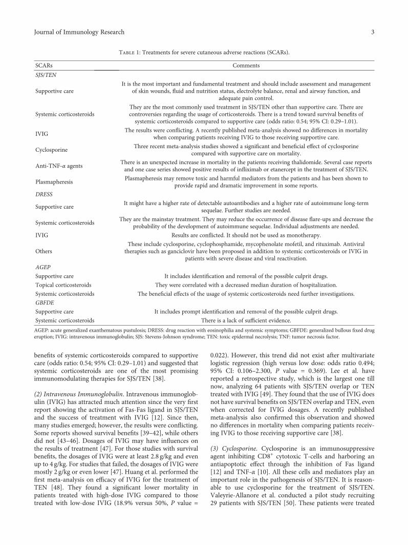
benefits of systemic corticosteroids compared to supportivecare (odds ratio: 0.54; 95% CI: 0.29–1.01) and suggested thatsystemic corticosteroids are one of the most promisingimmunomodulating therapies for SJS/TEN [38].
(2) Intravenous Immunoglobulin. Intravenous immunoglob-ulin (IVIG) has attracted much attention since the very firstreport showing the activation of Fas-Fas ligand in SJS/TENand the success of treatment with IVIG [12]. Since then,many studies emerged; however, the results were conflicting.Some reports showed survival benefits [39–42], while othersdid not [43–46]. Dosages of IVIG may have influences onthe results of treatment [47]. For those studies with survivalbenefits, the dosages of IVIG were at least 2.8 g/kg and evenup to 4 g/kg. For studies that failed, the dosages of IVIG weremostly 2 g/kg or even lower [47]. Huang et al. performed thefirst meta-analysis on efficacy of IVIG for the treatment ofTEN [48]. They found a significant lower mortality inpatients treated with high-dose IVIG compared to thosetreated with low-dose IVIG (18.9% versus 50%, P value =
0.022). However, this trend did not exist after multivariatelogistic regression (high versus low dose: odds ratio 0.494;95% CI: 0.106–2.300, P value = 0.369). Lee et al. havereported a retrospective study, which is the largest one tillnow, analyzing 64 patients with SJS/TEN overlap or TENtreated with IVIG [49]. They found that the use of IVIG doesnot have survival benefits on SJS/TEN overlap and TEN, evenwhen corrected for IVIG dosages. A recently publishedmeta-analysis also confirmed this observation and showedno differences in mortality when comparing patients receiv-ing IVIG to those receiving supportive care [38].
(3) Cyclosporine. Cyclosporine is an immunosuppressiveagent inhibiting CD8+ cytotoxic T-cells and harboring anantiapoptotic effect through the inhibition of Fas ligand[12] and TNF-α [10]. All these cells and mediators play animportant role in the pathogenesis of SJS/TEN. It is reason-able to use cyclosporine for the treatment of SJS/TEN.Valeyrie-Allanore et al. conducted a pilot study recruiting29 patients with SJS/TEN [50]. These patients were treated
Table 1: Treatments for severe cutaneous adverse reactions (SCARs).
SCARs Comments
SJS/TEN
Supportive careIt is the most important and fundamental treatment and should include assessment and management
of skin wounds, fluid and nutrition status, electrolyte balance, renal and airway function, andadequate pain control.
Systemic corticosteroidsThey are the most commonly used treatment in SJS/TEN other than supportive care. There arecontroversies regarding the usage of corticosteroids. There is a trend toward survival benefits ofsystemic corticosteroids compared to supportive care (odds ratio: 0.54; 95% CI: 0.29–1.01).
IVIGThe results were conflicting. A recently published meta-analysis showed no differences in mortality
when comparing patients receiving IVIG to those receiving supportive care.
CyclosporineThree recent meta-analysis studies showed a significant and beneficial effect of cyclosporine
compared with supportive care on mortality.
Anti-TNF-α agentsThere is an unexpected increase in mortality in the patients receiving thalidomide. Several case reportsand one case series showed positive results of infliximab or etanercept in the treatment of SJS/TEN.
PlasmapheresisPlasmapheresis may remove toxic and harmful mediators from the patients and has been shown to
provide rapid and dramatic improvement in some reports.
DRESS
Supportive careIt might have a higher rate of detectable autoantibodies and a higher rate of autoimmune long-term
sequelae. Further studies are needed.
Systemic corticosteroidsThey are the mainstay treatment. They may reduce the occurrence of disease flare-ups and decrease the
probability of the development of autoimmune sequelae. Individual adjustments are needed.
IVIG Results are conflicted. It should not be used as monotherapy.
OthersThese include cyclosporine, cyclophosphamide, mycophenolate mofetil, and rituximab. Antiviral
therapies such as ganciclovir have been proposed in addition to systemic corticosteroids or IVIG inpatients with severe disease and viral reactivation.
AGEP
Supportive care It includes identification and removal of the possible culprit drugs.
Topical corticosteroids They were correlated with a decreased median duration of hospitalization.
Systemic corticosteroids The beneficial effects of the usage of systemic corticosteroids need further investigations.
GBFDE
Supportive care It includes prompt identification and removal of the possible culprit drugs.
Systemic corticosteroids There is a lack of sufficient evidence.
AGEP: acute generalized exanthematous pustulosis; DRESS: drug reaction with eosinophilia and systemic symptoms; GBFDE: generalized bullous fixed drugeruption; IVIG: intravenous immunoglobulin; SJS: Stevens-Johnson syndrome; TEN: toxic epidermal necrolysis; TNF: tumor necrosis factor.
3Journal of Immunology Research
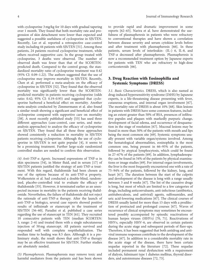
with cyclosporine 3mg/kg for 10 days with gradual taperingover 1 month. They found that both mortality rate and pro-gression of skin detachment were lower than expected andsuggested a possible usefulness of cyclosporine in SJS/TEN.Recently, Lee et al. reported a retrospective case-controlstudy including 44 patients with SJS/TEN [51]. Among thesepatients, 24 patients received cyclosporine treatment, whileothers received supportive care. In the group treated withcyclosporine, 3 deaths were observed. The number ofobserved death was fewer than that of the SCORTEN-predicted death. Compared to the control group, the stan-dardized mortality ratio of cyclosporine treatment was 0.42(95% CI: 0.09–1.22). The authors suggested that the use ofcyclosporine may improve mortality in SJS/TEN. Recently,Chen et al. performed a meta-analysis on the efficacy ofcyclosporine in SJS/TEN [52]. They found that the observedmortality was significantly lower than the SCORTEN-predicted mortality in patients receiving cyclosporine (oddsratio: 0.42; 95% CI: 0.19–0.95) and suggested that cyclo-sporine harbored a beneficial effect on mortality. Anothermeta-analysis conducted by Zimmermann et al. also founda similar result showing a significant and beneficial effect ofcyclosporine compared with supportive care on mortality[38]. A most recently published study [53] has used threedifferent approaches (case-control, case series, and meta-analysis approaches) to analyze the efficacy of cyclosporineon SJS/TEN. They found that all these three approachesshowed consistently a reduction in mortality in SJS/TENpatients receiving cyclosporine. Although the use of cyclo-sporine in SJS/TEN is not quite popular [4], it seems tobe a promising treatment. Further large-scale randomizedcontrolled studies are needed to confirm this observation.
(4) Anti-TNF-α Agents. Increased expressions of TNF-α inskin specimens [54], in blister fluid, and in serum [17] ofSJS/TEN patients justified the strategy of anti-TNF-α treat-ment. With this regard, thalidomide had been chosen asone of the options because of its anti-TNF-α property.Wolkenstein et al. had conducted a double-blind, random-ized, placebo-controlled trial to evaluate the efficacy ofthalidomide [55]. However, it terminated earlier as an unex-pected increase in mortality in the patients receiving thalid-omide. Nevertheless, the failure of thalidomide did not rejectthe rationale of anti-TNF-α therapy. After the launch ofanti-TNF-α biologics, several case reports showed positiveresults of infliximab or etanercept in the treatment ofSJS/TEN [56–60]. Paradisi et al. published a case seriesregarding the use of etanercept in TEN [61]. They recruited10 consecutive patients with TEN (median SCORTEN:3, range: 2–6) and treated them with a single subcutaneousinjection of 50mg etanercept. All patients survived andresponded well with complete reepithelialization. Themedian time to healing was 8.5 days. Although it is a pre-liminary study, the result shows that anti-TNF-α therapymay be an effective treatment for SJS/TEN. Further studiesare absolutely needed.
(5) Plasmapheresis. Plasmapheresis may remove toxic andharmful mediators from the patients and has been shown
to provide rapid and dramatic improvement in somereports [62–65]. Narira et al. have demonstrated the use-fulness of plasmapheresis in patients who were refractoryto conventional therapies and have shown a correlationbetween disease severity and serum cytokine levels beforeand after treatment with plasmapheresis [66]. In thesepatients, serum levels of interleukin- (IL-) 6, IL-8, andTNF-α decreased after plasmapheresis. Plasmapheresis isnow a recommended treatment option by Japanese expertsfor patients with TEN who are refractory to high-dosecorticosteroids [66].
3. Drug Reaction with Eosinophilia andSystemic Symptoms (DRESS)
3.1. Basic Characteristics. DRESS, which is also named asdrug-induced hypersensitivity syndrome (DiHS) by Japaneseexperts, is a life-threatening disease presenting with fever,cutaneous eruptions, and internal organ involvement [67].The mortality rate of DRESS is about 10% [68]. Skin lesionsin patients with DRESS have some common features, includ-ing an extent greater than 50% of BSA, presences of infiltra-tive papules and plaques with markedly purpuric change,development of facial edema, and occurrence of desquama-tion in the stage of resolution [67]. Mucosal lesions may befound in more than 50% of the patients with mouth and lipsbeing the most common site [69]. Systemic symptoms usu-ally present with variable organ/systems involved. Amongthe hematological abnormalities, eosinophilia is the mostcommon one, being present in 66–95% of the patients,followed by atypical lymphocytosis, which could be foundin 27–67% of the patients [69]. In addition, lymphadenopa-thy can be found in 54% of the patients by physical examina-tions or image studies [69]. For internal organ involvements,the liver is the most frequently encountered one with a rate of75–94% of the patients, followed by the kidney, lung, andheart [67]. The duration between the start of the culpritsand development of the disease is long with a range usuallybetween 3 and 8 weeks [67]. The list of the causative drugsis long, but most of which are limited to a few categories ofdrugs, including anticonvulsants, anti-infectious (antibiotics,antituberculosis, and antiviral) agents, sulfonamides, anduric acid-lowering medications [67]. The clinical courses ofDRESS usually lasted for more than 15 days with a predilec-tion of protracted and prolonged courses [67]. Waves ofrecurrence of clinical symptoms may sometimes be encoun-tered possibly accompanied by episodic reactivations ofhuman herpes viruses (HHVs) [70, 71]. Reactivations ofHHVs, especially HHV-6, are observed in certain patientsduring the acute stage and subsequent periods of flare-ups.Therefore, it has been suggested that both antidrug and anti-viral immune responses contribute to the development of thedisease [67]. In addition to a considerable mortality rate inthe acute stage of the disease, there have been certainsequelae reported in the literature [72]. These sequelaeincluded permanent renal dysfunction with a requirementof dialysis, fulminant type 1 diabetes mellitus, thyroid disor-ders, and autoimmune diseases [72, 73].
4 Journal of Immunology Research

3.2. Treatment. For treatments of patients with DRESS,there is still insufficient clinical evidence because mostof the suggestions are derived from case series or experts’opinions [67]. Immediate withdrawal of the culprit drugsis unsurprisingly the most important thing to do in themanagement of patients with DRESS. There have beenseveral options of systemic treatments suggested in theliterature (Table 1).
3.2.1. Supportive Care Only. Supportive care only may beconsidered a treatment option for patients with DRESS. Afew case series supported this notion. Uhara et al. havereported 12 patients with DiHS who received hydration withor without topical steroids [74]. All the patients recoveredwell within 7 to 37 days after the withdrawal of the culpritdrugs. Ushigome et al. have also presented 17 cases of DiHStreated with only supportive care [75]. All of them recoveredsmoothly except for those with a higher rate of detectableautoantibodies and a higher rate of autoimmune long-termsequelae. However, the number of patients with DRESS orDiHS treated with only supportive care is still limited. Fur-ther studies including a larger number of patients are neededto confirm the observation.
3.2.2. Corticosteroids. Systemic corticosteroids are themainstay treatment for patients with DRESS. There is stilla lack of consensus regarding the dosage and the durationof systemic corticosteroids. A starting dose of 0.5–1.0mg/kg/day of prednisolone with a gradual tapering over 2-3months has been suggested by some experts [67]. Thisapproach may reduce the occurrence of disease flare-upsand decrease the probability of the development of auto-immune sequelae [67]. Nevertheless, a prolonged durationof systemic corticosteroid usage may be associated with ahigher rate of opportunistic infections and with the possi-bility of many complications. Funck-Brentano et al. havereported a retrospective study of 38 patients with DRESS[76]. Among these patients, some received supportive carewith topical steroids, while others received systemic steroids.The authors found higher rates of infections, septicemia, andthe need for intensive care in patients receiving systemicsteroid and suggested that systemic steroids should bereserved for those with severe presentations. Thus, individ-ual adjustments are needed for each case based on theseverity of the disease and underlying comorbidities. Onegroup of the French Society of Dermatology has recom-mended that the use of systemic corticosteroids may beconsidered when 5-fold elevation of serum transaminaselevels or involvement of any other organs, such as the kidney,lung, and heart, occurs [77].
3.2.3. Intravenous Immunoglobulin. The results of the use ofIVIG in the treatment of patients with DRESS are conflicting.Several studies have reported the successful results [78, 79].On the other hand, Joly et al. reported their unfavorableexperience of using IVIG treatment in 6 DRESS patients[80]. Among them, 5 of the patients had severe adverseeffects, with 4 patients requiring systemic corticosteroidsdue to the adverse effects of IVIG or uncontrolled diseases.
Therefore, the authors suggested that IVIG should not beused as monotherapy in treating DRESS syndrome. Obvi-ously, the use of IVIG in the treatment of DRESS needsfurther investigations.
3.2.4. Other Treatments. Anecdotal reports have shown thetreatment effectiveness of several immunosuppressive agentsother than those of corticosteroids. These include cyclospor-ine [81], cyclophosphamide [82], mycophenolate mofetil,and rituximab [67]. Antiviral therapies such as ganciclovirhave been proposed in addition to systemic corticosteroidsor IVIG to be used in patients with severe disease and confir-mation of viral reactivation [77]. However, such treatmentshould be thoroughly considered by the judgment betweenbenefits and harms.
4. Acute Generalized ExanthematousPustulosis (AGEP)
4.1. Basic Characteristics. AGEP is characterized by a suddenonset of at least dozens and often hundreds of sterile, nonfol-licular pustules on an edematous erythema with a predilec-tion at the major folds [83]. Sometimes, facial edema,blisters, or atypical target lesions may develop. Mucosallesions are rare and usually mild. Fever and leukocytosis arecommonly accompanied by cutaneous eruptions. Systemicinvolvements have been reported to develop in less than20% of the patients with AGEP [84]. Liver involvementis the most common one, followed by the kidney, lung,and bone marrow. Although AGEP may result from viralinfections [85], it is primarily a hypersensitivity reactionto drugs. The most strongly associated drugs are pristinamy-cin, ampicillin/amoxicillin, quinolones, hydroxychloroquine,anti-infective sulfonamides, terbinafine, and diltiazem basedon a multinational case-control EuroSCAR study [86]. Thelatent periods between the drug intake and development ofthe disease showed two different patterns [86]. For thoseexposed to antibiotics, the median duration was 1 day, whilefor those using other medications, the median duration was11 days. The explanation for these differences is largely unex-plored. The prognosis of AGEP is generally very good. Mostof the patients recovered without sequelae.
4.2. Treatment. The mainstay of treatment for AGEP is theidentification and removal of the possible culprit drugs(Table 1) [83]. Recovery and resolution of the skin eruptionsusually develop within several days after withdrawal of theculprit drugs [83]. The mean durations between the resolu-tion of the pustules and cessation of the culprit drugs havebeen reported to be 6 days [87] and 7.6 days [88] in twodifferent studies, respectively. Hospitalization may berequired in some patients, especially those with extensivecutaneous eruptions, altered general condition, and sys-temic involvement. Topical corticosteroid may be usedand has been correlated with a decreased median durationof hospitalization [89]. Systemic corticosteroids have beenused in some patients with AGEP [88]. However, becauseof the benign courses in most of the patients with AGEP,
5Journal of Immunology Research

the beneficial effects of the usage of systemic corticosteroidsneed further investigations.
5. Generalized Bullous Fixed DrugEruption (GBFDE)
5.1. Basic Characteristics. GBFDE is a rare and severe formof fixed drug eruption (FDE). It is characterized by largeareas of well-demarcated erythematous or hyperpigmentedpatches with blisters or erosions developed soon afteradministrating the culprit drugs [90]. It exhibits typicalfeatures of FDE but may resemble the presentations ofSJS/TEN. To differentiate these two diseases is important.One previous study identified that patients with GBFDEhad a shorter latent period and less mucosal involvementcompared to those with SJS/TEN [90]. The mean durationof the latent period was 3.2 days in GBFDE. Mucosal involve-ments were only identified in 43% of the patients. Upon his-topathological examination, skin specimens of patients withGBFDE showed more eosinophil infiltration and more der-mal melanophages. Lesional infiltrates in GBFDE had moredermal CD4+ cells including Foxp3+ cells, less intraepidermalCD56+ cells, and fewer intraepidermal granulysin+ cellscompared to those in SJS/TEN. The serum level of granuly-sin in GBFDE was significantly lower than that in SJS/TEN[90]. These features may help to differentiate the two dis-eases when skin lesions are ambiguous. The common culpritdrugs in GBFDE were antibiotics, including cephalosporins,penicillins, and anti-infective sulfonamides, followed bynonsteroid anti-inflammatory drugs [90]. Traditionally, theprognosis of GBFDE is thought be better than that ofSJS/TEN. However, a large retrospective case-control studyincluding 58 patients with GBFDE and 170 patients withSJS/TEN matched for age and extent of skin detachmentfailed to support this traditional concept [91]. The authorsfound that the mortality rate was slightly but not significantlylower for patients with GBFDE than for controls (22%versus 28%, multivariate odds ratio: 0.6, 95% CI: 0.3–1.4).Although some selection bias may exist in this study, theobservation highlights the nature of GBFDE as SCAR mightbe overlooked before.
5.2. Treatment. Currently, there is still a lack of reportsregarding the treatment of patients with GBFDE. Just likeall other drug reactions, prompt identification and removalof the possible culprit drugs are the most important steps tomanage the disease (Table 1). Skin lesions of GBFDE patientsusually recover gradually after withdrawal of the causativedrugs as that usually seen in patients with conventionalFDE. However, for those patients with extensive areas of skindetachment, intensive supportive care should be applied asthat used in treating patients with SJS/TEN. Systemic cortico-steroids may be used as a treatment option for GBFDE andmay be considered effective. Our own unpublished data con-sisting of 32 patients with GBFDE showed only one deathoccurring during the acute stage of the disease. Most of thesepatients were treated with systemic corticosteroids. Never-theless, due to a lack of sufficient evidence regarding thetreatments of GBFDE, further investigations are needed.
6. Conclusion
The rarity of SCAR cannot dampen the importance ofmanagement of these diseases. All these diseases, includingSJS/TEN, DRESS, AGEP, and GBFDE, harbor considerablerates of morbidities and mortalities, which could not beoverlooked. However, indeed, the low incidence of SCARlimits the execution of large-scale randomized trials, whichin turn, leads to a lack of sufficient clinical evidence in themanagement of these diseases. Except the existence of somemeta-analyses in the treatment of patients with SJS/TEN,for patients with other SCARs, there is a big gap betweenclinical practice and evidence-based management. Furtherefforts are needed on these issues to improve the knowledgeof SCAR management.
Conflicts of Interest
The authors indicated no potential conflicts of interest.
Acknowledgments
This work was supported by the National Taiwan Univer-sity Hospital (NTUH 106-S3535) and National TaiwanUniversity (105A165).
References
[1] M. Paulmann and M. Mockenhaupt, “Severe drug-inducedskin reactions: clinical features, diagnosis, etiology, and ther-apy,” Journal der Deutschen Dermatologischen Gesellschaft,vol. 13, no. 7, pp. 625–643, 2015.
[2] B. Rzany, M. Mockenhaupt, S. Baur et al., “Epidemiology oferythema exsudativum multiforme majus, Stevens-Johnsonsyndrome, and toxic epidermal necrolysis in Germany(1990–1992): structure and results of a population-based regis-try,” Journal of Clinical Epidemiology, vol. 49, no. 7, pp. 769–773, 1996.
[3] M. Mockenhaupt, C. Viboud, A. Dunant et al., “Stevens-Johnson syndrome and toxic epidermal necrolysis: assessmentof medication risks with emphasis on recently marketed drugs.The EuroSCAR-study,” Journal of Investigative Dermatology,vol. 128, no. 1, pp. 35–44, 2008.
[4] B. F. Firoz, J. S. Henning, L. A. Zarzabal, and B. H. Pollock,“Toxic epidermal necrolysis: five years of treatment experiencefrom a burn unit,” Journal of the American Academy ofDermatology, vol. 67, no. 4, pp. 630–635, 2012.
[5] S. Bastuji-Garin, B. Rzany, R. S. Stern, N. H. Shear, L. Naldi,and J. C. Roujeau, “Clinical classification of cases of toxicepidermal necrolysis, Stevens-Johnson syndrome, and ery-thema multiforme,” Archives of Dermatology, vol. 129, no. 1,pp. 92–96, 1993.
[6] R. A. Shwartz, P. H. McDonough, and B. W. Lee, “Toxicepidermal necrolysis. Part I. Introduction, history, classifica-tion, clinical features, systemic manifestations, etiology, andimmunopathogenesis,” Journal of the American Academy ofDermatology, vol. 69, no. 2, pp. 173.e1–173.e13, 2013.
[7] T. Harr and L. E. French, “Toxic epidermal necrolysis andStevens-Johnson syndrome,” Orphanet Journal of Rare Dis-eases, vol. 5, no. 1, p. 39, 2010.
6 Journal of Immunology Research
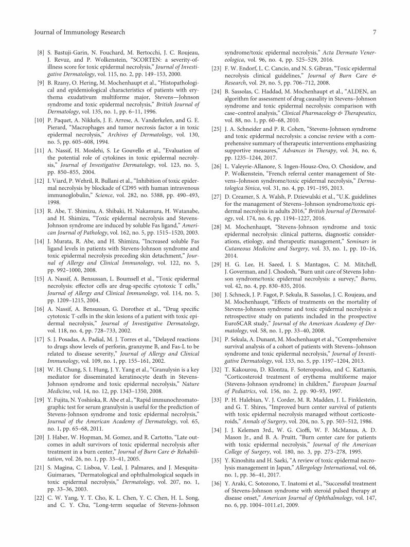
[8] S. Bastuji-Garin, N. Fouchard, M. Bertocchi, J. C. Roujeau,J. Revuz, and P. Wolkenstein, “SCORTEN: a severity-of-illness score for toxic epidermal necrolysis,” Journal of Investi-gative Dermatology, vol. 115, no. 2, pp. 149–153, 2000.
[9] B. Rzany, O. Hering, M. Mochenhaupt et al., “Histopathologi-cal and epidemiological characteristics of patients with ery-thema exudativum multiforme major, Stevens—Johnsonsyndrome and toxic epidermal necrolysis,” British Journal ofDermatology, vol. 135, no. 1, pp. 6–11, 1996.
[10] P. Paquet, A. Nikkels, J. E. Arrese, A. Vanderkelen, and G. E.Pierard, “Macrophages and tumor necrosis factor a in toxicepidermal necrolysis,” Archives of Dermatology, vol. 130,no. 5, pp. 605–608, 1994.
[11] A. Nassif, H. Moslehi, S. Le Gouvello et al., “Evaluation ofthe potential role of cytokines in toxic epidermal necroly-sis,” Journal of Investigative Dermatology, vol. 123, no. 5,pp. 850–855, 2004.
[12] I. Viard, P. Wehril, R. Bullani et al., “Inhibition of toxic epider-mal necrolysis by blockade of CD95 with human intravenousimmunoglobulin,” Science, vol. 282, no. 5388, pp. 490–493,1998.
[13] R. Abe, T. Shimizu, A. Shibaki, H. Nakamura, H. Watanabe,and H. Shimizu, “Toxic epidermal necrolysis and Stevens-Johnson syndrome are induced by soluble Fas ligand,” Ameri-can Journal of Pathology, vol. 162, no. 5, pp. 1515–1520, 2003.
[14] J. Murata, R. Abe, and H. Shimizu, “Increased soluble Fasligand levels in patients with Stevens-Johnson syndrome andtoxic epidermal necrolysis preceding skin detachment,” Jour-nal of Allergy and Clinical Immunology, vol. 122, no. 5,pp. 992–1000, 2008.
[15] A. Nassif, A. Bensussan, L. Boumsell et al., “Toxic epidermalnecrolysis: effector cells are drug-specific cytotoxic T cells,”Journal of Allergy and Clinical Immunology, vol. 114, no. 5,pp. 1209–1215, 2004.
[16] A. Nassif, A. Bensussan, G. Dorothee et al., “Drug specificcytotoxic T-cells in the skin lesions of a patient with toxic epi-dermal necrolysis,” Journal of Investigative Dermatology,vol. 118, no. 4, pp. 728–733, 2002.
[17] S. J. Posadas, A. Padial, M. J. Torres et al., “Delayed reactionsto drugs show levels of perforin, granzyme B, and Fas-L to berelated to disease severity,” Journal of Allergy and ClinicalImmunology, vol. 109, no. 1, pp. 155–161, 2002.
[18] W. H. Chung, S. I. Hung, J. Y. Yang et al., “Granulysin is a keymediator for disseminated keratinocyte death in Stevens-Johnson syndrome and toxic epidermal necrolysis,” NatureMedicine, vol. 14, no. 12, pp. 1343–1350, 2008.
[19] Y. Fujita, N. Yoshioka, R. Abe et al., “Rapid immunochromato-graphic test for serum granulysin is useful for the prediction ofStevens-Johnson syndrome and toxic epidermal necrolysis,”Journal of the American Academy of Dermatology, vol. 65,no. 1, pp. 65–68, 2011.
[20] J. Haber, W. Hopman, M. Gomez, and R. Cartotto, “Late out-comes in adult survivors of toxic epidermal necrolysis aftertreatment in a burn center,” Journal of Burn Care & Rehabili-tation, vol. 26, no. 1, pp. 33–41, 2005.
[21] S. Magina, C. Lisboa, V. Leal, J. Palmares, and J. Mesquita-Guimaraes, “Dermatological and ophthalmological sequels intoxic epidermal necrolysis,” Dermatology, vol. 207, no. 1,pp. 33–36, 2003.
[22] C. W. Yang, Y. T. Cho, K. L. Chen, Y. C. Chen, H. L. Song,and C. Y. Chu, “Long-term sequelae of Stevens-Johnson
syndrome/toxic epidermal necrolysis,” Acta Dermato Vener-eologica, vol. 96, no. 4, pp. 525–529, 2016.
[23] F. W. Endorf, L. C. Cancio, and N. S. Gibran, “Toxic epidermalnecrolysis clinical guidelines,” Journal of Burn Care &Research, vol. 29, no. 5, pp. 706–712, 2008.
[24] B. Sassolas, C. Haddad, M. Mochenhaupt et al., “ALDEN, analgorithm for assessment of drug causality in Stevens–Johnsonsyndrome and toxic epidermal necrolysis: comparison withcase–control analysis,” Clinical Pharmacology & Therapeutics,vol. 88, no. 1, pp. 60–68, 2010.
[25] J. A. Schneider and P. R. Cohen, “Stevens-Johnson syndromeand toxic epidermal necrolysis: a concise review with a com-prehensive summary of therapeutic interventions emphasizingsupportive measures,” Advances in Therapy, vol. 34, no. 6,pp. 1235–1244, 2017.
[26] L. Valeyrie-Allanore, S. Ingen-Housz-Oro, O. Chosidow, andP. Wolkenstein, “French referral center management of Ste-vens–Johnson syndrome/toxic epidermal necrolysis,” Derma-tologica Sinica, vol. 31, no. 4, pp. 191–195, 2013.
[27] D. Creamer, S. A. Walsh, P. Dziewulski et al., “U.K. guidelinesfor the management of Stevens–Johnson syndrome/toxic epi-dermal necrolysis in adults 2016,” British Journal of Dermatol-ogy, vol. 174, no. 6, pp. 1194–1227, 2016.
[28] M. Mochenhaupt, “Stevens-Johnson syndrome and toxicepidermal necrolysis: clinical patterns, diagnostic consider-ations, etiology, and therapeutic management,” Seminars inCutaneous Medicine and Surgery, vol. 33, no. 1, pp. 10–16,2014.
[29] H. G. Lee, H. Saeed, I. S. Mantagos, C. M. Mitchell,J. Goverman, and J. Chodosh, “Burn unit care of Stevens John-son syndrome/toxic epidermal necrolysis: a survey,” Burns,vol. 42, no. 4, pp. 830–835, 2016.
[30] J. Schneck, J. P. Fagot, P. Sekula, B. Sassolas, J. C. Roujeau, andM. Mochenhaupt, “Effects of treatments on the mortality ofStevens-Johnson syndrome and toxic epidermal necrolysis: aretrospective study on patients included in the prospectiveEuroSCAR study,” Journal of the American Academy of Der-matology, vol. 58, no. 1, pp. 33–40, 2008.
[31] P. Sekula, A. Dunant, M. Mochenhaupt et al., “Comprehensivesurvival analysis of a cohort of patients with Stevens–Johnsonsyndrome and toxic epidermal necrolysis,” Journal of Investi-gative Dermatology, vol. 133, no. 5, pp. 1197–1204, 2013.
[32] T. Kakourou, D. Klontza, F. Soteropoulou, and C. Kattamis,“Corticosteroid treatment of erythema multiforme major(Stevens-Johnson syndrome) in children,” European Journalof Pediatrics, vol. 156, no. 2, pp. 90–93, 1997.
[33] P. H. Halebian, V. J. Corder, M. R. Madden, J. L. Finklestein,and G. T. Shires, “Improved burn center survival of patientswith toxic epidermal necrolysis managed without corticoste-roids,” Annals of Surgery, vol. 204, no. 5, pp. 503–512, 1986.
[34] J. J. Kelemen 3rd., W. G. Cioffi, W. F. McManus, A. D.Mason Jr., and B. A. Pruitt, “Burn center care for patientswith toxic epidermal necrolysis,” Journal of the AmericanCollege of Surgery, vol. 180, no. 3, pp. 273–278, 1995.
[35] Y. Kinoshita and H. Saeki, “A review of toxic epidermal necro-lysis management in Japan,” Allergology International, vol. 66,no. 1, pp. 36–41, 2017.
[36] Y. Araki, C. Sotozono, T. Inatomi et al., “Successful treatmentof Stevens-Johnson syndrome with steroid pulsed therapy atdisease onset,” American Journal of Ophthalmology, vol. 147,no. 6, pp. 1004–1011.e1, 2009.
7Journal of Immunology Research

[37] K. Hirahara, Y. Kano, Y. Sato et al., “Methylprednisolonepulse therapy for Stevens-Johnson syndrome/toxic epidermalnecrolysis: clinical evaluation and analysis of biomarkers,”Journal of the American Academy of Dermatology, vol. 69,no. 3, pp. 496–498, 2013.
[38] S. Zimmermann, P. Sekula, M. Venhoff et al., “Systemic immu-nomodulating therapies for Stevens-Johnson syndrome andtoxic epidermal necrolysis. A systematic review and meta-analysis,” JAMA Dermatology, vol. 153, no. 6, pp. 514–522,2017.
[39] C. Prins, F. A. Kerdel, R. S. Padilla et al., “Treatment of toxicepidermal necrolysis with high-dose intravenous immuno-globulins: multicenter retrospective analysis of 48 consecutivecases,” Archives of Dermatology, vol. 139, no. 1, pp. 26–32,2003.
[40] J. T. Trent, R. S. Kirsner, P. Romanelli, and F. A. Kerdel,“Analysis of intravenous immunoglobulin for the treatmentof toxic epidermal necrolysis using SCORTEN: the Universityof Miami experience,” Archives of Dermatology, vol. 139, no. 1,pp. 39–43, 2003.
[41] N. Al-Mutairi, J. Arun, N. E. Osama et al., “Prospective, non-comparative open study from Kuwait of the role of intrave-nous immunoglobulin in the treatment of toxic epidermalnecrolysis,” International Journal of Dermatology, vol. 43,no. 11, pp. 847–851, 2004.
[42] M. Stella, P. Cassano, D. Bollero, A. Clemente, and G. Giorio,“Toxic epidermal necrolysis treated with intravenous high-dose immunoglobulins: our experience,” Dermatology,vol. 203, no. 1, pp. 45–49, 2001.
[43] K. M. Brown, G. M. Silver, M. Halerz, P. Walaszek,A. Sandroni, and R. L. Gamelli, “Toxic epidermal necrolysis:does immunoglobulin make a difference?,” Journal of BurnCare & Rehabilitation, vol. 25, no. 1, pp. 81–88, 2004.
[44] R. Shortt, M. Gomez, N. Mittman, and R. Cartotto, “Intrave-nous immunoglobulin does not improve outcome in toxic epi-dermal necrolysis,” Journal of Burn Care & Rehabilitation,vol. 25, no. 3, pp. 246–255, 2004.
[45] N. Bachot, J. Revuz, and J. C. Roujeau, “Intravenous immuno-globulin treatment for Stevens-Johnson syndrome and toxicepidermal necrolysis: a prospective noncomparative studyshowing no benefit on mortality or progression,” Archives ofDermatology, vol. 139, no. 1, pp. 33–36, 2003.
[46] Y. Yang, J. Xu, F. Li, and X. Zhu, “Combination therapy ofintravenous immunoglobulin and corticosteroid in the treat-ment of toxic epidermal necrolysis and Stevens-Johnson syn-drome: a retrospective comparative study in China,”International Journal of Dermatology, vol. 48, no. 10,pp. 1122–1128, 2009.
[47] R. A. Shwartz, P. H. McDonough, and B. W. Lee, “Toxicepidermal necrolysis. Part II. Prognosis, sequelae, diagnosis,differential diagnosis, prevention, and treatment,” Journal ofthe American Academy of Dermatology, vol. 69, no. 2,pp. 187. e1–187.e16, 2013.
[48] Y. C. Huang, Y. C. Li, and T. J. Chen, “The efficacy of intrave-nous immunoglobulin for the treatment of toxic epidermalnecrolysis: a systematic review and meta-analysis,” BritishJournal of Dermatology, vol. 167, no. 2, pp. 424–432, 2012.
[49] H. Y. Lee, Y. L. Lim, T. Thirumoorthy, and S. M. Pang,“The role of intravenous immunoglobulin in toxic epidermalnecrolysis: a retrospective analysis of 64 patients managed ina specialized centre,” British Journal of Dermatology, vol. 169,no. 6, pp. 1304–1309, 2013.
[50] L. Valeyrie-Allanore, P. Wolkenstein, L. Brochard et al., “Opentrial of ciclosporin treatment for Stevens-Johnson syndromeand toxic epidermal necrolysis,” British Journal of Dermatol-ogy, vol. 163, no. 4, pp. 847–853, 2010.
[51] H. Y. Lee, S. Fook-Chong, H. Y. Koh, T. Thirumoorthy, andS. M. Pang, “Cyclosporine treatment for Stevens-Johnsonsyndrome/toxic epidermal necrolysis: retrospective analysisof a cohort treated in a specialized referral center,” Journalof the American Academy of Dermatology, vol. 76, no. 1,pp. 106–113, 2017.
[52] Y. T. Chen, C. Y. Hsu, Y. N. Chien, W. R. Lee, and Y. C.Huang, “Efficacy of cyclosporine for the treatment ofStevens-Johnson syndrome and toxic epidermal necrolysis:systemic review and meta-analysis,” Dermatologica Sinica,vol. 35, no. 3, pp. 131–137, 2017.
[53] C. Gonzalez-Herrada, S. Rodriguez-Martin, L. Cachafeiroet al., “Ciclosporin use in epidermal necrolysis is associatedwith an important mortality reduction: evidence from threedifferent approaches,” Journal of Investigative Dermatology,vol. 137, no. 10, pp. 2092–2100, 2017.
[54] P. Paquet, F. Paquet, W. Al Saleh, P. Reper, A. Vanderkelen,and G. E. Pierard, “Immunoregulatory effector cells in drug-induced toxic epidermal necrolysis,” The American Journal ofDermatopathology, vol. 22, no. 5, pp. 413–417, 2000.
[55] P. Wolkenstein, J. Latarjet, J. C. Roujeau et al., “Randomisedcomparison of thalidomide versus placebo in toxic epidermalnecrolysis,” The Lancet, vol. 352, no. 9140, pp. 1586–1589,1998.
[56] M. Fischer, E. Fiedler, W. C. Marsch, and J. Wohlrab, “Antitu-mour necrosis factor-α antibodies (infliximab) in the treat-ment of a patient with toxic epidermal necrolysis,” BritishJournal of Dermatology, vol. 146, no. 4, pp. 707–709, 2002.
[57] R. E. Hunger, T. Hunziker, U. Buettiker, L. R. Braathen, andN. Yawalkar, “Rapid resolution of toxic epidermal necrolysiswith anti-TNF-α treatment,” Journal of Allergy and ClinicalImmunology, vol. 116, no. 4, pp. 923-924, 2005.
[58] A. Wojtkiewicz, M. Wysocki, J. Fortuna, M. Chrupek,M. Matczuk, and A. Koltan, “Beneficial and rapid effect ofinfliximab on the course of toxic epidermal necrolysis,” ActaDermato-Venereologica, vol. 88, no. 4, pp. 420-421, 2008.
[59] G. Famularo, B. Didona, F. Canzona, C. R. Girardelli, andG. Cruciani, “Etanercept for toxic epidermal necrolysis,”Annals of Pharmacotherapy, vol. 41, no. 6, pp. 1083-1084,2007.
[60] E. Gubinelli, F. Canzona, T. Tonanzi, D. Raskovic, andB. Didona, “Toxic epidermal necrolysis successfully treatedwith etanercept,” The Journal of Dermatology, vol. 36, no. 3,pp. 150–153, 2009.
[61] A. Paradisi, D. Abeni, F. Bergamo, F. Ricci, D. Didona, andB. Didona, “Etanercept therapy for toxic epidermal necroly-sis,” Journal of the American Academy of Dermatology,vol. 71, no. 2, pp. 278–283, 2014.
[62] H. Yamada and K. Takamori, “Status of plasmapheresis for thetreatment of toxic epidermal necrolysis in Japan,” TherapeuticApheresis and Dialysis, vol. 12, no. 5, pp. 355–359, 2008.
[63] M. Lissia, A. Figus, and C. Rubino, “Intravenous immunoglob-ulins and plasmapheresis combined treatment in patients withsevere toxic epidermal necrolysis: preliminary report,” BritishJournal of Plastic Surgery, vol. 58, no. 4, pp. 504–510, 2005.
[64] D. Kamanabroo, W. Schmitz-Landgraf, and B. M. Czarnetzki,“Plasmapheresis in severe drug-induced toxic epidermal
8 Journal of Immunology Research

necrolysis,” Archives of Dermatology, vol. 121, no. 12,pp. 1548-1549, 1985.
[65] G. Bamichas, T. Natse, F. Christidou et al., “Plasma exchangein patients with toxic epidermal necrolysis,” Therapeutic Aphe-resis and Dialysis, vol. 6, no. 3, pp. 225–228, 2002.
[66] Y. M. Narira, K. Hirahara, Y. Mizukawa, Y. Kano, andT. Shiohara, “Efficacy of plasmapheresis for the treatment ofsevere toxic epidermal necrolysis: is cytokine expression anal-ysis useful in predicting its therapeutic efficacy?,” The Journalof Dermatology, vol. 38, no. 3, pp. 236–245, 2011.
[67] Y. T. Cho, C. W. Yang, and C. Y. Chu, “Drug reaction witheosinophilia and systemic symptoms (DRESS): an interplayamong drugs, viruses, and immune system,” InternationalJournal of Molecular Sciences, vol. 18, no. 6, article E1243,2017.
[68] Y. C. Chen, H. C. Chiu, and C. Y. Chu, “Drug reactionwith eosinophilia and systemic symptoms: a retrospectivestudy of 60 cases,” Archives of Dermatology, vol. 146, no. 12,pp. 1373–1379, 2010.
[69] S. H. Karduan, P. Sekula, L. Valeyrie-Allanore et al., “Drugreaction with eosinophilia and systemic symptoms (DRESS):an original multisystem adverse drug reaction. Results fromthe prospective RegiSCAR study,” British Journal of Dermatol-ogy, vol. 169, no. 5, pp. 1071–1080, 2013.
[70] M. Tohyama, K. Hashimoto, M. Yasukawa et al., “Associationof human herpesvirus 6 reactivation with the flaring and sever-ity of drug-induced hypersensitivity syndrome,” British Jour-nal of Dermatology, vol. 157, no. 5, pp. 934–940, 2007.
[71] M. Seishima, S. Yamanaka, T. Fujisawa, M. Tohyama, andK. Hashimoto, “Reactivation of human herpesvirus (HHV)family members other than HHV-6 in drug-induced hyper-sensitivity syndrome,” British Journal of Dermatology,vol. 155, no. 2, pp. 344–349, 2006.
[72] Y. Kano, M. Tohyama, M. Aihara et al., “Sequelae of 145patients with drug-induced hypersensitivity syndrome/drugreaction with eosinophilia and systemic symptoms: surveyconducted by the Asian Research Committee on Severe Cuta-neous Adverse Reaction (ASCAR),” The Journal of Dermatol-ogy, vol. 42, no. 3, pp. 276–282, 2015.
[73] Y. C. Chen, C. Y. Chang, Y. T. Cho, H. C. Chiu, and C. Y. Chu,“Long-term sequelae of drug reaction with eosinophilia andsystemic symptoms: a retrospective cohort study fromTaiwan,” Journal of the American Academy of Dermatology,vol. 68, no. 3, pp. 459–465, 2013.
[74] H. Uhara, M. Saiki, S. Kawachi, A. Ashida, S. Oguchi, andR. Okuyama, “Clinical course of drug-induced hypersensitivitysyndrome treated without systemic corticosteroids,” Journal ofthe European Academy of Dermatology and Venereology,vol. 27, no. 6, pp. 722–726, 2013.
[75] Y. Ushigome, Y. Kano, T. Ishida, K. Hirahara, and T. Shiohara,“Short- and long-term outcomes of 34 patients with drug-induced hypersensitivity syndrome in a single institution,”Journal of the American Academy of Dermatology, vol. 68,no. 5, pp. 721–728, 2013.
[76] E. Funck-Brentano, T. A. Duong, S. Bouvresse et al., “Thera-peutic management of DRESS: a retrospective study of 38cases,” Journal of the American Academy of Dermatology,vol. 72, no. 2, pp. 246–252, 2015.
[77] V. Descamps, B. Ben Said, B. Sassolas et al., “Management ofdrug reaction with eosinophilia and systemic symptoms(DRESS),” Annales De Dermatologie Et De Venereologie,vol. 137, no. 11, pp. 703–708, 2010.
[78] O. Scheuerman, Y. Nofech-Moses, A. Rachmel, andS. Ashkenazi, “Successful treatment of antiepileptic drughypersensitivity syndrome with intravenous immune globu-lin,” Pediatrics, vol. 107, no. 1, article e14, 2001.
[79] K. S. Fields, M. J. Petersen, E. Chiao, and P. Tristani-Firouzi,“Case reports: treatment of nevirapine-associated dress syn-drome with intravenous immune globulin (IVIG),” Journalof Drugs in Dermatology, vol. 4, no. 4, pp. 510–513, 2005.
[80] P. Joly, B. Janela, F. Tetart et al., “Poor benefit/risk balance ofintravenous immunoglobulins in DRESS,” Archives of Derma-tology, vol. 148, no. 4, pp. 543-544, 2012.
[81] G. K. Mark, A. Wong, and J. P. Dutz, “Cyclosporine treatmentof drug-induced hypersensitivity syndrome,” JAMADermatol-ogy, vol. 152, no. 11, pp. 1254–1257, 2016.
[82] E. Laban, E. Hainaut-Wierzbicka, F. Pourreau, M. Yacoub,E. Sztermer, and G. Guillet, “Cyclophosphamide therapy forcorticoresistant drug reaction with eosinophilia and systemicsymptoms (DRESS) syndrome in a patient with severe kidneyand eye involvement and Epstein-Barr virus reactivation,”American Journal of Kidney Diseases, vol. 55, no. 3, pp. e11–e14, 2010.
[83] A. Sidoroff, “Acute generalized exanthematous pustulosis,”Chemical Immunology and Allergy, vol. 97, pp. 139–148, 2012.
[84] C. Hotz, L. Valeyrie-Allanore, C. Haddad et al., “Systemicinvolvement of acute generalized exanthematous pustulosis: aretrospective study of 58 patients,” British Journal of Derma-tology, vol. 169, no. 6, pp. 1223–1232, 2013.
[85] B. Rouchouse, M. Bonnefoy, B. Pallot, L. Jacquelin,G. Dimoux-Dime, and A. L. Claudy, “Acute generalized exan-thematous pustular dermatitis and viral infection,” Dermatol-ogy, vol. 173, no. 4, pp. 180–184, 1986.
[86] A. Sidoroff, A. Dunant, C. Viboud et al., “Risk factors for acutegeneralized exanthematous pustulosis (AGEP)—results of amultinational case-control study (EuroSCAR),” British Journalof Dermatology, vol. 157, no. 5, pp. 989–996, 2007.
[87] H. Y. Lee, D. Chou, S. M. Pang, and T. Thirumoorthy, “Acutegeneralized exanthematous pustulosis: analysis of cases man-aged in a tertiary hospital in Singapore,” International Journalof Dermatology, vol. 49, no. 5, pp. 507–512, 2010.
[88] D. T. Alniemi, D. A. Wetter, A. G. Bridges et al., “Acute gener-alized exanthematous pustulosis: clinical characteristics, etio-logic associations, treatments, and outcomes in a series of 28patients at Mayo Clinic, 1996-2013,” International Journal ofDermatology, vol. 56, no. 4, pp. 405–414, 2017.
[89] S. Ingen-Housz-Oro, C. Hotz, L. Valeyrie-Allanore et al.,“Acute generalized exanthematous pustulosis: a retrospectiveaudit of practice between 1994 and 2011 at a single centre,”British Journal of Dermatology, vol. 172, no. 5, pp. 1455–1457, 2015.
[90] Y. T. Cho, J. W. Lin, Y. C. Chen et al., “Generalized bullousfixed drug eruption is distinct from Stevens-Johnsonsyndrome/toxic epidermal necrolysis by immunohistopatho-logical features,” Journal of the American Academy of Derma-tology, vol. 70, no. 3, pp. 539–548, 2014.
[91] S. Lipowicz, P. Sekula, S. Ingen-Housz-Oro et al., “Prognosis ofgeneralized bullous fixed drug eruption: comparison withStevens-Johnson syndrome and toxic epidermal necrolysis,”British Journal of Dermatology, vol. 168, no. 4, pp. 726–732,2013.
9Journal of Immunology Research

Research ArticleComparison between the HLA-B∗58 : 01 Allele andSingle-Nucleotide Polymorphisms in Chromosome 6for Prediction of Allopurinol-Induced Severe CutaneousAdverse Reactions
Niwat Saksit,1,2 Nontaya Nakkam,1 Parinya Konyoung,3 Usanee Khunarkornsiri,3
Wongwiwat Tassaneeyakul,4 Pansu Chumworathayi,5 Sirimas Kanjanawart,1
Chonlaphat Sukasem,6,7 Alisara Sangviroon,8 Oranuch Pattanacheewapull,9
and Wichittra Tassaneeyakul1
1Department of Pharmacology, Faculty of Medicine, Khon Kaen University, Khon Kaen, Thailand2School of Pharmaceutical Sciences, University of Phayao, Phayao, Thailand3Pharmacy Unit, Udon Thani Hospital, Udon Thani, Thailand4Faculty of Pharmaceutical Sciences, Khon Kaen University, Khon Kaen, Thailand5Pharmacy Unit, Faculty of Medicine, Khon Kaen University, Khon Kaen, Thailand6Department of Pathology, Division of Pharmacogenomics and Personalized Medicine, Faculty of Medicine Ramathibodi Hospital,Mahidol University, Bangkok, Thailand7Laboratory for Pharmacogenomics, Somdech Phra DebaratanaMedical Center (SDMC), Faculty of Medicine Ramathibodi Hospital,Mahidol University, Bangkok, Thailand8Pharmacy Unit, Police General Hospital, Bangkok, Thailand9Pharmacy Department, Khon Kaen Hospital, Khon Kaen, Thailand
Correspondence should be addressed to Wichittra Tassaneeyakul; [email protected]
Received 24 July 2017; Accepted 8 November 2017; Published 17 December 2017
Academic Editor: Ethan M. Shevach
Copyright © 2017 Niwat Saksit et al. This is an open access article distributed under the Creative Commons Attribution License,which permits unrestricted use, distribution, and reproduction in any medium, provided the original work is properly cited.
Severe cutaneous adverse drug reactions (SCARs) are life-threatening reactions. The strong association between the HLA-B∗58 : 01allele and allopurinol-induced SCARs is well recognized. Screening for HLA-B∗58 : 01 allele before prescribing allopurinol in somepopulations has been recommended. Several single-nucleotide polymorphisms (SNPs) in chromosome 6 have been found to betightly linked with the HLA allele, and these SNPs have been proposed as surrogate markers of the HLA-B∗58 : 01 allele. Thisstudy aimed to evaluate the association between three SNPs in chromosome 6 and allopurinol-induced SCARs in a Thaipopulation. The linkage disequilibrium between the HLA-B∗58 : 01 allele and these SNPs was also evaluated. Results showed thatthree SNPs including rs9263726, rs2734583, and rs3099844 were significantly associated with allopurinol-induced SCARs butwith a lower degree of association when compared with the HLA-B∗58 : 01 allele. The sensitivity, specificity, PPV, and NPV ofthese SNPs were comparable to those of the HLA-B∗58 : 01 allele. Although detection of the SNP is simpler and less expensivecompared with that of the HLA-B∗58 : 01 allele, these SNPs were not perfectly linked with the HLA-B∗58 : 01 allele. Screeningusing these SNPs as surrogate markers of the HLA-B∗58 : 01 allele to avoid SCARs prior to allopurinol administration needscaution because of their imperfect linkage with the HLA-B∗58 : 01 allele.
HindawiJournal of Immunology ResearchVolume 2017, Article ID 2738784, 9 pageshttps://doi.org/10.1155/2017/2738784

1. Introduction
Allopurinol, a uric acid-lowering agent, is one of the mostcommon culprit drugs for severe cutaneous adverse drugreactions (SCARs). These reactions range from Stevens-Johnson syndrome (SJS) and toxic epidermal necrolysis(TEN) to drug reaction with eosinophilia and systemicsymptoms (DRESS). Although the incidence of allopurinol-induced SCARs is rare, they are life-threatening reactions.Data from systematic reviews shows that although theprevalence of gout in Asian population is lower than that inCaucasian populations, the hypersensitivity caused by allo-purinol reported in Asians was about 73% of the reportedcases [1]. In Taiwan, the annual incidence rates for allopuri-nol hypersensitivity were 4.68 per 1000 new users, 2.02 per1000 new users for related hospitalization, and 0.39 per1000 new users for related mortality [2]. The mortality rateof allopurinol hypersensitivity in Taiwan was about 8.3%[2]. Similarly, the incidence rate of allopurinol-inducedSCARs in Thailand reported from the biggest hospital inThailand was about 2.13 per 1000 new users [3]. Comparedwith that of other drug-induced SCARs, the mortality rateof allopurinol-induced SCARs observed in a Thai populationis the highest up to 11% [4].
Although allopurinol-induced SCARs are considered asidiosyncratic reactions, current studies have identified severalrisk factors of such fatal reactions that include both geneticand nongenetic factors [4–6]. For genetic factors, the specificallele of the human leukocyte antigen (HLA), namely, theHLA-B∗58 : 01 allele, is the first genetic marker that wasfound to be strongly associated with allopurinol-inducedSCARs in a Taiwanese population [7]. This association hasbeen confirmed in Thai [4, 8, 9] and Han Chinese [10, 11]populations. Unlike theHLA-B∗15 : 02 allele which is specificto Chinese and Southeast Asian population, the associationsbetween the HLA-B∗58 : 01 allele and allopurinol-inducedSCARs were also demonstrated in Japanese [12, 13], Korean[14], and Caucasian populations [15]. The strength of associ-ation ranges from an odds ratio (OR) of 39 to 696 [16], andthe sensitivity and specificity of the HLA-B∗58 : 01 allele forthe prediction of allopurinol-induced SCARs were 93%(95% CI: 85–97%) and 89% (95% CI, 87–91%) across Asianand Caucasian populations [16]. To date, the AmericanCollege of Rheumatology recommends the testing for theHLA-B∗58 : 01 allele in certain ethnicities with a high fre-quency of this allele and showing an elevated risk forallopurinol-induced SCARs in HLA-B∗58 : 01 allele carrierssuch as Han Chinese, Thai, and Korean populations [17].
Due to the highly polymorphic nature of the HLA gene, aspecific method is required in order to determine a specificHLA allele, particularly the HLA-B alleles in which exon 2and 3 regions exhibit the highest variability. Several molecu-lar techniques including specific sequencing primers (SSP)PCR, sequence-specific oligonucleotide (SSO) probes, andsequencing-based typing (SBT) have been demonstrated tobe specific methods for the determination of HLA genotype;however, these techniques are quite expensive, time-con-suming, and not commonly available in hospital laborato-ries. A recent genome-wide association study in a Japanese
population has discovered a number of single-nucleotidepolymorphisms (SNPs) in chromosome 6 that werestrongly associated with allopurinol-induced SCARs [13].These SNPs included rs2734583 in the HLA-B-associatedtranscript 1 (BAT1) gene, rs3099844 in the HLA complex P5(HCP5) gene, and rs9263726 in the psoriasis susceptibility 1candidate 1 (PSORS1C1) gene. Due to the absolute linkagedisequilibrium between rs9263726 and the HLA-B∗58 : 01allele found in 27 Japanese patients who suffered fromallopurinol-induced SCARs, the rs9263726 has beenproposed as a surrogate marker for allopurinol-inducedSJS/TEN [18]. Similarly, an absolute linkage disequilibriumbetween the rs9263726 and allopurinol-induced SCARs hasalso been recently reported in 17 Eastern Chinese patients[19]. Ethnic specificity of the associations between HLAalleles and drug-induced SCARs is well recognized [20].Whether these SNPs are good surrogates of allopurinol-induced SCARs in other ethnic societies or not needs to beevaluated. The present study aimed to evaluate the degreeof relationship between the three selected SNPs in chromo-some 6 including rs9263726, rs2734583, and rs3099844 andallopurinol-induced SCARs in a Thai population that has arelatively high frequency of the HLA-B∗58 : 01 allele. Inaddition, the sensitivity and specificity for these selectedSNPs for the prediction of allopurinol-induced SCARs andtheir linkage disequilibrium with the HLA-B∗58 : 01 alleleare characterized in the present study.
2. Materials and Methods
2.1. Study Population Assessment and Enrollment. A total of96 allopurinol-induced SCARs patients including 23 DRESSand 73 SJS/TEN patients were recruited for the study. Thesepatients had been diagnosed with allopurinol-inducedSCARs within the first 3 months of allopurinol exposure.The phenotype of SCARs in an individual patient was scoredand assessed by the ALDEN [21] or the RegiSCAR algo-rithms [22] whereas the assessment of causative drugs wasperformed using Naranjo’s algorithm [23]. All of the SCARspatients who represented at least a probable score wererecruited for the study.
For the control cohort comparison, 193 patients wererecruited from patients who had used allopurinol for morethan 6 months without any evidence of cutaneous reactions.Written informed consent was obtained from each patient.The study protocol within the hospital network of KhonKaen University was approved by the Khon Kaen EthicsCommittee for Human Research, Khon Kaen University,Thailand (HE510837). The study protocol was also approvedby each hospital, where IRB function was available.
2.2. Genomic DNA Preparation. Leukocytes were separatedfrom peripheral blood by centrifugation at 3500 rpm for15min or buccal swab. Genomic DNA (gDNA) was isolatedfrom leukocytes using a QIAamp DNA Blood mini kit (QIA-GEN GmbH, Hilden, Germany). The quantity and quality ofgDNA were checked by a Nano Drop machine (ThermoScientific NanoDrop 2000c) and kept at −20°C until used.
2 Journal of Immunology Research
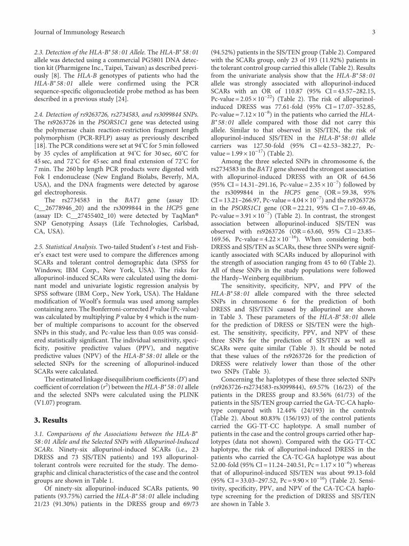
2.3. Detection of the HLA-B∗58 : 01 Allele. The HLA-B∗58 : 01allele was detected using a commercial PG5801 DNA detec-tion kit (Pharmigene Inc., Taipei, Taiwan) as described previ-ously [8]. The HLA-B genotypes of patients who had theHLA-B∗58 : 01 allele were confirmed using the PCRsequence-specific oligonucleotide probe method as has beendescribed in a previous study [24].
2.4. Detection of rs9263726, rs2734583, and rs3099844 SNPs.The rs9263726 in the PSORS1C1 gene was detected usingthe polymerase chain reaction-restriction fragment lengthpolymorphism (PCR-RFLP) assay as previously described[18]. The PCR conditions were set at 94°C for 5min followedby 35 cycles of amplification at 94°C for 30 sec, 60°C for45 sec, and 72°C for 45 sec and final extension of 72°C for7min. The 260 bp length PCR products were digested withFok I endonuclease (New England Biolabs, Beverly, MA,USA), and the DNA fragments were detected by agarosegel electrophoresis.
The rs2734583 in the BAT1 gene (assay ID:C__26778946_20) and the rs3099844 in the HCP5 gene(assay ID: C__27455402_10) were detected by TaqMan®SNP Genotyping Assays (Life Technologies, Carlsbad,CA, USA).
2.5. Statistical Analysis. Two-tailed Student’s t-test and Fish-er’s exact test were used to compare the differences amongSCARs and tolerant control demographic data (SPSS forWindows; IBM Corp., New York, USA). The risks forallopurinol-induced SCARs were calculated using the domi-nant model and univariate logistic regression analysis bySPSS software (IBM Corp., New York, USA). The Haldanemodification of Woolf’s formula was used among samplescontaining zero. The Bonferroni-corrected P value (Pc-value)was calculated by multiplying P value by 4 which is the num-ber of multiple comparisons to account for the observedSNPs in this study, and Pc-value less than 0.05 was consid-ered statistically significant. The individual sensitivity, speci-ficity, positive predictive values (PPV), and negativepredictive values (NPV) of the HLA-B∗58 : 01 allele or theselected SNPs for the screening of allopurinol-inducedSCARs were calculated.
The estimated linkage disequilibrium coefficients (D’) andcoefficient of correlation (r2) between theHLA-B∗58 : 01 alleleand the selected SNPs were calculated using the PLINK(V1.07) program.
3. Results
3.1. Comparisons of the Associations between the HLA-B∗
58 : 01 Allele and the Selected SNPs with Allopurinol-InducedSCARs. Ninety-six allopurinol-induced SCARs (i.e., 23DRESS and 73 SJS/TEN patients) and 193 allopurinol-tolerant controls were recruited for the study. The demo-graphic and clinical characteristics of the case and the controlgroups are shown in Table 1.
Of ninety-six allopurinol-induced SCARs patients, 90patients (93.75%) carried the HLA-B∗58 : 01 allele including21/23 (91.30%) patients in the DRESS group and 69/73
(94.52%) patients in the SJS/TEN group (Table 2). Comparedwith the SCARs group, only 23 of 193 (11.92%) patients inthe tolerant control group carried this allele (Table 2). Resultsfrom the univariate analysis show that the HLA-B∗58 : 01allele was strongly associated with allopurinol-inducedSCARs with an OR of 110.87 (95% CI= 43.57–282.15,Pc-value = 2.05× 10⁻²²) (Table 2). The risk of allopurinol-induced DRESS was 77.61-fold (95% CI= 17.07–352.85,Pc-value = 7.12× 10⁻⁸) in the patients who carried the HLA-B∗58 : 01 allele compared with those did not carry thisallele. Similar to that observed in SJS/TEN, the risk ofallopurinol-induced SJS/TEN in the HLA-B∗58 : 01 allelecarriers was 127.50-fold (95% CI=42.53–382.27, Pc-value= 1.99× 10⁻¹⁷) (Table 2).
Among the three selected SNPs in chromosome 6, thers2734583 in the BAT1 gene showed the strongest associationwith allopurinol-induced DRESS with an OR of 64.56(95% CI= 14.31–291.16, Pc-value = 2.35× 10⁻⁷) followed bythe rs3099844 in the HCP5 gene (OR=59.38, 95%CI=13.21–266.97, Pc-value = 4.04× 10⁻⁷) and the rs9263726in the PSORS1C1 gene (OR=22.21, 95% CI=7.10–69.46,Pc-value = 3.91× 10⁻⁷) (Table 2). In contrast, the strongestassociation between allopurinol-induced SJS/TEN wasobserved with rs9263726 (OR=63.60, 95% CI=23.85–169.56, Pc-value = 4.22× 10⁻¹⁶). When considering bothDRESS and SJS/TEN as SCARs, these three SNPs were signif-icantly associated with SCARs induced by allopurinol withthe strength of association ranging from 45 to 60 (Table 2).All of these SNPs in the study populations were followedthe Hardy–Weinberg equilibrium.
The sensitivity, specificity, NPV, and PPV of theHLA-B∗58 : 01 allele compared with the three selectedSNPs in chromosome 6 for the prediction of bothDRESS and SJS/TEN caused by allopurinol are shownin Table 3. These parameters of the HLA-B∗58 : 01 allelefor the prediction of DRESS or SJS/TEN were the high-est. The sensitivity, specificity, PPV, and NPV of thesethree SNPs for the prediction of SJS/TEN as well asSCARs were quite similar (Table 3). It should be notedthat these values of the rs9263726 for the prediction ofDRESS were relatively lower than those of the othertwo SNPs (Table 3).
Concerning the haplotypes of these three selected SNPs(rs9263726-rs2734583-rs3099844), 69.57% (16/23) of thepatients in the DRESS group and 83.56% (61/73) of thepatients in the SJS/TEN group carried the GA-TC-CA haplo-type compared with 12.44% (24/193) in the controls(Table 2). About 80.83% (156/193) of the control patientscarried the GG-TT-CC haplotype. A small number ofpatients in the case and the control groups carried other hap-lotypes (data not shown). Compared with the GG-TT-CChaplotype, the risk of allopurinol-induced DRESS in thepatients who carried the CA-TC-GA haplotype was about52.00-fold (95% CI=11.24–240.51, Pc= 1.17× 10⁻⁶) whereasthat of allopurinol-induced SJS/TEN was about 99.13-fold(95% CI=33.03–297.52, Pc= 9.90× 10⁻¹⁶) (Table 2). Sensi-tivity, specificity, PPV, and NPV of the CA-TC-CA haplo-type screening for the prediction of DRESS and SJS/TENare shown in Table 3.
3Journal of Immunology Research
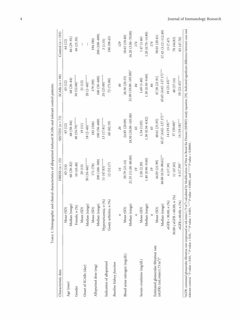
Table1:Dem
ograph
icandclinicalcharacteristicsof
allopu
rino
l-indu
cedSC
ARsandtolerant
controlp
atients.
Characteristicdata
DRESS
(n=23)
SJS/TEN
(n=73)
SCARs(n
=96)
Con
trol
(n=193)
Age
(year)
Mean(SD)
65(14)
65(12)
65(12)
64(12)
Median(range)
68(28–82)
68(38–84)
68(28–84)
66(29–91)
Gender
Female;n(%
)10
(43.48)
40(54.79)∗
∗∗∗∗
50(52.08)∗
∗∗∗∗
49(25.39)
Onsetof
SCARs(day)
Mean(SD)
29(13)
19(11)
21(13)
—
Median(range)
30(10–60)∗
∗∗∗∗
18(2–60)
∗∗∗∗
∗20
(2–60)
∗∗∗∗
∗—
Allopu
rino
ldose(m
g)Mean(SD)
171(78)
182(106)
179(99)
194(90)
Median(range)
200(100–300)
100(50–600)
100(50–600)
200(100–600)
Indication
ofallopu
rino
lHyperuricem
ia;n
(%)
11(47.83)∗
∗∗∗∗
13(17.81)∗
∗∗∗∗
24(25.00)∗
∗∗∗∗
3(1.55)
Gou
tyarthritis;n(%
)12
(52.17)
60(82.19)
72(75.00)
190(98.45)
Baselinekidn
eyfunction
Blood
urinenitrogen
(mg/dL
)
n14
2640
129
Mean(SD)
29.79(21.14)
24.85(20.09)
26.58(20.33)
18.63(10.40)
Median(range)
21.35(11.00–80.00)
18.50(10.00–105.00)
21.00(10.00–105.00)
∗16.20(3.00–70.00)
Serum
creatinine
(mg/dL
)
n19
6584
174
Mean(SD)
2.20
(2.20)
1.54
(1.05)
1.69
(1.40)
1.47
(1.46)
Median(range)
1.40
(0.90–9.60)
1.30
(0.50–8.82)
1.30
(0.50–9.60)
1.20
(0.70–14.80)
Estim
ated
glom
erular
filtrationrate
(eGFR
)(m
L/min/1.73m
2 )a
n19
6584
174
Mean(SD)
44.05(21.99)
48.61(21.95)
47.58(21.91)
58.01(20.41)
Median(range)
46.00(6.54–88.61)
∗∗45.27(4.62–127.17)∗
∗45.65(4.62–127.17)∗
∗∗57.45(3.12–112.49)
eGFR
<30.00;n(%
)4(17.39)
14(19.18)∗
∗18
(21.43)∗
∗13
(7.47)
30.00≤eG
FR<60.00;n(%
)11
(47.83)
37(50.68)∗
48(57.14)
78(44.83)
eGFR
≥60.00;n(%
)4(17.39)∗
14(19.18)∗
∗∗18
(21.43)∗
∗∗∗
83(47.70)
a eGFR
:estim
ated
glom
erular
filtrationrate(expressed
asmL/min/1.73m
2 )calculated
byMod
ification
ofDietinRenaltheDisease
(MDRD)stud
yequation
[39].Ind
icated
significant
difference
betweencase
and
tolerant
control.
∗Pvalue<0.05,∗
∗Pvalue<0.01,∗
∗∗Pvalue<0.001,
∗∗∗∗Pvalue<0.0001,and
∗∗∗∗
∗Pvalue<0.00001.
4 Journal of Immunology Research

Table2:Univariateanalysisof
theassociationbetweenHLA
-B∗58
:01alleleandSN
Pswithallopu
rino
l-indu
cedSC
ARs.
Allele/SNPs
Con
trol
(n=193)
DRESS
(n=23)
SJS/TEN
(n=73)
SCARs(n
=96)
n(%
)n(%
)OR
[95%
CI]
Pc-value
n(%
)OR
[95%
CI]
Pc-value
n(%
)OR
[95%
CI]
Pc-value
HLA
-B∗58
:01
Negative
170(88.08)
2(8.70)
Reference
4(5.48)
Reference
6(6.25)
Reference
Positive
23(11.92)
21(91.30)
77.61
[17.07–352.85]
7.12
×10
−8
69(94.52)
127.50
[42.53–382.27]
1.99
×10
−17
90(93.75)
110.87
[43.57–282.15]
2.05
×10
−22
rs9263726(G
/A)
Negative(G
G)
159(82.38)
4(17.39)
Reference
5(6.85)
Reference
9(9.38)
Reference
Positive(G
A/A
A)
34(17.62)
19(82.61)
22.21
[7.10–69.46]
3.91
×10
−7
68(93.15)
63.60
[23.85–169.56]
4.22
×10
−16
87(90.63)
45.21
[20.73–98.60]
3.92
×10
−21
rs2734583(T/C)
Negative(TT)
166(86.01)
2(8.70)
Reference
7(9.59)
Reference
9(9.37)
Reference
Positive(TC/CC)
27(13.99)
21(91.30)
64.56
[14.31–291.16]
2.35
×10
−7
66(90.41)
57.97
[24.07–139.60]
5.51
×10
−19
87(90.63)
59.43
[26.76–131.97]
4.24
×10
−23
rs3099844(C/A
)
Negative(CC)
164(84.97)
2(8.70)
Reference
7(9.59)
Reference
9(9.38)
Reference
Positive(CA/A
A)
29(15.03)
21(91.30)
59.38
[13.21–266.97]
4.04
×10
−7
66(90.41)
53.32
[22.26–127.71]
1.82
×10
−18
87(90.63)
54.67
[22.77–120.66]
1.58
×10
−22
SNPshaplotypes
(rs9263726-rs2734583-rs3099844)
GG-TT-C
C156(80.83)
2(8.70)
Reference
4(5.48)
Reference
6(6.25)
Reference
GA-TC-C
A24
(12.44)
16(69.57)
52.00
[11.24–240.51]
1.17
×10
−6
61(83.56)
99.13
[33.03–297.52]
9.90
×10
−16
77(80.21)
83.42
[32.74–212.55]
7.43
×10
−20
Pc-value:Bon
ferron
i-correctedPvalue,calculated
bymultiplying
Pvalueby
4which
isthenu
mberof
detected
SNPs.
5Journal of Immunology Research
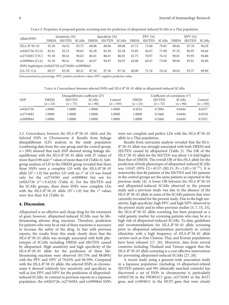
3.2. Concordance between the HLA-B∗58 : 01 Allele and theSelected SNPs in Chromosome 6. Results from linkagedisequilibrium (LD) analysis in the study population(combining data from the case group and the control group,n = 289) showed that each SNP showed strong linkage dis-equilibrium with the HLA-B∗58 : 01 allele with D’ values ofmore than 0.90 and r2 values of more than 0.8 (Table 4). Sub-group analysis of LD in the DRESS group revealed that thesethree SNPs were a complete LD with the HLA-B∗58 : 01allele (D’=1.0) but perfect LD with an r2 of 1.0 was foundonly for the rs2734583 and rs3099844 but not forrs9263726 (r2 = 0 4524) (Table 4). For the SJS/TEN andthe SCARs groups, these three SNPs were complete LDswith the HLA-B∗58 : 01 allele (D’=1.0) but the r2 valueswere less than 0.8 (Table 4).
4. Discussion
Allopurinol is an effective and cheap drug for the treatmentof gout; however, allopurinol-induced SCARs may be life-threatening adverse drug reactions. Therefore, predictionfor patients who may be at risk of these reactions is necessaryto increase the safety of the drug. In line with previousreports, the results from this study clearly show that theHLA-B∗58 : 01 allele was strongly associated with both phe-notypes of SCARs including DRESS and SJS/TEN causedby allopurinol. High sensitivity and high specificity of theHLA-B∗58 : 01 allele for the prediction of these life-threatening reactions were observed (93.75% and 88.08%)with the PPV and NPV of 79.65% and 96.59%. Comparedwith the HLA-B∗58 : 01 allele, the selected SNPs in chromo-some 6 showed relatively low sensitivity and specificity aswell as low PPV and NPV for the prediction of allopurinol-induced SCARs. In contrast to previous reports in a Japanesepopulation, the rs9263726, rs2734583, and rs3099844 SNPs
were not complete and perfect LDs with the HLA-B∗58 : 01allele in a Thai population.
Results from univariate analysis revealed that theHLA-B∗58 : 01 allele was strongly associated with both DRESS andSJS/TEN caused by allopurinol (Table 2). The OR of theHLA∗58 : 01 allele for the SJS/TEN was about 1.6-fold higherthan that of DRESS. The overall OR of thisHLA allele for theprediction of both phenotypes of allopurinol-induced SCARswas 110.87 (95% CI= 43.57–282.15, Pc= 2.05× 10⁻²²). It isnoteworthy that 86 patients of the SJS/TEN and 182 patientsin the control groups are the same patients as reported in theprevious study [4]. A lower OR between the HLA-B∗58 : 01and allopurinol-induced SCARs observed in the presentstudy and a previous study was due to the absence of theHLA-B∗58 : 01 allele in some of the SCARs patients that werecurrently recruited for the present study. Due to the high sen-sitivity, high specificity, high PPV, and high NPV observed inthe present study and in other previous studies [5, 7, 15, 25],the HLA-B∗58 : 01 allele screening has been proposed as avalid genetic marker for screening patients who may be at ahigh risk of allopurinol-induced SCARs. To date, guidelinesand recommendations for HLA-B∗58 : 01 allele screeningprior to allopurinol administration particularly in certainethnicities with a high frequency of HLA-B∗58 : 01 allelecarriers such as Han Chinese, Thai, and Korean populationshave been released [17, 26]. Moreover, data from severalcountries including Thailand and Taiwan suggest that theHLA-B∗58 : 01 allele screening is a cost-effective interventionfor preventing allopurinol-induced SCARs [27, 28].
A recent study using a genome-wide association studyin a Japanese population (including 14 allopurinol-relatedSJS/TEN patients and 991 ethnically matched controls) hasdiscovered a set of SNPs in chromosome 6, particularlyrs9263726 in the PSORS1C1 gene, rs2734583 in the BAT1gene, and rs3094011 in the HCP5 gene that were closely
Table 3: Properties of proposed genetic screening tests for prediction of allopurinol-induced SCARs in a Thai population.
Allele/SNPsSensitivity (%) Specificity (%) PPV (%) NPV (%)
DRESS SJS/TEN SCARs DRESS SJS/TEN SCARs DRESS SJS/TEN SCARs DRESS SJS/TEN SCARs
HLA-B∗58 : 01 91.30 94.52 93.75 88.08 88.08 88.08 47.73 75.00 79.65 98.84 97.70 96.59
rs9263726 (G/A) 82.61 93.15 90.63 82.38 82.38 82.38 35.85 66.67 71.90 97.55 96.95 94.64
rs2734583 (T/C) 91.30 90.41 90.63 86.01 86.01 86.01 43.75 70.97 76.32 98.81 95.95 94.86
rs3099844 (C/A) 91.30 90.41 90.63 84.97 84.97 84.97 42.00 69.47 75.00 98.80 95.91 94.80
SNPs haplotypes (rs9263726-rs2734583-rs3099844)
GA-TC-CA 69.57 83.56 80.21 87.56 87.56 87.56 40.00 71.76 76.24 96.02 93.37 89.89
Data presented as percentage. PPV: positive predictive value; NPV: negative predictive value.
Table 4: Concordance between selected SNPs and HLA-B∗58 : 01 allele in allopurinol-induced SCARs.
SNPDisequilibrium coefficient (D’) Coefficient of correlation (r2)
DRESS(n = 23)
SJS/TEN(n = 73)
SCARs(n = 96)
Control(n = 193)
DRESS(n = 23)
SJS/TEN(n = 73)
SCARs(n = 96)
Control(n = 193)
rs9263726 1.0000 1.0000 1.0000 1.0000 0.4524 0.7884 0.6444 0.6327
rs2734583 1.0000 1.0000 1.0000 1.0000 1.0000 0.5466 0.6444 0.8318
rs3099844 1.0000 1.0000 1.0000 1.0000 1.0000 0.5466 0.6444 0.7651
6 Journal of Immunology Research
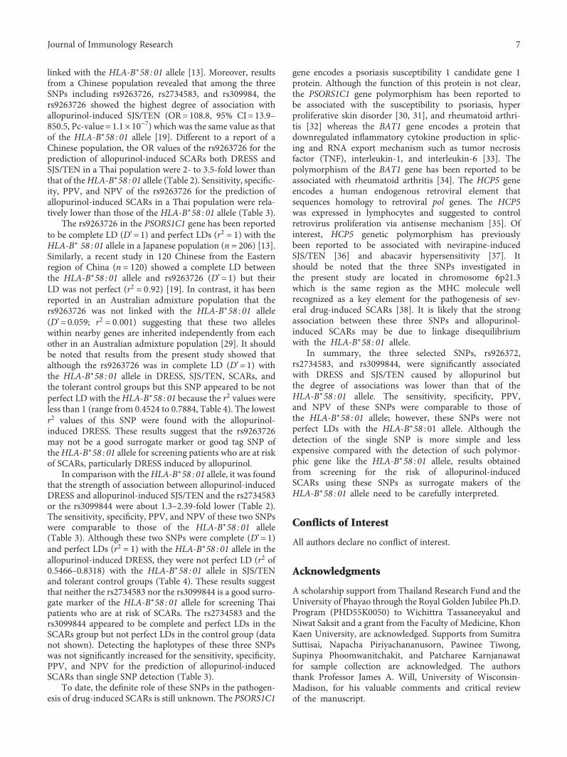
linked with the HLA-B∗58 : 01 allele [13]. Moreover, resultsfrom a Chinese population revealed that among the threeSNPs including rs9263726, rs2734583, and rs309984, thers9263726 showed the highest degree of association withallopurinol-induced SJS/TEN (OR=108.8, 95% CI=13.9–850.5, Pc-value = 1.1× 10⁻⁷) which was the same value as thatof the HLA-B∗58 : 01 allele [19]. Different to a report of aChinese population, the OR values of the rs9263726 for theprediction of allopurinol-induced SCARs both DRESS andSJS/TEN in a Thai population were 2- to 3.5-fold lower thanthat of theHLA-B∗58 : 01 allele (Table 2). Sensitivity, specific-ity, PPV, and NPV of the rs9263726 for the prediction ofallopurinol-induced SCARs in a Thai population were rela-tively lower than those of the HLA-B∗58 : 01 allele (Table 3).
The rs9263726 in the PSORS1C1 gene has been reportedto be complete LD (D’=1) and perfect LDs (r2 = 1) with theHLA-B∗ 58 : 01 allele in a Japanese population (n = 206) [13].Similarly, a recent study in 120 Chinese from the Easternregion of China (n = 120) showed a complete LD betweenthe HLA-B∗58 : 01 allele and rs9263726 (D’=1) but theirLD was not perfect (r2 = 0 92) [19]. In contrast, it has beenreported in an Australian admixture population that thers9263726 was not linked with the HLA-B∗58 : 01 allele(D’=0.059; r2 = 0 001) suggesting that these two alleleswithin nearby genes are inherited independently from eachother in an Australian admixture population [29]. It shouldbe noted that results from the present study showed thatalthough the rs9263726 was in complete LD (D’=1) withthe HLA-B∗58 : 01 allele in DRESS, SJS/TEN, SCARs, andthe tolerant control groups but this SNP appeared to be notperfect LD with theHLA-B∗58 : 01 because the r2 values wereless than 1 (range from 0.4524 to 0.7884, Table 4). The lowestr2 values of this SNP were found with the allopurinol-induced DRESS. These results suggest that the rs9263726may not be a good surrogate marker or good tag SNP oftheHLA-B∗58 : 01 allele for screening patients who are at riskof SCARs, particularly DRESS induced by allopurinol.
In comparison with theHLA-B∗58 : 01 allele, it was foundthat the strength of association between allopurinol-inducedDRESS and allopurinol-induced SJS/TEN and the rs2734583or the rs3099844 were about 1.3–2.39-fold lower (Table 2).The sensitivity, specificity, PPV, and NPV of these two SNPswere comparable to those of the HLA-B∗58 : 01 allele(Table 3). Although these two SNPs were complete (D’=1)and perfect LDs (r2 = 1) with the HLA-B∗58 : 01 allele in theallopurinol-induced DRESS, they were not perfect LD (r2 of0.5466–0.8318) with the HLA-B∗58 : 01 allele in SJS/TENand tolerant control groups (Table 4). These results suggestthat neither the rs2734583 nor the rs3099844 is a good surro-gate marker of the HLA-B∗58 : 01 allele for screening Thaipatients who are at risk of SCARs. The rs2734583 and thers3099844 appeared to be complete and perfect LDs in theSCARs group but not perfect LDs in the control group (datanot shown). Detecting the haplotypes of these three SNPswas not significantly increased for the sensitivity, specificity,PPV, and NPV for the prediction of allopurinol-inducedSCARs than single SNP detection (Table 3).
To date, the definite role of these SNPs in the pathogen-esis of drug-induced SCARs is still unknown. The PSORS1C1
gene encodes a psoriasis susceptibility 1 candidate gene 1protein. Although the function of this protein is not clear,the PSORS1C1 gene polymorphism has been reported tobe associated with the susceptibility to psoriasis, hyperproliferative skin disorder [30, 31], and rheumatoid arthri-tis [32] whereas the BAT1 gene encodes a protein thatdownregulated inflammatory cytokine production in splic-ing and RNA export mechanism such as tumor necrosisfactor (TNF), interleukin-1, and interleukin-6 [33]. Thepolymorphism of the BAT1 gene has been reported to beassociated with rheumatoid arthritis [34]. The HCP5 geneencodes a human endogenous retroviral element thatsequences homology to retroviral pol genes. The HCP5was expressed in lymphocytes and suggested to controlretrovirus proliferation via antisense mechanism [35]. Ofinterest, HCP5 genetic polymorphism has previouslybeen reported to be associated with nevirapine-inducedSJS/TEN [36] and abacavir hypersensitivity [37]. Itshould be noted that the three SNPs investigated inthe present study are located in chromosome 6p21.3which is the same region as the MHC molecule wellrecognized as a key element for the pathogenesis of sev-eral drug-induced SCARs [38]. It is likely that the strongassociation between these three SNPs and allopurinol-induced SCARs may be due to linkage disequilibriumwith the HLA-B∗58 : 01 allele.
In summary, the three selected SNPs, rs926372,rs2734583, and rs3099844, were significantly associatedwith DRESS and SJS/TEN caused by allopurinol butthe degree of associations was lower than that of theHLA-B∗58 : 01 allele. The sensitivity, specificity, PPV,and NPV of these SNPs were comparable to those ofthe HLA-B∗58 : 01 allele; however, these SNPs were notperfect LDs with the HLA-B∗58 : 01 allele. Although thedetection of the single SNP is more simple and lessexpensive compared with the detection of such polymor-phic gene like the HLA-B∗58 : 01 allele, results obtainedfrom screening for the risk of allopurinol-inducedSCARs using these SNPs as surrogate makers of theHLA-B∗58 : 01 allele need to be carefully interpreted.
Conflicts of Interest
All authors declare no conflict of interest.
Acknowledgments
A scholarship support from Thailand Research Fund and theUniversity of Phayao through the Royal Golden Jubilee Ph.D.Program (PHD55K0050) to Wichittra Tassaneeyakul andNiwat Saksit and a grant from the Faculty of Medicine, KhonKaen University, are acknowledged. Supports from SumitraSuttisai, Napacha Piriyachananusorn, Pawinee Tiwong,Supinya Phoomwanitchakit, and Patcharee Karnjanawatfor sample collection are acknowledged. The authorsthank Professor James A. Will, University of Wisconsin-Madison, for his valuable comments and critical reviewof the manuscript.
7Journal of Immunology Research
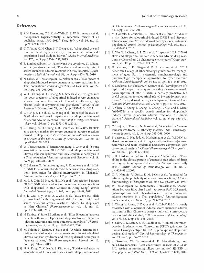
References
[1] S. N. Ramasamy, C. S. Korb-Wells, D. R. W. Kannangara et al.,“Allopurinol hypersensitivity: a systematic review of allpublished cases, 1950–2012,” Drug Safety, vol. 36, no. 10,pp. 953–980, 2013.
[2] C. Y. Yang, C. H. Chen, S. T. Deng et al., “Allopurinol use andrisk of fatal hypersensitivity reactions: a nationwidepopulation-based study in Taiwan,” JAMA Internal Medicine,vol. 175, no. 9, pp. 1550–1557, 2015.
[3] S. Limkobpaiboon, D. Panomvana Na Ayudhya, N. Dhana,and K. Jongjareanprasert, “Prevalence and mortality rate ofsevere cutaneous adverse reactions in Siriraj hospital,” Chula-longkorn Medical Journal, vol. 54, no. 5, pp. 467–478, 2010.
[4] N. Saksit, W. Tassaneeyakul, N. Nakkam et al., “Risk factors ofallopurinol-induced severe cutaneous adverse reactions in aThai population,” Pharmacogenetics and Genomics, vol. 27,no. 7, pp. 255–263, 2017.
[5] W. H. Chung, W. C. Chang, S. L. Stocker et al., “Insights intothe poor prognosis of allopurinol-induced severe cutaneousadverse reactions: the impact of renal insufficiency, highplasma levels of oxypurinol and granulysin,” Annals of theRheumatic Diseases, vol. 74, no. 12, pp. 2157–2164, 2015.
[6] C. Y. Ng, Y. T. Yeh, C. W. Wang et al., “Impact of the HLA-B∗
58:01 allele and renal impairment on allopurinol-inducedcutaneous adverse reactions,” Journal of Investigative Derma-tology, vol. 136, no. 7, pp. 1373–1381, 2016.
[7] S. I. Hung, W. H. Chung, L. B. Liou et al., “HLA-B∗5801 alleleas a genetic marker for severe cutaneous adverse reactionscaused by allopurinol,” Proceedings of the National Academyof Sciences of the United States of America, vol. 102, no. 11,pp. 4134–4139, 2005.
[8] W. Tassaneeyakul, T. Jantararoungtong, P. Chen et al., “Strongassociation between HLA-B∗5801 and allopurinol-inducedStevens-Johnson syndrome and toxic epidermal necrolysis ina Thai population,” Pharmacogenetics and Genomics, vol. 19,no. 9, pp. 704–709, 2009.
[9] C. Sukasem, T. Jantararoungtong, P. Kuntawong et al., “HLA-B∗58:01 for allopurinol-induced cutaneous adverse drug reac-tions: implication for clinical interpretation in Thailand,”Frontiers in Pharmacology, vol. 7, p. 186, 2016.
[10] M. L. S. Chiu, M. Hu, M. H. L. Ng et al., “Association betweenHLA-B∗58:01 allele and severe cutaneous adverse reactionswith allopurinol in Han Chinese in Hong Kong,” BritishJournal of Dermatology, vol. 167, no. 1, pp. 44–49, 2012.
[11] Z.-h. Cao, Z.-y. Wei, Q.-y. Zhu et al., “HLA-B∗58:01 alleleis associated with augmented risk for both mild andsevere cutaneous adverse reactions induced by allopurinolin Han Chinese,” Pharmacogenomics, vol. 13, no. 10,pp. 1193–1201, 2012.
[12] N. Kaniwa, Y. Saito, M. Aihara et al., “HLA-B locus in Japanesepatients with anti-epileptics and allopurinol-related Stevens–Johnson syndrome and toxic epidermal necrolysis,” Pharma-cogenomics, vol. 9, no. 11, pp. 1617–1622, 2008.
[13] M. Tohkin, N. Kaniwa, Y. Saito et al., “A whole-genome asso-ciation study of major determinants for allopurinol-relatedStevens–Johnson syndrome and toxic epidermal necrolysis inJapanese patients,” The Pharmacogenomics Journal, vol. 13,no. 1, pp. 60–69, 2013.
[14] H. R. Kang, Y. K. Jee, Y. S. Kim et al., “Positive and negativeassociations of HLA class I alleles with allopurinol-induced
SCARs in Koreans,” Pharmacogenetics and Genomics, vol. 21,no. 5, pp. 303–307, 2011.
[15] M. Goncalo, I. Coutinho, V. Teixeira et al., “HLA-B∗58:01 isa risk factor for allopurinol-induced DRESS and Stevens–Johnson syndrome/toxic epidermal necrolysis in a Portuguesepopulation,” British Journal of Dermatology, vol. 169, no. 3,pp. 660–665, 2013.
[16] R. Wu, Y. J. Cheng, L. L. Zhu et al., “Impact of HLA-B∗58:01allele and allopurinol-induced cutaneous adverse drug reac-tions: evidence from 21 pharmacogenetic studies,” Oncotarget,vol. 7, no. 49, pp. 81870–81879, 2016.
[17] D. Khanna, J. D. Fitzgerald, P. P. Khanna et al., “2012American College of Rheumatology guidelines for manage-ment of gout. Part 1: systematic nonpharmacologic andpharmacologic therapeutic approaches to hyperuricemia,”Arthritis Care & Research, vol. 64, no. 10, pp. 1431–1446, 2012.
[18] K. Maekawa, J. Nishikawa, N. Kaniwa et al., “Development of arapid and inexpensive assay for detecting a surrogate geneticpolymorphism of HLA-B∗58:01: a partially predictive butuseful biomarker for allopurinol-related Stevens-Johnson syn-drome/toxic epidermal necrolysis in Japanese,” Drug Metabo-lism and Pharmacokinetics, vol. 27, no. 4, pp. 447–450, 2012.
[19] Z. Chen, S. Zhang, J. Zhang, Y. Zhang, L. Xue, and L. Miao,“rs9263726 is a specific genetic marker for allopurinol-induced severe cutaneous adverse reactions in Chinesepatients,” Personalized Medicine, vol. 12, no. 6, pp. 585–592,2015.
[20] C. Lonjou, L. Thomas, N. Borot et al., “A marker for Stevens-Johnson syndrome ...: ethnicity matters,” The Pharmacoge-nomics Journal, vol. 6, no. 4, pp. 265–268, 2006.
[21] B. Sassolas, C. Haddad, M. Mockenhaupt et al., “ALDEN, analgorithm for assessment of drug causality in Stevens–Johnsonsyndrome and toxic epidermal necrolysis: comparison withcase–control analysis,” Clinical Pharmacology & Therapeutics,vol. 88, no. 1, pp. 60–68, 2010.
[22] S. H. Kardaun, A. Sidoroff, L. Valeyrie-Allanore et al., “Vari-ability in the clinical pattern of cutaneous side-effects of drugswith systemic symptoms: does a DRESS syndrome reallyexist?,” British Journal of Dermatology, vol. 156, no. 3,pp. 609–611, 2007.
[23] C. A. Naranjo, U. Busto, E. M. Sellers et al., “A method forestimating the probability of adverse drug reactions,” ClinicalPharmacology&Therapeutics, vol. 30, no. 2, pp. 239–245, 1981.
[24] W. Tassaneeyakul, N. Prabmeechai, C. Sukasem et al., “Associ-ations between HLA class I and cytochrome P450 2C9 geneticpolymorphisms and phenytoin-related severe cutaneousadverse reactions in a Thai population,” Pharmacogeneticsand Genomics, vol. 26, no. 5, pp. 225–234, 2016.
[25] L. Cheng, Y. Xiong, C. Z. Qin et al., “HLA-B∗58:01 is stronglyassociated with allopurinol-induced severe cutaneous adversereactions in Han Chinese patients: a multicentre retrospectivecase-control clinical study,” British Journal of Dermatology,vol. 173, no. 2, pp. 555–558, 2015.
[26] Y. Saito, L. K. Stamp, K. E. Caudle et al., “Clinical Pharmaco-genetics Implementation Consortium (CPIC) guidelines forhuman leukocyte antigen B (HLA-B) genotype and allopurinoldosing: 2015 update,” Clinical Pharmacology & Therapeutics,vol. 99, no. 1, pp. 36-37, 2015.
[27] S. Saokaew, W. Tassaneeyakul, R. Maenthaisong, andN. Chaiyakunapruk, “Cost-effectiveness analysis of HLA-B∗
5801 testing in preventing allopurinol-induced SJS/TEN inThai population,” PLoS One, vol. 9, no. 4, article e94294, 2014.
8 Journal of Immunology Research

[28] C. H. Ke, W. H. Chung, Y. H. Wen et al., “Cost-effectivenessanalysis for genotyping before allopurinol treatment to pre-vent severe cutaneous adverse drug reactions,” The Journal ofRheumatology, vol. 44, no. 6, pp. 835–843, 2017.
[29] C. Vidal, J. Li, R. Fulton, and S. L. Fernando, “A polymorphismwithin the psoriasis susceptibility 1 candidate 1 (PSORS1C1)gene is not linked to HLA-B∗58:01 in an Australian cohort,”Drug Metabolism and Pharmacokinetics, vol. 31, no. 3,pp. 252–255, 2016.
[30] S. J. Holm, L. M. Carlen, L. Mallbris, M. Stahle-Backdahl, andK. P. O'Brien, “Polymorphisms in the SEEK1 and SPR1 geneson 6p21.3 associate with psoriasis in the Swedish population,”Experimental Dermatologyyl, vol. 12, no. 4, pp. 435–444, 2003.
[31] P. Rahman, C. Butt, F. Siannis et al., “Association of SEEK1and psoriatic arthritis in two distinct Canadian populations,”Annals of the Rheumatic Diseases, vol. 64, no. 9, pp. 1370–1372, 2005.
[32] H. Sun, Y. Xia, L. Wang, Y. Wang, and X. Chang, “PSORS1C1may be involved in rheumatoid arthritis,” Immunology Letters,vol. 153, no. 1-2, pp. 9–14, 2013.
[33] S. Limou, S. Le Clerc, C. Coulonges et al., “Genomewide asso-ciation study of an AIDS-nonprogression cohort emphasizesthe role played by HLA genes (ANRS Genomewide Associa-tion Study 02),” The Journal of Infectious Diseases, vol. 199,no. 3, pp. 419–426, 2009.
[34] A. Quinones-Lombrana, A. Lopez-Soto, F. J. Ballina-Garciaet al., “BAT1 promoter polymorphism is associated with rheu-matoid arthritis susceptibility,” The Journal of Rheumatology,vol. 35, no. 5, pp. 741–744, 2008.
[35] J. Fellay, K. V. Shianna, D. Ge et al., “A whole-genome associ-ation study of major determinants for host control of HIV-1,”Science, vol. 317, no. 5840, pp. 944–947, 2007.
[36] P. Borgiani, D. Di Fusco, F. Erba et al., “HCP5 genetic variant(RS3099844) contributes to nevirapine-induced StevensJohnsons syndrome/toxic epidermal necrolysis susceptibilityin a population from Mozambique,” European Journal ofClinical Pharmacology, vol. 70, no. 3, pp. 275–278, 2014.
[37] S. Colombo, A. Rauch, M. Rotger et al., “The HCP5 single-nucleotide polymorphism: a simple screening tool for predic-tion of hypersensitivity reaction to abacavir,” The Journal ofInfectious Diseases, vol. 198, no. 6, pp. 864–867, 2008.
[38] W. H. Chung, C. W. Wang, and R. L. Dao, “Severe cutaneousadverse drug reactions,” The Journal of Dermatology, vol. 43,no. 7, pp. 758–766, 2016.
[39] A. S. Levey, J. P. Bosch, J. B. Lewis, T. Greene, N. Rogers, andD. Roth, “A more accurate method to estimate glomerularfiltration rate from serum creatinine: a new predictionequation. Modification of Diet in Renal Disease Study Group,”Annals of Internal Medicine, vol. 130, no. 6, pp. 461–470, 1999.
9Journal of Immunology Research

Review ArticleHLA Association with Drug-Induced Adverse Reactions
Wen-Lang Fan,1,2 Meng-Shin Shiao,3 Rosaline Chung-Yee Hui,2,4 Shih-Chi Su,1,2,4
Chuang-Wei Wang,2 Ya-Ching Chang,2 and Wen-Hung Chung1,2,4,5,6
1Whole-Genome Research Core Laboratory of Human Diseases, Chang Gung Memorial Hospital, Keelung, Taiwan2Department of Dermatology, Drug Hypersensitivity Clinical and Research Center, Chang Gung Memorial Hospital, Linkou,Taipei, Taiwan3Research Center, Faculty of Medicine Ramathibodi Hospital, Mahidol University, Bangkok, Thailand4Chang Gung Immunology Consortium, Chang Gung Memorial Hospital and Chang Gung University, Taoyuan, Taiwan5Department of Dermatology, Xiamen Chang Gung Hospital, Xiamen, China6College of Medicine, Chang Gung University, Taoyuan, Taiwan
Correspondence should be addressed to Wen-Hung Chung; [email protected]
Received 1 September 2017; Accepted 24 October 2017; Published 23 November 2017
Academic Editor: Mahboobeh Mahdavinia
Copyright © 2017 Wen-Lang Fan et al. This is an open access article distributed under the Creative Commons Attribution License,which permits unrestricted use, distribution, and reproduction in any medium, provided the original work is properly cited.
Adverse drug reactions (ADRs) remain a common and major problem in healthcare. Severe cutaneous adverse drug reactions(SCARs), such as Stevens–Johnson syndrome (SJS)/toxic epidermal necrolysis (TEN) with mortality rate ranges from 10% tomore than 30%, can be life threatening. A number of recent studies demonstrated that ADRs possess strong geneticpredisposition. ADRs induced by several drugs have been shown to have significant associations with specific alleles of humanleukocyte antigen (HLA) genes. For example, hypersensitivity to abacavir, a drug used for treating of human immunodeficiencyvirus (HIV) infection, has been proposed to be associated with allele 57:01 of HLA-B gene (terms HLA-B∗57:01). The incidencesof abacavir hypersensitivity are much higher in Caucasians compared to other populations due to various allele frequencies indifferent ethnic populations. The antithyroid drug- (ATDs- ) induced agranulocytosis are strongly associated with two alleles:HLA-B∗38:02 and HLA-DRB1∗08:03. In addition, HLA-B∗15:02 allele was reported to be related to carbamazepine-induced SJS/TEN, and HLA-B∗57:01 in abacavir hypersensitivity and flucloxacillin induced drug-induced liver injury (DILI). In this review,we summarized the alleles of HLA genes which have been proposed to have association with ADRs caused by different drugs.
1. Introduction
Major histocompatibility complex (MHC) are a group of cellsurface proteins that can bind to foreign molecules in orderto be recognized by corresponding T cells followed by induct-ing immune systems. MHC is highly conserved and presentsin all vertebrate species. In human, MHC is also known ashuman leukocyte antigen (HLA) complex, which consistsmore than 200 genes on chromosome 6 and can be catego-rized into three subgroups: class I, class II, and class III. ClassI MHC, being recognized by CD8+ T cells, consists of threemain genes, that is, HLA-A, HLA-B, and HLA-C. Class IIMHC, being recognized by CD4+ T cells, consists of 6 maingenes, that is, HLA-DPA1, HLA-DPB1, HLA-DQA1, HLA-DQB1, HLA-DRA, and HLA-DRB1.
HLA class I molecules are expressed in almost all the cellsand are responsible for presenting peptides to immune cells.Generally, old proteins in the cells will be broken down con-sistently in order to synthesize new peptides. Some of thesebroken peptide pieces attach to the MHC molecules and arefurther recognized by immune cells as “self.” In another situ-ation, if a cell is infected by pathogens, pathogenic peptidesattached to MHC molecules will be recognized as “nonself”and further trigger the downstream immune responsesagainst the antigens [1]. HLA genes are found to be numer-ous and highly polymorphic in order to bind various kindsof peptides originated from self or foreign antigens. A totalof more than 1500 alleles ofHLA-B gene have been identified[2]. Variations in the HLA genes play an important role indetermining the susceptibility to autoimmune disease and
HindawiJournal of Immunology ResearchVolume 2017, Article ID 3186328, 10 pageshttps://doi.org/10.1155/2017/3186328

infections; they are also critical in the field of transplantsurgery where the donors and the recipients must beHLA-compatible [3].
In rare cases, some drugs are capable of inducingimmune responses through interactions with MHC mole-cules, known as adverse drug reactions (ADRs). ADRs areone of the most common causes of hospitalization and mor-tality in healthcare. The definition of an ADR has been chan-ged from time to time. In the 1970s, the World HealthOrganization (WHO) has first defined that an ADR is “aresponse to a drug that is noxious and unintended and occursat doses normally used in man for the prophylaxis, diagnosisor therapy of disease, or for modification of physiologicalfunction” [4]. However, in most cases, the ADRs might notresult in effects as severe as harms or injuries like the word“noxious” addressed by WHO. Therefore, Edwards andAronson [5] suggested to use an alternative definition; thatis, an ADR is “an appreciably harmful or unpleasant reaction,resulting from an intervention related to the use of a medic-inal product, which predicts hazard from future administra-tion and warrants prevention or specific treatment, oralteration of the dosage regimen, or withdrawal of the prod-uct.” It is also defined as “an undesirable effect, reasonablyassociated with the use of the drug that may occur as a partof the pharmacological action of a drug or may be unpredict-able in its occurrence.”
2. ADRs Associated withImmunological Reactions
Several studies showed that ADRs are a major public healthproblem worldwide, which account for about 6.5% of all hos-pitalizations in the United States, Canada, and the UnitedKingdom, and it also resulted in a mortality rate approximateto 0.13% [6–8]. ADRs could be categorized into 6 differenttypes [5]. Among them, type B, also known as non-dose-related or bizarre, is unpredictable and results in a high mor-tality rate oftentimes. This type of ADRs is usually associatedwith immunological reactions involving different HLA allelesand resulted in skin injury, hepatic failure, or dramaticallyreduced numbers of white blood cells.
Skin injury includes various kinds of spectrum such asmild rash maculopapular exanthema (MPE), fixed drugeruption (FDE), acute generalized exanthematous pustulosis(AGEP), and life-threatening severe cutaneous adverse drugreactions (SCARs) including drug reactions with eosinophiliaand systemic symptoms (DRESS), Stevens–Johnson syn-drome (SJS), and toxic epidermal necrolysis (TEN) [9].Patients who developed MPE are usually observed with gen-eralized, widespread mild skin rashes with red macular (notelevated) or papular (elevated) eruptions. FDE can be diag-nosed by observing one or more local annular or oval ery-thematous patches without hyperpigmentation. The term“fixed” is considered as its recurrent lesion due to reexposureof the same culprit drug, and the lesions always occur at thesame locations on the skin. AGEP is a rare, acute eruptioncharacterized by the rapid development of many numerouspustules, which are nonfollicular sterile pustules, and is
located in the epidermis. Fever, leukocytosis, and eosino-philia are usually present in AGEP patients.
SJS, SJS, and TEN overlap (SJS/TEN) and TEN are classi-fied as the same disease spectrum with increasing severityand with extent of widespread epidermal detachment, knownas SCARs [10]. All of them usually present with a variety ofskin lesions, including patches, atypical targetoid macules,and erythematous or violaceous macules. In addition, SJS/TEN often has mucocutaneous involvement, which is thecharacteristic feature of SJS/TEN. In addition, the oralmucosa is more commonly involved than the ocular, genital,or anal mucosa. The degree of skin detachments of SJS, SJS/TEN, and TEN are defined as less than 10%, 10–30%, and30% of body surface area, respectively. Full-thickness epider-mal necrosis is a typical pathological feature of SJS/TEN. Theclinical characteristics of DRESS are different from SJS/TEN.DRESS usually presents less or no skin detachment and nomucocutaneous involvement but with more internal organinvolvement and hematological abnormalities such as typicaleosinophilia, atypical lymphocytes, hepatitis, and high feverwith frequent reactivation of human herpesvirus. Histopath-ological characters of DRESS are epidermal spongiosis, dys-keratosis, and interface vacuolization.
Other than skin injury, hepatic failure, such as drug-induced liver injury (DILI), is rare but life threatening.DILI is different from drug overdose toxicity in whichthe risk and severity of such kind of liver injury usuallyincreases with the dose taken. DILI accounts for 7–15%of the cases of acute liver failure in Europe and the UnitedStates [11–13]. Up to 10% of DILI can progress to acuteliver failure in the US and European studies, and the inci-dence is estimated to be 2.4 per 100,000 person-years (in aretrospective population-based study of 1.64 million UK sub-jects) [14] to 13.9 per 100,000 inhabitants (in a prospectiveanalysis in France) [15].
Agranulocytosis, also known as agranulosis or granulo-penia, is an acute condition involving a severe and dramaticdecreasing of white blood cell counts, which is life threaten-ing. It is recently reported to be induced by antithyroid drugsin rare situations and is associated with HLA alleles [16].
3. Hypothesis of Immune Response
Drugs or its reactive metabolites are considered as foreignantigens that bind to T cell receptors (TCR) and furtheractivate immune response. Four hypotheses have beenproposed to explain how the immune system is activatedin a HLA molecule-dependent manner: (i) the “hapten/prohapten” theory, (ii) the “p-i” concept, (iii) the “alteredpeptide repertoire” model, and (iv) the “altered TCR reper-toire” model [17, 18].
The “hapten/prohapten” theory proposes that a drugor its reactive metabolite may bind covalently to an endog-enous peptide to form an antigenic hapten-carrier com-plex. In this model, the covalent bonds are establishedamong the drug (or its metabolite), self-peptides, andHLA molecule. It then results in the induction of drug-specific immune responses.
2 Journal of Immunology Research

The “pharmacological interaction with immune recep-tors (p-i)” concept postulates that a drug or its reactivemetabolite may directly, reversibly, and noncovalently bindto the HLA and/or TCR without binding to the antigenicpeptide. In this “p-i” model, the classic antigen-processingpathway in antigen-presenting cells may be bypassed.
The “altered peptide repertoire” model proposes that adrug could strongly bind to the self-peptide repertoire andalter the conformation of this peptide repertoire presentedto HLA and TCR. In the “altered peptide repertoire” model,the drug may not directly bind to HLA.
Finally, the “altered TCR repertoire” model suggeststhat the drug (e.g., sulfamethoxazole) binds to the specificTCR and alters the conformation of TCR, which has thepotential to bind a HLA-self peptide complex to elicitimmune reaction. In the “altered TCR repertoire” model,the TCR serves as an initial drug interaction molecular.With the binding of an offending drug presented to theHLA molecule or TCR, the HLA-drug-TCR complexmay trigger a series of activations of cell signaling andresult in an expansion of cytotoxic T lymphocytes (CTL),cytotoxic protein secretions, and keratinocyte death inpatients with SJS/TEN. A recent study has shown theimportance of TCR in the pathogenic mechanism of SJS/TEN onset by clarifying the shared and restricted TCR usein carbamazepine-induced SJS/TEN patients [19]. Addition-ally, another interesting study demonstrated that the endog-enous peptide-bound HLA-B∗15:02 molecule presentscarbamazepine to TCR of CTL to initiate the immune reac-tions in carbamazepine-induced SJS/TEN [20].
4. Drugs and HLA Alleles
A couple of drugs have been proposed to induceHLA-associ-ated ADRs (Table 1). In this section, we summarized some ofthe well-known drugs and the HLA alleles associated withADRs induced by these drugs. For more detailed informa-tion, please see the list in Table 1.
4.1. Abacavir Hypersensitivity and HLA-B∗57:01 (Skin).Abacavir is a nucleotide reverse transcriptase inhibitorused as part of adjuvant therapy in human immunodefi-ciency virus- (HIV-) infected patients. In 5–8% of treatedpatients, abacavir can cause hypersensitivity responses. Morethan 90% of the patients with hypersensitive syndrome startwithin 6 weeks of treatment and require immediate cessationof the medication. Re-exposure of the abacavir leads to rapidappearance of symptoms and higher chance to induce moresevere symptoms [21]. Symptoms reported including fever,rash, malaise/fatigue, and gastrointestinal symptoms suchas nausea, vomiting, and diarrhea. Respiratory symptomsoccurred in 30% of cases including dyspnea, cough, andpharyngitis. In very rare cases, abacavir might result in moresevere reaction such as SJS/TEN [22, 23].
In 2002, two publications first proposed that abacavirhypersensitivity was significantly associated with the pres-ence of allele HLA-B∗57:01 in Australian and British cohorts[24, 25]. Saag et al. [26] further demonstrated that there is ahigher chance of developing hypersensitivity in Caucasians
than African-Americans who were treated with abacavir.Among those suffered from abacavir hypersensitivity, 44%of Caucasians and 100% of African-Americans showed posi-tive of HLA-B∗57:01 allele. Recently, HAL-B∗57:01 wasscreened in other populations (summarized in Martin et al.[27]). In general, the frequency of HLA-B∗57:01 allele ismuch higher in Caucasians than in other populations. InTaiwan, abacavir hypersensitivity is less frequent, as itoccurs in approximately 0.3% of HIV-infected patientswho undergo abacavir-containing combination antiretrovi-ral therapy (a total of 320 patients studied). The possiblereason might be the low frequency of the HLA-B∗5701allele in Taiwanese population [28].
The mechanisms of abacavir hypersensitivity is betterstudied compared to other drug-induced ADRs. It isthought that short peptide fragments, derived from eitherthe drug or its metabolites, form a peptide-HLA complexspecifically with HLA-B∗57:01. This complex activatesCD8+ T cells, which release inflammatory cytokines and startthe hypersensitivity response. More recently, it has beenshown that abacavir might occupy a space below the regionof HLA that presents peptides, which leads to an alteredpeptide presentation and trigger an autoimmune reaction[29]. By using X-ray crystallography and structural analy-sis, Yerly et al. further proposed that the hypersensitivityreaction is due to both types of T cells that recognizeself-peptide/HLA-B∗57:01 complexes and cross react withviral peptide/HLA-B∗57:01 complexes due to similarity indrug-specific T cell receptors contact residues [30].
As genetic screens for HLA-B∗57:01 could signifi-cantly reduce the incidence of abacavir hypersensitivityin Caucasians, the European Medicines Agency and US Foodand Drug Administration (FDA) recommend prospectivescreening for HLA-B∗57:01 for patients who are consideredto undergo abacavir treatment [27, 31].
4.2. Carbamazepine and Oxcarbazepine Hypersensitivity andHLA-B∗15:02, HLA-B∗15:11, and HLA-A∗31:01 (Skin). Car-bamazepine is an important drug used in the treatment ofepilepsy, trigeminal neuralgia, and bipolar disorder [32–34].In 2004, Carbamazepine was first reported to be stronglyassociated with allele HLA-B∗15:02 by studying patientsdeveloped SJS/TEN in Taiwan (OR> 1000) [35]. This associ-ation was validated in different populations, including thosein Thailand, Malaysia, Singapore, and India [36–38]. Alarge scale of prospective study including almost 5000 par-ticipants from 23 hospitals in Taiwan showed that 7.7% ofthe subjects are HLA-B∗15:02 positive. These subjects carry-ing HLA-B∗15:02 were then advised to take alternative drugsother than carbamazepine [39]. Consequently, taking thealternative drug greatly reduced the change of developingSCARs, especially SJS/TEN. Based on the findings from thesestudies, the genetic screening ofHLA-B∗15:02 prior to the useof carbamazepine for certain Asian populations are recom-mended by different health regulatory agencies [40].
HLA-B∗15:02 association and carbamazepine-inducedSJS/TEN is also proposed to be ethnic specific. It is likelydue to different genetic background as the allele frequencyvaries among different populations. It is relatively high in
3Journal of Immunology Research

Han Chinese (0.057–0.145), Malaysians (0.12–0.157), andThai (0.085–0.275) compared to Japanese (0.002), Koreans(0.004), and Europeans (0.01–0.02) [41–47].
In the populations with lower frequency ofHLA-B∗15:02,that is, Northern Europeans, Japanese, and Koreans, morerecent genome-wide association studies (GWAS) showedthat HLA-A∗31:01 allele has relatively stronger associationwith carbamazepine-induced hypersensitivity (OR=25.93,10.8, and 7.3 in the three populations, resp.) [41, 48–50]. Inaddition, HLA-B∗15:11 allele was shown to be associatedwith carbamazepine-induced SJS/TEN in Japanese andKorean populations as well (OR=9.8 and 18.1 in thetwo populations, resp.) [41, 47]. The different strength andspecificity of HLA association with carbamazepine-induced
SCARs further suggest that it is necessary to perform differ-ent genetic tests for different populations.
Oxcarbazepine is also an important drug used in thetreatment of epilepsy. Oxcarbazepine-induced cutaneousADRs presented with less clinical severity including lim-ited skin detachment (all ≦ 5%) and no mortality com-pared to carbamazepine. Therefore, it is commonly usedas an alternative to carbamazepine. A most recent studywhich enrolled 50 patients in Taiwan and Thailand from2006 to 2014 identified a significant association betweenHLA-B∗15:02 and SJS/TEN (OR=27.90; P = 1 87 × 10−10).The results of study suggested that although oxcarbazepineis used as an alternative due to the less severity of drugreactions, genetic test should also be considered further,
Table 1: Drug-induced ADRs and HLA allele associations.
Drug HLA allele Phenotype Population Reference
Abacavir B∗57:01 HSS
Australian [25]
African American [26]
Brazilian [79]
British [24]
Indian [80]
Iranian [81]
Carbamazepine B∗15:02 SJS/TEN
Taiwanese [35, 82]
Han Chinese [83]
Thai [37]
Malaysian [44]
Asian [46]
A∗31:01 MPE, HSS, SJS/TEN
Han Chinese [49, 82]
Caucasian [48]
Japanese [50]
B∗15:11 SJS/TEN Japanese [47, 84]
Allopurinol B∗58:01 SJS/TEN
Han Chinese [53]
Caucasian [57]
Thai [59]
Japanese [59]
SCARS Taiwanese [85]
Dapsone B∗13:01 HSS [60]
Phenytoin B∗15:02 SJS/TENHan Chinese [83]
Thai [37]
LamotrigineA∗31:01 HSS British [86]
B∗15:02 SJS/TEN Han Chinese [83]
Nevirapine DRB1∗01:01 DRESS Hispanics, African [87]
B∗14:02 HSS Sardinian [88, 89]
Sulphamethoxazole B∗38 SJS/TEN European [57]
Methazolamide B∗59:01, CW∗01:02 SJS/TEN Korean and Japanese [84]
Amoxicillin-clavulanate DRB1∗15:01-DQB1∗06:02 DILI Caucasian [64]
Flucloxacillin B∗57:01 DILI Caucasian [65]
Lumiracoxib DRB1∗15:01 DILI Not available [90]
Ticlopidine A∗33:03 DILI Japanese [91]
Antithyroid drugs B∗38:02-DRB1∗08:03 Agranulocytosis Taiwanese [16]
4 Journal of Immunology Research

particularly for the populations with higher frequency ofHLA-B∗15:02 [51].
4.3. Allopurinol Hypersensitivity and HLA-B∗58:01 (Skin).Allopurinol is a xanthine oxidase inhibitor used in the treat-ment of gout and hyperuricemia. A study comparing the datain 2005 and in 2011 from Taiwan’s National Health Insur-ance Research Database, which belongs to the nationwidepopulation database with more than 23 million insuredenrollees, demonstrated that allopurinol hypersensitivityhappened in about 0.4% of the new users every year. Abouthalf of them required hospitalization [52]. Patients whounderwent hospitalization had very high mortality rate(0.39/1000 new users). In 2005, the first case-control studyin Taiwan showed that HLA-B∗58:01 allele is the geneticmarker of allopurinol-induced SCARs in Han Chinese(OR=580.3, P = 4 7 × 10⁻²⁴) [53]. This association was thenvalidated in different populations, such as Thailand, Japan,South Korea, Hong Kong, Australia, Portugal, and Europe[54–59]. Currently, HLA-B∗58:01 is considered as a usefulgenetic marker for allopurinol-SCARs in multiple ethnicpopulations worldwide [31]. The American College of Rheu-matology guideline thus recommends the HLA-B∗58:01genetic screening for allopurinol new users in Asia popula-tions since 2012.
A most recent study in Taiwan further enrolled a largenumber of patients with allopurinol-induced ADRs in orderto investigate the associations between HLA-B∗58:01, renalfunction, gene dosage, and drug dosage with the risk ofallopurinol-induced ADRs development [58]. The authorsshowed that HLA-B∗58:01 was strongly associated withADRs (OR=44.0; P = 2 6 × 10−41) and was also highly cor-related with disease severity. That is, patients carryingHLA-B∗58:01 had much higher chance to develop SCARscomparing to MPE, particularly those individuals withhomozygous HLA-B∗58:01. Furthermore, coexistence ofHLA-B∗58:01 and renal impairment increased the risk andpredictive accuracy of allopurinol-induced ADRs. This studysuggests that patients with the coexistence of HLA-B∗58:01and renal impairment should be cautious and avoid touse allopurinol.
4.4. Dapsone Hypersensitivity and HLA-B∗13:01 (Skin).Dapsone alone or in-combination with other drugs areeffective for the treatment or prevention of infectious diseases(e.g. leprosy, malaria and pneumocystis pneumonia).However, about 0.5–3.6% of persons who were treated withdapsone developed hypersensitivity reactions. A recentgenome-wide association study involving 872 participants(39 participants showed dapsone hypersensitivity syn-drome and 833 controls) identified that SNP rs2844573,located between the HLA-B and MICA loci, was signifi-cantly associated with the dapsone hypersensitivity(OR=6.18; P = 3 84 × 10−13) [60]. The authors further con-firmed that HLA-B∗13:01 is associated with the dapsonehypersensitivity (OR=20.53; P = 6 84 × 10−25). The alleleshowed a sensitivity of 85.5% and a specificity of 85.7%for dapsone hypersensitivity from this study and thuscan be used as a marker of dapsone hypersensitivity.
However, the hypersensitivity has not been studied in otherethnic populations.
4.5. Amoxicillin-Clavulanate-Induced DILI and HLAHaplotypes. Amoxicillin-clavulanate (AC) is one of the mostcommonly prescribed antimicrobial drugs worldwide.However, it is a known cause of DILI and accounts for10–13% of hospitalizations. Hautekeete et al. first reporteda strong association between HLA and AC-induced DILIin Europeans [61]. The authors observed a much higherfrequency of DRB1∗15:01-DRB5∗01:01-DQB1∗06:02 haplo-type in patients with AC-induced DILI compared to normalhealthy controls (57.1% in cases versus 11.7% in controls,P < 10−6). The association was further validated in two UKpopulations (OR=2.3 and 9.3 for the two populations, resp.)[62, 63]. A recent study by performing GWAS in 201 patientsfurther confirmed the association of AC-induced DILI withDRB1∗15:01 allele (OR=4.2; P = 4 6 × 10⁻¹⁰) [64]. In addi-tion, the study further identified two novel HLA alleles as riskfactors of AC-induced DILI: HLA-A∗02:01 in all patients(OR=2.2; P = 1 8 × 10⁻¹⁰) and HLA-B∗18:01 with nominalsignificance independently of HLA-A∗02:01 and HLA-DQB1∗06:02 in Spanish patients only.
4.6. Flucloxacillin-Induced DILI and HLA-B∗57:01Association. Flucloxacillin is an antibiotic belonging to peni-cillin class and is used widely for the treatment for staphylo-coccal infection in Europe. Flucloxacillin is a common causeof DILI and is also reported to be associated with cholestaticliver disease. A GWAS study enrolling 51 patients showed astrong association between flucloxacillin-induced DILI anda marker, rs2395029[G]. This marker is in complete linkagedisequilibrium with HLA-B∗5701 (P = 8 7 × 10⁻³³) [65].The authors further performed MHC genotyping and con-firmed the association of flucloxacillin induced DILI withHLA-B∗5701 (OR=80.6, P = 9 0 × 10−19). This is an interest-ing finding because HLA-B∗57:01 is also associated with aba-cavir hypersensitivity, but these patients were not reported todevelop liver injury. It still remains unclear whether itresulted from the binding of different drugs/metabolites tothe same HLA allele and subsequent initiation of immuneresponses or it is merely a coincidental event. As the positivepredictive value of HLA-B∗57:01 is as low as 0.12% [31], thegenetic screening for HLA-B∗57:01 before the prescription offlucloxacillin to new users may not be clinically relevant.
4.7. Antithyroid Drug-Induced Agranulocytosis and HLA-B∗
38:02-HLA-DRB1∗08:03 Haplotype.Other than skin and liverinjures, drug reactions could also affect the immune systemdirectly. Antithyroid drugs (ATDs) have been the corner-stones treatment of Graves’ disease (GD), which is the lead-ing cause of hyperthyroidism. It has been reported thatATDs may induce agranulocytosis resulting in lower numberof white blood cells and is likely to be life threatening. How-ever, the genetic risk factors have not been identified untilrecently. Chen et al. conducted both classic genotyping andGWAS to elucidate the genetic association between ATD-induced agranulocytosis and HLA genes in Taiwan [16]. Firstof all, they performed direct HLA genotyping including 6
5Journal of Immunology Research
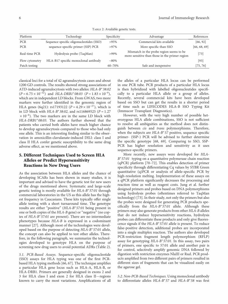
classical loci for a total of 42 agranulocytosis cases and about1200 GD controls. The results showed strong associations ofATD-induced agranulocytosis with two alleles:HLA-B∗38:02(P = 6 75 × 10−32) and HLA-DRB1∗08:03 (P = 1 83 × 10−9),which are in independent LD blocks. From GWAS, two moremarkers were further identified in the genomic region ofHLA genes (6q21): rs17193122 (P = 4 29 × 10−27), which isin LD block with HLA-B∗38:02, and rs116869525 (P = 1 27× 10−8). The two markers are in the same LD block withHLA-DRB1∗08:03. The authors further showed that thepatients who carried both alleles have much higher chanceto develop agranulocytosis compared to those who had onlyone allele. This is an interesting finding similar to the obser-vation in amoxicillin-clavulanate-induced DILI: class I andclass II HLA confer genetic susceptibility to the same drugadverse effect, as we mentioned above.
5. Different Techniques Used to Screen HLAAlleles or Predict HypersensitivityReactions in New Drug Users
As the association between HLA alleles and the chance ofdeveloping SCARs has been shown in many studies, it isimportant and advised to have the genetic test for new usersof the drugs mentioned above. Systematic and large-scalegenetic testing is mostly available for HLA-B∗57:01 throughcommercial laboratories in the US as this allele has the high-est frequency in Caucasians. These kits typically offer singleallele testing with a short turnaround time. The genotyperesults are either “positive” (HLA-B∗57:01 being present inone or both copies of theHLA-B gene) or “negative” (no cop-ies of HLA-B∗57:01 are present). There are no intermediatephenotypes because HLA-B is expressed in a codominantmanner [27]. Although most of the technologies were devel-oped based on the purpose of detecting HLA-B∗57:01 allele,the concept can also be applied to test other alleles. There-fore, in the following paragraph, we summarize the technol-ogies developed to genotype HLA on the purpose ofscreening new drug users to avoid potential ADRs (Table 2).
5.1. PCR-Based Assays. Sequence-specific oligonucleotide(SSO) assays for HLA typing was one of the first PCR-based HLA typing methods [66, 67]. The technique amplifiesa particular HLA gene locus such as HLA-A, HLA-B, orHLA-DRB1. Primers are generally designed in exons 2 and3 for HLA class I and exon 2 for HLA class II—regionsknown to carry the most variations. Amplifications of all
the alleles of a particular HLA locus can be performedin one PCR tube. PCR products of a particular HLA locusis then hybridized with labelled oligonucleutides specifi-cally to a particular HLA allele or a group of alleles.Recently, several commercial kits have been developedbased on SSO but can get the results in a shorter periodof time such as LIFECODES HLA-B SSO Typing Kit(Immucor Transplant Diagnostics).
However, with the very high number of possible het-erozygous HLA allele combinations, SSO is not sufficientto resolve all ambiguities as the method does not distin-guish between cis and trans polymorphisms. Therefore,when the subjects are HLA-B∗57 positive, sequence specificprimer- (SSP-) PCR will be advised to further determinethe specific genotype [68, 69]. Comparing to SSO, SSP-PCR has higher resolution and sensitivity as it usessequence-specific primers.
More recently, new assays were developed for HLA-B∗57:01 typing on a quantitative polymerase chain reaction(qPCR) platform [70–72]. This enables detection of primerspecificity through differentiating Cq values by SYBR Greenquantitative (q)PCR or analysis of allele-specific PCR byhigh-resolution melting. Implementation of these assays ona qPCR platform significantly decreases the processing andreaction time as well as reagent costs. Jung et al. furtherdesigned primers and probes based on DNA polymorphismsusing hydrolysis probes (oftentimes referred to TaqMantechnology) [73]. In their study, not only the primers but alsothe probes were designed for generating PCR products spe-cifically from the HLA-B∗57:01 allele. Although theseprimers may also generate products from otherHLA-B allelesthat do not induce hypersensitivity reactions, hydrolysisprobes can differentiate these products and only give fluores-cence signals if the HLA-B∗57:01 allele is present. To reducefalse-positive detection, additional probes are incorporatedinto a single multiplex reaction. The authors also developedPCR-restriction fragment length polymorphism (RFLP)assay for genotyping HLA-B∗57:01. In this assay, two pairsof primers, one specific to 57:01 allele and another pair isfor control, selectively amplify genomic DNA followed bydigestion with restriction enzymes NlaIII or RsaI. PCR prod-ucts amplified from two different pairs of primers resulted indifferent sizes of fragments that can be visualized easily onthe agarose gel.
5.2. Non-PCR-Based Techniques.Using monoclonal antibodyto differentiate alleles HLA-B∗57 and HLA-B∗58 was first
Table 2: Available genetic tests.
Platform Technology Specificity Advantage References
PCR Sequence specific oligonucleotides (SSO) >95% Commercial kits available [66, 92]
PCR sequence-specific primer (SSP) PCR >97% More specific than SSO [66, 68, 69]
Real-time PCR Hydrolysis probe (TaqMan) >99% Mismatch in the probe region seems to bemore sensitive than those in the primer region
[73]
Flow cytometry HLA-B17 specific monoclonal antibody ~80% [93]
Patch testing 60–70% Safe and inexpensive [75, 76]
6 Journal of Immunology Research
proposed by Kostenko et al. [74]. MonoclonalHLA-B17 anti-bodies (mAb 3E12), which recognized both HLA-B∗57 andHLA-B∗58 allotypes (members of the group specificity,HLA-B17), was labelled with phycoerythrin while anti-CD45 labelled with FITC were incubated with blood fromsubjects. Lymphocytes were gated based upon scatter andCD45 bright expression, and mean fluorescence intensityfor HLA-B17 expression was then measured by flowcytometry. Although this is an inexpensive and rapidapproach to detect the presence of two allotypes, it is pro-posed to be less specific and sensitive than PCR-basedapproaches. The subjects who test positive by mAb screen-ing are recommended to proceed with high-resolutiongold-standard typing, such as SSO and SSP-PCR, to ascer-tain the presence of HLA-B∗5701 or HLA-B∗5801.
Patch testing is also used to predict hypersensitivityreaction of abacavir [75] and carbamazepine [76]. Giorginiand others performed patch testing on 100 subjectsincluding 20 cases who had experienced a hypersensitivityreaction when treated with highly active antiretroviraltherapy including abacavir. Among the cases with positivepatch testing results, about 50% of them carry HLA-B∗57:01allele. More recently, Lin et al. proposed to use patch testingto predict carbamazepine induced hypersensitivity. Theyshowed that about 60–70% of the cases who developedSJS/TEN and DRESS to carbamazepine had positive reac-tions in the patch testing. Although drug patch testing isa safe and inexpensive method for the identification ofhypersensitivity, the sensitivity and specificity is not as goodas PCR-based approaches.
6. Conclusion
In this review, we summarize the HLA alleles associated withADRs induced by different drugs. From the literature, welearned that most of the HLA-associated ADRs have ethnicspecificity. It is likely due to the different allele frequencybetween populations. Gonzalez-Galarza et al. summarizethe allele frequencies of all the HLA genes and showed thatthe frequencies differ a lot [77]. For example, HLA-B∗57:01has the highest frequency in Ireland, but has the lowest fre-quency in Cuba (African populations). The frequencies canof each allele in different populations be found in the publicdatabase (http://www.allelefrequencies.net/default.asp) [78].
Other than the HLA-associated ADRs being ethnicspecific, there are also two interesting questions: (1) whyone locus contributes to different ADRs as we haveobserved on HLA-B∗57:01 in abacavir hypersensitivity andflucloxacillin-induced DILI. (2) How different loci contrib-ute to the same ADRs as we have seen in antithyroiddrug-induced agranulocytosis and amoxicillin-clavulanate-induced DILI. As class I and class II HLA genes have dif-ferent structures, cell-type distributions, and functionalroles in the immune system, the genetic susceptibility fromboth classes for a phenotype are expected to be intriguing.How both class I and class II HLA genes confer geneticsusceptibility to the same ADR requires further patho-physiological investigations.
Abbreviations
ADR: Adverse drug reactionsHLA: Human leukocyte antigenDRESS: Drug reaction with eosinophilia and systemic
symptomsSCAR: Severe cutaneous adverse drug reactionsSJS: Stevens–Johnson syndromeTEN: Toxic epidermal necrolysis.
Conflicts of Interest
The authors declare no conflict of interest.
Authors’ Contributions
Wen-Lang Fan, Meng-Shin Shiao, Rosaline Chung-YeeHui, Shih-Chi Su, Chuang-Wei Wang, and Ya-ChingChang contributed to the conception and writing of themanuscript. Wen-Hung Chung reviewed the manuscript.Wen-Lang Fan and Meng-Shin Shiao contributed equallyto this work.
Acknowledgments
This work was supported by grants from the NationalScience Council, Taiwan (MOST101-2628-B-182-001-MY3,MOST103-2321-B-182-001, MOST103-2325-B-182A-004,MOST104-2325-B-182A-006, MOST105-2325-B-182A-007,and MOST104-2314-B-182A-148-MY3), and grants fromChang Gung Memorial Hospital (CLRPG2E0051-3,CMRPG290051-3, CMRPG3D0351-3, CMRPG-3D0361-3,CMRPG1F0111, CORPG3F0041-2, OMRPG2C0021, andOMRPG3E0041).
References
[1] P. Cresswell, A. L. Ackerman, A. Giodini, D. R. Peaper, andP. A. Wearsch, “Mechanisms of MHC class I-restricted anti-gen processing and cross-presentation,” ImmunologicalReviews, vol. 207, no. 1, pp. 145–157, 2005.
[2] T. Shiina, K. Hosomichi, H. Inoko, and J. K. Kulski, “TheHLA genomic loci map: expression, interaction, diversityand disease,” Journal of Human Genetics, vol. 54, no. 1,pp. 15–39, 2009.
[3] R. A. Bray, C. K. Hurley, N. R. Kamani et al., “National marrowdonor program HLA matching guidelines for unrelated adultdonor hematopoietic cell transplants,” Biology of Blood andMarrow Transplantation, vol. 14, no. 9, pp. 45–53, 2008.
[4] International drug monitoring, “The role of national centres.Report of a WHOmeeting,”World Health Organization Tech-nical Report Series, vol. 498, pp. 1–25, 1972.
[5] I. R. Edwards and J. K. Aronson, “Adverse drug reactions: def-initions, diagnosis, and management,” The Lancet, vol. 356,no. 9237, pp. 1255–1259, 2000.
[6] J. Lazarou, B. H. Pomeranz, and P. N. Corey, “Incidence ofadverse drug reactions in hospitalized patients: a meta-analysis of prospective studies,” JAMA, vol. 279, no. 15,pp. 1200–1205, 1998.
7Journal of Immunology Research
[7] M. Kvasz, I. E. Allen, M. J. Gordon et al., “Adverse drug reac-tions in hospitalized patients: a critique of a meta-analysis,”Medscape General Medicine, vol. 2, no. 2, article E3, 2000.
[8] A. Miguel, L. F. Azevedo, M. Araujo, and A. C. Pereira, “Fre-quency of adverse drug reactions in hospitalized patients: asystematic review and meta-analysis,” Pharmacoepidemiologyand Drug Safety, vol. 21, no. 11, pp. 1139–1154, 2012.
[9] F. Falasca, C. Dello Russo, B. Mora et al., “Comparative analy-sis of real-time polymerase chain reaction methods to typingHLA-b∗57:01 in HIV-1-positive patients,” AIDS Research andHuman Retroviruses, vol. 32, no. 7, pp. 654–657, 2016.
[10] W. H. Chung, C. W. Wang, and R. L. Dao, “Severe cutaneousadverse drug reactions,” The Journal of Dermatology, vol. 43,no. 7, pp. 758–766, 2016.
[11] G. Ostapowicz, R. J. Fontana, F. V. Schiodt et al., “Results of aprospective study of acute liver failure at 17 tertiary care cen-ters in the united states,” Annals of Internal Medicine,vol. 137, no. 12, pp. 947–954, 2002.
[12] M. W. Russo, J. A. Galanko, R. Shrestha, M. W. Fried, andP. Watkins, “Liver transplantation for acute liver failure fromdrug induced liver injury in the united states,” Liver Trans-plantation, vol. 10, no. 8, pp. 1018–1023, 2004.
[13] E. Bjornsson, P. Jerlstad, A. Bergqvist, and R. Olsson, “Fulmi-nant drug-induced hepatic failure leading to death or livertransplantation in Sweden,” Scandinavian Journal of Gastroen-terology, vol. 40, no. 9, pp. 1095–1101, 2005.
[14] F. J. de Abajo, D. Montero, M. Madurga, and L. A. GarciaRodriguez, “Acute and clinically relevant drug-induced liverinjury: a population based case-control study,” British Journalof Clinical Pharmacology, vol. 58, no. 1, pp. 71–80, 2004.
[15] C. Sgro, F. Clinard, K. Ouazir et al., “Incidence of drug-induced hepatic injuries: a French population-based study,”Hepatology, vol. 36, no. 2, pp. 451–455, 2002.
[16] P. L. Chen, S. R. Shih, P. W. Wang et al., “Genetic determi-nants of antithyroid drug-induced agranulocytosis by humanleukocyte antigen genotyping and genome-wide associationstudy,” Nature Communications, vol. 6, p. 7633, 2015.
[17] W. J. Pichler, “Pharmacological interaction of drugs withantigen-specific immune receptors: the p-i concept,” CurrentOpinion in Allergy and Clinical Immunology, vol. 2, no. 4,pp. 301–305, 2002.
[18] S. Watkins and W. J. Pichler, “Sulfamethoxazole induces aswitch mechanism in T cell receptors containing TCRVΒ20-1, altering PHLA recognition,” PLoS One, vol. 8, no. 10,article e76211, 2013.
[19] T. M. Ko, W. H. Chung, C. Y. Wei et al., “Shared and restrictedT-cell receptor use is crucial for carbamazepine-inducedStevens-Johnson syndrome,” The Journal of Allergy and Clini-cal Immunology, vol. 128, no. 6, pp. 1266–1276.e11, 2011.
[20] C. Y. Wei, W. H. Chung, H. W. Huang, Y. T. Chen, and S. I.Hung, “Direct interaction between HLA-B and carbamazepineactivates T cells in patients with Stevens-Johnson syndrome,”The Journal of Allergy and Clinical Immunology, vol. 129,no. 6, pp. 1562–1569.e5, 2012.
[21] S. Hetherington, S. McGuirk, G. Powell et al., “Hypersensi-tivity reactions during therapy with the nucleoside reversetranscriptase inhibitor abacavir,” Clinical Therapeutics,vol. 23, no. 10, pp. 1603–1614, 2001.
[22] P. Bossi, J. C. Roujeau, F. Bricaire, and E. Caumes, “Stevens-Johnson syndrome associated with abacavir therapy,” ClinicalInfectious Diseases, vol. 35, no. 7, p. 902, 2002.
[23] R. Pahk, M. C. Azu, B. R. Taira, and S. Sandoval, “Antiretrovi-ral-induced toxic epidermal necrolysis in a patient positive forhuman immunodeficiency virus,” Clinical and ExperimentalDermatology, vol. 34, no. 8, pp. e775–e777, 2009.
[24] S. Hetherington, A. R. Hughes, M. Mosteller et al., “Geneticvariations in HLA-B region and hypersensitivity reactions toabacavir,” The Lancet, vol. 359, no. 9312, pp. 1121-1122, 2002.
[25] S. Mallal, D. Nolan, C. Witt et al., “Association between pres-ence ofHLA-B∗5701,HLA-DR7, andHLA-DQ3 and hypersen-sitivity to HIV-1 reverse-transcriptase inhibitor abacavir,” TheLancet, vol. 359, no. 9308, pp. 727–732, 2002.
[26] M. Saag, R. Balu, E. Phillips et al.A. Hughes, D. Sutherland‐Phillips, S. Mallal et al., “High sensitivity of human leuko-cyte antigen-B∗5701 as a marker for immunologically con-firmed abacavir hypersensitivity in white and black patients,”Clinical Infectious Diseases, vol. 46, no. 7, pp. 1111–1118,2008.
[27] M. A. Martin, T. E. Klein, B. J. Dong et al., “Clinical pharma-cogenetics implementation consortium guidelines for HLA-Bgenotype and abacavir dosing,” Clinical Pharmacology & Ther-apeutics, vol. 91, pp. 734–738, 2012.
[28] H. Y. Sun, C. C. Hung, P. H. Lin et al., “Incidence of abacavirhypersensitivity and its relationship with HLA-B∗5701 inHIV-infected patients in Taiwan,” Journal of AntimicrobialChemotherapy, vol. 60, no. 3, pp. 599–604, 2007.
[29] M. Pirmohamed, D. A. Ostrov, and B. K. Park, “New geneticfindings lead the way to a better understanding of fundamentalmechanisms of drug hypersensitivity,” The Journal of Allergyand Clinical Immunology, vol. 136, no. 2, pp. 236–244, 2015.
[30] D. Yerly, Y. Pompeu, R. Schutte et al., “Structural elements rec-ognized by abacavir-induced T cells,” International Journal ofMolecular Sciences, vol. 18, no. 7, p. 1464, 2017.
[31] V. L. M. Yip, A. Alfirevic, and M. Pirmohamed, “Genetics ofimmune-mediated adverse drug reactions: a comprehensiveand clinical review,” Clinical Reviews in Allergy & Immunol-ogy, vol. 48, no. 2-3, pp. 165–175, 2015.
[32] A. G. Marson, A. M. Al-Kharusi, M. Alwaidh et al., “TheSANAD study of effectiveness of carbamazepine, gabapentin,lamotrigine, oxcarbazepine, or topiramate for treatment ofpartial epilepsy: an unblinded randomised controlled trial,”The Lancet, vol. 369, no. 9566, pp. 1000–1015, 2007.
[33] R. M. Post, T. A. Ketter, T. Uhde, and J. C. Ballenger, “Thirtyyears of clinical experience with carbamazepine in the treat-ment of bipolar illness: principles and practice,” CNS Drugs,vol. 21, no. 1, pp. 47–71, 2007.
[34] G. Cruccu, G. Gronseth, J. Alksne et al., “AAN-EFNS guide-lines on trigeminal neuralgia management,” European Journalof Neurology, vol. 15, no. 10, pp. 1013–1028, 2008.
[35] W. H. Chung, S. I. Hung, H. S. Hong et al., “Medical genetics: amarker for Stevens-Johnson syndrome,” Nature, vol. 428,no. 6982, p. 486, 2004.
[36] W. Tangamornsuksan, N. Chaiyakunapruk, R. Somkrua,M. Lohitnavy, and W. Tassaneeyakul, “Relationship betweenthe HLA-B∗1502 allele and carbamazepine-induced Stevens-Johnson syndrome and toxic epidermal necrolysis: a system-atic review and meta-analysis,” JAMA Dermatology, vol. 149,no. 9, pp. 1025–1032, 2013.
[37] C. Locharernkul, J. Loplumlert, C. Limotai et al., “Carbamaze-pine and phenytoin induced Stevens-Johnson syndrome isassociated with HLA-B∗1502 allele in Thai population,”Epilepsia, vol. 49, pp. 2087–2091, 2008.
8 Journal of Immunology Research

[38] T. Y. Mehta, L. M. Prajapati, B. Mittal et al., “Association ofHLA-B∗1502 allele and carbamazepine-induced Stevens-Johnson syndrome among Indians,” Indian Journal of Derma-tology, Venereology and Leprology, vol. 75, no. 6, pp. 579–582,2009.
[39] P. Chen, J. J. Lin, C. S. Lu et al., “Carbamazepine-induced toxiceffects and HLA-B∗1502 screening in Taiwan,” The NewEngland Journal of Medicine, vol. 364, no. 12, pp. 1126–1133,2011.
[40] P. B. Ferrell Jr. and H. L. McLeod, “Carbamazepine, HLA-B∗
1502 and risk of Stevens-Johnson syndrome and toxic epider-mal necrolysis: US FDA recommendations,” Pharmacoge-nomics, vol. 9, no. 10, pp. 1543–1546, 2008.
[41] S. H. Kim, K. W. Lee, W. J. Song et al.K. U. Min, Y. S. Chang,Adverse Drug Reaction Research Group in Korea et al., “Car-bamazepine-induced severe cutaneous adverse reactions andHLA genotypes in Koreans,” Epilepsy Research, vol. 97,no. 1-2, pp. 190–197, 2011.
[42] Y. Kano, K. Hirahara, Y. Asano, and T. Shiohara, “HLA-Ballele associations with certain drugs are not confirmed in Jap-anese patients with severe cutaneous drug reactions,” ActaDermato-Venereologica, vol. 88, no. 6, pp. 616–618, 2008.
[43] R. Aggarwal, M. Sharma, M. Modi, V. K. Garg, andM. Salaria, “HLA-B∗ 1502 is associated with carbamazepineinduced Stevens-Johnson syndrome in North Indian popu-lation,” Human Immunology, vol. 75, no. 11, pp. 1120–1122, 2014.
[44] C. C. Chang, C. L. Too, S. Murad, and S. H. Hussein, “Associ-ation of HLA-B∗1502 allele with carbamazepine-induced toxicepidermal necrolysis and Stevens-Johnson syndrome in themulti-ethnic Malaysian population,” International Journal ofDermatology, vol. 50, no. 2, pp. 221–224, 2011.
[45] X. J. He, L. Y. Jian, X. L. He et al., “Association between theHLA-B∗15:02 allele and carbamazepine-induced Stevens-Johnson syndrome/toxic epidermal necrolysis in HAN indi-viduals of Northeastern China,” Pharmacological Reports,vol. 65, no. 5, pp. 1256–1262, 2013.
[46] A. Alfirevic, A. L. Jorgensen, P. R. Williamson, D. W.Chadwick, B. K. Park, and M. Pirmohamed, “HLA-B locusin Caucasian patients with carbamazepine hypersensitivity,”Pharmacogenomics, vol. 7, no. 6, pp. 813–818, 2006.
[47] N. Kaniwa, Y. Saito, M. Aihara et al., “HLA-B∗1511 is a riskfactor for carbamazepine-induced Stevens-Johnson syndromeand toxic epidermal necrolysis in Japanese patients,” Epilepsia,vol. 51, no. 12, pp. 2461–2465, 2010.
[48] M.McCormack, A. Alfirevic, S. Bourgeois et al., “HLA-A∗3101and carbamazepine-induced hypersensitivity reactions inEuropeans,” The New England Journal of Medicine, vol. 364,no. 12, pp. 1134–1143, 2011.
[49] E. Genin, D. P. Chen, S. I. Hung et al., “HLA-A∗31:01 and dif-ferent types of carbamazepine-induced severe cutaneousadverse reactions: an international study and meta-analysis,”The Pharmacogenomics Journal, vol. 14, no. 3, pp. 281–288,2014.
[50] T. Ozeki, T. Mushiroda, A. Yowang et al., “Genome-wide asso-ciation study identifiesHLA-A∗3101 allele as a genetic risk fac-tor for carbamazepine-induced cutaneous adverse drugreactions in Japanese population,”HumanMolecular Genetics,vol. 20, no. 5, pp. 1034–1041, 2011.
[51] C. B. Chen, Y. H. Hsiao, T. Wu et al., “Risk and association ofHLA with oxcarbazepine-induced cutaneous adverse reactionsin Asians,” Neurology, vol. 88, no. 1, pp. 78–86, 2017.
[52] C. Y. Yang, C. H. Chen, S. T. Deng et al., “Allopurinol useand risk of fatal hypersensitivity reactions: a nationwidepopulation-based study in Taiwan,” JAMA Internal Medicine,vol. 175, no. 9, pp. 1550–1557, 2015.
[53] S. I. Hung, W. H. Chung, L. B. Liou et al., “HLA-B∗5801 alleleas a genetic marker for severe cutaneous adverse reactionscaused by allopurinol,” Proceedings of the National Academyof Sciences of the United States of America, vol. 102, no. 11,pp. 4134–4139, 2005.
[54] H. R. Kang, Y. K. Jee, Y. S. Kim et al., “Positive and negativeassociations of HLA class I alleles with allopurinol-inducedscars in Koreans,” Pharmacogenetics and Genomics, vol. 21,no. 5, pp. 303–307, 2011.
[55] N. Kaniwa, Y. Saito, M. Aihara et al., “HLA-B locus in Japanesepatients with anti-epileptics and allopurinol-related Stevens–Johnson syndrome and toxic epidermal necrolysis,” Pharma-cogenomics, vol. 9, no. 11, pp. 1617–1622, 2008.
[56] M. H. Lee, S. L. Stocker, J. Anderson et al., “Initiating allopuri-nol therapy: do we need to know the patient’s human leucocyteantigen status?,” Internal Medicine Journal, vol. 42, no. 4,pp. 411–416, 2012.
[57] C. Lonjou, N. Borot, P. Sekula et al., “A European study ofHLA-B in Stevens-Johnson syndrome and toxic epidermalnecrolysis related to five high-risk drugs,” Pharmacogeneticsand Genomics, vol. 18, no. 2, pp. 99–107, 2008.
[58] C. Y. Ng, Y. T. Yeh, C. W. Wang et al., “Impact of the HLA-B∗
58:01 allele and renal impairment on allopurinol-inducedcutaneous adverse reactions,” The Journal of InvestigativeDermatology, vol. 136, no. 7, pp. 1373–1381, 2016.
[59] W. Tassaneeyakul, T. Jantararoungtong, P. Chen et al., “Strongassociation between HLA-B∗5801 and allopurinol-inducedStevens-Johnson syndrome and toxic epidermal necrolysis ina Thai population,” Pharmacogenetics and Genomics, vol. 19,no. 9, pp. 704–709, 2009.
[60] F. R. Zhang, H. Liu, A. Irwanto et al., “HLA-B∗13:01 andthe dapsone hypersensitivity syndrome,” The New EnglandJournal of Medicine, vol. 369, no. 17, pp. 1620–1628, 2013.
[61] M. L. Hautekeete, Y. Horsmans, C. Van Waeyenbergeet al., “HLA association of amoxicillin-clavulanate–inducedhepatitis,” Gastroenterology, vol. 117, no. 5, pp. 1181–1186,1999.
[62] P. T. Donaldson, A. K. Daly, J. Henderson et al., “Human leu-cocyte antigen class II genotype in susceptibility and resistanceto co-amoxiclav-induced liver injury,” Journal of Hepatology,vol. 53, no. 6, pp. 1049–1053, 2010.
[63] J. O'Donohue, K. A. Oien, P. Donaldson et al., “Co-amoxiclavjaundice: clinical and histological features and HLA class IIassociation,” Gut, vol. 47, no. 5, pp. 717–720, 2000.
[64] M. I. Lucena, M. Molokhia, Y. Shen et al., “Susceptibility toamoxicillin-clavulanate-induced liver injury is influenced bymultiple HLA class I and II alleles,” Gastroenterology,vol. 141, no. 1, pp. 338–347, 2011.
[65] A. K. Daly, P. T. Donaldson, P. Bhatnagar et al., “HLA-B∗5701genotype is a major determinant of drug-induced liver injurydue to flucloxacillin,” Nature Genetics, vol. 41, no. 7,pp. 816–819, 2009.
[66] H. Dunckley, “HLA typing by SSO and SSP methods,”Methods in Molecular Biology, vol. 882, pp. 9–25, 2012.
[67] B. P. Wordsworth, C. E. Allsopp, R. P. Young, and J. I. Bell,“HLA-DR typing using DNA amplification by the polymerasechain reaction and sequential hybridization to sequence-
9Journal of Immunology Research
specific oligonucleotide probes,” Immunogenetics, vol. 32,no. 6, pp. 413–418, 1990.
[68] A. M. Martin, D. Nolan, and S. Mallal, “HLA-B∗5701 typingby sequence-specific amplification: validation and comparisonwith sequence-based typing,” Tissue Antigens, vol. 65, no. 6,pp. 571–574, 2005.
[69] O. Olerup and H. Zetterquist, “HLA-DR typing by PCR ampli-fication with sequence-specific primers (PCR-SSP) in 2 hours:an alternative to serological DR typing in clinical practiceincluding donor-recipient matching in cadaveric transplanta-tion,” Tissue Antigens, vol. 39, no. 5, pp. 225–235, 1992.
[70] L. Stocchi, R. Cascella, S. Zampatti, A. Pirazzoli, G. Novelli,and E. Giardina, “The pharmacogenomic HLA biomarkerassociated to adverse abacavir reactions: comparative analysisof different genotyping methods,” Current Genomics, vol. 13,no. 4, pp. 314–320, 2012.
[71] C. Dello Russo, L. Lisi, A. Lofaro et al., “Novel sensitive,specific and rapid pharmacogenomic test for the predictionof abacavir hypersensitivity reaction: HLA-B∗57:01 detectionby real-time PCR,” Pharmacogenomics, vol. 12, no. 4,pp. 567–576, 2011.
[72] E. Hammond, C. Mamotte, D. Nolan, and S. Mallal, “HLA-B∗
5701 typing: evaluation of an allele-specific polymerase chainreaction melting assay,” Tissue Antigens, vol. 70, no. 1,pp. 58–61, 2007.
[73] H. S. Jung, G. J. Tsongalis, and J. A. Lefferts, “Development ofHLA-B∗57:01 genotyping real-time PCR with optimizedhydrolysis probe design,” The Journal of Molecular Diagnos-tics, vol. 19, no. 5, pp. 742–754, 2017.
[74] L. Kostenko, L. Kjer-Nielsen, I. Nicholson et al., “Rapid screen-ing for the detection of HLA-B57 and HLA-B58 in preventionof drug hypersensitivity,” Tissue Antigens, vol. 78, no. 1,pp. 11–20, 2011.
[75] S. Giorgini, C. Martinelli, L. Tognetti et al., “Use of patchtesting for the diagnosis of abacavir-related hypersensitivityreaction in HIV patients,” Dermatologic Therapy, vol. 24,no. 6, pp. 591–594, 2011.
[76] Y.-T. Lin, Y.-C. Chang, R. C.-Y. Hui et al., “A patch testingand cross-sensitivity study of carbamazepine-induced severecutaneous adverse drug reactions,” Journal of the EuropeanAcademy of Dermatology and Venereology, vol. 27, no. 3,pp. 356–364, 2013.
[77] F. F. Gonzalez-Galarza, L. Y. C. Takeshita, E. J. M. Santos et al.,“Allele frequency net 2015 update: new features for HLAepitopes, KIR and disease and HLA adverse drug reactionassociations,” Nucleic Acids Research, vol. 43, no. D1,pp. D784–D788, 2015.
[78] G. S. Ghattaoraya, Y. Dundar, F. F. González-Galarza et al.,“A web resource for mining HLA associations with adversedrug reactions: HLA-ADR,” Database, vol. 2016, articlebaw069, 2016.
[79] S. Crovella, L. Biller, S. Santos et al., “Frequency of HLA B∗
5701 allele carriers in abacavir treated-HIV infected patientsand controls from northeastern Brazil,” Clinics, vol. 66, no. 8,pp. 1485–1488, 2011.
[80] J. Chakravarty, S. Sharma, A. Johri, A. Chourasia, andS. Sundar, “Clinical abacavir hypersensitivity reaction amongchildren in India,” Indian Journal of Pediatrics, vol. 83, no. 8,pp. 855–858, 2016.
[81] S. Baniasadi, S. B. Shokouhi, P. Tabarsi et al., “Prevalence ofHLA-B∗5701 and its relationship with abacavir
hypersensitivity reaction in Iranian HIV-infected patients,”Tanaffos, vol. 15, no. 1, pp. 48–52, 2016.
[82] S. I. Hung, W. H. Chung, S. H. Jee et al., “Genetic susceptibilityto carbamazepine-induced cutaneous adverse drug reactions,”Pharmacogenetics and Genomics, vol. 16, no. 4, pp. 297–306,2006.
[83] C. B. Man, P. Kwan, L. Baum et al., “Association betweenHLA-B∗1502 allele and antiepileptic drug-induced cutaneousreactions in Han Chinese,” Epilepsia, vol. 48, no. 5, pp. 1015–1018, 2007.
[84] S. H. Kim, M. Kim, K. W. Lee et al., “HLA-B∗5901 is stronglyassociated with methazolamide-induced Stevens–Johnsonsyndrome/toxic epidermal necrolysis,” Pharmacogenomics,vol. 11, no. 6, pp. 879–884, 2010.
[85] T. M. Ko, C. Y. Tsai, S. Y. Chen et al., “Use of HLA-B∗58:01genotyping to prevent allopurinol induced severe cutaneousadverse reactions in Taiwan: national prospective cohortstudy,” BMJ, vol. 351, article h4848, 2015.
[86] M. McCormack, T. J. Urban, K. V. Shianna et al.,“Genome-wide mapping for clinically relevant predictors oflamotrigine- and phenytoin-induced hypersensitivity reac-tions,” Pharmacogenomics, vol. 13, no. 4, pp. 399–405, 2012.
[87] A. M. Martin, D. Nolan, I. James et al., “Predisposition to nevi-rapine hypersensitivity associated with HLA-DRB1∗0101 andabrogated by low CD4 T-cell counts,” AIDS, vol. 19, no. 1,pp. 97–99, 2005.
[88] R. Littera, C. Carcassi, A. Masala et al., “HLA-dependenthypersensitivity to nevirapine in Sardinian HIV patients,”AIDS, vol. 20, no. 12, pp. 1621–1626, 2006.
[89] P. Arnaldo, R. E. Thompson, M. Q. Lopes, P. N. Suffys, andA. R. Santos, “Frequencies of cytochrome P450 2B6 and2C8 allelic variants in the Mozambican population,” TheMalaysian Journal of Medical Sciences, vol. 20, no. 4,pp. 13–23, 2013.
[90] J. B. Singer, S. Lewitzky, E. Leroy et al., “A genome-wide studyidentifies HLA alleles associated with lumiracoxib-related liverinjury,” Nature Genetics, vol. 42, no. 8, pp. 711–714, 2010.
[91] K. Hirata, H. Takagi, M. Yamamoto et al., “Ticlopidine-induced hepatotoxicity is associated with specific humanleukocyte antigen genomic subtypes in Japanese patients: apreliminary case-control study,” The PharmacogenomicsJournal, vol. 8, no. 1, pp. 29–33, 2007.
[92] J. Ng, C. K. Hurley, L. A. Baxter-Lowe et al., “Large-scaleoligonucleotide typing for HLA-DRB1/3/4 and HLA-DQB1is highly accurate, specific, and reliable,” Tissue Antigens,vol. 42, no. 5, pp. 473–479, 1993.
[93] W. De Spiegelaere, J. Philippe, K. Vervisch et al., “Comparisonof methods for in-house screening of HLA-B∗57:01 to preventabacavir hypersensitivity in HIV-1 care,” PLoS One, vol. 10,no. 4, article e0123525, 2015.
10 Journal of Immunology Research
Review ArticleImmunohistopathological Findings of Severe Cutaneous AdverseDrug Reactions
Mari Orime
Division of Dermatology, Niigata University Graduate School of Medical and Dental Sciences, 1-757 Asahimachi-dori, Chuo-ku,Niigata 951-8510, Japan
Correspondence should be addressed to Mari Orime; [email protected]
Received 24 August 2017; Accepted 3 October 2017; Published 31 October 2017
Academic Editor: Wen-Hung Chung
Copyright © 2017 Mari Orime. This is an open access article distributed under the Creative Commons Attribution License, whichpermits unrestricted use, distribution, and reproduction in any medium, provided the original work is properly cited.
Diagnosis of severe cutaneous adverse drug reactions should involve immunohistopathological examination, which gives insightinto the pathomechanisms of these disorders. The characteristic histological findings of erythema multiforme (EM), Stevens–Johnson syndrome (SJS), and toxic epidermal necrolysis (TEN) provide conclusive evidence demonstrating that SJS/TEN can bedistinguished from EM. Established SJS/TEN shows full-thickness, extensive keratinocyte necrosis that develops intosubepidermal bullae. Drug-induced hypersensitivity syndrome (DIHS) and exanthema in drug reaction with eosinophilia andsystemic symptoms (DRESS) each display a variety of histopathological findings, which may partly correlate with the clinicalmanifestations. Although the histopathology of DRESS is nonspecific, the association of two or more of the fourpatterns—eczematous changes, interface dermatitis, acute generalized exanthematous pustulosis- (AGEP-) like patterns, andEM-like patterns—might appear in a single biopsy specimen, suggesting the diagnosis and severe cutaneous manifestations ofDRESS. Cutaneous dendritic cells may be involved in the clinical course. AGEP typically shows spongiform superficialepidermal pustules accompanied with edema of the papillary dermis and abundant mixed perivascular infiltrates. Mutations inIL36RN may have a definite effect on pathological similarities between AGEP and generalized pustular psoriasis.
1. Introduction
Typical cutaneous adverse drug reactions (cADRs), suchas maculopapular eruptions (MPEs), often show varyingdegrees of vacuolar interface dermatitis associated withnonspecific eosinophilic and/or neutrophilic infiltrates [1].Nonetheless, the histopathologies of most of the severecADRs are unique to each condition. The followingreviews the immunohistopathological features of severalsevere cADRs.
2. Stevens–Johnson Syndrome (SJS)/ToxicEpidermal Necrolysis (TEN)
The general histological findings of SJS/TEN are subepider-mal bullae with overlying confluent necrosis of the epidermisand a few perivascular lymphocytic infiltrates (Figure 1(a))
[2]. In the early stages of SJS/TEN, scattered necrotic kerati-nocytes appear in the lower layer of the epidermis, histologi-cally resembling a feature of erythema multiforme (EM)major: necrotic keratinocytes spread around the epidermiswith vacuolization at the epidermal-dermal junction(Figure 1(b)) [3, 4]. In established SJS/TEN, extensive full-thickness keratinocyte necrosis is seen, which results in theformation of subepidermal bullae. The epidermis exhibitsmajor epidermal necrosis in SJS/TEN, whereas in EM major,the epidermis exhibits less necrosis, with changes appearingpredominantly in the basal layer. The Japanese diagnosticcriteria for SJS/TEN propose that at least ten necrotic kerati-nocytes be seen at a magnification of 200x. In the upper der-mis, perivascular inflammatory infiltrates and exocytosis areminimal to absent. SJS/TEN tends to show less dermalinflammation than is seen in the pronounced dermal infiltra-tion and extravasation of erythrocytes in EM major [5, 6]. By
HindawiJournal of Immunology ResearchVolume 2017, Article ID 6928363, 5 pageshttps://doi.org/10.1155/2017/6928363

contrast, the degree of inflammation was shown in a studyof 37 TEN patients to correlate with a worse prognosis,with the quantification of dermal mononuclear cell infil-tration approximately as accurate as the TEN-specificseverity-of-illness score (SCORTEN) in predicting patientoutcome [7].
In SJS/TEN patients showing EM-like lesions, the ini-tial diagnosis and prediction of disease activity can benefitfrom information gleaned from snap-frozen, immediatelycryostat-sectioned hematoxylin and eosin-stained skinspecimens [8].
Differential diagnoses other than EM major includestaphylococcal scalded skin syndrome (SSSS), linear immu-noglobulin A (IgA) bullous dermatosis, acute graft-versus-host disease (GVHD), and generalized bullous fixed drugeruption (GBFDE). SSSS displays only superficial, ratherthan full-thickness, epidermal necrosis, and the pathogenesisis staphylococcal exfoliative toxins that cleave a specific pep-tide bond on desmoglein 1 [9]. Linear IgA bullous dermatosiscan be clinically similar to TEN, although the former showsno necrotic epidermis [10–12]. Complete epidermal necrosismay point to the need to distinguish severe acute GVHDfrom TEN. The most conspicuous epidermal change of acuteGVHD is satellite cell necrosis comprising apoptotic kerati-nocytes adjacent to lymphocytes in the epidermis; however,when the epidermal necrosis is prominent, it can be hardto distinguish between the two diseases [13]. If the earlyexanthema of acute GVHD displays erythematous follicu-lar papules showing folliculotropic infiltrates accompaniedby basal vacuolization and satellite cell necrosis, the pap-ules might help distinguish severe acute GVHD fromTEN [14]. GBFDE also displays apoptotic keratinocytesthroughout the epidermis, whereas infiltrating eosinophilsand dermal melanophages are more frequently found inGBFDE than in SJS/TEN. Compared with SJS/TEN, thedermal CD4+ T cells, including Foxp3+ regulatory Tcells, infiltrate to a greater extent in GBFDE. Additionally,both serum granulysin levels and the number of intraepi-dermal granulysin-expressing cells are much lower inGBFDE [15].
3. Drug-Induced Hypersensitivity Syndrome(DIHS)/Exanthema in Drug Reaction withEosinophilia and SystemicSymptoms (DRESS)
Histopathological investigation is not critical for the diagno-sis of DIHS according to diagnostic criteria established by aJapanese consensus group [16, 17], nor is it critical for thediagnosis of DRESS according to diagnostic criteria proposedby the European registry of severe cutaneous adverse reactionto drugs group (EuroSCAR/RegiSCAR) [16].
The heterogeneous histopathology of DRESS entails nospecific diagnostic feature. Frequently reported findingsinclude spongiosis, various degrees of basal vacuolization,necrotic keratinocytes, dense and diffuse dermal-epidermalinfiltrates with lymphocytic exocytosis, dermal edema, andsuperficial perivascular infiltrates of mostly lymphocytes withor without eosinophils (Figures 2(a) and 2(b)) [18–20].Clinicopathological investigations of DRESS have suggestedthat an association between two or more of four patterns—eczematous alterations, interface dermatitis, acute general-ized exanthematous pustulosis- (AGEP-) like pattern, andEM-like pattern—in a single biopsy specimen may lead tothe diagnosis and suggest the risk of severe cutaneous mani-festations. These characteristics are remarkably more promi-nent in DRESS cases than in MPE cases [21]. Apoptotickeratinocytes have been shown to be more closely related toliver and/or renal complications [21–24]. Additionally, arecent study has demonstrated a close relationship betweeninterface changes and cholestatic-type liver injury, whichmight imply an immunoallergic reaction in cholestatic-typeliver injury in DRESS [25]. The intensity of the dermallymphocytic infiltrates could correlate with DRESS severity[26]. Conversely, epidermal spongiosis correlates with theabsence of renal complications and with nonsevere forms ofDRESS [23]. Immunohistochemically, the number of plas-macytoid dendritic cells, a subset of leukocytes with the abil-ity to produce interferon-α upon viral infection, increases inDIHS skin, and the number of these cells in the peripheral
(a) (b)
Figure 1: Hematoxylin-eosin (HE) sections of toxic epidermal necrolysis (TEN) (a) and erythema multiforme (EM) (b). (a) Subepidermalbullae under full-thickness epidermal necrosis. Note: the cell-poor dermal inflammation. (b) An interface reaction pattern with infiltratesof lymphocytes and scattered necrotic keratinocytes. Lymphocyte infiltrates are much denser in EM than in TEN. Bar = 100μm.
2 Journal of Immunology Research

blood is diminished around the viral reactivation period [27].Thymus and activation-regulated chemokine (TARC), afamily of CC chemokines known to be vital for Th2-typeimmune response and to potentially reflect the activity ofskin eruptions in DRESS, is expressed on CD11c+ dendriticcells in the dermis of the lesion site [28]. This indicates thatsuch cells may be a major cause of TARC in DRESS [28].
The clinical features of SJS/TEN and AGEP may be sim-ilar to those of DRESS [29, 30]. However, the histopathologyof DRESS differs substantially from that of TEN and AGEP;DRESS presents neither full-thickness necrosis nor sterilesubcorneal pustules [31–33]. In our clinical experience, noneof the following have been found to associate with DRESSseverity: interface dermatitis, spongiosis, the degree ofnecrotic keratinocytes, and vascular damage (unpublisheddata). A recent publication showed that the coexistence ofthree patterns—eczematous, vascular, and interface dermati-tis—was frequently observed in definite DRESS cases withhigh grades of cutaneous and hematological abnormalities[34]. The differences between our observations and those ofthis study might be due to our smaller sample. Differencesin DRESS case definitions and the skin lesions’ stages of evo-lution may account for the differences observed amongdiverse case reports and clinical studies [2]. The various clin-ical appearances, such as MPE-like and EM-like eruptions,might be responsible for the wide variety of histopathologicalfindings observed in DRESS patients. In performing biopsies,it is recommended that the type of biopsy lesion—that is,mac-ular or confluent erythema, purpura, papule, or pustule—bedescribed in detail, for more than one area, and at severalpoints in time. The relation between the onset of the skineruption and the time of biopsy should bementioned in termsof hours or days, instead of “early” or “late.”
The reactivation of several viruses, such as human her-pesvirus- (HHV-) 6, HHV-7, cytomegalovirus (CMV), andEpstein-Barr virus, sometimes occurs over the prolongedclinical course [35]. Cutaneous lesions emerging as latesystemic manifestations of CMV tend to be rare, presentingas ulcerated erythematous papules that histopathologicallyexhibit intranuclear inclusion [36]. Because cutaneous
manifestations are associated with fatal gastrointestinal com-plications, early identification of CMV reactivation is crucialfor effective management.
4. Acute Generalized ExanthematousPustulosis (AGEP)
The histopathology of AGEP is typically spongiform subcor-neal and/or superficial intraepidermal pustules accompaniedwith edematous papillary dermis and large amounts of peri-vascular infiltrates (Figure 3) [37, 38]. A large series of AGEPcases revealed several unique features: a higher prevalence ofnecrotic keratinocytes (67%), which was described as a majorepidermal feature, and a conspicuously high prevalence ofdermal infiltrates (93–100%) containing neutrophils (100%)as well as eosinophils (81%) [31]. The prevalence of leukocy-toclastic vasculitis ranges from less than 1% to 20% of cases[39]. This difference might be attributed to misinterpretingerythrocyte extravasation as vasculitis [31].
AGEP and generalized pustular psoriasis (GPP) sharecommon clinical manifestations: diffuse pustules over theentire body and systemic symptoms of high fever and
(a) (b)
Figure 2: HE sections of drug-induced hypersensitivity syndrome/exanthema in drug reaction with eosinophilia and systemic syndrome.Two cases that are associated with liver function deficiency show different histopathologies: intermittent interface change, few necrotickeratinocytes, and slight spongiosis in (a); diffuse interface change, several necrotic keratinocytes, and considerable spongiosis withspongiotic bullae in (b). Bar = 100μm.
Figure 3: An HE section of acute generalized exanthematouspustulosis shows spongiform superficial intraepidermal pustulesand polymorphous perivascular infiltrates containing mostlyneutrophils. Bar = 100 μm.
3Journal of Immunology Research

neutrophil-predominant hyperleukocytosis [39]. Morphol-ogy of the spongiotic pustules is indistinguishable betweenthat seen in AGEP or the acute phase of GPP. In one studyof 43 cases of AGEP and 24 cases of GPP, AGEP was success-fully differentiated from GPP by necrotic keratinocytes,mixed neutrophil-rich interstitial and middermal perivascu-lar infiltrates, the presence of eosinophils in the pustules ordermis, and the absence of tortuous or dilated blood vessels.Furthermore, chronic GPP with pustules on prolongedexisting lesions displays significant epidermal psoriasiformchanges, such as hyperkeratosis and parakeratosis [32].These pathological similarities between AGEP and GPPmight stem from a mutually occurring mutation in IL36RNencoding the interleukin-36 receptor antagonist. Severalcases of patients with AGEP with homozygous or heterozy-gous IL36RN mutations have been reported, particularly inpatients presenting with intraoral involvement, which mightunderlie the defect in some forms of AGEP [40–42].
5. Conclusion
SJS/TEN might present particular histopathological findingsif the condition is because of viral infection. Secondary cuta-neous eruptions following immune checkpoint blockadetherapy appear to show many histological findings distinctfrom those of classic cADRs [43].
Evaluating the histopathological features of these diseases,in combination with their severity, can lead to accuratediagnoses.
Conflicts of Interest
The author declares that there is no conflict of interestregarding the publication of this article.
References
[1] W. Weyers and D. Metze, “Histopathology of drug eruptions-general criteria, common patterns, and differential diagnosis,”Dermatology Practical & Conceptual, vol. 1, no. 1, pp. 33–47,2011.
[2] S. M. Goldstein, B. W. Wintroub, P. M. Elias, and K. D.Wuepper, “Toxic epidermal necrolysis. Unmuddying thewaters,” Archives of Dermatology, vol. 123, no. 9, pp. 1153–1156, 1987.
[3] R. A. Schwartz, P. H. McDonough, and B. W. Lee, “Toxic epi-dermal necrolysis: part I. Introduction, history, classification,clinical features, systemic manifestations, etiology, and immu-nopathogenesis,” Journal of the American Academy of Derma-tology, vol. 69, no. 2, pp. 173.e1–173.e13, 2013, quiz 185-176.
[4] A. B. Ackerman, Histologic Diagnosis of Inflammatory SkinDiseases: An Algorithmic Method Based on Pattern Analysis,Williams & Wilkins, Baltimore, MD, USA, 1997.
[5] B. Cote, J. Wechsler, S. Bastuji-Garin, H. Assier, J. Revuz, andJ. C. Roujeau, “Clinicopathologic correlation in erythemamultiforme and Stevens-Johnson syndrome,” Archives ofDermatology, vol. 131, no. 11, pp. 1268–1272, 1995.
[6] B. Rzany, O. Hering, M. Mockenhaupt et al., “Histopatho-logical and epidemiological characteristics of patients witherythema exudativum multiforme major, Stevens–Johnson
syndrome and toxic epidermal necrolysis,” British Journalof Dermatology, vol. 135, no. 1, pp. 6–11, 1996.
[7] A. M. Quinn, K. Brown, B. K. Bonish et al., “Uncovering histo-logic criteria with prognostic significance in toxic epidermalnecrolysis,” Archives of Dermatology, vol. 141, no. 6, pp. 683–687, 2005.
[8] H. Hosaka, S. Ohtoshi, T. Nakada, and M. Iijima, “Erythemamultiforme, Stevens-Johnson syndrome and toxic epidermalnecrolysis: frozen-section diagnosis,” The Journal of Dermatol-ogy, vol. 37, no. 5, pp. 407–412, 2010.
[9] M. Z. Handler and R. A. Schwartz, “Staphylococcal scaldedskin syndrome: diagnosis and management in children andadults,” Journal of the European Academy of Dermatologyand Venereology, vol. 28, no. 11, pp. 1418–1423, 2014.
[10] S. Coelho, O. Tellechea, J. P. Reis, A. Mariano, andA. Figueiredo, “Vancomycin-associated linear IgA bullousdermatosis mimicking toxic epidermal necrolysis,” Interna-tional Journal of Dermatology, vol. 45, no. 8, pp. 995-996, 2006.
[11] R. P. Dellavalle, J. M. Burch, S. Tayal, L. E. Golitz, J. E.Fitzpatrick, and P. Walsh, “Vancomycin-associated linearIgA bullous dermatosis mimicking toxic epidermal necroly-sis,” Journal of the American Academy of Dermatology,vol. 48, no. 5, Supplement 5, pp. S56–S57, 2003.
[12] I. Khan, R. Hughes, S. Curran, and P. Marren, “Drug-associ-ated linear IgA disease mimicking toxic epidermal necrolysis,”Clinical and Experimental Dermatology, vol. 34, no. 6, pp. 715–717, 2009.
[13] R. Chavan and R. el-Azhary, “Cutaneous graft-versus-host dis-ease: rationales and treatment options,”Dermatologic Therapy,vol. 24, no. 2, pp. 219–228, 2011.
[14] K. J. Friedman, P. E. LeBoit, and E. R. Farmer, “Acute folliculargraft-vs-host reaction. A distinct clinicopathologic presenta-tion,” Archives of Dermatology, vol. 124, no. 5, pp. 688–691,1988.
[15] Y. T. Cho, J. W. Lin, Y. C. Chen et al., “Generalized bullousfixed drug eruption is distinct from Stevens-Johnsonsyndrome/toxic epidermal necrolysis by immunohistopatho-logical features,” Journal of the American Academy of Derma-tology, vol. 70, no. 3, pp. 539–548, 2014.
[16] S. H. Kardaun, A. Sidoroff, L. Valeyrie-Allanore et al., “Vari-ability in the clinical pattern of cutaneous side-effects of drugswith systemic symptoms: does a DRESS syndrome reallyexist?,” British Journal of Dermatology, vol. 156, no. 3,pp. 609–611, 2007.
[17] T. Shiohara, M. Inaoka, and Y. Kano, “Drug-induced hyper-sensitivity syndrome (DIHS): a reaction induced by a complexinterplay among herpesviruses and antiviral and antidrugimmune responses,” Allergology International, vol. 55, no. 1,pp. 1–8, 2006.
[18] H. Bocquet, M. Bagot, and J. C. Roujeau, “Drug-induced pseu-dolymphoma and drug hypersensitivity syndrome (drug rashwith eosinophilia and systemic symptoms: DRESS),” Seminarsin Cutaneous Medicine and Surgery, vol. 15, no. 4, pp. 250–257, 1996.
[19] Y. C. Chen, H. C. Chiu, and C. Y. Chu, “Drug reaction witheosinophilia and systemic symptoms: a retrospective study of60 cases,” Archives of Dermatology, vol. 146, no. 12,pp. 1373–1379, 2010.
[20] C. C. Chiou, L. C. Yang, S. I. Hung et al., “Clinicopathologicalfeatures and prognosis of drug rash with eosinophilia and sys-temic symptoms: a study of 30 cases in Taiwan,” Journal of the
4 Journal of Immunology Research
European Academy of Dermatology and Venereology, vol. 22,no. 9, pp. 1044–1049, 2008.
[21] N. Ortonne, L. Valeyrie-Allanore, S. Bastuji-Garin et al.,“Histopathology of drug rash with eosinophilia and systemicsymptoms syndrome: a morphological and phenotypicalstudy,” British Journal of Dermatology, vol. 173, no. 1,pp. 50–58, 2015.
[22] M. H. Chi, R. C. Hui, C. H. Yang et al., “Histopathologicalanalysis and clinical correlation of drug reaction with eosino-philia and systemic symptoms (DRESS),” British Journal ofDermatology, vol. 170, no. 4, pp. 866–873, 2014.
[23] F. Skowron, B. Bensaid, B. Balme et al., “Drug reaction witheosinophilia and systemic symptoms (DRESS): clinicopatho-logical study of 45 cases,” Journal of the European Academyof Dermatology and Venereology, vol. 29, no. 11, pp. 2199–2205, 2015.
[24] S. Walsh, S. Diaz-Cano, E. Higgins et al., “Drug reaction witheosinophilia and systemic symptoms: is cutaneous phenotypea prognostic marker for outcome? A review of clinicopatholog-ical features of 27 cases,” British Journal of Dermatology,vol. 168, no. 2, pp. 391–401, 2013.
[25] I. C. Lin, H. C. Yang, C. Strong et al., “Liver injury inpatients with DRESS: a clinical study of 72 cases,” Journalof the American Academy of Dermatology, vol. 72, no. 6,pp. 984–991, 2015.
[26] M. M. Goncalo, J. C. Cardoso, M. P. Gouveia et al., “Histopa-thology of the exanthema in DRESS is not specific but mayindicate severity of systemic involvement,” The AmericanJournal of Dermatopathology, vol. 38, no. 6, pp. 423–433, 2016.
[27] K. Sugita, M. Tohyama, H. Watanabe et al., “Fluctuation ofblood and skin plasmacytoid dendritic cells in drug-inducedhypersensitivity syndrome,” The Journal of Allergy and Clini-cal Immunology, vol. 126, no. 2, pp. 408–410, 2010.
[28] K. Ogawa, H. Morito, A. Hasegawa et al., “Identification ofthymus and activation-regulated chemokine (TARC/CCL17)as a potential marker for early indication of disease andprediction of disease activity in drug-induced hypersensitiv-ity syndrome (DIHS)/drug rash with eosinophilia and sys-temic symptoms (DRESS),” Journal of Dermatological Science,vol. 69, no. 1, pp. 38–43, 2013.
[29] H. Matsuda, K. Saito, Y. Takayanagi et al., “Pustular-typedrug-induced hypersensitivity syndrome/drug reaction witheosinophilia and systemic symptoms due to carbamazepinewith systemic muscle involvement,” The Journal of Dermatol-ogy, vol. 40, no. 2, pp. 118–122, 2013.
[30] Y. Teraki, M. Shibuya, and S. Izaki, “Stevens-Johnson syn-drome and toxic epidermal necrolysis due to anticonvulsantsshare certain clinical and laboratory features with drug-induced hypersensitivity syndrome, despite differences incutaneous presentations,” Clinical and Experimental Derma-tology, vol. 35, no. 7, pp. 723–728, 2010.
[31] S. Halevy, S. H. Kardaun, B. Davidovici, J. Wechsler, andEuroSCAR and RegiSCAR study group, “The spectrum ofhistopathological features in acute generalized exanthema-tous pustulosis: a study of 102 cases,” British Journal ofDermatology, vol. 163, no. 6, pp. 1245–1252, 2010.
[32] S. H. Kardaun, H. Kuiper, V. Fidler, and M. F. Jonkman,“The histopathological spectrum of acute generalized exan-thematous pustulosis (AGEP) and its differentiation fromgeneralized pustular psoriasis,” Journal of Cutaneous Pathol-ogy, vol. 37, no. 12, pp. 1220–1229, 2010.
[33] S. H. Kardaun, P. Sekula, L. Valeyrie-Allanore et al., “Drugreaction with eosinophilia and systemic symptoms (DRESS):an original multisystem adverse drug reaction. Results fromthe prospective RegiSCAR study,” British Journal of Dermatol-ogy, vol. 169, no. 5, pp. 1071–1080, 2013.
[34] Y. T. Cho, J. Y. Liau, C. Y. Chang et al., “Co-existence ofhistopathological features is characteristic in drug reactionwith eosinophilia and systemic symptoms and correlateswith high grades of cutaneous abnormalities,” Journal ofthe European Academy of Dermatology and Venereology,vol. 30, no. 12, pp. 2077–2084, 2016.
[35] M. Tohyama and K. Hashimoto, “New aspects of drug-induced hypersensitivity syndrome,” The Journal of Dermatol-ogy, vol. 38, no. 3, pp. 222–228, 2011.
[36] Y. Asano, H. Kagawa, Y. Kano, and T. Shiohara, “Cytomegalo-virus disease during severe drug eruptions: report of 2 casesand retrospective study of 18 patients with drug-inducedhypersensitivity syndrome,” Archives of Dermatology, vol. 145,no. 9, pp. 1030–1036, 2009.
[37] C. Beylot, M. S. Doutre, and M. Beylot-Barry, “Acute gener-alized exanthematous pustulosis,” Seminars in CutaneousMedicine and Surgery, vol. 15, no. 4, pp. 244–249, 1996.
[38] N. P. Burrows and R. R. Russell Jones, “Pustular drug erup-tions: a histopathological spectrum,” Histopathology, vol. 22,no. 6, pp. 569–573, 1993.
[39] J. C. Roujeau, P. Bioulac-Sage, C. Bourseau et al., “Acutegeneralized exanthematous pustulosis. Analysis of 63 cases,”Archives of Dermatology, vol. 127, no. 9, pp. 1333–1338, 1991.
[40] A. A. Navarini, L. Valeyrie-Allanore, N. Setta-Kaffetzi et al.,“Rare variations in IL36RN in severe adverse drug reactionsmanifesting as acute generalized exanthematous pustulosis,”Journal of Investigative Dermatology, vol. 133, no. 7,pp. 1904–1907, 2013.
[41] N. Nakai, K. Sugiura, M. Akiyama, and N. Katoh, “Acute gen-eralized exanthematous pustulosis caused by dihydrocodeinephosphate in a patient with psoriasis vulgaris and a heterozy-gous IL36RN mutation,” JAMA Dermatology, vol. 151, no. 3,pp. 311–315, 2015.
[42] A. A. Navarini, M. A. Simpson, L. Borradori, N. Yawalkar, andC. Schlapbach, “Homozygous missense mutation in IL36RN ingeneralized pustular dermatosis with intraoral involvementcompatible with both AGEP and generalized pustular psoria-sis,” JAMA Dermatology, vol. 151, no. 4, pp. 452-453, 2015.
[43] R. E. Perret, N. Josselin, A. C. Knol et al., “Histopathologicalaspects of cutaneous erythematous-papular eruptions inducedby immune checkpoint inhibitors for the treatment of metasta-tic melanoma,” International Journal of Dermatology, vol. 56,no. 5, pp. 527–533, 2017.
5Journal of Immunology Research
Related Documents