Neutrophils Promote Mycobacterial Trehalose Dimycolate-Induced Lung Inflammation via the Mincle Pathway Wook-Bin Lee 1 , Ji-Seon Kang 1 , Ji-Jing Yan 1 , Myeong Sup Lee 1 , Bo-Young Jeon 2 , Sang-Nae Cho 2 , Young- Joon Kim 1,3 * 1 Department of Biochemistry, College of Life Science and Biotechnology, Yonsei University, Seoul, Republic of Korea, 2 Department of Microbiology and Institute of Immunology and Immunological Disease, Yonsei University College of Medicine, Seoul, Republic of Korea, 3 Department of Integrated Omics for Biomedical Science, WCU Program of Graduate School, Yonsei University, Seoul, Republic of Korea Abstract Trehalose 6,69-dimycolate (TDM), a cord factor of Mycobacterium tuberculosis (Mtb), is an important regulator of immune responses during Mtb infections. Macrophages recognize TDM through the Mincle receptor and initiate TDM-induced inflammatory responses, leading to lung granuloma formation. Although various immune cells are recruited to lung granulomas, the roles of other immune cells, especially during the initial process of TDM-induced inflammation, are not clear. In this study, Mincle signaling on neutrophils played an important role in TDM-induced lung inflammation by promoting adhesion and innate immune responses. Neutrophils were recruited during the early stage of lung inflammation following TDM-induced granuloma formation. Mincle expression on neutrophils was required for infiltration of TDM- challenged sites in a granuloma model induced by TDM-coated-beads. TDM-induced Mincle signaling on neutrophils increased cell adherence by enhancing F-actin polymerization and CD11b/CD18 surface expression. The TDM-induced effects were dependent on Src, Syk, and MAPK/ERK kinases (MEK). Moreover, coactivation of the Mincle and TLR2 pathways by TDM and Pam3CSK4 treatment synergistically induced CD11b/CD18 surface expression, reactive oxygen species, and TNFa production by neutrophils. These synergistically-enhanced immune responses correlated with the degree of Mincle expression on neutrophil surfaces. The physiological relevance of the Mincle-mediated anti-TDM immune response was confirmed by defective immune responses in Mincle 2/2 mice upon aerosol infections with Mtb. Mincle-mutant mice had higher inflammation levels and mycobacterial loads than WT mice. Neutrophil depletion with anti-Ly6G antibody caused a reduction in IL-6 and monocyte chemotactic protein-1 expression upon TDM treatment, and reduced levels of immune cell recruitment during the initial stage of infection. These findings suggest a new role of Mincle signaling on neutrophils during anti-mycobacterial responses. Citation: Lee W-B, Kang J-S, Yan J-J, Lee MS, Jeon B-Y, et al. (2012) Neutrophils Promote Mycobacterial Trehalose Dimycolate-Induced Lung Inflammation via the Mincle Pathway. PLoS Pathog 8(4): e1002614. doi:10.1371/journal.ppat.1002614 Editor: Vojo Deretic, University of New Mexico, United States of America Received August 24, 2011; Accepted February 15, 2012; Published April 5, 2012 Copyright: ß 2012 Lee et al. This is an open-access article distributed under the terms of the Creative Commons Attribution License, which permits unrestricted use, distribution, and reproduction in any medium, provided the original author and source are credited. Funding: This work was supported by the Global Research Laboratory (GRL) program of the Ministry of Education, Science and Technology of Korea (MEST, K20704000006-10A0500-00610 to Y.J.K.) and the World Class University (WCU) program funded by the Korean government (MEST) through the National Research Foundation of Korea (R312010000100860 to Y.J.K.). The funders had no role in study design, data collection and analysis, decision to publish, or preparation of the manuscript. Competing Interests: The authors have declared that no competing interests exist. * E-mail: [email protected] Introduction Mycobacterium tuberculosis (Mtb) is estimated to infect one-third of the world’s population and is one of the most common causes of death by infectious diseases [1]. Infection by this bacterium mainly results in pulmonary disease, specifically the formation of granulomas, which are intended to wall-off the resistant bacteria. Initially, the granulomas consist of a center of infected macro- phages surrounded by a mass of recruited monocytes and neutrophils. After lymphocytes arrive and acquired immunity develops, the granulomas attain delineated peripheral structures [2,3]. Although the exact mechanisms of granuloma development underlying early immune responses have not been fully elucidated, it is believed that the local interaction of bacteria and host immune cells promotes local inflammation toward granuloma formation. Diverse bacterial pathogen-associated molecular patterns (PAMPs) are thought to be involved in Mtb pathogenesis. Of the Mtb glycolipid cell wall components, trehalose 6,69-dimycolate (TDM) is the most abundant lipid produced by virulent Mtb. TDM possesses immunostimulatory properties, including granu- lomagenesis and adjuvant activity for cell-mediated and humoral immune responses [1,4]. In mice, purified TDM causes immuno- pathologies, including the release of proinflammatory cytokines and the formation of granulomas similar to those observed during Mtb infections [5]. Thus, how TDM induces inflammatory responses upon Mtb infection is a key question that must be addressed. Cells of the innate immune system detect PAMPs through germline-encoded pattern recognition receptors (PRRs) [6]. Currently, four different classes of PRRs have been identified: (1) Toll-like receptors (TLRs), (2) RIG-I like receptors, (3) Nod-like receptors, and (4) C-type lectin receptors (CLRs). Among the PRRs, CLRs compose the largest family of cell-surface molecules with a carbohydrate-recognition domain [7]. Recently, Mincle PLoS Pathogens | www.plospathogens.org 1 April 2012 | Volume 8 | Issue 4 | e1002614

Welcome message from author
This document is posted to help you gain knowledge. Please leave a comment to let me know what you think about it! Share it to your friends and learn new things together.
Transcript

Neutrophils Promote Mycobacterial TrehaloseDimycolate-Induced Lung Inflammation via the MinclePathwayWook-Bin Lee1, Ji-Seon Kang1, Ji-Jing Yan1, Myeong Sup Lee1, Bo-Young Jeon2, Sang-Nae Cho2, Young-
Joon Kim1,3*
1 Department of Biochemistry, College of Life Science and Biotechnology, Yonsei University, Seoul, Republic of Korea, 2 Department of Microbiology and Institute of
Immunology and Immunological Disease, Yonsei University College of Medicine, Seoul, Republic of Korea, 3 Department of Integrated Omics for Biomedical Science, WCU
Program of Graduate School, Yonsei University, Seoul, Republic of Korea
Abstract
Trehalose 6,69-dimycolate (TDM), a cord factor of Mycobacterium tuberculosis (Mtb), is an important regulator of immuneresponses during Mtb infections. Macrophages recognize TDM through the Mincle receptor and initiate TDM-inducedinflammatory responses, leading to lung granuloma formation. Although various immune cells are recruited to lunggranulomas, the roles of other immune cells, especially during the initial process of TDM-induced inflammation, are notclear. In this study, Mincle signaling on neutrophils played an important role in TDM-induced lung inflammation bypromoting adhesion and innate immune responses. Neutrophils were recruited during the early stage of lung inflammationfollowing TDM-induced granuloma formation. Mincle expression on neutrophils was required for infiltration of TDM-challenged sites in a granuloma model induced by TDM-coated-beads. TDM-induced Mincle signaling on neutrophilsincreased cell adherence by enhancing F-actin polymerization and CD11b/CD18 surface expression. The TDM-inducedeffects were dependent on Src, Syk, and MAPK/ERK kinases (MEK). Moreover, coactivation of the Mincle and TLR2 pathwaysby TDM and Pam3CSK4 treatment synergistically induced CD11b/CD18 surface expression, reactive oxygen species, andTNFa production by neutrophils. These synergistically-enhanced immune responses correlated with the degree of Mincleexpression on neutrophil surfaces. The physiological relevance of the Mincle-mediated anti-TDM immune response wasconfirmed by defective immune responses in Mincle2/2 mice upon aerosol infections with Mtb. Mincle-mutant mice hadhigher inflammation levels and mycobacterial loads than WT mice. Neutrophil depletion with anti-Ly6G antibody caused areduction in IL-6 and monocyte chemotactic protein-1 expression upon TDM treatment, and reduced levels of immune cellrecruitment during the initial stage of infection. These findings suggest a new role of Mincle signaling on neutrophils duringanti-mycobacterial responses.
Citation: Lee W-B, Kang J-S, Yan J-J, Lee MS, Jeon B-Y, et al. (2012) Neutrophils Promote Mycobacterial Trehalose Dimycolate-Induced Lung Inflammation via theMincle Pathway. PLoS Pathog 8(4): e1002614. doi:10.1371/journal.ppat.1002614
Editor: Vojo Deretic, University of New Mexico, United States of America
Received August 24, 2011; Accepted February 15, 2012; Published April 5, 2012
Copyright: � 2012 Lee et al. This is an open-access article distributed under the terms of the Creative Commons Attribution License, which permits unrestricteduse, distribution, and reproduction in any medium, provided the original author and source are credited.
Funding: This work was supported by the Global Research Laboratory (GRL) program of the Ministry of Education, Science and Technology of Korea (MEST,K20704000006-10A0500-00610 to Y.J.K.) and the World Class University (WCU) program funded by the Korean government (MEST) through the National ResearchFoundation of Korea (R312010000100860 to Y.J.K.). The funders had no role in study design, data collection and analysis, decision to publish, or preparation of themanuscript.
Competing Interests: The authors have declared that no competing interests exist.
* E-mail: [email protected]
Introduction
Mycobacterium tuberculosis (Mtb) is estimated to infect one-third of
the world’s population and is one of the most common causes of
death by infectious diseases [1]. Infection by this bacterium mainly
results in pulmonary disease, specifically the formation of
granulomas, which are intended to wall-off the resistant bacteria.
Initially, the granulomas consist of a center of infected macro-
phages surrounded by a mass of recruited monocytes and
neutrophils. After lymphocytes arrive and acquired immunity
develops, the granulomas attain delineated peripheral structures
[2,3]. Although the exact mechanisms of granuloma development
underlying early immune responses have not been fully elucidated,
it is believed that the local interaction of bacteria and host immune
cells promotes local inflammation toward granuloma formation.
Diverse bacterial pathogen-associated molecular patterns
(PAMPs) are thought to be involved in Mtb pathogenesis. Of the
Mtb glycolipid cell wall components, trehalose 6,69-dimycolate
(TDM) is the most abundant lipid produced by virulent Mtb.
TDM possesses immunostimulatory properties, including granu-
lomagenesis and adjuvant activity for cell-mediated and humoral
immune responses [1,4]. In mice, purified TDM causes immuno-
pathologies, including the release of proinflammatory cytokines
and the formation of granulomas similar to those observed during
Mtb infections [5]. Thus, how TDM induces inflammatory
responses upon Mtb infection is a key question that must be
addressed.
Cells of the innate immune system detect PAMPs through
germline-encoded pattern recognition receptors (PRRs) [6].
Currently, four different classes of PRRs have been identified:
(1) Toll-like receptors (TLRs), (2) RIG-I like receptors, (3) Nod-like
receptors, and (4) C-type lectin receptors (CLRs). Among the
PRRs, CLRs compose the largest family of cell-surface molecules
with a carbohydrate-recognition domain [7]. Recently, Mincle
PLoS Pathogens | www.plospathogens.org 1 April 2012 | Volume 8 | Issue 4 | e1002614

(Clec4e, Clecsf9), belonging to the CLR family, was found to
recognize TDM, as well as a synthetic derivative, trehalose 6,6-
dibehenate [8,9]. Mincle also recognizes various pathogens, such
as C. albicans, Malasezzia spp., F. pedrosoi, and an endogenous ligand,
SAP130, from dead cells [10–13]. In macrophages, activated
Mincle selectively associates with the immunoreceptor tyrosine-
based activation motif-containing Fc receptor common c-chain
(FcRc). The Mincle-FcRc complex activates Syk kinase through
an immunoreceptor tyrosine-based activation motif. This signaling
event leads to heterotypic aggregation of Card9 with the adaptor
protein Bcl10 and paracaspase Malt1, triggering the production of
TNFa, IL-6, and macrophage inflammatory protein (MIP)-2
[13,14]. Therefore, TDM-activated macrophages can induce
cytokine/chemokine production, which can trigger robust recruit-
ment and activation of inflammatory effector cells leading to
pulmonary granuloma formation [15]. Thus, the activation of the
Mincle signaling pathway in macrophages may be a key event in
granuloma formation.
However, during mycobacterial infections, neutrophils and
other immune cells are also augmented in the infected lung.
Neutrophils become the predominant cell type infected by rapidly-
replicating intracellular Mtb in patients with tuberculosis, which
results in the over-activation of IFNc and type-I IFN [16,17].
Human neutrophils stimulated by Mtb produce several proin-
flammatory cytokines and chemokines, such as TNFa, IL-1b, IL-
8, and MIP-1a [18,19]. These results indicate that neutrophils, in
addition to macrophages, can initiate a key effector response to
Mtb. However, the mechanisms by which neutrophils regulate the
initial stage of mycobacterial infections are not fully understood.
To understand the inflammatory responses of the initial phase
of mycobacterial infections, we explored the role of Mincle
signaling on neutrophils during TDM-induced lung inflammation
and the subsequent immune responses. We demonstrated that
neutrophils were recruited during the initial phase of TDM-
induced inflammation in a Mincle-dependent manner. TDM-
induced Mincle signaling on neutrophils resulted in surface
expression of the CD11b/CD18 integrin, thereby augmenting
neutrophil adhesion. TDM-induced Mincle signaling was depen-
dent on Src, Syk, MAPK/ERK kinases (MEK), and MAPK.
Moreover, coactivation of the Mincle and TLR2 pathways caused
neutrophils to be in a highly-activated state through the induction
of robust inflammatory responses, including high CD11b/CD18
surface expression, adhesion, ROS production, and TNFaproduction. The physiological relevance of the TDM-induced
immune response was confirmed by the requirement of Mincle for
efficient eradication of Mtb upon aerosol infection and by the
defects of the neutrophil-depleted mice in the production of key
cytokines/chemokines during TDM-induced inflammation. These
results indicate that the Mincle pathway in neutrophils plays an
important role in mycobacterial TDM-induced lung inflamma-
tion.
Results
Neutrophils were actively recruited to lung tissue duringthe initial stage of a TDM-induced granuloma model
A single dose of TDM in mice results in the development of lung
granulomas that peak in number and size after 7 days and slowly
resolve afterward [20]. Thus, this model provides an opportunity
to investigate the recruitment of immune cells during the initial
phase of inflammation leading to granuloma development. To this
end, WT and Mincle2/2 mice were intravenously injected with a
TDM water-in-oil emulsion. Lung tissues were analyzed 1, 2, 5,
and 7 days after the challenge. WT mice formed transient
granulomas after TDM injection (Figure 1A). Small focal clusters
were noticeable 2 days post-TDM administration. The clusters
became more complex by days 5 and 7, increasing in both size and
number. However, Mincle2/2 lung tissue showed no detectable
changes following the TDM injection. TDM-induced inflamma-
tory lung swelling, as assessed by lung weight index, was
augmented on days 5 and 7 post-TDM administration in WT
mice, but was not discernible in Mincle2/2 mice (Figure 1B).
To enumerate the various leukocytes recruited to lungs of WT
mice treated with a single TDM dose, flow cytometric analyses
were performed with viable lung cells. The number of neutrophils
(CD11b+, Ly6G+) was elevated on day 2 post-TDM administra-
tion, and was maintained through day 7 (Figure 1C). The numbers
of monocytes and macrophages (CD11b+, Gr-1+, Ly6G2) were
abruptly increased on day 5 after challenge. B-cell (CD19+) and T-
cell (CD3+) numbers were not altered throughout the observed
period. To confirm the abundance of recruited immune cells, lung
sections were analyzed using immunohistochemistry with neutro-
phil (Ly6G+) and mature macrophage (F4/80+) markers
(Figure 1D). Ly6G+ neutrophils accumulated around blood vessels
on the day after TDM administration, and were maintained in
WT mice. Mincle2/2 lungs did not contain recruited cells at any
point. However, the number of F4/80+ cells, which represent the
resident alveolar macrophages, remained constant during granu-
loma development in WT mice (Figure 1D, F4/80), even though a
vast number of monocytes (CD11b+, Gr-1+, Ly6G2) infiltrated the
lung on day 5 after TDM challenge (Figure 1C). Mincle2/2 lung
tissues did not indicate increased levels of F4/80+ cells. These
results suggest that two major types of effector cells, neutrophils
and monocytes, are involved in the TDM-induced inflammation
that leads to granuloma formation, and that neutrophils react
immediately to TDM prior to monocytes.
To examine the physiological changes associated with immune
cell recruitment, we measured the transcript levels of proinflam-
matory cytokines/chemokines during TDM-induced lung inflam-
mation in WT and Mincle2/2 mice. In WT mice, IL-6 and MCP-
1 expression peaked at day 1, and TNFa expression was elevated
for a prolonged period (Figure 1E). Although the induction pattern
of TNFa assimilated with the recruitment kinetics of monocytes,
the early expression of IL-6 and MCP-1 correlated with the
Author Summary
Tuberculosis is one of the world’s most perniciousdiseases. Mycobacterium tuberculosis (Mtb), the causativeagent of tuberculosis, has a lipid-rich cell wall that containsimmunostimulatory properties. One of the lipid cell wallcomponents, trehalose 6,69-dimycolate (TDM), is a Mincleligand and an immunogenic factor of Mtb that inducesinflammatory responses leading to granuloma formation.Defining the major target and cellular functions of TDMmay be requisite for delaying or preventing mycobacterialTDM-induced inflammation. Here, we demonstrated thatneutrophils are important for the early phase of TDM-induced lung inflammation. Neutrophils are recruitedduring the initial stage of TDM-induced lung inflammationand Mincle is required for neutrophil access to TDM-challenged sites by enhancing neutrophil integrin expres-sion, cytoskeleton remodeling, and cell adhesion. Further-more, neutrophils aggravate TDM-induced lung inflamma-tion by producing proinflammatory cytokines/chemokines.These findings open new perspectives for the role ofMincle signaling on neutrophils during TDM-inducedinflammatory responses.
TDM-Induced Mincle Signaling in Neutrophils
PLoS Pathogens | www.plospathogens.org 2 April 2012 | Volume 8 | Issue 4 | e1002614

Figure 1. Kinetics of TDM-induced lung granuloma formation in WT and Mincle2/2 mice. Wild-type (WT) and Mincle2/2 mice wereinjected intravenously with an oil-in-water emulsion containing TDM. Emulsion without TDM was injected as a vehicle control. Mice were sacrificed atdays 0, 1, 2, 5, and 7 post-TDM challenge. (A) Hematoxylin and eosin (H&E)-stained lung histology. Original magnification was 106. Scale barsrepresent 100 mm. (B) Lungs from TDM-challenged mice were removed each day and inflammatory intensities were measured by calculating the lungweight index (LWI). n = 4–6 mice per group. Statistical significance: **p,0.01 and ***p,0.001. (C) Identification of leukocyte subsets in lunggranulomas by flow cytometry. The number of neutrophils (CD11b+ Ly6G+), monocytes and macrophages (Mono/Macro, CD11b+ Ly6G2), T cells(CD3+), and B cells (CD19+) are indicated. Statistical significance is shown relative to day 0 for each group. *p,0.05 (in neutrophils) and WWWp,0.001(in monocytes and macrophages). (D) Immunohistochemical Ly6G staining of neutrophils. Arrow heads indicate Ly6G+ cells near blood vessels (upperpanels). Immunohistochemical F4/80 staining of macrophages. Asterisks indicate individual F4/80+ cells (lower panels). Sections are representative of4–6 mice per group. Original magnification was 406. Scale bars represent 50 mm. (E) TNFa, IL-6, and MCP-1 mRNA levels in whole-lung cellhomogenates from WT and Mincle2/2 mice following TDM administration were measured by quantitative RT-PCR. Statistical significance is shownrelative to control (con) for each group. *p,0.05, **p,0.01 and ***p,0.001. Data represent means 6 SEM from five independent experiments.doi:10.1371/journal.ppat.1002614.g001
TDM-Induced Mincle Signaling in Neutrophils
PLoS Pathogens | www.plospathogens.org 3 April 2012 | Volume 8 | Issue 4 | e1002614

recruitment of neutrophils during the initial stage of inflammation.
Taken together with the constant level of mature macrophages at
infection sites, these results indicate that neutrophils could be the
major source of the key inflammatory cytokines produced during
the early stage of TDM-induced inflammation.
Mincle expression on neutrophils was critical for theirrecruitment to TDM-challenged sites
Although several current models suggest that neutrophils are
recruited to infection sites by chemokines released from activated
macrophages, the concurrent recruitment of neutrophils with the
immediate release of the key inflammatory cytokines suggests
direct recruitment of neutrophils by TDM, possibly through
recognition by Mincle. Although neutrophils express a vast
repertoire of PRRs, including Dectin-1 [21] and CLEC-2 [22],
the expression of Mincle, a known TDM receptor, has not been
previously reported. When we examined Mincle expression in
neutrophils, Mincle mRNA was induced rapidly by lipopolysac-
charide (LPS), a TLR4 agonist (Figure 2A). Intriguingly, TDM
treatment also induced Mincle expression at the transcription and
cell surface expression levels (Figure 2A–B and Figure S1). Thus,
we hypothesized that neutrophils can recognize TDM directly
through autonomous induction of Mincle.
To determine if neutrophils directly recognize TDM, we took
advantage of the TDM-coated bead-induced granuloma model
[2,23], with a minor modification of adding activated bone-
marrow macrophages (BMMs) to the beads to mimic the function
of resident macrophages. Subcutaneous insertion of TDM-coated
beads induced a strong neutrophil recruitment around the
inoculated beads within 20 h even in the absence of the
additionally-supplied BMMs. However, beads containing only
activated BMMs failed to recruit neutrophils under the same
conditions (Figure 2C). These results indicate that TDM, not
activated resident macrophages, is the major determinant of
neutrophil recruitment to the beads.
To confirm the requirement of Mincle for neutrophil recruit-
ment, we repeated the above experiments in Mincle2/2 mice in
the presence and absence of WT BMMs. Contrary to WT mice,
Mincle2/2 mice inoculated with TDM-coated beads showed no
neutrophil recruitment even in the presence of the supplemented
WT BMMs. This indicates that Mincle expression by neutrophils
is required for neutrophil recruitment to the beads. Although the
WT BMMs were dispensable for neutrophil recruitment in the
TDM-bead model, the BMMs appeared to enhance the
recruitment of neutrophils to the beads in Mincle2/2 mice. In
WT mice, beads mixed with WT BMMs induced higher levels of
neutrophil recruitment than did beads mixed with Mincle2/2
BMMs (Figure 2D and 2E). Therefore, it is highly probable that
BMMs enhanced the recruitment of neutrophils by releasing
TDM-induced chemotactic factors in a Mincle-dependent man-
ner. Under these conditions, the number of macrophages
remained similar to that of the initially-inoculated BMMs,
suggesting that macrophage recruitment was not affected by
genetic backgrounds within 20 h (Figure 2F). Therefore, Mincle
expression was necessary for neutrophils to infiltrate TDM-
challenged sites, and the proinflammatory cytokines/chemokines
produced by resident macrophages promoted their recruitment.
TDM increased neutrophil adhesion and F-actinpolymerization
For the previous histology data (Figure 2C and 2D), neutrophils
were recruited and adhered to the TDM-coated beads. Therefore,
we examined whether neutrophil adhesion increased following
TDM stimulation. WT neutrophils showed a gradual increase in
adhesion (6 h), and reached a strong level of adhesion 18 h after
TDM stimulation (Figure 3A and Figure S2). However, Mincle2/2
neutrophils showed no induction of adhesion upon TDM treatment.
TNFa increases neutrophil activation [24,25]. Therefore, we
examined the results of TNFa/TDM co-stimulation of neutrophils.
TNFa/TDM co-stimulated neutrophils adhered to plates and
changed morphology more rapidly than did TNFa only stimulated
neutrophils (Figure 3B).
To investigate whether TDM-induced adhesion accompanies
actin remodeling, we stained TDM-stimulated neutrophils with
fluorescently-labeled phalloidin and analyzed the cells by fluores-
cence microscope and flow cytometry. As a result, actin
polymerization of WT neutrophils was increased by TDM
stimulation, but which of Mincle2/2 neutrophils was not changed
(Figure 3C and Figure S3). To test the specificity of Mincle on
actin remodeling and cell adhesion, we examined the effect of
Mincle mutations in response to other types of inflammatory
stimuli, such as LPS, Pam3CSK4, and E. coli peptidoglycan.
Although LPS and Pam3CSK4 treatments also induced strong
actin remodeling in neutrophils, their effects on cell-adhesion
activities were not comparable to those of TDM (Figure 3D and
3E). Additionally, Mincle was dispensable for the uncoupled cell-
adhesion and actin remodeling induced by other PAMPs. These
results support a specific requirement of Mincle signaling for
neutrophil adhesion through F-actin polymerization.
TDM-induced surface expression of CD11b/CD18 andenhanced neutrophil adhesion
After neutrophil activation, cell spreading, cellular adhesion,
and F-actin polymerization are mainly induced through CD11b/
CD18 (aMb2-integrin, Mac-1) signaling [26], coincident with loss
of CD62L (L-selectin) from the surface [27]. CD62L shedding
regulates the velocity of leukocyte rolling in the early step of
leukocyte-endothelial interaction [28]. Therefore, we examined
induction of CD11b/CD18 and loss of CD62L following TDM
treatment. The mRNA levels of CD11b and CD18 were not
significantly affected by TDM treatment in WT and Mincle2/2
neutrophils (Figure 4A). However, TDM treatment induced a
significant increase in CD11b/CD18 cell surface expression on
WT neutrophils and the concurrent loss of CD62L (Figure 4B).
Mincle2/2 neutrophils failed to up-regulate CD11b/CD18 and
shed CD62L after TDM stimulation. To confirm CD11b/CD18-
mediated neutrophil adhesion, we used a CD11b-specific function-
blocking mAb (M1/70) in neutrophil adhesion experiments.
Blocking CD11b/CD18 signaling resulted in impressive suppres-
sion of neutrophil adhesion in response to TDM (Figure 4C).
These results indicate that TDM-induced neutrophil adhesion is
mediated by the up-regulation of the inside-out CD11b/CD18
signaling.
TDM-induced neutrophil adhesion requiredphosphorylation of Syk and MAP kinases
Previous studies show that actin remodeling associated with cell
adhesion is regulated by MAP kinases activated by diverse
receptors [29,30]. However, Mincle signaling in macrophages
has been suggested to involve the Src and Syk kinase pathways to
produce TNFa and MIP-2 [13]. Therefore, we investigated
whether similar pathways are used in TDM-mediated Mincle
signaling during neutrophil adhesion. After TDM stimulation, WT
neutrophils showed increased levels of tyrosine phosphorylation of
Erk, p38, and Jnk MAP kinases, whereas Mincle2/2 neutrophils
did not show altered tyrosine phosphorylation (Figure 5A). To
TDM-Induced Mincle Signaling in Neutrophils
PLoS Pathogens | www.plospathogens.org 4 April 2012 | Volume 8 | Issue 4 | e1002614

Figure 2. Neutrophils were recruited to TDM-coated-beads by recognition of TDM through Mincle. (A) Bone marrow (BM) neutrophilsfrom C57BL/6 mice were stimulated with 10 ng/ml LPS or 25 mg/ml trehalose dimycolate (TDM). Mincle mRNA expression was measured byquantitative RT-PCR and normalized to Hprt mRNA levels. Significantly different levels from 0 h are indicated. *p,0.05 and ***p,0.001. (B) Minclesurface expression was determined by flow cytometry. BM neutrophils from WT and Mincle2/2 mice were stimulated with 25 mg/ml TDM for 18 h and
TDM-Induced Mincle Signaling in Neutrophils
PLoS Pathogens | www.plospathogens.org 5 April 2012 | Volume 8 | Issue 4 | e1002614

confirm the requirement of these kinases for neutrophil adhesion,
we used various kinase inhibitors to block cell adhesion. Pre-
treating neutrophils with the Src kinase inhibitor PP1, the Syk
kinase inhibitor Piceatannol, or the MAPK/ERK kinase (MEK)
inhibitor U0126 blocked TDM-mediated cell adhesion (Figure 5B)
and actin remodeling (Figure 5C) induced by TDM stimulation.
Treatment with AG490, a Jak2 inhibitor, had no effect. The same
kinase inhibitors also caused down regulation of CD11b/CD18
surface expression (Figure 5D and 5E). These findings suggest that
activation of Src, Syk, and MAP kinases is essential for the up-
regulation of CD11b/CD18 surface expression that leads to
neutrophil adhesion following TDM stimulation.
Co-stimulation of the TLR2 pathway greatly potentiatedMincle-mediated neutrophil responses
TLR2 is involved in recognition of Mtb, and TLR2 signaling
through MyD88 plays an important role in the initiation of innate
host defenses [31]. TLR22/2 mice show defective granuloma
formation, and are more susceptible to Mtb infection as compared
to WT mice [32,33]. Thus, coactivation of the TLR2 and Mincle
pathways by distinct Mtb PAMPs is likely required for the full
activation of neutrophil responses to Mtb infection. To dissect the
contribution of these pathways to the response of activated
neutrophils during Mtb infection, we analyzed the kinetic profiles
of ROS production and surface expression of CD11b, CD18, and
CD62L on neutrophils stimulated with Pam3CSK4 and/or TDM
under various genetic background combinations of MyD88 and
Mincle mutations (Figure 6). The up-regulation of released and
intracellular ROS production and the up- and down-regulation of
CD11b/CD18 and CD62L surface expression, respectively, by
TDM-stimulated neutrophils were greatly enhanced following co-
stimulation with Pam3CSK4. However, Pam3CSK4 treatment
alone caused only a minor stimulatory effect on the neutrophils.
Similar experiments in MyD88 or Mincle2/2 mice further
confirmed that Mincle activation by TDM is the primary signaling
pathway for neutrophil activation. MyD882/2 neutrophils showed
TDM-induced responses similar to those of WT neutrophils.
Therefore, these responses likely occurred without the co-
stimulatory effect of the activated TLR pathway.
Moreover, WT and MyD882/2 neutrophils induced cell
adhesion and TNFa production by TDM stimulation, and WT
and Mincle2/2 neutrophils produced TNFa by Pam3CSK4
(Figure 7A–B and Figure S4). However, co-stimulatory effects of
cell adhesion and TNFa production were detected in only WT
neutrophils. Thus, the primary requirement of the TDM-activated
Mincle pathway in neutrophils was further confirmed by cell
adhesion and TNFa production assays.
Mincle mRNA expression was induced by both LPS and TDM;
however, the LPS treatment produced a much stronger and faster
change in Mincle expression than did TDM stimulation
(Figure 2A). This finding prompted us to examine whether the
higher Mincle surface expression induced by TLR signaling
caused the synergistic TDM-induced inflammatory responses on
neutrophils. Indeed, Pam3CSK4 caused a strong induction of
Mincle protein on neutrophils in a MyD88-dependent fashion
(Figure 7C). Although TDM treatment induced Mincle expression
on neutrophil surfaces, Pam3CSK4-mediated induction was the
most rapid and primary source of Mincle overexpression.
Therefore, TLR signaling likely potentiates the Mincle pathway
by inducing Mincle surface expression, and the higher level of
activation of the TDM-induced Mincle pathway promotes
neutrophil adherence and ROS production.
Mincle signaling was required for immune responsesagainst Mtb infection
To validate the physiological significance of the in vitro results
showing the essential role of Mincle in TDM-induced inflamma-
tion, we examined the requirement of Mincle for defense against
mycobacterial infection in mice. WT and Mincle2/2 mice were
aerosol-infected with approximately 100 CFU of Mtb. The Mtb
load and the expression of key inflammatory cytokines in lung
tissue were measured 2 and 8 weeks after the infection to examine
the effect during innate and adaptive immune responses,
respectively. As shown in Figure 8A, Mincle2/2 mice had a
higher bacterial burden than did WT control mice at both time
points, indicating a defect in the clearance of Mtb in Mincle2/2
mice. Although Mincle is required for the production of key
inflammatory cytokines in an in vitro system, the mutant mice
showed higher levels of proinflammatory cytokines, including
TNFa, IL-6, IFNc, and IL-1b, than did WT mice. These results
likely reflect the indirect consequence of a higher bacterial burden
in the Mincle mutant mice, which causes additional inflammatory
responses, rather than reflecting the direct requirement of Mincle
for the production of key inflammatory cytokines (Figure 8B–8E).
Despite these differences, granuloma formation measured by H&E
staining was similar in WT and mutant lungs infected with
aerosolized Mtb (Figure 8F). Therefore, the higher inflammation
levels in the mutants may be a result of the failure to clear the
infected Mtb, rather than the development of granulomas. These
data indicate that Mincle signaling is required to control Mtb
proliferation.
Neutrophils promoted lung inflammation in vivofollowing TDM-challenge in mice
Once the physiological relevance of the Mincle-mediated anti-
TDM response was established, we wanted to confirm the
importance of Mincle signaling in neutrophils during TDM-
driven lung inflammation. Our data hint at the importance of
neutrophils in TDM-induced lung inflammation in that neutro-
phils accumulated in the infected sites before any other types of
immune cells arrived (Figure 1), and that neutrophils induced
strong immune responses against TDM (Figures 6 and 7). To
investigate the effect on host immune responses by the early
accumulation of neutrophils, we depleted neutrophils in mice
using a Ly6G monoclonal antibody (1A8) that specifically-depletes
neutrophils without impacting Gr-1+ monocyte populations [34].
Because antibody-mediated neutrophil-depleted mice regenerate
their neutrophils within several days, this type of neutrophil
depletion is not compatible with experiments investigating chronic
Mtb-induced inflammation after 2 weeks to detect distinct immune
analyzed by flow cytometry. See also Figure S1. (C–D) Matrices were injected into mice (s.c.) and harvested after 20 h. Formaldehyde-fixed paraffin-embedded sections were Hematoxylin and eosin stained. (C) Matrices contained non-coated beads with C57BL/6 bone marrow macrophages (BMMs)(left panel) or TDM-coated beads without BMMs (right panel). (D) Matrices containing TDM-coated beads were injected into wild-type (WT, upperpanels) or Mincle2/2 (KO, lower panels) mice with WT BMMs (left panels) or Mincle2/2 BMMs (right panels). Photomicrographs are representativeregions from each section. Scale bars represent 100 mm. (E–F) Ly6G+ neutrophils (E) or F4/80+ macrophages (F) adjacent to TDM-coated beads werecounted from at least five randomly-selected fields from each stained section. Statistical significance: **p,0.01. Data are representative of at leastthree independent experiments.doi:10.1371/journal.ppat.1002614.g002
TDM-Induced Mincle Signaling in Neutrophils
PLoS Pathogens | www.plospathogens.org 6 April 2012 | Volume 8 | Issue 4 | e1002614

Figure 3. TDM-induced neutrophil adhesion and F-actin polymerization are Mincle dependent. (A) Firmly-adherent wild-type (WT) andMincle2/2 bone marrow (BM) neutrophils were counted after incubation in the presence or absence of 25 mg/ml trehalose dimycolate (TDM).Statistical significance is shown relative to unstimulated control (con) for each group. *p,0.05 and **p,0.01. (B) Firmly-adherent WT and Mincle2/2
BM neutrophils treated with TNFa and/or TDM for 60 min were imaged by phage-contrast microscopy. Original magnification was 4006. (C) BMneutrophils were cultured on TDM-coated coverslips for 60 min, and F-actin polymerization was monitored by immunofluorescent microscopy(right). Images are representative of three independent experiments. Alexa 568-phalloidin staining was quantified by mean fluorescence intensity(MFI, left). Statistical significance: *p,0.05 and **p,0.01. (D–E) Neutrophil adherence (D) and F-actin polymerization (E) stimulated by TDM, LPS,
TDM-Induced Mincle Signaling in Neutrophils
PLoS Pathogens | www.plospathogens.org 7 April 2012 | Volume 8 | Issue 4 | e1002614

responses. Thus, mice were intravenously injected with TDM
during neutrophil depletion and the inflammatory responses in the
lung were analyzed. Ly6G antibody injections were sufficient to
maintain the neutrophil-depleted condition for at least five days.
TDM administration led to rapid neutrophil recruitment into the
lung tissue, followed by recruitment of a large number of
monocytes in the control IgG-injected WT mice. On the contrary,
following neutrophil depletion with anti-Ly6G antibody, neutro-
phil infiltration into lung tissue was greatly reduced, while the
recruitment of other types of immune cells was not much affected,
except for monocyte recruitment that was slightly increased at day
5 (Figure 9A).
To study the effect of neutrophil depletion on TDM-induced
inflammation leading to granuloma formation, the development of
inflammatory foci with infiltrated immune cells was examined.
Lung sections prepared from Ly6G mAb-treated mice and control
mice challenged with TDM were H&E stained. Two days after the
TDM treatment, lungs from control mice had higher numbers of
foci with accumulated neutrophils and macrophages than did
lungs from Ly6G mAb-treated mice. However, there were no
discernible differences in granuloma formation 5 days after the
TDM treatment (Figure 9B and 9C). Immunohistochemical
analysis identified Ly6G+ neutrophils around the vessels and in
the granulomas of the control lung tissues, but these cells were
absent in the lung tissues from Ly6G mAb-treated mice
(Figure 9C). These data indicate that infiltrated neutrophils may
affect lung inflammation during the early stage following TDM
administration. In addition, Ly6G mAb treated mice had reduced
Pam3CSK4, or peptidoglycan for 18 h (D) or 1 h (E). Statistical significance: *p,0.05 and **p,0.01. Data are expressed as means 6 SEM from at leastthree independent experiments.doi:10.1371/journal.ppat.1002614.g003
Figure 4. Up-regulated CD11b/CD18 surface expression following TDM-Mincle signaling enhanced neutrophil adhesion. (A) Bonemarrow (BM) neutrophils from wild-type (WT) and Mincle2/2 mice were stimulated with 25 mg/ml trehalose dimycolate (TDM). CD11b and CD18mRNA expression was measured by quantitative RT-PCR. (B) Surface CD11b, CD18, and CD62L levels of control or 18 h TDM stimulated BMneutrophils were quantified by flow cytometry. MFI, mean fluorescence intensity (MFI). Statistical significance: *p,0.05 and **p,0.01. (C) Firmly-adherent neutrophils were counted after incubation in the presence or absence of TDM and were treated with anti-CD11b (M1/70) or controlimmunoglobulin G (IgG). Statistical significance: *p,0.05 and **p,0.01. Data are expressed as means 6 SEM from three independent experiments.doi:10.1371/journal.ppat.1002614.g004
TDM-Induced Mincle Signaling in Neutrophils
PLoS Pathogens | www.plospathogens.org 8 April 2012 | Volume 8 | Issue 4 | e1002614

IL-6 and MCP-1 protein concentrations in whole-lung homoge-
nates 2 days after TDM administration (Figure 9D). TNFaconcentrations were lower in serum from Ly6G mAb treated mice
than in that from control mice (Figure 9E), even though TNFaconcentrations were not altered in whole-lung homogenates.
Because the number of monocytes recruited to the TDM-
challenged lung is higher than that of neutrophils, TNFa produced
by the monocytes may compensate the loss from the depleted
neutrophils. Although these neutrophil depletion conditions were
not sufficient to prevent granuloma formation, the reduction of IL-
6 and MCP-1 production during the early stage of TDM-induced
lung inflammation indicates that mycobacterial TDM can activate
neutrophils to produce key inflammatory factors that contribute to
the amplification of acute lung inflammation.
Figure 5. Activation of Syk and MAP kinase during TDM-mediated neutrophil adhesion. (A) Levels of phosphorylated Erk, p38, and Jnk inwild-type (WT) and Mincle2/2 (KO) bone marrow (BM) neutrophils 30 min after TNFa (Con) and TNFa/trehalose dimycolate (TDM) stimulation asdetermined by immunoblot analysis. (B–C) Neutrophil adherence (B) and F-actin polymerization (C) were measured in C57BL/6 BM neutrophilsstimulated with TDM in the presence of PP1 (5 mM), Piceatannol (40 mM), AG490 (25 mM), or U0126 (10 mM). Statistical significance is shown relative toTDM-treated neutrophils. **p,0.01, and ***p,0.001 (D–E) Surface expression of CD11b (B) and CD18 (C) was measured by flow cytometry in WT andMincle2/2 BM neutrophils stimulated with TDM for 18 h in the presence of specific inhibitors. Statistical significance is shown relative to TDM-treatedWT neutrophils. *p,0.05. Data are expressed as means 6 SEM from three independent experiments.doi:10.1371/journal.ppat.1002614.g005
TDM-Induced Mincle Signaling in Neutrophils
PLoS Pathogens | www.plospathogens.org 9 April 2012 | Volume 8 | Issue 4 | e1002614

Figure 6. Co-stimulation with TDM and Pam3CSK4 synergistically up-regulated CD11b/CD18 expression and ROS production. Wild-type (WT), Mincle2/2, MyD882/2, and double knockout (DKO) bone marrow neutrophils were stimulated with Pam3CSK4 (10 ng/ml) and/or trehalosedimycolate (TDM, 25 mg/ml) for the indicated times (h). Surface expression of CD11b, CD18, and CD62L was analyzed by flow cytometry. Reactive
TDM-Induced Mincle Signaling in Neutrophils
PLoS Pathogens | www.plospathogens.org 10 April 2012 | Volume 8 | Issue 4 | e1002614

Discussion
Here, we report several interesting findings on the role of
Mincle signaling on neutrophils during the early stage of
mycobacterial TDM-induced inflammation. First, neutrophils
were recruited to TDM-challenged sites through Mincle signaling
and contributed to the innate immune response by producing
proinflammatory cytokines/chemokines. Second, TDM-induced
Mincle signaling promoted neutrophil adhesion by up-regulating
CD11b/CD18 surface expression through Src, Syk, and MEK
signaling. Third, coactivation of TLR2 potentiated TDM-
activated Mincle signaling by up-regulating Mincle surface
expression on neutrophils. Fourth, in a murine model, Mincle
activity was required for defense against Mtb infection, and the
lack of neutrophils during the TDM-induced inflammation caused
defects in early immune responses. These properties of Mincle
signaling during TDM-induced lung inflammation may provide
valuable clues for determining the pathological role of neutrophils
during Mtb-induced diseases.
Mycobacterial cell walls contain diverse PAMPs (such as
phosphatidylinositol dimannoside, phosphatidylinositol hexaman-
noside, and TDM) that can trigger granuloma formation [35–38].
Among these components, TDM is a virulence factor that can
mimic the pathogenesis of pulmonary tuberculosis, including
excessive proinflammatory cytokine production, granulomatous
responses, weight loss, and caseous necrosis when administered as
a monolayer or part of an oil-in-water emulsion [4,39–41]. TDM-
deprived Mtb remains viable, but fails to induce accelerating
infections when injected into mice. Removal of TDM from the
surface of mycobacteria reduces their ability to survive in
macrophages and in the lungs and spleens of mice [42–44]. A
loss of virulence is also correlated with a disruption of the ability of
Mtb to assemble excess TDM [45,46]. Moreover, mutations that
alter the structure of mycolic acids on TDM can suppress or
increase the virulence of Mtb. For example, a pcaA mutant form of
Mtb that is unable to modify the cis-cyclopropane of mycolic acid
on TDM, has attenuated virulence and invokes less severe
immunopathology than WT Mtb [47]. However, a cmaA2 mutant
Mtb that lacks trans-cycloproponation of the mycolic acids on
TDM is hypervirulent while inducing larger granulomas than does
WT Mtb [48]. Therefore, TDM may be a key driver of Mtb-
induced tuberculosis. Thus, the regulatory mechanisms of TDM-
induced immune responses will provide fundamental clues in the
understanding of Mtb-induced pathogenesis. However, TDM-
induced lung inflammation delivered through intravenously
injection may be different from Mtb infection through aerosol-
inhalation because additional ligands of mycobacteria activate
diverse immune signals directly on the alveolar cells, and the
additional immune evading mechanisms of live mycobacteria can
induce complex responses in vivo.
Neutrophils play very important roles in innate immune
responses against bacterial infections. However, the role of
neutrophils in Mtb infection remains controversial. Pedrosa and
colleagues indicated that the absence of neutrophils significantly
impaired the defense against the initial stages of mycobacterial
infections [49]. Additionally, LPS-induced transient neutrophil
recruitment prevents early mycobacterial infections [50]. On the
other hand, Seiler and colleagues showed that neutrophils are not
involved in the early control of Mtb infections [51]. Some of these
differences may be due to the specificity of the antibodies used for
neutrophil depletion or the mycobacteria strains used. Although
our neutrophil depletion experiments in mice showed no major
defects in granuloma formation, the initiation of immune cell
recruitment appears to have been retarded with defects in IL-6
and MCP-1 production. Considering the higher bacterial load in
the Mincle-mutant mice and the presence of macrophage-driven
granulomas equivalent to those found in WT mice, Mincle–
mediated immune responses may be required to clear Mtb
infections during the initial phase of inflammation. However, the
use of neutrophil-specific Mincle knockout mice is needed to
confirm the distinct function of neutrophils against Mtb infection.
Although we demonstrated TDM-Mincle mediated inflamma-
tory responses in neutrophils, Mincle signaling is also activated by
endogenous protein, SAP130 from dead cells [17]. It is known that
TDM administration induces necrosis of host cells, and necrotic
bodies are indeed existed in the center of granulomas [1,52].
Therefore, it is possible that both TDM from mycobacterial
PAMP and endogenous ligand from dead cells may contribute to
induce inflammatory responses through Mincle signaling after
TDM administration in vivo.
In this report, we show that the Mincle downstream molecules
Srk, Syk, and MAPK/ERK kinases play a key role in neutrophil
adherence to TDM. Our results support and extend previous
findings demonstrating that the Mincle signaling pathway
produces proinflammatory cytokines via the FcRc-Syk pathway
in macrophages [13]. One important difference between macro-
phages and neutrophils is that neutrophils induce up-regulation of
cell-surface CD11b/CD18 in response to TDM. Generally,
CD11b/CD18 is not constitutively in its active form. However,
when an immune response stimulates neutrophils, the CD11b/
CD18 activity is controlled by signaling through immune
receptors. This ‘inside-out’ signaling converts integrins from
inactive to active forms [26,53]. Fc receptors for IgG induce
CD11b/CD18 activation via inside-out signaling [54], and the up-
regulation of CD11b/CD18 by activated FccRIIA and FccRIIIB
is mediated through Src and Syk kinase activation [55,56]. These
CD11b/CD18 conformational changes result in increased ligand
binding affinities and clustering of integrins on the membrane
leading to cell attachment. Our work identified that robust
neutrophil adherence is also induced by Mincle-mediated inside-
out CD11b/CD18 surface expression. Mincle-induced CD11b/
CD18 clustering may also affect leukocyte rolling and transmi-
gration on the vascular endothelium to initiate innate immune
responses following Mtb infection.
Although both Mincle and TLR activation induced actin
remodeling, these receptors likely mediate different cellular
reorganizations. TLR ligation leads to the coordinated redeploy-
ment of actin to provide fuel for endocytosis by dendritic cells [57].
However, active F-actin polymerization induced by TDM/Mincle
signaling promoted cell adhesion by inducing high levels of
CD11b/CD18 surface expression. Therefore, Mincle signaling on
neutrophils plays a unique role in the remodeling of the actin
cytoskeleton that is required for cell adhesion.
Several studies have shown that synergistic proinflammatory
cytokine production and TLR (TLR2 and TLR4) signaling is
necessary for the coactivation of CLRs, such as Dectin-1 and
MICL/DCAL-2 in macrophages and DCs [58–60]. In this report,
we describe the synergistic interactions between the Mincle and
oxygen species (ROS) production was assessed by the oxidation of H2DCFDA derivatives as measured with a microplate reader at 485/520 nm andDHR123 as measured by flow cytometry. Statistical significance: *p,0.05. Data are expressed as means 6 SEM from at least three independentexperiments.doi:10.1371/journal.ppat.1002614.g006
TDM-Induced Mincle Signaling in Neutrophils
PLoS Pathogens | www.plospathogens.org 11 April 2012 | Volume 8 | Issue 4 | e1002614

Figure 7. Cell adhesion and TNFa production following co-stimulation with TDM/Pam3CSK4 correlated with Mincle expression.Wild-type (WT), Mincle2/2, MyD882/2, and Mincle2/2MyD882/2 (DKO) bone marrow neutrophils were stimulated with Pam3CSK4 (10 ng/ml) and/ortrehalose dimycolate (TDM, 25 mg/ml). (A) Firmly-adherent neutrophils were counted after incubation for 6 h with Pam3CSK4 and/or TDM. (B) TNFaproduction was measured 24 h after stimulation. (C) Mincle expression on neutrophils 18 h after stimulation was measured by flow cytometry.Statistical significance: *p,0.05 and **p,0.01. Data are expressed as means 6 SEM from more than three independent experiments.doi:10.1371/journal.ppat.1002614.g007
TDM-Induced Mincle Signaling in Neutrophils
PLoS Pathogens | www.plospathogens.org 12 April 2012 | Volume 8 | Issue 4 | e1002614
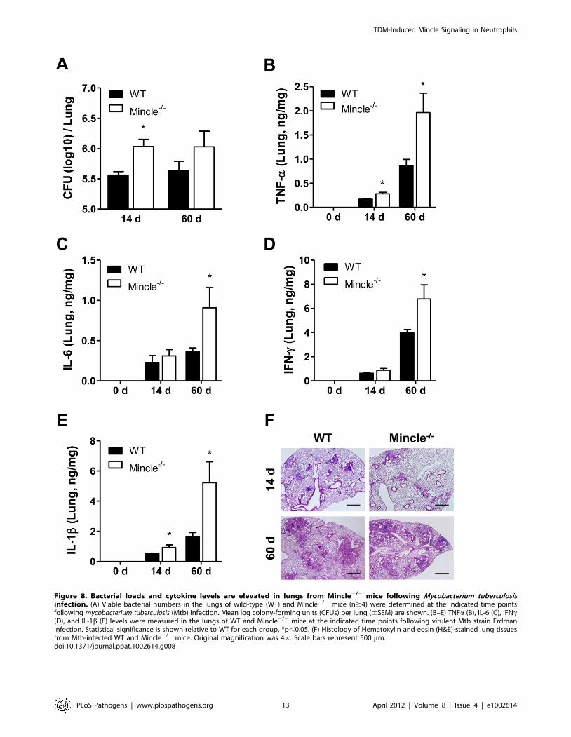
Figure 8. Bacterial loads and cytokine levels are elevated in lungs from Mincle2/2 mice following Mycobacterium tuberculosisinfection. (A) Viable bacterial numbers in the lungs of wild-type (WT) and Mincle2/2 mice (n$4) were determined at the indicated time pointsfollowing mycobacterium tuberculosis (Mtb) infection. Mean log colony-forming units (CFUs) per lung (6SEM) are shown. (B–E) TNFa (B), IL-6 (C), IFNc(D), and IL-1b (E) levels were measured in the lungs of WT and Mincle2/2 mice at the indicated time points following virulent Mtb strain Erdmaninfection. Statistical significance is shown relative to WT for each group. *p,0.05. (F) Histology of Hematoxylin and eosin (H&E)-stained lung tissuesfrom Mtb-infected WT and Mincle2/2 mice. Original magnification was 46. Scale bars represent 500 mm.doi:10.1371/journal.ppat.1002614.g008
TDM-Induced Mincle Signaling in Neutrophils
PLoS Pathogens | www.plospathogens.org 13 April 2012 | Volume 8 | Issue 4 | e1002614

TLR2 signaling pathways in neutrophils. Whereas TDM stimu-
lation resulted in the slow and steady induction of diverse
neutrophil responses (CD11b/CD18 surface expression, cell
adhesion, ROS release, and TNFa production), co-stimulation
with TDM and Pam3CSK4 rapidly and strongly up-regulated
these responses. One possible explanation is that the large
induction of Mincle by TLR ligands may result in additional
TDM signaling. A similar mechanism was used with epithelial
cells, and the level of TLR2 on unstimulated epithelial cells was
markedly enhanced in response to invading microbes [61,62].
Figure 9. Neutrophil-depleted mice had weakened immunity during TDM-elicited inflammation. C57BL/6 mice received intravenousinjections of Ly6G mAb or IgG control mAb 1 day before trehalose dimycolate (TDM) administration (n = 3 mice/group). After TDM administration (2or 5 days), lung tissues and peripheral blood were obtained. (A) Whole-lung cells were analyzed using flow cytometry. Statistical significance: *p,0.05and **p,0.01. (B) Image quantification of relative granuloma areas. Statistical significance: *p,0.05. (C) Hematoxylin and eosin (H&E) staining andLy6G-immunohistochemical staining of neutrophils in lung tissues. Original magnification was 406. Scale bars represent 50 mm. (D) TNFa, IL-6, andMCP-1 protein levels from whole-lung homogenates were determined by CBA. (E) Serum TNFa levels in peripheral blood from mice were measuredby CBA. Statistical significance: *p,0.05. Data are expressed as means 6 SEM from more than three independent experiments.doi:10.1371/journal.ppat.1002614.g009
TDM-Induced Mincle Signaling in Neutrophils
PLoS Pathogens | www.plospathogens.org 14 April 2012 | Volume 8 | Issue 4 | e1002614

Mincle was also up-regulated on macrophages after exposure to
various stimuli and cellular stresses, such as LPS, TNFa, and IL-6
[63]. Our studies confirmed that Mincle expression on neutrophil
surfaces was highly up-regulated following Pam3CSK4 stimula-
tion. Therefore, the synergistic inflammatory responses of
neutrophils likely resulted from the positive enforcement of Mincle
expression by TLR activation.
During the initiation of Mtb-induced granuloma formation, it is
thought that TNFa production by infected macrophages drives the
recruitment of neutrophils, which in turn produce diverse
cytokines and chemokines. Consequently, a large number of
lymphocytes are recruited to the site of infection. In murine
models of Mtb infection, TNFa is essential for the formation of
granulomas [64,65]. However, the early recruitment of neutro-
phils prior to the majority of other cell types and the dramatic
induction of immune responses by neutrophils following coactiva-
tion of Mincle and TLR2, indicates that neutrophils may be
recruited to the site of Mtb infection by direct recognition of
TDM. In particular, IL-6 and MCP-1 expressions in infected lung
were strongly correlated with the early recruitment of neutrophils.
Despite of the strong Mincle-mediated responses of neutrophils at
the initiation of inflammation, the redundant function of
macrophages appears to be sufficient for the granuloma
maturation. Therefore, neutrophils may play a major role in the
initiation of immune responses against Mtb infection rather than
working in the granuloma formation after being passively recruited
by activated immune cells.
Although serum TNFa levels were also decreased in neutrophil-
depleted mice during Mtb infection conditions, lung TNFaexpression levels were not significantly affected by the depletion
of neutrophils. Contrary to the early expression pattern of IL-6
and MCP-1, TNFa expression was extended for a few more days,
thus overlapping with monocyte recruitment to the infection sites,
suggesting that monocytes may contribute to TNFa production as
well. Therefore, the large number of recruited monocytes may
compensate for the loss of TNFa expression caused by neutrophil
depletion at the localized infection site.
In summary, our study provides evidence that the Mincle
signaling pathway in neutrophils promotes diverse neutrophil
activation responses (cell adherence, CD11b/CD18 surface
expression, ROS release, and TNFa production) in response to
mycobacterial TDM. These immune responses to Mtb via Mincle
signaling were enhanced by coactivation of TLR2. Furthermore,
neutrophils produced proinflammatory cytokines/chemokines
during the early stage of TDM-induced pulmonary inflammation.
These findings suggest that the Mincle pathway in neutrophils is
able to modulate lung inflammation.
Materials and Methods
Ethics statementAll animal experiments were performed in accordance with the
Korean Food and Drug Administration (KFDA) guidelines.
Protocols were approved by the Institutional Animal Care and
Use Committees of the Laboratory Animal Research Center at
Yonsei University (Permit Number: 2007-0001).
AnimalsC57BL/6 mice, aged 6–7 weeks, were purchased from Orient
Bio (Gwangju, Gyeonggi, Korea). Mincle2/2 mice (Clec4eMNA)
were kindly provided by the Consortium for Functional Glycomics
(http://www.functionalglycomics.org) and were backcrossed for
nine generations to the C57BL/6 background. MyD88-deficient
mice (C57BL/6 background) were purchased from Oriental
BioService (Kyoto, Japan). All mice were maintained in the
specific pathogen-free facility of the Laboratory Animal Research
Center at Yonsei University.
Induction of pulmonary granulomas throughadministration of TDM
Because a proper orientation of TDM on the mycobacterial
surface is required for its inflammatory and immunoregulatory
properties [1], TDM was prepared as a water-in-oil emulsion to
potentiate its immunostimulatory activity as shown previously
[5,40]. Mice (9–11 weeks old) were injected intravenously in the
tail vein with 100 ml emulsion containing 100 mg TDM (Sigma, St.
Louis, MO). Mice were sacrificed at days 1, 2, 5, and 7 post-TDM
challenge. Lungs were weighed and fixed in 10% formaldehyde for
H&E staining and immunohistochemistry. Lung sections were
frozen for quantitative reverse transcription-polymerase chain
reaction (RT-PCR). A previously-described lung weight index [66]
was used. Neutrophil depletion was achieved by intravenous tail
injection of 200 mg anti-Ly6G (1A8) mAb or isotype control
antibodies 1 day prior to TDM administration. Purified 1A8 mAb
for neutrophil depletion was purchased from BioXcell (West
Lebanon, NH). Quantitation of granuloma areas from at least five
randomly-selected fields from each slide of 3 independent
experiments was performed using Adobe Photoshop software
(Adobe Systems, San Jose, CA). Results are expressed as relative
granuloma area per fixed field of view at a magnification of 640.
Delivery of TDM-bead-bearing matricesAn in vivo murine granuloma model was prepared as described
previously [52]. Briefly, 26103 90-mm polystyrene microspheres
(Polysciences, Inc., Warrington, PA) were coated with 15 mg
TDM. The TDM-coated beads were mixed with bone marrow-
derived macrophages (BMMs; 16107 cells/ml) in 300 ml ice-cold
growth factor-reduced Matrigel (BD Bioscience, Franklin Lakes,
NJ). Matrices were mixed with the indicated BMM genotypes and
subcutaneously injected at each site in the flank of WT or
Mincle2/2 mice. Three mice were injected with TDM-coated
bead matrices. The experiment was repeated more than three
times. Matrices were harvested at 20 h post-inoculation and fixed
in 10% formaldehyde for H&E staining and immunohistochemical
analysis. Neutrophils (Ly6G+) or macrophages (F4/80+) surround-
ing the TDM-coated beads were counted under a light microscope
(Nikon ECLIPSE 80i) on at least five randomly-selected fields.
Mtb infection and bacterial countsMice were challenged by aerosol exposure with WT virulent
Mtb Erdman using an inhalation device (Glas-Col, Terre Haute,
IN) calibrated to deliver approximately 100 bacteria into the lungs.
Five mice per group were sacrificed on days 14 and 60 post-
challenge, and bacteria in lung homogenates were counted.
Numbers of viable bacteria in lungs were determined by plating
serial dilutions of whole organ homogenates on Middlebrook
7H11 agar (Difco, Detroit, MI). Colonies were counted after 3–4
weeks of incubation at 37uC.
Preparation of bone marrow neutrophils and neutrophilassay conditions
Murine bone marrow neutrophils were isolated using Percoll
(GE Healthcare, Little Chalfont, UK) density gradient centrifu-
gation and hypotonic lysis of red blood cells. Murine bone marrow
cells were layered on top of a 53/63/76% three-layer Percoll
gradient following removal of red blood cells. Following
centrifugation, mature neutrophils were recovered at the interface
TDM-Induced Mincle Signaling in Neutrophils
PLoS Pathogens | www.plospathogens.org 15 April 2012 | Volume 8 | Issue 4 | e1002614

of the 63% and 76% fractions and were .90% pure and .95%
viable in the neutrophil-rich fraction as determined by Diff-Quick
staining and trypan blue exclusion, respectively. Neutrophils were
suspended in RPMI-1640 medium with 5% fetal calf serum
(Invitrogen, Grand Island, NY).
F-actin polymerizationNeutrophils were pre-incubated with the indicated inhibitors in
RPMI-1640 medium with 5% fetal bovine serum for 30 min at
37uC. Cells were stimulated for 40 min at 37uC with TDM
(25 mg/ml, Sigma), ultrapure LPS (1 mg/ml, InvivoGen, San
Diego, CA), Pam3CSK4 (1 mg/ml, InvivoGen), or E. coli-
peptidoglycan (0.2 mg/ml, Sigma) on coverslips. After stimulation,
cells were fixed with 4% paraformaldehyde, permeabilized with
BD Perm/Wash Buffer (BD Biosciences), and stained with
Alexa568-phalloidin (Molecular Probes, Eugene, Oregon). Micro-
scopic analysis was performed using an Olympus DP-40
microscope. Mean fluorescence intensities were determined from
the measurement of individual cells’ fluorescence intensities. More
than 20 cells from at least five randomly-selected fields from each
slide were analyzed.
Alternatively, measurement of actin polymerization was done
by a flow-cytometry based phalloidin-binding assay [67]. After
stimulation with TDM for 18 h, neutrophils were fixed in 2%
para-formaldehyde for 15 min at 37uC. Cells were then stained
with fluorescein phalloidin (Invitrogen) in permeabilization buffer
(BD biosciences), and analyzed by flow cytometry.
Adhesion assayNeutrophils (36105 cells/well) were allowed to adhere to 48-
well plates in the presence or absence of TDM (25 mg/ml) with the
indicated inhibitors or mAb M1/70 (0.5 mg/ml, BD Biosciences)
for 20 h at 37uC. In some experiments, cells were stimulated with
TDM and TNFa (50 ng/ml) for 1 h. After stimulation, the non-
adherent cells were washed away. Adherent neutrophils were
quantified with a myeloperoxidase (MPO) assay. MPO activities
were determined through H2O2-dependent oxidation of 3,39,5,59-
tetramethylbenzine (Sigma). Adherence was expressed as the ratio
of adherent neutrophil MPO activity to that of total neutrophils.
Alternatively, neutrophils were incubated with 5 mM calcein-
acetoxymethyl ester at 37uC for 30 min. Thereafter, the cells were
washed, resuspended in RPMI containing 10% FCS media, and
allowed to adhere to TDM-coated 48-well plates for 6 h. The cells
were washed three times and the nonadherent cells were removed.
The fluorescence in each well measured by a fluorescence
microplate reader both before and after washing, and the
percentage of adherent cells was calculated.
Flow cytometric analysisLungs were recovered, weighed, incubated in 2 mg/ml
collagenase D (Roche, Basel, Switzerland) and 40 U/ml DNase
I (Roche) solution, and dispersed by passage through a 70 mm
mesh. After lysis of red blood cells, viable cells were counted. For
immunophenotyping, cells were incubated with fluorescence-
conjugated antibodies. Antibodies (BD Pharmingen, San Jose,
CA) used were against Gr-1 (RB6-8C5), CD11b (M1/70), Ly6G
(1A8), CD3e (145-2C11), and CD19 (1D3). Cells positive for
propidium iodide were excluded by gating prior to collecting at
least 10,000 events. To quantify CD11b, CD18, and CD62L
expression levels on the surface of neutrophils, cells were incubated
with mAbs (1:50) labeled with fluorescein isothiocyanate (FITC) or
phycoerythrin (PE). Stained neutrophils were washed with staining
buffer, and the fluorescence of 36105 cells per sample was
analyzed on a FACS Calibur (BD Biosciences). Results were
expressed as fluorescence intensities on a logarithmic scale. Anti-
mouse CD11b (M1/70), CD18 (C71/16), CD62L (MEL-14), and
isotype control antibodies were obtained from BD Pharmingen.
To quantify Mincle expression, cells were incubated in a blocking
solution containing mouse serum. Blocked cells were incubated
with an anti-mouse Mincle antibody (4A9, MBL International,
Woburn, MA).
ROS assayROS were analyzed with H2DCFDA assays. For the detection
of ROS production following TDM stimulation, neutrophils
(16105 cells/well in 96-well plates) were pre-incubated with
10 mM H2DCFDA (Molecular Probes) in PBS for 30 min allowing
the dye to enter the cells. Cells were washed with PBS prior to
stimulation with TDM for 6 h. Plates were analyzed at 495 nm/
520 nm on a microplate reader. Intracellular ROS levels were
detected by dihydrorhodamine123 (DHR123). Neutrophils
(56105 cells) were incubated in 5 mM DHR123 for 30 min prior
to TDM and Pam3CSK4 stimulation. ROS expression levels were
analyzed by flow cytometry.
Measurement of cytokinesCytokine levels in neutrophil culture supernatants and mouse
sera were assayed using Cytometric Bead Arrays (CBA) from BD
Biosciences according to the manufacturer’s instructions (Mouse
Inflammation Kit: TNFa, IL-6, MCP-1, IL-10, IL-12p40, and
IFNc).
Western blottingNeutrophils were stimulated with TNFa (50 ng/ml) and TDM
(25 mg/ml) for 30 min at 37uC. Cell lysates were prepared as
described previously [68]. Anti-phospho-Erk (Thr202/Tyr204), -
phospho-p38, -phospho-Jnk, -Erk, -p38, and -Jnk antibodies were
obtained from Cell Signaling Technology (Beverly, MA).
RNA extraction and quantitative RT-PCRLung tissues from TDM-challenged and control mice were
homogenized and frozen in Trizol Reagent (Invitrogen). Total
RNA was extracted according to the manufacturer’s protocol.
cDNA was synthesized using SuperScript II Reverse Transcriptase
(Invitrogen). Quantitative real-time RT-PCR analysis of the cDNA
was performed using a LightCycler 480 Real-Time PCR System
(Roche) with a SYBR Green dye. The relative mRNA expression of
each gene was determined using the DDCt calculation method with
Hprt as the internal control gene. The following primer sequences
were used: 59-CTGAGGCGGCGAGGGAGAG-39 and 59-AAG-
CGGTCTGAGGAGGAAGCC-39 for Hprt; 59-AGTGAGGC-
ATCAGGTTCAGTCAAG-39 and 59-GACCAGGTCAAGGT-
TGTCGTAGAG-39 for Mincle; 59-CCAAGACCATCCAATT-
CATC-39 and 59-CCACAAACTGATATGCTTAGG-39 for IL-6;
and 59-TTCCACAACCACCTCAAGCACTTC-39 and 59-TT-
AAGGCATCACAGTCCGAGTCAC-39 for MCP-1.
Statistical analysisData are presented as means 6 standard error of the mean
(SEM). Statistical comparisons between groups were performed
using one-way analysis of variance followed by Student’s t-tests.
Supporting Information
Figure S1 Surface expression of Mincle on neutrophilswas elevated by TDM stimulation. Surface expression of
Mincle was determined by flow cytometry. Neutrophils from wild-
type (WT) and Mincle2/2 mice were stimulated with 25 mg/ml
TDM-Induced Mincle Signaling in Neutrophils
PLoS Pathogens | www.plospathogens.org 16 April 2012 | Volume 8 | Issue 4 | e1002614

trehalose dimycolate (TDM) for 18 h and analyzed by flow
cytometry. Level of Mincle surface expression was quantified by
mean fluorescence intensity (MFI). Statistical significance:
*p,0.05 and **p,0.01. Data are expressed as means 6 SEM
from three independent experiments.
(TIF)
Figure S2 Neutrophil adhesion was increased by TDMstimulation. Suspended wild-type (WT) and Mincle2/2 neu-
trophils were preincubated with calcein-acetoxymethyl ester at
37uC for 30 min. Then, the cells were incubated for 6 h on
trehalose dimycolate (TDM)-coated plate. Nonadherent cells were
removed, and then the adhered neutrophils were imaged by
fluorescent microscopy. Original magnification was 2006. And
fluorescence of each well was measured by using a fluorescence
microplate reader (means 6 SEM). Statistical significance:
**p,0.01 and ***p,0.001. Data are expressed as means 6
SEM from three independent experiments.
(TIF)
Figure S3 Actin polymerization was induced in re-sponse to TDM. Wild-type (WT) and Mincle2/2 neutrophils
were stimulated with trehalose dimycolate (TDM) for 18 h, and
actin polymerization was measured by phalloidin staining and flow
cytometry analysis. Results are given as mean 6 SEM from three
independent experiments. Statistical significance: *p,0.05.
(TIF)
Figure S4 Cell adhesion was synergistically increasedfollowing co-stimulation with TDM/Pam3CSK4. Wild-
type (WT), Mincle2/2, MyD882/2, and Mincle2/2MyD882/2
(DKO) bone marrow neutrophils were preincubated with calcein-
acetoxymethyl ester at 37uC for 30 min. Then, the cells were
stimulated with Pam3CSK4 (10 ng/ml) and/or trehalose dimy-
colate (TDM, 25 mg/ml) for 6 h. The adhered neutrophils were
imaged by fluorescent microscopy. Original magnification was
2006. And fluorescence of each well was measured by using a
fluorescence microplate reader (means 6 SEM). Statistical
significance: **p,0.01 and ***p,0.001. Data are expressed as
means 6 SEM from three independent experiments.
(TIF)
Acknowledgments
We thank S. J. Lee and J. E. See for caring for the mice and for their
excellent technical assistance.
Author Contributions
Conceived and designed the experiments: WBL YJK. Performed the
experiments: WBL JSK JJY BYJ. Analyzed the data: WBL JSK JJY MSL
SNC. Contributed reagents/materials/analysis tools: JJY MSL SNC.
Wrote the paper: WBL JSK YJK.
References
1. Hunter RL, Olsen MR, Jagannath C, Actor JK (2006) Multiple roles of cordfactor in the pathogenesis of primary, secondary, and cavitary tuberculosis,
including a revised description of the pathology of secondary disease. Ann ClinLab Sci 36: 371–386.
2. Russell DG (2006) Who puts the tubercle in tuberculosis? Nat Rev Microbiol 5:39–47.
3. Russell DG, Barry CE, Flynn JL (2010) Tuberculosis: What We Don’t Know
Can, and Does, Hurt Us. Science 328: 852–856.
4. Hunter RL, Olsen M, Jagannath C, Actor JK (2006) Trehalose 6,69-dimycolate
and lipid in the pathogenesis of caseating granulomas of tuberculosis in mice.
Am J Pathol 168: 1249–1261.
5. Perez RL, Roman J, Roser S, Little C, Olsen M, et al. (2000) Cytokine messageand protein expression during lung granuloma formation and resolution induced
by the mycobacterial cord factor trehalose-6,69-dimycolate. J Interferon
Cytokine Res 20: 795–804.
6. Janeway CA, Jr., Medzhitov R (2002) Innate immune recognition. Annu RevImmunol 20: 197–216.
7. Robinson MJ, Sancho D, Slack EC, LeibundGut-Landmann S, Reis e Sousa C(2006) Myeloid C-type lectins in innate immunity. Nat Immunol 7: 1258–1265.
8. Ishikawa E, Ishikawa T, Morita YS, Toyonaga K, Yamada H, et al. (2009)Direct recognition of the mycobacterial glycolipid, trehalose dimycolate, by C-
type lectin Mincle. J Exp Med 206: 2879–2888.
9. Schoenen H, Bodendorfer B, Hitchens K, Manzanero S, Werninghaus K, et al.
(2010) Cutting Edge: Mincle is essential for recognition and adjuvanticity of themycobacterial cord factor and its synthetic analog trehalose-dibehenate.
J Immunol 184: 2756–2760.
10. Wells CA, Salvage JAJ, Li X, Hitchens K, Butcher S, et al. (2008) The
macrophage-inducible C-type lectin, mincle, is an essential component of theinnate immune response to candida albicans. J Immunol 180: 7404–7413.
11. Yamasaki S, Matsumoto M, Takeuchi O, Matsuzawa T, Ishikawa E, et al.(2009) C-type lectin Mincle is an activating receptor for pathogenic fungus,
Malassezia. Proc Natl Acad Sci U S A 106: 1897–1902.
12. da Gloria Sousa M, Reid Delyth M, Schweighoffer E, Tybulewicz V, Ruland J,
et al. (2011) Restoration of Pattern Recognition Receptor Costimulation toTreat Chromoblastomycosis, a Chronic Fungal Infection of the Skin. Cell Host
Microbe 9: 436–443.
13. Yamasaki S, Ishikawa E, Sakuma M, Hara H, Ogata K, et al. (2008) Mincle is
an ITAM-coupled activating receptor that senses damaged cells. Nat Immunol9: 1179–1188.
14. Werninghaus K, Babiak A, Gross O, Holscher C, Dietrich H, et al. (2009)Adjuvanticity of a synthetic cord factor analogue for subunit Mycobacterium
tuberculosis vaccination requires FcR -Syk-Card9-dependent innate immuneactivation. J Exp Med 206: 89–97.
15. Welsh KJ, Abbott AN, Hwang SA, Indrigo J, Armitige LY, et al. (2008) A rolefor tumour necrosis factor-, complement C5 and interleukin-6 in the initiation
and development of the mycobacterial cord factor trehalose 6,69-dimycolateinduced granulomatous response. Microbiology 154: 1813–1824.
16. Eum SY, Kong JH, Hong MS, Lee YJ, Kim JH, et al. (2010) Neutrophils are the
predominant infected phagocytic cells in the airways of patients with active
pulmonary TB. Chest 137: 122–128.
17. Berry MP, Graham CM, McNab FW, Xu Z, Bloch SA, et al. (2010) Aninterferon-inducible neutrophil-driven blood transcriptional signature in human
tuberculosis. Nature 466: 973–977.
18. Aleman M, Beigier-Bompadre M, Borghetti C, de la Barrera S, Abbate E, et al.
(2001) Activation of peripheral blood neutrophils from patients with activeadvanced tuberculosis. Clin Immunol 100: 87–95.
19. Kasahara K, Sato I, Ogura K, Takeuchi H, Kobayashi K, et al. (1998)Expression of chemokines and induction of rapid cell death in human blood
neutrophils by Mycobacterium tuberculosis. J Infect Dis 178: 127–137.
20. Perez RL, Roman J, Staton GW, Jr., Hunter RL (1994) Extravascular
coagulation and fibrinolysis in murine lung inflammation induced by themycobacterial cord factor trehalose-6,69-dimycolate. Am J Respir Crit Care
Med 149: 510–518.
21. Greenblatt MB, Aliprantis A, Hu B, Glimcher LH (2010) Calcineurin regulates
innate antifungal immunity in neutrophils. J Exp Med 207: 923–931.
22. Kerrigan AM, Dennehy KM, Mourao-Sa D, Faro-Trindade I, Willment JA,
et al. (2009) CLEC-2 is a phagocytic activation receptor expressed on murineperipheral blood neutrophils. J Immunol 182: 4150–4157.
23. Kim MJ, Wainwright HC, Locketz M, Bekker LG, Walther GB, et al. (2010)
Caseation of human tuberculosis granulomas correlates with elevated host lipid
metabolism. EMBO Mol Med 2: 258–274.
24. Lauterbach M, O’Donnell P, Asano K, Mayadas TN (2008) Role of TNFpriming and adhesion molecules in neutrophil recruitment to intravascular
immune complexes. J Leukoc Biol 83: 1423–1430.
25. Mocsai A, Abram CL, Jakus Z, Hu Y, Lanier LL, et al. (2006) Integrin signaling
in neutrophils and macrophages uses adaptors containing immunoreceptortyrosine-based activation motifs. Nat Immunol 7: 1326–1333.
26. Evans R, Patzak I, Svensson L, De Filippo K, Jones K, et al. (2009) Integrins inimmunity. J Cell Sci 122: 215–225.
27. Tellier E, Canault M, Rebsomen L, Bonardo B, Juhan-Vague I, et al. (2006)The shedding activity of ADAM17 is sequestered in lipid rafts. Exp Cell Res
312: 3969–3980.
28. Walcheck B, Kahn J, Fisher JM, Wang BB, Fisk RS, et al. (1996) Neutrophil
rolling altered by inhibition of L-selectin shedding in vitro. Nature 380:720–723.
29. Wang Q, Doerschuk CM (2001) The p38 mitogen-activated protein kinase
mediates cytoskeletal remodeling in pulmonary microvascular endothelial cells
upon intracellular adhesion molecule-1 ligation. J Immunol 166: 6877–6884.
30. Pichon S, Bryckaert M, Berrou E (2004) Control of actin dynamics by p38 MAPkinase - Hsp27 distribution in the lamellipodium of smooth muscle cells. J Cell
Sci 117: 2569–2577.
31. Kleinnijenhuis J, Oosting M, Joosten LA, Netea MG, Van Crevel R (2011)
Innate Immune Recognition of Mycobacterium tuberculosis. Clin Dev Immunol2011: 405310.
TDM-Induced Mincle Signaling in Neutrophils
PLoS Pathogens | www.plospathogens.org 17 April 2012 | Volume 8 | Issue 4 | e1002614

32. Reiling N, Holscher C, Fehrenbach A, Kroger S, Kirschning CJ, et al. (2002)
Cutting edge: Toll-like receptor (TLR)2- and TLR4-mediated pathogen
recognition in resistance to airborne infection with Mycobacterium tuberculosis.
J Immunol 169: 3480–3484.
33. Drennan MB, Nicolle D, Quesniaux VJ, Jacobs M, Allie N, et al. (2004) Toll-like
receptor 2-deficient mice succumb to Mycobacterium tuberculosis infection.
Am J Pathol 164: 49–57.
34. Daley JM, Thomay AA, Connolly MD, Reichner JS, Albina JE (2008) Use of
Ly6G-specific monoclonal antibody to deplete neutrophils in mice. J Leukoc Biol
83: 64–70.
35. Bekierkunst A (1968) Acute granulomatous response produced in mice by
trehalose-6,6-dimycolate. J Bacteriol 96: 958–961.
36. Gilleron M, Ronet C, Mempel M, Monsarrat B, Gachelin G, et al. (2001)
Acylation state of the phosphatidylinositol mannosides from Mycobacterium
bovis bacillus Calmette Guerin and ability to induce granuloma and recruit
natural killer T cells. J Biol Chem 276: 34896–34904.
37. Apostolou I, Takahama Y, Belmant C, Kawano T, Huerre M, et al. (1999)
Murine natural killer T(NKT) cells [correction of natural killer cells] contribute
to the granulomatous reaction caused by mycobacterial cell walls. Proc Natl
Acad Sci U S A 96: 5141–5146.
38. Yamagami H, Matsumoto T, Fujiwara N, Arakawa T, Kaneda K, et al. (2001)
Trehalose 6,69-dimycolate (cord factor) of Mycobacterium tuberculosis induces
foreign-body- and hypersensitivity-type granulomas in mice. Infect Immun 69:
810–815.
39. Bekierkunst A, Levij IS, Yarkoni E, Vilkas E, Adam A, et al. (1969) Granuloma
formation induced in mice by chemically defined mycobacterial fractions.
J Bacteriol 100: 95–102.
40. Yarkoni E, Rapp HJ (1977) Granuloma formation in lungs of mice after
intravenous administration of emulsified trehalose-6,69-dimycolate (cord factor):
reaction intensity depends on size distribution of the oil droplets. Infect Immun
18: 552–554.
41. Geisel RE, Sakamoto K, Russell DG, Rhoades ER (2005) In vivo activity of
released cell wall lipids of Mycobacterium bovis bacillus Calmette-Guerin is due
principally to trehalose mycolates. J Immunol 174: 5007–5015.
42. Indrigo J (2003) Cord factor trehalose 6,69-dimycolate (TDM) mediates
trafficking events during mycobacterial infection of murine macrophages.
Microbiology 149: 2049–2059.
43. Silva CL, Ekizlerian SM, Fazioli RA (1985) Role of cord factor in the
modulation of infection caused by mycobacteria. Am J Pathol 118: 238–247.
44. Lima VMF, Bonato VLD, Lima KM, Dos Santos SA, Dos Santos RR, et al.
(2001) Role of Trehalose Dimycolate in Recruitment of Cells and Modulation of
Production of Cytokines and NO in Tuberculosis. Infect Immun 69: 5305–5312.
45. Hunter RL, Venkataprasad N, Olsen MR (2006) The role of trehalose
dimycolate (cord factor) on morphology of virulent M. tuberculosis in vitro.
Tuberculosis (Edinb) 86: 349–356.
46. Armitige LY, Jagannath C, Wanger AR, Norris SJ (2000) Disruption of the
genes encoding antigen 85A and antigen 85B of Mycobacterium tuberculosis
H37Rv: effect on growth in culture and in macrophages. Infect Immun 68:
767–778.
47. Rao V, Fujiwara N, Porcelli SA, Glickman MS (2005) Mycobacterium
tuberculosis controls host innate immune activation through cyclopropane
modification of a glycolipid effector molecule. J Exp Med 201: 535–543.
48. Rao V, Gao F, Chen B, Jacobs Jr. WR, Glickman MS (2006) Trans-
cyclopropanation of mycolic acids on trehalose dimycolate suppresses Myco-
bacterium tuberculosis-induced inflammation and virulence. J Clin Invest 116:
1660–1667.
49. Pedrosa J, Saunders BM, Appelberg R, Orme IM, Silva MT, et al. (2000)
Neutrophils play a protective nonphagocytic role in systemic Mycobacteriumtuberculosis infection of mice. Infect Immun 68: 577–583.
50. Sugawara I, Udagawa T, Yamada H (2004) Rat neutrophils prevent the
development of tuberculosis. Infect Immun 72: 1804–1806.51. Seiler P, Aichele P, Raupach B, Odermatt B, Steinhoff U, et al. (2000) Rapid
neutrophil response controls fast-replicating intracellular bacteria but not slow-replicating Mycobacterium tuberculosis. J Infect Dis 181: 671–680.
52. Sakamoto K, Geisel RE, Kim MJ, Wyatt BT, Sellers LB, et al. (2009) Fibrinogen
Regulates the Cytotoxicity of Mycobacterial Trehalose Dimycolate but Is NotRequired for Cell Recruitment, Cytokine Response, or Control of Mycobac-
terial Infection. Infect Immun 78: 1004–1011.53. Abram CL, Lowell CA (2009) The ins and outs of leukocyte integrin signaling.
Annu Rev Immunol 27: 339–362.54. Ortiz-Stern A, Rosales C (2003) Cross-talk between Fc receptors and integrins.
Immunol Lett 90: 137–143.
55. Ortiz-Stern A, Rosales C (2005) Fc gammaRIIIB stimulation promotes beta1integrin activation in human neutrophils. J Leukoc Biol 77: 787–799.
56. Kocher M, Siegel ME, Edberg JC, Kimberly RP (1997) Cross-linking of Fcgamma receptor IIa and Fc gamma receptor IIIb induces different proadhesive
phenotypes on human neutrophils. J Immunol 159: 3940–3948.
57. West MA, Wallin RP, Matthews SP, Svensson HG, Zaru R, et al. (2004)Enhanced dendritic cell antigen capture via toll-like receptor-induced actin
remodeling. Science 305: 1153–1157.58. Dennehy KM, Ferwerda G, Faro-Trindade I, Pyz E, Willment JA, et al. (2008)
Syk kinase is required for collaborative cytokine production induced throughDectin-1 and Toll-like receptors. Eur J Immunol 38: 500–506.
59. Gantner BN, Simmons RM, Canavera SJ, Akira S, Underhill DM (2003)
Collaborative induction of inflammatory responses by dectin-1 and Toll-likereceptor 2. J Exp Med 197: 1107–1117.
60. Chen CH, Floyd H, Olson NE, Magaletti D, Li C, et al. (2006) Dendritic-cell-associated C-type lectin 2 (DCAL-2) alters dendritic-cell maturation and
cytokine production. Blood 107: 1459–1467.
61. Sakai A, Han J, Cato AC, Akira S, Li JD (2004) Glucocorticoids synergize withIL-1beta to induce TLR2 expression via MAP Kinase Phosphatase-1-dependent
dual Inhibition of MAPK JNK and p38 in epithelial cells. BMC Mol Biol 5: 2.62. Shuto T, Imasato A, Jono H, Sakai A, Xu H, et al. (2002) Glucocorticoids
synergistically enhance nontypeable Haemophilus influenzae-induced Toll-likereceptor 2 expression via a negative cross-talk with p38 MAP kinase. J Biol
Chem 277: 17263–17270.
63. Matsumoto M, Tanaka T, Kaisho T, Sanjo H, Copeland NG, et al. (1999) Anovel LPS-inducible C-type lectin is a transcriptional target of NF-IL6 in
macrophages. J Immunol 163: 5039–5048.64. Flynn JL, Goldstein MM, Chan J, Triebold KJ, Pfeffer K, et al. (1995) Tumor
necrosis factor-a is required in the protective immune response against
mycobacterium tuberculosis in mice. Immunity 2: 561–572.65. Bean AGD, Roach DR, Briscoe H, France MP, Korner H, et al. (1999)
Structural deficiencies in granuloma formation in TNF gene-targeted miceunderlie the heightened susceptibility to aerosol Mycobacterium tuberculosis
infection, which is not compensated for by lymphotoxin. J Immunol 162:3504–3511.
66. Guidry TV, Olsen M, Kil KS, Hunter RL, Jr., Geng YJ, et al. (2004) Failure of
CD1D2/2 mice to elicit hypersensitive granulomas to mycobacterial cord factortrehalose 6,69-dimycolate. J Interferon Cytokine Res 24: 362–371.
67. Van Ziffle JA, Lowell CA (2009) Neutrophil-specific deletion of Syk kinaseresults in reduced host defense to bacterial infection. Blood 114: 4871–4882.
68. Mocsai A, Zhou M, Meng F, Tybulewicz VL, Lowell CA (2002) Syk is required
for integrin signaling in neutrophils. Immunity 16: 547–558.
TDM-Induced Mincle Signaling in Neutrophils
PLoS Pathogens | www.plospathogens.org 18 April 2012 | Volume 8 | Issue 4 | e1002614
Related Documents