1 NEUTROPHIL TOXIC GRANULATION IN PREGNANCY IN ONITSHA BY EZIGBO, EYIUCHE DORIS PG/M.Sc/05/45082 DEPARTMENT OF MEDICAL LABORATORY SCIENCE. FACULTY OF HEALTH SCIENCES AND TECHNOLOGY UNIVERSITY OF NIGERIA, ENUGU CAMPUS APRIL2010

Welcome message from author
This document is posted to help you gain knowledge. Please leave a comment to let me know what you think about it! Share it to your friends and learn new things together.
Transcript
1
NEUTROPHIL TOXIC GRANULATION IN PREGNANCY IN ONITSHA
BY
EZIGBO, EYIUCHE DORIS PG/M.Sc/05/45082 DEPARTMENT OF MEDICAL LABORATORY SCIENCE. FACULTY OF HEALTH SCIENCES AND TECHNOLOGY UNIVERSITY OF NIGERIA, ENUGU CAMPUS
APRIL2010
2
i TITLE PAGE
NEUTROPHIL TOXIC GRANULATION IN PREGNANCY IN ONITSHA
A DISSERTATION PRESENTED TO THE UNIVERSITY OF NIGERIA, FOR THE DEGREE OF MASTER OF SCIENCE
BY
EZIGBO, EYIUCHE DORIS PG/M.Sc/05/45082
DEPARMENT OF MEDICAL LABORATORY SCIENCE. FACULTY OF HEALTH SCIENCES AND TECHNOLOGY
UNIVERSITY OF NIGERIA, ENUGU CAMPUS.

3
APRIL 2010
iii
DEDICATION To my husband, Mr Benjamin, Mootol Ezigbo and our children -
Amarachukwu, Oluoma, Toochukwu and Chukwuagoziem

4
iv
ACKNOWLEDGEMENT I wish to acknowledge my sincerest indebtedness and gratitude to all whom in one way or the other contributed to the completion of this work. First my appreciation goes to God, who in His infinite mercy made His grace abundant for me. My profound appreciation and gratitude go to Ven.Prof. E.O.Ukaejiofo, my able supervisor, for showing me the way forward, I am also grateful to all my lecturers in the Department for their advice. I want to thank Dr P.N.Nwagbara Consultant Obstetrician and Gynaecologist/Chief Medical Director Shalom Foundation Specialist Hospital and Maternity Onitsha and the then HOD Department of Obstetrics and Gynaecology, General Hospital Onitsha, for allowing his patients to participate in this research, providing his ideas and personal journals. Dr Valentine Obidi, of the Department of Obstetrics and Gynaecology, General Hospital, Onitsha for his assistance in providing the patients files. This acknowledgement will not be complete without placing on record the generous assistance received from my colleagues Mrs. Nwagu N. of General Hospital, Onitsha, Mr. Festus C. Emengaha, Chemical Pathology Department, St. Charles Borromeo Hospital and Mr. Akagozirim Murphy then Chief Scientist New Hope Medical Diagnostic Centre, Onitsha. For the enthusiastic support in providing some of the materials I used, I wish to thank Mrs. Nosike, O.K, then Circulation Librarian Medical Library University of Nigeria Enugu Campus (UNEC) and all the staff of Nnamdi Azikiwe University Teaching Hospital (NAUTH) Library Nnewi. I am indebted to Mrs.Akanyak Nkoyo.B, of the Department of Pharmacology and Therapeutics UNEC, for the statistical analysis of this work.

5
My warmest appreciation to my very dear husband Mr. Benjamin M.Ezigbo, you pushed me to start, to continue and provided for everything I needed to accomplish this work, favour will always go with you. A million thanks goes to my lovely parents Mr. and Mrs. Christian.A.Anyadike for the foundation they laid. May God bless you all Amen. EZIGBO, EYIUCHE DORIS.
v TABLE OF CONTENTS
PAGE TITLE PAGE ………………………………….. i CERTIFICATION ………………………………….. ii DEDICATION ………………………………….. iii ACKNOWLEDGEMENT ………………………………….. iv TABLE OF CONTENTS ………………………………….. v LIST OF FIGURES ………………………………….. vii LIST OF TABLES ………………………………….. ix ABSTRACT ………………………………………….. x CHAPTER ONE 1.0 INTRODUCTION ………………………… 1 1.1 BACKGROUND OF THE STUDY …………. 1 1.2 RESEARCH PROBLEM................................ 2 1.3 HYPOTHESIS................................................ 2 1.4 SIGNIFICANCE OF STUDY........................ 3 1.5 AIMS AND OBJECTIVES OF STUDY ……. 4 CHAPTER TWO 2.0 REVIEW OF LITERATURE ………… 5
6
2.1 THE PREGNACY STATE ………… 5 2.1.2 PHYSIOLOGICAL CHANGES DURING
PREGNANCY ………………… 5 2.1.3 THE KIDNEYS AND URINARY TRACT ….. 5 2.1.4 HAEMATOLOGIC CHANGES ……………… 7 2.2 THE NEUTROPHILS …………………. 8 2.2.1 LIFE HISTORY …………………………. 9 2.2.2 SUBCELLULAR STRUCTURE OF NEUTROPHILS …. 10 2.2.3 NEUTROPHIL ALKALINE PHOSPHATASE ………. 11 2.3 COMPLICATIONS IN PREGNACY ………. 12
iv 2.3.1 URINARY TRACT INFECTIONS ………… 12 2.3.2 HIV INFECTIONS ………………………….. 15 2.3.3 MALARIA ………………………………. 17 2.4 UTERINE FIBROIDS ………………. 17 CHAPTER THREE 3.0 STUDY DESIGN ……….. 21 3.1 POPULATION ……………….. 21 3.2 SAMPLING TECHNIQUE ………………………. 22 3.2.1 INCLUSION AND EXCLUSION CRITERIA 22 3.3.0 RESEARCH METHODOLOGY ………………… 23 3.3.1 NEUTROPHIL ALKALINE POSPHATASE ACTIVITY ……….................................... 23 3.3.1.2 ISOLATION OF NEUTROPHILS ………. 23 3.3.1.3 NEUTROPHIL ALKALINE PHOSPHATASE ASSAY... 24 3.3.2 TOXIC NEUTROPHIL COUNTS ………… 25 3.3.3 MALARIA PARASITE EXAMINATION…. 26 3.3.4 TOTAL LEUCOCYTE COUNT …………… 27

7
3.3.5 URINE CULTURE …………………………. 28 3.3.6 STATISTICAL ANALYSIS........................... 29 CHAPTER FOUR 4.0 RESULTS …………………………………… 30 CHAPTER FIVE 5.0 DISCUSSION AND CONCLUSION ……… 47 5.1 RECOMMENDATION ………………………. 49 REFERENCES ………………………………………………... 50
vii
LIST OF FIGURES FIGURE 2.1 Neutrophil Maturation FIGURE 4.1 A. Relationship between Neutrophil alkaline
phosphatase activity and Toxic Neutrophil count in the control subjects.
B. Relationship between Neutrophil alkaline
phosphatase activity and Toxic Neutrophil count in normal pregnant subjects.
FIGURE 4.2 A. Relationship between Neutrophil alkaline
phosphatase activity and Toxic Neutrophil count in fibroid subjects.
8
B. Relationship between Neutrophil alkaline phosphatase activity and Toxic Neutrophil count in pregnant subjects with UTI.
C. Relationship between Neutrophil alkaline
phosphatase activity and Toxic Neutrophil count in pregnant subjects with Malaria.
viii
D. Relationship between Neutrophil alkaline phosphatase activity and Toxic Neutrophil count in pregnant subjects with HIV.
FIGURE 4.3 Normal peripheral blood films FIGURE 4.4 Peripheral blood films of pregnant women with UTI,
Malaria, HIV and women with fibroid.
9
ix LIST OF TABLES TABLE 4.1A Mean± SE in TNC and NAP for control, normal
pregnancy, Women with Fibroid and pregnant women with UTI, HIV and malaria.
10
TABLE 4.1B Mean Total white blood cell count (TWBC) Toxic neutrophil count (TNC) and Neutrophil alkaline phosphatase activity (NAP) in control subjects and normal pregnant subjects
TABLE 4.2 Mean TWBC, TNC and NAP activity in the control
group and pregnant subjects with UTI TABLE 4.3 Mean TWBC, TNC and NAP activity in the control
group and pregnant women with Malaria TABLE 4.4 Mean TWBC, TNC and NAP activity in the control
group and pregnant subjects with HIV TABLE 4.5 Mean NAP activity in normal pregnant and pregnant
subjects with UTI, HIV and Malaria TABLE 4.6 Mean TNC in normal pregnant subjects and pregnant
subjects with UTI, HIV and Malaria. TABLE 4.7 Mean TWBC in normal pregnant subjects and pregnant
subjects with UTI, HIV and Malaria. TABLE 4.8 TNC count and urine culture result. x
ABSTRACT Screening for asymptomatic bacteriuria does remain relevant in improving pregnancy
outcome but inexpensive and effective techniques are required. The objective of this

11
study was to evaluate the white cell morphology in pregnant women with urinary tract
infection (UTI) and study their diagnostic utility. Three hundred and eighty one (381)
subjects were studied. This comprised: Three hundred and twenty three (323) pregnant
subjects and Fifty eight (58) fibroid cases. One hundred (100) subjects matched for age
were used as control. The pregnant women were those attending General Hospital
Onitsha and Shalom Foundation Specialist Hospital Onitsha for routine antenatal
serological testing. Toxic neutrophil count (TNC), Neutrophil alkaline phosphatase
assay, total white blood cell count (TWBC) and urine cultures were carried out.
Conventional microscopy was used for TNC and TWBC. Neutrophil alkaline
phophatase activity was analysed using Refloctron Plus System by Roche. Statistical
analysis was performed with SPSS software using student’s t-test and the sensitivity
and specificity of various TNC cut offs were computed. In the presence of pregnancy
and infection NAP activity correlated positively with TNC. At a cut off of 10 in TNC a
specificity of 79% and a sensitivity of 18% were achieved. With a cut off of
xi
70 specificity was 97% and sensitivity was 0%. TNC although a specific indicator for
infection does not serve as a sensitive screening procedure in pregnant women with
UTI.
12
CHAPTER ONE
1.0 INTRODUCTION
1.1 BACKGROUND OF THE STUDY:
Several of the changes seen in neutrophils from pregnant women, including
neutrophilia, increased staining of granules and alteration of neutrophil alkaline
phosphatase activity are similar to those seen in infections in pregnancy and in patients
with myeloid metaplasia (EL-Maalem and Fletcher, 1976). Thus the same pattern of
changes occurs in a number of different conditions suggesting a common underlying
mechanism.
In infections the circulating neutrophils are already partially exhausted by
previous ingestion of opsonised particles (antigen-antibody complexes). In normal
pregnancy there are circulating immune complexes of a type which should be ingested
by neutrophils (EL-Maalem and Fletcher, 1980).
13
1.2 Research problem
Despite numerous studies over many years, the issues relating to the
prevention and management of asymptomatic bacteriuria and UTI during
pregnancy remain unresolved.
Routine cultures for the diagnosis of asymptomatic bacteriuria and UTI are
expensive and both false positive and false negative results can occur.
Screening of asymptomatic bacteriuria does remain relevant in improving
pregnancy outcome (Akerele, et al 2001), however inexpensive and
effective techniques are required.
1.3 Hypothesis:
Neutrophil toxic granulation is the prominent staining by Romanowsky dyes
of cytoplasmic granules –as a result of the abnormal maturation of
azurophilic granules with persistence of acid mucosubstance.
In the normal uninfected neutrophil these granules are fine and evenly
distributed. During infections and pregnancy the granules become more
prominent and are unevenly distributed.
14
1.4 Significance of study
The incidence of UTI in pregnancy can be as high as 6.2% in pregnant
women (Ezechi, et al 2003).
Acute pylonephritis occurs during pregnancy and more commonly in
women, who have had asymptomatic bacteriuria.
Prematurity and low birth weights are common conditions associated with
untreated asymptomatic bacteriuria.
Some pregnant women with symptoms of UTI who had negative culture
results still respond to antibiotic therapy.
15
1.5 AIMS OF STUDY
This project is aimed at developing a cost effective and sensitive method of
screening for UTI in pregnancy, and to compare toxic granulation in pregnancy
with those in other disease conditions. Hence, attempt would be made in this
research work to:
Carry out toxic Neutrophil count in pregnant women in Onitsha
Assay Neutrophil Alkaline phosphatase activity in pregnant women in
Onitsha
Correlate toxic Neutrophil count with Neutrophil alkaline phosphatase
activity.
Use toxic Neutrophil count as an index in screening for UTI in
pregnancy.
16
CHAPTER TWO
2.0 REVIEW OF LITERATURE
2.1 The Pregnancy State: Pregnancy constitutes a special condition in a woman’s
life that affects various physiologic and endocrinologic systems.
2.1.2 Physiological Changes during Pregnancy: The physiologic, biochemical and
anatomic changes that occur during pregnancy are extensive and may be
systemic or local.
Hormonal changes: The large amounts of oestrogen, progesterone, human
placental lactogen, and corticosteroids produced during pregnancy affect
various metabolic, physiologic and endocrine systems. The secretion of
estrogens and progesterone throughout pregnancy assures appropriate
development of the endometrium, uterine growth, adequate uterine blood
supply, and preparation of uterus for onset of labour.
2.1.3 The Kidneys and Urinary Tract
Renal Dilatation: During pregnancy, each kidney increases in length by 1-
1.5cm with a concomitant increase in weight (Delzell et al, 2000). The renal
pelvis is dilated. The ureters are dilated above the brain of the bony pelvis. The
17
ureters also elongate, widen and become more curved. Thus there is an increase
in urinary stasis. This may lead to infection.
The absolute cause of hydronephrosis and hydroureter in pregnancy is unknown
and there may be several contributing factors:
Elevated progesterone levels may contribute to hypotonia of the smooth
muscles in the ureter.
The ovarian vein complex in the suspensory ligament of the ovary may
enlarge enough to compress the ureter at the brim of the bony pelvis,
thus causing dilation above the level.
Dextorotation of the uretus during pregnancy may explain why the right
ureter is usually more dilated than the left.
Hyperplasia of smooth muscles in distal one-third of the ureter may
cause reduction in the luminal size (Wing et al, 2000).
Renal Function: The glomerular filtration rate (GFR) increases during
pregnancy by about 50%. The renal plasma flow rate increases by as much as
25-50%. Urinary flow and sodium excretion rate in late pregnancy can be
altered by posture, being twice as great in the lateral recumbent position as in
the supine position. Even though the GFR increased dramatically during
pregnancy, the volume of urine passed each day is not increased. Thus, the
urinary system appears to be even more efficient during pregnancy. With the
increase in GFR, there is an increase in endogenous clearance of creatinine.
18
The concentration of creatinine in serum is reduced in proportion to increase in
GFR, and concentration of blood urea nitrogen is similarly reduced.
Glucosuria during pregnancy is not necessarily abnormal, and may be
explained by the increase in GFR with impairment of tubular reabsorption
capacity for filtered glucose. Increased levels of urinary glucose also
contribute to increased susceptibility of pregnant women to urinary tract
infection.
Proteinuria changes little during pregnancy and if more than 500mg/24 hr is
lost, a decrease process should be suspected.
Levels of the enzyme rennin, which is produced in the kidney, increase early
in the first trimester, and continue to rise until term. This enzyme acts on its
substrate angiotensinogen, to first form angiotensin 1 and then angiotensin 2,
which acts as a vaso constrictor.
The Bladder: As the uterus enlarges, the urinary bladder is displaced upward
and flattened in the anterior-posterior or diameter. Pressure from the uterus
leads to increase in urinary frequency. Bladder vascularity increase and
muscle tone decreases, increase in capacity up to 1500ml (Babior and Golde,
2001).
2.1.4 Haematologic Changes: Maternal blood volume increases during
pregnancy by an average of 45%. Plasma volume increases more rapidly than
red cell mass, therefore, in spite of augmented erythropoiesis, the
19
concentration of haemoglobin, the erythrocyte count, and the haematocrit
commonly decrease during normal pregnancy. Haemoglobin concentration at
term average 12.1 g/dl compared with 12.8 g/dl (Ukaejiofo et al, 1999), for
the non pregnant state. The total white blood cell count increases during
pregnancy from a pre-pregnancy level of 4.3 – 4.5 x 10⁹/L to 5 – 12 x 109/l in
the last trimester (Bainton, 2001) although counts as high as 16 x 109/L have
been observed in the last trimester. Lymphocyte and monocyte numbers stay
essentially the same throughout pregnancy (Brambald et al, 1997).
Polymorphonuclaer leucocytes are the primary contributors to the increase
(Brambald et al, 1997).
2.2 THE NEUTROPHILS
Neutrophils are granulocytes, which form an essential component of the
cellular innate system involved in killing bacteria and fungi. They play
critical role in host defence by phagocytising and digesting micro - organisms.
Neutrophils are so named because of their neutral staining with Wright’ Stain.
They are also known as PMNs or Polys or microphages. They are round cells
approximately 12-14µm in diameter. The multilobed nucleus contributes to
the extreme elasticity of the cell, which is of importance for the cell to make
rapid transit from the blood through tight gaps in the endothelium. In the
resting uninfected host, the production and elimination of neutrophils are
20
balanced, resulting in fairly constant concentration of neutrophils in peripheral
blood. When infection occurs, chemotactic agents are generated, that result in
migration of neutrophils to the site of the infection and activation of
neutrophil defensive function. Cytoplasmic modifications that occur include:
Alteration of the staining character of the cytoplasm.
Abnormal granulation.
Vacuolization (Meranze et al, 1935).
2.2.1 Life History
The normal human neutrophil production rate is 0.85 to 1.6 x 10⁹/L cells
per kilogram per day (Babior et al, 2001). Neutrophils are abundant in the
circulation, present at concentration of 2 x 10⁹/L to 7 x 10⁹/L and equal
numbers are marginated on vessel walls or sequestedrated in closed
capillaries. The half-life in the blood is 6-7 hrs and in the tissue is about
1- 4days. The blood and bone marrow form an abundant pool of cells. In the
bone marrow, the myeloid precursor cells mature to segmented neutrophils in
about 9 days.
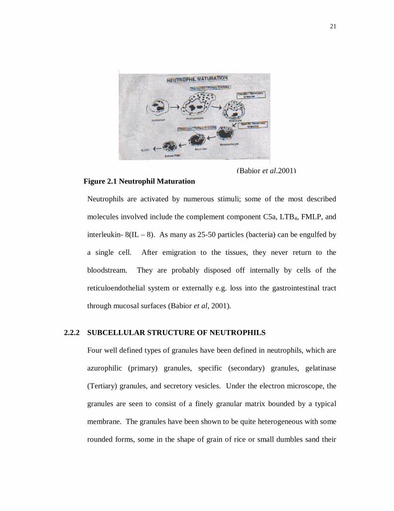
21
Figure 2.1 Neutrophil Maturation
Neutrophils are activated by numerous stimuli; some of the most described
molecules involved include the complement component C5a, LTB4, FMLP, and
interleukin- 8(IL – 8). As many as 25-50 particles (bacteria) can be engulfed by
a single cell. After emigration to the tissues, they never return to the
bloodstream. They are probably disposed off internally by cells of the
reticuloendothelial system or externally e.g. loss into the gastrointestinal tract
through mucosal surfaces (Babior et al, 2001).
2.2.2 SUBCELLULAR STRUCTURE OF NEUTROPHILS
Four well defined types of granules have been defined in neutrophils, which are
azurophilic (primary) granules, specific (secondary) granules, gelatinase
(Tertiary) granules, and secretory vesicles. Under the electron microscope, the
granules are seen to consist of a finely granular matrix bounded by a typical
membrane. The granules have been shown to be quite heterogeneous with some
rounded forms, some in the shape of grain of rice or small dumbles sand their
(Babior et al,2001)
22
number has been shown to vary from 500 – 1500/granulocyte (Rashmi et al,
2006).
Granules contain large amount of protein and traces of lipids and nucleic acids.
These granules are essential for post phagocytic function of PMN leucocytes.
Their constituents are essential in inflammation and are determinants of
intraleucocytic antimicrobial events.
Among the azurophilic contents are myeloperoxidase, defensins lysozyme,
azurocidin, etc. that have antibacterial function. These granules fuse with
phagocytes vesicles, resulting in the delivery of their contents to the ingested
organism. The greenish coloration of pus is imparted by myeloperoxidase
(Rashmi et al, 2006). Specific granules are three times more common in the
cytoplasm. Release of specific granule contents like collagenase, apolactoferrin
lysozyme, histaminase, etc may modify inflammatory process. Collagenase and
elastase breakdown fibrous structures in the extracellular matrix, facilitating
progress of the neutrophil through tissue. The tertiary granules content include
gelatinase, alkaline phosphatase and CD11b/CD18 (Smolen and Boxer, 2001).
2.2.3 Neutrophil Alkaline Phosphatase (NAP)
The NAP is an enzyme expressed on the external aspect of the neutrophilic
granulocyte plasma membrane, and represents a specific marker for the fully
differentiated granulocyte (Rambaldi et al, 1997). It is Zinc – containing
23
phosphomonoesterase with a pH optimum near 10 (Beutler, 2001). The activity
of NAP is limited to the neutrophilic series; it appears first in the myelocyte and
rapidly increases with maturation of the cell to the segmented
polymorphonuclear neutrophil.
NAP is used in the investigation of Neutrophil leucocytosis and Erythrocytosis.
It differentiates chronic myeloid leukaemia (Low) from reactive leucocytosis
(high) e.g. bacterial infection. It may assist in the differentiation of Polycytemia
rubra vera (PRV) (high) from other causes of erthrocytosis (normal). Used in
the diagnosis of Paroxysmal noctural haemoglobinuria (very low); (Normal to
high) in other haemolytic and/or hypoplastic anaemia. The NAP is moderately
elevated in pregnancy, with oestrogen therapy (e.g. Oral Contraception) and
corticosteroid therapy.
2.3 COMPLICATIONS IN PREGNANCY
2.3.1 Urinary tract infections in pregnancy
Urinary tract infections (UTI) are the most common bacterial infections during
pregnancy. The incidence of UTI in pregnancy can be as high as 6.2% (Ezechi
et al, 2003)
Pathogenesis
Pregnant women are at increased risk for UTIs. Beginning in week 6 and
peaking during weeks 22 to 24. Approximately 90% of pregnant women
24
develop ureteral dilatation, which will remain until delivery (hydronephrosis of
pregnancy). Increase bladder volume and decrease bladder tone, along with
decreased ureteral tone, contribute to increased urinary stasis and uretero vesical
reflux (Delzell and Lefevre 2000). Additionally, the physiologic increase in
plasma volume during pregnancy decreases urine concentration. Up to 70%
pregnant women develop glycosuria, which encourage bacteria growth in the
urine. Increase in urinary progestin and estrogens may lead to a decreased
ability of the lower urinary tract to resist invading bacteria. This decreased
ability may be caused by decreased ureteral tone or possibly by allowing some
strains of bacteria to selectively grow. (Lucas and Cunningham, 1993).
UTI have three principle presentations: asymptomatic bacteriuria acute cystitis
and pyelonephritis.
Asymptomatic Bacteriuria:
Significant bacteriuria may exist in asymptomatic patient with a prevalence of
10% during pregnancy (Delzell and Lefevre, 2000). Untreated asymptomatic
bacteriuria leads to the development of symptomatic cystitis (infection confined
to the bladder) in approximately 30% of patients and can lead to the
development of pyelonephritis (infection involving the kidney) in up to 50%.
Asymptomatic bacteriuria is associated with an increased risk of intrauterine
growth retardation and low-birth-weight infants (Harris et al, 1976). The
relatively high prevalence of asymptomatic bacteriuria during pregnancy, the
25
significant consequence for women and for the pregnancy plus the ability to
avoid squeal with treatment, justify screening pregnant women for bacteriuria.
Acute Cystitis:
Acute cystitis is distinguished from asymptomatic bacteriuria by the presence of
symptoms such as dysuria, urgency and frequency in a febrile patient with no
evidence of systemic illness. Up to 30% of patients with untreated asymptomic
bacteriuria latter develop symptomatic cystitis.
Pyelonephritis:
Acute pyelonephritis during pregnancy is a serious systemic illness that can
progress to maternal sepsis, preterm labour and premature delivery. The
presence of bacteriuria is accompanied by systemic symptoms such as fever,
chills, nausea, vomiting and loin pain. Symptoms of lower tract infection
urethritis and cystitis (i.e. frequency and dysuria) may or may not be present.
Pyelonephritis occurs in 2% of pregnant women (Gilstrap et al, 1981).
2.3.2 HIV Infections
The human immunodeficiency virus (HIV) was discovered in 1983 (two years
after the diseases AIDS was described) when Barresinousi Montagnier and
colleagues at the Institute Pasteur, Paris, France, isolated the virus from the T
cells of a patient with generalised lymphadenopathy and gave it the name
26
Lymphadenopathy associated virus (LAV, now HIV 1). In the same year,
Robert Gallo and colleagues, working at the National Cancer Institute (NCI),
USA made a similar discovery while in their quest to find cancer-causing
viruses. In 1986 a second closely related virus, termed HIV 2 was isolated from
a patient from West Africa suffering from acquired immune deficiency
syndrome (AIDS). The first case of HIV/AIDS in Nigeria was reported in 1986
and by 2002, with a national sero-prevalence of 5.8%, 14 million people have
died and 3.43million are living with disease. (Sagay et al, 2004).
HIV is one of the several complex retroviruses in the genus lentivirus.
The virus contains three genes required for a replicating retrovirus - gab, pol
and env. About three additional genes regulate virus expression and are
important in disease pathogenesis in vivo. These other gene products include
the Tat, Rev and Nef regulatory proteins that are translated from spliced MRNA
species. The HIV structure consists of a lipoprotein surface studded by about
72 envelop knobs consisting of the glycoprotein gp 120, the surface (SU)
protein and gp41, the transmembrane (TM) protein. Inside this lipid bilayer is a
matrix (MA) protein (P17). Below the matrix is the nucleoprotein made up of a
capsid (CA) protein (P24, P25). Inside the core are various cone shaped
nucleocapsid protein (P9, P7), as well as polymerase enzyme containing reverse
transcriptase (RT, P63), the Protease (PR, P15) and the integrase (IN), P11 (Levy,
1993).
27
HIV is transmitted through blood products (including blood
transfusions, intravenous drug abuse in sharing of needles, health care workers
in needle stick injuries and mucocutaneous exposures), organ transplants, sexual
intercourse (both homosexual and heterosexual exposures) and vertical
transmission (10-40% of babies born of infected mothers will be infected).
Infection may occur in utero, during birth, postnatally or through breast feeding,
(Noel, 2005). Virus enters the cell by fusing with the cell membrane. This
event involves an interaction with a cellular receptor (the major one being CD4)
followed by conformational changes in the viral envelop to permit viral-cell
fusion (Hardie, 1999).
HIV is one of the major causes of infant and maternal mortality in
resource-poor settings with more than 600,000 children infected each year or
1700 day, with most of the infections occurring in Africa (UNAIDS 2004). Of
815 pregnant women studied in South-eastern Nigeria 31 were HIV +. Severe
anaemia has a significant correlation with HIV infections in these women
(Uneke et al, 2007). HIV infections have also been associated with
neutropenia. HIV can cause decreased growth of progenitor cell, CFU-GM,
decreased endogenous G-CSF (Moore, 2008). The patient is exposed to
increased risk of bacterial and fungal infection.
28
2.3.3 MALARIA
Malaria in pregnancy is frequently under-estimated, both as a public health
problem and by clinicians who treat individual cases. It is one of the major
causes of maternal, foetal and neonatal morbidity and mortality worldwide
(Whitty et al, 2005). A woman who is pregnant is at significantly greater risk
from malaria than one who is not, irrespective of the setting and malaria poses a
severe threat to the pregnancy of any woman. Non-immune pregnant women
are at high risk of complicated malaria and foetal loss. Women with previously
reasonable immunity to malaria lose part of that protection in pregnancy,
especially in first pregnancies, in which there is appreciable mortality.
Miscarriages, stillbirths and preterm births are common in pregnant women
with malaria. In addition to the huge burden of maternal anaemia caused by
malaria, low birth weights are common in infants born to women who had
malaria in pregnancy, with over 5% of prenatal deaths in many low resources
setting thought to be caused by this mechanism. In our environment malaria has
a prevalence of 19.7% in pregnant women, with primigravidae being
significantly more infected with malaria. (Uneke et al, 2008)
2.4 UTERINE FIBROIDS
Fibroids are growths of the uterus, or womb. They are also called uterine
leimyomas or myomas. They grow from the muscle cells of the uterus and may
29
protrude from the inside or outside surface of the uterus. Fibroids may also be
found within the muscular wall. Fibroids are very common. A study carried
out at the University of Ilorin Teaching Hospital, Ilorin, Nigeria shows that of
five hundred and sixty-nine cases of confirmed uterine fibroid, uterine
fibromyoma constituted 10% of gynaecological admission and were responsible
for 26.2% of major gynaecological surgery. (Aboyeji and Ijaiya, 2005).
Causes- Although the exact cause of fibroid is unknown, their growth seems to
be related to the hormones oestrogen and progesterone. When these hormone
levels decrease at menopause, many of the symptoms of fibroid begin to
resolve. However, it is not clear that hormones actually cause the fibroids. As
an example, women who have had high levels of both of these hormones as a
result of pregnancy or birth control pills have lower incidence of fibroids later
in life.
Risk Factors
A number of factors influence the risk of developing fibroids. These include:
Ethnic Background – Fibroids are three times more common in black women
as compared with white. In studies of women undergoing hysterectomy
(removal of the uterus), black women were significantly more likely to have
fibroids, were younger at the time of diagnosis hysterectomy, and had more
severe problems associated with fibroids as compared to white women.
(Mohammed et al, 2005)
30
Number of pregnancies – Women with one or more pregnancies that
extended beyond 5 months have a decreased risk of fibroid formation.
Use of birth control - Women who use birth control pills have lower
risk of developing fibroids, although women who use the pill at an early
age (between age 13 and 16) may have an increased risk.
Diet – Significant consumption of beef, ham, or other red meats is
associated with increased risks of fibroids, while consumption of green
vegetables decreased risks. However, no study has shown that changes in
diet influence changes in the incidence or symptoms of fibroids. Women
who consume alcohol, especially beer, have an increased risk of
developing fibroids. (Lee, 2003).
Clinical presentation
Many women with fibroids have significant bleeding and/or pain that
interfere with one aspect of their lives. The severity of symptoms is related to
the number, size, and location of the fibroids, and fall into three main groups:
increased uterine bleeding, pelvic pressure and pain, and problems related to
pregnancy and fertility.
Increased Uterine Bleeding – Fibroids can cause an increase in the amount of
blood flow and length of a women’s menstrual period. The presence and
amount of uterine bleeding is determined mainly by the location and size of the
fibroids. Women with fibroids that protrude into the uterus are more likely to
31
have significant increases in bleeding, although women with all types of
fibroids can have this problem. If the bleeding is very heavy, anaemia can
occur. (Lee, 2003).
Jadhav et al (2003) studied the prognostic implications of white cell differential count
in malaria; Birdi et al (1990) studied the early diagnosis of Kawasaki disease using
toxic neutrophil count as an index. The prognostic significance of a left shift at the
time of diagnosis of acute lymphocytic leukaemia was investigated by Shen et al
(1984). Al Gwaiz et al (2007) studied the diagnostic value of Absolute Neutrophil
count, band count etc of neutrophils in predicting bacterial infections. In 2005 Chaves
et al studied the use of neutrophil mean channels of cell volume, conductivity, and light
scatter (VCS parameters) using coulter automated haematology analyzer, as an
indicator for bacterial infection. In this study, we investigated the significance of toxic
neutrophil count in pregnant women.
32
CHAPTER THREE
3.0 STUDY DESIGN
3.1 Population
Pregnant Women: Three Hundred and Twenty-Three (323) pregnant women attending
General Hospital, Onitsha and Shalom Foundation Specialist Hospital, Onitsha for
routine antenatal serological testing were enrolled for this study. Information was
available concerning gestational age, and pregnancy complications. This study lasted
from February 2007 - March 2008.
Normal Controls: The normal controls were mostly patient’s relatives, blood donors,
matched for age.
Women with Fibroids: The 58 women were those confirmed for intrauterine or
extramural fibroids after ultrasonography by a physician.
33
3.2 Sampling technique
Blood and urine samples were collected from the pregnant and non-pregnant women
who fulfil the inclusion criteria
3.2.1 Inclusion and exclusion criteria
The following categories of subjects were included after oral interviews and study of
individual case files.
For the pregnant women:
Pregnant women on antiretroviral therapy.
Pregnant women with clinical symptoms of UTI e.g. dysuria, urgency and
frequency of urination in a febrile patient with no evidence of systemic
illness.
Those excluded were:
For the pregnant women
Those with gestational hypertension.
Diabetic pregnant women.
For the control
Menstruating women
Women taking oral contraceptives.
34
3.3.0 RESEARCH METHODOLOGY
3.3.1 NEUTROPHIL ALKALINE PHOSPHATASE ACTIVITY
3.3.1.2 ISOLATION OF NEUTROPHILS:
METHOD:
(Percy and Brandy (1968) modified (Brien, 1986).
PRINCIPLE:
Whole blood is allowed to stand at room temperature for 30 – 60mins.
Erythrocytes settle first, then leucocytes, whose buoyant density is less than that
of red blood cells (rbc) sediment more slowly and form a “buffy coat” between
red blood cells (rbc) and the supernatant plasma. If the density of the medium
through which sedimentation occurs is increased e.g. by addition of dextran,
leucocytes remain suspended. Recovery of the supernatant and centrifugation at
low centrifugal force results in a pellet made up largely of leucocytes.
Contaminating erythrocytes can be eliminated by selective shock treatment,
since they are less resistant than white cells to lysis in hypotonic solutions.
Procedure:
1. 5ml of whole blood was collected in acid – citrate phosphate dextrose
adenine solution in a 15ml centrifuge tube.
2. 1ml of a freshly prepared solution of dextran 5g/dl, in Sodium Chloride
(Nacl) 0.7g/dl was added and mixed gently by inversion.
3. Mixture was allowed to stand at room temperature for

35
45mins.
4. Supernatant was drawn-off with plastic disposable pipette and
discharged into another centrifuge tube.
5. Centrifugation at 500xg for 10mins was done and the supernatant
discarded.
6. White blood cells at the bottom of the tube were resuspended in 1.0ml of
cold Sodium Chloride (Nacl) 0.9g/dl.
For Shock Treatment:
7. 3.0ml of ice-cold distilled water was added and mixed gently for 45 sec.
8. Immediately 3.0ml of cold Sodium Chloride (Nacl) (1.8g/dl) was added
and mixed.
9. Centrifugation was done at 500xg for 10 mins and supernatant
discarded.
10. Steps 7 through 9 were repeated for a second shock treatment.
3.3.1.3 NEUTROPHIL ALKALINE PHOSPHATASE (NAP) ASSAY
(El – Maallem and Fletcher, 1980)
Neutrophils suspended in saline were disrupted by forceful ejection through 22-
G needles three burst of 10s each, 10µl of the disrupted cells were then
resuspended in 90µl of Saline. Alkaline phosphatase in the homogenate (50µl)
was then measured using reflotron machine. The Reflotron plus is an
36
invitrodiagnostic device designed for the quantitative determination of clinical
chemistry parameters using reflotron reagent strips. It works on the principle of
reflectance photometry and ensures rapid and reliable results while being simple
to use.
Measuring Principle: Measuring the reflectance with the aid of an Ulbricht
sphere using a reference beam for compensation.
Procedure: 50µl of the sample was placed on the Reflotron strip, which was
then inserted into the machine and the result read out from the screen. This
enzyme is present mainly in neutrophils and therefore neutrophils were not
separated from other white cells. Enzyme activity was expressed as units per
106 neutrophils.
3.3.2 TOXIC NEUTROPHIL COUNT
Specimen: The thin films were made immediately the blood was collected
(without anticoagulant) using the two-slide method and air dried. Staining was
done using Leishman’s stain.
Principle: With Romanowsky dyes, the acid groupings of the nucleic acids,
proteins of the cell nuclei and primitive cytoplasm will determine the uptake of
the basic groupings on the Haemoglobin (Hb) molecule results in its affinity for
acidic dye and its staining by Eosin (Baker and Silverton, 1985).
The toxic granules stains blue black and are larger in size (500nm) than the
normal granules (200-300nm) (Bainton et al, 2001).

37
Staining Thin Films (Bain and Lewis, 2003)
1. 1.5g of Leishman’s powder was added to 500mls of methanol.
2. After shaking the mixture was incubated at 37oc and left overnight.
Filtration was done the following day.
3. The dried slides were flooded with the stain for 2mins, after which
double the volume of buffered distilled water was added and staining
done for 8mins.
4. The slides were washed with buffered distilled water until it acquires a
pinkish tinge (up to 2mins). The back of the slides were cleaned and the
slides set up right to air dry.
Toxic Neutrophil Count (TNC), defined as the sum of the number of peripheral
blood neutrophils with vacuoles plus the number with toxic granulation per 100
neutrophils examined (Birdi et al, 1999) was then carried out.
3.3.3 MALARIA PARASITE EXAMINATION
Making Thick Film
- The thick films were made by placing a small drop of blood on the slide.
- This was then spread out with the corner of another slide so as to cover
an area about four times its original area.
- The films were allowed to air dry thoroughly for at least 30mins at 37oc
38
Staining the Blood Films using Giemsa’s Stain (Dacie and Lewis, 1994)
- The Giemsa stain was diluted with 20 volumes of buffered water (pH
7.2).
- The dried slides were then immersed into the freshly diluted Giemsa
stain without fixing.
- After 20 – 30 mins. The slides were washed with buffered water (pH
7.2) for 3 mins.
- The slides were then stood upright to air dry. They were then viewed
under the microscope using the oil immersion objective, for the ring
forms of malaria parasite.
3.3.4 TOTAL LEUCOCYTE COUNT
Principle: When anticoagulated blood is mixed with Turk’s solution, the
red cells are lysed by the diluting fluid but the leucocytes remain intact, their
nuclei staining deep violet black.
Procedure:
i) To 0.38ml of Turk’s solution in a glass tube, 0.02ml of EDTA
anticoagulated blood was added.
ii) The mixture was thoroughly mixed by rotation for 1min.

39
iii) The Improved Neubauer counting chamber was cleaned and charged
with the dilution and was allowed to stand for 1 min. for proper settling
of the cells.
iv) The count was done using the x10 eyepiece lens. The value obtained
was multiplied by 50 to obtain the total leucocytes count.
White cell count: N x DF x 106 per litre
A x D
Where: N: Number of cells
DF: Dilution factor
106: Converts to cell per litre.
A: Area of chamber counted
DF: Depth of chamber
3.3.5 URINE CULTURES
Preparation of Culture Plates (Cheesbrough, 1984)
Blood Agar Plates: Nutrient agar was prepared from the dehydrated
media. 2.8g was dissolved in 100ml of distilled water.
Sterilization was done by autoclaving at 121oc for 15mins.
The agar was transferred to a 50oc water bath
5mls of sterile blood was aseptically added and mixed gently
with the agar.
15ml of the media were dispensed aseptically into sterile Petri
dishes.
40
MacConkey Plates
This was prepared using the dehydrated media 5.5g of the agar was dissolved in
100ml of distilled water. Sterilization was done by autoclaving at 121oc for
15mins. The media was cooled to 45 – 50oc and poured aseptically into sterile
Petri dishes (15ml each). Mid – stream urine collected in a sterile universal
bottles form the pregnant women were cultured on both culture plates and
incubated at 37oc for 24hrs.
Results: Those with significant bacterial growth (presence of >105
cfu/ml) of single bacterial specie in one specimen of voided urine
(Cheesbrough, 1984) were grouped as having urinary tract infection.
3.3.6 Statistical Analysis
The statistical analysis was performed with SPSS soft ware using students t-test.
Specificity and sensitivity of various TNC cut offs were computed from:
Sensitivity=TP÷TP+FN
Specificity=TN÷FP+TN
Were; TP=true positive
TN=true negative
FP=false positive
FN=false negative
41
CHAPTER FOUR
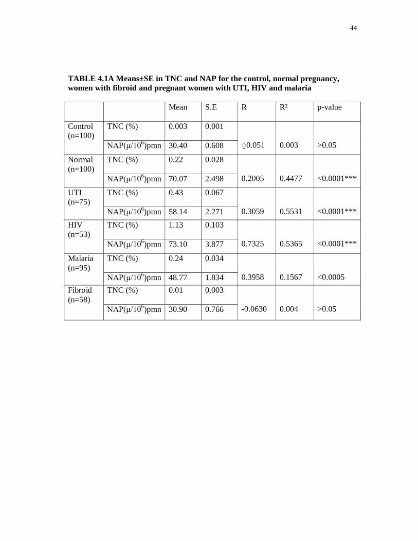
4.0 RESULTS The result of the present study demonstrated the mean TNC and NAP (table 4.1A)
activity for the control subjects to be .003 x 109/L±0.001 and 30.40±0.608
(µ/x106pmn) respectively.
For normal pregnant women it was 0.22±0.028 x 10⁹/L and 70.07± 2.498 (µ/ x 106pmn).
Pregnant women with UTI it was 0.43 x 10⁹L±0.067 and 58.14±2.271 (µ/106pm), those
with HIV 1.13 x 10⁹/L±0.103 and 73.10±3.877 (µ/x106pmn) respectively.
For pregnant women with malaria 0.23 x 10⁹/L±0.034 and 48.77±1.834 (µ/x106pmn)
respectively, and for the fibroid subjects it is 0.01±0.003x10⁹/L and 30.90±0.766
(µ/x106pmn). Pregnant women with HIV had the highest number of Neutrophils with
toxic granules, followed by those with UTI then those with malaria. This may reflect
the relative risk of infection in these subjects. Normal pregnant women had higher
values than the control and women with fibroid.
For NAP activity subjects with HIV also show the highest value for mean NAP
activity, this finding has not been reported earlier.
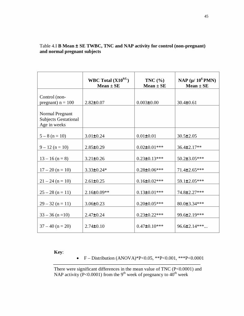
Significant difference exists (table 4.1B) between the control subjects and
normal pregnant women, mean TNC (P<0.0001) from the 9th week, for NAP activity
(P<0.0001) from 13th week and at 9 – 12 week (P<0.001). There was also significant
differences in both parameters (P<0.0001) between the control and subjects with
42
complications in pregnancy (HIV (table 4.4), UTI (table 4.2) and Malaria (table 4.3))
and also between the control and subjects with fibroids (P<0.0001). This may reflect
the different states of the patients – pregnancy and infection.
Between the normal pregnant subjects and those with complications in
pregnancy there was no significant difference (P>0.05) in NAP activity (table 4.5),
except at 5 – 8 weeks where (P< 0.05) in UTI and malaria subjects. TNC shows
significant differences (table 4.6) in UTI subjects (P<0.0001) at weeks 5 – 8, 25 – 28
and 37 – 40 weeks, at 9 – 12 weeks (P<0.001) and (P<0.05) at 21 – 24 and 33 -36
weeks. In HIV subjects (P<0.0001) across the gestational period, in subjects with
malaria (P<0.001) at weeks 5 – 8, 9 – 12, 21 – 24 and 25 – 28. At 13 – 16 and 17 – 20
weeks there was no significant difference. This indicates a response in TNC in the
presence of infection.
Subjects with HIV showed no significant difference in TWBC (table 4.7) when
compared with the normal pregnant subjects except at week 13 – 16 weeks (P<0.05).
Those with malaria show significant difference (P<0.001) at 5 – 8 weeks and 21 – 24
(P<0.0001) at 25 – 28 weeks and at 9 – 12 weeks (P<0.05)
In subjects with UTI there was significant difference in TWBC (P<0.0001) at 21 – 24
and 25 – 28 weeks, (P<0.001) at 9 – 12 weeks and 33 – 36 weeks and (P<0.05) at 5 – 8
weeks. This also signifies response in TWBC to infection.
Between HIV subjects and the control group there was no significant difference
in TWBC except at 13 – 16 weeks were (P<0.05). The same pattern of difference
43
occurred between HIV subjects and normal pregnant subjects. This is not as expected
and may be attributed to:
1. The prevention of mother-to-child transmission of HIV (PMTCT)
antiretroviral regimens being administered to the patients – Zidovudine.
2. Pregnancy - which causes mild leucocytosis 6.3 – 16.0 x 10⁹/L
El-maalem et al 1980).
3. Nutritional - Plus Environmental Factors.
44
TABLE 4.1A Means±SE in TNC and NAP for the control, normal pregnancy, women with fibroid and pregnant women with UTI, HIV and malaria
Mean S.E R R² p-value
Control (n=100)
TNC (%)
0.003 0.001 ֵ◌0.051
0.003
>0.05 NAP(μ/106)pmn 30.40 0.608
Normal (n=100)
TNC (%)
0.22 0.028 0.2005
0.4477
<0.0001*** NAP(μ/106)pmn 70.07 2.498
UTI (n=75)
TNC (%)
0.43 0.067 0.3059
0.5531
<0.0001*** NAP(μ/106)pmn 58.14 2.271
HIV (n=53)
TNC (%)
1.13 0.103 0.7325
0.5365
<0.0001*** NAP(μ/106)pmn 73.10 3.877
Malaria (n=95)
TNC (%)
0.24 0.034 0.3958
0.1567
<0.0005 NAP(μ/106)pmn 48.77 1.834
Fibroid (n=58)
TNC (%)
0.01 0.003 -0.0630
0.004
>0.05 NAP(μ/106)pmn 30.90 0.766
45
Table 4.I B Mean ± SE TWBC, TNC and NAP activity for control (non-pregnant) and normal pregnant subjects
Key:
F – Distribution (ANOVA)*P<0.05, **P<0.001, ***P<0.0001 There were significant differences in the mean value of TNC (P<0.0001) and NAP activity (P<0.0001) from the 9th week of pregnancy to 40th week
WBC Total (X109/L)
Mean ± SE
TNC (%)
Mean ± SE
NAP (µ/ 106 PMN)
Mean ± SE Control (non-pregnant) n = 100
2.82±0.07
0.003±0.00
30.4±0.61
Normal Pregnant Subjects Gestational Age in weeks
5 – 8 (n = 10)
3.01±0.24
0.01±0.01
30.5±2.05
9 – 12 (n = 10)
2.85±0.29
0.02±0.01***
36.4±2.17**
13 – 16 (n = 8)
3.21±0.26
0.23±0.13***
50.2±3.05***
17 – 20 (n = 10)
3.33±0.24*
0.28±0.06***
71.4±2.65***
21 – 24 (n = 10)
2.61±0.25
0.16±0.02***
59.1±2.05***
25 – 28 (n = 11)
2.16±0.09**
0.13±0.01***
74.8±2.27***
29 – 32 (n = 11)
3.06±0.23
0.20±0.05***
80.0±3.34***
33 – 36 (n =10)
2.47±0.24
0.23±0.22***
99.6±2.19***
37 – 40 (n = 20)
2.74±0.10
0.47±0.10***
96.6±2.14***...
46
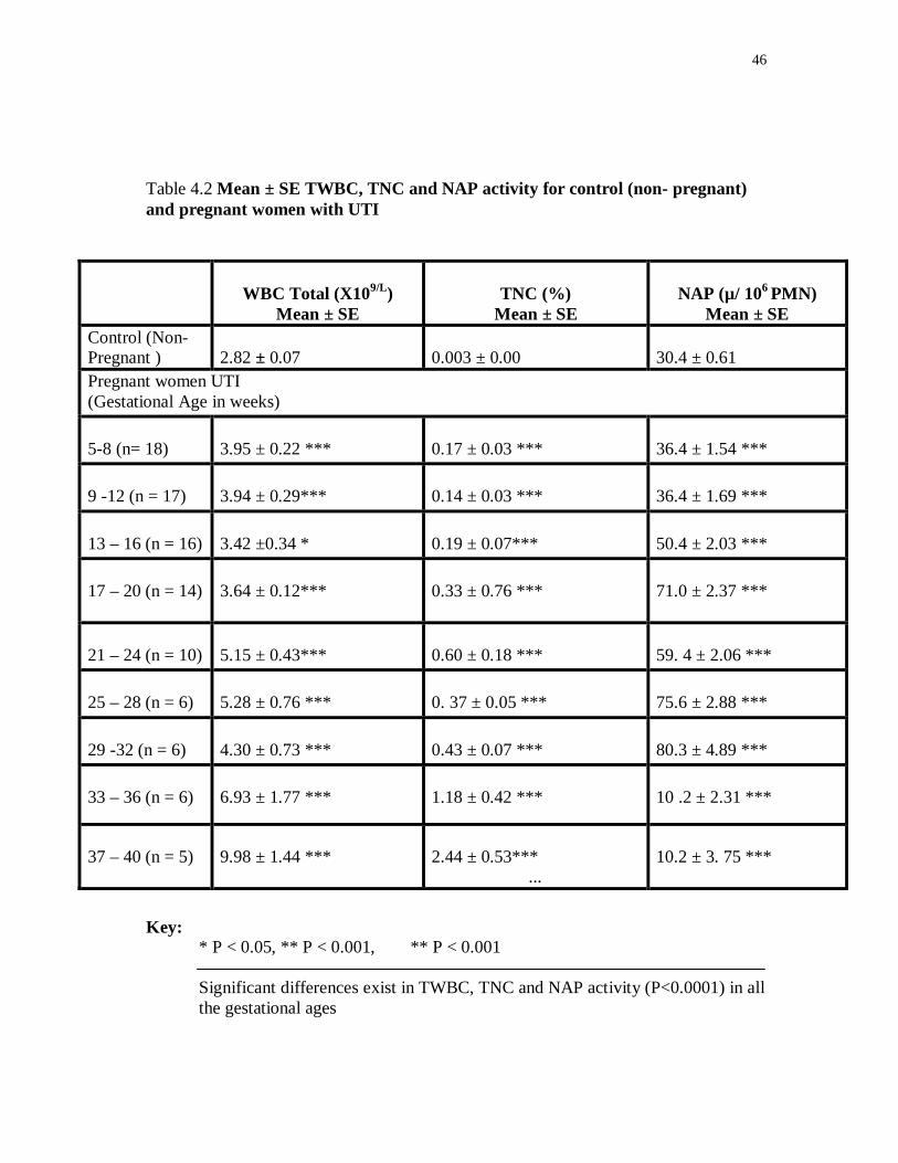
Table 4.2 Mean ± SE TWBC, TNC and NAP activity for control (non- pregnant) and pregnant women with UTI
WBC Total (X109/L)
Mean ± SE
TNC (%)
Mean ± SE
NAP (µ/ 106 PMN)
Mean ± SE Control (Non-Pregnant )
2.82 ± 0.07
0.003 ± 0.00
30.4 ± 0.61
Pregnant women UTI (Gestational Age in weeks) 5-8 (n= 18)
3.95 ± 0.22 ***
0.17 ± 0.03 ***
36.4 ± 1.54 ***
9 -12 (n = 17)
3.94 ± 0.29***
0.14 ± 0.03 ***
36.4 ± 1.69 ***
13 – 16 (n = 16)
3.42 ±0.34 *
0.19 ± 0.07***
50.4 ± 2.03 ***
17 – 20 (n = 14)
3.64 ± 0.12***
0.33 ± 0.76 ***
71.0 ± 2.37 ***
21 – 24 (n = 10)
5.15 ± 0.43***
0.60 ± 0.18 ***
59. 4 ± 2.06 ***
25 – 28 (n = 6)
5.28 ± 0.76 ***
0. 37 ± 0.05 ***
75.6 ± 2.88 ***
29 -32 (n = 6)
4.30 ± 0.73 ***
0.43 ± 0.07 ***
80.3 ± 4.89 ***
33 – 36 (n = 6)
6.93 ± 1.77 ***
1.18 ± 0.42 ***
10 .2 ± 2.31 ***
37 – 40 (n = 5)
9.98 ± 1.44 ***
2.44 ± 0.53*** ...
10.2 ± 3. 75 ***
Key: * P < 0.05, ** P < 0.001, ** P < 0.001
Significant differences exist in TWBC, TNC and NAP activity (P<0.0001) in all the gestational ages
47
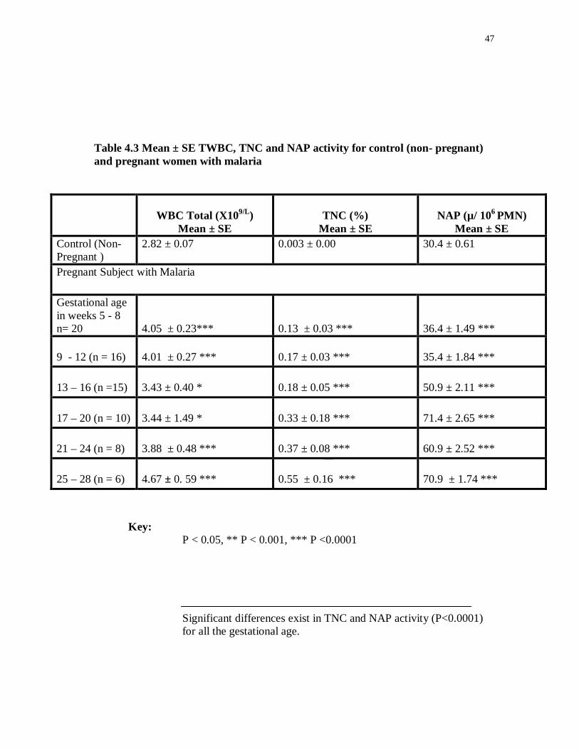
Table 4.3 Mean ± SE TWBC, TNC and NAP activity for control (non- pregnant) and pregnant women with malaria
WBC Total (X109/L)
Mean ± SE
TNC (%)
Mean ± SE
NAP (µ/ 106 PMN)
Mean ± SE Control (Non-Pregnant )
2.82 ± 0.07 0.003 ± 0.00 30.4 ± 0.61
Pregnant Subject with Malaria
Gestational age in weeks 5 - 8 n= 20
4.05 ± 0.23***
0.13 ± 0.03 ***
36.4 ± 1.49 ***
9 - 12 (n = 16)
4.01 ± 0.27 ***
0.17 ± 0.03 ***
35.4 ± 1.84 ***
13 – 16 (n =15)
3.43 ± 0.40 *
0.18 ± 0.05 ***
50.9 ± 2.11 ***
17 – 20 (n = 10)
3.44 ± 1.49 *
0.33 ± 0.18 ***
71.4 ± 2.65 ***
21 – 24 (n = 8)
3.88 ± 0.48 ***
0.37 ± 0.08 ***
60.9 ± 2.52 ***
25 – 28 (n = 6)
4.67 ± 0. 59 ***
0.55 ± 0.16 ***
70.9 ± 1.74 ***
Key:
P < 0.05, ** P < 0.001, *** P <0.0001 Significant differences exist in TNC and NAP activity (P<0.0001) for all the gestational age.
48
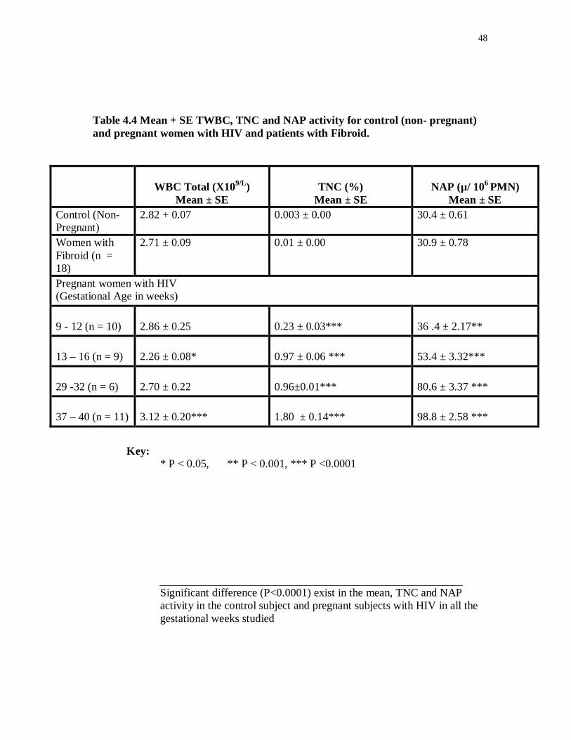
Table 4.4 Mean + SE TWBC, TNC and NAP activity for control (non- pregnant) and pregnant women with HIV and patients with Fibroid.
WBC Total (X109/L)
Mean ± SE
TNC (%)
Mean ± SE
NAP (µ/ 106 PMN)
Mean ± SE Control (Non-Pregnant)
2.82 + 0.07 0.003 ± 0.00 30.4 ± 0.61
Women with Fibroid (n = 18)
2.71 ± 0.09 0.01 ± 0.00 30.9 ± 0.78
Pregnant women with HIV (Gestational Age in weeks) 9 - 12 (n = 10)
2.86 ± 0.25
0.23 ± 0.03***
36 .4 ± 2.17**
13 – 16 (n = 9)
2.26 ± 0.08*
0.97 ± 0.06 ***
53.4 ± 3.32***
29 -32 (n = 6)
2.70 ± 0.22
0.96±0.01***
80.6 ± 3.37 ***
37 – 40 (n = 11)
3.12 ± 0.20***
1.80 ± 0.14***
98.8 ± 2.58 ***
Key: * P < 0.05, ** P < 0.001, *** P <0.0001
Significant difference (P<0.0001) exist in the mean, TNC and NAP activity in the control subject and pregnant subjects with HIV in all the gestational weeks studied
49
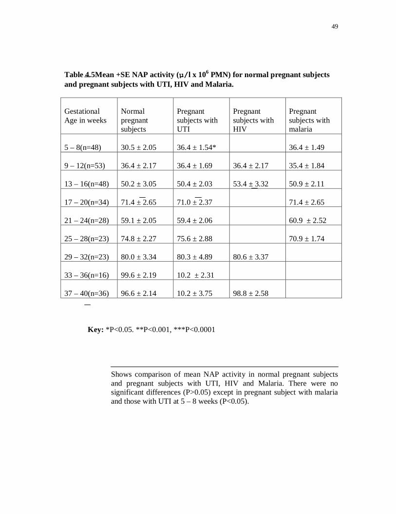
Table 4.5Mean +SE NAP activity (µ/l x 106 PMN) for normal pregnant subjects and pregnant subjects with UTI, HIV and Malaria. Gestational Age in weeks
Normal pregnant subjects
Pregnant subjects with UTI
Pregnant subjects with HIV
Pregnant subjects with malaria
5 – 8(n=48)
30.5 ± 2.05
36.4 ± 1.54*
36.4 ± 1.49
9 – 12(n=53)
36.4 ± 2.17
36.4 ± 1.69
36.4 ± 2.17
35.4 ± 1.84
13 – 16(n=48)
50.2 ± 3.05
50.4 ± 2.03
53.4 ± 3.32
50.9 ± 2.11
17 – 20(n=34)
71.4 ± 2.65
71.0 ± 2.37
71.4 ± 2.65
21 – 24(n=28)
59.1 ± 2.05
59.4 ± 2.06
60.9 ± 2.52
25 – 28(n=23)
74.8 ± 2.27
75.6 ± 2.88
70.9 ± 1.74
29 – 32(n=23)
80.0 ± 3.34
80.3 ± 4.89
80.6 ± 3.37
33 – 36(n=16)
99.6 ± 2.19
10.2 ± 2.31
37 – 40(n=36)
96.6 ± 2.14
10.2 ± 3.75
98.8 ± 2.58
Key: *P<0.05. **P<0.001, ***P<0.0001
Shows comparison of mean NAP activity in normal pregnant subjects and pregnant subjects with UTI, HIV and Malaria. There were no significant differences (P>0.05) except in pregnant subject with malaria and those with UTI at 5 – 8 weeks (P<0.05).
50
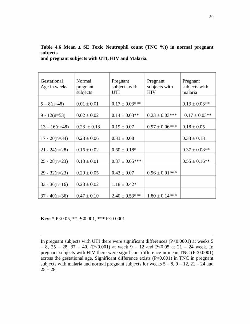
Table 4.6 Mean ± SE Toxic Neutrophil count (TNC %)) in normal pregnant subjects and pregnant subjects with UTI, HIV and Malaria.
Gestational Age in weeks
Normal pregnant subjects
Pregnant subjects with UTI
Pregnant subjects with HIV
Pregnant subjects with malaria
5 – 8(n=48)
0.01 ± 0.01
0.17 ± 0.03***
0.13 ± 0.03**
9 - 12(n=53)
0.02 ± 0.02
0.14 ± 0.03**
0.23 ± 0.03***
0.17 ± 0.03**
13 – 16(n=48)
0.23 ± 0.13
0.19 ± 0.07
0.97 ± 0.06***
0.18 ± 0.05
17 - 20(n=34)
0.28 ± 0.06
0.33 ± 0.08
0.33 ± 0.18
21 - 24(n=28)
0.16 ± 0.02
0.60 ± 0.18*
0.37 ± 0.08**
25 - 28(n=23)
0.13 ± 0.01
0.37 ± 0.05***
0.55 ± 0.16**
29 - 32(n=23)
0.20 ± 0.05
0.43 ± 0.07
0.96 ± 0.01***
33 - 36(n=16)
0.23 ± 0.02
1.18 ± 0.42*
37 - 40(n=36)
0.47 ± 0.10
2.40 ± 0.53***
1.80 ± 0.14***
Key: * P<0.05, ** P<0.001, *** P<0.0001
In pregnant subjects with UTI there were significant differences (P<0.0001) at weeks 5 – 8, 25 – 28, 37 – 40, (P<0.001) at week 9 – 12 and P<0.05 at 21 – 24 week. In pregnant subjects with HIV there were significant difference in mean TNC (P<0.0001) across the gestational age. Significant difference exists (P<0.001) in TNC in pregnant subjects with malaria and normal pregnant subjects for weeks 5 – 8, 9 – 12, 21 – 24 and 25 – 28.
51
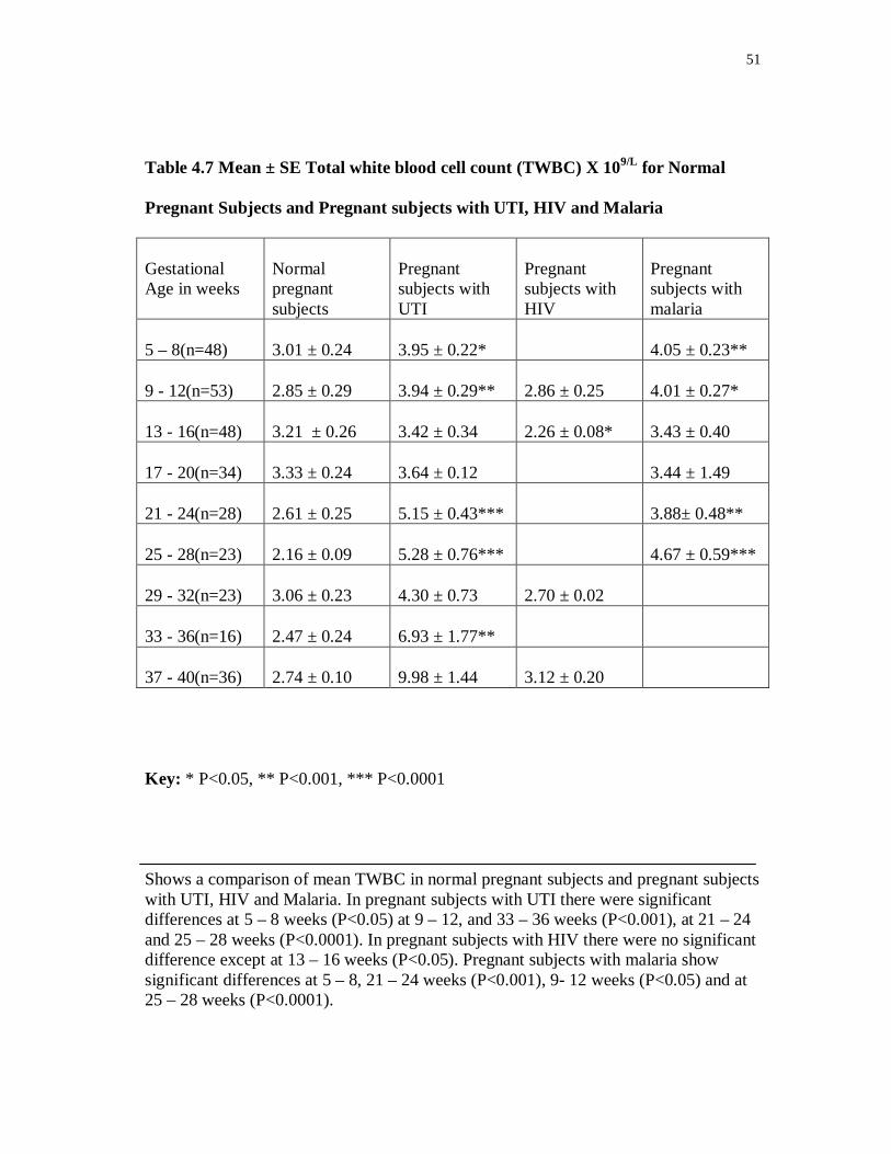
Table 4.7 Mean ± SE Total white blood cell count (TWBC) X 109/L for Normal
Pregnant Subjects and Pregnant subjects with UTI, HIV and Malaria
Gestational Age in weeks
Normal pregnant subjects
Pregnant subjects with UTI
Pregnant subjects with HIV
Pregnant subjects with malaria
5 – 8(n=48)
3.01 ± 0.24
3.95 ± 0.22*
4.05 ± 0.23**
9 - 12(n=53)
2.85 ± 0.29
3.94 ± 0.29**
2.86 ± 0.25
4.01 ± 0.27*
13 - 16(n=48)
3.21 ± 0.26
3.42 ± 0.34
2.26 ± 0.08*
3.43 ± 0.40
17 - 20(n=34)
3.33 ± 0.24
3.64 ± 0.12
3.44 ± 1.49
21 - 24(n=28)
2.61 ± 0.25
5.15 ± 0.43***
3.88± 0.48**
25 - 28(n=23)
2.16 ± 0.09
5.28 ± 0.76***
4.67 ± 0.59***
29 - 32(n=23)
3.06 ± 0.23
4.30 ± 0.73
2.70 ± 0.02
33 - 36(n=16)
2.47 ± 0.24
6.93 ± 1.77**
37 - 40(n=36)
2.74 ± 0.10
9.98 ± 1.44
3.12 ± 0.20
Key: * P<0.05, ** P<0.001, *** P<0.0001
Shows a comparison of mean TWBC in normal pregnant subjects and pregnant subjects with UTI, HIV and Malaria. In pregnant subjects with UTI there were significant differences at 5 – 8 weeks (P<0.05) at 9 – 12, and 33 – 36 weeks (P<0.001), at 21 – 24 and 25 – 28 weeks (P<0.0001). In pregnant subjects with HIV there were no significant difference except at 13 – 16 weeks (P<0.05). Pregnant subjects with malaria show significant differences at 5 – 8, 21 – 24 weeks (P<0.001), 9- 12 weeks (P<0.05) and at 25 – 28 weeks (P<0.0001).
52
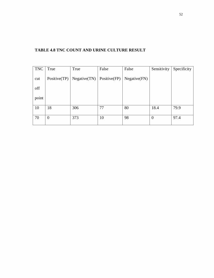
TABLE 4.8 TNC COUNT AND URINE CULTURE RESULT
TNC
cut
off
point
True
Positive(TP)
True
Negative(TN)
False
Positive(FP)
False
Negative(FN)
Sensitivity Specificity
10 18 306 77 80 18.4 79.9
70 0 373 10 98 0 97.4
53
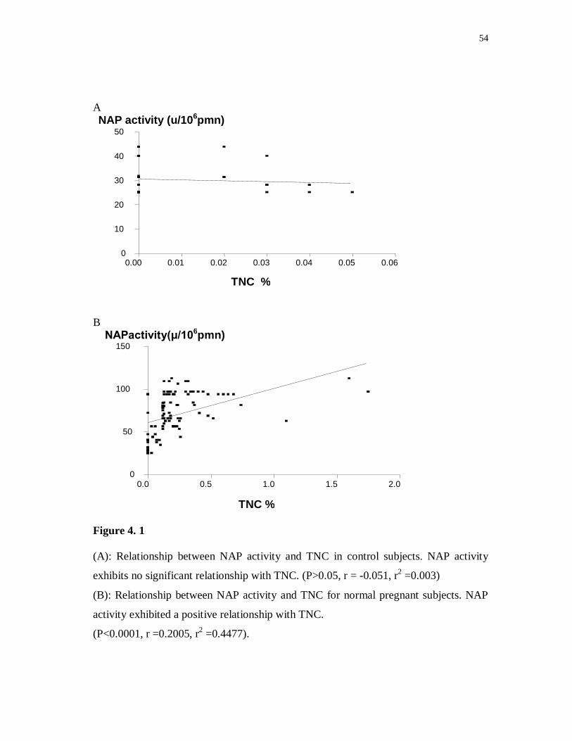
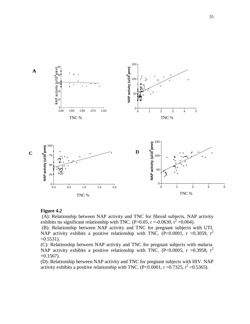
Significant relationship exists between NAP activity and TNC in pregnant subjects with
HIV (P<0.0001, r =0.7325, r2 = 0.5365), Pregnant subjects with UTI (P<0.0001, r =
0.3059, r2 = 0.5531), Pregnant subjects with malaria (P<0.0005, r = 0.3958, r2 = 0.1567)
and also for normal pregnant subjects (P<0.0001, r = 0.2005, r2 = 0.4477) Fig. 4.2DBC,
B, C and Fig 4.1B There was no relationship between NAP activity and TNC for the
control fig.4.1A (P>0.05, r = 0.051, r2 = 0.003) and fibroid subjects fig.4.2A (P>0.05, r
= 0.3013, r2 = 0.091). These points to a relationship between NAP activity and TNC in
the presence of a common factor – pregnancy and infection. These results could not be
readily compared with any, as none, to the best of our knowledge existed.
54
A
B
Figure 4. 1 (A): Relationship between NAP activity and TNC in control subjects. NAP activity
exhibits no significant relationship with TNC. (P>0.05, r = -0.051, r2 =0.003)
(B): Relationship between NAP activity and TNC for normal pregnant subjects. NAP
activity exhibited a positive relationship with TNC.
(P<0.0001, r =0.2005, r2 =0.4477).
0.00 0.01 0.02 0.03 0.04 0.05 0.060
10
20
30
40
50
TNC %
NAP activity (u/106pmn)
0.0 0.5 1.0 1.5 2.0 0
50
100
150
TNC %
NAPactivity(μ/106pmn)
55
0.000 0.025 0.050 0.075 0.1000
10
20
30
40
50
TNC (x106)
NA
P ac
tivity
(u/1
06 pmn)
0 1 2 3 4 5
0
50
100
150
TNC (x106)
NA
P ac
tivity
(u/1
06 pmn)
0.0 0.5 1.0 1.5 2.00
25
50
75
100
TNC (x106)
NA
P ac
tivity
(u/1
06 pmn)
Figure 4.2 (A): Relationship between NAP activity and TNC for fibroid subjects. NAP activity exhibits no significant relationship with TNC. (P>0.05, r =-0.0630, r2 =0.004). (B): Relationship between NAP activity and TNC for pregnant subjects with UTI. NAP activity exhibits a positive relationship with TNC. (P<0.0001, r =0.3059, r2 =0.5531). (C): Relationship between NAP activity and TNC for pregnant subjects with malaria. NAP activity exhibits a positive relationship with TNC. (P<0.0005, r =0.3958, r2 =0.1567). (D): Relationship between NAP activity and TNC for pregnant subjects with HIV. NAP activity exhibits a positive relationship with TNC. (P<0.0001, r =0.7325, r2 =0.5365).
0 1 2 3 40
50
100
150
TNC (x106)
NA
P ac
tivity
(u/1
06 pmn)
C D
TNC % TNC %
A
TNC % TNC %
A
56
zzzz
hhhhhhhhhhhhhhhhhhhhhh
Z
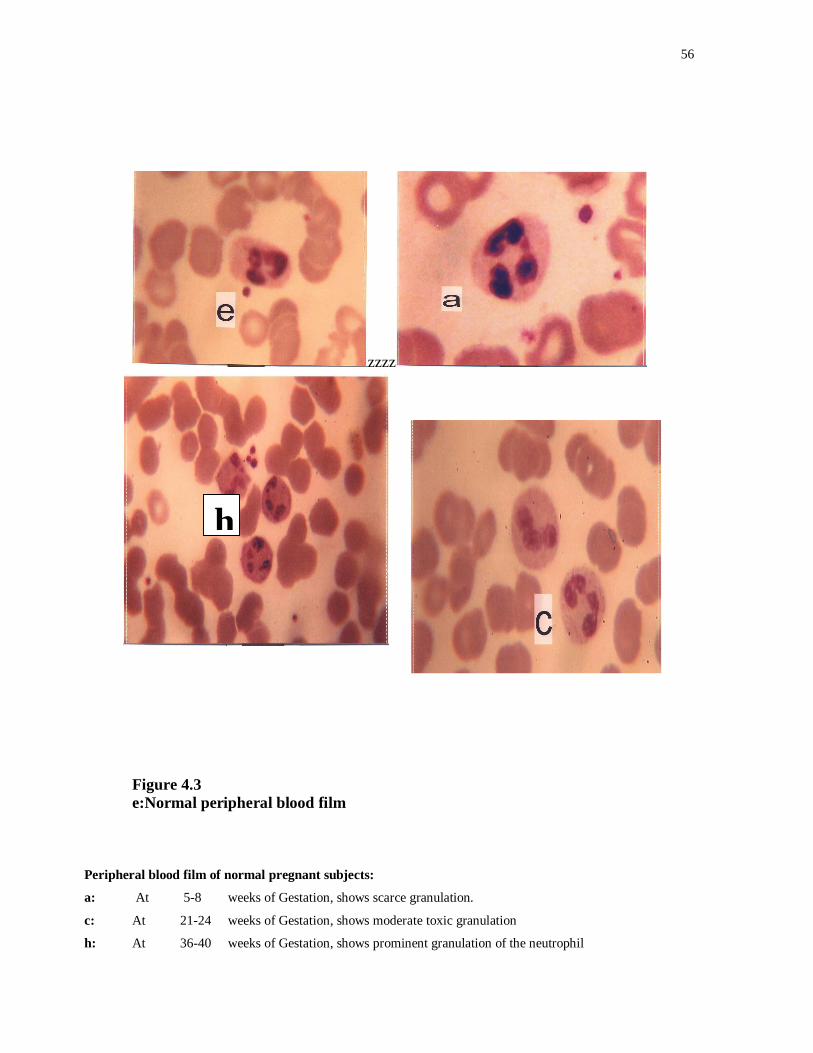
Figure 4.3 e:Normal peripheral blood film
h
Peripheral blood film of normal pregnant subjects:
a: At 5-8 weeks of Gestation, shows scarce granulation.
c: At 21-24 weeks of Gestation, shows moderate toxic granulation
h: At 36-40 weeks of Gestation, shows prominent granulation of the neutrophil
h
57
Gestation
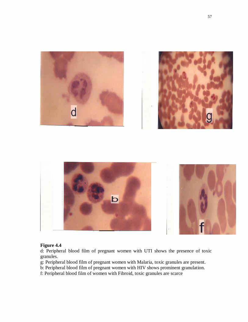
Figure 4.4 d: Peripheral blood film of pregnant women with UTI shows the presence of toxic granules. g: Peripheral blood film of pregnant women with Malaria, toxic granules are present. b: Peripheral blood film of pregnant women with HIV shows prominent granulation. f: Peripheral blood film of women with Fibroid, toxic granules are scarce
58
CHAPTER FIVE
5.0 DISCUSSION AND CONCLUSION Toxic granules are dark blue or purple cytoplasmic granules in the
band/polymorph cells. Toxic granules are azurophilic that retain basophilic staining
reaction by lack of maturation or increased basophilia. The granules in the neutrophil
represent packages of enzymes, which are involved in the killing of ingested microbes
and the digestion of phagocytised material. Based on differences in enzyme content,
the granules can be classified into two different subtypes. The primary lightly
basophilic staining granules contain, for example, peroxidase plus acid hydrolytic
enzymes, while secondary granules contain alkaline phosphatase and certain other
enzymes (Jadhav et al (2003)).
There was a gradual rise in NAP activity across the gestational age. This agrees with an
earlier report by EL –Maalem et al (1980), however contradicts with the cytochemical
study of Beal et al (1967) who reported no differences between the means for tests
done in early pregnancy and those performed later in pregnancy.
Hence, the relationship between the quantitative biochemical measurements of NAP by
direct assay of leucocytes and the NAP cytochemical score need to be established.
Several studies have evaluated the complete blood count and leukocyte differential
counts for disease detection and found the utility to be very low.
59
The result of our study showed TNC to have a specificity of 79% at a cut off of 10 and
97% at a cut off of 70 and a sensitivity of 18%at a cut off of 10 and 0% at a cut off of
70.
Our present study of the significance of the morphological changes in neutrophil (toxic
granulation) has also followed the same pattern of low utility, probably due to
biological variability. When expressed as coefficient of variance it was found that the
CV for granulocytes in the elderly was 17.3-19.3% within subjects and 21.2-29%
between subjects. (Gwendolyn, 1991).
The within individual values may provide a much more sensitive index to changes with
disease.

60
5.1 RECOMMENDATIONS
There appears to be no previous study reporting about the significanc= of
Neutrophil toxic granulation in pregnancy. The present study should evoke interest
amongst researchers spurring them to undertake larger studies on the subject.
61
REFERENCES
Aboyeji, A.P. and Ijaiya, M.A., (2005). Uterine Fibroids: A ten year clinical review in Ilorin, Nigeria. Nigeria Journal of Medicine 11:16 – 19. Akerele, J., Abhulimen, P., Okonofua, F., (2001). Prevalence of Asymptomatic bacteiuria among pregnant women in Benin City, Nigeria. Journal of Obstetrics and Gynaecology 21: 141 – 144. Al-Gwaiz Layla, Hanan H. Babay (2007). The Diagnostic value of Absolute Neutrophil Count, Band Count and Morphologic changes of Neutrophils in predicting bacterial infections. Medicine Principles and Practice 16:344 – 347. Babior Bernard.M, and Golde, David W., (2001) Production, Distribution and fate of neutrophils. Williams Haematology 6th Ed. Mac Graw Hill. PP. 753 – 756. Bachman, J.W., Heise R.H., Naessens, J.M., Timmerman, M.G. (1993). A study of various tests to detect asymptomatic Urinary tract infections in an obstetric population. JAMA.270:1971 – 1974. Bainton, Dorothy F. (2001). Morphology of Neutrophils, Eosinophils And Basophils. Williams Haematology 6th Ed
Mac Graw Hill. PP. 729 – 735. Bain, J.Barbara and Lewis.S. Mitchell (2003). Preparation and
Staining methods for blood and bone marrow films. Practical Haematology 9th ed. Churchill. Livingstone, PP. 47 – 63. Baker, F.J., and Silverton (1985) Introduction to medical Laboratory Technology. 6th Ed. Butterworth’s. London, PP 316 – 334. Beal, R.W., Read, W.M.F. and Turvey, P.A. (1967). Neutrophil Alkaline phosphatase in pregnancy. Journal of Clinical Pathology. Vol.20:749 – 751.
62
Butler, Ernest (2001) composition and metabolism of Neutrophils. Williams Haematology 6th Ed. Mac Graw Hill. PP 745 – 747. Birdi, N.Klassen, T.P., Quinlan, A., Clarke, W., Hoking, M., Momy, J.A., Rowe, P.C. (1999). Role of the toxic Neutrophil Count in the early diagnosis of Kawasaki disease. Journal of Rheumatology, 26:945 – 1006. Brien, John F.O. (1986) the lysosmal enzymes. Textbook of clinical Chemistry. W.B. Saunders Company. PP 771. Chaves Fernando, Bethany Teino, Dongsheng X’, (2005). Quantitative Determination of Neutrophil VCS parameters By the Coulter Automated Haematology Analyser
American Journal of Clinical Pathology 124. (3) 440 – 444. Cheesbrough, M. (1984). Preparation of culture media. Medical
Laboratory manual for Tropical Countries, Microbiolgy Vol. 11 PP. 392 – 415. Cheesbrough, M. (1984). Collection, transportation and examination Of urine. Medical Laboratory manual for Tropical Countries. Microbiology Vol.11 P.154. Dacie, John.V. and Lewis,.M., (1994). Preparation and staining methods
For blood and bone – marrow films. Practical Haematology 8th Ed. ELBS Hong Kong PP 89 – 90. Delzell, J.E., and Lefevre, Michael L. (2000).Urinary tract infections During pregnancy. American family physicians 61:3 El – Maallem, H. and Fletcher, J. (1976). Defective Neutrophil function and In chronic granulocytic Leukaemia. British Journal Haematology 34:95 – 103. EL – Maallem, H, and Fletcher, J. (1980). Impaired Neutrophil function and Myeloperoxidase deficiency in pregnancy. British Journal Haematology 44:375 – 381
63
Ezechi, Oliver C., Fasuba Olusola, B., Dare, Francis O., (2003). Antibiotic Sensitivity patterns of microbial isolates from urine of pregnant Women with urinary tract infection. Tropical Journal of Obstetrics and Gynaecology 20:113 – 115. Gilstrap, L.C., Cunningham, F.G., Whalley, P.J., (1981). Acute pyelonephritis in pregnancy: An anterospective study, Obstetric and Gynaecology 57:409 – 413. Gwendolyn L. Gilbert (1991). Urinary tract infections in pregnancy Infections disease in pregnancy and the new born. Harwood Academic. Switzerland PP. 413 – 429. Hardie, D. (1999) Human reteroviruses. www.virology.org. Harris, R.E., Thomas, V., Shelokov, A. (1976). Asymptomatic bacteriuria in Pregnancy: antibody – coated bacteria, renal function, and intrauterine growth retardation.Am.J.Obstet and Gynae.126:20 – 25. Jadhav, U.M., Singhvia, .R. Shah, R., (2003). Diagnostic implications of white cell differential count and white cell morphology in malaria Journal of Post Graduate Medicine .49:218 – 221. Lee (2003). Treats Fibroid, Tumours successfully without surgery. www.bioline.org. Levy, J.A. (1993). Structure of the human immune deficiency virus. www.academicjournals.org. Lucas, M.J., Cunningham, F.G., (1993). Urinary tract infections In pregnancy. Clinical Obstricts and Gynaecology 36:855 – 868. Meranze, D.R., Mendell T.H., Meranze, T., (1935). Cytoplasmic Changes in the peripheral Neutrophil. American Journal Medicine Sciences 189:639.
64
Moore, R.D.(2008). Neutropenia. HIV Guide. www.hopkins - hivguide.org./diagnosis/organ system/hematologic/neutropenia.html.2/6/2000. Mohammed, A., Sheu S.M., Ahmed, A.A., Mayun, A.A, Tiffin, I.U., Alkali, G., Abubakar, A.L. (2005). Uterine leiomyomata: A five year clinicopathological review in Zaria Nigeria. Nigerian Journal of Surgical Research. 7:1. Noel, H. (2005). AIDS at 20: A look back, a look ahead. www.umm.ed. Rambaldi, A., Masuhara,.K., Borleri, G.M., Amaru, R., Gianni, M., Terao, M., Barbui, T., Garattini, E. (1997). Flow cytometry of leucyte alkaline phosphatase in normal and pathologic leucocytes. Bri.J.Haem. 96:815 – 822. Rashmi., S.M., Alka, D.K., Ramakant, S.N., (2006). Neutrophils in health and diseases. Journal of Oral Maxillofacial Pathology. 10:3 – 8. Sagay, A.S., Imade, G.E., Kapiga, S., Omoregie, .R. Egah, D.Z., Falusi, A.O, Ekawempu, C.C., Uju, A.C., Sankale, J.L., Kanti, P. (2004) Risk factors of HIV infection in pregnant women in northern Nigeria. Int conf.AIDS.http//gatemkiy nlm – nih.gor/meeting abstract. Shen, B.J.,Ekert, H. Tauro, G.P., and Balderas, A (1984). Left shift in the Peripheral blood count at diagnosis in acute Lymphocytic Leukaemia is significantly correlated with duration of complete remission. Blood. 63:216 – 218. Smolen, J.E., and Boxer, L.A., (2001). Functions of Neutrophils. Williams Haematology. 6th Ed.Mc. Graw Hill. PP. 761 – 773. . UNAIDS, (2004). AIDS Epidemic up date 2004. Geneva UNAIDS, 2004. Uneke, C.J., Duhlinska, D.D., Igbinedion, E.B (2007). Prevalence of public – health significance of HIV infection and anaemia among pregnant women in South – Eastern Nigeria. J.Health, Population and Nutrition. 3:328 – 335.
65
Uneke, C.J. Festus, E. Iyare, Patrick Oke, Dochka, D., Duhlinska, (2008). Assessment of malaria in pregnancy using rapid diagnostic tests and its association with HIV infection and haematologic parameters in South – Eastern Nigeria. Haematologica 93:143 – 144. Ukaejiofo, E.O., W.A. Isaacs – Sodeye, E. Seyide Adigun and A. Ipadeola (1979) Normal Haematological values in adult Nigerians. Nigerian Medical Journal 9:119. Whitty, C.J.M., Sally. E., Mutabingwa, T.K., (2005). Malaria in pregnancy. British Journal of Obstetrics and Gynaecology 112:1189 – 1195. Wing, D.A., Park, A.S., De Bugue, L., Millar, L.K. (2000) Limited clinical utility of blood and urine culture in the treatment of acute pyelonephritis during pregnancy. American Journal Obstetrics and Gynaecology 182:1437 – 1441.
Related Documents