REVIEW Open Access Neutrophil roles in left ventricular remodeling following myocardial infarction Yonggang Ma 1,2* , Andriy Yabluchanskiy 1,2 and Merry L Lindsey 1,2,3* Abstract Polymorphonuclear granulocytes (PMNs; neutrophils) serve as key effector cells in the innate immune system and provide the first line of defense against invading microorganisms. In addition to producing inflammatory cytokines and chemokines and undergoing a respiratory burst that stimulates the release of reactive oxygen species, PMNs also degranulate to release components that kill pathogens. Recently, neutrophil extracellular traps have been shown to be an alternative way to trap microorganisms and contain infection. PMN-derived granule components are also involved in multiple non-infectious inflammatory processes, including the response to myocardial infarction (MI). In this review, we will discuss the biological characteristics, recruitment, activation, and removal of PMNs, as well as the roles of PMN-derived granule proteins in inflammation and innate immunity, focusing on the MI setting when applicable. We also discuss future perspectives that will direct research in PMN biology. Keywords: PMNs, Myocardial infarction, Inflammation, Innate immunity, Degranulation, Matrix metalloproteinases Review Introduction Polymorphonuclear granulocytes (PMNs; neutrophils) are a type of leukocyte of approximately 10 μm in diameter that play vital roles in the innate immunity response to pathogens. PMNs are the first responders to infection or injury. Persistent neutropenia leads to increased risk of microorganism infections, while excessive recruitment and activation or delayed removal of PMNs results in tissue damage in inflammatory disorders [1]. Following myocardial infarction (MI), numbers of circulating PMNs increase, and the post-MI PMN to lymphocyte ratio has been reported by Akpek and colleagues to predict major adverse cardiac events in MI patients [2]. While PMN counts do not improve the ability to diagnose MI, they are a prognostic biomarker of chronic remodeling of the left ventricle (LV) [3]. Increased PMN counts after percutaneous coronary intervention for ST-elevation MI associates with larger infarct sizes and worse cardiac function [4]. Neutrophil depletion reduces infarct size and the extent of injury in a canine model [5,6]. As such, PMNs have been shown to mediate MI-induced cardiac injury and remodeling. However, the potential mechanisms by which neutrophils regulate MI-induced LV remodeling are not well understood, and PMN depletion strategies in humans increased adverse outcomes post-MI [7]. This review will discuss our current understanding of PMN biology, including recruitment, activation, clearance, and function. We also discuss the roles of PMN-derived components in inflammation and innate immunity, focusing on the MI setting. In addition, we propose future directions that may advance the PMN research arena. Biological characteristics of PMNs PMNs are the most abundant leukocyte cell type in mammals, accounting for ~35-75% of circulating leuko- cytes under normal conditions [8]. PMNs are the first-line immune cells recruited to sites of injury as a defense against microorganisms. PMN microbicidal mechanisms include receptor-mediated phagocytosis and intracellular killing, release of antimicrobial granule contents by degranulation, and the formation of neutrophil extracellular traps (NETs) [9]. In addition to their antimicrobial activity, growing evidence suggests that PMNs play an essential role in non-infectious inflammation, innate immunity, and tissue remodeling [10]. Based on ex vivo evaluation, murine and human PMNs have a circulating lifespan of 5–10 h [11,12]. However, * Correspondence: [email protected]; [email protected] 1 San Antonio Cardiovascular Proteomics Center, San Antonio, TX, USA 2 Jackson Center for Heart Research, Department of Physiology and Biophysics, University of Mississippi Medical Center, Jackson, MS, USA Full list of author information is available at the end of the article © 2013 Ma et al.; licensee BioMed Central Ltd. This is an Open Access article distributed under the terms of the Creative Commons Attribution License (http://creativecommons.org/licenses/by/2.0), which permits unrestricted use, distribution, and reproduction in any medium, provided the original work is properly cited. Ma et al. Fibrogenesis & Tissue Repair 2013, 6:11 http://www.fibrogenesis.com/content/6/1/11

Welcome message from author
This document is posted to help you gain knowledge. Please leave a comment to let me know what you think about it! Share it to your friends and learn new things together.
Transcript
REVIEW Open Access
Neutrophil roles in left ventricular remodelingfollowing myocardial infarctionYonggang Ma1,2*, Andriy Yabluchanskiy1,2 and Merry L Lindsey1,2,3*
Abstract
Polymorphonuclear granulocytes (PMNs; neutrophils) serve as key effector cells in the innate immune system andprovide the first line of defense against invading microorganisms. In addition to producing inflammatory cytokinesand chemokines and undergoing a respiratory burst that stimulates the release of reactive oxygen species, PMNsalso degranulate to release components that kill pathogens. Recently, neutrophil extracellular traps have beenshown to be an alternative way to trap microorganisms and contain infection. PMN-derived granule componentsare also involved in multiple non-infectious inflammatory processes, including the response to myocardial infarction(MI). In this review, we will discuss the biological characteristics, recruitment, activation, and removal of PMNs, aswell as the roles of PMN-derived granule proteins in inflammation and innate immunity, focusing on the MI settingwhen applicable. We also discuss future perspectives that will direct research in PMN biology.
Keywords: PMNs, Myocardial infarction, Inflammation, Innate immunity, Degranulation, Matrix metalloproteinases
ReviewIntroductionPolymorphonuclear granulocytes (PMNs; neutrophils) area type of leukocyte of approximately 10 μm in diameterthat play vital roles in the innate immunity response topathogens. PMNs are the first responders to infection orinjury. Persistent neutropenia leads to increased risk ofmicroorganism infections, while excessive recruitment andactivation or delayed removal of PMNs results intissue damage in inflammatory disorders [1]. Followingmyocardial infarction (MI), numbers of circulating PMNsincrease, and the post-MI PMN to lymphocyte ratio hasbeen reported by Akpek and colleagues to predict majoradverse cardiac events in MI patients [2]. While PMNcounts do not improve the ability to diagnose MI, they area prognostic biomarker of chronic remodeling of theleft ventricle (LV) [3]. Increased PMN counts afterpercutaneous coronary intervention for ST-elevationMI associates with larger infarct sizes and worse cardiacfunction [4]. Neutrophil depletion reduces infarct size andthe extent of injury in a canine model [5,6]. As such, PMNshave been shown to mediate MI-induced cardiac injury
and remodeling. However, the potential mechanisms bywhich neutrophils regulate MI-induced LV remodeling arenot well understood, and PMN depletion strategies inhumans increased adverse outcomes post-MI [7]. Thisreview will discuss our current understanding of PMNbiology, including recruitment, activation, clearance, andfunction. We also discuss the roles of PMN-derivedcomponents in inflammation and innate immunity,focusing on the MI setting. In addition, we propose futuredirections that may advance the PMN research arena.
Biological characteristics of PMNsPMNs are the most abundant leukocyte cell type inmammals, accounting for ~35-75% of circulating leuko-cytes under normal conditions [8]. PMNs are the first-lineimmune cells recruited to sites of injury as a defense againstmicroorganisms. PMN microbicidal mechanisms includereceptor-mediated phagocytosis and intracellular killing,release of antimicrobial granule contents by degranulation,and the formation of neutrophil extracellular traps (NETs)[9]. In addition to their antimicrobial activity, growingevidence suggests that PMNs play an essential role innon-infectious inflammation, innate immunity, andtissue remodeling [10].Based on ex vivo evaluation, murine and human PMNs
have a circulating lifespan of 5–10 h [11,12]. However,
* Correspondence: [email protected]; [email protected] Antonio Cardiovascular Proteomics Center, San Antonio, TX, USA2Jackson Center for Heart Research, Department of Physiology andBiophysics, University of Mississippi Medical Center, Jackson, MS, USAFull list of author information is available at the end of the article
© 2013 Ma et al.; licensee BioMed Central Ltd. This is an Open Access article distributed under the terms of the CreativeCommons Attribution License (http://creativecommons.org/licenses/by/2.0), which permits unrestricted use, distribution, andreproduction in any medium, provided the original work is properly cited.
Ma et al. Fibrogenesis & Tissue Repair 2013, 6:11http://www.fibrogenesis.com/content/6/1/11
recent work by Pillay and colleagues using in vivo PMNlabeling has shown that the circulating lifespan ofhuman PMNs can last up to 5.4 days, indicating thatin vivo characteristics of PMNs may be altered byex vivo manipulation or that in vivo stimuli can preventPMN apoptosis [13]. In the proinflammatory environment,for example, PMN lifespan can be prolonged by tumornecrosis factor (TNF)-α- or interleukin (IL)-1β-stimulatedinhibition of apoptosis [14].PMN development and maturation take place in the
bone marrow. In the presence of growth factors andcytokines, pluripotent hematopoietic cells differentiateinto myeloblasts, which are the precursor cells of PMNs[15]. PMNs synthesize components stored in differentgranules as part of the maturation process [10]. It isestimated that PMNs are produced at ~1 × 10⁹ cells perkilogram body weight daily under physiological conditions[16]. Only 1-2% of mature PMNs circulate, while 98-99%remain in the bone marrow [17]. Circulating PMNs aremature, terminally differentiated cells that have lost theirproliferative capacity. In response to a challenge, maturePMNs in the bone marrow mobilize into the blood andare recruited to injury sites. PMN chemoattraction isregulated by chemokines, cytokines, and microbialproducts [1].
PMN extravasation and recruitment in response to MIIn the setting of MI, chemokines that recruit PMNs to sitesof ischemia include macrophage inflammatory protein-2α(MIP-2α, CXCL2, GRO β), leukotriene B4 (LTB4), CINC-1(CXCL1, GRO α, KC), IL-8 (CXCL8), and complement 5a[18,19]. PMN-attracting CXC chemokines are rapidly andprofoundly increased post-MI and have been localizedbound to glycosaminoglycans on endothelial cell surfacesor in the extracellular matrix. The accumulation of highconcentrations of chemokines at the ischemic site attractsPMNs to the injury area by interaction with cell surfacechemokine receptors [20].PMNs leave the circulation and infiltrate to the infarct
region through several sequential steps, collectively knownas extravasation. The extravasation of PMNs occursprimarily in post-capillary venules, where hemodynamicshear forces are diminished and the vessel wall is thin. Asa first step, PMNs are arrested from the fast-flowing bloodstream and roll on endothelial cells. This reaction is medi-ated through binding of P-selectin ligand 1 and L-selectinconstitutively expressed on PMNs to P-selectin, E-selectin,intercellular adhesion molecules (ICAMs), and vascularcell adhesion molecules expressed by activated endothelialcells [15]. Second, firm adhesion occurs by interaction ofthe β2 integrin lymphocyte function-associated antigen-1(αLβ2, LFA-1, CD11a/CD18) and macrophage-1 antigen(Mac-1, αMβ2, CD11b/CD18, CR3) present on PMNs withtheir ligands ICAM-1 and ICAM-2 on endothelial cells.
Next, PMN transendothelial migration takes place byparacellular or intracellular trafficking. While most PMNssqueeze between endothelial cells (paracellular trafficking),a small fraction penetrates and passes through pores inthe cytoplasm of individual endothelial cell (intracellulartrafficking) [15]. Mediators that guide migration are thesame as those of firm adhesion, namely integrins αLβ2 andαMβ2, ICAM-1, and ICAM-2. PMN homing to the infarctsite is similar to PMN extravasation into other tissues aspart of a common wound healing response to injury.In the absence of reperfusion, PMNs are the first
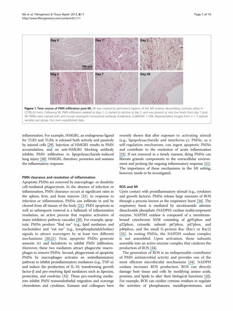
inflammatory cells recruited to the infarct area. Withpermanent occlusion in C57BL/6J mice, PMN infiltrationoccurs within hours post-MI, peaks at days 1–3, starts todecline at day 5, and is present at very low levels from day7 post-MI (Figure 1). As such, PMNs primarily regulatethe early LV remodeling response. PMNs initiate the acuteinflammatory response to engulf dead cells and tissuedebris and facilitate post-MI repair. However, excessivePMN infiltration or delayed regression exacerbates tissueinjury by the abundant release of inflammatory mediatorsand proteinases [21]. Hence, PMN infiltration and removalneed to be tightly controlled.
PMN activation post-MIIn response to infection, PMNs can be activated bypathogen-associated molecular patterns from pathogensor danger-associated molecular patterns (DAMPs) fromhost tissue via engagement with pattern recognitionreceptors expressed on the surface or within the cytoplasmof PMNs. PMNs express a wide array of pattern recognitionreceptors, including 12 of the13 known toll-like receptors(TLRs; the exception is TLR3), C-type lectin receptorsdectin 1 (CLEC7A) and CLEC2, NOD-like receptors(NLRs), and cytoplastic sensors of ribonucleic acids, includ-ing retinoic acid-inducible gene 1 (RIG-I) and melanomadifferentiation-associated protein 5 (MDA5) [22-26].Activated PMNs kill invading pathogens by the mecha-nisms of release of reactive oxygen species (ROS) andgranule proteins, as well as NETs. However, uncontrolledPMN accumulation can lead to injury to host tissueand cells.DAMPs are molecules that can initiate and perpetuate
the immune response in non-infectious inflammatoryconditions, and DAMPs are produced from hosttissue or immune cells in response to stress or injury.MI-associated DAMPs include heat shock proteins,high-mobility group box (HMGB)-1, low molecularhyaluronic acid, and fibronectin fragments [27]. DAMPs, asendogenous danger signal and secondary injury-promotingfactors, engage with pattern recognition receptors toactivate PMNs, other immune cells, or parenchymal cells[28]. This leads to the development of a proinflammatoryautocrine loop that can result in chronic or unresolved
Ma et al. Fibrogenesis & Tissue Repair 2013, 6:11 Page 2 of 10http://www.fibrogenesis.com/content/6/1/11
inflammation. For example, HMGB1, an endogenous ligandfor TLR2 and TLR4, is released both actively and passivelyby injured cells [29]. Injection of HMGB1 results in PMNaccumulation, and an anti-HMGB1 blocking antibodyinhibits PMN infiltration in lipopolysaccharide-inducedlung injury [30]. HMGB1, therefore, promotes and sustainsthe inflammatory response.
PMN clearance and resolution of inflammationApoptotic PMNs are removed by macrophage- or dendriticcell-mediated phagocytosis. In the absence of infection orinflammation, PMN clearance occurs at significant rates inthe spleen, liver, and bone marrow [31]. In response toinfection or inflammation, PMNs can infiltrate in and becleared from all tissues of the body [21]. PMN apoptosis aswell as subsequent removal is a hallmark of inflammationresolution, an active process that requires activation ofmany inhibitory pathway cascades [20]. For example, apop-totic PMNs produce “find me” (e.g., lipid mediators andnucleotides) and “eat me” (e.g., lysophosphatidylcholine)signals to attract scavengers by at least two differentmechanisms [20,21]. First, apoptotic PMNs generateannexin A1 and lactoferrin to inhibit PMN infiltration.Moreover, these two mediators attract phagocytic macro-phages to remove PMNs. Second, phagocytosis of apoptoticPMNs by macrophages activates an antiinflammatorypathway to inhibit proinflammatory mediators (e.g., TNF-α)and induce the production of IL-10, transforming growthfactor-β and pro-resolving lipid mediators such as lipoxins,protectins, and resolvins [32]. These pro-resolving media-tors inhibit PMN transendothelial migration and scavengechemokines and cytokines. Esmann and colleagues have
recently shown that after exposure to activating stimuli(e.g., lipopolysaccharide and interferon-γ), PMNs, as aself-regulation mechanism, can ingest apoptotic PMNsand contribute to the resolution of acute inflammation[33]. If not removed in a timely manner, dying PMNs canliberate granule components to the extracellular environ-ment and prolong the ongoing inflammatory response [21].The importance of these mechanisms in the MI setting,however, needs to be investigated.
ROS and MIUpon contact with proinflammatory stimuli (e.g., cytokinesand growth factors), PMNs release large amounts of ROSthrough a process known as the respiratory burst [34]. Therespiratory burst is mediated by nicotinamide adeninedinucleotide phosphate (NADPH) oxidase multicomponentenzyme. NAPDH oxidase is composed of a membrane-bound cytochrome b558 consisting of gp91phox andp22phox, cytosolic subunit p67phox, p47phox, andp40phox, and the small G-protein Rac (Rac1 or Rac2)[35]. In resting PMNs, the NAPDH oxidase complexis not assembled. Upon activation, these subunitsassemble into an active enzyme complex that catalyzes theproduction of ROS [34].The generation of ROS is an indispensable contributor
of PMN antimicrobial activity and provides one of themost efficient microbicidal mechanisms [34]. NADPHoxidase increases ROS production. ROS can directlydamage host tissue and cells by modifying amino acids,proteins, and lipids to alter their biological functions [10].For example, ROS can oxidize cysteine residues to regulatethe activities of phosphatases, metalloproteinases, and
Figure 1 Time course of PMN infiltration post-MI. MI was created by permanent ligation of the left anterior descending coronary artery inC57BL/6J mice. Following MI, PMN infiltration peaked at days 1–3, started to decline at day 5, and was present at very low levels from day 7 post-MI. PMNs were stained with anti-mouse neutrophil monoclonal antibody (Cederlane, CL8993AP, 1:100). Representative images from n = 3 stainedsamples per group. Our own unpublished data.
Ma et al. Fibrogenesis & Tissue Repair 2013, 6:11 Page 3 of 10http://www.fibrogenesis.com/content/6/1/11
caspases [10]. Antioxidant pre-treatment in rats decreasesmicrovascular density in the infarct region at day 7 post-MI, and inhibition of NADPH oxidase attenuates post-MIcardiac fibrosis in rats or rabbits, indicating pro-angiogenicand pro-fibrotic roles of ROS [36-38]. While an appropriateamount of ROS generation is beneficial to post-MI cardiacrepair, excessive ROS are detrimental.
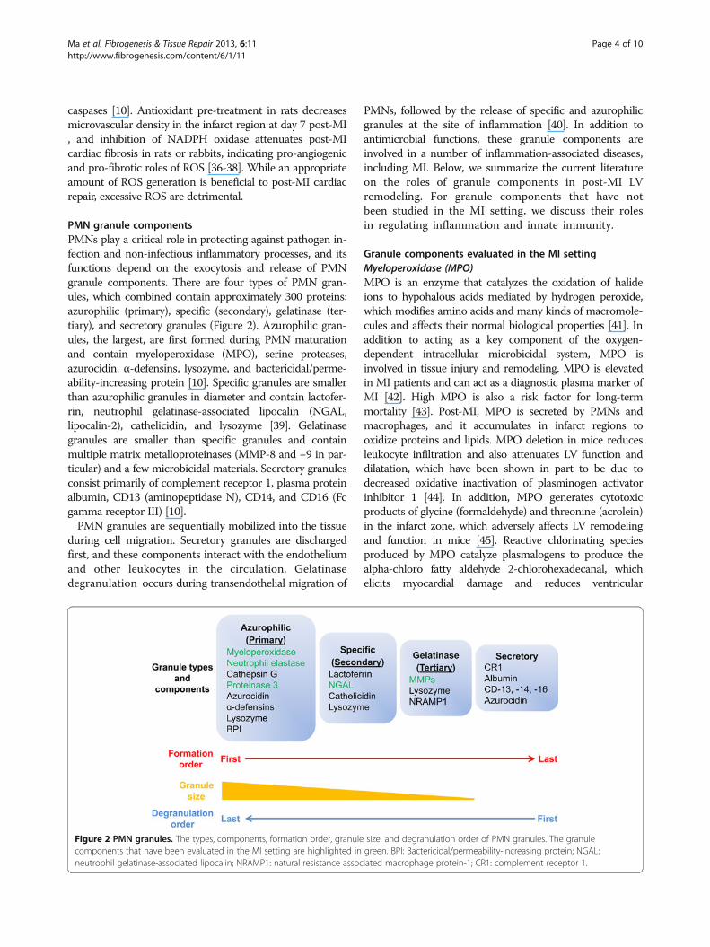
PMN granule componentsPMNs play a critical role in protecting against pathogen in-fection and non-infectious inflammatory processes, and itsfunctions depend on the exocytosis and release of PMNgranule components. There are four types of PMN gran-ules, which combined contain approximately 300 proteins:azurophilic (primary), specific (secondary), gelatinase (ter-tiary), and secretory granules (Figure 2). Azurophilic gran-ules, the largest, are first formed during PMN maturationand contain myeloperoxidase (MPO), serine proteases,azurocidin, α-defensins, lysozyme, and bactericidal/perme-ability-increasing protein [10]. Specific granules are smallerthan azurophilic granules in diameter and contain lactofer-rin, neutrophil gelatinase-associated lipocalin (NGAL,lipocalin-2), cathelicidin, and lysozyme [39]. Gelatinasegranules are smaller than specific granules and containmultiple matrix metalloproteinases (MMP-8 and −9 in par-ticular) and a few microbicidal materials. Secretory granulesconsist primarily of complement receptor 1, plasma proteinalbumin, CD13 (aminopeptidase N), CD14, and CD16 (Fcgamma receptor III) [10].PMN granules are sequentially mobilized into the tissue
during cell migration. Secretory granules are dischargedfirst, and these components interact with the endotheliumand other leukocytes in the circulation. Gelatinasedegranulation occurs during transendothelial migration of
PMNs, followed by the release of specific and azurophilicgranules at the site of inflammation [40]. In addition toantimicrobial functions, these granule components areinvolved in a number of inflammation-associated diseases,including MI. Below, we summarize the current literatureon the roles of granule components in post-MI LVremodeling. For granule components that have notbeen studied in the MI setting, we discuss their rolesin regulating inflammation and innate immunity.
Granule components evaluated in the MI settingMyeloperoxidase (MPO)MPO is an enzyme that catalyzes the oxidation of halideions to hypohalous acids mediated by hydrogen peroxide,which modifies amino acids and many kinds of macromole-cules and affects their normal biological properties [41]. Inaddition to acting as a key component of the oxygen-dependent intracellular microbicidal system, MPO isinvolved in tissue injury and remodeling. MPO is elevatedin MI patients and can act as a diagnostic plasma marker ofMI [42]. High MPO is also a risk factor for long-termmortality [43]. Post-MI, MPO is secreted by PMNs andmacrophages, and it accumulates in infarct regions tooxidize proteins and lipids. MPO deletion in mice reducesleukocyte infiltration and also attenuates LV function anddilatation, which have been shown in part to be due todecreased oxidative inactivation of plasminogen activatorinhibitor 1 [44]. In addition, MPO generates cytotoxicproducts of glycine (formaldehyde) and threonine (acrolein)in the infarct zone, which adversely affects LV remodelingand function in mice [45]. Reactive chlorinating speciesproduced by MPO catalyze plasmalogens to produce thealpha-chloro fatty aldehyde 2-chlorohexadecanal, whichelicits myocardial damage and reduces ventricular
Figure 2 PMN granules. The types, components, formation order, granule size, and degranulation order of PMN granules. The granulecomponents that have been evaluated in the MI setting are highlighted in green. BPI: Bactericidal/permeability-increasing protein; NGAL:neutrophil gelatinase-associated lipocalin; NRAMP1: natural resistance associated macrophage protein-1; CR1: complement receptor 1.
Ma et al. Fibrogenesis & Tissue Repair 2013, 6:11 Page 4 of 10http://www.fibrogenesis.com/content/6/1/11
performance in rats [46]. Targeting MPO signaling mayrepresent a promising way to alleviate MI-induced LVremodeling.
Serine proteasesSerine proteases stored in azurophilic granules includeneutrophil elastase (NE), cathepsin G, proteinase 3, andneutrophil serine protease-4. Neutrophil serine protease-4has recently been identified and shows 39% identity to NEand proteinase 3 [47]. In the presence of ROS, serineproteinases can break down internalized pathogens, pro-teolytically degrade cytokines and chemokines, and activatecell surface receptors [48]. In addition, serine proteinasesactivate the coagulation cascade and platelets to stimulatethrombus formation [49]. During systemic infection,activation of coagulation facilitates compartmentalizationof pathogens in liver microvessels and limits infectionexpansion. In contrast, in the absence of microorganismchallenge, coagulation induces large vessel thrombosis andcontributes to a risk for MI and stroke.NE degrades elastin, collagens, and fibrinogen and
contributes to cardiac damage post-MI. NE induces IL-6secretion to impair cardiac contractility by a nitric oxide-dependent pathway [50]. NE can cleave and activate pro-MMP-9, indicating an interactive action of PMN-derivedmolecules [51]. NE is released in the early stages ofischemia, and inhibition of NE has been shown to reduceinfarct size [52]. Similarly, a selective NE inhibitor protectsagainst myocardial stunning after ischemia/reperfusion inswine [53]. Proteinase 3 is stored in both azurophilicand secretory granules. Proteinase 3 induces endothelialcell apoptosis by caspase-like activity [54], cleavesangiotensinogen to generate angiotensin I and II [55],activates proinflammatory factors (e.g., TNF-α, IL-1β, andIL-18), and degrades extracellular matrix (e.g., fibronectinand collagen IV) [56]. Proteinase 3 levels in the plasma arehigher in chronic post-MI patients who later die or are re-admitted for heart failure compared to event-free survivors[56]. This indicates that proteinase 3 may exacerbate heartfailure and serve as a prognostic marker.
NGALNGAL is a glycoprotein with bacteriostatic propertiesstored in specific granules of mature PMNs. In humans,NGAL binds directly with MMP-9 to form a high molecu-lar weight complex, protecting MMP-9 from degradation[57]. This binding occurs at the 87 amino acid of theNGAL, which is a cysteine in humans [58]. Mouse NGALdoes not have this cysteine and does not bind directly toMMP-9. NGAL levels significantly increase in both ratsand patients post-MI and associate with adverse outcomes[59]. High plasma NGAL before intervention has beenshown to independently predict all-cause mortality for MIpatients treated with primary percutaneous coronary
intervention [60]. The NGAL mechanisms of regulatingLV remodeling have not been revealed, but may involveboth direct interactions with MMP-9 as well as growthfactor functions independent of complex formation.
MMP-8Despite originally being classified as the neutrophil colla-genase, MMP-8 is secreted not only by PMNs, but also bymacrophages [61]. MMP-8 promotes PMN migration bydegrading collagens [62], and PMN depletion inhibitsearly collagen degradation because of the lack of MMP-8[63]. MMP-8 degrades fibrillar collagen by binding andcleavage of collagen type I α1 and α 2 chains [64]. Thequantities of total and active MMP-8 were shown to behigher in patients with LV rupture than those withoutrupture [65], indicating that MMP-8 may promote infarctrupture in humans by degrading collagen.
MMP-9MMP-9 is one of the most widely investigated MMPs incardiovascular disease. Infiltrating PMNs are an early sourceof MMP-9 after MI both with and without reperfusion inhumans and multiple animal models, including mice, rab-bits, and canines [66-69]. PMN-derived MMP-9 is stored ingelatinase granules and released upon chemotactic stimula-tion. MMP-9 is also be secreted by macrophages, myocytes,fibroblasts, vascular smooth muscle cells, and endothelialcells [61]. MMP-9 is significantly elevated in the first weekafter MI in mice, consistent with the time course of PMNand macrophage infiltration. MMP-9 deletion attenuatesLV dysfunction and collagen deposition and promotesangiogenesis post-MI in mice [70,71]. Neutrophil-derivedMMP-9 may exert very early effects in the MI setting by de-grading extracellular matrix and promoting leukocyte cellinfiltration into the infarct area, while MMP-9 from othercells may regulate scar formation [72,73].
Granule components that have not been evaluated in theMI settingCathepsin GCathepsin G has biphasic regulation of leukocytechemotaxis, serving as both a stimulator and repressorof chemotaxis. Substrate availability determines its action,as cathepsin G enhances PMN and monocyte chemotaxisby cleaving the N-terminal residues of CXCL5 and CCL15to increase their chemotactic activities [74]. Conversely,cathepsin G also degrades CCL5, CCL3, CXCL12, andCXCR4 to reduce PMN and monocyte chemotaxis[75,76]. Cathepsin G is a potent platelet activator andpromotes intravascular thrombosis, thus contributingto the formation of a thrombus clot [77].
Ma et al. Fibrogenesis & Tissue Repair 2013, 6:11 Page 5 of 10http://www.fibrogenesis.com/content/6/1/11
AzurocidinAzurocidin, also known as cationic antimicrobial protein of37 kDa (CAP37) or heparin-binding protein (HBP), isstored in both azurophilic and secretory granules.Azurocidin is released at both the very early phase and thelater phase of PMN recruitment to sites of inflammation[78]. Azurocidin induces monocyte recruitment and en-hances cytokine production in monocytes/macrophages,signifying the ability of azurocidin to regulate monocytes/macrophage infiltration and activation in the post-MIsetting [79-81]. The effect of azurocidin on leukocytes isdependent on β2 integrins and the formyl peptide receptor.Originally considered devoid of proteinase activity,azurocidin can actually cleave insulin-like growth factorbinding protein-1, -2, and −4 in vitro [82]. The LTB4-induced increase in vascular permeability is mediated byazurocidin [83], suggesting that azurocidin may promoteleukocyte extravasation.
α-defensinsThe α-defensins, also referred to as human neutrophilpeptides (HNPs), are small cationic antimicrobial peptidesmainly present in the azurophilic granules. The α-defensinsnot only have antimicrobial function, but also possessimmunoregulatory properties mediated by direct inter-action with innate immune cells [84]. HNP-1 and −2 arepotent chemoattractants for monocyte, naïve T cells, andimmature dendritic cells, but not for mature dendritic cellsor PMNs [85,86]. In addition, HNP-1 is able to activatemonocyte-derived dendritic cells and upregulate theproduction of proinflammatory cytokines [87]. In view oftheir immunoregulatory activities, future studies to explorethe functions of α-defensins in MI are warranted.
LactoferrinLactoferrin is an iron-binding glycoprotein of thetransferrin family present in the specific granules. Itis also synthesized by epithelial cells [88]. In additionto direct antimicrobial activity, lactoferrin inhibits theupregulation of adhesion molecules, limits iron-mediateddamage to host tissue, suppresses proinflammatory cytokineproduction, and limits PMN recruitment [89]. Post-MI,lactoferrin may have protective effects by inhibitingexcessive inflammation and ROS production.
CathelicidinCathelicidin, also known as cathelicidin-related antimicro-bial peptide (CRAMP) in mouse and LL-37 or hCAP18 inhuman, resides in specific granules. In addition to potentmicrobicidal activity, LL-37 inhibits PMN apoptosis andstimulates monocyte recruitment, angiogenesis, and tissueregeneration [90]. LL37 elevates IL-1β-induced release ofcytokines (IL-6 and IL-10) and chemokines such as MCP-1,MCP-3, and IL-8 in macrophages [91,92]. LL-37 deposits at
sites of endothelial injury, facilitates re-endothelization, andlimits neointima formation after stent implantation byenhancing early outgrowth cell recruitment and release ofgrowth factors [93]. Further, stents coated with LL-37 havereduced re-stenosis, indicating that LL-37 may promote thehealing response [93]. Doring and colleagues showthat lack of CRAMP reduces atherosclerotic lesionsize by restraining monocyte recruitment and byreducing the adhesion of classical monocytes and PMNsin a formyl peptide receptor-dependent way [94]. In earlystages of atherosclerosis, CRAMP is specifically expressedin PMNs, but not in monocytes or macrophages.Therefore, cathelicidin may modulate LV remodelingafter MI by regulating leukocyte infiltration, apoptosis,and angiogenesis.
MMP-25MMP-25, also known as MT6-MMP or leukolysin, isa membrane-type MMP. In PMNs, MMP-25 ispresent in gelatinase granules and is also found innuclear/endoplasmic reticulum/Golgi fractions [95]. Invitro studies show that MMP-25 cleaves CXCL5,CCL15, and CCL23 to activate these chemokines, andthus promotes the recruitment of PMNs and monocytes
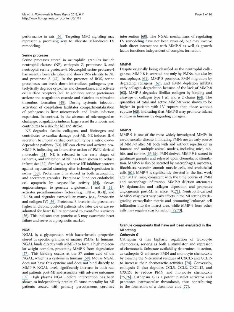
Figure 3 Mechanisms of action of PMNs on post-MI LVremodeling. Infiltrating PMNs release a wide range of cytokines andchemokines, granule components, and reactive oxygen species,which directly and indirectly regulate immune cell infiltration andfunction to modulate remodeling response.
Ma et al. Fibrogenesis & Tissue Repair 2013, 6:11 Page 6 of 10http://www.fibrogenesis.com/content/6/1/11
[96]. MMP-25 roles, however, remain unknown, andMMP-25 levels have not even been measured post-MI.
NETsPMNs release granule antimicrobial proteins and nuclearcomponents (DNA, histones) into the extracellular environ-ment that form NETs to trap invading pathogens. Thisprocess is referred to as NETosis and is an alternative toPMN apoptosis [97]. NETs degrade virulent factors and killmicroorganisms to prevent infection from spreading [98].NETs also have detrimental influences on the host. NETsactivate the complement system, and the complementcomponent C1q can inhibit NETs degradation, thusestablishing a positive feedback loop to exacerbate diseaseprogression [99]. It has been shown that NETs facilitatethrombosis in MI patients, probably by promoting fibrindeposition and platelet aggregation [100]. The role of NETsin the progression of MI-induced heart failure, however,has not been investigated.
ConclusionsThis review summarizes the roles of PMNs andPMN-derived granule components in inflammation,innate immunity, and MI. PMNs regulate the post-MIwound healing response through several mechanisms(Figure 3). PMNs are activated by cytokines andchemokines, and activated PMNs in turn release cytokinesand chemokines to potentiate the inflammatory componentof wound healing [101]. PMN degranulation releasesan array of proteases that regulate LV remodeling bymodulating immune cell infiltration and function,including ROS production. The PMN respiratory burstgenerates ROS to directly modify biological molecules.However, several aspects remain to be elucidated in orderto better understand PMN roles after MI.First, PMN roles post-MI need to be better understood,
using systematic approaches that distinguish the negativeand positive roles. In order for therapeutic strategies to bedeveloped that promote healing while preventing adverseremodeling, we need to better understand the complexityof PMNs in mediating the early inflammatory response.Second, there may be different activation phenotypes of
PMNs following MI [102-104]. A recent study byFridlender and colleagues suggests that tumor associatedPMNs can be polarized towards different phenotypes[104]. Blocking TGF-β slows tumor growth by increasingthe influx of PMNs to produce higher levels ofproinflammatory cytokines, which are more cytotoxic[104]. PMN depletion without TGF-β blockade, however,also decreases tumor growth. TGF-β, therefore, promotesa PMN pro-tumor phenotype, while blocking TGF-βinduces a PMN anti-tumor phenotype [104]. TGF-βeffects on tumors and the post-MI LV are likely opposite,as TGF-β promotes post-MI infarct healing and blocking
TGF-β increases MI-induced mortality and LV dilation[105]. PMN phenotypes should be examined by isolatingPMNs from post-MI hearts at different time points andmeasuring the expression of key effector molecules. Beforethis can be accomplished, however, we need to determinewhat markers can be used to differentiate phenotypes andwhether overall inflammatory status is sufficient.Third, whether PMNs directly or indirectly regulate
macrophage polarization (M1 or M2 activation) or func-tion is not currently well understood. This could be evalu-ated by incubating resting macrophages with conditionedmedia from activated PMNs and monitoring the macro-phages for M1 and M2 markers [106]. It may be thatPMNs from different post-MI times promote differentialmacrophage activation patterns.Fourth, whether PMNs regulate cardiac fibroblast pheno-
type and post-MI scar formation is not known [107]. Therole of macrophages in activating fibroblasts has been stud-ied, but whether PMNs exert similar or different activationfunctions is unknown. This can be addressed by incubatingisolated cardiac fibroblasts with activated PMNs and meas-uring fibroblast phenotype and secretion of extracellularmatrix [106].In conclusion, understanding how PMNs regulate post-
MI LV remodeling may provide promising interventiontargets for MI patients. Understanding the detrimentaland beneficial roles will provide mechanistic insight intohow PMNs regulate inflammatory responses, both in theMI setting and in other diseases that have inflammation asa common response.
AbbreviationsDAMPs: Damage-associated molecular patterns; HNPs: Human neutrophilpeptides; ICAMs: Intercellular adhesion molecules; IL: Interleukin;LTB4: Leukotriene B4; LV: Left ventricle; MCP-1: Monocyte chemoattractantprotein-1; MI: Myocardial infarction; MMPs: Matrix metalloproteinases;MPO: Myeloperoxidase; NE: Neutrophil elastase; NETs: Neutrophil extracellulartraps; NGAL: Neutrophil gelatinase-associated lipocalin; ROS: Reactive oxygenspecies; TLR: Toll-like receptor; TNF: Tumor necrosis factor.
Competing interestsThe authors declare that they have no competing interests.
Authors’ contributionsYM and MLL conceived the concept of the review. YM wrote the first draft.YM, AY, and MLL edited and revised the manuscript. All authors read andapproved the final manuscript.
AcknowledgementsWe acknowledge support from NIH/NHLBI HHSN 268201000036C (N01-HV-00244) for the San Antonio Cardiovascular Proteomics Center and R01HL075360, and from the Biomedical Laboratory Research and DevelopmentService of the Veterans Affairs Office of Research and Development Award5I01BX000505 to MLL.
Author details1San Antonio Cardiovascular Proteomics Center, San Antonio, TX, USA.2Jackson Center for Heart Research, Department of Physiology andBiophysics, University of Mississippi Medical Center, Jackson, MS, USA.3Research and Medicine Services, G.V. (Sonny) Montgomery Veterans AffairsMedical Center, Jackson, MS, USA.
Ma et al. Fibrogenesis & Tissue Repair 2013, 6:11 Page 7 of 10http://www.fibrogenesis.com/content/6/1/11
Received: 29 December 2012 Accepted: 11 April 2013Published: 3 June 2013
References1. Day RB, Link DC: Regulation of neutrophil trafficking from the bone
marrow. Cell Mol Life Sci 2012, 69:1415–1423.2. Akpek M, Kaya MG, Lam YY, Sahin O, Elcik D, Celik T, Ergin A, Gibson CM:
Relation of neutrophil/lymphocyte ratio to coronary flow to in-hospitalmajor adverse cardiac events in patients with ST-elevated myocardialinfarction undergoing primary coronary intervention. Am J Cardiol 2012,110:621–627.
3. Meissner J, Irfan A, Twerenbold R, Mueller S, Reiter M, Haaf P, Reichlin T,Schaub N, Winkler K, Pfister O, et al: Use of neutrophil count in earlydiagnosis and risk stratification of AMI. Am J Med 2011, 124:534–542.
4. Chia S, Nagurney JT, Brown DF, Raffel OC, Bamberg F, Senatore F, WackersFJ, Jang IK: Association of leukocyte and neutrophil counts with infarctsize, left ventricular function and outcomes after percutaneous coronaryintervention for ST-elevation myocardial infarction. Am J Cardiol 2009,103:333–337.
5. Jolly SR, Kane WJ, Hook BG, Abrams GD, Kunkel SL, Lucchesi BR: Reductionof myocardial infarct size by neutrophil depletion: effect of duration ofocclusion. Am Heart J 1986, 112:682–690.
6. Romson JL, Hook BG, Kunkel SL, Abrams GD, Schork MA, Lucchesi BR:Reduction of the extent of ischemic myocardial injury by neutrophildepletion in the dog. Circulation 1983, 67:1016–1023.
7. Hammerman H, Kloner RA, Hale S, Schoen FJ, Braunwald E: Dose-dependent effects of short-term methylprednisolone on myocardialinfarct extent, scar formation, and ventricular function. Circulation 1983,68:446–452.
8. Borregaard N, Theilgaard-Monch K, Cowland JB, Stahle M, Sorensen OE:Neutrophils and keratinocytes in innate immunity–cooperative actionsto provide antimicrobial defense at the right time and place. J LeukocBiol 2005, 77:439–443.
9. Barletta KE, Ley K, Mehrad B: Regulation of neutrophil function byadenosine. Arterioscler Thromb Vasc Biol 2012, 32:856–864.
10. Amulic B, Cazalet C, Hayes GL, Metzler KD, Zychlinsky A: Neutrophilfunction: from mechanisms to disease. Annu Rev Immunol 2012,30:459–489.
11. Dancey JT, Deubelbeiss KA, Harker LA, Finch CA: Neutrophil kinetics inman. J Clin Invest 1976, 58:705–715.
12. Galli SJ, Borregaard N, Wynn TA: Phenotypic and functional plasticity ofcells of innate immunity: macrophages, mast cells and neutrophils.Nat Immunol 2011, 12:1035–1044.
13. Pillay J, den Braber I, Vrisekoop N, Kwast LM, de Boer RJ, Borghans JA,Tesselaar K, Koenderman L: In vivo labeling with 2H2O reveals a humanneutrophil lifespan of 5.4 days. Blood 2010, 116:625–627.
14. Colotta F, Re F, Polentarutti N, Sozzani S, Mantovani A: Modulation ofgranulocyte survival and programmed cell death by cytokines andbacterial products. Blood 1992, 80:2012–2020.
15. Borregaard N: Neutrophils, from marrow to microbes. Immunity 2010,33:657–670.
16. Mary JY: Normal human granulopoiesis revisited. II. Bone marrow data.Biomed Pharmacother 1985, 39:66–77.
17. Semerad CL, Liu F, Gregory AD, Stumpf K, Link DC: G-CSF is an essentialregulator of neutrophil trafficking from the bone marrow to the blood.Immunity 2002, 17:413–423.
18. Ivey CL, Williams FM, Collins PD, Jose PJ, Williams TJ: Neutrophilchemoattractants generated in two phases during reperfusion ofischemic myocardium in the rabbit. Evidence for a role for C5a andinterleukin-8. J Clin Invest 1995, 95:2720–2728.
19. Kim D, Haynes CL: Neutrophil chemotaxis within a competing gradient ofchemoattractants. Anal Chem 2012, 84:6070–6078.
20. Frangogiannis NG: Regulation of the inflammatory response in cardiacrepair. Circ Res 2012, 110:159–173.
21. Bratton DL, Henson PM: Neutrophil clearance: when the party is over,clean-up begins. Trends Immunol 2011, 32:350–357.
22. Hayashi F, Means TK, Luster AD: Toll-like receptors stimulate humanneutrophil function. Blood 2003, 102:2660–2669.
23. Greenblatt MB, Aliprantis A, Hu B, Glimcher LH: Calcineurin regulatesinnate antifungal immunity in neutrophils. J Exp Med 2010, 207:923–931.
24. Kerrigan AM, Dennehy KM, Mourao-Sa D, Faro-Trindade I, Willment JA,Taylor PR, Eble JA, Reis e Sousa C, Brown GD: CLEC-2 is a phagocyticactivation receptor expressed on murine peripheral blood neutrophils.J Immunol 2009, 182:4150–4157.
25. Clarke TB, Davis KM, Lysenko ES, Zhou AY, Yu Y, Weiser JN: Recognition ofpeptidoglycan from the microbiota by Nod1 enhances systemic innateimmunity. Nat Med 2010, 16:228–231.
26. Tamassia N, Le Moigne V, Rossato M, Donini M, McCartney S, Calzetti F,Colonna M, Bazzoni F, Cassatella MA: Activation of an immunoregulatoryand antiviral gene expression program in poly(I:C)-transfected humanneutrophils. J Immunol 2008, 181:6563–6573.
27. Timmers L, Pasterkamp G, de Hoog VC, Arslan F, Appelman Y, de Kleijn DP:The innate immune response in reperfused myocardium. Cardiovasc Res2012, 94:276–283.
28. Prince LR, Whyte MK, Sabroe I, Parker LC: The role of TLRs in neutrophilactivation. Curr Opin Pharmacol 2011, 11:397–403.
29. Ma Y, Zhang X, Bao H, Mi S, Cai W, Yan H, Wang Q, Wang Z, Yan J, Fan G, etal: Toll-like receptor (TLR) 2 and TLR4 differentially regulate doxorubicininduced cardiomyopathy in mice. PLoS One 2012, 7:e40763.
30. Ueno H, Matsuda T, Hashimoto S, Amaya F, Kitamura Y, Tanaka M,Kobayashi A, Maruyama I, Yamada S, Hasegawa N, et al: Contributions ofhigh mobility group box protein in experimental and clinical acute lunginjury. Am J Respir Crit Care Med 2004, 170:1310–1316.
31. Furze RC, Rankin SM: Neutrophil mobilization and clearance in the bonemarrow. Immunology 2008, 125:281–288.
32. Soehnlein O, Lindbom L: Phagocyte partnership during the onset andresolution of inflammation. Nat Rev Immunol 2010, 10:427–439.
33. Esmann L, Idel C, Sarkar A, Hellberg L, Behnen M, Moller S, van Zandbergen G,Klinger M, Kohl J, Bussmeyer U, et al: Phagocytosis of apoptotic cells byneutrophil granulocytes: diminished proinflammatory neutrophil functionsin the presence of apoptotic cells. J Immunol 2010, 184:391–400.
34. Ciz M, Denev P, Kratchanova M, Vasicek O, Ambrozova G, Lojek A:Flavonoids inhibit the respiratory burst of neutrophils in mammals.Oxid Med Cell Longev 2012, 2012:181295.
35. Kleniewska P, Piechota A, Skibska B, Goraca A: The NADPH oxidase familyand its inhibitors. Arch Immunol Ther Exp (Warsz) 2012, 60:277–294.
36. Liu XH, Pan LL, Deng HY, Xiong QH, Wu D, Huang GY, Gong QH, Zhu YZ:Leonurine (SCM-198) attenuates myocardial fibrotic response viainhibition of NADPH oxidase 4. Free Radic Biol Med 2013, 54:93–104.
37. Qin F, Simeone M, Patel R: Inhibition of NADPH oxidase reduces myocardialoxidative stress and apoptosis and improves cardiac function in heartfailure after myocardial infarction. Free Radic Biol Med 2007, 43:271–281.
38. Zhao W, Zhao T, Chen Y, Ahokas RA, Sun Y: Reactive oxygen speciespromote angiogenesis in the infarcted rat heart. Int J Exp Pathol 2009,90:621–629.
39. Faurschou M, Borregaard N: Neutrophil granules and secretory vesicles ininflammation. Microbes Infect 2003, 5:1317–1327.
40. Soehnlein O, Weber C, Lindbom L: Neutrophil granule proteins tunemonocytic cell function. Trends Immunol 2009, 30:538–546.
41. Prokopowicz Z, Marcinkiewicz J, Katz DR, Chain BM: Neutrophilmyeloperoxidase: soldier and statesman. Arch Immunol Ther Exp (Warsz)2012, 60:43–54.
42. Rudolph V, Goldmann BU, Bos C, Rudolph TK, Klinke A, Friedrichs K, Lau D,Wegscheider K, Haddad M, Meinertz T, Baldus S: Diagnostic value of MPOplasma levels in patients admitted for suspected myocardial infarction.Int J Cardiol 2011, 153:267–271.
43. Mocatta TJ, Pilbrow AP, Cameron VA, Senthilmohan R, Frampton CM, RichardsAM, Winterbourn CC: Plasma concentrations of myeloperoxidase predictmortality after myocardial infarction. J Am Coll Cardiol 2007, 49:1993–2000.
44. Askari AT, Brennan ML, Zhou X, Drinko J, Morehead A, Thomas JD, Topol EJ,Hazen SL, Penn MS: Myeloperoxidase and plasminogen activator inhibitor1 play a central role in ventricular remodeling after myocardialinfarction. J Exp Med 2003, 197:615–624.
45. Vasilyev N, Williams T, Brennan ML, Unzek S, Zhou X, Heinecke JW, Spitz DR,Topol EJ, Hazen SL, Penn MS: Myeloperoxidase-generated oxidantsmodulate left ventricular remodeling but not infarct size aftermyocardial infarction. Circulation 2005, 112:2812–2820.
46. Thukkani AK, Martinson BD, Albert CJ, Vogler GA, Ford DA: Neutrophil-mediated accumulation of 2-ClHDA during myocardial infarction:2-ClHDA-mediated myocardial injury. Am J Physiol Heart Circ Physiol2005, 288:H2955–H2964.
Ma et al. Fibrogenesis & Tissue Repair 2013, 6:11 Page 8 of 10http://www.fibrogenesis.com/content/6/1/11
47. Perera NC, Schilling O, Kittel H, Back W, Kremmer E, Jenne DE: NSP4, anelastase-related protease in human neutrophils with arginine specificity.Proc Natl Acad Sci U S A 2012, 109:6229–6234.
48. Pham CT: Neutrophil serine proteases: specific regulators ofinflammation. Nat Rev Immunol 2006, 6:541–550.
49. Afshar-Kharghan V, Thiagarajan P: Leukocyte adhesion and thrombosis.Curr Opin Hematol 2006, 13:34–39.
50. Yu X, Kennedy RH, Liu SJ: JAK2/STAT3, not ERK1/2, mediates interleukin-6-induced activation of inducible nitric-oxide synthase and decrease incontractility of adult ventricular myocytes. J Biol Chem 2003,278:16304–16309.
51. Jackson PL, Xu X, Wilson L, Weathington NM, Clancy JP, Blalock JE, GaggarA: Human neutrophil elastase-mediated cleavage sites of MMP-9 andTIMP-1: implications to cystic fibrosis proteolytic dysfunction. Mol Med2010, 16:159–166.
52. Bidouard JP, Duval N, Kapui Z, Herbert JM, O’Connor SE, Janiak P: SSR69071,an elastase inhibitor, reduces myocardial infarct size followingischemia-reperfusion injury. Eur J Pharmacol 2003, 461:49–52.
53. Akiyama D, Hara T, Yoshitomi O, Maekawa T, Cho S, Sumikawa K:Postischemic infusion of sivelestat sodium hydrate, a selectiveneutrophil elastase inhibitor, protects against myocardial stunning inswine. J Anesth 2010, 24:575–581.
54. Pendergraft WF 3rd, Rudolph EH, Falk RJ, Jahn JE, Grimmler M, Hengst L, JennetteJC, Preston GA: Proteinase 3 sidesteps caspases and cleaves p21(Waf1/Cip1/Sdi1) to induce endothelial cell apoptosis. Kidney Int 2004, 65:75–84.
55. Ramaha A, Patston PA: Release and degradation of angiotensin I andangiotensin II from angiotensinogen by neutrophil serine proteinases.Arch Biochem Biophys 2002, 397:77–83.
56. Ng LL, Khan SQ, Narayan H, Quinn P, Squire IB, Davies JE: Proteinase 3 andprognosis of patients with acute myocardial infarction. Clin Sci (Lond)2011, 120:231–238.
57. Yan L, Borregaard N, Kjeldsen L, Moses MA: The high molecular weighturinary matrix metalloproteinase (MMP) activity is a complex ofgelatinase B/MMP-9 and neutrophil gelatinase-associated lipocalin(NGAL). Modulation of MMP-9 activity by NGAL. J Biol Chem 2001,276:37258–37265.
58. Coles M, Diercks T, Muehlenweg B, Bartsch S, Zolzer V, Tschesche H, KesslerH: The solution structure and dynamics of human neutrophil gelatinase-associated lipocalin. J Mol Biol 1999, 289:139–157.
59. Yndestad A, Landro L, Ueland T, Dahl CP, Flo TH, Vinge LE, Espevik T,Froland SS, Husberg C, Christensen G, et al: Increased systemic andmyocardial expression of neutrophil gelatinase-associated lipocalin inclinical and experimental heart failure. Eur Heart J 2009, 30:1229–1236.
60. Lindberg S, Pedersen SH, Mogelvang R, Jensen JS, Flyvbjerg A, Galatius S,Magnusson NE: Prognostic utility of neutrophil gelatinase-associatedlipocalin in predicting mortality and cardiovascular events in patientswith ST-segment elevation myocardial infarction treated with primarypercutaneous coronary intervention. J Am Coll Cardiol 2012, 60:339–345.
61. Lindsey ML, Zamilpa R: Temporal and spatial expression of matrixmetalloproteinases and tissue inhibitors of metalloproteinases followingmyocardial infarction. Cardiovasc Ther 2012, 30:31–41.
62. Lin M, Jackson P, Tester AM, Diaconu E, Overall CM, Blalock JE, Pearlman E:Matrix metalloproteinase-8 facilitates neutrophil migration throughthe corneal stromal matrix by collagen degradation and productionof the chemotactic peptide Pro-Gly-Pro. Am J Pathol 2008, 173:144–153.
63. Harty MW, Muratore CS, Papa EF, Gart MS, Ramm GA, Gregory SH, Tracy TFJr: Neutrophil depletion blocks early collagen degradation in repairingcholestatic rat livers. Am J Pathol 2010, 176:1271–1281.
64. Gioia M, Monaco S, Fasciglione GF, Coletti A, Modesti A, Marini S, Coletta M:Characterization of the mechanisms by which gelatinase A, neutrophilcollagenase, and membrane-type metalloproteinase MMP-14 recognizecollagen I and enzymatically process the two alpha-chains. J Mol Biol2007, 368:1101–1113.
65. van den Borne SW, Cleutjens JP, Hanemaaijer R, Creemers EE, Smits JF,Daemen MJ, Blankesteijn WM: Increased matrix metalloproteinase-8and −9 activity in patients with infarct rupture after myocardialinfarction. Cardiovasc Pathol 2009, 18:37–43.
66. Romanic AM, Harrison SM, Bao W, Burns-Kurtis CL, Pickering S, Gu J, Grau E,Mao J, Sathe GM, Ohlstein EH, Yue TL: Myocardial protection fromischemia/reperfusion injury by targeted deletion of matrixmetalloproteinase-9. Cardiovasc Res 2002, 54:549–558.
67. Lindsey M, Wedin K, Brown MD, Keller C, Evans AJ, Smolen J, Burns AR,Rossen RD, Michael L, Entman M: Matrix-dependent mechanism ofneutrophil-mediated release and activation of matrix metalloproteinase9 in myocardial ischemia/reperfusion. Circulation 2001, 103:2181–2187.
68. Romanic AM, Burns-Kurtis CL, Gout B, Berrebi-Bertrand I, Ohlstein EH: Matrixmetalloproteinase expression in cardiac myocytes following myocardialinfarction in the rabbit. Life Sci 2001, 68:799–814.
69. Kelly D, Cockerill G, Ng LL, Thompson M, Khan S, Samani NJ, Squire IB:Plasma matrix metalloproteinase-9 and left ventricular remodelling afteracute myocardial infarction in man: a prospective cohort study. Eur HeartJ 2007, 28:711–718.
70. Ducharme A, Frantz S, Aikawa M, Rabkin E, Lindsey M, Rohde LE, Schoen FJ,Kelly RA, Werb Z, Libby P, Lee RT: Targeted deletion of matrixmetalloproteinase-9 attenuates left ventricular enlargement andcollagen accumulation after experimental myocardial infarction. J ClinInvest 2000, 106:55–62.
71. Lindsey ML, Escobar GP, Dobrucki LW, Goshorn DK, Bouges S, Mingoia JT,McClister DM Jr, Su H, Gannon J, MacGillivray C, et al: Matrixmetalloproteinase-9 gene deletion facilitates angiogenesis aftermyocardial infarction. Am J Physiol Heart Circ Physiol 2006, 290:H232–H239.
72. Yabluchanskiy A, Li Y, Chilton RJ, Lindsey ML: Matrix metalloproteinases:drug targets for myocardial infarction. Curr Drug Targets 2013, 14:276–286.
73. Zamilpa R, Ibarra J, de Castro Bras LE, Ramirez TA, Nguyen N, Halade GV,Zhang J, Dai Q, Dayah T, Chiao YA, et al: Transgenic overexpression ofmatrix metalloproteinase-9 in macrophages attenuates the inflammatoryresponse and improves left ventricular function post-myocardialinfarction. J Mol Cell Cardiol 2012, 53:599–608.
74. Richter R, Bistrian R, Escher S, Forssmann WG, Vakili J, Henschler R,Spodsberg N, Frimpong-Boateng A, Forssmann U: Quantum proteolyticactivation of chemokine CCL15 by neutrophil granulocytes modulatesmononuclear cell adhesiveness. J Immunol 2005, 175:1599–1608.
75. Pham CT: Neutrophil serine proteases fine-tune the inflammatoryresponse. Int J Biochem Cell Biol 2008, 40:1317–1333.
76. Lim JK, Lu W, Hartley O, DeVico AL: N-terminal proteolytic processing bycathepsin G converts RANTES/CCL5 and related analogs into a truncated4–68 variant. J Leukoc Biol 2006, 80:1395–1404.
77. Herrmann SM, Funke-Kaiser H, Schmidt-Petersen K, Nicaud V, Gautier-Bertrand M, Evans A, Kee F, Arveiler D, Morrison C, Orzechowski HD, et al:Characterization of polymorphic structure of cathepsin G gene: role incardiovascular and cerebrovascular diseases. Arterioscler Thromb Vasc Biol2001, 21:1538–1543.
78. Tapper H, Karlsson A, Morgelin M, Flodgaard H, Herwald H: Secretion of heparin-binding protein from human neutrophils is determined by its localization inazurophilic granules and secretory vesicles. Blood 2002, 99:1785–1793.
79. Soehnlein O, Lindbom L: Neutrophil-derived azurocidin alarms theimmune system. J Leukoc Biol 2009, 85:344–351.
80. Rasmussen PB, Bjorn S, Hastrup S, Nielsen PF, Norris K, Thim L, Wiberg FC,Flodgaard H: Characterization of recombinant human HBP/CAP37/azurocidin,a pleiotropic mediator of inflammation-enhancing LPS-induced cytokinerelease from monocytes. FEBS Lett 1996, 390:109–112.
81. Soehnlein O, Kai-Larsen Y, Frithiof R, Sorensen OE, Kenne E, Scharffetter-Kochanek K, Eriksson EE, Herwald H, Agerberth B, Lindbom L: Neutrophilprimary granule proteins HBP and HNP1-3 boost bacterial phagocytosisby human and murine macrophages. J Clin Invest 2008, 118:3491–3502.
82. Brandt K, Lundell K, Brismar K: Neutrophil-derived azurocidin cleavesinsulin-like growth factor-binding protein-1, -2 and −4. Growth Horm IGFRes 2011, 21:167–173.
83. Di Gennaro A, Kenne E, Wan M, Soehnlein O, Lindbom L, Haeggstrom JZ:Leukotriene B4-induced changes in vascular permeability are mediatedby neutrophil release of heparin-binding protein (HBP/CAP37/azurocidin). FASEB J 2009, 23:1750–1757.
84. Yang D, Biragyn A, Hoover DM, Lubkowski J, Oppenheim JJ: Multiple rolesof antimicrobial defensins, cathelicidins, and eosinophil-derivedneurotoxin in host defense. Annu Rev Immunol 2004, 22:181–215.
85. Yang D, Chen Q, Chertov O, Oppenheim JJ: Human neutrophil defensinsselectively chemoattract naive T and immature dendritic cells. J LeukocBiol 2000, 68:9–14.
86. Territo MC, Ganz T, Selsted ME, Lehrer R: Monocyte-chemotactic activity ofdefensins from human neutrophils. J Clin Invest 1989, 84:2017–2020.
87. Presicce P, Giannelli S, Taddeo A, Villa ML, Della Bella S: Human defensinsactivate monocyte-derived dendritic cells, promote the production of
Ma et al. Fibrogenesis & Tissue Repair 2013, 6:11 Page 9 of 10http://www.fibrogenesis.com/content/6/1/11
proinflammatory cytokines, and up-regulate the surface expression ofCD91. J Leukoc Biol 2009, 86:941–948.
88. Ward PP, Paz E, Conneely OM: Multifunctional roles of lactoferrin: a criticaloverview. Cell Mol Life Sci 2005, 62:2540–2548.
89. Crouch SP, Slater KJ, Fletcher J: Regulation of cytokine release frommononuclear cells by the iron-binding protein lactoferrin. Blood 1992,80:235–240.
90. Bucki R, Leszczynska K, Namiot A, Sokolowski W: Cathelicidin LL-37: a multitaskantimicrobial peptide. Arch Immunol Ther Exp (Warsz) 2010, 58:15–25.
91. Lai Y, Gallo RL: AMPed up immunity: how antimicrobial peptides havemultiple roles in immune defense. Trends Immunol 2009, 30:131–141.
92. Scott MG, Davidson DJ, Gold MR, Bowdish D, Hancock RE: The humanantimicrobial peptide LL-37 is a multifunctional modulator of innateimmune responses. J Immunol 2002, 169:3883–3891.
93. Soehnlein O, Wantha S, Simsekyilmaz S, Doring Y, Megens RT, Mause SF,Drechsler M, Smeets R, Weinandy S, Schreiber F, et al: Neutrophil-derivedcathelicidin protects from neointimal hyperplasia. Sci Transl Med 2011,3:103ra198.
94. Doring Y, Drechsler M, Wantha S, Kemmerich K, Lievens D, Vijayan S, GalloRL, Weber C, Soehnlein O: Lack of neutrophil-derived CRAMP reducesatherosclerosis in mice. Circ Res 2012, 110:1052–1056.
95. Fortin CF, Sohail A, Sun Q, McDonald PP, Fridman R, Fulop T: MT6-MMP ispresent in lipid rafts and faces inward in living human PMNs but translocatesto the cell surface during neutrophil apoptosis. Int Immunol 2010, 22:637–649.
96. Starr AE, Bellac CL, Dufour A, Goebeler V, Overall CM: Biochemicalcharacterization and N-terminomics analysis of leukolysin, themembrane-type 6 matrix metalloprotease (MMP25): chemokine andvimentin cleavages enhance cell migration and macrophage phagocyticactivities. J Biol Chem 2012, 287:13382–13395.
97. Remijsen Q, Kuijpers TW, Wirawan E, Lippens S, Vandenabeele P, VandenBerghe T: Dying for a cause: NETosis, mechanisms behind anantimicrobial cell death modality. Cell Death Differ 2011, 18:581–588.
98. Brinkmann V, Reichard U, Goosmann C, Fauler B, Uhlemann Y, Weiss DS,Weinrauch Y, Zychlinsky A: Neutrophil extracellular traps kill bacteria.Science 2004, 303:1532–1535.
99. Leffler J, Martin M, Gullstrand B, Tyden H, Lood C, Truedsson L, BengtssonAA, Blom AM: Neutrophil extracellular traps that are not degraded insystemic lupus erythematosus activate complement exacerbating thedisease. J Immunol 2012, 188:3522–3531.
100. de Boer OJ, Li X, Teeling P, Mackaay C, Ploegmakers HJ, van der Loos CM,Daemen MJ, de Winter RJ, van der Wal AC: Neutrophils, neutrophilextracellular traps and interleukin-17 associate with the organisation ofthrombi in acute myocardial infarction. Thromb Haemost 2013, 109:290–297.
101. Soehnlein O, Zernecke A, Eriksson EE, Rothfuchs AG, Pham CT, Herwald H,Bidzhekov K, Rottenberg ME, Weber C, Lindbom L: Neutrophil secretionproducts pave the way for inflammatory monocytes. Blood 2008,112:1461–1471.
102. Tsuda Y, Takahashi H, Kobayashi M, Hanafusa T, Herndon DN, Suzuki F:Three different neutrophil subsets exhibited in mice with differentsusceptibilities to infection by methicillin-resistant Staphylococcusaureus. Immunity 2004, 21:215–226.
103. Mantovani A, Cassatella MA, Costantini C, Jaillon S: Neutrophils in theactivation and regulation of innate and adaptive immunity. Nat RevImmunol 2011, 11:519–531.
104. Fridlender ZG, Sun J, Kim S, Kapoor V, Cheng G, Ling L, Worthen GS, AlbeldaSM: Polarization of tumor-associated neutrophil phenotype by TGF-beta:“N1” versus “N2” TAN. Cancer Cell 2009, 16:183–194.
105. Frantz S, Hu K, Adamek A, Wolf J, Sallam A, Maier SK, Lonning S, Ling H, ErtlG, Bauersachs J: Transforming growth factor beta inhibition increasesmortality and left ventricular dilatation after myocardial infarction. BasicRes Cardiol 2008, 103:485–492.
106. Ma Y, Halade GV, Zhang J, Ramirez TA, Levin D, Voorhees A, Jin YF, Han HC,Manicone AM, Lindsey ML: Matrix metalloproteinase-28 deletionexacerbates cardiac dysfunction and rupture after myocardial infarction inmice by inhibiting m2 macrophage activation. Circ Res 2013, 112:675–688.
107. Ma Y, Halade GV, Lindsey ML: Extracellular matrix and fibroblastcommunication following myocardial infarction. J Cardiovasc Transl Res2012, 5:848–857.
doi:10.1186/1755-1536-6-11Cite this article as: Ma et al.: Neutrophil roles in left ventricularremodeling following myocardial infarction. Fibrogenesis & Tissue Repair2013 6:11.
Submit your next manuscript to BioMed Centraland take full advantage of:
• Convenient online submission
• Thorough peer review
• No space constraints or color figure charges
• Immediate publication on acceptance
• Inclusion in PubMed, CAS, Scopus and Google Scholar
• Research which is freely available for redistribution
Submit your manuscript at www.biomedcentral.com/submit
Ma et al. Fibrogenesis & Tissue Repair 2013, 6:11 Page 10 of 10http://www.fibrogenesis.com/content/6/1/11
Related Documents