doi:10.1182/blood-2012-03-416156 Prepublished online August 29, 2012; 2012 120: 3007-3018 Casalini, Silvia Piconese, Mariella Parenza, Cristiana Guiducci, Caterina Vitali and Mario P. Colombo Sabina Sangaletti, Claudio Tripodo, Claudia Chiodoni, Carla Guarnotta, Barbara Cappetti, Patrizia associated autoimmunity antigens to myeloid dendritic cells toward ANCA induction and Neutrophil extracellular traps mediate transfer of cytoplasmic neutrophil http://bloodjournal.hematologylibrary.org/content/120/15/3007.full.html Updated information and services can be found at: (414 articles) Phagocytes, Granulocytes, and Myelopoiesis (5092 articles) Immunobiology (1961 articles) Free Research Articles Articles on similar topics can be found in the following Blood collections http://bloodjournal.hematologylibrary.org/site/misc/rights.xhtml#repub_requests Information about reproducing this article in parts or in its entirety may be found online at: http://bloodjournal.hematologylibrary.org/site/misc/rights.xhtml#reprints Information about ordering reprints may be found online at: http://bloodjournal.hematologylibrary.org/site/subscriptions/index.xhtml Information about subscriptions and ASH membership may be found online at: Copyright 2011 by The American Society of Hematology; all rights reserved. Washington DC 20036. by the American Society of Hematology, 2021 L St, NW, Suite 900, Blood (print ISSN 0006-4971, online ISSN 1528-0020), is published weekly For personal use only. at Ist.Naz.St.Cura Tumori / Celdes on October 15, 2013. bloodjournal.hematologylibrary.org From For personal use only. at Ist.Naz.St.Cura Tumori / Celdes on October 15, 2013. bloodjournal.hematologylibrary.org From For personal use only. at Ist.Naz.St.Cura Tumori / Celdes on October 15, 2013. bloodjournal.hematologylibrary.org From For personal use only. at Ist.Naz.St.Cura Tumori / Celdes on October 15, 2013. bloodjournal.hematologylibrary.org From For personal use only. at Ist.Naz.St.Cura Tumori / Celdes on October 15, 2013. bloodjournal.hematologylibrary.org From For personal use only. at Ist.Naz.St.Cura Tumori / Celdes on October 15, 2013. bloodjournal.hematologylibrary.org From For personal use only. at Ist.Naz.St.Cura Tumori / Celdes on October 15, 2013. bloodjournal.hematologylibrary.org From For personal use only. at Ist.Naz.St.Cura Tumori / Celdes on October 15, 2013. bloodjournal.hematologylibrary.org From For personal use only. at Ist.Naz.St.Cura Tumori / Celdes on October 15, 2013. bloodjournal.hematologylibrary.org From For personal use only. at Ist.Naz.St.Cura Tumori / Celdes on October 15, 2013. bloodjournal.hematologylibrary.org From For personal use only. at Ist.Naz.St.Cura Tumori / Celdes on October 15, 2013. bloodjournal.hematologylibrary.org From For personal use only. at Ist.Naz.St.Cura Tumori / Celdes on October 15, 2013. bloodjournal.hematologylibrary.org From For personal use only. at Ist.Naz.St.Cura Tumori / Celdes on October 15, 2013. bloodjournal.hematologylibrary.org From

Welcome message from author
This document is posted to help you gain knowledge. Please leave a comment to let me know what you think about it! Share it to your friends and learn new things together.
Transcript
doi:10.1182/blood-2012-03-416156Prepublished online August 29, 2012;2012 120: 3007-3018
Casalini, Silvia Piconese, Mariella Parenza, Cristiana Guiducci, Caterina Vitali and Mario P. ColomboSabina Sangaletti, Claudio Tripodo, Claudia Chiodoni, Carla Guarnotta, Barbara Cappetti, Patrizia associated autoimmunityantigens to myeloid dendritic cells toward ANCA induction and Neutrophil extracellular traps mediate transfer of cytoplasmic neutrophil
http://bloodjournal.hematologylibrary.org/content/120/15/3007.full.htmlUpdated information and services can be found at:
(414 articles)Phagocytes, Granulocytes, and Myelopoiesis � (5092 articles)Immunobiology �
(1961 articles)Free Research Articles �Articles on similar topics can be found in the following Blood collections
http://bloodjournal.hematologylibrary.org/site/misc/rights.xhtml#repub_requestsInformation about reproducing this article in parts or in its entirety may be found online at:
http://bloodjournal.hematologylibrary.org/site/misc/rights.xhtml#reprintsInformation about ordering reprints may be found online at:
http://bloodjournal.hematologylibrary.org/site/subscriptions/index.xhtmlInformation about subscriptions and ASH membership may be found online at:
Copyright 2011 by The American Society of Hematology; all rights reserved.Washington DC 20036.by the American Society of Hematology, 2021 L St, NW, Suite 900, Blood (print ISSN 0006-4971, online ISSN 1528-0020), is published weekly
For personal use only. at Ist.Naz.St.Cura Tumori / Celdes on October 15, 2013. bloodjournal.hematologylibrary.orgFrom For personal use only. at Ist.Naz.St.Cura Tumori / Celdes on October 15, 2013. bloodjournal.hematologylibrary.orgFrom For personal use only. at Ist.Naz.St.Cura Tumori / Celdes on October 15, 2013. bloodjournal.hematologylibrary.orgFrom For personal use only. at Ist.Naz.St.Cura Tumori / Celdes on October 15, 2013. bloodjournal.hematologylibrary.orgFrom For personal use only. at Ist.Naz.St.Cura Tumori / Celdes on October 15, 2013. bloodjournal.hematologylibrary.orgFrom For personal use only. at Ist.Naz.St.Cura Tumori / Celdes on October 15, 2013. bloodjournal.hematologylibrary.orgFrom For personal use only. at Ist.Naz.St.Cura Tumori / Celdes on October 15, 2013. bloodjournal.hematologylibrary.orgFrom For personal use only. at Ist.Naz.St.Cura Tumori / Celdes on October 15, 2013. bloodjournal.hematologylibrary.orgFrom For personal use only. at Ist.Naz.St.Cura Tumori / Celdes on October 15, 2013. bloodjournal.hematologylibrary.orgFrom For personal use only. at Ist.Naz.St.Cura Tumori / Celdes on October 15, 2013. bloodjournal.hematologylibrary.orgFrom For personal use only. at Ist.Naz.St.Cura Tumori / Celdes on October 15, 2013. bloodjournal.hematologylibrary.orgFrom For personal use only. at Ist.Naz.St.Cura Tumori / Celdes on October 15, 2013. bloodjournal.hematologylibrary.orgFrom For personal use only. at Ist.Naz.St.Cura Tumori / Celdes on October 15, 2013. bloodjournal.hematologylibrary.orgFrom
IMMUNOBIOLOGY
Neutrophil extracellular traps mediate transfer of cytoplasmic neutrophil antigensto myeloid dendritic cells toward ANCA induction and associated autoimmunity*Sabina Sangaletti,1 *Claudio Tripodo,2 Claudia Chiodoni,1 Carla Guarnotta,2 Barbara Cappetti,1 Patrizia Casalini,3
Silvia Piconese,1 Mariella Parenza,1 Cristiana Guiducci,4 Caterina Vitali,1 and Mario P. Colombo1
1Molecular Immunology Unit, Department of Experimental Oncology and Molecular Medicine, Fondazione Istituti di Ricovero e Cura a Carattere ScientificoIstituto Nazionale Tumori, Milan, Italy; 2Department of Human Pathology, University of Palermo, Palermo, Italy; 3Molecular Targeting Unit, Department ofExperimental Oncology and Molecular Medicine, Fondazione Istituti di Ricovero e Cura a Carattere Scientifico Istituto Nazionale Tumori, Milan, Italy; and4Dynavax Technologies Corporation, Berkeley, CA
Antineutrophil cytoplasmic antibodies(ANCAs) target proteins normally re-tained within neutrophils, indicating thatcell death is involved in the autoimmunityprocess. Still, ANCA pathogenesis re-mains obscure. ANCAs activate neutro-phils inducing their respiratory burst anda peculiar form of cell death, named NETo-sis, characterized by formation of neutro-phil extracellular traps (NETs), decon-densed chromatin threads decorated withcytoplasmic proteins endorsed with anti-microbial activity. NETs have been consis-
tently detected in ANCA-associated small-vessel vasculitis, and this associationprompted us to test whether the peculiarstructure of NET favors neutrophil pro-teins uploading into myeloid dendriticcells and the induction of ANCAs andassociated autoimmunity. Here we showthat myeloid DCs uploaded with and acti-vated by NET components induce ANCAand autoimmunity when injected into na-ive mice. DC uploading and autoimmunityinduction are prevented by NET treatmentwith DNAse, indicating that NET struc-
tural integrity is needed to maintain theantigenicity of cytoplasmic proteins. Wefound NET intermingling with myeloiddendritic cells also positive for neutrophilmyeloperoxidase in myeloperoxidase-ANCA-associated microscopic poliangi-itis providing a potential correlative pic-ture in human pathology. These dataprovide the first demonstration that NETstructures are highly immunogenic suchto trigger adaptive immune responserelevant for autoimmunity. (Blood. 2012;120(15):3007-3018)
Introduction
Polymorphonuclear leukocytes (PMNs) are harnessed to attackinvading pathogens through a variety of antimicrobial peptides andenzymes but are also capable of regulatory functions toward manyaspects of innate responses.1 Less established is their participationto adaptive immune responses, although they take part to theeffector phase of anticancer immunity and autoimmunity.2,3
A peculiar feature of PMN is their extrusion of neutrophilextracellular traps (NETs), structures of decondensed chromatindecorated with antimicrobial peptides, such as myeloperoxidase(MPO), elastase, proteinase 3 (PR3), cathepsin G, lactoferrin, andothers. Like a bee that dies losing its sting, PMNs extrude NETwhile dying to trap bacteria.4 Accordingly, this type of cell death,which is distinct from necrosis or apoptosis,5 has been also definedantimicrobial cell death.6 The contribution of PMNs, and specifi-cally of NETs, to autoimmunity has gathered ever-increasingattention. Specific neutrophil-7 and granulopoiesis-related8 signa-tures have been identified in patients with antineutrophil cytoplas-mic antibody (ANCA)–related autoimmunity and systemic lupuserythematosus (SLE), respectively, and NET formation has beenobserved in these pathologic autoimmune settings. Neutrophilsfrom SLE patients, chronically activated by autoantibodies, releaseNETs that induce plasmacytoid dendritic cells (pDCs) to secreteIFN-� toward exacerbation of the autoimmune process.9-11
In the renal parenchyma of patients affected by systemicautoimmune small-vessel vasculitis (SVV),12 NETs have been
shown to actively participate to the endothelial damage engenderedby ANCA. Thus, there is a close bidirectional relationship betweenANCA and NET in autoimmune settings, through which patho-genic ANCA prompt NET formation in turn contributing to theparenchymal damage. Although the pathogenic role of ANCA hasbeen described in different pathologic contexts, little is knownabout ANCA development. The knowledge that patients’ ANCAdirected against MPO and PR3 neutrophil proteins recognizeconformational epitopes13,14 suggests that the break of tolerance tothese antigens should occur in a context requiring antigen structuraland functional integrity. NETs might represent a suitable contextfor the loss of tolerance toward ANCA targets, being the cytoplas-mic antimicrobial proteins associated with NET chromatin threadspreserved structurally and functionally. This could be true also inthe SLE pathologic setting, in which Lande et al have identifiedhigh levels of serum immunogenic complexes composed ofneutrophil-derived antimicrobial peptides and self-DNA promptingthe idea that such complexes could derive from NET and serve asautoantigens to trigger B-cell activation.9
Herein we evaluated whether NET can serve as active vehiclesto transfer neutrophil autoantigens to DCs. To this aim, weinvestigated the effects of the in vitro interaction between myeloidDCs (mDCs) and neutrophils undergoing NETosis and evaluatedthe outcome of in vivo immunization with mDCs that interactedwith NETotic PMN.
Submitted March 8, 2012; accepted August 12, 2012. Prepublished online asBlood First Edition paper, August 29, 2012; DOI 10.1182/blood-2012-03-416156.
*S.S. and C.T. contributed equally to this study.
The online version of this article contains a data supplement.
The publication costs of this article were defrayed in part by page chargepayment. Therefore, and solely to indicate this fact, this article is herebymarked ‘‘advertisement’’ in accordance with 18 USC section 1734.
© 2012 by The American Society of Hematology
3007BLOOD, 11 OCTOBER 2012 � VOLUME 120, NUMBER 15
Methods
Animals and in vivo treatments
Female BALB/cAnNCr and C57BL/6N mice, 8-10 weeks old, werepurchased from Charles River. TNF KO (B6;129S-Tnftm1Gkl/J), IL-6KO (B6.129S2-Il6tm1Kopf/J), and IFN� KO [C.129S7(B6)–Ifngtm1Ts/J]were from The Jackson Laboratory. Animals were maintained in filtered topcages at the Istituto Nazionale Tumori. All procedures involving animalswere approved by the Institute Ethical Committee and performed inaccordance to institutional guidelines and national law (DL116/92), as wellas the Declaration of Helsinki.
PMN isolation
Subcutaneously elicited PMNs were collected from blocks of 2% agaroseand 0.2% gelatin in saline 5 days after subcutaneous implant by washing3 times agar blocks with IMDM supplemented with 10% of FCS (GIBCO,Life Technologies). A total of 70%-75% of these cells are PMNs that areenriched up to the 95% after 30 minutes of adherence on plastics.15 In somecases, naive PMNs were obtained through immunomagnetic cell separationfrom spleen cells suspension using a specific kit (anti-Ly6G MicroBeadKit, Miltenyi).
NET detection and quantification
To evaluate NET formation, subcutaneously elicited PMNs were seededonto poly-D-lysine–coated glasses 2 hours or overnight in the presence ofthe DNA dye SYTOX green (Invitrogen) or of a polyclonal Ab to histones(pan Histones, Chemicon Temecula) conjugated with AlexaFluor-488 dye(labeling kit for mouse IgG); fixed with 2% paraformaldehyde (PFA) andstained with mAbs for MPO (goat polyclonal), PR3 (goat polyclonal), orelastase (rabbit polyclonal), all from Santa Cruz Biotechnology. NETs wereevaluated and counted under a microscope. To induce NETosis, naivePMNs were treated with phorbol 12-myristate 13-acetate (PMA; from20-80nM) in IMDM 2% FCS.
Generation of mDCs from BM precursors
Bone marrow cells were resuspended at 2 � 106 cells/mL in cRPMI1640 (Invitrogen) supplemented with 5 ng/mL of rGM-CSF and 10 ng/mL ofmurine rIL-4 (Endogen). On day 3 and 5 of culture, half of the medium wasreplaced with fresh medium containing GM-CSF and IL-4. On day 7, looselyadherent cells were harvested by gentle pipetting and mDCs were purifiedwith CD11c MiniMacs columns according to the manufacturer’s instruc-tions (Miltenyi Biotec). Phenotype of the purified CD11c� fraction wasanalyzed by flow cytometry using Abs to CD11c, CD11b, CD45RA, CD40,H-2Kd, and I-Ad/I-Ed. According to the nomenclature of dentritic cells,16
CD11c�, CD11b�, and CD45RA� cells are referred to as mDCs thereafter.For in vivo migration, mDCs were incubated with green-latex (LX)-microspheres (1:100, DC: microspheres; Fluoresbrite, Polysciences Inc) for30 minutes at 37°C, and uptake of the LX-microspheres was determined byFACS analysis of CD11c-gated cells following our published procedures.17
LX microspheres-loaded DCs were then cocultured with PMNs or treatedwith CpG (5 mg/mL) o/n and injected (2 � 106 cells) intraperitoneally intomice that were killed 48 hours after injection, draining lymph nodescollected and cells analyzed by FACS.
DC-PMN coculture for live cell imaging experiments
PMNs (2 � 105) were seeded onto poly-D-lysine–coated tissue culturedishes (35 mm) specific for immunofluorescence and confocal microscopy(FuoroDish, Word Precision Instruments, Inc) and allowed to adhere for 2 hoursin the presence of the DNA SYTOXgreen dye (1:5000, Invitrogen), whichstains only the dsDNA of NETotic cells and is excluded from live cells, andDraq5 that stains both the nuclei of live cells and NETs. Medium wasreplaced before adding 2 � 105 DCs in IMDM 2%. DCs were stained withthe vital membrane dye PKH-26 (Sigma-Aldrich) according to the manufac-turer’s instructions. NET formation and DC-NET/PMN interaction were
observed by live cell imaging performed on a BioRad laser scannerconfocal (Microradiance 2000) using a � 20, 0.5 NA Plan-Fluor DIC dryobjective. The microscope (NIKON TE300 ECLIPSE) was equipped withincubation chamber, which provided a humidified atmosphere at 37°C with5% CO2.
Imaging of NET protein transfer to mDCs
To assess the transfer of NET-associated proteins (MPO, PR3) to mDCs,subcutaneously elicited PMNs were cocultured with DCs (stained PKH-26 dye)onto poly-D-lysine–coated glasses in IMDM supplemented with 2% of FCSin the presence of 2 mg/mL of a polyclonal anti-MPO rabbit antibodyconjugated with the AlexaFluor-488 dye (AlexaFluor-488, labeling kit forrabbit IgG). The use of a conjugated MPO mAb in our experiments alloweddetecting MPO in mDC without permeabilization, thus ensuring that thesignal only derived from material taken up from NET threads.
Induction of neutrophil apoptosis and necrosis
Neutrophils freshly isolated from the spleen were rendered apoptotic byincubating them with Fas/CD95 (from BD Biosciences) agonistic mAb.Less than 5% of cells were necrotic as assessed by trypan blue dyeexclusion and 7-amino-actinomycin D (7-AAD) incorporation. Neverthe-less, preparations were washed to remove necrotic debris before use.Necrosis was induced in naive PMNs with 4 cycles of freezing (liquidnitrogen) and thawing (37°C), leading to complete fragmentation of thecells, with � 1% intact cells remaining. To confirm apoptosis and necrosis,cells were stained with FITC-anti–mouse GR-1 and PercPcy5.5 anti–mouseCD11b, allophycocyanin-conjugated annexin V, and 7-AAD (all frome-Bioscience) according to the manufacturer’s instruction.
DC-PMN coculture for immunization experiments
PMNs were seeded onto poly-D-lysine–coated tissue culture dishes inIMDM 2% FCS, allowed to adhere for 30 minutes, and added to mDCs(1:1; PMNs/DCs) for 16 hours. DCs were then recovered from the coculturethrough immune-magnetic separation after washing cultured cells withEDTA 0.2mM. mDCs were then identified and quantified by hemocytom-etry based on the typical hairy appearance and injected intraperitoneally(2.5 � 106 cells, once a week for a total of 6 injection) into naive mice.
We performed a total of 3 experiments for each type of immunization(mDCs alone, PMNs alone, DCs � PMNs, DCs � PMNs � DNAse) with5 mice/group. The sera from immunized mice were evaluated for thepresence of autoantibodies starting 1 month after the last immunization, for3 consecutive months. Kidneys and lungs were evaluated for damage3 months after the last immunization. Experiments requiring NET inhibi-tion by DNAse were performed by adding DNAse (100 U/mL) to PMNsalong the coculture with mDCs.
ELISA measurement of serum autoantibodies
To detect autoantibodies in the serum of immunized mice, we used differentELISA kits. Quantitative determination of MPO-ANCA (IgG) and PR3-ANCA (IgG) were performed with kits from Cusabio (CSB-E08676m) andUSCN (E91434Mu), respectively. Determination of dsDNA, ssDNA, and ANAAb was performed with kits from Alpha Diagnostic (#5110, 5210, and 5310).
Histopathology and immunohistochemistry
The severity of the renal damage in immunized and control mice wasassessed by histopathologic analysis. A total of 9 mice (3 randomly chosenfrom each immunization experiment) have been analyzed and scored. Amodified version of the National Institutes of Health semiquantitativescoring system for human lupus nephritis was adopted.18 The followingvariables relative to glomerular and tubulointerstitial lesions were analyzed:glomerular cellularity, glomerular necrosis, glomerular crescents, glomeru-lar sclerosis, interstitial inflammatory infiltration, and interstitial fibrosis.Glomerular and tubulointerstitial morphologic lesions were graded on asemiquantitative scale ranging from 0 to 3: 0 � absence of lesions;1 � lesions involving up to 25% of the component considered; 2 � lesions
3008 SANGALETTI et al BLOOD, 11 OCTOBER 2012 � VOLUME 120, NUMBER 15
involving 25%-50%; and 3 � lesions involving � 50% of the component.The overall renal parenchyma damage score was determined by summingthe scores relative to each of the variables. The histopathologic score wasindependently evaluated by 2 of the authors with specific training in mousepathology (C.T. and S.S.). Cases in which the individual scores werediscordant were discussed under a 2-headed microscope until consensuswas reached.
Vasculitic pulmonary lesions were analyzed and scored according to asemiquantitative scoring system based on the histomorphologic analysis ofvascular areas. Briefly, the extension and intensity of the inflammatoryinfiltrates involving lung vessels were graded as follows: extension-0 � absent; extension-1 � focal lesion; extension-2 � multifocal lesions;extension-3 � diffuse lesions; intensity-0 � absence of inflammatory cellinfiltration; intensity-1 � perivascular infiltration partially encircling ves-sels; intensity-2 � perivascular infiltration totally encircling vessels withextension to the vessel wall; and intensity-3 � inflammatory infiltrationeffacing vessel wall architecture.
For immunohistochemistry, sections (8 m) were incubated with anti–mouse Ly6G (GR-1, BD Biosciences), anti–mouse C3 (Abcam), andpolyclonal goat anti–mouse AlexaFluor-488 for the analysis of neutrophilicinfiltration of C3 or IgG deposition, respectively.
In situ detection of PMN-DC interaction
PFA-fixed skin biopsies from 6 patients diagnosed with microscopicpolyangiitis, 5 patients diagnosed with SLE, and 5 patients diagnosed withpsoriasis (supplemental Table 1, available on the Blood Web site; see theSupplemental Materials link at the top of the online article), were selectedfor in situ analysis of NETotic PMN-DC interaction. Four-micrometer-thicksections were cut and mounted onto slides. Sections underwent eitherdouble-immunohistochemistry or double-immunofluorescence staining forthe PMN marker MPO and for the mDC marker DC-Sign (CD209). Doubleimmunostainings were performed as previously reported.19 Briefly, sectionsunderwent antigen retrieval with pH 6 citrate buffer and were subsequentlytreated with blocking buffer (Novocastra). Two consecutive rounds ofimmunostaining were then performed using unconjugated primary antibodiesagainst MPO (mouse-anti–human monoclonal antibody, clone 59A5, Novocas-tra) and DC-Sign (rabbit anti–human polyclonal antibody, Abcam). Forimmunohistochemistry, binding of primary antibodies was detected byspecies-specific secondary antibodies and revealed by the peroxidase(LSAB� kit HRP, Dako Denmark) or alkaline phosphatase (LSAB� kit AP,Dako Denmark) method. Immunostained sections were then counterstainedwith hematoxylin and evaluated under a Leica DM 2000 optical microscopeusing 20� (numeric aperture 0.4) and 40� (numeric aperture 0.65) objectivelenses. Microphotographs were collected using a Leica DFC 320 digitalcamera and the Leica IM50 imaging software (Leica Microsystems).Double immunofluorescence was performed using secondary antibodiesconjugated with Alexa-488 and Alexa-546 fluorochromes and the nucleardye Draq5. Sections were analyzed with the confocal microscope Microra-diance 2000 (Bio-Rad Laboratories) equipped with Ar (488 nm), HeNe(543 nm) and Red Laser Diode (638 nm) lasers. Confocal images(512 � 512 pixels) were obtained using a 20�, 0.5 NA Plan Fluor DIC or60�, 1.4 NA oil immersion lens and analyzed using ImagePro 7.0.1 software.Immersion oil (ISO 8036) was from Merck.
Statistical analysis
Continuous and categorical variables were compared using the Mann-Whitney U test, using Prism Version 5.0d software (GraphPad).
Correlation between variables was assessed using Pearson and Spear-man correlation coefficients using the Statistical Package for the SocialSciences Version 13.0 (IBM) software. Analysis of the effects of differenttypes of immunization on generation of autoantibodies was performed by1-way ANOVA with posttest Dunn correction, using Prism Version 5.0dsoftware (GraphPad).
Results
Inflammatory skin-elicited PMNs are prone to NETosis
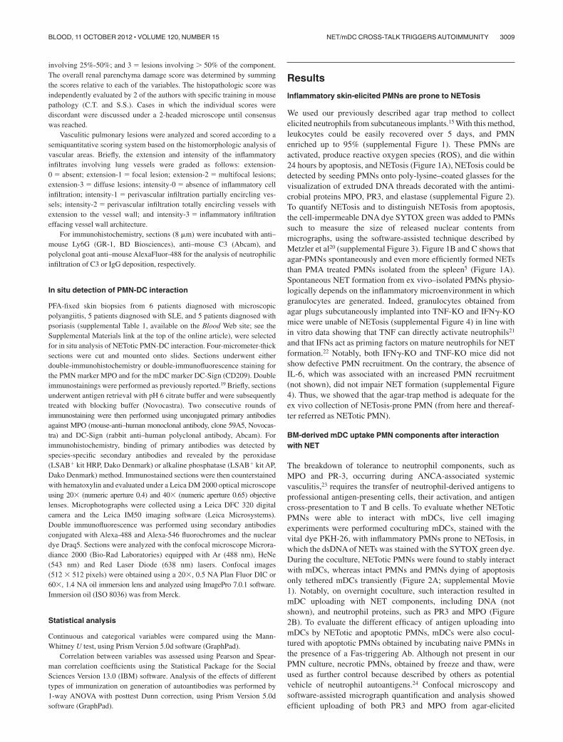
We used our previously described agar trap method to collectelicited neutrophils from subcutaneous implants.15 With this method,leukocytes could be easily recovered over 5 days, and PMNenriched up to 95% (supplemental Figure 1). These PMNs areactivated, produce reactive oxygen species (ROS), and die within24 hours by apoptosis, and NETosis (Figure 1A), NETosis could bedetected by seeding PMNs onto poly-lysine–coated glasses for thevisualization of extruded DNA threads decorated with the antimi-crobial proteins MPO, PR3, and elastase (supplemental Figure 2).To quantify NETosis and to distinguish NETosis from apoptosis,the cell-impermeable DNA dye SYTOX green was added to PMNssuch to measure the size of released nuclear contents frommicrographs, using the software-assisted technique described byMetzler et al20 (supplemental Figure 3). Figure 1B and C shows thatagar-PMNs spontaneously and even more efficiently formed NETsthan PMA treated PMNs isolated from the spleen5 (Figure 1A).Spontaneous NET formation from ex vivo–isolated PMNs physio-logically depends on the inflammatory microenvironment in whichgranulocytes are generated. Indeed, granulocytes obtained fromagar plugs subcutaneously implanted into TNF-KO and IFN�-KOmice were unable of NETosis (supplemental Figure 4) in line within vitro data showing that TNF can directly activate neutrophils21
and that IFNs act as priming factors on mature neutrophils for NETformation.22 Notably, both IFN�-KO and TNF-KO mice did notshow defective PMN recruitment. On the contrary, the absence ofIL-6, which was associated with an increased PMN recruitment(not shown), did not impair NET formation (supplemental Figure4). Thus, we showed that the agar-trap method is adequate for theex vivo collection of NETosis-prone PMN (from here and thereaf-ter referred as NETotic PMN).
BM-derived mDC uptake PMN components after interactionwith NET
The breakdown of tolerance to neutrophil components, such asMPO and PR-3, occurring during ANCA-associated systemicvasculitis,23 requires the transfer of neutrophil-derived antigens toprofessional antigen-presenting cells, their activation, and antigencross-presentation to T and B cells. To evaluate whether NEToticPMNs were able to interact with mDCs, live cell imagingexperiments were performed coculturing mDCs, stained with thevital dye PKH-26, with inflammatory PMNs prone to NETosis, inwhich the dsDNA of NETs was stained with the SYTOX green dye.During the coculture, NETotic PMNs were found to stably interactwith mDCs, whereas intact PMNs and PMNs dying of apoptosisonly tethered mDCs transiently (Figure 2A; supplemental Movie1). Notably, on overnight coculture, such interaction resulted inmDC uploading with NET components, including DNA (notshown), and neutrophil proteins, such as PR3 and MPO (Figure2B). To evaluate the different efficacy of antigen uploading intomDCs by NETotic and apoptotic PMNs, mDCs were also cocul-tured with apoptotic PMNs obtained by incubating naive PMNs inthe presence of a Fas-triggering Ab. Although not present in ourPMN culture, necrotic PMNs, obtained by freeze and thaw, wereused as further control because described by others as potentialvehicle of neutrophil autoantigens.24 Confocal microscopy andsoftware-assisted micrograph quantification and analysis showedefficient uploading of both PR3 and MPO from agar-elicited
NET/mDC CROSS-TALK TRIGGERS AUTOIMMUNITY 3009BLOOD, 11 OCTOBER 2012 � VOLUME 120, NUMBER 15
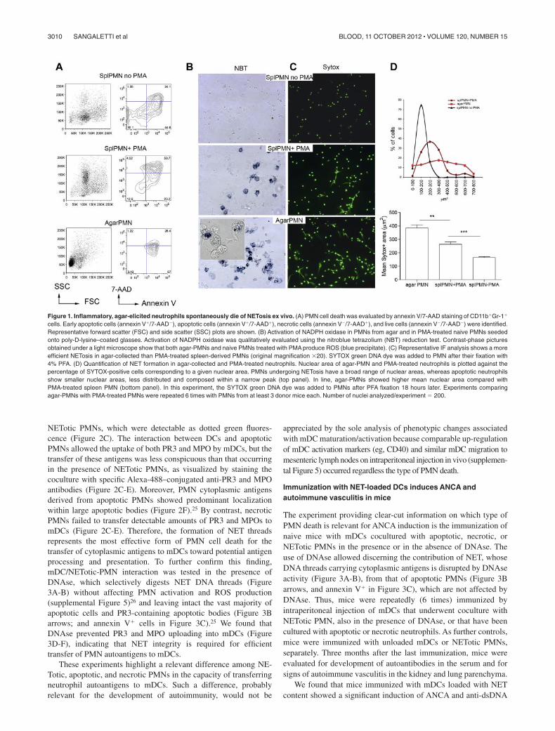
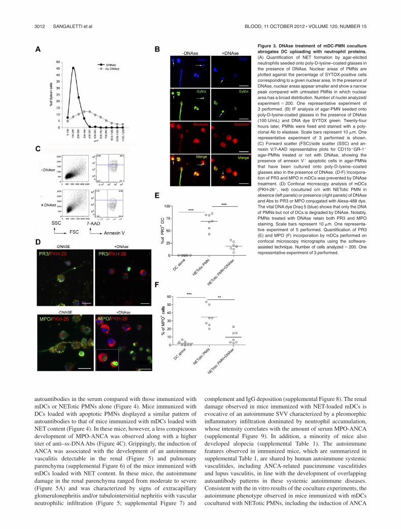
NETotic PMNs, which were detectable as dotted green fluores-cence (Figure 2C). The interaction between DCs and apoptoticPMNs allowed the uptake of both PR3 and MPO by mDCs, but thetransfer of these antigens was less conspicuous than that occurringin the presence of NETotic PMNs, as visualized by staining thecoculture with specific Alexa-488–conjugated anti-PR3 and MPOantibodies (Figure 2C-E). Moreover, PMN cytoplasmic antigensderived from apoptotic PMNs showed predominant localizationwithin large apoptotic bodies (Figure 2F).25 By contrast, necroticPMNs failed to transfer detectable amounts of PR3 and MPOs tomDCs (Figure 2C-E). Therefore, the formation of NET threadsrepresents the most effective form of PMN cell death for thetransfer of cytoplasmic antigens to mDCs toward potential antigenprocessing and presentation. To further confirm this finding,mDC/NETotic-PMN interaction was tested in the presence ofDNAse, which selectively digests NET DNA threads (Figure3A-B) without affecting PMN activation and ROS production(supplemental Figure 5)26 and leaving intact the vast majority ofapoptotic cells and PR3-containing apoptotic bodies (Figure 3Barrows; and annexin V� cells in Figure 3C).25 We found thatDNAse prevented PR3 and MPO uploading into mDCs (Figure3D-F), indicating that NET integrity is required for efficienttransfer of PMN autoantigens to mDCs.
These experiments highlight a relevant difference among NE-Totic, apoptotic, and necrotic PMNs in the capacity of transferringneutrophil autoantigens to mDCs. Such a difference, probablyrelevant for the development of autoimmunity, would not be
appreciated by the sole analysis of phenotypic changes associatedwith mDC maturation/activation because comparable up-regulationof mDC activation markers (eg, CD40) and similar mDC migration tomesenteric lymph nodes on intraperitoneal injection in vivo (supplemen-tal Figure 5) occurred regardless the type of PMN death.
Immunization with NET-loaded DCs induces ANCA andautoimmune vasculitis in mice
The experiment providing clear-cut information on which type ofPMN death is relevant for ANCA induction is the immunization ofnaive mice with mDCs cocultured with apoptotic, necrotic, orNETotic PMNs in the presence or in the absence of DNAse. Theuse of DNAse allowed discerning the contribution of NET, whoseDNA threads carrying cytoplasmic antigens is disrupted by DNAseactivity (Figure 3A-B), from that of apoptotic PMNs (Figure 3Barrows, and annexin V� in Figure 3C), which are not affected byDNAse. Thus, mice were repeatedly (6 times) immunized byintraperitoneal injection of mDCs that underwent coculture withNETotic PMN, also in the presence of DNAse, or that have beencultured with apoptotic or necrotic neutrophils. As further controls,mice were immunized with unloaded mDCs or NETotic PMNs,separately. Three months after the last immunization, mice wereevaluated for development of autoantibodies in the serum and forsigns of autoimmune vasculitis in the kidney and lung parenchyma.
We found that mice immunized with mDCs loaded with NETcontent showed a significant induction of ANCA and anti-dsDNA
Figure 1. Inflammatory, agar-elicited neutrophils spontaneously die of NETosis ex vivo. (A) PMN cell death was evaluated by annexin V/7-AAD staining of CD11b�Gr-1�
cells. Early apoptotic cells (annexin V�/7-AAD�), apoptotic cells (annexin V�/7-AAD�), necrotic cells (annexin V�/7-AAD�), and live cells (annexin V�/7-AAD�) were identified.Representative forward scatter (FSC) and side scatter (SSC) plots are shown. (B) Activation of NADPH oxidase in PMNs from agar and in PMA-treated naive PMNs seededonto poly-D-lysine–coated glasses. Activation of NADPH oxidase was qualitatively evaluated using the nitroblue tetrazolium (NBT) reduction test. Contrast-phase picturesobtained under a light microscope show that both agar-PMNs and naive PMNs treated with PMA produce ROS (blue precipitate). (C) Representative IF analysis shows a moreefficient NETosis in agar-collected than PMA-treated spleen-derived PMNs (original magnification �20). SYTOX green DNA dye was added to PMN after their fixation with4% PFA. (D) Quantification of NET formation in agar-collected and PMA-treated neutrophils. Nuclear area of agar-PMN and PMA-treated neutrophils is plotted against thepercentage of SYTOX-positive cells corresponding to a given nuclear area. PMNs undergoing NETosis have a broad range of nuclear areas, whereas apoptotic neutrophilsshow smaller nuclear areas, less distributed and composed within a narrow peak (top panel). In line, agar-PMNs showed higher mean nuclear area compared withPMA-treated spleen PMN (bottom panel). In this experiment, the SYTOX green DNA dye was added to PMNs after PFA fixation 18 hours later. Experiments comparingagar-PMNs with PMA-treated PMNs were repeated 6 times with PMNs from at least 3 donor mice each. Number of nuclei analyzed/experiment � 200.
3010 SANGALETTI et al BLOOD, 11 OCTOBER 2012 � VOLUME 120, NUMBER 15
Figure 2. mDCs interact with NETotic PMNs and are loaded with proteins from NET. (A) NETs persistently interact with mDCs. In this live imaging experiment, agar-PMNswere seeded onto culture dishes and allowed to adhere 30 minutes before adding mDCs previously labeled with the membrane vital dye PKH-26. The cocultures were alsoperformed in the presence of the DNA dye SYTOX green, which stains only PMNs undergoing NETosis. The coculture was maintained o/n in a humidified chamber underconfocal microscope, acquiring a picture every 10 minutes to highlight any interaction between PMNs and mDCs. PMNs in the cellular mixture form NETs (turquoise arrows)and NETs interact with mDCs (red arrows). Black arrows indicate naive neutrophils. Scale bars represent 5 m. (B) Uploading of mDCs with NET components. After NETinteraction, mDCs are uploaded with the neutrophil protein PR3 and MPO. In this live imaging experiment, labeled PKH-26 mDCs were added to agar-PMN in the presence of amAb to PR3 or MPO directly conjugated with Alexa-488 dye. After 18 hours, cells were fixed with PFA 4% and observed under a confocal microscope. Scale bars represent10 m. One representative experiment of 5 performed. (C) Uploading of PR3 and MPO by mDCs tested in the presence of different type of PMN death. Apoptotic PMNs,generated by Fas triggering Ab and necrotic PMNs obtained by freeze and thaw, were compared with NETotic PMNs for their capability to transfer PR3 and MPO to mDCs incoculture experiments. Representative confocal IF analysis showing that necrotic PMNs almost failed to transfer neutrophil PR3 or MPO to mDCs; on the contrary, PR3 andMPO were detectable as dotted green fluorescence in mDCs that interacted with NETotic PMNs and, to a lesser extent, in mDCs cultured with apoptotic PMNs.(D-E) Software-assisted micrograph quantification of mDCs uploading of PR3 (D) and MPO (E) performed on a total of 200 cells that were imaged by confocal microscopy. DCscultured with naive PMNs, maintained alive by incubating them in the presence of 30% of FCS and DCs alone. were the negative controls for PR3 transfer and MPO. Scale barsrepresent 30 m. (F) Appearance of PR3� apoptotic bodies that are taken up by mDCs cocultured with apoptotic cells.
NET/mDC CROSS-TALK TRIGGERS AUTOIMMUNITY 3011BLOOD, 11 OCTOBER 2012 � VOLUME 120, NUMBER 15
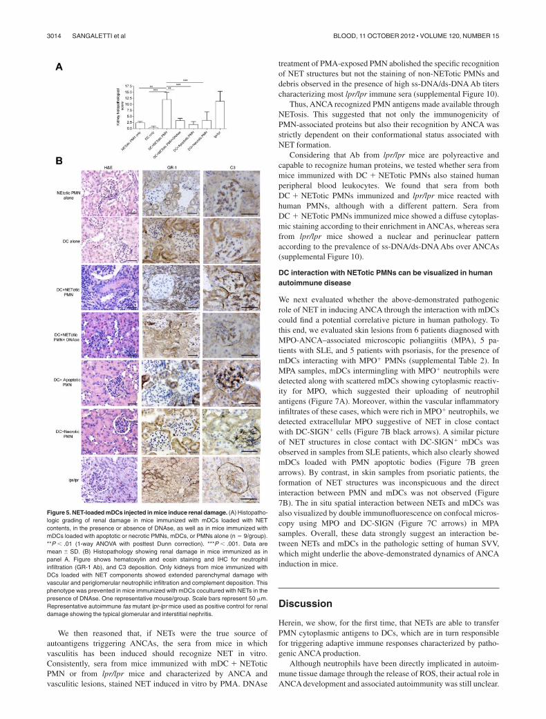
autoantibodies in the serum compared with those immunized withmDCs or NETotic PMNs alone (Figure 4). Mice immunized withDCs loaded with apoptotic PMNs displayed a similar pattern ofautoantibodies to that of mice immunized with mDCs loaded withNET content (Figure 4). In these mice, however, a less conspicuousdevelopment of MPO-ANCA was observed along with a highertiter of anti–ss-DNA Abs (Figure 4C). Grippingly, the induction ofANCA was associated with the development of an autoimmunevasculitis detectable in the renal (Figure 5) and pulmonaryparenchyma (supplemental Figure 6) of the mice immunized withmDCs loaded with NET content. In these mice, the autoimmunedamage in the renal parenchyma ranged from moderate to severe(Figure 5A) and was characterized by signs of extracapillaryglomerulonephritis and/or tubulointerstitial nephritis with vascularneutrophilic infiltration (Figure 5; supplemental Figure 7) and
complement and IgG deposition (supplemental Figure 8). The renaldamage observed in mice immunized with NET-loaded mDCs isevocative of an autoimmune SVV characterized by a pleomorphicinflammatory infiltration dominated by neutrophil accumulation,whose intensity correlates with the amount of serum MPO-ANCA(supplemental Figure 9). In addition, a minority of mice alsodeveloped alopecia (supplemental Table 1). The autoimmunefeatures observed in immunized mice, which are summarized insupplemental Table 1, are shared by human autoimmune systemicvasculitides, including ANCA-related paucimmune vasculitidesand lupus vasculitis, in line with the development of overlappingautoantibody patterns in these systemic autoimmune diseases.Consistent with the in vitro results of the coculture experiments, theautoimmune phenotype observed in mice immunized with mDCscocultured with NETotic PMNs, including the induction of ANCA
Figure 3. DNAse treatment of mDC-PMN cocultureabrogates DC uploading with neutrophil proteins.(A) Quantification of NET formation by agar-elicitedneutrophils seeded onto poly-D-lysine–coated glasses inthe presence of DNAse. Nuclear areas of PMNs areplotted against the percentage of SYTOX-positive cellscorresponding to a given nuclear area. In the presence ofDNAse, nuclear areas appear smaller and show a narrowpeak compared with untreated PMNs in which nucleararea has a broad distribution. Number of nuclei analyzed/experiment � 200. One representative experiment of3 performed. (B) IF analysis of agar-PMN seeded ontopoly-D-lysine-coated glasses in the presence of DNAse(100 U/mL) and DNA dye SYTOX green. Twenty-fourhours later, PMNs were fixed and stained with a poly-clonal Ab to elastase. Scale bars represent 10 m. Onerepresentative experiment of 3 performed is shown.(C) Forward scatter (FSC)/side scatter (SSC) and an-nexin V/7-AAD representative plots for CD11b�GR-1�
agar-PMNs treated or not with DNAse, showing thepresence of annexin V� apoptotic cells in agar-PMNsthat have been cultured onto poly-D-lysine–coatedglasses also in the presence of DNAse. (D-F) Incorpora-tion of PR3 and MPO in mDCs was prevented by DNAsetreatment. (D) Confocal microscopy analysis of mDCs(PKH-26�, red) cocultured o/n with NETotic PMN inabsence (left panels) or presence (right panels) of DNAseand Abs to PR3 or MPO conjugated with Alexa-488 dye.The vital DNA dye Draq 5 (blue) shows that only the DNAof PMNs but not of DCs is degraded by DNAse. Notably,PMNs treated with DNAse retain both PR3 and MPOstaining. Scale bars represent 10 m. One representa-tive experiment of 5 performed. Quantification of PR3(E) and MPO (F) incorporation by mDCs performed onconfocal microscopy micrographs using the software-assisted technique. Number of cells analyzed � 200. Onerepresentative experiment of 3 performed.
3012 SANGALETTI et al BLOOD, 11 OCTOBER 2012 � VOLUME 120, NUMBER 15
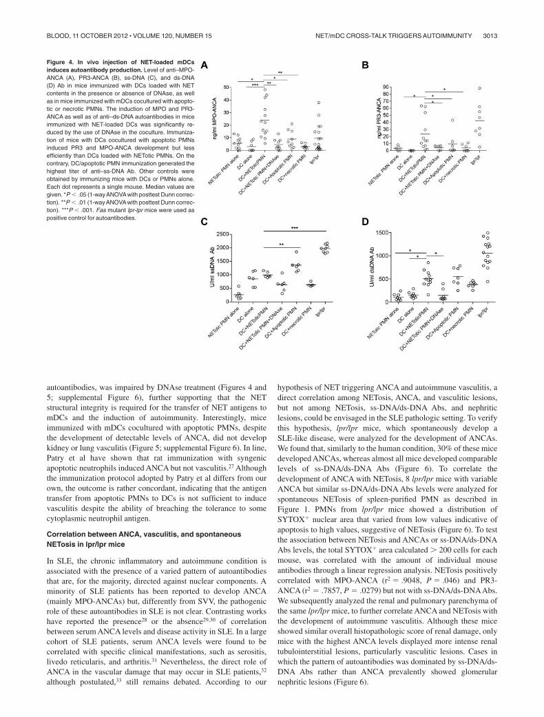
autoantibodies, was impaired by DNAse treatment (Figures 4 and5; supplemental Figure 6), further supporting that the NETstructural integrity is required for the transfer of NET antigens tomDCs and the induction of autoimmunity. Interestingly, miceimmunized with mDCs cocultured with apoptotic PMNs, despitethe development of detectable levels of ANCA, did not developkidney or lung vasculitis (Figure 5; supplemental Figure 6). In line,Patry et al have shown that rat immunization with syngenicapoptotic neutrophils induced ANCA but not vasculitis.27 Althoughthe immunization protocol adopted by Patry et al differs from ourown, the outcome is rather concordant, indicating that the antigentransfer from apoptotic PMNs to DCs is not sufficient to inducevasculitis despite the ability of breaching the tolerance to somecytoplasmic neutrophil antigen.
Correlation between ANCA, vasculitis, and spontaneousNETosis in lpr/lpr mice
In SLE, the chronic inflammatory and autoimmune condition isassociated with the presence of a varied pattern of autoantibodiesthat are, for the majority, directed against nuclear components. Aminority of SLE patients has been reported to develop ANCA(mainly MPO-ANCAs) but, differently from SVV, the pathogenicrole of these autoantibodies in SLE is not clear. Contrasting workshave reported the presence28 or the absence29,30 of correlationbetween serum ANCA levels and disease activity in SLE. In a largecohort of SLE patients, serum ANCA levels were found to becorrelated with specific clinical manifestations, such as serositis,livedo reticularis, and arthritis.31 Nevertheless, the direct role ofANCA in the vascular damage that may occur in SLE patients,32
although postulated,33 still remains debated. According to our
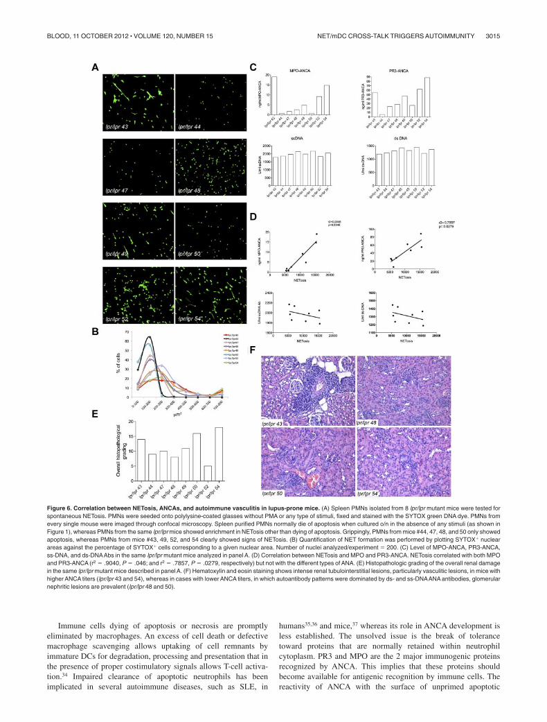
hypothesis of NET triggering ANCA and autoimmune vasculitis, adirect correlation among NETosis, ANCA, and vasculitic lesions,but not among NETosis, ss-DNA/ds-DNA Abs, and nephriticlesions, could be envisaged in the SLE pathologic setting. To verifythis hypothesis, lpr/lpr mice, which spontaneously develop aSLE-like disease, were analyzed for the development of ANCAs.We found that, similarly to the human condition, 30% of these micedeveloped ANCAs, whereas almost all mice developed comparablelevels of ss-DNA/ds-DNA Abs (Figure 6). To correlate thedevelopment of ANCA with NETosis, 8 lpr/lpr mice with variableANCA but similar ss-DNA/ds-DNA Abs levels were analyzed forspontaneous NETosis of spleen-purified PMN as described inFigure 1. PMNs from lpr/lpr mice showed a distribution ofSYTOX� nuclear area that varied from low values indicative ofapoptosis to high values, suggestive of NETosis (Figure 6). To testthe association between NETosis and ANCAs or ss-DNA/ds-DNAAbs levels, the total SYTOX� area calculated � 200 cells for eachmouse, was correlated with the amount of individual mouseantibodies through a linear regression analysis. NETosis positivelycorrelated with MPO-ANCA (r2 � .9048, P � .046) and PR3-ANCA (r2 � .7857, P � .0279) but not with ss-DNA/ds-DNAAbs.We subsequently analyzed the renal and pulmonary parenchyma ofthe same lpr/lpr mice, to further correlate ANCA and NETosis withthe development of autoimmune vasculitis. Although these miceshowed similar overall histopathologic score of renal damage, onlymice with the highest ANCA levels displayed more intense renaltubulointerstitial lesions, particularly vasculitic lesions. Cases inwhich the pattern of autoantibodies was dominated by ss-DNA/ds-DNA Abs rather than ANCA prevalently showed glomerularnephritic lesions (Figure 6).
Figure 4. In vivo injection of NET-loaded mDCsinduces autoantibody production. Level of anti–MPO-ANCA (A), PR3-ANCA (B), ss-DNA (C), and ds-DNA(D) Ab in mice immunized with DCs loaded with NETcontents in the presence or absence of DNAse, as wellas in mice immunized with mDCs cocultured with apopto-tic or necrotic PMNs. The induction of MPO and PR3-ANCA as well as of anti–ds-DNA autoantibodies in miceimmunized with NET-loaded DCs was significantly re-duced by the use of DNAse in the coculture. Immuniza-tion of mice with DCs cocultured with apoptotic PMNsinduced PR3 and MPO-ANCA development but lessefficiently than DCs loaded with NETotic PMNs. On thecontrary, DC/apoptotic PMN immunization generated thehighest titer of anti–ss-DNA Ab. Other controls wereobtained by immunizing mice with DCs or PMNs alone.Each dot represents a single mouse. Median values aregiven. *P � .05 (1-wayANOVAwith posttest Dunn correc-tion). **P � .01 (1-way ANOVA with posttest Dunn correc-tion). ***P � .001. Fas mutant lpr-lpr mice were used aspositive control for autoantibodies.
NET/mDC CROSS-TALK TRIGGERS AUTOIMMUNITY 3013BLOOD, 11 OCTOBER 2012 � VOLUME 120, NUMBER 15
We then reasoned that, if NETs were the true source ofautoantigens triggering ANCAs, the sera from mice in whichvasculitis has been induced should recognize NET in vitro.Consistently, sera from mice immunized with mDC � NEToticPMN or from lpr/lpr mice and characterized by ANCA andvasculitic lesions, stained NET induced in vitro by PMA. DNAse
treatment of PMA-exposed PMN abolished the specific recognitionof NET structures but not the staining of non-NETotic PMNs anddebris observed in the presence of high ss-DNA/ds-DNA Ab titerscharacterizing most lpr/lpr immune sera (supplemental Figure 10).
Thus, ANCA recognized PMN antigens made available throughNETosis. This suggested that not only the immunogenicity ofPMN-associated proteins but also their recognition by ANCA wasstrictly dependent on their conformational status associated withNET formation.
Considering that Ab from lpr/lpr mice are polyreactive andcapable to recognize human proteins, we tested whether sera frommice immunized with DC � NETotic PMNs also stained humanperipheral blood leukocytes. We found that sera from bothDC � NETotic PMNs immunized and lpr/lpr mice reacted withhuman PMNs, although with a different pattern. Sera fromDC � NETotic PMNs immunized mice showed a diffuse cytoplas-mic staining according to their enrichment in ANCAs, whereas serafrom lpr/lpr mice showed a nuclear and perinuclear patternaccording to the prevalence of ss-DNA/ds-DNA Abs over ANCAs(supplemental Figure 10).
DC interaction with NETotic PMNs can be visualized in humanautoimmune disease
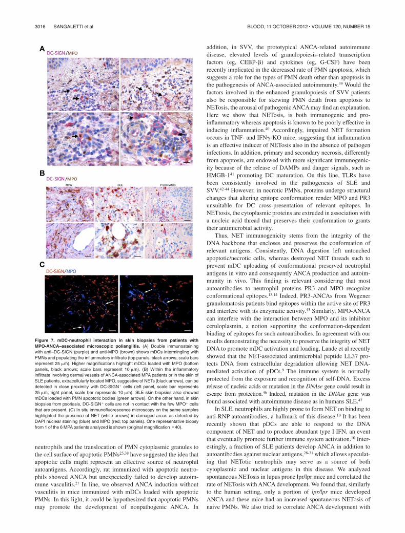
We next evaluated whether the above-demonstrated pathogenicrole of NET in inducing ANCA through the interaction with mDCscould find a potential correlative picture in human pathology. Tothis end, we evaluated skin lesions from 6 patients diagnosed withMPO-ANCA–associated microscopic poliangiitis (MPA), 5 pa-tients with SLE, and 5 patients with psoriasis, for the presence ofmDCs interacting with MPO� PMNs (supplemental Table 2). InMPA samples, mDCs intermingling with MPO� neutrophils weredetected along with scattered mDCs showing cytoplasmic reactiv-ity for MPO, which suggested their uploading of neutrophilantigens (Figure 7A). Moreover, within the vascular inflammatoryinfiltrates of these cases, which were rich in MPO� neutrophils, wedetected extracellular MPO suggestive of NET in close contactwith DC-SIGN� cells (Figure 7B black arrows). A similar pictureof NET structures in close contact with DC-SIGN� mDCs wasobserved in samples from SLE patients, which also clearly showedmDCs loaded with PMN apoptotic bodies (Figure 7B greenarrows). By contrast, in skin samples from psoriatic patients, theformation of NET structures was inconspicuous and the directinteraction between PMN and mDCs was not observed (Figure7B). The in situ spatial interaction between NETs and mDCs wasalso visualized by double immunofluorescence on confocal micros-copy using MPO and DC-SIGN (Figure 7C arrows) in MPAsamples. Overall, these data strongly suggest an interaction be-tween NETs and mDCs in the pathologic setting of human SVV,which might underlie the above-demonstrated dynamics of ANCAinduction in mice.
Discussion
Herein, we show, for the first time, that NETs are able to transferPMN cytoplasmic antigens to DCs, which are in turn responsiblefor triggering adaptive immune responses characterized by patho-genic ANCA production.
Although neutrophils have been directly implicated in autoim-mune tissue damage through the release of ROS, their actual role inANCA development and associated autoimmunity was still unclear.
Figure 5. NET-loaded mDCs injected in mice induce renal damage. (A) Histopatho-logic grading of renal damage in mice immunized with mDCs loaded with NETcontents, in the presence or absence of DNAse, as well as in mice immunized withmDCs loaded with apoptotic or necrotic PMNs, mDCs, or PMNs alone (n � 9/group).**P � .01 (1-way ANOVA with posttest Dunn correction). ***P � .001. Data aremean SD. (B) Histopathology showing renal damage in mice immunized as inpanel A. Figure shows hematoxylin and eosin staining and IHC for neutrophilinfiltration (GR-1 Ab), and C3 deposition. Only kidneys from mice immunized withDCs loaded with NET components showed extended parenchymal damage withvascular and periglomerular neutrophilic infiltration and complement deposition. Thisphenotype was prevented in mice immunized with mDCs cocultured with NETs in thepresence of DNAse. One representative mouse/group. Scale bars represent 50 m.Representative autoimmune fas mutant lpr-lpr mice used as positive control for renaldamage showing the typical glomerular and interstitial nephritis.
3014 SANGALETTI et al BLOOD, 11 OCTOBER 2012 � VOLUME 120, NUMBER 15
Immune cells dying of apoptosis or necrosis are promptlyeliminated by macrophages. An excess of cell death or defectivemacrophage scavenging allows uptaking of cell remnants byimmature DCs for degradation, processing and presentation that inthe presence of proper costimulatory signals allows T-cell activa-tion.34 Impaired clearance of apoptotic neutrophils has beenimplicated in several autoimmune diseases, such as SLE, in
humans35,36 and mice,37 whereas its role in ANCA development isless established. The unsolved issue is the break of tolerancetoward proteins that are normally retained within neutrophilcytoplasm. PR3 and MPO are the 2 major immunogenic proteinsrecognized by ANCA. This implies that these proteins shouldbecome available for antigenic recognition by immune cells. Thereactivity of ANCA with the surface of unprimed apoptotic
Figure 6. Correlation between NETosis, ANCAs, and autoimmune vasculitis in lupus-prone mice. (A) Spleen PMNs isolated from 8 lpr/lpr mutant mice were tested forspontaneous NETosis. PMNs were seeded onto polylysine-coated glasses without PMA or any type of stimuli, fixed and stained with the SYTOX green DNA dye. PMNs fromevery single mouse were imaged through confocal microscopy. Spleen purified PMNs normally die of apoptosis when cultured o/n in the absence of any stimuli (as shown inFigure 1), whereas PMNs from the same lpr/lpr mice showed enrichment in NETosis other than dying of apoptosis. Grippingly, PMNs from mice #44, 47, 48, and 50 only showedapoptosis, whereas PMNs from mice #43, 49, 52, and 54 clearly showed signs of NETosis. (B) Quantification of NET formation was performed by plotting SYTOX� nuclearareas against the percentage of SYTOX� cells corresponding to a given nuclear area. Number of nuclei analyzed/experiment � 200. (C) Level of MPO-ANCA, PR3-ANCA,ss-DNA, and ds-DNAAbs in the same lpr/lpr mutant mice analyzed in panel A. (D) Correlation between NETosis and MPO and PR3-ANCA. NETosis correlated with both MPOand PR3-ANCA (r2 � .9040, P � .046; and r2 � .7857, P � .0279, respectively) but not with the different types of ANA. (E) Histopathologic grading of the overall renal damagein the same lpr/lpr mutant mice described in panel A. (F) Hematoxylin and eosin staining shows intense renal tubulointerstitial lesions, particularly vasculitic lesions, in mice withhigher ANCA titers (lpr/lpr 43 and 54), whereas in cases with lower ANCA titers, in which autoantibody patterns were dominated by ds- and ss-DNAANA antibodies, glomerularnephritic lesions are prevalent (lpr/lpr 48 and 50).
NET/mDC CROSS-TALK TRIGGERS AUTOIMMUNITY 3015BLOOD, 11 OCTOBER 2012 � VOLUME 120, NUMBER 15
neutrophils and the translocation of PMN cytoplasmic granules tothe cell surface of apoptotic PMNs25,38 have suggested the idea thatapoptotic cells might represent an effective source of neutrophilautoantigens. Accordingly, rat immunized with apoptotic neutro-phils showed ANCA but unexpectedly failed to develop autoim-mune vasculitis.27 In line, we observed ANCA induction withoutvasculitis in mice immunized with mDCs loaded with apoptoticPMNs. In this light, it could be hypothesized that apoptotic PMNsmay promote the development of nonpathogenic ANCA. In
addition, in SVV, the prototypical ANCA-related autoimmunedisease, elevated levels of granulopoiesis-related transcriptionfactors (eg, CEBP-�) and cytokines (eg, G-CSF) have beenrecently implicated in the decreased rate of PMN apoptosis, whichsuggests a role for the types of PMN death other than apoptosis inthe pathogenesis of ANCA-associated autoimmunity.39 Would thefactors involved in the enhanced granulopoiesis of SVV patientsalso be responsible for skewing PMN death from apoptosis toNETosis, the arousal of pathogenic ANCA may find an explanation.Here we show that NETosis, is both immunogenic and pro-inflammatory whereas apoptosis is known to be poorly effective ininducing inflammation.40 Accordingly, impaired NET formationoccurs in TNF- and IFN�-KO mice, suggesting that inflammationis an effective inducer of NETosis also in the absence of pathogeninfections. In addition, primary and secondary necrosis, differentlyfrom apoptosis, are endowed with more significant immunogenic-ity because of the release of DAMPs and danger signals, such asHMGB-141 promoting DC maturation. On this line, TLRs havebeen consistently involved in the pathogenesis of SLE andSVV.42-44 However, in necrotic PMNs, proteins undergo structuralchanges that altering epitope conformation render MPO and PR3unsuitable for DC cross-presentation of relevant epitopes. InNETtosis, the cytoplasmic proteins are extruded in association witha nucleic acid thread that preserves their conformation to grantstheir antimicrobial activity.
Thus, NET immunogenicity stems from the integrity of theDNA backbone that encloses and preserves the conformation ofrelevant antigens. Consistently, DNA digestion left untouchedapoptotic/necrotic cells, whereas destroyed NET threads such toprevent mDC uploading of conformational preserved neutrophilantigens in vitro and consequently ANCA production and autoim-munity in vivo. This finding is relevant considering that mostautoantibodies to neutrophil proteins PR3 and MPO recognizeconformational epitopes.13,14 Indeed, PR3-ANCAs from Wegenergranulomatosis patients bind epitopes within the active site of PR3and interfere with its enzymatic activity.45 Similarly, MPO-ANCAcan interfere with the interaction between MPO and its inhibitorceruloplasmin, a notion supporting the conformation-dependentbinding of epitopes for such autoantibodies. In agreement with ourresults demonstrating the necessity to preserve the integrity of NETDNA to promote mDC activation and loading, Lande et al recentlyshowed that the NET-associated antimicrobial peptide LL37 pro-tects DNA from extracellular degradation allowing NET DNA-mediated activation of pDCs.9 The immune system is normallyprotected from the exposure and recognition of self-DNA. Excessrelease of nucleic acids or mutation in the DNAse gene could result inescape from protection.46 Indeed, mutation in the DNAse gene wasfound associated with autoimmune disease as in humans SLE.47
In SLE, neutrophils are highly prone to form NET on binding toanti-RNP autoantibodies, a hallmark of this disease.10 It has beenrecently shown that pDCs are able to respond to the DNAcomponent of NET and to produce abundant type I IFN, an eventthat eventually promote further immune system activation.10 Inter-estingly, a fraction of SLE patients develop ANCA in addition toautoantibodies against nuclear antigens,28-31 which allows speculat-ing that NETotic neutrophils may serve as a source of bothcytoplasmic and nuclear antigens in this disease. We analyzedspontaneous NETosis in lupus prone lpr/lpr mice and correlated therate of NETosis with ANCA development. We found that, similarlyto the human setting, only a portion of lpr/lpr mice developedANCA and these mice had an increased spontaneous NETosis ofnaive PMNs. We also tried to correlate ANCA development with
Figure 7. mDC-neutrophil interaction in skin biopsies from patients withMPO-ANCA–associated microscopic poliangiitis. (A) Double immunostainingwith anti–DC-SIGN (purple) and anti-MPO (brown) shows mDCs intermingling withPMNs and populating the inflammatory infiltrate (top panels, black arrows; scale barsrepresent 25 m). Higher magnifications highlight mDCs loaded with MPO (bottompanels, black arrows; scale bars represent 10 m). (B) Within the inflammatoryinfiltrate involving dermal vessels of ANCA-associated MPA patients or in the skin ofSLE patients, extracellularly located MPO, suggestive of NETs (black arrows), can bedetected in close proximity with DC-SIGN� cells (left panel, scale bar represents25 m; right panel, scale bar represents 10 m). SLE skin biopsies also showedmDCs loaded with PMN apoptotic bodies (green arrows). On the other hand, in skinbiopsies from psoriasis, DC-SIGN� cells are not in contact with the few MPO� cellsthat are present. (C) In situ immunofluorescence microscopy on the same sampleshighlighted the presence of NET (white arrows) in damaged areas as detected byDAPI nuclear staining (blue) and MPO (red; top panels). One representative biopsyfrom 1 of the 6 MPA patients analyzed is shown (original magnification �40).
3016 SANGALETTI et al BLOOD, 11 OCTOBER 2012 � VOLUME 120, NUMBER 15
features of autoimmune vasculitis in these same mice and foundthat, despite the prominent development of a glomerular lupus-likenephritis, mice with the highest ANCA titers clearly showed moreconspicuous interstitial vasculitic lesions.
Herein we have extended the correlation between ANCAinduction of NETosis and autoimmune vascular damage in humanpathology,12 demonstrating the primary role of NET in pathogenicANCA induction through mDC cross-presentation in mice. Inaddition, we corroborated this finding in humans, showing mDCsloaded with MPO and directly interacting with NET structures inskin lesions from patients diagnosed with MPO-ANCA–associatedmicroscopic poliangiitis and SLE. Our study contributes to the ideathat NET, although conceived as weapon to defeat extracellular patho-gens, may operate hostilely by inducing and fostering autoimmunity.
Acknowledgments
This work was supported by the Italian Association for CancerResearch and the Association for International Cancer Research.
Authorship
Contribution: S.S. and C.T. designed and performed experimentsand data analysis and wrote the paper; C. Guarnotta providedhistopathology and immunohistochemical expertise; C.C. and C.V.performed FACS analysis; B.C. performed immunohistochemicalanalysis of mouse tissue; M.P. performed in vivo experiments; P.C.performed confocal microscopy experiments; S.P. contributed to invitro experiments; C. Guiducci provided critical reagent andreviewed the manuscript; and M.P.C. contributed to study designand data analysis and wrote the paper.
Conflict-of-interest disclosure: C. Guiducci is a full-time em-ployee at Dynavax Technologies Corporation. The remainingauthors declare no competing financial interests.
Correspondence: Mario P. Colombo, Molecular ImmunologyUnit, Department of Experimental Oncology and Molecular Medi-cine, Fondazione Istituti di Ricovero e Cura a Carattere ScientificoIstituto Nazionale Tumori Via Amadeo 42, 20133 Milano, Italy;e-mail: [email protected].
References
1. Nathan C. Neutrophils and immunity: challengesand opportunities. Nat Rev Immunol. 2006;6(3):173-182.
2. Di Carlo E, Forni G, Lollini P, Colombo MP,Modesti A, Musiani P. The intriguing role of poly-morphonuclear neutrophils in antitumor reactions.Blood. 2001;97(2):339-345.
3. Cassatella MA, Locati M, Mantovani A. Never un-derestimate the power of a neutrophil. Immunity.2009;31(5):698-700.
4. Brinkmann V, Reichard U, Goosmann C, et al.Neutrophil extracellular traps kill bacteria. Sci-ence. 2004;303(5663):1532-1535.
5. Fuchs TA, Abed U, Goosmann C, et al. Novel celldeath program leads to neutrophil extracellulartraps. J Cell Biol. 2007;176(2):231-241.
6. Remijsen Q, Kuijpers TW, Wirawan E, Lippens S,Vandenabeele P, Vanden Berghe T. Dying for acause: NETosis, mechanisms behind an antimi-crobial cell death modality. Cell Death Differ.2011;18(4):581-588.
7. Alcorta DA, Barnes DA, Dooley MA, et al. Leuko-cyte gene expression signatures in antineutrophilcytoplasmic autoantibody and lupus glomerulone-phritis. Kidney Int. 2007;72(7):853-864.
8. Bennett L, Palucka AK, Arce E, et al. Interferonand granulopoiesis signatures in systemic lupuserythematosus blood. J Exp Med. 2003;197(6):711-723.
9. Lande R, Ganguly D, Facchinetti V, et al. Neutro-phils activate plasmacytoid dendritic cells by re-leasing self-DNA-peptide complexes in systemiclupus erythematosus. Sci Transl Med. 2011;3(73):73ra19.
10. Garcia-Romo GS, Caielli S, Vega B, et al. Nettingneutrophils are major inducers of type I IFN pro-duction in pediatric systemic lupus erythemato-sus. Sci Transl Med. 2011;3(73):73ra20.
11. Villanueva E, Yalavarthi S, Berthier CC, et al. Net-ting neutrophils induce endothelial damage, infil-trate tissues, and expose immunostimulatorymolecules in systemic lupus erythematosus. J Im-munol. 2011;187(1):538-552.
12. Kessenbrock K, Krumbholz M, Schonermarck U,et al. Netting neutrophils in autoimmune small-vessel vasculitis. Nat Med. 2009;15(6):623-625.
13. Falk RJ, Becker M, Terrell R, Jennette JC. Anti-myeloperoxidase autoantibodies react with nativebut not denatured myeloperoxidase. Clin Exp Im-munol. 1992;89(2):274-278.
14. Specks U. What you should know about PR3-ANCA: conformational requirements of protei-
nase 3 (PR3) for enzymatic activity and recogni-tion by PR3-ANCA. Arthritis Res. 2000;2(4):263-267.
15. Colombo MP, Lombardi L, Stoppacciaro A, et al.Granulocyte colony-stimulating factor (G-CSF)gene transduction in murine adenocarcinomadrives neutrophil-mediated tumor inhibition invivo: neutrophils discriminate between G-CSF-producing and G-CSF-nonproducing tumor cells.J Immunol. 1992;149(1):113-119.
16. Ziegler-Heitbrock L, Ancuta P, Crowe S, et al. No-menclature of monocytes and dendritic cells inblood. Blood. 2010;116(16):e74-e80.
17. Sangaletti S, Gioiosa L, Guiducci C, et al. Accel-erated dendritic-cell migration and T-cell primingin SPARC-deficient mice. J Cell Sci. 2005;118(16):3685-3694.
18. Wernick RM, Smith DL, Houghton DC, et al. Reli-ability of histologic scoring for lupus nephritis: acommunity-based evaluation. Ann Intern Med.1993;119(8):805-811.
19. Tripodo C, Gri G, Piccaluga PP, et al. Mast cellsand Th17 cells contribute to the lymphoma-associated pro-inflammatory microenvironment ofangioimmunoblastic T-cell lymphoma. Am JPathol. 2010;177(2):792-802.
20. Metzler KD, Fuchs TA, Nauseef WM, et al. Myelo-peroxidase is required for neutrophil extracellulartrap formation: implications for innate immunity.Blood. 2011;117(3):953-959.
21. Jarius S, Eichhorn P, Albert MH, et al. Intrave-nous immunoglobulins contain naturally occurringantibodies that mimic antineutrophil cytoplasmicantibodies and activate neutrophils in aTNFalpha-dependent and Fc-receptor-independent way. Blood. 2007;109(10):4376-4382.
22. Martinelli S, Urosevic M, Daryadel A, et al. Induc-tion of genes mediating interferon-dependent ex-tracellular trap formation during neutrophil differ-entiation. J Biol Chem. 2004;279(42):44123-44132.
23. Witko-Sarsat V, Daniel S, Noel LH, Mouthon L.Neutrophils and B lymphocytes in ANCA-associatedvasculitis. APMIS Suppl. 2009(127):27-31.
24. Clayton AR, Prue RL, Harper L, Drayson MT,Savage CO. Dendritic cell uptake of human apo-ptotic and necrotic neutrophils inhibits CD40,CD80, and CD86 expression and reduces alloge-neic T cell responses: relevance to systemic vas-culitis. Arthritis Rheum. 2003;48(8):2362-2374.
25. Kantari C, Pederzoli-Ribeil M, Amir-Moazami O,
et al. Proteinase 3, the Wegener autoantigen, isexternalized during neutrophil apoptosis: evi-dence for a functional association with phospho-lipid scramblase 1 and interference with macro-phage phagocytosis. Blood. 2007;110(12):4086-4095.
26. Munafo DB, Johnson JL, Brzezinska AA, Ellis BA,Wood MR, Catz SD. DNase I inhibits a late phaseof reactive oxygen species production in neutro-phils. J Innate Immun. 2009;1(6):527-542.
27. Patry YC, Trewick DC, Gregoire M, et al. Ratsinjected with syngenic rat apoptotic neutrophilsdevelop antineutrophil cytoplasmic antibodies.J Am Soc Nephrol. 2001;12(8):1764-1768.
28. Chin HJ, Ahn C, Lim CS, et al. Clinical implica-tions of antineutrophil cytoplasmic antibody test inlupus nephritis. Am J Nephrol. 2000;20(1):57-63.
29. Spronk PE, Bootsma H, Horst G, et al. Antineu-trophil cytoplasmic antibodies in systemic lupuserythematosus. Br J Rheumatol. 1996;35(7):625-631.
30. Manolova I, Dancheva M, Halacheva K. Antineu-trophil cytoplasmic antibodies in patients withsystemic lupus erythematosus: prevalence, anti-gen specificity, and clinical associations. Rheu-matol Int. 2001;20(5):197-204.
31. Galeazzi M, Morozzi G, Sebastiani GD, et al.Anti-neutrophil cytoplasmic antibodies in 566 Eu-ropean patients with systemic lupus erythemato-sus: prevalence, clinical associations and correla-tion with other autoantibodies. EuropeanConcerted Action on the Immunogenetics of SLE.Clin Exp Rheumatol. 1998;16(5):541-546.
32. Drenkard C, Villa AR, Reyes E, Abello M,Alarcon-Segovia D. Vasculitis in systemic lupuserythematosus. Lupus. 1997;6(3):235-242.
33. Pradhan VD, Badakere SS, Bichile LS,Almeida AF. Anti-neutrophil cytoplasmic antibod-ies (ANCA) in systemic lupus erythematosus:prevalence, clinical associations and correlationwith other autoantibodies. J Assoc PhysiciansIndia. 2004;52:533-537.
34. Steinman RM. Linking innate to adaptive immu-nity through dendritic cells. Novartis Found Symp.2006;279:101-109; discussion 109-113.
35. Donnelly S, Roake W, Brown S, et al. Impairedrecognition of apoptotic neutrophils by the C1q/calreticulin and CD91 pathway in systemic lupuserythematosus. Arthritis Rheum. 2006;54(5):1543-1556.
36. Gaipl US, Sheriff A, Franz S, et al. Inefficientclearance of dying cells and autoreactivity. CurrTop Microbiol Immunol. 2006;305:161-176.
NET/mDC CROSS-TALK TRIGGERS AUTOIMMUNITY 3017BLOOD, 11 OCTOBER 2012 � VOLUME 120, NUMBER 15

37. Potter PK, Cortes-Hernandez J, Quartier P,Botto M, Walport MJ. Lupus-prone mice have anabnormal response to thioglycolate and an im-paired clearance of apoptotic cells. J Immunol.2003;170(6):3223-3232.
38. Gilligan HM, Bredy B, Brady HR, et al. Antineutro-phil cytoplasmic autoantibodies interact with pri-mary granule constituents on the surface of apo-ptotic neutrophils in the absence of neutrophilpriming. J Exp Med. 1996;184(6):2231-2241.
39. Abdgawad M, Pettersson A, Gunnarsson L, et al.Decreased neutrophil apoptosis in quiescentANCA-associated systemic vasculitis. PLoS One.2012;7(3):e32439.
40. Griffith TS, Ferguson TA. Cell death in the main-
tenance and abrogation of tolerance: the five Wsof dying cells. Immunity. 2011;35(4):456-466.
41. Bianchi ME, Manfredi AA. High-mobility groupbox 1 (HMGB1) protein at the crossroads be-tween innate and adaptive immunity. ImmunolRev. 2007;220:35-46.
42. Guiducci C, Gong M, Xu Z, et al. TLR recognitionof self nucleic acids hampers glucocorticoid activ-ity in lupus. Nature. 2010;465(7300):937-941.
43. Santiago-Raber ML, Dunand-Sauthier I, Wu T,et al. Critical role of TLR7 in the acceleration ofsystemic lupus erythematosus in TLR9-deficientmice. J Autoimmun. 2010;34(4):339-348.
44. van Timmeren MM, Heeringa P. Pathogenesis ofANCA-associated vasculitis: recent insights from
animal models. Curr Opin Rheumatol. 2012;24(1):8-14.
45. van der Geld YM, Stegeman CA, Kallenberg CG.B cell epitope specificity in ANCA-associated vas-culitis: does it matter? Clin Exp Immunol. 2004;137(3):451-459.
46. Nagata S, Nagase H, Kawane K, Mukae N,Fukuyama H. Degradation of chromosomal DNAduring apoptosis. Cell Death Differ. 2003;10(1):108-116.
47. Dittmar M, Bischofs C, Matheis N, Poppe R,Kahaly GJ. A novel mutation in the DNASE1 geneis related with protein instability and decreasedenzyme activity in thyroid autoimmunity. J Auto-immun. 2009;32(1):7-13.
3018 SANGALETTI et al BLOOD, 11 OCTOBER 2012 � VOLUME 120, NUMBER 15
Related Documents