Neutralisation of uPA with a Monoclonal Antibody Reduces Plasmin Formation and Delays Skin Wound Healing in tPA-Deficient Mice Annika Jo ¨ gi ¤a , Birgitte Rønø ¤d , Ida K. Lund, Boye S. Nielsen ¤b , Michael Ploug, Gunilla Høyer-Hansen, John Rømer ¤c , Leif R. Lund* ¤d Finsen Laboratory, Copenhagen University Hospital, Copenhagen Biocenter, Copenhagen, Denmark Abstract Background: Proteolytic degradation by plasmin and metalloproteinases is essential for epidermal regeneration in skin wound healing. Plasminogen deficient mice have severely delayed wound closure as have mice simultaneously lacking the two plasminogen activators, urokinase-type plasminogen activator (uPA) and tissue-type plasminogen activator (tPA). In contrast, individual genetic deficiencies in either uPA or tPA lead to wound healing kinetics with no or only slightly delayed closure of skin wounds. Methodology/Principal Findings: To evaluate the therapeutic potential in vivo of a murine neutralizing antibody directed against mouse uPA we investigated the efficacy in skin wound healing of tPA-deficient mice. Systemic administration of the anti-mouse uPA monoclonal antibody, mU1, to tPA-deficient mice caused a dose-dependent delay of skin wound closure almost similar to the delayed kinetics observed in uPA;tPA double-deficient mice. Analysis of wound extracts showed diminished levels of plasmin in the mU1-treated tPA-deficent mice. Immunohistochemistry revealed that fibrin accumulated in the wounds of such mU1-treated tPA-deficent mice and that keratinocyte tongues were aberrant. Together these abnormalities lead to compromised epidermal closure. Conclusions/Significance: Our findings demonstrate that inhibition of uPA activity with a monoclonal antibody in adult tPA-deficient mice mimics the effect of simultaneous genetic ablation of uPA and tPA. Thus, application of the murine inhibitory mU1 antibody provides a new and highly versatile tool to interfere with uPA-activity in vivo in mouse models of disease. Citation: Jo ¨ gi A, Rønø B, Lund IK, Nielsen BS, Ploug M, et al. (2010) Neutralisation of uPA with a Monoclonal Antibody Reduces Plasmin Formation and Delays Skin Wound Healing in tPA-Deficient Mice. PLoS ONE 5(9): e12746. doi:10.1371/journal.pone.0012746 Editor: Joanna Mary Bridger, Brunel University, United Kingdom Received April 25, 2010; Accepted August 10, 2010; Published September 15, 2010 Copyright: ß 2010 Jo ¨ gi et al. This is an open-access article distributed under the terms of the Creative Commons Attribution License, which permits unrestricted use, distribution, and reproduction in any medium, provided the original author and source are credited. Funding: Research grants supporting this work: Swedish Cancer Society and Svenska Medicinsk Forskning (Fellowship to A.J.), European Union Grant 201279, MICROENVIMET, Aage Bangs Foundation, ‘‘Grosserer Alfred Nielsen og Hustrus Foundation, Danish Cancer Society, the Danish Cancer Research Foundation,’’ Agnes og Poul Friis Foundation, and the Lundbeck Foundation. The funders, had no role in study design, data collection and analysis, decision to publish, preparation of the manuscript. There is no restriction in the authors’ right to follow all PLoS ONE policies on sharing data and materials. Competing Interests: The authors have declared that no competing interests exist. * E-mail: [email protected] ¤a Current address: Center for Molecular Pathology, Lund University, Ska ˚ne University Hospital Malmo ¨ , Malmo ¨ , Sweden ¤b Current address: Exiqon A/S, Diagnostic Product Development, Vedbæk, Denmark ¤c Current address: Histology, Biopharmaceuticals Research Unit, Novo Nordisk A/S, Ma ˚løv, Denmark ¤d Current address: Department of Cellular and Molecular Medicine, Faculty of Health Sciences, University of Copenhagen, Copenhagen, Denmark Introduction Tissue remodeling and confined degradation of the extracellular matrix (ECM) is pivotal in several physiological and pathological processes involving cell migration [1–5]. This tightly controlled proteolytic degradation of the ECM is mainly performed by the serine protease plasmin and members of the matrix metallopro- teinase (MMP) family [3,6]. Plasmin is synthesized as a precursor, plasminogen (Plg), in the liver, and is present throughout the body in micromolar concentrations. Plg is activated at its site of action by proteolytical cleavage by one of three proteases, urokinase-type plasminogen activator (uPA), tissue-type plasminogen activator (tPA) [7,8,] or the newly identified Plg activator, plasma kallikrein [9]. Plg deficiency has severe physiological consequences, primarily due to diminished fibrinolysis, in both humans and mice [10–12]. Furthermore, gene disruption studies in mice have proven plasmin(ogen) to be required for the proper execution of processes involving ECM remodeling, such as cancer metastasis [13], neointima formation after vascular injury [14], placental development [15], post-lactational mammary gland involution [16], and skin wound healing [17]. In Plg-deficient mice there is a marked delay in healing of incisional skin wounds, presumably due to a diminished ability of the leading-edge keratinocytes at the wound edges to proteolytically dissect their way through the fibrin- rich wound matrix, as fibrin is accumulating around these keratinocytes [17]. The previous finding that lack of fibrin(ogen) in the wound field rescues the requirement for Plg to achieve timely healing [18] further corroborates that the primary role for PLoS ONE | www.plosone.org 1 September 2010 | Volume 5 | Issue 9 | e12746

Welcome message from author
This document is posted to help you gain knowledge. Please leave a comment to let me know what you think about it! Share it to your friends and learn new things together.
Transcript
Neutralisation of uPA with a Monoclonal AntibodyReduces Plasmin Formation and Delays Skin WoundHealing in tPA-Deficient MiceAnnika Jogi¤a, Birgitte Rønø¤d, Ida K. Lund, Boye S. Nielsen¤b, Michael Ploug, Gunilla Høyer-Hansen,
John Rømer¤c, Leif R. Lund*¤d
Finsen Laboratory, Copenhagen University Hospital, Copenhagen Biocenter, Copenhagen, Denmark
Abstract
Background: Proteolytic degradation by plasmin and metalloproteinases is essential for epidermal regeneration in skinwound healing. Plasminogen deficient mice have severely delayed wound closure as have mice simultaneously lacking thetwo plasminogen activators, urokinase-type plasminogen activator (uPA) and tissue-type plasminogen activator (tPA). Incontrast, individual genetic deficiencies in either uPA or tPA lead to wound healing kinetics with no or only slightly delayedclosure of skin wounds.
Methodology/Principal Findings: To evaluate the therapeutic potential in vivo of a murine neutralizing antibody directedagainst mouse uPA we investigated the efficacy in skin wound healing of tPA-deficient mice. Systemic administration of theanti-mouse uPA monoclonal antibody, mU1, to tPA-deficient mice caused a dose-dependent delay of skin wound closurealmost similar to the delayed kinetics observed in uPA;tPA double-deficient mice. Analysis of wound extracts showeddiminished levels of plasmin in the mU1-treated tPA-deficent mice. Immunohistochemistry revealed that fibrin accumulatedin the wounds of such mU1-treated tPA-deficent mice and that keratinocyte tongues were aberrant. Together theseabnormalities lead to compromised epidermal closure.
Conclusions/Significance: Our findings demonstrate that inhibition of uPA activity with a monoclonal antibody in adulttPA-deficient mice mimics the effect of simultaneous genetic ablation of uPA and tPA. Thus, application of the murineinhibitory mU1 antibody provides a new and highly versatile tool to interfere with uPA-activity in vivo in mouse models ofdisease.
Citation: Jogi A, Rønø B, Lund IK, Nielsen BS, Ploug M, et al. (2010) Neutralisation of uPA with a Monoclonal Antibody Reduces Plasmin Formation and Delays SkinWound Healing in tPA-Deficient Mice. PLoS ONE 5(9): e12746. doi:10.1371/journal.pone.0012746
Editor: Joanna Mary Bridger, Brunel University, United Kingdom
Received April 25, 2010; Accepted August 10, 2010; Published September 15, 2010
Copyright: � 2010 Jogi et al. This is an open-access article distributed under the terms of the Creative Commons Attribution License, which permits unrestricteduse, distribution, and reproduction in any medium, provided the original author and source are credited.
Funding: Research grants supporting this work: Swedish Cancer Society and Svenska Medicinsk Forskning (Fellowship to A.J.), European Union Grant 201279,MICROENVIMET, Aage Bangs Foundation, ‘‘Grosserer Alfred Nielsen og Hustrus Foundation, Danish Cancer Society, the Danish Cancer Research Foundation,’’Agnes og Poul Friis Foundation, and the Lundbeck Foundation. The funders, had no role in study design, data collection and analysis, decision to publish,preparation of the manuscript. There is no restriction in the authors’ right to follow all PLoS ONE policies on sharing data and materials.
Competing Interests: The authors have declared that no competing interests exist.
* E-mail: [email protected]
¤a Current address: Center for Molecular Pathology, Lund University, Skane University Hospital Malmo, Malmo, Sweden¤b Current address: Exiqon A/S, Diagnostic Product Development, Vedbæk, Denmark¤c Current address: Histology, Biopharmaceuticals Research Unit, Novo Nordisk A/S, Maløv, Denmark¤d Current address: Department of Cellular and Molecular Medicine, Faculty of Health Sciences, University of Copenhagen, Copenhagen, Denmark
Introduction
Tissue remodeling and confined degradation of the extracellular
matrix (ECM) is pivotal in several physiological and pathological
processes involving cell migration [1–5]. This tightly controlled
proteolytic degradation of the ECM is mainly performed by the
serine protease plasmin and members of the matrix metallopro-
teinase (MMP) family [3,6]. Plasmin is synthesized as a precursor,
plasminogen (Plg), in the liver, and is present throughout the body
in micromolar concentrations. Plg is activated at its site of action
by proteolytical cleavage by one of three proteases, urokinase-type
plasminogen activator (uPA), tissue-type plasminogen activator
(tPA) [7,8,] or the newly identified Plg activator, plasma kallikrein
[9]. Plg deficiency has severe physiological consequences,
primarily due to diminished fibrinolysis, in both humans and
mice [10–12]. Furthermore, gene disruption studies in mice have
proven plasmin(ogen) to be required for the proper execution of
processes involving ECM remodeling, such as cancer metastasis
[13], neointima formation after vascular injury [14], placental
development [15], post-lactational mammary gland involution
[16], and skin wound healing [17]. In Plg-deficient mice there is a
marked delay in healing of incisional skin wounds, presumably due
to a diminished ability of the leading-edge keratinocytes at the
wound edges to proteolytically dissect their way through the fibrin-
rich wound matrix, as fibrin is accumulating around these
keratinocytes [17]. The previous finding that lack of fibrin(ogen)
in the wound field rescues the requirement for Plg to achieve
timely healing [18] further corroborates that the primary role for
PLoS ONE | www.plosone.org 1 September 2010 | Volume 5 | Issue 9 | e12746
Plg in wound healing is fibrinolysis. In addition, we have recently
demonstrated that Plg activation in wounds is actually dependent
on the presence of this fibrin-rich provisional matrix [19].
During the invasive phase of wound healing, the migrating
leading-edge keratinocytes express uPA and its cell surface
receptor uPAR [20,21], whereas tPA has been detected only in
a few keratinocytes late in the re-epithelialization of human
wounds [20–22]. In addition to the expression of components of
the Plg activation system, several members of the MMP family,
including MMP-3, MMP-9 and MMP-13, are expressed in the
leading-edge keratinocytes in mice [23,24]. The physiological
process, whereby keratinocytes detach from the epithelium and
invade into the wound matrix during the healing process, has been
described as epithelial to mesenchymal transition with many
similarities to the pathological process of tumor invasion and
metastasis (for overview see [25]). This suggests that wound
healing can be used as a model system for studies of cancer cell
invasion (for reviews see [5,26]).
Recently, it was demonstrated that systemic administration of
an anti-catalytic monoclonal antibody (mAb) against uPA (termed
mU1) rescues mice treated with an otherwise lethal dose of a uPA
activity-dependent bacterial pro-toxin and that it successfully
impairs uPA-mediated fibrinolysis in tPA-deficient mice [27].
Targeting a protease with an inhibitory antibody provides an
opportunity to study tissue remodeling processes in adult mice in a
well-defined time period as opposed to gene targeting approaches.
We have previously demonstrated that mice double-deficient for
both uPA and tPA have a prolonged mean healing time in a full-
thickness incisional skin wound model compared to wild type mice
[28,29]. In the present study, we provide evidence that systemic
treatment with the neutralizing mAb mU1 [27] delays wound
healing in tPA-deficient mice in a dose-dependent manner.
Materials and Methods
Animals and animal treatmentAll breeding and experimental procedures took place at the
Department of Experimental Medicine, Copenhagen University,
Denmark and were performed according to institutional and
national guidelines and approved by the Danish Animal Experi-
ments Inspectorate (2005/561-1014). tPA-deficient mice [30] were
backcrossed to C57Bl/6J mice for 22 generations, and used for
breeding of heterozygous parents that yielded gene-deficient and
wild type littermates. uPA;tPA double-deficient mice were obtained
by intercrosses of double heterozygous uPA2/+;tPA2/+ mice as
described previously [29]. All mice in this study were 6–8 weeks old
at the start of experiments.
Full-thickness incisional skin wounds were made, measured over
time, and collected for histological analysis as described previously
[24]. Briefly, mice were anaesthetized with a mixture of Fentanyl,
Dormicum, and Midazolam, shaved on the back, and a 20 mm
full-thickness skin incision was made exactly along the back
midline. The length and width of the wounds were measured every
second day until two successive measurements were considered
fully healed, i.e. loss of the wound scab and complete re-
epithelialization. Previous studies revealed that this is an easy,
robust, and reproducible method for analysis of overall healing of
large insicional wound [29]. Statistical analysis of wound length
and healing time were performed using GraphPad Prism 3.0 and
Kaplan-Meier analysis (Figure 1A) Mann-Whitney test (Figure 1B).
The null hypothesis was rejected when the P-value was less than
0.05. Wound tissue from the mice given the highest antibody dose
were collected for histology on day 7, day 10 and day 21 after
wounding (3 mice per time-point). Mice were anaesthetized as
described above and perfused by intracardial injection of 10 mL
cold phosphate buffered saline and 10 mL 4% paraformaldehyde
in phosphate buffered saline. The wound area and adjacent tissue
was dissected and fixed over night in 4% paraformaldehyde,
dehydrated and subsequently embedded in paraffin.
The mouse mAb against mouse uPA, termed mU1 [27], or anti-
trinitrophenyl (anti-TNP) [31], which was used as an IgG1 subtype-
matched control antibody, were injected intraperitoneally into tPA-
deficient mice and wild type siblings. Doses were 5 (13 mice), 10 (9
mice), 30 (10 mice), or 60 mg/kg (9 mice, plus 9 mice for histology)
of mU1 and 60 mg/kg (12 mice, plus 9 mice for histology) of anti-
TNP per mU1 half-life (3.2 days) [27]. The injections were given
once (5 mg/kg and 10 mg/kg) or twice (due to larger injection
volumes, 30 and 60 mg/kg) weekly until the wounds were fully re-
epithelialized and the scab came off. To reach steady-state levels of
the antibody in question an initial dose of twice the maintaining
dose was given 4 to 24 hours before wounding. For comparision 14
tPA;uPA double deficient mice were included, these were not given
any antibody. Injections, measurement of wound length, tissue
preparation, and analyses were performed by persons unaware of
the treatment and the genotype of the mice.
Production and purification of monoclonal antibodiesThe mAb against mouse uPA, mU1, was generated by
immunization of uPA-deficient mice with a recombinant pro-
form of mouse uPA [27]. Large-scale production and purification
of mAb mU1 and anti-TNP has previously been described [32].
The mAbs were routinely analyzed for endotoxin levels using the
Limulus Amebocyte QCL-1000 lysate method (Bio Whittaker,
MD, USA), and always found to be below 0.4 EU/g mouse per
injection.
Histological analysis and immunofluorescenceexamination
Paraffin-embedded wounds obtained from untreated tPA-
deficient and uPA;tPA double- deficient mice as well as from
tPA deficient mice, which had been treated with either 60 mg/kg
mU1 or control mAb twice a week for 7 or 21 days post wounding,
were sectioned and stained according to standard procedures.
Immunofluorescence stainings of fibrin by a rabbit anti-mouse
fibrin(ogen) antibody [10] (1:2000) and of cytokeratin by a
polyclonal goat antibody (1:500, ab 8572; Abcam, Cambridge,
USA), were performed essentially as previously described [29,32].
Acquisition was performed with a Leica DM4000B microscope
using a Leica 106magnification lens.
Wound extracts and immunoblot analysisIn a separate experiment tPA-deficient mice were given either
mU1 (n = 9) or anti-TNP (n = 8) (60 mg/kg) and inflicted with a
skin wound along the back mid-line, as described above. Wound
tissues were harvested from 4 mice from each group 7 days after
wounding, and from 5 mU1-treated and 3 control mAb-treated 21
days after wounding (one mouse in the control group was
prematurely euthanatized, due to injection failure). Wound
extracts from mice treated with either 60 mg/kg mU1 or control
mAb were analyzed by immunoblotting, performed as described
previously [29]. Briefly, frozen tissue powder of the wound rim
and granulation tissue was resuspended in 5 mL of 0.1 M Tris/
HCl, pH 7.4, 10 mg/mL aprotinin per mg wet weight of tissue,
and subjected to ultrasound for two times 8 minutes. After
centrifugation at 12000 rcf for 30 minutes the supernatants were
collected. Samples were separated electrophoretically under
reduced condition and electroblotted onto PVDF membranes.
Anti-uPA mAb Treatment
PLoS ONE | www.plosone.org 2 September 2010 | Volume 5 | Issue 9 | e12746
Plasminogen/plasmin was detected using polyclonal rabbit anti-
human plasminogen antibody (DAKO A0081, diluted to 0.5 mg/
mL) and HRP-linked swine anti-rabbit antibody (DAKO, P0217)
as previously described [29].
Results
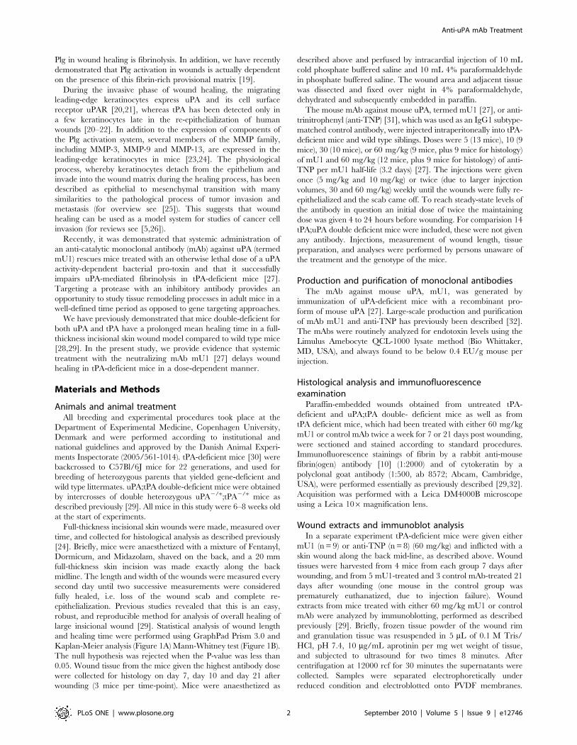
Dose-dependent delay in wound healing after systemictreatment with mU1
Cohorts of tPA-deficient mice received mU1 intraperitoneally at
the doses 5 (13 mice), 10 (9 mice), 30 (10 mice), or 60 mg/kg (9
mice) per mU1 half-life, or an IgG subtype control mAb of
irrelevant specificity (anti-TNP) 60 mg/kg (12 mice). The half-life
of mU1 in vivo was previously determined to be approximately 3
days [27]. For comparison uPA;tPA double-deficient mice were
included (14 mice). Administration of mU1 to the tPA-deficient
mice caused a dose-dependent increase in the healing time of the
incisional skin wounds, measured as the time to complete re-
epithelialization (Figure 1A and B). Since the healing times for
mU1-treated mice at the highest dose (i.e. 60 mg/kg) approached
that of uPA;tPA double-deficient mice (Figure 1A), and only mU1-
treated animals displayed a significant increase in the mean
healing times compared to control mAb-treated tPA-deficient
mice, the delay in healing was caused by in vivo inhibition of uPA
activity by mU1 and not an effect of IgG1 administration. There
was no statistically significant difference in mean healing time
between tPA-deficient mice given the highest dose of mU1 and the
mock-treated uPA;tPA double-deficient mice (p = 0.27 Mann-
Whitney unpaired t-test; Figure 1B). Importantly, mU1 adminis-
tration to wild type mice did not result in any significant delay in
healing time (data not shown), which is in accordance with the
healing kinetics observed in uPA-deficient mice [29].
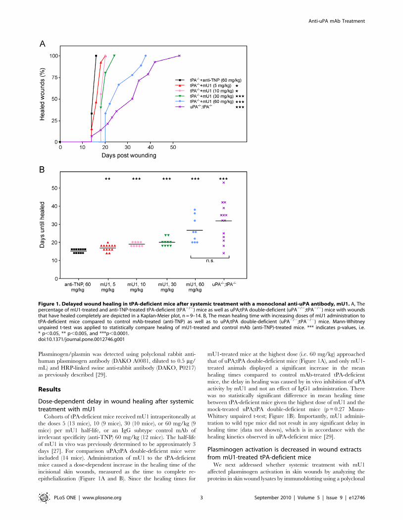
Plasminogen activation is decreased in wound extractsfrom mU1-treated tPA-deficient mice
We next addressed whether systemic treatment with mU1
affected plasminogen activation in skin wounds by analyzing the
proteins in skin wound lysates by immunoblotting using a polyclonal
Figure 1. Delayed wound healing in tPA-deficient mice after systemic treatment with a monoclonal anti-uPA antibody, mU1. A, Thepercentage of mU1-treated and anti-TNP-treated tPA-deficient (tPA2/2) mice as well as uPA;tPA double-deficient (uPA2/2;tPA2/2) mice with woundsthat have healed completely are depicted in a Kaplan-Meier plot, n = 9–14. B, The mean healing time with increasing doses of mU1 administration totPA-deficient mice compared to control mAb-treated (anti-TNP) as well as to uPA;tPA double-deficient (uPA2/2;tPA2/2) mice. Mann-Whitneyunpaired t-test was applied to statistically compare healing of mU1-treated and control mAb (anti-TNP)-treated mice. *** indicates p-values, i.e.* p,0.05, ** p,0.005, and ***p,0.0001.doi:10.1371/journal.pone.0012746.g001
Anti-uPA mAb Treatment
PLoS ONE | www.plosone.org 3 September 2010 | Volume 5 | Issue 9 | e12746
antibody that recognizes both plasminogen and plasmin [29]. We
found reduced levels of plasmin in extracts prepared from 7 days old
wounds in mU1-treated mice, compared to time-matched wound
extracts from control mAb-treated tPA-deficient mice (Figure 2,
lanes 8–10 versus lanes 5–7). The plasmin levels in the mU1-treated
tPA-deficient mice was almost undetectable, hence resembling the
levels in uPA;tPA double-deficient mice (Figure 2, lanes 8–10 versus
lane 11), in which plasmin is known only to be detectable in wound
extracts after a purification step [29]. This result clearly demon-
strates that systemic administration of the inhibitory mU1 mAb
reduces plasminogen activation in vivo.
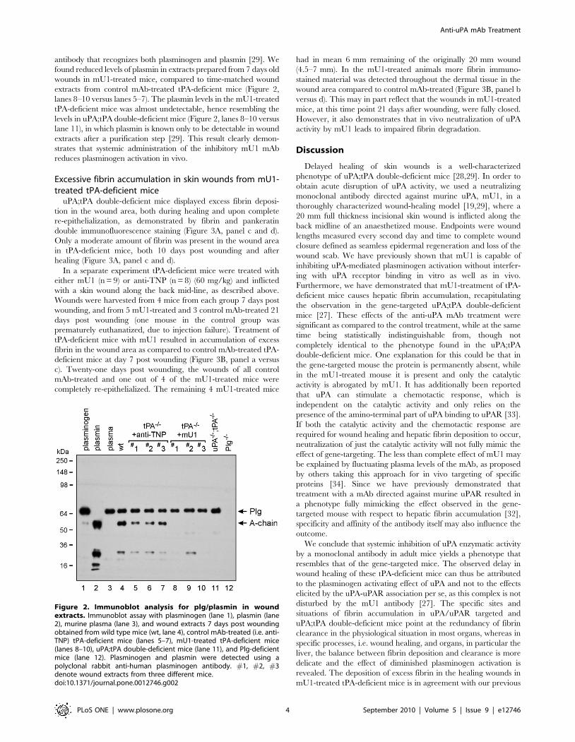
Excessive fibrin accumulation in skin wounds from mU1-treated tPA-deficient mice
uPA;tPA double-deficient mice displayed excess fibrin deposi-
tion in the wound area, both during healing and upon complete
re-epithelialization, as demonstrated by fibrin and pankeratin
double immunofluorescence staining (Figure 3A, panel c and d).
Only a moderate amount of fibrin was present in the wound area
in tPA-deficient mice, both 10 days post wounding and after
healing (Figure 3A, panel c and d).
In a separate experiment tPA-deficient mice were treated with
either mU1 (n = 9) or anti-TNP (n = 8) (60 mg/kg) and inflicted
with a skin wound along the back mid-line, as described above.
Wounds were harvested from 4 mice from each group 7 days post
wounding, and from 5 mU1-treated and 3 control mAb-treated 21
days post wounding (one mouse in the control group was
prematurely euthanatized, due to injection failure). Treatment of
tPA-deficient mice with mU1 resulted in accumulation of excess
fibrin in the wound area as compared to control mAb-treated tPA-
deficient mice at day 7 post wounding (Figure 3B, panel a versus
c). Twenty-one days post wounding, the wounds of all control
mAb-treated and one out of 4 of the mU1-treated mice were
completely re-epithelialized. The remaining 4 mU1-treated mice
had in mean 6 mm remaining of the originally 20 mm wound
(4.5–7 mm). In the mU1-treated animals more fibrin immuno-
stained material was detected throughout the dermal tissue in the
wound area compared to control mAb-treated (Figure 3B, panel b
versus d). This may in part reflect that the wounds in mU1-treated
mice, at this time point 21 days after wounding, were fully closed.
However, it also demonstrates that in vivo neutralization of uPA
activity by mU1 leads to impaired fibrin degradation.
Discussion
Delayed healing of skin wounds is a well-characterized
phenotype of uPA;tPA double-deficient mice [28,29]. In order to
obtain acute disruption of uPA activity, we used a neutralizing
monoclonal antibody directed against murine uPA, mU1, in a
thoroughly characterized wound-healing model [19,29], where a
20 mm full thickness incisional skin wound is inflicted along the
back midline of an anaesthetized mouse. Endpoints were wound
lengths measured every second day and time to complete wound
closure defined as seamless epidermal regeneration and loss of the
wound scab. We have previously shown that mU1 is capable of
inhibiting uPA-mediated plasminogen activation without interfer-
ing with uPA receptor binding in vitro as well as in vivo.
Furthermore, we have demonstrated that mU1-treatment of tPA-
deficient mice causes hepatic fibrin accumulation, recapitulating
the observation in the gene-targeted uPA;tPA double-deficient
mice [27]. These effects of the anti-uPA mAb treatment were
significant as compared to the control treatment, while at the same
time being statistically indistinguishable from, though not
completely identical to the phenotype found in the uPA;tPA
double-deficient mice. One explanation for this could be that in
the gene-targeted mouse the protein is permanently absent, while
in the mU1-treated mouse it is present and only the catalytic
activity is abrogated by mU1. It has additionally been reported
that uPA can stimulate a chemotactic response, which is
independent on the catalytic activity and only relies on the
presence of the amino-terminal part of uPA binding to uPAR [33].
If both the catalytic activity and the chemotactic response are
required for wound healing and hepatic fibrin deposition to occur,
neutralization of just the catalytic activity will not fully mimic the
effect of gene-targeting. The less than complete effect of mU1 may
be explained by fluctuating plasma levels of the mAb, as proposed
by others taking this approach for in vivo targeting of specific
proteins [34]. Since we have previously demonstrated that
treatment with a mAb directed against murine uPAR resulted in
a phenotype fully mimicking the effect observed in the gene-
targeted mouse with respect to hepatic fibrin accumulation [32],
specificity and affinity of the antibody itself may also influence the
outcome.
We conclude that systemic inhibition of uPA enzymatic activity
by a monoclonal antibody in adult mice yields a phenotype that
resembles that of the gene-targeted mice. The observed delay in
wound healing of these tPA-deficient mice can thus be attributed
to the plasminogen activating effect of uPA and not to the effects
elicited by the uPA-uPAR association per se, as this complex is not
disturbed by the mU1 antibody [27]. The specific sites and
situations of fibrin accumulation in uPA/uPAR targeted and
uPA;tPA double-deficient mice point at the redundancy of fibrin
clearance in the physiological situation in most organs, whereas in
specific processes, i.e. wound healing, and organs, in particular the
liver, the balance between fibrin deposition and clearance is more
delicate and the effect of diminished plasminogen activation is
revealed. The deposition of excess fibrin in the healing wounds in
mU1-treated tPA-deficient mice is in agreement with our previous
Figure 2. Immunoblot analysis for plg/plasmin in woundextracts. Immunoblot assay with plasminogen (lane 1), plasmin (lane2), murine plasma (lane 3), and wound extracts 7 days post woundingobtained from wild type mice (wt, lane 4), control mAb-treated (i.e. anti-TNP) tPA-deficient mice (lanes 5–7), mU1-treated tPA-deficient mice(lanes 8–10), uPA;tPA double-deficient mice (lane 11), and Plg-deficientmice (lane 12). Plasminogen and plasmin were detected using apolyclonal rabbit anti-human plasminogen antibody. #1, #2, #3denote wound extracts from three different mice.doi:10.1371/journal.pone.0012746.g002
Anti-uPA mAb Treatment
PLoS ONE | www.plosone.org 4 September 2010 | Volume 5 | Issue 9 | e12746
finding that mU1-treated tPA-deficient mice are not able to clear
fibrin from the hepatic sinusoids [27]. Accumulation of fibrin in
the liver also occurs in tPA-deficient mice treated with a
monoclonal antibody against uPAR, and in these mice the hepatic
fibrin plaques were shown to hold uPAR-expressing macrophages,
indicating a role for hepatic macrophages in fibrin-clearance [32].
Prognostic and predictive studies have demonstrated the
importance of uPA in cancer [35] and uPA is expressed at the
invasive front by stromal cells in human breast and colon cancer
[36,37]. Several poly- and monoclonal antibodies as well as
peptide antagonists have been generated and tested in different
xenografted tumor models. However, they all target the proteins
Figure 3. Cytokeratin and fibrin immunofluorescence stainings of wound areas from mU1- or control mAb (i.e. anti-TNP)-treatedmice. Double immunofluorescence staining of cytokeratin (546 nm, red) and fibrin/fibrinogen (488 nm, green) in wound sections. A, Wounds fromtPA-deficient and uPA;tPA double-deficient mice during healing (left panel, 10 days post wounding) and after re-epithelialization (right panel).B, Micrographs of wounds isolated from control mAb-treated (upper panels) and mU1-treated (lower panels) mice during healing (left panel, 7 dayspost wounding) and upon healing (right panel, 21 days post wounding).HE; hyperproliferative epidermis, GT; granulation tissue.doi:10.1371/journal.pone.0012746.g003
Anti-uPA mAb Treatment
PLoS ONE | www.plosone.org 5 September 2010 | Volume 5 | Issue 9 | e12746
of the human plasminogen activation cascade, making them
insufficient as tools in preclinical mouse models, where the roles
of host-derived proteases are crucial [6]. The migration and
proliferation of keratinocytes during wound closure have
similarities to cancer invasion and is thus employed to model
this event. Conclusively, systemic treatment with anti-uPA
monoclonal antibody, mU1, was effective in delaying wound
healing and may thus have a therapeutic potential in mouse
cancer models.
Acknowledgments
The expert technical assistance of M. Musfelth Andersen, L. Frederiksen,
K. Lund Jacobsen, A. Læssøe Møller, and G. Juhl Funch is gratefully
acknowledged. We thank J. Post for figure preparation.
Author Contributions
Conceived and designed the experiments: AJ MP GHH JR LRL.
Performed the experiments: AJ BR BSN JR LRL. Analyzed the data: AJ
BR BSN MP GHH JR LRL. Contributed reagents/materials/analysis
tools: AJ BR IKL MP GHH JR. Wrote the paper: AJ BR IKL BSN MP
GHH JR LRL.
References
1. Coussens LM, Tinkle CL, Hanahan D, Werb Z (2000) MMP-9 supplied by bone
marrow-derived cells contributes to skin carcinogenesis. Cell 103: 481–490.
2. Danø K, Rømer J, Nielsen BS, Bjørn S, Pyke C, et al. (1999) Cancer invasionand tissue remodeling–cooperation of protease systems and cell types. APMIS
107: 120–127.3. Egeblad M, Werb Z (2002) New functions for the matrix metalloproteinases in
cancer progression. Nat Rev Cancer 2: 161–174.
4. Page-McCaw A, Ewald AJ, Werb Z (2007) Matrix metalloproteinases and theregulation of tissue remodelling. Nat Rev Mol Cell Biol 8: 221–233.
5. Schafer M, Werner S (2008) Cancer as an overhealing wound: an old hypothesisrevisited. Nature reviews 9: 628–638.
6. Danø K, Behrendt N, Høyer-Hansen G, Johnsen M, Lund LR, et al. (2005)Plasminogen activation and cancer. Thromb Haemost 93: 676–681.
7. Collen D, Lijnen HR (2005) Thrombolytic agents. Thromb Haemost 93:
627–630.8. Danø K, Andreasen PA, Grøndahl-Hansen J, Kristensen P, Nielsen LS,
Skriver L (1985) Plasminogen activators, tissue degradation, and cancer. AdvCancer Res 44: 139–266.
9. Selvarajan S, Lund LR, Takeuchi T, Craik CS, Werb Z (2001) A plasma
kallikrein-dependent plasminogen cascade required for adipocyte differentiation.Nature cell biology 3: 267–275.
10. Bugge TH, Flick MJ, Daugherty CC, Degen JL (1995) Plasminogen deficiencycauses severe thrombosis but is compatible with development and reproduction.
Genes Dev 9: 794–807.
11. Drew AF, Kaufman AH, Kombrinck KW, Danton MJ, Daugherty CC, et al.(1998) Ligneous conjunctivitis in plasminogen-deficient mice. Blood 91:
1616–1624.12. Mingers AM, Heimburger N, Zeitler P, Kreth HW, Schuster W (1997)
Homozygous type I plasminogen deficiency. Semin Thromb Hemost 23:259–269.
13. Bugge TH, Lund LR, Kombrinck KK, Nielsen BS, Holmback K, et al. (1998)
Reduced metastasis of Polyoma virus middle T antigen-induced mammarycancer in plasminogen-deficient mice. Oncogene 16: 3097–3104.
14. Lijnen HR, Van Hoef B, Lupu F, Moons L, Carmeliet P, et al. (1998) Functionof the plasminogen/plasmin and matrix metalloproteinase systems after vascular
injury in mice with targeted inactivation of fibrinolytic system genes. Arterioscler
Thromb Vasc Biol 18: 1035–1045.15. Solberg H, Rinkenberger J, Danø K, Werb Z, Lund LR (2003) A functional
overlap of plasminogen and MMPs regulates vascularization during placentaldevelopment. Development 130: 4439–4450.
16. Lund LR, Bjørn SF, Sternlicht MD, Nielsen BS, Solberg H, et al. (2000)Lactational competence and involution of the mouse mammary gland require
plasminogen. Development 127: 4481–4492.
17. Rømer J, Bugge TH, Pyke C, Lund LR, Flick MJ, et al. (1996) Impaired woundhealing in mice with a disrupted plasminogen gene. Nature medicine 2:
287–292.18. Bugge TH, Kombrinck KW, Flick MJ, Daugherty CC, Danton MJ, et al. (1996)
Loss of fibrinogen rescues mice from the pleiotropic effects of plasminogen
deficiency. Cell 87: 709–719.19. Green KA, Almholt K, Ploug M, Rønø B, Castellino FJ, et al. (2008)
Profibrinolytic effects of metalloproteinases during skin wound healing in theabsence of plasminogen. J Invest Dermatol 128: 2092–2101.
20. Rømer J, Lund LR, Eriksen J, Ralfkiaer E, Zeheb R, et al. (1991) Differentialexpression of urokinase-type plasminogen activator and its type-1 inhibitor
during healing of mouse skin wounds. J Invest Dermatol 97: 803–811.
21. Rømer J, Lund LR, Eriksen J, Pyke C, Kristensen P, et al. (1994) The receptor
for urokinase-type plasminogen activator is expressed by keratinocytes at the
leading edge during re-epithelialization of mouse skin wounds. J Invest Dermatol
102: 519–522.
22. Grøndahl-Hansen J, Lund LR, Ralfkiaer E, Ottevanger V, Danø K (1988)
Urokinase- and tissue-type plasminogen activators in keratinocytes during
wound reepithelialization in vivo. J Invest Dermatol 90: 790–795.
23. Madlener M, Parks WC, Werner S (1998) Matrix metalloproteinases (MMPs)
and their physiological inhibitors (TIMPs) are differentially expressed during
excisional skin wound repair. Exp Cell Res 242: 201–210.
24. Lund LR, Rømer J, Bugge TH, Nielsen BS, Frandsen TL, et al. (1999)
Functional overlap between two classes of matrix-degrading proteases in wound
healing. EMBO J 18: 4645–4656.
25. Weinberg RA (2007) The Biology of Cancer. Garland Science.
26. Gurtner GC, Werner S, Barrandon Y, Longaker MT (2008) Wound repair and
regeneration. Nature 453: 314–321.
27. Lund IK, Jogi A, Rønø B, Rasch MG, Lund LR, et al. (2008) Antibody-
mediated targeting of the uPA proteolytic function neutralizes fibrinolysis in
vivo. J Biol Chem 283: 32506–32515.
28. Bugge TH, Flick MJ, Danton MJ, Daugherty CC, Rømer J, et al. (1996)
Urokinase-type plasminogen activator is effective in fibrin clearance in the
absence of its receptor or tissue-type plasminogen activator. Proc Natl Acad Sci
USA 93: 5899–5904.
29. Lund LR, Green KA, Stoop AA, Ploug M, Almholt K, et al. (2006) Plasminogen
activation independent of uPA and tPA maintains wound healing in gene-
deficient mice. EMBO J 25: 2686–2697.
30. Carmeliet P, Schoonjans L, Kieckens L, Ream B, Degen J, et al. (1994)
Physiological consequences of loss of plasminogen activator gene function in
mice. Nature 368: 419–424.
31. Shulman M, Wilde CD, Kohler G (1978) A better cell line for making
hybridomas secreting specific antibodies. Nature 276: 269–70.
32. Jogi A, Pass J, Høyer-Hansen G, Lund LR, Nielsen BS, et al. (2007) Systemic
administration of anti-urokinase plasminogen activator receptor monoclonal
antibodies induces hepatic fibrin deposition in tissue-type plasminogen activator
deficient mice. J Thromb Haemost 5: 1936–1944.
33. Resnati M, Guttinger M, Valcamonica S, Sidenius N, Blasi F, et al. (1996)
Proteolytic cleavage of the urokinase receptor substitutes for the agonist-induced
chemotactic effect. EMBO J 15: 1572–82.
34. Scott KA, Moore RJ, Arnott CH, East N, Thompson RG, et al. (2003) An anti-
tumor necrosis factor-alpha antibody inhibits the development of experimental
skin tumors. Mol Cancer Ther 2: 445–451.
35. Harbeck N, Kates RE, Schmitt M, Gauger K, Kiechle M, et al. (2004)
Urokinase-type plasminogen activator and its inhibitor type 1 predict disease
outcome and therapy response in primary breast cancer. Clin Breast Cancer 5:
348–52.
36. Nielsen BS, Rank F, Illemann M, Lund LR, Danø K (2007) Stromal cells
associated with early invasive foci in human mammary ductal carcinoma in situ
co-express urokinase and urokinase receptor. Int J Cancer 120: 2086–2095.
37. Illemann M, Bird N, Majeed A, Laerum OD, Lund LR, et al. (2009) Two
distinct growth patterns of colorectal cancer liver metastasis show differential
expression of urokinase, urokinase receptor and plasminogen activator inhibitor-
1. Int J Cancer 124: 1860–1870.
Anti-uPA mAb Treatment
PLoS ONE | www.plosone.org 6 September 2010 | Volume 5 | Issue 9 | e12746
Related Documents