Brain Research Bulletin 77 (2008) 129–135 Contents lists available at ScienceDirect Brain Research Bulletin journal homepage: www.elsevier.com/locate/brainresbull Research report Neurotensin modulation of acetylcholine, GABA, and aspartate release from rat prefrontal cortex studied in vivo with microdialysis Polina Petkova-Kirova a,1 , Angelina Rakovska b,∗ , Laura Della Corte c,2 , Galina Zaekova b,3 , Radomir Radomirov b,4 , Aliz Mayer d,5 a Institute of Biophysics, Bulgarian Academy of Sciences, Acad. G. Bonchev Street, bl. 21, 1113 Sofia, Bulgaria b Institute of Neurobiology, Bulgarian Academy of Sciences, Acad. G. Bonchev Street, bl. 23, 1113 Sofia, Bulgaria c Department of Preclinical and Clinical Pharmacology, University of Florence, Viale Pieraccini 6, 50139 Florence, Italy d Institute of Experimental Medicine, Hungarian Academy of Sciences, H-1450 Budapest, P.O. Box 67, Hungary article info Article history: Received 9 April 2008 Accepted 14 April 2008 Available online 7 May 2008 Keywords: Prefrontal cortex Neurotensin Amino acids Acetylcholine Microdialysis abstract The effects of the peptide transmitter neurotensin (NT) on the release of acetylcholine (ACh), - aminobutyric acid (GABA), glutamate (Glu), aspartate (Asp), and taurine from the prefrontal cortex (PFC) of freely moving rats were studied by transversal microdialysis. Neurotensin (0.2 and 1 M) administered locally in the PFC produced a concentration-dependent increase in the extracellular levels of ACh, GABA, and Asp, but not of Glu or taurine. The increase produced by 1 M NT reached a maximum of about 240% for ACh, 370% for GABA, and 380% for Asp. Lower doses of NT (0.05 M) did not cause a significant change in ACh, GABA, or Asp output in the PFC. Higher concentrations of NT (2 M) did not induce further increases in the level of neurotransmitters. A high-affinity selective neurotensin receptor (NTR1) antago- nist SR 48692 (0.5 M) perfused locally blocked neurotensin (1 M)-evoked ACh, GABA, and Asp release. Local infusion of the sodium channel blocker tetrodotoxin (TTX) (1 M) decreased the release of ACh, had no significant effect on GABA or Asp release, and prevented the 1 M neurotensin-induced increase in ACh, GABA, and Asp output. Removal of calcium from the Ringer’s solution prevented the peptide from having any effects on the neurotransmitters. Thus, in vivo NT plays a modulatory role in the PFC by inter- acting with cortical neurons releasing GABA and Asp and with ACh-containing neurons projecting to the PFC. The NT effects are of neural origin, as they are TTX-sensitive, and mediated by the NTR1 receptor, as they are antagonized by SR 48692. © 2008 Elsevier Inc. All rights reserved. 1. Introduction Neurotensin (NT) is an endogenous tridecapeptide [10] known to play a role as a neurotransmitter and neuromodulator both in the central and peripheral nervous systems [38,17,27,7]. The pep- tide and its receptors are widely distributed throughout the brain, including the basal forebrain and cerebral cortex [36,21,7,65]. In ∗ Corresponding author at: Institute of Neurobiology, Lab. ‘Neuropeptides’, Bul- garian Academy of Sciences, Acad. G. Bonchev Street, bl. 23, 1113 Sofia, Bulgaria. Tel.: +359 2 9792305; fax: +359 2 8719109. E-mail addresses: [email protected] (P. Petkova-Kirova), [email protected] (A. Rakovska), laura.dellacorte@unifi.it (L. Della Corte), ashlie @mail.bg (G. Zaekova), [email protected] (R. Radomirov), [email protected] (A. Mayer). 1 Tel.: +359 2 9792130; fax: +359 2 9712493. 2 Tel.: +39 055 4271226; fax: +39 055 410778. 3 Tel.: +359 2 9792387; fax: +359 2 8719109. 4 Tel.: +359 2 9792164; fax: +359 2 8719109. 5 Tel.: +36 1 2109979; fax: +36 1 2109423. the prefrontal cortex (PFC), the basal concentration of neurotensin measured in awake, freely moving rats is found to be 2–5 pg/sample [45]. There are no neurotensin-containing cell bodies in the PFC, and the only source of NT arises from axons derived from the ventral tegmental area (VTA) [25,56,60] that contain tyrosine hydroxylase and the dopamine synthetic enzyme [60,15]. In vivo release of coex- istent NT and dopamine was measured from the prefrontal cortex after electrical stimulation of mesocortical axons [5]. Thus, neu- rotensin in the PFC is exclusively localized to dopamine axons. Dopamine synapses are found on PFC pyramidal cells as well as interneurons [58], making neurotensin a likely regulator of the cognitive functions of the prefrontal cortex via an effect on glu- tamatergic and GABAergic function. Cholinergic neurons of the basal forebrain constitute the pri- mary source of ACh to the cerebral cortex [50,18,33] and are thought to play a major role in mediating cortical activation and plastic- ity. Autoradiographic studies combined with acetylcholinesterase histochemistry have shown an association of NT receptors with cholinergic neurons in the rat basal forebrain [62]. Furthermore, 0361-9230/$ – see front matter © 2008 Elsevier Inc. All rights reserved. doi:10.1016/j.brainresbull.2008.04.003

Welcome message from author
This document is posted to help you gain knowledge. Please leave a comment to let me know what you think about it! Share it to your friends and learn new things together.
Transcript

Brain Research Bulletin 77 (2008) 129–135
Contents lists available at ScienceDirect
Brain Research Bulletin
journa l homepage: www.e lsev ier .com/ locate /bra inresbul l
Research report
Neurotensin modulation of acetylcholine, GABA, and aspartate releasefrom rat prefrontal cortex studied in vivo with microdialysis
Polina Petkova-Kirovaa,1, Angelina Rakovskab,∗, Laura Della Cortec,2,Galina Zaekovab,3, Radomir Radomirovb,4, Aliz Mayerd,5
a Institute of Biophysics, Bulgarian Academy of Sciences, Acad. G. Bonchev Street, bl. 21, 1113 Sofia, Bulgariab Institute of Neurobiology, Bulgarian Academy of Sciences, Acad. G. Bonchev Street, bl. 23, 1113 Sofia, Bulgariac Department of Preclinical and Clinical Pharmacology, University of Florence, Viale Pieraccini 6, 50139 Florence, Italyd Institute of Experimental Medicine, Hungarian Academy of Sciences, H-1450 Budapest, P.O. Box 67, Hungary
a r t i c l e i n f o
Article history:Received 9 April 2008Accepted 14 April 2008Available online 7 May 2008
Keywords:Prefrontal cortexNeurotensinAmino acidsAcetylcholineMicrodialysis
a b s t r a c t
The effects of the peptide transmitter neurotensin (NT) on the release of acetylcholine (ACh), �-aminobutyric acid (GABA), glutamate (Glu), aspartate (Asp), and taurine from the prefrontal cortex (PFC)of freely moving rats were studied by transversal microdialysis. Neurotensin (0.2 and 1 �M) administeredlocally in the PFC produced a concentration-dependent increase in the extracellular levels of ACh, GABA,and Asp, but not of Glu or taurine. The increase produced by 1 �M NT reached a maximum of about240% for ACh, 370% for GABA, and 380% for Asp. Lower doses of NT (0.05 �M) did not cause a significantchange in ACh, GABA, or Asp output in the PFC. Higher concentrations of NT (2 �M) did not induce furtherincreases in the level of neurotransmitters. A high-affinity selective neurotensin receptor (NTR1) antago-nist SR 48692 (0.5 �M) perfused locally blocked neurotensin (1 �M)-evoked ACh, GABA, and Asp release.Local infusion of the sodium channel blocker tetrodotoxin (TTX) (1 �M) decreased the release of ACh, hadno significant effect on GABA or Asp release, and prevented the 1 �M neurotensin-induced increase inACh, GABA, and Asp output. Removal of calcium from the Ringer’s solution prevented the peptide from
having any effects on the neurotransmitters. Thus, in vivo NT plays a modulatory role in the PFC by inter-acting with cortical neurons releasing GABA and Asp and with ACh-containing neurons projecting to thePFC. The NT effects are of neural origin, as they are TTX-sensitive, and mediated by the NTR1 receptor, asR 486
1
ttti
gT
(r
tm[
0d
they are antagonized by S
. Introduction
Neurotensin (NT) is an endogenous tridecapeptide [10] known
o play a role as a neurotransmitter and neuromodulator both inhe central and peripheral nervous systems [38,17,27,7]. The pep-ide and its receptors are widely distributed throughout the brain,ncluding the basal forebrain and cerebral cortex [36,21,7,65]. In∗ Corresponding author at: Institute of Neurobiology, Lab. ‘Neuropeptides’, Bul-arian Academy of Sciences, Acad. G. Bonchev Street, bl. 23, 1113 Sofia, Bulgaria.el.: +359 2 9792305; fax: +359 2 8719109.
E-mail addresses: [email protected] (P. Petkova-Kirova), [email protected]. Rakovska), [email protected] (L. Della Corte), ashlie @mail.bg (G. Zaekova),[email protected] (R. Radomirov), [email protected] (A. Mayer).1 Tel.: +359 2 9792130; fax: +359 2 9712493.2 Tel.: +39 055 4271226; fax: +39 055 410778.3 Tel.: +359 2 9792387; fax: +359 2 8719109.4 Tel.: +359 2 9792164; fax: +359 2 8719109.5 Tel.: +36 1 2109979; fax: +36 1 2109423.
ttaiarDict
mtihc
361-9230/$ – see front matter © 2008 Elsevier Inc. All rights reserved.oi:10.1016/j.brainresbull.2008.04.003
92.© 2008 Elsevier Inc. All rights reserved.
he prefrontal cortex (PFC), the basal concentration of neurotensineasured in awake, freely moving rats is found to be 2–5 pg/sample
45]. There are no neurotensin-containing cell bodies in the PFC, andhe only source of NT arises from axons derived from the ventralegmental area (VTA) [25,56,60] that contain tyrosine hydroxylasend the dopamine synthetic enzyme [60,15]. In vivo release of coex-stent NT and dopamine was measured from the prefrontal cortexfter electrical stimulation of mesocortical axons [5]. Thus, neu-otensin in the PFC is exclusively localized to dopamine axons.opamine synapses are found on PFC pyramidal cells as well as
nterneurons [58], making neurotensin a likely regulator of theognitive functions of the prefrontal cortex via an effect on glu-amatergic and GABAergic function.
Cholinergic neurons of the basal forebrain constitute the pri-
ary source of ACh to the cerebral cortex [50,18,33] and are thoughto play a major role in mediating cortical activation and plastic-ty. Autoradiographic studies combined with acetylcholinesteraseistochemistry have shown an association of NT receptors withholinergic neurons in the rat basal forebrain [62]. Furthermore,
1 esear
etantteeaissslic
tevs[[u
2
2
wwta(wftrfaATtEEmpcwLp
t
2
m
M1cwidbawGot
2
iiduLidab
2
c[rwpaccHtAdfaaue
2
[saimA(cwi2w
2
fh3r3k
30 P. Petkova-Kirova et al. / Brain R
lectrophysiological data have demonstrated that NT not only bindso, but is also internalized in cholinergic cells of the basal forebrainnd directly affects multiple electrophysiological features of theseeurons [2]. In vitro experiments indicate that NT enhanced spon-aneous, electrically stimulated, potassium-evoked ACh release inhe striatum [29,64] and potassium-evoked ACh release in the pari-tal cortex [30]. In frontal cortical slices, NT significantly inhibitedvoked ACh release via a tetrodotoxin (TTX)-insensitive mechanismnd had no effect on spontaneous release [30]. In our previous stud-es using in vivo microdialysis, we found that neurotensin increasespontaneous ACh release from the rat hippocampus [46]. Such atimulant effect of neurotensin was not found to occur for in vivotriatal ACh levels [40,59], although it was established that NT regu-ates acetylcholine release when D2-dopaminergic inhibitory inputs suppressed [59]. So far, the role of neurotensin in the regulation ofholinergic neuronal activity in vivo in the cortex remains unclear.
The present study sought to characterize the neuromodula-ory role of neurotensin in the prefrontal cortex. We examined theffects of neurotensin on PFC Glu, Asp, GABA, and ACh release inivo in awake and freely moving rats using transversal microdialy-is, i.e. we measured their concentrations in the extracellular space61] where they exert their actions [69] on nonsynaptic receptors67,68,48,55,35,34] and extrasynaptic transporters involved in theirptake [61,28].
. Materials and methods
.1. Animals and surgery
Male adult Wistar rats weighing 250–300 g were used. Prior to surgery, theyere housed in groups in macrolon cages with ad libitum access to food andater. Rats were housed on a 12 h light/12 h dark cycle. On the day of surgery,
he rats were anaesthetized with chloral hydrate (400 mg/kg, i.p.) and placed instereotaxic apparatus (Stoeling, Stellar, Wood Dale, Il, USA). Microdialysis tubes
AN 69 membrane; Dasco, Bologna, Italy, 220 �m i.d. and 310 �m o.d., moleculareight cut-off 15,000 Da) were inserted transversally into the prefrontal cortex
ollowing the procedure described by Giovannini et al. [19]. The microdialysisubes were covered with super-epoxy glue along their entire length except for theegion corresponding to the cortex (8 mm) (see scheme). The coordinates usedor the implantation of the microdialysis tubing in the cortex were as follows:nterior–posterior +1.0 mm and −1.8 mm, according to Paxinos and Watson [41].ll coordinates refer to Bregma, with bregma and lambda on a horizontal plane.he day after surgery, each rat was placed in a Plexiglas cage with free accesso food and water. All animal manipulations were carried out according to theuropean Community guidelines for animal care (DL 116/92, application of theuropean Community Council Directive 86/609/EEC). All efforts were made toinimize animal suffering and to use only the number of animals necessary to
roduce reliable scientific data. Procedures involving animals and their care wereonducted in conformity with the institutional guidelines that are in complianceith national and international laws and policies (EEC Council Directive 86/609, OJ358, 1 Dec. 12. 1987; NIH Guide for the Care and Use of Laboratory animals, NIHublication No. 85-23, 1985).
Schematic diagram showing the positioning of the microdialysis membrane in
he prefrontal cortex. For stereotaxic coordinates, see Section 2..2. Microdialysis procedure
One day after surgery, each rat was placed in a Plexiglas cage. The inlet of theicrodialysis probe was connected to a microperfusion pump (Carnegie Medicine,
2
apD
ch Bulletin 77 (2008) 129–135
od. CMA/100, Sweden), and the PFC was perfused with Ringer’s solution (NaCl47 mM, CaCl2 1.2 mM, KCl 4.0 mM) containing 0.1 �M physostigmine sulfate at aonstant flow rate of 2 �l/min. After a 1-h recovery period, during which the PFCas perfused without collecting the dialysate, samples were collected at 20 min
ntervals. After collecting the first three samples to measure the basal outflow, drugsissolved in Ringer’s solution were administered locally through the dialysis mem-rane for 80 min (4th–7th samples) or until the end of the experiment (TTX or TTXnd NT). Thereafter, the drugs were withdrawn and the membranes were perfusedith normal Ringer’s solution until the end of experiment. The content of ACh, GABA,lu, Asp, and taurine in the dialysate was analyzed by HPLC as described below. Inrder to assay all neurotransmitters simultaneously when necessary, samples fromhe same animal were split into two halves.
.3. Histological control
At the end of the experiments, rats were anaesthetized with urethane (1.2 �g/kg,.p.) and sacrificed by decapitation. The brain was rapidly removed and placedn a vial containing 9% formaldehyde solution in phosphate buffer. Two or threeays later, the brains were frozen with liquid CO2 and coronal slices (50 �m) cutsing a freezing microtome. The slices were examined by light microscopy (Nikon,abophot-2) to verify the position of the dialysis membrane. Data obtained from ratsn which the dialysis membrane was positioned outside the defined structure wereiscarded (<5%). The condition of the cortical neurons around the probe was checkedt the end of the experiment by staining coronal sections of formaldehyde-fixedrains with Richardson’s solution (modified Nisal’s staining).
.4. Assay of ACh in the dialysate
ACh was directly assayed in the dialysate using an HPLC method with a post-olumn enzyme reactor and an electrochemical detector as previously described12,19]. ACh was separated on a cation-exchange column, prepared by loading aeverse-phase column (Chromspher 5 C18, Chrompack, Middelburg, Netherlands)ith sodium lauryl sulfate (0.5 mg/ml). The mobile phase consisted of 0.2 M phos-hate buffer (pH 8.0) containing 5 mM KCl, 1 mM tetramethylammonium (TMA),nd 0.3 mM EDTA. The flow rate was 0.7 ml/min. ACh was hydrolyzed by acetyl-holine esterase (AChE) to acetate and choline in a post-column enzyme reactor;holine was oxidized by choline oxidase to produce betaine and hydrogen peroxide.ydrogen peroxide was electrochemically detected by a platinum-working elec-
rode at +500 mV with Ag/AgCl reference electrode. For the quantitative analysis ofCh, we constructed a calibration curve by spiking the Ringer’s solution with stan-ard ACh in the concentration range we expected to find in the dialysates. Three orour concentrations for each calibration curve were then injected at the beginningnd end of the analysis, and the heights of the recorded peaks were then plottedgainst the concentrations. A regression line was calculated, and quantification ofnknown samples was carried out by the method of inverse prediction. Under thesexperimental conditions, the sensitivity limit for ACh was 100 fmol (s/n ratio > 3/1).
.5. Assay of excitatory amino acids GABA, Glu, Asp, and taurine in the dialysate
GABA, Glu, Asp, and taurine analyses were carried out as previously described6]. We performed HPLC with fluorimetric detection at the excitation and emis-ion wavelengths of 340 and 455 nm, respectively, after derivatization of the aminocids with o-phthalaldehyde (OPA). A 5-�m nucleosil C18 column (200 mm × 4 mm.d., Macherey-Nagel, Duren, Germany) was used. The mobile phase consisted of
ethanol–potassium acetate (0.1 M) adjusted to pH 5.52 with glacial acetic acid.gradient (flow rate 0.9 ml/min) of three linear steps – from 25 to 43% methanol
1 min), 43 to 70% methanol (10 min), and 70 to 90% (1 min) – followed by an iso-ratic hold at 90% methanol (1 min) was used. One volume (10 �l) of the dialysateas mixed with 1 �l of the OPA derivatization reagent [6] in a glass capillary tube and
njected after 1.5 min. Standard curves were linear over the concentration range of5–1000 fmol/�l. The minimum detectable concentration of the neurotransmittersas 2 fmol/�l.
.6. Chemicals
All reagents were of analytical grade. The following chemicals, purchasedrom Sigma Chemical Co. (St. Louis, MO, USA), were used: physostigmine, OPA,omoserine, neurotensin, tetrodotoxin (TTX), acetylcholine esterase, AChE (EC.1.1.7., Grade VI-S), choline oxidase (EC 1.1.3.17), and p-formaldehyde. The neu-otensin antagonist 2-(1-(7-chloro-4-quinolinyl)-5-(2,6-dimethoxyphenyl)pyrazol-yl)carbonylamino tricyclo (3.3.1.1.3.7)decan-2-carboxylic acid (SR 48692) wasindly supplied by Sanofi Recherche, France.
.7. Statistical analysis
ACh, GABA, Asp, and Glu release rates are expressed as pmol/40 �l or presenteds the percent variation over the basal output (average of the first three sam-les taken before drug administration). All values are given as the mean ± S.E.M.ifferences among experimental groups are evaluated by comparing areas under

P. Petkova-Kirova et al. / Brain Research Bulletin 77 (2008) 129–135 131
Fig. 1. (A) Effects of 1 �M neurotensin (NT) on ACh release from the prefrontal cortex (PFC) in freely moving rats in the absence and presence of TTX and 2-(1-(7-chloro-4-quinolinyl)-5-(2,6-dimethoxyphenyl)pyrazol-3yl)carbonylamino tricyclo (3.3.1.1.3.7)decan-2-carboxylic acid (SR 48692). NT and SR 48692 were administered for 80 minlocally to the PFC via the dialysis membrane after collection of three samples (horizontal bar). TTX was introduced into the perfusion medium after collection of the threesamples and maintained in the medium until the end of the experiment. ACh output is expressed as the percentage change over the mean of the first three control samples. Themeans ± S.E.M. of at least six different rats in each group are presented. Dialysate samples were collected for 20 min (2 �l/min). (B) Effects of 1 �M NT on ACh release from theP inolin2 alculag sis ofc 1 NT +
tNa
3
3
mt
hcf
3b
Fdtiamaeb
FC in freely moving rats in the absence and presence of TTX and 2-(1-(7-chloro-4-qu-carboxylic acid (SR 48692). Values are expressed as the mean percentage change croups were evaluated by comparing the area under curve (AUC) via one-way analyomparison test (#P < 0.05 NT vs. basal release; ***P < 0.001 NT + TTX vs. NT; **P < 0.0
he curves (AUC) via a one-way analysis of variance (ANOVA) followed by theewman–Keuls multiple comparison test. Differences were considered significantt P < 0.05.
. Results
.1. Neuroanatomical localization of microdialysis probes
According to the maps of Zilles and Wree [71], the transverseicrodialysis membrane in our experiments was inserted through
he parietal region of one hemisphere and reached the opposite
ete
ig. 2. (A) Effects of 1 �M neurotensin (NT) on GABA release from the PFC in freely movinimethoxyphenyl)pyrazol-3yl)carbonylamino tricyclo (3.3.1.1.3.7)decan-2-carboxylic acidhe dialysis membrane after collection of three samples (horizontal bar). TTX was introducn the medium until the end of the experiment. GABA output is expressed as the percentt least six different rats in each group are presented. Dialysate samples were collected fooving rats in the absence and presence of TTX and 2-(1-(7-chloro-4-quinolinyl)-5-(2,6-d
cid (SR 48692). Values are expressed as the mean percentage change calculated for the 80valuated by comparing the area under curve (AUC) via one-way ANOVA (F3,11 = 30.3; P < 0asal release; ***P < 0.001 NT + TTX vs. NT; ***P < 0.001 NT + SR 48692 vs. NT).
yl)-5-(2,6-dimethoxyphenyl)pyrazol-3yl)carbonylamino tricyclo (3.3.1.1.3.7)decan-ted for the 80–200 min period. Significant differences among the four experimentalvariance (ANOVA) (F3,14 = 13.93; P < 0.0002) followed by a Newman–Keuls multipleSR 48692 vs. NT).
emisphere by running under the frontal and through prefrontalortices. These areas are involved in sensorimotor and cognitiveunctions.
.2. Effects of neurotensin, SR 48692, and tetrodotoxin on theasal release of ACh, GABA, and Asp
After a 60 min recovery period, ACh, GABA, Glu, and Asp lev-ls were measured in the microdialysis perfusates collected fromhe PFC of control rats not treated with any chemicals. These lev-ls remained relatively constant from one collection period to the
g rats in the absence and presence of TTX and 2-(1-(7-chloro-4-quinolinyl)-5-(2,6-(SR 48692). NT and SR 48692 were administered for 80 min locally to the PFC viaed into the perfusion medium after collection of the three samples and maintainedage change over the mean of the first three control samples. The means ± S.E.M. ofr 20 min (2 �l/min). (B) Effects of 1 �M NT on GABA release from the PFC in freely
imethoxyphenyl)pyrazol-3yl)carbonylamino tricyclo (3.3.1.1.3.7)decan-2-carboxylic–200 min period. Significant differences among the four experimental groups were.0001) followed by a Newman–Keuls multiple comparison test (###P < 0.001 NT vs.

132 P. Petkova-Kirova et al. / Brain Research Bulletin 77 (2008) 129–135
Fig. 3. (A) Effects of 1 �M neurotensin (NT) on aspartate (Asp) release from the PFC in freely moving rats in the absence and presence of TTX and 2-(1-(7-chloro-4-quinolinyl)-5-(2,6-dimethoxyphenyl)pyrazol-3yl)carbonylamino tricyclo (3.3.1.1.3.7)decan-2-carboxylic acid (SR 48692). NT and SR 48692 were administered for 80 min locally to the PFC viathe dialysis membrane after collection of three samples (horizontal bar). TTX was introduced into the perfusion medium after collection of the three samples and maintainedin the medium until the end of the experiment. Asp output is expressed as the percentage change over the mean of the first three control samples. The means ± S.E.M. of at leastsix different rats in each group are presented. Dialysate samples were collected for 20 min (2 �l/min). (B) Effects of 1 �М NT on aspartate (Asp) release from the PFC in freelym (2,6-da 80–22b 001)r
n±cp
a(1AwnArta(1olAiNot
1ts
TBm
N
AGGA
D
Ba
wdntatntion of 1 �M, TTX caused a significant decrease in basal ACh outputin the PFC to a minimum of 55 ± 4% (n = 3) and had no significanteffect on basal GABA or Asp levels (data not shown). The significantdecrease in basal ACh release in the presence of TTX reveals the
oving rats in the absence and presence of TTX and 2-(1-(7-chloro-4-quinolinyl)-5-cid (SR 48692). Values are expressed as the mean percentage change calculated fory comparing the area under curve (AUC) via one-way ANOVA (F3,16 = 106.4; P < 0.0elease; ***P < 0.001 NT + TTX vs. NT; ***P < 0.001 NT + SR 48692 vs. NT).
ext throughout each experiment (for up to 5 h; range of variation10–15% n.s.; Figs. 1A, 2A, 3A, and 4). Table 1 shows the extra-
ellular ACh, GABA, Glu, and Asp levels found in the microdialysiserfusates in control rats.
Neurotensin applied for 80 min locally in the PFC inducedn increase in extracellular ACh, GABA, and Asp levelsFigs. 1A, 2A, and 3A). Two concentrations of NT (0.2 and�M) were used to study neurotensin’s effects on ACh, GABA, andsp release. The stimulant effects on the three neurotransmittersere concentration-dependent and reached a maximum at 1 �Meurotensin. The peak values of the effects are shown in Table 2.t a concentration of 1 �M, the stimulant effects of NT on theelease of ACh, GABA, and Asp began roughly 15–20 min afterhe administration of the peptide. They lasted for 40 min (AChnd GABA) or 140 min (Asp) and then returned to basal levelsFigs. 1A, 2A, and 3A). A second application of 1 �M NT given80 min after the first application failed to increase ACh, GABA,r Asp release, suggesting that receptor desensitization occurs. Aower dose of NT (0.1 �M) did not cause a change in ACh, GABA, orsp output in the PFC. Higher concentrations of NT (2 �M) did not
nduce further increase in any of the neurotransmitters measured.T had no effect on Glu (Fig. 4) or taurine outflow. After perfusionf neurotensin, changes in the behavior of the rats studied duringhe experiments were not observed.
Removal of calcium from Ringer’s solution for a period of20 min before 1 �M NT administration reduced baseline levels ofhe neurotransmitters and prevented the peptide from having anyignificant effects on the levels of the studied neurotransmitters.
able 1asal release of acetylcholine (ACh), GABA, glutamate (Glu), and aspartate (Asp)easured by transversal microdialysis from the PFC of freely moving rats
eurotransmitters Basal concentration (pmol/40 �l) Number of animals (n)
Ch 12.39 ± 1.26 38ABA 0.40 ± 0.08 20lu 65.50 ± 12.60 20sp 7.60 ± 1.46 20
ialysate outputs are expressed as the mean ± S.E.M. of the first three basal samples.
Ffmipc
imethoxyphenyl)pyrazol-3yl)carbonylamino tricyclo (3.3.1.1.3.7)decan-2-carboxylic0 min. Significant differences among the four experimental groups were evaluated
followed by a Newman–Keuls multiple comparison test (###P < 0.001 NT vs. basal
aseline levels of the studied neurotransmitters returned to normalfter Ringer’s solution containing calcium was reinfused.
Experiments with tetrodotoxin (TTX), a sodium channel blocker,ere also performed to determine the contribution of neuronalepolarization to the changes in extracellular levels of the studiedeurotransmitters after introduction of NT. After collection of thehree basal samples, 1 �M TTX (dissolved in Ringer’s solution) wasdded locally in the PFC via a dialysis probe and maintained untilhe end of the experiment. Behavioral activation of the rats wasot observed during this in vivo perfusion of TTX. At a concentra-
ig. 4. Effect of 1 �M neurotensin (NT) on glutamate (Glu) release from the PFC inreely moving rats. NT was administered locally for 80 min to PFC via the dialysis
embrane after collection of three samples (horizontal bar and arrow). Glu outputs expressed as the percentage change over the mean of the first three control sam-les. The means ± S.E.M. of six different rats are presented. Dialysate samples wereollected for 20 min (2 �l/min).
P. Petkova-Kirova et al. / Brain Research Bulletin 77 (2008) 129–135 133
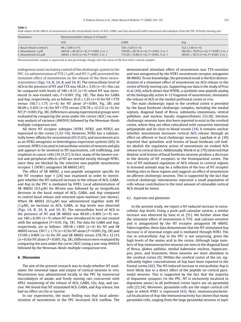
Table 2Peak values of the effects of neurotensin on the extracellular levels of ACh, GABA, and Asp measured with transversal microdialysis from the PFC of freely moving rats
Parameters Neurotransmitter release (% of basal)
ACh GABA Asp
1. Basal release (control) 98 ± 3.80 (n = 9) 110 ± 4.20 (n = 6) 112 ± 3.40 (n = 6)2 370.03 175.2
N e first
ePstAbdav9Newm
etacattsr
tgaSdtW((tswr41(cf
4
uNmHrh
i
daSsceot
bspccpweririiliboccA
4
citaTirhboptnfmmo
. Neurotensin 1 �M 240.56 ± 48.50 (n = 9), P < 0.001, 2 vs. 1
. Neurotensin 0.2 �M 160.81 ± 10.12 (n = 6), P < 0.001, 3 vs. 1
eurotransmitter output is expressed as the percentage change over the mean of th
ndogenous tonic excitatory control of the cholinergic system in theFC. Co-administration of TTX (1 �M) and NT (1 �M) prevented thetimulant effect of neurotensin on the release of the three neuro-ransmitters (Figs. 1A, B; 2A, B, and 3A, B). The extracellular level ofCh in the presence of NT and TTX was 58.24 ± 3.9% (n = 6); this cane compared with levels of 140 ± 8.1% (n = 9) when NT was intro-uced in non-treated rats, P < 0.001 (Fig. 1B). The data for GABAnd Asp, respectively, are as follows: 81.6 ± 1.2% (n = 6) for NT + TTXersus 150.7 ± 1.7% (n = 6) for NT alone (P < 0.001, Fig. 2B) and6.09 ± 3.45% (n = 6) for NT + TTX versus 278.78 ± 12.11% (n = 6) forT (P < 0.001, Fig. 3B). Differences among experimental groups werevaluated by comparing the areas under the curves (AUC) via one-ay analysis of variance (ANOVA) followed by the Newman–Keulsultiple comparison test.All three NT receptor subtypes (NTR1, NTR2, and NTR3) are
xpressed in the cortex [1,52–54]. However, NTR2 has a substan-ially lower affinity for neurotensin [63,11,65], and neurotensin actss an NTR2 antagonist in heterologous expression systems [66]. Inontrast, NTR3 is located in intracellular vesicles of neurons and gliand appears to be involved in NT inactivation, cell trafficking, androphism in cancer cells [39,31,37]. In fact, many of the known cen-ral and peripheral effects of NT are exerted mostly through NTR1,ince they are blocked by the selective non-peptide neurotensineceptor 1 (NTR1) antagonist SR 48692 [47].
The effect of SR 48692, a non-peptide antagonist specific forhe NT receptor type 1 [24] was examined in order to investi-ate whether the NT-induced increase in the release of ACh, GABA,nd Asp in the PFC is mediated by NTR1. Local administration ofR 48692 (0.5 �M) for 80 min was followed by an insignificantecrease in the basal output of ACh, GABA, and Asp comparedo control basal release and returned upon washout (not shown).
hen SR 48692 (0.5 �M) was administered together with NT1 �M), no increase in ACh, GABA, or Asp levels was observedFigs. 1A, B; 2A, B, and 3A, B). The extracellular level of ACh inhe presence of NT and SR 48692 was 89.00 ± 9.40% (n = 9) ver-us 140 ± 8.10% (n = 9) when NT was introduced in rats not treatedith the antagonist (P < 0.01, Fig. 1B). The data for GABA and Asp,
espectively, are as follows: 109.18 ± 1.80% (n = 6) for NT and SR8692 versus 150.7 ± 1.7% (n = 6) for NT alone (P < 0.001, Fig. 2B) and17.00 ± 9.80% (n = 6) for NT and SR 48692 versus 278.78 ± 12.11%n = 6) for NT alone (P < 0.001, Fig. 3B). Differences were evaluated byomparing the area under the curve (AUC) using a one-way ANOVAollowed by the Newman–Keuls multiple comparison test.
. Discussion
The aim of the present research was to study whether NT mod-lates the neuronal input and output of cortical neurons in vivo.eurotensin was administered locally in the PFC by transversalicrodialysis of awake and freely moving rats concurrent with
PLC monitoring of the release of ACh, GABA, Glu, Asp, and tau-ine. We found that NT stimulated ACh, GABA, and Asp release, butad no effect on Glu or taurine.
In our experiments, the main finding was that local admin-stration of neurotensin in the PFC increased ACh outflow. The
dctcp
0 ± 28.70 (n = 6), P < 0.001, 2 vs. 1 382.00 ± 70.93 (n = 6), P < 0.001, 2 vs. 10 ± 12.80 (n = 6), P < 0.001, 3 vs. 1 189.01 ± 18.12 (n = 6), P < 0.001, 3 vs. 1
three control samples.
emonstrated stimulant effect of neurotensin was TTX-sensitivend was antagonized by the NTR1 neurotensin receptor antagonistR 48692. To our knowledge, the presented result is the first demon-tration of a stimulant effect of neurotensin on ACh release in theortex of freely moving rats. Supporting our data is the study of Prust al. [44], which shows that NT69L, a synthetic non-peptide analogf the biologically active 8–13 fragment of neurotensin, stimulateshe release of ACh in the medial prefrontal cortex in vivo.
The main cholinergic input to the cerebral cortex is providedy the basal forebrain cholinergic complex, including the medialeptum, diagonal band of Broca, substantia innominata, ventralallidum, and nucleus basalis magnocellularis [33,18]. Intrinsicholinergic neurons have also been reported to exist in the cerebralortex, where they are often colocalized with vasoactive intestinalolypeptide and lie close to blood vessels [14]. It remains unclearhether neurotensin increases cortical ACh release through an
ffect on afferent or local cholinergic neurons. Lapchak et al. [30]eported that quinolinic acid lesions of basal forebrain cell bod-es abolish the regulatory action of neurotensin on evoked AChelease in cortical slices. Additionally, Wenk et al. [70] observed thatbotenic acid lesions of basal forebrain neurons produce a decreasen the density of NT receptors in the frontoparietal cortex. Theoss of NT-mediated regulation of ACh release in cortical regionsn lesioned animals may be a reflection of the loss of NT receptorinding sites in these regions and suggests an effect of neurotensinn afferent cholinergic neurons. This is supported by the fact thatortical cholinergic interneurons represent a small population ofells whose contribution to the total amount of releasable corticalCh should be minor.
.1. Aspartate and glutamate
In the present study, we report a NT-induced increase in extra-ellular Asp levels. Using a push–pull cannulae system, a similarncrease was observed by Sanz et al. [51]. We further show thathe stimulant effect of neurotensin is TTX- and calcium-sensitivend is antagonized by the NT receptor antagonist SR 48692.aken together, these data demonstrate that the NT-stimulated Aspncrease is of neuronal origin and is mediated through NTR1. Theise in extracellular Asp in the PFC is not surprising, given theigh levels of the amino acid in the cortex. Although large num-ers of Asp-immunoreactive neurons are seen in the diagonal bandf Broca, globus pallidus, medial habenular nucleus, hippocam-us, pons, and brainstem, these neurons are most abundant inhe cerebral cortex [9]. Within the cerebral cortex of the rat, sig-ificantly higher concentrations of Asp have been reported in the
rontal cortex [42]. The NT-induced increase in extracellular Asp isost likely due to a direct effect of the peptide on cortical pyra-idal neurons. This is supported by the fact that the majority
f dopamine synapses (in the PFC, NT is exclusively localized to
opamine axons) in all prefrontal cortex layers are on pyramidalells [22,54]. Moreover, pyramidal cells are the major cortical cellype in which NTR1 is expressed [43]. Next, immunocytochemi-al localization of Asp-like immunoreactivity has shown that manyyramidal cells, ranging from the large pyramidal neurons in layer
1 esear
VAtucicidtAcAti
acpAustbbslihWetTrrea1n[
4
bc(tsniocmeNbmnringee
C
e
A
a(cglHP
R
[
[
[
[
[
[
[
[
[
34 P. Petkova-Kirova et al. / Brain R
to the smaller ones in layers VI, III, and II, are Asp-positive [9,13].utoradiographic studies using 3[H]-d-Asp have also demonstratedhat many pyramidal neurons in layers V, VI, and III of the cortexse Glu/Asp as transmitters [49,4]. This finding has been furtheronfirmed by autoradiography combined with immunocytochem-stry [20]. As mesocortical projections from the VTA innervate deeportical layers V and VI of the PFC [8] and neurotensin is colocal-zed with dopamine in all of the mesocortical projections to theeep cortical layers [15], a possible target for neurotensin involveshe efferent fibers to subcortical areas (cortical layers V and VI).lthough they project to other brain areas, efferent cells may alsoontribute to the cortical Asp/Glu pool measurable by microdialysis.lmost all efferent cells have axon collaterals projecting to intracor-
ical neurons [26], and these collaterals allow them to participaten the intracortical circuitry.
Our data show that glutamate levels in the PFC are not increasedfter NT perfusion. Since a number of studies demonstrate colo-alization of Asp with Glu [32], the selective increase in Asproduced by neurotensin is intriguing. It is possible that Glu- andsp-immunoreactive neurons represent in part two distinct pop-lations. This is supported by Giuffrida and Rustioni [20], whohow that a large fraction of cortico-cortical neurons in the cor-ex of rat are immunoreactive for either Asp or Glu, but nototh. Dori et al. [13] further detect morphological differencesetween the two groups of neurons, reporting that aspartate-tained neurons are larger than glutamate-positive cells in everyayer in rat visual cortex. Our results are consistent with the find-ngs of Petrie et al. [43], which show that neurotensin does notave an effect on glutamate release in vivo in conscious rats.hile neurotensin was perfused through a vertical probe in their
xperiments, we administered the neuropeptide transversally sohat it could activate cortical neurons in a larger area (8 mm).he question about the effect of NT on Glu levels in the PFCemains controversial, as two other groups report NT-inducedises in glutamate [3,17]. However, one of the groups studies theffect of NT in vitro in rat primary cultures of cortical neuronsnd cortical slices [3,16]. In the latter, NT (1–13) in the range of00–1000 nM only slightly increased spontaneously released Glu;eurotensin’s effect was mainly on potassium-evoked Glu release16].
.2. GABA
Interneurons play a central role in cortical function, and a num-er of neuropathological studies have indicated a dysfunction ofortical GABAergic neurons in schizophrenia and bipolar disorderfor a review, see [23]). Ultrastructural studies of the cortex showhat dopamine axons synapse on both the dendritic shafts andpines of pyramidal neurons and also the dendrites of local circuiteurons [58] that are immunoreactive for GABA [57]. These stud-
es estimate that ∼40% of the dopamine synapses in the PFC arrivento GABA-containing local circuit neurons. Although pyramidalells are the major cortical cell type in which NTR1 is expressed,any interneurons defined on the basis of calcium-binding protein
xpression also display NTR1 immunoreactivity [43]. Therefore,T might be a modulator of interneurons that is implicated inoth normal and symptomatic cortical function. In our experi-ents, NT increased GABA release in the PFC. This effect was of
eural origin, as it was TTX-sensitive, and mediated by the NTR1eceptor, as it was antagonized by SR 48692. The effect of NT
s most likely exerted directly on cortical interneurons, becauseeurotensin failed to increase Glu that can further activate GABAer-ic interneurons. The possibility exists, however, that part of thisffect is secondary to a neurotensin increase in aspartate lev-ls.[
[
ch Bulletin 77 (2008) 129–135
onflict of interest
The authors have no conflicting professional and personal inter-sts.
cknowledgements
This work was supported financialy by Ente CRF (Firenze, Italy)nd Fondazione MPS (Siena, Italy). The authors are grateful to CNRRome, Italy) and the EU COST D34 for supporting our internationalooperation and to Dr. Maffrand, Sanofi Recherche, France for theenerous supply of the neurotensin antagonist SR 48692. For excel-ent assistance in measuring amino acid transmitters release byPLC we are grateful to Ms. Alesandra Colivicchi (Department ofharmacology, University of Florence).
eferences
[1] M.J. Alexander, S.E. Leeman, Widespread expression in adult rat forebrain ofmRNA encoding high-affinity neurotensin receptor, J. Comp. Neurol. 402 (1998)475–500.
[2] A. Alonso, M.P. Faure, A. Beaudet, Neurotensin promotes oscillatory burstingbehavior and is internalized in basal forebrain cholinergic neurons, J. Neurosci.14 (1994) 5778–5792.
[3] T. Antonelli, L. Ferraro, K. Fuxe, S. Finetti, J. Fournier, S. Tanganelli, M. De Mat-tei, M.C. Tomasini, Neurotensin enhances endogenous extracellular glutamatelevels in primary cultures of rat cortical neurons: involvement of neurotensinreceptor in NMDA induced excitotoxicity, Cereb. Cortex 14 (2004) 466–473.
[4] P. Barbaresi, M. Fabri, F. Conti, T. Manzo, d-[3H]aspartate retrograde labellingof callosal and association neurones of somatosensory areas I and II of cats, J.Comp. Neurol. 236 (1987) 159–178.
[5] A.J. Bean, M.J. During, R.H. Roth, Stimulation-induced release of coexis-tent transmitters in the prefrontal cortex: an in vivo microdialysis study ofdopamine and neurotensin release, J. Neurochem. 53 (1989) 655–657.
[6] L. Bianchi, L. Della Corte, K.F. Tipton, Simultaneous determination of basal andevoked output levels of aspartate, glutamate, taurine and 4-aminobutyric acidduring microdialysis and from superfused brain slises, J. Chromatogr. Biol. 723(1999) 47–59.
[7] E.B. Binder, B. Kinkead, M.J. Owens, C.B. Nemeroff, Neurotensin and dopamineinteractions, Pharmacol. Rev. 53 (2001) 453–486.
[8] A. Bjorklund, O. Lindvall, Dopamine containing systems in the CNS, in: A.Bjorklund, T. Hokfelt (Eds.), Handbook of Chemical Neuroanatomy: ClassicalTransmitters in the CNS, vol. 2, Elsevier, Amsterdam, 1984, pp. 55–122.
[9] G. Campistron, R.M. Buijs, M. Geffard, Specific antibodies against aspartate andtheir immunocytochemical application in the rat brain, Brain Res. 365 (1986)179–184.
10] R. Carraway, S.E. Leeman, The isolation of a new hypotensive peptide, neu-rotensin from bovine hypothalamic, J. Biol. Chem. 248 (1973) 6854–6861.
11] P. Chalon, N. Vita, M. Kaghad, M. Guillemot, J. Bonnin, B. Delpech, G. Le Fur, P.Ferrara, D. Caput, Molecular cloning of a levocabastine-sensitive neurotensinbinding site, FEBS Lett. 386 (1996) 91–94.
12] G. Damsma, D. Lammerts Van Bueren, B.H.C. Westering, A.S. Horn, Determina-tion of acetylcholine in the femtomole range by means of HPLC, a post-columnenzyme reactor, and electrochemical detection, Chromatographia 24 (1987)827–831.
13] I. Dori, M. Petrou, J.G. Parnavelas, Excitatory transmitter amino acid-containingneurons in the rat visual cortex: a light and electron microscopic immunocy-tochemical study, J. Comp. Neurol. 290 (1989) 169–184.
14] F. Eskenstein, R.W. Baughman, Two types of cholinergic innervation in cortex,one co-localized with vasoactive interstinal polypeptide, Nature 309 (1984)153–155.
15] A. Febvret, B. Berger, P. Gaspar, C. Verney, Further indication that distinctdopaminergic subsets project to the rat cerebral cortex: lack of colocaliza-tion with neurotensin in the superficial dopaminergic fields of the anteriorcingulate, motor, retrosplenial and visual cortices, Brain Res. 547 (1991)37–52.
16] L. Ferraro, M.C. Tomasini, A. Siniscalchi, K. Fuxe, S. Tanganelli, T. Antonelli, Neu-rotensin increases endogenous glutamate release in rat cortical slices, Life Sci.66 (2000) 927–936.
17] C.F. Ferris, in: G.M. Marklouf (Ed.), The Gastrointestinal System: Neural andEndocrine Biology, Betseda, 1989, pp. 559–586.
18] R.P. Gaykema, P.G. Luiten, C. Nyakas, J. Traber, Cortical projection patterns of themedial septum-diagonal band complex, J. Comp. Neurol. 293 (1990) 103–124.
19] M.G. Giovannini, F. Camilli, A. Mundula, G. Pepeu, Glutamatergic regulation ofacetylcholine output in different brain regions: a microdialysis study in the rat,Neurochem. Int. 25 (1994) 23–26.
20] R. Giuffrida, A. Rustioni, Glutamate and aspartate immunoreactivity in cortico-cortical neurons of the sensorimotor cortex of rats, Exp. Brain Res. 74 (1989)41–46.

esear
[
[
[
[
[
[
[
[
[
[
[
[
[
[
[
[
[
[
[
[
[
[
[
[
[
[
[
[
[
[
[
[
[
[
[
[
[
[
[
[
[
[
[
[
[
[
[
[
[
P. Petkova-Kirova et al. / Brain R
21] M. Goedert, K. Pittaway, B.J. Williams, P.C. Emson, Specific binding of tritiatedneurotensin to rat brain membranes: characterization and regional distribu-tion, Brain Res. 304 (1984) 71–81.
22] P.S. Goldman-Rakic, C. Leranth, S.M. Williams, N. Mons, M. Geffard, Dopaminesynaptic complex with pyramidal neurons in primate cerebral cortex, Proc. Natl.Acad. Sci. U.S.A. 86 (1989) 9015–9019.
23] A. Guidotti, J. Auta, J.M. Davis, E. Dong, D.R. Grayson, M. Veldic, X. Zang, E.Costa, GABAergic dysfunction in schizophrenia: new treatment strategies onthe horizon, Psychopharmacology (Berl.) 180 (2005) 191–205.
24] D. Gully, M. Canton, R. Boigegrain, F. Jeanjean, J.C. Molimard, M. Pon-celet, C. Gueudet, M. Heaulme, R. Leyris, A. Brouard, et al., Biochemicaland pharmacological profile of a potent and selective nonpeptide antag-onist of the neurotensin receptor, Proc. Natl. Acad. Sci. U.S.A. 90 (1993)65–69.
25] T. Hokfelt, B.J. Everitt, E. Theodorsson-Norheim, M. Goldstein, Occurrence ofneurotensinlike immunoreactivity in subpopulations of hypothalamic, mes-encephalic, and medullary catecholamine neurons, J. Comp. Neurol. (1984)543–559.
26] E.J. Jones, Laminar distribution of cortical efferent cells, in: A. Peters, E.G. Jones(Eds.), Cerebral Cortex, vol. 1, Plenum Press, New York, 1984, pp. 521–553.
27] J. Kasckow, C.B. Nemeroff, The neurobiology of neurotensin: focus onneurotensin–dopamine interactions, Regul. Pept. 36 (1991) 153–164.
28] J.P. Kiss, Theory of active antidepressants: a nonsynaptic approach to the treat-ment of depression, Neurochem. Int. 52 (2008) 34–39.
29] P.A. Lapchak, D.M. Araujo, R. Quirion, A. Beaudet, Neurotensin regulation ofendogenous acetylcholine release from rat striatal slices is independent ofdopaminergic tone, J. Neurochem. 56 (1991) 651–657.
30] P.A. Lapchak, D.M. Araujo, R. Quirion, A. Beaudet, Neurotensin regulation ofendogenous acetylcholine release from rat cerebral cortex. Effect of guinolinicacid lesions of the basal forebrain, J. Neurochem. 55 (1990) 1397–1403.
31] J. Mazella, Sortilin/neurotensin receptor-3: a new tool to investigate neu-rotensin signaling and cellular trafficking? Cell. Signal. 13 (2001) 1–6.
32] A.J. Mcdonald, Glutamate and aspartate immunoreactive neurons of the ratbasolateral amygdala: colocalization of excitatory amino acids and projectionsto the limbic circuit, J. Comp. Neurol. 365 (1996) 367–379.
33] M. McKinney, J.T. Coyle, J.C. Hedreen, Topographic analysis of the innervation ofthe rat neocortex and hippocampus by the basal forebrain cholinergic system,J. Comp. Neurol. 217 (1983) 103–121.
34] E.A. Mitchell, M.B. Herd, B.G. Gunn, J.J. Lambert, D. Belelli, Neurosteroid modu-lation of GABA(A) receptors: molecular determinants and significance in healthand disease, Neurochem. Int. 52 (2008) 588–595.
35] I. Mody, Extrasynaptic GABAA receptors in the crosshairs of hormones andethanol, Neurochem. Int. 52 (2008) 60–64.
36] E. Moyse, W. Rostene, M. Vial, K. Leonard, J. Mazella, P. Kitabgi, J.P. Vincent,A. Beaudet, Distribution of neurotensin binding sites in rat brain: a lightmicroscopic radioautographic study using monoiodo [125I] Tyr3-neurotensin,Neuroscience 22 (1987) 525–536.
37] V. Navarro, S. Martin, P. Sarret, M.S. Nielsen, C.M. Petersen, J. Vincent, J. Mazella,Pharmacological properties of the mouse neurotensin receptor 3. Maintenanceof cell surface receptor during internalization of neurotensin, FEBS Lett. 495(2001) 100–105.
38] C.B. Nemeroff, D. Luttinger, A.J. Prange, Neurotensin and bombesin, in: L.L.Iversen, S.D. Iversen, S.H. Snyder (Eds.), Handbook of Psychopharmacology,Plenum Publishing Corp., New York, 1982, pp. 363–385.
39] D. Nouel, P. Sarret, J.P. Vincent, J. Mazella, A. Beaudet, Pharmacological, molecu-lar and functional characterization of glial neurotensin receptors, Neuroscience94 (1999) 1189–1197.
40] W.T. O’Connor, S. Tanganelli, U. Ungerstedt, K. Fuxe, The effect of NT on GABA andacetylcholine release in the dorsal striatum of the rat: an in vivo microdialysisstudy, Brain Res. 573 (1992) 209–216.
41] G. Paxinos, G. Watson, The Rat Brain in Stereotaxic Coordinates, Academic Press,New York, 1982.
42] J.M. Peinado, J.A. Gomez-Capilla, F. Mora, Cerebral cortex and amino acid neu-rotransmitters: higher levels of aspartic acid but not GABA in the frontal cortexof the rat, Brain Res. Bull. 12 (1984) 625–627.
43] K. Petrie, D. Schmidt, M. Bubser, J. Fadel, R. Carraway, A. Deutch, Neurotensinactivates GABAergic interneurons in the prefrontal cortex, J. Neurosci. 25 (2005)1629–1636.
44] A.J. Prus, M. Huang, Z. Li, J. Dai, H.Y. Meltzer, The neurotensin analog NT69L
enhances medial prefrontal cortical dopamine and acetylcholine efflux: poten-tiation of risperidone-, but not haloperidol-, induced dopamine efflux, BrainRes. 1184 (2007) 354–364.45] J.M. Radke, M.J. Owens, C.B. Nemeroff, The effects of glutamate receptor agonistson neurotensin release using in vivo microdialysis, Eur. J. Pharmacol. 411 (2001)129–134.
[
[
ch Bulletin 77 (2008) 129–135 135
46] A. Rakovska, M.G. Giovannini, L. Della Corte, R. Kalfin, L. Bianchi, G. Pepeu,Neurotensin modulation of acetylcholine and GABA release from the rat hip-pocampus: an in vivo microdialysis study, Neurochem. Int. 33 (1998) 335–340.
47] W. Rostene, M. Azzi, H. Boudin, I. Lepee, F. Souaze, M. Mendez-Ubach, C. Betan-cur, D. Gully, Use of nonpeptide antagonists to explore the physiological rolesof neurotensin. Focus on brain neurotensin/dopamine interactions, Ann. N. Y.Acad. Sci. 814 (1997) 125–141.
48] B. Rozsa, G. Katona, A. Kaszas, R. Szipocs, E.S. Vizi, Dendritic nicotinic recep-tors modulate backpropagating action potentials and long-term plasticity ofinterneurons, Eur. J. Neurosci. 27 (2008) 364–377.
49] Rustioni, M. Cuenod, Selective retrograde transport of d-aspartate in spinalinterneurons and cortical neurons of rats, Brain Res. 236 (1982) 143–155.
50] D.B. Rye, B.H. Wainer, M.M. Mesulam, E.J. Mufson, C.B. Saper, Cortical projec-tions arising from the basal forebrain: a study of cholinergic and noncholinergiccomponents employing combined retrograde tracing and immunohistochem-ical localization of choline acetyltransferase, Neuroscience 13 (1984) 627–643.
51] B. Sanz, I. Exposito, F. Mora, Effects of neurotensin on the release of glutamic acidin the prefrontal cortex and striatum of the rat, Neuroreport 4 (1993) 1194–1196.
52] P. Sarret, P. Krzywkowski, L. Segal, M.S. Nielsen, C.M. Petersen, J. Mazella, T.Stroh, A. Beaudet, Distribution of ntrs3 receptor/sortilin mRNA and protein inthe rat central nervous system, J. Comp. Neurol. 461 (2003) 483–505.
53] P. Sarret, A. Perron, T. Stroh, A. Beaudet, Immunohistochemical distribution ofnts2 neurotensin receptors in the rat central nervous system, J. Comp. Neurol.461 (2003) 520–538.
54] P. Seguela, K.C. Watkins, L. Descarries, Ultrastructural features of dopamineaxon terminals in the anteromedial and the suprarhinal cortex of adult rat,Brain Res. 442 (1988) 11–22.
55] A. Semyanov, Can diffuse extrasynaptic signaling form a guiding template?Neurochem. Int. 52 (2008) 31–33.
56] K.B. Seroogy, A. Mehta, J.H. Fallon, Neurotensin and cholecystokinin coexistencewithin neurons of the ventral mesencephalon: projections to forebrain, Exp.Brain Res. 68 (1987) 277–289.
57] S.R. Sesack, C.N. Bressler, D.A. Lewis, Ultrastructural associations betweendopamine terminals and local circuit neurons in the monkey prefrontal cortex:a study of calretinin-immunoreactive cells, Neurosci. Lett. 200 (1995) 9–12.
58] J.F. Smiley, P.S. Goldman-Rakic, Silver-enhanced diaminobenzidine-sulfide(SEDS): a technique for high-resolution immunoelectron microscopy demon-strated with monoamine immunoreactivity in monkey cerebral cortex andcaudate, J. Histochem. Cytochem. 41 (1993) 1393–1404.
59] R. Steinberg, D. Rodier, G. Mons, D. Gully, G. Le Fur, P. Soubrie, SR 48692-sensitiveneurotensin receptors modulate acetylcholine release in the rat striatum, Neu-ropeptides 29 (1995) 27–31.
60] J.M. Studler, P. Kitabgi, G. Tramu, D. Herve, J. Glowinski, J.P. Tassin, Exten-sive colocalization of neurotensin with dopamine in rat meso-cortoco-frontaldopaminergic neurons, Neuropeptides 11 (1988) 95–100.
61] E. Sykova, L. Vargova, Extrasynaptic transmission and the diffusion parametersof the extracellular space, Neurochem. Int. 52 (2008) 5–13.
62] E. Szigethy, A. Beaudet, Selective association of neurotensin receptors withcholinergic neurons in the rat basal forebrain, Neurosci. Lett. 83 (1987) 47–52.
63] K. Tanaka, M. Masu, S. Nakanishi, Structure and functional expression of thecloned rat neurotensin receptor, Neuron 4 (1990) 847–854.
64] A. Torocsik, A. Rakovska, T. Gorcs, E.S. Vizi, Effects of neu-rotensin and immunneutralization with anti-neurotensin-serum ondopaminergic–cholinergic interaction in the striatum, Brain Res. 612 (1993)306–312.
65] J.P. Vincent, J. Mazella, P. Kitabgi, Neurotensin and neurotensin receptors, TrendsPharmacol. Sci. 20 (1999) 302–309.
66] N. Vita, F. Oury-Donat, P. Chalon, M. Guillemot, M. Kaghad, A. Bachy, O. Thur-neyssen, S. Garcia, C. Poinot-Chazel, P. Casellas, P. Keane, G. Le Fur, J.P. Maffrand,P. Soubrie, D. Caput, P. Ferrara, Neurotensin is an antagonist of the humanneurotensin NT2 receptor expressed in Chinese hamster ovary cells, Eur. J.Pharmacol. 360 (1998) 265–272.
67] E.S. Vizi, A. Mike, Nonsynaptic receptors for GABA and glutamate, Curr. Top.Med. Chem. 6 (2006) 941–948.
68] E.S. Vizi, Role of high-affinity receptors and membrane transporters in non-synaptic communication and drug action in the central nervous system,Pharmacol. Rev. 52 (2000) 63–89.
69] E.S. Vizi, Non-synaptic Interactions Between Neurons: Modulation of Neuro-chemical Transmission. Pharmacological and Clinical Aspects, John Wiley and
Sons, Chichester, New York, 1984.70] G.L. Wenk, A.L. Markowska, D.S. Olton, Basal forebrain lesions and memory:alterations in neurotensin, not acetylcholine, may cause amnesia, Behav. Neu-rosci. 103 (1989) 765–769.
71] K. Zilles, A. Wree, Cortex: areal and laminar structure, in: G. Paxinos (Ed.), TheRat Nervous System, vol. 1, Academic Press, New York, 1985, pp. 375–415.
Related Documents