J. Clin. Med. 2022, 11, 776. https://doi.org/10.3390/jcm11030776 www.mdpi.com/journal/jcm Review Neurostimulation in People with Oropharyngeal Dysphagia: A Systematic Review and Meta‐Analyses of Randomised Controlled Trials—Part I: Pharyngeal and Neuromuscular Electrical Stimulation Renée Speyer 1,2,3, *, Anna‐Liisa Sutt 4,5 , Liza Bergström 6,7 , Shaheen Hamdy 8 , Bas Joris Heijnen 3 , Lianne Remijn 9 , Sarah Wilkes‐Gillan 10 and Reinie Cordier 2,11 1 Department Special Needs Education, Faculty of Educational Sciences, University of Oslo, 0318 Oslo, Norway 2 Curtin School of Allied Health, Faculty of Health Sciences, Curtin University, Perth, WA 6102, Australia 3 Department of Otorhinolaryngology and Head and Neck Surgery, Leiden University Medical Centre, 1233 ZA Leiden, The Netherlands; [email protected] 4 Critical Care Research Group, The Prince Charles Hospital, Brisbane, QLD 4032, Australia; [email protected] 5 School of Medicine, University of Queensland, Brisbane, QLD 4072, Australia 6 Remeo Stockholm, 128 64 Stockholm, Sweden; [email protected] 7 Speech Therapy Clinic, Danderyd University Hospital, 182 88 Stockholm, Sweden 8 GI Sciences, School of Medical Sciences, Faculty of Biology, Medicine and Health, University of Manchester, Manchester M13 9PL, UK; [email protected] 9 School of Allied Health, HAN University of Applied Sciences, 6525 EN Nijmegen, The Netherlands; [email protected] 10 Discipline of Occupational Therapy, Sydney School of Health Sciences, Faculty of Medicine and Health, The University of Sydney, Sydney, NSW 2006, Australia; sarah.wilkes‐[email protected] 11 Department of Social Work, Education and Community Wellbeing, Faculty of Health & Life Sciences, Northumbria University, Newcastle upon Tyne NE7 7XA, UK; [email protected] * Correspondence: [email protected] Abstract: Objective. To assess the effects of neurostimulation (i.e., neuromuscular electrical stimulation (NMES) and pharyngeal electrical stimulation (PES)) in people with oropharyngeal dysphagia (OD). Methods. Systematic literature searches were conducted to retrieve randomised controlled trials in four electronic databases (CINAHL, Embase, PsycINFO, and PubMed). The methodological quality of included studies was assessed using the Revised Cochrane risk‐of‐bias tool for randomised trials (RoB 2). Results. In total, 42 studies reporting on peripheral neurostimulation were included: 30 studies on NMES, eight studies on PES, and four studies on combined neurostimulation interventions. When conducting meta analyses, significant, large and significant, moderate pre‐post treatment effects were found for NMES (11 studies) and PES (five studies), respectively. Between‐group analyses showed small effect sizes in favour of NMES, but no significant effects for PES. Conclusion. NMES may have more promising effects compared to PES. However, NMES studies showed high heterogeneity in protocols and experimental variables, the presence of potential moderators, and inconsistent reporting of methodology. Therefore, only conservative generalisations and interpretation of meta‐analyses could be made. To facilitate comparisons of studies and determine intervention effects, there is a need for more randomised controlled trials with larger population sizes, and greater standardisation of protocols and guidelines for reporting. Keywords: deglutition; swallowing disorders; RCT; intervention; neuromuscular electrical stimulation; pharyngeal electrical stimulation; PES; NMES Citation: Speyer, R.; Sutt, A.‐L.; Bergström, L.; Hamdy, S.; Heijnen, B.J.; Remijn, L.; Wilkes‐Gillan, S.; Cordier, R. Neurostimulation in People with Oropharyngeal Dysphagia: A Systematic Review and Meta‐Analyses of Randomised Controlled Trials−Part I: Pharyngeal and Neuromuscular Electrical Stimulation. J. Clin. Med. 2022, 11, 776. https://doi.org/10.3390/ jcm11030776 Academic Editor: Michael Setzen Received: 7 December 2021 Accepted: 27 January 2022 Published: 31 January 2022 Publisher’s Note: MDPI stays neutral with regard to jurisdictional claims in published maps and institutional affiliations. Copyright: © 2022 by the authors. Licensee MDPI, Basel, Switzerland. This article is an open access article distributed under the terms and conditions of the Creative Commons Attribution (CC BY) license (https://creativecommons.org/license s/by/4.0/).

Welcome message from author
This document is posted to help you gain knowledge. Please leave a comment to let me know what you think about it! Share it to your friends and learn new things together.
Transcript
J. Clin. Med. 2022, 11, 776. https://doi.org/10.3390/jcm11030776 www.mdpi.com/journal/jcm
Review
Neurostimulation in People with Oropharyngeal Dysphagia:
A Systematic Review and Meta‐Analyses of Randomised
Controlled Trials—Part I: Pharyngeal and Neuromuscular
Electrical Stimulation
Renée Speyer 1,2,3,*, Anna‐Liisa Sutt 4,5, Liza Bergström 6,7, Shaheen Hamdy 8, Bas Joris Heijnen 3, Lianne Remijn 9,
Sarah Wilkes‐Gillan 10 and Reinie Cordier 2,11
1 Department Special Needs Education, Faculty of Educational Sciences, University of Oslo,
0318 Oslo, Norway 2 Curtin School of Allied Health, Faculty of Health Sciences, Curtin University, Perth, WA 6102, Australia 3 Department of Otorhinolaryngology and Head and Neck Surgery, Leiden University Medical Centre,
1233 ZA Leiden, The Netherlands; [email protected] 4 Critical Care Research Group, The Prince Charles Hospital, Brisbane, QLD 4032, Australia;
[email protected] 5 School of Medicine, University of Queensland, Brisbane, QLD 4072, Australia 6 Remeo Stockholm, 128 64 Stockholm, Sweden; [email protected] 7 Speech Therapy Clinic, Danderyd University Hospital, 182 88 Stockholm, Sweden 8 GI Sciences, School of Medical Sciences, Faculty of Biology, Medicine and Health, University of Manchester,
Manchester M13 9PL, UK; [email protected] 9 School of Allied Health, HAN University of Applied Sciences, 6525 EN Nijmegen, The Netherlands;
[email protected] 10 Discipline of Occupational Therapy, Sydney School of Health Sciences, Faculty of Medicine and Health,
The University of Sydney, Sydney, NSW 2006, Australia; sarah.wilkes‐[email protected] 11 Department of Social Work, Education and Community Wellbeing, Faculty of Health & Life Sciences,
Northumbria University, Newcastle upon Tyne NE7 7XA, UK; [email protected]
* Correspondence: [email protected]
Abstract: Objective. To assess the effects of neurostimulation (i.e., neuromuscular electrical
stimulation (NMES) and pharyngeal electrical stimulation (PES)) in people with oropharyngeal
dysphagia (OD). Methods. Systematic literature searches were conducted to retrieve randomised
controlled trials in four electronic databases (CINAHL, Embase, PsycINFO, and PubMed). The
methodological quality of included studies was assessed using the Revised Cochrane risk‐of‐bias
tool for randomised trials (RoB 2). Results. In total, 42 studies reporting on peripheral
neurostimulation were included: 30 studies on NMES, eight studies on PES, and four studies on
combined neurostimulation interventions. When conducting meta analyses, significant, large and
significant, moderate pre‐post treatment effects were found for NMES (11 studies) and PES (five
studies), respectively. Between‐group analyses showed small effect sizes in favour of NMES, but no
significant effects for PES. Conclusion. NMES may have more promising effects compared to PES.
However, NMES studies showed high heterogeneity in protocols and experimental variables, the
presence of potential moderators, and inconsistent reporting of methodology. Therefore, only
conservative generalisations and interpretation of meta‐analyses could be made. To facilitate
comparisons of studies and determine intervention effects, there is a need for more randomised
controlled trials with larger population sizes, and greater standardisation of protocols and
guidelines for reporting.
Keywords: deglutition; swallowing disorders; RCT; intervention; neuromuscular electrical
stimulation; pharyngeal electrical stimulation; PES; NMES
Citation: Speyer, R.; Sutt, A.‐L.;
Bergström, L.; Hamdy, S.; Heijnen,
B.J.; Remijn, L.; Wilkes‐Gillan, S.;
Cordier, R. Neurostimulation in
People with Oropharyngeal
Dysphagia: A Systematic Review
and Meta‐Analyses of Randomised
Controlled Trials−Part I: Pharyngeal
and Neuromuscular Electrical
Stimulation. J. Clin. Med. 2022, 11,
776. https://doi.org/10.3390/
jcm11030776
Academic Editor: Michael Setzen
Received: 7 December 2021
Accepted: 27 January 2022
Published: 31 January 2022
Publisher’s Note: MDPI stays
neutral with regard to jurisdictional
claims in published maps and
institutional affiliations.
Copyright: © 2022 by the authors.
Licensee MDPI, Basel, Switzerland.
This article is an open access article
distributed under the terms and
conditions of the Creative Commons
Attribution (CC BY) license
(https://creativecommons.org/license
s/by/4.0/).
J. Clin. Med. 2022, 11, 776 2 of 68
1. Introduction
The aerodigestive tract facilitates the combined functions of breathing, vocalising,
and swallowing. Any dysfunction in this system may lead to oropharyngeal dysphagia
(OD) or swallowing problems [1]. OD can be the result of underlying diseases such as
stroke or a progressive neurological disease (e.g., Parkinson’s disease, multiple sclerosis)
or an adverse effect after head and neck oncological interventions (e.g., radiation or
surgery) or intensive care treatment (e.g., intubation and tracheostomy). Prevalence
estimates of OD have been reported to be as high as 50% in cerebral palsy [2], 80% in
stroke and Parkinson’s disease, and over 90% in people with community‐acquired
pneumonia [3]. OD can have a severe impact on a person’s health as it may lead to
dehydration, malnutrition, and even death. Research has identified inverse bidirectional
relationships between decreased health‐related quality of life and increased OD severity
[4].
Traditional OD therapy may include physical interventions such as: bolus
modification and management (e.g., adjusting the viscosity, volume, temperature and/or
acidity of food and drinks); oromotor exercises; body and head postural adjustments; and
swallow manoeuvres (e.g., manoeuvres to improve food propulsion into the pharynx and
airway protection) [1]. Therapy may also include sensory stimulation, which involves
applying techniques like thermal stimulation and chemical stimulation using natural
agonists of polymodal sensory receptors (e.g., capsaicin, the spicy component of peppers)
[5].
Another type of stimulation considered to be beneficial for promoting rehabilitation
of swallowing dysfunction is acupuncture. This practice emerged from traditional
Chinese medicine and exerts therapeutic effects by inserting thin needles at strategic
places, termed acupuncture points, on the body surface aiming to rebalance the flow of
energy or life force (‘qi’). Needles are then activated through specific manual movements
or electrical stimulation. Although stimulation of acupuncture points seems to be
associated with places where nerves, muscles, and connective tissues may be stimulated
[6], their intrinsic mechanisms are still part of a continuing scientific debate on
acupuncture.
Recently, an increasing number of studies have been published on alternative
interventions aiming to enhance neural plasticity by using non‐invasive brain stimulation
(NIBS) techniques. Repetitive transcranial magnetic stimulation (rTMS) and transcranial
direct current stimulation (tDCS) are cortically or centrally applied NIBS techniques.
Using electromagnetic induction, rTMS results in depolarisation of post‐synaptic
connections, whereas tDCS uses direct electrical current to shift the polarity of nerve cells
[7]. Alternatively, electrical stimulation techniques like pharyngeal electrical stimulation
(PES) and neuromuscular electrical stimulation (NMES) target the peripheral neural
pathways [8]. NMES aims to strengthen muscular contractions during swallowing and
uses stimulation by electrodes placed on the skin over the anterior neck muscles to
activate sensory pathways [9–11]. In contrast, PES has been shown to drive neuroplasticity
in the pharyngeal motor cortex through direct stimulation of the pharyngeal mucosa via
intraluminal catheters [7].
Over the past decade, several reviews have been published on the effects of
neurostimulation in patients with OD. Most of these reviews focused on selected types of
neurostimulation: NMES [10,12], rTMS [13,14], tDCS [15], or rTMS and tDCS [16,17]. Only
two systematic reviews included both cortical (rTMS and tDCS) and peripheral
neurostimulation (PES and NMES) [18,19]. All reviews targeted interventions in post‐
stroke populations except one review that broadened inclusion criteria to patients with
acquired brain injury including stroke [16]. To date, all systematic reviews on
neurostimulation as a treatment for OD set boundaries for inclusion based on medical
diagnoses.
The aim of this systematic review is to determine the effects of neurostimulation in
people with OD without excluding populations based on medical diagnoses. Findings are
J. Clin. Med. 2022, 11, 776 3 of 68
based on the highest level of evidence only, namely randomised controlled trials (RCTs),
and summarised by conducting meta‐analyses. The results of this review will be
presented in two companion papers. This paper (Part I) reports on pharyngeal and
neuromuscular electrical stimulation (PES and NMES) while the second paper (Part II)
will report on brain stimulation (i.e., rTMS and tDCS).
2. Methods
The methodology and reporting of this systematic review were based on the
Preferred Reporting Items for Systematic Reviews and Meta‐Analyses (PRISMA) 2020
statement and checklist (Supplementary Tables S1 and S2) which aim to enhance the
essential and transparent reporting of systematic reviews [20,21]. The protocol for this
review was registered at PROSPERO, the international prospective register of systematic
reviews (registration number: CRD42020179842).
2.1. Information Sources and Search Strategies
Literature searches to identify studies were conducted on 6 March 2021, across four
databases: CINAHL, Embase, PsycINFO, and PubMed. Publication dates of coverage
ranged from 1937–2021, 1902–2021, 1887–2021, and 1809–2021, respectively. Additional
searches, including checking the reference lists of eligible articles, were performed. Two
main categories of terms were used in combination: (1) dysphagia and (2) randomised
control trials. Search strategies were performed in all four electronic databases using
subheadings (e.g., MeSH and Thesaurus terms) and free text terms. The full electronic
search strategies for each database are reported in Table 1. To identify other literature
beyond that found using these strategies, the reference lists of each eligible article were
checked.
Table 1. Search strategies.
Database and Search Terms Number of
Records
Cinahl: ((MH “Deglutition”) OR (MH “Deglutition Disorders”)) AND
(MH “Randomized Controlled Trials”) 239
Embase: (swallowing/OR dysphagia/) AND (randomization/or
randomized controlled trial/OR “randomized controlled trial (topic)”/OR
controlled clinical trial/)
4550
PsycINFO: (swallowing/OR dysphagia/) AND (RCT OR (Randomised
AND Controlled AND Trial) OR (Randomized AND Clinical AND Trial)
OR (Randomised AND Clinical AND Trial) OR (Controlled AND Clinical
AND Trial)).af.
231
PubMed: (“Deglutition”[Mesh] OR “Deglutition Disorders”[Mesh]) AND
(“Randomized Controlled Trial” [Publication Type] OR “Randomized
Controlled Trials as Topic”[Mesh] OR “Controlled Clinical Trial”
[Publication Type] OR “Pragmatic Clinical Trials as Topic”[Mesh])
3039
2.2. Inclusion and Exclusion Criteria
Studies were included in this systematic review if they met the following criteria: (1)
participants had a diagnosis of oropharyngeal dysphagia; (2) the study included non‐
invasive neurostimulation interventions aimed at reducing swallowing or feeding
problems; (3) the study included a control group or comparison intervention group; (4)
participants were randomly assigned to one of the study arms or groups; and (5) the study
was published in the English language.
Interventions such as non‐electrical peripheral stimulation (e.g., air‐puff or gustatory
stimulation), pharmacological interventions and acupuncture, were considered out of the

J. Clin. Med. 2022, 11, 776 4 of 68
scope of this review, and thus were excluded. Invasive techniques and/or those that did
not specifically target OD (i.e., deep‐brain stimulation studies after neurosurgical
implementation of a neurostimulator) were also excluded. Conference abstracts, doctoral
theses, editorials, and reviews were excluded.
Finally, only studies reporting on peripheral neurostimulation (i.e., PES and NMES)
were included in this review (Part I). Studies on brain neurostimulation (i.e., rTMS and
tDCS) will be reported on in a companion paper (Part II).
3. Systematic Review
3.1. Methodological Quality and Risk of Bias
The methodological quality of the included studies was assessed using the Revised
Cochrane risk‐of‐bias tool for randomised trials (RoB 2) [22]. The RoB 2 tool identifies five
domains to consider when assessing where bias may have been introduced into a
randomised trial: (1) bias arising from the randomisation process; (2) bias due to
deviations from intended interventions; (3) bias due to missing outcome data; (4) bias in
measurement of the outcome; and (5) bias in selection of the reported result. The RoB 2
gives a series of signalling questions for each domain whose answers give a judgement
(i.e., “low risk of bias,” “some concerns,” or “high risk of bias”), which can be evaluated
to determine a study’s overall risk of bias [22].
3.2. Data Collection Process
A data extraction form was created to extract data from the included studies under
the following categories: participant diagnosis, inclusion and exclusion criteria, sample
size, age, gender, intervention goal, intervention agent/delivery/dosage, outcome
measures, and treatment outcome.
3.3. Data, Items and Synthesis of Results
Titles and abstracts of included studies were screened for eligibility by two
independent reviewers, after which the eligibility of selected original articles was assessed
by these same two reviewers. If agreement could not be reached between the first two
reviewers, a third reviewer was consulted to reach consensus. Two independent
researchers also assessed the methodological study quality and, where necessary,
consensus was reached with involvement of a third reviewer. As none of the reviewers
have formal or informal affiliations with any of the authors of the included studies, no
evident bias in article selection or methodological study quality rating was present.
Data points across all studies were extracted using comprehensive data extraction
forms. Risk of bias per individual study was assessed using the RoB 2 tool [22]. Data were
extrapolated and synthesized using the following categories: participant characteristics,
inclusion criteria, intervention conditions, outcome measures and intervention outcomes.
Effect sizes and significance of findings were the main summary measures for assessing
treatment outcome.
4. Meta‐Analysis
Data Analysis. Data were extracted from each study to compare the effect sizes for the
following: (1) pre‐post outcome measures of OD and (2) mean difference between
neurostimulation and comparison controls in outcome measures from pre‐ to post‐
intervention. Control groups may receive no treatment, sham stimulation and/or
traditional dysphagia therapy (DT; e.g., bolus modification, oromotor exercises, body and
head postural adjustments, and swallow manoeuvres). Only studies using instrumental
assessment (e.g., videofluoroscopic swallow study (VFSS) or fiberoptic endoscopic
evaluation of swallowing (FEES)) to confirm OD were included.
Data collected using outcome measures based on visuoperceptual evaluation of
instrumental assessment were preferred over clinical non‐instrumental assessments. Oral
J. Clin. Med. 2022, 11, 776 5 of 68
intake measures were only included if no other clinical data were available, whereas
screening tools and patient self‐report measures were excluded from meta‐analyses
altogether. When selecting outcome measures for meta‐analyses, reducing heterogeneity
between studies was a priority. Consequently, measures other than the authors’ primary
outcomes may have been preferred if these measures contributed to greater homogeneity.
To compare effect sizes, group means, standard deviations, and sample sizes for pre‐
and post‐measurements, data were entered into Comprehensive Meta‐Analysis Version
3.3.070 [23]. If only non‐parametric data were available (i.e., medians, interquartile
ranges), data were converted into parametric data for meta‐analytic purposes. Studies
with multiple intervention groups were analysed separately for each experimental‐
control comparison. If studies included the same participants, only one study was
included in the meta‐analysis. For studies providing insufficient data for meta‐analysis,
authors were contacted by e‐mail to request additional data.
Effect sizes were calculated in Comprehensive Meta‐Analysis using a random‐effects
model since it was unlikely that studies would have similar true effects due to variations
in sampling, participant characteristics, intervention approaches, and outcome
measurements. Heterogeneity was estimated using the 𝑄 statistic to determine the spread
of effect sizes about the mean and 𝐼2 was used to estimate the ratio of true variance to total
variance. 𝐼2‐values of less than 50%, 50% to 74%, and higher than 75% denote low,
moderate, and high heterogeneity, respectively [24]. Effect sizes were generated using the
Hedges’ g formula for standardized mean difference with a confidence interval of 95%.
Effects sizes were interpreted using Cohen’s 𝑑 convention as follows: g ≤ 0.2 as no or
negligible effect; 0.2 < g ≤ 0.5 as small effect; 0.5 < g ≤ 0.8 as moderate effect; and g > 0.8 as
large effect [25].
Forest plots of effect sizes for OD outcome scores were generated for PES and NMES
separately: (1) pre‐post neurostimulation and (2) neurostimulation interventions versus
comparison groups. Subgroup analyses were used to explore effect sizes as a function of
various moderators depending on neurostimulation type. For example, outcome
measures, medical diagnoses, total treatment duration, total neurostimulation time, and
stimulation characteristics (e.g., pulse duration, pulse rate, electrode configuration). To
account for the possibility of spontaneous recovery during the intervention period, only
between‐subgroup meta‐analyses were conducted using post‐intervention data.
Comprehensive Data Analysis software was utilized to evaluate publication bias. The
Begg and Muzumdar’s test [26] was used to calculate the rank correlation between the
standardised effect size and the ranks of their variances. The Begg and Muzumdar test
calculates both a tau and a two tailed p value, with values of close to zero indicating no
correlation, while results closer to 1 suggest a correlation. Where asymmetry is the result
of publication bias, high standard error values would correspond with larger effect sizes.
Where larger effects correspond to low values, tau would be positive (with the inverse
also being true). Conversely, when larger effects correspond to high values, tau would be
negative.
Publication bias was also evaluated utilising a fail‐safe N test. This measure
addresses the question of how many omitted studies would be necessary to nullify the
effect. It refers to the number of studies where the effect size was zero being included in
the meta‐analysis prior to the result becoming statistically insignificant [27]. When this
value is comparably low, there may be reason to treat the results with caution. When the
value is comparably high, however, it can be reasonably concluded that the treatment
effect is not nil, although it may be increased due to the omission of some studies.
5. Results
5.1. Study Selection
A total of 8059 studies were identified through subject heading and free text searches
from the four databases: CINAHL (n = 239), Embase (n = 4550), PsycINFO (n = 231), and
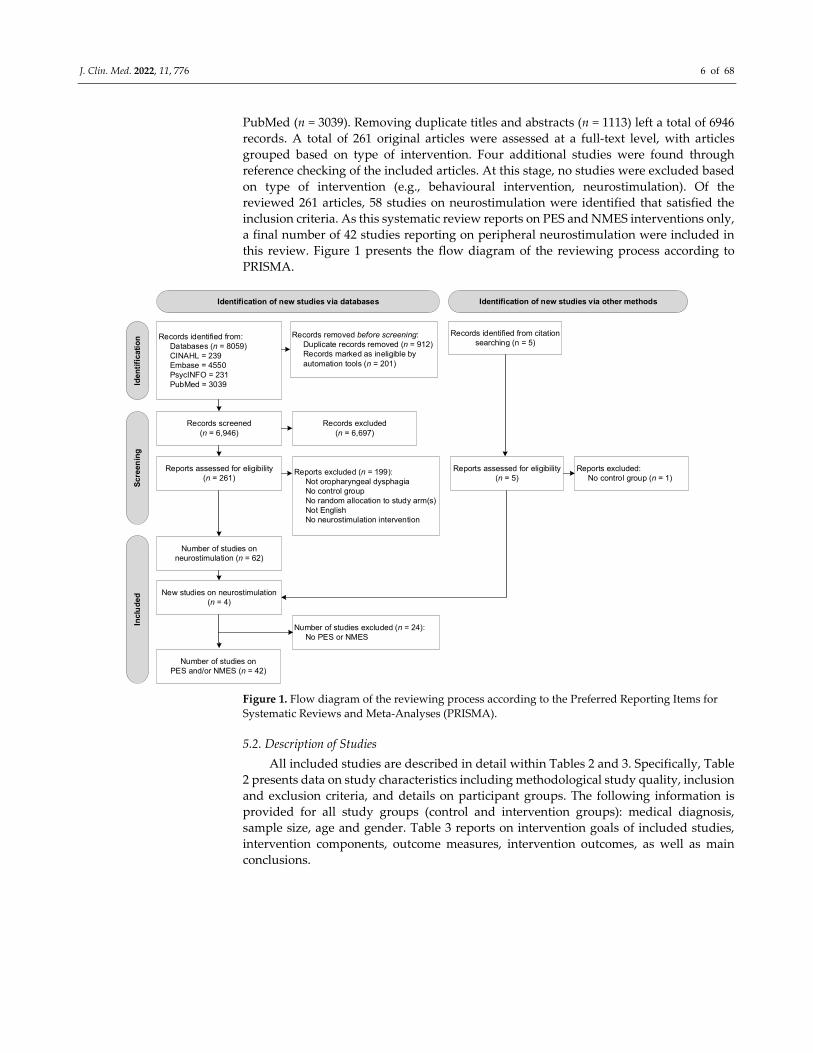
J. Clin. Med. 2022, 11, 776 6 of 68
PubMed (n = 3039). Removing duplicate titles and abstracts (n = 1113) left a total of 6946
records. A total of 261 original articles were assessed at a full‐text level, with articles
grouped based on type of intervention. Four additional studies were found through
reference checking of the included articles. At this stage, no studies were excluded based
on type of intervention (e.g., behavioural intervention, neurostimulation). Of the
reviewed 261 articles, 58 studies on neurostimulation were identified that satisfied the
inclusion criteria. As this systematic review reports on PES and NMES interventions only,
a final number of 42 studies reporting on peripheral neurostimulation were included in
this review. Figure 1 presents the flow diagram of the reviewing process according to
PRISMA.
Identification of new studies via databases Identification of new studies via other methods
Iden
tifi
cati
on
Scr
een
ing
Incl
ud
ed
Records identified from:Databases (n = 8059)CINAHL = 239Embase = 4550PsycINFO = 231PubMed = 3039
Records removed before screening:Duplicate records removed (n = 912)Records marked as ineligible by automation tools (n = 201)
Records identified from citation searching (n = 5)
Records screened (n = 6,946)
Records excluded(n = 6,697)
Reports assessed for eligibility(n = 261)
Reports excluded (n = 199):Not oropharyngeal dysphagiaNo control groupNo random allocation to study arm(s)Not EnglishNo neurostimulation intervention
Reports assessed for eligibility(n = 5)
Number of studies on neurostimulation (n = 62)
New studies on neurostimulation (n = 4)
Number of studies on PES and/or NMES (n = 42)
Reports excluded:No control group (n = 1)
Number of studies excluded (n = 24):No PES or NMES
Figure 1. Flow diagram of the reviewing process according to the Preferred Reporting Items for
Systematic Reviews and Meta‐Analyses (PRISMA).
5.2. Description of Studies
All included studies are described in detail within Tables 2 and 3. Specifically, Table
2 presents data on study characteristics including methodological study quality, inclusion
and exclusion criteria, and details on participant groups. The following information is
provided for all study groups (control and intervention groups): medical diagnosis,
sample size, age and gender. Table 3 reports on intervention goals of included studies,
intervention components, outcome measures, intervention outcomes, as well as main
conclusions.
J. Clin. Med. 2022, 11, 776 7 of 68
Table 2. Study characteristics of studies on NMES and PES interventions for people with oropharyngeal dysphagia.
Study
Country Inclusion/Exclusion Criteria
Sample (N)
Groups
Group Descriptives (Mean ± SD)
Age, Gender, Medical Diagnoses
NeuroMuscular Electrical Stimulation (NMES) a ― n = 30
Beom, et al. [28]
Country: Korea
OD as per VFSS
Inclusion: stroke, traumatic brain injury or brain
tumour >1 week ago; hemiplegia caused by hemispheric
lesion; able to respond to pain
Exclusion: no potential for recovery; severe
communication difficulties; contraindications for
neuromuscular electrical stimulation (NMES)
n = 132
Treatment group 1 (66), 50%
NMES (Suprahyoid muscle
stimulation) + DT [Denoted as ‘Beom
et al. (2015a)’ in Figure 4.]
Treatment group 2 (66), 50%
NMES (suprahyoid and infrahyoid
muscles stimulation) + DT [Denoted as
‘Beom et al. (2015b)’ in Figure 4.]
Treatment group 1: Age 64.4 ± 12.0
50% male
Location of lesion: cortex (29),
subcortex (20), brainstem (16),
cerebellum (1)
Treatment group 2: Age 59.8 ± 15.9
66.6% male
Location of lesion: cortex (29),
subcortex (14), brainstem (19),
cerebellum (4)
NS difference between groups
Bülow, et al. [29]
Country: Sweden,
The Netherlands,
France
OD as per VFSS
Inclusion: 50–80 years old; hemispheric stroke > 3 months;
ability to swallow: ability to communicate
Exclusion: brainstem involvement; progressive
cerebrovascular disease; other neurologic diseases such as
ALS, MS, or Parkinson’s disease; patients with tumors of
the swallowing apparatus + radiotherapy/surgery to the
neck; patients with no pharyngeal swallow; nasogastric
tube insitu
n = 25
Treatment group 1 (13), 52%
DT
Treatment group 2 (12), 48%
NMES (suprahyoid and infrahyoid
muscles stimulation) + Diet
modification
Combined treatment groups data:
64% male
Treatment group 1: Age 71 (SD not
reported)
Treatment group 2: Age 70
Statistical difference between groups =
NR
El‐Tamawy, et al. [30]
Country: Egypt
OD as per bedside screening (confirmed by VFSS once
enrolled)
Inclusion: acute stroke, severe dysphagia; able to
ambulate; normal attention and communication skills; no
other neurological disease; able to perform sit to stand test
n = 30
Treatment group 1 (15), 50%
NMES + DT + (Medical treatment)
Control group 2 (15), 50%
Medical treatment
Treatment group 1: Age 61.5 ± 7.3
Control group 2: Age 61.3 ± 6.6
No further details on subjects within
the groups.
J. Clin. Med. 2022, 11, 776 8 of 68
Exclusion: disturbed level of consciousness; dementia;
psychiatric disorders; syncope; previous operation or
injury to the head and neck area
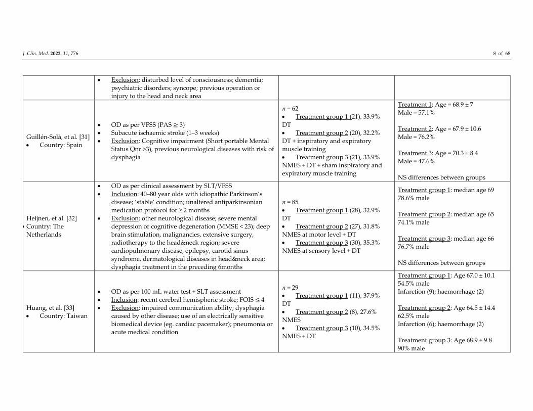
Guillén‐Solà, et al. [31]
Country: Spain
OD as per VFSS (PAS 3)
Subacute ischaemic stroke (1–3 weeks)
Exclusion: Cognitive impairment (Short portable Mental
Status Qnr >3), previous neurological diseases with risk of
dysphagia
n = 62
Treatment group 1 (21), 33.9%
DT
Treatment group 2 (20), 32.2%
DT + inspiratory and expiratory
muscle training
Treatment group 3 (21), 33.9%
NMES + DT + sham inspiratory and
expiratory muscle training
Treatment 1: Age = 68.9 ± 7
Male = 57.1%
Treatment 2: Age = 67.9 ± 10.6
Male = 76.2%
Treatment 3: Age = 70.3 ± 8.4
Male = 47.6%
NS differences between groups
Heijnen, et al. [32]
Country: The Netherlands
OD as per clinical assessment by SLT/VFSS
Inclusion: 40–80 year olds with idiopathic Parkinson’s
disease; ‘stable’ condition; unaltered antiparkinsonian
medication protocol for ≥ 2 months
Exclusion: other neurological disease; severe mental
depression or cognitive degeneration (MMSE < 23); deep
brain stimulation, malignancies, extensive surgery,
radiotherapy to the head&neck region; severe
cardiopulmonary disease, epilepsy, carotid sinus
syndrome, dermatological diseases in head&neck area;
dysphagia treatment in the preceding 6months
n = 85
Treatment group 1 (28), 32.9%
DT
Treatment group 2 (27), 31.8%
NMES at motor level + DT
Treatment group 3 (30), 35.3%
NMES at sensory level + DT
Treatment group 1: median age 69
78.6% male
Treatment group 2: median age 65
74.1% male
Treatment group 3: median age 66
76.7% male
NS differences between groups
Huang, et al. [33]
Country: Taiwan
OD as per 100 mL water test + SLT assessment
Inclusion: recent cerebral hemispheric stroke; FOIS 4 Exclusion: impaired communication ability; dysphagia
caused by other disease; use of an electrically sensitive
biomedical device (eg. cardiac pacemaker); pneumonia or
acute medical condition
n = 29
Treatment group 1 (11), 37.9%
DT
Treatment group 2 (8), 27.6%
NMES
Treatment group 3 (10), 34.5%
NMES + DT
Treatment group 1: Age 67.0 ± 10.1
54.5% male
Infarction (9); haemorrhage (2)
Treatment group 2: Age 64.5 ± 14.4
62.5% male
Infarction (6); haemorrhage (2)
Treatment group 3: Age 68.9 ± 9.8
90% male
J. Clin. Med. 2022, 11, 776 9 of 68
Infarction (9); haemorrhage (1)
NS differences between groups
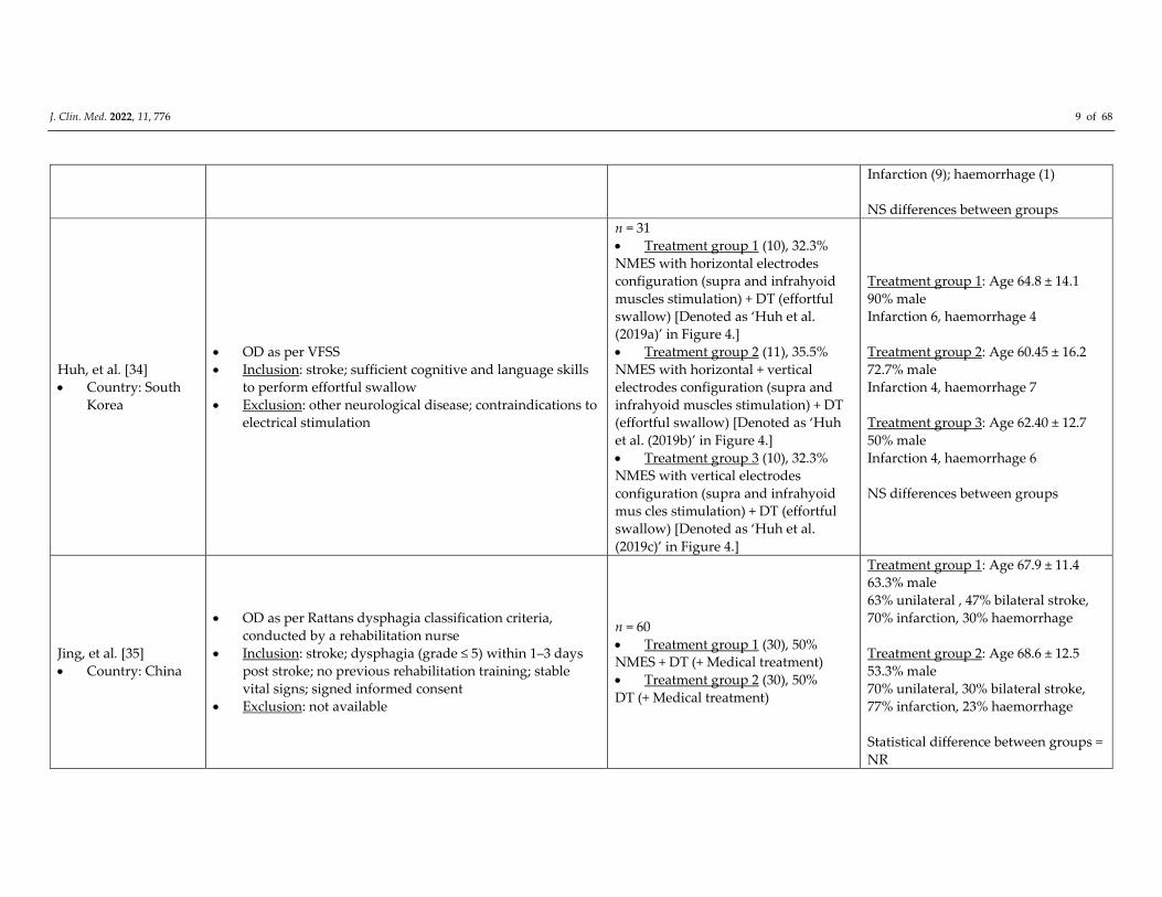
Huh, et al. [34]
Country: South
Korea
OD as per VFSS
Inclusion: stroke; sufficient cognitive and language skills
to perform effortful swallow
Exclusion: other neurological disease; contraindications to
electrical stimulation
n = 31
Treatment group 1 (10), 32.3%
NMES with horizontal electrodes
configuration (supra and infrahyoid
muscles stimulation) + DT (effortful
swallow) [Denoted as ‘Huh et al.
(2019a)’ in Figure 4.]
Treatment group 2 (11), 35.5%
NMES with horizontal + vertical
electrodes configuration (supra and
infrahyoid muscles stimulation) + DT
(effortful swallow) [Denoted as ‘Huh
et al. (2019b)’ in Figure 4.]
Treatment group 3 (10), 32.3%
NMES with vertical electrodes
configuration (supra and infrahyoid
mus cles stimulation) + DT (effortful
swallow) [Denoted as ‘Huh et al.
(2019c)’ in Figure 4.]
Treatment group 1: Age 64.8 ± 14.1
90% male
Infarction 6, haemorrhage 4
Treatment group 2: Age 60.45 ± 16.2
72.7% male
Infarction 4, haemorrhage 7
Treatment group 3: Age 62.40 ± 12.7
50% male
Infarction 4, haemorrhage 6
NS differences between groups
Jing, et al. [35]
Country: China
OD as per Rattans dysphagia classification criteria,
conducted by a rehabilitation nurse
Inclusion: stroke; dysphagia (grade ≤ 5) within 1–3 days
post stroke; no previous rehabilitation training; stable
vital signs; signed informed consent
Exclusion: not available
n = 60
Treatment group 1 (30), 50%
NMES + DT (+ Medical treatment)
Treatment group 2 (30), 50%
DT (+ Medical treatment)
Treatment group 1: Age 67.9 ± 11.4
63.3% male
63% unilateral , 47% bilateral stroke,
70% infarction, 30% haemorrhage
Treatment group 2: Age 68.6 ± 12.5
53.3% male
70% unilateral, 30% bilateral stroke,
77% infarction, 23% haemorrhage
Statistical difference between groups =
NR
J. Clin. Med. 2022, 11, 776 10 of 68
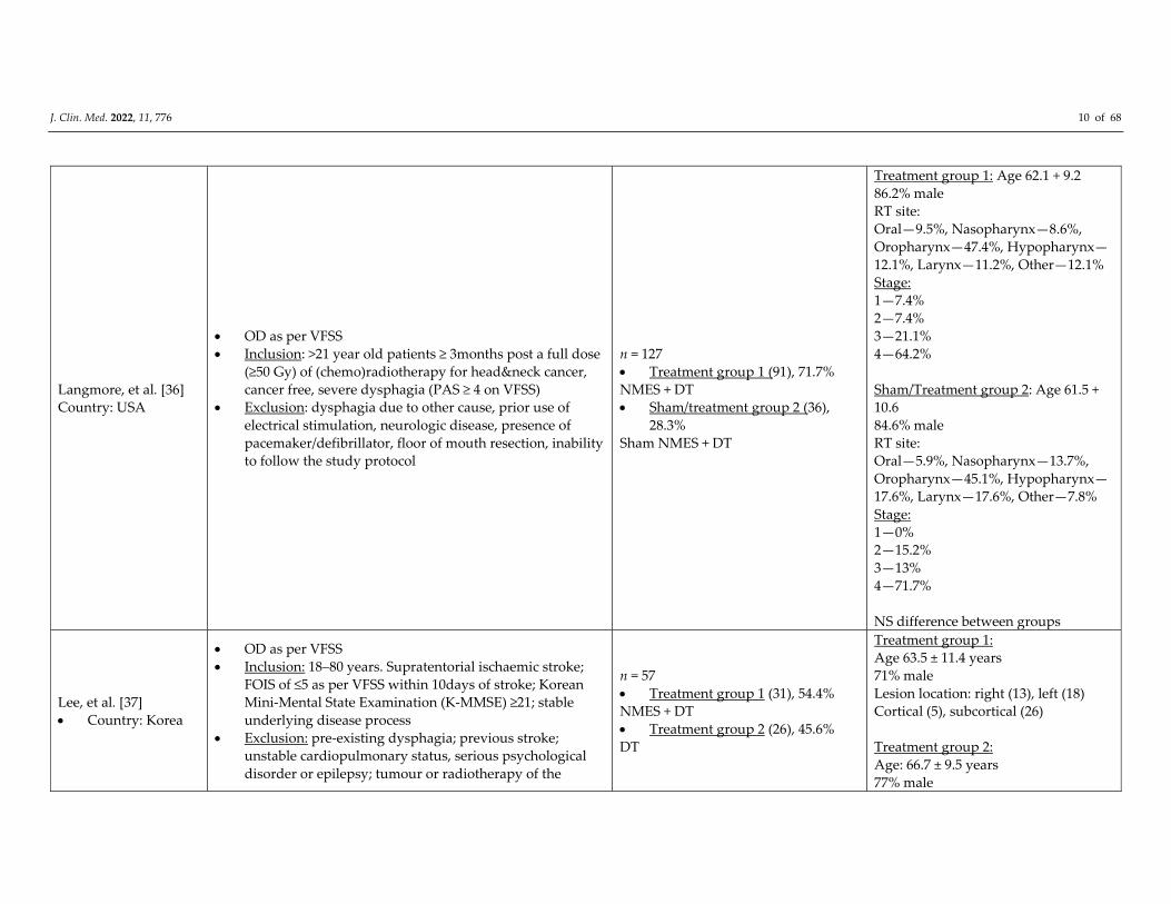
Langmore, et al. [36]
Country: USA
OD as per VFSS
Inclusion: >21 year old patients ≥ 3months post a full dose
(≥50 Gy) of (chemo)radiotherapy for head&neck cancer,
cancer free, severe dysphagia (PAS ≥ 4 on VFSS)
Exclusion: dysphagia due to other cause, prior use of
electrical stimulation, neurologic disease, presence of
pacemaker/defibrillator, floor of mouth resection, inability
to follow the study protocol
n = 127
Treatment group 1 (91), 71.7%
NMES + DT
Sham/treatment group 2 (36),
28.3%
Sham NMES + DT
Treatment group 1: Age 62.1 + 9.2
86.2% male
RT site:
Oral—9.5%, Nasopharynx—8.6%,
Oropharynx—47.4%, Hypopharynx—
12.1%, Larynx—11.2%, Other—12.1%
Stage:
1—7.4%
2—7.4%
3—21.1%
4—64.2%
Sham/Treatment group 2: Age 61.5 +
10.6
84.6% male
RT site:
Oral—5.9%, Nasopharynx—13.7%,
Oropharynx—45.1%, Hypopharynx—
17.6%, Larynx—17.6%, Other—7.8%
Stage:
1—0%
2—15.2%
3—13%
4—71.7%
NS difference between groups
Lee, et al. [37]
Country: Korea
OD as per VFSS
Inclusion: 18–80 years. Supratentorial ischaemic stroke;
FOIS of ≤5 as per VFSS within 10days of stroke; Korean
Mini‐Mental State Examination (K‐MMSE) ≥21; stable
underlying disease process
Exclusion: pre‐existing dysphagia; previous stroke;
unstable cardiopulmonary status, serious psychological
disorder or epilepsy; tumour or radiotherapy of the
n = 57
Treatment group 1 (31), 54.4%
NMES + DT
Treatment group 2 (26), 45.6%
DT
Treatment group 1:
Age 63.5 ± 11.4 years
71% male
Lesion location: right (13), left (18)
Cortical (5), subcortical (26)
Treatment group 2:
Age: 66.7 ± 9.5 years
77% male
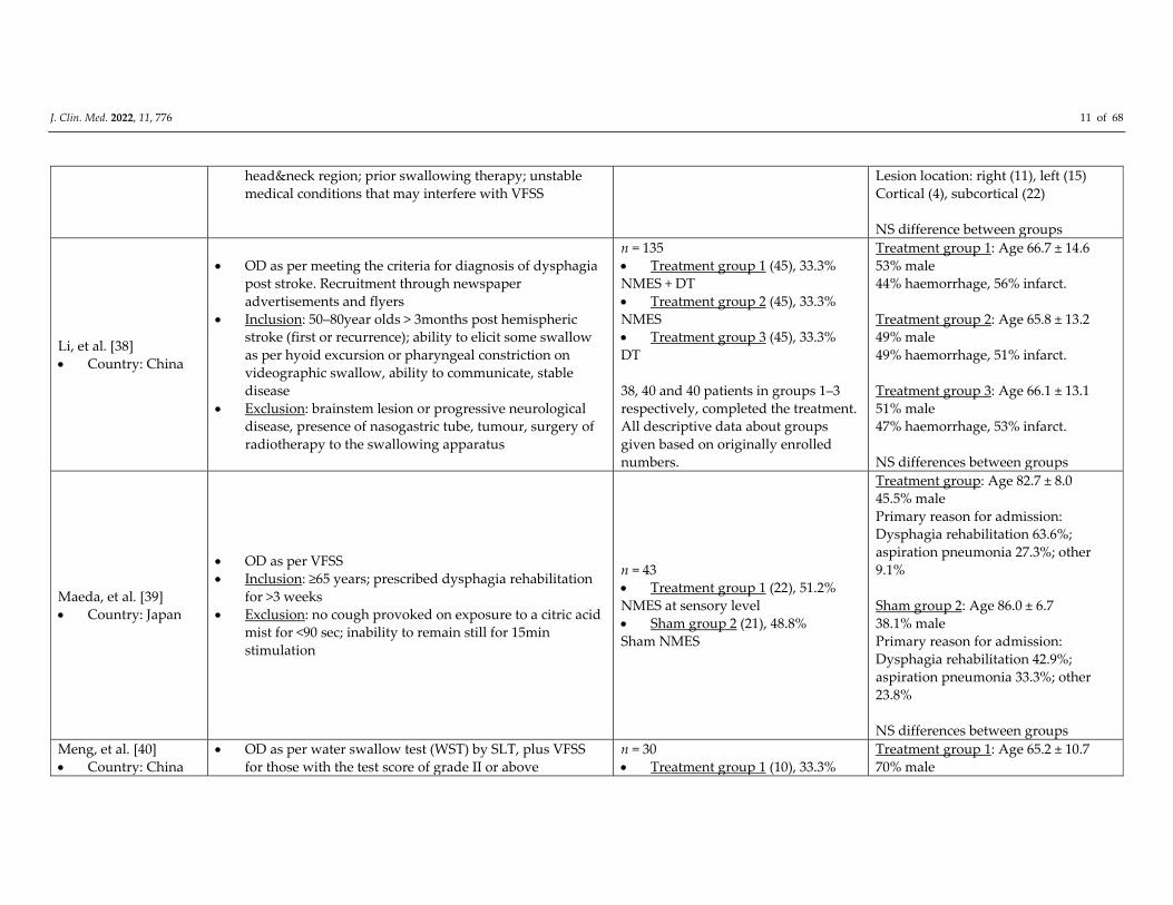
J. Clin. Med. 2022, 11, 776 11 of 68
head&neck region; prior swallowing therapy; unstable
medical conditions that may interfere with VFSS
Lesion location: right (11), left (15)
Cortical (4), subcortical (22)
NS difference between groups
Li, et al. [38]
Country: China
OD as per meeting the criteria for diagnosis of dysphagia
post stroke. Recruitment through newspaper
advertisements and flyers
Inclusion: 50–80year olds > 3months post hemispheric
stroke (first or recurrence); ability to elicit some swallow
as per hyoid excursion or pharyngeal constriction on
videographic swallow, ability to communicate, stable
disease
Exclusion: brainstem lesion or progressive neurological
disease, presence of nasogastric tube, tumour, surgery of
radiotherapy to the swallowing apparatus
n = 135
Treatment group 1 (45), 33.3%
NMES + DT
Treatment group 2 (45), 33.3%
NMES
Treatment group 3 (45), 33.3%
DT
38, 40 and 40 patients in groups 1–3
respectively, completed the treatment.
All descriptive data about groups
given based on originally enrolled
numbers.
Treatment group 1: Age 66.7 ± 14.6
53% male
44% haemorrhage, 56% infarct.
Treatment group 2: Age 65.8 ± 13.2
49% male
49% haemorrhage, 51% infarct.
Treatment group 3: Age 66.1 ± 13.1
51% male
47% haemorrhage, 53% infarct.
NS differences between groups
Maeda, et al. [39]
Country: Japan
OD as per VFSS
Inclusion: ≥65 years; prescribed dysphagia rehabilitation
for >3 weeks
Exclusion: no cough provoked on exposure to a citric acid
mist for <90 sec; inability to remain still for 15min
stimulation
n = 43
Treatment group 1 (22), 51.2%
NMES at sensory level
Sham group 2 (21), 48.8%
Sham NMES
Treatment group: Age 82.7 ± 8.0
45.5% male
Primary reason for admission:
Dysphagia rehabilitation 63.6%;
aspiration pneumonia 27.3%; other
9.1%
Sham group 2: Age 86.0 ± 6.7
38.1% male
Primary reason for admission:
Dysphagia rehabilitation 42.9%;
aspiration pneumonia 33.3%; other
23.8%
NS differences between groups
Meng, et al. [40]
Country: China
OD as per water swallow test (WST) by SLT, plus VFSS
for those with the test score of grade II or above
n = 30
Treatment group 1 (10), 33.3%
Treatment group 1: Age 65.2 ± 10.7
70% male
J. Clin. Med. 2022, 11, 776 12 of 68
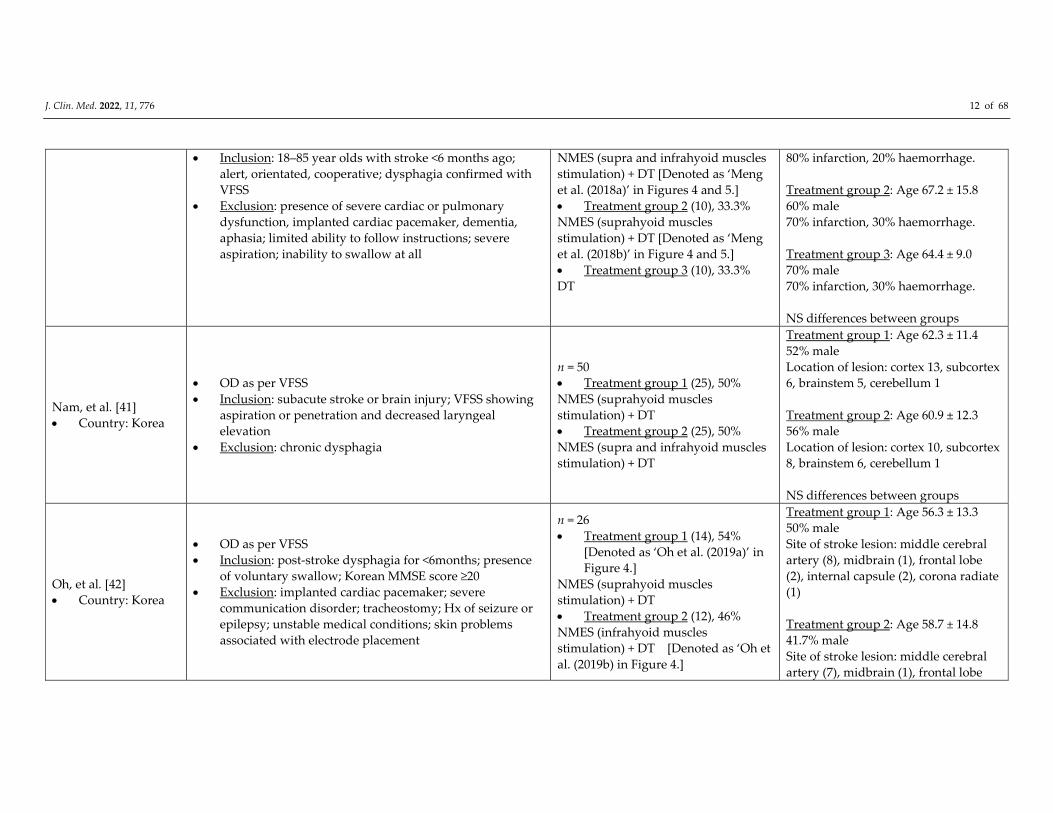
Inclusion: 18–85 year olds with stroke <6 months ago;
alert, orientated, cooperative; dysphagia confirmed with
VFSS
Exclusion: presence of severe cardiac or pulmonary
dysfunction, implanted cardiac pacemaker, dementia,
aphasia; limited ability to follow instructions; severe
aspiration; inability to swallow at all
NMES (supra and infrahyoid muscles
stimulation) + DT [Denoted as ‘Meng
et al. (2018a)’ in Figures 4 and 5.]
Treatment group 2 (10), 33.3%
NMES (suprahyoid muscles
stimulation) + DT [Denoted as ‘Meng
et al. (2018b)’ in Figure 4 and 5.]
Treatment group 3 (10), 33.3%
DT
80% infarction, 20% haemorrhage.
Treatment group 2: Age 67.2 ± 15.8
60% male
70% infarction, 30% haemorrhage.
Treatment group 3: Age 64.4 ± 9.0
70% male
70% infarction, 30% haemorrhage.
NS differences between groups
Nam, et al. [41]
Country: Korea
OD as per VFSS
Inclusion: subacute stroke or brain injury; VFSS showing
aspiration or penetration and decreased laryngeal
elevation
Exclusion: chronic dysphagia
n = 50
Treatment group 1 (25), 50%
NMES (suprahyoid muscles
stimulation) + DT
Treatment group 2 (25), 50%
NMES (supra and infrahyoid muscles
stimulation) + DT
Treatment group 1: Age 62.3 ± 11.4
52% male
Location of lesion: cortex 13, subcortex
6, brainstem 5, cerebellum 1
Treatment group 2: Age 60.9 ± 12.3
56% male
Location of lesion: cortex 10, subcortex
8, brainstem 6, cerebellum 1
NS differences between groups
Oh, et al. [42]
Country: Korea
OD as per VFSS
Inclusion: post‐stroke dysphagia for <6months; presence
of voluntary swallow; Korean MMSE score ≥20
Exclusion: implanted cardiac pacemaker; severe
communication disorder; tracheostomy; Hx of seizure or
epilepsy; unstable medical conditions; skin problems
associated with electrode placement
n = 26
Treatment group 1 (14), 54%
[Denoted as ‘Oh et al. (2019a)’ in
Figure 4.]
NMES (suprahyoid muscles
stimulation) + DT
Treatment group 2 (12), 46%
NMES (infrahyoid muscles
stimulation) + DT [Denoted as ‘Oh et
al. (2019b) in Figure 4.]
Treatment group 1: Age 56.3 ± 13.3
50% male
Site of stroke lesion: middle cerebral
artery (8), midbrain (1), frontal lobe
(2), internal capsule (2), corona radiate
(1)
Treatment group 2: Age 58.7 ± 14.8
41.7% male
Site of stroke lesion: middle cerebral
artery (7), midbrain (1), frontal lobe
J. Clin. Med. 2022, 11, 776 13 of 68
(1), internal capsule (2), corona radiate
(1)
NS differences between groups
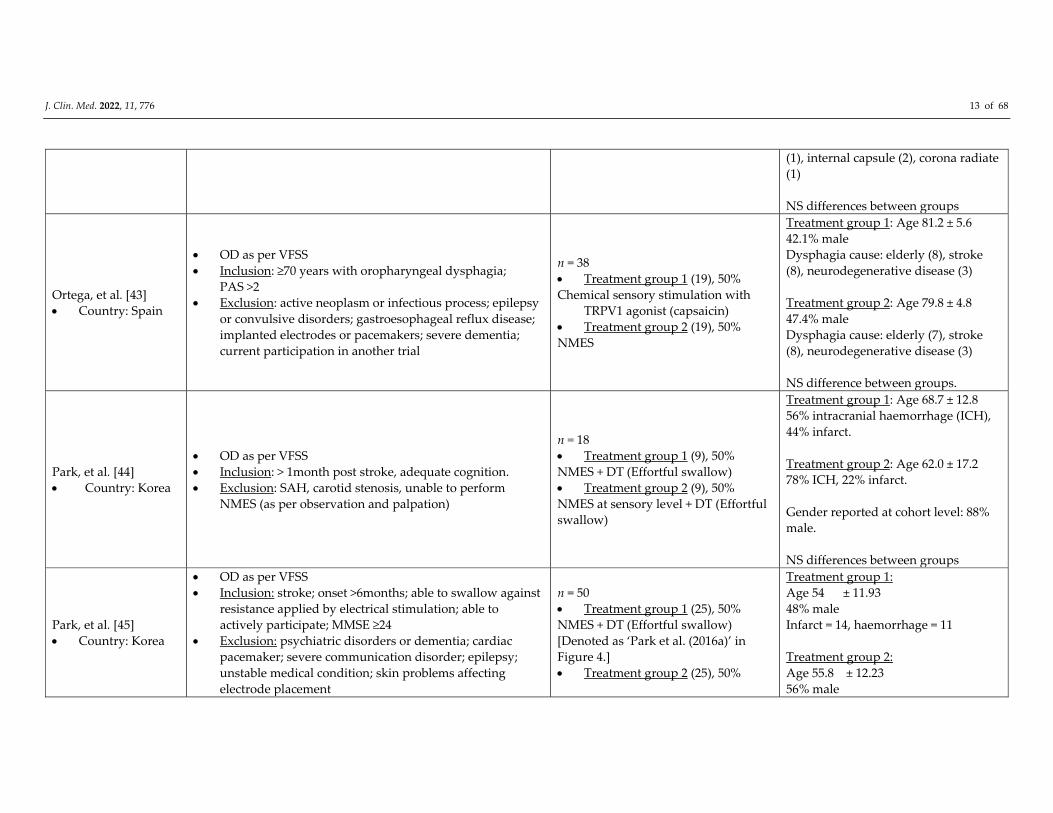
Ortega, et al. [43]
Country: Spain
OD as per VFSS
Inclusion: ≥70 years with oropharyngeal dysphagia;
PAS >2
Exclusion: active neoplasm or infectious process; epilepsy
or convulsive disorders; gastroesophageal reflux disease;
implanted electrodes or pacemakers; severe dementia;
current participation in another trial
n = 38
Treatment group 1 (19), 50%
Chemical sensory stimulation with
TRPV1 agonist (capsaicin)
Treatment group 2 (19), 50%
NMES
Treatment group 1: Age 81.2 ± 5.6
42.1% male
Dysphagia cause: elderly (8), stroke
(8), neurodegenerative disease (3)
Treatment group 2: Age 79.8 ± 4.8
47.4% male
Dysphagia cause: elderly (7), stroke
(8), neurodegenerative disease (3)
NS difference between groups.
Park, et al. [44]
Country: Korea
OD as per VFSS
Inclusion: > 1month post stroke, adequate cognition.
Exclusion: SAH, carotid stenosis, unable to perform
NMES (as per observation and palpation)
n = 18
Treatment group 1 (9), 50%
NMES + DT (Effortful swallow)
Treatment group 2 (9), 50%
NMES at sensory level + DT (Effortful
swallow)
Treatment group 1: Age 68.7 ± 12.8
56% intracranial haemorrhage (ICH),
44% infarct.
Treatment group 2: Age 62.0 ± 17.2
78% ICH, 22% infarct.
Gender reported at cohort level: 88%
male.
NS differences between groups
Park, et al. [45]
Country: Korea
OD as per VFSS
Inclusion: stroke; onset >6months; able to swallow against
resistance applied by electrical stimulation; able to
actively participate; MMSE ≥24
Exclusion: psychiatric disorders or dementia; cardiac
pacemaker; severe communication disorder; epilepsy;
unstable medical condition; skin problems affecting
electrode placement
n = 50
Treatment group 1 (25), 50%
NMES + DT (Effortful swallow)
[Denoted as ‘Park et al. (2016a)’ in
Figure 4.]
Treatment group 2 (25), 50%
Treatment group 1:
Age 54 ± 11.93
48% male
Infarct = 14, haemorrhage = 11
Treatment group 2:
Age 55.8 ± 12.23
56% male
J. Clin. Med. 2022, 11, 776 14 of 68
NMES at sensory level + DT (Effortful
swallow) [Denoted as ‘Park et al.
(2016b)’ in Figure 4.]
Infarct = 12, haemorrhage = 13
NS differences between groups
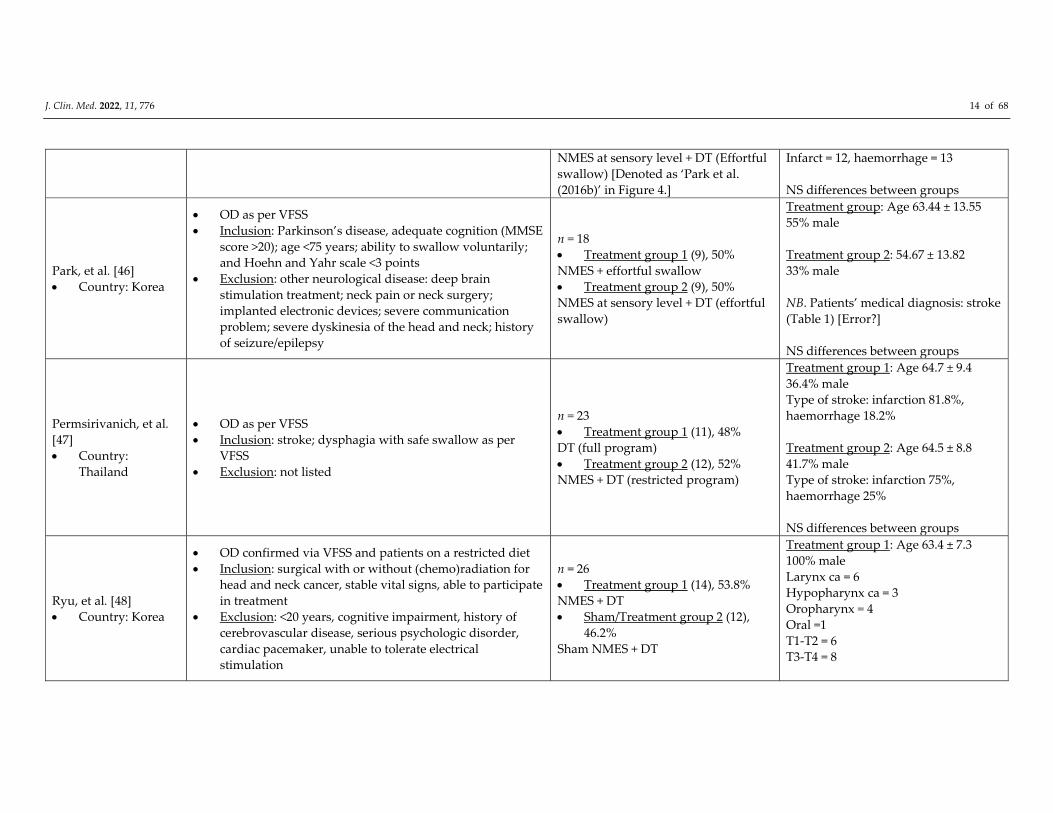
Park, et al. [46]
Country: Korea
OD as per VFSS
Inclusion: Parkinson’s disease, adequate cognition (MMSE
score >20); age <75 years; ability to swallow voluntarily;
and Hoehn and Yahr scale <3 points
Exclusion: other neurological disease: deep brain
stimulation treatment; neck pain or neck surgery;
implanted electronic devices; severe communication
problem; severe dyskinesia of the head and neck; history
of seizure/epilepsy
n = 18
Treatment group 1 (9), 50%
NMES + effortful swallow
Treatment group 2 (9), 50%
NMES at sensory level + DT (effortful
swallow)
Treatment group: Age 63.44 ± 13.55
55% male
Treatment group 2: 54.67 ± 13.82
33% male
NB. Patients’ medical diagnosis: stroke
(Table 1) [Error?]
NS differences between groups
Permsirivanich, et al.
[47]
Country:
Thailand
OD as per VFSS
Inclusion: stroke; dysphagia with safe swallow as per
VFSS
Exclusion: not listed
n = 23
Treatment group 1 (11), 48%
DT (full program)
Treatment group 2 (12), 52%
NMES + DT (restricted program)
Treatment group 1: Age 64.7 ± 9.4
36.4% male
Type of stroke: infarction 81.8%,
haemorrhage 18.2%
Treatment group 2: Age 64.5 ± 8.8
41.7% male
Type of stroke: infarction 75%,
haemorrhage 25%
NS differences between groups
Ryu, et al. [48]
Country: Korea
OD confirmed via VFSS and patients on a restricted diet
Inclusion: surgical with or without (chemo)radiation for
head and neck cancer, stable vital signs, able to participate
in treatment
Exclusion: <20 years, cognitive impairment, history of
cerebrovascular disease, serious psychologic disorder,
cardiac pacemaker, unable to tolerate electrical
stimulation
n = 26
Treatment group 1 (14), 53.8%
NMES + DT
Sham/Treatment group 2 (12),
46.2%
Sham NMES + DT
Treatment group 1: Age 63.4 ± 7.3
100% male
Larynx ca = 6
Hypopharynx ca = 3
Oropharynx = 4
Oral =1
T1‐T2 = 6
T3‐T4 = 8
J. Clin. Med. 2022, 11, 776 15 of 68
Sham/Treatment group 2: Age 60.8 ±
12.0
92% male
Larynx ca = 5
Hypopharynx ca = 1
Oropharynx = 4
Oral =2
T1‐T2 = 7
T3‐T4 = 4
Unknown = 1
Statistical difference between groups =
NR
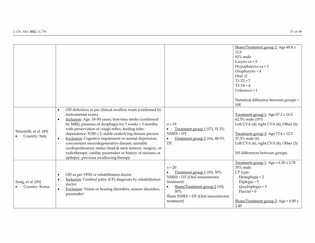
Simonelli, et al. [49]
Country: Italy
OD definition as per clinical swallow exam (confirmed by
instrumental exam)
Inclusion: Age: 18–85 years; first‐time stroke (confirmed
by MRI); presence of dysphagia for 3 weeks > 3 months,
with preservation of cough reflex; feeding tube‐
dependence, FOIS ≤ 2; stable underlying disease process
Exclusion: Cognitive impairment or mental depression;
concomitant neurodegenerative disease; unstable
cardiopulmonary status; head & neck tumour, surgery, or
radiotherapy; cardiac pacemaker or history of seizures or
epilepsy; previous swallowing therapy
n = 33
Treatment group 1 (17), 51.5%
NMES + DT
Treatment group 2 (16), 48.5%
DT
Treatment group 1: Age 67.2 ± 16.2
62.5% male (10?)
Left CVA (4), right CVA (6), Other (6)
Treatment group 2: Age 72.4 ± 12.3
37.5% male (6)
Left CVA (6), right CVA (6), Other (3)
NS differences between groups.
Song, et al. [50]
Country: Korea
OD as per VFSS or rehabilitation doctor
Inclusion: Cerebral palsy (CP) diagnosis by rehabilitation
doctor
Exclusion: Vision or hearing disorders, seizure disorders,
pacemaker
n = 20
Treatment group 1 (10), 50%
NMES + DT (Oral sensorimotor
treatment)
Sham/Treatment group 2 (10),
50%
Sham NMES + DT (Oral sensorimotor
treatment)
Treatment group 1: Age = 6.20 ± 2.78
70% male
CP type:
Hemiplegia = 2
Diplegia = 5
Quadriplegia = 3
Flaccid = 0
Sham/Treatment group 2: Age = 6.00 ±
2.40
J. Clin. Med. 2022, 11, 776 16 of 68
60% male
CP type:
Hemiplegia = 4
Diplegia = 3
Quadriplegia = 2
Flaccid = 1
NS differences between groups.
Sproson, et al. [51]
Country: UK
OD as per VFSS
Inclusion: medically stable; >1 month post‐stroke; no other
neurological disease; dysphagia incorporating reduced
laryngeal elevation (confirmed by VFSS)
Exclusion: <18 years; pacemaker; serious cardiac disease;
severe cognitive/communication difficulties;
lesions/infections in the treatment site
n = 24
Treatment group 1 (12), 50%
NMES + DT
Usual care group 2 (12), 50%
Usual care (Different from DT)
Treatment group 1: Age 76 ± 11.4
67% male
33% >1 stroke event
Time post‐stroke 17.3 months ± 25.0
Usual care group 2: Age 79 ± 11.4
66.7% male
33% >1 stroke event
Time post‐stroke 9.1 months ± 20.5
Significant difference between groups
= NR.
Terré and Mearin [52]
Country: Spain
OD as per VFSS demonstrating aspiration
Inclusion: >18 years, acquired brain injury (stroke, TBI); <6
months since insult; able to understand and follow
instructions for treatment; medically stable
Exclusion: previous stroke or TBI; previous dysphagia
secondary to other aetiology; no other metabolic or
neurological diseases
n = 20
Treatment group 1 (10), 50%
NMES + DT
Sham/Treatment group 2 (10),
50%
Sham NMES + DT
Treatment group 1: Age 46.0 ± 16
60% male
70% stroke, (haemorrhagic = 5,
ischaemic = 2)
30% TBI
Sham/Treatment group 2: Age 51 ± 23
60% male
70% stroke (haemorrhagic = 6,
ischaemic = 1)
30% TBI
Significant difference between groups
= NR.
J. Clin. Med. 2022, 11, 776 17 of 68
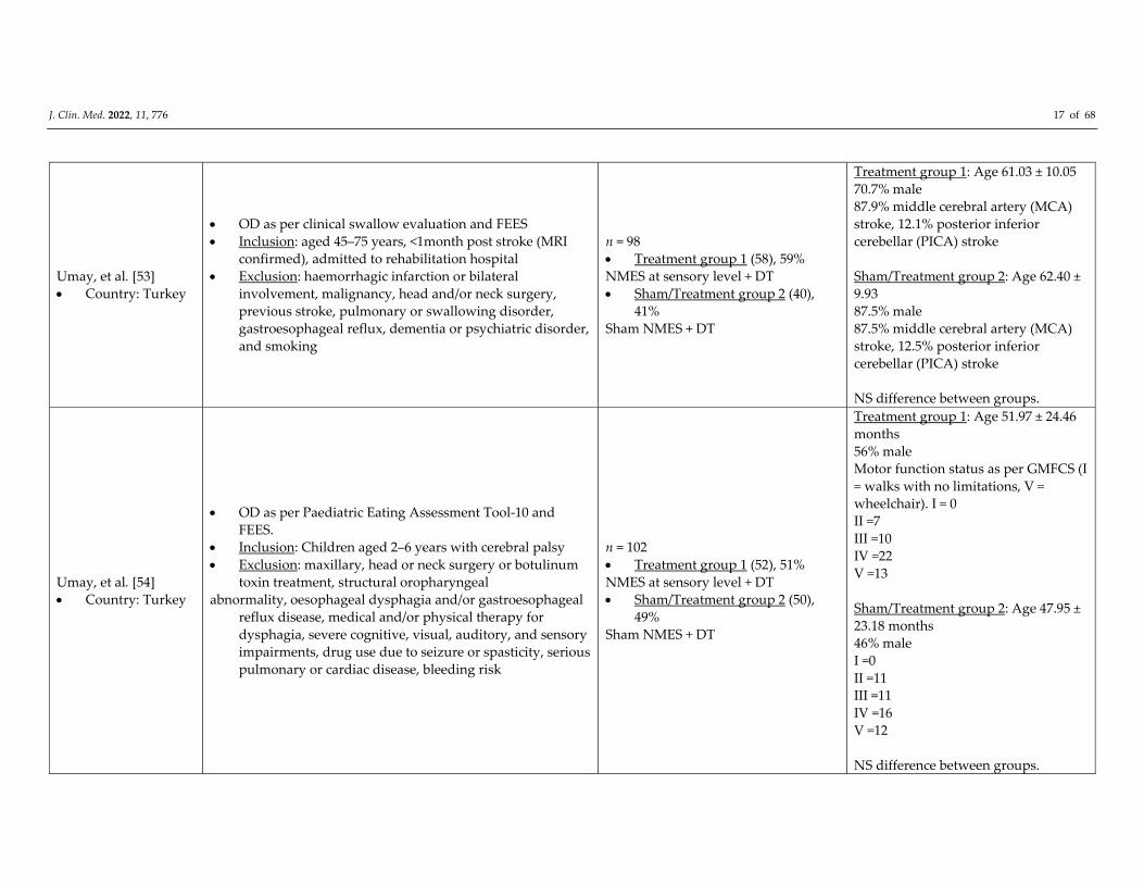
Umay, et al. [53]
Country: Turkey
OD as per clinical swallow evaluation and FEES
Inclusion: aged 45–75 years, <1month post stroke (MRI
confirmed), admitted to rehabilitation hospital
Exclusion: haemorrhagic infarction or bilateral
involvement, malignancy, head and/or neck surgery,
previous stroke, pulmonary or swallowing disorder,
gastroesophageal reflux, dementia or psychiatric disorder,
and smoking
n = 98
Treatment group 1 (58), 59%
NMES at sensory level + DT
Sham/Treatment group 2 (40),
41%
Sham NMES + DT
Treatment group 1: Age 61.03 ± 10.05
70.7% male
87.9% middle cerebral artery (MCA)
stroke, 12.1% posterior inferior
cerebellar (PICA) stroke
Sham/Treatment group 2: Age 62.40 ±
9.93
87.5% male
87.5% middle cerebral artery (MCA)
stroke, 12.5% posterior inferior
cerebellar (PICA) stroke
NS difference between groups.
Umay, et al. [54]
Country: Turkey
OD as per Paediatric Eating Assessment Tool‐10 and
FEES.
Inclusion: Children aged 2–6 years with cerebral palsy
Exclusion: maxillary, head or neck surgery or botulinum
toxin treatment, structural oropharyngeal
abnormality, oesophageal dysphagia and/or gastroesophageal
reflux disease, medical and/or physical therapy for
dysphagia, severe cognitive, visual, auditory, and sensory
impairments, drug use due to seizure or spasticity, serious
pulmonary or cardiac disease, bleeding risk
n = 102
Treatment group 1 (52), 51%
NMES at sensory level + DT
Sham/Treatment group 2 (50),
49%
Sham NMES + DT
Treatment group 1: Age 51.97 ± 24.46
months
56% male
Motor function status as per GMFCS (I
= walks with no limitations, V =
wheelchair). I = 0
II =7
III =10
IV =22
V =13
Sham/Treatment group 2: Age 47.95 ±
23.18 months
46% male
I =0
II =11
III =11
IV =16
V =12
NS difference between groups.
J. Clin. Med. 2022, 11, 776 18 of 68
Xia, et al. [55]
Country: China
OD as per Standardised Swallow Assessment (SSA) and
VFSS
Inclusion: cerebral infarction or haemorrhage (diagnosed
by CT or MRI); no pulmonary diseases; 40–80 years old;
cognitively intact and able to cooperate
Exclusion: None
n = 120
Treatment group 1 (40), 33.3%
DT
Treatment group 2 (40), 33.3%
NMES [Denoted as ‘Xia et al. (2011a)’
in Figure 4 and 5.]
Treatment group 3 (40), 33.3%
NMES + DT [Denoted as ‘Xia et al.
(2011b)’ in Figures 4 and 5.]
Treatment group 1: Age 65.32 ± 14.29
62.5% male
42.5% haemorrhage, 45% infarct,
12.5% other stroke.
Treatment group 2: Age 66.40 ± 15.63
57.5% male
35% haemorrhage, 55% infarct, 10%
other stroke.
Treatment group 3: Age 65.85 ± 14.63
70% male
32.5% haemorrhage, 62.5% infarct,
0.5% other stroke.
NS difference between groups.
Zeng, et al. [56]
Country: China
OD as per Kubota water‐drinking test
Inclusion: first‐onset stroke (confirmed via MRI); able to
actively cooperate; no significant cognitive disorder,
aphasia, or other diseases affecting understanding
Exclusion: critical condition or vital organ failure; cardiac
pacemaker; metal implants or internal orthotics;
comorbidities of malignant tumours, skin damage, heart
disease, acute seizure/epilepsy, peripheral nerve damage
n = 112
Treatment group 1 (59), 52.7%
DT
Treatment group 2 (53), 47.3%
NMES + DT
Treatment group 1: Age 66.13 ± 13.03
73.5% male
NIHSS score = 4.25 ± 2.45
Treatment group 2: Age 67.92 ± 12.31
69.4% male
NIHSS score = 5.02 ± 2.32
NS differences between groups at
baseline.
Zhang, et al. [57]
Country: China
OD as per VFSS
Inclusion: primary diagnosis of medullary infarction
confirmed via CT/MRI); onset <1 month; age 40–80 years;
no severe cognitive impairment
Exclusion: unstable vital signs, inflammatory markers;
cardiac pacemaker or other electrical implants; dysphagia
caused by structural lesions; skin lesions or metal
implants in area of treatment; a history of epilepsy,
n = 82
Treatment group 1 (27), 32.9%
DT
Treatment group 2 (28), 34.2%
NMES at sensory level + DT
Treatment group 3 (27), 32.9%
NMES at motor level + DT
Treatment group 1: Age 62.6 ± 8.7
62.9% male
Time since infarct: 21.3 ± 4.1 days
Treatment group 2: Age 61.3 ± 7.1
57.1% male
Time since infarct: 22.1 ± 4.0 days
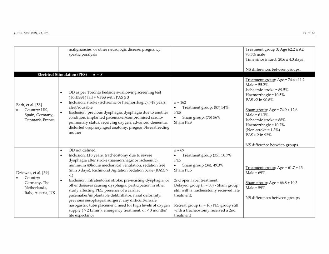
J. Clin. Med. 2022, 11, 776 19 of 68
malignancies, or other neurologic disease; pregnancy;
spastic paralysis
Treatment group 3: Age 62.2 ± 9.2
70.3% male
Time since infarct: 20.6 ± 4.3 days
NS differences between groups.
Pharyngeal Electrical Stimulation (PES) ― n = 8
Bath, et al. [58]
Country: UK,
Spain, Germany,
Denmark, France
OD as per Toronto bedside swallowing screening test
(TorBSST) fail + VFSS with PAS ≥ 3
Inclusion: stroke (ischaemic or haemorrhagic); >18 years;
alert/rousable
Exclusion: previous dysphagia, dysphagia due to another
condition, implanted pacemaker/compromised cardio‐
pulmonary status, receiving oxygen, advanced dementia,
distorted oropharyngeal anatomy, pregnant/breastfeeding
mother
n = 162
Treatment group: (87) 54%
PES
Sham group: (75) 56%
Sham PES
Treatment group: Age = 74.4 ±11.2
Male = 55.2%
Ischaemic stroke = 89.5%
Haemorrhagic = 10.5%
PAS >2 in 90.8%
Sham group: Age = 74.9 ± 12.6
Male = 61.3%
Ischaemic stroke = 88%
Haemorrhagic = 10.7%
(Non‐stroke = 1.3%)
PAS > 2 in 92%
NS difference between groups
Dziewas, et al. [59]
Country:
Germany, The
Netherlands,
Italy, Austria, UK
OD not defined
Inclusion: ≥18 years, tracheostomy due to severe
dysphagia after stroke (haemorrhagic or ischaemic);
minimum 48hours mechanical ventilation, sedation free
(min 3 days), Richmond Agitation Sedation Scale (RASS >
‐1)
Exclusion: infratentorial stroke, pre‐existing dysphagia, or
other diseases causing dysphagia; participation in other
study affecting PES, presence of a cardiac
pacemaker/implantable defibrillator, nasal deformity,
previous oesophageal surgery, any difficult/unsafe
nasogastric tube placement, need for high levels of oxygen
supply ( > 2 L/min), emergency treatment, or < 3 months’
life expectancy
n = 69
Treatment group (35), 50.7%
PES
Sham group (34), 49.3%
Sham PES
2nd open label treatment:
Delayed group (n = 30) ‐ Sham group
still with a tracheostomy received late
treatment;
Retreat group (n = 16) PES group still
with a tracheostomy received a 2nd
treatment
Treatment group: Age = 61.7 ± 13
Male = 69%
Sham group: Age = 66.8 ± 10.3
Male = 59%
NS differences between groups
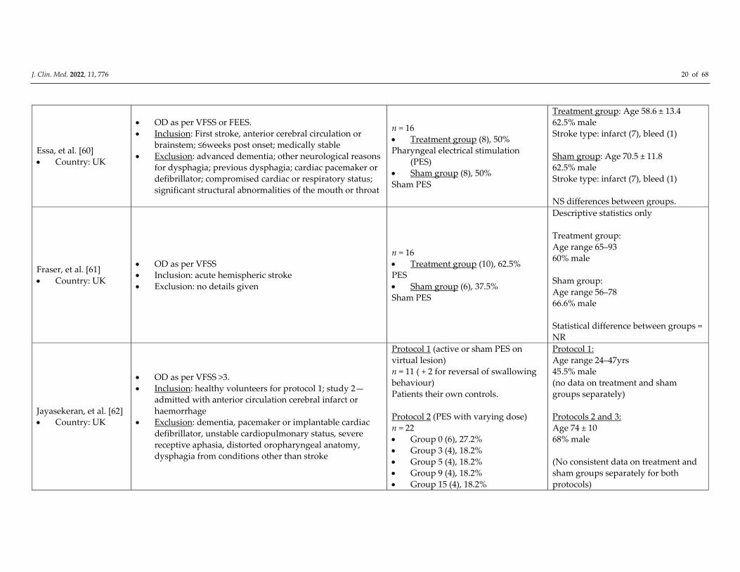
J. Clin. Med. 2022, 11, 776 20 of 68
Essa, et al. [60]
Country: UK
OD as per VFSS or FEES.
Inclusion: First stroke, anterior cerebral circulation or
brainstem; ≤6weeks post onset; medically stable
Exclusion: advanced dementia; other neurological reasons
for dysphagia; previous dysphagia; cardiac pacemaker or
defibrillator; compromised cardiac or respiratory status;
significant structural abnormalities of the mouth or throat
n = 16
Treatment group (8), 50%
Pharyngeal electrical stimulation
(PES)
Sham group (8), 50%
Sham PES
Treatment group: Age 58.6 ± 13.4
62.5% male
Stroke type: infarct (7), bleed (1)
Sham group: Age 70.5 ± 11.8
62.5% male
Stroke type: infarct (7), bleed (1)
NS differences between groups.
Fraser, et al. [61]
Country: UK
OD as per VFSS
Inclusion: acute hemispheric stroke
Exclusion: no details given
n = 16
Treatment group (10), 62.5%
PES
Sham group (6), 37.5%
Sham PES
Descriptive statistics only
Treatment group:
Age range 65–93
60% male
Sham group:
Age range 56–78
66.6% male
Statistical difference between groups =
NR
Jayasekeran, et al. [62]
Country: UK
OD as per VFSS >3.
Inclusion: healthy volunteers for protocol 1; study 2—
admitted with anterior circulation cerebral infarct or
haemorrhage
Exclusion: dementia, pacemaker or implantable cardiac
defibrillator, unstable cardiopulmonary status, severe
receptive aphasia, distorted oropharyngeal anatomy,
dysphagia from conditions other than stroke
Protocol 1 (active or sham PES on
virtual lesion)
n = 11 ( + 2 for reversal of swallowing
behaviour)
Patients their own controls.
Protocol 2 (PES with varying dose)
n = 22
Group 0 (6), 27.2%
Group 3 (4), 18.2%
Group 5 (4), 18.2%
Group 9 (4), 18.2%
Group 15 (4), 18.2%
Protocol 1:
Age range 24–47yrs
45.5% male
(no data on treatment and sham
groups separately)
Protocols 2 and 3:
Age 74 ± 10
68% male
(No consistent data on treatment and
sham groups separately for both
protocols)

J. Clin. Med. 2022, 11, 776 21 of 68
Protocol 3 (active or sham PES in acute
stroke)
n = 28
Treatment group (16), 57%
Sham group (12), 43%
Difference between groups NS
Restivo, et al. [63]
Country: Italy
OD as per VFSS
Inclusion: Patients with stable multiple sclerosis (MS) with
dysphagia for > 2 months; no dysphagia intervention in
the preceding 3 months; >18 years; Expanded Disability
Status Scale (EDSS) <7.5
Exclusion: neurologic disease other than MS; age >60
years; concomitant illness or upper gastrointestinal
disease; inability to give informed consent because of
cognitive impairment
n = 20
Treatment group (10), 50%
Pharyngeal stimulation
Sham group (10), 50%
Sham pharyngeal stimulation
Cohort demographics supplied, no
group descriptives given.
Mean age = 39.7 ± 6.5 years
35% male
Relapsing‐remitting MS = 14,
Secondary progressive MS = 6
Mean EDSS= 5.7 ± 0.8; mean disease
duration = 9.8 ± 2.4 years; mean
dysphagia duration = 22.0 ± 7.4
months
Statistical difference between groups =
NR
Suntrup, et al. [64]
Country:
Germany
OD as per FEES
Inclusion: tracheostomised, weaned off mechanical
ventilation, unable to be decannulated due to severe
persistent dysphagia
Exclusion: pre‐existing dysphagia; presence of implanted
electronic devices of any kind
n = 30
Treatment group (20), 66.6%
Pharyngeal stimulation
Sham group (10), 33.3%
Sham pharyngeal stimulation
Treatment group:
Age 63.0 ± 14.5 years
45% male
90% ischaemic, 10% haemorrhagic
stroke. 70% supratentorial, 30%
infratentorial
Sham group:
Age: 66.7 ± 14.5 years
60% male
80% ischaemic, 20% haemorrhagic
stroke. 90% supratentorial, 10%
infratentorial
J. Clin. Med. 2022, 11, 776 22 of 68
Difference between groups NS
Vasant, et al. [65]
Country: UK
OD as per TOR‐BSST confirmed by MBS or FEES with
most (but not all) patients
Inclusion: dysphagia following anterior or posterior
cerebral circulation infarct (ischemic and haemorrhagic)
<6 weeks ago; medically stable at inclusion; no history of
intubation/tracheotomy
Exclusion: advanced dementia, other
neurological conditions causing dysphagia, previous history of
dysphagia, presence of cardiac pacemaker or implanted
cardiac defibrillator, other severe cardiac or respiratory
conditions, significant oral/pharyngeal structural
abnormalities, continuous oxygen requirements
n = 35 at 2 weeks post treatment,
n = 33 at 3 months post treatment.
Treatment group (15), 48.4%
PES
Sham group (16), 51.6%
Sham PES
Treatment group: (median) age = 71
Interquartile range (IQR) =56–79.
50% male
NIHSS: median score = 10.0 (IQR= 5.2,
18.5)
Sham group: (median) age = 71 (IQR =
61–78)
72% male
NIHSS: median score = 12.5 (IQR = 9.2,
16.8)
No other stroke, site of lesion details
reported.
NS differences between groups.
Combined Neurostimulation Interventions ― n = 4
Cabib, et al. [66]
Country: Spain
OD as per VFSS
Inclusion: > 3 months post unilateral stroke, stable
medical condition
Exclusion: neurodegenerative disorders, epilepsy, drug
dependency, brain or head trauma or surgery, structural
causes of OD, pacemaker or metallic body implants, and
pregnancy or lactation
n = 36
Treatment group 1 (12), 33.3%.
rTMS
Treatment group 2 (12), 33.3%
Capsaicin
Treatment group 3 (12), 33.3%
PES
Treatment group 1: Age 70.0 ± 8.6
75% male
0% haemorrhage, 100% infarct.
Treatment group 2: Age 74.3 ± 7.8
58% male
8% haemorrhage, 92% infarction
Treatment group 3: Age 70.0 ± 14.2
92% male
25% haemorrhage, 75% infarction
Difference between groups NS
Lim, et al. [67]
Country: Korea
OD as per VFSS
Inclusion: primary diagnosis unilateral cerebral infarction
or haemorrhage (CT or MRI); stroke onset <3 months;
n = 47
Treatment group 1 (15), 32%
DT
Treatment group 1: Age 62.5 ± 8.2
60% male
34% haemorrhage, 66% infarction
J. Clin. Med. 2022, 11, 776 23 of 68
patients who could maintain balance during evaluation +
treatment; and adequate cognitive function to participate
Exclusion: could not complete VFSS/failed the
examination; presence of dysphagia pre stroke; history of
prior stroke, epilepsy, tumor, radiotherapy in the head
and neck, or other neurological diseases;unstable medical
condition; and contraindication to magnetic or electrical
stimulation
Treatment group 2 (14), 30%
DT + rTMS
Treatment group 3 (18), 38%
DT + NMES
Treatment group 2: Age 59.8 ± 11.8
43% male
71% haemorrhage, 29% infarction
Treatment group 3: Age 66.3 ± 15.4
67% male
66% haemorrhage, 44% infarction
Difference between groups NS
Michou, et al. [68]
Country: UK
OD as per diagnoses made by SLT (confirmed with VFSS
at start of treatment)
Inclusion: post stroke dysphagia for >6weeks
Exclusion: Hx of dementia, cognitive impairment,
epilepsy, head&neck surgery; neurological defects prior to
stroke; cardiac pacemaker or defibrillator in‐situ; severe
concomitant medical conditions; structural oropharyngeal
pathology; intracranial metal; pregnancy; medications
acting on CNS
n = 18
Treatment group 1 (6), 33.3%
Pharyngeal electrical stimulation
(PES)
Treatment group 2 (6), 33.3%
Paired associative stimulation (PAS)
Treatment group 3 (6), 33.3%
Repetitive transcranial magnetic
stimulation (rTMS)
Treatment group: Avg age 60.3
83% male
Treatment group 2: Avg age 67.3
100% male
Treatment group 3: Avg age 67.8
66.7% male
Overall: 63 ± 15 weeks post stroke
with 7.6 ± 1 on NIHHS
Statistical difference between groups =
NR
Zhang, et al. [69]
Country: China
OD as per DOSS by a well trained doctor
Inclusion: stroke as per MRI <2 months earlier; aged 50–
75yrs; normal consciousness, stable vital signs, presence of
dysdipsia and dysphagia
Exclusion: brain trauma or other central nervous system
disease; unstable arrhythmia, fever, infection, epilepsy, or
use of sedative drugs; poor cooperation due to serious
aphasia or cognitive disorders; contraindications to
magnetic or electrical stimulation
n = 64
Treatment group 1 (16), 25%.
Sham rTMS + NMES
Treatment group 2 (16), 25%
Ipsilateral rTMS + NMES
Treatment group 3 (16), 25%
Contralateral rTMS + NMES
Treatment group 4 (16), 25%
Bilateral rTMS + NMES
Treatment group 1: Age 55.9 ± 8.9
43% male
61.5% subcortical, 38.5% brainstem
Treatment group 2: Age 56.8 ± 9.7
54% male
30.8% subcortical, 69.2% brainstem
Treatment group 3: Age 56.5 ± 10.1
50% male
J. Clin. Med. 2022, 11, 776 24 of 68
58.3% subcortical, 41.7% brainstem
Treatment group 4: Age 53.1 ± 10.6
31% male
61.5% subcortical, 38.5% brainstem
*All data given on participants that
finished the trial and follow‐up period
(n = 52) a NMES is at motor stimulation level unless explicitly mentioned. Notes. CNS—central nervous system; CP—cerebral palsy; CT–computed tomography; CVA–
cerebrovascular accident; DOSS–dysphagia outcome and severity scale; DT–dysphagia therapy; FEES–fiberoptic endoscopic evaluation of swallowing; FOIS–
functional oral intake scale; ICH–intracranial haemorrhage; MMSE–Mini‐Mental State Exam; MRI–magnetic resonance imaging; MS–multiple sclerosis; NIHSS–
National Institutes of Health Stroke Scale; NMES–neuromuscular electrical stimulation; OD–oropharyngeal dysphagia; OST–oral sensorimotor treatment; PAS–
penetration–aspiration score; PES–pharyngeal electrical stimulation; rTMS–repetitive transcranial magnetic stimulation; SAH–subarachnoid haemorrhage;
sEMG–surface electromyography; SLT–Speech and Language Therapist; TBI–traumatic brain injury; tDCS–transcranial direct current stimulation; TOR‐BSST–
Toronto Bedside Swallowing Screening test; VFSS–videofluoroscopic swallowing study.
Table 3. Outcome of NMES and PES interventions for people with oropharyngeal dysphagia.
Study Intervention Goal Procedure, Delivery and Dosage Per
Intervention Group Outcome Measures
Intervention
Outcomes/Conclusions
NeuroMuscular Electrical Stimulation (NMES) a ― n = 30
Beom, et al. [28]
To investigate the effectiveness
of NMES to suprahyoid
muscle compared with NMES
to infrahyoid muscle in brain‐
injured (stroke) patients with
dysphagia
Procedure:
NMES as per VitalStim therapy
training manual
10–15 sessions, 30 min each, over
2–3 weeks
DT during NMES sessions, as per
videofluoroscopy swallow study
(VFSS)
Treatment group 1:
NMES to the suprahyoid muscles
(4 electrodes)
Primary outcomes:
FDS b; SFS; aspiration/penetration
based off VFSS pre and post
treatment.
Secondary outcome: N/R
No statistically significant
differences between groups
Both treatments showed
significant improvement in FDS
(p < 0.001) and SFS (p < 0.001),
and non‐significant
improvements in penetration or
aspiration
J. Clin. Med. 2022, 11, 776 25 of 68
60 Hz pulse frequency, 500 ms
pulse interval, using Stimplus
Treatment group 2:
NMES to the suprahyoid muscles
(2 electrodes) and infrahyoid
muscles (2 electrodes)
80 Hz pulse frequency, 700 ms
pulse interval, using Vitalstim
Bülow, et al.[29]
To evaluate and compare the
outcome of NMES versus
traditional swallowing therapy
(TT) in stroke patients
Procedure:
NMES as per VitalStim therapy
training manual (Placement 3B)
15 sessions, 60 min each,
5days/week for 3 weeks
Diet modifications as per SLT
recommendations
Treatment group 1:
NMES to supra & infra hyoid
4.5–25 mA (mean = 13 mA)
Treatment group 2:
clinician determined manoeuvers/
treatment techniques
Primary outcomes:
Patient reported VAS (swallowing
complaints);
VFSS measure b (performed day of
last treatment).
Secondary outcome: N/R
No statistically significant
differences between groups
VAS = No significant
improvement for NMES.
Significant improvement (p <
0.01) noted for combined group
effect
VFSS parameters = No
significant improvement for
NMES nor combined group
effect
El‐Tamawy, et
al. [30]
Assess the effect of NMES and
physical therapy program on
severe poststroke dysphagia
Treatment group 1:
Standard medical treatment
NMES: 30 min of 80 Hz frequency
0–150 V amplitude stimulation,
intensity 0–25 mA at motor level.
Electrodes placed horizontally on
the submental region 1cm lateral
to the midline above hyoid bone
and the other 1cm latero‐posterior
Primary outcomes:
Swallowing variables (OTT, hyoid
elevation, laryngeal elevation,
oesophageal sphincter opening,
aspiration/penetration) as per VFSS.
Secondary outcome: N/R
OTT significantly improved in
Treatment group 1 post
intervention (p = 0.001)
Significantly higher number of
patients in Treatment group 1
who had lower
aspiration/penetration rate (p =
0.008), improved hyoid
elevation (p = 0.002) and
laryngeal elevation (p = 0.001).
J. Clin. Med. 2022, 11, 776 26 of 68
to the midline just below the
hyoid bone.
Physical therapy program (45min,
range of oromotor and oral
stimulation exercises—unclear if
these were individualised)
3 times a week for 6 weeks (plus 3
times daily independently)
Control group 2:
Standard medical treatment only
No differences seen in
oesophageal sphincter opening
Guillén‐Solà, et
al. [31]
Assess the therapeutic
effectiveness of NMES and
inspiratory and expiratory
muscle training (IEMT) in
dysphagic subacute stroke
patients, compared to
standard swallow therapy
(DT)
Procedure: DT, IEMT, NMES
Delivery and dosage:
DT: 5 days a week. Self‐management
education, individualised oral
exercises, compensatory techniques
based on VFSS
IEMT: 5 sets of 10 respirations twice a
day 5 days per week for 3 weeks.
Loads were set to 30% of max insp and
exp pressures, increased weekly by
10cmH2O
Sham IEMT: same frequency, but with
set workloads of 10cmH2O
NMES: 40min a day 5 days per week
for 3 weeks at 80Hz on suprahyoid
muscles
Primary outcomes:
Max inspiratory + expiratory muscle
function (MicroRPM), dysphagia
severity (VFSS, PAS), respiratory
complications.
Secondary outcomes:
Swallowing parameter changes as per
voice changes, coughing,
desaturation (> 3%), piecemeal
deglutition, oropharyngeal residue
(V‐VST), FOIS, DOSS. (Not reported
in study)
Assessed at baseline, 3 weeks post
(by V‐VST), and 3 months post
intervention (VFSS).
Respiratory muscle strength:
Positive treatment effect in the
IEMT group at 3 weeks only
Dysphagia severity:
No significant differences for
PAS scores between groups
Improved safety at 3 weeks for
IEMT and NMES; improved
efficacy at 3 months for IEMT
Respiratory complications:
No adverse effects reported
15.5% with lung infection (4 in
DT, 3 in NMES, 2 in IEMT)
throughout the follow‐up
period
Heijnen, et al.,
[32]
To compare the effects of
traditional speech therapy
exercises to those combined
with NMES on motor or
sensory level on dysphagia
Procedure:
NMES with VitalStim protocol
DT included oromotor exercises,
swallow manoeuvres and
strategies
Primary outcomes:
Health related quality of life (SWAL‐
QOL; MDADI).
Secondary outcomes:
No significant differences
between groups
Significant improvement (p <
0.001) on Dysphagia Severity
Scale for all groups. Restricted
positive effects on QOL
J. Clin. Med. 2022, 11, 776 27 of 68
and quality of life of patients
with Parkinson’s Disease
13–15 sessions, 30 min each, on
five consecutive days a week over
3–5weeks
Treatment group 1
DT
Treatment group 2
DT
NMES to the suprahyoid muscle
Stimulation to motor level
Treatment group 3
DT
NMES to the suprahyoid muscle
Stimulation to sensory level
Dysphagia severity (single‐item
Dysphagia Severity Scale)
Huang, et al.
[33]
To compare functional
dysphagia recovery in acute
stroke patients using
traditional dysphagia therapy,
NMES or the two combined
Procedure:
NMES, DT
10 sessions, 3 x a week, 60 min each
VitalStim protocol with electrode
placement in a vertical line with
one above and one below the
thyroid notch
Intensity level individual—
determined once patient felt a
tingling sensation and a muscle
contraction
DT: oromotor exercises,
compensatory techniques,
thermal‐tactile stimulation,
swallow manoeuvres
individualised as per VFSS
Treatment group 1
DT
Primary outcomes:
FOIS, PAS, FDS as per VFSS before
and after treatment.
Secondary outcome: N/R
No significant differences
between groups post therapy in
FOIS or PAS scales
For FDS, 2 of 4 scales were
significantly different
(improved) in Treatment group
3 (p = 0.03) compared with
Treatment groups 1 and 2
Significant differences in FOIS
before and after therapy in all 3
groups (p = 0.03; p = 0.01; p =
0.005)
Significant differences in PAS
before and after therapy in
treatment groups 1 and 3 (p =
0.04 for both)
J. Clin. Med. 2022, 11, 776 28 of 68
Treatment group 2
NMES
Treatment group 3
DT + NMES
DT performed during NMES
Huh, et al. [34]
To investigate the effect of
different electrode placement
in NMES in poststroke
dysphagia rehabilitation
Procedure:
NMES (VitalStim protocol with
stimulation at motor level) +
effortful swallow
five 20min sessions weekly for four
weeks
Treatment group 1
NMES with horizontal electrode
placement
One pair of electrodes on the
suprahyoid muscles, second pair
on the infrahyoid muscles
Treatment group 2
NMES with horizontal + vertical
electrode placement
One pair horizontally on the
suprahyoid muscles, second pair
vertically on the infrahyoid
muscles
Treatment group 3
NMES with vertical electrode
placement along the midline from
hyoid bone down to below the
thyroid cartilage
Primary outcomes:
VFSS performed at baseline and post
treatment.
FDS—both oral phase (FDS‐O)
and pharyngeal phase (FDS‐p)
separately, also
DOSS b
Secondary outcome: N/R
Treatment Group 1 scores for
FDS and FDS‐p were
significantly higher than those
in Groups 2 and 3
No statistically significant
differences between groups in
FDS‐O or DOSS scores post
treatment
All groups showed significant
improvement in FDS (p < 0.01)
and DOSS (p < 0.01) scores post
treatment
Horizontal electrode placement
on the suprahyoid and
infrahyoid muscles was found
to be more beneficial for
dysphagia recovery
J. Clin. Med. 2022, 11, 776 29 of 68
Jing, et al. [35]
To investigate the effect of
NMES on post stroke
dysphagia
Procedure:
NMES, DT
Treatment for consecutive 10 days
Both groups received general
medical treatment, and DT
(exercises for tongue, mouth and
facial muscle function; sensory
stimulation; vocal cord; chewing
training; therapeutic feeding)
Treatment group 1:
VitalStim as per protocol, though
intensity of 6 to 21mV.
Electrode placement selected
based on the patient’s dysphagia
presentation:
(a) vertical distribution on each
side of the midline with
lowest electrode just above
the superior thyroid notch
(b) 1st channel horizontally and
close to the surface of the
hyoid bone with 2nd channel
horizontally along the
midline just below the
superior thyroid notch
(c) 1st channel vertically below
the chin and 2nd channel
along the buccal branch of
the facial nerve
Treatment group 2:
Intensity of swallow
rehabilitation exercises NR
Primary outcomes:
Swallow efficacy, swallow function
scores, laryngeal elevation, severity
of aspiration, amount of food intake,
residue scores. All based on Rattans
dysphagia classification criteria.
Secondary outcome: N/R
Efficacy, laryngeal elevation
and severity of aspiration in the
treatment group were
significantly better post
treatment than in the control
group (p < 0.05)
Swallow function scores
improved in both groups, but
more pronounced in the
treatment group (p < 0.05)
Amount of food intake or
residue scores were not
significantly different between
the two groups
J. Clin. Med. 2022, 11, 776 30 of 68
Langmore, et al.
[36]
To investigate the efficacy of
NMES combined with
swallow exercises in
improving dysphagia post
radiotherapy for head & neck
cancer
Procedure:
NMES (BMR NeuroTech 2000
default settings with minor
alterations) or sham
Electrodes placed supra‐hyoid
region.
Home‐based protocol, performed
2 x day, 6 days/week, for 12
weeks (3 training sessions to
ensure competence). 16–20 min
per session
DT during treatment sessions: 10
x super‐supraglottic, 10x
Mendelsohn, 10 x effortful
swallows
Sham/Treatment group:
Sham‐NMES delivered via a
similar device with wires inside
the equipment disconnected.
Same session structure and
intensity of treatment
Primary outcome:
Swallowing function as measured by
PAS on VFSS.
Secondary outcomes:
OPSE, hyoid excursion, diet
measured by the PSS, and quality of
life as measured by HNCI.
Assessments were performed prior
to, midway through (week 7) and at
the end of the treatment (week 13).
Mean PAS: greater
improvement in the sham group
(p = 0.027). No other outcomes
showed a significant difference
between the two groups.
Treatment group:
No significant change in PAS
score
Significant decrease in the
anterior hyoid excursion (p
= .038
No sigificant differences in
OPSE
Significant improvement in diet
(total PSS score, p < 0.001) and
HNCI quality of life scores for
eating (p < 0.001) and speech (p
= 0.016)
Sham/Treatment group:
Significant improvement in PAS
score (p < 0.001)
No significant differences in
OPSE
Significant improvement in diet
(total PSS score, p < 0.046) and
HNCI quality of life scores for
eating (p = 0.003) and speech (p
= 0.001)
Lee, et al. [37]
To compare early NMES
combined with DT versus DT
only on dysphagia outcomes
in acute/subacute ischaemic
Procedure:
DT in both groups included
thermal‐tactile stimulation with
any combination of lingual
strengthening exercises, laryngeal
Primary outcome:
FOIS b as per VFSS at 3, 6, and 12
weeks post treatment.
Secondary outcome: N/R
FOIS: Both groups showed
significant improvement in
FOIS 3 & 6 weeks post
treatment
J. Clin. Med. 2022, 11, 776 31 of 68
stroke patients with moderate
to severe dysphagia
adduction‐elevation exercises,
and swallow manoeuvres by SLP
60min/day for 15 days
Treatment group 1:
NMES simultaneously with DT
for first 30min
max tolerable intensity (120% of
the mean threshold value) on
both suprahyoid muscles. Pulse
rate of 80 Hz with 700microsec
duration
30min a day, 5 days per week for
3 weeks
Treatment group 2:
DT only, as per above
Treatment group 1 showed
significant improvement at 12
weeks
FOIS: significantly greater
improvement in treatment
group 1 (at all timepoints) when
compared to the treatment
group 2 (p < 0.05)
Li, et al. [38]
To assess whether adding
NMES to the conventional
swallow therapy improves
post‐stroke dysphagia
Procedure:
NMES with VitalStim, electrical
current level approx 7mA. No
other stimulation data given
Electrodes placed supra‐hyoid
(top electrodes) and infra‐hyoid
(bottom electrodes)
4 weeks of treatment, 1 hour
sessions, 5 x week
DT included basic training of
organs related to food intake and
swallowing (no further details
given) and direct food intake
training (intake environment,
body posture for swallowing and
removal of residue)
Primary outcomes:
VAS to compare the differences of
muscle pain pre and post treatment;
SSA, sEMG, OTT, PTT, LCD and
Standardised swallowing PAS were
measured using VFSS.
Secondary outcome: N/R
SSA scores significantly higher
in Treatment Group 1 (p < 0.01)
compared to groups 2 and 3
Significant decrease in OTT and
PTT for liquid and paste bolus
(p < 0.05 for both) in Treatment
Group 1 compared to Groups 2
and 3
No change in LCD
Significant increase in max
amplitude of sEMG signal in
Treatment Group 1 compared to
Groups 2 and 3
No significant changes between
Groups 2 and 3
J. Clin. Med. 2022, 11, 776 32 of 68
Treatment group 1:
NMES + DT
Treatment group 2:
NMES
Treatment group 3:
DT
SSA scores and maximum
amplitude of sEMG signal
increased significantly within
each group
Maeda, et al.
[39]
To investigate the effect of
transcutaneous electrical
sensory stimulation (TESS)
without muscle contraction in
patients undergoing
dysphagia rehabilitation
Procedure:
Sensory stimulation or sham, plus
usual treatment (details NR) for
both groups using Gentle Stim (J
Craft, Osaka, Japan). Beat
frequency of 50 Hz, other details
NR
2 pairs of electrodes (frequencies
of 2000 and 2050 Hz).
Anterior electrodes placed at the
edge of the thyroid cartilage and
the posterior electrodes 4cm from
the ipsilateral electrode along the
mandible
15 min of twice daily
intervention, 5 days per week for
2 weeks
Treatment group:
Stimulation intensity set at 3.0
mA
Sham group:
Stimulation intensity set at 0.1
mA
Primary outcomes:
Cough latency time against 1% citric
acid mist.
Secondary outcomes:
FOIS, oral nutritional intake
outcomes measured at study entry,
and after the 2nd and 3rd week
following treatment initiation
No statistically significant
differences were found between
or within groups
Changes in cough latency time
and FOIS scores indicated better
outcomes in the TESS group,
based on substantial effect sizes
J. Clin. Med. 2022, 11, 776 33 of 68
Meng, et al. [40]
To assess the effectiveness of
surface NMES with various
electrode placements on
patients with post‐stroke
dysphagia
Procedure:
All groups received DT 30 min
per treatment, 5 x week. 10
sessions
NMES with VitalStim (Treatment
Groups 1 and 2) for 30min prior
to daily DT
NMES as per VitalStim with
minimum degree of stimulation
to induce visible muscle
contraction
DT combination of therapeutic
exercises, compensatory
manoeuvres and diet texture
modifications. It remains unclear
whether these were standard or
individual according to VFSS
results
Treatment Group 1:
Electrode placement: 1 pair of
electrodes on the surface of both sides
of suprahyoid, and another pair on
surface of upper and lower edge of
thyroid cartilage
Treatment Group 2:
Electrode placement: 2 pairs of
electrodes on the surface of
suprahyoid (geniohyoid + mylohyoid)
Treatment Group 3:
DT
Primary outcomes:
VFSS pre and post treatment.
Hyoid excursion, DOSSb, WST and
RSST.
Secondary outcome: N/R
WST, RSST and DOSS scores
improved significantly more for
Treatment Groups 1 and 2
compared to Control Group (p <
0.05)
Differences not statistically
different between treatment
group 1 and 2
WST, RSST and DOSS improved
significantly in all groups
comparing pre‐and post‐
treatment (p < 0.05)
VFSS: only increased anterior
movement of the hyoid
improved statistically
significantly and only in
Treatment Group 2, pre‐post
treatment (p = 0.006)
Nam, et al. [41]
To assess the effect of repeated
sessions of NMES with two Procedure: Primary outcomes:
No significant differences
between groups
J. Clin. Med. 2022, 11, 776 34 of 68
different electrode placements
on dysphagia following brain
injury
Hyolaryngeal electrical
stimulation
10–15 sessions over 2–3 weeks,
one session daily for 30 min
Both groups also received
simultaneous DT—individual
swallow manoeuvres based on
VFSS findings
Treatment Group 1:
Electrode placement on the
suprahyoid muscles
Stimulation delivered using
Stimplus (Cuber‐Medic Corp.,
Iksan, South Korea)
Pulse frequency 60Hz with 500ms
pulse interval
Treatment Group 2:
Electrode placement on the
suprahyoid and infrahyoid
muscles
Stimulation delivered using
VitalStim (Chattanooga Group,
Hixson, TN, USA) as per
VitalStim protocol
Motion analysis of the hyolaryngeal
excursion according to VFSS
conducted before and after the
treatment
Secondary outcome: N/R
Treatment Group 1 showed a
significant increase in the
maximal anterior excursion of
the hyoid (p = 0.008) and the
anterior excursion velocity (p =
0.017)
Treatment Group 2 showed a
significant increase in the
maximal superior excursion and
the maximal absolute excursion
distance of laryngeal elevation
(p = 0.013 for both)
Oh, et al. [42]
To identify the effects of
NMES with two different
electrode placements on post‐
stroke dysphagia
Procedure:
NMES with VitalStim, as per
protocol
30 min/day, 5 days/week for 4
weeks
Effortful swallow performed
during stimulation
Both groups received DT—
unclear if this was individualised
Primary outcomes:
VDS, PAS b and FOIS
Secondary outcome: N/R
PAS improved more in
Treatment Group 1 compared to
Group 2 (p = 0.036). No other
significant differences between
groups.
Treatment Group 1:

J. Clin. Med. 2022, 11, 776 35 of 68
Treatment group 1:
Electrode placement on the
suprahyoid muscles
Treatment group 2:
Electrode placement on the
infrahyoid muscles
DT included thermal‐tactile
stimulation, various exercises,
manoeuvres, modified food material,
viscosity and posture
Significant improvement in VDS
(p = 0.001), PAS (p = 0.002) and
FOIS (p = 0.014)
Treatment Group 2:
Significant improvement in VDS
(p = 0.002), PAS (p = 0.045) and
FOIS (p = 0.026)
NB. Data as per Table 2
(inconsistencies between text vs
table)
Ortega, et al.
[43]
To evaluate the effectiveness
of two different sensory
stimulation treatments on
oropharyngeal dysphagia in
the elderly
Procedure:
Sensory stimulation for 2 weeks
Treatment group 1:
Chemical sensory stimulation
with a natural TRPV1 (capsaicin)
agonist solution.
Treatment was taken by patients
three times a day before each
meal and 5 days per week (Mon‐
Fri) for 2 weeks
Treatment group 2:
Electrical stimulation using the
thyroid position (VitalStim, as per
protocol)
Intensity 75% of the motor
threshold
Once a day 5 days per week
(Mon‐Fri) for 2 weeks
Primary outcome:
VFSS measurements, PAS (measured
before and 5 days after the treatment)
Secondary outcomes: EAT‐10, V‐VST,
No between group differences
reported
Treatment group 1:
Significant improvement in
EAT‐10 scores (p = 0.016), and
safety based on VFSS (p = 0.019)
Treatment group 2:
Significant improvement in
safety (p = 0.019) and
penetrations (p = 0.044) based on
VFSS
J. Clin. Med. 2022, 11, 776 36 of 68
Park, et al. [44]
To determine whether effortful
swallow training combined
with surface electrical
stimulation
as a form of resistance training
has an effect on post‐stroke
dysphagia
Procedure: NMES with VitalStim, 2
sets of electrodes placed on infrahyoid
muscles (working against resistance)
3 sets of 20min exercise/week
over 4 weeks
Treatment group 1:
Effortful swallow + NMES
(treatment level)
NMES as per VitalStim protocol,
intensity increased until muscle
activation
Treatment group 2:
Effortful swallow + NMES (non‐
treatment level)
Primary outcome:
Hyolaryngeal excursion (max
anterior hyoid displacement, max
vertical hyoid displacement),
maximum vertical laryngeal
displacement, UES opening (width),
PAS (as per VFSS), pre and post
treatment.
Secondary outcome: N/R
Between groups significant
difference post treatment NR
Treatment group 1:
Significant increase in laryngeal
elevation (p > 0.05). NS increase
in vertical hyoid motion and
UES opening
Treatment group 2:
NS difference between any pre‐
post measures
Park, et al. [45]
To investigate the effects of
effortful swallowing combined
with NMES on hyoid bone
movement and swallowing
function in stroke patients
Procedure: NMES (VitalStim, as per
protocol), electrodes placed on
infrahyoid muscles (targeting
sternohyoid muscle, working against
resistance)
Delivery and dosage: 30 min per
session, 5 sessions a week for 6 weeks.
Treatment group 1:
Effortful swallow + NMES
(treatment level)
NMES intensity gradually
increased until grabbing
sensation
Treatment group 2:
Effortful swallow + NMES
(placebo level)
Primary outcomes:
As per VDS pre and post treatment (6
weeks).
Kinematics of the hyoid bone
(analysed with Image J Program);
swallow function (as per VDS and
PAS b);
VDS measures: Oral phase (lip
closure, bolus formation, mastication,
apraxia, tongue to palate contact,
premature bolus loss and OTT);
Pharyngeal phase (pharyngeal
triggering, vallecular residues,
pyriform sinus resides, laryngeal
elevation, pharyngeal wall coating,
Significantly greater
improvements shown by the
treatment group versus the
placebo group
Treatment group 1:
Significant improvements post
treatment for VDS total score (p
< 0.01), VDS pharyngeal phase
(p < 0.01), vertical and
horizontal hyoid bone
displacement (p < 0.01) and PAS
(p < 0.01). Improvement for VDS
oral phase = NS.
Treatment group 2:
Vertical and anterior hyoid
elevation = NS (p = 0.06, p = 0.09
respectively)
J. Clin. Med. 2022, 11, 776 37 of 68
Sensory NMES intensity
gradually increased until tingling
sensation
pharyngeal transit time and
aspiration).
Secondary outcome: N/R
Significant improvement in total
VDS score (p = 0.02) and oral
phase (0.04). Pharyngeal phase
improvement = NS (p = 0.07)
PAS improvement = NS (p =
0.06)
Park, et al. [46]
To identify the effect of
effortful swallowing combined
with neuromuscular electrical
stimulation NMES in treating
dysphagia in Parkinson’s
disease
Procedure: NMES (VitalStim) 5
days/week, for 4 weeks, 30min each
session
During stimulation, patient
produced effortful swallow
(saliva)
Infrahyoid electrode placement
After NMES, patients received 30
min DT (orofacial exercises,
thermal tactile stimulation and
manoeuvres)
Treatment group 1:
NMES + effortful swallow
Treatment group 2:
Sensory NMES + effortful
swallow
Stimulation applied at 1.0mA, no
increase
Primary outcome:
Kinematics of the hyoid bone
(analysed with Image J Program);
swallow function (as per VDS and
PAS b)
Secondary outcomes:
VDS measures: Oral phase (lip
closure, bolus formation, mastication,
apraxia, tongue to palate contact,
premature bolus loss and OTT);
Pharyngeal phase (pharyngeal
triggering, vallecular residues,
pyriform sinus resides, laryngeal
elevation, pharyngeal wall coating,
pharyngeal transit time and
aspiration)
Hyoid bone movement:
Significant improvement (p <
0.05) with vertical and
horizontal movement versus
sensory NMES
PAS: Significant improvement
(p < 0.05) as compared with
sensory NMES
No significant difference
between groups with any VDS
parameters
Permsirivanich,
et al. [47]
To compare the treatment
outcomes between dysphagia
rehabilitation exercises and
NMES in post‐stroke
dysphagia
Procedure:
Treatment administered 5 days a
week (Mon‐Fri) for 4 weeks
Both groups received diet
modifications and oromotor
exercises if weakness present
Treatment group 1:
Primary outcomes:
Changes in FOIS b, complications
related to treatment and number of
therapy sessions.
VFSS only performed pre‐treatment.
Secondary outcome: N/R
Improvement in FOIS was
significantly greater for
Treatment group 2 (p < 0.001)
No complications related to
treatment, no significant
difference in the number of
sessions received
J. Clin. Med. 2022, 11, 776 38 of 68
Swallowing rehabilitation
exercises
Individual based on VFSS
findings, may have included
thermal stimulation, head & neck
positioning and swallow
manoeuvres
Treatment group 2:
NMES using VitalStim, as per
protocol
Vertical electrode placement—
from 1mm above the thyroid
notch down past the thyroid
notch
Treatment level at grabbing
sensation
60min per session
Ryu, et al. [48]
To evaluate the effect of
NMES on dysphagia following
treatment for head and neck
cancer
Procedure:
1. 30 min of NMES (VitalStim) or
transcutaneous electrical
stimulation (TENS)
2. Followed by 30 min DT for (oral
motor exercises, pharyngeal
swallowing exercises, use of
compensatory strategies during
meals, thermal/tactile stimulation,
Mendelsohn manoeuvre and diet‐
texture modifications)
3. 5 days per week for 2 weeks
Treatment group 1:
Electrodes placed horizontally
immediately above the thyroid
Primary outcome measures:
FDS, CDS, ASHA‐NOMS and
MDADI
Secondary outcome: N/R
Significant difference (p= 0.04)
between the treatment and
sham group post intervention
for FDS only
No significant difference
between groups for CDS,
ASHA‐NOMS nor MDADI
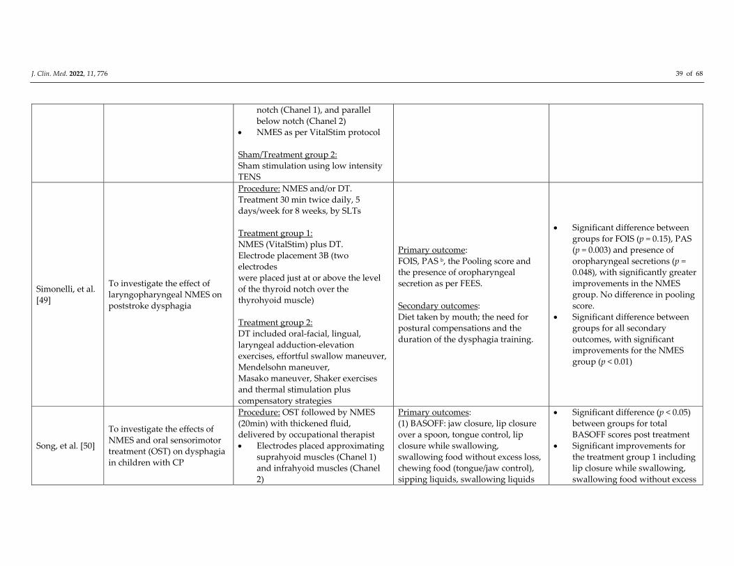
J. Clin. Med. 2022, 11, 776 39 of 68
notch (Chanel 1), and parallel
below notch (Chanel 2)
NMES as per VitalStim protocol
Sham/Treatment group 2:
Sham stimulation using low intensity
TENS
Simonelli, et al.
[49]
To investigate the effect of
laryngopharyngeal NMES on
poststroke dysphagia
Procedure: NMES and/or DT.
Treatment 30 min twice daily, 5
days/week for 8 weeks, by SLTs
Treatment group 1:
NMES (VitalStim) plus DT.
Electrode placement 3B (two
electrodes
were placed just at or above the level
of the thyroid notch over the
thyrohyoid muscle)
Treatment group 2:
DT included oral‐facial, lingual,
laryngeal adduction‐elevation
exercises, effortful swallow maneuver,
Mendelsohn maneuver,
Masako maneuver, Shaker exercises
and thermal stimulation plus
compensatory strategies
Primary outcome:
FOIS, PAS b, the Pooling score and
the presence of oropharyngeal
secretion as per FEES.
Secondary outcomes:
Diet taken by mouth; the need for
postural compensations and the
duration of the dysphagia training.
Significant difference between
groups for FOIS (p = 0.15), PAS
(p = 0.003) and presence of
oropharyngeal secretions (p =
0.048), with significantly greater
improvements in the NMES
group. No difference in pooling
score.
Significant difference between
groups for all secondary
outcomes, with significant
improvements for the NMES
group (p < 0.01)
Song, et al. [50]
To investigate the effects of
NMES and oral sensorimotor
treatment (OST) on dysphagia
in children with CP
Procedure: OST followed by NMES
(20min) with thickened fluid,
delivered by occupational therapist
Electrodes placed approximating
suprahyoid muscles (Chanel 1)
and infrahyoid muscles (Chanel
2)
Primary outcomes:
(1) BASOFF: jaw closure, lip closure
over a spoon, tongue control, lip
closure while swallowing,
swallowing food without excess loss,
chewing food (tongue/jaw control),
sipping liquids, swallowing liquids
Significant difference (p < 0.05)
between groups for total
BASOFF scores post treatment
Significant improvements for
the treatment group 1 including
lip closure while swallowing,
swallowing food without excess
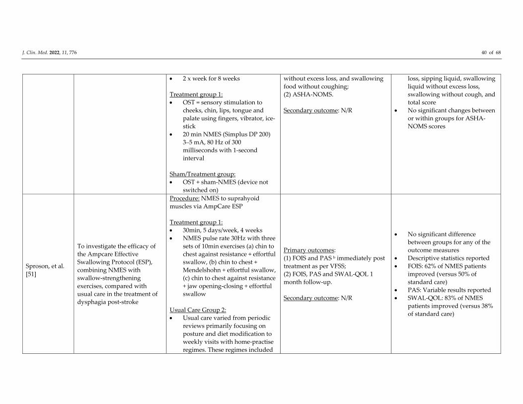
J. Clin. Med. 2022, 11, 776 40 of 68
2 x week for 8 weeks
Treatment group 1:
OST = sensory stimulation to
cheeks, chin, lips, tongue and
palate using fingers, vibrator, ice‐
stick
20 min NMES (Simplus DP 200)
3–5 mA, 80 Hz of 300
milliseconds with 1‐second
interval
Sham/Treatment group:
OST + sham‐NMES (device not
switched on)
without excess loss, and swallowing
food without coughing;
(2) ASHA‐NOMS.
Secondary outcome: N/R
loss, sipping liquid, swallowing
liquid without excess loss,
swallowing without cough, and
total score
No significant changes between
or within groups for ASHA‐
NOMS scores
Sproson, et al.
[51]
To investigate the efficacy of
the Ampcare Effective
Swallowing Protocol (ESP),
combining NMES with
swallow‐strengthening
exercises, compared with
usual care in the treatment of
dysphagia post‐stroke
Procedure: NMES to suprahyoid
muscles via AmpCare ESP
Treatment group 1:
30min, 5 days/week, 4 weeks
NMES pulse rate 30Hz with three
sets of 10min exercises (a) chin to
chest against resistance + effortful
swallow, (b) chin to chest +
Mendelshohn + effortful swallow,
(c) chin to chest against resistance
+ jaw opening‐closing + effortful
swallow
Usual Care Group 2:
Usual care varied from periodic
reviews primarily focusing on
posture and diet modification to
weekly visits with home‐practise
regimes. These regimes included
Primary outcomes:
(1) FOIS and PAS b immediately post
treatment as per VFSS;
(2) FOIS, PAS and SWAL‐QOL 1
month follow‐up.
Secondary outcome: N/R
No significant difference
between groups for any of the
outcome measures
Descriptive statistics reported
FOIS: 62% of NMES patients
improved (versus 50% of
standard care)
PAS: Variable results reported
SWAL‐QOL: 83% of NMES
patients improved (versus 38%
of standard care)
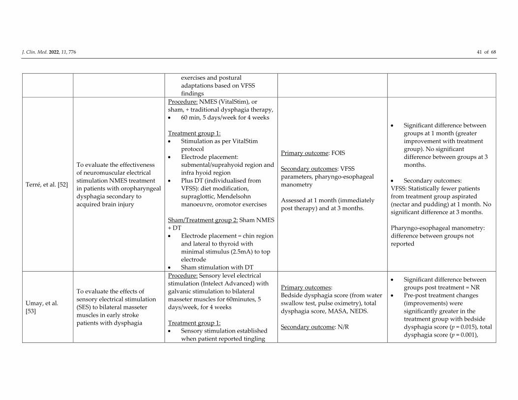
J. Clin. Med. 2022, 11, 776 41 of 68
exercises and postural
adaptations based on VFSS
findings
Terré, et al. [52]
To evaluate the effectiveness
of neuromuscular electrical
stimulation NMES treatment
in patients with oropharyngeal
dysphagia secondary to
acquired brain injury
Procedure: NMES (VitalStim), or
sham, + traditional dysphagia therapy,
60 min, 5 days/week for 4 weeks
Treatment group 1:
Stimulation as per VitalStim
protocol
Electrode placement:
submental/suprahyoid region and
infra hyoid region
Plus DT (individualised from
VFSS): diet modification,
supraglottic, Mendelsohn
manoeuvre, oromotor exercises
Sham/Treatment group 2: Sham NMES
+ DT
Electrode placement = chin region
and lateral to thyroid with
minimal stimulus (2.5mA) to top
electrode
Sham stimulation with DT
Primary outcome: FOIS
Secondary outcomes: VFSS
parameters, pharyngo‐esophageal
manometry
Assessed at 1 month (immediately
post therapy) and at 3 months.
Significant difference between
groups at 1 month (greater
improvement with treatment
group). No significant
difference between groups at 3
months.
Secondary outcomes:
VFSS: Statistically fewer patients
from treatment group aspirated
(nectar and pudding) at 1 month. No
significant difference at 3 months.
Pharyngo‐esophageal manometry:
difference between groups not
reported
Umay, et al.
[53]
To evaluate the effects of
sensory electrical stimulation
(SES) to bilateral masseter
muscles in early stroke
patients with dysphagia
Procedure: Sensory level electrical
stimulation (Intelect Advanced) with
galvanic stimulation to bilateral
masseter muscles for 60minutes, 5
days/week, for 4 weeks
Treatment group 1:
Sensory stimulation established
when patient reported tingling
Primary outcomes:
Bedside dysphagia score (from water
swallow test, pulse oximetry), total
dysphagia score, MASA, NEDS.
Secondary outcome: N/R
Significant difference between
groups post treatment = NR
Pre‐post treatment changes
(improvements) were
significantly greater in the
treatment group with bedside
dysphagia score (p = 0.015), total
dysphagia score (p = 0.001),
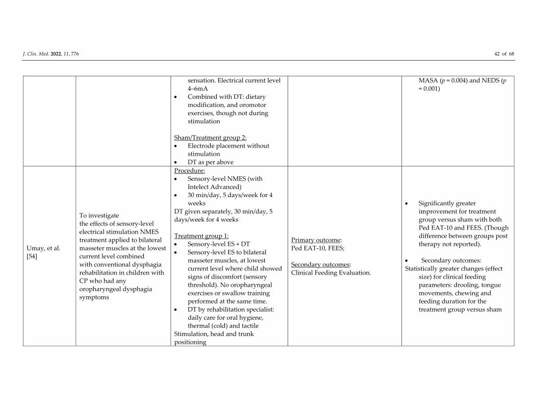
J. Clin. Med. 2022, 11, 776 42 of 68
sensation. Electrical current level
4–6mA
Combined with DT: dietary
modification, and oromotor
exercises, though not during
stimulation
Sham/Treatment group 2:
Electrode placement without
stimulation
DT as per above
MASA (p = 0.004) and NEDS (p
= 0.001)
Umay, et al.
[54]
To investigate
the effects of sensory‐level
electrical stimulation NMES
treatment applied to bilateral
masseter muscles at the lowest
current level combined
with conventional dysphagia
rehabilitation in children with
CP who had any
oropharyngeal dysphagia
symptoms
Procedure:
Sensory‐level NMES (with
Intelect Advanced)
30 min/day, 5 days/week for 4
weeks
DT given separately, 30 min/day, 5
days/week for 4 weeks
Treatment group 1:
Sensory‐level ES + DT
Sensory‐level ES to bilateral
masseter muscles, at lowest
current level where child showed
signs of discomfort (sensory
threshold). No oropharyngeal
exercises or swallow training
performed at the same time.
DT by rehabilitation specialist:
daily care for oral hygiene,
thermal (cold) and tactile
Stimulation, head and trunk
positioning
Primary outcome:
Ped EAT‐10, FEES;
Secondary outcomes:
Clinical Feeding Evaluation.
Significantly greater
improvement for treatment
group versus sham with both
Ped EAT‐10 and FEES. (Though
difference between groups post
therapy not reported).
Secondary outcomes:
Statistically greater changes (effect
size) for clinical feeding
parameters: drooling, tongue
movements, chewing and
feeding duration for the
treatment group versus sham

J. Clin. Med. 2022, 11, 776 43 of 68
and dietary modification. Oral motor
exercises included for children who
could participate.
Sham/Treatment group 2:
Sham ES + DT
Sham ES = same electrode
placement, no stimulus
DT as per above
Xia, et al. [55]
To investigate the effects of
VitalStim therapy coupled
with conventional swallowing
training on recovery of post‐
stroke dysphagia
Treatment group 1:
Standard swallow therapy (DT).
Schedule not reported
Direct and indirect OD training
related to food intake and
swallowing, body posture and
removal of pharyngeal food
residue
Treatment group 2:
NMES (VitalStim), 30 min, 2 x
day. 5 days/week for 4 weeks
Treatment group 3:
DT + VitalStim
Schedule not reported
Primary outcome:
Dysphagia Rating Scale b (as per
VFSS);
Secondary outcomes:
Maximum amplitude of surface
electromyography (sEMG) signals of
hyoid muscles; SWAL‐QOL.
Primary outcomes: All 3 groups
significantly improved post
treatment.
Significant greater improvement (p <
0.01) for group 3 (DT + VitalStim)
versus other 2 groups (DT only
group and VitalStim only group).
Secondary outcomes.
SWAL‐QOL and sEMG signals
significantly increased in all groups.
Significant difference between DT +
VitalStim (greater improvement)
versus DT group and VitalStim
group.
Zeng, et al. [56]
To observe the improvement
of swallow function and
negative affect disorders in
patients with cerebral
infarction and dysphagia by
NMES
Procedure:
1. NMES and/or swallow training
2. NMES via YS1002T
Glossopharyngeal Nerve and
Muscle Electrical Stimulator
(Changzhou Yasi Medical
Instruments Co)
Primary outcome:
Swallow function as per Kubota
water‐drinking test;
Secondary outcomes:
Negative affect disorders as per
Hamilton anxiety scale and
depression scale test.
Primary outcomes: Both groups
improved swallow function
post treatment, significantly
greater improvements (p =
0.035) for group 2 (swallow
training + NMES)
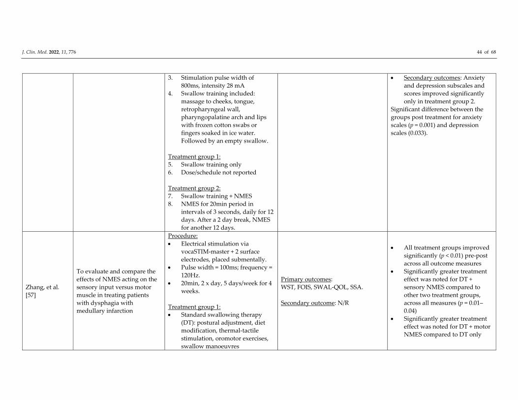
J. Clin. Med. 2022, 11, 776 44 of 68
3. Stimulation pulse width of
800ms, intensity 28 mA
4. Swallow training included:
massage to cheeks, tongue,
retropharyngeal wall,
pharyngopalatine arch and lips
with frozen cotton swabs or
fingers soaked in ice water.
Followed by an empty swallow.
Treatment group 1:
5. Swallow training only
6. Dose/schedule not reported
Treatment group 2:
7. Swallow training + NMES
8. NMES for 20min period in
intervals of 3 seconds, daily for 12
days. After a 2 day break, NMES
for another 12 days.
Secondary outcomes: Anxiety
and depression subscales and
scores improved significantly
only in treatment group 2.
Significant difference between the
groups post treatment for anxiety
scales (p = 0.001) and depression
scales (0.033).
Zhang, et al.
[57]
To evaluate and compare the
effects of NMES acting on the
sensory input versus motor
muscle in treating patients
with dysphagia with
medullary infarction
Procedure:
Electrical stimulation via
vocaSTIM‐master + 2 surface
electrodes, placed submentally.
Pulse width = 100ms; frequency =
120Hz.
20min, 2 x day, 5 days/week for 4
weeks.
Treatment group 1:
Standard swallowing therapy
(DT): postural adjustment, diet
modification, thermal‐tactile
stimulation, oromotor exercises,
swallow manoeuvres
Primary outcomes:
WST, FOIS, SWAL‐QOL, SSA.
Secondary outcome: N/R
All treatment groups improved
significantly (p < 0.01) pre‐post
across all outcome measures
Significantly greater treatment
effect was noted for DT +
sensory NMES compared to
other two treatment groups,
across all measures (p = 0.01–
0.04)
Significantly greater treatment
effect was noted for DT + motor
NMES compared to DT only
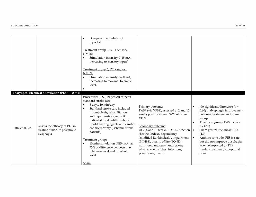
J. Clin. Med. 2022, 11, 776 45 of 68
Dosage and schedule not
reported
Treatment group 2, DT + sensory
NMES:
Stimulation intensity 0–15 mA,
increasing to ‘sensory input´.
Treatment group 3, DT + motor
NMES:
Stimulation intensity 0–60 mA,
increasing to maximal tolerable
level.
Pharyngeal Electrical Stimulation (PES) ― n = 8
Bath, et al. [58]
Assess the efficacy of PES in
treating subacute poststroke
dysphagia
Procedure: PES (Phagenyx) catheter +
standard stroke care
3 days, 10 min/day
Standard stroke care included
thrombolysis; rehabilitation;
antihypertensive agents; if
indicated, oral antithrombotic,
lipid‐lowering agents and carotid
endarterectomy (ischemic stroke
patients)
Treatment group:
10 min stimulation, PES (mA) at
75% of difference between max
tolerance level and threshold
level
Sham:
Primary outcome:
PAS b (via VFSS), assessed at 2 and 12
weeks post treatment. 3–7 bolus per
VFSS.
Secondary outcome:
At 2, 6 and 12 weeks = DSRS, function
(Barthel Index), dependency
(modified Rankin Scale), impairment
(NIHSS), quality of life (EQ‐5D),
nutritional measures and serious
adverse events (chest infections,
pneumonia, death).
No significant difference (p =
0.60) in dysphagia improvement
between treatment and sham
group
Treatment group: PAS mean =
3.7 (2.0)
Sham group: PAS mean = 3.6
(1.9)
Authors conclude: PES is safe
but did not improve dysphagia.
May be impacted by PES
‘under‐treatment’/suboptimal
dose
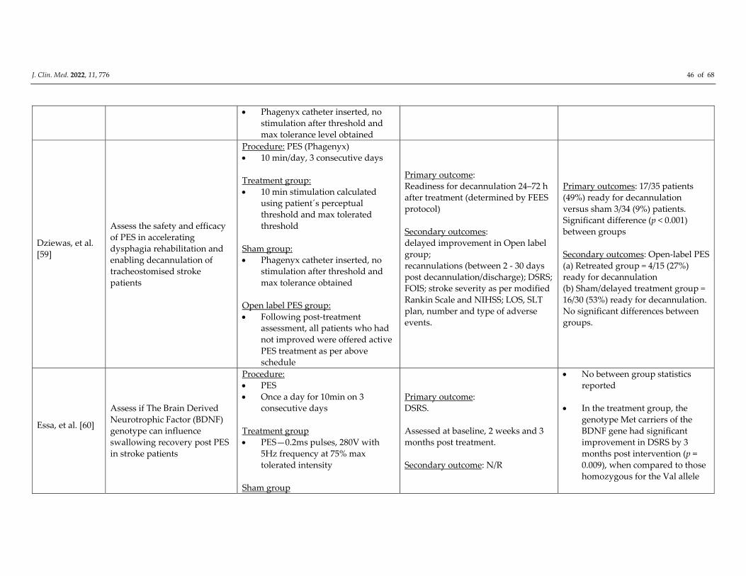
J. Clin. Med. 2022, 11, 776 46 of 68
Phagenyx catheter inserted, no
stimulation after threshold and
max tolerance level obtained
Dziewas, et al.
[59]
Assess the safety and efficacy
of PES in accelerating
dysphagia rehabilitation and
enabling decannulation of
tracheostomised stroke
patients
Procedure: PES (Phagenyx)
10 min/day, 3 consecutive days
Treatment group:
10 min stimulation calculated
using patient´s perceptual
threshold and max tolerated
threshold
Sham group:
Phagenyx catheter inserted, no
stimulation after threshold and
max tolerance obtained
Open label PES group:
Following post‐treatment
assessment, all patients who had
not improved were offered active
PES treatment as per above
schedule
Primary outcome:
Readiness for decannulation 24–72 h
after treatment (determined by FEES
protocol)
Secondary outcomes:
delayed improvement in Open label
group;
recannulations (between 2 ‐ 30 days
post decannulation/discharge); DSRS;
FOIS; stroke severity as per modified
Rankin Scale and NIHSS; LOS, SLT
plan, number and type of adverse
events.
Primary outcomes: 17/35 patients
(49%) ready for decannulation
versus sham 3/34 (9%) patients.
Significant difference (p < 0.001)
between groups
Secondary outcomes: Open‐label PES
(a) Retreated group = 4/15 (27%)
ready for decannulation
(b) Sham/delayed treatment group =
16/30 (53%) ready for decannulation.
No significant differences between
groups.
Essa, et al. [60]
Assess if The Brain Derived
Neurotrophic Factor (BDNF)
genotype can influence
swallowing recovery post PES
in stroke patients
Procedure:
PES
Once a day for 10min on 3
consecutive days
Treatment group
PES—0.2ms pulses, 280V with
5Hz frequency at 75% max
tolerated intensity
Sham group
Primary outcome:
DSRS.
Assessed at baseline, 2 weeks and 3
months post treatment.
Secondary outcome: N/R
No between group statistics
reported
In the treatment group, the
genotype Met carriers of the
BDNF gene had significant
improvement in DSRS by 3
months post intervention (p =
0.009), when compared to those
homozygous for the Val allele
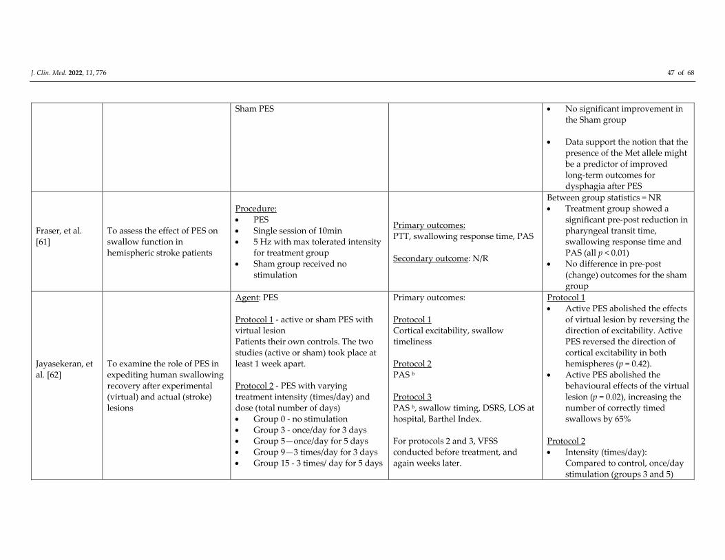
J. Clin. Med. 2022, 11, 776 47 of 68
Sham PES No significant improvement in
the Sham group
Data support the notion that the
presence of the Met allele might
be a predictor of improved
long‐term outcomes for
dysphagia after PES
Fraser, et al.
[61]
To assess the effect of PES on
swallow function in
hemispheric stroke patients
Procedure:
PES
Single session of 10min
5 Hz with max tolerated intensity
for treatment group
Sham group received no
stimulation
Primary outcomes:
PTT, swallowing response time, PAS
Secondary outcome: N/R
Between group statistics = NR
Treatment group showed a
significant pre‐post reduction in
pharyngeal transit time,
swallowing response time and
PAS (all p < 0.01)
No difference in pre‐post
(change) outcomes for the sham
group
Jayasekeran, et
al. [62]
To examine the role of PES in
expediting human swallowing
recovery after experimental
(virtual) and actual (stroke)
lesions
Agent: PES
Protocol 1 ‐ active or sham PES with
virtual lesion
Patients their own controls. The two
studies (active or sham) took place at
least 1 week apart.
Protocol 2 ‐ PES with varying
treatment intensity (times/day) and
dose (total number of days)
Group 0 ‐ no stimulation
Group 3 ‐ once/day for 3 days
Group 5—once/day for 5 days
Group 9—3 times/day for 3 days
Group 15 ‐ 3 times/ day for 5 days
Primary outcomes:
Protocol 1
Cortical excitability, swallow
timeliness
Protocol 2
PAS b
Protocol 3
PAS b, swallow timing, DSRS, LOS at
hospital, Barthel Index.
For protocols 2 and 3, VFSS
conducted before treatment, and
again weeks later.
Protocol 1
Active PES abolished the effects
of virtual lesion by reversing the
direction of excitability. Active
PES reversed the direction of
cortical excitability in both
hemispheres (p = 0.42).
Active PES abolished the
behavioural effects of the virtual
lesion (p = 0.02), increasing the
number of correctly timed
swallows by 65%
Protocol 2
Intensity (times/day):
Compared to control, once/day
stimulation (groups 3 and 5)

J. Clin. Med. 2022, 11, 776 48 of 68
Protocol 3 ‐ active or sham PES with
acute stroke.
Once daily on three consecutive days.
Secondary outcome: N/R produced the greatest reduction
in aspiration (p = .04)
Dose: Compared to control,
total of 3 days of stimulation
(groups 3 and 9) showed the
greatest reduction in aspiration
scores (p = 0.038)
Protocol 3
Reduction of PAS post
intervention for the active PES
group compared to sham = NS
(p = 0.49)
No significant changes in
swallow timing for either group
Significantly reduced DSRS in
the PES group (p = 0.04)
NS shorter stay in hospital for
the PES group (p = 0.38)
Restivo, et al.
[63]
To investigate whether
intraluminal electrical
pharyngeal stimulation
facilitates swallowing recovery
in dysphagic multiple sclerosis
(MS) patients
Procedure: PES (bipolar platinum
pharyngeal ring electrodes built into 3
mm‐diameter intraluminal catheter)
using constant/current electrical
simulator (DS7)
Stimulation 10 min, 5 consecutive
days
Treatment group:
5 Hz pharyngeal stimulation (mA
calculated using sensory
threshold and pain thresholds,
mean = 14.2 ± 0.6 mA)
Sham:
Same catheter, no stimulation
Primary outcome:
PAS via VFSS at pre‐treatment (T0),
immediately after treatment (T1),
after two (T2), and four (T3) weeks of
PES.
Secondary outcomes:
sEMG measure of:
(1) duration of laryngeal excursion;
(2) duration of the sEMG activity of
suprahyoid/submental muscles; (3)
duration of the inhibition of the CP
muscle; and (4) interval between
onset of suprahyoid/submental
muscles and onset of laryngeal
elevation.
Significant difference between
treatment and sham group
immediately and 4 week post
treatment, for PAS (p < 0.0001)
and all secondary measures (p <
0.0001)
Treatment group improved
significantly across all
measures, sham group did not
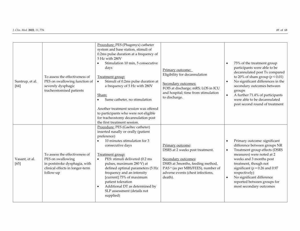
J. Clin. Med. 2022, 11, 776 49 of 68
Suntrup, et al.
[64]
To assess the effectiveness of
PES on swallowing function of
severely dysphagic
tracheostomised patients
Procedure: PES (Phagenyx) catheter
system and base station, stimuli of
0.2ms pulse duration at a frequency of
5 Hz with 280V
Stimulation 10 min, 5 consecutive
days
Treatment group:
Stimuli of 0.2ms pulse duration at
a frequency of 5 Hz with 280V
Sham:
Same catheter, no stimulation
Another treatment session was offered
to participants who were not eligible
for tracheostomy decannulation post
the first treatment session.
Primary outcome:
Eligibility for decannulation
Secondary outcomes:
FOIS at discharge; mRS; LOS in ICU
and hospital; time from stimulation
to discharge.
75% of the treatment group
participants were able to be
decannulated post Tx compared
to 20% of sham group (p < 0.01)
No significant differences in the
secondary outcomes between
groups
A further 71.4% of participants
were able to be decannulated
post second round of treatment
Vasant, et al.
[65]
To assess the effectiveness of
PES on swallowing
in poststroke dysphagia, with
clinical effects in longer‐term
follow‐up
Procedure: PES (Gaeltec catheter)
inserted nasally or orally (patient
preference)
10 minutes stimulation for 3
consecutive days
Treatment group:
PES: stimuli delivered (0.2 ms
pulses, maximum 280 V) at
defined optimal parameters (5 Hz
frequency and an intensity
[current] 75% of maximum
patient toleration
Additional DT as determined by
SLP assessment (details not
supplied)
Primary outcome:
DSRS at 2 weeks post treatment.
Secondary outcomes:
DSRS at 3months, feeding method,
PAS b (as per MBS/FEES), number of
adverse events (chest infections,
death).
Primary outcome: significant
difference between groups NR
Treatment group effects (DSRS
measures) were noted at 2
weeks and 3 months post
treatment, though not
significant (p = 0.26 and 0.97
respectively)
No significant difference
reported between groups for
most secondary outcomes
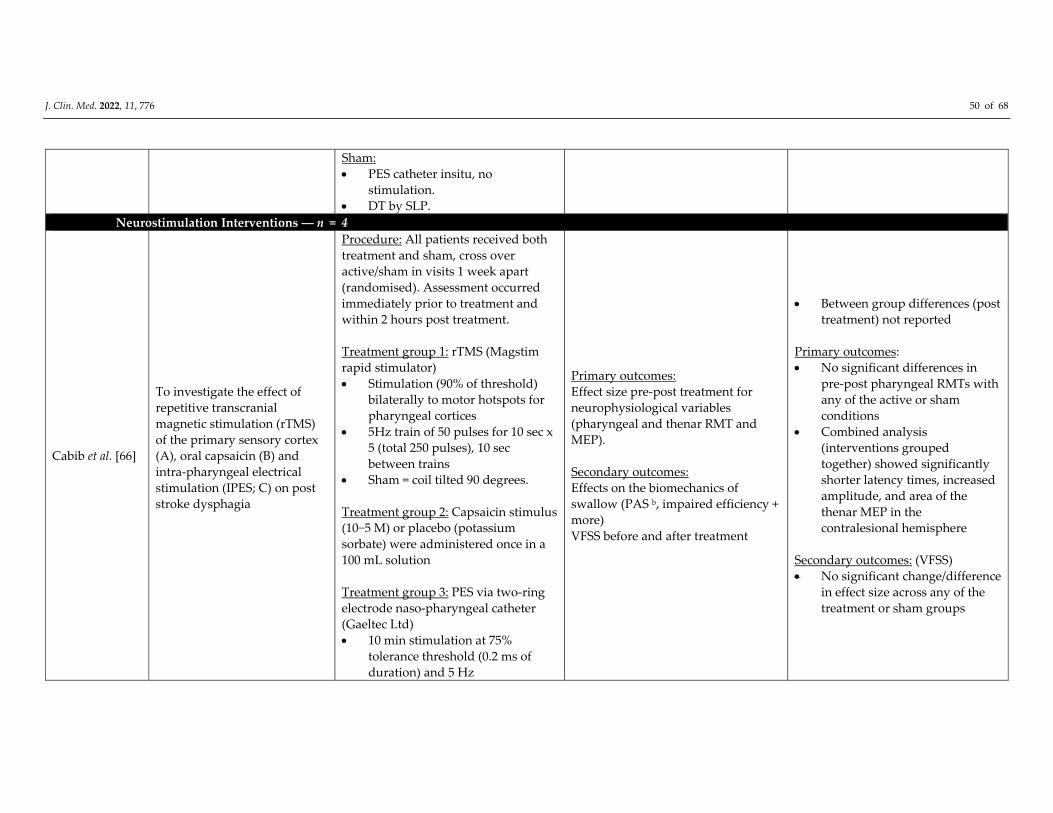
J. Clin. Med. 2022, 11, 776 50 of 68
Sham:
PES catheter insitu, no
stimulation.
DT by SLP.
Combined Neurostimulation Interventions ― n = 4
Cabib et al. [66]
To investigate the effect of
repetitive transcranial
magnetic stimulation (rTMS)
of the primary sensory cortex
(A), oral capsaicin (B) and
intra‐pharyngeal electrical
stimulation (IPES; C) on post
stroke dysphagia
Procedure: All patients received both
treatment and sham, cross over
active/sham in visits 1 week apart
(randomised). Assessment occurred
immediately prior to treatment and
within 2 hours post treatment.
Treatment group 1: rTMS (Magstim
rapid stimulator)
Stimulation (90% of threshold)
bilaterally to motor hotspots for
pharyngeal cortices
5Hz train of 50 pulses for 10 sec x
5 (total 250 pulses), 10 sec
between trains
Sham = coil tilted 90 degrees.
Treatment group 2: Capsaicin stimulus
(10−5 M) or placebo (potassium
sorbate) were administered once in a
100 mL solution
Treatment group 3: PES via two‐ring
electrode naso‐pharyngeal catheter
(Gaeltec Ltd)
10 min stimulation at 75%
tolerance threshold (0.2 ms of
duration) and 5 Hz
Primary outcomes:
Effect size pre‐post treatment for
neurophysiological variables
(pharyngeal and thenar RMT and
MEP).
Secondary outcomes:
Effects on the biomechanics of
swallow (PAS b, impaired efficiency +
more)
VFSS before and after treatment
Between group differences (post
treatment) not reported
Primary outcomes:
No significant differences in
pre‐post pharyngeal RMTs with
any of the active or sham
conditions
Combined analysis
(interventions grouped
together) showed significantly
shorter latency times, increased
amplitude, and area of the
thenar MEP in the
contralesional hemisphere
Secondary outcomes: (VFSS)
No significant change/difference
in effect size across any of the
treatment or sham groups
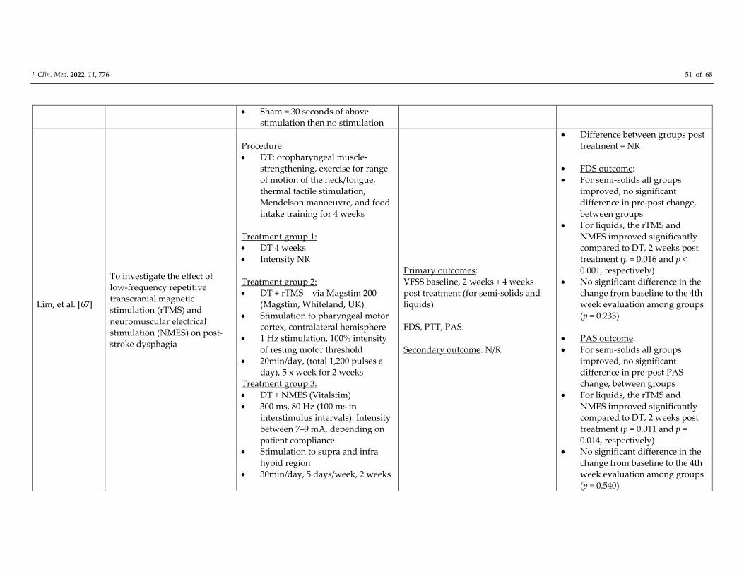
J. Clin. Med. 2022, 11, 776 51 of 68
Sham = 30 seconds of above
stimulation then no stimulation
Lim, et al. [67]
To investigate the effect of
low‐frequency repetitive
transcranial magnetic
stimulation (rTMS) and
neuromuscular electrical
stimulation (NMES) on post‐
stroke dysphagia
Procedure:
DT: oropharyngeal muscle‐
strengthening, exercise for range
of motion of the neck/tongue,
thermal tactile stimulation,
Mendelson manoeuvre, and food
intake training for 4 weeks
Treatment group 1:
DT 4 weeks
Intensity NR
Treatment group 2:
DT + rTMS via Magstim 200
(Magstim, Whiteland, UK)
Stimulation to pharyngeal motor
cortex, contralateral hemisphere
1 Hz stimulation, 100% intensity
of resting motor threshold
20min/day, (total 1,200 pulses a
day), 5 x week for 2 weeks
Treatment group 3:
DT + NMES (Vitalstim)
300 ms, 80 Hz (100 ms in
interstimulus intervals). Intensity
between 7–9 mA, depending on
patient compliance
Stimulation to supra and infra
hyoid region
30min/day, 5 days/week, 2 weeks
Primary outcomes:
VFSS baseline, 2 weeks + 4 weeks
post treatment (for semi‐solids and
liquids)
FDS, PTT, PAS.
Secondary outcome: N/R
Difference between groups post
treatment = NR
FDS outcome:
For semi‐solids all groups
improved, no significant
difference in pre‐post change,
between groups
For liquids, the rTMS and
NMES improved significantly
compared to DT, 2 weeks post
treatment (p = 0.016 and p <
0.001, respectively)
No significant difference in the
change from baseline to the 4th
week evaluation among groups
(p = 0.233)
PAS outcome:
For semi‐solids all groups
improved, no significant
difference in pre‐post PAS
change, between groups
For liquids, the rTMS and
NMES improved significantly
compared to DT, 2 weeks post
treatment (p = 0.011 and p =
0.014, respectively)
No significant difference in the
change from baseline to the 4th
week evaluation among groups
(p = 0.540)
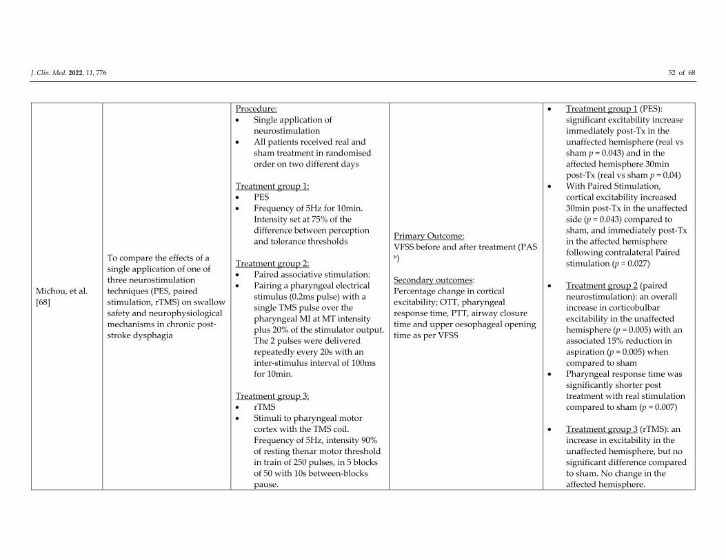
J. Clin. Med. 2022, 11, 776 52 of 68
Michou, et al.
[68]
To compare the effects of a
single application of one of
three neurostimulation
techniques (PES, paired
stimulation, rTMS) on swallow
safety and neurophysiological
mechanisms in chronic post‐
stroke dysphagia
Procedure:
Single application of
neurostimulation
All patients received real and
sham treatment in randomised
order on two different days
Treatment group 1:
PES
Frequency of 5Hz for 10min.
Intensity set at 75% of the
difference between perception
and tolerance thresholds
Treatment group 2:
Paired associative stimulation:
Pairing a pharyngeal electrical
stimulus (0.2ms pulse) with a
single TMS pulse over the
pharyngeal MI at MT intensity
plus 20% of the stimulator output.
The 2 pulses were delivered
repeatedly every 20s with an
inter‐stimulus interval of 100ms
for 10min.
Treatment group 3:
rTMS
Stimuli to pharyngeal motor
cortex with the TMS coil.
Frequency of 5Hz, intensity 90%
of resting thenar motor threshold
in train of 250 pulses, in 5 blocks
of 50 with 10s between‐blocks
pause.
Primary Outcome:
VFSS before and after treatment (PAS b)
Secondary outcomes:
Percentage change in cortical
excitability; OTT, pharyngeal
response time, PTT, airway closure
time and upper oesophageal opening
time as per VFSS
Treatment group 1 (PES):
significant excitability increase
immediately post‐Tx in the
unaffected hemisphere (real vs
sham p = 0.043) and in the
affected hemisphere 30min
post‐Tx (real vs sham p = 0.04)
With Paired Stimulation,
cortical excitability increased
30min post‐Tx in the unaffected
side (p = 0.043) compared to
sham, and immediately post‐Tx
in the affected hemisphere
following contralateral Paired
stimulation (p = 0.027)
Treatment group 2 (paired
neurostimulation): an overall
increase in corticobulbar
excitability in the unaffected
hemisphere (p = 0.005) with an
associated 15% reduction in
aspiration (p = 0.005) when
compared to sham
Pharyngeal response time was
significantly shorter post
treatment with real stimulation
compared to sham (p = 0.007)
Treatment group 3 (rTMS): an
increase in excitability in the
unaffected hemisphere, but no
significant difference compared
to sham. No change in the
affected hemisphere.
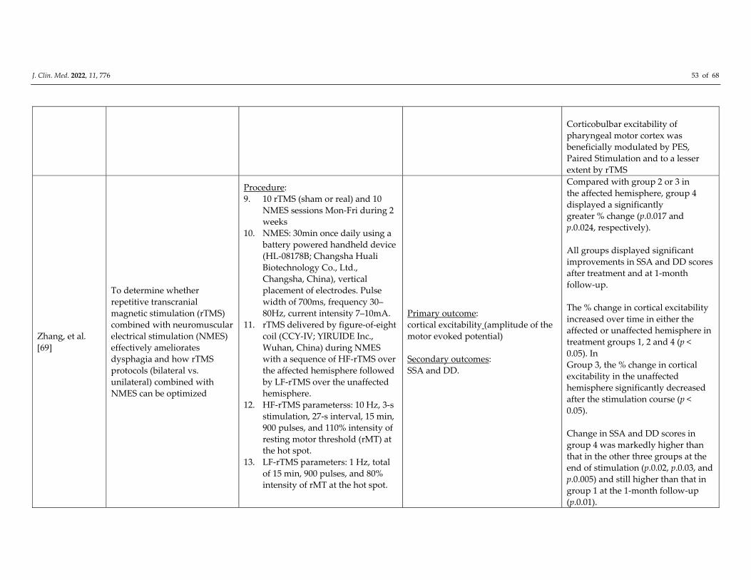
J. Clin. Med. 2022, 11, 776 53 of 68
Corticobulbar excitability of
pharyngeal motor cortex was
beneficially modulated by PES,
Paired Stimulation and to a lesser
extent by rTMS
Zhang, et al.
[69]
To determine whether
repetitive transcranial
magnetic stimulation (rTMS)
combined with neuromuscular
electrical stimulation (NMES)
effectively ameliorates
dysphagia and how rTMS
protocols (bilateral vs.
unilateral) combined with
NMES can be optimized
Procedure:
9. 10 rTMS (sham or real) and 10
NMES sessions Mon‐Fri during 2
weeks
10. NMES: 30min once daily using a
battery powered handheld device
(HL‐08178B; Changsha Huali
Biotechnology Co., Ltd.,
Changsha, China), vertical
placement of electrodes. Pulse
width of 700ms, frequency 30–
80Hz, current intensity 7–10mA.
11. rTMS delivered by figure‐of‐eight
coil (CCY‐IV; YIRUIDE Inc.,
Wuhan, China) during NMES
with a sequence of HF‐rTMS over
the affected hemisphere followed
by LF‐rTMS over the unaffected
hemisphere.
12. HF‐rTMS parameterss: 10 Hz, 3‐s
stimulation, 27‐s interval, 15 min,
900 pulses, and 110% intensity of
resting motor threshold (rMT) at
the hot spot.
13. LF‐rTMS parameters: 1 Hz, total
of 15 min, 900 pulses, and 80%
intensity of rMT at the hot spot.
Primary outcome:
cortical excitability (amplitude of the
motor evoked potential)
Secondary outcomes:
SSA and DD.
Compared with group 2 or 3 in
the affected hemisphere, group 4
displayed a significantly
greater % change (p.0.017 and
p.0.024, respectively).
All groups displayed significant
improvements in SSA and DD scores
after treatment and at 1‐month
follow‐up.
The % change in cortical excitability
increased over time in either the
affected or unaffected hemisphere in
treatment groups 1, 2 and 4 (p <
0.05). In
Group 3, the % change in cortical
excitability in the unaffected
hemisphere significantly decreased
after the stimulation course (p <
0.05).
Change in SSA and DD scores in
group 4 was markedly higher than
that in the other three groups at the
end of stimulation (p.0.02, p.0.03, and
p.0.005) and still higher than that in
group 1 at the 1‐month follow‐up
(p.0.01).
J. Clin. Med. 2022, 11, 776 54 of 68
Treatment group 1: Sham rTMS +
NMES
10‐Hz sham rTMS delivered to
the hot spot for the mylohyoid
muscle at the ipsilesional
hemisphere followed by 1‐Hz
sham rTMS over the
corresponding position of the
contralesional hemisphere.
Delivered using a vertical coil tilt,
generating the same noise as real
rTMS without cortical
stimulation.
Treatment group 2: Ipsilateral rTMS +
NMES
10‐Hz real rTMS was delivered to the
hot spot for the mylohyoid muscle at
the ipsilesional hemisphere followed
by 1‐Hz sham rTMS over the
corresponding position of the
contralesional hemisphere.
Treatment group 3: Contralateral
rTMS + NMES
10‐Hz sham rTMS was delivered to the
hot spot for the mylohyoid muscle at
the ipsilesional hemisphere followed
by 1‐Hz real rTMS over the
corresponding position of the
contralesional hemisphere
Treatment group 4: Bilateral rTMS +
NMES
J. Clin. Med. 2022, 11, 776 55 of 68
10‐Hz real rTMS was delivered to the
hot spot for the mylohyoid muscle at
the ipsilesional hemisphere followed
by 1‐Hz real rTMS over the
corresponding position of the
contralesional hemisphere a NMES is at motor stimulation level unless explicitly mentioned. b Data included in meta‐analyses. Notes. ASHA‐NOMS–American speech‐language‐hearing
association national outcome measurement system; BASOFF– behavioural assessment scale of oral functions in feeding; BI–Barthel index; CDS–clinical dysphagia
scale; CNS—central nervous system; CP—cerebral palsy; CT–computed tomography; CVA–cerebrovascular accident; DD–degree of dysphagia; A‐DHI–Arabic
dysphagia handicap index; DOSS–dysphagia outcome and severity scale; DSRS–dysphagia severity rating scale; DT–dysphagia therapy; EAT‐10–eating
assessment tool‐10; EES– electrokinesiographic/electromyographic study of swallowing; EQ‐5D–European Quality of Life Five Dimension; FDS—functional
dysphagia scale; FOIS–functional oral intake scale; FEDSS–fiberoptic endoscopic dysphagia severity scale; FEES–fiberoptic endoscopic evaluation of swallowing;
HNCI–head neck cancer inventory; IADL–instrumental activities of daily living; ICH–intracranial haemorrhage; ICU–intensive care unit; LPM–laryngeal‐
pharyngeal mechanogram; MASA–Mann assessment of swallowing ability; MDADI–M.D. Anderson dysphagia inventory; LCD–laryngeal closure duration; LOS–
length of stay; MBS–modified barium swallow; MBSImp–modified barium swallow impairment profile; MEG–magnetoencephalography; MMSE–mini‐mental
state exam; MEP–motor evoked potentials; MRI–magnetic resonance imaging; mRS–modified rankin scale; MS–multiple sclerosis; NEDS–neurological
examination dysphagia score; NIHSS–national institutes of health stroke scale; NIHSS–National Institutes of Health Stroke Scale; NMES–neuromuscular electrical
stimulation; NS–not significant; OD–oropharyngeal dysphagia; OPSE–oropharyngeal swallow efficiency; OST–oral sensorimotor treatment; OTT–oral transit time;
PAS–penetration–aspiration score; PED EAT‐10 pediatric eating assessment tool‐10;PES–pharyngeal electrical stimulation; PESO– pharyngoesophageal segment
opening; PPS–performance status scale; PTT–pharyngeal transit time; RMT– resting motor threshold; RSST–repetitive saliva swallowing test; rTMS–repetitive
transcranial magnetic stimulation; SAH–subarachnoid haemorrhage; SAPP–swallowing activity and participation profile; SDQ–swallowing disturbance
questionnaire; sEMG–surface electromyography; SFS–swallow function score; SHEMG– electromyographic activity of the submental/suprahyoid muscles
complex; SI–similarity index; SLT–speech and language therapist; SSA–standardised swallowing assessment; SWAL‐QOL—swallowing quality of life; TBI–
traumatic brain injury; tDCS–transcranial direct current stimulation; TOR‐BSST–Toronto bedside swallowing screening test; UES–upper esophageal sphincter;
UPDRS–unified Parkinson’s disease rating scale; VAS–visual analogue scale; VFSS–videofluoroscopic swallowing study; VVS‐T–volume viscosity swallow test;
WST–water swallow test.
J. Clin. Med. 2022, 11, 776 56 of 68
Peripheral Neurostimulation Interventions. Across the 42 included studies, 30 studies
reported on NMES and eight studies reported on PES. Four studies used another type of
neurostimulation (i.e., rTMS) in addition to NMES or PES, either within the same group
or different treatment groups.
Participants (Table 2). The 42 studies included a total of 2281 participants (mean 54.3;
SD 39.1). The sample sizes ranged from the smallest sample of 16 participants [60,61] to
the largest sample of 162 participants [36]. By intervention type, samples were
characterized as follows: NMES total 1706, mean 56.9, SD 38.9, range 18–135; PES total
410, mean 51.3, SD 49.0, range 16–162; and combined neurostimulation total 165, mean
41.3, SD 19.3, range 18–64. The mean age of participants across all studies was 61.8 years
(SD 15.3), with one study reporting age range only (65–93 years) [61]. Participant mean
age across all studies ranged from 4.2 years [54] to 84.4 years [39]. The mean age of
participants by intervention group was: NMES 60.9 years (SD 16.9), PES 64.7 years (SD
11.9), and combined neurostimulation 63.8 years (SD 6.4).
Across all studies, 61.0% (SD 13.5) of participants were male and one study did not
report gender distribution [30]. Percentage of males by intervention group was NMES
62.6% (SD 14.0), PES 56.7% (SD 9.6), and other/combined 65.4% (SD 12.3). Most studies
included stroke patients (n = 31), while three studies included mixed populations
[28,41,43] and one study reported OD without further underlying medical diagnosis [39].
Other diagnoses by intervention group were: Parkinson’s disorder (n = 2)[32,46], cerebral
palsy (n = 2) [50,54], and head and neck cancer (n = 2) [36,48] in NMES; and multiple
sclerosis (n = 1) [63] in PES.
Across the 42 studies, VFSS was most frequently used to confirm participant’s
diagnosis of OD (n = 31), whereas six studies used FEES [49,53,54,60,64,65]. Several of
these studies combined instrumental assessment with either a screen (n = 2) [58,65] or
clinical assessment (n = 6) [49,50,53–55,68]. One study used either clinical assessment or
VFSS [50]. One study used a single screen [56], three studies used clinical assessment only
[35,38,59], and one study used both [33]. The studies were conducted across 14 countries,
with studies most frequently conducted in Korea (n = 11), China (n = 7), the UK (n = 7),
Spain (n = 4), Italy (n = 2), Turkey (n = 2), and Germany (n = 2).
Outcome Measures (Table 2). Outcomes measures varied greatly across all studies
included in the review, covering several domains within the area of OD. The Penetration
Aspiration Score was the most reported outcome measure (PAS; 18 studies), followed by
Functional Oral Intake Scale (FOIS; 12 studies), Functional Dysphagia Scale (FDS; 5
studies), Dysphagia Severity Rating Scale (DSRS; 5 studies), Swallowing Quality of Life
questionnaire (SWAL‐QOL; 4 studies), and Dysphagia Outcome and Severity Scale
(DOSS; 3 studies).
NMES Intervention (n = 30: Tables 2 and 3). In total, 22 studies included two study
arms or groups, whereas eight studies included three groups [31–34,38,40,55,57]. All but
five NMES studies [29,39,43,53,54] combined neurostimulation with simultaneous DT
consisting of a wide range of behavioural interventions (e.g., head and body positioning,
bolus modification, oromotor exercises, or swallow manoeuvres). Six studies included a
NMES only group without DT [29,33,38,39,43,55], with five of these studies using NMES
at motor stimulation level [29,33,38,43,55] and one study using NMES at sensory
stimulation level [39]. An additional seven studies included a treatment arm with NMES
at sensory stimulation level combined with DT [32,44–46,53,54,57]. All other participants
in NMES groups received stimulation at motor level. Five studies compared different
NMES electrode positions [28,34,40–42] and seven studies included a sham stimulation
group [36,39,48,50,52–54].
Control groups included mostly sham NMES stimulation and/or DT. Only one study
included a control group receiving neither DT nor NMES [30], and one study included
usual care across different healthcare settings as the comparison group [51].
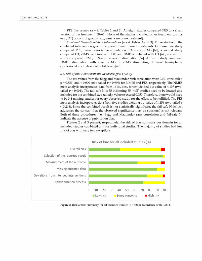
J. Clin. Med. 2022, 11, 776 57 of 68
PES Intervention (n = 8: Tables 2 and 3). All eight studies compared PES to a sham
version of the treatment [58–65]. None of the studies included other treatment groups
(e.g., DT) or control groups (e.g., usual care or no treatment).
Combined Neurostimulation Interventions (n = 4: Tables 2 and 3). Three studies in the
combined intervention group compared three different treatments. Of these, one study
compared PES, paired associative stimulation (PAS) and rTMS [68], a second study
compared DT, rTMS combined with DT, and NMES combined with DT [67], and a third
study compared rTMS, PES and capsaicin stimulation [66]. A fourth study combined
NMES stimulation with sham rTMS or rTMS stimulating different hemispheres
(ipsilesional, contralesional or bilateral) [69].
5.3. Risk of Bias Assessment and Methodological Quality
The tau values from the Begg and Mazumdar rank correlation were 0.101 (two‐tailed
𝑝 = 0.589) and < 0.000 (two‐tailed 𝑝 > 0.999) for NMES and PES, respectively. The NMES
meta‐analysis incorporates data from 16 studies, which yielded a z‐value of 4.107 (two‐
tailed p < 0.001). The fail‐safe N is 55 indicating 55 ‘null’ studies need to be located and
included for the combined two‐tailed p‐value to exceed 0.050. Therefore, there would need
to be 3.4 missing studies for every observed study for the effect to be nullified. The PES
meta‐analysis incorporates data from five studies yielding a z‐value of 1.156 (two‐tailed p
< 0.248). Since the combined result is not statistically significant, the fail‐safe N (which
addresses the concern that the observed significance may be spurious) is not relevant.
Both of these procedures (i.e., Begg and Mazumdar rank correlation and fail‐safe N)
indicate the absence of publication bias.
Figures 2 and 3 present, respectively, the risk of bias summary per domain for all
included studies combined and for individual studies. The majority of studies had low
risk of bias with very few exceptions.
Figure 2. Risk of bias summary for all included studies (n = 42) in accordance with RoB‐2.
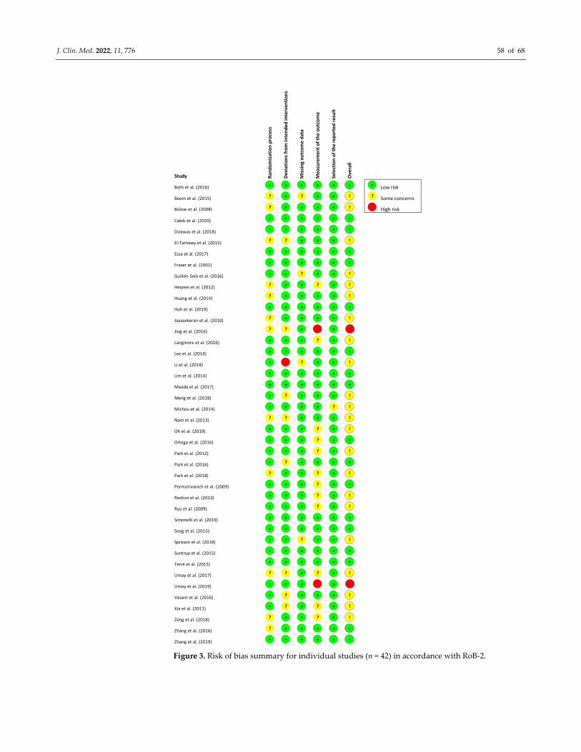
J. Clin. Med. 2022, 11, 776 58 of 68
Figure 3. Risk of bias summary for individual studies (n = 42) in accordance with RoB‐2.
Study Ran
domization process
Deviations from intended
interven
tions
Missing outcome data
Measurement of the outcome
Selection of the reported
result
Overall
Bath et al. (2016) Low risk
Beom et al. (2015) Some concerns
Bülow et al. (2008) High risk
Cabib et al. (2020)
Dziewas et al. (2018)
El‐Tamawy et al. (2015)
Essa et al. (2017)
Fraser et al. (2002)
Guillén‐Solà et al. (2016)
Heijnen et al. (2012)
Huang et al. (2014)
Huh et al. (2019)
Jayasekeran et al. (2010)
Jing et al. (2016)
Langmore et al. (2016)
Lee et al. (2014)
Li et al. (2018)
Lim et al. (2014)
Maeda et al. (2017)
Meng et al. (2018)
Michou et al. (2014)
Nam et al. (2013)
Oh et al. (2019)
Ortega et al. (2016)
Park et al. (2012)
Park et al. (2016)
Park et al. (2018)
Permsirivanich et al. (2009)
Restivo et al. (2013)
Ryu et al. (2009)
Simonelli et al. (2019)
Song et al. (2015)
Sproson et al. (2018)
Suntrup et al. (2015)
Terré et al. (2015)
Umay et al. (2017)
Umay et al. (2019)
Vasant et al. (2016)
Xia et al. (2011)
Zeng et al. (2018)
Zhang et al. (2016)
Zhang et al. (2019)
+
?
?
+
+
?
+
+
+
?
?
+
?
?
+
+
+
+
+
+
+
?
+
+
+
+
?
+
+
+
+
+
+
+
+
?
+
+
+
?
?
+
+
+
+
+
+
?
+
+
+
+
+
+
+
?
+
+
‹
+
+
?
+
?
+
+
+
?
+
+
+
+
+
+
+
+
+
?
+
?
?
+
+
+
+
?
+
+
+
+
+
+
?
+
+
+
+
+
+
+
?
+
+
+
+
+
+
+
+
+
+
+
+
+
+
+
?
+
+
+
+
+
+
+
+
+
+
+
+
+
+
+
+
+
+
?
+
+
+
‹
?
+
+
+
+
+
+
+
?
?
?
+
?
?
?
?
+
+
+
+
+
?
‹
+
?
?
+
+
+
+
+
+
+
+
+
+
+
+
+
+
+
+
+
+
+
+
+
+
?
+
+
+
+
+
+
+
+
+
+
+
+
+
+
+
+
+
+
+
+
+
+
!
!
+
+
!
+
+
!
!
!
+
!
‹
!
+
!
+
+
!
!
!
!
+
!
+
!
+
!
!
+
+
!
+
+
!
‹
!
!
!
+
+
+
?
‹
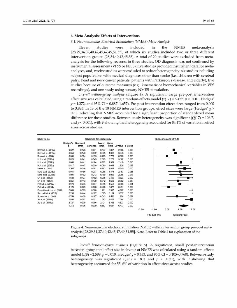
J. Clin. Med. 2022, 11, 776 59 of 68
6. Meta‐Analysis: Effects of Interventions
6.1. Neuromuscular Electrical Stimulation (NMES) Meta‐Analysis
Eleven studies were included in the NMES meta‐analysis
[28,29,34,37,40,42,45,47,49,51,55], of which six studies included two or three different
intervention groups [28,34,40,42,45,55]. A total of 20 studies were excluded from meta‐
analysis for the following reasons: in three studies, OD diagnosis was not confirmed by
instrumental assessment (VFSS or FEES); five studies provided insufficient data for meta‐
analyses; and, twelve studies were excluded to reduce heterogeneity: six studies including
subject populations with medical diagnoses other than stroke (i.e., children with cerebral
palsy, head and neck cancer patients, patients with Parkinson’s disease, and elderly), five
studies because of outcome measures (e.g., kinematic or biomechanical variables in VFS
recordings), and one study using sensory NMES stimulation.
Overall within‐group analysis (Figure 4). A significant, large pre‐post intervention
effect size was calculated using a random‐effects model (z(17) = 6.477, p < 0.001, Hedges’
g = 1.272, and 95% CI = 0.887–1.657). Pre‐post intervention effect sizes ranged from 0.000
to 3.826. In 13 of the 18 NMES intervention groups, effect sizes were large (Hedges’ g >
0.8), indicating that NMES accounted for a significant proportion of standardized mean
difference for these studies. Between‐study heterogeneity was significant (Q(17) = 106.7,
and p < 0.001), with I2 showing that heterogeneity accounted for 84.1% of variation in effect
sizes across studies.
Figure 4. Neuromuscular electrical stimulation (NMES) within intervention group pre‐post meta‐
analysis [28,29,34,37,40,42,45,47,49,51,55]. Note. Refer to Table 2 for explanation of the
subgroups.
Overall between‐group analysis (Figure 5). A significant, small post‐intervention
between‐group total effect size in favour of NMES was calculated using a random‐effects
model (z(8) = 2.589, p = 0.010, Hedges’ g = 0.433, and 95% CI = 0.105–0.760). Between‐study
heterogeneity was significant (Q(8) = 18.0, and p = 0.021), with I2 showing that
heterogeneity accounted for 55. 6% of variation in effect sizes across studies.
Study name Statistics for each study Hedges's g and 95% CI
Hedges's Standard Lower Upper g error Variance limit limit Z-Value p-Value
Beom et al. (2015a) 0.522 0.176 0.031 0.177 0.867 2.968 0.003Beom et al. (2015b) 0.653 0.178 0.032 0.305 1.001 3.676 0.000Bulow et al. (2008) 0.000 0.394 0.155 -0.773 0.773 0.000 1.000Huh et al. (2019a) 3.826 0.741 0.549 2.373 5.279 5.162 0.000Huh et al. (2019b) 1.066 0.441 0.194 0.202 1.929 2.419 0.016Huh et al. (2019c) 0.817 0.447 0.200 -0.060 1.694 1.826 0.068Lee et al. (2014) 1.365 0.246 0.061 0.882 1.848 5.540 0.000Meng et al. (2018a) 0.981 0.456 0.207 0.088 1.873 2.153 0.031Meng et al. (2018b) 1.094 0.462 0.213 0.189 1.999 2.368 0.018Oh et al. (2019a) 1.632 0.427 0.182 0.795 2.469 3.823 0.000Oh et al. (2019b) 0.851 0.413 0.170 0.042 1.660 2.062 0.039Park et al. (2016a) 0.973 0.295 0.087 0.395 1.551 3.300 0.001Park et al. (2016b) 0.126 0.279 0.078 -0.420 0.672 0.451 0.652Permsirivanich et al. (2009) 2.809 0.565 0.320 1.701 3.917 4.967 0.000Simonelli et al. (2019) 2.235 0.444 0.197 1.365 3.104 5.037 0.000Sproson et al. (2018) 0.759 0.409 0.167 -0.043 1.560 1.854 0.064Xia et al. (2011a) 1.886 0.267 0.071 1.363 2.409 7.064 0.000Xia et al. (2011b) 2.727 0.309 0.096 2.121 3.332 8.823 0.000
1.272 0.196 0.039 0.887 1.657 6.477 0.000
-2.00 -1.00 0.00 1.00 2.00
Favours Pre Favours Post
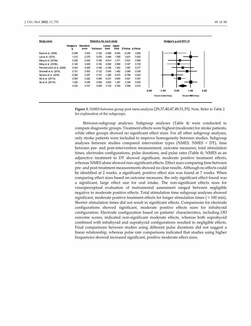
J. Clin. Med. 2022, 11, 776 60 of 68
Figure 5. NMES between group post meta‐analysis [29,37,40,47,49,51,55]. Note. Refer to Table 2
for explanation of the subgroups.
Between‐subgroup analyses. Subgroup analyses (Table 4) were conducted to
compare diagnostic groups. Treatment effects were highest (moderate) for stroke patients,
while other groups showed no significant effect sizes. For all other subgroup analyses,
only stroke patients were included to improve homogeneity between studies. Subgroup
analyses between studies compared intervention types (NMES, NMES + DT), time
between pre‐ and post‐intervention measurement, outcome measures, total stimulation
times, electrodes configurations, pulse durations, and pulse rates (Table 4). NMES as an
adjunctive treatment to DT showed significant, moderate positive treatment effects,
whereas NMES alone showed non‐significant effects. Effect sizes comparing time between
pre‐ and post‐treatment measurements showed no clear results. Although no effects could
be identified at 2 weeks, a significant, positive effect size was found at 7 weeks. When
comparing effect sizes based on outcome measures, the only significant effect found was
a significant, large effect size for oral intake. The non‐significant effects sizes for
visuoperceptual evaluation of instrumental assessment ranged between negligible
negative to moderate positive effects. Total stimulation time subgroup analyses showed
significant, moderate positive treatment effects for longer stimulation times ( > 100 min).
Shorter stimulation times did not result in significant effects. Comparisons for electrode
configurations showed significant, moderate positive effects sizes for infrahyoid
configuration. Electrode configuration based on patients’ characteristics, including OD
outcome scores, indicated non‐significant moderate effects, whereas both suprahyoid
combined with infrahyoid and suprahyoid configurations resulted in negligible effects.
Final comparisons between studies using different pulse durations did not suggest a
linear relationship, whereas pulse rate comparisons indicated that studies using higher
frequencies showed increased significant, positive moderate effect sizes.
Study name Statistics for each study Hedges's g and 95% CI
Hedges's Standard Lower Upper g error Variance limit limit Z-Value p-Value
Bulow et al. (2008) -0.099 0.403 0.162 -0.888 0.690 -0.246 0.806
Lee et al. (2014) 1.013 0.279 0.078 0.466 1.559 3.631 0.000
Meng et al. (2018a) 0.228 0.430 0.185 -0.614 1.071 0.531 0.595
Meng et al. (2018b) 0.149 0.429 0.184 -0.692 0.990 0.347 0.729
Permsirivanich et al. (2009) 0.443 0.408 0.166 -0.356 1.242 1.087 0.277
Simonelli et al. (2019) 0.751 0.363 0.132 0.040 1.462 2.069 0.039
Sproson et al. (2018) -0.304 0.397 0.157 -1.082 0.473 -0.768 0.443
Xia et al. (2011a) 0.204 0.222 0.049 -0.231 0.640 0.921 0.357
Xia et al. (2011b) 1.025 0.236 0.056 0.563 1.487 4.348 0.000
0.433 0.167 0.028 0.105 0.760 2.589 0.010
-2.00 -1.00 0.00 1.00 2.00
Favours Control Group Favours NMES Group
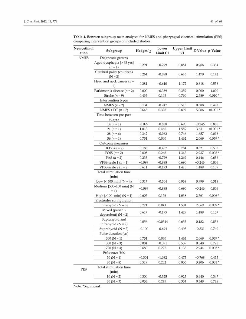
J. Clin. Med. 2022, 11, 776 61 of 68
Table 4. Between subgroup meta‐analyses for NMES and pharyngeal electrical stimulation (PES)
comparing intervention groups of included studies.
Neurostimul
ation Subgroup Hedges’ g
Lower
Limit CI
Upper Limit
CI Z‐Value p‐Value
NMES Diagnostic groups
Aged dysphagia [> 65 yrs]
(n = 1) 0.291 −0.299 0.881 0.966 0.334
Cerebral palsy (children)
(N = 2) 0.264 −0.088 0.616 1.470 0.142
Head and neck cancer (n =
2) 0.281 −0.610 1.172 0.618 0.536
Parkinson’s disease (n = 2) 0.000 −0.359 0.359 0.000 1.000
Stroke (n = 9) 0.433 0.105 0.760 2.589 0.010 *
Intervention types
NMES (n = 2) 0.134 −0.247 0.515 0.688 0.492
NMES + DT (n = 7) 0.648 0.398 0.897 5.086 <0.001 *
Time between pre‐post
(days)
14 (n = 1) −0.099 −0.888 0.690 −0.246 0.806
21 (n = 1) 1.013 0.466 1.559 3.631 <0.001 *
28 (n = 6) 0.342 −0.062 0.746 1.657 0.098
56 (n = 1) 0.751 0.040 1.462 2.069 0.039 *
Outcome measures
DOSS (n = 2) 0.188 −0.407 0.784 0.621 0.535
FOIS (n = 2) 0.805 0.268 1.343 2.937 0.003 *
PAS (n = 2) 0.235 −0.799 1.269 0.446 0.656
VFSS‐scale 1 (n = 1) −0.099 −0.888 0.690 −0.246 0.806
VFSS‐scale 2 (n = 2) 0.611 −0.193 1.415 1.489 0.137
Total stimulation time
(min)
Low [< 500 min] (N = 4) 0.317 −0.304 0.938 0.999 0.318
Medium [500–100 min] (N
= 1) −0.099 −0.888 0.690 −0.246 0.806
High [>100‐ min] (N = 4) 0.607 0.176 1.038 2.761 0.006 *
Electrodes configuration
Infrahyoid (N = 3) 0.771 0.041 1.501 2.069 0.039 *
Mixed (patient‐
dependent) (N = 2) 0.617 −0.195 1.429 1.489 0.137
Suprahyoid and
infrahyoid (N = 2) 0.056 −0.0544 0.655 0.182 0.856
Suprahyoid (N = 2) −0.100 −0.694 0.493 −0.331 0.740
Pulse duration (μs)
300 (N = 1) 0.751 0.040 1.462 2.069 0.039 *
350 (N = 3) 0.084 −0.391 0.559 0.348 0.728
700 (N = 4) 0.680 0.227 1.133 2.944 0.003 *
Pulse rates (Hz)
30 (N = 1) −0.304 −1.082 0.473 −0.768 0.433
80 (N = 8) 0.519 0.202 0.836 3.206 0.001 *
PES Total stimulation time
(min)
10 (N = 2) 0.300 −0.325 0.925 0.940 0.347
30 (N = 3) 0.053 0.245 0.351 0.348 0.728
Note. *Significant.
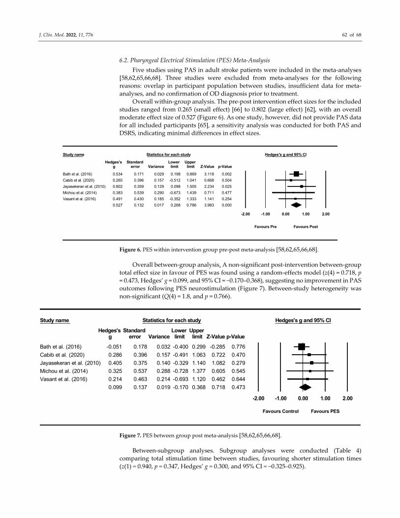
J. Clin. Med. 2022, 11, 776 62 of 68
6.2. Pharyngeal Electrical Stimulation (PES) Meta‐Analysis
Five studies using PAS in adult stroke patients were included in the meta‐analyses
[58,62,65,66,68]. Three studies were excluded from meta‐analyses for the following
reasons: overlap in participant population between studies, insufficient data for meta‐
analyses, and no confirmation of OD diagnosis prior to treatment.
Overall within‐group analysis. The pre‐post intervention effect sizes for the included
studies ranged from 0.265 (small effect) [66] to 0.802 (large effect) [62], with an overall
moderate effect size of 0.527 (Figure 6). As one study, however, did not provide PAS data
for all included participants [65], a sensitivity analysis was conducted for both PAS and
DSRS, indicating minimal differences in effect sizes.
Figure 6. PES within intervention group pre‐post meta‐analysis [58,62,65,66,68].
Overall between‐group analysis. A non‐significant post‐intervention between‐group
total effect size in favour of PES was found using a random‐effects model (z(4) = 0.718, p
= 0.473, Hedges’ g = 0.099, and 95% CI = −0.170–0.368), suggesting no improvement in PAS
outcomes following PES neurostimulation (Figure 7). Between‐study heterogeneity was
non‐significant (Q(4) = 1.8, and p = 0.766).
Figure 7. PES between group post meta‐analysis [58,62,65,66,68].
Between‐subgroup analyses. Subgroup analyses were conducted (Table 4)
comparing total stimulation time between studies, favouring shorter stimulation times
(z(1) = 0.940, p = 0.347, Hedges’ g = 0.300, and 95% CI = −0.325–0.925).
Study name Statistics for each study Hedges's g and 95% CI
Hedges's Standard Lower Upper g error Variance limit limit Z-Value p-Value
Bath et al. (2016) 0.534 0.171 0.029 0.198 0.869 3.118 0.002
Cabib et al. (2020) 0.265 0.396 0.157 -0.512 1.041 0.668 0.504
Jayasekeran et al. (2010) 0.802 0.359 0.129 0.098 1.505 2.234 0.025
Michou et al. (2014) 0.383 0.539 0.290 -0.673 1.439 0.711 0.477
Vasant et al. (2016) 0.491 0.430 0.185 -0.352 1.333 1.141 0.254
0.527 0.132 0.017 0.268 0.786 3.983 0.000
-2.00 -1.00 0.00 1.00 2.00
Favours Pre Favours Post
Study name Statistics for each study Hedges's g and 95% CI
Hedges's Standard Lower Upper g error Variance limit limit Z-Value p-Value
Bath et al. (2016) -0.051 0.178 0.032 -0.400 0.299 -0.285 0.776
Cabib et al. (2020) 0.286 0.396 0.157 -0.491 1.063 0.722 0.470
Jayasekeran et al. (2010) 0.405 0.375 0.140 -0.329 1.140 1.082 0.279
Michou et al. (2014) 0.325 0.537 0.288 -0.728 1.377 0.605 0.545
Vasant et al. (2016) 0.214 0.463 0.214 -0.693 1.120 0.462 0.644
0.099 0.137 0.019 -0.170 0.368 0.718 0.473
-2.00 -1.00 0.00 1.00 2.00
Favours Control Favours PES
J. Clin. Med. 2022, 11, 776 63 of 68
7. Discussion
This study (Part I) aimed to determine the effects of PES and NMES in people with
OD without excluding populations based on medical diagnoses. To base findings on the
highest level of evidence, only RCTs were included. This systematic review and meta‐
analysis were conducted using PRISMA procedures as a guide.
7.1. Systematic Review Findings
When comparing RCTs in pharyngeal and neuromuscular electrical stimulation (i.e.,
PES and NMES), various methodological problems became apparent. Some studies did
not define OD or used divergent definitions, whereas other studies applied different
inclusion criteria. Most studies included patients with confirmed OD by instrumental
assessment, but several studies used screening, patient self‐report or clinical assessments
instead. Consequently, participant characteristics may differ widely between studies.
Despite most studies included stroke patients, meta‐analysis comparing diagnostic
groups other than stroke was possible for NMES, however this could not be conducted
for PES.
Furthermore, the great variety in outcome measures also restricted comparisons by
meta‐analysis. As heterogeneity between studies indicates that no estimated overall effect
by meta‐analysis should be determined, combining studies targeting different domains
within the area of OD will have similar implications. For instance, meta‐analyses based
on both patients’ self‐reported health‐related quality of life and visuoperceptual
evaluation of instrumental assessments would very likely lead to inappropriate estimated
overall effects. Thus, to reduce heterogeneity between outcome measures, some studies
were excluded from the meta‐analysis. This strong focus on reducing heterogeneity
between studies when performing meta‐analysis also implies that data other than the
authors’ primary outcomes may have been preferably included in this analysis. For
example, the primary outcome for Dziewas, Stellato, Van Der Tweel, Walther, Werner,
Braun, Citerio, Jandl, Friedrichs, Nötzel, Vosko, Mistry, Hamdy, McGowan, Warnecke,
Zwittag and Bath [59] and Suntrup, Marian, Schröder, Suttrup, Muhle, Oelenberg,
Hamacher, Minnerup, Warnecke and Dziewas [64] was readiness for decannulation,
which was considered too different from outcomes in the other included studies.
All eight PES studies compared neurostimulation with sham stimulation. However,
among the 30 NMES studies, the comparison group variably consisted of usual care, DT,
another dysphagia treatment or a combination of treatments. In contrast to PES studies
that did not include any DT groups, most NMES studies combined neurostimulation with
simultaneous DT. However, DT consisted of a wide range of behavioural interventions,
using different treatment dosages, timings, and durations. Moreover, DT was referred to
by many different names and acronyms (e.g., dysphagia training, behavioural
intervention, classic treatment, or standard care). This suggest that care should be taken
with the use of DT as an overarching term to group many different behavioural
interventions to estimate overall effect sizes in meta‐analyses.
Furthermore, RCTs are characterised by random allocation of participants to
intervention groups and blinding or masking the nature of treatment for participants.
However, in neurostimulation studies, blinding is frequently not feasible and participants
may identify what treatment arm they have been assigned to (e.g., the presence of
neurostimulation equipment, the experience of active stimulation). Also, since
neurostimulation thresholding in PES is frequently applied in all groups to mask
treatment assignment, patients receiving sham stimulation would still have been exposed
to a certain level of neurostimulation during thresholding. Those studies not using
thresholding in sham groups (e.g., [59,64]) might show larger treatment effect differences
when comparing neurostimulation versus sham stimulation.
J. Clin. Med. 2022, 11, 776 64 of 68
7.2. NMES
When considering meta‐analyses for NMES, the highest effect sizes were found for
stroke populations. As existing reviews in NMES [10,12,18,19] excluded other patient
populations, no comparisons could be made between clinical populations. In addition,
only two reviews conducted meta‐analyses [18,19] selecting studies using different
inclusion criteria (e.g., excluding comparison groups with active treatment components
[18] or excluding chronic stroke patients [19]). Reviews may also prefer different outcome
data for meta‐analyses, especially in the case of RCTs using a large battery of assessments.
As such, total numbers of included studies vary per review, but comparisons between
reviews may be falsely estimated due to differences in methodology.
In this systematic review, a wide range in effect sizes was found in NMES RCTs
depending on outcome measures used. However, oral intake scales showed highest
effects sizes when compared to visuoperceptual evaluation of instrumental assessment or
clinical assessment. This might be explained by NMES treatment usually taking place over
consecutive weeks, in contrast to other neurostimulation techniques (e.g., PES or rTMS)
that may be restricted to limited sessions over a few days only.
The great heterogeneity between DT groups also impeded comparisons between
NMES only, NMES plus DT, and DT‐only groups. No RCTs provided adequate DT group
data to be included in the meta‐analysis. For NMES groups, only two studies were
included. As a result, information about the effects of DT is lacking. The negligible effect
sizes found for NMES without DT were based on only two studies and the moderate effect
sizes for combined NMES and DT were based on a total of seven studies.
Most studies performed NMES at motor stimulation level, whereas only a few
studies included a group receiving NMES at sensory stimulation level. As none of these
latter studies could be included in meta‐analyses, no further details are available on
comparisons between effect sizes for sensory versus motor stimulation. Also, terminology
was confusing as sensory stimulation was sometimes referred to as sham stimulation [39].
NMES studies showed marked variation in the technical parameters and protocols
applied. When comparing electrode configurations, both hyoid and combined hyoid and
suprahyoid configurations showed negligible effects, whereas infrahyoid configurations
resulted in moderate effects. A study using patient‐dependent configurations showed
promising results as well [55]. However, it remained unclear which criteria were used to
decide on individual configurations. Furthermore, reporting on many technical
parameters proved to be either incomplete or unclear for several studies (e.g., data on
pulse duration, pulse rate, or stimulation time). As technical parameters may depend on
medical device manufacturers, comparisons between brands may be warranted. For
example, when considering pulse duration, a clear distinction in effect sizes is found
between one study using a lower pulse rate—indicating a negative effect size—versus
eight studies using higher pulse rates with moderate effect sizes.
7.3. PES
Compared to NMES, fewer PES studies were identified and thus a more limited
meta‐analysis was conducted. RCTs included stroke populations, except for one study
that included patients with multiple sclerosis [63]. All studies compared active PES with
sham treatment in stroke patients and used mostly visuoperceptual evaluation of
radiographic recordings of the swallowing act as an outcome measure. Meta‐analysis
identified a non‐significant post‐intervention between‐group total effect size in favour of
PES. This finding seemed in line with findings by Chiang, Lin, Hsiao, Yeh, Liang and
Wang [19], but this comparison is limited as it is based on only two studies. Additionally,
Bath, Lee and Everton [18] reported that PES studies did not show an effect for many
outcome measures (e.g., post‐treatment proportions of participants with dysphagia,
swallowing ability, penetration and aspiration scores or nutrition). However, in contrast
to previous reviews, Cheng, Sasegbon and Hamdy [7] found a significant, moderate effect
J. Clin. Med. 2022, 11, 776 65 of 68
size in favour of PES when conducting meta‐analysis. Again, inclusion criteria between
reviews differed. For example, two studies [59,64] were excluded from meta‐analysis in
this review as well as the reviews by Chiang, Lin, Hsiao, Yeh, Liang and Wang [19] and
Bath, Lee and Everton [18], but were included in the review by Cheng, Sasegbon and
Hamdy [7]. This may have impacted the overall effect size as both PES studies showed
significant treatment effects.
7.4. Moderators
Differences between NMES and PES studies made comparisons between RCTs
difficult and hindered meta‐analyses. Studies used different participant inclusion criteria
in relation to underlying medical diagnoses or chronicity of stroke and used a large variety
of outcome measures covering different domains within the area of OD. Outcome
measures may also lack responsiveness, thus lack sensitivity to change during treatment.
Moreover, studies varied significantly in technical parameters of neurostimulation. The
number of studies and participants restricted the ability of statistical analyses to consider
how each variable may have impacted the effects of neurostimulation.
Studies frequently neglected to report on potential moderators of stimulation effects
in sufficient detail. For example, stroke severity and OD severity are inextricably linked
and may moderate stimulation effects, yet only very few studies provided data on stroke
severity. Similar problems occur when the chronicity of a stroke is not reported or the
possibility of spontaneous recovery is ignored. This is especially true during NMES
treatment, which may span a period of several weeks. In addition, no consensus was
reached regarding the optimal moment for outcome measurement. Consequently, in this
review, between‐subgroup meta‐analyses were conducted using post‐intervention data
only, so that the possibility of spontaneous recovery during the intervention period was
taken into consideration.
7.5. Limitations
Despite a rigorous reviewing process following PRISMA guidelines and the use of
RoB 2 to reduce bias, this review is subject to some limitations. Only RCTs published in
English were included in this current study. Thus, some RCTs may have been excluded
based on language criteria when their findings could have contributed to the current
meta‐analysis. Furthermore, meta‐analyses included mostly stroke studies, thereby not
providing effect sizes for other diagnostic patient populations. However, the main
limitation of this review originates from the high degree of heterogeneity between studies,
making comparisons across studies challenging. As such, generalisations and meta‐
analyses should be interpreted with care.
8. Conclusions
Meta‐analyses for RCTS in NMES found a significant, large pre‐post intervention
effect size and significant, small post‐intervention between‐group effect size in favour of
NMES. For PES studies, the meta‐analyses showed a significant, moderate effect size for
pre‐post intervention, whereas overall between‐group analysis did not result in
significant treatment effects. Based on these results, NMES seems to have a more
promising outcome compared to PES. However, only careful generalisations and
interpretations of these meta‐analyses can be made due to the NMES studies showing
high heterogeneity in protocols and experimental variables, including potential
moderators, and featuring inconsistent methodological reporting.
There is a need for more RCTs with larger sample sizes in addition to the
standardisation of protocols and guidelines for reporting. These changes would better
facilitate comparisons of studies and help to determine intervention effects more
definitively. Delphi studies involving international experts might allow for a consensus
to be reached, thus supporting future research, comparability and generalisability.
J. Clin. Med. 2022, 11, 776 66 of 68
Supplementary Materials: The following supporting information can be downloaded at:
www.mdpi.com/article/10.3390/jcm11030776/s1, Table S1: PRISMA 2020 for Abstracts Checklist;
Table S2: PRISMA 2020 Checklist.
Author Contributions: Conceptualization: R.S., R.C., A.‐L.S., L.B., S.H. Formal analysis: R.S., R.C.
Methodology: R.S., R.C. Project administration: R.S., R.C. Validation: R.S., R.C. Writing—review &
editing: R.S., R.C., A.‐L.S., L.B., S.H., B.J.H., L.R., S.W.‐G. All authors have read and agreed to the
published version of the manuscript.
Funding: This research received no external funding.
Conflicts of Interest: The authors declare no conflict of interest.
References
1. Speyer, R. (Ed.) Behavioural Treatment of Oropharyngeal Dysphagia; Springer: Berlin, Germany, 2018. 2. Speyer, R.; Cordier, R.; Kim, J.H.; Cocks, N.; Michou, E.; Wilkes‐Gillan, S. Prevalence of drooling, feeding and swallowing
problems in cerebral palsy across the lifespan: Systematic review and meta‐analysis. Dev. Med. Child Neurol. 2019, 61, 1249–
1258.
3. Takizawa, C.; Gemmell, E.; Kenworthy, J.; Speyer, R. A Systematic Review of the Prevalence of Oropharyngeal Dysphagia
in Stroke, Parkinson’s Disease, Alzheimer’s Disease, Head Injury, and Pneumonia. Dysphagia 2016, 31, 434–441.
4. Jones, E.; Speyer, R.; Kertscher, B.; Denman, D.; Swan, K.; Cordier, R. Health‐related quality of life in oropharyngeal
dysphagia. Dysphagia 2018, 33, 141–172.
5. Wang, Z.; Wu, L.; Fang, Q.; Shen, M.; Zhang, L.; Liu, X. Effects of capsaicin on swallowing function in stroke patients with
dysphagia: A randomized controlled trial. J. Stroke Cerebrovasc. Dis. 2019, 28, 1744–1751.
6. Quiroz‐González, S.; Torres‐Castillo, S.; López‐Gómez, R.E.; Estrada, I.J. Acupuncture points and their relationship with
multireceptive fields of neurons. J. Acupunct. Meridian Stud. 2017, 10, 81–89.
7. Cheng, I.; Sasegbon, A.; Hamdy, S. Effects of neurostimulation on poststroke dysphagia: A synthesis of current evidence
from randomised controlled trials. Neuromodulation Technol. Neural Interface 2021, 24, 1388–1401.
8. Michou, E.; Sasegbon, A.; Hamdy, S. Neurostimulation for the treatment of dysphagia after stroke: Behavioural treatment
of oropharyngeal dysphagia. In Dysphagia: Diagnosis and Treatment, 2nd ed.; Ekberg, O., Ed.; Medical Radiology: Diagnostic
Imaging; Springer: Berlin/Heidelberg, Germany, 2019.
9. Carnaby, G.D.; LaGorio, L.; Silliman, S.; Crary, M. Exercise‐based swallowing intervention (McNeill Dysphagia Therapy)
with adjunctive NMES to treat dysphagia post‐stroke: A double‐blind placebo‐controlled trial. J. Oral Rehabil. 2020, 47, 501–
510.
10. Alamer, A.; Melese, H.; Nigussie, F. Effectiveness of Neuromuscular Electrical Stimulation on Post‐Stroke Dysphagia: A
Systematic Review of Randomized Controlled Trials. Clin. Interv. Aging 2020, 15, 1521–1531.
11. Baijens, L.W.J.; Speyer, R.; Passos, V.L.; Pilz, W.; Van Der Kruis, J.; Haarmans, S.; Desjardins‐Rombouts, C. Surface electrical
stimulation in dysphagic parkinson patients: A randomized clinical trial. Laryngoscope 2013, 123, E38–E44.
12. Clark, H.; Lazarus, C.; Arvedson, J.; Schooling, T.; Frymark, T. Evidence‐Based Systematic Review: Effects of Neuromuscular
Electrical Stimulation on Swallowing and Neural Activation. Am. J. Speech Lang. Pathol. 2009, 18, 361–375.
13. Dionisio, A.; Duarte, I.C.; Patricio, M.; Castelo‐Branco, M. Transcranial magnetic stimulation as an intervention tool to
recover from language, swallowing and attentional deficits after stroke: A systematic review. Cerebrovasc. Dis. 2018, 18, 176–
183.
14. Liao, X.; Xing, G.; Guoqiang, X.; Jin, Y.; Tang, Q.; He, B.; McClure, M.A.; Liu, H.; Chen, H.; Mu, Q. Repetitive transcranial
magnetic stimulation as an alternative therapy for dysphagia after stroke: A systematic review and meta‐analysis. Clin.
Rehabil. 2017, 31, 289–298.
15. Marchina, S.; Pisegna, J.M.; Massaro, J.M.; Langmore, S.E.; McVey, C.; Wang, J.; Kumar, S. Transcranial direct current
stimulation for post‐stroke dysphagia: A systematic review and meta‐analysis of randomized controlled trials. J. Neurol.
2021, 268, 293–304.
16. Momosaki, R.; Kinoshita, S.; Kakuda, W.; Yamada, N.; Abo, M. Noninvasive brain stimulation for dysphagia after acquired
brain injury: A systematic review. J. Med Investig. 2016, 63, 153–158.
17. Pisegna, J.M.; Kaneoka, A.; Pearson, W.G.; Kumar, S.; Langmore, S.E. Effects of non‐invasive brain stimulation on post‐
stroke dysphagia: A systematic review and meta‐analysis of randomized controlled trials. Clin. Neurophysiol. 2016, 127, 956–
968.
18. Bath, P.M.; Lee, H.S.; Everton, L.F. Swallowing therapy for dysphagia in acute and subacute stroke (Review). Cochrane
Database Syst. Rev. 2018, 10, CD000323.
19. Chiang, C.‐F.; Lin, M.‐T.; Hsiao, M.‐Y.; Yeh, Y.‐C.; Liang, Y.‐C.; Wang, T.‐G. Comparative Efficacy of Noninvasive
Neurostimulation Therapies for Acute and Subacute Poststroke Dysphagia: A Systematic Review and Network Meta‐
analysis. Arch. Phys. Med. Rehabil. 2019, 100, 739–750.e4.
20. Page, M.J.; McKenzie, J.E.; Bossuyt, P.M.; Boutron, I.; Hoffmann, T.C.; Mulrow, C.D.; Moher, D. The PRISMA 2020 statement:
An updated guideline for reporting systematic reviews. BMJ 2021, 372, n71.
J. Clin. Med. 2022, 11, 776 67 of 68
21. Page, M.J.; Moher, D.; Bossuyt, P.M.; Boutron, I.; Hoffmann, T.C.; Mulrow, C.D.; McKenzie, J.E. PRISMA 2020 explanation
and elaboration: Updated guidance and exemplars for reporting systematic reviews. BMJ 2021, 372, n160. 22. Sterne, J.A.; Savović, J.; Page, M.J.; Elbers, R.G.; Blencowe, N.S.; Boutron, I.; Cates, C.J.; Cheng, H.Y.; Corbett, M.S.; Eldridge,
S.M.; et al. RoB 2: A revised tool for assessing risk of bias in randomised trials. BMJ 2019, 366, l4898.
23. Borenstein, M.; Hedges, L.; Higgins, J.; Rothstein, H. Comprehensive Meta‐Analysis; Biostat: Englewood, NJ, USA, 2014;
Volume 3.
24. Higgins, J.P.T.; Thompson, S.G.; Deeks, J.J.; Altman, D.G. Measuring inconsistency in meta‐analyses. BMJ 2003, 327, 557–
560.
25. Cohen, J. Statistical Power Analysis for the Behavioural Sciences, 2nd ed.; Routledge: New York, NY, USA, 1988.
26. Begg, C.B.; Mazumdar, M. Operating Characteristics of a Rank Correlation Test for Publication Bias. Biometrics 1994, 50,
1088–1101.
27. Rosenthal, R. The file drawer problem and tolerance for null results. Psychol. Bull. 1979, 86, 638–664.
28. Beom, J.; Oh, B.‐M.; Choi, K.H.; Kim, W.; Song, Y.J.; You, D.S.; Kim, S.J.; Han, T.R. Effect of Electrical Stimulation of the
Suprahyoid Muscles in Brain‐Injured Patients with Dysphagia. Dysphagia 2015, 30, 423–429.
29. Bülow, M.; Speyer, R.; Baijens, L.; Woisard, V.; Ekberg, O. Neuromuscular Electrical Stimulation (NMES) in Stroke Patients
with Oral and Pharyngeal Dysfunction. Dysphagia 2008, 23, 302–309.
30. El‐Tamawy, M.S.; Darwish, M.H.; El‐Azizi, H.S.; Abdelalim, A.M.; Taha, S.I. The influence of physical therapy on
oropharyngeal dysphagia in acute stroke patients. Egypt. J. Neurol. Psychiatry Neurosurg. 2015, 52, 201–205.
31. Guillén‐Solà, A.; Sartor, M.M.; Soler, N.B.; Duarte, E.; Barrera, M.C.; Marco, E. Respiratory muscle strength training and
neuromuscular electrical stimulation in subacute dysphagic stroke patients: A randomized controlled trial. Clin.
Rehabilitation 2017, 31, 761–771.
32. Heijnen, B.J.; Speyer, R.; Baijens, L.W.J.; Bogaardt, H.C.A. Neuromuscular electrical stimulation versus traditionaltherapy
in patients with Parkinson’s Disease and propharyngeal dysphagia: Effects on quality of life. Dysphagia 2012, 27, 336–345.
33. Huang, K.‐L.; Liu, T.‐Y.; Huang, Y.‐C.; Leong, C.‐P.; Lin, W.‐C.; Pong, Y.‐P. Functional Outcome in Acute Stroke Patients
with Oropharyngeal Dysphagia after Swallowing Therapy. J. Stroke Cerebrovasc. Dis. 2014, 23, 2547–2553.
34. Huh, J.; Park, E.; Min, Y.; Kim, A.; Yang, W.; Oh, H.; Nam, T.; Jung, T. Optimal placement of electrodes for treatment of post‐
stroke dysphagia by neuromuscular electrical stimulation combined with effortful swallowing. Singap. Med. J. 2020, 61, 487–
491.
35. Jing, Q.; Yang, X.; Reng, Q. Effect of Neuromuscular Electrical Stimulation in Patients with Post‐Stroke Dysphagia. Med Sci.
Technol. 2016, 57, 1–5.
36. Langmore, S.E.; McCulloch, T.M.; Krisciunas, G.P.; Lazarus, C.L.; Van Daele, D.J.; Pauloski, B.R.; Rybin, D.; Doros, G.
Efficacy of electrical stimulation and exercise for dysphagia in patients with head and neck cancer: A randomized clinical
trial. Head Neck 2015, 38, E1221–E1231.
37. Lee, K.W.; Kim, S.B.; Lee, J.H.; Lee, S.J.; Ri, J.W.; Park, J.G. The Effect of Early Neuromuscular Electrical Stimulation Therapy
in Acute/Subacute Ischemic Stroke Patients with Dysphagia. Ann. Rehabilitation Med. 2014, 38, 153–159.
38. Li, L.; Li, Y.; Huang, R.; Yin, J.; Shen, Y.; Shi, J. The value of adding transcutaneous neuromuscular electrical stimulation
(VitalStim) to traditional therapy for post‐stroke dysphagia: A randomized controlled trial. Eur. J. Phys. Rehabil. Med. 2014,
51, 200–206.
39. Maeda, K.; Koga, T.; Akagi, J. Interferential current sensory stimulation, through the neck skin, improves airway defense
and oral nutrition intake in patients with dysphagia: A double‐blind randomized controlled trial. Clin. Interv. Aging 2017,
12, 1879–1886.
40. Meng, P.; Zhang, S.; Wang, Q.; Wang, P.; Han, C.; Gao, J.; Yue, S. The effect of surface neuromuscular electrical stimulation
on patients with post‐stroke dysphagia. J. Back Musculoskelet. Rehabil. 2018, 31, 363–370.
41. Nam, H.S.; Beom, J.; Oh, B.‐M.; Han, T.R. Kinematic Effects of Hyolaryngeal Electrical Stimulation Therapy on Hyoid
Excursion and Laryngeal Elevation. Dysphagia 2013, 28, 548–556.
42. Oh, D.‐H.; Park, J.‐S.; Kim, H.‐J.; Chang, M.‐Y.; Hwang, N.‐K. The effect of neuromuscular electrical stimulation with
different electrode positions on swallowing in stroke patients with oropharyngeal dysphagia: A randomized trial. J. Back
Musculoskelet. Rehabil. 2020, 33, 637–644.
43. Ortega, O.; Rofes, L.; Martin, A.; Arreola, V.; López, I.; Clavé, P. A Comparative Study Between Two Sensory Stimulation
Strategies After Two Weeks Treatment on Older Patients with Oropharyngeal Dysphagia. Dysphagia 2016, 31, 706–716.
44. Park, J.‐W.; Kim, Y.; Oh, J.C.; Lee, H.‐J. Effortful Swallowing Training Combined with Electrical Stimulation in Post‐Stroke
Dysphagia: A Randomized Controlled Study. Dysphagia 2012, 27, 521–527.
45. Park, J.‐S.; Oh, D.‐H.; Hwang, N.‐K.; Lee, J.H. Effects of neuromuscular electrical stimulation combined with
effortful swallowing on post‐stroke oropharyngeal dysphagia: a randomised controlled trial. J. Oral Rehabil.
2016, 43, 426–434. https://doi.org/10.1111/joor.12390 46. Park, J.S.; Oh, D.H.; Hwang, N.K.; Lee, J.H. Effects of neuromuscular electrical stimulation in patients with Parkinson’s
disease and dysphagia: A ran‐domized, single‐blind, placebo‐controlled trial. Neurorehabilitation 2018, 42, 457–463.
47. Permsirivanich, W.; Tipchatyotin, S.; Wongchai, M.; Leelamanit, V.; Setthawatcharawanich, S.; Sathirapanya, P.;
Boonmeeprakob, A. Comparing the effects of rehabilitation swallowing therapy vs. neuromuscular electrical stimu‐lation
J. Clin. Med. 2022, 11, 776 68 of 68
therapy among stroke Pptients with persistent pharyngeal dysphagia: A randomized controlled study. Med. J. Med. Assoc.
Thail. 2009, 92, 259.
48. Ryu, J.S.; Kang, J.Y.; Park, J.Y.; Nam, S.Y.; Choi, S.H.; Roh, J.L.; Kim, S.Y.; Choi, K.H. The effect of electrical stimulation
therapy on dysphagia following treatment for head and neck cancer. Oral Oncol. 2009, 45, 665–668.
49. Simonelli, M.; Ruoppolo, G.; Iosa, M.; Morone, G.; Fusco, A.; Grasso, M.G.; Gallo, A.; Paolucci, S. A stimulus for eating. The
use of neuromuscular transcutaneous electrical stimulation in patients affected by severe dysphagia after subacute stroke:
A pilot randomized controlled trial. Neurorehabilitation 2019, 44, 103–110.
50. Song, W.J.; Park, J.H.; Lee, J.H.; Kim, M.Y. Effects of Neuromuscular Electrical Stimulation on Swallowing Functions in
Children with Cerebral Palsy: A Pilot Randomised Controlled Trial. Hong Kong J. Occup. Ther. 2015, 25, 1–6.
51. Sproson, L.; Pownall, S.; Enderby, P.; Freeman, J. Combined electrical stimulation and exercise for swallow rehabilitation
post‐stroke: A pilot randomized control trial. Int. J. Lang. Commun. Disord. 2018, 53, 405–417.
52. Terré, R.; Mearin, F. A randomized controlled study of neuromuscular electrical stimulation in oropharyngeal dysphagia
secondary to acquired brain injury. Eur. J. Neurol. 2015, 22, 687–e44.
53. Umay, E.K.; Yaylaci, A.; Saylam, G.; Gundogdu, I.; Gurcay, E.; Akcapinar, D.; Kirac, Z. The effect of sensory level electrical
stimulation of the masseter muscle in early stroke patients with dysphagia: A randomized controlled study. Neurol. India
2017, 65, 734.
54. Umay, E.; Gurcay, E.; Ozturk, E.A.; Akyuz, E.U. Is sensory‐level electrical stimulation effective in cerebral palsy children
with dysphagia? A randomized controlled clinical trial. Acta Neurol. Belg. 2020, 120, 1097–1105.
55. Xia, W.; Zheng, C.; Lei, Q.; Tang, Z.; Hua, Q.; Zhang, Y.; Zhu, S. Treatment of post‐stroke dysphagia by vitalstim therapy
coupled with conventional swallowing training. J. Huazhong Univ. Sci. Technol. 2011, 31, 73–76.
56. Zeng, Y.; Yip, J.; Cui, H.; Guan, L.; Zhu, H.; Zhang, W.; Du, H.; Geng, X. Efficacy of neuromuscular electrical stimulation in
improving the negative psychological state in patients with cerebral infarction and dysphagia. Neurol. Res. 2018, 40, 473–
479.
57. Zhang, M.; Tao, T.; Zhang, Z.‐B.; Zhu, X.; Fan, W.‐G.; Pu, L.‐J.; Chu, L.; Yue, S.‐W. Effectiveness of Neuromuscular Electrical
Stimulation on Patients with Dysphagia with Medullary Infarction. Arch. Phys. Med. Rehabil. 2016, 97, 355–362.
58. Bath, P.M.; Scutt, P.; Love, J.; Clavé, P.; Cohen, D.; Dziewas, R.; Hamdy, S. Pharyngeal electrical stimulation for treatment
of dysphagia in subacute stroke: A randomised controlled trial. Stroke 2016, 47, 1–19.
59. Dziewas, R.; Stellato, R.; van der Tweel, I.; Walther, E.; Werner, C.J.; Braun, T.; Citerio, G.; Jandl, M.; Friedrichs, M.; Nötzel,
K.; et al. Pharyngeal electrical stimulation for early decannulation in tracheotomised patients with neurogenic dysphagia
after stroke (PHAST‐TRAC): A prospective, single‐blinded, randomised trial. Lancet Neurol. 2018, 17, 849–859.
60. Essa, H.; Vasant, D.H.; Raginis‐Zborowska, A.; Payton, A.; Michou, E.; Hamdy, S. The BDNF polymorphism Val66Met may
be predictive of swallowing improvement post pharyngeal electrical stimulation in dysphagic stroke patients.
Neurogastroenterol. Motil. 2017, 29, e13062.
61. Fraser, C.; Power, M.; Hamdy, S.; Rothwell, J.; Hobday, D.; Hollander, I.; Tyrell, P.; Hobson, A.; Williams, S.; Thompson, D.
Driving Plasticity in Human Adult Motor Cortex Is Associated with Improved Motor Function after Brain Injury. Neuron
2002, 34, 831–840.
62. Jayasekeran, V.; Singh, S.; Tyrrell, P.; Michou, E.; Jefferson, S.; Mistry, S.; Gamble, E.; Rothwell, J.; Thompson, D.; Hamdy,
S. Adjunctive functional pharyngeal elctrical stimulation reverses swallowing disability after brain lesions. Gastroentrology
2010, 138, 1737–1746.
63. Restivo, D.A.; Casabona, A.; Centonze, D.; Ragona, R.M.; Maimone, D.; Pavone, A. Pharyngeal Electrical Stimulation for
Dysphagia Associated with Multiple Sclerosis: A Pilot Study. Brain Stimul. 2013, 6, 418–423.
64. Suntrup, S.; Marian, T.; Schröder, J.B.; Suttrup, I.; Muhle, P.; Oelenberg, S.; Hamacher, C.; Minnerup, J.; Warnecke, T.;
Dziewas, R. Electrical pharyngeal stimulation for dysphagia treatment in tracheotomized stroke patients: A randomized
controlled trial. Intensiv. Care Med. 2015, 41, 1629–1637.
65. Vasant, D.H.; Michou, E.; O’Leary, N.; Vail, A.; Mistry, S.; Hamdy, S.; Greater Manchester Stroke Research Network.
Pharyngeal electrical stimulation in dysphagia poststroke: A prospective, randomized single‐blinded interventional study.
Neurorehabilit. Neural Repair 2016, 30, 866–875.
66. Cabib, C.; Nascimento, W.; Rofes, L.; Arreola, V.; Tomsen, N.; Mundet, L.; Palomeras, E.; Michou, E.; Clavé, P.; Ortega, O.
Short‐term neurophysiological effects of sensory pathway neurorehabilitation strategies on chronic post‐stroke
oropharyngeal dysphagia. Neurogastroenterol. Motil. 2020, 32, 1–14.
67. Lim, K.‐B.; Lee, H.‐J.; Yoo, J.; Kwon, Y.‐G. Effect of Low‐Frequency rTMS and NMES on Subacute Unilateral Hemispheric
Stroke with Dysphagia. Ann. Rehabil. Med. 2014, 38, 592–602.
68. Michou, E.; Mistry, S.; Jefferson, S.; Tyrrell, P.; Hamdy, S. Characterizing the Mechanisms of Central and Peripheral Forms
of Neurostimulation in Chronic Dysphagic Stroke Patients. Brain Stimul. 2014, 7, 66–73.
69. Zhang, C.; Zheng, X.; Lu, R.; Yun, W.; Yun, H.; Zhou, X. Repetitive transcranial magnetic stimulation in combination with
neuromuscular electrical stimulation for treatment of post‐stroke dysphagia. J. Int. Med Res. 2019, 47, 662–672.
Related Documents