Neuroprotective effects of Ptychopetalum olacoides Bentham (Olacaceae) on oxygen and glucose deprivation induced damage in rat hippocampal slices Ionara R. Siqueira a,b, * , Helena Cimarosti c , Cı ´ntia Fochesatto c , Domingos S. Nunes d , Christianne Salbego c , Elaine Elisabetsky a,e , Carlos A. Netto a,c a Programa de Po ´s-Graduac ßa ˜o em Cie ˆncias Biolo ´gicas-Fisiologia, Universidade Federal do Rio Grande do Sul, Porto Alegre, RS, Brazil b Centro Universita ´rio UNIVATES, Lajeado, RS, Brazil c Departamento de Bioquı ´mica, Universidade Federal do Rio Grande do Sul, Porto Alegre, RS, Brazil d Departamento de Quı ´mica, Universidade Estadual de Ponta Grossa, Ponta Grossa, PR, Brazil e Departamento de Farmacologia, Universidade Federal do Rio Grande do Sul, Porto Alegre, RS, Brazil Received 18 January 2004; accepted 3 June 2004 Abstract Alcoholic infusions of Ptychopetalum olacoides Bentham (PO, Olacaceae) are used in traditional medicine by patients presenting age associated symptoms and those recovering from stroke. The aim of this study is to evaluate the neuroprotective properties of PO ethanol extract (POEE) using hippocampal slices from Wistar rats exposed to oxygen and glucose deprivation (OGD, followed by reoxygenation). Mitochondrial activity, an index of cell viability, was assessed by the MTT assay; in addition, the free radicals content was estimated by the use of dichlorofluorescein diacetate as probe. The OGD ischemic condition significantly impaired cellular viability, and increased free radicals generation. In non-OGD slices, incubation with POEE (0.6 Ag/ml) increased (c 40%) mitochondrial activity, without affecting free radicals levels. In comparison to OGD controls, slices incubated with POEE (0.6 Ag/ml) during and after OGD exposure had significantly increased cellular viability. In addition, at this same concentration, POEE prevented the increase of free radicals content induced by OGD. In view of the fact that respiratory chain inhibition and increased generation of free radicals are major consequences of the ischemic injury, this 0024-3205/$ - see front matter D 2004 Elsevier Inc. All rights reserved. doi:10.1016/j.lfs.2004.06.001 * Corresponding author. Departamento III, Centro Universita ´rio UNIVATES, Rua Barbedo 426, 03, 90110-260, Porto Alegre, RS, Brazil. Tel.: +55-51-3316-3664; fax: +55-51-3316-4085. E-mail address: [email protected] (I.R. Siqueira). www.elsevier.com/locate/lifescie Life Sciences 75 (2004) 1897 – 1906

Welcome message from author
This document is posted to help you gain knowledge. Please leave a comment to let me know what you think about it! Share it to your friends and learn new things together.
Transcript

www.elsevier.com/locate/lifescie
Life Sciences 75 (2004) 1897–1906
Neuroprotective effects of Ptychopetalum olacoides Bentham
(Olacaceae) on oxygen and glucose deprivation induced
damage in rat hippocampal slices
Ionara R. Siqueiraa,b,*, Helena Cimarostic, Cıntia Fochesattoc, Domingos S. Nunesd,Christianne Salbegoc, Elaine Elisabetskya,e, Carlos A. Nettoa,c
aPrograma de Pos-Graduac�ao em Ciencias Biologicas-Fisiologia,
Universidade Federal do Rio Grande do Sul, Porto Alegre, RS, BrazilbCentro Universitario UNIVATES, Lajeado, RS, Brazil
cDepartamento de Bioquımica, Universidade Federal do Rio Grande do Sul, Porto Alegre, RS, BrazildDepartamento de Quımica, Universidade Estadual de Ponta Grossa, Ponta Grossa, PR, Brazil
eDepartamento de Farmacologia, Universidade Federal do Rio Grande do Sul, Porto Alegre, RS, Brazil
Received 18 January 2004; accepted 3 June 2004
Abstract
Alcoholic infusions of Ptychopetalum olacoides Bentham (PO, Olacaceae) are used in traditional
medicine by patients presenting age associated symptoms and those recovering from stroke. The aim of this
study is to evaluate the neuroprotective properties of PO ethanol extract (POEE) using hippocampal slices
from Wistar rats exposed to oxygen and glucose deprivation (OGD, followed by reoxygenation).
Mitochondrial activity, an index of cell viability, was assessed by the MTT assay; in addition, the free
radicals content was estimated by the use of dichlorofluorescein diacetate as probe. The OGD ischemic
condition significantly impaired cellular viability, and increased free radicals generation. In non-OGD slices,
incubation with POEE (0.6 Ag/ml) increased (c 40%) mitochondrial activity, without affecting free radicals
levels. In comparison to OGD controls, slices incubated with POEE (0.6 Ag/ml) during and after OGD
exposure had significantly increased cellular viability. In addition, at this same concentration, POEE
prevented the increase of free radicals content induced by OGD. In view of the fact that respiratory chain
inhibition and increased generation of free radicals are major consequences of the ischemic injury, this
0024-3205/$ - see front matter D 2004 Elsevier Inc. All rights reserved.
doi:10.1016/j.lfs.2004.06.001
* Corresponding author. Departamento III, Centro Universitario UNIVATES, Rua Barbedo 426, 03, 90110-260, Porto
Alegre, RS, Brazil. Tel.: +55-51-3316-3664; fax: +55-51-3316-4085.
E-mail address: [email protected] (I.R. Siqueira).
I.R. Siqueira et al. / Life Sciences 75 (2004) 1897–19061898
study suggests that Ptychopetalum olacoides contains useful neuroprotective compounds and, therefore,
deserves further scrutiny.
D 2004 Elsevier Inc. All rights reserved.
Keywords: Ptychopetalum olacoides; Neuroprotection; Oxygen and glucose deprivation; Hippocampal slices
Introduction
Ischemia leads to massive depletion of glucose and oxygen. Because the brain is highly dependent on
the continuous provision of oxygen and glucose, cerebral ischemia results in severe cellular degener-
ation. Cerebral ischemia occurs after focal reductions of blood flow to brain areas, as well as a global
deficit following cardiac arrest episodes. Transient global ischemia results in selective neuronal damage,
especially to hippocampal Cornus Ammon’s field 1 -CA1- pyramidal neurons. Damage to CA1
pyramidal cells becomes irreversible after a few minutes of complete ischemia, although no histology
changes are apparent until 48 h after reperfusion (Kirino and Sano, 1984).
The pathogenesis of cerebral ischemia/reperfusion has been associated with depletion of cellular
energy sources, release of excitatory amino acids, mitochondrial dysfunction, and excessive
generation of free radicals, all of which are contributing factors to the oxidative damage (Sims
and Zaidan, 1995; White et al., 2000). Oxidative stress, due to excessive formation of hydrogen
peroxide and oxygen-derived free radicals, causes cell damage through chain reactions of membrane
lipid peroxidation, and/or alterations in membrane fluidity. To date, glutamate receptor antagonists,
free radical scavengers and voltage-gated channel blockers have been examined for the development
of neuroprotective therapeutic agents. Because neurotoxicity mechanisms actually involve numerous
interdependent processes, the development of novel drugs acting at multiple sites of the neurotoxic
cascade, and/or at some crucial common step, is considered to be advantageous (Morin et al., 2001;
Siqueira et al., 2003).
Rat forebrain slices exposed to oxygen and glucose deprivation (OGD) have been used to model
ischemic events, to investigate mechanisms of cell death and neuroprotection (Moro et al., 1998;
Cardenas et al., 2000). This model offers important advantages because cell composition, such as
functional neurons, inflammatory competent cells, locally released effectors and intercellular connec-
tions are preserved, allowing the elucidation of meaningful mechanisms of neuronal damage and
protection (Cohen et al., 1984; Newman et al., 1989; Pellmar, 1995; Taylor et al., 1995).
Complex chemical mixtures have the potential to bring about interactions, potentiation, and/or
combinatorial responses (Elisabetsky, 2002; Panossian et al., 1999). It has been repeatedly demonstrated
that a spectrum of substances are responsible for the polyvalent therapeutic action of the Ginkgo biloba
extract, EGb 761, whose beneficial effects result from ‘‘the combination of its various protective,
curative and modulating properties against the pathological process’’ (Borchers et al., 2000).
Alcoholic infusions of Ptychopetalum olacoides Bentham (PO, Olacaceae), known as ‘‘Marapuama’’,
are used in Brazilian traditional medicine by patients presenting age associated symptoms, or those
recovering from conditions associated with damage of the central nervous system (such as stroke), and/
or going through highly stressful periods. These remedies are associated to the concept of ‘‘cerebral
tonics’’, a common one to culturally distinct systems of medicine (Elisabetsky et al., 1992; Grenand et
al., 1987; Steinmetz, 1962; Pio Correa, 1978). Of particular interest are the uses amongst elders to

I.R. Siqueira et al. / Life Sciences 75 (2004) 1897–1906 1899
ameliorate cognitive functions, sexual performance and/or handling tremors and fatigue (Elisabetsky and
Siqueira, 1998). The traditional uses of PO have been extensively exploited by the herbal industry, with
the species or its extracts being currently found in dozens of phytomedicines (in the form of pills,
tinctures, tablets, and compounded extracts) as well as multivitamin dietary supplements claimed to
enhance physical and/or cognitive performance (Siqueira, 1997).
We have previously reported that the ethanol extract of PO roots possesses various central nervous
system activities, including reversal of reserpine-induced ptosis, mild anxiogenic effect on the hole-
board, and enhancement of memory retrieval in young and aged mice (Siqueira et al., 1998; Silva et al.,
2002; da Silva, 2001). Additionally, we have recently reported marked free radicals scavenging activity
of PO ethanol extract (POEE) in several in vitro assays (Siqueira et al., 2002), as well as a significant
inhibition of acethylcholinesterase in different brain regions (Siqueira et al., 2003). Relevant to this
study, it was also found that the acute (ip) administration of POEE in mice leads to significant reduction
on free radical generation, lipid peroxidation and protein-bound carbonyl content, and increased
activities of the antioxidant enzymes catalase and glutathione peroxidase, pertinent indexes of oxidative
status in diverse brain structures (Siqueira et al., 2004).
Considering the alleged therapeutic properties for Ptychopetalum olacoides remedies and its
multifaceted antioxidant properties in vitro and ex-vivo, the aim of this study was to evaluate the
neuroprotective properties of POEE on rat hippocampal slices submitted to ischemic conditions (OGD
and reoxygenation).
Methods
Plant material
Roots of Ptychopetalum olacoides Bentham (PO, Olacaceae) were collected in Para State (Brazil),
and identified by Nelson Rosa (Goeldi Museum Herbarium, voucher MG 108036).
Preparation of ethanol extract
To obtain the ethanol extract (POEE) root barks were extracted with ethanol using a Sohxlet
apparatus. The evaporation of ethanol under reduced pressure yielded POEE (6%), a brown syrup.
For the assays POEE initially dissolved in 5% dimethyl sulfoxyde (DMSO) was further adjusted, so
that a maximum final concentration of 0.01% DMSO was present in control and/or POEE-treated
samples.
Slice preparation
Male Wistar rats (2-4 months) maintained under standard conditions (12-h light/dark, 22 F 2 jC)with food and water ad libitum were used. The Animal Care Committee approved all handling and
experimental conditions. Rats were decapitated and the hippocampi were quickly dissected out and
transverse sections (400 Am) were prepared using a McIlwain tissue chopper. Hippocampal slices were
divided in two equal sets (control and OGD) and placed into separate 24-well culture plates, and pre-
incubated for 15 min in a modified Krebs-Henseleit solution (pre-incubation solution) (mM): 120 NaCl,

I.R. Siqueira et al. / Life Sciences 75 (2004) 1897–19061900
2 KCl, 0.5 CaCl2, 26 NaHCO3, 10 MgSO4, 1.18 KH2PO4, 11 glucose, in a tissue culture incubator at 37
jC with 95% air/5% CO2 (Cardenas et al., 2000).
Oxygen and glucose deprivation (OGD) followed by reoxygenation
After pre-incubation, the medium in the control plate was replaced with another modified Krebs-
Henseleit solution (KHS incubation solution, pH 7.4) (mM): 120 NaCl, 2 KCl, 2 CaCl2, 2.6 NaHCO3,
1.19 MgSO4, 1.18 KH2PO4, 11 glucose, and incubated for 45 min in a tissue culture incubator at 37jwith 95% air/5% CO2. Control slices were incubated in a modified Krebs-Henseleit solution in the
presence or absence of POEE (final concentration 0.2 to 0.6 Ag/ml). After 45 minutes, the control
medium was replaced by a fresh one and slices incubated for another 3 hours in the same conditions.
To model ischemic conditions, after pre-incubation OGD slices were washed twice with a KHS
medium without glucose saturated with N2 and incubated for 45 min (OGD period) in the presence or
absence of POEE (0.2 to 0.6 Ag/ml) at 37 jC in an anaerobic chamber saturated with nitrogen, as fully
detailed elsewhere (Cardenas et al., 2000; Cimarosti et al., 2001; Porciuncula et al., 2003). After 45 min
of incubation, the medium from both control and OGD slices was removed and the two groups received
KHS with glucose. Then, the slices were incubated for 3 h (recovery period) in the presence or absence
of POEE (0.2 to 0.6 Ag/ml) in the culture incubator. Control and OGD experiments were run
concomitantly using four slices of the same animal in each plate.
Free radical levels
The formation of free radicals was assessed by the use of 2V-7V-dichlorofluorescein diacetate
(DCFH-DA, Sigma Chemicals) as probe. After 3 h of reoxygenation, DCFH-DA (100 AM) was
added to the wells; after 45 min of incubation at 37 jC, the medium was replaced by a fresh one and
the plates placed on ice. The formation of the oxidized fluorescent derivative (DCF) was monitored
after homogenization, using excitation and emission wavelengths of 488 nm and 525 nm, respectively
(fluorescence spectrophotometer Hitachi F-2000) (Sriram et al., 1997; Wang and Joseph, 1999a). All
procedures were performed in the dark, and blanks containing DCFH-DA (no sample) and sample
(no DCFH-DA) were processed for measurement of autofluorescence. The results of hippocampal
control slices (non-OGD, DMSO/POEE free) were taken as 100% DCF level, and data analyzed by
ANOVA/Duncan.
Cellular viability
Cellular viability, as assessed by mitochondrial activity, was determined using the tetrazolium salt
MTT (3-[4,5-dimethylthiazol-2-yl]-2,5-diphenyltetrazoliumbromide, Sigma Chemicals). After 3 h of
reoxygenation, the hippocampal slices were incubated for 45 min at 37 jC in the presence 45 Ag/ml
MTT. Active mitochondrial dehydrogenases of living cells cause cleavage and reduction of the
soluble yellow MTT dye to the insoluble purple formazan (Mosmann, 1983). The formazan product
was extracted in dimethyl sulfoxide (DMSO). Extinction was measured at 560 and 630 nm and the
net A570-A630 was taken as an index of cell viability. Results were compared (ANOVA/Duncan) to
those obtained with control samples (non-OGD, DMSO/POEE free) to which 100% viability was
attributed.
I.R. Siqueira et al. / Life Sciences 75 (2004) 1897–1906 1901
Results
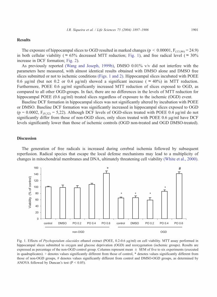
The exposure of hippocampal slices to OGD resulted in marked changes (p < 0.00001, F(13,86) = 24.9)
in both cellular viability (c 65% decreased MTT reduction; Fig. 1), and free radical level (c 30%
increase in DCF formation; Fig. 2).
As previously reported (Wang and Joseph, 1999b), DMSO 0.01% v/v did not interfere with the
parameters here measured, with almost identical results obtained with DMSO alone and DMSO free
slices submitted or not to ischemic conditions (Figs. 1 and 2). Hippocampal slices incubated with POEE
0.6 Ag/ml (but not 0.2 or 0.4 Ag/ml) showed a significant increase (c 40%) in MTT reduction.
Furthermore, POEE 0.6 Ag/ml significantly increased MTT reduction of slices exposed to OGD, as
compared to all other OGD-groups. In fact, there are no differences in the levels of MTT reduction for
hippocampal POEE (0.6 Ag/ml) treated slices regardless of exposure to the ischemic (OGD) event.
Baseline DCF formation in hippocampal slices was not significantly altered by incubation with POEE
or DMSO. Baseline DCF formation was significantly increased in hippocampal slices exposed to OGD
(p = 0.0002, F(9,32) = 5,22). Although DCF levels of OGD-slices treated with POEE 0.4 Ag/ml do not
significantly differ from those of non-OGD slices, only slices treated with POEE 0.6 Ag/ml have DCF
levels significantly lower than those of ischemic controls (OGD non-treated and OGD DMSO-treated).
Discussion
The generation of free radicals is increased during cerebral ischemia followed by subsequent
reperfusion. Radical species that escape the local defense mechanisms may lead to a multiplicity of
changes in mitochondrial membranes and DNA, ultimately threatening cell viability (White et al., 2000).
Fig. 1. Effects of Ptychopetalum olacoides ethanol extract (POEE, 0.2-0.6 Ag/ml) on cell viability. MTT assay performed in
hippocampal slices submitted to oxygen and glucose deprivation (OGD) and reoxygenation (ischemic groups). Results are
expressed as percentage of the non-OGD control group. Columns represent mean F SEM of five to six experiments (executed
in quadruplicates). + denotes values significantly different from those of control, * denotes values significantly different from
those of non-OGD groups, # denotes values significantly different from control and DMSO-OGD groups, as determined by
ANOVA followed by Duncan’s test (P < 0.05).

Fig. 2. Effects of Ptychopetalum olacoides ethanol extract (POEE) at 0.4 ad 0.6 Ag/ml on free radical generation, using DCF
assay, in hippocampal slices exposed to oxygen and glucose deprivation (OGD) and reoxygenation - the ischemic groups.
Results are expressed as percentage of the control non-OGD group. Columns represent mean F SEM of quadruplicates for five
to six experiments. * denotes values significantly different from those of non-OGD groups, # denotes values significantly
different from control and DMSO- OGD groups, as determined by ANOVA followed by Duncan’s test (P < 0.05).
I.R. Siqueira et al. / Life Sciences 75 (2004) 1897–19061902
Accordingly, free radicals (Clemens, 2000) and mitochondria (Morin et al., 2001) have been considered
as rational targets for developing effective therapies for ischemia-induced brain damage.
The exposure of hippocampal slices to an in vitro ischemic event (OGD) followed by reoxygenation
significantly affected cellular viability, by causing a decline in mitochondrial activity in comparison to
control (non-OGD) slices. In this study, mitochondrial activity was evaluated by the highly sensitive
MTT assay, based on the capability of succinate dehydrogenase to reduce a tetrazolium salt to a colored
product. Furthermore, OGD followed by 3 h of reoxygenation lead to increases on free radicals
production, expressed by the amount of DCF formed. DCFH-DA was first described as a probe to
evaluate hydrogen peroxide (Keston and Brandt, 1965); subsequently, it has been suggested that
increases in DCF fluorescence actually reflect the overall cellular oxidative stress (Wang and Joseph,
1999a), since other forms of free radicals such as peroxyl radical, peroxynitrite, nitric oxide and
dopamine can also oxidize DCFH.
Incubation of rat hippocampal slices with POEE (0.6 Ag/ml) during the OGD and reoxygenation
periods lead to a significant increase in MTT reduction as compared to ischemic controls (OGD non-
treated and DMSO). In addition, POEE also produced a reduction of DCF levels in OGD (but not in
non-OGD) slices. Taken together, these results indicate a neuroprotective effect of the extract. POEE
concentrations used in this study (0.2 to 0.6 Ag/ ml) were those active at free radicals scavenging in
vitro assays (total antioxidant reactivity and total antioxidant potential assays, Siqueira et al., 2002).
The fact that a neuroprotective effect was observed only at the highest dose used implies that the
extract sample in this ex vivo assay was retained at intra and extra intracellular compartments,
effective doses starting at the highest dose here used. One has to bear in mind that the extract is still a
complex mixture of compounds, of which only one or a few may be relevant for the observed activity.
Therefore, effective doses of active compound(s) can be expected to be lower than those of the extract
here studied.

I.R. Siqueira et al. / Life Sciences 75 (2004) 1897–1906 1903
Ischemic injuries have been associated with functional and morphological impairment of mitochon-
dria, including inhibition of the respiratory chain (Borutaite et al., 1995). Accordingly, it has been
suggested that strategies that maintain higher respiratory chain activity without increasing free radical
levels, may be a useful neuroprotective therapeutic intervention (Bouaziz et al., 2002). It is therefore
noteworthy that incubation of non-OGD slices with POEE (0.6 Ag/ml) lead to an increased mitochon-
drial respiratory activity, with no significant changes in DCF formation. It has been shown that catalases
(both, mitochondrial and cytosolic) enhance respiration through complexes I and II (Rodriguez et al.,
2000). We here observed that POEE seems to increase respiratory chain activity, which might be related
to POEE-induced increase in brain catalase activity (Siqueira et al., 2004). Furthermore, the superoxide
anion scavenging porperties of POEE (Siqueira et al., 2002) is of relevance, since mitochondria
complexes I and II and aconitase, iron-sulfur cluster-containing enzymes, may suffer free radical
inactivation (Melov et al., 1999). It is conceivable that POEE may also inhibit the generation of free
radicals, along with its scavenging activity. It would be o interest to scrutinize the effects of POEE in
submitochondrial particles.
Although generally accepted that Ginkgo biloba neuroprotection is related to its flavonoids
antioxidant properties (De Feudis, 1998; Seif-El-Nasr and El-Fattah, 1995; Marcocci et al., 1994), it
was recently reported that EGb 761 and its non-flavone fraction prolonged the survival time of mice
under lethal hypoxia, by retarding the breakdown of brain energy metabolism and increasing the local
cerebral blood flow (Oberpichler et al., 1988). Moreover, oral administration of bilobalide (a terpene
lactone present in EGb 761) devoid of free radicals scavenging properties in cell-free systems, protected
against ischemia-induced delayed neuronal death, presumably by acting via mitochondria (Janssens et
al., 1999; Chandrasekaran et al., 2001). In addition, bilobalide was able to inhibit ischemia-induced
decreases of ATP content in endothelial cells (Janssens et al., 1995) and to suppress hypoxia-induced
membrane breakdown (Klein et al., 1997; Weichel et al., 1999).
Despite the understanding of molecular mechanisms involved with the proposed neuroprotective
action of POEE is incomplete, we suggest that its antioxidant properties (Siqueira et al., 2002; Siqueira et
al., 2004) plays a significant role given that POEE reduced OGD-induced increase in the free radical
content. The detailed mechanism by which POEE reduced OGD-induced free radical production remains
a subject for further investigation. POEE acted as superoxide and nitric oxide scavenger in vitro, both of
which are increased during ischemia/reperfusion (Mason et al., 2000). Moreover, POEE peroxyl
scavenger action can be regarded as a ‘‘chain-breaking antioxidant’’ property useful in protecting
membranes lipids (Siqueira et al., 2002). It is important to note that the administration of POEE to mice
leads to significant changes on indexes of oxidative status in brain structures, including increases in
catalase and glutathione peroxidase activities and reduction of free radical levels, lipid peroxidation and
protein-bound carbonyl content (Siqueira et al., 2004).
Consistently, by protecting submitochondrial particles from free radical attack POEE facilitates the
maintenance of mitochondria respiratory processes under ischemic conditions. Another mechanisms
may be the improvement of respiratory chain activity and the maintenance of adequate ATP production
under ischemic conditions, while preventing the increased levels of free radicals generation that usually
accompany such circumstances.
This study offers evidence of, and a possible explanation for, neuroprotective properties of
Ptychopetalum olacoides. It is clear that additional work is required before a comprehensive
knowledge of POEE neuroprotective potential and mechanisms of action can be achieved. Other in
vitro models such as organotypic slice culture (currently being implemented in our laboratory) as well

I.R. Siqueira et al. / Life Sciences 75 (2004) 1897–19061904
as other in vivo ischemia models must be used to confirm and better characterize POEE neuro-
protective properties.
Nevertheless, effects here reported are consistent with the medicinal use of this species by traditional
communities in the Brazilian portion of the Amazon basin. Considering the activities already identified
(antioxidant in vitro and ex vivo, facilitation of long and short term memory in mice, among others) and
the traditional use of this medicinal species, data presented are a valuable contribution to the
development of this plant-based drug.
Acknowledgements
The authors gratefully acknowledge financial support received from FINEP/PRONEX, CNPq,
CAPES, FAPERGS and PROPESQ-UFRGS. There is a patent request associated with this study.
References
Borchers, A., Sakao, S., Henderson, G.L., Harkey, M.R., Keen, C.L., Stern, J.S., Terasawa, K., Gershwin, M.E., 2000.
Shosaiko-to and other Kampo (Japanese herbal) Medicines: a Review of their Immunomodulatory Activities. Journal of
Ethnopharmacology 73, 1–13.
Borutaite, V., Mildaziene, V., Brown, G.C., Brand, M.D., 1995. Control and kinetic analysis of ischemia-damaged heart
mitochondria: which parts of the oxidative phosphorylation system are affected by ischemia?. Biochimica et Biophysica
Acta 1272, 154–158.
Bouaziz, N., Redon, M., Quere, L., Remacle, J., Michiels, C., 2002. Mitochondrial respiratory chain as a new target for anti-
ischemic molecules. European Journal of Pharmacology 441, 35–45.
Cardenas, A., Moro, M.A., Hurtado, O., Leza, J.C., Lorenzo, P., Castrillo, A., Bodelon, O.G., Bosca, O.G., Lizasoain, I., 2000.
Implication of glutamate in the expression of inducible nitric oxide synthase after oxygen and glucose deprivation in rat
forebrain slices. Journal of Neurochemistry 74, 2041–2048.
Chandrasekaran, K., Mehrabian, Z., Spinnewyn, B., Drieu, K., Fiskum, G., 2001. Neuroprotective effects of bilobalide, a
component of the Ginkgo biloba extract (EGb 761), in gerbil global brain ischemia. Brain Research 922, 282–292.
Cimarosti, H., Rodnight, R., Tavares, A., Paiva, R., Valentim, L., Rocha, E., Salbego, C., 2001. An investigation of the
neuroprotective effect of lithium in organotypic slice cultures of rat hippocampus exposed to oxygen and glucose depriva-
tion. Neuroscience Letters 315, 33–36.
Clemens, J.A., 2000. Cerebral ischemia: gene activation, neuronal injury, and the protective role of antioxidants. Free Radical
Biology and Medicine 28, 1526–1531.
Cohen, M.M., Pettegrew, J.W., Kopp, S.J., Minshew, N., Glonek, T., 1984. P-31 nuclear magnetic resonance analysis of brain:
normoxic and anoxic brain slices. Neurochemical Research 9, 785–801.
da Silva, A.L., 2001. Efeitos de Ptychopetalum olacoides (marapuama) sobre a ansiedade e memoria em camundongos. Porto
Alegre: UFRGS. Master Thesis in Biological Sciences, Federal University of Rio Grande do Sul.
De Feudis, F.V., 1998. Ginkgo Biloba extract (EGb 761): from chemistry to clinic Ullstein Medical, Wiesbaden.
Elisabetsky, E., 2002. Traditional medicines and the new paradigm of psychotropic drug action. In: Iwu, M.M., Wootton, J.
(Eds.), Ethnomedicine and Drug Development. Elsevier, Oxford, pp. 133–144.
Elisabetsky, E., Fiqueiredo, W., Oliveira, G., 1992. Traditional amazonian nerve tonics as antidepressant agents: Chaunochiton
kapleri, a case study. Journal of Herbs, Spices and Medicinal Plants 1, 125–162.
Elisabetsky, E., Siqueira, I.R., 1998. Is there a psychopharmacological meaning for tradicional tonics?. In: Prendergast, H.D.,
Etkin, N., Harris, D.R., Houghton, P.J. (Eds.), Plants for Food and Medicine, 373–385.
Grenand, P., Moretti, C., Jacquemin, H., 1987. Pharmacopees traditionnelles en Guyane, 326–328.
Janssens, D., Michiels, C., Delaive, E., Eliaers, F., Drieu, K., Remacle, J., 1995. Protection of hypoxia-induced ATP decrease in
endothelial cells by Ginkgo biloba extract and bilobalide. Biochemical Pharmacology 50, 991–999.

I.R. Siqueira et al. / Life Sciences 75 (2004) 1897–1906 1905
Janssens, D., Remacle, J., Drieu, K., Michiels, C., 1999. Protection of mitochondrial respiration activity by bilobalide.
Biochemical Pharmacology 58, 109–119.
Keston, A.S., Brandt, R., 1965. The fluorometric analysis of ultra micro quantities of hydrogen peroxide. Analytical Bio-
chemistry 11, 1–5.
Kirino, T., Sano, K., 1984. Selective vulnerability in the gerbil hippocampus following transient ischemia. Acta Neuropatho-
logica 62, 201–208.
Klein, J., Chatterjee, S.S., Loffelholz, K., 1997. Phospholipid breakdown and choline release under hypoxic conditions:
inhibition by bilobalide, a constituent of Ginkgo biloba. Brain Research 755, 347–350.
Marcocci, L., Packer, L., Droy-Lefaix, M., Sakaki, A., Guardes-Albert, M., 1994. Antioxidant action of Ginkgo biloba extract
EGb 761. Methods in Enzymology 234, 462–475.
Mason, R.B., Pluta, R.M., Walbridge, S., Wink, D.A., Oldfield, E.H., Boock, R.J., 2000. Production of reactive oxygen species
after reperfusion in vitro and in vivo: protective effect of nitric oxide. Journal of Neurosurgery 93, 99–107.
Melov, S., Coskun, P., Wallace, D.C., 1999. Mouse models of mitochondrial disease, oxidative stress, and senescence. Mutation
Research 434, 233–242.
Morin, D., Hauet, T., Spedding, M., Tillement, J., 2001. Mitochondria as target for antiischemic drugs. Advanced Drug
Delivery Reviews 49, 151–174.
Moro, M.A., De Alba, J., Leza, J.C., Lorenzo, P., Fernandez, A.P., Bentura, M.L., Bosca, L., Rodrigo, J., Lizasoain, I., 1998.
Neuronal expression of inducible nitric oxide synthase after oxygen and glucose deprivation in rat forebrain slices. The
European Journal of Neuroscience 10, 445–446.
Mosmann, T., 1983. Rapid colorimetric assay of cellular growth and survival: application to proliferation and cytotoxicity
assays. Journal of Immunological Methods 65, 55–63.
Newman, G.C., Hospod, F.E., Wu, P., 1989. Glucose utilization of ischemic hippocampal slices. Journal of Neuroscience
Methods 28, 23–34.
Oberpichler, H., Beck, T., Abdel-Rahman, M.M., Bielenberg, G.W., Krieglstein, J., 1988. Effects of Ginkgo biloba constituents
related to protection against brain damage caused by hypoxia. Pharmacological Research Communications 20, 349–368.
Panossian, A.G., Wikman, G., Wagner, H., 1999. Plant Adaptagens III. Earlier and More Recent Aspects and Concepts on their
Mode of Action. Phytomedicine 6, 287–300.
Pellmar, T.C., 1995. Use of brain slices in the study of free-radical actions. Journal of Neuroscience Methods 59, 93–98.
Pio Correa, M., 1978. Dicionario de Plantas Uteis do Brasil, 256.
Porciuncula, L.O., Rocha, J.B., Cimarosti, H., Vinade, L., Ghisleni, G., Salbego, C.G., Souza, D.O., 2003. Neuroprotective
effect of ebselen on rat hippocampal slices submitted to oxygen-glucose deprivation: correlation with immunocontent of
inducible nitric oxide synthase. Neuroscience Letters 346, 101–104.
Rodriguez, A.M., Carrico, P.M., Mazurkiewicz, J.E., Melendez, J.A., 2000. Mitochondrial or cytosolic catalase reverses the
MnSOD-dependent inhibition of proliferation by enhancing respiratory chain activity, net ATP production, and decreasing
the steady state levels of H2O2. Free Radical Biology and Medicine 29, 801–813.
Seif-El-Nasr, S., El-Fattah, A.A., 1995. Lipid peroxide, phospholipids, glutathione levels and superoxide dismutase activity in
rat brain after ischaemia: effect of Ginkgo biloba extract. Pharmacological Research 32, 273–278.
Silva, A.L., Bardini, S., Nunes, D.S., Elisabetsky, E., 2002. Anxiogenic properties of Ptychopetalum olacoides Bentham
(Marapuama). Phytotherapy Research 16, 223–226.
Sims, N.R., Zaidan, E., 1995. Biochemical changes associated with selective neuronal death following short-term cerebral
ischaemia. The International Journal of Biochemistry and Cell Biology 27, 531–550.
Siqueira, I.R., 1997. Contribuic�ao ao Estudo Etnofarmacologico de Ptychopetalum olacoides Bentham: Propriedades Psico-
farmacologicas. Master Thesis in Biological Sciences, Federal University of Rio Grande do Sul, Porto Alegre, p. 14.
Siqueira, I.R., Cordova, C.A.S., Creczynski-Pasa, T.B., Elisabetsky, E., Nunes, D.S., Netto, C.A., 2002. Antioxidant Action of
an Ethanolic Extract of Ptychopetalum olacoides Bentham (Olacaceae). Pharmaceutical Biology 40, 374–379.
Siqueira, I.R., Fochesatto, C., da Silva, A.L., Nunes, D.S., Battastini, A.M., Netto, C.A., Elisabetsky, E., 2003. Ptychopetalum
olacoides, a traditional Amazonian ‘‘nerve tonic’’, possesses anticholinesterase activity. Pharmacology Biochemistry and
Behavior 75 (3), 645–650.
Siqueira, I.R., Fochesatto, C., Torres, I.L.S., Silva, A.L., Nunes, D.S., Elisabetsky, E., Netto, C.A., 2004. Antioxidant activities
of the ethanol extract from Ptychopetalum olacoides in mice brain. Phytomedicine (submitted for publication).
Siqueira, I.R., Lara, D.R., Gaieski, F., Silva, F.S., Nunes, D.S., Elisabetsky, E., 1998. Psychopharmacological Properties of
Ptychopetalum olacoides (Olacaceae). Pharmaceutical Biology 36, 327–334.

I.R. Siqueira et al. / Life Sciences 75 (2004) 1897–19061906
Sriram, K., Pai, K.S., Boyd, M.R., Ravindranath, V., 1997. Evidence for generation of oxidative stress in brain by MPTP: in
vitro and in vivo studies in mice. Brain Research 749, 44–52.
Steinmetz, E.F., 1962. Muira puama (‘‘potency wood’’). Quarterly Journal Crude Drug Research 2, 229–232.
Taylor, C.P., Burke, S.P., Weber, M.L., 1995. Hippocampal slices: glutamate overflow and cellular damage from ischemia are
reduced by sodium-channel blockade. Journal of Neuroscience Methods 59, 121–128.
Wang, H., Joseph, J.A., 1999a. Quantifying cellular oxidative stress by a dichlorofluorescein assay using microplate reader.
Free Radical Biology and Medicine 27, 612–616.
Wang, H., Joseph, J.A., 1999b. Structure-activity relationships of quercetin in antagonizing hydrogen peroxide-induced calcium
dysregulation in PC12 cells. Free Radical Biology and Medicine 27, 683–694.
Weichel, O., Hilgert, M., Chatterjee, S.S., Lehr, M., Klein, J., 1999. Bilobalide, a constituent of Ginkgo biloba, inhibits NMDA-
induced phospholipase A2 activation and phospholipid breakdown in rat hippocampus. Naunyn-Schmiedeberg’s Archives of
Pharmacology 360, 609–615.
White, B.C., Sullivan, J.M., De Gracia, D.J., O’Neil, B.J., Neumar, R.W., Grossman, L.I., Rafols, J.A., Krause, G.S., 2000.
Brain ischemia and reperfusion: molecular mechanisms of neuronal injury. Journal of Neurological Sciences 179, 1–33.
Related Documents


