Neuroprotection by Acetyl-11-Keto-b-Boswellic Acid, in Ischemic Brain Injury Involves the Nrf2/ HO-1 defense Pathway Yi Ding 1 *, MinChun Chen 1 *, Min Wang 3 *, MingMing Wang 1 *, Tiejun Zhang 2 , Jongsun Park 2 , YanRong Zhu 1 , Chao Guo 1 , YanYan Jia 1 , YuWen Li 1 & AiDong Wen 1 1 Department of Pharmacy, Xijing Hospital, Fourth Military Medical University, 2 Department of Pharmacology, Chungnam National University, 3 Department of Pharmacology, School of Pharmacy, Fourth Military Medical University. Stroke is a complex disease involved oxidative stress-related pathways in its pathogenesis. The nuclear factor erythroid-2-related factor 2 (Nrf2)/heme oxygenase-1 (HO-1) pathway has been considered a potential target for neuroprotection in stroke. Acetyl-11-Keto-b-Boswellic Acid (AKBA) is an active triterpenoid compound from the extract of Boswellia serrate. The present study was to determine whether AKBA, a novel Nrf2 activator, can protect against cerebral ischemic injury. The stroke model was produced in Sprague– Dawley rats via middle cerebral artery occlusion. To model ischemia-like conditions in vitro, primary cultured cortical neurons were exposed to transient oxygen and glucose deprivation (OGD). Treatment of AKBA significantly reduced infarct volumes and apoptotic cells, and also increased neurologic scores by elevating the Nrf2 and HO-1 expression in brain tissues in middle cerebral artery occlusion (MCAO) rats at 48 hours post reperfusion. In primary cultured neurons, AKBA increased the Nrf2 and HO-1 expression, which provided protection against OGD-induced oxidative insult. Additionally, AKBA treatment increased Nrf2 binding activity to antioxidant-response elements (ARE). The protective effect of AKBA was attenuated by knockdown of Nrf2 or HO-1. In conclusion, these findings provide evidence that AKBA protects neurons against ischemic injury, and this neuroprotective effect involves the Nrf2/HO-1 pathway. I schemic stroke due to occlusion of brain vasculature is a leading source of mortality amounting to 9% of total deaths each year 1 . Although thrombolysis is the sole presently effective available stroke treatment, it is limited to a small proportion of patients with stroke because it carries the risk of intracranial hemorrhagic trans- formation. The importance of developing an effective treatment remains essential. Evidence has accumulated that excessive reactive oxygen species (ROS) are closely related to cerebral ischemia/reperfusion (I/R) injury in stroke 2 . Brain tissue is particularly susceptible to oxidative damage. Thus, antioxidants are considered in treatment and prevention of stroke. Acetyl 11-keto-b-boswellic acid (AKBA), a pentacyclic triterpenoid compound, is among the most important active principles within the multi-component mixture of Boswellia serrata resin 3 . Boswellia serrata resin extracts (Boswellic acids) shows an in vivo antioxidant activity in many conditions that include bowel disease 4 , myocardial I/R injury 5 and pulmonary fibrosis 6 . The neuroprotective property of pentacyclic triterpenoid has attracted increasing attention recently. For example, oleanolic acid shows protective effects on cerebral ischemic damage and H 2 O 2 -induced injury in vitro 7 . Recently, it was reported that ursolic acid, a naturally occurring pentacyclic triterpenoid, promotes the neuroprotection after cerebral ischemia in mice by activating Nrf2 pathway 8 . Another study revealed that AKBA may have a better antioxidant effect compared with ursolic acid in mice 9 . Based on these studies, we hypothesized that AKBA, which has the similar chemical structure to ursolic acid, may promote the neuroprotection via the Nrf2 pathway. Nrf2 controls the coordinated expression of important antioxidant and detoxification genes through a pro- moter sequence termed the antioxidant response element (ARE). Phase II genes, including heme oxygenase-1 (HO-1), glutathione S-transferases (GSTs) and NAD(P)H quinone oxidoreductase, work in synergy to provide protection by regulating and maintaining intracellular redox states 10,11 . Of these, HO-1 has been reported to have the most AREs on its promoter, making it a highly effective therapeutic target for protection against neurode- OPEN SUBJECT AREAS: STROKE THERAPEUTICS Received 13 August 2014 Accepted 24 October 2014 Published 11 November 2014 Correspondence and requests for materials should be addressed to Y.W.L. (liyuwenzs@ gmail.com) or A.D.W. (adwen-2004@ hotmail.com) * These authors contributed equally to this work. SCIENTIFIC REPORTS | 4 : 7002 | DOI: 10.1038/srep07002 1

Welcome message from author
This document is posted to help you gain knowledge. Please leave a comment to let me know what you think about it! Share it to your friends and learn new things together.
Transcript
Neuroprotection byAcetyl-11-Keto-b-Boswellic Acid, inIschemic Brain Injury Involves the Nrf2/HO-1 defense PathwayYi Ding1*, MinChun Chen1*, Min Wang3*, MingMing Wang1*, Tiejun Zhang2, Jongsun Park2,YanRong Zhu1, Chao Guo1, YanYan Jia1, YuWen Li1 & AiDong Wen1
1Department of Pharmacy, Xijing Hospital, Fourth Military Medical University, 2Department of Pharmacology, Chungnam NationalUniversity, 3Department of Pharmacology, School of Pharmacy, Fourth Military Medical University.
Stroke is a complex disease involved oxidative stress-related pathways in its pathogenesis. The nuclear factorerythroid-2-related factor 2 (Nrf2)/heme oxygenase-1 (HO-1) pathway has been considered a potentialtarget for neuroprotection in stroke. Acetyl-11-Keto-b-Boswellic Acid (AKBA) is an active triterpenoidcompound from the extract of Boswellia serrate. The present study was to determine whether AKBA, a novelNrf2 activator, can protect against cerebral ischemic injury. The stroke model was produced in Sprague–Dawley rats via middle cerebral artery occlusion. To model ischemia-like conditions in vitro, primarycultured cortical neurons were exposed to transient oxygen and glucose deprivation (OGD). Treatment ofAKBA significantly reduced infarct volumes and apoptotic cells, and also increased neurologic scores byelevating the Nrf2 and HO-1 expression in brain tissues in middle cerebral artery occlusion (MCAO) rats at48 hours post reperfusion. In primary cultured neurons, AKBA increased the Nrf2 and HO-1 expression,which provided protection against OGD-induced oxidative insult. Additionally, AKBA treatment increasedNrf2 binding activity to antioxidant-response elements (ARE). The protective effect of AKBA wasattenuated by knockdown of Nrf2 or HO-1. In conclusion, these findings provide evidence that AKBAprotects neurons against ischemic injury, and this neuroprotective effect involves the Nrf2/HO-1 pathway.
Ischemic stroke due to occlusion of brain vasculature is a leading source of mortality amounting to 9% of totaldeaths each year1. Although thrombolysis is the sole presently effective available stroke treatment, it is limitedto a small proportion of patients with stroke because it carries the risk of intracranial hemorrhagic trans-
formation. The importance of developing an effective treatment remains essential. Evidence has accumulated thatexcessive reactive oxygen species (ROS) are closely related to cerebral ischemia/reperfusion (I/R) injury in stroke2.Brain tissue is particularly susceptible to oxidative damage. Thus, antioxidants are considered in treatment andprevention of stroke.
Acetyl 11-keto-b-boswellic acid (AKBA), a pentacyclic triterpenoid compound, is among the most importantactive principles within the multi-component mixture of Boswellia serrata resin3. Boswellia serrata resin extracts(Boswellic acids) shows an in vivo antioxidant activity in many conditions that include bowel disease4, myocardialI/R injury5 and pulmonary fibrosis6. The neuroprotective property of pentacyclic triterpenoid has attractedincreasing attention recently. For example, oleanolic acid shows protective effects on cerebral ischemic damageand H2O2-induced injury in vitro7. Recently, it was reported that ursolic acid, a naturally occurring pentacyclictriterpenoid, promotes the neuroprotection after cerebral ischemia in mice by activating Nrf2 pathway8. Anotherstudy revealed that AKBA may have a better antioxidant effect compared with ursolic acid in mice9. Based onthese studies, we hypothesized that AKBA, which has the similar chemical structure to ursolic acid, may promotethe neuroprotection via the Nrf2 pathway.
Nrf2 controls the coordinated expression of important antioxidant and detoxification genes through a pro-moter sequence termed the antioxidant response element (ARE). Phase II genes, including heme oxygenase-1(HO-1), glutathione S-transferases (GSTs) and NAD(P)H quinone oxidoreductase, work in synergy to provideprotection by regulating and maintaining intracellular redox states10,11. Of these, HO-1 has been reported to havethe most AREs on its promoter, making it a highly effective therapeutic target for protection against neurode-
OPEN
SUBJECT AREAS:STROKE
THERAPEUTICS
Received13 August 2014
Accepted24 October 2014
Published11 November 2014
Correspondence andrequests for materials
should be addressed toY.W.L. (liyuwenzs@
gmail.com) or A.D.W.(adwen-2004@
hotmail.com)
* These authorscontributed equally to
this work.
SCIENTIFIC REPORTS | 4 : 7002 | DOI: 10.1038/srep07002 1
generative diseases. It gives protection in part by degrading its pro-oxidant substrate, heme, and generating the antioxidants biliverdinand bilirubin12. Recently, studies have provided evidence for thetherapeutic potential of targeting the Nrf2/HO-1 pathway in braininjury after ischemic stroke13,14.
Considering the beneficial properties of triterpenoids as well as thepossible role of the Nrf2/HO-1 pathway, in this study we applied invivo and in vitro ischemic paradigms to analyze the protective prop-erties of AKBA. We hypothesized that AKBA would provide neuro-protection against I/R injury induced by transient middle cerebralartery occlusion (MCAO) in rats and oxygen and glucose deprivation(OGD) in primary cultured neurons, and that the protection wouldoccur through activation of the Nrf2/HO1 pathway.
MethodsMCAO model. All procedures involving animals were approved by the InstitutionalAnimal Care and Use Committee of the Fourth Military Medical University andwritten up following the ARRIVE guidelines. Experiments were performed inaccordance with published National Institutes of Health guidelines. Adult maleSprague-Dawley rats aged 8–10 weeks from the Experimental Animal Center ofFourth Military Medical University (Xi’an, China) (280–300 g). All rats were dividedrandomly into the following 3 groups using a random number table generated bySPSS 16.0 (SPSS Inc., Chicago, IL, USA): sham-operated group (sham), vehicle-treated I/R group (vehicle 1 I/R), and AKBA-treated I/R group (AKBA 1 I/R). In allthe three groups, eight rats were used for physiologic parameters and infarct sizemeasurement, eight rats were used for HE staining and TUNEL staining, eight ratswere used for determination of oxidative stress, six rats were used for Westernblotting, and six rats were used for immunostaining.
Rats were anesthetized using 2.0 to 3.0% isoflurane and maintained using 1.0 to1.5% isoflurane (both in 70% N2O/30% O2). Focal cerebral ischemia was performedusing the method of right MCAO with an intraluminal filament as described prev-iously15. Cerebral blood flow (CBF) was monitored using laser Doppler flowmetry(Perimed AB, PeriFlux System 5000, Stockholm, Sweden) in the ipsilateral cortex(2 mm posterior and 5 mm lateral to bregma). Sham operated rats were manipulatedin the same way, but the MCA was not occluded. Animals that did not show a CBFreduction of at least 70% and animals that died after ischemia induction wereexcluded from the groups. At 2 h after the induction of ischemia, the filament wasslowly withdrawn. The neck incision was closed and rats were allowed to recover.After revival from anesthesia, the animals were put back into cages with the roomtemperature maintained at 25 6 2uC. The animals were allowed to survive for 2 dayswith free access to water and food. Mean arterial blood pressure, pH, arterial bloodgases, and blood glucose levels during treatment were evaluated.
AKBA (reagent grade, purity . 90%, Santa Ana, CA) diluted with physiologicalsaline (20 mg/kg) was administered by intraperitoneal injection. Vehicle of 2 ml/kgphysiological saline (vehicle 1 I/R group) and 20 mg/kg AKBA (AKBA 1 I/R group)were given immediately after the onset of reperfusion. The dose of 20 mg/kg AKBAadministered to rats (corresponding to about 350 mg Boswellia serrata extract/kg)was chosen based on previous study4. Meanwhile, in a preliminary experiment, adose-response (5 mg/kg, 10 mg/kg and 20 mg/kg administered by intraperitonealinjection) study was conducted (data was shown in Figs. 1). From infarct volumemeasurement, we demonstrated that the dosage of AKBA at 20 mg/kg the besttherapeutic effects among three doses, and therefore we focused on the AKBAtreatment at 20 mg/kg for biochemical and molecular analysis.
Neurological function evaluation and quantification of infarct volume. Accordingto the Zea Longa standard15, neurological deficits were blindly evaluated 48 h afterreperfusion with a 5-point scale system: 0, no deficit; 1, not being able to completelystretch the contralateral torso and forelimb; 2, turning to the ipsilateral side when heldby the tail; 3, falling over to the affected side; 4, not being able to walk and nospontaneous locomotor activity.
To calculate infarct volume, brains were removed at 48 h after MCAO and were cutinto 2 mm thick coronal sections and subjected to 2, 3, 5-triphenyltetrazoliumchloride (TTC) staining. Unstained areas were defined as infarcts and were measuredusing image analysis software (Adobe Photoshop CS3, San Jose, CA, USA). Thepercentage of the infarct volume was calculated by the following formula: ([totalcontralateral hemispheric volume]- [total ipsilateral hemispheric stained volume])/(total contralateral hemispheric volume) 3 100%.
HE and TUNEL staining. Hematoxylin and eosin (HE) staining was performed toreveal the morphological features of injured neurons in cerebral cortex. TUNELstaining was performed on paraffin-embedded sections. Commercially availablereagents (Promega, DeadEnd Flurometric Tunel System) were used to performTUNEL analysis. The total number of TUNEL positive neurons in the ipsilateralhemisphere was counted in three different fields for each section by an investigatorwho was blinded to the studies by light microscopy.
Determination of oxidative stress. Animals were killed at 48 h after reperfusion andbrains were removed rapidly. Right cortical samples (n 5 8 for each group) were
weighed. Homogenates were centrifuged at 15,000 g for 10 minutes, and thesupernatant obtained was used for the following measurements. The superoxidedismutase (SOD) activity was determined spectrophotometrically usingcommercially available assay kits obtained from Cayman Chemicals (Ann Arbor, MI,USA). Malondialdehyde (MDA) were measured in deproteinized brain extracts byhigh performance liquid chromatography (HPLC)16. The chromatography systemconsisted of Agilent 1200 Series LC System equipped with a UV detector, anautosampler (Agilent Technologies, USA). Chromatographic separation wasachieved on a Dikma column (Dikma, Beijing, China. 150 mm 3 4.6 mm, 5 mm).
Immunofluorescence staining. The paraffin sections, 5 mm thick, which weredrawn 48 h after reperfusion, were first deparaffinized in xylene, rehydrated withvarious grades of ethanol, and pretreated with 10 mg/ml proteinase K for 30 min at37uC. By incubating the sections in 10% bovine serum albumin, nonspecific bindingof immunoglobulins was blocked for 20 min. Then the sections were incubatedovernight at room temperature in with anti-Nrf2 (ab31163, Abcam) or anti-HO-1(ab13243, Abcam), and with anti-NeuN antibody to mark to neurons (monoclonalclone A60, Chemicon). Sections were then incubated with Alexa Fluor s 488 anti-rabbit IgG or Alexa Fluor s 594 anti-mouse IgG secondary antibodies for 2 h. Then,the sections were incubated with DAPI for nuclear counterstaining. Finally, thesections were coverslipped. The stained sections were examined under a fluorescencemicroscope (Olympus, Tokyo, Japan).
Western blot analysis. Right cortical samples were weighed and protein wasextracted. Cortical neurons were washed twice with ice-cold PBS. Nuclear protein ofNrf2 was obtained using a Neuclear-Cytosol Extraction Kit (Applygen TechnologiesInc., Beijing) and total protein of HO-1 in extraction was obtained using Total ProteinExtraction Kit (Applygen Technologies Inc., Beijing) following the manufacturer’sprotocols. Equal amounts of total protein extracts or nuclear protein extracts wereseparated by SDS-PAGE and transferred onto polyvinylidenedifluoride membranes(Millipore, Bedford, MA, USA) by electrophoresis, and membranes were blockedwith 5% nonfat milk in TBST (0.1% Tween 20 in TBS) for 1 h at room temperature.Blots were then incubated with the antibody for Nrf2, HO-1, Histone H3 (CellSignaling Technology) and b-actin (Cell Signaling Technology).
Primary culture of rat cortical neurons. Cortical neurons were prepared from brainsof one-day-old Sprague-Dawley rats. Approximately 30,000 cells in 50 ml neurobasalmedium containing glutamine (1 mmol/L), 1% penicillin, streptomycin (Pen/Strep),and 10% fetal bovine serum were seeded into 6-well plates. After 2 h, 0.5 mlneurobasal medium containing the serum-free B27 supplement (2%), Pen/Strep, andglutamine were added to each well. After 2 days in vitro, 5 mM cytosinearabinofuranoside was added to inhibit glial proliferation. At 5 days in vitro, themedium was changed to fresh neurobasal medium containing B27. Neurons werecultured at 37uC in a humidified 5% CO2 atmosphere and used after 7 days in vitro.
Oxygen-glucose deprivation and reoxygenation. Cells seeded in 96-well plates werepretreated with AKBA at the indicated concentrations for 2 h. Then, to modelischemia-like conditions in vitro, primary cultured cortical neurons were exposed totransient OGD for 60 min. Finally, the neurons were incubated again in theincubator with 95% air and 5% CO2 with or without AKBA for an additional 24 h.OGD-induced cell death was quantified using the 3-[4,5-dimethylthiazol-2-yl]-2,5-diphenyltetrazolium bro-mide (MTT) assay. Intracellular ROS levels were measuredusing the dye 29,79-dichlorofluorescein diacetate (DCF-DA) as described previously17.
Nrf2 and HO-1 siRNA transfection. Neuronal cells were transiently transfected withsiRNA targeting to Nrf2 or HO-1 by Lipofectamine 2000 TM (Invitrogen) accordingto the manufacturer’s protocol. Knockdown efficiency of Nrf2 and HO-1 wasdetermined by Western blot analysis. Cells were transfected with siRNA or with anontargeting scramble control siRNA for 48 h, followed by treatment with AKBA forthe indicated times. Rat Nrf2-specific siRNA (59-ACGCAGGAGAGGGAAGA-AUAAAGUU-39 and 59-AACUUUAUUCUUCCCUCUCCUGCGU-39), rat HO-1-specific siRNA (59-AUGGCAUAAAUUCCCACUGCCACGG-39 and 59-CCG-UGGCAGUGGGAAUUUAUGCCAU-39) and a nonspecific siRNA (59-AUGCA-CGAUAUAACCUCACCGUCGG-39 and 59-CCGACGGUGAGGUUA-UAUCGUGCAU-39) were provided by Invitrogen (Carlsbad, CA, USA). Cellsamples were then collected for Western blot analysis, MTT assay and measurementof intracellular ROS levels.
Electrophoretic mobility shift assay (EMSA). EMSA was performed using thecommercial Chemiluminescent EMSA kit (Pierce Biotechnology). For EMSA, 5 mgof total extracted nuclear proteins was incubated with 1 pmol double stranded ATPend-labeled oligonucleotide probe containing the sequences in binding buffer(10 mM HEPES, pH 7.9, 80 mM NaCl, 3 mM MgCl2, 0.1 mM EDTA, 1 mMdithiothreitol, 1 mM phenylmethylsulfonyl fluoride, and 10% glycerol). After theincubation, the samples were loaded on a 5% TBE-polyacrylamide gel (Bio-Rad) andelectrophoretically separated in 0.53 TBE buffer. Levels of Nrf2 DNA bindingactivity were quantified by computer-assisted densitometric analysis.
Statistical AnalysisThe statistical analyses were performed using SPSS 16.0 (SPSS Inc.,Chicago, IL, USA). All of the values were presented as mean 6
www.nature.com/scientificreports
SCIENTIFIC REPORTS | 4 : 7002 | DOI: 10.1038/srep07002 2
standard deviation (SD), except for the neurological deficit score, anddifferences between groups were compared with one-way ANOVAfollowed by followed by Fisher’s post hoc test. P , 0.05 was regardedstatistically significant. Neurological deficit scores were expressed asthe median (range) and were analyzed using a nonparametricmethod (Kruskal–Wallis test) followed by the Mann–Whitney U-test using Bonferroni correction.
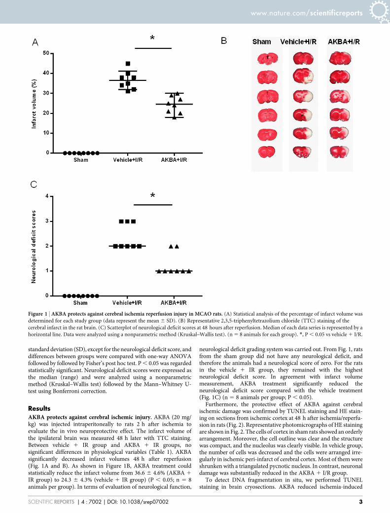
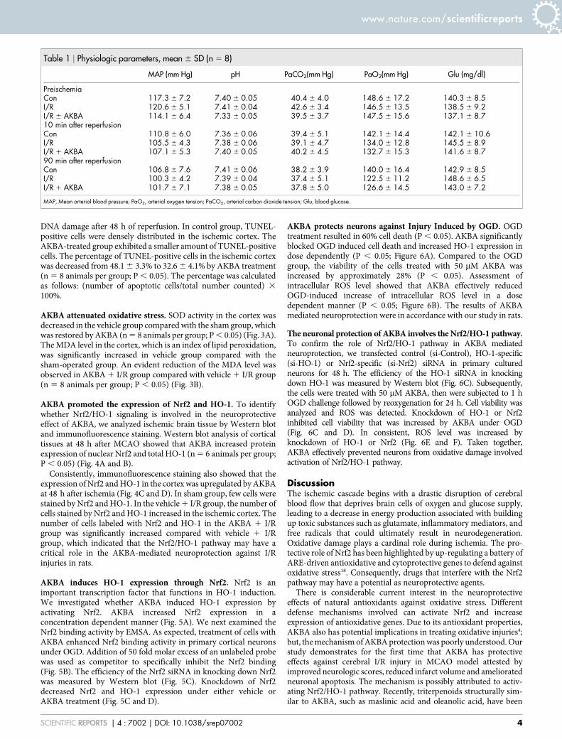
ResultsAKBA protects against cerebral ischemic injury. AKBA (20 mg/kg) was injected intraperitoneally to rats 2 h after ischemia toevaluate the in vivo neuroprotective effect. The infarct volume ofthe ipsilateral brain was measured 48 h later with TTC staining.Between vehicle 1 IR group and AKBA 1 IR groups, nosignificant differences in physiological variables (Table 1). AKBAsignificantly decreased infarct volumes 48 h after reperfusion(Fig. 1A and B). As shown in Figure 1B, AKBA treatment couldstatistically reduce the infarct volume from 36.6 6 4.6% (AKBA 1
IR group) to 24.3 6 4.3% (vehicle 1 IR group) (P , 0.05; n 5 8animals per group). In terms of evaluation of neurological function,
neurological deficit grading system was carried out. From Fig. 1, ratsfrom the sham group did not have any neurological deficit, andtherefore the animals had a neurological score of zero. For the ratsin the vehicle 1 IR group, they remained with the highestneurological deficit score. In agreement with infarct volumemeasurement, AKBA treatment significantly reduced theneurological deficit score compared with the vehicle treatment(Fig. 1C) (n 5 8 animals per group; P , 0.05).
Furthermore, the protective effect of AKBA against cerebralischemic damage was confirmed by TUNEL staining and HE stain-ing on sections from ischemic cortex at 48 h after ischemia/reperfu-sion in rats (Fig. 2). Representative photomicrographs of HE stainingare shown in Fig. 2. The cells of cortex in sham rats showed an orderlyarrangement. Moreover, the cell outline was clear and the structurewas compact, and the nucleolus was clearly visible. In vehicle group,the number of cells was decreased and the cells were arranged irre-gularly in ischemic peri-infarct of cerebral cortex. Most of them wereshrunken with a triangulated pycnotic nucleus. In contrast, neuronaldamage was substantially reduced in the AKBA 1 I/R group.
To detect DNA fragmentation in situ, we performed TUNELstaining in brain cryosections. AKBA reduced ischemia-induced
Figure 1 | AKBA protects against cerebral ischemia reperfusion injury in MCAO rats. (A) Statistical analysis of the percentage of infarct volume was
determined for each study group (data represent the mean 6 SD). (B) Representative 2,3,5-triphenyltetrazolium chloride (TTC) staining of the
cerebral infarct in the rat brain. (C) Scatterplot of neurological deficit scores at 48 hours after reperfusion. Median of each data series is represented by a
horizontal line. Data were analyzed using a nonparametric method (Kruskal–Wallis test). (n 5 8 animals for each group). *, P , 0.05 vs vehicle 1 I/R.
www.nature.com/scientificreports
SCIENTIFIC REPORTS | 4 : 7002 | DOI: 10.1038/srep07002 3
DNA damage after 48 h of reperfusion. In control group, TUNEL-positive cells were densely distributed in the ischemic cortex. TheAKBA-treated group exhibited a smaller amount of TUNEL-positivecells. The percentage of TUNEL-positive cells in the ischemic cortexwas decreased from 48.1 6 3.3% to 32.6 6 4.1% by AKBA treatment(n 5 8 animals per group; P , 0.05). The percentage was calculatedas follows: (number of apoptotic cells/total number counted) 3
100%.
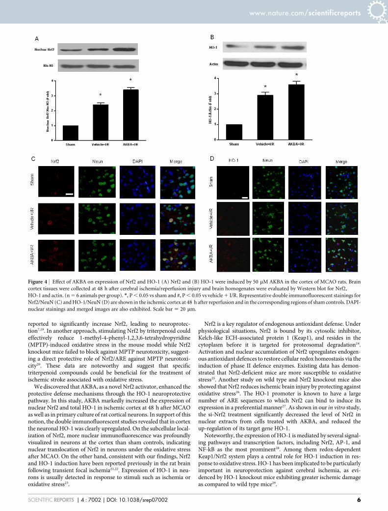
AKBA attenuated oxidative stress. SOD activity in the cortex wasdecreased in the vehicle group compared with the sham group, whichwas restored by AKBA (n 5 8 animals per group; P , 0.05) (Fig. 3A).The MDA level in the cortex, which is an index of lipid peroxidation,was significantly increased in vehicle group compared with thesham-operated group. An evident reduction of the MDA level wasobserved in AKBA 1 I/R group compared with vehicle 1 I/R group(n 5 8 animals per group; P , 0.05) (Fig. 3B).
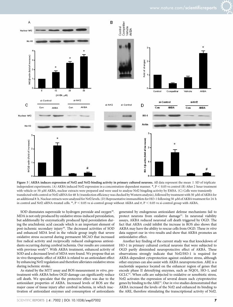
AKBA promoted the expression of Nrf2 and HO-1. To identifywhether Nrf2/HO-1 signaling is involved in the neuroprotectiveeffect of AKBA, we analyzed ischemic brain tissue by Western blotand immunofluorescence staining. Western blot analysis of corticaltissues at 48 h after MCAO showed that AKBA increased proteinexpression of nuclear Nrf2 and total HO-1 (n 5 6 animals per group;P , 0.05) (Fig. 4A and B).
Consistently, immunofluorescence staining also showed that theexpression of Nrf2 and HO-1 in the cortex was upregulated by AKBAat 48 h after ischemia (Fig. 4C and D). In sham group, few cells werestained by Nrf2 and HO-1. In the vehicle 1 I/R group, the number ofcells stained by Nrf2 and HO-1 increased in the ischemic cortex. Thenumber of cells labeled with Nrf2 and HO-1 in the AKBA 1 I/Rgroup was significantly increased compared with vehicle 1 I/Rgroup, which indicated that the Nrf2/HO-1 pathway may have acritical role in the AKBA-mediated neuroprotection against I/Rinjuries in rats.
AKBA induces HO-1 expression through Nrf2. Nrf2 is animportant transcription factor that functions in HO-1 induction.We investigated whether AKBA induced HO-1 expression byactivating Nrf2. AKBA increased Nrf2 expression in aconcentration dependent manner (Fig. 5A). We next examined theNrf2 binding activity by EMSA. As expected, treatment of cells withAKBA enhanced Nrf2 binding activity in primary cortical neuronsunder OGD. Addition of 50 fold molar excess of an unlabeled probewas used as competitor to specifically inhibit the Nrf2 binding(Fig. 5B). The efficiency of the Nrf2 siRNA in knocking down Nrf2was measured by Western blot (Fig. 5C). Knockdown of Nrf2decreased Nrf2 and HO-1 expression under either vehicle orAKBA treatment (Fig. 5C and D).
AKBA protects neurons against Injury Induced by OGD. OGDtreatment resulted in 60% cell death (P , 0.05). AKBA significantlyblocked OGD induced cell death and increased HO-1 expression indose dependently (P , 0.05; Figure 6A). Compared to the OGDgroup, the viability of the cells treated with 50 mM AKBA wasincreased by approximately 28% (P , 0.05). Assessment ofintracellular ROS level showed that AKBA effectively reducedOGD-induced increase of intracellular ROS level in a dosedependent manner (P , 0.05; Figure 6B). The results of AKBAmediated neuroprotection were in accordance with our study in rats.
The neuronal protection of AKBA involves the Nrf2/HO-1 pathway.To confirm the role of Nrf2/HO-1 pathway in AKBA mediatedneuroprotection, we transfected control (si-Control), HO-1-specific(si-HO-1) or Nrf2-specific (si-Nrf2) siRNA in primary culturedneurons for 48 h. The efficiency of the HO-1 siRNA in knockingdown HO-1 was measured by Western blot (Fig. 6C). Subsequently,the cells were treated with 50 mM AKBA, then were subjected to 1 hOGD challenge followed by reoxygenation for 24 h. Cell viability wasanalyzed and ROS was detected. Knockdown of HO-1 or Nrf2inhibited cell viability that was increased by AKBA under OGD(Fig. 6C and D). In consistent, ROS level was increased byknockdown of HO-1 or Nrf2 (Fig. 6E and F). Taken together,AKBA effectively prevented neurons from oxidative damage involvedactivation of Nrf2/HO-1 pathway.
DiscussionThe ischemic cascade begins with a drastic disruption of cerebralblood flow that deprives brain cells of oxygen and glucose supply,leading to a decrease in energy production associated with buildingup toxic substances such as glutamate, inflammatory mediators, andfree radicals that could ultimately result in neurodegeneration.Oxidative damage plays a cardinal role during ischemia. The pro-tective role of Nrf2 has been highlighted by up-regulating a battery ofARE-driven antioxidative and cytoprotective genes to defend againstoxidative stress18. Consequently, drugs that interfere with the Nrf2pathway may have a potential as neuroprotective agents.
There is considerable current interest in the neuroprotectiveeffects of natural antioxidants against oxidative stress. Differentdefense mechanisms involved can activate Nrf2 and increaseexpression of antioxidative genes. Due to its antioxidant properties,AKBA also has potential implications in treating oxidative injuries4;but, the mechanism of AKBA protection was poorly understood. Ourstudy demonstrates for the first time that AKBA has protectiveeffects against cerebral I/R injury in MCAO model attested byimproved neurologic scores, reduced infarct volume and amelioratedneuronal apoptosis. The mechanism is possibly attributed to activ-ating Nrf2/HO-1 pathway. Recently, triterpenoids structurally sim-ilar to AKBA, such as maslinic acid and oleanolic acid, have been
Table 1 | Physiologic parameters, mean 6 SD (n 5 8)
MAP (mm Hg) pH PaCO2(mm Hg) PaO2(mm Hg) Glu (mg/dl)
PreischemiaCon 117.3 6 7.2 7.40 6 0.05 40.4 6 4.0 148.6 6 17.2 140.3 6 8.5I/R 120.6 6 5.1 7.41 6 0.04 42.6 6 3.4 146.5 6 13.5 138.5 6 9.2I/R 6 AKBA 114.1 6 6.4 7.33 6 0.05 39.5 6 3.7 147.5 6 15.6 137.1 6 8.710 min after reperfusionCon 110.8 6 6.0 7.36 6 0.06 39.4 6 5.1 142.1 6 14.4 142.1 6 10.6I/R 105.5 6 4.3 7.38 6 0.06 39.1 6 4.7 134.0 6 12.8 145.5 6 8.9I/R 1 AKBA 107.1 6 5.3 7.40 6 0.05 40.2 6 4.5 132.7 6 15.3 141.6 6 8.790 min after reperfusionCon 106.8 6 7.6 7.41 6 0.06 38.2 6 3.9 140.0 6 16.4 142.9 6 8.5I/R 100.3 6 4.2 7.39 6 0.04 37.4 6 5.1 122.5 6 11.2 148.6 6 6.5I/R 1 AKBA 101.7 6 7.1 7.38 6 0.05 37.8 6 5.0 126.6 6 14.5 143.0 6 7.2
MAP, Mean arterial blood pressure; PaO2, arterial oxygen tension; PaCO2, arterial carbon dioxide tension; Glu, blood glucose.
www.nature.com/scientificreports
SCIENTIFIC REPORTS | 4 : 7002 | DOI: 10.1038/srep07002 4
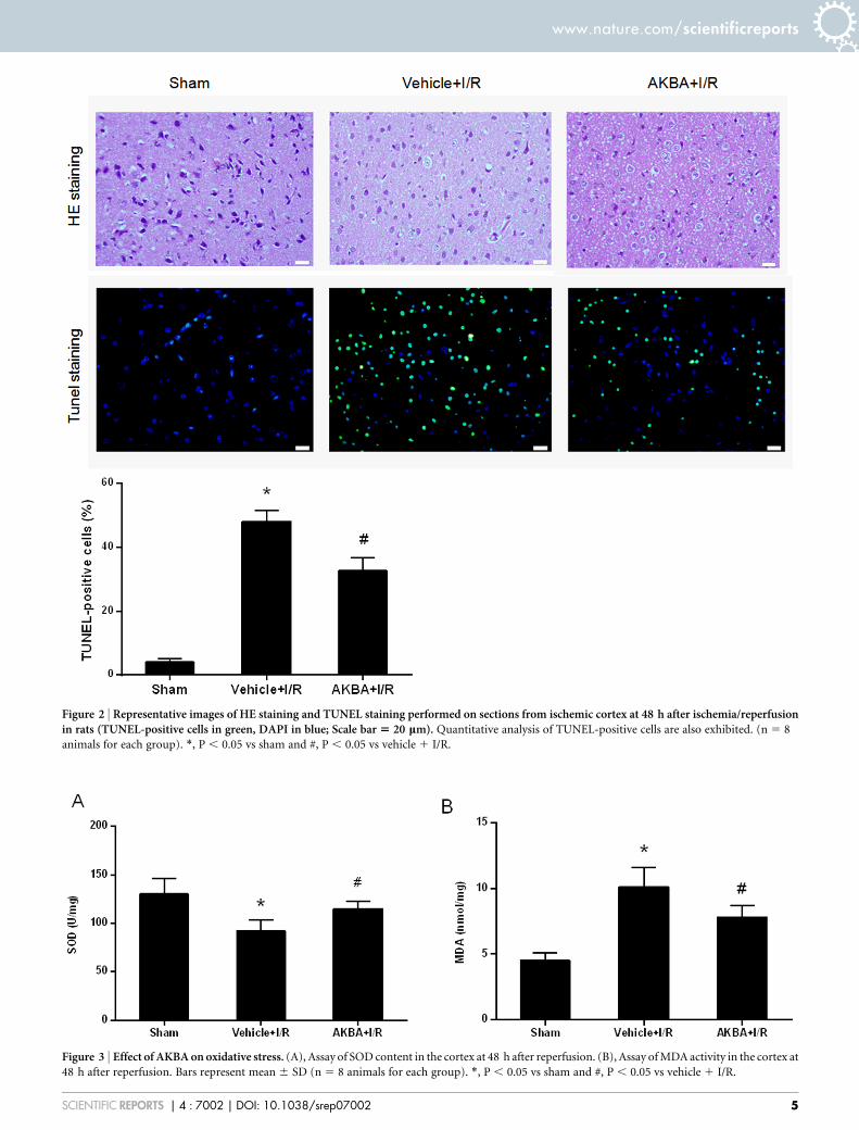
Figure 2 | Representative images of HE staining and TUNEL staining performed on sections from ischemic cortex at 48 h after ischemia/reperfusionin rats (TUNEL-positive cells in green, DAPI in blue; Scale bar 5 20 mm). Quantitative analysis of TUNEL-positive cells are also exhibited. (n 5 8
animals for each group). *, P , 0.05 vs sham and #, P , 0.05 vs vehicle 1 I/R.
Figure 3 | Effect of AKBA on oxidative stress. (A), Assay of SOD content in the cortex at 48 h after reperfusion. (B), Assay of MDA activity in the cortex at
48 h after reperfusion. Bars represent mean 6 SD (n 5 8 animals for each group). *, P , 0.05 vs sham and #, P , 0.05 vs vehicle 1 I/R.
www.nature.com/scientificreports
SCIENTIFIC REPORTS | 4 : 7002 | DOI: 10.1038/srep07002 5
reported to significantly increase Nrf2, leading to neuroprotec-tion7,19. In another approach, stimulating Nrf2 by triterpenoid couldeffectively reduce 1-methyl-4-phenyl-1,2,3,6-tetrahydropyridine(MPTP)-induced oxidative stress in the mouse model while Nrf2knockout mice failed to block against MPTP neurotoxicity, suggest-ing a direct protective role of Nrf2/ARE against MPTP neurotoxi-city20. These data are noteworthy and suggest that specifictriterpenoid compounds could be beneficial for the treatment ofischemic stroke associated with oxidative stress.
We discovered that AKBA, as a novel Nrf2 activator, enhanced theprotective defense mechanisms through the HO-1 neuroprotectivepathway. In this study, AKBA markedly increased the expression ofnuclear Nrf2 and total HO-1 in ischemic cortex at 48 h after MCAOas well as in primary culture of rat cortical neurons. In support of thisnotion, the double immunofluorescent studies revealed that in cortexthe neuronal HO-1 was clearly upregulated. On the subcellular local-ization of Nrf2, more nuclear immunofluorescence was profoundlyvisualized in neurons at the cortex than sham controls, indicatingnuclear translocation of Nrf2 in neurons under the oxidative stressafter MCAO. On the other hand, consistent with our findings, Nrf2and HO-1 induction have been reported previously in the rat brainfollowing transient focal ischemia21,22. Expression of HO-1 in neu-rons is usually detected in response to stimuli such as ischemia oroxidative stress23.
Nrf2 is a key regulator of endogenous antioxidant defense. Underphysiological situations, Nrf2 is bound by its cytosolic inhibitor,Kelch-like ECH-associated protein 1 (Keap1), and resides in thecytoplasm before it is targeted for proteosomal degradation24.Activation and nuclear accumulation of Nrf2 upregulates endogen-ous antioxidant defences to restore cellular redox homeostasis via theinduction of phase II defence enzymes. Existing data has demon-strated that Nrf2-deficient mice are more susceptible to oxidativestress25. Another study on wild type and Nrf2 knockout mice alsoshowed that Nrf2 reduces ischemic brain injury by protecting againstoxidative stress26. The HO-1 promoter is known to have a largenumber of ARE sequences to which Nrf2 can bind to induce itsexpression in a preferential manner27. As shown in our in vitro study,the si-Nrf2 treatment significantly decreased the level of Nrf2 innuclear extracts from cells treated with AKBA, and reduced theup-regulation of its target gene HO-1.
Noteworthy, the expression of HO-1 is mediated by several signal-ing pathways and transcription factors, including Nrf2, AP-1, andNF-kB as the most prominent28. Among them redox-dependentKeap1/Nrf2 system plays a central role for HO-1 induction in res-ponse to oxidative stress. HO-1 has been implicated to be particularlyimportant in neuroprotection against cerebral ischemia, as evi-denced by HO-1 knockout mice exhibiting greater ischemic damageas compared to wild type mice29.
Figure 4 | Effect of AKBA on expression of Nrf2 and HO-1 (A) Nrf2 and (B) HO-1 were induced by 50 mM AKBA in the cortex of MCAO rats. Brain
cortex tissues were collected at 48 h after cerebral ischemia/reperfusion injury and brain homogenates were evaluated by Western blot for Nrf2,
HO-1 and actin. (n 5 6 animals per group). *, P , 0.05 vs sham and #, P , 0.05 vs vehicle 1 I/R. Representative double immunofluorescent stainings for
Nrf2/NeuN (C) and HO-1/NeuN (D) are shown in the ischemic cortex at 48 h after reperfusion and in the corresponding regions of sham controls. DAPI-
nuclear stainings and merged images are also exhibited. Scale bar 5 20 mm.
www.nature.com/scientificreports
SCIENTIFIC REPORTS | 4 : 7002 | DOI: 10.1038/srep07002 6
SOD dismutates superoxide to hydrogen peroxide and oxygen30.MDA is not only produced by oxidative stress-induced peroxidation,but additionally by enzymatically produced lipid peroxidation dur-ing the arachidonic acid cascade which is an important element ofpost-ischemic secondary injury30. The decreased activities of SODand enhanced MDA level in the vehicle group imply that severeoxidative stress occurred during permanent MCAO that increasedfree radical activity and reciprocally reduced endogenous antioxi-dants occurring during cerebral ischemia. Our results are consistentwith previous work4,9. With AKBA treatment, enhanced activity ofSOD and a decreased level of MDA were found. We propose that anin vivo therapeutic effect of AKBA is related to an antioxidant effectby enhancing Nrf2 regulation and therefore alleviates oxidative stressduring ischemic stroke.
As stated by the MTT assay and ROS measurement in vitro, pre-treatment with AKBA before OGD damage can significantly reducecell death. We speculate that the protective effect was due to theantioxidant properties of AKBA. Increased levels of ROS are themajor cause of tissue injury after cerebral ischemia, in which inac-tivation of antioxidant enzymes and consumption of antioxidants
generated by endogenous antioxidant defense mechanisms fail toprotect neurons from oxidative damage31. In neuronal viabilityassays, AKBA reduced neuronal cell death triggered by OGD. Thefact that AKBA could inhibit the increase in ROS also shows thatAKBA may have the ability to rescue cells from OGD. These in vitrodata support our in vivo results and show that AKBA promotes anantioxidative effect.
Another key finding of the current study was that knockdown ofHO-1 in primary cultured cortical neurons that were subjected toOGD partly diminished neuroprotective effect of AKBA. Theseobservations strongly indicate that Nrf2/HO-1 is required forAKBA-dependent cytoprotection against oxidative stress, althoughother enzymes can also assist with AKBA neuroprotection. ARE is anucleotide sequence located on the enhancer region of genes thatencode phase II detoxifying enzymes, such as NQO1, HO-1, andGCLC32. When cells are subjected to oxidative or xenobiotic stress,Nrf2 activates the expression of several dozen such cytoprotectivegenes by binding to the ARE33. Our in vivo studies demonstrated thatAKBA increased the levels of the Nrf2 and enhanced its binding tothe ARE, therefore stimulating the transcriptional activity of Nrf2.
Figure 5 | AKBA induces expression of Nrf2 and Nrf2-binding activity in primary cultured neurons. All data represent the mean 6 SD of triplicate
independent experiments. (A) AKBA induced Nrf2 expression in a concentration-dependent manner. *, P , 0.05 vs control (B) After 2 hour treatment
with vehicle or 50 mM AKBA, nuclear extracts were prepared and were used to analyze Nrf2 bingding activity by EMSA. (C) Cells were transiently
transfected with control or Nrf2 siRNA for 48 h (transfection efficiency was checked by Western analysis), followed by treatment with 50 mM of AKBA for
an additional 8 h. Nuclear extracts were analyzed for Nrf2 levels. (D) Representative immunoblots for HO-1 following 50 mM of AKBA treatment for 24 h
in control and Nrf2 siRNA-treated cells. *, P , 0.05 vs si-control group without AKBA and #, P , 0.05 vs si-control group with AKBA.
www.nature.com/scientificreports
SCIENTIFIC REPORTS | 4 : 7002 | DOI: 10.1038/srep07002 7
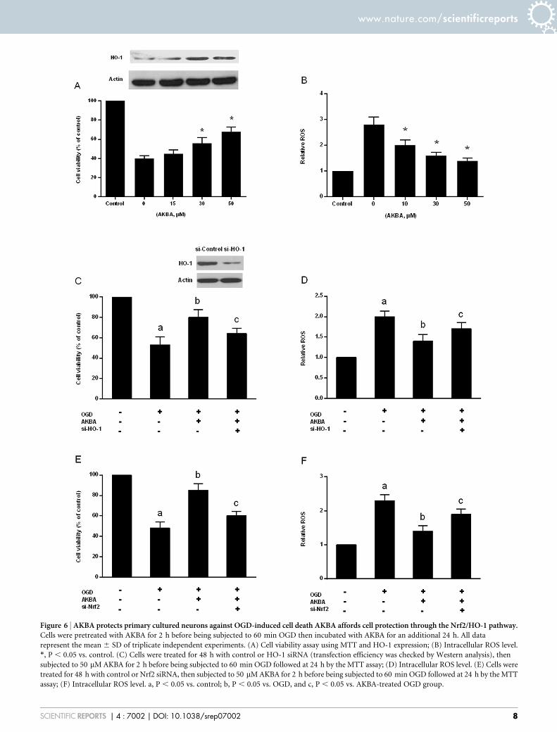
Figure 6 | AKBA protects primary cultured neurons against OGD-induced cell death AKBA affords cell protection through the Nrf2/HO-1 pathway.Cells were pretreated with AKBA for 2 h before being subjected to 60 min OGD then incubated with AKBA for an additional 24 h. All data
represent the mean 6 SD of triplicate independent experiments. (A) Cell viability assay using MTT and HO-1 expression; (B) Intracellular ROS level.
*, P , 0.05 vs. control. (C) Cells were treated for 48 h with control or HO-1 siRNA (transfection efficiency was checked by Western analysis), then
subjected to 50 mM AKBA for 2 h before being subjected to 60 min OGD followed at 24 h by the MTT assay; (D) Intracellular ROS level. (E) Cells were
treated for 48 h with control or Nrf2 siRNA, then subjected to 50 mM AKBA for 2 h before being subjected to 60 min OGD followed at 24 h by the MTT
assay; (F) Intracellular ROS level. a, P , 0.05 vs. control; b, P , 0.05 vs. OGD, and c, P , 0.05 vs. AKBA-treated OGD group.
www.nature.com/scientificreports
SCIENTIFIC REPORTS | 4 : 7002 | DOI: 10.1038/srep07002 8
AKBA induced HO-1 expression by stimulating Nrf2-ARE bindingactivity. Although the mechanisms leading to nuclear translocationof Nrf2 are poorly defined, we believe that Nrf2 and HO-1 induced byAKBA decreased ROS in cortical neurons, and that it is responsible,at least in part, for the protective effects against OGD.
AKBA in preventing focal cerebral ischaemic damage in additionto the reality that AKBA is an all-natural plant-derived compound’spotency makes AKBA a promising therapeutic agent. Previous clin-ical trials indicated that boswellic acids had lower toxicity and werewell tolerated in humans34. However, several issues still must beaddressed in the future, including whether other signaling pathwayscontribute to the neuroprotective effects of AKBA. It should be men-tioned that AKBA is a 5-lipoxygenase inhibitor and its own anti-inflammatory effect in vivo will likely contribute to the protectiveeffects of this compound in stroke35. For the short-term study (48 h),we confirmed the protective effects of AKBA on the acute phase ofstroke. Therefore, therapeutic time window of AKBA injectionagainst cerebral I/R injury in rat should be determined in futureand the possible long-term curative effects of AKBA remain to beclarified.
In this study, we firstly demonstrated that AKBA could attenuateischemic neuronal injury using a MCAO model. Furthermore, invivo studies have shown that AKBA can protect neurons againstOGD-induced cell death activating the Nrf2/HO-1 pathway. Theprotective effect of AKBA was partly blocked when HO-1 or Nrf2was knocked down. Collectively, we demonstrated the unexploredpotential of AKBA for the treatment of cerebral I/R damage and thatpharmacological activation of the Nrf2/HO-1 pathway can provideneuroprotection.
1. Go, A. S. et al. Heart disease and stroke statistics--2013 update: a report from theAmerican Heart Association. Circulation 127, e6–e245 (2013).
2. Chan, P. H. Oxygen radicals in focal cerebral ischemia. Brain Pathol 4, 59–65(1994).
3. Safayhi, H. et al. Boswellic acids: novel, specific, nonredox inhibitors of 5-lipoxygenase. J Pharmacol Exp Ther 261, 1143–6 (1992).
4. Hartmann, R. M., Morgan Martins, M. I., Tieppo, J., Fillmann, H. S. & Marroni,N. P. Effect of Boswellia serrata on antioxidant status in an experimental model ofcolitis rats induced by acetic acid. Dig Dis Sci 57, 2038–44 (2012).
5. Elshazly, S. M., Abd El Motteleb, D. M. & Nassar, N. N. The selective 5-LOXinhibitor 11-keto-beta-boswellic acid protects against myocardial ischemiareperfusion injury in rats: involvement of redox and inflammatory cascades.Naunyn Schmiedebergs Arch Pharmacol 386, 823–33 (2013).
6. Ali, E. N. & Mansour, S. Z. Boswellic acids extract attenuates pulmonary fibrosisinduced by bleomycin and oxidative stress from gamma irradiation in rats. ChinMed 6, 36 (2011).
7. Rong, Z. T., Gong, X. J., Sun, H. B., Li, Y. M. & Ji, H. Protective effects of oleanolicacid on cerebral ischemic damage in vivo and H(2)O(2)-induced injury in vitro.Pharm Biol 49, 78–85 (2011).
8. Li, L. et al. Ursolic acid promotes the neuroprotection by activating Nrf2 pathwayafter cerebral ischemia in mice. Brain Res 1497, 32–9 (2013).
9. Yin, M. C., Lin, M. C., Mong, M. C. & Lin, C. Y. Bioavailability, distribution, andantioxidative effects of selected triterpenes in mice. J Agric Food Chem 60,7697–701 (2012).
10. Kang, K. W., Lee, S. J. & Kim, S. G. Molecular mechanism of nrf2 activation byoxidative stress. Antioxid Redox Signal 7, 1664–73 (2005).
11. Kobayashi, M. & Yamamoto, M. Molecular mechanisms activating the Nrf2-Keap1 pathway of antioxidant gene regulation. Antioxid Redox Signal 7, 385–94(2005).
12. Maines, M. D. Heme oxygenase: function, multiplicity, regulatory mechanisms,and clinical applications. FASEB J 2, 2557–68 (1988).
13. Shah, Z. A. et al. The flavanol (2)-epicatechin prevents stroke damage through theNrf2/HO1 pathway. Journal of Cerebral Blood Flow Metabolism 30, 1951–1961(2010).
14. Alfieri, A. et al. Sulforaphane preconditioning of the Nrf2/HO-1 defense pathwayprotects the cerebral vasculature against blood–brain barrier disruption andneurological deficits in stroke. Free Radical Biology and Medicine 65, 1012–1022(2013).
15. Longa, E. Z., Weinstein, P. R., Carlson, S. & Cummins, R. Reversible middlecerebral artery occlusion without craniectomy in rats. stroke 20, 84–91 (1989).
16. Draper, H. H. et al. A comparative evaluation of thiobarbituric acid methods forthe determination of malondialdehyde in biological materials. Free Radic BiolMed 15, 353–63 (1993).
17. Serkova, N. et al. Comparison of the effects of cyclosporin a on the metabolism ofperfused rat brain slices during normoxia and hypoxia. J Cereb Blood Flow Metab22, 342–52 (2002).
18. Schafer, F. Q. & Buettner, G. R. Redox environment of the cell as viewed throughthe redox state of the glutathione disulfide/glutathione couple. Free RadicalBiology and Medicine 30, 1191–1212 (2001).
19. Qian, Y. et al. Maslinic acid, a natural triterpenoid compound from Olea europaea,protects cortical neurons against oxygen-glucose deprivation-induced injury. EurJ Pharmacol 670, 148–53 (2011).
20. Kaidery, N. A. et al. Targeting Nrf2-mediated gene transcription by extremelypotent synthetic triterpenoids attenuate dopaminergic neurotoxicity in the MPTPmouse model of Parkinson’s disease. Antioxid Redox Signal 18, 139–57 (2013).
21. Tanaka, N. et al. Expression of Keap1-Nrf2 system and antioxidative proteins inmouse brain after transient middle cerebral artery occlusion. Brain Res 1370,246–53 (2011).
22. Nimura, T., Weinstein, P. R., Massa, S. M., Panter, S. & Sharp, F. R. Hemeoxygenase-1 (HO-1) protein induction in rat brain following focal ischemia.Brain Res Mol Brain Res 37, 201–8 (1996).
23. Matsuoka, Y. et al. Induction of heme oxygenase-1 and major histocompatibilitycomplex antigens in transient forebrain ischemia. J Cereb Blood Flow Metab 18,824–32 (1998).
24. Stepkowski, T. M. & Kruszewski, M. K. Molecular cross-talk between the NRF2/KEAP1 signaling pathway, autophagy, and apoptosis. Free Radical Biology andMedicine 50, 1186–1195 (2011).
25. Zhao, Z. et al. Age-related retinopathy in NRF2-deficient mice. PLoS One 6,e19456 (2011).
26. Shah, Z. A. et al. Role of reactive oxygen species in modulation of Nrf2 followingischemic reperfusion injury. Neuroscience 147, 53–9 (2007).
27. Kensler, T. W., Wakabayashi, N. & Biswal, S. Cell survival responses toenvironmental stresses via the Keap1-Nrf2-ARE pathway. Annu Rev PharmacolToxicol 47, 89–116 (2007).
28. Durante, W. Targeting heme oxygenase-1 in vascular disease. Curr Drug Targets11, 1504–16 (2010).
29. Kim, Y. M. et al. Heme oxygenase in the regulation of vascular biology: frommolecular mechanisms to therapeutic opportunities. Antioxid Redox Signal 14,137–67 (2011).
30. Warner, D. S., Sheng, H. & Batinic-Haberle, I. Oxidants, antioxidants and theischemic brain. J Exp Biol 207, 3221–31 (2004).
31. Broughton, B. R., Reutens, D. C. & Sobey, C. G. Apoptotic mechanisms aftercerebral ischemia. Stroke 40, e331–9 (2009).
32. Rushmore, T. H., Morton, M. R. & Pickett, C. B. The antioxidant responsiveelement. Activation by oxidative stress and identification of the DNA consensussequence required for functional activity. J Biol Chem 266, 11632–9 (1991).
33. Wakabayashi, N. et al. Keap1-null mutation leads to postnatal lethality due toconstitutive Nrf2 activation. Nature genetics 35, 238–245 (2003).
34. Madisch, A. et al. Boswellia serrata extract for the treatment of collagenous colitis.A double-blind, randomized, placebo-controlled, multicenter trial. Int J ColorectalDis 22, 1445–51 (2007).
35. Safayhi, H., Sailer, E. R. & Ammon, H. P. Mechanism of 5-lipoxygenase inhibitionby acetyl-11-keto-beta-boswellic acid. Mol Pharmacol 47, 1212–6 (1995).
AcknowledgmentsThis work was supported by the grant from the Key Technologies for New Drug Innovationand Development of China (No. 2011ZXJ09202-13; No. 2012BAK25B00) and NationalNatural Science Foundation of China (No.81373947; No.81201985).
Author contributionsY.D., Y.W.L. and A.D.W. conceived and designed the experiments. M.C.C., M.M.W. andY.S. performed the experiments. Y.Y.J. and M.W. analyzed the data. M.W., T.J.Z. and J.P.contributed reagents/materials/analysis tools. Y.D., Y.R.Z. and C.G. wrote the paper. Allauthors reviewed and approved the final manuscript.
Additional informationSupplementary information accompanies this paper at http://www.nature.com/scientificreports
Competing financial interests: The authors declare no competing financial interests.
How to cite this article: Ding, Y. et al. Neuroprotection by Acetyl-11-Keto-b-BoswellicAcid, in Ischemic Brain Injury Involves the Nrf2/HO-1 defense Pathway. Sci. Rep. 4, 7002;DOI:10.1038/srep07002 (2014).
This work is licensed under a Creative Commons Attribution 4.0 InternationalLicense. The images or other third party material in this article are included in thearticle’s Creative Commons license, unless indicated otherwise in the credit line; ifthe material is not included under the Creative Commons license, users will needto obtain permission from the license holder in order to reproduce the material. Toview a copy of this license, visit http://creativecommons.org/licenses/by/4.0/
www.nature.com/scientificreports
SCIENTIFIC REPORTS | 4 : 7002 | DOI: 10.1038/srep07002 9
Related Documents