Research Article Neuroplastic Effects of Combined Computerized Physical and Cognitive Training in Elderly Individuals at Risk for Dementia: An eLORETA Controlled Study on Resting States Charis Styliadis, 1 Panagiotis Kartsidis, 1 Evangelos Paraskevopoulos, 1 Andreas A. Ioannides, 2 and Panagiotis D. Bamidis 1 1 Lab of Medical Physics, Medical School, Faculty of Health Sciences, Aristotle University of essaloniki, P.O. Box 376, 54124 essaloniki, Greece 2 Laboratory for Human Brain Dynamics, AAI Scientific Cultural Services Ltd., Office 501, Galaxias Building Block A, 33 Arch. Makarios III Avenue, 1065 Nicosia, Cyprus Correspondence should be addressed to Panagiotis D. Bamidis; [email protected] Received 15 December 2014; Revised 9 March 2015; Accepted 16 March 2015 Academic Editor: Michel Baudry Copyright © 2015 Charis Styliadis et al. is is an open access article distributed under the Creative Commons Attribution License, which permits unrestricted use, distribution, and reproduction in any medium, provided the original work is properly cited. e present study investigates whether a combined cognitive and physical training may induce changes in the cortical activity as measured via electroencephalogram (EEG) and whether this change may index a deceleration of pathological processes of brain aging. Seventy seniors meeting the clinical criteria of mild cognitive impairment (MCI) were equally divided into 5 groups: 3 experimental groups engaged in eight-week cognitive and/or physical training and 2 control groups: active and passive. A 5-minute long resting state EEG was measured before and aſter the intervention. Cortical EEG sources were modelled by exact low resolution brain electromagnetic tomography (eLORETA). Cognitive function was assessed before and aſter intervention using a battery of neuropsychological tests including the minimental state examination (MMSE). A significant training effect was identified only aſter the combined training scheme: a decrease in the post- compared to pre-training activity of precuneus/posterior cingulate cortex in delta, theta, and beta bands. is effect was correlated to improvements in cognitive capacity as evaluated by MMSE scores. Our results indicate that combined physical and cognitive training shows indices of a positive neuroplastic effect in MCI patients and that EEG may serve as a potential index of gains versus cognitive declines and neurodegeneration. is trial is registered with ClinicalTrials.gov Identifier NCT02313935. 1. Introduction As humans advance through middle age and beyond brain function oſten changes into a mildly impaired state (mild cognitive impairment, MCI) or even deteriorates into neu- rodegenerative diseases like dementia and Alzheimer’s dis- ease (AD) [1, 2]. AD-related pathological changes may begin decades prior to the clinical diagnosis, with symptoms oſten regarded as consistent with healthy aging. ere is a growing consensus that age-related cognitive decline can be slowed down or even prevented when the brain retains its flexibility. is is underpinned by the effective reor- ganization of brain’s structural and functional components so as to compensate for the physiological and physical changes that eventually cause cognitive decline. Recent studies have addressed this issue in two ways: (i) developing diagnostic biomarkers that can reliably identify early signs along the continuum of MCI and AD-related pathology and if possible continuing with the longitudinal evaluation and monitoring of the progression of the disease [3–6] and (ii) modifying lifestyle behaviours to promote neu- roplasticity and hence healthy aging and to prevent or at least slow down cognitive decline and AD. Clinical MCI is among the most consistently reported risk factors for the development of AD-related pathology [7, 8] and therefore is of special interest for the aforementioned Hindawi Publishing Corporation Neural Plasticity Volume 2015, Article ID 172192, 12 pages http://dx.doi.org/10.1155/2015/172192

Welcome message from author
This document is posted to help you gain knowledge. Please leave a comment to let me know what you think about it! Share it to your friends and learn new things together.
Transcript
Research ArticleNeuroplastic Effects of Combined Computerized Physical andCognitive Training in Elderly Individuals at Risk for Dementia:An eLORETA Controlled Study on Resting States
Charis Styliadis,1 Panagiotis Kartsidis,1 Evangelos Paraskevopoulos,1
Andreas A. Ioannides,2 and Panagiotis D. Bamidis1
1Lab of Medical Physics, Medical School, Faculty of Health Sciences, Aristotle University of Thessaloniki,P.O. Box 376, 54124 Thessaloniki, Greece2Laboratory for Human Brain Dynamics, AAI Scientific Cultural Services Ltd., Office 501, Galaxias Building Block A,33 Arch. Makarios III Avenue, 1065 Nicosia, Cyprus
Correspondence should be addressed to Panagiotis D. Bamidis; [email protected]
Received 15 December 2014; Revised 9 March 2015; Accepted 16 March 2015
Academic Editor: Michel Baudry
Copyright © 2015 Charis Styliadis et al.This is an open access article distributed under the Creative Commons Attribution License,which permits unrestricted use, distribution, and reproduction in any medium, provided the original work is properly cited.
The present study investigates whether a combined cognitive and physical training may induce changes in the cortical activity asmeasured via electroencephalogram (EEG) and whether this change may index a deceleration of pathological processes of brainaging. Seventy seniors meeting the clinical criteria of mild cognitive impairment (MCI) were equally divided into 5 groups: 3experimental groups engaged in eight-week cognitive and/or physical training and 2 control groups: active and passive. A 5-minutelong resting state EEGwasmeasured before and after the intervention. Cortical EEG sources weremodelled by exact low resolutionbrain electromagnetic tomography (eLORETA). Cognitive function was assessed before and after intervention using a battery ofneuropsychological tests including theminimental state examination (MMSE). A significant training effect was identified only afterthe combined training scheme: a decrease in the post- compared to pre-training activity of precuneus/posterior cingulate cortexin delta, theta, and beta bands. This effect was correlated to improvements in cognitive capacity as evaluated by MMSE scores.Our results indicate that combined physical and cognitive training shows indices of a positive neuroplastic effect in MCI patientsand that EEG may serve as a potential index of gains versus cognitive declines and neurodegeneration. This trial is registered withClinicalTrials.gov Identifier NCT02313935.
1. Introduction
As humans advance through middle age and beyond brainfunction often changes into a mildly impaired state (mildcognitive impairment, MCI) or even deteriorates into neu-rodegenerative diseases like dementia and Alzheimer’s dis-ease (AD) [1, 2]. AD-related pathological changes may begindecades prior to the clinical diagnosis, with symptoms oftenregarded as consistent with healthy aging.
There is a growing consensus that age-related cognitivedecline can be slowed downor even preventedwhen the brainretains its flexibility.This is underpinned by the effective reor-ganization of brain’s structural and functional components so
as to compensate for the physiological and physical changesthat eventually cause cognitive decline.
Recent studies have addressed this issue in two ways: (i)developing diagnostic biomarkers that can reliably identifyearly signs along the continuum of MCI and AD-relatedpathology and if possible continuing with the longitudinalevaluation and monitoring of the progression of the disease[3–6] and (ii) modifying lifestyle behaviours to promote neu-roplasticity and hence healthy aging and to prevent or at leastslow down cognitive decline and AD.
ClinicalMCI is among themost consistently reported riskfactors for the development of AD-related pathology [7, 8]and therefore is of special interest for the aforementioned
Hindawi Publishing CorporationNeural PlasticityVolume 2015, Article ID 172192, 12 pageshttp://dx.doi.org/10.1155/2015/172192
2 Neural Plasticity
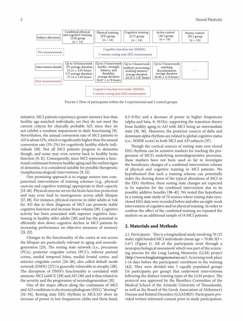
Subject allocation
Pre-measurements
Intervention details
Post-measurements
Combined physicaland cognitive training
LLM group(n = 14)
Up to 10 hours/weekPT average duration25.21 ± 4.91 hours
CT average duration27.14 ± 5.65 hours
Physical training
(n = 14)
Cognitive training
(n = 14)
Cognitive function test (MMSE),5-minute resting state EEG measurement
(PT) group (CT) groupPassive control
(PC) groupActive control
(AC) group(n = 14)(n = 14)
Up to 5 hours/weekaerobic, strength,
balance, and
average duration26.07 ± 6.78 hours
Cognitive function test retake (MMSE),5-minute resting state EEG measurement
Up to 5 hours/weekauditory processing,
working memoryaverage duration24.35 ± 1.87 hours
Up to 5 hours/weekwatching
documentariesaverage duration26.85 ± 4.76 hours
· · ·
flexibility
Figure 1: Flow of participants within the 3 experimental and 2 control groups.
initiative. MCI patients experience greater memory loss thanhealthy age-matched individuals, yet they do not meet thecurrent criteria for clinically probable AD, since they donot exhibit a resultant impairment in daily functioning [9].Nevertheless, the annual conversion rate of MCI patients toAD is about 12%,which is significantly higher than the annualconversion rate (1%-2%) for cognitively healthy elderly indi-viduals [10]. Not all MCI patients progress to dementiathough, and some may even reverse back to healthy brainfunction [9, 11]. Consequently, since MCI represents a func-tional continuumbetween healthy aging and the earliest signsof dementia, it is considered suitable for possible therapeutic(nonpharmacological) interventions [9, 12].
One promising approach is to engage seniors into com-puterized interventions of training schemes (e.g., physicalexercise and cognitive training) appropriate to their capacity[13–16]. Physical exercise serves the brain function protectionand may even lead to neurogenesis across adult lifespan[17, 18]. For instance, physical exercise in older adults at riskfor AD due to their diagnosis of MCI can promote stablecognitive function and increase brain volume [19]. Cognitiveactivity has been associated with superior cognitive func-tioning in healthy older adults [20] and has the potential toefficiently slow down cognitive decline in MCI patients byincreasing performance on objective measures of memory[21, 22].
Changes in the functionality of the cortex at rest acrossthe lifespan are particularly relevant to aging and neurode-generation [23]. The resting state network (i.e., precuneus(PCu), posterior cingulate cortex (PCC), inferior parietalcortex, medial temporal lobes, medial frontal cortex, andanterior cingulate cortex [24–26], also called default modenetwork (DMN) [27]) is generally vulnerable to atrophy [28].The disruption of DMN’s functionality is correlated withamnesticMCI (aMCI) [29] and AD [30] and is thus related tothe severity and the progression of neurodegeneration [31].
One of the major effects along the continuum of MCIandAD conditions is electroencephalogram (EEG) “slowing”[32–34]. Resting state EEG rhythms in MCI/AD show anincrease of power in low frequencies (delta and theta band,
0.5–8Hz) and a decrease of power in higher frequencies(alpha and beta, 8–30Hz), supporting the transition theoryfrom healthy aging to AD with MCI being an intermediatestate [35, 36]. Moreover, the posterior sources of delta anddominant alpha rhythms are related to global cognitive status(i.e., MMSE score) in both MCI and AD subjects [37].
Though the cortical sources of resting state eyes-closedEEG rhythms can be sensitive markers for tracking the pro-gression of MCI’s underlying neurodegenerative processes,these markers have not been used so far to investigatethe beneficiary changes of a combined intervention schemeof physical and cognitive training in MCI patients. Wehypothesized that such a training scheme can potentiallyindex the slowing down of the typical alterations of MCI inthe EEG rhythms; these resting state changes are expectedto be superior for the combined intervention due to itspossibly additive benefits [38–41]. We tested this hypothesisin a resting state study of 70 seniors where resting state eyes-closed EEGdatawere recorded before and after an eight-weekintervention of cognitive and/or physical training. In order toconfirm the effect of the combined training we repeated theanalysis on an additional sample of 14 MCI patients.
2. Materials and Methods
2.1. Participants. This is a longitudinal study involving 70 (25male) right handedMCI individuals (mean age = 70.80; SD =5.67) (Figure 1). All of the participants went through aneuropsychological assessment which was part of the screen-ing process for the Long Lasting Memories (LLM) project(http://www.longlastingmemories.eu/). Screening took place1–14 days before the participants’ enrolment to the training[42]. They were divided into 5 equally populated groups(14 participants per group) that underwent interventionsfollowing the distinct training types of the LLM project. Theprotocol was approved by the Bioethics Committee of theMedical School of the Aristotle University of Thessaloniki,as well as the Board of the Greek Association of Alzheimer’sDisease and RelatedDisorders (GAADRD). Participants pro-vided written informed consent prior to study participation.
Neural Plasticity 3
Table 1: Subject pool (means ± SDs) and training type details.
LLM PT CT AC PCNumber of subjects 14 14 14 14 14Number of males/ratio 5 (35.71%) 5 (35.71%) 5 (35.71%) 5 (35.71%) 5 (35.71%)Age 71.21 ± 4.52 70.42 ± 6.63 72.71 ± 6.57 71.07 ± 4.38 67.64 ± 3.97Pre-MMSE 25.85 ± 2.09 26.21 ± 2.33 25.14 ± 3.22 26.21 ± 1.97 25 ± 1.77Post-MMSE 27.14 ± 2.06 27.42 ± 2.06 25.42 ± 2.35 27.28 ± 1.97 25.21 ± 2.42yoe 8.14 ± 3.06 6.14 ± 1.45 6.14 ± 3.22 7.14 ± 3.04 7.35 ± 2.37
Intervention details PT and CTAerobics, strength,
balance, andflexibility
Auditory processing andworking memory
Watchingdocumentaries on
YouTube—
Sessions Up to 10 h/w Up to 5 h/w 4 exer × 15min, 3 to 5 h/w Up to 5 h/w —
Duration PT: 25.21 ± 4.91 hCT: 27.14 ± 5.65 h 26.07 ± 6.78 h 24.35 ± 1.87 h 26.85 ± 4.76 h —
Note: LLM, combined training; PT, physical training; CT, cognitive training; AC, active control; PC, passive control; MMSE, minimental state examination(where the range from best to worst performance is 30–0); yoe, years of education; exer, exercise; min, minutes; h, hour; w, week.
Prior to neurophysiological acquisition, the participants wereinformed that they could terminate the experiment at anytime without the need to provide any justification for theirdecision (no one did). LLM’s interventions took place at theGAADRD clinic. The LLM project was conducted in accor-dance with the Helsinki Declaration for Human Rights.
2.1.1. Neuropsychological Examination. The neuropsycholog-ical examination consisted of tests allowing assessment on theparticipant’s generic cognitive status and other specific cogni-tive domains (verbal memory, executive functions, indepen-dent living, etc.) that are essential to the diagnostic procedureand the group formation. Further details are available inBamidis [43].
2.1.2. Diagnostic Procedure. A dementia expert neurologist,naıve regarding the treatment each subject received, per-formed the diagnosis of the participants taking into consid-eration the neurophysiological as well as the medical exam-ination [44]. MCI patients met Petersen’s criteria based onsubjective and objective cognitive impairment, predomi-nantly affecting memory, in the absence of dementia orsignificant functional loss [9, 12]. All MCI participants hada Clinical Dementia Rating score of 0.5 [45].
2.1.3. Cognitive Status Test. The minimental state examina-tion (MMSE) is a brief, 30-pointmeasure that is used to assesscognitive status [46]. MMSE is routinely used in clinicalpractice to screen for dementia, but here it only served asan index of patient’s response to treatment. Specifically, it isused to estimate the severity of cognitive impairment and tofollow the longitudinal cognitive changes in a patient. Normalcognition MMSE score is greater than or equal to 27 points.Various proposals on the score level to be used as the cut-offpoint for the diagnosis of dementia have been made (i.e.,23/24 [47] or even 20/21 [48]). In spite of the methodologicaldifferences, the cut-off value of 23/24 [49] has been regarded
as a valid cut-off level for the diagnosis of dementia in Greece[50].
2.1.4. Inclusion Criteria for the Current Study. They are asfollows: (i) ages ≥ 60 years, (ii) 23 ≤MMSE score ≤ 27 points,(iii) normal or corrected-to-normal hearing and vision, (iv)fluent language skills, and (v) agreement of a medical doctorand time commitment to the intervention protocol.
2.1.5. Exclusion Criteria. They are as follows: (i) unrecoveredneurological disorders (i.e., stroke, traumatic brain injury),(ii) severe depression or psychological disorder, (iii) unstablemedication within the last 3 months, (iv) severe physicaldisorder, and (v) concurrent participation in another study.
2.1.6.Matching. Theparticipants of each groupwerematchedon age, years of education (yoe), and male-to-female ratio,as well as cognitive state as screened by the MMSE [46] (seeTable 1).
2.1.7. Categorization. Participants of the LLM group (meanage = 71.21; SD = 4.52; meanMMSE = 25.85; SD = 2.09; meanyoe = 8.14; SD = 3.06; 5 males) attended a training protocolconsisting of physical and cognitive exercises. Participantsof the physical training (PT) group underwent only physicaltraining (mean age = 70.42; SD = 6.63; mean MMSE = 26.21;SD = 2.33; mean yoe = 6.14; SD = 1.45; 5 males) whereasparticipants in the cognitive training (CT) group performedcognitive tasks (mean age = 72.71; SD = 6.57; mean MMSE =25.14; SD = 3.22; mean yoe = 6.14; SD = 3.22; 5 males).Moreover, two control groups were employed: the activecontrol group (AC) (mean age = 71.07; SD = 4.38; meanMMSE = 26.21; SD = 1.97; mean yoe = 7.14; SD = 3.04; 5males) in which participants underwent a training protocolconsisting of watching a documentary and answering aquestionnaire and a passive control group (PC) (mean age =67.64; SD = 3.97; mean MMSE = 25; SD = 1.77; mean
4 Neural Plasticity
yoe = 7.35; SD = 2.37; 5 males) in which participants did notengage in any activity.
2.2. Long Lasting Memories (LLM) Intervention. LLM is anintegrated training system that targets nondemented anddemented aging population and adopts an approach of cog-nitive [51] and physical training [52, 53] in order to improvethe quality of life and prolong the functionality of the elders.All training components of the intervention were comput-erized, centre-based, and under supervision. The combinedcognitive and physical training sessions were performed ina pseudorandomized counterbalanced sequence. The detailsof each training intervention are described in detail in [43,51, 53, 54] and are summarized in Figure 1 and Table 1.Our experimental design allows for the exploration of thedistinct mechanisms crucial for transferring the combinedtraining effects.The trial was registered retrospectively (Clin-icalTrials.gov Identifier: NCT02313935). This was a result ofstrict project timeline but also unclear areas of responsibilityin the project (as the trial did not involve any medicinalproducts covered by Directive 2001/20/EC, guidelines fromthe European Medicines Agency and Eudra CT specificallyindicated that there was no legal obligation from the sponsorto register it into a trial database).
2.2.1. Cognitive Training (CT). The CT component of LLMis a Greek adaptation of the Brain Fitness software (PositScience Corporation, San Francisco, CA, USA). It employsauditory stimuli and comprises six exercises of self-paced lev-els of difficulty. Each exercise lasted fifteen minutes. Each CTsession consisted of four out of six exercises with an overallduration of one hour. CT was performed for one hour perday, three to five days perweek during a period of eight weeks.CT targeted auditory processing and working memory [55].Details on the benefits of auditory training on age-relatedcognitive decline are discussed elsewhere [56].
2.2.2. Physical Training (PT). The PT component of LLM,FitForAll (FFA) [53], is an elderly tailored environment [52]where physical exercise is blended by games (exergaming)with the use of supporting hardware like Nintendo Wii, Wiiremote, andWii balance-board, to enable an enjoyable digitaltraining experience. PT was performed for five sessions perweek one hour per day during a period of eight weeks. PTwasperformed in the context of computer-based games whichwere appropriately adjusted to elder’s capacity. The games’scenarios targeted body flexibility, balance, and strength aswell as physical endurance through aerobic training. Eachparticipant had to accomplish 20minutes of aerobic exercises,8–10 resistance exercises, 10 minutes of flexibility exercises,and a set of balance targeted exercises. The warm-up andcool-down processes constituted the initial and final session’scomponents, respectively. The effects of combined aerobicand strength exercisewhich is thought to be themost effectiveexercise training for improving cognitive function are dis-cussed elsewhere [15, 57, 58].
2.2.3. Active Control (AC). AC aids in controlling for poten-tial confound factors such as willingness to adopt an active
aging profile, computer skills, and social interaction [51]. Inthe current study, though the participants in AC group wereexposed to similar training parameters (e.g., computer use,intensity, and duration), they just viewed documentaries onnature, art, and history and completed questionnaires aboutthe documentaries [59]. AC did not involve any PT.
2.3. Experimental Design and EEG Recording . The pre- andpost-intervention EEG recordings were performed undermedical supervision using a Nihon Kohden EEG device with57 scalp electrodes and a sampling rate of 500Hz. Electrodeimpedances of brain signals, ground electrode, and ref-erences were kept lower than 2 kΩ. Five-minute restingstate (eyes closed) EEG signals were recorded prior to theinitiation of the intervention phase (8 weeks) and followingits completion. Participants were instructed to keep theireyes closed and to maintain a resting yet wakeful condition.Active scalp electrodes were placed on a cap (EASYCAP,http://www.easycap.de/easycap/) according to the 10–20 sys-tem.The electrodes were commonly referenced to the averageof the two linked mastoid electrodes. EOG signals wererecorded simultaneously by means of four Ag/AgCl elec-trodes (one above and one below the right eye and anothertwo placed at the outer canthi of each eye). The verticalEOG (VEOG) was calculated as the difference between thetwo signals recorded above and below the right eye, whilethe horizontal EOG (HEOG) was calculated as the differencebetween the signals recorded from the left and the rightelectrodes, respectively. ECG signal was also recorded simul-taneously by means of two Ag/AgCl electrodes.
2.4. Data Preprocessing and Resting State Neuroimaging. Thedata preprocessing was performed via the FieldTrip toolboxfor MATLAB [60]. The EEG recordings were filtered using ahigh-pass IIR filter at 1Hz, a notch IIR filter at 48–52Hz, and alow-pass IIR filter at 97Hz. Independent component analysis(ICA) was applied to the filtered EEG data for removing EOGand ECG artefacts [61]. The ICA components were visu-ally inspected and the artifactual ones were removed. TheEEG recordings were further inspected after the removalof the ICA components and all remaining visible artefactswere removed. A random process was used to select 15segments each one with duration of 4 seconds from eachEEG recording. The selected segments were imported tosLORETA/eLORETA software [62]. The 15 segments weretransformed to the frequency domain in cross-spectral form.The exact low resolution brain electromagnetic tomography(eLORETA) [62] current density reconstructions (CDR)werecalculated and projected on a generic MNI-152 head modelseparately for delta (2–4Hz), theta (4–8Hz), alpha (8–12Hz),beta 1 (12–18Hz), and beta 2 bands (18–30Hz). Finally, theresulting eLORETA images of each participant were normal-ized by scaling the total average power equal to unity [63].
2.5. Statistics. Thestatistical nonparametricmapping (SnPM)method as implemented in sLORETA/eLORETA softwarepackage was used to perform the statistical analyses [64].Theempirical probability distribution of the maximum 𝐹 statistic
Neural Plasticity 5
was estimated via randomization, under the null hypothesisof equality between pre- and post-intervention, for eachdiscrete frequency band within the groups.The same analysiswas performed on an additional sample (𝑛 = 14) of MCIpatients who received the combined training so as to checkwhether our results would be replicated. For the between-groups comparison, we estimated, via randomization, theempirical probability distribution of themaximum logarithmof the 𝑡 statistic under the null hypothesis of equality of thedifference between pre and post measurements of one groupto another, for each discrete frequency band. This methodol-ogy corrects for multiple testing for all discrete frequencies[65]. Due to the nonparametric nature of the method, itsvalidity need does not rely on any assumption of Gaussianity[64]. For the comparisons to themean cortical activations, weused SnPM8b (http://warwick.ac.uk/snpm). NonparametricSpearman’s rho was used to test the correlation between theMMSE pre- to post-difference of each participant and theactivity of a region of interest (ROI) that included only thecurrent density cluster showing significant differences beforeto after training.
3. Results
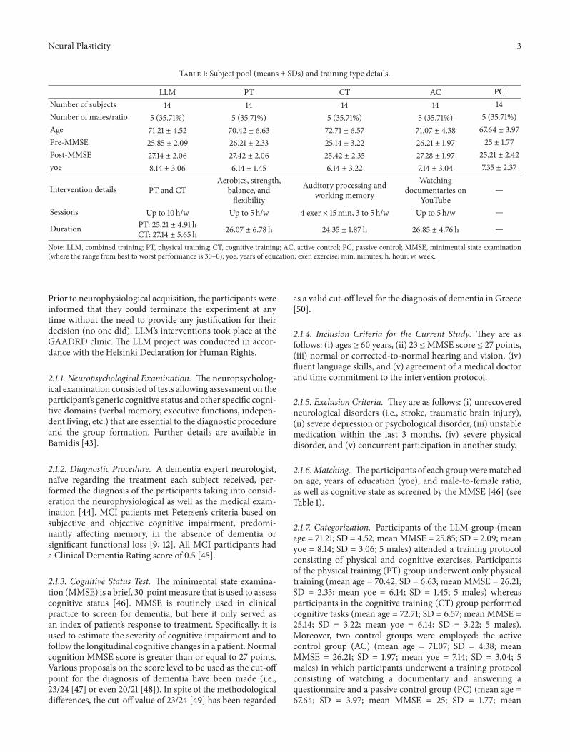
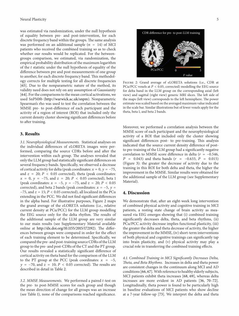
3.1. Neurophysiological Measurements. Statistical analyses onthe individual differences of eLORETA images were per-formed, comparing the source CDRs before and after theintervention within each group. The analyses revealed thatonly the LLMgroup had statistically significant differences onseveral frequency bands. Specifically, we observed a decreaseof cortical activity for delta (peak coordinates:𝑥 = 0,𝑦 = −75,and 𝑧 = 20; 𝑃 < 0.05 corrected), theta (peak coordinates:𝑥 = 0, 𝑦 = −75, and 𝑧 = 20; 𝑃 < 0.05 corrected), beta 1(peak coordinates: 𝑥 = −5, 𝑦 = −75, and 𝑧 = 20; 𝑃 < 0.05corrected), and beta 2 bands (peak coordinates: 𝑥 = −5, 𝑦 =−75, and 𝑧 = 15; 𝑃 < 0.05 corrected), all localized in the PCuextending in the PCC. We did not find significant differencesin the alpha band. For illustrative purposes, Figure 2 mapsthe grand average of the eLORETA solutions (i.e., relativecurrent density at PCu/PCC) for the LLM group modellingthe EEG source only for the delta rhythm. The results ofthe additional sample of the LLM group are very similarto our main results (see Supplementary Material availableonline at http://dx.doi.org/10.1155/2015/172192). The differ-ences between groups were compared in order for the effectof each training element to be determined. Specifically, wecompared the pre- and post-training sourceCDRs of the LLMgroup to the pre- and post-CDRs of the CT and the PT group.Our results revealed a statistically significant difference ofcortical activity on theta band for the comparison of the LLMto the PT group at the PCC (peak coordinates: 𝑥 = −5,𝑦 = −70, and 𝑧 = 10; 𝑃 < 0.05 corrected). The results aredescribed in detail in Table 2.
3.2. MMSE Measurements. We performed a paired 𝑡-test onthe pre- to post-MMSE scores for each group and thoughthe mean direction of change for all groups was an increase(see Table 1), none of the comparisons reached significance.
0 5
CDR difference for pre- to post-LLM training
PCu/PCC
F value
Figure 2: Grand average of eLORETA solutions (i.e., CDR atPCu/PCC voxels at 𝑃 < 0.05, corrected) modelling the EEG sourcefor delta band in the LLM group on the corresponding axial (leftview) and sagittal (right view) generic MRI slices. The left side ofthe maps (left view) corresponds to the left hemisphere. The powerestimatewas scaled based on the averagedmaximumvalue indicatedin the scale bar. Similar illustrations but of fewer voxels apply for thetheta, beta 1, and beta 2 bands.
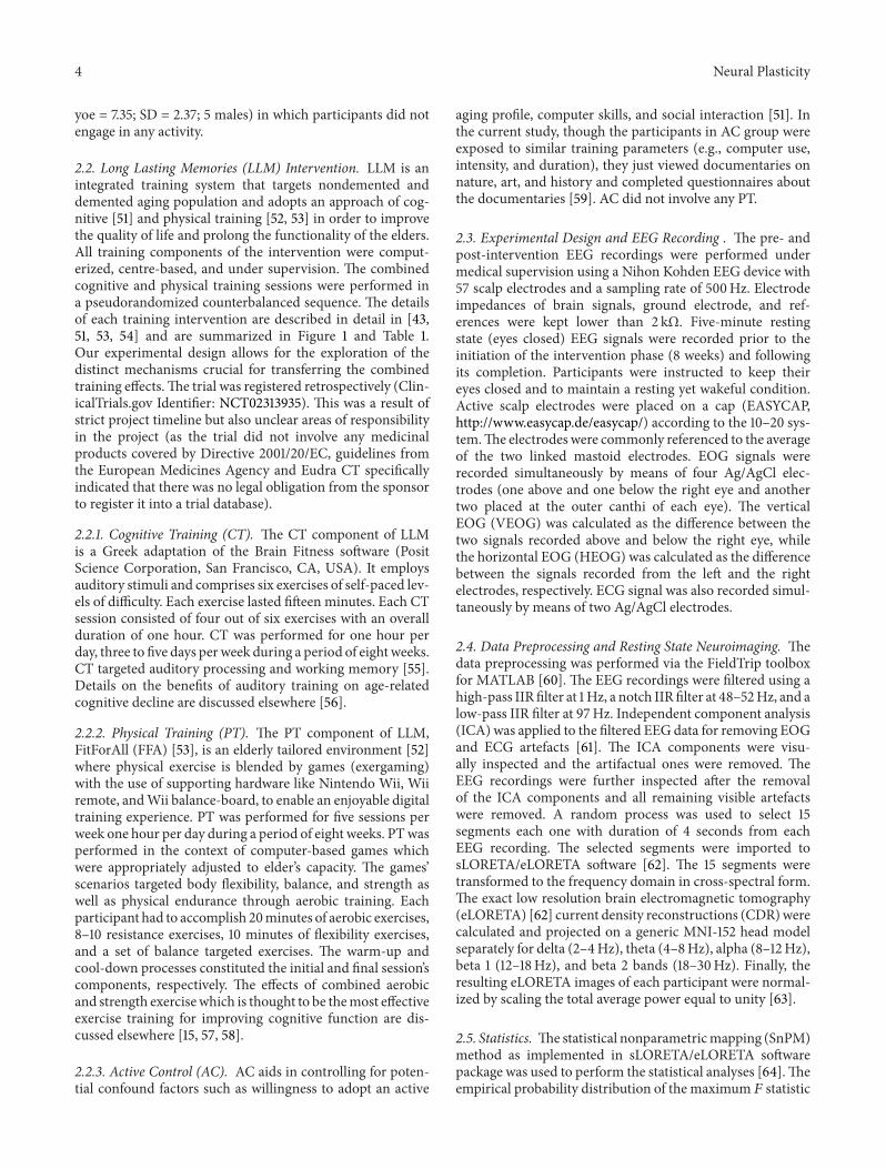
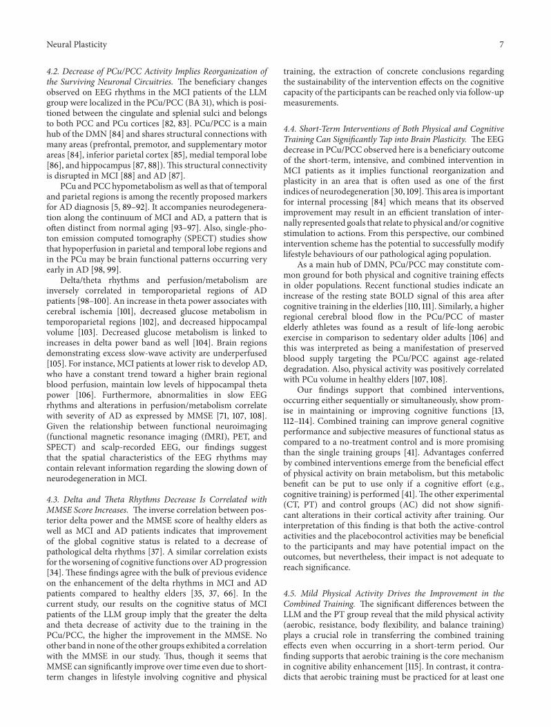
Moreover, we performed a correlation analysis between theMMSE score of each participant and the neurophysiologicalactivity of a ROI that included only the cluster showingsignificant differences post- to pre-training. This analysisindicated that the source current density difference of post-to pre-training of the LLM group had a significantly negativecorrelation to MMSE score difference in delta (𝑟 = −0.546,𝑃 = 0.043) and theta bands (𝑟 = −0.633, 𝑃 = 0.015)(Figure 3): the greater the decrease of activity due to thetraining in this ROI for both delta and theta, the higher theimprovement in theMMSE. Similar results were obtained forthe additional sample of the LLM group (see SupplementaryMaterial).
4. Discussion
We demonstrate that, after an eight-week long interventionof combined physical activity and cognitive training in MCIpatients, a resting state change of brain activity as mea-sured via EEG emerges showing that (i) combined trainingsignificantly decreases delta, theta, and beta rhythms, (ii)PCu/PCC activity decrease implies functional plasticity, (iii)the greater the delta and theta decrease of activity, the higherthe improvement in theMMSE, (iv) short-term interventionsof both physical and cognitive trainings can significantly tapinto brain plasticity, and (v) physical activity may play acrucial role in transferring the combined training effects.
4.1. Combined Training in MCI Significantly Decreases Delta,Theta, and Beta Rhythms. Increases in delta and theta powerare consistent changes in the continuum along MCI and ADconditions [66, 67].With reference to healthy elderly subjects,MCI patients exhibit theta increases [68, 69], whereas deltaincreases are more evident in AD patients [36, 70–72].Longitudinally, theta power is found to be particularly highin baseline evaluations of MCI patients who show declineat a 7-year follow-up [73]. We interpret the delta and theta
6 Neural Plasticity
Table 2: EEG source maps at the differences at 𝑃 < 0.05, corrected.
LLM
Band Anatomical area BA CS 𝐹MNI coordinates (mm)
𝑥, 𝑦, 𝑧Delta (2–4Hz) PCu/PCC 31 56 3.9294 0, −75, 20Theta (4–8Hz) PCu/PCC 31 9 2.7361 0, −75, 20Beta 1 (12–18Hz) PCu/PCC 31 10 2.9856 −5, −75, 20Beta 2 (18–30Hz) PCu/PCC 31 15 3.3563 −5, −75, 15
LLM versus PT
Band Anatomical area BA CS 𝑇MNI coordinates (mm)
𝑥, 𝑦, 𝑧Theta (4–8Hz) PCC 30 1 4.1581 −5, −70, 10Note: results are superimposed on standardized MNI coordinates; BA, Brodmann area; 𝑥, left/right; 𝑦, anterior/posterior; 𝑧, superior/inferior; CS, cluster sizein number of activated voxels; 𝐹, 𝐹 value; 𝑇, 𝑡 value; PCu/PCC, precuneus/posterior cingulate cortex; significant at 𝑃 < 0.05, corrected.
0 321−1−2−3
0
2
1
−1
−2
Delt
a CD
R di
ffere
nce
MMSE difference
r = −0.546, P = 0.043
(a)
0
2
1
−1
−2
Thet
a CD
R di
ffere
nce
0 321−1−2−3
MMSE difference
r = −0.633, P = 0.015
(b)
Figure 3: Visualization of the negative correlation of delta (𝑟 = −0.546, 𝑃 = 0.043) (a) and theta (𝑟 = −0.633, 𝑃 = 0.015) (b) bands amongthe MMSE score post- to pre-difference of each participant and the CDR post- to pre-difference of PCu/PCC activity that was statisticallysignificant at 𝑃 < 0.05, corrected.
decreases inMCI patients, induced by the combined training,as a beneficiary neuroplastic outcome that may index apossible deceleration of the underlying neurodegeneration.Our results show promise given that delta occipital sourcespresent a progressive decreasing trend with physiologicalaging [74] and age is inversely related to the amount of slowactivity (delta/theta), indicating that increase of slow activityis not a marker of physiological aging [75]. Moreover, as thetaband is highly correlated with loss of memory function inMCI, its increase may index memory capacity deterioration[69, 76]. In addition, delta and theta increases are strictlyrelated to the bilateral reduction of memory circuits (e.g.,hippocampus, entorhinal volumes) of AD patients [77, 78]and global gray matter volume is inversely related to thepower of pathological delta sources in MCI and AD [79].
Our beta band findings are not consistent with evidencethat MCI progression to dementia is usually characterized bybeta band decrease [35, 36]. We could speculate that either
the tomographic and analysis methods used herein couldnot reveal such an effect or beta band is not yet impairedin our sample (its impairment could be expressed later).Nevertheless, we found beta 1 and beta 2 decrease only in thePCu/PCC. Given the wide variations in beta activity withingroups (e.g., beta 2 is highest in the healthy controls anddecreases in the aMCI and AD patients in the frontal andtemporal scalp regions, whereas beta can vary among thesegroups [80]), it appears that beta band population propertiesare less consistent among the MCI pathology and possiblyspecific to certain lobes [81]. Overall, our results for delta,theta, and beta bands are in line with reports that MCIpatients with increased MMSE scores have relatively lessamplitude in delta, theta, and beta 1 rhythms than those whohave decreased their MMSE scores [81]. The exciting impli-cation of our results is that MCI patients may decelerate theprogress of dementia with combined short-term (but intense)physical and cognitive training.
Neural Plasticity 7
4.2. Decrease of PCu/PCC Activity Implies Reorganization ofthe Surviving Neuronal Circuitries. The beneficiary changesobserved on EEG rhythms in the MCI patients of the LLMgroup were localized in the PCu/PCC (BA 31), which is posi-tioned between the cingulate and splenial sulci and belongsto both PCC and PCu cortices [82, 83]. PCu/PCC is a mainhub of the DMN [84] and shares structural connections withmany areas (prefrontal, premotor, and supplementary motorareas [84], inferior parietal cortex [85], medial temporal lobe[86], and hippocampus [87, 88]).This structural connectivityis disrupted in MCI [88] and AD [87].
PCu and PCChypometabolism as well as that of temporaland parietal regions is among the recently proposed markersfor AD diagnosis [5, 89–92]. It accompanies neurodegenera-tion along the continuum of MCI and AD, a pattern that isoften distinct from normal aging [93–97]. Also, single-pho-ton emission computed tomography (SPECT) studies showthat hypoperfusion in parietal and temporal lobe regions andin the PCu may be brain functional patterns occurring veryearly in AD [98, 99].
Delta/theta rhythms and perfusion/metabolism areinversely correlated in temporoparietal regions of ADpatients [98–100]. An increase in theta power associates withcerebral ischemia [101], decreased glucose metabolism intemporoparietal regions [102], and decreased hippocampalvolume [103]. Decreased glucose metabolism is linked toincreases in delta power band as well [104]. Brain regionsdemonstrating excess slow-wave activity are underperfused[105]. For instance, MCI patients at lower risk to develop AD,who have a constant trend toward a higher brain regionalblood perfusion, maintain low levels of hippocampal thetapower [106]. Furthermore, abnormalities in slow EEGrhythms and alterations in perfusion/metabolism correlatewith severity of AD as expressed by MMSE [71, 107, 108].Given the relationship between functional neuroimaging(functional magnetic resonance imaging (fMRI), PET, andSPECT) and scalp-recorded EEG, our findings suggestthat the spatial characteristics of the EEG rhythms maycontain relevant information regarding the slowing down ofneurodegeneration in MCI.
4.3. Delta and Theta Rhythms Decrease Is Correlated withMMSE Score Increases. The inverse correlation between pos-terior delta power and the MMSE score of healthy elders aswell as MCI and AD patients indicates that improvementof the global cognitive status is related to a decrease ofpathological delta rhythms [37]. A similar correlation existsfor the worsening of cognitive functions over AD progression[34]. These findings agree with the bulk of previous evidenceon the enhancement of the delta rhythms in MCI and ADpatients compared to healthy elders [35, 37, 66]. In thecurrent study, our results on the cognitive status of MCIpatients of the LLM group imply that the greater the deltaand theta decrease of activity due to the training in thePCu/PCC, the higher the improvement in the MMSE. Noother band in none of the other groups exhibited a correlationwith the MMSE in our study. Thus, though it seems thatMMSE can significantly improve over time even due to short-term changes in lifestyle involving cognitive and physical
training, the extraction of concrete conclusions regardingthe sustainability of the intervention effects on the cognitivecapacity of the participants can be reached only via follow-upmeasurements.
4.4. Short-Term Interventions of Both Physical and CognitiveTraining Can Significantly Tap into Brain Plasticity. The EEGdecrease in PCu/PCC observed here is a beneficiary outcomeof the short-term, intensive, and combined intervention inMCI patients as it implies functional reorganization andplasticity in an area that is often used as one of the firstindices of neurodegeneration [30, 109].This area is importantfor internal processing [84] which means that its observedimprovement may result in an efficient translation of inter-nally represented goals that relate to physical and/or cognitivestimulation to actions. From this perspective, our combinedintervention scheme has the potential to successfully modifylifestyle behaviours of our pathological aging population.
As a main hub of DMN, PCu/PCC may constitute com-mon ground for both physical and cognitive training effectsin older populations. Recent functional studies indicate anincrease of the resting state BOLD signal of this area aftercognitive training in the elderlies [110, 111]. Similarly, a higherregional cerebral blood flow in the PCu/PCC of masterelderly athletes was found as a result of life-long aerobicexercise in comparison to sedentary older adults [106] andthis was interpreted as being a manifestation of preservedblood supply targeting the PCu/PCC against age-relateddegradation. Also, physical activity was positively correlatedwith PCu volume in healthy elders [107, 108].
Our findings support that combined interventions,occurring either sequentially or simultaneously, show prom-ise in maintaining or improving cognitive functions [13,112–114]. Combined training can improve general cognitiveperformance and subjective measures of functional status ascompared to a no-treatment control and is more promisingthan the single training groups [41]. Advantages conferredby combined interventions emerge from the beneficial effectof physical activity on brain metabolism, but this metabolicbenefit can be put to use only if a cognitive effort (e.g.,cognitive training) is performed [41].The other experimental(CT, PT) and control groups (AC) did not show signifi-cant alterations in their cortical activity after training. Ourinterpretation of this finding is that both the active-controlactivities and the placebocontrol activities may be beneficialto the participants and may have potential impact on theoutcomes, but nevertheless, their impact is not adequate toreach significance.
4.5. Mild Physical Activity Drives the Improvement in theCombined Training. The significant differences between theLLM and the PT group reveal that the mild physical activity(aerobic, resistance, body flexibility, and balance training)plays a crucial role in transferring the combined trainingeffects even when occurring in a short-term period. Ourfinding supports that aerobic training is the core mechanismin cognitive ability enhancement [115]. In contrast, it contra-dicts that aerobic training must be practiced for at least one
8 Neural Plasticity
consecutive year to produce cognitive benefits in elders [116].Thus, though the duration of the physical activity is short inour intervention, it is plausible that its nature of both aerobicand resistance training and body flexibility and balance areimportant factors for the manifestation of the neuroplasticitychanges. Indeed, combined aerobic and strength training hasa greater effect compared to single mode exercise training incognitively nonimpaired [15] and cognitively impaired olderadults [117].
4.6. Strengths and Limitations. The main strengths of thepresent study are the 5-group design and a well-distributedsocial support across all intervention groups. One limitationof the study is the fact that it was not conducted in a blindedfashion. Moreover, the small sample size (𝑛 = 14) per groupmight have limited the intervention effects. Nevertheless,since the results of the additional sample of the LLM groupare quite similar to our main results (see SupplementaryMaterial), we are confident for the validity and replicabilityof our promising results. Finally, the lack of clinical follow-up of our MCI patients does not provide further interestinginsights on whether the combined training is an adequatemeasure to decelerate or prevent conversion to AD. We note,despite the limitations, that our findings support the claimthat physical exercise and cognitive stimulation have thepotential to improve cognitive performance in cognitivelypathological populations.
5. Conclusion
Our study brings forth new insights on the benefits fromcombined physical exercise and cognitive training by reveal-ing beneficiary neuroplasticity changes across elderly indi-viduals at risk for AD. Here, we provide evidence of changesthat are realized after only eight weeks of intensive train-ing. The implication is that even short training combiningcognitive and physical components has the potential tosignificantly improve daily life functioning. Our findingssuggest that EEG can be considered as a potential indexof slowing MCI through a nonpharmaceutical interventionas the one employed here. Nevertheless, we acknowledgethat the resulting brain patterns are primarily correlationaland, therefore, more studies are needed to elucidate how thecombined intervention induced the improvement [13, 118].
Disclosure
Charis Styliadis and Panagiotis Kartsidis are joint firstauthors.
Conflict of Interests
The authors declare that there is no conflict of interestsregarding the publication of this paper.
Acknowledgments
This study was funded by the EuropeanCIP-ICTPSP. 2008.1.4Long Lasting Memories (LLM) project (Project no. 238904)(http://www.longlastingmemories.eu/). LLM was run fromJune 2009 to March 2012 under the coordination of theauthor Panagiotis D. Bamidis and it was a partnership of5 EU Member countries (Austria, France, Greece, Spain,and Cyprus). C. Styliadis was funded by the Opera-tional Program “Education and Lifelong Learning” of theGreek Ministry of Education and Religious Affairs, Cultureand Sports (ref. no. 2012ΣΕ24580284) (STHENOS Project,http://www.sthenos.gr/). The authors would like to thankVicky Zilidou, Anastasia Semertzidou, Manousos Klados,and Christos Frantzidis for efficiently supporting EEG dataacquisition, Antonis Billis and Evdokimos Konstantinidis forsoftware development and technical support, and EvangeliaRomanopoulou Maria Karagianni, Eirini Grigoriadou, Aris-tea Ladas, Athina Kyrillidou, Anthoula Tsolaki, Ana B. Vivas,and Magda Tsolaki for pilot execution and neuropsychologi-cal/neurological assessments.
References
[1] L. Fratiglioni, S. Paillard-Borg, and B. Winblad, “An active andsocially integrated lifestyle in late life might protect againstdementia,” The Lancet Neurology, vol. 3, no. 6, pp. 343–353,2004.
[2] J. L. Woodard, M. A. Sugarman, K. A. Nielson et al., “Lifestyleand genetic contributions to cognitive decline and hippocampalstructure and function in healthy aging,” Current AlzheimerResearch, vol. 9, no. 4, pp. 436–446, 2012.
[3] J. L. Woodard, M. Seidenberg, K. A. Nielson et al., “Predictionof cognitive decline in healthy older adults using fMRI,” Journalof Alzheimer’s Disease, vol. 21, no. 3, pp. 871–885, 2010.
[4] C. M. Clark, C. Davatzikos, A. Borthakur et al., “Biomarkers forearly detection of Alzheimer pathology,” NeuroSignals, vol. 16,no. 1, pp. 11–18, 2008.
[5] R. A. Sperling, P. S. Aisen, L. A. Beckett et al., “Toward definingthe preclinical stages of Alzheimer’s disease: recommendationsfrom the National Institute on Aging-Alzheimer’s Associationworkgroups on diagnostic guidelines for Alzheimer’s disease,”Alzheimer’s & Dementia, vol. 7, no. 3, pp. 280–292, 2011.
[6] C. Styliadis, P. Kartsidis, and E. Paraskevopoulos, “Neuroimag-ing approaches for elderly studies,” in Handbook of Research onInnovations in the Diagnosis and Treatment of Dementia, P. D.Bamidis, I. Tarnanas, L. Hadjileontiadis, and M. Tsolaki, Eds.,pp. 1–439, IGI Global, Hershey, Pa, USA, 2015.
[7] E. Arnaiz and O. Almkvist, “Neuropsychological features ofmild cognitive impairment and preclinical Alzheimer’s disease,”Acta Neurologica Scandinavica, Supplement, vol. 107, supple-ment 179, pp. 34–41, 2003.
[8] S. Galluzzi, L. Cimaschi, L. Ferrucci, and G. B. Frisoni, “Mildcognitive impairment: clinical features and review of screeninginstruments,” Aging Clinical and Experimental Research, vol. 13,no. 3, pp. 183–202, 2001.
[9] R. C. Petersen, R. Doody, A. Kurz et al., “Current concepts inmild cognitive impairment,” Archives of Neurology, vol. 58, no.12, pp. 1985–1992, 2001.
[10] R. C. Petersen, G. E. Smith, R. J. Ivnik et al., “Apolipoprotein Estatus as a predictor of the development of Alzheimer’s disease
Neural Plasticity 9
in memory-impaired individuals,” The Journal of the AmericanMedical Association, vol. 273, no. 16, pp. 1274–1278, 1995.
[11] S. Larrieu, L. Letenneur, J. M. Orgogozo et al., “Incidence andoutcome of mild cognitive impairment in a population-basedprospective cohort,” Neurology, vol. 59, no. 10, pp. 1594–1599,2002.
[12] R. C. Petersen, “Mild cognitive impairment as a diagnosticentity,” Journal of Internal Medicine, vol. 256, no. 3, pp. 183–194,2004.
[13] P. D. Bamidis, A. B. Vivas, C. Styliadis et al., “A review ofphysical and cognitive interventions in aging,” Neuroscience &Biobehavioral Reviews, vol. 44, pp. 206–220, 2014.
[14] A. L. Busse, G. Gil, J. M. Santarem, and W. J. Filho, “Physicalactivity and cognition in the elderly: a review,” Dementia eNeuropsychologia, vol. 3, no. 3, pp. 204–208, 2009.
[15] S. J. Colcombe andA. F. Kramer, “Fitness effects on the cognitivefunction of older adults: a meta-analytic study,” PsychologicalScience, vol. 14, no. 2, pp. 125–130, 2003.
[16] S. Tardif and M. Simard, “Cognitive stimulation programs inhealthy elderly: a review,” International Journal of Alzheimer’sDisease, vol. 2011, Article ID 378934, 13 pages, 2011.
[17] A. F. Kramer and K. I. Erickson, “Capitalizing on corticalplasticity: influence of physical activity on cognition and brainfunction,” Trends in Cognitive Sciences, vol. 11, no. 8, pp. 342–348, 2007.
[18] C. W. Cotman, N. C. Berchtold, and L.-A. Christie, “Exercisebuilds brain health: key roles of growth factor cascades andinflammation,” Trends in Neurosciences, vol. 30, no. 9, pp. 464–472, 2007.
[19] J. C. Smith, K. Nielson, J. Woodard, M. Seidenberg, and S. M.Rao, “Physical activity and brain function in older adults atincreased risk for Alzheimer's disease,” Brain Sciences, vol. 3, no.1, pp. 54–83, 2013.
[20] C. Schooler and M. S. Mulatu, “The reciprocal effects of leisuretime activities and intellectual functioning in older people: alongitudinal analysis,” Psychology and Aging, vol. 16, no. 3, pp.466–482, 2001.
[21] S. Belleville, “Cognitive training for persons withmild cognitiveimpairment,” International Psychogeriatrics, vol. 20, no. 1, pp.57–66, 2008.
[22] S. S. Simon, J. E. Yokomizo, and C. M. C. Bottino, “Cognitiveintervention in amnesticMild Cognitive Impairment: a system-atic review,” Neuroscience & Biobehavioral Reviews, vol. 36, no.4, pp. 1163–1178, 2012.
[23] J. S. Damoiseaux, C. F. Beckmann, E. J. S. Arigita et al., “Reducedresting-state brain activity in the ‘default network’ in normalaging,” Cerebral Cortex, vol. 18, no. 8, pp. 1856–1864, 2008.
[24] M. E. Raichle and A. Z. Snyder, “A default mode of brainfunction: a brief history of an evolving idea,” NeuroImage, vol.37, no. 4, pp. 1083–1090, 2007.
[25] M. D. Greicius, B. Krasnow, A. L. Reiss, and V. Menon,“Functional connectivity in the resting brain: a network analysisof the default mode hypothesis,” Proceedings of the NationalAcademy of Sciences of the United States of America, vol. 100, no.1, pp. 253–258, 2003.
[26] M. D. Greicius and V. Menon, “Default-mode activity during apassive sensory task: uncoupled from deactivation but impact-ing activation,” Journal of Cognitive Neuroscience, vol. 16, no. 9,pp. 1484–1492, 2004.
[27] M. E. Raichle, A. M. MacLeod, A. Z. Snyder, W. J. Powers, D. A.Gusnard, andG. L. Shulman, “Adefaultmode of brain function,”
Proceedings of the National Academy of Sciences of the UnitedStates of America, vol. 98, no. 2, pp. 676–682, 2001.
[28] R. L. Buckner, A. Z. Snyder, B. J. Shannon et al., “Molecu-lar, structural, and functional characterization of Alzheimer’sdisease: evidence for a relationship between default activity,amyloid, and memory,” Journal of Neuroscience, vol. 25, no. 34,pp. 7709–7717, 2005.
[29] P. Garces, J. Angel Pineda-Pardo, L. Canuet et al., “The defaultmode network is functionally and structurally disrupted inamnestic mild cognitive impairment—a bimodal MEG–DTIstudy,” NeuroImage: Clinical, vol. 6, pp. 214–221, 2014.
[30] M. D. Greicius, G. Srivastava, A. L. Reiss, and V. Menon,“Default-mode network activity distinguishes Alzheimer’s dis-ease from healthy aging: Evidence from functional MRI,”Proceedings of the National Academy of Sciences of the UnitedStates of America, vol. 101, no. 13, pp. 4637–4642, 2004.
[31] J. R. Petrella, F. C. Sheldon, S. E. Prince, V. D. Calhoun, and P.M. Doraiswamy, “Default mode network connectivity in stablevs progressive mild cognitive impairment,” Neurology, vol. 76,no. 6, pp. 511–517, 2011.
[32] M. Baker, K. Akrofi, R. Schiffer, and M. W. O. Boyle, “EEGpatterns in mild cognitive impairment (MCI) patients,” TheOpen Neuroimaging Journal, vol. 2, pp. 52–55, 2008.
[33] K. van der Hiele, A. A. Vein, R. H. A. M. Reijntjes et al.,“EEG correlates in the spectrum of cognitive decline,” ClinicalNeurophysiology, vol. 118, no. 9, pp. 1931–1939, 2007.
[34] L. R. R. Gianotti, G. Kunig, D. Lehmann et al., “Correlationbetween disease severity and brain electric LORETA tomogra-phy in Alzheimer’s disease,” Clinical Neurophysiology, vol. 118,no. 1, pp. 186–196, 2007.
[35] V. Jelic, S.-E. Johansson, O. Almkvist et al., “Quantitativeelectroencephalography inmild cognitive impairment: longitu-dinal changes and possible prediction of Alzheimer’s disease,”Neurobiology of Aging, vol. 21, no. 4, pp. 533–540, 2000.
[36] C. Huang, L.-O.Wahlund, T. Dierks, P. Julin, B.Winblad, and V.Jelic, “Discrimination of Alzheimer’s disease andmild cognitiveimpairment by equivalent EEG sources: a cross-sectional andlongitudinal study,” Clinical Neurophysiology, vol. 111, no. 11, pp.1961–1967, 2000.
[37] C. Babiloni, G. Binetti, E. Cassetta et al., “Sources of corticalrhythms change as a function of cognitive impairment in patho-logical aging: amulticenter study,”Clinical Neurophysiology, vol.117, no. 2, pp. 252–268, 2006.
[38] K. Fabel, S. A. Wolf, D. Ehninger, H. Babu, P. Leal-Galicia,and G. Kempermann, “Additive effects of physical exercise andenvironmental enrichment on adult hippocampal neurogenesisin mice,” Frontiers in Neuroscience, vol. 3, article 50, 2009.
[39] K. Fabel and G. Kempermann, “Physical activity and theregulation of neurogenesis in the adult and aging brain,”NeuroMolecular Medicine, vol. 10, no. 2, pp. 59–66, 2008.
[40] F. Gonzalez-Palau, M. Franco, P. D. Bamidis et al., “The effectsof a computer-based cognitive and physical training program ina healthy and mildly cognitive impaired aging sample,”Aging &Mental Health, vol. 18, no. 7, pp. 838–846, 2014.
[41] W. D. Oswald, T. Gunzelmann, R. Rupprecht, and B. Hagen,“Differential effects of single versus combined cognitive andphysical training with older adults: the SimA study in a 5-yearperspective,” European Journal of Ageing, vol. 3, no. 4, pp. 179–192, 2006.
[42] C. A. Frantzidis, A.-K. I. Ladas, A. B. Vivas, M. Tsolaki, andP. D. Bamidis, “Cognitive and physical training for the elderly:
10 Neural Plasticity
evaluating outcome efficacy by means of neurophysiologicalsynchronization,” International Journal of Psychophysiology, vol.93, pp. 1–11, 2014.
[43] P. D. Bamidis, “Long LastingMemories Project DeliverableD1.4Final Report,” 2012.
[44] C. A. Frantzidis, A. B. Vivas, A. Tsolaki, M. A. Klados, M.N. Tsolaki, and P. D. Bamidis, “Functional disorganization ofsmall-world brain networks in mild Alzheimer’s Disease andamnestic Mild Cognitive Impairment: an EEG study using Rel-ative Wavelet Entropy (RWE),” Frontiers in Aging Neuroscience,vol. 6, article 224, 2014.
[45] C. P. Hughes, L. Berg, W. L. Danziger, L. A. Coben, and R. L.Martin, “A new clinical scale for the staging of dementia,”BritishJournal of Psychiatry, vol. 140, no. 6, pp. 566–572, 1982.
[46] M. F. Folstein, S. E. Folstein, and P. R. McHugh, “‘Mini-mentalstate’. A practical method for grading the cognitive state ofpatients for the clinician,” Journal of Psychiatric Research, vol.12, pp. 189–198, 1975.
[47] M. Roth, E. Tym, C. Q. Mountjoy et al., “CAMDEX. Astandardised instrument for the diagnosis of mental disorderin the elderly with special reference to the early detection ofdementia,” British Journal of Psychiatry, vol. 149, pp. 698–709,1986.
[48] J. C. Anthony, L. LeResche, U. Niaz, M. R. von Korff, and M. F.Folstein, “Limits of the ‘mini-mental state’ as a screening test fordementia and delirium among hospital patients,” PsychologicalMedicine, vol. 12, no. 2, pp. 397–408, 1982.
[49] M. Grut, L. Fratiglioni, M. Viitanen, and B.Winblad, “Accuracyof the Mini-Mental Status Examination as a screening test fordementia in a Swedish elderly population,” Acta NeurologicaScandinavica, vol. 87, no. 4, pp. 312–317, 1993.
[50] K. N. Fountoulakis, M. Tsolaki, H. Chantzi, and A. Kazis,“Mini mental state examination (MMSE): a validation study inGreece,” American Journal of Alzheimer’s Disease, vol. 15, no. 6,pp. 342–345, 2000.
[51] G. E. Smith, P. Housen, K. Yaffe et al., “A cognitive trainingprogram based on principles of brain plasticity: results fromthe improvement in memory with plasticity-based adaptivecognitive training (IMPACT) study,” Journal of the AmericanGeriatrics Society, vol. 57, no. 4, pp. 594–603, 2009.
[52] E. I. Konstantinidis, A. S. Billis, C. A. Mouzakidis, V. I. Zilidou,P. E. Antoniou, and P. D. Bamidis, “Design, implementation andwide pilot deployment of FitForAll: an easy to use exergamingplatform improving physical fitness and life quality of seniorcitizens,” IEEE Journal of Biomedical and Health Informatics,2014.
[53] A. S. Billis, E. I. Konstantinidis, C. A.Mouzakidis,M.N. Tsolaki,C. Pappas, and P. D. Bamidis, “A game-like interface for trainingseniors’ dynamic balance and coordination,” in XII Mediter-ranean Conference on Medical and Biological Engineering andComputing 2010, P. Bamidis and N. Pallikarakis, Eds., vol. 29of IFMBE Proceedings, pp. 691–694, Springer, Berlin, Germany,2010.
[54] P. D. Bamidis, P. Fissler, S. G. Papageorgiou et al., “Gains incognition through combined cognitive and physical training:dosage and severity of neurocognitive disorder matters,” Fron-tiers in Aging Neuroscience. Under review.
[55] H. W. Mahncke, B. B. Connor, J. Appelman et al., “Memoryenhancement in healthy older adults using a brain plasticity-based training program: a randomized, controlled study,” Pro-ceedings of the National Academy of Sciences of the United Statesof America, vol. 103, no. 33, pp. 12523–12528, 2006.
[56] H. W. Mahncke, A. Bronstone, and M. M. Merzenich, “Chapter6 Brain plasticity and functional losses in the aged: scientificbases for a novel intervention,” Progress in Brain Research, vol.157, pp. 81–109, 2006.
[57] M. Snowden, L. Steinman, K. Mochan et al., “Effect of exerciseon cognitive performance in community-dwelling older adults:review of intervention trials and recommendations for publichealth practice and research,” Journal of the American GeriatricsSociety, vol. 59, no. 4, pp. 704–716, 2011.
[58] C.-N. Tseng, B.-S. Gau, and M.-F. Lou, “The effectiveness ofexercise on improving cognitive function in older people: asystematic review,” The Journal of Nursing Research, vol. 19, no.2, pp. 119–131, 2011.
[59] T.Miron-Shatz,M.M.Hansen, F. J. Grajales, F.Martin-Sanchez,and P. D. Bamidis, “Social media for the promotion of holisticself-participatory care: an evidence based approach. Contri-bution of the IMIA social media working group,” Yearbook ofMedical Informatics, vol. 8, no. 1, pp. 162–168, 2013.
[60] R. Oostenveld, P. Fries, E. Maris, and J.-M. Schoffelen, “Field-Trip: open source software for advanced analysis of MEG,EEG, and invasive electrophysiological data,” ComputationalIntelligence and Neuroscience, vol. 2011, Article ID 156869, 9pages, 2011.
[61] T.-P. Jung, S. Makeig, C. Humphries et al., “Removing elec-troencephalographic artifacts by blind source separation,” Psy-chophysiology, vol. 37, no. 2, pp. 163–178, 2000.
[62] S. Wyckoff, K. Mayer, L. Sherlin, and U. Strehl, “Exact low-resolution electromagnetic brain tomography (eloreta) of adultADHD: pre/post findings following neurofeedback therapy,”Journal of Neurotherapy, pp. 420–422, 2011.
[63] M. Y. Lakshminarayanan and R. Horton, “The general linearmodel,” Technometrics, vol. 30, no. 1, p. 130, 1988.
[64] T. E. Nichols and A. P. Holmes, “Nonparametric permutationtests for functional neuroimaging: a primer with examples,”Human Brain Mapping, vol. 15, no. 1, pp. 1–25, 2002.
[65] T. E. Nichols, “Multiple testing corrections, nonparametricmethods, and random field theory,” NeuroImage, vol. 62, no. 2,pp. 811–815, 2012.
[66] L. S. Prichep, E. R. John, S. H. Ferris et al., “Quantitative EEGcorrelates of cognitive deterioration in the elderly,”Neurobiologyof Aging, vol. 15, no. 1, pp. 85–90, 1994.
[67] U. Schreiter-Gasser, T. Gasser, and P. Ziegler, “QuantitativeEEG analysis in early onset Alzheimer’s disease: correlationswith severity, clinical characteristics, visual EEG and CCT,”Electroencephalography and Clinical Neurophysiology, vol. 90,no. 4, pp. 267–272, 1994.
[68] M. Grunwald, F. Busse, A. Hensel et al., “Correlation betweencortical 𝜃 activity and hippocampal volumes in health, mildcognitive impairment, and mild dementia,” Journal of ClinicalNeurophysiology, vol. 18, no. 2, pp. 178–184, 2001.
[69] V. Jelic, M. Shigeta, P. Jnlin, O. Almkvist, B. Winblad, and L.O. Wahhmd, “Quantitative electroencephalography power andcoherence in Alzheimer’s disease and mild cognitive impair-ment,” Dementia, vol. 7, no. 6, pp. 314–323, 1996.
[70] C. Babiloni, G. Binetti, E. Cassetta et al., “Mapping distributedsources of cortical rhythms in mild Alzheimer’s disease. Amulticentric EEG study,” NeuroImage, vol. 22, no. 1, pp. 57–67,2004.
[71] J. Jeong, “EEG dynamics in patients with Alzheimer’s disease,”Clinical Neurophysiology, vol. 115, no. 7, pp. 1490–1505, 2004.
Neural Plasticity 11
[72] T. Dierks, R. Ihl, L. Frolich, and K. Maurer, “Dementia of theAlzheimer type: effects on the spontaneous EEG described bydipole sources,” Psychiatry Research—Neuroimaging, vol. 50, no.3, pp. 151–162, 1993.
[73] L. S. Prichep, E. R. John, S. H. Ferris et al., “Prediction oflongitudinal cognitive decline in normal elderly with subjectivecomplaints using electrophysiological imaging,”Neurobiology ofAging, vol. 27, no. 3, pp. 471–481, 2006.
[74] C. Babiloni, G. Binetti, A. Cassarino et al., “Sources of corticalrhythms in adults during physiological aging: a multicentricEEG study,” Human Brain Mapping, vol. 27, no. 2, pp. 162–172,2006.
[75] P. Hartikainen, H. Soininen, J. Partanen, E. L. Helkala, andP. Riekkinen, “Aging and spectral analysis of EEG in normalsubjects: a link to memory and CSF AChE,” Acta NeurologicaScandinavica, vol. 86, no. 2, pp. 148–155, 1992.
[76] V. Jelic, T. Dierks, K. Amberla, O. Almkvist, B. Winblad,and A. Nordberg, “Longitudinal changes in quantitative EEGduring long-term tacrine treatment of patients withAlzheimer’sdisease,” Neuroscience Letters, vol. 254, no. 2, pp. 85–88, 1998.
[77] M. Grunwald, A. Hensel, H. Wolf, T. Weiss, and H.-J. Gertz,“Does the hippocampal atrophy correlate with the corticaltheta power in elderly subjects with a range of cognitiveimpairment?” Journal of Clinical Neurophysiology, vol. 24, no.1, pp. 22–26, 2007.
[78] A. Fernandez, J. Arrazola, F. Maestu et al., “Correlationsof hippocampal atrophy and focal low-frequency magneticactivity in Alzheimer disease: volumetric MR imaging—magnetoencephalographic study,”TheAmerican Journal of Neu-roradiology, vol. 24, no. 3, pp. 481–487, 2003.
[79] C. Babiloni, F. Carducci, R. Lizio et al., “Resting state corti-cal electroencephalographic rhythms are related to gray mat-ter volume in subjects with mild cognitive impairment andAlzheimer’s disease,” Human Brain Mapping, vol. 34, no. 6, pp.1427–1446, 2013.
[80] J. H. Roh, M. H. Park, D. Ko et al., “Region and frequencyspecific changes of spectral power in Alzheimer’s disease andmild cognitive impairment,” Clinical Neurophysiology, vol. 122,no. 11, pp. 2169–2176, 2011.
[81] C. Babiloni, G. B. Frisoni, F. Vecchio et al., “Stability of clinicalcondition in mild cognitive impairment is related to corticalsources of alpha rhythms: an electroencephalographic study,”Human Brain Mapping, vol. 32, no. 11, pp. 1916–1931, 2011.
[82] R. Frackowiak, K. Friston, C. Frith, R. J. Dolan, and J.Mazziotta,Human Brain Function, Academic Press, New York, NY, USA,1997.
[83] G.W. vanHoesen, R. J.Morecraft, and B. A. Vogt, “Connectionsof the monkey cingulate cortex,” in Neurobiology of CingulateCortex and Limbic Thalamus, pp. 249–284, Birkhauser, Boston,Mass, USA, 1993.
[84] A. E. Cavanna and M. R. Trimble, “The precuneus: a review ofits functional anatomy and behavioural correlates,” Brain, vol.129, no. 3, pp. 564–583, 2006.
[85] S. Khalsa, S. D. Mayhew, M. Chechlacz, M. Bagary, and A. P.Bagshaw, “The structural and functional connectivity of theposterior cingulate cortex: comparison between deterministicand probabilistic tractography for the investigation of structure-function relationships,”NeuroImage, vol. 102, part 1, pp. 118–127,2014.
[86] M. D. Greicius, K. Supekar, V. Menon, and R. F. Dougherty,“Resting-state functional connectivity reflects structural con-nectivity in the default mode network,” Cerebral Cortex, vol. 19,no. 1, pp. 72–78, 2009.
[87] Y. Zhou, J. H. Dougherty Jr., K. F. Hubner, B. Bai, R. L. Cannon,and R. K. Hutson, “Abnormal connectivity in the posteriorcingulate and hippocampus in early Alzheimer’s disease andmild cognitive impairment,” Alzheimer’s and Dementia, vol. 4,no. 4, pp. 265–270, 2008.
[88] A. Fellgiebel, M. J. Muller, P. Wille et al., “Color-codeddiffusion-tensor-imaging of posterior cingulate fiber tracts inmild cognitive impairment,” Neurobiology of Aging, vol. 26, no.8, pp. 1193–1198, 2005.
[89] M. S. Albert, S. T. DeKosky, D. Dickson et al., “The diag-nosis of mild cognitive impairment due to Alzheimer’s dis-ease: recommendations from the National Institute on Aging-Alzheimer’s Association workgroups on diagnostic guidelinesfor Alzheimer’s disease,” Alzheimer’s and Dementia, vol. 7, no. 3,pp. 270–279, 2011.
[90] G.M.McKhann, D. S. Knopman, H. Chertkow et al., “The diag-nosis of dementia due toAlzheimer’s disease: recommendationsfrom the National Institute on Aging-Alzheimer’s Associationworkgroups on diagnostic guidelines for Alzheimer’s disease,”Alzheimer’s and Dementia, vol. 7, no. 3, pp. 263–269, 2011.
[91] C. R. Jack Jr., M. S. Albert, D. S. Knopman et al., “Introductionto the recommendations from the National Institute on Aging-Alzheimer’s Association workgroups on diagnostic guidelinesfor Alzheimer’s disease,” Alzheimer’s and Dementia, vol. 7, no. 3,pp. 257–262, 2011.
[92] B. Dubois, H. H. Feldman, C. Jacova et al., “Research criteriafor the diagnosis of Alzheimer’s disease: revising the NINCDS-ADRDA criteria,” The Lancet Neurology, vol. 6, no. 8, pp. 734–746, 2007.
[93] S. Minoshima, B. Giordani, S. Berent, K. A. Frey, N. L. Foster,and D. E. Kuhl, “Metabolic reduction in the posterior cingulatecortex in very early Alzheimer’s disease,” Annals of Neurology,vol. 42, no. 1, pp. 85–94, 1997.
[94] L. Mosconi, “Brain glucose metabolism in the early and specificdiagnosis of Alzheimer’s disease: FDG-PET studies in MCIand AD,” European Journal of Nuclear Medicine and MolecularImaging, vol. 32, no. 4, pp. 486–510, 2005.
[95] D. Anchisi, B. Borroni, M. Franceschi et al., “Heterogeneity ofbrain glucose metabolism in mild cognitive impairment andclinical progression to alzheimer disease,”Archives of Neurology,vol. 62, no. 11, pp. 1728–1733, 2005.
[96] K. Herholz, “Cerebral glucose metabolism in preclinical andprodromal Alzheimers disease,” Expert Review of Neurothera-peutics, vol. 10, no. 11, pp. 1667–1673, 2010.
[97] G. E. Alexander, K. Chen, P. Pietrini, S. I. Rapoport, andE. M. Reiman, “Longitudinal PET evaluation of cerebralmetabolic decline in dementia: a potential outcome measurein Alzheimer’s disease treatment studies,” American Journal ofPsychiatry, vol. 159, no. 5, pp. 738–745, 2002.
[98] D. Mattia, F. Babiloni, A. Romigi et al., “Quantitative EEG anddynamic susceptibility contrast MRI in Alzheimer’s disease: acorrelative study,” Clinical Neurophysiology, vol. 114, no. 7, pp.1210–1216, 2003.
[99] S. Passero, R. Rocchi, G. Vatti, L. Burgalassi, and N. Battistini,“Quantitative EEG mapping, regional cerebral blood flow, andneuropsychological function in Alzheimer’s disease,”Dementia,vol. 6, no. 3, pp. 148–156, 1995.
12 Neural Plasticity
[100] R. J. Buchan, K. Nagata, E. Yokoyama et al., “Regional correla-tions between the EEG and oxygen metabolism in dementia ofAlzheimer’s type,” Electroencephalography and Clinical Neuro-physiology, vol. 103, no. 3, pp. 409–417, 1997.
[101] E. J. Jonkman, D. C. J. Poortvliet, M. M. Veering, A. W. deWeerd, and E. R. John, “The use of neurometrics in the study ofpatients with cerebral ischaemia,” Electroencephalography andClinical Neurophysiology, vol. 61, no. 5, pp. 333–341, 1985.
[102] B. Szelies, M. Grond, K. Herholz, J. Kessler, T. Wullen, and W.-D. Heiss, “Quantitative EEG mapping and PET in Alzheimer’sdisease,” Journal of the Neurological Sciences, vol. 110, no. 1-2, pp.46–56, 1992.
[103] E.-L. Helkala, T. Hanninen, M. Kononen et al., “Slow-waveactivity in the spectral analysis of the electroencephalogram andvolumes of hippocampus in subgroups of Alzheimer’s diseasepatients,” Behavioral Neuroscience, vol. 110, no. 6, pp. 1235–1243,1996.
[104] D. C. Valladares-Netoa, M. S. Buchsbaum, W. J. Evans et al.,“EEG delta, positron emission tomography, andmemory deficitin Alzheimer’s disease,” Neuropsychobiology, vol. 31, no. 4, pp.173–181, 1995.
[105] L. S. Prichep, “Quantitative EEG and electromagnetic brainimaging in aging and in the evolution of dementia,” Annals ofthe New York Academy of Sciences, vol. 1097, pp. 156–167, 2007.
[106] B. P. Thomas, U. S. Yezhuvath, B. Y. Tseng et al., “Life-long aerobic exercise preserved baseline cerebral blood flowbut reduced vascular reactivity to CO
2,” Journal of Magnetic
Resonance Imaging, vol. 38, no. 5, pp. 1177–1183, 2013.[107] K. I. Erickson, C. A. Raji, O. L. Lopez et al., “Physical activity
predicts graymatter volume in late adulthood: theCardiovascu-lar Health Study,”Neurology, vol. 75, no. 16, pp. 1415–1422, 2010.
[108] C. Benedict, S. J. Brooks, J. Kullberg et al., “Association betweenphysical activity and brain health in older adults,” Neurobiologyof Aging, vol. 34, no. 1, pp. 83–90, 2013.
[109] G. Karas, P. Scheltens, S. Rombouts et al., “Precuneus atrophyin early-onset Alzheimer’s disease: a morphometric structuralMRI study,” Neuroradiology, vol. 49, no. 12, pp. 967–976, 2007.
[110] B. M. Hampstead, A. Y. Stringer, R. F. Stilla et al., “Activationand effective connectivity changes following explicit-memorytraining for face-name pairs in patients with mild cognitiveimpairment: a pilot study,”Neurorehabilitation&Neural Repair,vol. 25, no. 3, pp. 210–222, 2011.
[111] J. van Paasschen, L. Clare, K. S. L. Yuen et al., “Cognitiverehabilitation changes memory-related brain activity in peoplewith Alzheimer disease,”Neurorehabilitation andNeural Repair,vol. 27, no. 5, pp. 448–459, 2013.
[112] K. D. Langdon and D. Corbett, “Improved working memoryfollowing novel combinations of physical and cognitive activity,”Neurorehabilitation and Neural Repair, vol. 26, no. 5, pp. 523–532, 2012.
[113] S. Schaefer and V. Schumacher, “The interplay between cog-nitive and motor functioning in healthy older adults: findingsfromdual-task studies and suggestions for intervention,”Geron-tology, vol. 57, no. 3, pp. 239–246, 2011.
[114] E. Shatil, “Does combined cognitive training and physicalactivity training enhance cognitive abilities more than eitheralone? A four-condition randomized controlled trial amonghealthy older adults,” Frontiers in Aging Neuroscience, vol. 5,article 8, 2013.
[115] A. F. Kramer, S. Hahn, N. J. Cohen et al., “Ageing, fitness andneurocognitive function,” Nature, vol. 400, no. 6743, pp. 418–419, 1999.
[116] M. W. Voss, R. S. Prakash, K. I. Erickson et al., “Plasticity ofbrain networks in a randomized intervention trial of exercisetraining in older adults,” Frontiers in Aging Neuroscience, vol. 2,article 32, 2010.
[117] P. C. Heyn, K. E. Johnson, and A. F. Kramer, “Enduranceand strength training outcomes on cognitively impaired andcognitively intact older adults: a meta-analysis,” Journal ofNutrition, Health and Aging, vol. 12, no. 6, pp. 401–409, 2008.
[118] P. Fissler, O. Kuster, W. Schlee, and I.-T. Kolassa, “Noveltyinterventions to enhance broad cognitive abilities and preventdementia: synergistic approaches for the facilitation of positiveplastic change,”Progress in Brain Research, vol. 207, pp. 403–434,2013.

Submit your manuscripts athttp://www.hindawi.com
Neurology Research International
Hindawi Publishing Corporationhttp://www.hindawi.com Volume 2014
Alzheimer’s DiseaseHindawi Publishing Corporationhttp://www.hindawi.com Volume 2014
International Journal of
ScientificaHindawi Publishing Corporationhttp://www.hindawi.com Volume 2014
Hindawi Publishing Corporationhttp://www.hindawi.com Volume 2014
BioMed Research International
Hindawi Publishing Corporationhttp://www.hindawi.com Volume 2014
Research and TreatmentSchizophrenia
The Scientific World JournalHindawi Publishing Corporation http://www.hindawi.com Volume 2014
Hindawi Publishing Corporationhttp://www.hindawi.com Volume 2014
Neural Plasticity
Hindawi Publishing Corporationhttp://www.hindawi.com Volume 2014
Parkinson’s Disease
Hindawi Publishing Corporationhttp://www.hindawi.com Volume 2014
Research and TreatmentAutism
Sleep DisordersHindawi Publishing Corporationhttp://www.hindawi.com Volume 2014
Hindawi Publishing Corporationhttp://www.hindawi.com Volume 2014
Neuroscience Journal
Epilepsy Research and TreatmentHindawi Publishing Corporationhttp://www.hindawi.com Volume 2014
Hindawi Publishing Corporationhttp://www.hindawi.com Volume 2014
Psychiatry Journal
Hindawi Publishing Corporationhttp://www.hindawi.com Volume 2014
Computational and Mathematical Methods in Medicine
Depression Research and TreatmentHindawi Publishing Corporationhttp://www.hindawi.com Volume 2014
Hindawi Publishing Corporationhttp://www.hindawi.com Volume 2014
Brain ScienceInternational Journal of
StrokeResearch and TreatmentHindawi Publishing Corporationhttp://www.hindawi.com Volume 2014
Neurodegenerative Diseases
Hindawi Publishing Corporationhttp://www.hindawi.com Volume 2014
Journal of
Cardiovascular Psychiatry and NeurologyHindawi Publishing Corporationhttp://www.hindawi.com Volume 2014
Related Documents